The influence of brown algae alginates on phenolic compounds capability of ultraviolet radiation...
10
BRAZILIAN JOURNAL OF OCEANOGRAPHY, 55(2): 2007 THE INFLUENCE OF BROWN ALGAE ALGINATES ON PHENOLIC COMPOUNDS CAPABILITY OF ULTRAVIOLET RADIATION ABSORPTION IN VITRO* Leonardo Tavares Salgado 1 ; Rodrigo Tomazetto 2 ; Leonardo Paes Cinelli 3 ; Marcos Farina 1 & Gilberto Menezes Amado Filho 2** 1 Universidade Federal do Rio de Janeiro Instituto de Ciências Biomédicas / CCS - Laboratório de Biomineralização (Cidade Universitária 21941-590 Rio de Janeiro, RJ, Brasil) 2 Instituto de Pesquisas Jardim Botânico/MMA Programa Zona Costeira (Rua Pacheco Leão, 915, 22460-030, Rio de Janeiro, RJ, Brasil) 3 Universidade Federal do Rio de Janeiro, Hospital Universitário Clementino Fraga Filho Laboratório de Tecido Conjuntivo e Instituto de Bioquímica Médica / CCS (Cidade Universitária 21941-590, Rio de Janeiro, RJ, Brasil) **[email protected] A B S T R A C T Brown algae phenolic compounds (PC) are secondary metabolites that participate in many biological processes, such as ultraviolet radiation (UV) protection, polyspermy blocking and trace metals bounding. Recently, PC has also been studied due to possible interactions with cell wall polysaccharides. However, there are few evidences of these interactions and their influence in physiological processes. The interactions between PC from the brown alga Padina gymnospora and alginates and the influence of these interactions on the UV absorption properties of PC were investigated in this work. Chromatography and spectrophotometry techniques were used to isolate, characterize and determine UV absorption capacity of studied compounds. Even after the P. gymnospora polysaccharide extraction and isolating methods, the PC was maintained linked to the alginate. The interaction of alginates with PC did not cause modifications on absorbance pattern of electromagnetic spectrum (UV-VIS-IR). The UV absorbance capability of PC linked to alginate was maintained for a longer period of time if compared with the purified PC. The obtained results reveal the strong linkage between PC and alginates and that these linkages preserve the UV absorption capability of PC along time. R E S U M O Os compostos fenólicos (PC) de algas pardas são metabólitos secundários que participam de diversos processos biológicos, como proteção contra radiação ultravioleta (UV), bloqueio de poliespermia e ligação de metais. Recentemente, os PC têm sido estudados devido a possíveis interações com polissacarídeos da parede celular. Entretanto, existem poucas evidências sobre estas interações e sua influência em processos fisiológicos. Neste trabalho, foram investigadas as interações entre os PC de Padina gymnospora e os alginatos e a influência destas interações na capacidade de absorção de UV pelos PC. Foram utilizadas técnicas cromatográficas e espectrofotométricas para o isolamento, a caracterização e a determinação da capacidade de absorção de UV dos compostos estudados. Mesmo após a extração dos polissacarídeos de P. gymnospora e a utilização dos métodos de isolamento, os PC permaneceram ligados ao alginato. A interação de alginato com PC não causou modificações no padrão de absorção do espectro eletromagnético (UV-VIS-IR). A capacidade de absorção de UV dos PC ligados aos alginatos foi mantida por um tempo mais longo do que a do extrato de PC puros. Os resultados obtidos demonstram que há uma forte ligação entre PC e alginatos e que estas ligações preservam a capacidade de absorção de UV dos PC ao longo do tempo. Descriptors: Alginate, Phenolic compounds, Ultraviolet radiation, Polysaccharides. Descritores: Alginato, Compostos fenólicos, Radiação ultravioleta, Polissacarídeos. __________ (*) Paper presented at the 1 st Brazilian Congress of Marine Biology, on 15-19 May 2006. Rio de Janeiro, Brazil.
Transcript of The influence of brown algae alginates on phenolic compounds capability of ultraviolet radiation...
BRAZILIAN JOURNAL OF OCEANOGRAPHY, 55(2): 2007
THE INFLUENCE OF BROWN ALGAE ALGINATES ON PHENOLIC COMPOUNDS CAPABILITY OF ULTRAVIOLET RADIATION ABSORPTION IN VITRO*
Leonardo Tavares Salgado1; Rodrigo Tomazetto2; Leonardo Paes Cinelli3; Marcos Farina1 &
Gilberto Menezes Amado Filho2**
1Universidade Federal do Rio de Janeiro Instituto de Ciências Biomédicas / CCS - Laboratório de Biomineralização
(Cidade Universitária 21941-590 Rio de Janeiro, RJ, Brasil)
2Instituto de Pesquisas Jardim Botânico/MMA Programa Zona Costeira
(Rua Pacheco Leão, 915, 22460-030, Rio de Janeiro, RJ, Brasil)
3Universidade Federal do Rio de Janeiro, Hospital Universitário Clementino Fraga Filho Laboratório de Tecido Conjuntivo e Instituto de Bioquímica Médica / CCS
(Cidade Universitária 21941-590, Rio de Janeiro, RJ, Brasil)
A B S T R A C T Brown algae phenolic compounds (PC) are secondary metabolites that participate in many biological processes, such as ultraviolet radiation (UV) protection, polyspermy blocking and trace metals bounding. Recently, PC has also been studied due to possible interactions with cell wall polysaccharides. However, there are few evidences of these interactions and their influence in physiological processes. The interactions between PC from the brown alga Padina gymnospora and alginates and the influence of these interactions on the UV absorption properties of PC were investigated in this work. Chromatography and spectrophotometry techniques were used to isolate, characterize and determine UV absorption capacity of studied compounds. Even after the P. gymnospora polysaccharide extraction and isolating methods, the PC was maintained linked to the alginate. The interaction of alginates with PC did not cause modifications on absorbance pattern of electromagnetic spectrum (UV-VIS-IR). The UV absorbance capability of PC linked to alginate was maintained for a longer period of time if compared with the purified PC. The obtained results reveal the strong linkage between PC and alginates and that these linkages preserve the UV absorption capability of PC along time.
R E S U M O Os compostos fenólicos (PC) de algas pardas são metabólitos secundários que participam de diversos processos biológicos, como proteção contra radiação ultravioleta (UV), bloqueio de poliespermia e ligação de metais. Recentemente, os PC têm sido estudados devido a possíveis interações com polissacarídeos da parede celular. Entretanto, existem poucas evidências sobre estas interações e sua influência em processos fisiológicos. Neste trabalho, foram investigadas as interações entre os PC de Padina gymnospora e os alginatos e a influência destas interações na capacidade de absorção de UV pelos PC. Foram utilizadas técnicas cromatográficas e espectrofotométricas para o isolamento, a caracterização e a determinação da capacidade de absorção de UV dos compostos estudados. Mesmo após a extração dos polissacarídeos de P. gymnospora e a utilização dos métodos de isolamento, os PC permaneceram ligados ao alginato. A interação de alginato com PC não causou modificações no padrão de absorção do espectro eletromagnético (UV-VIS-IR). A capacidade de absorção de UV dos PC ligados aos alginatos foi mantida por um tempo mais longo do que a do extrato de PC puros. Os resultados obtidos demonstram que há uma forte ligação entre PC e alginatos e que estas ligações preservam a capacidade de absorção de UV dos PC ao longo do tempo. Descriptors: Alginate, Phenolic compounds, Ultraviolet radiation, Polysaccharides. Descritores: Alginato, Compostos fenólicos, Radiação ultravioleta, Polissacarídeos.
__________ (*) Paper presented at the 1st Brazilian Congress of Marine Biology, on 15-19 May 2006. Rio de Janeiro, Brazil.
INTRODUCTION
The solar radiation that reaches Earth’s surface is composed by a very broad spectrum, which mainly comprehends ultra-violet radiation (UV), photosynthetically active radiation (PAR) and infrared radiation (IR) (Diffey, 2002). The UV radiation is subdivided in three different spectral regions (A, B and C) which are normally defined as: UVA (from 400 to 315 nm), UVB (from 315 to 280 nm), and UVC (from 280 to 100 nm) (Madronich et al., 1999). The UVA radiation is absorbed weakly only by O3, the UVB radiation is absorbed efficiently though not completely by O3, and the UVC radiation is completely absorbed by O3 (Madronich et al., 1998).
Alterations of ozone layer, associated with solar short-wavelength radiation that reaches ecologically significant depths in marine ecosystems, can be responsible for an increase in the amount of UV radiation in the euphotic zone (Häder et al., 1998). Consequently, it may provoke a significant impact in ecosystem balance due to a variety of damages caused to marine organisms, including algae, responsible for primary production in aquatic ecosystems.
Marine organisms living in the euphotic zone have different mechanisms responsible for UV radiation protection (Franklin & Forster, 1997; Hanelt et al., 1997). One possible mechanism for reducing UV damage is the production of compounds capable to absorb UV radiation. In red algae, the presence of mycosporine-like amino acids was confirmed (MMAs), while, in brown algae, the UV-absorbing property has been related to PC (Schoenwaelder, 2002; Arroniz-Crespo et al., 2005; Korbee et al., 2005; Holzinger & Lütz, 2006).
The PC from brown algae, named phlorotannins, are secondary metabolites constituted by polymers of phloroglucinol (Ragan & Glombtiza, 1986). The compartmented form of soluble PC is known as physode (Arnold & Targett, 2003) and, in many different works, it has been related to a variety of physiological properties, such as: UV protectors (Pavia et al., 1997), herbivore deterrents (Ceh et al., 2005), polyspermy blockers (Shoenwaelder, 1996), heavy metals bounders (Karez & Pereira, 1995; Salgado et al., 2005) and spores adherents (Berglin et al., 2004).
Levels of PC can be increased by excessive algae exposure to UV radiation (Henry & Alstyne, 2004; Abdala-Díaz et al., 2006). Works on cell structure have demonstrated that physodes distribution protects nuclear material from UV radiation damage by shading the nuclear region (Schoenwaelder, 2002; Holzinger & Lütz, 2006).
The PC in brown algae cell walls has been qualified as insoluble form due to possible interactions
and linkage with polysaccharides (Arnold, 2003). In addition, the presence of cell wall polysaccharides (fucans and alginates) in physodes was confirmed (Salgado et al., 2005, Vreeland & Laetsch, 1988).
Brown algae cell walls are mainly composed by cellulose microfibrils, several proteins and by two main acidic polysaccharides (fucans and alginates). The alginate plays an essential role on the cell wall functionality, acting mainly as an ionic barrier (Kloareg & Quatrano, 1988). It is a linear copolymer composed by α-1,4-L-guluronic and β-1,4-D-mannuronic acids organized in blocks of polyguluronic, polymannuronic, and heteropolymeric sequences (Kloareg & Quatrano, 1988). There is no information on the possible role of alginates modulating UV absorption by PC in brown algae. Hence, in this paper, we investigated the chemical interaction of alginates and PC from the brown alga Padina gymnospora in relation to UV absorptions properties of PC.
MATERIAL AND METHODS
Algae Sample Collection
The alga P. gymnospora presents a broad latitudinal geographical distribution along the Brazilian coast growing mainly in shallow waters, preferentially at the upper region of the sub-tidal zone. Adult individuals were collected in Sepetiba Bay, located in Rio de Janeiro State, Brazil (22o57’05’’S and 43o54’28’’W). Fresh algae were cleaned from epiphytes, briefly washed in distilled water and dried at 60o C.
Purification and Characterization of Soluble PC
This method was applied in order to extract from P. gymnospora the soluble PC only. The dried sample (50 g) was maintained in 70% propanone aqueous solution (2000 L) for 48 hours (adapted from Koivikko et al., 2005). The soluble fraction was filtered in whatman filter (n. 1), and centrifuged at 3,000g for 20 min. Thereafter, the propanone of soluble fraction was evaporated and the concentrated materials were diluted in distilled water and transferred to a flask with ligroin (ligroin: water, 1:1). After 72 hours, the aqueous solution (with PC) was isolated and centrifuged (10,000g during 10 min.). The soluble PC were isolated using a preparative column of C-18 resin. The column was pre-activated with ethanol 100% and washed with distilled water. PC extract was solubilized in distilled water and injeted in C-18 column. Column was washed again with distilled water to remove all the non-linked molecules and the material was eluted with 100% ethanol and lyophilized.
BRAZILIAN JOURNAL OF OCEANOGRAPHY, 55(2), 2007
The lyophilized material was diluted in distilled water (25 µg/ml) and, thereafter, the maximum peak of UV-VIS-IR wavelengths absorption (≅ 210 nm) was determined using an UV-mini 1240 UV-VIS Shimadzu spectrophotometer. The molecular weight of purified PC was determined by size exclusion chromatography (Sepharose CL 4B column, Amersham Pharmacia Biotech, 5000 kDa=V0 and 30kDa=Vt) equilibrated with 100 mM sodium acetate buffer 20 mM EDTA 250 mM NaCl pH 6.0. The flow rate of the column was 8 ml/hour. The molecular weight markers were dextran blue (marks column V0) and glicin (marks column Vt).
Extraction of Alginate
The alginate was extracted by using the adapted methods of Reviers (1989) and Andrade et al. (2004). Fresh algae (20 g dry weight) were previously fixed with 4% formaldehyde for two hours to avoid PC extraction (Reviers, 1989) and then, briefly washed in distilled water. Extraction was performed in papain 10% (related to sample dry weight) diluted in 500mL of distilled water (under agitation for 24 h at 60o C). The extracted material was centrifuged and the soluble polysaccharides were precipitated with 70% ethanol. The precipitates were isolated, dissolved and dialyzed against distilled water for 72 h and lyophilized. The powder obtained (total polysaccharides) was diluted in distilled water and, thereafter, CaCl2 was added to the solution in order to precipitate the alginate. Finally, the sample was centrifuged and the precipitated content was dialyzed against EDTA 10% and in distilled water for 72 hours and then lyophilized.
Isolation and Characterization of the Alginate-bound PC
The powder of Padina gymnospora extracted polysaccharide was fractioned by anion-exchange chromatography in a Mono-Q column-FPLC (HR 5/5) (Amersham Pharmacia Biotech) equilibrated in 20 mM Tris-HCl 5mM EDTA pH 8.0. The column elution was performed with a linear gradient of 0–3.0 M NaCl in the same buffer. The flow rate of the column was 0.45 ml/min. Fractions were collected and checked by phenol-H2SO4 reaction (to determine total polysaccharides), for metachromasia with DMB (to determine sulphated fucans presence) and for NaCl concentration. To determine uronic acid presence carbazole reactions were performed and the reaction product was revealed by measuring absorbance at 525 nm. The presence of PC was determined by absorbance at 210 nm. Afterwards, the purified fractions were dialyzed against distilled water for 72 hours and lyophilized.
An agarose gel electrophoresis of the isolated and the total of polysaccharides from P.
gymnospora was performed to determine the efficiency of the fractioning process applied. Fractions (≅15 µg) were applied to a 0.5% agarose gel and run for 1 hour at 110 V in 0.05 M 1,3-diaminopropane/acetate (pH 9.0). The polysaccharides in the gel were fixed with 0.1% N-cetyl-N,N,N-trimethylammonium bromide solution. After 12 hours, the gel was dried and stained with 0.1% toluidine blue in acetic acid/ethanol/water (0.1:5:5, v/v). The monosaccharides composition of isolated polysaccharides was estimated by paper chromatography in order to confirm the separation of alginates, fucans and other polysaccharides (from P. gymnospora and from Sigma-Aldrich). The assay was performed in 1-butanol/pyridine/water (3:2:1, v/v) for 48 hours. The molecular weight of the isolated alginates (from P. gymnospora and from Sigma-Aldrich) was determined by size exclusion chromatography (sepharose CL 4B column).
Analysis of Phlorotannins and Polysaccharides Interactions
Size-exclusion Chromatography Experiments The PC are known as low molecular weight
compounds and alterations observed in the elution pattern of size-exclusion chromatography were related to the interactions with alginate. A possible co-localization of peaks of PC and alginate would indicate the formation of a high molecular weight complex formed by both molecules. In order to compare with Padina gymnospora extracted polysaccharide, alginate purchased from Sigma-Aldrich (alginic acid sodium salt, from brown algae Ref. code: A2158) was purified in a gel-filtration chromatography column (Sepharose CL 4B). The purified PC mixed with alginate from Sigma-Aldrich (1:1, v:v) and the alginate fraction isolated from P. gymnospora were diluted in acetate buffer 100mM EDTA 20mM NaCL 250mM pH 6.0. Then, these fractions were eluted in a gel filtration column Sepharose CL 4B. Fractions eluted from Sepharose CL 4B were checked for uronic acid and phlorotannins content.
UV-VIS-IR Electromagnetic Radiation Absorbance Profile Experiments
The UV-VIS-IR radiation absorbance
spectrum (190 nm/1100 nm) was acquired using an UV-mini 1240 UV-VIS Shimadzu spectrophotometer. The solutions analyzed were: 1) purified alginate from Sigma-Aldrich (100 µg/mL); 2) isolated alginate from P. gymnospora (100 µg/mL); 3) purified PC (25 µg/ml); and 4) purified alginate from Sigma-Aldrich (25 µg/ml) mixed with purified PC at 25 µg/ml (1:1, v:v). The reference sample used was distilled water.
SALGADO ET AL.: INFLUENCE OF ALGINATES ON UV ABSORPTION BY PHENOLS
Influence of Alginates and PC in pH
The alginates were diluted in distilled water and also in PC solutions and their capacity to modify pH were verified. The assays of pH measurement were performed with a pre-calibrated pHmeter Analion PM 608 and the solutions analyzed were: 1) distilled water; 2) purified alginate from Sigma-Aldrich; 3) purified PC; and 4) purified alginate from Sigma-Aldrich mixed with purified PC (1 :1, v:v). All the substances tested were diluted in distilled water at six different concentrations: 100, 75, 50, 25, 10 and 5 µg/ml.
UV absorption properties of PC
The UV absorption capability of purified PC diluted in solutions of distilled water or distilled water plus alginate (from Sigma-Aldrich) was verified along time. It was also verified the UV absorption capability of the isolated alginate obtained from Padina gymnospora (linked to PC). The analyzed values were obtained from absorbance of 210 nm wavelength (maximum absorbance peak) by using an UV-mini 1240 UV-VIS Shimadzu spectrophotometer. Tests
were developed in a dark cold chamber (≅ 4oC) with 1 mL of each solution. Absorbance values were acquired immediately after the beginning of the experiment (T0) and after 24 (T24), 48 (T48) and 72 (T72) hours. The solutions analyzed were (five replicates): 1) purified alginate from Sigma-Aldrich (at 100 µg/ml); 2) isolated alginate from P. gymnospora (at 100 µg/ml); 3) purified PC (at 25 µg/ml); and 4) purified alginate from Sigma-Aldrich mixed with purified PC (1 :1, v:v, each at 25 µg/ml).
RESULTS
Characterization of PC from Padina gymnospora
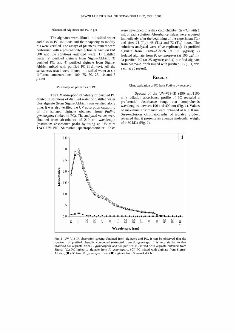
Spectra of the UV-VIS-IR (190 nm/1100 nm) radiation absorbance profile of PC revealed a preferential absorbance range that comprehends wavelengths between 190 and 400 nm (Fig. 1). Values of maximum absorbance were obtained at ≅ 210 nm. Size-exclusion chromatography of isolated product revealed that it presents an average molecular weight of ≅ 30 kDa (Fig. 2).
Fig. 1. UV-VIS-IR absorption spectra obtained from alginates and PC. It can be observed that the spectrum of purified phenolic compound (extracted from P. gymnospora) is very similar to that observed for alginate from P. gymnospora and for purified PC mixed with alginate obtained from Sigma. ( ) PC linked to alginate from P. gymnospora, ( ) PC mixed with alginate from Sigma-Aldrich, ( ) PC from P. gymnospora, and ( ) alginate from Sigma-Aldrich.
BRAZILIAN JOURNAL OF OCEANOGRAPHY, 55(2), 2007
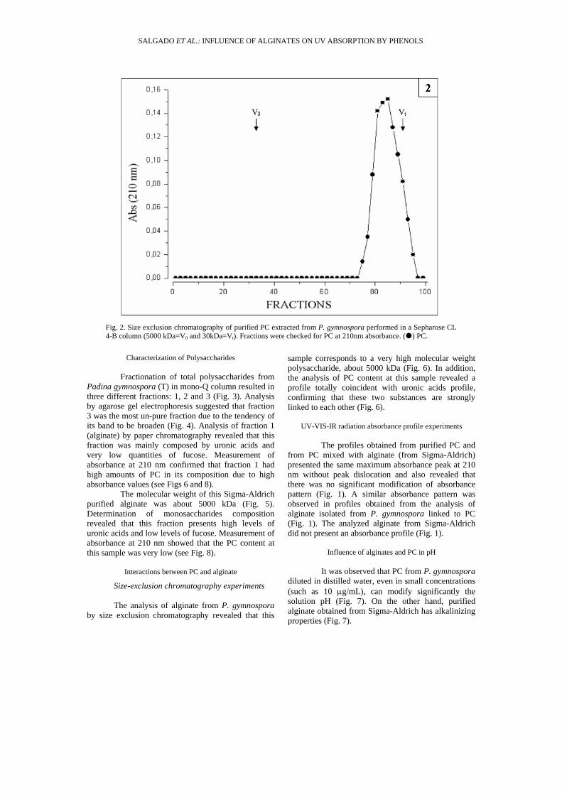
Fig. 2. Size exclusion chromatography of purified PC extracted from P. gymnospora performed in a Sepharose CL 4-B column (5000 kDa=V0 and 30kDa=Vt). Fractions were checked for PC at 210nm absorbance. ( ) PC.
Characterization of Polysaccharides
Fractionation of total polysaccharides from Padina gymnospora (T) in mono-Q column resulted in three different fractions: 1, 2 and 3 (Fig. 3). Analysis by agarose gel electrophoresis suggested that fraction 3 was the most un-pure fraction due to the tendency of its band to be broaden (Fig. 4). Analysis of fraction 1 (alginate) by paper chromatography revealed that this fraction was mainly composed by uronic acids and very low quantities of fucose. Measurement of absorbance at 210 nm confirmed that fraction 1 had high amounts of PC in its composition due to high absorbance values (see Figs 6 and 8).
The molecular weight of this Sigma-Aldrich purified alginate was about 5000 kDa (Fig. 5). Determination of monosaccharides composition revealed that this fraction presents high levels of uronic acids and low levels of fucose. Measurement of absorbance at 210 nm showed that the PC content at this sample was very low (see Fig. 8).
Interactions between PC and alginate
Size-exclusion chromatography experiments
The analysis of alginate from P. gymnospora by size exclusion chromatography revealed that this
sample corresponds to a very high molecular weight polysaccharide, about 5000 kDa (Fig. 6). In addition, the analysis of PC content at this sample revealed a profile totally coincident with uronic acids profile, confirming that these two substances are strongly linked to each other (Fig. 6).
UV-VIS-IR radiation absorbance profile experiments
The profiles obtained from purified PC and from PC mixed with alginate (from Sigma-Aldrich) presented the same maximum absorbance peak at 210 nm without peak dislocation and also revealed that there was no significant modification of absorbance pattern (Fig. 1). A similar absorbance pattern was observed in profiles obtained from the analysis of alginate isolated from P. gymnospora linked to PC (Fig. 1). The analyzed alginate from Sigma-Aldrich did not present an absorbance profile (Fig. 1).
Influence of alginates and PC in pH
It was observed that PC from P. gymnospora diluted in distilled water, even in small concentrations (such as 10 µg/mL), can modify significantly the solution pH (Fig. 7). On the other hand, purified alginate obtained from Sigma-Aldrich has alkalinizing properties (Fig. 7).
SALGADO ET AL.: INFLUENCE OF ALGINATES ON UV ABSORPTION BY PHENOLS
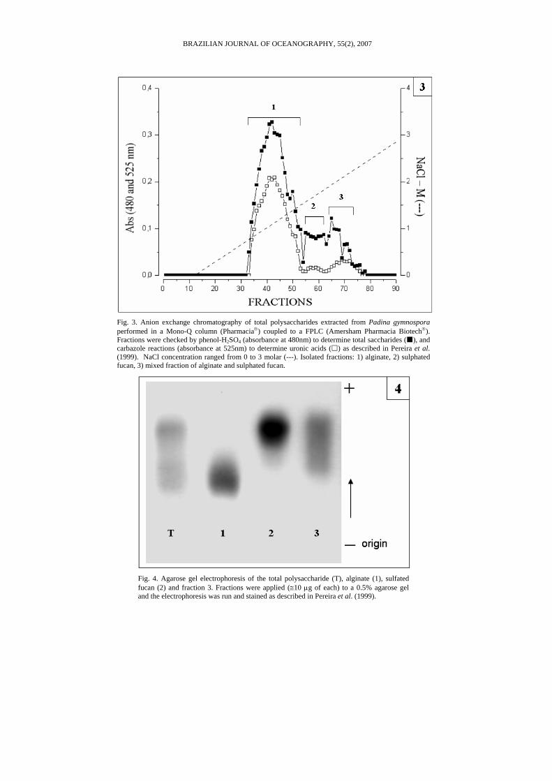
Fig. 3. Anion exchange chromatography of total polysaccharides extracted from Padina gymnospora performed in a Mono-Q column (Pharmacia®) coupled to a FPLC (Amersham Pharmacia Biotech®). Fractions were checked by phenol-H2SO4 (absorbance at 480nm) to determine total saccharides ( ), and carbazole reactions (absorbance at 525nm) to determine uronic acids ( ) as described in Pereira et al. (1999). NaCl concentration ranged from 0 to 3 molar (---). Isolated fractions: 1) alginate, 2) sulphated fucan, 3) mixed fraction of alginate and sulphated fucan.
Fig. 4. Agarose gel electrophoresis of the total polysaccharide (T), alginate (1), sulfated fucan (2) and fraction 3. Fractions were applied (≅10 µg of each) to a 0.5% agarose gel and the electrophoresis was run and stained as described in Pereira et al. (1999).
BRAZILIAN JOURNAL OF OCEANOGRAPHY, 55(2), 2007
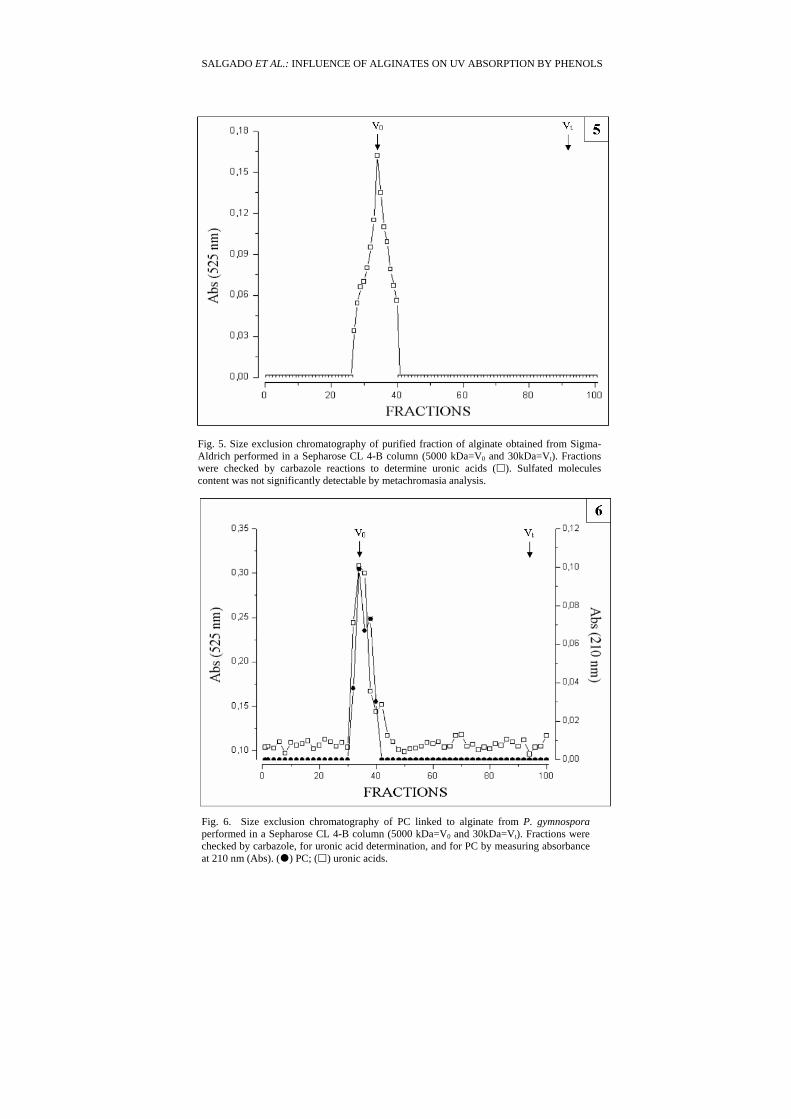
Fig. 5. Size exclusion chromatography of purified fraction of alginate obtained from Sigma-Aldrich performed in a Sepharose CL 4-B column (5000 kDa=V0 and 30kDa=Vt). Fractions were checked by carbazole reactions to determine uronic acids ( ). Sulfated molecules content was not significantly detectable by metachromasia analysis.
Fig. 6. Size exclusion chromatography of PC linked to alginate from P. gymnospora performed in a Sepharose CL 4-B column (5000 kDa=V0 and 30kDa=Vt). Fractions were checked by carbazole, for uronic acid determination, and for PC by measuring absorbance at 210 nm (Abs). ( ) PC; ( ) uronic acids.
SALGADO ET AL.: INFLUENCE OF ALGINATES ON UV ABSORPTION BY PHENOLS
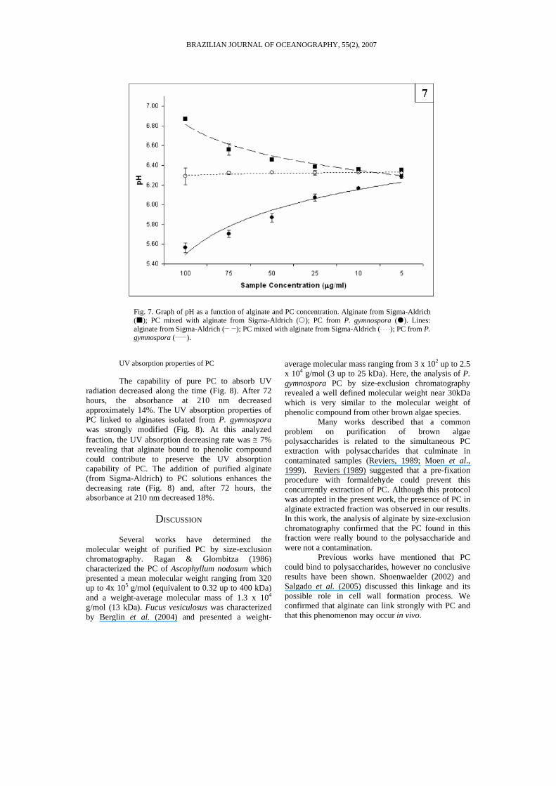
Fig. 7. Graph of pH as a function of alginate and PC concentration. Alginate from Sigma-Aldrich ( ); PC mixed with alginate from Sigma-Aldrich ( ); PC from P. gymnospora ( ). Lines: alginate from Sigma-Aldrich (__ __); PC mixed with alginate from Sigma-Aldrich (¨ ¨ ¨ ¨); PC from P. gymnospora (_____).
UV absorption properties of PC
The capability of pure PC to absorb UV radiation decreased along the time (Fig. 8). After 72 hours, the absorbance at 210 nm decreased approximately 14%. The UV absorption properties of PC linked to alginates isolated from P. gymnospora was strongly modified (Fig. 8). At this analyzed fraction, the UV absorption decreasing rate was ≅ 7% revealing that alginate bound to phenolic compound could contribute to preserve the UV absorption capability of PC. The addition of purified alginate (from Sigma-Aldrich) to PC solutions enhances the decreasing rate (Fig. 8) and, after 72 hours, the absorbance at 210 nm decreased 18%.
DISCUSSION
Several works have determined the molecular weight of purified PC by size-exclusion chromatography. Ragan & Glombitza (1986) characterized the PC of Ascophyllum nodosum which presented a mean molecular weight ranging from 320 up to 4x 10⁵ g/mol (equivalent to 0.32 up to 400 kDa) and a weight-average molecular mass of 1.3 x 10⁴ g/mol (13 kDa). Fucus vesiculosus was characterized by Berglin et al. (2004) and presented a weight-
average molecular mass ranging from 3 x 10² up to 2.5 x 10⁴ g/mol (3 up to 25 kDa). Here, the analysis of P. gymnospora PC by size-exclusion chromatography revealed a well defined molecular weight near 30kDa which is very similar to the molecular weight of phenolic compound from other brown algae species.
Many works described that a common problem on purification of brown algae polysaccharides is related to the simultaneous PC extraction with polysaccharides that culminate in contaminated samples (Reviers, 1989; Moen et al., 1999). Reviers (1989) suggested that a pre-fixation procedure with formaldehyde could prevent this concurrently extraction of PC. Although this protocol was adopted in the present work, the presence of PC in alginate extracted fraction was observed in our results. In this work, the analysis of alginate by size-exclusion chromatography confirmed that the PC found in this fraction were really bound to the polysaccharide and were not a contamination.
Previous works have mentioned that PC could bind to polysaccharides, however no conclusive results have been shown. Shoenwaelder (2002) and Salgado et al. (2005) discussed this linkage and its possible role in cell wall formation process. We confirmed that alginate can link strongly with PC and that this phenomenon may occur in vivo.
BRAZILIAN JOURNAL OF OCEANOGRAPHY, 55(2), 2007
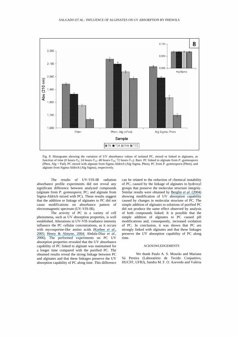
Fig. 8. Histograms showing the variation of UV absorbance values of isolated PC, mixed or linked to alginates, as function of time (0 hours-T0; 24 hours-T24; 48 hours-T48; 72 hours-T72). Bars: PC linked to alginate from P. gymnospora (Phen, Alg + Pad), PC mixed with alginate from Sigma-Aldrich (Alg Sigma, Phen), PC from P. gymnospora (Phen), and alginate from Sigma-Aldrich (Alg Sigma), respectively.
The results of UV-VIS-IR radiation absorbance profile experiments did not reveal any significant difference between analyzed compounds (alginate from P. gymnospora; PC; and alginate from Sigma-Aldrich mixed with PC). These results suggest that the addition or linkage of alginates to PC did not cause modifications on absorbance pattern of electromagnetic spectrum (UV-VIS-IR).
The activity of PC in a variety of cell phenomena, such as UV absorption properties, is well established. Alterations in UV-VIS irradiance intensity influence the PC cellular concentrations, as it occurs with mycosporine-like amino acids (Korbee et al., 2005; Henry & Alstyne, 2004; Abdala-Díaz et al., 2006). The performed experiments on PC UV absorption properties revealed that the UV absorbance capability of PC linked to alginate was maintained for a longer time compared with the purified PC. The obtained results reveal the strong linkage between PC and alginates and that these linkages preserve the UV absorption capability of PC along time. This difference
can be related to the reduction of chemical instability of PC, caused by the linkage of alginates to hydroxyl groups that preserve the molecular structure integrity. Similar results were obtained by Berglin et al. (2004) showing modification of UV absorption capability caused by changes in molecular structure of PC. The simple addition of alginates to solutions of purified PC did not produce the same effect observed by analysis of both compounds linked. It is possible that the simple addition of alginates to PC caused pH modifications and, consequently, increased oxidation of PC. In conclusion, it was shown that PC are strongly linked with alginates and that these linkages preserve the UV absorption capability of PC along time.
ACKNOWLEDGEMENTS
We thank Paulo A. S. Mourão and Mariana Sá Pereira (Laboratório de Tecido Conjuntivo, HUCFF, UFRJ), Sandra M. F. O. Azevedo and Valéria
8
SALGADO ET AL.: INFLUENCE OF ALGINATES ON UV ABSORPTION BY PHENOLS
F. Magalhães (Laboratório de Ecofisiologia e Toxicologia de Cianobactérias, IBCCF, UFRJ) for laboratory facilities. This study is part of the PhD thesis developed by L. T. Salgado at UFRJ, Brazil. Financial support: CNPq and FAPERJ to G. M. Amado Filho and M. Farina.
REFERENCES Abdala-Díaz, R. T.; Cabello-Pasini, A.; Pérez-Rodríguez, E.;
Conde Álvarez, R. M.C. & Figueroa, F. L.. 2006. Daily and seasonal variations of optimum quantum yield and PC in Cystoseira tamariscifolia (Phaeophyta). Mar. Biol., 148:459-465.
Andrade, L. R.; Salgado, L. T.; Farina, M.; Pereira, M. S.; Mourão, P. A. S. & Amado Filho, G. M. 2004. Ultrastructure of acidic polysaccharides from the cell walls of brown algae. J. Struct. Biol., 145:216-225.
Arnold, T. M. & Targett, N. M. 2003. To grow and defend: Lack of tradeoffs for brown algae phlorotannins. Oikos, 100 (2):406-408.
Arroniz-Crespo, M.; Sinha, R. P.; Martinez-Abaigar, J.; Nunez-Olivera, E. & Hader, D. P. 2005. Ultraviolet radiation-induced changes in mycosporine-like amino acids and physiological variables in the red alga Lemanea fluviatilis. J. Freshwat. Ecol., 20 (4):677-687.
Berglin, M.; Delage, L.; Potin, P.; Vilter, H. & Elwing, H. 2004. Enzymatic cross-linking of a phenolic polymer extracted from the marine alga Fucus serratus. Biomacromolecules, 5:2376-2383.
Ceh, J.; Molis, M.; Dzeha, T.M. & Wahl, M. 2005. Induction and reduction of anti-herbivore defenses in brown and red macroalgae of the Kenyan coast. J. Phycol., 41:726–731.
Diffey, B. L. 2002. Sources and measurement of ultraviolet radiation. Methods, 28:4-13.
Franklin, L. A. & Forster, R. M. 1997. The changing irradiance environment: consequences for marine macrophyte physiology, productivity and ecology. Eur. J. Phycol., 32:207-232.
Häder, D. P.; Kumar, H. D.; Smith, R. C. & Worrest, R. C. 1998. Effects on aquatic ecosystems. J. Photochem. Photobiol. B, 46:53–68.
Hanelt, D.; Wiencke, C. & Nultsch, W. 1997. Influence of UV-radiation on the photosynthesis of artic macroalgae in the field. J. Photochem. Photobiol. B, 38:40-47.
Henry, B. E. & Alstyne, K. L. V. 2004. Effects of UV radiation on growth and phlorotannins in Fucus gardneri (Phaeophyceae) juveniles and embryos. J. Phycol., 40:527–533.
Holzinger, A. & Lütz, C. 2006. Algae and UV irradiation: Effects on ultrastructure and related metabolic functions. Micron, 37:190–207.
Karez, C. S. & Pereira, R. C. 1995. Metal contents in polyphenolic fractions extracted from the brown alga Padina gymnospora. Botanica Mar., 38:151-155.
Kloareg, B. & Quatrano, R. S. 1988. Structure of the cell walls of marine algae and ecophysiological functions of the matrix polysaccharides. Oceanogr. Mar. Biol. A. Rev., 26:259-315.
Koivikko, R.; Loponen, J.; Honkanen, T. & Jormalainen, V. 2005. Contents of soluble, cell-wall-bound and exuded phlorotannins in the brown alga Fucus vesiculosus, with implication on their ecological functions. J. Chem. Ecol., 31 (1):195-212.
Korbee, N.; Figueroa, F. L. & Aguilera, J. 2005. Effect of light quality on the accumulation of photosynthetic pigments, proteins and mycosporine-like amino acids in the red alga Porphyra leucosticta (Bangiales, Rhodophyta). J. Photochem. Photobiol. B, 80:71–78.
Madronich, S.; McKenzie, R. L.; Bjorn, L. O. & Caldwell, M. M. 1998. Changes in biologically active ultraviolet radiation reaching the Earth's surface. J. Photochem. Photobiol. B, 46(1-3):5-19.
Moen, E.; Larsen, B.; Østgaard, K. & Jensen, A. 1999. Alginate stability during high salt preservation of Ascophyllum nodosom. J. Appl. Phycol., 11:21-25.
Pavia, H.; Cervin, G.; Lindgren, A. & Ålberg, P. 1997. Effects of UV-B radiation and simulated herbivory on phlorotannins in the brown alga Ascophyllum nodosom. Mar. Ecol. Prog. Ser., 157:139-156.
Ragan, M. A. & Glombitza, K. W. 1986. Phlorotannins, brown algal polyphenols. Progress Phycol. Res., 4:129-241.
Reviers, B. 1989. Fucans and alginates without PC. J. Appl. Phycol., 1:75-76.
Salgado, L. T.; Andrade, L. R. & Amado Filho, G. M. 2005. Localisation of specific monosaccharides in cells of the brown alga Padina gymnospora and the relation to heavy metal accumulation. Protoplasma, 225:123-128.
Shoenwaelder, M. 1996. The distribution and secretion of PC in the early development of Acrocarpia paniculata and Hormosira Banksii (Phaeophyceae). Ph.D. Thesis. Monash University, Australia. 117 p.
Shoenwaelder, M. 2002. Phycological Reviews 21. The occurrence and cellular significance of physodes in the brown algae. Phycologia, 41(2):125-139.
Vreeland, V. & Laetsch, W. M. 1988. Role of alginate self-associating subunits in the assembly of Fucus embryo cell walls. In: Varner J.E., ed. Self assembling architecture. New York, Alan R. Liss, p. 77-96.
(Manuscript received 09 June 2006; revised 06 November 2006; accepted 27 February 2007)
BRAZILIAN JOURNAL OF OCEANOGRAPHY, 55(2), 2007