
The hibernating South American marsupial, Dromiciops ...
11
1 SCIENTIFIC REPORTS | 6:24627 | DOI: 10.1038/srep24627 www.nature.com/scientificreports The hibernating South American marsupial, Dromiciops gliroides, displays torpor-sensitive microRNA expression patterns Hanane Hadj-Moussa 1,* , Jason A. Moggridge 1,* , Bryan E. Luu 1 , Julian F. Quintero-Galvis 2 , Juan Diego Gaitán-Espitia 3 , Roberto F. Nespolo 2 & Kenneth B. Storey 1 When faced with adverse environmental conditions, the marsupial Dromiciops gliroides uses either daily or seasonal torpor to support survival and is the only known hibernating mammal in South America. As the sole living representative of the ancient Order Microbiotheria, this species can provide crucial information about the evolutionary origins and biochemical mechanisms of hibernation. Hibernation is a complex energy-saving strategy that involves changes in gene expression that are elicited in part by microRNAs. To better elucidate the role of microRNAs in orchestrating hypometabolism, a modified stem-loop technique and quantitative PCR were used to characterize the relative expression levels of 85 microRNAs in liver and skeletal muscle of control and torpid D. gliroides. Thirty-nine microRNAs were differentially regulated during torpor; of these, 35 were downregulated in liver and 11 were differentially expressed in skeletal muscle. Bioinformatic analysis predicted that the downregulated liver microRNAs were associated with activation of MAPK, PI3K-Akt and mTOR pathways, suggesting their importance in facilitating marsupial torpor. In skeletal muscle, hibernation-responsive microRNAs were predicted to regulate focal adhesion, ErbB, and mTOR pathways, indicating a promotion of muscle maintenance mechanisms. These tissue-specific responses suggest that microRNAs regulate key molecular pathways that facilitate hibernation, thermoregulation, and prevention of muscle disuse atrophy. To survive in challenging environments, many animals employ metabolic rate depression to strongly reduce their energy needs and extend the time that they can live using only internal fuel reserves. Survival strategies including daily torpor, hibernation, estivation, anaerobiosis, and freeze tolerance all make use of hypometabolism 1,2 . Many mammalian species use hibernation to endure seasonal periods of cold exposure and food scarcity; indeed, hiber- nation has been identified in selected species from eleven different mammalian orders including, monotremes, marsupials, rodents, bats and others 3,4 . Hibernation is an energy-saving strategy that involves a seasonal retreat into cycles of prolonged torpor where metabolic rate and core body temperatures are depressed 5–7 . Hibernators with small body masses can save 40–60% of their total daily energy needs by maintaining a decreased core tem- perature, compared with the high thermoregulatory costs of continuous endothermy 8 . e temperate rainforests of Chile are home to the only known South American hibernator and the last living representative of the Microbiotheria order, the monito del monte (Dromiciops gliroides) 8,9 . is cold-adapted mar- supial uses both daily torpor and multi-day hibernation as an opportunistic response to stress, where the duration and depth of torpor bouts are proportional to the intensity of cold exposure and food scarcity 8,10 . Bioenergetic studies have described the physiological features of D. gliroides hibernation; for example, when ambient temper- ature decreased to 12.5 °C, deep torpor bouts were found to last for five days and during this period core body temperatures fell to near ambient levels, metabolic rate dropped to 1–5% of the euthermic rate, and respiratory frequency fell from an average of 370 to 2 breaths per min with extended periods of apnea 6,8,11 . roughout the 1 Department of Biology and Institute of Biochemistry, Carleton University, 1125 Colonel By Drive, Ottawa, Ontario K1S 5B6, Canada. 2 Instituto de Ciencias Ambientales y Evolutivas, Facultad de Ciencias, Universidad Austral de Chile, Campus Isla Teja, Valdivia, Chile. 3 CSIRO Oceans & Atmosphere, GPO Box 1538, Hobart 7001, TAS, Australia. * These authors contributed equally to this work. Correspondence and requests for materials should be addressed to K.B.S. (email: [email protected]) Received: 08 January 2016 Accepted: 31 March 2016 Published: 19 April 2016 OPEN
-
Upload
khangminh22 -
Category
Documents
-
view
0 -
download
0
Transcript of The hibernating South American marsupial, Dromiciops ...
1Scientific RepoRts | 6:24627 | DOI: 10.1038/srep24627
www.nature.com/scientificreports
The hibernating South American marsupial, Dromiciops gliroides, displays torpor-sensitive microRNA expression patternsHanane Hadj-Moussa1,*, Jason A. Moggridge1,*, Bryan E. Luu1, Julian F. Quintero-Galvis2, Juan Diego Gaitán-Espitia3, Roberto F. Nespolo2 & Kenneth B. Storey1
When faced with adverse environmental conditions, the marsupial Dromiciops gliroides uses either daily or seasonal torpor to support survival and is the only known hibernating mammal in South America. As the sole living representative of the ancient Order Microbiotheria, this species can provide crucial information about the evolutionary origins and biochemical mechanisms of hibernation. Hibernation is a complex energy-saving strategy that involves changes in gene expression that are elicited in part by microRNAs. To better elucidate the role of microRNAs in orchestrating hypometabolism, a modified stem-loop technique and quantitative PCR were used to characterize the relative expression levels of 85 microRNAs in liver and skeletal muscle of control and torpid D. gliroides. Thirty-nine microRNAs were differentially regulated during torpor; of these, 35 were downregulated in liver and 11 were differentially expressed in skeletal muscle. Bioinformatic analysis predicted that the downregulated liver microRNAs were associated with activation of MAPK, PI3K-Akt and mTOR pathways, suggesting their importance in facilitating marsupial torpor. In skeletal muscle, hibernation-responsive microRNAs were predicted to regulate focal adhesion, ErbB, and mTOR pathways, indicating a promotion of muscle maintenance mechanisms. These tissue-specific responses suggest that microRNAs regulate key molecular pathways that facilitate hibernation, thermoregulation, and prevention of muscle disuse atrophy.
To survive in challenging environments, many animals employ metabolic rate depression to strongly reduce their energy needs and extend the time that they can live using only internal fuel reserves. Survival strategies including daily torpor, hibernation, estivation, anaerobiosis, and freeze tolerance all make use of hypometabolism1,2. Many mammalian species use hibernation to endure seasonal periods of cold exposure and food scarcity; indeed, hiber-nation has been identified in selected species from eleven different mammalian orders including, monotremes, marsupials, rodents, bats and others3,4. Hibernation is an energy-saving strategy that involves a seasonal retreat into cycles of prolonged torpor where metabolic rate and core body temperatures are depressed5–7. Hibernators with small body masses can save 40–60% of their total daily energy needs by maintaining a decreased core tem-perature, compared with the high thermoregulatory costs of continuous endothermy8.
The temperate rainforests of Chile are home to the only known South American hibernator and the last living representative of the Microbiotheria order, the monito del monte (Dromiciops gliroides)8,9. This cold-adapted mar-supial uses both daily torpor and multi-day hibernation as an opportunistic response to stress, where the duration and depth of torpor bouts are proportional to the intensity of cold exposure and food scarcity8,10. Bioenergetic studies have described the physiological features of D. gliroides hibernation; for example, when ambient temper-ature decreased to 12.5 °C, deep torpor bouts were found to last for five days and during this period core body temperatures fell to near ambient levels, metabolic rate dropped to 1–5% of the euthermic rate, and respiratory frequency fell from an average of 370 to 2 breaths per min with extended periods of apnea6,8,11. Throughout the
1Department of Biology and Institute of Biochemistry, Carleton University, 1125 Colonel By Drive, Ottawa, Ontario K1S 5B6, Canada. 2Instituto de Ciencias Ambientales y Evolutivas, Facultad de Ciencias, Universidad Austral de Chile, Campus Isla Teja, Valdivia, Chile. 3CSIRO Oceans & Atmosphere, GPO Box 1538, Hobart 7001, TAS, Australia. *These authors contributed equally to this work. Correspondence and requests for materials should be addressed to K.B.S. (email: [email protected])
Received: 08 January 2016
Accepted: 31 March 2016
Published: 19 April 2016
OPEN
www.nature.com/scientificreports/
2Scientific RepoRts | 6:24627 | DOI: 10.1038/srep24627
year, when faced with less extreme environmental conditions, the nocturnal D. gliroides employs a different strat-egy, retreating daily into shallow torpor to manage energy expenditure and mass balance11 During these periods of shallow torpor, body temperature decreases to 11–28 °C for a few hours, and metabolism is suppressed to 10–60% of basal rate8,11.
A molecular hallmark of torpor is the reprioritization of ATP consumption through the suppression of ener-getically costly cellular metabolic processes such as gene transcription and translation. Yet, despite the observed global suppression of metabolism, selected genes that facilitate the transition between torpor/arousal states, pro-mote the torpid state, or enhance pro-survival pathways have been found to be overexpressed during hiber-nation12. Apart from these metabolic changes that facilitate hypometabolism in all organs, selected organs can have specific roles to play during torpor/arousal. For example, skeletal muscle is crucial for arousal from torpor/hibernation since it provides shivering thermogenesis to help rewarm the animal. Therefore, it is important for D. gliroides to maintain muscle mass and function despite prolonged inactivity and, indeed, other mammalian hibernators show little or no sign of disuse atrophy during hibernation13,14. In eutherian mammals, thermogenesis during arousal also strongly depends on the action of brown adipose tissue but the presence of this heat gener-ating organ has yet to be confirmed in marsupials. Detecting the occurrence of brown adipose mediated adap-tive non-shivering thermogenesis in marsupials has yielded controversial results, where thermogenic responses were found in some marsupials15–18, while no such findings were observed in others19. Moreover, evidence of the morphological and molecular characteristics of brown adipose in marsupials are inconclusive17,20. Although orthologues of the three uncoupling proteins, evolutionary markers for brown fat, have been identified in a few marsupial genomes21,22, upon characterization it was found that they may not be contributing to classical non-shivering thermogenesis, and may have evolved different roles that have yet to be elucidated23. Therefore, if marsupials indeed lack brown adipose they must have evolved an alternative heat generating mechanism such as changes to liver metabolism that may be contributing to this function. Villarin et al.24 reported that sustained cold exposure elevated liver oxidative capacity in Monodelphis domestica (the North American opossum); liver mass increased by ~50% and liver mitochondrial volume rose ~20%24. These changes could account for nearly half of the increase in animal oxygen consumption at low temperatures suggesting that the liver has the potential to con-tribute significantly to the thermogenic efforts for marsupial life in the cold. Hence, these results, along with other studies, suggest that liver metabolic processes are actively modified to contribute to the heat production necessary for marsupial survival throughout torpor/hibernation5,18,25. Despite the differences in thermogenic mechanisms, normothermic body temperatures, and basal metabolic rates between marsupials and eutherian mammals, there appears to be no significant difference between heat production rates, suggesting that marsupials are able to warm-up as fast as eutherians despite a potential absence of brown adipose tissue26.
A complex balance of gene activation and suppression is needed to achieve hypometabolism and this can be accomplished by various molecular mechanisms, including reversible protein phosphorylation of transcrip-tion and translation factors, the use of epigenetic mechanisms to directly modify DNA and histone proteins, as well as post-transcriptional controls on mRNA through the action of microRNAs (miRNAs)2. MiRNAs are proving to be master regulators of virtually all cell functions with broad controls over many activities includ-ing the cell cycle, signal transduction pathways, and energy metabolism, among others27,28. While this large group of highly-conserved, short, non-coding RNA transcripts (~22 nt) comprises just 1–2% of the number of protein-coding genes, recent computational predictions indicate that they target more than 60% of protein-coding genes in humans, and influence almost every aspect of biological function29,30. MiRNAs are initially transcribed mostly by RNA polymerase II into long (~70 nt) secondary hairpin structures known as pri-miRNAs. The 5′ and 3′ ends are cleaved by the enzymes Drosha and DCGR8 to form a pre-miRNA, which is subsequently exported to the cytoplasm. At this stage, the stem-loop is cleaved by Dicer to create a ds-miRNA duplex and, from this, the guide strand is then incorporated into the miRNA-induced silencing complex (miRISC)31. The 5′ seed region of the guide strand, involving nucleotides two to eight, directs RISC activity towards target mRNAs with com-plementary motifs in their 3′ UTR32. The brevity of the seed region is what allows miRNAs to exert their broad controls, wherein a single miRNA can regulate multiple mRNA transcripts, and one mRNA transcript may be subject to regulation by multiple miRNAs31. Once bound, the degree of complementarity between the miRNA and mRNA transcript dictates the fate of the mRNA transcript, providing another level of control. Imperfect complementarity causes mRNA transcripts to be sequestered to cytoplasmic loci such as P-bodies and stress granules, where they are subject to translational repression; in contrast, perfect complementarity triggers mRNA degradation by Argonaute endonucleases31.
A growing number of studies by our lab, and others, have demonstrated the essential role of miRNA differ-ential expression in modulating global gene suppression and coordinating the up-regulation of selected genes in response to torpor, estivation, anaerobiosis, and freezing27,33–41. To our knowledge, the present study is the first to investigate miRNA expression in relation to torpor in a marsupial species. Eighty-five known miRNAs were quantified in the liver and skeletal muscle of control versus torpid D. gliroides using RT-qPCR. Genome align-ments confirmed that the miRNAs analyzed were highly conserved as compared with other vertebrates. MiRNA expression was found to be differentially regulated in a tissue-specific manner, with significant suppression of many miRNA species observed in liver and the differential regulation of a subset of miRNAs in skeletal muscle. Bioinformatic target enrichment analyses suggested that the differentially expressed miRNAs may play a crucial role in facilitating hibernation and thermoregulation through the regulation of MAPK, mTOR, and PI3K-Akt signalling in liver. The skeletal muscle-specific miRNA expression profile was predicted to regulate the mainte-nance of muscle mass and function through the targeting and induction of the focal adhesion, ErbB, and mTOR signalling pathways during marsupial torpor. Understanding the role that miRNAs play as central regulators of metabolic reorganization will provide valuable insights into the molecular underpinnings of adaptation to envi-ronmental stress.
www.nature.com/scientificreports/
3Scientific RepoRts | 6:24627 | DOI: 10.1038/srep24627
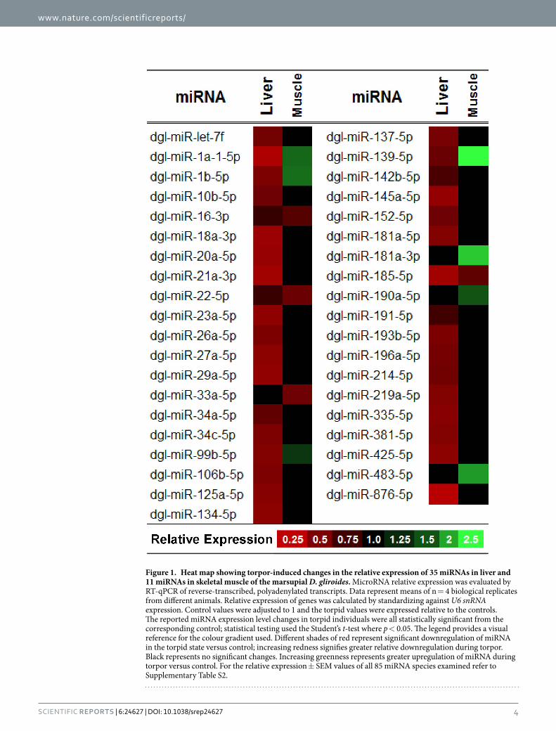
ResultsDifferential expression of miRNA in liver and skeletal muscle during torpor. The primary goal of this study was to characterize hibernation-specific patterns of miRNA expression in liver and skeletal muscle of the marsupial D. gliroides. Of the 85 miRNAs successfully quantified by RT-qPCR, 35 miRNAs were significantly down-regulated in liver during torpor, with miRNA relative abundances decreasing by 0.21–0.72 fold change from the control condition (p < 0.05; Supplementary Table S2 and Fig. 1). A different pattern was observed in skeletal muscle, where seven miRNAs showed elevated expression during torpor, increasing by 0.32–1.52 fold change from control levels (dgl-miR-1a-1-5p, dgl-miR-1b-5p, dgl-miR-139-5p, dgl-miR-181a-3p, dgl-miR-190a-5p, dgl-miR-483-5p, and dgl-miR-99b-5p), whereas, 4 miRNAs showed decreased expression of 0.33-0.43 fold change compared to control (dgl-miR-16-1-3p, dgl-miR-185-5p, dgl-miR-22-5p, and dgl-miR-33a-5p) (p < 0.05; Supplementary Table S2 and Fig. 1). Of these, three miRNAs showed a pattern of decreased expres-sion that was consistent in both tissues during torpor (dgl-miR-16-3p, dgl-miR-22-5p, and dgl-miR-185-5p). In contrast, four miRNAs (dgl-miR-1a1-5p, dgl-miR-1b-5p, dgl-miR-99b-5p, and dgl-miR-139-5p) showed a torpor-specific increase in skeletal muscle, but with a concomitant decrease in liver tissue.
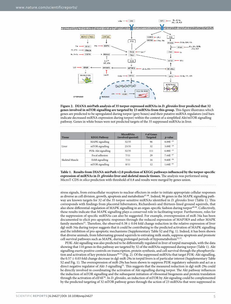
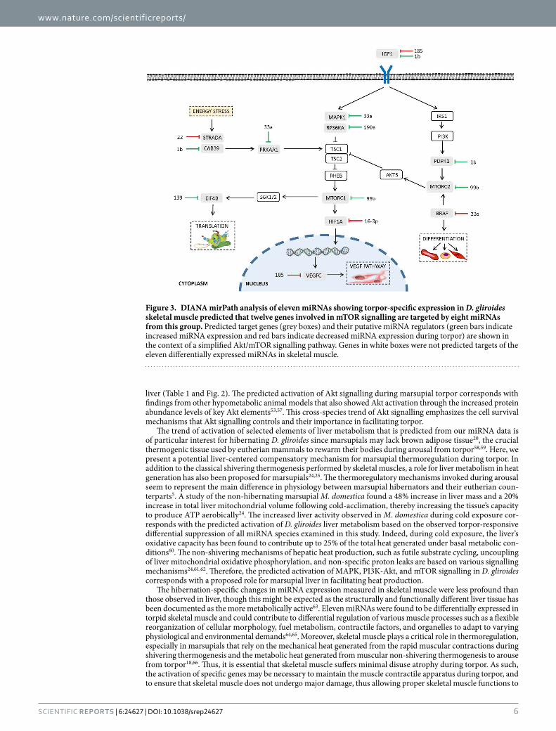
Bioinformatic analyses of pathways regulated by miRNA during torpor. MicroRNAs with altered expression during torpor in D. gliroides liver and skeletal muscle were subjected to bioinformatic target enrich-ment analyses using DIANA mirPath v2.0. This program identifies the potential KEGG pathways that are collec-tively targeted by the query miRNAs. DIANA mirPath indicated that decreased expression of miRNAs in liver of torpid animals were most closely associated with three signalling pathways [1] MAPK signalling (p = 8.09E−25), with 96 genes thought to be targeted by 32 of the 35 down-regulated miRNAs; [2] mTOR signalling (p = 3.09E−20), with 32 genes targeted by 23 of 35 torpor-sensitive miRNAs (Fig. 2); and [3] PI3K/Akt signalling (p = 7.44E−07), with 110 genes targeted by 33 of 35 differentially expressed miRNAs (Table 1, Fig. 2). In skeletal muscle, eleven torpor-associated miRNAs were linked to pathways of [1] focal adhesion (p = 7.44E−07), with 29 genes targeted by seven miRNAs; [2] ErbB signalling (p = 9.60E−06), with sixteen genes targeted by seven miRNAs; and [3] mTOR signalling (p = 1.64E−05), involving twelve genes and eight miRNAs (Table 1 and Fig. 3).
Conservation of miRNAs across vertebrate taxa. To provide support for the conservation of these miRNAs in D. gliroides, a species that is not genome-sequenced, multiple sequence alignments were performed on orthologous pre-miRNA sequences from M. musculus, M. domestica, and G. gallus. In cases where the sequences from these species were not annotated in NCBI GenBank, the alignments were instead performed using sequences from Homo sapiens, Bos taurus, Ornithorhynchus anatinus, Eptesicus fuscus, Equus caballus, Ovis aries, Sus scofa, or Danio rerio. Alignments were assessed for percent identity of the mature miRNA sequence; only miRNAs with a high degree of conservation in the mature miRNA sequence were quantified in the D. gliroides samples. Of the 85 miRNAs analyzed in this study, 50 showed 100% conservation in the mature sequence across taxa, 18 showed at least 90% identity, ten miRNAs had between 80–89% identity, and a small subset of seven mature miRNAs displayed between 70–80% identity across distant species.
DiscussionThe marsupial D. gliroides has evolved behavioural, physiological, and biochemical adaptations that facilitate entry into a hypometabolic state during periods of cold exposure and food deprivation11. Various studies have examined the ecological, phylogenetic, and physiological nature of D. gliroides hypometabolism, but little work has gone into characterizing the biochemical and molecular adaptations of its hibernation response11,42–44. The phenomenon of hibernation involves the intricate coordination of various biological functions to conserve energy, mainly through the strong depression of cellular ATP-expensive processes such as gene transcription, protein synthesis, transmembrane ion pumping, and others2. Recent findings have identified miRNAs as key orchestrators of these cellular adaptations via their actions in inhibiting mRNA translation and/or directing mRNA transcripts into degradation28,45. The nature of miRNAs makes them excellent master cellular regulators of stress responses: [1] they can exert broad controls over many different genes due to their target promiscuity and dynamic structures31, [2] they are easily inducible and readily reversible27, and [3] they can preserve mRNA transcripts by sequestering them into RISCs and P-bodies during hypometabolism and then make them availa-ble for translation upon arousal32. The present study aimed to expand our understanding of miRNA regulation of hibernation by examining the expression responses of 85 highly conserved miRNAs in the liver and skeletal muscle of D. gliroides. Although D. gliroides is not genome sequenced, miRNA analysis was possible because of the very high degree of evolutionary conservation of miRNAs across species, not only within mammals but across all vertebrates46. However, given the paucity of experimentally-validated miRNA-mRNA interactions, pathway analyses in this study necessitated the use of predicted miRNA interactions in mice from the algorithm-based target enrichment database, DIANA miRPath.
The liver is the body’s main metabolic center and the site of many important functions including ketogenesis, gluconeogenesis, processing of nutrients, detoxification reactions, and the synthesis of proteins that are secreted for use elsewhere in the body. D. gliroides liver showed significant decreases in the relative abundances of 35 miRNAs during torpor (Fig. 1). This strong miRNA suppression implies that during torpor there is an increased translation of a wide variety of mRNA transcripts that are targets of these miRNAs. It was predicted that the suppressed miRNAs in liver are involved in regulating genes of the MAPK, mTOR, and PI3K-Akt signalling path-ways, suggesting that these pathways become activated during marsupial torpor, and are necessary for regulating metabolic reorganization and proper hepatic function in the hypometabolic state (Table 1).
The regulation of MAPK and PI3K-Akt signalling pathways by miRNA could help to induce the metabolic reorganization required during torpor, since the protein kinase cascades of these pathways use reversible protein phosphorylation to induce rapid changes in many cellular functions. MAPK signal cascades relay stimuli, such as
www.nature.com/scientificreports/
4Scientific RepoRts | 6:24627 | DOI: 10.1038/srep24627
Figure 1. Heat map showing torpor-induced changes in the relative expression of 35 miRNAs in liver and 11 miRNAs in skeletal muscle of the marsupial D. gliroides. MicroRNA relative expression was evaluated by RT-qPCR of reverse-transcribed, polyadenylated transcripts. Data represent means of n = 4 biological replicates from different animals. Relative expression of genes was calculated by standardizing against U6 snRNA expression. Control values were adjusted to 1 and the torpid values were expressed relative to the controls. The reported miRNA expression level changes in torpid individuals were all statistically significant from the corresponding control; statistical testing used the Student’s t-test where p < 0.05. The legend provides a visual reference for the colour gradient used. Different shades of red represent significant downregulation of miRNA in the torpid state versus control; increasing redness signifies greater relative downregulation during torpor. Black represents no significant changes. Increasing greenness represents greater upregulation of miRNA during torpor versus control. For the relative expression ± SEM values of all 85 miRNA species examined refer to Supplementary Table S2.
www.nature.com/scientificreports/
5Scientific RepoRts | 6:24627 | DOI: 10.1038/srep24627
stress signals, from extracellular receptors to nuclear effectors in order to initiate appropriate cellular responses as diverse as cell division, growth, apoptosis and metabolism47,48. Indeed, 96 genes in the MAPK signalling path-way are known targets for 32 of the 35 torpor-sensitive miRNAs identified in D. gliroides liver (Table 1). This corresponds with findings from placental hibernators, Richardson’s and thirteen-lined ground squirrels, that also show differential-regulation of MAPK signalling in an organ-specific fashion during torpor49,50. Collectively, these results indicate that MAPK signalling plays a conserved role in facilitating torpor. Furthermore, roles for the suppression of specific miRNAs can also be suggested. For example, overexpression of miR-34a has been documented to elicit pro-apoptotic responses through the reduced expression of MAP3K9 and other MAPK family members51. Therefore, the observed 0.38 ± 0.04 fold change reduction in the relative expression of liver dgl-miR-34a during torpor suggests that it could be contributing to the predicted activation of MAPK signalling and the inhibition of pro-apoptotic mechanisms (Supplementary Table S2 and Fig. 1). Indeed, it has been shown that diverse animals, from hibernating ground squirrels to estivating milk snails, suppress apoptosis and promote cell survival pathways such as MAPK, during prolonged periods of hypometabolism52,53.
PI3K-Akt signalling was also predicted to be differentially regulated in liver of torpid marsupials, with the data showing that 110 genes in this pathway are targeted by 32 of the miRNAs suppressed during torpor (Table 1). Akt signalling exerts positive controls on transcription, protein synthesis, and cell survival through the phosphoryla-tion and activation of key protein kinases48,54 (Fig. 2). Of the suppressed miRNAs that target PI3K-Akt signalling, the 0.57 ± 0.03 fold change decrease in dgl-miR-29a in torpid livers is of particular interest (Supplementary Table S2 and Fig. 1). The overexpression of miR-29a has been shown to suppress PI3K regulatory subunits and act as a direct negative regulator of Akt-3 signalling55. This suggests that the measured reduction in dgl-miR-29a could be directly involved in coordinating the activation of Akt signalling during torpor. The Akt pathway influences the induction of mTOR signalling and the subsequent initiation of ribosomal biogenesis and protein translation through the activation of eIF4E56. In D. gliroides, an induction of mTOR by Akt signaling could be complemented by the predicted targeting of 32 mTOR pathway genes through the action of 23 miRNAs that were suppressed in
Figure 2. DIANA mirPath analysis of 35 torpor-repressed miRNAs in D. gliroides liver predicted that 32 genes involved in mTOR signalling are targeted by 23 miRNAs from this group. This figure illustrates which genes are predicted to be upregulated during torpor (grey boxes) and their putative miRNA regulators (red bars indicate decreased miRNA expression during torpor) within the context of a simplified Akt/mTOR signalling pathway. Genes in white boxes were not predicted targets of the 35 suppressed miRNAs in liver.
Tissue KEGG PathwayMicroRNAs
(involved/queried)# of Genes Targeted p-value
Liver
MAPK signalling 32/35 96 8.09E−25
mTOR signalling 23/35 32 3.09E−20
PI3k-Akt signalling 32/35 111 6.88E−15
Skeletal Muscle
Focal adhesion 7/11 29 7.44E−07
ErbB signalling 7/11 16 9.60E−06
mTOR signalling 8/11 12 1.64E−05
Table 1. Results from DIANA mirPath v2.0 prediction of KEGG pathways influenced by the torpor-specific expression of miRNAs in D. gliroides liver and skeletal muscle tissues. The analysis was performed using MicroT-CDS in silico prediction with threshold of 0.8 and results were merged by genes union.
www.nature.com/scientificreports/
6Scientific RepoRts | 6:24627 | DOI: 10.1038/srep24627
liver (Table 1 and Fig. 2). The predicted activation of Akt signalling during marsupial torpor corresponds with findings from other hypometabolic animal models that also showed Akt activation through the increased protein abundance levels of key Akt elements53,57. This cross-species trend of Akt signalling emphasizes the cell survival mechanisms that Akt signalling controls and their importance in facilitating torpor.
The trend of activation of selected elements of liver metabolism that is predicted from our miRNA data is of particular interest for hibernating D. gliroides since marsupials may lack brown adipose tissue20, the crucial thermogenic tissue used by eutherian mammals to rewarm their bodies during arousal from torpor58,59. Here, we present a potential liver-centered compensatory mechanism for marsupial thermoregulation during torpor. In addition to the classical shivering thermogenesis performed by skeletal muscles, a role for liver metabolism in heat generation has also been proposed for marsupials24,25. The thermoregulatory mechanisms invoked during arousal seem to represent the main difference in physiology between marsupial hibernators and their eutherian coun-terparts5. A study of the non-hibernating marsupial M. domestica found a 48% increase in liver mass and a 20% increase in total liver mitochondrial volume following cold-acclimation, thereby increasing the tissue’s capacity to produce ATP aerobically24. The increased liver activity observed in M. domestica during cold exposure cor-responds with the predicted activation of D. gliroides liver metabolism based on the observed torpor-responsive differential suppression of all miRNA species examined in this study. Indeed, during cold exposure, the liver’s oxidative capacity has been found to contribute up to 25% of the total heat generated under basal metabolic con-ditions60. The non-shivering mechanisms of hepatic heat production, such as futile substrate cycling, uncoupling of liver mitochondrial oxidative phosphorylation, and non-specific proton leaks are based on various signalling mechanisms24,61,62. Therefore, the predicted activation of MAPK, PI3K-Akt, and mTOR signalling in D. gliroides corresponds with a proposed role for marsupial liver in facilitating heat production.
The hibernation-specific changes in miRNA expression measured in skeletal muscle were less profound than those observed in liver, though this might be expected as the structurally and functionally different liver tissue has been documented as the more metabolically active63. Eleven miRNAs were found to be differentially expressed in torpid skeletal muscle and could contribute to differential regulation of various muscle processes such as a flexible reorganization of cellular morphology, fuel metabolism, contractile factors, and organelles to adapt to varying physiological and environmental demands64,65. Moreover, skeletal muscle plays a critical role in thermoregulation, especially in marsupials that rely on the mechanical heat generated from the rapid muscular contractions during shivering thermogenesis and the metabolic heat generated from muscular non-shivering thermogenesis to arouse from torpor18,66. Thus, it is essential that skeletal muscle suffers minimal disuse atrophy during torpor. As such, the activation of specific genes may be necessary to maintain the muscle contractile apparatus during torpor, and to ensure that skeletal muscle does not undergo major damage, thus allowing proper skeletal muscle functions to
Figure 3. DIANA mirPath analysis of eleven miRNAs showing torpor-specific expression in D. gliroides skeletal muscle predicted that twelve genes involved in mTOR signalling are targeted by eight miRNAs from this group. Predicted target genes (grey boxes) and their putative miRNA regulators (green bars indicate increased miRNA expression and red bars indicate decreased miRNA expression during torpor) are shown in the context of a simplified Akt/mTOR signalling pathway. Genes in white boxes were not predicted targets of the eleven differentially expressed miRNAs in skeletal muscle.

www.nature.com/scientificreports/
7Scientific RepoRts | 6:24627 | DOI: 10.1038/srep24627
be rapidly restored upon arousal. Previous studies have determined that hibernating animals, including bats and ground squirrels, experience little to no muscle disuse atrophy during extended periods of hypometabolism67,68. Furthermore, an examination of miRNA expression in torpid little brown bats highlighted differentially expressed miRNAs involved in mediating muscle atrophy resistance14.
Of the eleven miRNAs that changed in marsupial muscle during torpor, seven were found to be upregu-lated and four downregulated (Fig. 1). Bioinformatic target enrichment analyses predicted that the main KEGG pathways targeted by the changing muscle miRNAs were [1] focal adhesion, [2] ErbB, and [3] mTOR pathways (Table 1). Twenty nine genes involved in focal adhesion were targeted by a network of seven muscle-specific miR-NAs, a subset of which were upregulated during torpor. Indeed, focal adhesion has been implicated as an impor-tant processes for the structural and functional maintenance of skeletal muscle integrity and for transmission of contractile lateral forces69,70. The regulatory controls elicited by miRNAs may act to coordinate physiological changes that reduce skeletal muscle damage over repeated torpor/arousal cycling. In particular, the overexpres-sion of the muscle-specific miR-1 family has been shown to protect against atrophy through the regulation of skel-etal myogenesis, muscle differentiation, and maintenance mechanisms71. MiRNAs dgl-miR-1a and dgl-miR-1b were both upregulated, by 0.61 ± 0.08 and 0.65 ± 0.07 fold change respectively, in response to torpor in skeletal muscle (Supplementary Table S2 and Fig. 1). MiR-1 promotes myogenesis through the direct and indirect target-ing of various myogenic markers, including MEF2 transcription factors72. MEF2s are necessary for preventing muscle atrophy as their inactivation leads to the loss of oxidative capacity and a reduction in slow, type-I myosin heavy chain expression73. Moreover, an examination of MEF2s in hibernating thirteen-lined ground squirrels found that both mRNA and protein were upregulated during torpor68,74. This activation of pro-myogenic activity observed in hibernating squirrels and predicted in hibernating marsupials counteracts the physical inactivity of muscle experienced during winter months and likely helps maintain muscle mass and function. Furthermore, this upregulation appears to be a common trend amongst hibernators since miR-1a was also overexpressed in the skeletal muscle of hibernating little brown bats14.
Twelve genes in the mTOR signalling pathway were targeted by eight differentially regulated skeletal muscle miRNAs, half of which were upregulated and half were downregulated (Table 1; Fig. 3). The predicted regulation of mTOR corresponds with other studies that have implicated mTOR signalling as one of the key molecular path-ways involved in the development and maintenance of skeletal muscle mass75. Moreover, a study of thirteen-lined ground squirrels identified inhibition of mTOR signalling during hibernation in skeletal muscle76. Interestingly, mTOR signalling was a miRNA targeted pathway identified in both liver and skeletal muscle responses to marsu-pial torpor although the two tissues displayed different activation patterns. The miRNA response in liver during torpor suggested an activation of mTOR signalling and genes involved in promoting translation, VEGF-mediated vasculogenesis and angiogenesis, as well as autophagy (Fig. 2). Similarly, eight of the miRNAs that were dif-ferentially expressed in skeletal muscle tissue were also likely involved in regulating the translation of mTOR pathway genes, and regulating the VEGF pathway and differentiation (Fig. 3). These results demonstrate the var-ying tissue-specific effects that miRNAs can have on the same signalling pathway in response to the same stress. Overall, the results from DIANA mirPath are suggestive of a general activation and regulation of the highlighted pathways in liver and skeletal muscle of torpid marsupials and implicates their involvement in facilitating the metabolic changes necessary to survive torpor.
This study is the first to explore the role of miRNA as a regulator of torpor in the hibernating marsupial D. gliroides. Of the 85 miRNAs analyzed, 35 showed significantly reduced expression in liver, whereas in skeletal muscle, seven had reduced expression, and four miRNAs were upregulated (p < 0.05). Overall, the results show a tissue-specific response by miRNAs during marsupial hibernation and suggest that they may play a pivotal role in initiating and facilitating metabolic rate depression and/or other adjustments needed for sustained survival in a hypometabolic state. Torpor-specific miRNAs in liver suggested that MAPK, PI3K-Akt, and mTOR pathways play an active role in liver, potentially representing a compensatory mechanism for thermoregulation during torpor. Differentially expressed skeletal muscle miRNAs were predicted to target focal adhesion, ErbB, and mTOR signal-ling pathways that may play a role in maintaining muscle mass and function during the inactive periods of torpor. This study broadens our current understanding of the adaptive molecular mechanisms and miRNA involvement in safely and reversibly transitioning into extreme hypometabolic states.
MethodsTreatment of animals. Adult D. gliroides individuals were captured near Valdivia, Chile (39°48′ S, 73°14′ W; 9 m.a.s.l) during the austral summer (January-February) of 2014. Bananas and yeast were used as bait in modified tomahawk traps that were set up 1m above the ground in trees and shrubs. Upon capture, individuals were imme-diately transported to the laboratory where they were housed in plastic cages of 45 × 30 × 20 cm3 with 2 cm of bedding. All individuals were maintained in a climate controlled chamber (PiTec Instruments, Chile) at 20 ± 1 °C and with a 12 h: 12 h light:dark photoperiod for two weeks. Animals were fed a mix of mealworms and fruits with water ad libitum. After two weeks of acclimation, two experimental treatments were run in parallel with randomly selected individuals for torpor and control conditions. Active control animals were sampled from the conditions described above. In order to induce torpor, animals were subjected to a gradual decrease of ambient temperature (−1 °C every 12 h) until 10 °C was reached. Torpor incidence was verified with body temperature measurements and visual observation, as described in Franco et al. several times daily between 09:00–17:0077. Once in torpor, animals were kept under experimental conditions for four days with daily offers of food and water. Individuals in torpor versus active were then euthanized following protocols approved by the Committee on the Ethics of Animal Experiments of the Universidad Austral de Chile. Tissue samples were rapidly excised and immediately frozen in liquid nitrogen; skeletal muscle tissue was mixed leg muscle from the left, hind leg. Frozen samples were packed in a dry shipper and air freighted to Carleton University. All animal capture, handling and maintenance procedures were
www.nature.com/scientificreports/
8Scientific RepoRts | 6:24627 | DOI: 10.1038/srep24627
in accordance with the guidelines of the American Society of Mammalogists78 and were approved by the Chilean Agriculture and Livestock Bureau (SAG: Servicio Agrícola y Ganadero de Chile, permit resolution No. 1054/2014).
Total RNA isolation. Isolation of RNA was conducted as previously described37. For each sample, ~50 mg of tissue (liver or skeletal muscle) was briefly homogenized in 1 mL Trizol (Invitrogen) using a Polytron PT1200 homogenizer; a 200 μL aliquot of chloroform was then added, and samples were centrifuged at 10,000 rpm for 15 min at 4 °C. The upper aqueous phase was transferred to a new centrifuge tube and 500 μL of isopropanol was added, mixing well. RNA was allowed to precipitate for 10 min on ice, then samples were centrifuged at 12,000 rpm for 15 min at room temperature. Pellets were washed with 1 mL of 70% ethanol, then centrifuged at 7,500 rpm for 5 min. Ethanol was decanted and RNA precipitates were allowed to air-dry for 10 min, fol-lowed by resuspending RNA in 50 μL of RNase-free water. Quality of RNA isolations was verified by measuring the 260/280 nm ratio with a NanoDrop spectrometer (Fisher Scientific, Wilmington, DE); only samples with 260/280 nm > 1.8 were analyzed. Total RNA integrity was assessed by the presence of sharp bands for 28S and 18S ribosomal RNA on a 1% agarose gel stained with SYBR Green. All RNA samples were standardized to a final concentration of 1 μg/μL with RNase-free water. Samples were frozen at −20 °C until use.
MiRNA polyadenylation and stem-loop reverse transcription. RNA samples were prepared for miRNA analysis as previously described35. Polyadenylation of microRNA was carried out using PolyA tailing kit from Epi-Bio (Cat# PAP5104H; Epicentre, Madison, WI, USA). Each 10 μL reaction contained 3 μg total RNA, 1 mM ATP, and 0.5 μL (2 U) of E. coli poly (A) polymerase in buffered solution (0.1 M Tris-HCl pH 8.0, 0.25 M NaCl, and 10 mM MgCl2). Reactions were incubated at 37 °C for 30 min to adenylate, 95 °C for 5 min to arrest, and then chilled on ice. For reverse transcription, the polyadenylated products (10 μL samples) were combined with 5 μL of 250 pM stem-loop adapter primers (Supplementary Table S1) then heated to 95 °C for 5 min to denature RNA, cooled to 60 °C for 5 min to allow annealing, and placed on ice for 1 min. Mastermix was added such that each 25 μL sample contained 2 units of mouse Maloney leukemia virus (M-MLV) reverse transcriptase, 1 mM of each deoxynucleotide triphosphate, 1 mM dithiothreitol (DTT), 50 mM Tris-HCl pH 8.3, 40 mM KCl, and 6 mM MgCl2. Samples were incubated at 16 °C for 30 min, 42 °C for 30 min, and 85 °C for 5 min. Products were serially diluted and frozen at −20 °C.
MiRNA sequence conservation. The miRNA-specific forward primers were designed based on annotated miRNA sequences from the Mus musculus genome and the precursor miRNA stem-loop sequences of M. mus-culus were obtained from miRBase (Release 21)79. To validate the use of these primers for RT-qPCR of miRNAs from D. gliroides, the precursor miRNA stem-loop sequences of M. musculus were subjected to NCBI BLASTn to assess the miRNA conservation across taxa. Multiple sequence alignments were performed with EMBL-EBI Clustal W with the stem-loop pre-miRNA sequences from M. musculus, M. domestica, and G. gallus. In cases where these sequences were unavailable or unannotated in NCBI GenBank, the alignments were conducted using sequences from other diverse vertebrates, including Homo sapiens, Bos taurus, Ornithorhynchus anatinus, Takifugu rubripes, and Danio rerio.
Relative miRNA quantification. All RT-qPCR assays were performed as previously described80 using a BioRad MyIQ2 Detection System (BioRad, Hercules, CA, USA), following MIQE guidelines81. MiRNA-specific forward primers were designed using the previously described method to amplify conserved miRNAs82. These were used in conjunction with a universal reverse primer designed to amplify all products of reverse-transcription (Supplementary Table S1). Primers were synthesized by Integrated DNA Technologies (Coralville, Iowa, USA). To ensure the amplification of a single PCR product only, all PCR assays were subjected to post-run melt-curve analysis; reactions that amplified multiple non-specific products were rejected.
Relative Quantification and Statistics. The comparative ΔΔCq method was used to calculate relative levels of miRNA expression81. Raw Cq values were transformed to the 2-Cq form, such that the miRNA of interest could be normalized to the reference gene, U6 snRNA. U6 snRNA was experimentally determined to be a suitable reference gene for this study based on its stable expression in D. gliroides liver and skeletal muscle under both control and torpor conditions and using the calculations previously described by Schmittgen and Livak83. Data was collected and analyzed as mean relative expression (mean ± SEM, where n = 4 independent biological rep-licates of tissue from different animals with two highly comparable technical replicates for each quantification). Differences in miRNA relative expression between control and torpor conditions were considered significant when a Student’s t-test resulted in p < 0.05.
Predictions of miRNA-targeted pathways. MiRNAs that were differentially expressed in the torpid state were analyzed using the DNA Intelligent Analysis (DIANA) miRPath v2.0 program with M. musculus miRNA homologs (microT-CDS setting with threshold of 0.8)84,85. This bioinformatics tool uses in silico predicted miRNA-mRNA interactions and Kyoto Encyclopedia of Genes and Genomes (KEGG) to identify cellular path-ways enriched with target genes. Two groups were queried separately: [1] the 35 miRNAs with lower expression in liver from torpid animals, and [2] the 11 differentially regulated skeletal muscle miRNAs.
References1. Storey, K. B. & Storey, J. M. Aestivation: signaling and hypometabolism. J. Exp. Biol. 215, 1425–33 (2012).2. Storey, K. B. Regulation of hypometabolism: insights into epigenetic controls. J. Exp. Biol. 218, 150–159 (2015).3. McNab, B. K. The comparative energetics of neotropical marsupials. J. Comp. Physiol. B 125, 115–128 (1978).4. Ruf, T. & Geiser, F. Daily torpor and hibernation in birds and mammals. Biol. Rev. Camb. Philos. Soc. 90, 891–926 (2014).5. Cortés, P. A., Franco, M., Moreno-Gómez, F. N., Barrientos, K. & Nespolo, R. F. Thermoregulatory capacities and torpor in the South
American marsupial, Dromiciops gliroides. J. Therm. Biol. 45, 1–8 (2014).
www.nature.com/scientificreports/
9Scientific RepoRts | 6:24627 | DOI: 10.1038/srep24627
6. Geiser, F. Metabolic rate and body temperature reduction during hibernation and daily torpor. Annu. Rev. Physiol. 66, 239–274 (2004).
7. Storey, K. B. Out cold: Biochemical regulation of mammalian hibernation - A mini-review. Gerontology 56, 220–230 (2010).8. Bozinovic, F., Ruiz, G. & Rosenmann, M. Energetics and torpor of a South American ‘living fossil’, the microbiotheriid Dromiciops
gliroides. J. Comp. Physiol. B. 174, 293–7 (2004).9. Mitchell, K. J. et al. Molecular phylogeny, biogeography, and habitat preference evolution of marsupials. Mol. Biol. Evol. 31, 2322–30
(2014).10. Marshall, L. G. Dromiciops australis. Am. Soc. Mammal. 99, 1–5 (1978).11. Nespolo, R. F., Verdugo, C., Cortés, P. A. & Bacigalupe, L. D. Bioenergetics of torpor in the Microbiotherid marsupial, Monito del
Monte (Dromiciops gliroides): The role of temperature and food availability. J. Comp. Physiol. B Biochem. Syst. Environ. Physiol. 180, 767–773 (2010).
12. Storey, K. B. & Storey, J. M. Metabolic rate depression: the biochemistry of mammalian hibernation. Adv. Clin. Chem. 52, 77–108 (2010).
13. Güller, I. & Russell, A. P. MicroRNAs in skeletal muscle: their role and regulation in development, disease and function. J. Physiol. 588, 4075–4087 (2010).
14. Kornfeld, S. F., Biggar, K. K. & Storey, K. B. Differential expression of mature microRNAs involved in muscle maintenance of hibernating little brown bats, Myotis lucifugus: a model of muscle atrophy resistance. Genomics. Proteomics Bioinformatics 10, 295–301 (2012).
15. Nicol, S. C. Non-shivering thermogenesis in the potoroo, Potorous tridactylus (Kerr). Comp. Biochem. Physiol. Part C, Comp. 59, 33–37 (1978).
16. Ikonomopoulou, M. P. & Rose, R. W. The Development of Endothermy during Pouch Life in the Eastern Barred Bandicoot (Perameles gunnii), a Marsupial. Physiol. Biochem. Zool. Ecol. Evol. Approaches 79, 468–473 (2006).
17. Loudon, A., Rothwell, N. & Stock, M. Brown fat, thermogenesis and physiological birth in a marsupial. Comp. Biochem. Physiol. – Part A Physiol. 81, 815–819 (1985).
18. Rose, R. W., West, A. K., Ye, J., McCormack, G. H. & Colquhoun, E. Q. Nonshivering thermogenesis in a marsupial (the Tasmanian Bettong Bettongia gaimardi) is not attributable to brown adipose tissue. Physiol. Biochem. Zool. Ecol. Evol. Approaches 72, 699–704 (1999).
19. Nicol, S. C., Pavlides, D. & Andersen, N. A. Nonshivering thermogenesis in marsupials: Absence of thermogenic response to β 3-adrenergic agonists. Comp. Biochem. Physiol. - A Physiol. 117, 399–405 (1997).
20. Hayward, J. S. & Lisson, P. A. Evolution of brown fat: its absence in marsupials and monotremes. Can. J. Zool. 70, 171–179 (1992).21. Jastroch, M., Withers, K. & Klingenspor, M. Uncoupling protein 2 and 3 in marsupials: identification, phylogeny, and gene
expression in response to cold and fasting in Antechinus flavipes. Physiol. Genomics 17, 130–139 (2004).22. Jastroch, M. et al. Marsupial uncoupling protein 1 sheds light on the evolution of mammalian nonshivering thermogenesis. Physiol.
Genomics 32, 161–9 (2008).23. Polymeropoulos, E. T., Jastroch, M. & Frappell, P. B. Absence of adaptive nonshivering thermogenesis in a marsupial, the fat-tailed
dunnart (Sminthopsis crassicaudata). J. Comp. Physiol. B Biochem. Syst. Environ. Physiol. 182, 393–401 (2012).24. Villarin, J. J., Schaeffer, P. J., Markle, R. A. & Lindstedt, S. L. Chronic cold exposure increases liver oxidative capacity in the marsupial
Monodelphis domestica. Comp. Biochem. Physiol. Part A Mol. Integr. Physiol. 136, 621–630 (2003).25. Stoner, H. B. The role of the liver in non-shivering thermogenesis in the rat. J. Physiol. 232, 285–296 (1973).26. Stone, G. N. & Purvis, A. Warm-up rates during arousal from torpor in heterothermic mammals: physiological correlates and a
comparison with heterothermic insects. J. Comp. Physiol. B 162, 284–295 (1992).27. Biggar, K. K. & Storey, K. B. The emerging roles of microRNAs in the molecular responses of metabolic rate depression. J. Mol. Cell
Biol. 3, 167–175 (2011).28. Leung, A. K. L. & Sharp, P. A. MicroRNA functions in stress responses. Mol. Cell 40, 205–215 (2010).29. Bartel, D. P. MicroRNAs: target recognition and regulatory functions. Cell 136, 215–33 (2009).30. Ebert, M. S. & Sharp, P. A. Roles for microRNAs in conferring robustness to biological processes. Cell 149, 515–24 (2012).31. Bartel, D. P. MicroRNAs genomics, biogenesis, mechanism, and function. Cell 116, 281–297 (2004).32. Huang, Y. et al. Biological functions of microRNAs: a review. J. Physiol. Biochem. 67, 129–39 (2011).33. Biggar, K. K., Kornfeld, S. F., Maistrovski, Y. & Storey, K. B. MicroRNA regulation in extreme environments: differential expression
of microRNAs in the intertidal snail Littorina littorea during extended periods of freezing and anoxia. Genomics. Proteomics Bioinformatics 10, 302–9 (2012).
34. Biggar, K. K. & Storey, K. B. Evidence for cell cycle suppression and microRNA regulation of cyclin D1 during anoxia exposure in turtles. Cell Cycle 11, 1705–13 (2012).
35. Biggar, K. K. & Storey, K. B. Identification and expression of microRNA in the brain of hibernating bats, Myotis lucifugus. Gene 544, 67–74 (2014).
36. Chen, M., Zhang, X., Liu, J. & Storey, K. B. High-throughput sequencing reveals differential expression of miRNAs in intestine from sea cucumber during aestivation. Plos One 8, e76120 (2013).
37. Luu, B. E. & Storey, K. B. Dehydration triggers differential microRNA expression in Xenopus laevis brain. Gene 573, 64–69 (2015).38. Morin, P., Dubuc, A. & Storey, K. B. Differential expression of microRNA species in organs of hibernating ground squirrels: a role in
translational suppression during torpor. Biochim. Biophys. Acta 1779, 628–33 (2008).39. Wu, C. W., Biggar, K. K. & Storey, K. B. Dehydration mediated microRNA response in the African clawed frog Xenopus laevis. Gene
529, 269–275 (2013).40. Wu, C. W., Biggar, K. K. & Storey, K. B. Expression profiling and structural characterization of microRNAs in adipose tissues of
hibernating ground squirrels. Genomics. Proteomics Bioinformatics 12, 284–291 (2014).41. Yuan, L. et al. Down but not out: The role of microRNAs in hibernating bats. Plos One 10, e0135064 (2015).42. Fontúrbel, F. E., Silva-Rodríguez, E. A., Cárdenas, N. H. & Jiménez, J. E. Spatial ecology of monito del monte (Dromiciops gliroides)
in a fragmented landscape of southern Chile. Mamm. Biol. 75, 1–9 (2010).43. Palma, R. E. & Spotorno, A. E. Molecular systematics of marsupials based on the rRNA 12S mitochondrial gene: the phylogeny of
didelphimorphia and of the living fossil microbiotheriid Dromiciops gliroides Thomas. Mol. Phylogenet. Evol. 13, 525–35 (1999).44. Rodríguez-Cabal, M. A., Amico, G. C., Novaro, A. J. & Aizen, M. A. Population characteristics of Dromiciops gliroides (Philippi,
1893), an endemic marsupial of the temperate forest of Patagonia. Mamm. Biol. 73, 74–76 (2008).45. Biggar, K. K. & Storey, K. B. Insight into post-transcriptional gene regulation: stress-responsive microRNAs and their role in the
environmental stress survival of tolerant animals. J. Exp. Biol. 218, 1281–1289 (2015).46. Friedman, R. C., Farh, K. K.-H., Burge, C. B. & Bartel, D. P. Most mammalian mRNAs are conserved targets of microRNAs. Genome
Res. 19, 92–105 (2009).47. Cowan, K. J. Mitogen-activated protein kinases: new signaling pathways functioning in cellular responses to environmental stress.
J. Exp. Biol. 206, 1107–1115 (2003).48. Seger, R. & Krebs, E. G. The MAPK signaling cascade. FASEB J. 9, 726–35 (1995).49. Rouble, A. N., Tessier, S. N. & Storey, K. B. Characterization of adipocyte stress response pathways during hibernation in thirteen-
lined ground squirrels. Mol. Cell. Biochem. 393, 271–282 (2014).
www.nature.com/scientificreports/
1 0Scientific RepoRts | 6:24627 | DOI: 10.1038/srep24627
50. MacDonald, J. A. & Storey, K. B. Mitogen-activated protein kinases and selected downstream targets display organ-specific responses in the hibernating ground squirrel. Int. J. Biochem. Cell Biol. 37, 679–91 (2005).
51. Tivnan, A. et al. MicroRNA-34a is a potent tumor suppressor molecule in vivo in neuroblastoma. BMC Cancer 11, 33 (2011).52. Rouble, A. N., Hefler, J., Mamady, H., Storey, K. B. & Tessier, S. N. Anti-apoptotic signaling as a cytoprotective mechanism in
mammalian hibernation. PeerJ 1, e29 (2013).53. Ramnanan, C. J., Groom, A. G. & Storey, K. B. Akt and its downstream targets play key roles in mediating dormancy in land snails.
Comp. Biochem. Physiol. B. Biochem. Mol. Biol. 148, 245–55 (2007).54. Manning, B. D. & Cantley, L. C. AKT/PKB signaling: navigating downstream. Cell 129, 1261–74 (2007).55. Wei, W. et al. miR-29 targets Akt3 to reduce proliferation and facilitate differentiation of myoblasts in skeletal muscle development.
Cell Death Dis. 4, e668 (2013).56. Zhou, L. et al. 4E-binding protein phosphorylation and eukaryotic initiation factor-4E release are required for airway smooth
muscle hypertrophy. Am. J. Respir. Cell Mol. Biol. 33, 195–202 (2005).57. McMullen, D. C. & Hallenbeck, J. M. Regulation of Akt during torpor in the hibernating ground squirrel, Ictidomys
tridecemlineatus. J. Comp. Physiol. B 180, 927–934 (2010).58. Dawson, T. J. & Olson, J. M. Thermogenic capabilities of the opossum Monodelphis domestica when warm and cold acclimated:
Similarities between american and australian marsupials. Comp. Biochem. Physiol. Part A Physiol. 89, 85–91 (1988).59. Foster, D. O. & Frydman, M. L. Nonshivering thermogenesis in the rat. II. Measurements of blood flow with microspheres point to
brown adipose tissue as the dominant site of the calorigenesis induced by noradrenaline. Can. J. Physiol. Pharmacol. 56, 110–122 (1978).
60. Janský, L. Non-shivering thermogenesis and its thermoregulatory significance. Biol. Rev. Camb. Philos. Soc. 48, 85–132 (1973).61. Horwitz, B. A. The effect of cold exposure on liver mitochondrial and peroxisomal distribution in the rat, hamster and bat. Comp.
Biochem. Physiol. Part A Physiol. 54, 45–48 (1976).62. Rolfe, D. F., Newman, J. M., Buckingham, J. A., Clark, M. G. & Brand, M. D. Contribution of mitochondrial proton leak to respiration
rate in working skeletal muscle and liver and to SMR. Am. J. Physiol. 276, C692–C699 (1999).63. Berg, J. M., Tymoczko, J. L. & Stryer, L. Biochemistry. Each organ has a unique metabolic profile (2002).64. Fluck, M. Functional, structural and molecular plasticity of mammalian skeletal muscle in response to exercise stimuli. J. Exp. Biol.
209, 2239–48 (2006).65. Hoppeler, H. & Desplanches, D. Muscle structural modifications in hypoxia. Int. J. Sports Med. 13, S166–S168 (2008).66. Opazo, J. C., Nespolo, R. F. & Bozinovic, F. Arousal from torpor in the chilean mouse-opposum (Thylamys elegans): does non-
shivering thermogenesis play a role? Comp. Biochem. Physiol. Part A Mol. Integr. Physiol. 123, 393–397 (1999).67. Lee, K. et al. Overcoming muscle atrophy in a hibernating mammal despite prolonged disuse in dormancy: proteomic and molecular
assessment. J. Cell. Biochem. 104, 642–56 (2008).68. Tessier, S. N. & Storey, K. B. Expression of myocyte enhancer factor-2 and downstream genes in ground squirrel skeletal muscle
during hibernation. Mol. Cell. Biochem. 344, 151–62 (2010).69. Crossland, H. et al. Focal adhesion kinase is required for IGF-I-mediated growth of skeletal muscle cells via a TSC2/mTOR/S6K1-
associated pathway. Am. J. Physiol. Endocrinol. Metab. 305, E183–93 (2013).70. Fluck, M., Ziemiecki, A., Billeter, R. & Muntener, M. Fibre-type specific concentration of focal adhesion kinase at the sarcolemma:
influence of fibre innervation and regeneration. J. Exp. Biol. 205, 2337–2348 (2002).71. Safdar, A., Abadi, A., Akhtar, M., Hettinga, B. P. & Tarnopolsky, M. A. miRNA in the regulation of skeletal muscle adaptation to
acute endurance exercise in C57Bl/6J male mice. Plos One 4, e5610 (2009).72. Chen, J.-F. et al. The role of microRNA-1 and microRNA-133 in skeletal muscle proliferation and differentiation. Nat. Genet. 38,
228–233 (2006).73. Bassel-Duby, R. & Olson, E. N. Signaling pathways in skeletal muscle remodeling. Annu. Rev. Biochem. 75, 19–37 (2006).74. Tessier, S. N. & Storey, K. B. Myocyte enhancer factor-2 and cardiac muscle gene expression during hibernation in thirteen-lined
ground squirrels. Gene 501, 8–16 (2012).75. Bodine, S. C. et al. Akt/mTOR pathway is a crucial regulator of skeletal muscle hypertrophy and can prevent muscle atrophy in vivo.
Nat. Cell Biol. 3, 1014–9 (2001).76. Wu, C. W. & Storey, K. B. Regulation of the mTOR signaling network in hibernating thirteen-lined ground squirrels. J. Exp. Biol.
215, 1720–1727 (2012).77. Franco, M., Contreras, C. & Nespolo, R. F. Profound changes in blood parameters during torpor in a South American marsupial.
Comp. Biochem. Physiol. - A Mol. Integr. Physiol. 166, 338–342 (2013).78. Gannon, W. L. & Sikes, R. S. Guidelines of the American Society of Mammalogists for the use of wild mammals in research. J.
Mammal. 88, 809–823 (2007).79. Kozomara, A. & Griffiths-Jones, S. miRBase: annotating high confidence microRNAs using deep sequencing data. Nucleic Acids Res.
42, D68–D73 (2014).80. Pellissier, F., Glogowski, C. M., Heinemann, S. F., Ballivet, M. & Ossipow, V. Lab assembly of a low-cost, robust SYBR green buffer
system for quantitative real-time polymerase chain reaction. Anal. Biochem. 350, 310–2 (2006).81. Bustin, S. A. et al. The MIQE guidelines: minimum information for publication of quantitative real-time PCR experiments. Clin.
Chem. 55, 611–22 (2009).82. Biggar, K. K., Wu, C. W. & Storey, K. B. High-throughput amplification of mature microRNAs in uncharacterized animal models
using polyadenylated RNA and stem-loop reverse transcription polymerase chain reaction. Anal. Biochem. 462, 32–34 (2014).83. Schmittgen, T. D. & Livak, K. J. Analyzing real-time PCR data by the comparative C(T) method. Nat. Protoc. 3, 1101–1108 (2008).84. Vlachos, I. S. et al. DIANA miRPath v.2.0: investigating the combinatorial effect of microRNAs in pathways. Nucleic Acids Res. 40,
W498–504 (2012).85. Reczko, M., Maragkakis, M., Alexiou, P., Grosse, I. & Hatzigeorgiou, A. G. Functional microRNA targets in protein coding
sequences. Bioinformatics 28, 771–776 (2012).
AcknowledgementsWe thank J.M. Storey for editorial review of the manuscript. This work was supported by a Discovery grant (Grant # 6793) from the Natural Sciences and Engineering Research Council (NSERC) of Canada to KBS and a Fondecyt grant (#1130750) to RFN. KBS holds the Canada Research Chair in Molecular Physiology; HH and BEL hold NSERC Post-graduate scholarships; JAM held an NSERC USRA.
Author ContributionsAll authors contributed to the conception and design of the project and to the editing of the manuscript. R.F.N., J.D.G.E. and J.F.Q.G. conducted the hibernation experiments and provided the tissue samples. H.H., J.A.M. and B.E.L. conducted biochemical assays. Data analysis and assembly of the manuscript was carried out by H.H., J.A.M., B.E.L. and K.B.S. All authors read and approved the final manuscript.

www.nature.com/scientificreports/
1 1Scientific RepoRts | 6:24627 | DOI: 10.1038/srep24627
Additional InformationSupplementary information accompanies this paper at http://www.nature.com/srepCompeting financial interests: The authors declare no competing financial interests.How to cite this article: Hadj-Moussa, H. et al. The hibernating South American marsupial, Dromiciops gliroides , displays torpor-sensitive microRNA expression patterns. Sci. Rep. 6, 24627; doi: 10.1038/srep24627 (2016).
This work is licensed under a Creative Commons Attribution 4.0 International License. The images or other third party material in this article are included in the article’s Creative Commons license,
unless indicated otherwise in the credit line; if the material is not included under the Creative Commons license, users will need to obtain permission from the license holder to reproduce the material. To view a copy of this license, visit http://creativecommons.org/licenses/by/4.0/