The effects of mesial temporal and cerebellar hypometabolism on learning and memory
10
The effects of mesial temporal and cerebellar hypometabolism on learning and memory IRINA M. HARRIS, 1,2 MICHAEL J. FULHAM, 1,3,4 and LAURIE A. MILLER 1,3 1 Institute of Clinical Neurosciences, Royal Prince Alfred Hospital, Sydney, Australia 2 School of Behavioural Sciences, Macquarie University, North Ryde, Australia 3 Department of Medicine, University of Sydney, Sydney, Australia 4 Department of PET and Nuclear Medicine, Royal Prince Alfred Hospital, Sydney, Australia (Received April 12, 1999; Revised March 15, 2000; Accepted May 3, 2000) Abstract The effects of mesial temporal (MT) and cerebellar hypometabolism were studied using measures of verbal, visual and motor skill learning. Twelve patients with refractory temporal lobe epilepsy who showed asymmetrical mesial temporal lobe hypometabolism on [ 18 F] fluoro-2-deoxy-glucose positron emission tomography (FDG-PET) were given tests involving 4 consecutive learning trials and a 30-min delayed recall trial. Delayed recognition was also assessed for the words and designs, and skill transfer was evaluated for mirror drawing. Compared to 9 normal control participants, patients with more marked MT hypometabolism on the left had impaired delayed recall of words and patients with more marked MT hypometabolism on the right showed impaired learning of novel designs, but normal retention over delay. Patients were not impaired in their mirror-drawing performance. The findings for MT hypometabolism correspond well to those obtained in other studies where patients have been classified on the basis of side of hippocampal atrophy or temporal lobe excision. ( JINS, 2001, 7, 353–362.) Keywords: Temporal lobe, Memory, PET, Epilepsy, Cerebellum INTRODUCTION The ability to recall recently presented material is impaired in patients with structural lesions of the temporal lobe (e.g., Baxendale et al., 1998b; Helmstaedter et al., 1991; Meyer & Yates, 1955; Milner, 1968; Milner et al., 1968; Saykin et al., 1989). Depending on the type of stimuli, the nature and extent of the pathology (e.g., hippocampal sclerosis vs. anterior temporal lobectomy) and the side of the lesion, these deficits may manifest as an impaired ability to learn mate- rial presented repeatedly over trials (Barr et al., 1990; Gleiss- ner et al., 1998; Hudson et al., 1993; Saling et al., 1993), loss of material over a delay interval (Martin et al., 1991; Sass et al., 1992), or both (Helmstaedter et al., 1993; Miller et al., 1993; Rausch & Babb, 1993). In a study of epileptic patients with marked, unilateral hippocampal atrophy, Jones- Gotman (1996) neatly demonstrated dissociations in side- of-lesion effects on recall using a pair of word and design list-learning tests. Unoperated patients with left hippocam- pal atrophy showed significantly poorer retention of words over a 24-hr interval compared to patients with right hippo- campal atrophy, but there was no difference in their ability to acquire the words initially over four learning trials. Con- versely, patients with right hippocampal atrophy had more difficulty learning the abstract designs than did patients with left hippocampal atrophy, but they were able to retain both types of material over a delay. Other investigators have found similar results using alternative word and design list learn- ing tasks in patients with unilateral temporal lobe epilepsy (Helmstaedter et al., 1993) and in patients who have under- gone temporal lobectomy (Helmstaedter & Elger, 1996). When recognition memory has been studied preopera- tively in patients with temporal lobe epilepsy, results have been mixed. Some investigators have found recognition memory for words and faces to be intact (Helmstaedter & Elger, 1996; Miller et al., 1993, 1998). Others have noted that patients with left temporal lobe epilepsy made more errors than patients with right temporal lobe epilepsy on word recognition tasks (Bortz et al., 1995; Seidenberg et al., 1993), whereas patients with right temporal lobe epilepsy made more errors than patients with left temporal lobe epilepsy for recognition tasks involving complex scenes (Baxendale et al., 1998a). Importantly, it was noted by Baxendale (1997) that preoperative recognition memory (for words and faces) Reprint requests to: Dr. Laurie Miller, Neuropsychology Unit, Royal Prince Alfred Hospital, Camperdown, NSW 2050, Australia. E-mail: [email protected] Journal of the International Neuropsychological Society (2001), 7, 353–362. Copyright © 2001 INS. Published by Cambridge University Press. Printed in the USA. 353
Transcript of The effects of mesial temporal and cerebellar hypometabolism on learning and memory

The effects of mesial temporal and cerebellarhypometabolism on learning and memory
IRINA M. HARRIS,1,2 MICHAEL J. FULHAM,1,3,4 and LAURIE A. MILLER 1,3
1Institute of Clinical Neurosciences, Royal Prince Alfred Hospital, Sydney, Australia2School of Behavioural Sciences, Macquarie University, North Ryde, Australia3Department of Medicine, University of Sydney, Sydney, Australia4Department of PET and Nuclear Medicine, Royal Prince Alfred Hospital, Sydney, Australia
(Received April 12, 1999;Revised March 15, 2000;Accepted May 3, 2000)
Abstract
The effects of mesial temporal (MT) and cerebellar hypometabolism were studied using measures of verbal, visualand motor skill learning. Twelve patients with refractory temporal lobe epilepsy who showed asymmetrical mesialtemporal lobe hypometabolism on [18F] fluoro-2-deoxy-glucose positron emission tomography (FDG-PET) weregiven tests involving 4 consecutive learning trials and a 30-min delayed recall trial. Delayed recognition was alsoassessed for the words and designs, and skill transfer was evaluated for mirror drawing. Compared to 9 normalcontrol participants, patients with more marked MT hypometabolism on the left had impaired delayed recall ofwords and patients with more marked MT hypometabolism on the right showed impaired learning of novel designs,but normal retention over delay. Patients were not impaired in their mirror-drawing performance. The findings forMT hypometabolism correspond well to those obtained in other studies where patients have been classified on thebasis of side of hippocampal atrophy or temporal lobe excision. (JINS, 2001,7, 353–362.)
Keywords: Temporal lobe, Memory, PET, Epilepsy, Cerebellum
INTRODUCTION
The ability to recall recently presented material is impairedin patients with structural lesions of the temporal lobe (e.g.,Baxendale et al., 1998b; Helmstaedter et al., 1991; Meyer& Yates, 1955; Milner, 1968; Milner et al., 1968; Saykinet al., 1989). Depending on the type of stimuli, the natureand extent of the pathology (e.g., hippocampal sclerosisvs.anterior temporal lobectomy) and the side of the lesion, thesedeficits may manifest as an impaired ability to learn mate-rial presented repeatedly over trials (Barr et al., 1990; Gleiss-ner et al., 1998; Hudson et al., 1993; Saling et al., 1993),loss of material over a delay interval (Martin et al., 1991;Sass et al., 1992), or both (Helmstaedter et al., 1993; Milleret al., 1993; Rausch & Babb, 1993). In a study of epilepticpatients with marked, unilateral hippocampal atrophy, Jones-Gotman (1996) neatly demonstrated dissociations in side-of-lesion effects on recall using a pair of word and designlist-learning tests. Unoperated patients with left hippocam-pal atrophy showed significantly poorer retention of words
over a 24-hr interval compared to patients with right hippo-campal atrophy, but there was no difference in their abilityto acquire the words initially over four learning trials. Con-versely, patients with right hippocampal atrophy had moredifficulty learning the abstract designs than did patients withleft hippocampal atrophy, but they were able to retain bothtypes of material over a delay. Other investigators have foundsimilar results using alternative word and design list learn-ing tasks in patients with unilateral temporal lobe epilepsy(Helmstaedter et al., 1993) and in patients who have under-gone temporal lobectomy (Helmstaedter & Elger, 1996).
When recognition memory has been studied preopera-tively in patients with temporal lobe epilepsy, results havebeen mixed. Some investigators have found recognitionmemory for words and faces to be intact (Helmstaedter &Elger, 1996; Miller et al., 1993, 1998). Others have notedthat patients with left temporal lobe epilepsy made moreerrors than patients with right temporal lobe epilepsy on wordrecognition tasks (Bortz et al., 1995; Seidenberg et al., 1993),whereas patients with right temporal lobe epilepsy mademore errors than patients with left temporal lobe epilepsyfor recognition tasks involving complex scenes (Baxendaleet al., 1998a). Importantly, it was noted by Baxendale (1997)that preoperative recognition memory (for words and faces)
Reprint requests to: Dr. Laurie Miller, Neuropsychology Unit, RoyalPrince Alfred Hospital, Camperdown, NSW 2050, Australia. E-mail:[email protected]
Journal of the International Neuropsychological Society(2001),7, 353–362.Copyright © 2001 INS. Published by Cambridge University Press. Printed in the USA.
353

was worse in patients with cortical dysgenesis as well ashippocampal sclerosis than in patients with hippocampalsclerosis alone. Because none of these studies included amatched normal control group, the question of whether thenumber of recognition errors made by patients with tem-poral lobe epilepsy falls within normal limits remainsunanswered.
Hippocampal atrophy has been correlated with glucosehypometabolism on positron emission tomography (PET)studies (Engel et al., 1982; Gaillard et al., 1995; Semah et al.,1995). Therefore, one could expect to see similar patternsof cognitive impairment irrespective of whether patientsshow hypometabolism on functional imaging or atrophy onstructural imaging. Consistent with lateralization results fromlesion studies, two investigations of patients with temporallobe epilepsy have revealed that reduced glucose metabo-lism in the left temporal lobe is associated with poor verbalmemory (Rausch et al., 1994; Woodard et al., 1997). Fur-ther study in this area would provide a better understandingof the relationship between neuropsychological test perfor-mance and brain metabolism and, in turn, would facilitatedecision making when patients with focal seizures are con-sidered for surgery.
Given the extensive literature on other types of memory,it is somewhat surprising that there is a paucity of data re-garding procedural or skill learning in patients with tempo-ral lobe epilepsy. Because amnesic patients (many of whomhave temporal lobe pathology) improve on procedural learn-ing tasks over trials and can retain newly learned skills overa delay interval (Cohen & Squire, 1980; Gabrieli et al., 1993;Milner, 1962; Nichelli et al., 1988; Nissen et al., 1989), ithas been assumed that patients with temporal lobe epilepsyhave normal skill learning and retention abilities. However,patients with seizures often show cerebellar atrophy (e.g.,Haberland, 1962; McLain et al., 1980; Salcman et al., 1978)and0or cerebellar hypometabolism (Seitz et al., 1996) andthis structure is thought to play a role in motor skill learn-ing (Gomez-Beldarrain et al., 1997; Inhoff et al., 1989; Jen-kins et al., 1994; Jueptner & Weiller, 1998; Pascual-Leoneet al., 1993; Sanes et al., 1990; Seitz et al, 1994). Hence, itis possible that some patients with temporal lobe epilepsy,specifically those with cerebellar abnormalities, would havedifficulty learning and retaining new motor skills.
In the present study, we used [18F] fluoro-2-deoxyglucose(FDG) PET to investigate (1) the effects of asymmetricalmesial temporal hypometabolism on word and design listlearning, delayed recall and delayed recognition; (2) the ef-fects of mesial temporal and0or cerebellar hypometabolismon motor skill learning.
METHODS
Research Participants
The participants were selected from among patients withintractable temporal lobe epilepsy who underwent exten-
sive clinical work-up (including FDG-PET scanning) for pos-sible temporal lobectomy over a 2-year period. Patients wereexcluded if they had a seizure during scanning, signs of wide-spread cerebral hypometabolism or evidence of atypicalspeech representation. In the final sample, there was onlyone left-handed patient and, in her case, speech was foundto be lateralized to the left hemisphere using intracarotidsodium Amytal testing. All other participants were right-handed and presumed to have left-hemisphere speechrepresentation.
Twenty-one patients were originally considered for in-clusion. Of these, only 12 patients who showed asymmetricglucose hypometabolism in the mesial temporal region wereincluded in the final sample. In order to establish the amountof normal variability in the asymmetry of this brain region,data from 13 participants with no known neurological his-tory were taken from the FDG-PET database at Royal PrinceAlfred Hospital. This normative sample comprised 8 menand 5 women, ranging in age from 23 to 44 years (M 5 30.2years). Patients were included in the final sample if theirmesial temporal lobe asymmetry scores (see below) fell morethan 1 standard deviation away from the mean of the nor-mal control subjects. As can be seen in Table 1, 4 patientshad relatively reduced metabolic rates in the left mesial tem-poral region (LMT group) and 8 patients showed moremarked hypometabolism in the right mesial temporal re-gion (RMT group). These 12 patients were also classifiedon the basis of whether or not cerebellar hypometabolismwas evident on their PET scans. Nine participants (3 LMT,6 RMT) had a mean level of cerebellar metabolism that fellmore than 1 standard deviation below the normal mean (seeTable 1). Visual inspection gave no indication that those withcerebellar hypometabolism had generally more widespreadcerebral hypometabolism than those without cerebellarabnormalities.
The performance of the patient groups was compared tothat of a group of 9 nonepileptic, age- and education-matched volunteers (5 M04 F; M age5 31 years, range521–49 years,M education5 13 years, range5 11–15 years).These normal control (NC) participants were given the sameneuropsychological tests but did not have PET scans.
None of the participants had a history of head injury,neurological disorders other than epilepsy (for the patientgroups) or alcohol abuse, and all patients were receivinganticonvulsant medication at the time of testing. Written in-formed consent was obtained from all participants prior totesting and the study was approved by the institutional Hu-man Research Ethics Committees.
PET Methodology
All PET scans were done on an ECAT 951R whole bodytomograph (Siemens0CTI, Knoxville, TN) with a 60 cmtransaxial field-of-view (FOV) and an axial FOV of 10.8 cm.Thirty-one image planes were produced, spaced 3.38 mmapart (16 direct, 15 cross planes). Transaxial spatial resolu-tion was 5.9 mm full width at half-maximum at the center
354 I.M. Harris et al.

of the FOV with a z-axis resolution of 4.6 mm. Participantsfasted for at least 6 hr before the study. Cannulas were placedin the back of both hands for injection of isotope and forsampling of arterialized blood (Phelps et al., 1979). The par-ticipants’hands were heated prior to and throughout the studyin heated water baths. Injected dose of FDG was 5.3 Mbq0Kg. For the patients, video EEG monitoring was carried outusing scalp EEG electrodes for 30 min before the injectionof isotope and throughout the uptake and scanning periodto ensure that the PET scan was not carried out post- or in-traictally. At the time of injection, participants’eyes and earswere patched in a quiet, dimly lit room. After injection ofisotope, blood was sampled throughout the study at timedintervals to determine the input function for measurementof cerebral glucose metabolism (CMRGlu). The 30-min up-take period was carried out ‘off camera.’ The participantwas then positioned on the scanning bed and the head im-mobilized by a thermoplastic mask that was molded to thecontours of the face. Study duration was typically 70 minand emission data were acquired for the entire brain. Datawere reconstructed after correction for scatter and randomcoincidences. Measured attenuation correction was done witha postinjection method validated at this institution (Hooperet al., 1996). There were 13 normal participants, 8 men and5 women with an age range of 23 to 44 years and a mean of30.2 years, who were also scanned to provide normative data.
A neurologist experienced in interpreting PET scans(M.J.F.), who had no knowledge of the neuropsychology re-sults, performed quantitative analysis. Measurement of CM-RGlu was done through the placement of multiple circularregions of interest (ROIs) with a 7.2 mm diameter on thePET scans. The PET and MR imaging study of 1 participantwho had no detectable structural abnormalities served asa template for placement of the ROIs. The PET data were
resliced along the long axis of the temporal lobe to visual-ize the mesial temporal structures. The MR scan was alignedto the PET data using a registration algorithm (Ardekaniet al., 1995) developed and validated at our institution. ROIswere placed on the resliced PET scan slices and the coreg-istered MR study was used to verify the location of the ROIs.The other PET studies were then realigned to this referencestudy using another algorithm that was developed for thispurpose (Eberl et al., 1996). In each temporal lobe, 21 cor-tical ROIs were placed to obtain values for CMRGlu in theanteromesial temporal cortex, amygdala, hippocampus, andparahippocampal gyrus. However, this orientation of the PETslices is not suitable for the accurate measurement of cer-ebellar glucose metabolism because of partial volume er-rors from the occipital cortex. Thus, before measuringcerebellar glucose metabolism, the reference study was re-sliced along the canthomeatal line and the other studies werethen aligned to it. Metabolism was then assessed in eachcerebellar hemisphere at the level of the dentate nuclei. Thedentate nuclei are clearly visualized on transaxial PET im-age planes. Ten ROIs were placed in each cerebellar hemi-sphere adjacent to, but not overlapping, one another to samplethe underlying regional cortex. To account for any system-atic errors in the assessment of CMRGlu, the cortical ROIswere then normalized to the average value obtained fromsix ROIs that were placed in the white matter of the cen-trum semiovale of each hemisphere (three in each hemi-sphere). These white matter ROIs were the same dimensionsas the cortical ROIs and they were positioned in the ante-rior, middle and posterior parts of the centrum semiovaleapproximately 6 mm above the bodies of the caudate nu-clei. For the temporal lobes an asymmetry score was gen-erated by subtracting the mean score for the left temporallobe from the mean score for the right.
Table 1. Patient demographics
PatientMT
groupMT
asym score1Cb
score2 SexAge
(years)Education
(years)Age of onset
(years)Sz duration
(years)
K.R. LMT .36 2.7 M 25 17 20 5M.H. LMT .26 2.3 F 45 15 21 20G.O. LMT .25 2.8 F 32 11 27 5W.O. LMT .41 4.0 M 44 13 5 39
P.C. RMT 2.30 2.6 F 23 11 3 20Z.K. RMT 2.75 3.1 M 23 12 12 11C.L.3 RMT 2.42 2.5 M 23 10 21 2B.G. RMT 2.38 2.7 F 31 16 19 12G.M. RMT 2.61 2.1 M 24 16 15 9F.T. RMT 2.52 3.9 F 22 13 1 21L.T. RMT 2.58 3.0 F 43 17 28 15S.P. RMT 2.42 2.4 M 25 18 7 11
1For PET database normal controls,M asymmetry score (LMT-RMT)5 0.0,SD5 .25.2For PET database normal controls,M level of cerebellar metabolism5 4.14,SD5 1.07.3Participant did not complete the mirror-drawing task.Note.MT 5 mesial temporal; LMT5 greater left mesial temporal hypometabolism; RMT5 greater right mesial temporal hypo-metabolism; Cb5 Cerebellar metabolism level; Sz5 seizure.
Focal brain hypometabolism and memory 355
Materials
Word and design memory tests
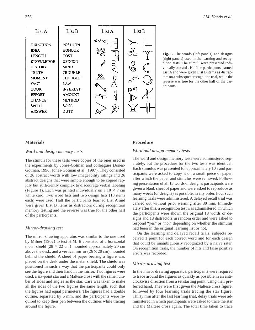
The stimuli for these tests were copies of the ones used inthe experiments by Jones-Gotman and colleagues (Jones-Gotman, 1996; Jones-Gotman et al., 1997). They consistedof 26 abstract words with low imageability ratings and 26abstract designs that were simple enough to be copied rap-idly but sufficiently complex to discourage verbal labeling(Figure 1). Each was printed individually on a 103 7 cmwhite card. Two word lists and two design lists (13 itemseach) were used. Half the participants learned List A andwere given List B items as distractors during recognitionmemory testing and the reverse was true for the other halfof the participants.
Mirror-drawing test
The mirror-drawing apparatus was similar to the one usedby Milner (1962) to test H.M. It consisted of a horizontalmetal shield (283 22 cm) mounted approximately 20 cmabove the desk, and a vertical mirror (263 20 cm) mountedbehind the shield. A sheet of paper bearing a figure wasplaced on the desk under the metal shield. The shield waspositioned in such a way that the participants could onlysee the figure and their hand in the mirror. Two figures wereused: a six-point star and a Maltese cross with the same num-ber of sides and angles as the star. Care was taken to makeall the sides of the two figures the same length, such thatthe figures had equal perimeters. The figures had a doubleoutline, separated by 5 mm, and the participants were re-quired to keep their pen between the outlines while tracingaround the figure.
Procedure
Word and design memory tests
The word and design memory tests were administered sep-arately, but the procedure for the two tests was identical.Each stimulus was presented for approximately 10 s and par-ticipants were asked to copy it on a small piece of paper,after which the paper and stimulus were removed. Follow-ing presentation of all 13 words or designs, participants weregiven a blank sheet of paper and were asked to reproduce asmany words (or designs) as possible, in any order. Four suchlearning trials were administered. A delayed recall trial wascarried out without prior warning after 30 min. Immedi-ately after this, a recognition test was administered, in whichthe participants were shown the original 13 words or de-signs and 13 distractors in random order and were asked torespond “yes” or “no,” depending on whether the stimulushad been in the original learning list or not.
On the learning and delayed recall trials, subjects re-ceived 1 point for each correct word and for each designthat could be unambiguously recognized by a naive rater.On recognition trials, the number of hits and false positiveerrors was recorded.
Mirror-drawing test
In the mirror drawing apparatus, participants were requiredto trace around the figures as quickly as possible in an anti-clockwise direction from a set starting point, using their pre-ferred hand. They were first given the Maltese cross figure,followed by four learning trials tracing the star figure.Thirty min after the last learning trial, delay trials were ad-ministered in which participants were asked to trace the starand the Maltese cross again. The total time taken to trace
Fig. 1. The words (left panels) and designs(right panels) used in the learning and recog-nition tests. The stimuli were presented indi-vidually on cards. Half the participants learnedList A and were given List B items as distrac-tors on a subsequent recognition trial, while thereverse was true for the other half of the par-ticipants.
356 I.M. Harris et al.

around the figure was recorded on each trial. Each time thepencil tracing went outside the double lines of the figure itwas counted as an error. An error score was thus achievedfor each trial.
Order of procedures
All but 1 patient were tested within 1 week of their PETimaging studies; the remaining patient was tested 6 monthsafter she had her scan. Each was given the memory tests ina single session lasting approximately 1.5 hr. The tests wereadministered in the following order: (1) word learning tri-als; (2) design learning trials; (3) mirror-drawing learningtrials; (4) word delayed recall and recognition; (5) designdelayed recall and recognition; and (6) mirror-drawing de-lay trials. Where necessary, the time intervals between Steps4, 5, and 6 were filled by obtaining demographic informa-tion from the participants, to ensure that the learning anddelayed trials of each test were separated by 30 min.
Statistical Analyses
For the word and design memory tests, two-way Group (threelevels)3 Trial (five levels: four learning trials, one delaytrial) ANOVAs with repeated measures on the trial factorwere performed. Percent retention was calculated using thisformula: ((Trial 42 Trial 5)0Trial 4) 3 100, and one-wayANOVAs were used to compare the groups for retention ofthe words and designs. Recognition memory was evaluatedby comparing groups on the number of correct hits and thenumber of false positive responses. Pearson product-moment
correlations between test scores and mesial temporal asym-metry scores were evaluated when the ANOVAs reachedsignificance.
Performance on the mirror drawing test was analyzedusing two-way Group3 Trial (seven levels: Maltese Cross,four star learning trials, delayed star trial, repeat MalteseCross) ANOVAs with repeated measures on trial. These werecarried out for both the time and error scores.
Significant interactions were broken down by comparingthe three groups on each trial. Scheffé pair-wisepost-hoctests were used to compare means when the ANOVA wassignificant. For mirror-drawing, data for 1 participant (C.L.)are missing because he refused to complete the test.
RESULTS
Demographic Comparisons
Comparing patient groups to the NC group yielded no sig-nificant differences in age or educational level. Similarly,no differences were found between the patient groups in ageof onset or years of seizure disorder (see Table 1).
Effects of Mesial Temporal Hypometabolismon Memory for Words and Designs
On the word list-learning test, there was an interaction be-tween Group3 Trial [F(8,72)5 2.11,p , .05] as well as amain effect of trial [F(4,72)5 36.2,p , .001], but no sig-nificant overall group effect [F(2,18)5 1.51,p 5 .25]. Ascan be seen in Figure 2a, participants improved over the
Fig. 2. Mean number of (a) words and (b) designs recalled on each of the four learning trials and after 30-min delay bypatients with left mesial temporal (LMT) hypometabolism, patients with right mesial temporal (RMT) hypometabo-lism, and normal controls (NC). Vertical bars represent standard error of the mean.
Focal brain hypometabolism and memory 357

four learning trials and then showed a drop between the lastlearning trial and the delayed recall trial. There were no sig-nificant group differences on any of the four learning trials,but there were on the delay trial [F(2,18)5 5.5, p , .05]and for percent retention [F(2,18)5 7.2,p , .01]. Scheffépost-hoccomparisons for the delay trial and percent reten-tion showed that the LMT group was significantly impairedcompared to the NC group. Pearson product-moment cor-relation tests indicated that the relationship between the PETasymmetry score and the word list percent retention scoreapproached significance [r (11)5 2.56,p 5 .058].
For word recognition, there were no significant between-group differences in the mean number of hits (LMT: 11.5;RMT: 11.6; NC: 12.9) or mean number of false positive re-sponses (LMT: 0.5; RMT:0.3; NC: 0.1). Recognition mem-ory scores were not correlated with the PET measure ofmesial temporal asymmetry [hits:r (11) 5 2.03, p 5 .94;false positives:r (11)5 .09,p 5 .78]. For design learning,the Group3Trial interaction was not significant [F(8,72)51.1, p 5 .40]. There was, however, a main effect of group[F(2,18)5 4.4,p , .05]. Scheffépost-hoccomparisons in-dicated that the RMT group differed significantly from theNC group (see Fig. 2b). There was a also main effect of trial[F(4,72)5 106.1,p , .001], with all groups showing grad-ual improvement over the learning trials and good retentionafter a delay. There were no differences between the NC,RMT, and LMT groups in their percent retention score fordesigns [F(2,18)5 .20, p 5 .82]. The total learning scorefor designs was not correlated with the mesial temporal lobeasymmetry score obtained from PET [r (11)5 .24,p5 .46].Furthermore, groups did not differ in design recognitionmemory; only 2 patients (B.P. and P.C.) made a single erroreach.
Effects of Mesial Temporal and CerebellarHypometabolism on Mirror Drawing
For both the time and the error scores, patients were com-pared to the NC group across the seven mirror drawing tri-als (Figure 3). For both scores, there was a significant effectof trial [time: F(6,102)5 30.1,p , .001; errors:F(6,102)58.0, p , .001], but no group effects [time:F(2,17)5.01,p 5 .98; errors:F(2,17), p 5 .88] or interactions [time:F(12,102)5 .37,p5 .97; errors:F(12,102)5 .37,p5 .97].Figures 3a and 3b show that for all three groups, there wasa reduction in tracing time and in error rate across the seventrials, indicating acquisition of the mirror-drawing skills andtransfer of these skills to an alternative figure. Mesial tem-poral asymmetry was not correlated with the differencescores for time [r (10) 5 .17, p 5 .62] or errors [r (10) 52.01, p 5 .98] on the two Maltese cross trials (the largestdifference observed).
Even when the NC group was compared to the more se-lect group of patients who had significant cerebellar hypo-metabolism (i.e., a level more than 1SD from the controlmean) there was no main effect for group [time:F(1,15)5
.04, p 5 .85; errors:F(1,15)5 .02, p 5 .89] and no inter-action [time:F(6,90)5 .24,p5 .96; errors:F(6,90)5 .26,p5 .95]. The main effect of trial again reached significance[time: F(6,90)5 31.2,p , .001; errors:F(6,90)5 10.2,p , .001]. Because we had hypothesized that cerebellar me-tabolism might be related to motor learning ability, Pearsonproduct-moment tests were carried out. Cerebellar metabo-lism did not predict learning ability, in that the mean levelof cerebellar metabolism was not correlated with either thechange in time taken to trace [r (10)5 .07,p 5 .85] or thechange in the number of errors made [r (10)5 .06,p5 .87]across the two trials on the Maltese cross.
Fig. 3. Comparison of patients with left mesial temporal (LTM)hypometabolism, right mesial temporal (RMT) hypometabolismand normal controls (NC) on the mirror-drawing task. Top panel(a) shows mean time taken to trace the Maltese cross (MC) andstar (S) figures on the learning and delayed (Sdelay and MC2) tri-als. The bottom panel (b) shows the mean number of errors. Ver-tical bars represent standard error of the mean.
358 I.M. Harris et al.

DISCUSSION
This study examined the effects of mesial temporal hypo-metabolism on tests of word-list, design-list and motor-skill learning and retention. Patients with relatively lowmetabolic rates in the left mesial temporal region had sig-nificant difficulty recalling a word list after a 30-min delayinterval but demonstrated adequate word-list learning overthe four presentation trials. In contrast, across the designlist-learning trials, the RMT group showed an impairmentcompared to normal control participants. Both patient groupsshowed normal retention of the designs over a delay inter-val. No impairments in word or design recognition memoryor in the ability to learn mirror drawing skills were foundwhen patients with mesial temporal hypometabolism werecompared to a normal control group. Moreover, cerebellarhypometabolism was not found to lead to impairments inmirror-drawing skills.
The pattern of impairments on the list-learning tests seenin association with left or right mesial temporal hypometab-olism in this study is similar to that found preoperativelywhen patients with temporal lobe epilepsy are classified onthe basis of MR evidence of hippocampal atrophy (Jones-Gotman, 1996). Thus, at least for this brain region, our re-sults support the hypothesis that the patterns of cognitiveimpairment associated with glucose hypometabolism areconsistent with those caused by structural lesions. How-ever, a direct evaluation of this hypothesis by applying bothfunctional and structural imaging along with neuropsycho-logical testing remains to be done. For clinical purposes, itis important to note that, for word retention, we found sim-ilar group differences to those reported by Jones-Gotmanet al. (1997) in spite of the fact that we shortened the delayinterval from 24 hr to 30 min.
In our study, patients with reduced metabolism in themesial temporal region did not have impaired recognitionmemory. Unfortunately, the excellent performance of allparticipants created a ceiling effect for both word and de-sign recognition, and this limits the conclusions that canbe drawn. Nevertheless, our findings are consistent withthose of other studies. Helmstaedter and Elger (1996) testeda large number of patients with temporal lobe epilepsy ona German version of the Rey Auditory Verbal Learning Test(Helmstaedter & Durwen, 1990) prior to surgery. Manyof the patients in their study were found to have hippo-campal sclerosis and yet their delayed recognition mem-ory was intact. In contrast, impaired recognition memoryfor words has been reported after temporal lobectomy(Helmstaedter & Elger, 1996; Majdan et al., 1996) and inpatients with cortical dysgenesis in addition to hippocam-pal sclerosis (Baxendale, 1997). Together, these results in-dicate that extramesial temporal lobe structures are requiredfor recognition, which offers some support for the hypoth-esis first put forward by Aggleton and Shaw (1996) andelaborated by Aggleton and Brown (1999) that the hippo-campus is not important for certain kinds of recognitionmemory.
The present study did not find a relationship between me-sial temporal hypometabolism or cerebellar hypometabo-lism and the ability to learn, retain or transfer the motor skillsrequired for mirror drawing. Although mesial temporal hy-pometabolism was not expected to impair skill learning, thenegative findings for the cerebellum may at first seem in-consistent with previous studies that ascribe a role for thecerebellum in motor skill learning. There are, however, afew possible explanations. For example, functional activa-tion studies indicate that the cerebellum contributes to later,but not to very early stages of skill learning (Halsband &Freund, 1993; Shadmehr & Holcomb, 1997). Thus, if ourparticipants had been tested over many more trials, perhapsthose with cerebellar hypometabolism would have eventu-ally shown impaired performance compared to those withnormal cerebellar function. However, Sanes et al.’s (1990)results would argue against this, because they found thatpatients with cerebellar atrophy were not impaired over aseries of 50 trials of mirror drawing. A more likely expla-nation is that the importance of the cerebellar component inmotor skill learning rests on the nature of the task. Studiesto date indicate that the cerebellum has a role in learning aseries of finger movements (Doyon et al., 1996; Gomez-Beldarrain et al., 1997; Jenkins et al., 1994; Jueptner et al.,1997; Pascual-Leone et al., 1993) and motor adaptation(Deuschl et al., 1996; Weiner et al., 1983), but not in learn-ing a pursuit motor task (Grafton et al., 1992) or in drawingpracticed designs in rotated orientations (Timmann et al.,1996). As stated by Desmond and Fiez (1998), a unified ex-planation of the role of the cerebellum in motor skill learn-ing has yet to be achieved, some 20 years after Eccles (1978)published his “comprehensive” theory on learning and thecerebellar cortex.
In summary, the present results clearly indicate that a num-ber of factors help to determine whether left or right mesialtemporal dysfunction causes a significant impairment on testsof learning and memory. These factors include type of stim-ulus material (e.g., wordsvs. novel designs), stage of mem-ory processing (acquisitionvs. retention) and means ofassessment (recallvs. recognition), as noted, in part, by otherresearchers (e.g., Dobbins et al., 1998; Jones-Gotman, 1996;Jones-Gotman et al., 1997; Milner, 1958, 1973; Saling et al.,1993). The left mesial temporal region is particularly im-portant when words must be recalled after a delay interval.In contrast, the right mesial temporal region plays a role inencoding novel designs. Neither the left nor the right me-sial temporal region is important for the retention of learneddesigns, at least over a 30 min interval (but see Jones-Gotman, 1986, for evidence that the right hippocampus isimportant for the retention of designs over a 24-hr period).It is generally argued that left temporal and right temporallesions have different effects on the mnemonic processingof word listsversusdesign lists, because a word list com-prises verbal material and a design list consists of visuospa-tial material. However, the possibility that novelty of thestimulus is an important factor (designs being novel andwords being nonnovel) has also been raised (Majdan et al.,
Focal brain hypometabolism and memory 359

1996; Owen et al., 1996) and we are currently investigatingthis issue.
ACKNOWLEDGMENTS
We are grateful to Dr. Marilyn Jones-Gotman for providing copiesof her original stimuli for use in the word and design list learningtests and to the Psychology Department, University of New SouthWales for lending us the mirror drawing apparatus. We would liketo thank Drs. J. Walsh, R. MacKenzie, E. Somerville, and A. Blea-sel for allowing us to test their patients as well as the participantsthemselves. The first author was supported by an Australian Post-graduate Award.
REFERENCES
Aggleton, J.P. & Brown, M.W. (1999). Episodic memory, amne-sia, and the hippocampal-anterior thalamic axis.Behavioral andBrain Sciences, 22, 425–489.
Aggleton, J.P. & Shaw, C. (1996). Amnesia and recognition mem-ory: A re-analysis of psychometric data.Neuropsychologia, 34,51–62.
Ardekani, B.A., Braun, M.A., Hutton, B.F., Kanno, I., & Iida, H.(1995). A fully automatic multimodality image registration al-gorithm. Journal of Computer Assisted Tomography, 19,615–623.
Barr, W.B., Ashtari, M., & Schaul, N. (1990). The role of the hip-pocampus in recognition of recurring figures.Journal of Clin-ical and Experimental Neuropsychology, 13, 68.
Baxendale, S.A. (1997). The role of the hippocampus in recogni-tion memory.Neuropsychologia, 35, 591–598.
Baxendale, S.A., Thompson, P.J., & Van-Paesschen, W. (1998a).A test of spatial memory and its clinical utility in the pre-surgical investigation of temporal lobe epilepsy patients.Neuro-psychologia, 36, 591–602.
Baxendale, S.A., Van-Paesschen, W., Thompson, P.J., Duncan, J.S.,Harkness, W.F., & Shorvon, S.D. (1998b). Hippocampal cellloss and gliosis: relationship to preoperative and postoperativememory function.Neuropsychiatry, Neuropsychology, andBehavioral Neurology, 11, 12–21.
Bortz, J.J., Prigatano, G.P., Blum, D., & Fisher, R.S. (1995). Dif-ferential response characteristics in nonepileptic and epilepticseizure patients on a test of verbal learning and memory.Neu-rology, 45, 2029–2034.
Cohen, N.J. & Squire, L.R. (1980). Preserved learning and reten-tion of pattern-analyzing skill in amnesia: Dissociation of know-ing how and knowing that.Science, 210, 207–209.
Desmond, J.E. & Fiez, J.A. (1998). Neuroimaging studies of thecerebellum: Language, learning and memory.Trends in Cog-nitive Science, 2, 355–362.
Deuschl, G., Toro, C., Zeffiro, T., Massaquoi, S., & Hallett, M.(1996). Adaptation motor learning of arm movements in pa-tients with cerebellar diseases.Journal of Neurology, Neuro-surgery and Psychiatry, 60, 515–519.
Dobbins, I.G., Kroll, N.E.A., Tulving, E., Knight, R.T., & Gazza-niga, M.S. (1998). Unilateral medial temporal lobe memoryimpairment: Type deficit, function deficit, or both?Neuro-psychologia, 36, 115–127.
Doyon, J., Owen, A.M., Petrides, M., Sziklas, V., & Evans, A.C.(1996). Functional anatomy of visuomotor skill learning inhuman subjects examined with positron emission tomography.European Journal of Neuroscience, 8, 637–648.
Eberl, S., Kanno, I., Fulton, R.R., Ryan, A., Hutton, B.F., & Ful-ham, M.J. (1996). Automated interstudy image registration tech-nique for SPECT and PET.Journal of Nuclear Medicine, 37,137–145.
Eccles, J.C. (1978). An instruction-selection theory of learning inthe cerebellar cortex.Brain Research, 127, 327–352.
Engel, J., Brown, W.J., Kuhl, D.E., Phelps, M.D., Mazziotta, J.C.,& Crandall, P.H. (1982). Pathological findings underlying fo-cal temporal lobe hypometabolism in partial epilepsy.Annalsof Neurology, 12, 518–528.
Gabrieli, J.D.E, Corkin, S., Mickel, S.F., & Growdon, J.H. (1993).Intact acquisition and long-term retention of mirror-tracing skillin Alzheimer’s disease and global amnesia.Behavioral Neuro-science, 107, 899–910.
Gaillard, W.D., Bhatia, S., Bookheimer, S.Y., Fazilat, S., Sato, S.,& Theodore, W.H. (1995). FDG-PET and volumetric MRI inthe evaluation of patients with partial epilepsy.Neurology, 45,123–126.
Gleissner, U., Helmstaedter, C., & Elger, C.E. (1998). Righthippocampal contribution to visual memory: A presurgical andpostsurgical study in patients with temporal lobe epilepsy.Journal of Neurology, Neurosurgery, and Psychiatry, 65,665–669.
Gomez-Beldarrain, M., Garcia-Monco, J.C., Quintana, J.M.,Llorens, V., & Rodeno, E. (1997). Diaschisis and neuropsycho-logical performance after cerebellar stroke.European Neurol-ogy, 37, 82–89.
Grafton, S.T., Mazziotta, J.C., Presty, S., Friston, K.J., Frackow-iak, R.S.J., & Phelps, M.E. (1992). Functional anatomy of hu-man procedural learning determined with regional cerebral bloodflow and PET.Journal of Neuroscience, 12, 2542–2548.
Haberland, C. (1962). Cerebellar degeneration with clinical man-ifestations in chronic epileptic patients.Psychiatria et Neuro-logia, 143, 29–44.
Halsband, U. & Freund, H-J. (1993). Motor learning.Current Opin-ion in Neurobiology, 3, 940–949.
Helmstaedter, C. & Durwen, H.F. (1990). Lern und Merk-fahigkeitstest. Ein praktikables und differenziertes Instrumen-tarium zur Prufung der verbalen Gedachtnisleistungun. [TheVerbal Learning and Retention Test. A useful and differentiatedtool for evaluating verbal memory performance.]SchweizerArchiv für Neurologie und Psychiatrie 141, 21–30.
Helmstaedter, C. & Elger C.E. (1996). Cognitive consequences oftwo-thirds anterior temporal lobectomy on verbal memory in144 patients: A three-month follow-up study.Epilepsia, 37,171–180.
Helmstaedter, C., Pohl, C., & Elger, C.E. (1993). Interrelations be-tween verbal and figural memory performance in right and lefttemporal lobe epilepsy.Epilepsia, 34 (Suppl. 6), 94.
Helmstaedter, C., Pohl, C., Hufnagel, A., & Elger, C.E. (1991).Visual learning deficits in nonresected patients with right tem-poral lobe epilepsy.Cortex, 27, 547–555.
Hooper, P.K., Meikle, S.R., Eberl, S., & Fulham, M.J. (1996). Val-idation of post-injection transmission measurements for atten-uation correction in neurologic FDG PET studies.Journal ofNuclear Medicine, 37, 128–136.
Hudson, L.P., Munoz, D.G., Miller, L., McLachlan, R.S., Girvin,J.P., & Blume, W.T. (1993). Amygdaloid sclerosis in temporallobe epilepsy.Annals of Neurology, 33, 622–631.
Inhoff, A.W., Diener, H.C., Rafal, R.D., & Ivry, R. (1989). Therole of the cerebellar structures in the execution of serial move-ments.Brain, 112, 565–581.
360 I.M. Harris et al.

Jenkins, I.H., Brooks, D.J., Nixon, P.D., Frackowiak, R.S.J., & Pass-ingham, R.E. (1994). Motor sequence learning: A study withPositron Emission Tomography.Journal of Neuroscience, 14,3775–3790.
Jones-Gotman, M. (1986). Memory for designs: The hippocampalcontribution.Neuropsychologia, 24, 193–203.
Jones-Gotman, M. (1996). Psychological evaluation for epilepsysurgery. In S. Shorvon, F. Dreifuss, D. Fish, & D. Thomas (Eds.)The treatment of epilepsy(pp. 621–630). Oxford, UK: Black-well Science.
Jones-Gotman, M., Zatorre, R.J., Olivier, A., Andermann, F., Cen-des, F., Staunton, H., McMackin, D., Siegel, A.M., & Wieser,H-G. (1997). Learning and retention of words and designs fol-lowing excision from medial or lateral temporal lobe struc-tures.Neuropsychologia, 35, 963–973.
Jueptner, M. & Weiller, C. (1998). A review of differences be-tween basal ganglia and cerebellar control of movements asrevealed by functional imaging studies.Brain, 121, 1437–1449.
Jueptner, M., Stephan, K.M., Frith, C.D., Brooks, D.J., Frackow-iak, R.S., & Passingham, R.E. (1997). Anatomy of motor learn-ing: I. Frontal cortex and attention to action.Journal ofNeurophysiology, 77, 1313–1324.
Majdan, A., Sziklas, V., & Jones-Gotman, M. (1996). Perfor-mance of healthy subjects and patients with resection from theanterior temporal lobe on matched tests of verbal and visuo-perceptual learning.Journal of Clinical and ExperimentalNeuropsychology, 18, 416–430.
Martin, R.C., Loring, D.W., Meador, K.J., Lee, G.P., Thrash, N., &Arena, J.G. (1991). Impaired long-term retention despite nor-mal verbal learning in patients with temporal lobe dysfunction.Neuropsychology, 5, 3–12.
McLain, L.W., Martin, J.T., & Allen, J.H. (1980). Cerebellar de-generation due to chronic phenytoin therapy.Annals of Neu-rology, 7, 18–23.
Meyer, V. & Yates, A.J. (1955). Intellectual changes following tem-poral lobectomy for psychomotor epilepsy.Journal of Neurol-ogy, Neurosurgery and Psychiatry, 18, 44–52.
Miller, L.A., Lai, R., & Munoz, D.G. (1998). Contributions of theentorhinal cortex, amygdala and hippocampus to human mem-ory. Neuropsychologia, 36, 1247–1256.
Miller, L.A., Munoz, D.G., & Finmore, M. (1993). Hippocampalsclerosis and human memory.Archives of Neurology, 50,391–394.
Milner, B. (1958). Psychological defects produced by temporal lobeexcisions.Research Publications of the Association for Re-search in Nervous and Mental Disease, 36, 244–257.
Milner, B. (1962). Les troubles de la memoire accompagnant deslesions hippocampiques bilaterales [Memory problems accom-pany bilateral hippocampal lesions]. InPhysiologie del’hippocampe(pp. 257–272). Paris: Centre National de la Re-cherche Scientifique.
Milner, B. (1968). Visual recognition and recall after right temporal-lobe excision in man.Neuropsychologia, 6, 191–209.
Milner, B. (1973). Hemispheric specialization: Scope and limits.In F.O. Schmitt & F.G. Worden (Eds.),The neurosciences: Thirdstudy program(pp. 75–89). Boston: MIT Press.
Milner, B., Corkin, S., & Teuber, H-L. (1968). Further analysis ofthe hippocampal amnesic syndrome: 14-year follow-up studyof H.M. Neuropsychologia, 6, 215–234.
Nichelli, P., Bhamanian-Behbahani, G., Gentilini, M., & Vecchi,A. (1988). Preserved memory abilities in thalamic amnesia.Brain, 111, 1337–1353.
Nissen, M.J., Willingham, D., & Hartman, M. (1989). Explicit andimplicit remembering: When is learning preserved in amnesia?Neuropsychologia, 27, 341–352.
Owen, A.M., Morris, R.G., Sahakian, B.J., Polkey, C.E., & Rob-bins, T.W. (1996). Double dissociations of memory and exec-utive functions in working memory tasks following frontallobe excisions, temporal lobe excisions or amygdalo-hippocampectomy in man.Brain, 119, 1597–1615.
Pascual-Leone, A., Grafman, J., Clark, K., Stewart, M., Massa-quoi, S., Lou, J-S., & Hallett, M. (1993). Procedural learningin Parkinson’s disease and cerebellar degeneration.Annals ofNeurology, 34, 594–602.
Phelps, M.E., Huang, S.C., Hoffman, E.J., Selin, C., Sokoloff, L.,& Kuhl, D.E. (1979). Tomographic measurement of local ce-rebral glucose metabolic rate in humans with [18F]2-fluoro-2-deoxy-d-glucose: Validation of method.Annals of Neurology,6, 371–388.
Rausch, R. & Babb, T.L. (1993). Hippocampal neuron loss andmemory scores before and after temporal lobe surgery for epi-lepsy.Archives of Neurology, 50, 812–817.
Rausch, R., Henry, T.R.H., Ary, C.M., Engel, J., & Mazziotta, J.(1994).Asymmetric interictal glucose hypometabolism and cog-nitive performance in epileptic patients.Archives of Neurol-ogy, 51, 139–144.
Salcman, M., Defendini, R., Correll, J., & Gilman, S. (1978). Neuro-pathological changes in cerebellar biopsies of epileptic pa-tients.Annals of Neurology, 3, 10–19.
Saling, M.M., Berkovic, S.F., O’Shea, M.F., Kalnins, R.M., Darby,D.G., & Bladin, P.F. (1993). Lateralization of verbal memoryand unilateral hippocampal sclerosis: Evidence of task-specificeffects.Journal of Clinical and Experimental Neuropsychol-ogy, 15, 608–618.
Sanes, J.N., Dimitrov, B., & Hallett, M. (1990). Motor learning inpatients with cerebellar dysfunction.Brain, 113, 103–120.
Sass, K.J., Sass, A., Westerveld, M., Lencz, T., Novelly, R.A., Kim,J.H., & Spencer, D.D. (1992). Specificity in the correlation ofverbal memory and hippocampal neuron loss: Dissociation ofmemory, language, and verbal intellectual ability.Journal ofClinical and Experimental Neuropsychology, 14, 662–672.
Saykin, A.J., Gur, R.C., Sussman, N.M., O’Connor, M.J., & Gur,R.E. (1989). Memory deficits before and after temporal lobec-tomy: Effects of laterality and age of onset.Brain and Cogni-tion, 9, 191–200.
Seidenberg, M., Hermann, B., Haltiner, A., & Wyler, A. (1993).Verbal recognition memory performance in unilateral tempo-ral lobe epilepsy.Brain and Language, 44, 191–200.
Seitz, R.J., Arnold, S., Schlaug, G., Ebner, A., Holthausen, H., Tux-horn, I., & Witte, O.W. (1996). Cerebellar hypometabolism infocal epilepsy is related to age of onset and drug intoxication.Epilepsia, 37, 1194–1199.
Seitz, R.J., Canavan, A.G.M., Yaguez, L., Herzog, H., Tellmann,L., Knorr, U., Huang, Y., & Homberg, V. (1994). Successiveroles of the cerebellum and premotor cortices in trajectoriallearning.NeuroReport, 5, 2541–2544.
Semah, F., Baulac, M., Hasboun, D., Frouin, V., Mangin, J-F., Papa-georgiou, S., Leroy-Wilig, A., Philippon, J., Laplane, D., & Sam-son, Y. (1995). Is interictal temporal hypometabolism relatedto mesial temporal sclerosis? A positron emission tomography0magnetic resonance imaging confrontation.Epilepsia, 36,447–456.
Shadmehr, R. & Holcomb, H.H. (1997). Neural correlates of mo-tor memory consolidation.Science, 277, 821–825.
Focal brain hypometabolism and memory 361

Timmann, D., Shimansky, Y., Larson, P.S., Wunderlich, D.A., Stel-mah, G.E., & Bloedel, J.R. (1996). Visuomotor learning in cer-ebellar patients.Behavioural Brain Research, 81, 99–113.
Weiner, M.J., Hallett, M., & Funkenstein, H.H. (1983). Adaptationto lateral displacement of vision in patients with lesions of thecentral nervous system.Neurology, 33, 766–772.
Woodard, A.R., Hermann, B.P., Bachhuber, A., Perlman, S.B., De
La Pena, R., Rosenbek, J., Cariski, D., Jones, J.C., Rutecki, P.,& Spencer, N.W. (1997). Preliminary examination of the rela-tionship between PET scan temporal lobe asymmetry and levelof performance on measures of memory, naming, and verbalintelligence in epilepsy surgery candidates (Abstract).Journalof the International Neuropsychological Society, 3, 2.
362 I.M. Harris et al.