the effectiveness of counseling strategies and rehabilitation ...
Upload
khangminh22Category
view
0download
0
344
Zakrzewska Marlena, Iłżecka Joanna. The effectiveness of PNF method in rehabilitation of patients after ischemic stroke. Journal of
Education, Health and Sport. 2018;8(3):344-361. eISNN 2391-8306. DOI http://dx.doi.org/10.5281/zenodo.1202395
http://ojs.ukw.edu.pl/index.php/johs/article/view/5369
The journal has had 7 points in Ministry of Science and Higher Education parametric evaluation. Part b item 1223 (26/01/2017).
1223 Journal of Education, Health and Sport eissn 2391-8306 7
© The Authors 2018;
This article is published with open access at Licensee Open Journal Systems of Kazimierz Wielki University in Bydgoszcz, Poland
Open Access. This article is distributed under the terms of the Creative Commons Attribution Noncommercial License which permits any noncommercial use, distribution, and reproduction in any medium,
provided the original author (s) and source are credited. This is an open access article licensed under the terms of the Creative Commons Attribution Non commercial license
(Http://creativecommons.org/licenses/by-nc/4.0/) which permits unrestricted, non commercial use, distribution and reproduction in any medium, provided the work is properly cited.
This is an open access article licensed under the terms of the Creative Commons Attribution Non commercial License (http://creativecommons.org/licenses/by-nc/4.0/) which permits unrestricted, non commercial
use, distribution and reproduction in any medium, provided the work is properly cited.
The authors declare that there is no conflict of interests regarding the publication of this paper.
Received: 01.03.2018. Revised: 10.03.2018. Accepted: 18.03.2018.
The effectiveness of PNF method in rehabilitation of patients after ischemic
stroke
Marlena Zakrzewska, Joanna Iłżecka
Independent Neurological Rehabilitation Unit, Medical University of Lublin
345
SUMMARY
Introduction. Stroke is a major medical and social problem. One of the methods used in
neurodevelopmental rehabilitation of patients after ischemic stroke is the method of
Proprioceptive Neuromuscular Facilitation (PNF).
Objective of the work. Evaluation of effectiveness of PNF method in rehabilitation of
patients after ischemic stroke.
Material and methods. The study included 100 patients with ischemic stroke. In the study
group (A) an individual rehabilitation program included the rehabilitation of the classical
method and PNF, in the control group (B) of the patients were only streamlines the traditional
individual rehabilitation. Degree of disability was evaluated on the basis of the modified
Rankin scale, the level of movement disorders examined scale Brunnström. To assess the
functional status of the patients was used functional indicator "Repty" and modified the RMA.
Results. After the rehabilitation method of PNF, an improvement in mobility upper and lower
limb in more than 60% of patients was observed. The degree of disability decreased in 40% of
patients. There has been improvement in overall motor performance of 25.57% and greater
independence in performing activities of daily living of 15.94%.
Conclusions. Rehabilitation method of PNF is effective in patients after ischemic stroke;
however, this method is not more efficient than conventional physiotherapy. Traditional
physiotherapy cannot be excluded from effective methods of rehabilitation of patients after
ischemic stroke.
Keywords: Ischemic stroke, rehabilitation, PNF method, classical physiotherapy
INTRODUCTION
Stroke is a clinical syndrome caused by focal or generalized brain damage that persists for
more than 24 hours or leading to death, and there is no cause other than vascular. This
constitutes a significant medical problem, social and economic, leading to physical disability
and dementia, epilepsy and depression. The consequence of a stroke is a reduction in the
quality of life of patients [1-4].
Rehabilitation is a form of therapy in stroke. Early initiation of rehabilitation reduces the
risk of extra cerebral complications associated with immobilization of the patient. It has been
shown that it reduces mortality in the early post-stroke and increases the independence of the
patient [5-7].
346
Proprioceptive Neuromuscular Facilitation (PNF) is a method of physical therapy based on
the neurophysiological substrate. The basic premise of this method is pioneering the proper
neuromuscular stimulation through the use of neurophysiological rules and the resulting
methods for controlled, additional stimulation of the sensory-motor through conduction and
proprioceptive stimuli. In the design method is based on the movements of global, consistent
with the nature and daily work. Learning technique uses coordination, stabilizers, stretching,
improving functions of vegetative and analgesic techniques. Through its variety of techniques
used in the method PNF allow not only the use of appropriate movement patterns for each
patient on an individual basis, they also facilitate the implementation of a particular
therapeutic purpose (improvement of strength, mobility, coordination, stabilization). The
choice of technique depends on the individual needs of the patient and its functional
problems. PNF techniques provide a treatment option for patients with almost all mobility
problems. Proposition stroke rehabilitation concept of PNF techniques can be improve, which
include movement patterns upper and lower limbs, techniques agonists and antagonists,
exercises using gum Thera-band, work in the open and closed chain, stable and unstable [8-
10].
OBJECTIVE OF THE WORK
The aim of the study was to evaluate the effectiveness of PNF method in rehabilitation
of patients after ischemic stroke. Formulated the following specific issues:
1. Do exercises using assumptions PNF method to minimize the degree of movement
disorders examined in the upper limb and lower limb,
2. To what extent the rehabilitation of the PNF method affects the functional status of
patients with ischemic stroke.
2. MATERIAL AND METHODS
The study received a positive opinion of the Bioethics Committee of the Medical
University of Lublin (No. approval: EC 0254/37/2012).
The study involved 100 people, including 49 women and 51 men, aged from 24 to 87
years. All had been diagnosed with ischemic stroke and underwent comprehensive treatment
in rehabilitation departments in medical facilities Lublin province. During the 21 days of
rehabilitation, subjects were systematically streamlining five days a week (Monday-Friday)
1.5 - 2 hours a day, on Saturday - about 50 minutes.
All persons participating in the study were divided into two groups. In group A - test (50
people) was used the individual traditional physiotherapy and method of PNF. In group B -
control (50 people) rehabilitation was based on individual traditional physiotherapy.

347
Additionally, patients of both groups were using the procedures in the field of physical
therapy.
Criteria for selection of groups:
Diagnosed with ischemic stroke,
Informed consent to participate in research,
Improvement using PNF or traditional physiotherapy,
Systematic rehabilitation for at least 21 days,
Double-patient study before and after 3 weeks of rehabilitation,
Behavior logical verbal contact with a therapist.
Traditional rehabilitation program included:
1. Kinesitherapy individual (passive exercises upper and lower limb covered by paresis,
exercise step-passive and active of the upper and lower limb, exercises the same
assisted, exercises in relieving upper and lower limb, exercise free active, vertical
position active exercises on a rotor upper and lower limb, breathing exercises,
education or improving gait, balance exercises and coordination).
2. Physical therapy (laser therapy, local cryotherapy, phototherapy, ultrasound,
hydrotherapy),
3. Classical massage upper and lower limbs.
Exercise and treatments were selected according to the functional status of the individual
and the needs of patients.
Physiotherapy using elements of the concept of PNF was carried out individually and
adapted to the current capabilities and needs of patients. Regimen consisted of the following:
the starting position, the main principles, techniques, movement patterns.
Depending on the patient's level of functioning introduced, among other things:
- stabilize the standing position with the help of technology: approximation on the ridge of the
iliac,
- stabilization of the seated by stabilizing the condenser,
- a combination of movement patterns, which will prepare the patient to alternate work
shoulder and pelvic girdle used during gait,
- learning weight transfer body in various positions,
- learning walk on flat terrain, obstacles and stairs.
A study of patients was performed twice: before improvement and after its completion.
The survey was carried out starting on the first day of admission to the rehabilitation unit,
while the final examination 21 days after the stationary rehabilitation.
348
Used in the study:
1. Modified Rankin Scale,
2. Brunnström scale - hand and upper limb and lower limb,
3. Rivermead motor assessment - global functions,
4. Functional index Repty (WFR).
Statistical analysis was performed using the statistical package R, version 3.3.1. The
analysis of the quantitative variables was performed by calculating the average, standard
deviation, median, quartiles, maximum and minimum. The following statistical tests were
used: chi-square test, Fisher's exact test, Student's t test, Mann-Whitney test, ANOVA,
Kruskal-Wallis test, Tukey HSD test, Dunn, Pearson's correlation coefficient or Spearman.
The level of statistically significant was p <0.05.
RESULTS
Group A (test) consisted of 50 patients, including 28 women (56%) and 22 men (44%).
Group B (control) consisted of 50 patients; 21 women (42%) and 29 men (58%). Patients
from Groups A and B do not differ statistically significant gender (P> 0.05).
The average age of the patients was 68.27 years (24-87 years). Patients in Groups A and B
did not differ significantly age (p> 0.05).
A majority of both groups had paralysis / paresis of the left-hand (68%), right-sided
paralysis / paresis occurred in 32% of patients (p> 0.05).
Patients in Groups A and B differed significantly from the time of the last stroke to the
start of rehabilitation (p <0.05). In group A patients were rehabilitated after a long lapse of
time since the stroke. The largest group (38%) patients were 1 to 3 months after stroke, and 1
month after stroke (24%). Over 1 year after the onset of stroke, the rehabilitation of patients
was 18%. Group B consisted mainly (78%) patients 1 month after falling ill, 10% are in the
range from 1 to 3 months after the stroke, more than one year from the occurrence of the
disease was only 6% of respondents.
With comorbidities hypertension and atrial fibrillation occurred more frequently in group
B.
Patients in Groups A and B differed statistically significant results on a scale Brunnström
for upper limb before rehabilitation. Patients in group A demonstrated a higher mobility of the
upper arm and hand at baseline than patients of group B (Table 1).
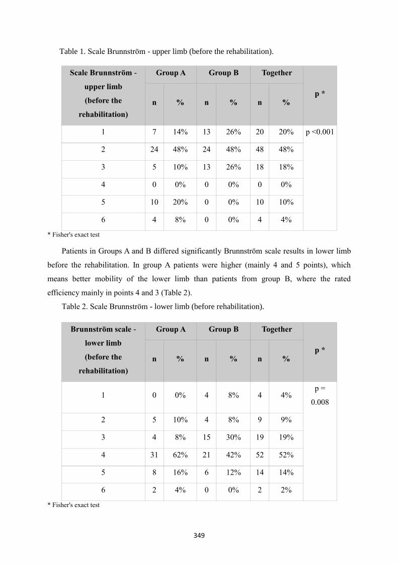
349
Table 1. Scale Brunnström - upper limb (before the rehabilitation).
Scale Brunnström -
upper limb
(before the
rehabilitation)
Group A Group B Together
p * n % n % n %
1 7 14% 13 26% 20 20% p <0.001
2 24 48% 24 48% 48 48%
3 5 10% 13 26% 18 18%
4 0 0% 0 0% 0 0%
5 10 20% 0 0% 10 10%
6 4 8% 0 0% 4 4%
* Fisher's exact test
Patients in Groups A and B differed significantly Brunnström scale results in lower limb
before the rehabilitation. In group A patients were higher (mainly 4 and 5 points), which
means better mobility of the lower limb than patients from group B, where the rated
efficiency mainly in points 4 and 3 (Table 2).
Table 2. Scale Brunnström - lower limb (before rehabilitation).
Brunnström scale -
lower limb
(before the
rehabilitation)
Group A Group B Together
p * n % n % n %
1 0 0% 4 8% 4 4% p =
0.008
2 5 10% 4 8% 9 9%
3 4 8% 15 30% 19 19%
4 31 62% 21 42% 52 52%
5 8 16% 6 12% 14 14%
6 2 4% 0 0% 2 2%
* Fisher's exact test
350
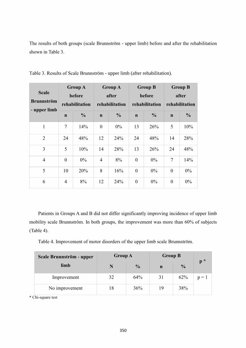
The results of both groups (scale Brunnström - upper limb) before and after the rehabilitation
shown in Table 3.
Table 3. Results of Scale Brunnström - upper limb (after rehabilitation).
Scale
Brunnström
- upper limb
Group A
before
rehabilitation
Group A
after
rehabilitation
Group B
before
rehabilitation
Group B
after
rehabilitation
n % n % n % n %
1 7 14% 0 0% 13 26% 5 10%
2 24 48% 12 24% 24 48% 14 28%
3 5 10% 14 28% 13 26% 24 48%
4 0 0% 4 8% 0 0% 7 14%
5 10 20% 8 16% 0 0% 0 0%
6 4 8% 12 24% 0 0% 0 0%
Patients in Groups A and B did not differ significantly improving incidence of upper limb
mobility scale Brunnström. In both groups, the improvement was more than 60% of subjects
(Table 4).
Table 4. Improvement of motor disorders of the upper limb scale Brunnström.
Scale Brunnström - upper
limb
Group A Group B p *
N % n %
Improvement 32 64% 31 62% p = 1
No improvement 18 36% 19 38%
* Chi-square test

351
The results of both groups before and after the rehabilitation is presented in Table 5.
Table 5. Results Scale Brunnström - lower limb (after rehabilitation).
Brunnström
scale –
lower limb
Group A
before
rehabilitation
Group A
after
rehabilitation
Group B
before
rehabilitation
Group B
after
rehabilitation
n % n % n % n %
1 0 0% 0 0% 4 8% 2 4%
2 5 10% 1 2% 4 8% 4 8%
3 4 8% 0 0% 15 30% 0 0%
4 31 62% 21 42% 21 42% 32 64%
5 8 16% 20 40% 6 12% 10 20%
6 2 4% 8 16% 0 0% 2 4%
Patients in Groups A and B did not differ significantly improve mobility occurrence of
the lower limb scale Brunnström. Improvement in the A group was slightly higher (60%) than
in group B (54%) (Table 6).
Table 6. Improvement of lower limb motor disorders Brunnström scale.
Brunnström scale -
lower limb
Group A Group B p *
N % n %
Improvement 30 60% 27 54% p = 0.686
No improvement 20 40% 23 46%
* Chi-square test
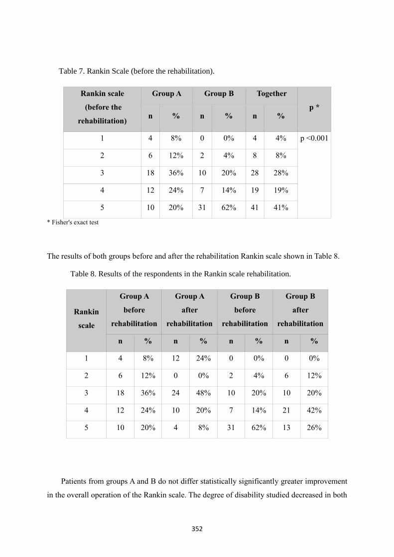
Patients in Groups A and B differed statistically significant results in the Rankin scale
before the rehabilitation. In Group A patients had a lesser degree of disability (mainly
breakpoint moderate disability, 3), and in the group B as many as 62% of patients presented
severe disability - 5 points (Table 7).
352
Table 7. Rankin Scale (before the rehabilitation).
Rankin scale
(before the
rehabilitation)
Group A Group B Together
p * n % n % n %
1 4 8% 0 0% 4 4% p <0.001
2 6 12% 2 4% 8 8%
3 18 36% 10 20% 28 28%
4 12 24% 7 14% 19 19%
5 10 20% 31 62% 41 41%
* Fisher's exact test
The results of both groups before and after the rehabilitation Rankin scale shown in Table 8.
Table 8. Results of the respondents in the Rankin scale rehabilitation.
Rankin
scale
Group A
before
rehabilitation
Group A
after
rehabilitation
Group B
before
rehabilitation
Group B
after
rehabilitation
n % n % n % n %
1 4 8% 12 24% 0 0% 0 0%
2 6 12% 0 0% 2 4% 6 12%
3 18 36% 24 48% 10 20% 10 20%
4 12 24% 10 20% 7 14% 21 42%
5 10 20% 4 8% 31 62% 13 26%
Patients from groups A and B do not differ statistically significantly greater improvement
in the overall operation of the Rankin scale. The degree of disability studied decreased in both
353
groups. Improvement was observed in approximately 40% of patients (Table 9).
Table 9. Reduction of the degree of disability in patients Rankin Scale after rehabilitation.
Rankin scale Group A Group B
p * n % n %
Improvement 20 40% 22 44% p = 0.839
No improvement 30 60% 28 56%
* Chi-square test
The results of the RMA on a scale of respondents are presented in Table 10.
Table 10. The results of the RMA on a scale of respondents after rehabilitation.
Group
RMA
N Avera
ge SD
Medi
an min max Q1 Q3
Group A –
before rehabilitation 50 7.9 3.89 9 0 12 6 11.75
Group A –
after rehabilitation 50 9.92 3.13 10 3 15 9 12
Group B –
before rehabilitation 50 4.5 4.19 4 0 13 1 8
Group B –
after rehabilitation 50 7.62 3.2 8 3 13 5 9
Patients in Groups A and B differed significantly resulting improvement in motor deficits
in a modified scale RMA. In group B reported a greater improvement in overall motor
performance. Patients in group A at baseline had a greater mobility and showed a lesser
degree of disability, because some simple steps in a motor test performed already in the first
study. Comparing the degree of improvement in motor performance tested after the
rehabilitation in group A has been shown that the improvement is not as visible as in group B
(Table 11).
354
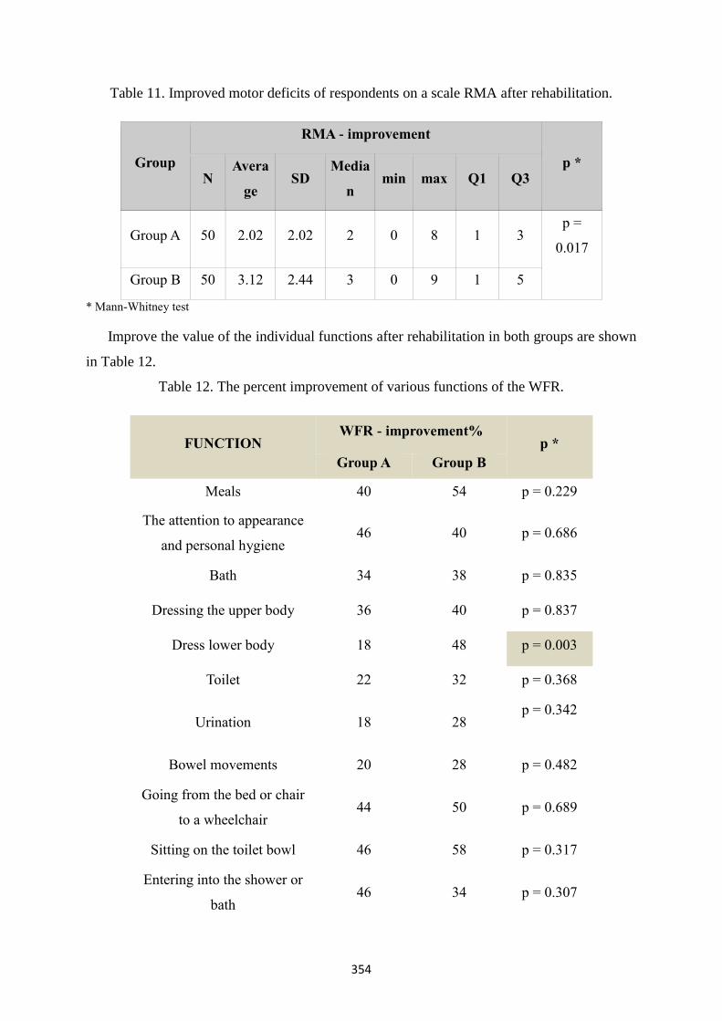
Table 11. Improved motor deficits of respondents on a scale RMA after rehabilitation.
Group
RMA - improvement
p * N
Avera
ge SD
Media
n min max Q1 Q3
Group A 50 2.02 2.02 2 0 8 1 3 p =
0.017
Group B 50 3.12 2.44 3 0 9 1 5
* Mann-Whitney test
Improve the value of the individual functions after rehabilitation in both groups are shown
in Table 12.
Table 12. The percent improvement of various functions of the WFR.
FUNCTION WFR - improvement%
p *
Group A Group B
Meals 40 54 p = 0.229
The attention to appearance
and personal hygiene 46 40 p = 0.686
Bath 34 38 p = 0.835
Dressing the upper body 36 40 p = 0.837
Dress lower body 18 48 p = 0.003
Toilet 22 32 p = 0.368
Urination 18 28 p = 0.342
Bowel movements 20 28 p = 0.482
Going from the bed or chair
to a wheelchair 44 50 p = 0.689
Sitting on the toilet bowl 46 58 p = 0.317
Entering into the shower or
bath 46 34 p = 0.307
355
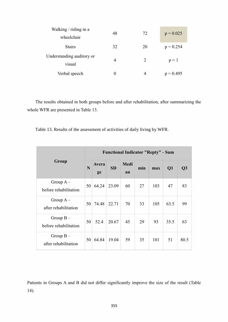
Walking / riding in a
wheelchair 48 72 p = 0.025
Stairs 32 20 p = 0.254
Understanding auditory or
visual 4 2 p = 1
Verbal speech 0 4 p = 0.495
The results obtained in both groups before and after rehabilitation, after summarizing the
whole WFR are presented in Table 13.
Table 13. Results of the assessment of activities of daily living by WFR.
Group
Functional Indicator "Repty" - Sum
N Avera
ge SD
Medi
an min max Q1 Q3
Group A –
before rehabilitation 50 64.24 23.09 60 27 103 47 83
Group A –
after rehabilitation 50 74.48 22.71 70 33 105 63.5 99
Group B –
before rehabilitation 50 52.4 20.67 45 29 93 35.5 63
Group B –
after rehabilitation 50 64.84 19.04 59 35 101 51 80.5
Patients in Groups A and B did not differ significantly improve the size of the result (Table
14).
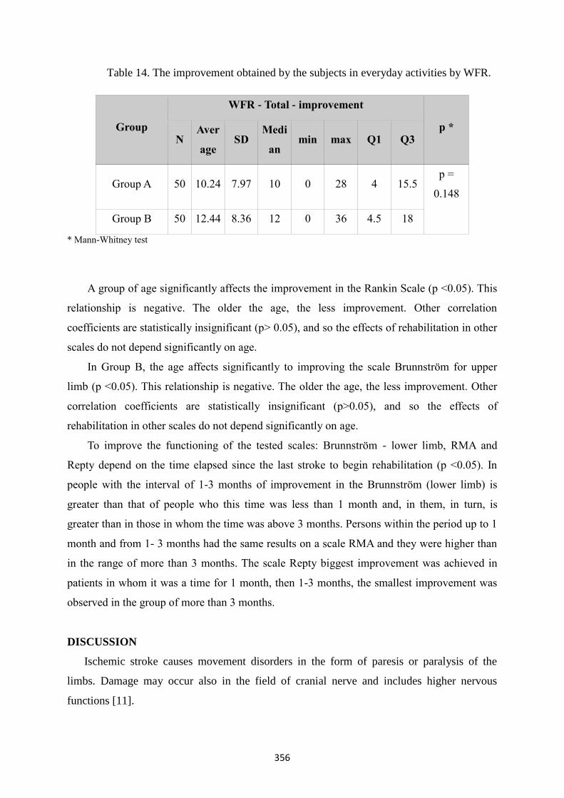
356
Table 14. The improvement obtained by the subjects in everyday activities by WFR.
Group
WFR - Total - improvement
p * N
Aver
age SD
Medi
an min max Q1 Q3
Group A 50 10.24 7.97 10 0 28 4 15.5 p =
0.148
Group B 50 12.44 8.36 12 0 36 4.5 18
* Mann-Whitney test
A group of age significantly affects the improvement in the Rankin Scale (p <0.05). This
relationship is negative. The older the age, the less improvement. Other correlation
coefficients are statistically insignificant (p> 0.05), and so the effects of rehabilitation in other
scales do not depend significantly on age.
In Group B, the age affects significantly to improving the scale Brunnström for upper
limb (p <0.05). This relationship is negative. The older the age, the less improvement. Other
correlation coefficients are statistically insignificant (p>0.05), and so the effects of
rehabilitation in other scales do not depend significantly on age.
To improve the functioning of the tested scales: Brunnström - lower limb, RMA and
Repty depend on the time elapsed since the last stroke to begin rehabilitation (p <0.05). In
people with the interval of 1-3 months of improvement in the Brunnström (lower limb) is
greater than that of people who this time was less than 1 month and, in them, in turn, is
greater than in those in whom the time was above 3 months. Persons within the period up to 1
month and from 1- 3 months had the same results on a scale RMA and they were higher than
in the range of more than 3 months. The scale Repty biggest improvement was achieved in
patients in whom it was a time for 1 month, then 1-3 months, the smallest improvement was
observed in the group of more than 3 months.
DISCUSSION
Ischemic stroke causes movement disorders in the form of paresis or paralysis of the
limbs. Damage may occur also in the field of cranial nerve and includes higher nervous
functions [11].

357
Seniów et al. [12] describe the need for a comprehensive rehabilitation of patients after
stroke. Very good results in improving motor describe Zhu et al. [13], who used repetitive
transcranial magnetic stimulation in rehabilitation of motor function. Increasing it causes
motor function improvement in activities of daily living patients. Rahman et al. [14]
conducted a study of stroke patients with paresis of the upper limb, using a robot called Mars-
5. The robot facilitates rehabilitation of the shoulder girdle, elbow and forearm for support of
movements. The test results show improved precision and accuracy of the tasks performed by
the patient.
According to Jankowska et al. [15] of stroke patients in the hospital rehabilitated scored
significantly greater improvement than those using environmental rehabilitation. Passive
motion used in traditional rehabilitation is performed in one plane in contrast to the practice
used in the method of PNF, where the motor pattern is guided in inclined planes. Passive
movement is often the cause of iatrogenic complications, especially in patients with flaccid
paresis of the upper extremity. Passive motion is one of the possible techniques for use in
patients with pain in the complex shoulder joint. The treatment program is recommended,
however, selected on the basis of subjective testing, and analysis of the priorities. Warns
against movements of large amplitude, as they may exacerbate the condition as a result of
tissue compression. The proposed movements are of small amplitude, conducted carefully and
slowly, to the border of pain. These observations are supported by a Pop [16].
Domanski et al. [17] evaluating the impact on early rehabilitation efficiency of the motor
stroke patients, show a clear improvement in mobility determined gait pattern and functional
status muscles. Data from the literature are consistent with its own research, which confirmed
a significant impact as soon as possible begun rehabilitation on improving the functioning of
patients after ischemic stroke.
Pop et al. [18] examining the factors affecting the symmetry of the loading of the lower
limbs and balance in stroke patients found no statistically significant effect on gait side
paralysis patients. They observed, however, that people with right-sided hemiparesis achieved
worse results than patients with left-sided hemiparesis. The difference demonstrated by the
paths takes the foot pressure center on the platform.
Extensive literature characterizing the effectiveness of PNF method in the rehabilitation of
stroke patients shows great interest in the above topic. Many studies have shown encouraging
results using the PNF method, particularly with regard to increasing the range of motion and
improve muscle function [19-23]. Wolny et al. [24-28] published a large number of studies on
the impact of the PNF method of rehabilitation of patients after stroke, taking into account the
358
various aspects of life of patients and improve. The study demonstrates significantly greater
effectiveness the methods of PNF than only the application of traditional methods.
Krukowska et al. [29] have demonstrated the beneficial effect of the method of PNF and
NDT-Bobath in stroke patients.
Own studies did not confirm statistically significant greater efficiency in the use of PNF
method than using only classical physiotherapy. According to the inter-group showed a
significant differences between groups were measured. Perhaps this is the reason for obtaining
the reverse-than-expected results. The results obtained show that, even before the start of the
rehabilitation of patients in the study group (using the method of PNF) has a higher mobility
and disability lower than the control group (only rehabilitated traditional physiotherapy). A
higher number of respondents moved alone, than in the control group, where patients often
have used wheelchairs. As a result of this situation, despite the high level of functioning
patients in the treatment group, comparing the level of improvement in mobility and the
effects obtained after rehabilitation, the study group obtained poorer results in some tests. In
addition, patients in the group A were rehabilitated after a long lapse of time from stroke than
in the comparison group.
Further research and introduction of new methods of rehabilitation in this ever-growing
group of patients can bring progress, improving the health of people after a stroke, and thus
tangible benefits for society as a whole [30].
CONCLUSIONS
1. Rehabilitation method of PNF is an effective method of rehabilitation after ischemic
stroke.
2. Rehabilitation using the concept of PNF not proved to be more effective in improving
patients after ischemic stroke compared with conventional physiotherapy.
3. The study confirmed that using only traditional rehabilitation does not exclude it from
effective methods of rehabilitation of patients after ischemic stroke.
4. It has been shown statistically significant correlation between the degree of
improvement of the functioning of patients with ischemic stroke, and patients' age and
the time of onset of stroke and the start of rehabilitation.

359
REFERENCES
1. Starostka-Tatar A, Łabuz-Roszak B, Skrzypek M, Gąsior M, Gierlotka M. Definition
and treatment of stroke over the centuries. Wiad Lek. 2017; 70(5): 982-987.
2. Alvarez-Sabín J, Quintana M, Masjuan J, Oliva-Moreno J, Mar J, Gonzalez-Rojas
N et al.; CONOCES Investigators Group. Economic impact of patients admitted
to stroke units in Spain. Eur J Health Econ. 2017; 18(4): 449-458.
3. Dąbrowska-Bender M, Milewska M, Gołąbek A, Duda-Zalewska A, Staniszewska A.
The impact of ischemic cerebral stroke on the quality of life of patients based on
clinical, social, and psychoemotional factors. J Stroke Cerebrovasc Dis. 2017; 26(1):
101-107.
4. Naess H, Waje-Andreassen U, Thomassen L, Nyland H, Myhr KM. Health-related
quality of life among young adults with ischemic stroke on long-term follow-up.
Stroke 2006; 37(5): 1232-1236.
5. Kwolek A, Szydełko M, Domka E. Granice przeciwwskazań do rehabilitacji po udarze
mózgu. Udar Mózgu 2005; 7 (1), 31-37.
6. Lee KH, Sohn MK, Jeong HS, Song HJ, Kim J, Kwon HJ et al. The effect of inter-
departmental stroke meetings on rehabilitation in a Comprehensive Cerebrovascular
Center. J Korean Med Sci. 2018; 33(11): e85.
7. Cheng YY, Shu JH, Hsu HC, Liang Y, Chang ST, Kao CL et al. The impact
of rehabilitation frequencies in the first year after stroke on the risk of
recurrent stroke and mortality. J Stroke Cerebrovasc Dis. 2017; 26(12): 2755-2762.
8. Lizak A. Prioprioceptive Neuromuscular Facilitation. Skrypt kursu podstawowego.
Kraków, Warszawa 2006.
9. Laidler P. Rehabilitacja po udarze mózgu. Wydawnictwo Lekarskie PZWL, Warszawa
2000.
10. Galasińska K, Buchalski P, Gajewska E. Zastosowanie koncepcji PNF w rehabilitacji
pacjentów po udarze mózgu. Now Lek. 2011; 80 (2): 126–133.
11. Piskorz J, Wójcik G, Iłżecka J, Kozak-Putowska D. Wczesna rehabilitacja pacjentów
po udarze niedokrwiennym mózgu. Med Og Nauk Zdr. 2014; 20 (4), 351-355.
12. Seniów J, Członkowska A. Poznawcze i emocjonalne konsekwencje udaru mózgu w
aspekcie procesu rehabilitacji. Rehabil Med. 2003; 7(1): 9–14.
360
13. Zhu Y, Yang YJ, Gu YH, Xie B, Jin HZ. Efficiency of repetitive transcranial magnetic
stimulation on rehabilitation of motor function in patients with stroke: a systematic
review. Chin J Tissue Engin Res. 2013; 17(50): 8758–8768.
14. Rahman MH, Kittel-Ouimet T, Saad M, Kenne JP, Archambault PS. Development and
control of a robotic exoskeleton for shoulder, elbow and forearm movement
assistance. Appl Bion Biomech. 2012; 9(3): 275–292.
15. Jankowska A, Klimkiewicz R, Krekora K, Klimkiewicz P, Woldańska-Okońska M.
Ocena skuteczności rehabilitacji u osób po udarze mózgu z dysfunkcjami
wykonawczymi. Wiad Lek. 2013; 66 (2), 145-152.
16. Pop T. Ocena wpływu rehabilitacji z wykorzystaniem koncepcji prorioceptywnego
nerwowo mięśniowego torowania na poprawę funkcji kończyny górnej i dynamikę
zmian w strukturach barku u chorych po udarze mózgu. Wydawnictwo Uniwersytetu
Rzeszowskiego. Rzeszów 2010.
17. Domański E, Wilk M, Kiebzak W, Śliwiński Z. Wpływ wczesnej rehabilitacji
na sprawność motoryczną pacjentów po udarach mózgu – doniesienie wstępne.
Fizjoter Pol. 2008; 1(4), 8, 83-95.
18. Pop T, Glista J, Rusek W. Czynniki kliniczne wpływające na równowagę
i symetrię obciążania kończyn dolnych u chorych po udarze mózgu. Fizjoter Pol.
2012; 12(3): 251-262.
19. Choi YK, Nam CW, Lee JH, Park YH. The effect of taping prior to PNF treatment on
lower extremity proprioception of hemiplegic patients. J Phys Ther Sci. 2013; 25(9),
1119-1122.
20. Hindle KB, Whitcomb TJ, Briggs WO, Hong J. Proprioceptive Neuromuscular
Facilitation (PNF): its mechanisms and effects on range of motion and muscular
function. J Hum Kinet. 2012; 31, 105-113.
21. Klimkiewicz P, Kubsik A, Jankowska A, Woldańska-Okońska M. Wpływ metod
kinezyterapii klasycznej oraz proprioceptywnego nerwowo-mięśniowego torowania
ruchu i kinezyterapii klasycznej na stan funkcjonalny i napięcie mięśni
u pacjentów po przebytym udarze niedokrwiennym mózgu. Pol Merkuriusz Lek.
2013; 35 (209), 268-271.
22. Maicki T, Trąbka R, Szwarczyk W, Wilk-Frańczuk M, Figura B. Analiza wyników
rehabilitacji pacjentów z bólami kręgosłupa szyjnego według programu usprawniania
opartego o koncepcję PNF i elementy terapii manualnej. Fizjoter Pol. 2012; 12(3),
263-273.

361
23. Westwater-Wood S, Adams N, Kerry R. The use of proprioceptive neuromuscular
facilitation in physiotherapy practice. J Phys Ther Rev. 2010; 15 (1), 23-28.
24. Wolny T, Saulicz E, Gnat R. Ocena efektywności metody PNF w usprawnianiu
czynności życia codziennego u pacjentów w okresie późnym po udarze mózgu.
Fizjoter Pol. 2009; 1(4), 9, 51-60.
25. Wolny T, Saulicz E, Gnat R, Kokosz M, Myśliwiec A, Kuszewski M. Ocena
efektywności metody PNF w symetryzacji obciążenia kończyn dolnych u pacjentów w
okresie późnym po udarze mózgu. Fizjoter Pol. 2010; 4(4), 10, 263-270.
26. Wolny T, Saulicz E, Gnat R, Kokosz M, Kuszewski M, Myśliwiec A. Subiektywna
ocena efektów różnych metod leczenia usprawniającego pacjentów po przebytym
udarze mózgu. Fizjoter Pol. 2009; 3(4), 9, 223-231.
27. Wolny T, Saulicz E, Gnat R, Kokosz M, Myśliwiec A, Kuszewski M. Wpływ metody
PNF na poziom spastyczności u pacjentów w okresie późnym po udarze mózgu.
Fizjoter Pol. 2011; 11, 1-8.
28. Wolny T, Saulicz E, Gnat R. Wykorzystanie metody PNF u chorych po udarze
mózgu. Rehabil Prakt. 2008; 3, 32-36.
29. Krukowska J, Bugajski M, Sienkiewicz M, Czernicki J. The influence of NDT-Bobath
and PNF methods on the field support and total path length measure foot pressure
(COP) in patients after stroke. Neurol Neuroch Pol. 2016; 50(6): 449-454.
30. Lutowski P, Sielski Ł, Lutowska K, Kasprzak P. Metoda PNF w analizie nauki chodu
u chorych po przebytym udarze niedokrwiennym mózgu. Fizjoter Pol. 2011; 11, 143-
153.
Copyright © 2022 FDOKUMEN