physical and rehabilitation medicine - UEMS PRM
192
PHYSICAL AND REHABILITATION MEDICINE for Medical Students European Union of Medical Specialists (UEMS) Board and Section of Physical and Rehabilitation Medicine Editors Maria Gabriella CERAVOLO Nicolas CHRISTODOULOU edi·ermes
-
Upload
khangminh22 -
Category
Documents
-
view
1 -
download
0
Transcript of physical and rehabilitation medicine - UEMS PRM
PHYSICAL AND REHABILITATION MEDICINEfor Medical Students
European Union of Medical Specialists (UEMS)Board and Section of Physical and Rehabilitation Medicine
EditorsMaria Gabriella CERAVOLONicolas CHRISTODOULOU
Project ManagersFranco FRANCHIGNONI
Nikolaos BAROTSIS
edi·ermes

PHYSICALand
REHABILITATION MEDICINEfor Medical Students

MARIA GABRIELLA CERAVOLONICOLAS CHRISTODOULOU
Editors
PHYSICALand
REHABILITATION MEDICINEfor Medical Students
FRANCO FRANCHIGNONI NIKOLAOS BAROTSISProject Managers
edi·ermes
IV PHYSICAL AND REHABILITATION MEDICINE for Medical Students
PHYSICAL AND REHABILITATION MEDICINE for Medical Students by Maria Gabriella Ceravolo - Nicolas Christodoulou (Editors)Franco Franchignoni - Nikolaos Barotsis (Project Managers)
Copyright 2018 Edi.Ermes - Milan (Italy)
ISBN 978-88-7051-636-4 - Digital edition
All rights reserved. No part of this publication may be reproduced, stored in a retrieval system,or transmitted in any form or by any means, electronic, mechanical, photocopying, recordingor otherwise, without the written permission of the publisher.
NoticesKnowledge and best practice in this field are constantly changing. As new research and experience broaden our understanding, changes in research methods, professional practices, or medical treatment may become necessary.
Practitioners and researchers must always rely on their own experience and knowledge in evaluating and using any information, methods, compounds, or experiments described herein. In using such information or methods they should be mindful of their own safety and the safety of others, including parties for whom they have a professional responsibility.
With respect to any drug or pharmaceutical products identified, readers are advised to check the most current infor-mation provided (i) on procedures featured or (ii) by the manufacturer of each product to be administered, to verify the recommended dose or formula, the method and duration of administration, and contraindications.
It lies within the responsibility of practitioners, relying on their own experience and knowledge of their patients, to make diagnoses, to determine dosages and the best treatment for each individual patient and to take all appropriate safety precautions. To the fullest extent of the law, neither the Publisher nor the authors, contributors, or editors, assume any liability for any injury and/or damage to persons or property as a matter of products liability, or arising from negligence or otherwise, or from any use or operation of any methods, products, instructions, or ideas con-tained in the material herein.
A book is the final product of a very complex series of operations that requires numerous tests on texts and images. It is almost impossible to publish a book with no errors. We will be grateful to those who find them and notify us. For enquiries or suggestions about this volume, please use the following address:
External relations - Edi.Ermes srl - Viale Enrico Forlanini, 65 - 20134 Milan (Italy)Voice +39.02.70.21.121 - Fax +39.02.70.21.12.83
The Publisher is available for intellectual property owners with whom it was not possible to communicate, as well as for any inadvertent omissions and inaccuracies in the quotation of sources reproduced in this volume.
Cover image: courtesy of Giusy Versace, Italian paralympic athlete, Onlus Disabili No Limits (Photo by F. Venturelli)
Forewords V
Physicians practice in a healthcare continuum that spans health, disease and disability.Undergraduate medical education has the ultimate goal of training future physicians for appropriate
competencies that meet reasonable patient expectations within a healthcare framework.As any patient, after a disease or injury, may require rehabilitation treatment, all medical students
need to gain a basic knowledge of Physical and Rehabilitation Medicine (PRM), an independent spe-cialty, member of the Union of European Medical Specialists (UEMS) with a PRM Section and Board, recognizing that most will not practice as specialists in the field or carry out specific rehabilitation in-terventions.
Since 2008, a motion from the PRM Section and Board has been agreed and approved by the UEMS Council, that “undergraduate education in all the EU Medical Schools should include a teaching pro-gram on disability and rehabilitation issues”.
Undergraduate training in PRM aims at basic knowledge in the social and medical model of disabil-ity, the ICF-model, as well as indications and contraindications of PRM-interventions and programs. These concepts already form part of obligatory training in PRM in most European countries.
The European Board of PRM has defined a core for an Undergraduate Training Curriculum with practical skills and definition of training period in a PRM department.
This e-book is a comprehensive guide that outlines what the Undergraduate PRM Curriculum needs to include, in order to support and enhance the development of undergraduate PRM education.
The book has been written by a group of well-known European PRM professors and clinicians, and in its 23 chapters, subdivided in five parts, it covers the development of skills, knowledge and abilities, that medical students must demonstrate by the time that they graduate. We have been particularly im-pressed by their excellent contributions!!
Special thanks to the book’s editors (Maria Gabriella Ceravolo, President of the European Board of PRM, and Nicolas Christodoulou, President of the European Section of PRM) and book’s project manag-ers (Franco Franchignoni and Nikolaos Barotsis), for their hard work of compiling and organizing this document.
We are very happy and honored for the request to write the foreword for this multi-author e-book on “Physical and Rehabilitation Medicine for Medical Students”, an educational initiative of the UEMS PRM Board, which in our opinion represents a remarkable achievement and an invaluable resource.
As Presidents of two PRM European bodies, we are delighted to endorse the initiative of this interest-ing e-book, which will enable medical teachers in PRM to develop excellent education in Undergraduate PRM training and inspire medical students.
Xanthi Michail Alain Delarque President of the European Academy President of the European Society of Rehabilitation Medicine of Physical & Rehabilitation Medicine
Forewords
European Academy of Rehabilitation Medicine
European Society of Physical& Rehabilitation Medicine
VI PHYSICAL AND REHABILITATION MEDICINE for Medical Students
International Society of Physicaland Rehabilitation Medicine
The World Health Organization (WHO) defines Health as “the complete physical, mental and social well-being and not merely the absence of disease or infirmity” (Preamble to the Constitution of the World Health Organization, April 1948).
Significant advances in science and medicine have resulted in a profound change in health policy from a focus on acute, communicable (and lethal) diseases to the non-communicable, disabling and chronic diseases, and health conditions. An increase in life expectancy and ageing of the population also has led to the development of new health priorities that health systems must address.
Currently, in developed countries, outpatient care is responsible for the highest health expenditure (1); the prevalence of health conditions associated with severe disability has increased by nearly 183 million (compared to 2005); 74% of the Years Lived with Disability (YLDs) are linked to health conditions for which rehabilitation is beneficial (2, 3).
The World Report on Disability (4) states that disability prevalence is increasing affecting 15% of the global population in the World, 2-4% experiencing significant difficulties on functioning. Persons with disabilities represent an important part of the population of the World. Persons “experiencing disability” due to sub-optimal health states interacting with the physical and social environment (5) represent a much higher number, at least transitorily (almost) the entire population.
Medical Doctors must be aware of the above-mentioned reality, learn the philosophy and methodol-ogy of Physical and Rehabilitation Medicine (PRM), and understand the concepts of functionality and disability. The medical specialty of PRM must become a mandatory part of the undergraduate medical curriculum, training the medical students with the proper skills and knowledge needed to meet the healthcare needs of people with disabilities. Medical students must understand and learn, how to diag-nose “disability” and the importance of the interaction between health conditions and the physical, so-cial, cultural, and personal environment of an individual.
PRM is the “...independent medical specialty concerned with the promotion of physical and cognitive functioning, activities (including behaviour), participation (including quality of life) and modifying per-sonal and environmental factors...”(6).
To include PRM in the medical student curriculum is the correct strategy to respond to the needs of people with disabilities in the 21st century. PRM can ensure healthy lives and promote well-being for all at all ages and can enable participation in education and gainful employment. PRM is essential in ad-dressing the full scope of health needs of a population and achieving the United Nations Sustainable Development Goal n. 3: Ensure healthy lives and promote well-being for all at all ages (7).
ISPRM wishes to congratulate the European Union of Medical Specialists - Board and Section of Physical and Rehabilitation Medicine, the authors, editors and all that were responsible for this out-standing book on “Physical and Rehabilitation Medicine for Medical Students”.
As President of ISPRM I am most honored for the invitation to write this foreword, wishing that this book become the standard for the undergraduate PRM education, not exclusively in Europe but also around the World. Jorge Lains President of the International Society of Physical and Rehabilitation Medicine
References1. Pritchard D, Petrilla A, Hallinan S, Taylor DH Jr, Schabert VF, Dubois RW.
What contributes most to high health care costs? Health care spending in high resource patients. J Manag Care Spec Pharm. 2016; 22: 102-9.
2. Murray CJ, Lopez AD. Measuring the global burden of disease. N Engl J Med. 2013; 369: 448-57.
3. GBD 2015 DALYs and HALE Collaborators. Global, regional, and national disability-adjusted life-years (DALYs) for 315 diseases and injuries and healthy life expectancy (HALE), 1990-2015: a systematic analysis for the Global Burden of Disease Study 2015. Lancet. 2016; 388(10053): 1603-58.
4. World Health Organization. World Report on Disability. Geneva: WHO; 2011. Available from: http://www.who.int/disabilities/world_report/2011/report/en/
5. Bickenbach J, Rubinelli S, Stucki G. Being a Person With Disabilities or Experiencing Disability: Two Perspectives on the Social Response To Dis-ability. J Rehabil Med. 2017; 49: 543-9.
6. European Physical and Rehabilitation Medicine Bodies Alliance. White Book on Physical and Rehabilitation Medicine in Europe. Introductions, Executive Summary, and Methodology. Eur J Phys Rehabil Med. 2018; 54: 125-55.
7. United Nations - Sustainable Development Knowledge Platform. Sustain-able development goal 3: Ensure healthy lives and promotes well-being for all at all ages. 2015. Available from: https://sustainabledevelopment.un.org/sdg3
List of Contributors VII
List of Contributors (in alphabetic order)
Nikolaos BarotsisAcademic Fellow, PRM Department,Patras University Hospital, Rion, Greece.Incoming President of the UEMS PRM Board
Helena BurgerUniversity Rehabilitation Institute, Republic of Slovenia, Ljubljana, Slovenia
Marianna CapecciMD, PhD, Assistant Professor of PRM at Politecnica delle Marche University, Ancona, Italy
Maria Gabriella CeravoloPresident EBPRM, Professor of PRM at Politecnica delle Marche University, Ancona, Italy
Nicolas ChristodoulouPhysical and Rehabilitation Medicine, Medical School, European University Cyprus, Nicosia, Cyprus. Limassol Centre of Physical and Rehabilitation Medicine NX, Limassol, Cyprus
Fitnat DincerDepartment of Physical and Rehabilitation Medicine, Hacettepe University, Ankara, Turkey
Sabrina DonzelliISICO (Italian Scientific Spine Institute), Milan, Italy
Calogero FotiPRM Chair, Clinical Sciences and Translational Medicine Department, Tor Vergata University, Rome, Italy.UOC Neuroriabilitazione, IRCCS Neuromed, Italy
Franco FranchignoniDepartment of Physical Medicine and Rehabilitation, Scientific Institute of Lissone, IRCCS, Istituti Clinici Scientifici Maugeri, Lissone (MB), ItalyLife Fellow EBPRM, Expert of the PRM Section & Board of UEMS
Rolf FrischknechtPast President EBPRM, Honorary Lecturer and Researcher at University of Lausanne, Switzerland
Christoph GutenbrunnerDepartment of Rehabilitation Medicine, Hannover Medical School, Hannover, Germany
Elena M. IlievaDepartment of Physical and Rehabilitation Medicine, Faculty of Medicine, Medical University of Plovdiv, Plovdiv, Bulgaria
Wim JanssenErasmus MC, Rijndam Rehabilitation, Rotterdam, The Netherlands
Alvydas JuoceviciusRehabilitation, Physical and Sports Medicine Department, Faculty of Medicine, Vilnius University.Rehabilitation, Physical and Sports Medicine Center, Vilnius. University Hospital Santariskiu Klinikos, Vilnius, Lithuania
Carlotte KiekensUniversity Hospitals Leuven, Department of Physical and Rehabilitation Medicine, Leuven, Belgium
Ayşe A. KüçükdeveciDepartment of Physical Medicine and Rehabilitation Ankara University, Faculty of Medicine, Ankara, Turkey
Sara LaxeInstitut Guttmann, Institut Universitari de Neurorehabilitació adscrit a la UAB, Badalona, Barcelona, Spain.Universidad Autonoma de Barcelona, Bellaterra (Cerdanyola del Vallès), Spain. Fundació Institut d’Investigació en Ciències de la Salut Germans Trias i Pujol, Badalona, Barcelona, Spain
VIII Musculoskeletal Ultrasound in Physical and Rehabilitation Medicine
Xanthi MichailPresident of European Academy of Rehabilitation Medicine, Athens, Greece
Marco MonticoneDepartment of Medical Sciences and Public Health, University of Cagliari, Italy.NeuroRehabilitation Unit, G. Brotzu Hospital, Cagliari, Italy
Aleksandra MoslavacSpinal Unit, Special Hospital for Medical RehabilitationReferral Centre for SCI rehabilitation, Ministry of HealthVarazdinske Toplice, Croatia
Saša MoslavacSpinal Unit, Special Hospital for Medical RehabilitationReferral Centre for SCI rehabilitation, Ministry of HealthVarazdinske Toplice, Croatia
Stefano NegriniPhysical and Rehabilitation MedicineDepartment of Clinical and Experimental Sciences,University of Brescia, Italy.IRCCS Fondazione Don Gnocchi, Milan, Italy
Aydan OralDepartment of Physical Medicine and Rehabilitation, Istanbul Faculty of Medicine, Istanbul University, Istanbul, Turkey
Levent ÖzçakarHacettepe University Medical SchoolDepartment of Physical and Rehabilitation Medicine, Ankara, Turkey
Victorine QuintainePRM Department, Paris Diderot UniversityGH Saint Louis - Lariboisière - F. Widal, Paris, France
Markos SgantzosDepartment of Anatomy, Faculty of Medicine, University of Thessaly, Larissa, Greece.Physical and Rehabilitation Medicine, Faculty of Medicine, University of Thessaly, Larissa, Greece
Piotr TederkoMedical University of Warsaw, First Faculty of MedicineDepartment of Rehabilitation,Warsaw, Poland
Ioannis-Alexandros TzanosPhysical and Rehabilitation Medicine, Faculty of Medicine, University of Thessaly, Larissa, Greece.Rehabilitation Department, University General Hospital of Patras, Greece
Raquel ValeroDepartment of Radiology, Rehabilitation, Physiotherapy,Complutense University, Madrid, Spain
Enrique Varela-DonosoDepartment of Radiology, Rehabilitation, Physiotherapy,Complutense University, Madrid, Spain
Andreas WinkelmannDepartment of Orthopedic Surgery, Physical Medicine and Rehabilitation, Medical Center of the University of Munich, Germany
Alain P. YelnikPRM Department, Paris Diderot UniversityGH Saint Louis - Lariboisière - F. Widal, Paris, France
Fabio ZainaISICO (Italian Scientific Spine Institute), Milan, Italy
Mauro ZampoliniDepartment of Rehabilitation, Italian National Health Service, USL Umbria2, Foligno, Italy
Contents IX
ContentsForewords . . . . . . . . . . . . . . . . . . . . . . . . . . . . . . . . . . . . . . . . . . . . . . . . . . . . . . . . . . . . . . . . . . . . . . . . . . . . . . . . . . . . VList of Contributors . . . . . . . . . . . . . . . . . . . . . . . . . . . . . . . . . . . . . . . . . . . . . . . . . . . . . . . . . . . . . . . . . . . . . . . . . . VII
Introduction . . . . . . . . . . . . . . . . . . . . . . . . . . . . . . . . . . . . . . . . . . . . . . . . . . . . . . . . . . . . . . . . . . . . . . . . . . . . . . . . . 1
PART I - FUNDAMENTALS OF PHYSICAL AND REHABILITATION MEDICINE
1 The cultural background of rehabilitation - M. Zampolini, C. Gutenbrunner . . . . . . . . . . . . 5 2 The biological and clinical background of rehabilitation - M.G. Ceravolo . . . . . . . . . . . . . 13
PART II - THE DISABLING CONSEQUENCES OF NEUROLOGICAL DISORDERS
Overview of the main acute traumatic and non-traumatic neurological disorders - S. Laxe 24 3 Traumatic brain injury - S. Laxe . . . . . . . . . . . . . . . . . . . . . . . . . . . . . . . . . . . . . . . . . . . . . . . . . . 27 4 Stroke - A.P. Yelnik, V. Quintaine . . . . . . . . . . . . . . . . . . . . . . . . . . . . . . . . . . . . . . . . . . . . . . . . . 33 5 Spinal cord injury - S. Moslavac, A. Moslavac . . . . . . . . . . . . . . . . . . . . . . . . . . . . . . . . . . . . . . 39 6 Multiple sclerosis - R. Frischknecht . . . . . . . . . . . . . . . . . . . . . . . . . . . . . . . . . . . . . . . . . . . . . . . 47 7 Rehabilitation of people with Parkinson’s disease - M.G. Ceravolo, M. Capecci . . . . . . . . . 55 8 Chronic progressive neurological disorders (with special attention to Amyotrophic Lateral Sclerosis) - R. Frischknecht, M.G. Ceravolo . . . . . . . . . . . . . . . . . . 63
PART III - THE DISABLING CONSEQUENCES OF MUSCULOSKELETAL DISORDERS
Overview of the main acute traumatic musculoskeletal disorders: epidemiology and emerging disability - S. Negrini, M. Monticone, C. Foti . . . . . . . . . . . . . . . . . . . . . . . . . . 70 9 Amputations - H. Burger . . . . . . . . . . . . . . . . . . . . . . . . . . . . . . . . . . . . . . . . . . . . . . . . . . . . . . . . . 73 10 Hip fractures - R. Valero, E. Varela-Donoso . . . . . . . . . . . . . . . . . . . . . . . . . . . . . . . . . . . . . . . . 81 11 Sport injuries - N. Christodoulou . . . . . . . . . . . . . . . . . . . . . . . . . . . . . . . . . . . . . . . . . . . . . . . . . 89 12 Degenerative and inflammatory joint disorders, fibromyalgia and osteoporosis F. Dincer, A. Winkelmann . . . . . . . . . . . . . . . . . . . . . . . . . . . . . . . . . . . . . . . . . . . . . . . . . . . . . . . . 93 13 Spinal deformities - S. Negrini, S. Donzelli . . . . . . . . . . . . . . . . . . . . . . . . . . . . . . . . . . . . . . . . . 101 14 Principles of management of acute and chronic pain: the example of low back pain S. Negrini, F. Zaina . . . . . . . . . . . . . . . . . . . . . . . . . . . . . . . . . . . . . . . . . . . . . . . . . . . . . . . . . . . . . . 107
PART IV - THE DISABLING CONSEQUENCES OF OTHER COMMON CLINICAL CONDITIONS DURING THE LIFE SPAN
15 Balance troubles and the risk for falls in the elderly - F. Franchignoni, L. Özçakar . . . . . 113 16 Chronic obstructive pulmonary disease - P. Tederko . . . . . . . . . . . . . . . . . . . . . . . . . . . . . . . . 119 17 Cardiac rehabilitation for people with cardiovascular diseases - A. Juocevicius . . . . . . . . 125 18 Cancer diseases - A.A. Küçükdeveci . . . . . . . . . . . . . . . . . . . . . . . . . . . . . . . . . . . . . . . . . . . . . . . 131 19 Disabling congenital and acquired disorders in the developmental age - W. Janssen . . . . 137
PART V - THE IMPORTANCE OF THE PRM PHYSICIAN ROLE IN THE HEALTHCARE SYSTEM
20 Essential methods of assessing patient’s needs - A. Oral, E.M. Ilieva . . . . . . . . . . . . . . . . . . 147 21 Rehabilitation settings and the concept of interdisciplinary care - C. Kiekens . . . . . . . . . . 161 22 Effectiveness of rehabilitation interventions - M. Sgantzos, I-A. Tzanos . . . . . . . . . . . . . . . 167 23 Ethical implications of working with people with disabilities - X. Michail, N. Barotsis . . 173

Introduction 1
Rehabilitation medicine is a team-based aspect of medical practice that is patient centered, goal di-rected and aims to optimize patient function and quality of life, prevent complications and increase community participation.
This book has been written for medical students with the aim of providing the newly qualified doctors with the knowledge to apply basic rehabilitation principles to their clinical practice and appropriately assess and refer a person with a disability to rehabilitation services.
The incorporation of Physical and Rehabilitation Medicine (PRM) concepts into the medical student curriculum is expected to provide multiple benefits to medical students (and patients as well).
Beyond the fact that an increased awareness of PRM can give students a potential career option for specialty training, it must be underlined that medical students will be responsible for the care of patients with disabilities regardless of what field they choose to enter, as postgraduate trainees.
In the present times, patients treated by virtually all specialties express rehabilitation needs. In fact, epidemiologists teach that people currently survive what had formerly been a lethal disease but are now left to struggle on with impairment and disability, or to better say, with limitations in their activities and participation.
Medical students will learn from this book the concepts of the International Classification of Func-tioning Disability and Health (ICF), thus capturing the multifaceted components of health status defini-tion and assessment.
Through an in depth knowledge of ICF, the students will be able to develop a framework in which to place the patient’s medical needs in the context of the whole person, thus learning to incorporate pa-tient’s beliefs and values in the design of treatment plans in all aspects of medicine.
By reading this book, the students will come to know: ū how to include functional aspects into the history, physical examination, assessment, and manage-
ment plan; ū which are the functional consequences and medical complications associated with certain diagnoses, ū which are the issues of preventive care for the physically impaired patient, and their potential for
functional recovery through rehabilitation.
Finally, the main PRM concept of an interdisciplinary team approach to the patient’ care needs will be described. The interdisciplinary premise is that the treatment team is an essential component in the delivery system of care, with application for all ages from pediatrics to geriatrics, and with a special emphasis on treating the individual through multiple stages and multiple settings, from the acute inpa-tient unit to the home-care environment.
Maria Gabriella Ceravolo Nicolas Christodoulou President UEMS PRM Board President UEMS PRM Section
Introduction

Part I
Fundamentals of Physical and Rehabilitation Medicine

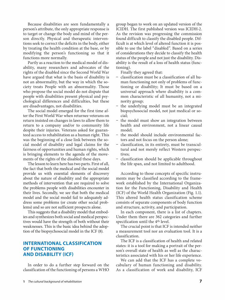
1 The cultural background of rehabilitation 5
proposed by Fulgence Raymond (1844-1910). The 20th Century was in fact the period in which bio-medical technology has an exponential develop-ment and in which the diffusion of specific dis-eases, such as poliomyelitis, determines the elabo-ration of prostheses and devices useful for the global rehabilitation of people disabled because of disease. Another major source of disability is rep-resented, in the 20th Century, by World Wars I and II. The great number of injured and mutilated soldiers induced the necessity to define the first Rehabilitation Unit within military hospitals or as charity product such as Stoke Mandeville Spinal Cord Unity in UK (2).
In Europe, the progress of medical rehabilita-tion went in the direction of further refinement of rehabilitative techniques and the proposition of new and original approaches. The physician Karel Bobath (1906-1991) and his wife Berta (1907-1991), physiotherapist, elaborated an innovative strategy for the rehabilitation of persons with dis-ability due to disorders of the central nervous system. Several other methods has been developed during the 20th Century including the more recent task oriented therapy, robotics and new technolo-gies (2).
Another historical root for rehabilitation in Europe and in particular for persons suffering from chronic diseases comes from balneology and climatology or health resort medicine, respec-tively. In the 19th and early 20th Century these treatments were used to cure chronic diseases like diabetes, cardio-vascular, lung or intestinal dis-eases as well as painful musculo-skeletal condi-tions. In some countries such as Germany this type of treatment (or rehabilitation) was included in the social security system, and some aspects of it later were integrated in modern rehabilitation concepts (3).
THE ROOTS OF REHABILITATION
Rehabilitation therapy is a very important part of PRM activity. Before 1000 BC, Taoists priests in China employed Cong Fu, as movement therapy to relieve pain. The ancient Hindus used exercises consistent with body positioning to cure chronic rheumatism (arthritis). Around 500 BC in ancient Greece, Herodicus, the Greek physician, described gymnastic exercises for the prevention and treat-ment of disease. Hippocrates was the first physi-cian to recommend therapeutic exercises. He un-derstood the principle of muscle, ligament, and bone atrophy due to inactivity (1). Hippocrates was the first to use electrical stimulation, applying torpedo-fish electric shock for headaches and Ar-istotle recommended massage rubbing with oil and water as a treatment for tiredness. The Roman physician Galen described interventions to reha-bilitate injuries in the second century, and be-lieved that moderate exercises strengthened the body, increased body temperature, allowed the pores of the skin to open, and improved a person’s spiritual well-being. During the Middle Ages, the philosopher-physician Maimonides emphasized Talmudic principles of healthy exercise habits, as well as diet, as preventive medicine in Medical Aphorisms, published between 1187-1190; and in 1569 the philologist-physician Mercurialis pro-moted gymnastics as both a preventive and a re-habilitative method in The Art of Gymnastics. In the eighteenth century, Niels Stenson explored the biomechanics of human motion and Joseph Clem-ent Tissot’s 1780 Medical and Surgical Gymnas-tics promoted the value of movement as an alter-native to bed rest for patients recovering from surgery, facing neurological conditions, and recu-perating after stroke. In the nineteenth century, the concept of neuromuscular re-education was
1The cultural background of rehabilitation
Mauro ZAMPOLINI, Christoph GUTENBRUNNER

6 PHYSICAL AND REHABILITATION MEDICINE for Medical Students
pairment; ū Handicap, disadvantage experienced by a per-
son as a result of disability or impairment/im-pairment.
This means that while disability is understood as the disadvantage that the person presents at a personal level, handicap represents the disadvan-tage of the person with disabilities.
The ICIDH provides the sequence: Impairment r Disability r Handicap, which, however, is not automatic, as the handicap may be direct as a re-sult of a disability without the mediation of the disability status.
FROM ICIDH TO ICF
The ICDH has been a step forward moving from the disease concept to their consequences but re-vealed some limitation. On the one hand ICIDH makes it clear that handicaps are primarily caused by the social reaction to people with disabilities, the key element of the social model. On the other hand, ICIDH suggests a linear pattern in which the dis-ease causes disabilities, causing disability and hence handicap, suggesting that all aspects of dis-ability start from medical conditions, the funda-mental element of the medical model.
Despite this confusion, ICIDH represents a significant advance in the disability debate. Con-ceptualization allows data collectors, political an-alysts, and researchers to identify what aspects are of relevance and to which are not. ICIDH also recognizes that disability is viewed in the light of the entire environment. ICIDH, however, was only published for field trial and derived from the consensus of a group of experts. It has not previ-ously been approved by the WHO.
The medical (or biological) pattern of disability has long been predominant, as it is in some ways closer to our disability. We commonly think that a person has a disability when there is “something wrong” with their body or mind. The medical model is just a more sophisticated version of this common idea: disabilities are deficits or physio-logical or psychological abnormalities that emerge directly from some adverse health state such as a disease, a disorder or a lesion. Disability, so to speak, resides in the person, though it has an ef-fect like the person living in his world, things he can do and the social roles he can cover.
MOVING FROM DISEASE TO DISABILITY
At its founding in 1947, the WHO left behind the old notion of health as the absence of disease. The WHO felt that health was a state of human functioning that involves the whole person in his environment.
This vision was strengthened in 1986 by the Ottawa Charter for Health Promotion, which em-phasized that although this was a feature of the person, the promotion and achievement of health necessarily involves the entire experience of the person and his environment (4).
Health promotion is the process of allowing people to increase control and improve their health. To achieve a state of complete physical, mental and social well-being, a person or group must be able to identify and realize aspirations, meet needs, change or face the environment. Health, therefore, is seen as a resource for every-day life, not a life goal. Health is a positive concept that emphasizes personal and social resources, as well as physical abilities. Therefore, health promo-tion is not only a responsibility of the health sector but goes beyond healthy lifestyles and wellbeing.
WHO’ definition of health according to Alma Ata declaration (5) envisioned health not only as the absence of a disease but also the complete physical and social wellbeing. This enlarges the perspectives of interventions far beyond the cure of diseases and including other aspects of human life experiences such as daily activities and inte-gration into society. With the UN-convention of the rights of people with disabilities (6) the con-cept of rehabilitation became part of the basic rights of persons experiencing disability. This it is consequent, that the WHO included this health strategy in its concept of Universal health cover-age and works towards implementation of reha-bilitation services wherever needed (7).
A step forward to move from the disease to the consequences has been done when, in the 1980 World Health Organization (WHO) Classifica-tion of Impairment Disabilities and Handicaps (ICIDH) distinguished between:
ū Impairment, as loss of physical or mental func-tions, and represents the extension of a patho-logical state. If this dysfunction is congenital it is a matter of disability;
ū Disability, or any limitation of the ability to act, natural consequence of a state of disability/im-
1 The cultural background of rehabilitation 7
group began to work on an updated version of the ICIDH. The first published version was ICIDH-2. As the revision was progressing the commission found difficult to classify the disabled people. Dif-ficult is at which level of altered function it is pos-sible to use the label “disabled”. Based on a series of considerations they decide to classify the health status of the people and not just the disability. Dis-ability is the result of a loss of health status (func-tioning).
Finally they agreed that: ū classification must be a classification of all hu-
man functioning not only of problems of func-tioning or disability; It must be based on a universal approach where disability is a com-mon characteristic of all humanity, not a mi-nority group;
ū the underlying model must be an integrated biopsychosocial model, not just medical or so-cial;
ū the model must show an integration between health and environment, not a linear causal model;
ū the model should include environmental fac-tors and not focus on the person alone;
ū classification, in its entirety, must be transcul-tural and not merely reflect Western perspec-tives;
ū classification should be applicable throughout the life span, and not limited to adulthood.
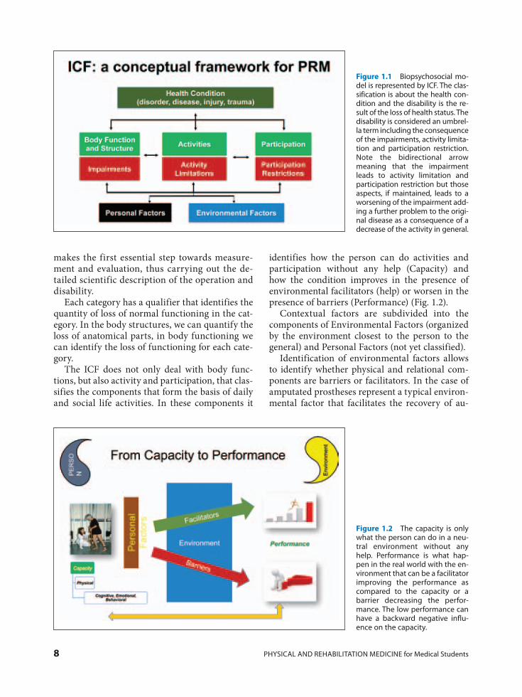
According to those concepts of specific instru-ments may be classified according to the frame-work established by the International Organiza-tion for the Functioning, Disability and Health (ICF) of the World Health Organization (Fig. 1.1). This altered health status classification scheme consists of separate components of body function and structure, activity, and participation.
In each component, there is a list of chapters. Under them there are 362 categories and further specification until the 4th level.
The crucial point is that ICF is intended neither a measurement tool nor an evaluation tool. It is a classification.
The ICF is a classification of health and related states: it is a tool for making a portrait of the per-son’s overall state of health as well as the charac-teristics associated with his or her life experience.
We can add that the ICF has a complete vo-cabulary of human functioning and disability. As a classification of work and disability, ICF
Because disabilities are seen fundamentally a person’s attribute, the only appropriate response is to target or change the body and mind of the per-son directly. Physical and therapeutic interven-tions seek to correct the deficits in the body, either by treating the health condition at the base, or by modifying the person’s functioning so that it functions more normally.
Partly as a reaction to the medical model of dis-ability, many researchers and advocates of the rights of the disabled since the Second World War have argued that what is the basis of disability is not an abnormality, but the way in which the so-ciety treats People with an abnormality. Those who propose the social model do not dispute that people with disabilities present physical and psy-chological differences and difficulties, but these are disadvantages, not disabilities.
The social model emerged for the first time af-ter the First World War when returnee veterans on return insisted on changes in laws to allow them to return to a company and/or to community life despite their injuries. Veterans asked for guaran-teed access to rehabilitation as a human right. This was the beginning of a close link between the so-cial model of disability and legal claims for the fairness of opportunities and human rights, which is bringing elements to the agenda of the move-ments of the rights of the disabled these days.
The lesson to learn here has two parts. First of all, the fact that both the medical and the social model provide us with essential elements of discovery about the nature of disability and the appropriate methods of intervention that are required to solve the problems people with disabilities encounter in their lives. Secondly, we see that both the medical model and the social model fail to adequately ad-dress some problems (or create other social prob-lems) and so are not sufficient prospects alone.
This suggests that a disability model that embod-ies and synthesizes both social and medical perspec-tives would have the strength of both without their weaknesses. This is the basic idea behind the adop-tion of the biopsychosocial model in the ICF (8).
INTERNATIONAL CLASSIFICATION OF FUNCTIONING AND DISABILITY (ICF)
In order to do a further step forward on the classification of the functioning of persons a WHO
8 PHYSICAL AND REHABILITATION MEDICINE for Medical Students
identifies how the person can do activities and participation without any help (Capacity) and how the condition improves in the presence of environmental facilitators (help) or worsen in the presence of barriers (Performance) (Fig. 1.2).
Contextual factors are subdivided into the components of Environmental Factors (organized by the environment closest to the person to the general) and Personal Factors (not yet classified).
Identification of environmental factors allows to identify whether physical and relational com-ponents are barriers or facilitators. In the case of amputated prostheses represent a typical environ-mental factor that facilitates the recovery of au-
makes the first essential step towards measure-ment and evaluation, thus carrying out the de-tailed scientific description of the operation and disability.
Each category has a qualifier that identifies the quantity of loss of normal functioning in the cat-egory. In the body structures, we can quantify the loss of anatomical parts, in body functioning we can identify the loss of functioning for each cate-gory.
The ICF does not only deal with body func-tions, but also activity and participation, that clas-sifies the components that form the basis of daily and social life activities. In these components it
Figure 1.1 Biopsychosocial mo-del is represented by ICF. The clas-sification is about the health con-dition and the disability is the re-sult of the loss of health status. The disability is considered an umbrel-la term including the consequence of the impairments, activity limita-tion and participation restriction. Note the bidirectional arrow meaning that the impairment leads to activity limitation and participation restriction but those aspects, if maintained, leads to a worsening of the impairment add-ing a further problem to the origi-nal disease as a consequence of a decrease of the activity in general.
Figure 1.2 The capacity is only what the person can do in a neu-tral environment without any help. Performance is what hap-pen in the real world with the en-vironment that can be a facilitator improving the performance as compared to the capacity or a barrier decreasing the perfor-mance. The low performance can have a backward negative influ-ence on the capacity.

1 The cultural background of rehabilitation 9
prototype environmental factors. Methodologi-cally, the development of the Core Sets has been obtained with a consensus process based on Del-phi round, a focus groups meeting.
In spite of the several years of implementation the diffusion of ICF in practical rehabilitation ac-tivities it is not yet widely diffused. One of the many challenging aspects is then to find a way to simplify selecting the most useful categories de-veloping a minimal generic set of the domain of rehabilitation (11). The perspective is to use the validated scales and translate the score in the qualifiers quantification of health loss (12).
Another very important issue is related to the use of ICF in quality management in rehabilita-tion (13).
In order to strengthen the rehabilitation and abilitation approach to the disability World Health Assembly (WHA) in May 2014 adopted a global disability action plan 2014-2021:“better health for all people with disabilities” (14). The slogan is “to contribute to achieving optimal health, function-ing, well-being and human rights of all persons with disabilities”. This action plan is based on the concept that all persons with disabilities and their families should live in dignity, with equal rights and opportunities and able to achieve their full potential (15). The action plan has three objec-tives:1. to remove barriers and improve access to health
services and programmes;2. to strengthen and extend rehabilitation, habili-
tation, assistive technology, assistance and sup-port services, and community-based rehabilita-tion;
3. to strengthen collection of relevant and inter-nationally comparable data on disability and support research on disability and related ser-vices (16).
The action plan has several implications for practice in rehabilitation.
The overall goals should contribute to achiev-ing optimal health, functioning, well-being and human rights for all persons with disabilities. There is explicit mention that the dignity of per-sons with old age need special attention. Rehabili-tation is needed in all phases of health care ser-vices including acute, post-acute, and long-term rehabilitation to guarantee the continuum of care.
The action plan emphasises the importance of the outcome research collecting data systemati-
tonomy and social reintegration by restoring the possibility of displacement.
An important achievement of this model is that it helps to understand disability as and interaction of a person with a health condition and the envi-ronment. This implies that disability is neither seen as a purely medical nor purely social problem but integrates both aspects. Furthermore it clarifies that disability cannot be seen as an attribute of a person.
ICF can thus become a universal language able to identify problems in a standardized manner, quantifying the severity both in terms of person’s ability and performance in real mono. This quan-tification can be achieved using the scales already available and inserting them into the ICF frame-work.
In order to have a scale based on ICF the WHO developed the WHO-DAS II is the WHO-based inclusive assessment tool. Its reliability and valid-ity are proven, and has been adopted by the WHO for its studies. WHO-DAS II is intended for adults only and relates directly to some ICF domains. It is prepared in either a short version of 12 ques-tions - and in a longer version of 36 questions.
After a few years, a new version of ICF has been developed inserting some additional categories appropriate for young and children.
The ICF model and classification also was used to describe rehabilitation as a health strategy to decrease disability and to support functioning (9). The goal of rehabilitation has been defined as “to enable people with health conditions experiencing or likely to experience disability to achieve and maintain optimal functioning in interaction with the environment”. In parallel the contribution of Physical and Rehabilitation Medicine has been defined using the same ICF-based concept (10).
The ICF classification is very extensive and the risk is to be complicated and time wasting to be applied in the practical activity. In order to over-come this problem, the German OMS Collabora-tor Center, ICF research branch, namely the De-partment of Physical Medicine and Rehabilitation at the University of Munich, Germany in collabo-ration with WHO, developed ICF Core Sets for different types of diseases, based on research projects and an international consensus process. Core Set must consider not only medical aspects of the health condition – impairments in body functions and structures – but also limitations on the activity and the associated restrictions on par-ticipation. In addition, the core set must include
10 PHYSICAL AND REHABILITATION MEDICINE for Medical Students
ing an extensive picture of the situation of people with disabilities (7).
THE ROLE OF CULTURAL AND PERSONAL ENVIRONMENT
The “social model of disability” was pioneered in the late 1960s and early 1970s, and continued to gain momentum and acceptance in the decades that followed. This model, introduced by people with disabilities, human rights activists and social theo-rists, specified that disability is not simply related to a person’s impairment, but rather a complex phe-nomenon, created in part by features of the physical and social world. It is the environment that acts to facilitate integration or contribute to isolation, in-fluencing a person’s ability to participate in society.
The recognition of the environment’s influence on the experience of disability and the implemen-tation of Disability Discrimination Acts in vari-ous countries have led to the development and implementation of programs and initiatives for improving the environment experienced by a per-son with disabilities.
ū The concept of Universal Design, where the un-derlying principle is the design of products, buildings and environments that are useable by all people. Issues such as accessibility, safety, individual ability and efficiency underpin the design of articles that make up the physical world, from buildings and forms of transporta-tion to computer and Internet access, and prod-ucts used in the home.
ū Integration of students with disabilities into mainstream or regular educational settings. En-abling students with disabilities to participate in a regular education setting is thought to improve rather than hinder both academic and social learning (17).
ū Schemes for the provision of aids and equipment, where individuals receive cost-free or low-cost equipment to help their performance of daily activities, such as self-care and mobility in and outside the house, and facilitate participation in sport, work, education and other activities.
ū Improvement of standards for accessible public transport. Transport is a fundamental human right, including having ready access to safe and disability-friendly forms of public transport, but remains a common problem for people with dis-abilities.
cally using ICF. The data collection should taking in account the perspective of the persons with disability into the decision making process in data collection and research (15).
PERSONS WITH DISABILITY AND THEIR RIGHTS
Persons with disability have their right aimed to obtain and independent living throughout the best inclusion possible.
To define that, a real milestone has been the Convention for the Rights of Persons with Dis-abilities (CRPD) approved in July 2009 from 59 countries, 37 of them have ratified its Optional Protocol and 139 have signed the CRPD (6).
The Convention on the Rights of Persons with Disabilities clearly define that persons with dis-abilities have equal access and a right to full and effective enjoyment of all human rights – the re-moval of barriers explicitly termed as a condition for access and the enjoyment of equality.
The Disability Convention features eight gen-eral principles which underpin all the rights con-tained within the Disability Convention. They are:
ū respect for inherent dignity, individual autono-my – including the freedom to make one’s own choices – and independence of persons
ū non-discrimination ū full and effective participation and inclusion in
society ū respect for difference and acceptance of per-
sons with disabilities as part of human diversity and humanity
ū equality of opportunity ū accessibility ū equality between men and women ū respect for the evolving capacities of children
with disabilities and respect for the right of chil-dren with disabilities to preserve their identities.
The implication for the practice in rehabilita-tion is the needs to involve the disabled person and their proxies to the rehabilitation programs sharing decision and tailoring the goals according with the needs and aspiration of the person.
In order to analyse the condition of disabled people The World Report on Disability is the first of its kind, providing global guidance on imple-menting the United Nations Convention on the Rights of persons with Disabilities (CRPD) and giv-
1 The cultural background of rehabilitation 11
two aspects can be referred to two conceptual and practical elements of the ICF. The capacity of the person must be evaluated from an external, objec-tive perspective, through validated measurement. The performance, on the other hand, that happens in the real life environment must be assessed tak-ing in account the perspective of the patient. In this case, it is increasingly necessary to develop the research to find and validate measuring scales that can capture this perspective. One of these is the WHO-DAS 2 which, starting with simple ques-tions, can capture the perspectives of the sick person always within the ICF reference system.
The ICF framework should be, at micro-level the basis for quality management in rehabilitation but also a reference point at meso-level to improve the quality of the organization of rehabilitation facilities and at the macro level to improve the planning of rehabilitation policies in favours of disabled people.
A WAY FORWARD
The main change of the disability concept of the problem related to the person (ICIDH) to a result of interaction among a person and environ-ment (ICF) has a number of practical implications. According to this vision the rehabilitation pro-grams should be focused not only on the improve-ment of the capacity of the person obtained with the physical exercise but the global goals should be aimed to obtain a good performance in the real. The rehabilitation process should be focused on improving the capacity of the patient and in adapting the environmental factors (physical bar-riers, human relationships, social policies, and others). Starting from now, it is very important to implement this methodology within the rehabili-tation process allowing fa quality improvement combining the patient’s perspective with the ap-propriateness of rehabilitation intervention. These
Key messages
• Since over 2000 years it is possible to find documents about the importance of exercises for the health. Only after the II World War there was a development of rehabilitation for the war injuries and for the consequences of infections such as poliomyelitis.
• Moving from the concept of health related to the absence of disease to the human functioning rehabilitation gained a further importance.
• The International Classification of Functioning (ICF) represents now the basic framework of re-habilitation introducing the concept of the disability as the result of interaction between person and environment.
• Several studies are carrying out aimed to use the ICF as assessment tools, to set the goals of the rehabilitation project and as a base of quality management in rehabilitation.
REFERENCES
1. Conti AA. Western medical rehabilitation through time: a historical and epistemological review. Sci World J. 2014; 432506.
2. Dreeben-Irimia O. Development of the physical therapy profession. In: Introduction to Physical Therapy for Physical Therapist Assistants, chapter 1. Sudbury, MA: Jones & Bartlett Learning, LLC; 2007. p. 3-22.
3. Amelung W, Gutenbrunner C. Handbuch der Bal-neologie und Medizinischen Klimatologie. Berlin: Springer; 1998.
4. Government of Canada. Ottawa Charter for Health Promotion: an International Conference on Health
Promotion. 1986. Available from: https://www.canada.ca/en/public-health/services/health-pro-motion/population-health/ottawa-charter-health-promotion-international-conference-on-health-promotion.html
5. International Conference on Primary Health Care. Declaration of Alma-Ata. WHO Chron. 1978 Nov; 32(11): 428-30.
6. United Nations. Division for Social Policy and De-velopment - Disability. Convention on the Rights of Persons with Disabilities (CRPD). 2006. Avail-able from: https://www.un.org/development/desa/disabilities/convention-on-the-rights-of-persons-with-disabilities.html
7. World Health Organization. The World Health Re-
12 PHYSICAL AND REHABILITATION MEDICINE for Medical Students
13. Stucki G, Zampolini M, Juocevicius A, Negrini S, Christodoulou N. Practice, science and gover-nance in interaction: European effort for the system-wide implementation of the Interna-tional Classification of Functioning, Disability and Health (ICF) in Physical and Rehabilitation Medicine. Eur J Phys Rehabil Med. 2017; 53: 299-307.
14. WHO. Global disability action plan 2014-2021 [Internet]. WHO. 2014 [cited 2018 Jan 26]. Avail-able from: http://www.who.int/disabilities/action-plan/en/
15. Gutenbrunner C, Negrini S, Kiekens C, Zampolini M, Nugraha B. The Global Disability Action Plan 2014-2021 of the World Health Organisation (WHO): a major step towards better health for all people with disabilities. Chance and challenge for Physical and Rehabilitation Medicine (PRM). Eur J Phys Rehabil Med. 2015; 51: 1-4.
16. World Health Organization. WHO Global disabil-ity action plan 2014-2021: Better health for all people with disabilities. Geneva: WHO; 2014.
17. Center Y, Curry C. A Feasibility Study of a Full Integration Model Developed for a Group of Stu-dents Classified as Mildly Intellectually Disabled. Int J Disabil Dev Educ. 1993; 40: 217-35.
port. Health systems financing: the path to univer-sal coverage. Geneva: WHO; 2010. Available from: http://www.who.int/whr/2010/en/
8. World Health Organization. International Classi-fication of Functioning, Disability and Health (ICF). Geneva: WHO; 2001. Available from: http://www.who.int/classifications/icf/en/
9. Meyer T, Gutenbrunner C, Bickenbach J, Cieza A, Melvin J, Stucki G. Towards a conceptual descrip-tion of rehabilitation as a health strategy. J Rehabil Med. 2011; 43: 765-9.
10. Gutenbrunner C, Meyer T, Melvin J, Stucki G. Towards a conceptual description of Physical and Rehabilitation Medicine. J Rehabil Med. 2011; 43(9): 760-4.
11. Prodinger B, Reinhardt JD, Selb M, Stucki G, Yan T, Zhang X, et al. Towards system-wide implemen-tation of the International Classification of Func-tioning, Disability and Health (ICF) in routine practice: Developing simple, intuitive descriptions of ICF categories in the ICF Generic and Rehabili-tation Set. J Rehabil Med. 2016; 48: 508-14.
12. Prodinger B, Tennant A, Stucki G. Standardized reporting of functioning information on ICF--based common metrics. Eur J Phys Rehabil Med. 2018; 54: 110-7.
2 The biological and clinical background of rehabilitation 13
wishes can significantly influence the outcome of a therapeutic approach and adherence to treat-ment. Therefore, the information obtained from the ICF is useful not only for studying disability but also and above all for choosing the most ap-propriate methods and interventions (2).
Rehabilitation is usually described using three axes:
ū structure, i.e. staff, equipment, facilities ū process, i.e. the whole set of actions aimed at
defining the rehabilitation plan, including the diagnostic and prognostic evaluation, goal set-ting and intervention scheduling
ū outcome, i.e. the level of functioning achieved by subjects after the rehabilitation intervention, not only in the short-term, but also in the me-dium- and long-term.
Rehabilitation as a problem-solving process, that is planned and implemented by a specialized team, has proven to be effective in reducing morbidity and mortality in most disabling disorders both in the acute phase and in the management of chronic conditions associated with reduced mobility.
Based on the evidence of the efficacy of either motor or cognitive training at re-shaping brain networks, due to the phenomenon of experience-driven brain plasticity, the interest into the theo-retical background of rehabilitation efficacy has grown significantly, leading to the diffuse aware-ness that any approach able to reduce motor, cog-nitive, behavioral or emotional impairment, will always involve a neural reorganization.
BASIC CONCEPTS OF FUNCTIONAL PROGNOSIS AFTER ACUTE DISABILITY ONSET
The onset of a sensor, motor or cognitive im-pairment as a result of injury or disease can deter-mine a variable limitation in the activities per-
INTRODUCTION
Rehabilitation is a problem-solving educational process, that is aimed at reducing activity limita-tions, optimizing social participation and patient well-being and limiting the stress of caregivers (1).
This definition highlights some important char-acteristics: 1) the attention of the rehabilitation is directed to treat the patient as a person and 2) the objectives refer to the social functioning, as well as to the health and the psycho-physical well-being, irrespective of disability kind (motor, sensory or cognitive), type of onset (acute or subacute), or se-verity.
Rehabilitation has a solid theoretical and con-ceptual basis derived from the International Clas-sification of Functioning, Disability and Health (ICF) of the World Health Organization (2), de-scribing the consequences of a health condition in terms of functioning and health experience. The description of functioning and disability takes into account three different perspectives: body, person, and person in a context. The body and the person are described in terms of body functions (physiological and psychological functions), body structures (anatomical parts of the body, organs, limbs and their components), activities (including the whole list of goal-oriented tasks any individu-al can perform) and participation (namely, the involvement of a person in a life situation). The ICF also provides a description of performance and capacity: the first indicates what the individ-ual does in his/her own real environment, while the second quantifies the highest level of func-tioning achievable without the help/interference of any environmental factor. Contextual factors include both personal factors, such as age, gender or education, and environmental factors, which refer to the physical and social environment in which people live and to attitudes of family, peers and other relevant individuals. Among the per-sonal factors, the patient’s will, expectations and
2The biological and clinical background of rehabilitation
Maria Gabriella CERAVOLO
14 PHYSICAL AND REHABILITATION MEDICINE for Medical Students
differing by age or gender, and are often associated to different patterns of clinical, functional, medical and social consequences. An important prognostic role is played by the site and size of damaged struc-tures, the severity of emerging motor, sensory or cognitive impairment and the combination of in-jured systems: in particular, the addition of a cog-nitive dysfunction to any emerging motor disabil-ity adversely affects the recovery, by either hinder-ing or slowing the motor re-learning process.
The severity of an emerging disability, as quan-tified using generic measures, like the Barthel ADL Index or the Functional Independence Mea-sure (FIM), is one of the most powerful predictors of recovering the pre-morbid independence level and returning home after discharge from the acute ward (5). For instance, the FIM total score at rehabilitation start is the main predictive factor of the FIM score achieved at the end of treatment, in any individual experiencing an acute disability onset; moreover, the trunk control measured in the acute stroke phase, using a simple quantitative instrument such as the Trunk Control test, allows to predict not only the probability of recovering the standing and walking ability, but also of achieving a high level of independence and being discharged home after the event (6).
On the other hand, severe disability, pre-exist-ing CNS damage, or severe cognitive impairment reduce, if not exclude, the effectiveness (and use-fulness) of the rehabilitative approach.
The use of a simple classification, like the modified Rankin scale, which ranks the severity of functional impairment in six levels (from 0 = no symptoms at all, to 5 = severe disability; bedrid-den, incontinent and requiring constant nursing care and attention) proves extremely useful if ap-plied retrospectively to outline the pre-morbid independence profile. Such information will strongly influence the functional prognosis, help-ing to determine the gap induced by the illness in the individual functioning, set the maximum level of expected recovery and help to define the risk for complications (the higher the Rankin score, the greater the risk).
Finally, the absence of any family/social net-work member, willing to play the caregiver role, would substantially increase the risk of subjects’ institutionalization, even in case of a mild residu-al disability at the end of the rehabilitation pro-cess, and could nullify the several benefits ob-tained with an intensive training: in such cases, it
formed by the individual, based on the complex interplay between preserved capacity and the personal and contextual factors.
This adaptation process can be particularly dif-ficult in the case of chronic-progressive diseases, in which subject’s expectations must be continu-ously re-modulated as functional capacities shrink.
The goal of rehabilitation is therefore to guide the individual in the difficult path of achieving the highest level of functioning made possible by circumstances. The process necessary for this purpose requires that any care/rehabilitation deci-sion be preceded by a correct clinical-functional assessment. This must use reliable, standardized tools in order to outline the individual function-ing profile, based on body structures and body functions, activities and behaviors implemented by individuals (3).
The first aim of the evaluation is to determine the gap between the present level of functioning (in terms of independence in basic and instru-mental activities of daily living) and that ex-pressed immediately before the onset of illness; in case of congenital disorders this assessment is not possible, so the reference is represented by the functional abilities of individuals of the same age.
The second objective is to establish the indi-vidual potential for recovering the observed gap, or otherwise the risk for further functional de-cline.
The third objective is to select the rehabilitation strategy that best suits the circumstances, with re-spect to the residual abilities of the subject, his/her expectations, the natural history of the disease and the environmental opportunities/constraints (4).
Given these premises, rehabilitation must be regarded as a process targeting non-stereotyped (unknown a priori) aims, making each rehabilita-tion project a unique experience. Notwithstand-ing, even with a large degree of flexibility, there are rules to be abided and precise steps to be fol-lowed when planning a rehabilitation interven-tion. In the subsequent section, a few basic con-cepts driving the formulation of the functional prognosis will be explained.
The functional prognosis: when and why
Central nervous system (CNS) disorders, such as stroke, head or spinal injury, infective or in-flammatory diseases, usually show incidence rates
2 The biological and clinical background of rehabilitation 15
traced back to a series of competitive processes, then they could end when the neural circuits reach an attitude that prevents any further competitive interaction. The end of the critical period could therefore be due not only to the loss of the inherent plastic capacities of the nervous system, but also to the fact that the neural circuits have reached a con-figuration of stable connections which effectively prevent any further interaction between the ner-vous elements. This hypothesis would explain, for instance, why the critical period is prolonged when experience is delayed or missing.
Irrespective of what interpretation will be ac-credited by future studies, it is shown that plastic-ity phenomena are maximally active in the devel-opmental phase of the CNS, during intrauterine life and in postnatal age, while they shrink sig-nificantly after the CNS has achieved complete maturation. Even in this phase, however, the CNS ability to undergo changes is not extinguished: in fact, not only children, though also adults, can change their behaviors, learn new information, memorize new events. The plastic modifications of the CNS which are the basis for learning and memory consist in continuous modifications of the effectiveness of signal transmission between neurons. Experiments conducted in the last 20-30 years have revealed that the efficacy of many syn-apses can be modified for very short time dura-tions (namely, milliseconds up to minutes, in the so-called short-term synaptic plasticity) or for up to several months/years (in the long-term synaptic plasticity). At present, it is assumed that the learn-ing process is associated to long-term modifica-tions of synapses: these variations can be regarded as the biological correlate of a mnemonic trace and the neural substrate of learning new motor skills (i.e. dancing, playing sports or music), in the healthy state, or re-learning motor skills, that have been compromised by an injury or illness.
POST-LESIONAL NEUROPLASTICITY AND THE PRINCIPLES OF MOTOR LEARNING
The concept of neuroplasticity is certainly in-novative. A few years ago, researchers in neurosci-ence hypothesized that rehabilitation efficacy could be attributable to the exercise-dependent change in brain function and structure. Thanks to some pio-neers (9, 10), it was possible to demonstrate that
would seem more appropriate, in the sake of cost-effectiveness, to involve the patient in an extensive rehabilitation program, rather than refer him to an intensive rehabilitation facility (7).
Whatever the setting defined in the acute phase for ensuring a continuum of care, the rehabilita-tion project recognizes a standard series of critical steps, whose ultimate goal is “to assist patients in achieving optimal physical, emotional, social, psychological and vocational functioning, within the limits imposed by the clinical picture and the available therapeutic options”.
In conclusion, the prognostic evaluation is mainly oriented to establish whether or not: a) the emerging disability is either severe or complex enough to make an intensive rehabilitation appro-priate; b) the individual expresses any recovery potential and a sufficient level of compliance to an intensive rehabilitation program, based on his/her tolerance to physical effort, learning ability and motivation; c) the environmental context will be able to cope with the subject’s health needs, either in the short or medium term, in order to allow him/her to return home.
The integration of this information will shape the delivery of a continuum of care across differ-ent rehabilitation setting in the post-acute phase.
NEUROPHYSIOLOGICAL BASIS OF RECOVERY AFTER ACUTE LESION OF THE NERVOUS SYSTEM THE CONCEPT OF NEUROPLASTICITY
The nervous system consists of complex neuro-nal networks specialized in the control of different vital functions, such as the sensory representation of the external world, the production of behaviors or the regulation of vegetative activities.
Neural plasticity is the ability of the CNS to change and adapt in response to environmental signals, experience, behavior and, eventually, acute or chronic progressive diseases (8).
Several observations indicate that the conditions necessary for the plastic processes to take place are present only during a precise time window, at the end of which such processes are actively sup-pressed. In other words, the genetic program deter-mines the start and the end of a critical period for the development of functionally specialized neural networks. However, there is also an alternative in-terpretation. If the plasticity phenomena can be
16 PHYSICAL AND REHABILITATION MEDICINE for Medical Students
picking a key and using it to unlock a door) induces a greater emotional arousal than per-forming intransitive gestures. Goal-oriented activities are especially useful due to their abil-ity to drive subject’s attention towards the task and improve learning capacity.
6. Contextual interference. Learning new tasks may be slowed down or prevented by the com-petition with previously acquired and consoli-dated skills or concomitant sensory experienc-es. In order to avoid such effect, it is recom-mended that different tasks are simultaneously trained in a random sequence rather than let patients master any ability at the maximum level before asking them to train a new one. For example, to develop a strong manual grip it is useful to ask the patient to pick a glass, then a spoon, then a mobile phone, according to a random, unpredictable sequence. A similar ap-proach stimulates the idea that each exercise represents a problem to be solved, rather than a temporal sequence of repetitive movements in a stereotypical mode.
7. Use it or lose it. If an intense sensory experi-ence, related to a repetitive practice, promotes learning and its consolidation over time, the lack of experience, or sensory deprivation, in-evitably leads to function decline and to the loss of previously learned skills. Hence, the lack of practice induces a phenomenon of “learned non-use”, following the missing activation of the neural substrates of that specific skill: the depotentiation of synaptic connections is the basis for the so-called “maladaptive plasticity”.
TRADITIONAL AND INNOVATIVE REHABILITATION TECHNIQUES AND THEIR RATIONALE OF USE
Neurophysiological techniques
Neurophysiological techniques refer to neuro-physiological mechanisms of motor impairment; they do not require the cooperation of the patient, who plays a passive role. The most important and used are:
ū Bobath Method (or Bobath Concept): it was developed as a technique for neuromotor re-conditioning in children with cerebral palsy and has been adapted and used also in adults. Currently, it is probably the most widely diffuse
repetitive motor training is able to induce persis-tent structural changes in the cortical representa-tions of the trained movements, and that these changes are directly related to the improvement in motor performances after the CNS damage.
Numerous experiments carried out in animal models have documented how, following an acute focal CNS lesion, a plastic reorganization takes place, both due to the activation of functionally quiescent anatomical connections, starting when the dominant connections are damaged, and to the sprouting of cortical axons, i.e. the growth of nerve fibers that reach new cellular targets, in re-sponse to sensory inputs, thus generating new synaptic contacts (synaptogenesis).
The remodeling of neural maps following in-jury does not however take place in a stereotypical mode, but is strictly experience-driven. The expo-sure to external sensory stimuli (and to the inter-nal feedbacks generated by the individual motor behavior) can modulate both the entity and speci-ficity of neuroplasticity processes, provided that specific requirements are met, as those highlight-ed below (11):1. Specificity. The congruence of the trained activ-
ity with the skill to be learned or re-learned plays a crucial role in brain reorganization. In practical terms, to learn to ride a bike you have to pedal, to relearn walking after suffering a lower limb paresis, you have to walk, and so on.
2. Repetition. Each single component of the task to be learned must be repeatedly trained, in order to facilitate the consolidation of the syn-aptic connection.
3. Intensity. The duration and frequency of train-ing sessions must be sufficiently high to obtain maximum benefits in the shortest possible time.
4. Timeliness. In those who have suffered a CNS injury, more than in other disease scenarios, the earlier the training is started the greater the opportunities for functional recovery through neuroplasticity mechanisms. The optimal time window for obtaining the maximum result from motor training, for the purposes of func-tional recovery after stroke, is estimated to be around 12 weeks.
5. Salience. The active participation of the subject in training is as important as doing a massive practice. For example, training movements usually performed in daily living (e.g. drinking from a cup, fastening or unfastening a button,

2 The biological and clinical background of rehabilitation 17
ties of daily life. It refers to the theory of a cen-tral control of movement, considering that the execution of goal-oriented movements (i.e. of movements performed in a specific context for the solution of a specific problem), is binding in order to allow the optimal recovery of impaired motor skills.
Constraint-Induced Movement Therapy
The Constraint-Induced Movement Therapy (CIMT) was developed by Edward Taub based on the observation of the “learned non-use” phenom-enon in hemiparetic stroke subjects. The treat-ment proposed by Taub combines an intensive use of the paretic limb with the movement restriction and sensory deprivation of the healthy limb, by the application of a bandage or splint. It has been shown that, following the use of CIMT, a cortical reorganization takes place, mostly favored by the positive component (intensive use of the paretic limb) rather than the negative one (sensory depri-vation of the healthy limb) (12).
Action observation, motor imagery and mirror therapy
Recent research suggests that both imagination and observation of movements may represent an additional source of information useful for the recovery of motor function in patients suffering a CNS injury. Using functional Magnetic Reso-nance Imaging, it has been shown how brain areas normally involved in the planning and execution of a movement (the prefrontal cortex, the premo-tor cortex and some extra motor area (like the cingulate cortex, the parietal cortex and the cere-bellum) are active even when the same movement is imagined, but not executed. Other studies have documented that even the observation of a gesture performed by others is sufficient to activate the same cortical areas competent for the execution of that gesture. The neural substrate that presides these phenomena is the system of mirror neurons. The mirror neuron function is relevant both for learning by imitation and for understanding oth-ers’ intentions. Rizzolatti et al. discovered that a group of neurons in the parietal cortex of mon-keys were active not only when the animals took a peanut, but also when they observed the experi-menter perform the same action. Afterwards, the
method in Europe. It is based on a passive limb mobilization, associated with tactile and pro-prioceptive stimuli, and the maintenance of postures, mainly oriented to reduce spasticity in order to allow a more effective activation of muscles used in functional movements.
ū Brunnstrom method: this technique is aimed at promoting motricity recovery, using both re-flex activity and proprioceptive and exterocep-tive stimuli to evoke desired motion or muscle tone changes.
ū Vojta method: developed to treat newborns with congenital brain damage, it is based on the stimulation of specific nerve endings to pro-mote the development of physiological move-ment patterns. It has been also applied to adults with focal brain injury.
ū Rood method: it refers to a hierarchical organi-zation of the CNS and emphasizes the use of sensory inputs to produce and modify motor responses; the developmental stage and abilities of the patient are used to develop purposeful responses.
ū Johnstone method: it mainly aims at control-ling the pathological reflex systems causing spasticity and the consequent alteration in vol-untary movements and posture. It uses posi-tioning and limb immobilization in order to inhibit pathological patterns, avoid muscle hy-pertonia and allow the restoration of central control.
Motor learning techniques While neurophysiological techniques are rela-
tively independent of the patient’s collaboration, motor learning techniques (11) need the active participation of the subject in the exercise as a mandatory requirement.
ū Perfetti method: widely used in Italy, it has been initially devised to manage spasticity, but has been subsequently applied in several disor-ders even not related to CNS damage. Also called “Cognitive Therapeutic Exercise”, it aims at stimulating the cortical mechanisms of learning such as perception, memory, concep-tion and motivation, looking to the intercon-nection between functional movements and the patient’s interaction with the environment.
ū Task-Oriented Training: the therapeutic inter-vention focuses on specific tasks and offers contextualized exercises that reproduce activi-

18 PHYSICAL AND REHABILITATION MEDICINE for Medical Students
movements just stimulate motor imagery, thus activating the networks responsible for the execu-tion of the same movements.
Non-invasive cortical stimulation techniques
The application of cortical stimulation, with the intent of increasing/decreasing the excitability threshold of the motor cortex, was introduced in the 1980s, using the repetitive Transcranial Mag-netic Stimulation (rTMS). This technique exploits the electrical impulse, flowing in a coil applied on the head, to induce a magnetic field of very short duration; the transition of a magnetic field, across the skull, to the underlying nervous tissue, gener-ates an electric field that is able to modify the neuron membrane potential.
The mechanisms by which rTMS determines persistent cortical reorganization include, hypo-thetically: a) the enhancement or suppression of synaptic circuits, b) the modulation of gabaergic and glutamatergic systems, through an action on the NMDA receptors, and c) the modulation of gene expression, through the increase of BDNF mRNA, a neurotrophic factor implicated in the mechanism of neuroplasticity. rTMS protocols exert variable neurophysiological effects, depend-ing on the stimulus frequency, the length of the interstimulus interval (ISIs) and the duration of the stimulation sessions. For example, while fre-quencies >5 Hz increase cortical excitability, those ≤1 Hz cause intra-cortical inhibition of the stimu-lated hemisphere. Short ISIs (2-5 ms duration), and long ISIs (100-200 ms) are followed by intra-cortical inhibition phenomena, whereas interme-diate ISIs (7 to 20 ms duration) obtain the facilita-tion of cortical activity and increase the excitabil-ity of glutamatergic circuits.
The clinical evidence of efficacy of rTMS ap-plication in rehabilitation mainly concerns sub-jects with stroke (17). An improvement in the pa-retic upper limb function was observed when the cortical stimulation session was combined with the motor training, and, more exactly, when it preceded exercise by a few minutes. This finding has led to the hypothesis that an rTMS protocol aimed at reducing the excitability threshold in the primary motor cortex exerts a priming effect, fa-cilitating the activation of the sensorimotor areas where movement is represented and the creation of new synaptic circuits.
same group of researchers provided the first evi-dence that mirror neurons also existed in hu-mans, demonstrating that the simple observation of a movement is effective at facilitating its subse-quent execution (13). Rehabilitation protocols based on action observation, as a means to foster motor re-learning, have successfully enhanced paretic upper limb recovery in stroke survivors (14), walking improvement in subjects with Par-kinson’s disease and verbs production in subjects with non-fluent aphasia (15).
Motor imagery can be defined as the cognitive process of imagining to move the body (limb) without actually moving it. Over the last decade, studies on motor control have shown that there are close analogies between real and imagined movements, like in the phenomenon known as “mental isocronia”, where the time taken to com-plete an imagined movement is similar to that needed to actually execute it. Moreover, in line with the theories and scientific evidence regard-ing mirror neurons, it has been found that the brain areas activated during the imagination of certain actions are the same as those activated during their actual execution.
The application of motor imagery protocols in rehabilitation is still considered experimental, even if preliminary experiences regarding the re-covery of the upper limb function after stroke have given encouraging results.
Mirror Therapy. Ramachandran was the first to use the visual illusion created by a mirror to treat phantom limb pain (16). Patients who looked at the image of the healthy limb in a mirror had the illusion that the observed movement was per-formed by the contralateral (amputated) limb: in this way, they developed the perception of being still able to move and relax the amputated arm and perceived relief from pain. Following this first evi-dence, mirror therapy has been used to treat com-plex regional pain syndrome (CRPS), and manage severe dysesthesia after hand trauma; moreover, it has been successfully applied to enhance upper limb motor recovery in stroke survivors, by creat-ing the illusion of the paretic limb movement.
The mechanisms explaining the effectiveness of mirror therapy are likely different.
Some believe that the proprioceptive inputs from the affected limb, triggered by the illusion provided by the mirrored healthy limb movement, enhances the neuron excitability in the premotor cortex. Others hypothesize that the mirrored
2 The biological and clinical background of rehabilitation 19
vation in the paretic lower limb, synchronized with the movement of the contralateral limb: the aim is to produce a symmetrical pedaling and thus train the cyclic gait pattern. A few studies suggest that FES-cycling is effective at strengthen-ing the paretic muscles and the cycling perfor-mance, while it is still argued whether it also im-proves walking significantly, especially in patients who have not yet recovered independent gait.
Virtual reality rehabilitationVirtual reality is a computerized technology
providing digital sensory information and allow-ing people to interact with/within three-dimen-sional environments. In recent years, such tech-nique has been frequently implemented in reha-bilitation protocols of motor and cognitive train-ing. The fundamental novelty of virtual reality-based rehabilitation protocols is their ability to induce the perception, in the subjects, to be part of a simulated environment.
Simulation can be achieved in an either immer-sive or non-immersive modality.
Immersive augmented reality is typically ob-tained using a head-mounted display (HMD), worn on the head, provided with a small display in front of one or both eyes. Virtual reality devices applied in rehabilitation usually display only com-puter-generated imagery: this mode provides a three-dimensional view and correlates the move-ments of the head with the virtual environment, with the result that the perceived environment changes with the movements of the user giving the feeling of being within the environment itself.
The non-immersive mode provides the vision of the virtual environment on a computer monitor or projected on a screen, giving the subject the perception of looking through a window (20).
To enhance feedbacks generated by the subject’s movements, special sensory gloves or joysticks can be used. The gloves allow subjects to see their hand movements projected onto the screen and feel feedbacks in the form of tactile sensation. The joysticks allow subjects to interact with the virtual environment, giving them different feedbacks de-pending on the speed and precision of move-ments, or on the power of muscle contraction.
In summary, virtual reality training: ū allows to shape training intensity and difficul-
ty, based on the patient’s progresses in motor performances;
Transcranial Direct Current Stimulation (tDCS) consists in the release of a continuous electric cur-rent, of low intensity (1-2 mA), that flows between two poles, an active electrode, applied on the head over the target cortical area, and a reference one, placed either on the supraorbital region or on the shoulder (18). The neuroplastic effects induced by tDCS are explained both by the action on the ionic channels, that leads to the redistribution of the Na+ and Ca+ ions at the intra- and extracellu-lar level, and by the action on the NMDA recep-tors, that modulates the neurotransmitter sys-tems: the net result is a change in the resting membrane potential, either for a hyperpolarizing inhibitory effect of a cathodal stimulation, or a depolarizing excitatory effect of an anodal stimu-lation. In stroke patients, the inhibition of the unaffected hemisphere, contralateral to the dam-aged brain area, is pursued in order to reduce the interhemispheric inhibition. The physiologic competition between the two hemispheres is often disrupted following a stroke, as the lesioned hemi-sphere shows a reduced activity and a decreased inhibitory capacity, whereas the healthy hemi-sphere manifests a hyperexcitability and exerts an enhanced inhibition of the contralateral damaged one, thus likely reducing its recovery potential.
Several pilot studies, and a few randomized controlled trials have produced conflicting results concerning the real efficacy of integrating tDCS protocols with rehabilitation treatment in subjects with subacute or chronic stroke outcomes (19).
Functional electrical stimulation techniques
Electrostimulation is essentially based on func-tional electrical stimulation techniques (FES), which induce muscle contraction in order to pro-mote recovery, reduce spasticity or favor align-ment of articular heads. Transcutaneous electrical nerve stimulation (TENS), or electro-analgesia, provides low intensity high frequency stimulation of the nociceptive afferent fibers, without produc-ing muscle contraction. The evidence supporting the use of either modality is based upon a few controlled studies and sparse meta-analyses, but is still undergoing revision.
An updated version of FES combined with cy-cling (so-called FES-cycling) has been used to promote gait recovery in stroke patients. The elec-trical stimulation ensures an effective muscle acti-
20 PHYSICAL AND REHABILITATION MEDICINE for Medical Students
chronic disability of any kind. The main factor of any rehabilitation protocol efficacy consists in its capacity to induce neuroplastic changes in the brain networks responsible for both motor and cognitive functions and for behavioral adapta-tion.
In order to ensure an optimal result, the col-laboration and active participation of the patient in the recovery/adaptation process are essential. In this frame, the rehabilitation team has the re-sponsibility of: a) allocating the most appropriate treatment to the individual according to his/her learning profile; b) interacting with other health professionals to share the ultimate goals of reha-bilitation care; c) ensuring a continuum of care through the different phases of disease/disability progression; d) adapting the goals and strategies of rehabilitation to the health needs of disabled subjects, as far as they grow up, get older and sicker; e) producing effective and sustainable or-ganizational solutions, able to meet the needs of individuals with acute, chronic or chronic-pro-gressive disability.
ū provides a quantitative assessment of the motor performances;
ū allows people with motor or cognitive impair-ments to engage, safely, in activities that would be risky for them to practice in the real envi-ronment (e.g. driving cars or playing sports)
ū provides a motivating goal-oriented experi-ence, able to stimulate patient’s willingness to keep on training, for longer periods and with greater compliance than that observed with standard rehabilitation; this advantage can be increased, thanks to the diffusion of commer-cial systems (like Wii-Balance for Nintendo®, or EyeSports for PlayStation®), that allow patients to train in their home environment, extending the exercise duration with great advantages in terms of rehabilitation cost-effectiveness (20).
CONCLUSIONS
The rehabilitation project is a quality element of the management of people with acute or
Key messages
• Rehabilitation is a problem-solving educational process, aimed at assisting patients in achieving optimal physical, emotional, social, psychological and vocational functioning, within the limits imposed by the clinical picture and the available therapeutic options.
• Any rehabilitation intervention must be preceded by a correct clinical-functional assessment. This must use reliable, standardized tools in order to outline the subject’s functioning profile, based on the interaction between body structures and body functions, environmental factors and individual expectations.
• Neural plasticity is the ability of the CNS to change and adapt in response to environmental sig-nals, to experience, to a certain behavior and, eventually, to acute or chronic progressive diseases.
• Cortical reorganization after brain damage does never take place in a stereotypical mode, but is strictly experience-driven. Hence, the kind, complexity, intensity and duration of subject’s expo-sure to external sensory stimuli (and to the internal feedbacks generated by the individual motor behavior) can modulate both the entity and specificity of neuroplasticity processes.
• The main factor of any rehabilitation protocol efficacy consists in its capacity to induce neuro-plastic changes in the brain networks responsible for both motor and cognitive functions and for behavioral adaptation.
2 The biological and clinical background of rehabilitation 21
after stroke: a multicenter trial. Neurorehabil Neural Repair. 2012; 26: 1035-45.
13. Rizzolatti G, Sinigaglia C. The mirror mechanism: a basic principle of brain function. Nat Rev Neuro-sci. 2016; 17: 757-65.
14. Franceschini M, Ceravolo MG, Agosti M, Cavalli-ni P, Bonassi S, Dall’Armi V, et al. Clinical rele-vance of action observation in upper-limb stroke rehabilitation: a possible role in recovery of func-tional dexterity. A randomized clinical trial. Neu-rorehabil Neural Repair. 2012; 26: 456-62.
15. Bonifazi S, Tomaiuolo F, Altoè G, Ceravolo MG, Provinciali L, Marangolo P. Action observation as a useful approach for enhancing recovery of verb production: new evidence from aphasia. Eur J Phys Rehabil Med. 2013; 49: 473-81.
16. Ramachandran VS. Plasticity and functional re-covery in neurology. Clin Med (Lond). 2005; 5: 368-73.
17. Avenanti A, Coccia M, Ladavas E, Provinciali L, Ceravolo MG. Low-frequency rTMS promotes use-dependent motor plasticity in chronic stroke: a randomized trial. Neurology. 2012; 78: 256-64.
18. Marangolo P, Marinelli CV, Bonifazi S, Fiori V, Ceravolo MG, Provinciali L, Tomaiuolo F. Electri-cal stimulation over the left inferior frontal gyrus (IFG) determines long-term effects in the recovery of speech apraxia in three chronic aphasics. Behav Brain Res. 2011; 225: 498-504.
19. Fiori V, Coccia M, Marinelli CV, Vecchi V, Boni-fazi S, Ceravolo MG, et al. Transcranial direct cur-rent stimulation improves word retrieval in healthy and nonfluent aphasic subjects. J Cogn Neurosci. 2011; 23: 2309-23.
20. Di Biagio L, Ferretti M, Cingolani D, Buzzatti L, Capecci M, Ceravolo MG. Virtual reality: a new rehabilitative approach in neurological disorders. In: Longhi S, Siciliano P, Germani M, Monteriù A, editors. Ambient Assisted Living - Italian Forum 2013. Cham (CH): Springer International Publ. 2014; pp. 167-76.
REFERENCES
1. Wade DT. Describing rehabilitation interventions. Clin Rehabil. 2005; 19: 811-8.
2. Brandt DE, Ho PS, Chan L, Rasch EK. Conceptu-alizing disability in US national surveys: applica-tion of the World Health Organization’s (WHO) International Classification of Functioning, Dis-ability, and Health (ICF) framework. Qual Life Res. 2014; 23: 2663-71.
3. Ceravolo MG. The challenge of uncovering out-come in neurological rehabilitation. Eura Medico-phys. 2006; 42: 223-34.
4. Ceravolo MG. Functional assessment in rehabili-tation: conceptual framework, goals and instru-ments. Eura Medicophys. 2007; 43: 511-3.
5. Shah S, Vanclay F, Cooper B. Improving the sensi-tivity of the Barthel Index for stroke rehabilitation. J Clin Epidemiol. 1989; 42: 703-9.
6. Franchignoni FP, Tesio L, Ricupero C, Martino MT. Trunk control test as an early predictor of stroke rehabilitation outcome. Stroke. 1997; 28: 1382-5.
7. Evans JJ. Goal setting during rehabilitation early and late after acquired brain injury. Curr Opin Neurol. 2012, 25: 651-5.
8. Nudo RJ. Plasticity. NeuroRx. 2006; 3: 420-7. 9. Neville H, Bavelier D. Human brain plasticity:
evidence from sensory deprivation and altered language experience. Prog Brain Res. 2002; 138: 177-88.
10. Nudo RJ. Neural bases of recovery after brain in-jury. J Commun Disord. 2011; 44: 515-20.
11. Pomeroy V, Aglioti SM, Mark VW, McFarland D, Stinear C, Wolf SL, et al. Neurological Principles and Rehabilitation of Action Disorders: Rehabili-tation Interventions. Neurorehabil Neural Repair. 2011; Suppl. to 25(5): 33S-43S.
12. Smania N, Gandolfi M, Paolucci S, Iosa M, Ianes P, Recchia S, et al. Reduced-intensity modified con-straint-induced movement therapy versus conven-tional therapy for upper extremity rehabilitation

Part II
The disabling consequences of neurological disorders
24 PHYSICAL AND REHABILITATION MEDICINE for Medical Students
increase in unhealthy living lifestyle such an in-crease of sedentarism in combination with diet rich in carbohydrates and fats as well as increased longevity have increased the incidence of cerebro-vascular diseases (4).
These three conditions have in common a sud-den insult to a part of the central nervous system that derivates in an acute loss of functioning (5).
The sudden loss of certain body functions and impairment in central nervous system structure has a direct repercussion on the individual’s abil-ity to carry out the basic daily life activities as well as guaranteeing the correct reintegration into his social and work life. In terms of the International Classification of Functioning, Disability and Health, functioning (understood as the relation of body functions, body structures, activities & par-ticipation) is affected.
After the insult and the initial treatment, some of the impaired body functions and structures can be recovered, but the extension of the recovery varies in each patient and many will remain with sequelae. Rehabilitation plays an important role during the recovery process in order to help with the restoration of the loss function, the adaptation of the sequelae and the personal adjustment to disability.
CHARACTERIZATION OF EMERGING DISABILITY AFTER BRAIN/SPINAL CORD DAMAGE AND IMPACT ON ACTIVITY AND PARTICIPATION IN THE SHORT AND LONG TERM
The repercussion of these diseases should be analysed in a holistic way in a patient-centered approach (6). This means that there is a need from a shift of focusing in the medical condition but to
This overview refers to those neurological con-ditions that comprise a diverse set of medical dis-eases or injuries to the nervous system that can abruptly happen in an individual and they can threaten life and lead to a lack of functioning such as Traumatic Brain Injury, Stroke or Spinal Cord Injury (1). The impact of the loss of functioning in the individual, the family but the whole society in addition to its high prevalence makes it very im-portant for government and institutions to have a plan for prevention and treatment.
The World Health Organization (WHO) in its report of the Global Burden of Disease Study alerts that disability death and disability due to neurological conditions, which also include con-ditions such as Alzheimer or Multiple Sclerosis, are increasing globally meaning a 7.1% of the global burden of diseases (2).
The overall costs of these conditions include direct costs and derived from the health service provision and institutionalization but also there are indirect costs associated with the inability to the return to work and caregiver care. Quality of life of individuals but also of the caregivers and family members are also reduced and can lead to secondary conditions (3).
Research has shown how governmental policies and legislation play a role in the prevention of these medical conditions. The introduction of policies on speed control, the use of restraint sys-tems such as seat belts or child restraints have re-duced the incidence of injuries such as head injury or spinal cord injury. This has resulted in a change in the epidemiological pattern, at least in devel-oped countries. In contrast, in developing coun-tries where road conditions and traffic safety leg-islation are not so strict, the first cause of SCI and TBI is still traffic accidents.
On the other hand, in developed countries, the
Overview of the main acute traumatic and non-traumatic neurological disordersSara LAXE
3 Overview of the main acute traumatic and non-traumatic neurological disorders 25
tive medicine, including the factors mentioned above regarding preventive policies, and should continue until the patient goes back to the com-munity. This implicates different phases. In these phases, the aims and set of objectives to establish the rehabilitation plan may vary according to the diseases, their severity, but also the patient s char-acteristics. Nevertheless, at the beginning, when prognostic outcome may be difficult to predict and the repair of the neurological process is still active, the main interest of the rehabilitation team is the recovery of lost function, paying particular attention to problems in body structures and functions, and applying restorative strategies. As time goes on (and underlying deficits cannot be effectively remediated), rehabilitation approach targets use of compensatory strategies, focusing on activites and participation. Environmental fac-tors, though they should be present through the whole process, tend to have a higher impact once the patient is at home, and normally after one year after the onset of the disease. Once the patient has realized that the recovery of functioning is stabi-lized, the circumstances and environmental fac-tors around him and his family have been related to impact more the quality of life (13).
focus on the functioning status of the patient.A practical way of considering the holistic
evaluation of the patients is the use of the ICF core sets. There are core sets to be used in an acute set-ting as well as in a post acute, there are ICF core sets in Neurological Conditions, TBI, Stroke and SCI (7-11).
It is important to make an assessment of the body functions and structures that are impaired in order to try to restore the loss through medical strategies such as pharmacotherapy interventions, surgical procedures, physiotherapy, neuropsycho-logical treatment etc. But, PRM doctors should also be aware of the repercussions of the impair-ments in the ability of performing the daily life routines and participation in society.
Rehabilitation strategies should start as soon as possible and carried out throughout time until the patient is reinserted in the society, independently from his or her level of recovery. There are pa-tients whose recovery will go faster and may have the possibility of living an independent life, whereas others will have a level of dependency on a third person (1, 12, 13).
Rehabilitation needs to be understood as a con-tinuum of care that should start even as a preven-
Key messages
• Disability due to neurological conditions is increasing and it is expected to be one of the most causes of disability worldwide.
• The burden of the consequences of neurological diseases not only is reflected at an individual but also at a societal level.
• Rehabilitation plays an important role in the regain of functioning or adjusting to disability.

26 PHYSICAL AND REHABILITATION MEDICINE for Medical Students
Rehabil. 2008; 18: 766-83. 7. Cieza A, Ewert T, Ustün TB, Chatterji S, Kostan-
jsek N, Stucki G. Development of ICF Core Sets for patients with chronic conditions. J Rehabil Med. 2004; (44 Suppl): 9-11.
8. Geyh S, Cieza A, Schouten J, Dickson H, Frommelt P, Omar Z, et.al. ICF Core Sets for stroke. J Rehabil Med. 2004; (44 Suppl): 135-41.
9. Cieza A, Kirchberger I, Biering-Sørensen F, Baum-berger M, Charlifue S, Post MW, et.al. ICF Core Sets for individuals with spinal cord injury in the long-term context. Spinal Cord. 2010; 48: 305-12.
10. Grill E, Strobl R, Müller M, Quittan M, Kostanjsek N, Stucki G. ICF Core Sets for early post-acute reha-bilitation facilities. J Rehabil Med. 2011; 43: 131-8.
11. Laxe S, Zasler N, Selb M, Tate R, Tormos JM, Ber-nabeu M. Development of the International Clas-sification of Functioning, Disability and Health core sets for traumatic brain injury: an Interna-tional consensus process. Brain Inj 2013; 27: 379-87.
12. Alford VM, Ewen S, Webb GR, McGinley J, Brookes A, Remedios LJ. The use of the Interna-tional Classification of Functioning, Disability and Health to understand the health and func-tioning experiences of people with chronic condi-tions from the person perspective: a systematic review. Disabil Rehabil. 2015; 37: 655-66.
13. Algurén B, Fridlund B, Cieza A, Sunnerhagen KS, Christensson L. Factors associated with health-related quality of life after stroke: a 1-year prospec-tive cohort study. Neurorehabil Neural Repair. 2012; 26: 266-74.
REFERENCES
1. Turner-Stokes L, Sykes N, Silber E, Guideline De-velopment Group. Long-term neurological condi-tions: management at the interface between neu-rology, rehabilitation and palliative care. Clin Med. (Lond) 2008; 8: 186-91.
2. World Health Organization (WHO). World Re-port on Disability. Available from: http://www.who.int/disabilities/world_report/2011/en/
3. Thakur KT, Albanese E, Giannakopoulos P, Jette N, Linde M, Prince MJ, et.al. Neurological Disor-ders. In: Patel V, Chisholm D, Dua T, Laxminara-yan R, Medina-Mora ME, editors. Mental, Neuro-logical, and Substance Use Disorders: Disease Control Priorities, Third Edition (Volume 4) [In-ternet]. Washington (DC): The International Bank for Reconstruction and Development / The World Bank; 2016. Available from: http://www.ncbi.nlm.nih.gov/books/NBK361950/
4. Kivimäki M, Kuosma E, Ferrie JE, Luukkonen R, Nyberg ST, Alfredsson L, et.al. Overweight, obe-sity, and risk of cardiometabolic multimorbidity: pooled analysis of individual-level data for 120 813 adults from 16 cohort studies from the USA and Europe. Lancet Public Health. 2017; 2(6): e277-85.
5. Ewert T, Grill E, Bartholomeyczik S, Finger M, Mokrusch T, Kostanjsek N, et.al. ICF Core Set for patients with neurological conditions in the acute hospital. Disabil Rehabil. 2005; 27: 367-73.
6. Coetzer R. Holistic neuro-rehabilitation in the community: is identity a key issue? Neuropsychol
3 Traumatic brain injury 27
cording to the initial Glasgow Coma Scale (GCS) into mild (GCS 13-15), moderate (GCS 9-12) and severe (<8) with a long term prognosis outcome. Additionally, the duration of the loss of conscious-ness (LOC) and the length of posttraumatic amne-sia have shown to be of use in the classification of severity. A LOC <30 min. would be mild TBI, be-tween 30 minutes and 6 hours moderate, and more than 6 hours severe TBI.
TBI in terms of pathophysiology can be catego-rized in closed injury or open injury, being the latest one with a worse prognosis. In the closed injury the acceleration of deceleration of the brain within the skull can produce a shear in the white matter and stretching the axons cause what is call the diffuse axonal injury. Imaging techniques show that MRI findings in mild TBI are located in the cortex, and they go deeper reaching the cor-pus callosum of brain stream in more severe cas-es. Local injuries such as contusions are most frequently situated in the frontal and temporal lobes.
After the primary injury, secondary mecha-nism of TBI damage can arise such as intracra-nial haemorrhage due to subdural, epidural or intracerebral hematomas, brain swelling, increase intracranial pressure, and due to the liberation of excitotoxic neurotransmitters, seizures and more neuronal death can add severity to the initial damage.
CONSEQUENCES AND FUNCTIONING IN TBI
The consequences of TBI are complex leading to problems in body functions, body structures, that can lead to activity limitations and participa-tion restriction. The heterogeneity of symptoms as well as the personal factors and environmental
INTRODUCTION
Traumatic brain injury (TBI) is a sudden an unexpected condition that results from an insult to the brain due to an external mechanical force that is evidenced by a loss of consciousness, a posttraumatic amnesia period or by the neuro-logical findings that can be reasonably attributed to the trauma. TBI is known as the “silence epid-emy” since many of its symptoms occur long after its happening, many of the TBI patients are not aware of their deficits and there is an underesti-mation, especially in mild and moderate TBI in the diagnosis.
Due to this, the World Health Organization (WHO) considers TBI as a chronic condition (1) that requires from different medical services dur-ing the acute phase, such as emergency and inten-sive care, neurosurgery, neurology and rehabilita-tion but also throughout time with an initial treatment in intensive inpatient rehabilitation, outpatient treatment, ambulatory rehabilitation, housing programmes or vocational rehabilitation. A good coordination among medical services but also with social services and environment of the patient is important to provide the patient to have an opportunity to regain functioning (2).
EPIDEMIOLOGY
TBI is an important health problem, literature reports an incidence of 200/100,000 severe inju-ries with a mortality initial rate of 15/100,000. The etiology has traditionally been due to traffic accidents, though the latest epidemiological stud-ies in Europe have shown a change in the patterns (3) and falls seem to be the first cause of TBI in Europe.
The severity of the TBI is typically classified ac-
3Traumatic brain injurySara LAXE
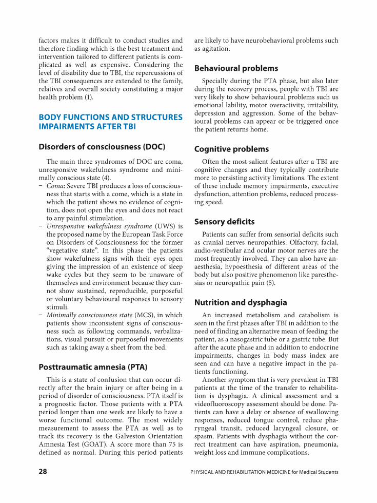
28 PHYSICAL AND REHABILITATION MEDICINE for Medical Students
are likely to have neurobehavioral problems such as agitation.
Behavioural problemsSpecially during the PTA phase, but also later
during the recovery process, people with TBI are very likely to show behavioural problems such us emotional lability, motor overactivity, irritability, depression and aggression. Some of the behav-ioural problems can appear or be triggered once the patient returns home.
Cognitive problemsOften the most salient features after a TBI are
cognitive changes and they typically contribute more to persisting activity limitations. The extent of these include memory impairments, executive dysfunction, attention problems, reduced process-ing speed.
Sensory deficitsPatients can suffer from sensorial deficits such
as cranial nerves neuropathies. Olfactory, facial, audio-vestibular and ocular motor nerves are the most frequently involved. They can also have an-aesthesia, hypoesthesia of different areas of the body but also positive phenomenon like paresthe-sias or neuropathic pain (5).
Nutrition and dysphagiaAn increased metabolism and catabolism is
seen in the first phases after TBI in addition to the need of finding an alternative mean of feeding the patient, as a nasogastric tube or a gastric tube. But after the acute phase and in addition to endocrine impairments, changes in body mass index are seen and can have a negative impact in the pa-tients functioning.
Another symptom that is very prevalent in TBI patients at the time of the transfer to rehabilita-tion is dysphagia. A clinical assessment and a videofluoroscopy assessment should be done. Pa-tients can have a delay or absence of swallowing responses, reduced tongue control, reduce pha-ryngeal transit, reduced laryngeal closure, or spasm. Patients with dysphagia without the cor-rect treatment can have aspiration, pneumonia, weight loss and immune complications.
factors makes it difficult to conduct studies and therefore finding which is the best treatment and intervention tailored to different patients is com-plicated as well as expensive. Considering the level of disability due to TBI, the repercussions of the TBI consequences are extended to the family, relatives and overall society constituting a major health problem (1).
BODY FUNCTIONS AND STRUCTURES IMPAIRMENTS AFTER TBI
Disorders of consciousness (DOC)
The main three syndromes of DOC are coma, unresponsive wakefulness syndrome and mini-mally conscious state (4).
ū Coma: Severe TBI produces a loss of conscious-ness that starts with a come, which is a state in which the patient shows no evidence of cogni-tion, does not open the eyes and does not react to any painful stimulation.
ū Unresponsive wakefulness syndrome (UWS) is the proposed name by the European Task Force on Disorders of Consciousness for the former “vegetative state”. In this phase the patients show wakefulness signs with their eyes open giving the impression of an existence of sleep wake cycles but they seem to be unaware of themselves and environment because they can-not show sustained, reproducible, purposeful or voluntary behavioural responses to sensory stimuli.
ū Minimally consciousness state (MCS), in which patients show inconsistent signs of conscious-ness such as following commands, verbaliza-tions, visual pursuit or purposeful movements such as taking away a sheet from the bed.
Posttraumatic amnesia (PTA)This is a state of confusion that can occur di-
rectly after the brain injury or after being in a period of disorder of consciousness. PTA itself is a prognostic factor. Those patients with a PTA period longer than one week are likely to have a worse functional outcome. The most widely measurement to assess the PTA as well as to track its recovery is the Galveston Orientation Amnesia Test (GOAT). A score more than 75 is defined as normal. During this period patients

3 Traumatic brain injury 29
The formation of mature bone in soft tissue, known as heterotopic ossification, may lead to a decrease in mobility, can be a source of pain and eventually diminish functionality.
Patients with severe injuries may have a trache-ostomy and during the rehabilitation process it has to be determined whether it is necessary its continuation or can be taken away.
ACTIVITY LIMITATION AND PARTICIPATION RESTRICTION
The ability of doing daily live activities, return-ing to work, driving, maintaining a social life are affected due to the aforementioned consequences of TBI, and are responsible for loss of quality of life in families and individuals with TBI.
Prognostic factors in TBIThe prediction of functional outcome after sus-
taining a traumatic brain injury is very uncertain due to the multitude of determinants that contrib-ute to the prognosis. Most of the literature avail-able is written from an isolated neurological, neurosurgical or from rehabilitation perspective alone (8) and therefore some authors suggest the need for a multidisciplinary research in this area to integrate the outcome prediction. The predic-tion of long term clinical outcome for patients is of an utmost importance to provide rehabilitation services but reliable guidelines for prediction the long term outcome are lacking (9).
Among the numerous barriers in determining the prognosis factors of outcome after TBI lead the heterogeneity of the injury and the personal factors involved.
Most of the studies regarding prognosis of TBI are centred on the severe and moderate lesions and as expected, moderate TBI patients have milder motor and cognitive impairments than those with a more severe lesion.
At an acute stage, an older age, a lower Glasgow Coma Scale, and absent pupil reactivity and the presence of a major extracranial injury have shown to predict a poor prognosis.
A worse handicap and social integration has been related to violence injuries, a severe or moder-ate injury, females older than 65, member of mi-norities and not working at injury time (10). In ad-dition, pre-injury unemployment, pre-injury sub-
Motor problems
Different degrees of loss of strength can happen due to the cortical lesion but also due to the lack of movement. But also movement related disorders like ataxia or tremor are frequent and can arise after the first onset of the symptoms and often once the patient has already been discharged (6, 7).
SpasticitySpasticity can occur after TBI and can deal to
secondary problems such as deformities, pain, im-pairment of the gait and grasping. Patients should be assessed not only with the neurological exami-nation but also considering how spasticity has pro-duced an impairment in gait or daily life activities.
Neurological medical complicationsPatients can complain about headaches, dizzi-
ness, problems in sleep. Seizures can occur after a TBI. Typically, seizures can happen in the first week due to the trauma, the bleeding and the liberation of neurotransmittors and prophylactic treatment is necessary. After the first week, the titration of medication can be done. If epilepsy occurs after the first week it is likely that the mechanism is due to a structural change after TBI and treatment may be used for a long period of time. There are different medications for the treatment of TBI-related sec-ondary epilepsy that can have a positive effect on behavioural problems and that do not produce more cognitive negative changes. For instance, valproic acid should be used instead of phenytoin since it can control the mood changes typically observed after TBI and may not interfere in cognitive problems.
Hydrocephalus is another complication that may arise in the first weeks and the patient may need a craniectomy, but also can have a delayed diagnosis and indeed posttraumatic hydrocephalus is the most common neurosurgical complication of persons with TBI. Any loss of acquired milestones should be carefully assessed by the clinician and if needed have a imaging study to be detected.
Other medical complicationsPatients with TBI may also experience other
medical changes such as endocrine dysfunctions that are commonly misdiagnosed and dysautonom-ic signs.

30 PHYSICAL AND REHABILITATION MEDICINE for Medical Students
consciousness and determine the level of cogni-tive functioning of the patient.
ū Stabilize the medical issues related to brain in-jury, for example reduce psychotropic medica-tion such as phenytoin or benzodiazepines that can interfere with regaining consciousness or cognitive problems.
ū Prevent secondary complication such as spas-ticity, contractures, pressure sores, aspiration due to dysphagia.
ū Restore the loss of functional abilities that limit activities and restrict participation.
ū Provide adaptive devices to enhance functional independence.
ū Provide educational sources for the patient and family.
ū Provide psychosocial care for TBI patients and their families.
The PRM specialist should lead the multidisci-plinary team and deliver treatments to a TBI pa-tient according to the Rehabilitation Cycle defined by Rauch (18), including the following parts:
ū Patient should be assessed medically but also regarding functioning. The ICF TBI Core Sets can provide a guideline for the assessment (19).
ū Use the common outcome measures to charac-terize the level of functioning and track chang-es across time (16). The Disability Rating Scale, the Glasgow Outcome scale, the Coma Recov-ery Scale and a neuropsychological battery should be administered.
ū According to the prognostic factors such as age, previous studies, medical complications, length of stay in intensive care, tracheostomy... the doctor should be able to make a prediction of the future level of disability and establish goals and objectives of rehabilitation (2).
ū Provide interventions such as:• Pharmacotherapy to improve disorders of
consciousness, cognitive and behavioural problems.
• Pain management.• Spasticity management.• Assessment of nutrition and dysphagia. Vid-
eofluoroscopy diagnosis and counselling.• Training of daily live activities with occupa-
tional therapy, family intervention and edu-cation, use of virtual reality.
• Neuropsychological assessment and treat-ment.
• Gait training and use of new technologies.
stance abuse and more disability at rehabilitation discharge are more important predictors of long term disability and predictors of being non-pro-ductive after TBI.
Patient s that developed epilepsy after TBI had a lower functional independence score compared to those no epileptics and also showed to have a higher disability score (11).
Studies report an incidence between 40-50% of depression after TBI and recently Bombardier et al have showed that depression was an outcome predictor of a poorer quality of life after TBI and was associated also with comorbid anxiety (12).
Post-traumatic amnesia (PTA) has shown in some studies (13) to be an indicator of prognosis during the early stage of recovery. Patients in a shorter period of PTA have shown to have better results than those that remained in that state for a longer period of time.
Some cognitive measures can contribute to the prediction of functional outcome that is not ac-counted by the injury severity, demographics or trauma variables. A shorter duration of PTA, less fatigue, absence of intracranial findings and a higher education can contribute to a better out-come (14). Some neuropsychological assessments for test orientation, verbal memory and process-ing speed have been found to be consistent predic-tors of outcome.
A highest functional score in the FIM scale was a positive predictor of functional level of dis-charge (15). Various other scales have been pro-posed to determine disability after TBI such as the Glasgow Outcome Scale (GOS), Disability Rating Scale (DRS), the Community Integration Questionnaire, and the Functional Status Exami-nation (16).
AIMS OF REHABILITATION
Rehabilitation as a multidisciplinary and holis-tic approach has shown to be beneficial for de-creasing hospitalization time, reducing health care costs and improving not only patient s qual-ity of life but the life of those around him like the family. Altogether this makes rehabilitation inter-ventions not only successful at an individual level but at a societal level (17).
The aims of rehabilitation in Traumatic Brain injury should be directed towards:
ū Make a clinical assessment of the disorder of
3 Traumatic brain injury 31
matic brain injury. Brain Inj. 2003; 17: 55-71. 11. Mazzini L, Campini R, Angelino E, Rognone F,
Pastore I, Oliveri G. Posttraumatic hydrocephalus: a clinical, neuroradiologic, and neuropsychologic assessment of long-term outcome. Arch Phys Med Rehabil. 2003; 84: 1637-41.
12. Bombardier CH, Fann JR, Temkin NR, Esselman PC, Barber J, Dikmen SS. Rates of major depres-sive disorder and clinical outcomes following traumatic brain injury. JAMA 2010; 303: 1938-45.
13. Ponsford J, Draper K, Schönberger M. Functional outcome 10 years after traumatic brain injury: its relationship with demographic, injury severity, and cognitive and emotional status. J Int Neuro-psychol Soc. 2008; 14: 233-42.
14. Sigurdardottir S, Andelic N, Roe C, Schanke A-K. Cognitive recovery and predictors of functional outcome 1 year after traumatic brain injury. J Int Neuropsychol Soc. 2009; 15: 740-50.
15. Sandhaug M, Andelic N, Vatne A, Seiler S, My-gland A. Functional level during sub-acute reha-bilitation after traumatic brain injury: course and predictors of outcome. Brain Inj. 2010; 24: 740-7.
16. Wilde EA, Whiteneck GG, Bogner J, Bushnik T, Cifu DX, Dikmen S, et.al. Recommendations for the use of common outcome measures in trau-matic brain injury research. Arch Phys Med Reha-bil. 2010; 91: 1650-1660.e17.
17. Takáč P, Petrovičová J, Delarque A, Stibrant Sun-nerhagen K, Neumann V, Vetra A, et.al. Position paper on PRM and persons with long term dis-abilities. Eur J Phys Rehabil Med. 2014; 50: 453-64.
18. Rauch A, Cieza A, Stucki G. How to apply the In-ternational Classification of Functioning, Disabil-ity and Health (ICF) for rehabilitation manage-ment in clinical practice. Eur J Phys Rehabil Med. 2008; 44: 329-42.
19. Laxe S, Zasler N, Selb M, Tate R, Tormos JM, Ber-nabeu M. Development of the International Classi-fication of Functioning, Disability and Health core sets for traumatic brain injury: An International consensus process. Brain Inj. 2013; 27: 379-87.
REFERENCES
1. Corrigan JD, Hammond FM. Traumatic brain in-jury as a chronic health condition. Arch Phys Med Rehabil. 2013; 94: 1199-201.
2. Jourdan C, Bosserelle V, Azerad S, Ghout I, Bayen E, Aegerter P, et.al. Predictive factors for 1-year outcome of a cohort of patients with severe trau-matic brain injury (TBI): results from the PariS-TBI study. Brain Inj. 2013; 27: 1000-7.
3. Brazinova A, Rehorcikova V, Taylor MS, Buckova V, Majdan M, Psota M, et.al. Epidemiology of Traumatic Brain Injury in Europe: A Living Sys-tematic Review. J Neurotrauma. 2016 Aug 25. [Epub ahead of print]
4. Whyte J, Nakase-Richardson R. Disorders of con-sciousness: outcomes, comorbidities, and care needs. Arch Phys Med Rehabil. 2013; 94: 1851-4.
5. Nampiaparampil DE. Prevalence of chronic pain after traumatic brain injury: a systematic review. JAMA 2008; 300: 711-9.
6. Koskinen S, Alaranta H. Traumatic brain injury in Finland 1991-2005: a nationwide register study of hospitalized and fatal TBI. Brain Inj. 2008; 22: 205-14.
7. Williams G, Morris ME, Schache A, McCrory PR. Incidence of gait abnormalities after traumatic brain injury. Arch Phys Med Rehabil. 2009; 90: 587-93.
8. Husson EC, Ribbers GM, Willemse-van Son AHP, Verhagen AP, Stam HJ. Prognosis of six-month functioning after moderate to severe traumatic brain injury: a systematic review of prospective cohort studies. J Rehabil Med. 2010; 42: 425-36.
9. Willemse-van Son AHP, Ribbers GM, Verhagen AP, Stam HJ. Prognostic factors of long-term func-tioning and productivity after traumatic brain in-jury: a systematic review of prospective cohort studies. Clin Rehabil. 2007; 21: 1024-37.
10. Mellick D, Gerhart KA, Whiteneck GG. Under-standing outcomes based on the post-acute hospi-talization pathways followed by persons with trau-
Key messages
• The consequences of TBI are complex leading to problems in body functions, body structures, that can lead to activity limitations and participation restriction.
• A highest functional score in the FIM scale was a positive predictor of functional level discharge.• A PRM specialist working in a multidisciplinary team and with an holistic approach is the key
for rehabilitation of TBI patients.

4 Stroke 33
Rehabilitation goals
The first goal of rehabilitation after stroke is to stimulate neuronal plasticity aiming at the best possible recovery of the different impaired func-tions (language, motor control, sensitivity, etc.). The recovery of a function can be due to a true recovery as after small infarcts, needing that some tissue that is crucial for function is spared (1), but most of the recovery proceedings are due to plas-ticity. Plasticity is mainly made of remapping of the cortex and neurons sprouting with regulation of synaptic activity and formation of new synaps-es. Motor, sensory and language cortices are loosely organized into somatotopic functional maps that exhibit high levels of use-dependent plasticity: the maps can be modified by experi-ence. After stroke, cortical remapping is both ac-tivity dependent and based on competition for available cortical map territory.
Recovery is likely to involve peri-infarct tissue that has a similar function. According to the size of the cortical lesion and to the persistence of in-tact corticofugal fibers to send the signal out of the ipsilesional cortex. The integrity of the corticospi-nal track is crucial (2). Neuronal plasticity de-pends on the integrity of the spared parenchyma then the pre-stroke brain status, is a critical factor.
To trigger and stimulate neuronal plasticity needs specific intensive training directed on im-pairments. A critical period of heightened neuro-plasticity might exist after stroke (3-5). Such physical or cognitive rehabilitation has been prov-en to be effective on neuronal plasticity then on recovery when provided at the sub-acute stage af-ter stroke. Most of the studies took place within the first 3 months, beginning after the 10-15th
days in rehabilitation departments after the stroke
AIMS AND STRATEGIES OF REHABILITATION MANAGEMENT FROM THE VERY ACUTE PHASE TO THE SUBACUTE AND CHRONIC PHASE
Main body function impairment
According to the site of the lesion, many differ-ent impairments can be variously associated lead-ing to a lot of possible clinical presentations. Language disorders (aphasia), dysexecutive disor-ders, other cognitive impairments as apraxia or agnosia, visuo-spatial neglect and neglect syn-drome, visual field amputation (hemianopsia, quadrantanopsia), swallowing disorders, paresis, spasticity, anesthesia or hypoesthesia, abnormal motor control. Each of them can be isolated or associated with others to different extent. They have to be carefully identified by a complete ex-amination. Consequences on activities and par-ticipation depend on their severity and associa-tion. For example, the walk may be impaired by paresis of course, but also by sensitive impairment and cognitive disorders as visuo-spatial neglect and anosognosia.
Some syndromes are typical, just mentioned here: middle cerebral artery with contralateral hemiparesis, hemi-anesthesia, hemianopsia and aphasia for left hemispheric lesion and neglect for right hemispheric lesion (in right handed subjects); superficial middle cerebral artery with upper limb and face dominance of the deficit, anterior cere-bral artery with lower limb deficit and dysexecu-tive disorders; posterior cerebral artery with hemi-anopsia and contralateral hemianesthesia; cerebel-lar syndrome; Wallenberg syndrome, etc.
4Stroke Alain P. YELNIK, Victorine QUINTAINE
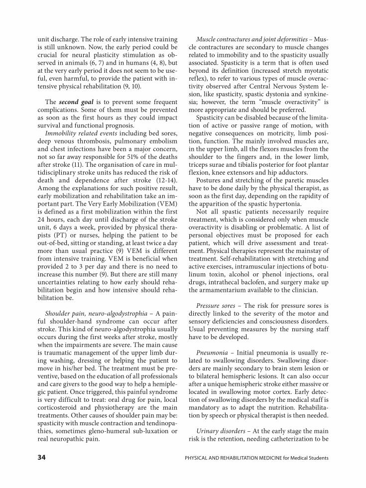
34 PHYSICAL AND REHABILITATION MEDICINE for Medical Students
Muscle contractures and joint deformities – Mus-cle contractures are secondary to muscle changes related to immobility and to the spasticity usually associated. Spasticity is a term that is often used beyond its definition (increased stretch myotatic reflex), to refer to various types of muscle overac-tivity observed after Central Nervous System le-sion, like spasticity, spastic dystonia and synkine-sia; however, the term “muscle overactivity” is more appropriate and should be preferred.
Spasticity can be disabled because of the limita-tion of active or passive range of motion, with negative consequences on motricity, limb posi-tion, function. The mainly involved muscles are, in the upper limb, all the flexors muscles from the shoulder to the fingers and, in the lower limb, triceps surae and tibialis posterior for foot plantar flexion, knee extensors and hip adductors.
Postures and stretching of the paretic muscles have to be done daily by the physical therapist, as soon as the first day, depending on the rapidity of the apparition of the spastic hypertonia.
Not all spastic patients necessarily require treatment, which is considered only when muscle overactivity is disabling or problematic. A list of personal objectives must be proposed for each patient, which will drive assessment and treat-ment. Physical therapies represent the mainstay of treatment. Self-rehabilitation with stretching and active exercises, intramuscular injections of botu-linum toxin, alcohol or phenol injections, oral drugs, intrathecal baclofen, and surgery make up the armamentarium available to the clinician.
Pressure sores – The risk for pressure sores is directly linked to the severity of the motor and sensory deficiencies and consciousness disorders. Usual preventing measures by the nursing staff have to be developed.
Pneumonia – Initial pneumonia is usually re-lated to swallowing disorders. Swallowing disor-ders are mainly secondary to brain stem lesion or to bilateral hemispheric lesions. It can also occur after a unique hemispheric stroke either massive or located in swallowing motor cortex. Early detec-tion of swallowing disorders by the medical staff is mandatory as to adapt the nutrition. Rehabilita-tion by speech or physical therapist is then needed.
Urinary disorders – At the early stage the main risk is the retention, needing catheterization to be
unit discharge. The role of early intensive training is still unknown. Now, the early period could be crucial for neural plasticity stimulation as ob-served in animals (6, 7) and in humans (4, 8), but at the very early period it does not seem to be use-ful, even harmful, to provide the patient with in-tensive physical rehabilitation (9, 10).
The second goal is to prevent some frequent complications. Some of them must be prevented as soon as the first hours as they could impact survival and functional prognosis.
Immobility related events including bed sores, deep venous thrombosis, pulmonary embolism and chest infections have been a major concern, not so far away responsible for 51% of the deaths after stroke (11). The organisation of care in mul-tidisciplinary stroke units has reduced the risk of death and dependence after stroke (12-14). Among the explanations for such positive result, early mobilization and rehabilitation take an im-portant part. The Very Early Mobilization (VEM) is defined as a first mobilization within the first 24 hours, each day until discharge of the stroke unit, 6 days a week, provided by physical thera-pists (PT) or nurses, helping the patient to be out-of-bed, sitting or standing, at least twice a day more than usual practice (9) VEM is different from intensive training. VEM is beneficial when provided 2 to 3 per day and there is no need to increase this number (9). But there are still many uncertainties relating to how early should reha-bilitation begin and how intensive should reha-bilitation be.
Shoulder pain, neuro-algodystrophia – A pain-ful shoulder-hand syndrome can occur after stroke. This kind of neuro-algodystrophia usually occurs during the first weeks after stroke, mostly when the impairments are severe. The main cause is traumatic management of the upper limb dur-ing washing, dressing or helping the patient to move in his/her bed. The treatment must be pre-ventive, based on the education of all professionals and care givers to the good way to help a hemiple-gic patient. Once triggered, this painful syndrome is very difficult to treat: oral drug for pain, local corticosteroid and physiotherapy are the main treatments. Other causes of shoulder pain may be: spasticity with muscle contraction and tendinopa-thies, sometimes gleno-humeral sub-luxation or real neuropathic pain.

4 Stroke 35
even 3 months. This is undoubted for motor con-trol altered by paresis. But this is not the case for language disorders, visual and sensitive neglect needing much more time, sometimes years be-fore achieving their maximum recovery. This is also not the case for the consequences of the de-ficiencies on activities and participation which can be improved long time after the stroke. Maintaining rehabilitation must be questioned when the patient’s gain between two evaluations is not obvious. Roughly, if there are no complica-tions, this is about between 3 to 6 months for walking, between 6 to 12 months for hand func-tion and between 12 to 24 months, for cognitive impairments. Then, the chronic stage depends on the function. The main reason to maintain physical rehabilitation is the persistence of dis-abling spasticity, physical therapy being the best treatment. Education is recommended as self-administered training must be learned: stretch-ing, activities, fighting functional decline, along with the secondary prevention of cardiovascular diseases (cf. infra).
General bases for rehabilitation
All the exercises devoted to recovery follow the same principles. They have to give an intensive stimulation to the patient, i.e. to be slightly more difficult and of longer duration than the patient could spontaneously tolerate, repetitive, motivat-ing. They must respect the fatigue but have to be repeated along the day and the weeks. An amount of 2 to 4 hours a day is needed according to the tolerance.
Rehabilitation of the balance and walking func-tion includes rehabilitation of motor control and sensitivity. If the deficiencies are severe, the use of robotic system can be recommended to train au-tomatisms as the main way to achieve the walk is to walk. An orthosis fixing lower limb joints is sometimes needed especially in case of proprio-ceptive disorder. An ankle-foot orthosis is often needed to avoid or to limit equino-varus foot dur-ing walking. The treatment of the main spastic muscles, triceps, tibialis posterior and quadriceps, is sometimes mandatory. Except in case of bilat-eral strokes or severe cerebellar lesion, usually the walk has to be achieved, at least for the in-house displacements.
replaced as soon as possible by intermittent cath-eterization if needed.
The third goal is to help the patient recovering the best autonomy despite the residual impair-ments. Consequences of impairments on activities and participation must be addressed and must be a main goal for rehabilitation, to reduce activity limitations and restriction of participation. The role of rehabilitation professionals and social workers is crucial. Many topics have to be ad-dressed: home adaptation, return to the job, drive a car, and so on.
REHABILITATION
Professionals
Rehabilitation usually involves different profes-sionals. Physical therapists, occupational thera-pists, speech therapists are the more often needed, sometimes also neuropsychologists, and orthotic and prosthetic technicians. Medical care, coordi-nation and supervision by a PRM specialist, from the early to the chronic stage are highly recom-mended.
Where rehabilitation should be done?At the very early stage, i.e. within the first 2
weeks, rehabilitation has to be provided in the acute unit, at the best being a stroke-unit with spe-cialized medical and paramedical teams.
As soon as possible, patient should be dis-charged and beneficiate from rehabilitation either at home or in a PRM specialised facility. A favour-able environment can be provided by the mean of the concept of “early supported discharge”, reha-bilitation being prepared and organized as soon as the first days in hospital, to be provided at home (15). When the deficiencies are severe or multiple, rehabilitation should be provided in an inpatient PRM department for weeks or months, the patient being then discharged with ambulatory rehabili-tation at home or in daily hospitalization.
How long rehabilitation should be provided?
Looking at body function impairment, most of the recovery is achieved within the first 6 months,

36 PHYSICAL AND REHABILITATION MEDICINE for Medical Students
MAIN PROGNOSTIC FACTORS OF FUNCTIONAL RECOVERY AFTER STROKE
Key period for plasticity: cf. supra.Previous quality of the central nervous system: as
plasticity depends on the quality of the spared pa-renchyma, previous stroke, small vessels disease, chronic high blood pressure are negative factors.
Side and site of the lesion: there is no major hemisphere when talking about autonomy. Apha-sia may limit participation of the patient but dys-executive and visuo-spatial disorders have a high negative impact on autonomy which can last along the life. The site of the lesion plays of course an important role: for example, integrity of the corti-co-spinal tract has been proven to be crucial for motor control and sensitivity recovery.
Age: there is always controversy about the role of the age. The main role of the age is related to the increasing frequency of associated diseases or dis-orders and previous neurological impairment playing a negative role on the functional progno-sis. Plasticity is still efficient after 80 years.
Prevention of complications: cf. supra.Cognitive impairments have a higher impact on
autonomy than motor impairment.Familial and social environment plays a crucial
role on quality of life.
RISKS FOR COMPLICATIONS/FUNCTIONAL DECLINE IN THE LONG TERM
After a stroke related to atherosclerosis, diabe-tes or high blood pressure, there is a high risk of cardiovascular diseases, heart failure, new stroke. The priority is the secondary prevention by treat-ing the cardio-vascular risks: treatment of high blood pressure, diabetes, dyslipidemia.
Functional decline of the physical condition must and can be prevented by daily stretching and regular cardiovascular training. Exercises have to be adapted to the deficiencies. They can include walking, bicycle, swimming two or three times a week.
Long term physical therapy is sometimes need-ed when there is a disabling spasticity, mainly as-sociated to severe impairments, to limit the risk of muscle contractures or maintain the ability to walk in house.
Rehabilitation of the hand function is mostly based on directed goal exercises. It includes al-so rehabilitation of motor control and sensitiv-ity. The use of robotic system could help recov-ery of severe deficiencies. Video and serious games, electrostimulation are in an increasing development but their real benefit has to be confirmed. Rehabilitation of hand function is strongly linked to rehabilitation of cognitive disorders.
Rehabilitation of the language must be pro-vided by a speech therapist. In case of non-flu-ent aphasia, it is recommended to begin as soon as possible to try to avoid the patient developing stereotypes. One hour a day is recommended. Initially the rehabilitation aims at restoring lexicon and grammar for a f luent normal lan-guage. When this appears to be impossible, the development of non-verbal communication is trained.
Rehabilitation of the visual neglect requires all the rehabilitation staff, speech therapists, occupa-tional and physical therapist as well as nurses. Two main approaches are developed, a top-down one based on the awareness of the disorder, and a bottom-up one using stimulation aiming at modi-fying the visual and corporeal referential. These stimulations are for example visual prisms deviat-ing the visual field, electric stimulations with TENS applied in the neck or transcranial mag-netic stimulation. Interesting results in research studies have to be confirmed for a daily use. Vi-sual neglect has a high impact on autonomy and risk of falls, it may require long rehabilitation be-yond one year.
Rehabilitation of the swallowing disorders: they have firstly to be detected by a cautious early clinical examination. Then measures to prevent pneumonia are mandatory. Persistent disorders in case of cerebellar disorder or multiple strokes need speech or physical therapists.
Rehabilitation of the urinary and bowel func-tion: the main risk is the retention at the early stage (cf. supra) but usually it does not last. The possible persistent disorder is a urinary inconti-nence usually manageable. There is no place for rehabilitation except in case of cognitive disorders needing behavior rehabilitation.
4 Stroke 37
ing in stroke recovery: a position paper from the First International Workshop on Neuroimaging and Stroke Recovery. Cerebrovasc Dis. 2004; 18: 260-7.
9. AVERT Trial Collaboration Group. Efficacy and safety of very early mobilisation within 24 h of stroke onset (AVERT): a randomised controlled trial. Lancet. 2015; 386(9988): 46-55.
10. Yelnik AP, Quintaine V, Andriantsifanetra C, Wannepain M, Reiner P, Marnef H, et al. AMOBES (Active Mobility Very Early After Stroke): A Ran-domized Controlled Trial. Stroke. 2017; 48: 400-5.
11. Bamford J, Dennis M, Sandercock P, Burn J, War-low C. The frequency, causes and timing of death within 30 days of a first stroke: the Oxfordshire Community Stroke Project. J Neurol Neurosurg Psychiatry. 1990; 53: 824-9.
12. How do stroke units improve patient outcomes? A collaborative systematic review of the randomized trials. Stroke Unit Trialists Collaboration. Stroke. 1997; 28: 2139-44.
13. Langhorne P, Stott DJ, Robertson L, MacDonald J, Jones L, McAlpine C, et al. Medical complications after stroke: a multicenter study. Stroke. 2000; 31: 1223-9.
14. Govan L, Langhorne P, Weir CJ, Stroke Unit Trial-ists C. Does the prevention of complications ex-plain the survival benefit of organized inpatient (stroke unit) care?: further analysis of a systematic review. Stroke. 2007; 38: 2536-40.
15. Langhorne P, Holmqvist LW; Early Supported Dis-charge Trialists. Early supported discharge after stroke. J Rehabil Med. 2007; 39: 103-8.
REFERENCES
1. Moon SK, Alaverdashvili M, Cross AR, Whishaw IQ. Both compensation and recovery of skilled reaching following small photothrombotic stroke to motor cortex in the rat. Exp Neurol. 2009; 218: 145-53.
2. Byblow WD, Stinear CM, Barber PA, Petoe MA, Ackerley SJ. Proportional recovery after stroke depends on corticomotor integrity. Ann Neurol. 2015; 78: 848-59.
3. Biernaskie J, Chernenko G, Corbett D. Efficacy of rehabilitative experience declines with time after focal ischemic brain injury. J Neurosci. 2004; 24: 1245-54.
4. Horn SD, DeJong G, Smout RJ, Gassaway J, James R, Conroy B. Stroke rehabilitation patients, prac-tice, and outcomes: is earlier and more aggressive therapy better? Arch Phys Med Rehabil. 2005; 86(12 Suppl 2): S101-S14.
5. Salter K, Jutai J, Hartley M, Foley N, Bhogal S, Bayona N, et al. Impact of early vs delayed admis-sion to rehabilitation on functional outcomes in persons with stroke. J Rehabil Med. 2006; 38: 113-7.
6. Krakauer JW, Carmichael ST, Corbett D, Witten-berg GF. Getting neurorehabilitation right: what can be learned from animal models? Neurorehabil Neural Repair. 2012; 26: 923-31.
7. Murphy TH, Corbett D. Plasticity during stroke recovery: from synapse to behaviour. Nat Rev Neurosci. 2009; 10: 861-72.
8. Baron JC, Cohen LG, Cramer SC, Dobkin BH, Johansen-Berg H, Loubinoux I, et al. Neuroimag-
Key messages
• The first goal of rehabilitation after stroke is to stimulate neuronal plasticity aiming at the best possible recovery of the different impaired functions.
• The second goal is to prevent some frequent complications among which immobility related events, consequences of spasticity, shoulder pain and neuro-algodystrophia.
• The third goal is to help the patient recovering the best autonomy despite the residual impair-ments.
• The main prognostic factors of functional recovery after stroke are the site and size of the lesion, previous quality of the central nervous system and potential co-morbidities affecting function.
• Rehabilitation usually involves different professionals as physical, occupational and speech therapists. Medical care, coordination and supervision by a PRM specialist, from the early to the chronic stage are highly recommended.


5 Spinal cord injury 39
leading to nerve compression, primary or second-ary tumours of the cord, and others.
The severity and extent of disability after SCI is largely related to the level of injury to the cord it-self. For instance, injuries at the thoracic, lumbar or sacral segments would commonly result in mo-tor and sensory loss in part of the trunk and legs (paraplegia), whereas, cervical SCI often leads to tetraplegia, where all the four limbs and the trunk are paralyzed. There are several factors which could potentially contribute to the severity and the nature of the resulted disability after SCI. Some of these factors are the severity of damage to the cord, mechanism of injury, and patients’ pre-mor-bid conditions such as obesity, old age, poly-phar-macy, advanced diabetes, cardio-vascular disease, mechanical or inflammatory multi-joint diseases, pre-existing neurological conditions, mental health problems and patients’ pre-injury lifestyle.
In cases of complete SCI, there would be a com-plete absence of transmission of motor as well as sensory signals across the point of damage. In in-complete cord injury, however, there could some-times be a degree of conduction preserved of these signals of variable velocities and potentials. These motor and sensory dysfunctions often result in peripheral motor weaknesses and impairment of different sensory modalities of skin. Moreover, SCI could produce a wide range of disruptions to the involuntary body activities below the level of injury, namely cardio-respiratory, gastrointesti-nal, urogenital and endocrinological. It can also impair immunological responses, thermoregula-tory and homeostatic mechanisms.
Immediately following SCI starts the phase of spinal shock. Soon after the shock phase starts to elapse, patients with suprasacral injuries will of-ten gradually retain reflex activities below the in-jury level, whereas patients with more caudal inju-ries are less likely to do so (2).
INTRODUCTION
Spinal Cord Injury (SCI) disrupts neurological communication between the brain and the body; consequently the motor signals from brain to the body and the peripheral sensory influx to brain centres are corrupted or completely fail to bypass the level of injury of the spinal cord. The outcome is often a complex and catastrophic injury which is immensely challenging to manage. SCI can lead to devastating medical, social, emotional, psycho-logical, and financial burdens. Such burdens in-evitably affect not only the patients, but also their families, friends, employers and ultimately the community as a whole. Additionally, SCI has a derogatory impact on school enrolment, educa-tion and economic participation. This could lead to substantially high financial negative influences on the individual and the society (1).
Regarding the incidence of SCI, there is no reli-able data for global prevalence; however the esti-mated annual global incidence is around 40 to 80 cases per million population. Males are the high-est risk group. The age distribution lies at the two age extremities of young adults (20-29 years) and elderly (70+). The peak age of incidence of SCI in females is at adolescence (15-19) as well as beyond 60 years of age. Studies reported that adult male-to-female ratio is around 2:1, yet this ratio often fluctuates and is sometimes considerably high-er. People with SCI are two to five times more likely to die prematurely, with worse survival rates in low and middle-income countries.
As far as the causes of SCI are concerned, me-chanical SCI is due to a variety of mechanisms such as road traffic collisions, falls, diving, vio-lence, work and sports related injuries. On the other hand, non-traumatic causes of SCI result from narrowing of the spinal canal, infections, vascular insufficiency, inter-vertebral disc disease
5Spinal cord injurySaša MOSLAVAC, Aleksandra MOSLAVAC
40 PHYSICAL AND REHABILITATION MEDICINE for Medical Students
takes into account the non-medical impact of such life-changing experience on the patients and their families. Following this, comes the stage which mainly deals with restrictions and limitations of activities and participations during the course of rehabilitation, which may last anytime from 3-6 months or more, depending of the level and the extent of the SCI, as well as any associated co-morbidities and complications. Adequate man-agement of affected body systems can potentially result in a reasonable or sometimes near-normal overall functionality. This would subsequently minimise the rate of morbidity, mortality, the length of hospital stay and the overall costs of stay, while maximising patient’s health, quality of life and productivity of the individuals to be able to live a fulfilling life.
THE INJURY, ASSESSMENT AND DIAGNOSIS
The mechanism of spinal injury may vary ac-cording to the direction and the velocity of the trauma to the cord. It can take one or combination of the following forms:
ū Hyperflexion; ū Hyperextension; ū Rotation: ū Luxation; ū Direct immediate damage to the cord.
The latter is commonly seen in traffic collisions or gunshot injuries. Primary impacts to the nervous tissue are commonly followed by secondary dam-ages due to vasogenic oedema and anoxia. The de-gree of motor and/or sensory impairment is deter-mined by the level and severity of the cord damage.
The most vitally crucial assessment of SCI in-jury patients is best to be carried out in the causal-ity unit in a systematic and succinct way. This al-lows clinicians to determine the patency of the patient’s airway, also to assess for respiratory or cardiovascular compromise and manage them ac-cordingly. It is additionally useful in fishing for any occults intra-abdominal, pelvic or limb active bleeding or injuries. After ensuring that SCI vic-tims are clinically and haemodynamically stable, they are then cautiously transferred to a SCI-spe-cialized centre.
Assessment of neurological deficit after trau-matic SCI should always be combined with screen-
At the scene of SCI, suspected SCI victims should be strictly immobilized until appropriate handling and transfer by well trained staff be-come available. Patients should then be carefully transported to dedicated casualty centres where an urgent initial medical assessment would be carried out. Once the patient has been resuscitated and haemodynamically stabilised, the next step is to consider further management of the SCI itself. This would entail either surgical or conservative approach depending on the individual case.
As far as rehabilitation is concerned, it is piv-otal that this is commenced as early as possible during the journey of SCI victim’s medical care. Rehabilitation intervention essentially includes a multiprofessional input from a wide range of health care providers including Physical and Re-habilitation Medicine (PRM) physicians, nurses, physical therapists, occupational therapists, psy-chologists, social workers/community liaisons, and others on demand e.g. speech and language therapists, respiratory therapists, kinesiologists, etc. It also involves working along other medical disciplines already involved or that can be benefi-cially involved in patients’ care such as neurosur-geons, vertebral surgeons, internal medical spe-cialists, urologists, etc.
The backbone of a successful and effective teamwork implies a clear communication amongst the team members plus a reasonable understand-ing of the patient’s impairment and the dynamic patho-physiology of it. It also involves establish-ing a familiarity with the symptoms and signs re-lated to the SCI of each patient. Rehabilitation is a dynamic process which should always emphasize on understanding the rationales behind holistic multidisciplinary care, plus respectfully establish-ing the real needs, priorities and predicaments of each individual patient at different stages of their post-injury care.
Rehabilitation team sets out realistic short-term and long-term goals for each SCI patient. These goals are framed around medical, rehabilitative and psycho-social needs. They also address any future vocational perspectives including reintro-ducing the post-injury individual back to the com-munity.
Rehabilitating SCI patients is usually done in a staged manner. The immediate focus of the pro-gramme is on the medical treatment of the frac-tured spines as well as focusing on the consequen-tial multi-system dysfunction. This phase also
5 Spinal cord injury 41
eral factors. Some of these factors are related to the degree of spine instability, progression of neu-rological impairment and patients’ clinical profile and age. This decision tends not to be a final one stop-shop. It is usually subject to regular assess-ments within the multi-professional team and ef-fective communication and liaising with the spi-nal surgeons.
PROGNOSIS, NATURAL HISTORY AND RELATIONSHIP TO IMPAIRMENT
Physiological and biomechanical stability deter-mines the outlook of post-SCI motor functionality. Most patients with motor or at least sacral sparing, i.e. incomplete AIS B-E, 72 hours following the injury will regain motor power sufficient to enable them to walk again. Patients with complete injury (AIS A) have the worst prognosis with only 10% chance of some recovery. Amongst those who progress from complete to incomplete injuries, around 3-6% will restore functional strength in the lower extremities.
Further recovery after SCI is possible within the zone of partial preservation, or when the bony fracture is below the neurological level of injury. It also happens in cases of non-functioning myo-tomes with existent pin-prick sensation. Both complete and incomplete SCI patients need a fo-cused rehabilitation management which would enable them to learn new skills to compensate for lost functionalities, it would also help them mini-mise limitations imposed by the impairment.
The functional capacity of the patient is largely dependent on the metameric level of the tetra/paraplegia as follows:
C1-C4 (High complete tetraplegia) – Patients have little or no movements of their arms, while movements of head and neck and shoulder shrug-ging are possible. While C4 tetraplegic patients may not require long-term mechanical ventilatory support, patients with C1-C3 injuries are likely to be dependent on it. This is because of the disrup-tion of the innervation to the diaphragm as part of the SCI. The physiological status of nearly all C1-C4 tetraplegic patients is insecure and may re-quire advanced and permanent assistance.
C5 and C6 tetraplegia – Patients generally have
an active elbow flexion; additionally C6 patients
ing for any potential spinal instability including bone fractures, disc injuries and ligament tears. It is pivotal to holistically examine patients with traumatic SCI and actively look for other non-spinal cord related impacts on other areas of the body such as head, ribs, visceral, genital, pelvic, limbs, and vascular injuries.
SCI can potentially lead to a profound auto-nomic system instability which almost always manifests itself as bradycardia and hypotension. These changes may determine the practicalities and the nature of both acute and rehabilitative managements of SCI patients. Moreover, because the autonomic system is malfunctioning after SCI, the body could potentially fail to effectively handle any excessive fluid input or early vertical-ization which may both further destabilize the already vulnerable area of SCI.
Different radiological modalities can be of help in assessing the extent of the cord injury. They are also useful in diagnosing possible associated liga-ment tears, local haematomas and vertebral frac-tures. These imaging modalities are chosen accord-ing to local facilities, clinical indications and, of course, justifiable contraindications. Examples of some of the widely used radiological investigations are: X-rays, computerised tomography (CT) scans and magnetic resonance imaging (MRI) scans.
The extent of the depth and the level of the ac-tual SCI are measured according to the American Spinal Injuries Association (ASIA) impairment scale (AIS) and the Standard Neurological Classi-fication of Spinal Cord Injury (3). The level of SCI and the functional residual ability can be better determined by physical examination of 10 key muscles on each side of the body. As a rule of thumb, the level of injury is determined by its cor-responding key muscles. These muscles will have a power grade of at least 3/5 i.e. movement against gravity or against some resistance. This is con-firmed by the fact that the proximally adjacent group of muscles should be fully intact with a power grade of 5/5 i.e. full strength is preserved. For instance, if the level of SCI is at C6, clinical examination would indicate 3/5 muscle power of wrist extensors and a normal 5/5 power of elbow flexors of the same limb. Detailed neurological examination between 72 hours and 1 week follow-ing injury may provide a rough predictive guide to the neurological and functional recovery.
Decision about the final treatment of SCI itself, whether surgical or conservative, depends on sev-
42 PHYSICAL AND REHABILITATION MEDICINE for Medical Students
Incomplete SCI patients may benefit from body-weight supported treadmill training, while functional electrical stimulation may be useful in both complete and incomplete SCI to achieve tasks in ventilation, self-care and ambulation. Tendon transfer surgery may be used as an option to promote limb functional movements and ad-vance these movements to more distal SCI levels.
Spasticity is very common after SCI and is one of the most challenging complications to effec-tively manage, however spasticity is not always a disadvantageous issue. Contrarily, spasticity can be of tremendous benefit to some patients. This is because preserving a degree of limb spasticity in some patients can help them perform some of their daily activities via using the spastic part of the limb e.g. turning, standing, operating assistive devices, etc. It is very crucial to bear in mind that spasticity can be aggravated by several factors such as pain, urinary retention, constipation, certain medications, mental status of the patient and ex-haustion. All these aggravators need careful screening and appropriate management to im-prove the outcome of spasticity management fol-lowing SCI. Generally speaking, for the majority of patients increased spasticity results in a wide range of complications including restricted movements, pain, poor hygiene, skin abrasions, pressure sores, infections, bone and joints dislocation and frac-tures. Tackling spasticity in these cases is a must.
Pressure sores are another difficult sequel of SCI. Impairment of sensory neuronal pathways post-SCI can cause a profound skin paraesthesia or anaesthesia which can endanger skin integrity and texture leading to pressure and shearing skin sores, more obviously over bony prominences and pressure areas of the body. It usually results from inadequate skin support and care by staff or pa-tients themselves at a later stage. Pressure sores can lead to significant health implications if not identified and managed appropriately whether conservatively, surgically or combination of both.
Neurogenic bladder can often follow SCI when innervation to bladder walls and sphincters is af-fected. Inappropriate bladder care without well-structured voiding regime can pose a seri-ous threat to the upper urinary tract and lead to significant morbidity and mortality due to risk of infections, stones formations and structural dam-
may preserve a possible wrist extension with teno-desis action. These movements would enable them to use assistive devices for feeding, grooming, and sometimes dressing. They can also use manual wheelchairs with grip enhancements, as well as power wheelchair for long distances and on un-even surfaces. In terms of other tasks such as full dressing, transfer mobility, bowel and bladder care, these patients often require assistance; how-ever some C6 injury patients may function inde-pendently, even including ability to drive adapted vehicles.
C7 tetraplegia – In this category, active elbow
extension remains intact. With the help of assis-tive technology patients may successfully achieve independence in feeding, dressing, bathing, grooming, bed mobility, transfers and commu-nity manual wheelchair propulsion, although not on very uneven surfaces and curbs. Male patients may master bladder management usually via in-termittent catheterisation. Oppositely females commonly require external help with intermit-tent catheterisation. C7 injured patients may po-tentially be able to use computers and phones, which can open some vocational perspectives for them.
C8 tetraplegia – These patients tend to have
good hand grasp and release movements. This enables them to comfortably achieve self-care and independent driving of adapted vehicles.
Paraplegia – Individuals with thoracic or lum-
bar injuries can regain a reasonable level of self-reliance with activities of daily-living, including advanced transfer from floor to wheelchair for instance. They can also reach a satisfactory stage of community skills and participations.
Patients with lower thoracic and lumbar injuries may be able to stand through using bilateral hip-knee-ankle-foot orthoses (HKAFO) or knee-an-kle-foot (KAFO) orthoses and crutches. The skills to use these aids can help them become indepen-dently ambulant in community. From T11 level downwards there would be a noticeable advance-ment in patient’s ambulatory skills. The lower the level the steadier the ambulation would be. For a safe and confident community ambulation func-tional hip flexion and knee extension should be preserved. This is usually the scenario in L2 and L3 SCI.

5 Spinal cord injury 43
Effective leadership skills is one of the most es-sential attributes PRM specialists have to adopt in order to successfully lead the team and to coordi-nate the team dynamics, undertake and delegate tasks appropriately, prescribe medications, liaise and refer to other specialities, also write discharge letters, and provide rehabilitation and orthotic aids. They should also have the skills and compe-tencies to perform certain procedures e.g. com-prehensive tracheostomy care or surgical removal of necrotic tissue from pressure sores. Such skills are driven by available resources, up-to-date staff training and clinical demands of the particular SCI unit.
Physical therapists are an essential element of productive rehabilitation. They continuously pro-vide and monitor progress and effectiveness of kinesio-therapy and hydrotherapy. Moreover, they provide patients with regular sessions to help them develop skills and muscle power for balance, sitting, standing, transfers, mobility, verticaliza-tion, ambulation and maintaining range of mo-tions. Other forms of therapy such as biofeedback, electrotherapy, thermal therapy, ultrasound, mag-netotherapy and balneotherapy may be used as well, however, robust evidence behind their effec-tiveness is still lacking. On the other hand, some studies succeeded to provide evidence for the use of functional electrical stimulation as an option to improve function of upper extremities (6).
One of the main pillars of rehabilitation team is occupational therapists. Their main focus is to improve patients’ posture, transfer, and undertak-ing activities of daily-living such as feeding, grooming, cooking, shopping and personal-care. Their role extends even further to involve evalua-tion of equipment e.g. wheelchairs, hand splints and orthotics, as well as housing adaptations and driving assessments. With the help of ever evolving technology, novel robotic and virtual reality tech-nologies may facilitate these skills in the future.
Nurses have the biggest and most effective share of SCI patients care. They play a golden role on a daily bases in assessing and treating bladder and bowel dysfunctions. They also provide round the clock skin care and ensure adequate dietary intake by patients particularly at the early cata-bolic stage of SCI. Intermittent catheterisation is the gold standard and most effective practice for neurogenic bladder. Sufficient education on self-catheterisation can be provided by the ward nurs-es to patients who would benefit from it and are
age to the renal tract. Bladder management pro-gramme should be designed around the nature and the severity of bladder dysfunction. It can take the form of spontaneous scheduled voiding, intermittent catheterisation, or rarely supra-pubic cystostomy. Sound and robust management of neurogenic bladder is essential for a better quality of life and to reduce the morbidity and mortality rates after SCI. Similarly, nerve supply to the bow-els can be involved in SCI which leads to neuro-genic bowels. The presentation of neurogenic bowels can take the shape on the scale from diar-rhoea to constipation. Bowel management with the use of various techniques and/or through regular administration of one or combination of laxatives is vital.
SCI-induced osteoporosis is a multi-factorial se-
quel where prevention, effective screening, early diagnosis and management are necessary to avoid pains and pathological fractures.
Haemodynamic instability with the potential to develop autonomic dysreflexia has been mainly observed in tetraplegic and high paraplegic pa-tients, and they must be monitored in all phases from early-acute to the long-term.
THE MULTIPROFESSIONAL TEAM APPROACH TO SCI
The main objective of acute care after SCI is to ensure a priority is given to maintain biomechan-ical and physiological stability. This may help preserve the unaffected fibres at the site of injury. Moreover, careful attention should be paid to de-tect and manage complications as early as possible (4). Certain pharmacological agents can be used in the immediate phase following traumatic SCI. The most popular is Methylprednisolone which is administered early after the onset of the injury (5).
Multiprofessional approach is the backbone of effective rehabilitation which should be proac-tively commenced as early as possible, ideally on the first day of admission. PRM specialists should holistically assess the patient through history tak-ing and a thorough clinical examination. This initial assessment helps identify and address any clinical, neurological, or functional issues. They should also set out realistic rehabilitation short and long-term goals and map out future manage-ment of the patient accordingly.

44 PHYSICAL AND REHABILITATION MEDICINE for Medical Students
GOALS OF THE REHABILITATION PLAN
The aim of rehabilitation management is to ad-dress any issues that can negatively alter the body function and structure; it also aims to minimise limitations and restrictions to participating in daily and social activities.
Due to impaired body mobility of SCI patients, they are often in need for an external help for turning in bed, sitting, transfer, personal care, walking, eating and grooming depending on the severity and the onset of the SCI. The vast major-ity of SCI cases are young who suddenly trans-form from independently functioning at the peak levels of productivity and energy into totally dis-abled and reliant on others for the simplest of hu-man needs to survive and minimize harm. The initial focus on rehabilitation programme should be around the loss of control of primary body functions before moving onto more complex is-sues such as social activities and participation.
Focusing on enhancing patients’ potential to participate in social and general life activities is one of the long-term goals of successful rehabilita-tion. This is usually the case where limited par-ticipation in life activities would likely be detri-mental to professional and personal lives. Reha-bilitation team has to think broadly to help such victims overcome these obstacles by the careful choice of methods and equipment used in the community to secure them decent and fulfilling living standards.
Spinal cord injury profoundly affects almost every single aspect of patient’s life. The more the complete and proximal the levels of injury, the more disturbed these functions are. Difficulties leading to restrictions in participation are seen in all ages and both sexes. The nature of these diffi-culties though varies depending on the age and commitments each patient has to inevitably ad-here to. For instance, younger individuals have difficulties in completing education not only be-cause of physical barriers, but also because the choice and the nature of their education have to be adjusted or even completely altered to realistically match their post-SCI status. Such adaptations are not necessarily appealing to patients and their relatives who would adopt a refractory denial state to the severity of injury and paralysis and the ac-tual impacts on the youngster’s capabilities.
Participation restrictions in professional life
keen to accept and perform it themselves. Like-wise, education of families and caregivers can be secured as it is proven to be a necessary part of rehabilitation and continuation of patients care.
SCI is a catastrophic and life-changing event, which may understandably leave a tremendous negative psychological impact on patients and their relatives. Here comes the duty of the Psy-chologist who should provide continuous sup-port to patients as well as their families and pro-vide feedback to other team members. Premorbid mental health illnesses should, always, be taken into account as it would help determining the level and urgency of the psychological interven-tion. Referral to specialist psychiatrists is some-times required in refractory cases or in suicidal patients.
Social workers/community liaisons provide early support during inpatient rehabilitation time and reaching to the community service that would enable the use of available resources and prepare for soft-landing of the patient to his community, schooling, professional and leisure activities.
Some SCI patients may develop swallowing and speech problems such as dysphagia, odyno-phagia, aphasia, dysphasia or dysarthria respec-tively. Swallowing and speech problems are par-ticularly common in cases of concomitant brain or high cervical injuries, post-intubation or post-tracheostomy. Speech and language therapists regularly screen for and address swallowing and speech problems related to SCI. Special tests can sometimes be used to assess swallowing such as the blue dye swallow testing and the fiberoptic endoscopic evaluation of swallowing (FEES), this largely depend on local availability and staff training.
Dieticians usually deal with each individual patient’s needs according to their medical back-ground, dietary requirements, body weight, as well as the stage after spinal cord injury whether at the early (catabolic) phase or late on to monitor and prevent obesity.
Finally, given the complexity of SCI, it is not uncommon to have a wide range of other special-ists and health care providers contributing to the care for SCI patients such as neurosurgeons, an-aesthetists, ICU staff, traumatologists, orthopae-dic surgeons, general medical specialists, etc. Therefore, maintaining a high quality communi-cation and continuous liaison with other speciali-ties is paramount in SCI patient’s care.

5 Spinal cord injury 45
care plan and recommendation is then sent out to the caring General Practitioner (GP). This care plan should clearly specify each patient’s expected needs and follow-up in the community including medications reviews and specific follow-up inves-tigations, physical therapy and necessary nursing care. Some patients may need a referral by their own GPs to the local PRM departments for fur-ther assessment; some of them are offered assess-ment at their own properties through organised home-visits.
Following the initial inpatient rehabilitation phase illustrated above, patients will be re-admit-ted to the SCI centre for a follow-up assessment 12 months post-discharge. The idea of this is to en-sure that initial plans have been implemented, and to also offer another round of inpatient physical and occupational rehabilitation if indicated. The length of stay in the SCI unit this time varies de-pending on the individual’s need, yet it tends to be considerably shorter than the initial admission episode following the SCI. Following this follow-up readmission, patients undergo annual read-missions to the SCI unit for the first 3-5 years, or until they establish consistent healthy routines and participations in the community, whichever comes first.
Once patients have consistently demonstrated their ability to well manage their complex and chronic conditions, a reduction in frequency of readmissions is possible to every other year, or sometimes even less often. This largely depends on the patients’ readiness, the level of support available at home, and the local provider’s facili-ties and expertise. The frequency of reassessing and readmitting SCI patients to the SCI unit is not rigidly fixed. We should always bear in our minds that SCI patients’ coping mechanism and physiol-ogy tend to cease as the time goes by, therefore their medical and functional conditions can im-mensely deteriorate. In this case they may require a more frequent reassessment which is usually kept at annual bases to early recognise and ad-dress new health issues and prevent major costly disasters.
Further information about SCI management can be found on website of The International Spi-nal Cord Society (ISCoS), which promotes the highest standard of care in the practice of SCI, through its medical and multidisciplinary team of professionals, to foster education, research and clinical excellence (7).
become more obvious in less educated patients with slimmer chances for potential new qualifica-tions or skills after SCI injuries. These skills could enable them to undertake non-physical desk jobs. This group of blue collar labourer become very disadvantaged after the disability as they tend to lose their jobs or take an early retirement then consequently lose the supportive role for them-selves and their families. Besides these vocational restrictions, social involvement and hobbies be-come a cumbersome. This not only because of physical limitations, but a hefty of patients’ time would be spent on their medical needs. Therefore, attempts to restoring these activities should con-centrate on patients’ transfers, movements and sphincter control. It also includes establishing a wide social network, return to own property, in-volvement in vocational and leisure activities.
In summary, the main goals of rehabilitation intervention are to return the patient to a reason-ably productive life, diminish the sequel of neuro-logical injuries and to improve the overall func-tional abilities, so that patients remain physically and mentally as healthy as possible, and complica-tions are minimised.
OUTCOME, DISCHARGE PLANNING AND LONG-TERM FOLLOW-UP
Effective rehabilitation program should com-prehensively evaluate every single aspect concern-ing patients’ needs including a review of medical records, extensive history taking, systematic physi-cal examination, blood investigations, radiological imaging, bowel and bladder assessment involving urological studies, skin care, medications review, physical and occupational therapy evaluations for motor and sensory changes, assessment of daily-living activities such as posture, transfers, personal care, grooming, cooking, shopping, eating and drinking. Moreover, it also looks into assessing the need for psychosocial input, walking aids and equipment, all in the short and long terms.
Generally speaking, discharge for inpatients commences approximately 3 months after admis-sion for paraplegic, and 6 months for tetraplegic patients. Planning entails continuous communi-cation and liaising with patients’ relatives and the relevant authorities at the patients’ locality to ad-dress specific needs of each patient individually after the discharge. A detailed written discharge

46 PHYSICAL AND REHABILITATION MEDICINE for Medical Students
Guideline for the Management of Acute Spinal Cord Injury: Introduction, Rationale, and Scope. Global Spine J. 2017; 7 (Suppl 3): S84-S94.
5. Fehlings MG, Wilson JR, Tetreault LA, Aarabi B, Anderson P, Arnold PM, et al. A Clinical Practice Guideline for the Management of Patients With Acute Spinal Cord Injury: Recommendations on the Use of Methylprednisolone Sodium Succinate. Global Spine J. 2017; 7 (Suppl 3): S203-S211.
6. Fehlings MG, Tetreault LA, Aarabi B, Anderson P, Arnold PM, Brodke DS, et al. A Clinical Prac-tice Guideline for the Management of Patients With Acute Spinal Cord Injury: Recommenda-tions on the Type and Timing of Rehabilitation. Global Spine J. 2017; 7 (Suppl 3): S231-S238.
7. International Spinal Cord Society (ISCOS). Avail-able from: http://iscos.org.uk/
REFERENCES
1. International Perspectives on Spinal Cord Injury. Geneva, Switzerland: World Health Organiza-tion, 2013. Available from: http://apps.who.int/iris/bitstream/10665/94190/1/9789241564663_eng.pdf
2. Ditunno JF, Little JW, Tessler A, Burns AS. Spinal shock revisited: a four-phase model. Spinal Cord. 2004; 42: 383-95.
3. Kirshblum SC, Burns SP, Biering-Sorensen F, Donovan W, Graves DE, Jha A, et al. Interna-tional standards for neurological classification of spinal cord injury (revised 2011). J Spinal Cord Med. 2011; 34: 535-46.
4. Fehlings MG, Tetreault LA, Wilson JR, Kwon BK, Burns AS, Martin AR, et al. A Clinical Practice
Key messages
• Spinal Cord Injury is a catastrophic event which leads to paralysis of essential parts of the body and multi-system dysfunctions. It is, therefore, vital to holistically rehabilitate SCI victims to restore and maintain a reasonable quality of life and to minimise the burden on their families and communities.
• Successful rehabilitation means a focused multiprofessional team effort in dedicated rehabilita-tion units with availability of equipment and expertise to deal with all potential presentations; this should be guided by a PRM specialist.
• Intelligent and timely approach to the acute medical and rehabilitative issues, followed by a ro-bust long-term care planning, may well lead to near-normal eventualities, which will in turns enhance patients’ chances in living a fulfilling life after the injury.
6 Multiple sclerosis 47
affected sometimes simultaneously sometimes sequentially at different moments of the evolution of the disease, leaving or not permanent clinical sequelae (2). Each MS patient has his own pattern of acute and chronic symptoms and impairments which also vary over time as the disease progress-es. Therefore, disease-related disability is very complex and differs from patient to patient, and from time to time in the same patient. Additional impairments secondary to those directly related to the disease further complicate the situation by extending and aggravating the primary impair-ments.
The diversity, multiplicity and complexity of the functional profiles of MS patients and their evolution in time make rehabilitation planning and implementation a challenging enterprise. On-ly an individual holistic approach by a medically coordinated multi-professional team can bring about optimal results.
The type of MS, either relapsing-remitting, pri-mary chronic progressive or progressive with su-perimposed acute episodes, also influences the planning and content of the rehabilitation inter-ventions.
In the case of the relapsing-remitting form, rehabilitation interventions focus on the preven-tion of secondary impairments during the acute phase of the relapse and the recovery of the lost functions. Special attention is given to the correc-tion of any loss of endurance during the acute relapse.
For the chronic progressive MS type, the reha-bilitation program aims at promoting functioning at the highest possible level. This includes uplift-ing weak functions to a state of training which allows compensating for their weakness by regu-lar exercise and practice, substitute activities or change the way they are performed (adaptation), provide orthosis or appropriate external aids to promote activity and participation as well as adapting the patient’s environment for maximal
INTRODUCTION
Multiple Sclerosis (MS) is due to a multifocal and progressive autoimmune demyelination re-sulting in characteristic plaques of different ages scattered throughout the Central Nervous System (CNS) (1). Not all of these lesions induce clinically evident impairments and symptoms. Some of them remain “silent” depending on their localisa-tion, but all together contribute to a progressive deterioration of the transmission and the neuro-nal treatment of information within the brain and the spinal cord. The CNS might be able to com-pensate such impairments for some time so as they might become clinically manifest and dis-abling only at a later stage of the disease. However, the CNS networks being forced to deal with an increasing number of impaired connections, the functioning of the CNS becomes more and more complicated resulting in physical and intellectual fatigue already at early stages of the disease.
At the onset of the disease, about two thirds of the MS patients present a relapsing-remitting form of the disease. The brisk onset of neurologi-cal symptoms and impairments is followed by a clinical remission lasting until the next exacerba-tion of the disease. Over the years most of them will convert to a progressive form. Only about 10% of the patients retain a relapsing-remitting course for their whole life. About 20% of the MS patients start with a primary chronically progres-sive form. The remaining patients start with a form which associates a progressive course with superimposed acute episodes.
VARIABLES INFLUENCING DISABILITY OCCURRENCE AND PROGRESSION
The disease-related impairments of MS are quite complex. Different parts of the CNS can be
6Multiple sclerosisRolf FRISCHKNECHT

48 PHYSICAL AND REHABILITATION MEDICINE for Medical Students
As the disease progresses rehabilitation inter-vention will focus more and more on compensa-tion for lost functions, boosting residual function, introducing mobility aids like crutches, splints, orthoses, wheelchairs, scooters, robotic devices, etc. Each technical aid must be wisely chosen and adapted to the patient special needs and context. It is important to teach the patient practically how to use the device through appropriate training and exercise sessions. The patient’s environment must be progressively adapted to the increasing disability which sometimes means to change ac-commodation as well. As MS is a progressive dis-ease, any function eventually changes, so a diag-nostic follow-up is needed in order to regularly update the management program.
In MS patients with severe disability, rehabilita-tion is centred on the preservation of adequate joint mobility and muscle length to allow optimal posi-tioning in bed and on the wheelchair and facilitate care. Optimal positioning reduces fatigue and pain and ensures the best possible level of quality of life.
REHABILITATION APPROACHES TO SPECIFIC FUNCTION IMPAIRMENTS
Gait and balance impairmentNearly all MS patients have difficulties with
posture, balance and mobility (3). The CNS sys-tems involved in the control of posture, balance and gait are progressively damage by the disease bringing on an increasing motor disability:
ū Damage to the pyramidal system induces a dysfunction of the modulation of the activity of the lower motor neurons. The regulation of muscle tone and the voluntary activation of the skeletal muscles are impaired producing weak-ness and spasticity. The coordination between agonists and the reciprocal inhibition between agonists and antagonists are disturbed. Gener-ally, the muscle strength is more affected at the lower than the upper limbs.
ū The cerebellar dysfunction induces ataxia of the trunk and the limbs, interferes with pos-tural control and causes postural and inten-tional tremor. Together with the impairment of central vestibular pathways the cerebellar dys-function decreases balance and delays postural adjustments to correct instability.
participation. The prevention of secondary im-pairments that enhance the burden of MS-related disability is also a compulsory component of any rehabilitation plan for these MS patients.
For the progressive MS type with superim-posed acute episodes the two strategies must be combined.
GENERAL PRINCIPLES OF REHABILITATION MANAGEMENT
MS is a chronic disease and MS patients need a lifelong rehabilitation program which must be regularly adapted to the progressive aggravation of the functional profile taking also into account the patient’s individual life habits, needs and con-text factors which might also change over time (2).
The first objective of rehabilitation interven-tions in MS patients is maintaining the best pos-sible levels of performance in all domains of func-tioning. Appropriate regular strength and endur-ance exercise programs allow to prevent weakness and muscle atrophy secondary to prolonged inac-tivity and generalized deconditioning. Exercise is safe and effective as long as it is used with mod-eration and adapted to the patient’s individual tolerance. Exercise programs should preferentially involve muscles that are needed for activities which are meaningful to the patient and which are adapted to his context and needs. A good level of force, endurance and cardio-vascular fitness in-creases tolerance to activity and exercise, delays the occurrence of fatigue and maintains function-ing as long as possible. Sometimes muscle strength-ening must be temporarily focused on muscle groups needed to compensate impaired or lost function or to use assistive devices (e.g., training of shoulder girdle and arm muscles for the use of crutches, for the performance of push-ups for wheelchair users and for transfers to and from the wheelchair).
After acute relapses of the disease, special at-tention should be paid to disuse weakness and deconditioning. Whereas the CNS function recu-perates, there is only limited spontaneous recov-ery from the loss of muscle strength, local and cardio-vascular endurance or motor skills unless an appropriate exercise program is initiated. Oth-erwise MS patients go on functioning at a lower level than what the recovered function of the CNS would allow.
6 Multiple sclerosis 49
Upper limb dexterity impairment
The rehabilitation of upper limb function fol-lows the same principles as the rehabilitation for posture, balance and mobility as the problems are the same: pyramidal weakness, spasticity, lack of intermuscular coordination, ataxia and sensory loss. In earlier phases of the disease, the upper limb dysfunction is manly linked to the loss of the precision of the neuro-motor command. At this stage, the activities which are important for the patient’s autonomy, profession or leisure might be improved by specific therapeutic exercise. Effec-tive upper limb movements are very dependent on an accurate control of posture and balance. It is therefore important to integrate upper limb exer-cise into global training situations which are as close as possible to real life situations.
At later stages it might be necessary to compen-sate the progressive loss of function by simplifica-tion of tasks, introducing technical aids and equipment to compensate the lack of precision (e.g., instruments to assist buttoning) or lost func-tion (e.g., electric toothbrush or computerized environment control).
At some stages, special strengthening of the shoulder girdle and the upper limbs might be nec-essary to enable patients to use walking aids like canes and crutches or to perform transfers to and from the wheelchair safely and do the regular push-ups to protect the skin of the bottom when sitting in a wheel chair.
Fatigue or impaired tolerance to effort
MS related fatigue is different from the fatigue experienced by healthy persons after a physical or intellectual effort or a sleepless night. The fatigue is physical as well as cognitive and the one of the most common symptoms observed in MS.
MS related fatigue occurs even during and after minimal activity and is perceived as a feeling of continuous exhaustion. MS fatigue is day time dependant and occurs preferentially during the afternoon and in the evening. It is thought to be related to an increase of energy required for nerves to transmit signals through zones with impaired or absent myelin sheets, which compli-cates the way the CNS works (4).
There is also an increased fatigue of function. Weak functions must operate at unusual high lev-
ū Sensory loss is another important component of motor dysfunction in MS with a pronounced impact on balance and gait.
Adequate levels of functioning can be pre-served by compensating the decrease of perfor-mance of the neural motor and postural control by regular training of the defective systems. Exer-cise for balance sitting and standing at rest and during activity are typical representatives of such rehabilitation interventions. Exercises should be performed in a low stimulus environment to in-crease the patient’s capacity to concentrate and enhance the learning effects.
To preserve mobility and balance at the highest possible level it is vital to maintain adequate joint mobility (range of motion) and muscle length (flexibility). Appropriate mobilisation and stretch-ing programs must be included into all rehabilita-tion interventions in MS patients especially in the presence of spasticity (see below).
Exercises in a pool are an interesting approach also in MS patients. However due to the heat sen-sitivity of the impairments of MS patients, care should be taken to exercise only in moderate warm or cold water. The microgravity environ-ment decreases the pressure in the joints thus reducing muscle tone and relieving pain and spasticity. It helps to keep and train up-right bal-ance and can be used to assist weak muscles. Exercise against water resistance allows to train strength and endurance; the resistance water op-poses to movements increases with speed. Fall-ing in the water is much slower than on ground and patient have much more time to correct a loss of balance. Therapists have also more time to react. This makes water an interesting place to practice static and dynamic postural control and balance.
Unfortunately, the evidence concerning the ef-ficacy of any physical therapy approach to gait/balance impairment in MS patient is sparse and hampered by several limitations.
According to a recent systematic review, 8 weeks of weekly home/outpatient physical therapy is probably effective for improving balance, dis-ability, and gait in MS subjects able to walk ≥5 meters (3).
Other exercise protocols have been found pos-sibly effective for improving gait and/or balance in MS subjects suffering from mild to moderate dis-ability.
50 PHYSICAL AND REHABILITATION MEDICINE for Medical Students
inhibition significantly. As a result, the motor impairment increases. The increased resistance to movement during motor tasks contributes to the increased fatigue seen in MS patients. Spasticity preferentially affects upper limb flexors and lower limb extensors and is generally most pronounced in lower limbs.
It is important to remember that spasticity can be increased by noxious sensory inputs from any body structure, but especially from internal or-gans, joints, soft tissue and skin (like skin ulcers, infections of the skin and the underlying tissues, nail lesions and infections, unstable joints, neuro-pathic pain, bladder or bowel distension, faecal impaction, appendicitis, uro-lithiasis, pyelo-ne-phritis). In case of a sudden increase of spasticity it is mandatory to search actively for these condi-tions and eliminate all identified causes promptly. Orthoses and wheelchair seating have to be checked and adapted periodically in order to avoid that they become a source of nociception.
Untreated spasticity leads to muscle shortening and muscle contractures, worsening of posture and function, which in turn increase the risk of further disability, pain, pressure sores and loss of quality of life.
The management is the same as for spasticity in other CNS diseases (7). Ideally the spastic muscles are stretched systematically every few hours, but this intervention is often difficult to put into prac-tice especially for stages beyond mild motor im-pairment. The stretching techniques used should make sure that muscles running over two joints are effectively stretched as well.
Oral antispastic drugs, are often helpful but one has to bear in mind that in many MS patients posture and motor activities rely to some extent on spasticity. The weakness induced by oral anti-spastic medication affect both spastic muscles as well as weak flaccid muscles and can disturb pos-ture, locomotion and other motor activities sig-nificantly, if their indication is not carefully thought over and their dosage is not cautiously adapted. Frequently, an inappropriately low tonus of the trunk muscles increases limb spasticity. As a consequence, limb spasticity can paradoxically increase if the trunk is weakened by a vigorous oral antispastic treatment. The somnolence and additional fatigue induced by such medications amplify the impairments due to the ongoing dis-ease and may increase existing disability.
Whenever possible, local and regional treat-
els of activity to respond to the functional perfor-mance imposed by everyday life. This leads to an accelerated fatigue of the involved CNS struc-tures. The occurrence of a foot drop after pro-longed walking is an example of fatigue of func-tion. The continuous fight of weak muscles against their spastic antagonists is another source of fa-tigue of function.
Fatigue can be very disabling even in patients with otherwise mild symptoms, in whom it can limit the ability to participate to intensive reha-bilitation programs and cause a reduced ability to work.
Moderate exercise might be the most appropri-ate approach, since even activities leading to an increased fatigue are not harmful by themselves, though the impact of the fatigue they induce must be considered and activities of daily life must be modulated accordingly (5).
An increase of body temperature can markedly enhance physical and cognitive impairments as the conduction of axons with impaired myelin sheets is much more affected by high temperature than in normally myelinated axons. Hence, care should be taken to avoid marked increases of the body temperature. It is recommended to exercise in low temperature environments. Body cooling by using ice-jackets can improve function and decrease fatigue induced by repetitive activities. Pools, baths and showers should not exceed about 29 °C which is a lower temperature than that con-sidered as neutrally comfortable.
Planning the rehabilitation intervention, it is better to proceed with several short exercise or training periods per day rather than one long ses-sion and allow plenty of rest between workouts.
For the daily living activities should be organ-ised so as to take advantage of the physically and cognitively favourable periods of the day and mi-nimise fatigue through periods of rest. Activities should be reviewed according to the principle of Energy Conservation (6). Unrewarding activities should be abandoned or delegated, rewarding ac-tivities made more ergonomic, simplified or pro-vided with assistance. Very rewarding activities must be prioritized.
Spasticity or muscle tone impairmentSpasticity is the most common symptom in MS
but mild forms are not always swiftly recognized. Yet even mild spasticity can affect the reciprocal
6 Multiple sclerosis 51
bladder sphincter leading and blocks the urinary flow during voiding) leads to high pressures in the urinary system with reflux towards the kidneys and risk of chronic pyelo-nephritis and impaired renal function.
At any stage of the MS bladder problems must be promptly detected by systematically question-ing the patients specifically about bladder symp-toms, toileting habits, self-imposed fluid restric-tion to control bladder symptoms, recurrent uri-nary infection etc. As symptoms are not reliable to determine the underlying pathophysiology, the urinary system should be investigated with uro-dynamic tests.
Bladder related impairments and disabilities are cared for by appropriate management and re-habilitation programs similar to those used for patients with spinal cord injury. It is however im-portant to adapt these techniques to the individu-al sensori-motor and cognitive disabilities of the patient. Context factors such as the level of care the family is able to provide, the availability of community nursing care, the built environment and others must also be taken into account in or-der to achieve an appropriate bladder manage-ment.
Bowel and anal sphincter dysfunction is com-mon in MS patients: about 50% of MS patients indicate at least one episode of faecal incontinence over the preceding 3 months and 25% of them state having one or more episodes of faecal incon-tinence per week. Combined with motor disabili-ties faecal incontinence can lead to disastrous sit-uations and impede Quality of Life considerably.
Constipation is a frequent problem and due to reduced gastro-intestinal motility, inactivity, side effect of anticholinergic drugs used for the blad-der management, weakness of abdominal mus-cles, diet low in fibre, voluntary fluid restriction related to bladder problems and postponement of defecation related to impaired mobility and self-sufficiency on toilets. Constipation is also a known cause of increased spasticity.
A bowel management program similar to that used for paraplegic persons and adapted to the patient’s special needs should be introduced. The patients should be carefully instructed how to put the program into practice and get the necessary support from specialised nursing practitioners.
About 75% of male and 50% of female MS pa-tients complain about difficulties regarding their sexual activity. However, these issues are rarely
ments should be preferred. Oral baclofen is the most recommended medication. In case of severe spasticity of the trunk or the lower limbs baclofen can also be administered locally to the spinal cord through an implantable pump. This technique al-lows reducing general side effects of baclofen but not muscle weakness. The benefits of a more flac-cid trunk and limbs for moving and care has to be carefully pondered against the loss of function induced.
Tizanidine and dantrolene sodium are not con-sidered as first choice medications, the former because of the marked somnolence it induces, the latter because of its hepatotoxicity which might increase the hepatotoxicity of drugs used for the basic treatment of the disease or pain medication. Cannabis preparations and derivatives might also reduce spasticity (and pain) in MS patients but are still under investigation.
Focal and regional treatments for spasticity such as botulinum toxin, motor point blocks by phenol or alcohol, nerve blocks by phenol and various surgical procedures are used in the same way as for treating spasticity in other CNS dis-eases. Focal and regional treatments should al-ways be strongly considered before introducing oral medication as these interventions avoid gen-eral side effects. They can be targeted on precise muscle groups or chains where needed to improve specific motor functions.
Bladder, bowel and sexual function impairment
Nearly all MS patients experience bladder prob-lems at some time point of the course of the dis-ease (8). There is a strong correlation between physical disability and bladder problems. Bladder problems are a significant cause of morbidity and contribute more to the overall disability of MS patients than any other impairment. Bladder problems have a major impact on self-esteem and a carry a high risk for social exclusion.
An overactive bladder or a weak sphincter make it difficult to store urine with symptoms of frequency, urgency, incontinence and nocturia. An underactive bladder or a hyperactive sphincter hinder voiding with symptoms of hesitancy, uri-nary retention, recurrent or chronic urinary in-fection and spill-over incontinency. Urinary re-tention due to detrusor-sphincter dyssynergia (detrusor contractions increase the tone of the
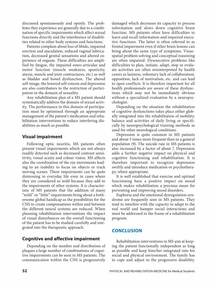
52 PHYSICAL AND REHABILITATION MEDICINE for Medical Students
damaged which decreases its capacity to process information and slows down cognitive brain function. MS patients often have difficulties to learn and recall information and impaired execu-tive functions. The latter is often referred to as frontal impairment even if other brain lesions can bring about the same type of symptoms. Visuo-spatial problem solving and conceptual reasoning are often impaired. Dysexecutive problems like difficulties to plan, initiate, adapt, stop or evalu-ate activities are often misinterpreted by health carers as laziness, voluntary lack of collaboration, opposition, lack of motivation, etc. and can lead to open conflicts. It is therefore important for all health professionals are aware of these dysfunc-tions which may not be immediately obvious without a specialized evaluation by a neuropsy-chologist.
Depending on the situation the rehabilitation of cognitive dysfunctions takes place either glob-ally integrated into the rehabilitation of mobility, balance and activities of daily living or specifi-cally by neuropsychological training methods as used for other neurological conditions.
Depression is quite common in MS patients and about 5 times more frequent than in a general population (9). The suicide rate in MS patients is also increased by a factor of about 7. Depression adds a further negative impact on physical and cognitive functioning and rehabilitation. It is therefore important to recognize depression swiftly and introduce medication or psychothera-py, where appropriate.
It is well established that exercise and optimal functioning have a positive impact on mood which makes rehabilitation a precious mean for preventing and improving mood disorders.
Euphoria and the emotional dysregulation syn-drome are frequently seen in MS patients. They tend to interfere with the capacity to adapt to the real world and hamper social interactions and must be addressed in the frame of a rehabilitation program.
CONCLUSION
Rehabilitation interventions in MS aim at keep-ing the patient functionally independent as long as possible and keep him/her integrated into his social and physical environment. The family has to cope and adjust to the progressive disability.
discussed spontaneously and openly. The prob-lems they experience are generally due to a combi-nation of specific impairments which affect sexual functions directly and the interference of disabili-ties related to other body systems and functions.
Patients complain about loss of libido, impaired erection and ejaculation, reduced vaginal lubrica-tion, decreased genital sensations and altered ex-perience of orgasm. These difficulties are ampli-fied by fatigue, the impaired osteo-articular and motor function (muscle weakness, spasticity, ataxia, muscle and joint contractures, etc.) as well as bladder and bowel dysfunction. The altered self-image, the lowered self-esteem and depression are also contributors to the restriction of partici-pation in the domain of sexuality.
Any rehabilitation plan of a MS patient should systematically address the domain of sexual activ-ity. The performance in this domain of participa-tion must be optimised through an appropriate management of the patient’s medication and reha-bilitation interventions to reduce interfering dis-abilities as much as possible.
Visual impairmentFollowing optic neuritis, MS patients often
present visual impairments which are not always readily detected such as decreased contrast sensi-tivity, visual acuity and colour vision. MS affects also the coordination of the eye movements lead-ing to an inability to focus on objects or track moving scenes. These impairments can be quite distressing in everyday life even in cases where they are considered as mild because they add to the impairments of other systems. It is character-istic of MS patients that the addition of many “mild” or “little” impairments bring about a both-ersome global handicap as the possibilities for the CNS to create compensations within and between the different neural systems are reduced. When planning rehabilitation interventions the impact of visual disturbances on the overall functioning of the patient has to be studied carefully and inte-grated into the therapeutic approach.
Cognitive and affective impairmentDepending on the number and distribution of
plaques a large number of combinations of cogni-tive impairments can be seen in MS patients. The communication within the CNS is progressively
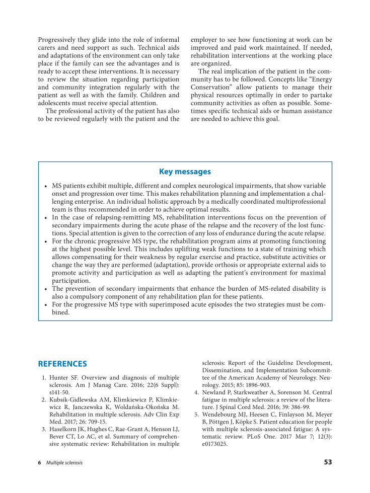
6 Multiple sclerosis 53
employer to see how functioning at work can be improved and paid work maintained. If needed, rehabilitation interventions at the working place are organized.
The real implication of the patient in the com-munity has to be followed. Concepts like “Energy Conservation” allow patients to manage their physical resources optimally in order to partake community activities as often as possible. Some-times specific technical aids or human assistance are needed to achieve this goal.
Progressively they glide into the role of informal carers and need support as such. Technical aids and adaptations of the environment can only take place if the family can see the advantages and is ready to accept these interventions. It is necessary to review the situation regarding participation and community integration regularly with the patient as well as with the family. Children and adolescents must receive special attention.
The professional activity of the patient has also to be reviewed regularly with the patient and the
REFERENCES
1. Hunter SF. Overview and diagnosis of multiple sclerosis. Am J Manag Care. 2016; 22(6 Suppl): s141-50.
2. Kubsik-Gidlewska AM, Klimkiewicz P, Klimkie-wicz R, Janczewska K, Woldańska-Okońska M. Rehabilitation in multiple sclerosis. Adv Clin Exp Med. 2017; 26: 709-15.
3. Haselkorn JK, Hughes C, Rae-Grant A, Henson LJ, Bever CT, Lo AC, et al. Summary of comprehen-sive systematic review: Rehabilitation in multiple
sclerosis: Report of the Guideline Development, Dissemination, and Implementation Subcommit-tee of the American Academy of Neurology. Neu-rology. 2015; 85: 1896-903.
4. Newland P, Starkweather A, Sorenson M. Central fatigue in multiple sclerosis: a review of the litera-ture. J Spinal Cord Med. 2016; 39: 386-99.
5. Wendebourg MJ, Heesen C, Finlayson M, Meyer B, Pöttgen J, Köpke S. Patient education for people with multiple sclerosis-associated fatigue: A sys-tematic review. PLoS One. 2017 Mar 7; 12(3): e0173025.
Key messages
• MS patients exhibit multiple, different and complex neurological impairments, that show variable onset and progression over time. This makes rehabilitation planning and implementation a chal-lenging enterprise. An individual holistic approach by a medically coordinated multiprofessional team is thus recommended in order to achieve optimal results.
• In the case of relapsing-remitting MS, rehabilitation interventions focus on the prevention of secondary impairments during the acute phase of the relapse and the recovery of the lost func-tions. Special attention is given to the correction of any loss of endurance during the acute relapse.
• For the chronic progressive MS type, the rehabilitation program aims at promoting functioning at the highest possible level. This includes uplifting weak functions to a state of training which allows compensating for their weakness by regular exercise and practice, substitute activities or change the way they are performed (adaptation), provide orthosis or appropriate external aids to promote activity and participation as well as adapting the patient’s environment for maximal participation.
• The prevention of secondary impairments that enhance the burden of MS-related disability is also a compulsory component of any rehabilitation plan for these patients.
• For the progressive MS type with superimposed acute episodes the two strategies must be com-bined.
54 PHYSICAL AND REHABILITATION MEDICINE for Medical Students
Consensus Conference on Pain in Neurorehabili-tation. Eur J Phys Rehabil Med. 2016; 52: 827-40.
8. Tracey JM, Stoffel JT. Secondary and tertiary treat-ments for multiple sclerosis patients with urinary symptoms. Investig Clin Urol. 2016; 57: 377-83.
9. Kidd T, Carey N, Mold F, Westwood S, Miklaucich M, Konstantara E, et al. A systematic review of the effectiveness of self-management interventions in people with multiple sclerosis at improving de-pression, anxiety and quality of life. PLoS One. 2017 Oct 11; 12(10): e0185931.
6. Blikman LJ, Huisstede BM, Kooijmans H, Stam HJ, Bussmann JB, van Meeteren J. Effectiveness of energy conservation treatment in reducing fatigue in multiple sclerosis: a systematic review and me-ta-analysis. Arch Phys Med Rehabil. 2013; 94: 1360-76.
7. Paolucci S, Martinuzzi A, Scivoletto G, Smania N, Solaro C, Aprile I, et al. Assessing and treating pain associated with stroke, multiple sclerosis, ce-rebral palsy, spinal cord injury and spasticity. Evi-dence and recommendations from the Italian
7 Rehabilitation of people with Parkinson’s disease 55
EPIDEMIOLOGY OF DISEASE AND DISABILITY PROGRESSION
PD prevalence increases steadily with age, with the onset age mostly ranging from 55 to 65 years (2). The crude prevalence rate in European coun-tries has been found to range from 41 per 100,000 in the fourth decade of life to over 1900 per 100,000 in people over 80 years of age. Significant differences by geographic location are only appre-ciated for individuals 70 to 79 years old, with a prevalence of 1601 (per 100,000) in individuals from North America, Europe, and Australia, compared with 646 in individuals from Asia.
Reported standardized incidence rates of PD are 8-18 per 100,000 person-years. Onset of dis-ease is rare before age 50 years and a sharp in-crease of the incidence is seen after age 60 years. As in prevalence studies, several of the prospec-tive studies found evidence for a higher incidence in men than in women. In the pre-levodopa era, excess mortality was reported to be 3-fold greater in patients with PD. The excess mortality rate was reduced to 1.6-fold greater than age-matched non-PD individuals after the introduction of le-vodopa. Today, patients with PD can live 20 or more years, depending on the age at onset. Death is usually due to some concurrent unrelated ill-ness or due to the effects of decreased mobility, aspiration, or increased falling with subsequent physical injury.
PD is considered a multifactorial disorder that results, in most cases, from the combined effects of multiple risk and protective factors, including gene and environmental ones. Current thinking is that major gene mutations cause only a small pro-portion of all cases and that in most cases, non-genetic factors play a part, probably in interaction with susceptibility genes. Multiple environmental factors that may be related to the etiology of PD include exposure to pesticides, herbicides and in-
INTRODUCTION
Parkinson’s disease (PD) is the second most important age-related neurodegenerative disorder in developed societies, after Alzheimer’s disease. The phenotype is mainly characterized by rigidity, resting tremor, and bradykinesia, making PD the most diffuse among chronically disabling move-ment disorders. The underlying biological damage is supposed to occur several decades before the onset of the motor symptoms. Potential risk fac-tors include environmental toxins, drugs, pesti-cides, repeated brain micro-trauma, focal cerebro-vascular damage, and genomic defects (1). Selec-tive damage is represented by a selective loss of dopaminergic neurons in the substantia nigra pars compacta, with widespread involvement of other central nervous system (CNS) structures and pe-ripheral tissues. Conventional pharmacological treatments for PD are dopamine precursors (le-vodopa), and other symptomatic treatments in-cluding dopamine agonists, monoamine oxidase (MAO) inhibitors and catechol-O-methyltransfer-ase (COMT) inhibitors. Although the motor symp-toms of PD can be well controlled by levodopa and other adjunctive medications in the early stages of the disease, treatment-related complications (prin-cipally, drug-effect fluctuations, with “on-off” phenomena and dyskinesias), will inevitably occur after 5-7 years. Functional surgery approaches, aimed at reducing the consequences of dopamine depletion in the basal ganglia circuits, ensure symptomatic treatment without drug-related side effects. However, neither drug nor surgical treat-ments can avoid disability occurrence and pro-gression due to levodopa resistant features.
Owing to the progressively reduced impact of PD on mortality, compared to the age-matched general population, people with PD face the risk of living with disability for an important part of their life.
7Rehabilitation of people with Parkinson’s diseaseMaria Gabriella CERAVOLO, Marianna CAPECCI

56 PHYSICAL AND REHABILITATION MEDICINE for Medical Students
taken to complete not only instrumental Ac-tivities of Daily Living (ADL), like using a mo-bile phone, cooking or gardening, but also basic ADL, like dressing, toileting or transferring. When it is not resolved by symptomatic treat-ment, it represents the main factor of physical activity restriction and early retirement from work in people with PD.
ū Rest tremor refers to a 4- to 6-Hz tremor in the fully resting limb, which is suppressed during movement initiation, and is often exacerbated during potential provocations, such walking and counting backwards (5). Due to its occur-rence at rest, it does not interfere with any mo-tor activity; conversely, it reduces well-being perception, being one of the main factors of stigma complained by patients.
ū Rigidity refers to “lead-pipe”, velocity-indepen-dent resistance to passive movement, not solely reflecting failure to relax (i.e., it is distinct from spasticity or paratonia). It may occur proxi-mally (e.g., neck, shoulder, and hip) and distally (e.g., wrist and ankle) and cause discomfort and pain. Painful shoulder, possibly due to ri-gidity, but frequently misdiagnosed as arthritis, bursitis, or rotator cuff, is one of the most fre-quent initial manifestations of PD (6).
Other motor features, like trunk abnormalities, postural instability and specific gait impairment, can also occur in a majority of people with PD, representing a major burden for patients as they are resistant to the available drug and surgery treatments.
ū Trunk abnormalities include neck flexion (an-terocollis), forward trunk flexion (camptocor-mia) and lateral trunk flexion (Pisa syndrome). The underlying pathophysiology of these defor-mities is largely unknown; different central and peripheral mechanisms have been taken into account and this partly explains why there are so many ineffective attempts of treatment. It is acknowledged that up to 18% PD patients show camptocormia and/or Pisa syndrome, while 6% develop anterocollis. The impact of trunk ab-normalities can be severe since they often cause muscle pain, limited tolerance to maintaining the upright posture, increased walking handi-cap, breathing difficulty (in case of severe trunk deviations) and poor social interaction.
ū Gait impairment. Since disease onset, people with PD exhibit a characteristic reduction in
take of various metals. A few epidemiological studies have shown that cigarette smoking, coffee and tea drinking are associated with a lower risk of PD. Similarly, a higher level of physical activity is hypothesized to lower the risk of PD (3).
CLINICAL AND FUNCTIONAL PROFILE OF PEOPLE WITH PARKINSON’S DISEASE
Although PD has been long regarded as a move-ment disorder, clinical features of disease are much more complex and include both motor, cog-nitive, affective and autonomic disorders. Accord-ing to their neurophysiological substrate, sensitiv-ity to dopaminergic drug treatment and func-tional prognosis, symptoms may be divided into two main categories respectively respondent or re-fractory to levodopa. The cardinal motor features of PD (bradykinesia, rest tremor and rigidity) typically belong to the first category; hence, severe as they may be, such symptoms can be managed by drugs or functional surgery (namely, deep brain stimulation, pallidotomy or thalamotomy). On the other hand, both axial motor symptoms (trunk abnormalities, postural instability, freezing of gait, speech disorders) and non-motor symp-toms are either poorly respondent or worsened by, if not refractory to, dopaminergic stimulation, thus exerting a progressively disabling impact.
The different prognostic role of disease symp-toms is also underlined by a cohort study of 800 PD patients, that divided them into two major phenotypes: the tremor-dominant form of PD, that is associated with a relatively preserved men-tal status, earlier age at onset, and a slower pro-gression of the disease, and the one dominated by postural-instability-gait-difficulty (PIGD), charac-terized by more severe bradykinesia and a more rapidly progressive course (4).
A short description of the main symptoms, with reference to their functional impact, is re-ported.
ū Bradykinesia, the clinical hallmark of PD, is defined as slowness of movement and decre-ment in amplitude or speed as movements are continued. It mainly affects automatic, repeat-ed movements, like those involved in talking, swallowing, gesturing, maintaining upright body posture, walking. Bradykinesia severely impacts quality of life, by increasing the time
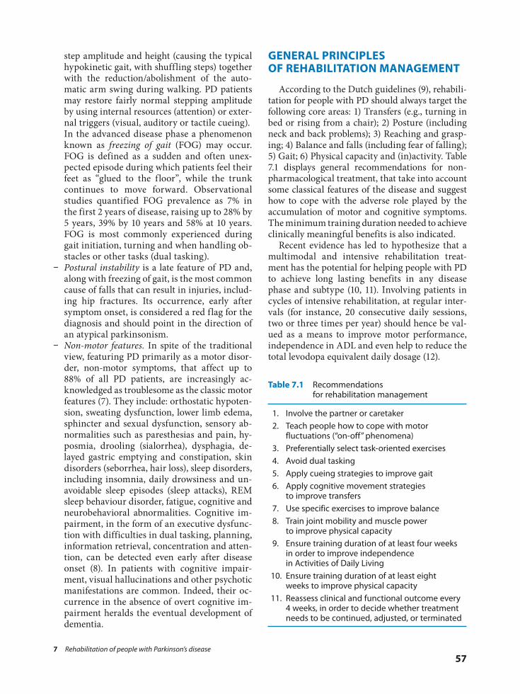
7 Rehabilitation of people with Parkinson’s disease 57
GENERAL PRINCIPLES OF REHABILITATION MANAGEMENT
According to the Dutch guidelines (9), rehabili-tation for people with PD should always target the following core areas: 1) Transfers (e.g., turning in bed or rising from a chair); 2) Posture (including neck and back problems); 3) Reaching and grasp-ing; 4) Balance and falls (including fear of falling); 5) Gait; 6) Physical capacity and (in)activity. Table 7.1 displays general recommendations for non-pharmacological treatment, that take into account some classical features of the disease and suggest how to cope with the adverse role played by the accumulation of motor and cognitive symptoms. The minimum training duration needed to achieve clinically meaningful benefits is also indicated.
Recent evidence has led to hypothesize that a multimodal and intensive rehabilitation treat-ment has the potential for helping people with PD to achieve long lasting benefits in any disease phase and subtype (10, 11). Involving patients in cycles of intensive rehabilitation, at regular inter-vals (for instance, 20 consecutive daily sessions, two or three times per year) should hence be val-ued as a means to improve motor performance, independence in ADL and even help to reduce the total levodopa equivalent daily dosage (12).
step amplitude and height (causing the typical hypokinetic gait, with shuffling steps) together with the reduction/abolishment of the auto-matic arm swing during walking. PD patients may restore fairly normal stepping amplitude by using internal resources (attention) or exter-nal triggers (visual, auditory or tactile cueing). In the advanced disease phase a phenomenon known as freezing of gait (FOG) may occur. FOG is defined as a sudden and often unex-pected episode during which patients feel their feet as “glued to the floor”, while the trunk continues to move forward. Observational studies quantified FOG prevalence as 7% in the first 2 years of disease, raising up to 28% by 5 years, 39% by 10 years and 58% at 10 years. FOG is most commonly experienced during gait initiation, turning and when handling ob-stacles or other tasks (dual tasking).
ū Postural instability is a late feature of PD and, along with freezing of gait, is the most common cause of falls that can result in injuries, includ-ing hip fractures. Its occurrence, early after symptom onset, is considered a red flag for the diagnosis and should point in the direction of an atypical parkinsonism.
ū Non-motor features. In spite of the traditional view, featuring PD primarily as a motor disor-der, non-motor symptoms, that affect up to 88% of all PD patients, are increasingly ac-knowledged as troublesome as the classic motor features (7). They include: orthostatic hypoten-sion, sweating dysfunction, lower limb edema, sphincter and sexual dysfunction, sensory ab-normalities such as paresthesias and pain, hy-posmia, drooling (sialorrhea), dysphagia, de-layed gastric emptying and constipation, skin disorders (seborrhea, hair loss), sleep disorders, including insomnia, daily drowsiness and un-avoidable sleep episodes (sleep attacks), REM sleep behaviour disorder, fatigue, cognitive and neurobehavioral abnormalities. Cognitive im-pairment, in the form of an executive dysfunc-tion with difficulties in dual tasking, planning, information retrieval, concentration and atten-tion, can be detected even early after disease onset (8). In patients with cognitive impair-ment, visual hallucinations and other psychotic manifestations are common. Indeed, their oc-currence in the absence of overt cognitive im-pairment heralds the eventual development of dementia.
Table 7.1 Recommendations for rehabilitation management
1. Involve the partner or caretaker 2. Teach people how to cope with motor
fluctuations (“on-off” phenomena) 3. Preferentially select task-oriented exercises 4. Avoid dual tasking 5. Apply cueing strategies to improve gait 6. Apply cognitive movement strategies
to improve transfers 7. Use specific exercises to improve balance 8. Train joint mobility and muscle power
to improve physical capacity 9. Ensure training duration of at least four weeks
in order to improve independence in Activities of Daily Living
10. Ensure training duration of at least eight weeks to improve physical capacity
11. Reassess clinical and functional outcome every 4 weeks, in order to decide whether treatment needs to be continued, adjusted, or terminated
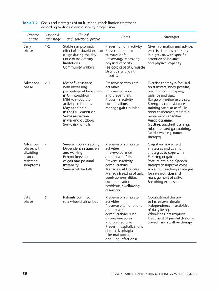
58 PHYSICAL AND REHABILITATION MEDICINE for Medical Students
Table 7.2 Goals and strategies of multi-modal rehabilitation treatment according to disease and disability progression
Disease phase
Hoehn & Yahr stage
Clinical and Functional profile Goals Strategies
Early phase
1-2 Stable symptomatic effect of antiparkinsonian drugs during the dayLittle or no Activity limitationsCommunity walkers
Prevention of inactivityPrevention of fear to move or fallPreserving/improving physical capacity (aerobic capacity, muscle strength, and joint mobility)
Give information and advice; exercise therapy (possibly in a group), with specific attention to balance and physical capacity
Advanced phase
2-4 Motor fluctuations with increasing percentage of time spent in OFF conditionMild to moderate activity limitationsMay need help in the OFF condition Some restriction in walking outdoors Some risk for falls
Preserve or stimulate activitiesImprove balance and prevent fallsPrevent inactivity complicationsManage gait troubles
Exercise therapy is focused on transfers, body posture, reaching and grasping, balance and gait. Range of motion exercises.Strength and resistance training are also useful in order to increase/maintain movement capacities.Aerobic training (cycling, treadmill training, robot-assisted gait training, Nordic walking, dance therapy)
Advanced phase, with disabling levodopa resistant symptoms
4 Severe motor disabilityDependent in transfers and walking Exhibit freezing of gait and postural instabilitySevere risk for falls
Preserve or stimulate activitiesImprove balance and prevent fallsPrevent inactivity complicationsManage gait troublesManage freezing of gait, trunk abnormalities, communication problems, swallowing disorders
Cognitive movement strategies and cueing strategies to cope with Freezing of gait.Postural training. Speech therapy to improve voice emission, teaching strategies for safe nutrition and management of saliva. Breathing exercises
Late phase
5 Patients confined to a wheelchair or bed
Preserve or stimulate activitiesPreserve vital functions and prevent complications, such as pressure sores and contracturesPrevent hospitalizations due to dysphagia (like malnutrition and lung infections)
Occupational therapy to increase/maintain independence in activities of daily living. Wheelchair prescription.Treatment of painful dystoniaSpeech and swallow therapy

7 Rehabilitation of people with Parkinson’s disease 59
Under this perspective, it can be stated that exercise targets many aspects of brain function and has broad effects on overall brain health; it is effective at improving gait, balance, freezing, and motor performance in all stages of PD. In particu-lar, recent studies have focused on identifying ex-ercise parameters that are essential for promoting activity-dependent neuroplasticity and ensuring the efficacy and the effectiveness lasting over time. Both basic research and clinical studies sug-gest that high intensity (i.e., high repetition, veloc-ity, complexity) is a characteristic of exercise that may be important in promoting activity- depen-dent neuroplasticity of the injured brain, includ-ing the basal ganglia and improving motor perfor-mance (15).
However, the amount of physical activity, the dose and the frequency of exercise, the type and modality of administration, are yet to be deter-mined.
THE MANAGEMENT OF SELECTIVE MOTOR DISORDERS
Freezing of gait
Although especially frequent in the advanced disease, FOG can occur at any phase of PD and is always associated with increased levels of activity limitation even after adjusting for disease severity. Although FOG pathogenesis has not yet been en-tirely understood, most researchers assume the involvement of both motor and cognitive path-ways as leading mechanisms.
The management of gait disability and, partic-ularly, FOG, has long been a neglected topic, pre-sumably because these axial symptoms – despite being common and often devastating – were per-ceived as largely untreatable. In fact, traditional treatment options, like dopaminergic drugs and deep brain stimulation, can sometime aggravate FOG, rather than suppress it.
Non pharmacological approaches have received much attention following the observation that people with PD spontaneously adopt a variety of cues or tricks to overcome the freezing attacks. They can maintain a rhythmic gait through counting their steps aloud, or marching to com-mand (“left, right, left, right”), stepping over ob-jects (the end of a walking stick, a pavement stone, cracks in the floor), walking at the rhythm of
Moreover, a multi-professional team delivering a multimodal treatment is warranted, in order to take the individual needs into account and shape training according to the patients’ clinical and functional profile.
Eventually, given the progressive, though pre-dictable, course of disability, it is possible to out-line specific rehabilitation goals and interven-tions, that differ from phase to phase (Table 7.2).
Although a slow, yet unavoidable, functional decline is expected in people with PD, the acute aggravation of the clinical picture can also be ob-served, thus requiring a prompt and expert man-agement.
The hospitalization, which could be necessary in case of fractures, worsening of swallow impair-ment, severe drug side effects (hallucinations, psychosis, severe motor fluctuations) or any surgi-cal interventions, represents an emergency for the patient in advanced phase, due to a high risk of complications during the hospital stay. Moreover, in order to avoid the threat of a persistent loss of the functional skills expressed before admission, a prompt management by the rehabilitation team is warranted. Whatever the setting and type of reha-bilitation delivery are, the key element of the in-tervention is represented by the multidisciplinary team approach that is the only possible answer to the multifaceted emerging disability.
THE ROLE OF EXERCISE AS A NEUROPROTECTIVE FACTOR
There is strong evidence from the literature that goal-based and aerobic exercise strengthen and improve motor circuitry through mecha-nisms that include increased synaptic strength resulting from raised dopamine and glutamate neurotransmission within the basal ganglia ac-companied by increased dendritic spine forma-tion. Exercise leads to improved brain health in-cluding increased expression of neurotrophic fac-tors, increased blood flow, altered immune re-sponse, increased neurogenesis (especially within the hippocampus), and altered metabolism (i.e., improved mitochondrial health) (13). Such chang-es might lead to enhanced neuronal circuitry be-tween the basal ganglia and its cortical and tha-lamic connections, which ultimately result in im-proved motor, non-motor, and cognitive behav-iour in patients with Parkinson’s disease (14).

60 PHYSICAL AND REHABILITATION MEDICINE for Medical Students
A few studies endorse the hypothesis that the use of a commercial device like the Nintendo® Wii Balance can be a safe, easy and effective approach. Nintendo® Wii, characterized as a disposable de-vice for home entertainment, is suitable to arouse patient’s motivation and could represent a useful tool for continuous self-administered home train-ing, allowing for the enhancement of formal reha-bilitation effects and the achievement of increas-ing levels of motor learning (20).
Other researchers have focused on the effective-ness of lower limb strength training as a means to improve balance. Strength training performed against an external resistance (cycle ergometer, weight machine, elastic band, punching bag, and water) is well tolerated in subjects with mild to moderate PD and helps improving both physical parameters and quality of life parameters, without determining a decline in other outcomes. Never-theless, the effect of strength training on balance, freezing and number of falls is still uncertain (21).
THE ROLE OF COMPLEMENTARY THERAPIES
Physical activity has long been considered as a complement to drug treatment to manage PD-re-lated disability. From the first experimental stud-ies until today, physical activity has been reported to exert positive effects on people with PD, in terms of improved mobility, gait, balance and muscle strength, whereas, effects on other health parameters such as cognitive functions, activities of daily living and psychosocial aspects of life re-main unclear.
In recent years, complementary therapies such as dancing, boxing, tai chi, virtual reality train-ing, robotic gait training, whole body vibration and hydrotherapy have conquered growing popu-larity as effective approaches for improving PD-related disability. At variance with conventional exercise therapy, complementary therapies target not only physical functions but also wellbeing, enjoyment and social participation, thus allowing for increased adherence and compliance to treat-ment over the long term.
There is a consistent evidence that dance, tai chi, water exercises, virtual reality training and Nordic walking enable people with mild to mod-erate PD to move more easily and to enjoy greater health and wellbeing (22).
music or a metronome, shifting body weight from side to side or using rocking movements (16).
Cues are defined as external temporal or spatial stimuli that aim to facilitate movement, gait ini-tiation and continuation. Their effectiveness in alleviating freezing is based on driving subject’s attention towards externally driven motor acts in order to compensate the reduced ability to recruit or maintain internally driven motor programs. Impaired movement initiation is linked to func-tional deficit in the frontal-striatal pathways. Since the cue directly activates the sensory-motor cortex, bypassing the subcortical system, the higher its capacity to drive patient’s attention to-wards the motor task the greater its effectiveness in activating and maintaining rhythmic stepping and avoiding freezing.
Encouraging results have been obtained with the use of acoustic cues (in particular rhythmic melodies like tango) (17), even combined with augmented-reality.
Treadmill training restores gait parameters through increasing stride length and encouraging gait start, thanks to the proprioceptive cueing of the automated carpet. External devices, like ro-bots, explicitly designed to guide repetitive, rhyth-mic, bilateral lower extremity movements, are ex-pected to restore a physiological gait cycle, provid-ing continuous, multimodal and rhythmic exter-nal cues. Robotic gait training has been reported to improve gait speed, walking capacity, stride length and fatigue in patients with PD (18). One of the most popular models seems to be the G-EO system that imposes the execution of alternate movements with the lower limbs, by applying a foot constraint and supporting body weight (19).
Balance impairmentPeople with PD commonly experience postural
instability during daily activities. Impaired senso-ry and visual inputs, and a reduced base of support are among the hypothesized causal mechanisms. Furthermore, deficits in the central integration of vision, somatic sensation, and vestibular inputs as well as a cortically mediated decline in attention capacities, may contribute to balance impairment. Several reports have documented how training on a balance platform, especially with virtual reality augmentation, can positively impact sensory orga-nization and increase attentional demand for pos-tural control in people with PD.
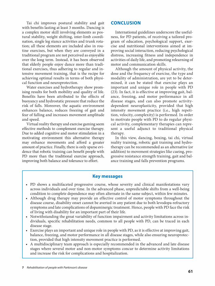
7 Rehabilitation of people with Parkinson’s disease 61
CONCLUSION
International guidelines underscore the useful-ness, for PD patients, of receiving a tailored pro-gram of education, psychological support, exer-cise and nutritional interventions aimed at im-proving social interaction, reducing psychological distress, increasing fitness and independence in activities of daily life, and promoting relearning of motor and communication skills.
Although the amount of physical activity, the dose and the frequency of exercise, the type and modality of administration, are yet to be deter-mined, it can be stated that exercise plays an important and unique role in people with PD (23). In fact, it is effective at improving gait, bal-ance, freezing, and motor performance in all disease stages, and can also promote activity-dependent neuroplasticity, provided that high intensity movement practice (i.e., high repeti-tion, velocity, complexity) is performed. In order to motivate people with PD to do regular physi-cal activity, complementary therapies can repre-sent a useful adjunct to traditional physical therapy.
In this view, dancing, boxing, tai chi, virtual reality training, robotic gait training and hydro-therapy can be recommended as an alternative (or addition) to movement strategies like cueing, pro-gressive resistance strength training, gait and bal-ance training and falls prevention programs.
Tai chi improves postural stability and gait with benefits lasting at least 3 months. Dancing is a complex motor skill involving elements as pos-tural stability, weight shifting, inter-limb coordi-nation, single leg stance activities and trunk rota-tion; all these elements are included also in rou-tine exercises, but when they are conveyed in a traditional program are not perceived as enjoyable over the long term. Instead, it has been observed that elderly people enjoy dance more than tradi-tional exercises, thus adhering to regular and in-tensive movement training, that is the recipe for achieving optimal results in terms of both physi-cal function and neuroprotection.
Water exercises and hydrotherapy show prom-ising results for both mobility and quality of life. Benefits have been attributed to the effects of buoyancy and hydrostatic pressure that reduce the risk of falls. Moreover, the aquatic environment enhances balance, reduces freezing of gait and fear of falling and increases movement amplitude and speed.
Virtual reality therapy and exercise gaming seem effective methods to complement exercise therapy. Due to added cognitive and motor stimulation in a motivating environment this alternative therapy may enhance movements and afford a greater amount of practice. Finally, there is only sparse evi-dence that robotic training can benefit people with PD more than the traditional exercise approach, improving both balance and tolerance to effort.
Key messages
• PD shows a multifaceted progressive course, whose severity and clinical manifestations vary across individuals and over time. In the advanced phase, unpredictable shifts from a well-being condition to complete dependence may often alternate in the same subject, within few minutes.
• Although drug therapy may provide an effective control of motor symptoms throughout the disease course, disability onset cannot be averted in any patient due to both levodopa-refractory symptoms and late complications of dopaminergic treatment. Hence, people with PD face the risk of living with disability for an important part of their life.
• Notwithstanding the great variability of function impairment and activity limitations across in-dividuals, specific rehabilitation needs, common to all people with PD, can be traced in each disease stage.
• Exercise plays an important and unique role in people with PD, as it is effective at improving gait, balance, freezing, and motor performance in all disease stages, while also ensuring neuroprotec-tion, provided that high intensity movement practice is performed.
• A multidisciplinary team approach is especially recommended in the advanced and late disease stages where several motor and non-motor symptoms concur to determine activity limitations and increase the risk for complications and hospitalization.
62 PHYSICAL AND REHABILITATION MEDICINE for Medical Students
comparing two different rehabilitative treatments. NeuroRehabilitation. 2012; 30: 295-301.
13. Hirsch MA, Iyer SS, Sanjak M. Exercise-induced neuroplasticity in human Parkinson’s disease: What is the evidence telling us? Parkinsonism Relat Disord. 2016; 22 Suppl 1: S78-81.
14. Petzinger GM, Fisher BE, McEwen S, Beeler JA, Walsh JP, Jakowec MW. Exercise-enhanced neuro-plasticity targeting motor and cognitive circuitry in Parkinson’s disease. Lancet Neurol. 2013; 12: 716-26.
15. Fisher BE, Wu AD, Salem GJ, Song J, Lin CH, Yip J, et al. The effect of exercise training in improving motor performance and corticomotor excitability in people with early Parkinson’s disease. Arch Phys Med Rehabil. 2008; 89: 1221-9.
16. Nieuwboer A. Cueing for freezing of gait in patients with Parkinson’s disease: a rehabilitation perspec-tive. Mov Disord. 2008; 23 Suppl 2: S475-81.
17. Duncan RP, Earhart GM. Randomized controlled trial of community-based dancing to modify dis-ease progression in Parkinson disease. Neuroreha-bil Neural Repair. 2012; 26: 132-43.
18. Picelli A, Melotti C, Origano F, Waldner A, Fiaschi A, Santilli V, et al. Robot-assisted gait training in patients with Parkinson disease: a randomized controlled trial. Neurorehabil Neural Repair. 2012; 26: 353-61.
19. Sale P, De Pandis MF, Le Pera D, Sova I, Cimolin V, Ancillao A, et al. Robot-assisted walking training for individuals with Parkinson’s disease: a pilot ran-domized controlled trial. BMC Neurol. 2013; 13: 50.
20. Di Biagio L, Ferretti M, Cingolani D, Buzzatti L, Capecci M, Ceravolo MG. Virtual reality: a new rehabilitative approach in neurological disorders. In: Longhi S, Siciliano P, Germani M, Monteriù A, editors. Ambient Assisted Living – Italian Forum 2013. Cham (CH): Springer, 2013; pp. 167-76.
21. Ramazzina I, Bernazzoli B, Costantino C. System-atic review on strength training in Parkinson’s disease: an unsolved question. Clin Interv Aging. 2017; 12: 619-28.
22. Alves Da Rocha P, McClelland J, Morris ME. Complementary physical therapies for movement disorders in Parkinson’s disease: a systematic re-view. Eur J Phys Rehabil Med. 2015; 51: 693-704.
23. Ceravolo MG Rehabilitation goals and strategies in Parkinson’s disease Eur J Phys Med Rehab 2009; 45: 205-8.
REFERENCES
1. Cacabelos R. Parkinson’s Disease: From Pathogen-esis to Pharmacogenomics. Int J Mol Sci. 2017; 18(3). pii: E551.
2. Lee A, Gilbert RM. Epidemiology of Parkinson Disease. Neurol Clin. 2016; 34: 955-65.
3. Chen H, Zhang SM, Schwarzschild MA, Hernán MA, Ascherio A. Physical activity and the risk of Parkinson disease. Neurology. 2005; 64: 664-9.
4. Post B, Merkus MP, de Haan RJ, Speelman JD; CARPA Study Group. Prognostic factors for the progression of Parkinson’s disease: a systematic review. Mov Disord. 2007; 22: 1839-51.
5. Raethjen J, Austermann K, Witt K, Zeuner KE, Papengut F, Deuschl G. Provocation of Parkinso-nian tremor. Mov Disord. 2008; 23: 1019-23.
6. Stamey W, Davidson A, Jankovic J. Shoulder pain: a presenting symptom of Parkinson disease. J Clin Rheumatol. 2008; 14: 253-4.
7. Barone P, Antonini A, Colosimo C, Marconi R, Morgante L, Avarello TP, et al. The PRIAMO study: A multicenter assessment of nonmotor symptoms and their impact on quality of life in Parkinson’s disease. Mov Disord. 2009; 24: 1641-9.
8. Marconi R, Antonini A, Barone P, Colosimo C, Avarello TP, Bottacchi E, et al. Frontal assessment battery scores and non-motor symptoms in par-kinsonian disorders. Neurol Sci. 2012; 33: 585-93.
9. Keus SH, Bloem BR, Hendriks EJ, Bredero-Cohen AB, Munneke M; Practice Recommendations De-velopment Group. Evidence-based analysis of physical therapy in Parkinson’s disease with rec-ommendations for practice and research. Mov Disord. 2007; 22: 451-60.
10. Frazzitta G, Bertotti G, Riboldazzi G, Turla M, Uc-cellini D, Boveri N, et al. Effectiveness of intensive inpatient rehabilitation treatment on disease pro-gression in parkinsonian patients: a randomized controlled trial with 1-year follow-up. Neuroreha-bil Neural Repair. 2012; 26: 144-50.
11. Frazzitta G, Abbruzzese G, Bertotti G, Boveri N, Pezzoli G, Maestri R. Effectiveness of an intensive rehabilitation treatment on different Parkinson’s disease subtypes. NeuroRehabilitation. 2013; 33: 299-303.
12. Frazzitta G, Bertotti G, Morelli M, Riboldazzi G, Pelosin E, Balbi P, et al. Rehabilitation improves dyskinesias in Parkinsonian patients: a pilot study
8 Chronic progressive neurological disorders (with special attention to Amyotrophic Lateral Sclerosis) 63
prevalence of chronic progressive disorders on the health system, the concept of disability-adjusted life years (DALY) has been developed, to quantify the proportion of life spent with disability by people affected by any disease/injury consequenc-es (1). From 1990 to 2010, DALYs from non-com-municable diseases had shifted from 43% to 54% of the global DALY burden, at the world level. The 15 causes with the largest increases include two major neurological disorders concentrated in old-er age-groups: dementia and Parkinson’s disease (1). The rise of dementia and Parkinson’s disease is almost entirely attributable to population ageing because age-specific rates have remained constant. In view of the global shifts in fertility and declines in age-specific mortality, it is reasonable to expect that the numbers of individuals with age-related disorders will further increase in coming decades. This shift in numbers of people with certain disor-ders will have substantial implications for health-service planning. The best approach to care man-agement will hopefully rely on an expert acknowl-edgment of patients’ needs according to the mod-ern health definition introduced by the Interna-tional Classification of Functioning (2).
GENERAL PRINCIPLES OF SYMPTOM MANAGEMENT
Disability in chronic progressive diseases can be either due to impairments directly related to the disease process or to deconditioning. Several symptoms are common to all progressive neuro-logical disorders provided that they induce mobil-ity restriction, reduced joint activity, with or without muscle tone impairment (either rigidity or spasticity). The adverse impact of motor disor-ders on the general level of subjects’ physical ac-
INTRODUCTION
Rehabilitation interventions are an important part of the care for people with chronic progres-sive neurological conditions. While medical treat-ments can attenuate or slow down the progression of irreversible damage to body functions and structures, induced by such diseases and delay the resulting disabilities, rehabilitation interventions are the only way to ensure that the functioning of these patients is kept at the highest possible level at any stage of the illness in spite of the underlying progressive damage to anatomy and function.
Unfortunately, many medical practitioners don’t have the knowledge about how rehabilita-tion interventions can influence human function-ing even in case of irreversibly damaged body functions and structures. They wrongly consider the disability related to these chronic conditions as immutable and not accessible to rehabilitation.
The aim of this chapter is to explain some gen-eral aspects of the rehabilitation of persons dis-abled by chronic progressive neurological condi-tions. It shows how disability must be analysed and describes the different rehabilitation strategies to be used to bring about an optimal functional out-come for these patients and increase their quality of life in spite of the chronic illness. Eventually, the rehabilitation of patients disabled by Amyotrophic Lateral Sclerosis is illustrated more specifically.
EPIDEMIOLOGY
Over the last 20 years, the global disease burden has continued to shift away from communicable to non-communicable diseases, and from premature death to years lived with disability. In order to bet-ter describe the challenge imposed by the growing
8Chronic progressive neurological disorders (with special attention
to Amyotrophic Lateral Sclerosis)Rolf FRISCHKNECHT, Maria Gabriella CERAVOLO
64 PHYSICAL AND REHABILITATION MEDICINE for Medical Students
can raise opposition especially if health profession-als are brought in to provide the additional work. They have difficulties to understand why a patient should not carry out activities he is physically and cognitively able to perform “independently”. Some-times the rehabilitation team leader has to spend a lot of effort to explain the concept to the carers and persuade them to adhere. It is therefore recom-mended to include all present or prospective carers into the approach right from the beginning.
The management of painOver 50% of people with neurodegenerative
disorders experience significant pain at some time point of the course of the disease. Several types of pain can occur: neuropathic pain related to the impairment of the nervous system, pain connect-ed with upper neuron dysfunction, like paroxys-mal tonic spasms, musculoskeletal pain due to al-tered biomechanics of movements, impaired joint control, muscle retractions and joint restrictions.
Both paroxysmal (trigeminal neuralgia, Lher-mitte syndrome, paroxysmal burning pain, pain-ful tonic seizures and painful tonic muscle spasms) and chronic neuropathic pain typically respond to anticonvulsants such as carbamazepine and gaba-pentin (4).
All types of musculoskeletal pain which com-monly occur in otherwise healthy people can also occur in patients suffering from chronic progres-sive neurological disorders, though such patients are more at risk to develop musculoskeletal prob-lems as motor and sensory dysfunctions alter the motor control of the spine and the peripheral joints. Loss of proprioception, muscle retractions, muscle tone impairment, impairment of reciprocal inhibition, co-contractions, ataxia and impaired coordination of muscle activation lead to an abnor-mal use of joint structures, ligaments, muscles and tendons. The consecutive misuse, overuse and disuse are detrimental to these structures and fre-quently result in chronic musculoskeletal pain (5). Pain enhances motor symptoms (tremor, rigidity, dystonia, spasticity) and induces a vicious circle of increased musculoskeletal dysfunction and pain.
Also in patients with neurological syndromes, musculoskeletal pain responds to the classical treatments of musculoskeletal disorders such as anti-inflammatory drugs, analgesics, steroid infil-trations, physiotherapy, occupational therapy, or-thoses and braces, but it is mandatory to reduce or
tivity is expected to activate a vicious circle that is often responsible of further functional decline, even in absence of a true disease progression. Hence, one of the main goals of rehabilitation in-terventions will be the management of those symptoms that most adversely affect individual ability to cope with motor disability, like pain and fatigue, so to prevent the occurrence of complica-tions due to deconditioning.
The management of Fatigue and the concept of Energy Conservation
Energy Conservation is a concept which in-tends to help persons with various disabilities to manage their activities during a day or a week so as to achieve a high level of quality of life without excessive pain, fatigue and exhaustion. This is achieved by selecting activities which promote well being and plan them in a way to take advan-tage of the physically and cognitively favourable periods of the day and minimise fatigue through periods of rest (3).
In a first step all the activities the patient wishes to perform to meet her or his expectations and the activities she or he is supposed to perform to com-ply with the expectations of the family and the so-ciety are listed. Pleasurable and satisfying activities are prioritized; if necessary, the level of fatigue they produce is limited by simplifying them, by choos-ing a more ergonomic way of doing, by introducing the suitable assistive devices or by organizing the appropriate human, animal or robotic assistance. Unnecessary ungratifying activities are abandoned. Necessary activities which generate a high load of fatigue are modified to reduce the amount of effort needed to a bearable level. If this turns out to be impossible, human or technical assistance is pro-vided to minimize effort and fatigue or the whole activity is delegated to informal or formal carers or other third persons. The retained activities are then distributed over a day or a week, and realized in an unassisted, modified or assisted way, allowing for adequate periods of rest so as to make them as en-joyable as possible, minimize fatigue and prevent exhaustion. For some patients minimizing pain and optimizing pain treatment throughout the day has to be included into this approach.
According to such approach, those activities that a patient is able to perform on its own, but at a prize of considerable fatigue and exhaustion, will be as-sisted or even carried out entirely by carers. This
8 Chronic progressive neurological disorders (with special attention to Amyotrophic Lateral Sclerosis) 65
cal neurological picture, consequent to the degen-eration of the first and second motor neurons. Normally, the motor impairment starts affecting either the lower or the upper limb on one side, then progresses by involving all the skeletal muscles, and eventually extends to the respiratory and swallowing muscles. Speech articulation is com-promised as well. Although such progression is unavoidable, a multipurpose rehabilitation plan must be always applied, adapting to:
ū the different chronology by which motor symp-toms (strength deficit, spasticity, muscle atro-phy, spasms and fasciculations) occur, develop and overlap
ū the various ways by which motor impairments translate into limitations of hand dexterity, transferring, walking, speech, swallowing and ventilation
ū the heterogeneous presentation of associated symptoms (fatigue, pain, sleep disorders, dys-pnea, depression and cognitive disorders) (9).
In agreement with international guidelines (10, 11), the recommended approach to ALS patients must include a detailed assessment of their health needs at the time of diagnosis and a subsequent intensive monitoring, at 3-month intervals, of their clinical and functional profile.
The clinical-functional assessment takes ad-vantage of both disease-specific, like the ALS-Functional Rating Scale revised (ALS-FRSr) and generic measures, like the Functional Indepen-dence Measure (FIM), or the Dysphagia Outcome Severity Scale (DOSS) (12).
The collection of quantitative parameters and their monitoring has the purpose of detecting problems that tend to emerge abruptly, so to guide the selection of the most appropriate care.
In particular, the early identification of adverse prognostic factors may allow the timely imple-mentation of prevention protocols, with striking effects on individual survival time and well-being perception. For instance, strength reduction be-low 80% of the predicted value in the hip extensor/flexor muscles, or below 60% of the predicted value in the knee extensor/flexor muscles, indi-cates the risk for imminent loss of independent walking ability in outdoor environment. Altera-tions in the voice quality, speech rhythm and ef-fectiveness of verbal communication are clinical signs of bulbar dysfunction, anticipating the loss of swallow ability (13).
correct the underlying causes by introducing ap-propriate rehabilitation interventions into the re-habilitation plan to lower the risk to develop musculoskeletal dysfunctions and pain.
The management of osteoporosisDecreased mobility and corticosteroid therapy
induce loss of bone mass and increase the risk of traumatic and pathological bone fractures (6). It is therefore necessary to keep an eye on bone min-eral content and introduce appropriate treat-ments. Rehabilitation interventions are excellent means to minimise the occurrence of falls and fractures by increasing overall balance and the capacity to move safely. Moreover, the increased individual capacity to be active has a favourable effect on bone architecture.
CURRENT MULTIDISCIPLINARY APPROACH TO PEOPLE WITH AMYOTROPHIC LATERAL SCLEROSIS
Amyotrophic Lateral Sclerosis (ALS) is a rapid-ly progressive neurodegenerative disease. Al-though it is assumed to be relatively rare, the eco-nomic and social burden is substantial. In Europe, the median prevalence is 5.40/100,000 inhabitants (IQR 4.06-7.89), corresponding to around 39,863 (29,971-58,244) affected cases (7). Although envi-ronmental factors and genetic predisposition can cause some epidemiological variability across Eu-ropean countries, it has been recently underlined that ALS prevalence has increased by around 50% in Western countries (comparing the decade 1990-2000 to 1960-1970), likely due to a substantial in-crease in life expectancy after the diagnosis (from 25 months in 1970 to about 33 months in 1990). Such improvement has consistently led to an in-crease of health needs from ALS patients, not simply attributable to end-of-life issues (8). Reha-bilitation is currently considered an essential com-ponent of the complex multidisciplinary manage-ment that has to care for ALS patients from diag-nosis to exitus. Rehabilitation delivery acknowl-edges different goals and actions in relation to the disease phase, the type and complexity of emerg-ing disability and the characteristics of the indi-vidual and its environment, according to the ICF frame (2). ALS comes with a relatively stereotypi-
66 PHYSICAL AND REHABILITATION MEDICINE for Medical Students
third parties represents the alternative for subjects who exhibit severe upper limb deficits, severe ven-tilator insufficiency or cognitively unable to man-age an electronic device. Adaptations to ensure weight bearing of body segments are often needed (seat and back tilting systems, adjustable footrests).
Adaptive strategies to ensure communication and promote interpersonal relationships
Interpersonal relationships can be drastically compromised by speech disorders, presenting as dysarthria and worsening up to anarthria. The simplest alternative to verbal communication is writing, which, however, may be hampered by a concomitant reduction of hand dexterity. In this condition, Alternative Augmentative Communi-cation (AAC) strategies, using low, medium and high technology aids, must be considered.
Alphabetic or symbolic ETRAN tables are 32 × 45 cm transparencies, displaying either letters or icons representing basic needs. The tables can ei-ther be used directly by the patient, who will have to compose the words with the finger, letter by letter, or be held in hand by the caregiver who will stand upright in order to cross the patient’s gaze through the transparency: the patient will com-municate using the eye movement to select the letters needed to compose the words. Tablets or smartphones can also be proposed, allowing pa-tients to type words/sentences with less effort compared to using pencil and paper.
Finally, people with complete upper limb pare-sis and reliable eye movement control may avail of eye-gaze tracking hardware to control hi-tech AAC devices. For individuals without reliable oc-ulomotor control, brain-computer interfaces may be the only method available. The use of AAC al-ways requires patients to learn the use of a com-plex device and to comply with a diverse mode of communicating, that can sometimes be regarded as too slow and frustrating.
Adaptive strategies to cope with swallow and ventilation impairment
Clinical and functional repeated assessments, at 2-3 month intervals, have mainly the role of educating people with ALS and their caregiver to cope with an unavoidable and rapidly progressing
Progressive lowering of the ALS-FRSr score be-low 25/48 is an expression of diffuse impairment in bulbar functions, warning that survival time will be less than one year, unless applying appropriate adaptive strategies for nutrition and ventilation.
The role of exercise in ALS According to the few available randomized con-
trolled trials, exercise is effective in ALS to slow down the decay of motor skills, maintain cardiore-spiratory efficiency and prevent joint complications due to reduced mobility. Strength exercises can be practiced in the early phase, in order to increase the power of muscles not yet affected by motor neuron disease. In fact, there is no evidence that muscle activity conducted under fatigue threshold can ac-celerate the neurodegeneration process. Aerobic activity, like walking at sustained speed or biking, is always recommended as long as the individual is able to practice it safely. In presence of marked strength deficit or spasticity, exercising in a micro-gravity environment is a useful alternative to train-ing in the gym. Muscle stretching and range of motion exercises are always recommended, in any disease phase, in order to prevent painful muscle contractures and connective tissue retractions.
Adaptive strategies to maintain independence in ADL
Motricity impairment, spasticity, fatigue and ventilatory failure are causes of a progressive limitation in activities of daily living for individu-als with ALS.
The prescription of and the patient training to the use of orthoses and walking aids has the pur-pose of promoting independent and safe gait, while also reducing energy expenditure.
In the presence of a “footdrop”, the application of an ankle-foot orthosis is indicated.
A cane can be recommended in the early stage, when motor impairment is predominantly unilat-eral, while a walker will be needed in case of bilat-eral lower limb impairment. As far as motor strength deficit progresses, the prescription of an electronic wheelchair can be considered. Howev-er, its use implies the preservation of adequate cognitive functions and is particularly appropri-ate in the case of subjects with great independence expectations.
A manual wheelchair that can be operated by

8 Chronic progressive neurological disorders (with special attention to Amyotrophic Lateral Sclerosis) 67
insomnia, nightmares (all indicators of noctur-nal apnea with desaturation).
Clinical evaluation must be integrated by the periodic execution of spirometry (to monitor the values of maximal inspiratory pressure, MIP, and forced ventilation capacity, FVC), blood gas analy-sis (to measure hypercapnia levels) and nocturnal oximetry (to define the entity and duration of de-saturation during sleep). Finally, the measurement of the Peak Expiratory Flow (PEF) during forced expiration provides a useful indicator of the re-sidual cough capacity, i.e., the individual ability of actively removing airway secretions. Cough assis-tance techniques can be taught in case of impair-ment.
The initiation of non-invasive ventilation (NIV) must be considered in presence of an insuf-ficient diaphragmatic action, as recommended by the European guidelines. However, also the use of NIV, as any other adaptive strategy, is both emo-tionally and cognitively demanding for people with ALS: in fact, it requires them to cope with the feeling of becoming partially or completely de-pendent from a device, and to learn a different way of activating respiratory muscles.
Therefore, in order to ensure a successful acti-vation of NIV, a multidisciplinary team approach is warranted, combining the roles of the medical specialist (who will explain the patient and the caregiver what benefits may come from the proce-dure), of the pneumologist (who will set the pa-rameters of NIV), of the physiotherapist (who will train the patient to the use of NIV), and the psy-chologist (who will discuss with the patient the pros and cons of coping with adaptive strategies).
disability, while maintaining an adequate quality of life; the prevention of hospitalizations due to malnutrition, lung infections, and other compli-cations caused by mobility problems should also be ensured (14).
The management of swallow impairment im-plies the periodic screening of nutritional deficits, the monitoring of the body mass index and the clinical assessment of aspiration risk by an experi-enced speech therapist. If needed, fiberoptic en-doscopy can be performed in order to rule out se-vere impairment of the pharyngeal swallow phase.
In case of mild to moderate dysphagia, with risk of liquid bolus aspiration, dietary adaptations will be recommended, including nutrition with foods of homogeneous cream-like consistency, and addi-tion of commercial thickeners to liquids. Patients will need being trained to the use of swallowing techniques in order to prevent bolus aspiration.
In case of severe dysphagia leading to lung in-fections, marked weight loss, with body mass in-dex reaching values below 18.5 kg/m2, or insuffi-cient water intake with dehydration, patients should be recommended to opt for enteral nutri-tion, through nasogastric tube or percutaneous endoscopy gastrostomy.
The regular assessment of the ventilation func-tion and the timely adoption of adaptive strategies aimed at ensuring not invasive ventilation, repre-sent a critical point in the management of patients with ALS. The following symptoms and signs should be periodically checked in order to rule out the worsening of the respiratory muscle strength:
ū dyspnea at rest, or orthopnea ū severe fatigue perception ū headache upon awakening, daytime sleepiness,
Key messages
• The prevalence and burden of chronic progressive neurological disorders are rapidly growing in Western countries, as well as all over the world.
• In spite of the traditional view considering disability progression unavoidable in such disorders, a lifestyle approach aimed at preventing inactivity, combined with a rehabilitation treatment of selective motor, cognitive or autonomic impairments and with a specialised care aimed at manag-ing distressing symptoms and preventing falls, infections, malnutrition or acute ventilation fail-ure, is probably effective at slowing down functional decline and reducing hospitalization rate in most subjects.
• A multi-disciplinary approach focusing on the education and empowerment of patients and car-ers is warranted in order to increase rehabilitation efficacy and allow for the long-term sustain-ability of care.
68 PHYSICAL AND REHABILITATION MEDICINE for Medical Students
9. Ceravolo MG, Costantini L. Malattie del moto-neurone: riabilitazione. In: Sandrini G, Dattola R, editors. Compendio di neuroriabilitazione. Roma, Verduci Editore; 2012, pp. 371-86.
10. Janssens AI, Ruytings M, Al-Chalabi A, Chio A, Hardiman O, Mcdermott CJ, et al.; ALS-CARE Consortium. A mapping review of international guidance on the management and care of amyo-trophic lateral sclerosis (ALS). Amyotroph Lateral Scler Frontotemporal Degener. 2016; 17: 325-36.
11. EFNS Task Force on Diagnosis and Management of Amyotrophic Lateral Sclerosis, Andersen PM, Abrahams S, Borasio GD, de Carvalho M, Chio A, Van Damme P, Hardiman O, Kollewe K, Morrison KE, Petri S, Pradat PF, Silani V, Tomik B, Wasner M, Weber M. EFNS guidelines on the clinical management of amyotrophic lateral sclerosis (MALS)--revised report of an EFNS task force. Eur J Neurol. 2012; 19: 360-75.
12. Jensen MP, Abresch RT, Carter GT. The reliability and validity of a self-report version of the FIM in-strument in persons with neuromuscular disease and chronic pain. Arch Phys Med Rehabil. 2005; 86: 116-22.
13. Ball LJ, Willis A, Beukelman DR, Pattee GL. A protocol for identification of early bulbar signs in amyotrophic lateral sclerosis. J Neurol Sci. 2001; 191: 43.
14. Miller RG, Jackson CE, Kasarskis EJ, England JD, Forshew D, Johnston W, et al.; Quality Stan-dards Subcommittee of the American Academy of Neurology. Practice parameter update: The care of the patient with amyotrophic lateral scle-rosis: drug, nutritional, and respiratory thera-pies (an evidence-based review): report of the Quality Standards Subcommittee of the Ameri-can Academy of Neurology. Neurology. 2009; 73: 1218-26.
REFERENCES
1. Murray CJ et al. Disability-adjusted life years (DALYs) for 291 diseases and injuries in 21 re-gions, 1990-2010: a systematic analysis for the Global Burden of Disease Study 2010. Lancet. 2012 Dec 15; 380(9859): 2197-223.
2. WHO. International Classification of Function-ing, Disability and Health (ICF) [Internet]. WHO. 2001 [cited 2018 Jan 28]. Available from: http://www.who.int/classifications/icf/en/
3. Patterson E, Wan YW, Sidani S. Nonpharmaco-logical nursing interventions for the management of patient fatigue: a literature review. J Clin Nurs. 2013; 22: 2668-78.
4. de Tommaso M, Kunz M, Valeriani M. Therapeu-tic approach to pain in neurodegenerative diseas-es: current evidence and perspectives. Expert Rev Neurother. 2017; 17: 143-53.
5. Allen NE, Moloney N, van Vliet V, Canning CG. The Rationale for Exercise in the Management of Pain in Parkinson’s Disease. J Parkinsons Dis. 2015; 5: 229-39.
6. Malochet-Guinamand S, Durif F, Thomas T. Par-kinson’s disease: A risk factor for osteoporosis. Joint Bone Spine. 2015; 82: 406-10.
7. Chiò A, Logroscino G, Traynor BJ, Collins J, Sime-one JC, Goldstein LA, White LA. Global epidemi-ology of amyotrophic lateral sclerosis: a systematic review of the published literature. Neuroepidemi-ology. 2013; 41: 118-30.
8. Factor-Litvak P, Al-Chalabi A, Ascherio A, Brad-ley W, Chíò A, Garruto R, Hardiman O, Kamel F, Kasarskis E, McKee A, Nakano I, Nelson LM, Eisen A. Current pathways for epidemiological research in amyotrophic lateral sclerosis. Amyo-troph Lateral Scler Frontotemporal Degener. 2013; 14 Suppl 1: 33-43.

Part III
The disabling consequences of musculoskeletal disorders
70 PHYSICAL AND REHABILITATION MEDICINE for Medical Students
periods. Lower limbs are involved more than upper limbs; the spine can be hurt mainly by overloads. They are subject to a variety of loads imposed by postures, muscular activity, gravity and forces coming up from the ground during sports, and leading to early degenerative conditions over time. Despite largely underestimated, the most frequent diagnoses in both acute and chronic conditions include tendinitis/bursitis, spinal and limbs muscle spasms, tears and strains, disk herniations, osteo-arthritis and fatigue fractures; in children and ado-lescents also spondylolysis, spondylolistesis and periostitis are described. Sports mostly involved: football, basketball, volleyball, tennis, gymnastics, rowing, field hockey, soccer, and snow sports.
Approximately 150,000 amputations occur in Europe annually, and more than 1 million people currently live with limb loss (3, 4). This is associ-ated with variable degrees of disability, given the wide range of co-morbidities and inciting factors leading to amputation. The most frequent causes are diabetes and peripheral artery diseases. Trau-ma accounts for about 40% of all cases, with cancer accounting for <1%. Cardiovascular diseases are an important source of disability and mortality, and when present as co-morbid conditions they contribute to worsening. Nearly half of persons who have an amputation due to vascular disease will die within five years in western countries. Several risk factors have been found to be associ-ated with poorer function and decreased indepen-dence, such as older ages, above knee amputation, homebound status, and mental illness They can additionally limit the almost 10 millions of people living with limb loss in Western countries, condi-tioning their ability to work, ambulate, or take care of themselves. The degree of each person’s specific impairment as well as the societal costs and the levels of chronic disability are increased as well.
Acute musculoskeletal disorders are among the most common problems of human beings (1-9). In most of the European Countries, PRM physicians have a direct treating role in the so-called “conser-vative” approach (sometimes also called “ortho-paedic medicine”) to improve and allow the best possible recovery of patients; in other health condi-tions leading to a permanent disability (like ampu-tations) they lead the whole clinical trajectory.
Bone deteriorates with age and bony fractures are primarily caused by falls, especially due to re-duced physical function and balance as well as to loss of muscle strength and power (5-7). Osteopo-rosis fractures have a big impact on health and economics, particularly hip fractures: their world-wide incidence is estimated to rise from 1.66 mil-lion in 1990 to 6.26 million in 2050. Spinal frac-tures are frequently related to osteoporosis, but also to a trauma (e.g. falling on the buttocks in adults, lifting a heavy object in the elderly). Fractures cause an increased risk of premature mortality, particu-larly hip fractures, and of a subsequent fracture that further increases mortality risk for the next 5 to 10 years. Reduced survival and quality of life are fre-quently observed following all types of fracture, except for minor fractures where mortality is in-creased only for those of >75 years old or greater. They are mostly due to chronic musculoskeletal diseases of the spine (e.g. chronic neck and back pain) and lower limbs (e.g. hip and knee osteoar-thritis, and rheumatoid arthritis), recurrent infec-tions, thromboembolism and subsequent surgery.
Sports may cause injuries responsible for re-duced physical activities and even inability to work, medical care including surgery and rehabili-tation, and consequently costs for individuals and society (8, 9). They can be traumatic or due to overuse, and depend on quantity of training, type of sport, level of competition, and yearly training
Overview of the main acute traumatic musculoskeletal disorders: epidemiology and emerging disabilityStefano NEGRINI, Marco MONTICONE, Calogero FOTI
9 Overview of the main acute traumatic musculoskeletal disorders: epidemiology and emerging disability 71
4. Elsayed S, Clavijo LC. Critical limb ischemia. Car-diol Clin. 2015; 33: 37-47.
5. Center JR. Fracture Burden: What Two and a Half Decades of Dubbo Osteoporosis Epidemiology Study Data Reveal About Clinical Outcomes of Os-teoporosis. Curr Osteoporos Rep. 2017; 15: 88-95.
6. Kassim Javaid M, Chana J, Cooper C. Hip fracture as the tracer condition. Best Pract Res Clin Rheu-matol. 2013; 27: 711-5.
7. Järvinen TL, Michaëlsson K, Aspenberg P, Sievän-en H. Osteoporosis: the emperor has no clothes. J Intern Med. 2015; 277: 662-73.
8. Chéron C, Le Scanff C, Leboeuf-Yde C. Associa-tion between sports type and overuse injuries of extremities in adults: a systematic review. Chiropr Man Therap. 2017 Jan 13; 25:4.
9. Giza CC, Kutcher JS. An introduction to sports concussions. Continuum (Minneap Minn). 2014 Dec; 20(6 Sports Neurology): 1545-51.
REFERENCES
1. Ma VY, Chan L, Carruthers KJ. Incidence, preva-lence, costs, and impact on disability of common conditions requiring rehabilitation in the United States: stroke, spinal cord injury, traumatic brain injury, multiple sclerosis, osteoarthritis, rheuma-toid arthritis, limb loss, and back pain. Arch Phys Med Rehabil. 2014; 95: 986-95.
2. GBD 2015 Disease and Injury Incidence and Prevalence Collaborators. Global, regional, and national incidence, prevalence, and years lived with disability for 310 diseases and injuries, 1990-2015: a systematic analysis for the Global Burden of Disease Study 2015. Lancet. 2016 Oct 8; 388(10053): 1545-1602.
3. Dua A, Lee CJ. Epidemiology of Peripheral Arte-rial Disease and Critical Limb Ischemia. Tech Vasc Interv Radiol. 2016 Jun; 19(2): 91-5.

9 Amputations 73
ū comprehensive rehabilitation (comprising prosthetic prescription, prosthetic training, community integration and vocational reha-bilitation), and follow-up.
In each phase, we have to focus on all levels of human functioning, which means health condi-tions (including all comorbidities), body struc-tures and functions, activities and participation, environmental and personal factors, as well as on the quality of life.
The key to successful rehabilitation is team-work (7). This improves both short- and long-term outcomes (8). The team (ideally) consists of the patient and his or her family, surgeon experi-enced in amputation surgery, specialist of physical and rehabilitation medicine, nurses, physiothera-pists, occupational therapists, certified prosthe-tists, psychologist, social worker, podiatrist, dieti-tian and vocational counsellors; all with specialist knowledge and experience in the rehabilitation of people after amputation (2, 5, 9-11).
The main goal of rehabilitation is to achieve maximum possible functioning, full reintegration into society and the best possible quality of life.
One of the main responsibilities of the PRM physician is to set the rehabilitation plan taking into account the interplay between the different components of health, according to ICF classifica-tion (i.e. Body Structures, Body Functions, Activi-ties and Participation, Environmental and Per-sonal Factors), as described below.
BODY STRUCTURES
Surgery and selection of amputation level
Before each planned operation, the surgeon has to consult the rehabilitation team and the patient
INTRODUCTION
Amputation is the intentional surgical removal of a limb or body part. It is performed to remove diseased tissue or relieve pain (1). From the Inter-national Classification of Functioning, Disability and Health (ICF) point of view, it is a change in body structure and has consequences for all other domains of human functioning (i.e., body func-tions, activities and participation). It also changes a person’s sense of body image and decrease qual-ity of life (2).
EPIDEMIOLOGY
The main cause of lower limb (LL) amputation (75-93%) in industrialised countries are peripher-al vascular diseases (PVD, including diabetes) (3). The incidence of LL amputation in patients with diabetes is 78 to 704 per 100,000 person-years (4). In nonindustrial countries and those countries that have had recent ongoing warfare or civil un-rest, the main cause are injuries (5). Injuries are also the main cause of upper limb (UL) amputa-tions (90% of upper limb amputation are due to injury), followed by tumours and congenital up-per limb loss (6).
REHABILITATION
Rehabilitation of patients after amputation has to start immediately after the injury or – in cases of planned surgery – even before the surgery (2, 6). The rehabilitation process can be divided into sev-eral phases, which intertwine. These phases are:
ū preoperative ū amputation surgery or reconstruction ū acute post-surgical ū pre-prosthetic
9AmputationsHelena BURGER

74 PHYSICAL AND REHABILITATION MEDICINE for Medical Students
(10, 11) to decide about the appropriate level of amputation. A stump of any length can be fitted with an appropriate prosthesis, but some stumps are easier to fit than the others (5). The amputa-tion levels are described in Table 9.1.
Structure of the skin – wound healing, oedema control and stump shaping
Two main types of perioperative dressing are soft (gauze) and rigid plaster dressing (RD, in-cluding removable). Churilov et al. (12) per-formed a meta-analysis and found out that the use of RD resulted in significantly shorter time from amputation to casting or fitting of the pros-thesis. When the wound is healed, the same two types – soft (elastic bandages, compressive socks, and liners) and RD – are used for oedema control and stump shaping. A slightly conical shape with smaller distal circumferences is required in order
Figure 9.1 How to apply elastic bandage for a transtibial amputation (modified from http://www.amputee-coalition.org/military-instep/wound-skin-care.pdf ).
Table 9.1 Amputation levels
Level of amputation Description
Lower limbs
Hemipelvectomy Amputation of entire lower limb and any part of the ilium, ischium or pubis
Hip disarticulation Through the hip joint (entire lower limb including proximal femur)
Transfemoral Amputation through the shaft of the femur
Knee disarticulation Through the knee joint
Transtibial Amputation through the shaft of the tibia
Ankle disarticulation Through the ankle joint
Partial foot Amputations through the structures of the foot (different levels)
Upper limbs
Forequarter Amputation of entire upper limb, clavicular and scapula
Shoulder disarticulation Through the shoulder joint
Transhumeral Amputation through the shaft of the humerus
Elbow disarticulation Through the elbow joint
Transradial Amputation through the shaft of the radius
Wrist disarticulation Through the wrist joint
Partial hand Amputations through the structures of the hand
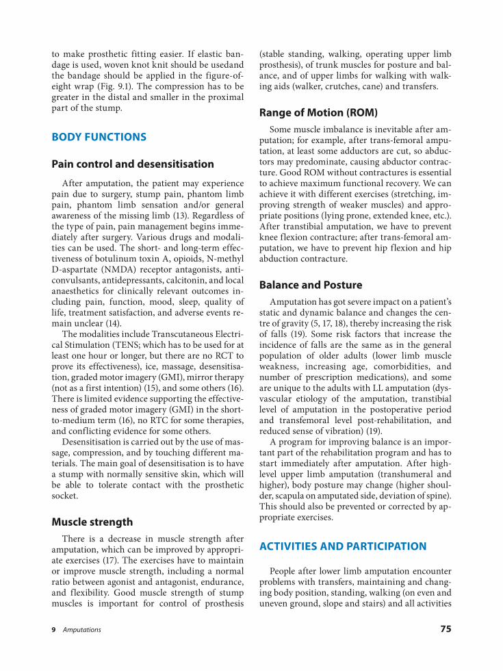
9 Amputations 75
(stable standing, walking, operating upper limb prosthesis), of trunk muscles for posture and bal-ance, and of upper limbs for walking with walk-ing aids (walker, crutches, cane) and transfers.
Range of Motion (ROM)Some muscle imbalance is inevitable after am-
putation; for example, after trans-femoral ampu-tation, at least some adductors are cut, so abduc-tors may predominate, causing abductor contrac-ture. Good ROM without contractures is essential to achieve maximum functional recovery. We can achieve it with different exercises (stretching, im-proving strength of weaker muscles) and appro-priate positions (lying prone, extended knee, etc.). After transtibial amputation, we have to prevent knee flexion contracture; after trans-femoral am-putation, we have to prevent hip flexion and hip abduction contracture.
Balance and PostureAmputation has got severe impact on a patient’s
static and dynamic balance and changes the cen-tre of gravity (5, 17, 18), thereby increasing the risk of falls (19). Some risk factors that increase the incidence of falls are the same as in the general population of older adults (lower limb muscle weakness, increasing age, comorbidities, and number of prescription medications), and some are unique to the adults with LL amputation (dys-vascular etiology of the amputation, transtibial level of amputation in the postoperative period and transfemoral level post-rehabilitation, and reduced sense of vibration) (19).
A program for improving balance is an impor-tant part of the rehabilitation program and has to start immediately after amputation. After high-level upper limb amputation (transhumeral and higher), body posture may change (higher shoul-der, scapula on amputated side, deviation of spine). This should also be prevented or corrected by ap-propriate exercises.
ACTIVITIES AND PARTICIPATION
People after lower limb amputation encounter problems with transfers, maintaining and chang-ing body position, standing, walking (on even and uneven ground, slope and stairs) and all activities
to make prosthetic fitting easier. If elastic ban-dage is used, woven knot knit should be usedand the bandage should be applied in the figure-of-eight wrap (Fig. 9.1). The compression has to be greater in the distal and smaller in the proximal part of the stump.
BODY FUNCTIONS
Pain control and desensitisation
After amputation, the patient may experience pain due to surgery, stump pain, phantom limb pain, phantom limb sensation and/or general awareness of the missing limb (13). Regardless of the type of pain, pain management begins imme-diately after surgery. Various drugs and modali-ties can be used. The short- and long-term effec-tiveness of botulinum toxin A, opioids, N-methyl D-aspartate (NMDA) receptor antagonists, anti-convulsants, antidepressants, calcitonin, and local anaesthetics for clinically relevant outcomes in-cluding pain, function, mood, sleep, quality of life, treatment satisfaction, and adverse events re-main unclear (14).
The modalities include Transcutaneous Electri-cal Stimulation (TENS; which has to be used for at least one hour or longer, but there are no RCT to prove its effectiveness), ice, massage, desensitisa-tion, graded motor imagery (GMI), mirror therapy (not as a first intention) (15), and some others (16). There is limited evidence supporting the effective-ness of graded motor imagery (GMI) in the short-to-medium term (16), no RTC for some therapies, and conflicting evidence for some others.
Desensitisation is carried out by the use of mas-sage, compression, and by touching different ma-terials. The main goal of desensitisation is to have a stump with normally sensitive skin, which will be able to tolerate contact with the prosthetic socket.
Muscle strengthThere is a decrease in muscle strength after
amputation, which can be improved by appropri-ate exercises (17). The exercises have to maintain or improve muscle strength, including a normal ratio between agonist and antagonist, endurance, and flexibility. Good muscle strength of stump muscles is important for control of prosthesis
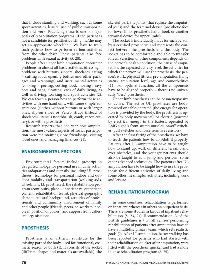
76 PHYSICAL AND REHABILITATION MEDICINE for Medical Students
skeletal part, the joints (that replace the amputat-ed joins) and the terminal device (prosthetic foot for lower limb, prosthetic hand, hook or another terminal device for upper limbs).
The socket is individually made for each person by a certified prosthetist and represents the con-tact between the prosthesis and the body. The socket has to be comfortable and able to transfer forces. Selection of other components depends on the person’s health condition, the cause of ampu-tation, the expected activity level, the activities for which the person will use the prosthesis, the per-son’s work, physical fitness, pre-amputation living status, amputation level, age and comorbidities (22). For optimal function, all the components have to be aligned properly – there is no univer-sally “best” prosthesis.
Upper limb prosthesis can be cosmetic/passive or active. The active UL prostheses are body-powered or cable-operated (the energy for opera-tion is provided by the body, the prosthesis is op-erated by body movements), or electric (powered by electrical energy in the battery, operated by EMG signals from stump muscles, on/off switch-es, pull switches and force sensitive resistors).
After the first fitting of the prosthesis, we have to teach the patients how to don/doff it properly. Patients after LL amputation have to be taught how to stand up, walk on different terrains and over obstacles, and the younger patients should also be taught to run, jump and perform some other advanced techniques. The patients after UL amputation have to be taught how to use the pros-thesis for different activities of daily living and some other meaningful activities, including work and leisure.
REHABILITATION PROGRAM
In some countries, rehabilitation is performed on inpatient, whereas in others on outpatient basis. There are some studies in favour of inpatient reha-bilitation (8, 23, 24). Recommendation A of the British guidelines is that all centres performing rehabilitation of patients after amputation have to have a multidisciplinary team, which sets realistic goals (9). After LL amputation, better walking has been reported for patients who had started with their rehabilitation quicker after amputation, were fitted with the prosthesis quicker and had a more intense rehabilitation program (8, 25).
that include standing and walking, such as some sport activities, leisure, use of public transporta-tion and work. Practicing these is one of major goals of rehabilitation programs. If the patient is not a candidate for prosthetic fitting, he/she may get an appropriate wheelchair. We have to train such patients how to perform various activities from the wheelchair. These patients also face problems with sexual activity (5, 20).
People after upper limb amputation encounter problems in almost all basic activities (dressing – problems with buttons, zippers, shoelaces; eating – cutting food, opening bottles and other pack-ages and wrappings) and instrumental activities (cooking – peeling, cutting food, moving heavy pots and pans, cleaning, etc.) of daily living, as well as driving, working and sport activities (2). We can teach a person how to perform these ac-tivities with one hand only, with some simple ad-aptations (clothes without buttons or with larger ones, slip-on shoes or Velcro straps instead of shoelaces), utensils (toothbrush, comb, razor, cut-lery), or with a prosthesis.
Research reports that one year post amputa-tion, the most valued aspects of social participa-tion were maintaining close friendships, visiting loved ones, and managing finances (21).
ENVIRONMENTAL FACTORS
Environmental factors include prescription drugs, technology for personal use in daily activi-ties (adaptations and utensils, including UL pros-theses), technology for personal indoor and out-door mobility and transportation (walking aids, wheelchair, LL prostheses), the rehabilitation pro-gram (continuity, place – inpatient vs. outpatient, content, rehabilitation team), physical geography, climate, cultural background, attitudes of profes-sionals and community, involvement of family and other people (friends, peers, co-workers, peo-ple in position of power), and support from differ-ent organisations.
PROSTHESIS
Prosthesis is an artificial substitute for the missing part of the body, used for functional, cos-metic reason or both (1). It consists of the socket (different shapes and materials are available), the
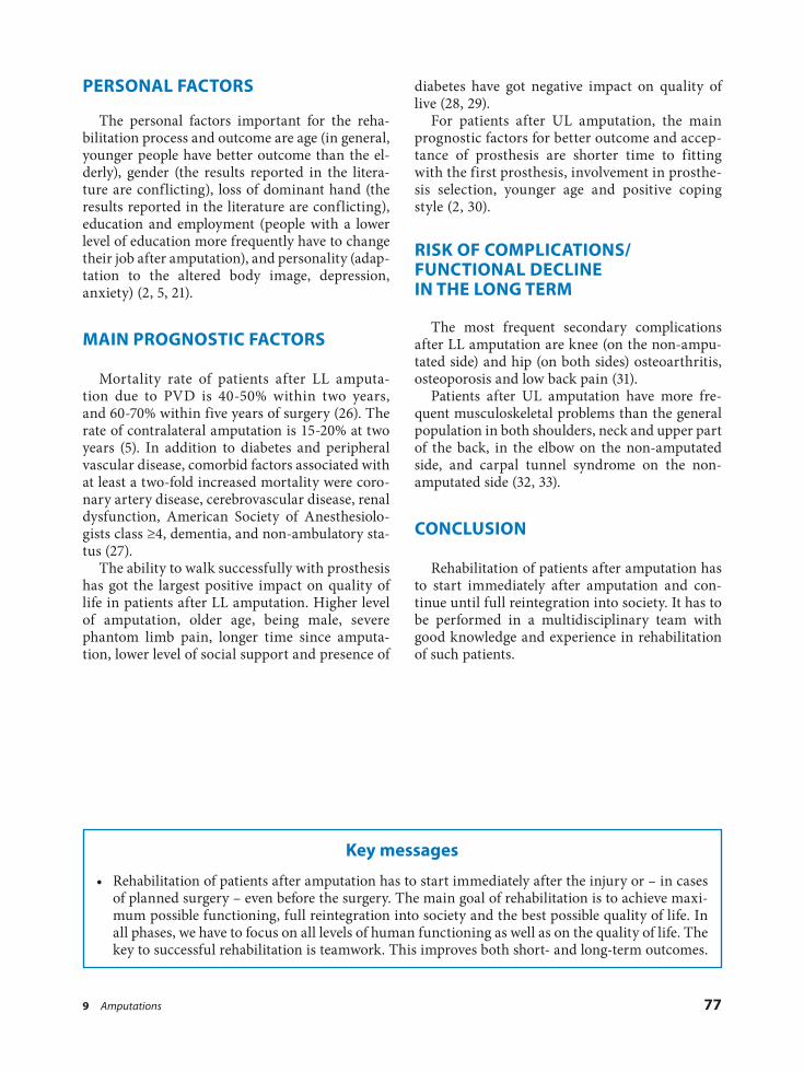
9 Amputations 77
diabetes have got negative impact on quality of live (28, 29).
For patients after UL amputation, the main prognostic factors for better outcome and accep-tance of prosthesis are shorter time to fitting with the first prosthesis, involvement in prosthe-sis selection, younger age and positive coping style (2, 30).
RISK OF COMPLICATIONS/FUNCTIONAL DECLINE IN THE LONG TERM
The most frequent secondary complications after LL amputation are knee (on the non-ampu-tated side) and hip (on both sides) osteoarthritis, osteoporosis and low back pain (31).
Patients after UL amputation have more fre-quent musculoskeletal problems than the general population in both shoulders, neck and upper part of the back, in the elbow on the non-amputated side, and carpal tunnel syndrome on the non-amputated side (32, 33).
CONCLUSION
Rehabilitation of patients after amputation has to start immediately after amputation and con-tinue until full reintegration into society. It has to be performed in a multidisciplinary team with good knowledge and experience in rehabilitation of such patients.
PERSONAL FACTORS
The personal factors important for the reha-bilitation process and outcome are age (in general, younger people have better outcome than the el-derly), gender (the results reported in the litera-ture are conflicting), loss of dominant hand (the results reported in the literature are conflicting), education and employment (people with a lower level of education more frequently have to change their job after amputation), and personality (adap-tation to the altered body image, depression, anxiety) (2, 5, 21).
MAIN PROGNOSTIC FACTORS
Mortality rate of patients after LL amputa-tion due to PVD is 40-50% within two years, and 60-70% within five years of surgery (26). The rate of contralateral amputation is 15-20% at two years (5). In addition to diabetes and peripheral vascular disease, comorbid factors associated with at least a two-fold increased mortality were coro-nary artery disease, cerebrovascular disease, renal dysfunction, American Society of Anesthesiolo-gists class ≥4, dementia, and non-ambulatory sta-tus (27).
The ability to walk successfully with prosthesis has got the largest positive impact on quality of life in patients after LL amputation. Higher level of amputation, older age, being male, severe phantom limb pain, longer time since amputa-tion, lower level of social support and presence of
Key messages
• Rehabilitation of patients after amputation has to start immediately after the injury or – in cases of planned surgery – even before the surgery. The main goal of rehabilitation is to achieve maxi-mum possible functioning, full reintegration into society and the best possible quality of life. In all phases, we have to focus on all levels of human functioning as well as on the quality of life. The key to successful rehabilitation is teamwork. This improves both short- and long-term outcomes.
78 PHYSICAL AND REHABILITATION MEDICINE for Medical Students
12. Churilov I, Churilov L, Murphy D. Do rigid dress-ings reduce the time from amputation to pros-thetic fitting? A systematic review and meta-anal-ysis. Ann Vasc Surg. 2014; 28: 1801-8.
13. Hunter JP, Katz J, Davis KD. Stability of phantom limb phenomena after upper limb amputation: a longitudinal study. Neuroscience. 2008; 156: 939-49.
14. Alviar MJ, Hale T, Dungca M. Pharmacologic in-terventions for treating phantom limb pain. Co-chrane Database Syst Rev. 2016 Oct 14; 10: CD006380.
15. Barbin J, Seetha V, Casillas JM, Paysant J, Péren-nou D. The effects of mirror therapy on pain and motor control of phantom limb in amputees: A systematic review. Ann Phys Rehabil Med 2016; 59: 270-5.
16. Batsford S, Ryan CG, Martin DJ. Non-pharmaco-logical conservative therapy for phantom limb pain: A systematic review of randomized con-trolled trials. Physiother Theory Pract 2017; 33: 173-83.
17. van Velzen JM, van Bennekom CA, Polomski W, Slootman JR, van der Woude LH, Houdijk H. Physical capacity and walking ability after lower limb amputation: a systematic review. Clin Reha-bil 2006; 20: 999-1016.
18. Seth M, Lamberg E. Standing balance in people with trans-tibial amputation due to vascular causes: A literature review. Prosthet Orthot Int. 2017; 41: 345-55.
19. Hunter SW, Batchelor F, Hill KD, Hill AM, Mack-intosh S, Payne M. Risk factors for falls in people with a lower limb amputation: a systematic review.PM R. 2017; 9: 170-80.
20. Woods L, Hevey D, Ryall N, O’Keeffe F. Sex after amputation: the relationships between sexual functioning, body image, mood and anxiety in persons with a lower limb amputation. Disabil Rehabil. 2017 Mar 30: 1-8. [Epub ahead of print]
21. Roepke AM, Williams RM, Turner AP, Henderson AW, Norvell DC, Henson H, et al. A longitudinal study of social participation after dysvascular lower extremity amputation. Am J Phys Med Re-habil. 2017; 96: 741-7.
22. Kahle JT, Highsmith MJ, Schaepper H, Johannes-son A, Orendurff MS, Kaufman K. Predicting walking ability following lower limb amputation: an updated systematic literature review. Technol Innov. 2016; 18: 125-37.
23. Burger H, Marincek C, Jaeger RJ. Prosthetic device provision to landmine survivors in Bosnia and Herzegovina: outcomes in 3 ethnic groups. Arch Phys Med Rehabil. 2004; 85: 19-28.
24. Dillingham TR, Pezzin LE, Mackenzie EJ. Dis-charge destination after dysvascular lower-limb
REFERENCES
1. The Free Dictionary © 2003-2018 Farlex, Inc. Available from: http://medical-dictionary.thefree-dictionary.com/amputation
2. Burger H. Upper limb amputation. In: Stam HJ, Buyruk HM (editors). Acute medical rehabilita-tion. Bodrum: VitalMed Medical Book, 2012. p. 93-106.
3. Walsh NE, Bosker G, Santa Maria D. Upper and lower extremities prosthetics. In: Frontera WR ed. DeLisa’s physical medicine and rehabilitation: principles and practice, chapt. 74. 5th ed. Philadel-phia: Lippincott Williams & Wilkins, 2010, pp. 2017-49.
4. Narres M, Kvitkina T, Claessen H, Droste S, Schuster B, Morbach S, Rümenapf G, Van Acker K, Icks A. Incidence of lower extremity amputations in the diabetic compared with the non-diabetic population: A systematic review. PLoS One. 2017 Aug 28; 12(8): e0182081.
5. Koehler F. Lower limb amputation. In: Stam HJ, Buyruk HM (editors). Acute medical rehabilita-tion. Bodrum: VitalMed Medical Book, 2012. p. 107-29.
6. The Rehabilitation of Lower Limb Amputation - Working Group. VA/DoD Clinical practice guide-line for rehabilitation of lower limb amputation. Washington: U.S. Department of Veterans Affairs; 2007. 166 p. Available from: https://www.healthqual-ity.va.gov/guidelines/Rehab/amp/amp_v652.pdf
7. Pasquina PF, Bryant PR, Huang ME, Roberts TL, Nelson VS, Flood KM. Advances in amputee care. Arch Phys Med Rehabil. 2006; 87(3 Suppl 1): S34-43.
8. Pezzin LE, Dillingham TR, MacKenzie EJ. Reha-bilitation and the long-term outcomes of persons with trauma-related amputations. Arch Phys Med Rehabil. 2000; 81: 292-300.
9. The Working Party of the British Society of Reha-bilitation Medicine. Amputee and prosthetic reha-bilitation – standards and guidelines. 9. Standards and guidelines in amputee and prosthetic rehabili-tation. London: British Society of Rehabilitation Medicine; 2003: 61-7. Available from: http://www.bsrm.org.uk/downloads/ars-gfinaltext.pdf
10. Amputee care standards in New South Wales. North Sydney: Ministry of Health; 2008. Available from: http://www1.health.nsw.gov.au/pds/Active-PDSDocuments/PD2008_015.pdf
11. Geertzen J, van der Linde H, Rosenbrand K, Con-radi M, Deckers J, Koning J, et al. Dutch evidence-based guidelines for amputation and prosthetics of the lower extremity: rehabilitation process and prosthetics. Part 2. Prosthet Orthot Int. 2015; 39: 361-71.

9 Amputations 79
life for veterans with lower-limb amputation(s): a systematic review. Disabil Rehabil. 2016; 38: 2345-53.
30. Biddiss EA, Chau TT. Multivariate prediction of upper limb prosthesis acceptance or rejection. Disabil Rehabil Assist Technol. 2008; 3: 181-92.
31. Gailey R, Allen K, Castles J, Kucharik J, Roeder M. Review of secondary physical conditions associat-ed with lower-limb amputation and long-term prosthesis use. J Rehabil Res Dev. 2008; 45: 15-29.
32. Burger H, Vidmar G. A survey of overuse prob-lems in patients with acquired or congenital upper limb deficiency. Prosthet Orthot Int. 2016; 40: 497-502.
33. Postema SG, Bongers RM, Brouwers MA, Burger H, Norling-Hermansson LM, Reneman MF, et al. Musculoskeletal complaints in transverse upper limb reduction deficiency and amputation in the Netherlands: prevalence, predictors, and effect on health. Arch Phys Med Rehabil. 2016; 97: 1137-45.
amputations. Arch Phys Med Rehabil. 2003; 84: 1662-8.
25. Sansam K, Neumann V, O’Connor R, Bhakta B. Predicting walking ability following lower limb amputation: a systematic review of the literature. J Rehabil Med. 2009; 41: 593-603.
26. Cutson TM, Bongiorni DR. Rehabilitation of the older lower limb amputee: a brief review. J Am Geriatr Soc. 1996; 44: 1388-93.
27. Stern JR, Wong CK, Yerovinkina M, Spindler SJ, See AS, Panjaki S, et al. A meta-analysis of long-term mortality and associated risk factors follow-ing lower extremity amputation. Ann Vasc Surg. 2017; 42: 322-7.
28. Davie-Smith F, Coulter E, Kennon B, Wyke S, Paul L. Factors influencing quality of life following lower limb amputation for peripheral arterial oc-clusive disease: A systematic review of the litera-ture. Prosthet Orthot Int. 2017; 41: 537-47.
29. Christensen J, Ipsen T, Doherty P, Langberg H. Physical and social factors determining quality of


10 Hip fractures 81
striction, hospital utilization and important bur-den of care in the community. Almost a 20% of older people who sustain a HF die within a year.
EPIDEMIOLOGY
The number of HF will increase enormously in the next decades, as well as the cost of treatment these patients do. It is estimated that 340,000 HP occur in the USA each year. The number of people older than age 65 years is expected to increase from 37.1 million to 77.2 million by the year 2040, and the occurrence of hip fractures is expected increase concomitantly, with an estimated 6.3 mil-lion hip fractures predicted worldwide by 2050 (1).
Nine out of ten HF occur in patients aged 65 years and older, and 3 out of 4 affect women, due to osteoporosis or osteopenia. Osteoporosis pre-vention is a key to reduce these numbers, as it re-mains the single most important contributing factor to HF.
Mortality is high: about 10% of people with a HF die within 1 month after it, and about one third within 12 months. Most of the deaths are due to associated comorbidities.
Living in institutional care is associated with a doubling of the risk of hip fracture in women and men compared with those living in a private home. Significant cognitive impairment is associ-ated with at least doubling of the risk of hip frac-ture in men and women.
HIP FRACTURE CLASSIFICATION
1. Intra-capsular fractures: a. Femoral head fractures; b. Femoral neck fractures.
2. Extracapsular fractures: a. Intertrochanteric fractures; b. Sub-trochanteric fractures.
INTRODUCTION
Hip fracture (HF) refers to a fracture (Fx) lo-cated in the area between the edge of the femoral head and 5 cm below the lesser trochanter. These fractures are generally divided into two main groups depending on their relationship to the cap-sule of the hip joint. In this way, they are classified as: a) intra-capsular Fx, which includes femoral head and neck fractures; and b) extracapsular fractures, which includes trochanteric, intertro-chanteric, and sub-trochanteric Fx. Fracture loca-tion, as well as the amount of angulation and comminution, play integral roles in the overall patient morbidity, as does the preexisting physical condition of the individual.
These processes represent an important public health issue in elderly, in all countries. They are the most common Fx among people over the age of 75; Fx of the proximal femur are extremely rare in young athletes and are usually caused by high-energy motor vehicle accidents or significant trauma during sport activity.
Thus, HF is by no means an exclusively surgical concern; its effective management requires the coordinated application of medical, surgical, an-aesthetic and multidisciplinary rehabilitation skills as well as a comprehensive approach covering the full time course of the condition from presentation to subsequent patient-follow-up, including the transition from hospital to community.
The recovery of people following a HF is often complex and involves a variety of factors such as physical, psychological and social components (1). Indeed, it is suggested that only 40-60% of people who suffer a HF recover their pre-fracture level of mobility and ability to perform activities of daily living (ADL). This process has significant impli-cations for mortality, as well as morbidity with patient activity limitation and participation re-
10Hip fracturesRaquel VALERO, Enrique VARELA-DONOSO

82 PHYSICAL AND REHABILITATION MEDICINE for Medical Students
such as Garden grades I-IV, offer no further diag-nostic, therapeutic or prognostic information (2).
Classification of intertrochanteric HF is based on a system introduced by Evans in 1949. This system is based on the fracture pattern and the ability to obtain a stable reduction. Evans recog-nized the importance of restoring the posterome-dial cortex as a contributing factor to fracture stability. Other authors classify intertrochanteric fractures by the number of fracture fragments present; however, for an ease description and sim-plicity, these fractures are best classified as follows:
ū Stable: Fractures with an intact posteromedial cortex
ū Unstable: Fractures with comminution of the posteromedial cortex, fractures with diaphyse-al extension.The usual clinical signs and symptoms related
to HF are listed in Table 10.1.
STAGES OF TREATMENT
In view of the potential for comorbidities or other injuries a four-stage treatment program is recommended (3):
Stage 1 – Identify the Fx on the basis of the his-tory and the findings from physical examination and X-ray .
X-ray films are always indicated to determine which type of Fx, if any, is present. Anteroposte-rior (AP) views of the pelvis and hip and cross-table lateral X-ray films are usually sufficient to evaluate potential fractures. Rotating the affected limb internally or externally can increase the sen-sitivity of these radiographs.
These images also define the inherent stability or instability of the fracture, the need for a reduc-tion of the fracture, and whether further manipu-lation is indicated to produce a reduction suffi-ciently stable to heal before the implant fixation is lost.
Stage 2 – In stage 2, stabilize the patient’s medical condition before surgical intervention. Patients do best if the fracture is stabilized surgi-cally within 24-48 hours of admission to the hos-pital.
Stage 3 – Stage 3 includes the surgical act and postoperative recovery in the acute care hospital.
A femoral-neck Fx in elderly is usually due to a simple fall when the bone density is already re-duced by osteoporosis. Intra-capsular Fx follow-ing high-energy trauma can also occur and are most often the result of a transferred energy fol-lowing a hip joint dislocation and/or an acetabu-lar Fx following an impact of the foot.
The most commonly used classification system for femoral neck Fx is the Garden classification. Fx are divided into 4 groups according to the de-gree of displacement and fracture fragments. This classification system gives guidance for treatment options and surgical implants. The following 4 groups comprise this classification system:
ū Garden type I: Incomplete fracture with valgus impaction, as shown below;
ū Garden type II: Complete fracture without dis-placement;
ū Garden type III: Complete fracture with partial displacement of the fracture fragments;
ū Garden type IV: Complete fracture with total displacement allowing the femoral head to ro-tate back to an anatomic position.
Other authors use to classify intra-capsular Fx as undisplaced and displaced. Older classifications,
Table 10.1 Clinical signs and symptoms related to hip fracture
Main clinical signs• Inguinal pain• Pressure pain on the greater trochanter• Haematoma• Shortening and external rotation
of the ipsilateral lower limb
Possible additional findings:• Limited and painful range of motion,
especially in internal rotation• Pain is noted upon attempted passive hip motion • Ecchymosis (may or may not be present)• An antalgic gait pattern• Discomfort at deep palpation in the inguinal area. Tenderness to palpation is noted over the femoral neck (this area may also be swollen)
• Increased pain on the extremes of hip rotation, an abduction lurch, and an inability to stand on the involved leg may indicate a femoral neck stress fracture
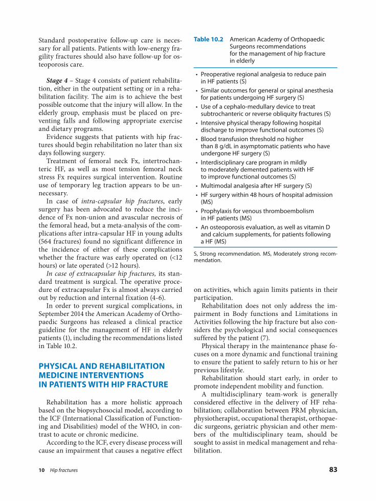
10 Hip fractures 83
on activities, which again limits patients in their participation.
Rehabilitation does not only address the im-pairment in Body functions and Limitations in Activities following the hip fracture but also con-siders the psychological and social consequences suffered by the patient (7).
Physical therapy in the maintenance phase fo-cuses on a more dynamic and functional training to ensure the patient to safely return to his or her previous lifestyle.
Rehabilitation should start early, in order to promote independent mobility and function.
A multidisciplinary team-work is generally considered effective in the delivery of HF reha-bilitation; collaboration between PRM physician, physiotherapist, occupational therapist, orthopae-dic surgeons, geriatric physician and other mem-bers of the multidisciplinary team, should be sought to assist in medical management and reha-bilitation.
Standard postoperative follow-up care is neces-sary for all patients. Patients with low-energy fra-gility fractures should also have follow-up for os-teoporosis care.
Stage 4 – Stage 4 consists of patient rehabilita-tion, either in the outpatient setting or in a reha-bilitation facility. The aim is to achieve the best possible outcome that the injury will allow. In the elderly group, emphasis must be placed on pre-venting falls and following appropriate exercise and dietary programs.
Evidence suggests that patients with hip frac-tures should begin rehabilitation no later than six days following surgery.
Treatment of femoral neck Fx, intertrochan-teric HF, as well as most tension femoral neck stress Fx requires surgical intervention. Routine use of temporary leg traction appears to be un-necessary.
In case of intra-capsular hip fractures, early surgery has been advocated to reduce the inci-dence of Fx non-union and avascular necrosis of the femoral head, but a meta-analysis of the com-plications after intra-capsular HF in young adults (564 fractures) found no significant difference in the incidence of either of these complications whether the fracture was early operated on (<12 hours) or late operated (>12 hours).
In case of extracapsular hip fractures, its stan-dard treatment is surgical. The operative proce-dure of extracapsular Fx is almost always carried out by reduction and internal fixation (4-6).
In order to prevent surgical complications, in September 2014 the American Academy of Ortho-paedic Surgeons has released a clinical practice guideline for the management of HF in elderly patients (1), including the recommendations listed in Table 10.2.
PHYSICAL AND REHABILITATION MEDICINE INTERVENTIONS IN PATIENTS WITH HIP FRACTURE
Rehabilitation has a more holistic approach based on the biopsychosocial model, according to the ICF (International Classification of Function-ing and Disabilities) model of the WHO, in con-trast to acute or chronic medicine.
According to the ICF, every disease process will cause an impairment that causes a negative effect
Table 10.2 American Academy of Orthopaedic Surgeons recommendations for the management of hip fracture in elderly
• Preoperative regional analgesia to reduce pain in HF patients (S)
• Similar outcomes for general or spinal anesthesia for patients undergoing HF surgery (S)
• Use of a cephalo-medullary device to treat subtrochanteric or reverse obliquity fractures (S)
• Intensive physical therapy following hospital discharge to improve functional outcomes (S)
• Blood transfusion threshold no higher than 8 g/dL in asymptomatic patients who have undergone HF surgery (S)
• Interdisciplinary care program in mildly to moderately demented patients with HF to improve functional outcomes (S)
• Multimodal analgesia after HF surgery (S)• HF surgery within 48 hours of hospital admission (MS)
• Prophylaxis for venous thromboembolism in HF patients (MS)
• An osteoporosis evaluation, as well as vitamin D and calcium supplements, for patients following a HF (MS)
S, Strong recommendation. MS, Moderately strong recom-mendation.
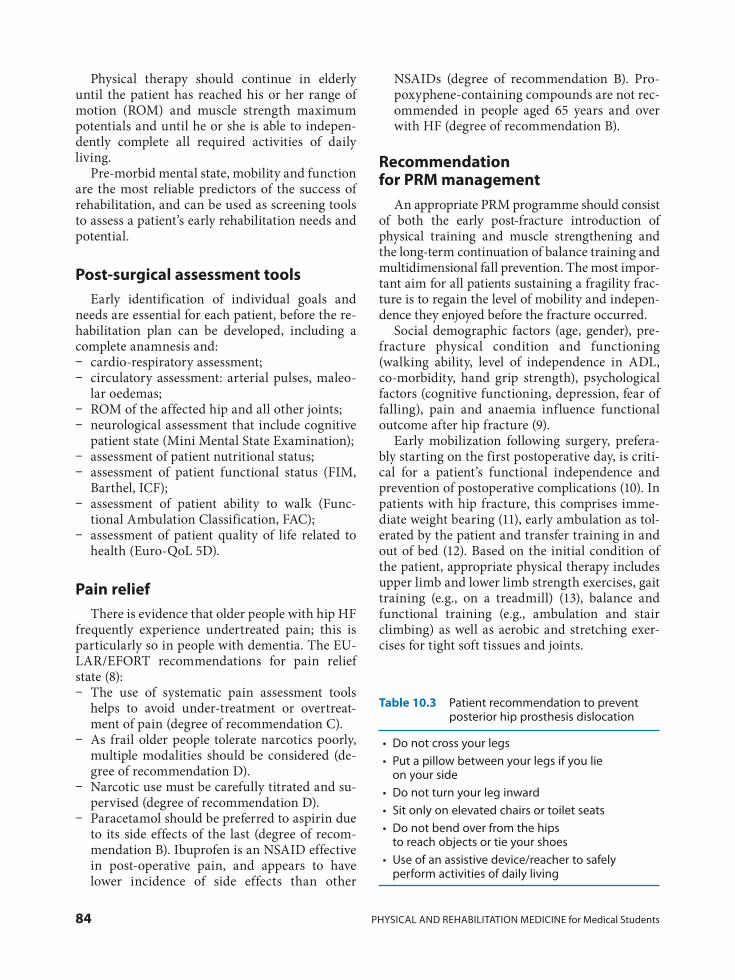
84 PHYSICAL AND REHABILITATION MEDICINE for Medical Students
NSAIDs (degree of recommendation B). Pro-poxyphene-containing compounds are not rec-ommended in people aged 65 years and over with HF (degree of recommendation B).
Recommendation for PRM management
An appropriate PRM programme should consist of both the early post-fracture introduction of physical training and muscle strengthening and the long-term continuation of balance training and multidimensional fall prevention. The most impor-tant aim for all patients sustaining a fragility frac-ture is to regain the level of mobility and indepen-dence they enjoyed before the fracture occurred.
Social demographic factors (age, gender), pre-fracture physical condition and functioning (walking ability, level of independence in ADL, co-morbidity, hand grip strength), psychological factors (cognitive functioning, depression, fear of falling), pain and anaemia influence functional outcome after hip fracture (9).
Early mobilization following surgery, prefera-bly starting on the first postoperative day, is criti-cal for a patient’s functional independence and prevention of postoperative complications (10). In patients with hip fracture, this comprises imme-diate weight bearing (11), early ambulation as tol-erated by the patient and transfer training in and out of bed (12). Based on the initial condition of the patient, appropriate physical therapy includes upper limb and lower limb strength exercises, gait training (e.g., on a treadmill) (13), balance and functional training (e.g., ambulation and stair climbing) as well as aerobic and stretching exer-cises for tight soft tissues and joints.
Physical therapy should continue in elderly until the patient has reached his or her range of motion (ROM) and muscle strength maximum potentials and until he or she is able to indepen-dently complete all required activities of daily living.
Pre-morbid mental state, mobility and function are the most reliable predictors of the success of rehabilitation, and can be used as screening tools to assess a patient’s early rehabilitation needs and potential.
Post-surgical assessment toolsEarly identification of individual goals and
needs are essential for each patient, before the re-habilitation plan can be developed, including a complete anamnesis and:
ū cardio-respiratory assessment; ū circulatory assessment: arterial pulses, maleo-
lar oedemas; ū ROM of the affected hip and all other joints; ū neurological assessment that include cognitive
patient state (Mini Mental State Examination); ū assessment of patient nutritional status; ū assessment of patient functional status (FIM,
Barthel, ICF); ū assessment of patient ability to walk (Func-
tional Ambulation Classification, FAC); ū assessment of patient quality of life related to
health (Euro-QoL 5D).
Pain reliefThere is evidence that older people with hip HF
frequently experience undertreated pain; this is particularly so in people with dementia. The EU-LAR/EFORT recommendations for pain relief state (8):
ū The use of systematic pain assessment tools helps to avoid under-treatment or overtreat-ment of pain (degree of recommendation C).
ū As frail older people tolerate narcotics poorly, multiple modalities should be considered (de-gree of recommendation D).
ū Narcotic use must be carefully titrated and su-pervised (degree of recommendation D).
ū Paracetamol should be preferred to aspirin due to its side effects of the last (degree of recom-mendation B). Ibuprofen is an NSAID effective in post-operative pain, and appears to have lower incidence of side effects than other
Table 10.3 Patient recommendation to prevent posterior hip prosthesis dislocation
• Do not cross your legs• Put a pillow between your legs if you lie
on your side• Do not turn your leg inward• Sit only on elevated chairs or toilet seats• Do not bend over from the hips
to reach objects or tie your shoes• Use of an assistive device/reacher to safely perform activities of daily living
10 Hip fractures 85
Postoperative protocol (day 2) ū Initiation of gait training with the use of assis-
tive devices, such as crutches and a walker. ū Continuation of functional transfer training.
Postoperative protocol (days 3-5 or on discharge to the rehabilitation unit)
ū Progression of ROM and muscle strengthening exercises to the patient’s tolerance.
ū Progression of ambulation on level surfaces and stairs (if applicable) with the least restric-tive device.
ū Progression of ADL training.
Rahmann et al. found that aquatic physiothera-py can promote early recovery of hip strength in patients who have undergone hip or knee arthro-plasty (16). In a randomized, controlled trial that compared the results of supplementary inpatient physiotherapies – aquatic physiotherapy, nonspe-cific water exercise, and additional ward physio-therapy – in 65 patients, a specific inpatient aquat-ic physiotherapy program, begun on day 4, was associated with significantly greater hip abductor strength by day (16).
Postoperative protocol (day 5 to 4 weeks) ū Strengthening exercises – For example, seated
leg extensions, side-lying/standing hip abduc-tion, standing hip extension and hip abduction, knee bends, bridging.
ū Stretching exercises to increase the flexibility of hip muscles.
ū Progression of ambulation distance. ū Progression of independence with ADL.
A study done by Husby et al. (17) indicated that maximal strength training, starting 1 week post-operatively, is a valuable addition to conventional rehabilitation after THR. In a randomized, con-trolled study in 24 patients, one group performed maximal strength training in leg press and abduc-tion only with the operated leg, 5 times weekly for 4 weeks; compared with patients who received only conventional rehabilitation, the patients who engaged in strength training demonstrated in-creased 1-repetition maximum leg press strength, an increased rate of force development, and a ten-dency toward improved work efficiency. No dif-ferences in gait patterns were noted between the groups.
According to Sherrington et al, there are a lack
Exercise programmes and fall prevention programmes are hallmarks of ideal non-phar-macological treatment for the prevention of fractures. Positive effects on bone mineral den-sity and muscle strength are described in pa-tients who exercise rigorously, as well as a reduc-tion in the frequency of falls, but the evidence for fracture prevention is limited (14).
Moreover, education of patients undergoing total hip replacement (THR) should include the delivery of the following recommendations (Table 10.3) to prevent posterior hip dislocation.
PRM treatment protocol for total hip replacement
A number of rehabilitation exercise protocols are used by various institutions for total knee or hip replacement; however, the functional goals of these protocols are the same. This sec-tion discusses total hip replacement (THR) protocol (15).
a) Preoperative protocol (1-2 weeks prior to surgery)
Preoperatively, the protocol includes educat-ing the patient about the surgical process and its outcomes, instructing him or her on a postop-erative exercise program, and assessing the pa-tient’s home environment. It also involves edu-cating the patient on total hip precautions, as follows:
ū Avoid hip flexion beyond 90°. ū Avoid crossing of the legs (hip adduction be-
yond neutral). ū Avoid hip internal rotation past neutral.
The above precautions apply to the posterior surgical approach to the hip. With the anterior hip approach, the patient can cross his or her legs and internally rotate the hip, although positions that involve extreme hip extension and external rota-tion will dislocate the hip.
b) Postoperative protocolPostoperative protocol (day 1)
ū Initiation of bedside exercises, such as ankle pumps, quadriceps sets, and gluteal sets.
ū Review of hip precautions and weight-bearing status.
ū Initiation of bed mobility and transfer training; bed to/from chair.
86 PHYSICAL AND REHABILITATION MEDICINE for Medical Students
strategies for enhancing mobility after hip frac-ture surgery (19).
A study done by Crotty et al agreed that further research on interventions to increase physical and psychological functioning (occupational therapy, etc.) is required, including attention to timing, duration, setting and administering discipline(s), as well as treatment across care settings (20).
of well-designed large-scale trials of exercise after hip fracture. The Cochrane reviews covering this field also do not draw firm conclusions. However, several individual trials have had promising find-ings and indicate some benefits of exercise after hip fracture (18). Another Cochrane systematic review concluded that there is insufficient evi-dence from randomised trials to establish the best
Key messages
• Hip fracture is a process very often seen by doctors specialized in physical & rehabilitation medicine. It represents an important public health issue in elderly, in all countries due to its fre-quency, co-morbidities and disability.
• The role of PRM physician in acute rehabilitation after hip fracture is required mostly in stage 4. However, its presence in the previous stages is important in order to prevent as much as possible all the possible complications that may appear as well as to prepare the patient for the real reha-bilitation phase.
• Patient acute rehabilitation must have a holistic approach based on the biopsychosocial model, according to the ICF. It has to be multidisciplinary and following a correct designed protocol based on scientific evidences.
• Patient education as well as physical therapy management with early mobilization and standing positioning are important parts of acute rehabilitation process after hip fracture.
REFERENCES
1. American Academy of Orthopaedic Surgeons. Management of hip fractures in the elderly: evi-dence-based clinical practice guideline. Rosemont (IL): AAOS, 2014. Available from http://www.aaos.org/research/guidelines/HipGuideline.pdf
2. Scottish Intercollegiate Guidelines Network. Man-agement of hip fracture in older people. A national clinical guideline. Edinburgh: SIGN, 2009. 56 p. Available from: http://www.sign.ac.uk/assets/sign111.pdf
3. Medscape - Orthopedic Surgery. Kellam JF. Intertro-chanteric hip fractures treatment & management. Medscape WebMD LLC ©2017. Available from: http://emedicine.medscape.com/article/1247210-treatment
4. Acute management and immediaterehabilitation-after hip fracture amongst people aged 65 years and over. Wellington: New Zealand Guidelines Group (NZGG); 2003. Available from: http://www.moh.govt.nz/notebook/nbbooks.nsf/0/BF485CCA409F38C5CC256DCE0070F7B5/$file/Hip_Frac-ture_Management.pdf
5. Parker MJ, Handoll HH. Gamma and other cepha-
locondylic intramedullary nails versus extramed-ullary implants for extracapsular hip fractures in adults. Cochrane Database Syst Rev. 2005 Oct 19; (4): CD000093.
6. Lorich DG, Geller DS, Nielson JH. Osteoporotic pertrochanteric hip fractures: management and current controversies. J Bone Joint Surg 2004; 86: 398-410,
7. Dreinhöfer KE, Hohenadel M. Hip fracture. In: Stam H, Buyruk H M, Melvin JL, Stucki G, Burg-graaf I. Acute Medical Rehabilitation Vol I. Chap-ter 10. Ankara-Odak Ofset 2012: 183-202.
8. Lems WF, Dreinhöfer KE, Bischoff-Ferrari H, Blauth M, Czerwinski E, da Silva J, et al. EULAR/EFORT recommendations for management of pa-tients older than 50 years with a fragility fracture and prevention of subsequent fractures. Ann Rheum Dis. 2017; 76: 802-10.
9. Scheffers-Barnhoorn MN, van Haastregt JC, Schols JM, Kempen GI, van Balen R, Visschedijk JH, et al. A multi-component cognitive behav-ioural intervention for the treatment of fear of falling after hip fracture (FIT-HIP): protocol of a randomised controlled trial. BMC Geriatr. 2017; 17: 71.
10 Hip fractures 87
16. Rahmann AE, Brauer SG, Nitz JC. A specific inpa-tient aquatic physiotherapy program improves strength after total hip or knee replacement sur-gery: a randomized controlled trial. Arch Phys Med Rehabil. 2009; 90: 745-55.
17. Husby VS, Helgerud J, Bjorgen S, Husby OS, Be-num P, Hoff J. Early maximal strength training is an efficient treatment for patients operated with total hip arthroplasty. Arch Phys Med Rehabil. 2009; 90: 1658-67.
18. Sherrington C, Tiedemann A, Cameron I. Physical exercise after hip fracture: an evidence overview. Eur J Phys Rehabil Med. 2011; 47: 297-307.
19. Handoll HH, Sherrington C, Mak JC. Interven-tions for improving mobility after hip fracture surgery in adults. Cochrane Database Syst Rev. 2011 Mar 16; (3): CD001704.
20. Crotty M, Unroe K, Cameron ID, Miller M, Ramirez G, Couzner L. Rehabilitation interven-tions for improving physical and psychosocial functioning after hip fracture in older people. Cochrane Database Syst Rev. 2010 Jan 20; (1): CD007624.
10. Levy C, Ocampo-Chan S, Huestis L, Renzetti D. Early rehabilitation for patients with hip fractures: spreading change across the system. Healthc Q. 2017; 20: 29-33.
11. British Orthopaedic Association. The care of pa-tients with fragility fracture. London: BOA; 2007. 80 p. Available from: http://www.bgs.org.uk/pdf_cms/pubs/Blue Book on fragility fracture care.pdf
12. Oldmeadow LB, Edwards ER, Kimmel LA, Kipen E, Robertson VJ, Bailey MJ. No rest for the wound-ed: early ambulation after hip surgery accelerates recovery. ANZ J Surg 2006; 76: 607-11.
13. Baker PA, Evans OM, Lee C. Treadmill gait re-training following fractured neck-of-femur. Arch Phys Med Rehabil 1991; 72: 649-52.
14. Mendelsohn ME, Overend TJ, Connelly DM, Pe-trella RJ. Improvement in aerobic fitness during rehabilitation after hip fracture. Arch Phys Med Rehabil 2008; 89: 609-17.
15. Schneider M, Kawahara I, Ballantyne G, McAuley C, Macgregor K, Garvie R, et al. Predictive factors influencing fast track rehabilitation following pri-mary total hip and knee arthroplasty. Arch Or-thop Trauma Surg. 2009; 129: 1585-91.

11 Sport injuries 89
mediators (such as histamine from mast cells) in-crease membrane permeability and vasodilatation. Phagocytic cells and leukocytes invade the area. Waste products are broken down and removed via local and vascular effects. At the site of vessel in-jury platelets adhere to the exposed collagen fibres starting clot formation. Fibrin clot formation oc-curs via the cascade stimulated by thromboplastin. Eventually, a walling off effect facilitates the heal-ing process. Clot formation begins about twelve hours post-injury and is completed within 48 hours. Initial rehabilitation techniques focus on: minimizing the inflammatory response prevent-ing further loss of function and decreasing pain.
The Fibroblastic-Repair Stage begins during the inflammatory response and continues for the next four to six weeks. It starts by scar filling in the injury defect. The fibrin clot is replaced with granulation tissue made up of collagen and fibro-blasts. Critical nutrients are delivered via new capillaries. The tensile strength of the scar is based on the collagen deposition.
The Maturation-Remodelling Stage begins as the tensile strength of the scar tissue increases and fibroblast activity declines. Stress on the collagen fibres causes them to realign in parallel to the forces applied. Controlled mobilization at this time maximizes the reorientation of healing fibres and results in improved tensile strength and function. Pain can guide the progression rate of stress placed on the remodelling tissue. Any exacerbation of pain in this phase indicates that the rehabilitation load is too high for the level of maturation. Joint proprioception must be addressed. Sport-specific training is necessary for neuromuscular re-educa-tion in addition to tissue remodelling.
INTRODUCTION
In case of a sport injury the Medical Rehabilita-tion begins at the time of injury and continues beyond the time the athlete returns to competi-tion. It is a comprehensive management which uses multiple approaches. It is directed to go be-yond resolution of the acute injury to prevent re-peated injuries.
The main principles in Sports Rehabilitation are to minimize the damage, the inflammation and/or the present pain at the site of injury. Also, to promote the healing, to maintain or increase the range of motion (ROM) of the affected joint, to prevent muscle atrophies and increase the muscles strength, to maintain or increase the en-durance, to facilitate the functional recovery and to avoid maladaptive compensatory movement patterns (1).
PATHOPHYSIOLOGY OF INJURY
Concerning the pathophysiology of an injury and its repair we must notice that the healing pro-cess involves three stages: the inflammatory stage, the fibroblastic-repair stage and the maturation-remodelling stage (1).
The Inflammatory Stage lasts from two to four days. Clinically on the injured site there is an in-creased warmth, skin redness, swelling and ten-derness. The initial trauma to the tissue is followed by secondary injury resulting from hypoxia and enzymatic activity. At the time of the injury a cas-cade of events is set into motion: after five to ten minutes of vasoconstriction a locally mediated in-flux of cells presents at the injury site. Chemical
11Sport injuriesNicolas CHRISTODOULOU
90 PHYSICAL AND REHABILITATION MEDICINE for Medical Students
bone fractures and fracture-dislocations as well as soft tissue complete tears. In these cases an ortho-paedic intervention is needed prior the rehabilita-tion program. For sport injuries which don’t need cast immobilization, as are the several partial ruptures of soft tissue and haematomas, we can start directly the rehabilitation program, as de-scribed above including always exercises.
THERAPEUTIC INTERVENTIONS
Kinds of Exercises that must be planned and applied at the injured area are mobility exercis-es, strength exercises and static stretching exer-cises. Also, exercises for coordination and bal-ance are almost always needed. They must be planned to facilitate the techniques of the spe-cific athlete’s sport. They must be performed during the phase of the athlete’s return to the training program, avoiding overpass the tired-ness threshold (5).
In all sport rehabilitation programs it is neces-sary the general coordination training of the big muscle groups of both the injured and the healthy side and in parallel (6, 7). Examples of exercises for balance and coordination are:
ū Stand on balancing board which allows 2 or 4 movements without looking to the board (8).
ū Control of the ball without looking to the ball. ū Climb up & step down stairs sideways without
looking to the stairs. It can be performed for both sides, first having the soles flat and then moving on tiptoe. Also climb first one step and then two steps at a time.
ū Jump on the spot: We start with low jumps and gradually we continue with higher and higher jumps without looking to the floor.
When the athlete returns to the training field, the close cooperation with the coach for solving any originating problem, having to do with the rehabilitation, is of great importance.
Additional provisions: Use of taping around injured joints or muscles helps significantly the athlete (9), Use of ankle aircast in cases of ankle injury supports this joint. Also use of elastic braces may support other injured joints. Always the athlete must use and wear proper sport shoes with the provision of shoe specifications accord-ing to the kind of sport of the athlete (10).
MEDICATION AND PHYSICAL MODALITIES
Medications that we can give as part of the re-habilitation process may include analgesics, non steroid anti-inflammatory drugs (NSAIDs), mus-cle relaxants and Autologous Blood Injections (2). The Autologous Blood Injections (ABI) are a new method of injecting at the injured area blood plasma of the patient, after centrifugation, con-taining platelet derived growth factor (PDGF) for commencing a faster cascade of natural healing. ABI is preferable to be delivered with the help of ultrasound.
Physical Modalities that can be used and ap-plied in sport injuries may include cold modalities (superficial cold), heat modalities (superficial heat or deep heat), electrical modalities and several modalities for kinesiotherapy (3).
PHASES OF REHABILITATION
The phases of rehabilitation are three: the acute phase which is extended to the first 48-72 hours; the subacute/recovery phase which lasts from 3 days to 3 weeks; and the functional phase which persists from weeks to months (1).
The management principles in Acute Phase have to do with the inflammation control, the ap-plication of the R.I.C.E. rule (rest-ice-compres-sion-elevation) (4), the prescription of medica-tions and or physical modalities, the maintaining of the muscles flexibility and the range of move-ment of the involved joint and cross-training.
The management principles in Subacute/Recov-ery Phase have to do with the restoration of joint and soft tissue flexibility, exercise prescription, stretching, muscles strengthening, resistance ex-ercise, stretch-shortening cycle (plyometric exer-cises) and endurance.
The management principles in Functional Phase have to do with the improvement of neu-romuscular control, the correction of maladap-tive behaviours, the sport-specific and multi-plane activity, the functional retraining, the ki-netic chain (sequence), the retraining of balance and proprioception and agility drills (“eukine-sia” exercises).
The sport injuries which need cast immobiliza-tion and/or surgical intervention are the several

11 Sport injuries 91
the key for the success. Today there are psycholo-gists, specialized in athletic psychology, who can advice all the involved persons and thus upgrade the team performance to the highest degree. The entire group is affected from the mood of each one of its members, who has its own personality and problems. The importance of a coach is to know each player separately and to know the psychological and sentimental situation of the team as a whole. He/she must lead the team in having unity and each member to be accepted by the rest members, in a spirit of common will for the victory. The returning athlete after an injury must be the opportunity of the coach, for the scientific implementation of all the above men-tioned points (11).
ATHLETIC PSYCHOLOGY PREPARATION
It is of great importance to focus on the ath-lete’s psychological preparation as he/she returns to full training, especially in team sports. The coach, the leader and the members of the team must know that the player, who is not perform-ing as well as is expected, needs to be encour-aged. If the people around him/her react nega-tively on each inadequate performance, the play-er under the hard criticism, may react with hesi-tation and lack of accuracy, leading to further deterioration of his/her performance. While, a planned atmosphere of positive and constructive comments and general positive encouragement is
Key messages
• Sport injuries comprehensive rehabilitation has to start immediately from the time of injury and continue far beyond the time the patient returns to competition. It aims not only to the treatment of sports injuries but to the best athletic performance and to the prevention of repeated injuries.
• During the process of rehabilitation several health professions are involved and their best results are achieved within the operation of a rehabilitation team, which is a multi-professional team, col-laborating with several disciplines, under the leadership of a Physical and Rehabilitation Medicine (PRM) physician.
REFERENCES
1. Peterson L, Renström P. Sports Injuries. Preven-tion, treatment and rehabilitation. 4th ed. Boca Raton: Taylor & Francis - CRC Press; 2016. 620 p.
2. Moraes VY, Lenza M, Tamaoki MJ, Faloppa F, Bel-loti JC. Platelet-rich therapies for musculoskeletal soft tissue injuries. Cochrane Database Syst Rev 2014; 4: CD010071.
3. Oral A, Ilieva E, Kucukdeveci A, Varela E, Valero R, Berteanu M, Christodoulou N. Local soft tissue musculoskeletal disorders and injuries. The role of physical and rehabilitation medicine physicians. The European perspective based on the best evi-dence. A paper by the UEMS-PRM Section Profes-sional Practice Committee. Eur J PhysRehabil Med. 2013; 49: 727-42.
4. Van den Bekerom MP, Struijs PA, Blankevoort L, Welling L, van Dijk CN, Kerkhoffs GM. What is the evidence for rest, ice, compression, and eleva-tion therapy in the treatment of ankle sprains in adults? J Athl Train 2012; 47: 435-43.
5. Mason DL, Dickens VA, Vail A. Rehabilitation for hamstring injuries. Cochrane Database Syst Rev 2012; 12: CD004575.
6. Zech A, Hubscher M, Vogt L, Banzer W, Hansel F, Pfeifer K. Neuromuscular training for rehabilita-tion of sports injuries: a systematic review. Med Sci Sports Exerc2009; 41: 1831-41.
7. McKeon P O, Hertel J. Systematic review of pos-tural control and lateral ankle instability, part II: is balance training clinically effective? J Athl Train 2008; 43: 305-15.
8. Valovich McLeod TC. The effectiveness of bal-
92 PHYSICAL AND REHABILITATION MEDICINE for Medical Students
11. National Collaborating Centre for Mental Health and Social Care Institute for Excellence. Antiso-cial behaviour and conduct disorders in children and young people. Recognition, intervention and management. National Clinical Guidelines No. 158. Leicester (UK): British Psychological Society; 2013. 468 p. Available from: https://www.nice.org.uk/guidance/cg158/evidence/conduct-disorders-in-chi ldren-and-young-people-f u l l-g uide-line-189848413.
ance training programs on reducing the incidence of ankle sprains in adolescent athletes. J Sport Re-habil 2008; 17: 316-23.
9. Zoch C, Fialka-Moser V, Quittan M. Rehabilita-tion of ligamentous ankle injuries: a review of re-cent studies. Br J Sports Med 2003; 37: 291-5.
10. Nigg BM, Baltich J, Hoerzer S, Enders H. Run-ning shoes and running injuries: myth-busting and a proposal for two new paradigms: ‘preferred movement path’ and ‘comfort filter’. Br J Sports Med 2015: Oct; 49(20): 1290-4.

12 Degenerative and inflammatory joint disorders, fibromyalgia, and osteoporosis 93
In this chapter, the burden of some major rheu-matic conditions (like osteoarthritis, osteoporosis, rheumatoid arthritis, fibromyalgia) will be de-scribed.
OSTEOARTHRITIS
Osteoarthritis (OA) is the most common age-related joint disorder throughout the world which is characterized by loss of joint cartilage that leads to pain and loss of function primarily in the joints.
Osteoarthritis is characterized by focal areas of loss of articular cartilage within synovial joints, which are associated with hypertrophy of bone (osteophytes and subchondral bone sclerosis) and thickening of the capsule. Clinically, the condi-tion is characterized by joint pain, tenderness, limitation of movement, crepitus, occasional effu-sion, and variable degrees of local inflammation. It can occur in any joint but the disease most com-monly affects the joints in the knees, hands, and spine and hip joints. In a survey of 500 patients, the joints mostly affected are knee (41%), hand (30%), and hip (19%) (3).
This disease affects 9.6% of men and 18% of women aged >60 years. Increases in life expec-tancy and ageing populations are expected to make osteoarthritis the fourth leading cause of disability by the year 2020 (1).
While OA is related to ageing, it is also associ-ated with a variety ofrisk factors, including: obe-sity, lack of exercise, genetic predisposition, bone density, occupational injury, trauma, and gender (4). Osteoarthritis is the single most common cause of disability in older adults.
INTRODUCTION
Degenerative and inflammatory joint disorders (and more in general progressive musculoskeletal disorders) are frequently seen, their impact is widespread, and they represent a major burden on individuals, health systems, and social care sys-tems, with indirect costs being predominant. These disorders are the most common cause of severe long-term pain and physical disability, and affect hundreds of millions of people around the world. The prevalence of many of these conditions increases markedly with age, and many are af-fected by lifestyle factors, such as obesity and lack of physical activity.
The increasing number of older people and the changes in lifestyle throughout the world mean that the burden on people and society will in-crease dramatically. This has been recognized by the United Nations and World Health Organiza-tion (WHO), with their endorsement of Bone and Joint Decade. The burden of musculoskeletal dis-orders can be measured in terms of the problems associated with them, that is the pain or impaired functioning (disability) related to the musculo-skeletal system, or in relation to the cause, such as joint disease or trauma (1).
Pain is the most prominent symptom and the most important determinant of disability in peo-ple with arthritis (2). Progressive musculoskeletal disorders cause more functional limitations in the adult population in most countries than any other group of disorders, their prevalence being higher among women and increasing markedly with age.
Hence, Physical and Rehabilitation Medicine (PRM) procedures, including exercise, are very important in their management.
12Degenerative and inflammatory joint disorders, fibromyalgia,
and osteoporosisFitnat DINCER, Andreas WINKELMANN

94 PHYSICAL AND REHABILITATION MEDICINE for Medical Students
(NSAIDs), or topical capsaicin, and with caution oral NSAIDs, intra-articular hyaluronate or corti-costeroids are recommended (6-14). Among pro-tective and management factors especially and essentially PRM and exercise have got high im-portance. Also healthy diet, and occupational in-juries should be addressed, whereas many risk factors (such as gender, age, and genetics) are not modifiable.
The physical disability arising from pain and loss of functional capacity reduces quality of life and increases the risk of further morbidity. Al-though there is a wide range of devices and pallia-tive medicines available that can relieve pain and improve quality of life, there is no pharmaceutical product that can stop or reverses the onset of OA. So the importance of PRM has gained wide ac-ceptance in the management of OA.
OSTEOPOROSIS
Osteoporosis (OP) is a systemic skeletal dis-ease which increases the risk of fragility frac-tures. Among three women and five men over the age of 50 years one of them will have the risk of fracture. OP fractures most frequently occur at the spine, hip, distal femur and proximal hu-merus. Fractures, especially at the hip, are the major causes of morbidity and mortality world-wide. Global burden of fractures increases as well due to the increase in elderly population in most of the countries. Age is the first and impor-tant risk factor of OP which should be mentioned for senile osteoporosis. It is recommended that PRM physicians, who come across with many elderly patients, screen this population for OP (15). The most common clinical symptom of OP is back pain which is one of the major progres-sive musculoskeletal disorders that PRM doctors deal with.
When undiagnosed, OP patients have back pain and then vertebral fractures. In patients with rheumatic diseases such as rheumatoid arthritis (RA) and ankylosing spondylitis, the risk of devel-oping OP is common. Especially in patients with RA, glucocorticoid treatment is frequently used. Glucocorticoids are the most important drugs which lead to drug-induced secondary OP. Dis-abling neurological diseases give rise to immobili-zation and decreased mobility, which are the main risk factors for OP. Another risk factor for these
The Global Burden of Disease Study reports that the burden of Musculoskeletal Disorders is much larger than estimated in previous assess-ments and accounts for 6.8% of Disability-Adjust-ed Life Years, worldwide (5). An estimated 10% to 15% of all adults aged over 60 have some degree of OA, with prevalence higher among women than men. The prevalence of OA is increasing due to population ageing and an increase in related fac-tors such as obesity.
According to the United Nations, by 2050 peo-ple aged over 60 will account for more than 20% of the world’s population (4).
Of that 20%, a conservative estimate of 15% will have symptomatic OA, and one-third of these people will be severely disabled. This means that by 2050, 130 million people will suffer from OA worldwide, of whom 40 million will be severely disabled by the disease (4).
Diagnosis and Management can be planned ac-cording to Guidelines/ Recommendations for Os-teoarthritis. In recent years, various guidelines have developed recommendations through Clini-cal Practice Guidelines (CPGs) to optimize the treatment of hand, hip and/or knee osteoarthritis (OA) based on a variable combination of expert consensus and systematic review of clinical re-search evidence (6-14). These guidelines have many commonalities.
In these guidelines for the management of OA non-pharmacological, pharmacological and sur-gical interventions are recommended and ex-plained in details (6-14).
Combination of treatment modalities, includ-ing non-pharmacological and pharmacological therapies is strongly recommended. In the core set,information/education, weight loss if over-weight, andexercise program (aerobic, strength-ening) are recommended (6-14).
Aims and strategies of rehabilitation manage-ment are essential in non-pharmacological ap-proach of OA which mainly targets application of Physical Therapy Modalities (hot-pack, therapeu-tic ultrasound, laser, electrotherapy) and espe-cially exercise program.
Also before and after surgical interventions PRM applications are recommended as well.
In the pharmacological management, paraceta-mol on a regular basis or Symptomatic Slow Act-ing Drugs for OA (SYSADOA; prescription of glucosamine sulfate and/or chondroitin sulfate), topical Non-Steroidal Anti-Inflammatory Drugs

12 Degenerative and inflammatory joint disorders, fibromyalgia, and osteoporosis 95
RHEUMATOID ARTHRITIS
Rheumatoid arthritis (RA) is a chronic, sys-temic autoimmune disease characterized by joint pain, swelling and stiffness, particularly affecting the small joints of hands and feet. Scleritis, peri-cardial effusion, rheumatoid nodules, interstitial lung disease, amyloidosis, may be seen as extra-articular manifestations. Cardiovascular disease, osteoporosis and premature mortality are accom-panying complications. Main joints that are in-volved are: hand and foot joints, knee and elbow. The deformities are mostly seen at hand joints. These are known as buttonhole deformity, swan neck deformity, ulnar deviation, hill and valley deformity.
Epidemiology and emerging disability: the prev-alence of RA ranges from 0.5% to 1%. It presents 3 times more frequently in women than in men (17). RA causes progressive functional and work dis-ability and diminution in quality of life. RA in-duces disability with musculoskeletal problems such as joint deformities, nerve entrapments, and extra-articular involvement. In first 2 years, 10% of patients have persistent joint inflammation that causes loss of function and impaired quality of life (18). Along with the increased duration of illness, work disability also increases. All impairments are worsened by an average of 0.6% each year. It was also indicated that RA patients have poor quality of life (17).
Among the aims and strategies of rehabilitation management, a multidisciplinary approach should be considered for the treatment and the follow-up. The main goal of RA management is early sup-pression of inflammation, and the treatment tar-get is clinical remission or low disease activity. If the treatment is successful, this will prevent pro-gressive joint damage and disability. Early detec-tion and initiation of therapy is important. In contrast, prolonged symptom duration is associ-ated with radiographic progression and is unlikely to develop permanent remissions (19). All patients should be assessed with clinical, laboratory and functional parameters. A lot of indexes (such as: Disease Activity Score-28, DAS-28; Simple Dis-ease Activity Index, SDAI; Clinical Disease Activ-ity Index, CDAI; Health Assessment Question-naire, HAQ; Rheumatoid Arthritis Disease Activ-ity Index, RADAI; Rheumatoid Arthritis Quality of Life, RAQoL) have been developed for RA pa-
patients is tendency to fall which can cause a frac-ture.
PRM doctors play an important role in the pri-mary prevention of OP. Education programmes are used for young adults to change their life style and daily activities of life regarding OP preven-tion. Another important point for the primary prevention is exercise. Exercise maximize bone mass in the young adults, maintain bone mass in the mature adults and lessen bone loss in the post-menopausal women. Besides, exercise is necessary for fall prevention.
In the OP management, the assessment of the patient’s absolute risk of OP related fractures is es-sential. WHO Fracture Risk Assessment Tool (FRAX) is used generally for this purpose (16). Patients with high fracture risk should be evaluat-ed in terms of risk for falling and appropriate PRM interventions must be made to reduce this risk.
Dual-energy X-ray absorbtiometry (DXA) at the spine and the hip to test bone mineral density (BMD) is the most widely used method for the diagnosis of OP. WHO criteria define OP with T-score ≤–2.5, while T-score between –1.0 and –2.5 shows osteopenia, and T-score ≥–1.0 is de-fined as the normal (15).
Management is planned according to the previ-ous fracture, the results of BMD test and a 10-year hip or major OP related fracture probability using FRAX (15).
Sufficient intake of vitamin D and calcium is essential for the prevention and treatment of OP. Bisphosphonates (ibandronate, alendronate, rise-dronate and zolendronic acid), denosumab, para-thyroid hormone derivatives, and strontium ranelate are most commonly used medications for the treatment of OP FRAX (15).
If a fracture occurred, along with OP treat-ment, patient needs rehabilitation. In case of acute vertebral fracture, pain reduction is the primary aim of rehabilitation. Fractures of the hip region have to be operated. After surgery, rehabilitation should start immediately and patients have to be mobilized as soon as possible.
PRM specialist has the key role to optimize patient’s functioning and promote their participa-tion. Many rehabilitation strategies are used for safety and independence in movements and ac-tivities of daily living of OP patients. PRM prac-tice aims to promote quality of life of OP patients in every aspect with prevention, screening, diag-nosis, treatment and rehabilitation.
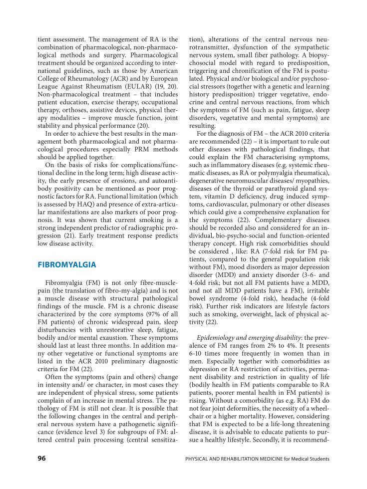
96 PHYSICAL AND REHABILITATION MEDICINE for Medical Students
tion), alterations of the central nervous neu-rotransmitter, dysfunction of the sympathetic nervous system, small fiber pathology. A biopsy-chosocial model with regard to predisposition, triggering and chronification of the FM is postu-lated. Physical and/or biological and/or psychoso-cial stressors (together with a genetic and learning history predisposition) trigger vegetative, endo-crine and central nervous reactions, from which the symptoms of FM (such as pain, fatigue, sleep disorders, vegetative and mental symptoms) are resulting.
For the diagnosis of FM – the ACR 2010 criteria are recommended (22) – it is important to rule out other diseases with pathological findings, that could explain the FM characterising symptoms, such as inflammatory diseases (e.g. systemic rheu-matic diseases, as RA or polymyalgia rheumatica), degenerative neuromuscular diseases/ myopathies, diseases of the thyroid or parathyroid gland sys-tem, vitamin D deficiency, drug induced symp-toms, cardiovascular, pulmonary or other diseases which could give a comprehensive explanation for the symptoms (22). Complementary diseases should be recorded also and considered for an in-dividual, bio-psycho-social and function-oriented therapy concept. High risk comorbidities should be considered , like: RA (7-fold risk for FM pa-tients, compared to the general population risk without FM), mood disorders as major depression disorder (MDD) and anxiety disorder (3-6- and 4-fold risk; but not all FM patients have a MDD, and not all MDD patients have a FM), irritable bowel syndrome (4-fold risk), headache (4-fold risk). Further risk indicators are lifestyle factors such as smoking, overweight, lack of physical ac-tivity (22).
Epidemiology and emerging disability: the prev-alence of FM ranges from 2% to 4%. It presents 6-10 times more frequently in women than in men. Especially together with comorbidities as depression or RA restriction of activities, perma-nent disability and restriction in quality of life (bodily health in FM patients comparable to RA patients, poorer mental health in FM patients) is rising. Without a comorbidity (as e.g. RA) FM do not fear joint deformities, the necessity of a wheel-chair or a higher mortality. However, considering that FM is expected to be a life-long threatening disease, it is advisable to educate patients to pur-sue a healthy lifestyle. Secondly, it is recommend-
tient assessment. The management of RA is the combination of pharmacological, non-pharmaco-logical methods and surgery. Pharmacological treatment should be organized according to inter-national guidelines, such as those by American College of Rheumatology (ACR) and by European League Against Rheumatism (EULAR) (19, 20). Non-pharmacological treatment – that includes patient education, exercise therapy, occupational therapy, orthoses, assistive devices, physical ther-apy modalities – improve muscle function, joint stability and physical performance (20).
In order to achieve the best results in the man-agement both pharmacological and not pharma-cological procedures especially PRM methods should be applied together.
On the basis of risks for complications/func-tional decline in the long term; high disease activ-ity, the early presence of erosions, and autoanti-body positivity can be mentioned as poor prog-nostic factors for RA. Functional limitation (which is assessed by HAQ) and presence of extra-articu-lar manifestations are also markers of poor prog-nosis. It was shown that current smoking is a strong independent predictor of radiographic pro-gression (21). Early treatment response predicts low disease activity.
FIBROMYALGIA
Fibromyalgia (FM) is not only fibre-muscle-pain (the translation of fibro-my-algia) and is not a muscle disease with structural pathological findings of the muscle. FM is a chronic disease characterized by the core symptoms (97% of all FM patients) of chronic widespread pain, sleep disturbancies with unrestorative sleep, fatigue, bodily and/or mental exaustion. These symptoms should last at least three months. In addition ma-ny other vegetative or functional symptoms are listed in the ACR 2010 preliminary diagnostic criteria for FM (22).
Often the symptoms (pain and others) change in intensity and/ or character, in most cases they are independent of physical stress, some patients complain of an increase in mental stress. The pa-thology of FM is still not clear. It is possible that the following changes in the central and periph-eral nervous system have a pathogenetic signifi-cance (evidence level 3) for subgroups of FM: al-tered central pain processing (central sensitiza-
12 Degenerative and inflammatory joint disorders, fibromyalgia, and osteoporosis 97
ming), land or water-based exercise, strength training, meditative movement therapies (e.g., qi-gong, yoga), relaxation procedures (e.g. Jacobson’s progressive muscle relaxation), cognitive behav-ioral therapy, Spa therapy, drug therapy (tricyclic antidepressants in small doses in the evening; duloxetine, in case of concomitant major depres-sion disorder; duloxetine or pregabalin, in case of concomitant generalized anxiety disorder), low-calorie diet for weight loss, where appropriate. Maintaining functional capacity and, in the case of regular long-term training, also improving the ability to function was shown in clinical trials for FM patients with the listed recommended strate-gies/therapies (24).
In cases where multiple comorbidities are pres-ent, a multidisciplinary approach should be pre-scribed. For example, in presence of lasting restric-tions in activities of daily living and not acceptable restrictions of quality of life (mentioned in a re-evaluation within the Rehab-Cycle) also without a mental comorbidity, a multidisciplinary (in some countries called multimodal) therapy, combining at least one body activating with one psychological/ psychotherapeutic procedure, is recommended.
The following long-term strategies/therapies should be first choice: self-employed methods, e.g. individually adapted endurance and/or strength training, stretching, heat therapy.
In the complex painful condition FM improve-ment in life quality can be reached. The main goal is to improve the function (the core of the PRM speciality), not to eliminate the pain. Exercise training and physical therapy as a core field in PRM are corner stones in the strategies/therapy of FM patients. Changes need time, even at thera-peutic measures adapted to individual perfor-mance and comorbidities, even at optimal therapy with own active action of the FM patients.
ed to exemplify strategies patients can apply to handle with their symptoms with respect to their preferences and comorbidities for best possible function and quality of life (23).
Aims, strategies and rehabilitation management: adapted to the individual restrictions and comor-bidities of the single FM patient the main aim is to promote activity and function in sense of the bio-psycho-social model. Therefore it is important to assess the FM patients collecting a complete medi-cal history and assessing symptoms and restric-tions in activities of daily living. In particular, pa-tients should be interviewed regarding mental disorders, drug use history, history of different therapies (frequency, dosage and period of differ-ent therapies/ strategies, with special attention to self-exercise); a complete physical examination (skin, orthopedic/PRM, internal and neurologic examination) should be carried out together with laboratory testing to rule out systemic inflamma-tion or other symptom-explaining diseases (dos-ing BCC, CRP, creatine kinase, TSH, calcium, 25-OH vitamin D), while further diagnostic testing should be used cautiously because a chronification can be promoted. Finally the capacity of function-ing should be ascertained by questionnaires (e.g. FIQ, SF36, PHQ4, HADS...) and physical testing (e.g. 6-minutes walk, one-leg-stand, sit-and-reach, functional reach, bicycle ergometer ECG...) (23).
After the assessment, the PRM Rehab-Cycle follows (Plan, Do, Check, Act and so on). Impor-tant is to choose realistic targets for obtaining successful interventions.
First, encourage patients to appropriate physi-cal and psychosocial activation. The best strate-gies/ therapies with evidence for an increase in quality of life and pain reduction are as follows: aerobic exercise (e.g., walking, cycling, swim-
Key messages
• In the management of some degenerative and inflammatory joint disorders (like osteoarthritis, rheumatoid arthritis, osteoporosis, and fibromyalgia), and – more in general – of progressive musculoskeletal disorders, both pharmacological and non pharmacological procedures are ap-plied, but essentially and especially the importance of PRM application has to be stressed and emphasized.
98 PHYSICAL AND REHABILITATION MEDICINE for Medical Students
CISIT). Ann Rheum Dis. 2007; 66: 377-88. 11. Zhang W, Doherty M, Arden N, Bannwarth B,
Bijlsma J, Gunther KP, et al. EULAR Standing Committee for International Clinical Studies In-cluding Therapeutics (ESCISIT). EULAR evidence based recommendations for the management of hip osteoarthritis: report of a task force of the EU-LAR Standing Committee for International Clini-cal Studies Including Therapeutics (ESCISIT). Ann Rheum Dis. 2005; 64: 669-81.
12. Zhang W, Doherty M, Leeb BF, Alekseeva L, Ar-den NK, Bijlsma JW, et al. EULAR evidence-based recommendations for the diagnosis of hand osteo-arthritis: report of a taskforce of ESCISIT. Ann Rheum Dis. 2009; 68: 8-17.
13. Jordan KM, Arden NK, Doherty M, Bannwarth B, Bijlsma JW, Dieppe P, et al. Standing Committee for International Clinical Studies Including Ther-apeutic Trials ESCISIT. EULAR Recommenda-tions 2003: an evidence based approach to the management of knee osteoarthritis: Report of a Task Force of the Standing Committee for Interna-tional Clinical Studies Including Therapeutic Tri-als (ESCISIT). Ann Rheum Dis. 2003; 62: 1145-55.
14. Meneses SR, Goode AP, Nelson AE, Lin J, Jordan JM, Allen KD, et al. Clinical algorithms to aid os-teoarthritis guideline dissemination. Osteoarthri-tis Cartilage. 2016; 24: 1487-99.
15. Cosman F, de Beur SJ, LeBoff MS, Lewiecki EM, Tanner B, Randall S, et al. National Osteoporosis Foundation. Clinician’s Guide to Prevention and Treatment of Osteoporosis. Osteoporos Int. 2014; 25: 2359-81. Erratum in: Osteoporos Int. 2015; 26: 2045-7.
16. Kanis JA, on behalf of the World Health Organiza-tion Scientific Group (2007). Assessment of osteo-porosis at the primary health-care level. Technical Report. World Health Organization Collaborating Center for Metabolic Bone Diseases. University of Sheffield (UK), 2007. Available at: https://www.sheffield.ac.uk/FRAX/pdfs/WHO_Technical_Re-port.pdf
17. Küçükdeveci AA, Oral A, Ilıeva EM, Varela E, Valero R, Berteanu M, et al. Inflammatory arthri-tis. The role of physical and rehabilitation medi-cine physicians. The European perspective based on the best evidence. A paper by the UEMS-PRM Section Professional Practice Committee. Eur J Phys Rehabil Med. 2013; 49: 551-64.
18. Toussirot E. Predictive factors for disability as evaluated by the health assessment questionnaire in rheumatoid arthritis: a literature review. In-flamm Allergy Drug Targets. 2010; 9: 51-9.
19. Combe B, Landewe R, Daien CI, Hua C, Aletaha D, Álvaro-Gracia JM, et al. 2016 update of the EU-LAR recommendations for the management of
REFERENCES
1. Woolf AD, Pfleger B. Burden of major musculo-skeletal conditions. Bull World Health Organ. 2003; 81: 646-56.
2. Hochberg MC, Altman RD, April KT, Benkhalti M, Guyatt G, McGowan J et al. American College of Rheumatology 2012 recommendations for the use of nonpharmacologic and pharmacologic ther-apies in osteoarthritis of the hand, hip, and knee. Arthritis Care Res (Hoboken). 2012; 64: 465-74.
3. Cushnaghan J, Dieppe P. Study of 500 patients with limb joint osteoarthritis. I. Analysis by age, sex, and distribution of symptomatic joint sites. Ann Rheum Dis. 1991; 50: 8-13.
4. World Health Organization (WHO). Priority dis-eases and reasons for inclusion. Chapt. 6.12 - Os-teoarthritis [Internet] Available from:http://www.who.int/medicines/areas/priority_medicines/Ch6_12Osteo.pdf
5. Smith E, Hoy DG, Cross M, Vos T, Naghavi M, Buchbinder R, et al. The global burden of other musculoskeletal disorders: estimates from the Global Burden of Disease 2010 study. Ann Rheum Dis. 2014; 73: 1462-9.
6. Hochberg MC, Altman RD, April KT, Benkhalti M, Guyatt G, McGowan J, et al. American College of Rheumatology. American College of Rheuma-tology 2012 recommendations for the use of non-pharmacologic and pharmacologic therapies in osteoarthritis of the hand, hip, and knee. Arthritis Care Res (Hoboken). 2012; 64: 465-74.
7. McAlindon TE, Bannuru RR, Sullivan MC, Arden NK, Berenbaum F, Bierma-Zeinstra SM, et al. OARSI guidelines for the non-surgical manage-ment of knee osteoarthritis. Osteoarthritis Carti-lage. 2014; 22: 363-88.
8. Rillo O, Riera H, Acosta C, Liendo V, Bolaños J, Monterola L, et al. PANLAR Consensus Recom-mendations for the Management in Osteoarthritis of Hand, Hip, and Knee. J Clin Rheumatol. 2016; 22: 345-54.
9. Bruyère O, Cooper C, Pelletier JP, Branco J, Luisa Brandi M, Guillemin F, et al. An algorithm recom-mendation for the management of knee osteoar-thritis in Europe and internationally: a report from a task force of the European Society for Clinical and Economic Aspects of Osteoporosis and Osteoarthritis (ESCEO). Semin Arthritis Rheum. 2014; 44: 253-63.
10. Zhang W, Doherty M, Leeb BF, Alekseeva L, Ar-den NK, Bijlsma JW, et al. I. EULAR evidence based recommendations for the management of hand osteoarthritis: report of a Task Force of the EULAR Standing Committee for International Clinical Studies Including Therapeutics (ES-
12 Degenerative and inflammatory joint disorders, fibromyalgia, and osteoporosis 99
DL, Katz RS, Mease P,et al. The American College of Rheumatology preliminary diagnostic criteria for fibromyalgia and measurement of symptom severity. Arthritis Care Res (Hoboken). 2010; 62: 600-10.
23. Wolfe F, Clauw DJ, Fitzcharles MA, Goldenberg DL, Häuser W, Katz RL, et al. 2016 Revisions to the 2010/2011 fibromyalgia diagnostic criteria. Semin Arthritis Rheum. 2016; 46: 319-29.
24. Macfarlane GJ, Kronisch C, Dean LE, Atzeni F, Häuser W, Fluß E, et al. EULAR revised recom-mendations for the management of fibromyalgia. Ann Rheum Dis. 2017; 76: 318-28.
early arthritis. Ann Rheum Dis. 2017; 76: 948-59. 20. Singh JA, Saag KG, Bridges SL Jr, Akl EA, Bannuru
RR, Sullivan MC,et al. 2015 American College of Rheumatology Guideline for the Treatment of Rheumatoid Arthritis. Arthritis Rheumatol. 2016; 68: 1-26.
21. Saevarsdottir S, Rezaei H, Geborek P, Petersson I, Ernestam S, Albertsson K, et al. Current smoking status is a strong predictor of radiographic pro-gression in early rheumatoid arthritis: results from the SWEFOT trial. Ann Rheum Dis. 2015; 74: 1509-14.
22. Wolfe F, Clauw DJ, Fitzcharles MA, Goldenberg

13 Spinal deformities 101
years after the age of 30, increasing progressively to 2/3 cm after the age of 70. In worst cases the progression of curves can lead to a hunchback (flexed posture), whose probability increases with age and with the severity of the curve (11, 12).
The three-dimensional scoliosis typically ap-pears during growth. Growth represents its prime mover, and periods of rapid growth are at high risk for progression (6). Without treatment the risks in adulthood of pain, disability and progres-sion will increase in direct relation with the curve magnitude (12-14): curves exceeding 30 degrees have an increasing risk of progression while those above 50 degrees have an almost 100% probability of progression (15). Therefore, early management can prevent problems in adulthood thus increas-ing quality of life (16).
Sagittal spine deformities are posterior (hyper-kyphosis) or anterior (lordosis), associated to vari-ous degrees of stiffness, being generated by the structural alterations due to the pathology. These deviations typically occur in the physiological curvatures, that appear increased (hyperkyphosis or round back) or reduced (flat back or hollow back); when the alteration affect the spinal shape, thus modifying the normal distribution of curves, a junctional kyphosis, or a junctional hyperky-phosis (also called thoraco-lumbar deformity) occurs (3).
Whatever is the original deformity, its progres-sion in adulthood and in the elderly is always to-ward flexion due to:
ū biomechanical reasons linked to the gravity force: in case of scoliosis, the progressive patho-logical rotation moves the vertebral body later-ally until the trunk and spine miss the anterior support and evolves in the final hunchback (10);
ū activity of Daily Life (ADL): a flexed posture is very common; for most activities we use to bend forward; ageing a lack in strength in the exten-
INTRODUCTION
Spinal deformities represent specific musculo-skeletal progressive health conditions, highly cor-related to pain syndromes and reduction of qual-ity of life with aging (1, 2). Spinal deformities can be classified as disorders of the sagittal plane (in-crease/decrease of the physiological curves – ky-phosis and lordosis – or distortion of their distri-bution – long kyphosis, junctional kyphosis, etc.) (3, 4), or three-dimensional deformities (scoliosis) characterized by a frontal curvature, an horizon-tal rotation and a sagittal distortion of the physi-ological curves (5-7). These deformities can be primary (so called idiopathic) or secondary to congenital bony alterations, neurological or ge-netic diseases, or other minor causes (6).
Spinal deformities usually occur during growth and progress in periods of height peak growth (8); moreover, they predispose to pain and disability in adulthood and to further progression due to the degenerative processes which characterize aging (9, 10). The main goals of treatment will change according to the different moment of life in which the diagnosis occurs and in strong relationship with the specific estimated risk factors.
DIAGNOSIS, EVOLUTION AND GENERAL MANAGEMENT
Spinal deformities progress with time. It is nor-mal for the human being that physiological curves in adulthood increase with aging, due to degen-eration first of soft tissues, then of bones. In case of pathology, this progression is in the direction of the pathological curve, due to mechanical disad-vantages generated by the deformity. This evolu-tion is correlated with a decrease in height with aging, that has been quantified in 1 cm every 10
13Spinal deformitiesStefano NEGRINI, Sabrina DONZELLI
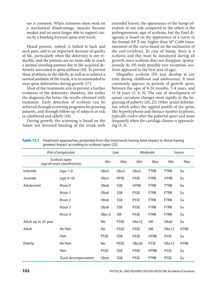
102 PHYSICAL AND REHABILITATION MEDICINE for Medical Students
extended knees); the appearance of the hump (el-evation of one side compared to the other) is the pathognomonic sign of scoliosis, but the final di-agnosis is based on the appearance of a curve in the frontal AP X-ray higher than 10° Cobb (mea-surement of the curve based on the inclination of the end-vertebrae). In case of hump, there is a scoliosis and this must be monitored during all growth, since scoliosis does not disappear sponta-neously (6, 19) (only possible rare exception: sco-liosis appeared in the first year of age).
Idiopathic scoliosis (IS) may develop at any time during childhood and adolescence. It most commonly appears in periods of growth spurt, between the ages of 6-24 months, 5-8 years, and 11-14 years (5, 6, 8). The rate of development of spinal curvature changes most rapidly at the be-ginning of puberty (20, 21). Other spinal deformi-ties which affect the sagittal profile of the spine, like hyperkyphosis and thoraco-lumbar kyphosis, typically evolve after the pubertal spurt and more frequently when the cartilage closure is approach-ing (3, 4).
sor is common. When extensors must work on a mechanical disadvantage, muscles become weaker and no more longer able to support cor-rectly a bending forward spine and trunk.
Flexed posture, indeed, is linked to back and neck pain, and to an important decrease of quality of life, particularly when the deformity is not re-ducible, and the patients are no more able to reach a normal standing posture due to the acquired de-formity associated to spine stiffness (10). To prevent these problems in the elderly, as well as to achieve a normal aesthetic of the trunk, it is recommended to treat spine deformities during growth (17).
Most of the treatments aim to prevent a further evolution of the deformity; therefore, the earlier the diagnosis the better the results obtained with treatment. Early detection of scoliosis can be achieved through screening programs for growing patients, and through follow-up of subjects at risk in adulthood and elderly (18).
During growth, the screening is based on the Adam test (forward bending of the trunk with
Table 13.1 Treatment approaches, presented from the treatments having least impact to those having greatest impact according to scoliosis types (22)
Risk of progression Low Moderate Severe
Scoliosis types (age of onset classification) Min Max Min Max Min Max
Infantile (age 1-3) Obs3 Obs3 Obs3 TTRB TTRB Su
Juvenile (age 4-10) Obs3 PPSE PSSE FTRB HTRB Su
Adolescent Risser 0 Obs6 SSB HTRB FTRB TTRB Su
Risser 1 Obs6 SSB PSSE FTRB FTRB Su
Risser 2 Obs6 SSB PSSE FTRB FTRB Su
Risser 3 Obs6 SSB PSSE FTRB FTRB Su
Risser 4 Obs12 SIR PSSE FTRB FTRB Su
Adult up to 25 year No PSSE Obs12 SIR Obs6 Su
Adult No Pain No PSSE PSSE SIR Obs12 HTRB
Pain PSSE SSB PSSE HTRB PSSE Su
Elderly No Pain No PSSE Obs36 PSSE Obs12 HTRB
Pain PSSE SSB PSSE HTRB PSSE Su
Trunk decompensation Obs6 SSB PSSE PTRB PSSE Su

13 Spinal deformities 103
treatment is not needed, observation to monitor growth and the evolution of deformities at lowest risks. Specific scoliosis exercises are usually pre-scribed for curves between 15-25°, normally in outpatient clinics. Soft braces are generally used for milder curves and in younger patients (15-30°), while for more important curves rigid braces (25-45°) and very rigid braces/casts (35-40° and above) are prescribed. The dosage of brace wear can vary from night time (8 hours per day), to part-time (14-18) or full time (21-23). Spinal fusion surgery is considered when scoliosis is above 45-50° and did not respond to rehabilitation treatment: since sur-gery is quoad valetudinem and not quoad vitam, patients values must be included in the decision.
For hyperkyphosis the recommended treat-ment are based on the presence of Scheuermann
Scoliosis and spine deformities must be diag-nosed by as many people as possible (any physi-cian, paramedic or educator checking for any reason the spine during growth), but must be fol-lowed-up by specific experts: these mainly are PRM physicians and/or orthopaedic surgeons.
All the possible treatment approaches are listed in Table 13.1 according to the actual Guidelines (22) for scoliosis and in Table 13.2 for kyphosis: treatments are presented from those having the least to those having the greatest impact. The treatment is prescribed according to the progres-sion risk estimation made by the expert physician and the type of scoliosis. Scoliosis are classified in the present table according to the age of onset, which represent the most used classification (6).
In summary treatment include nothing, when
Table 13.2 Treatment approaches for hyperkyphosis during growth*
Sagittal alterations: Hyperkyphosis
Postural deformity
Structural deformity
Scheuermann disease
Min Max Min Max Min Max
No PSSE Obs6 FTRB PSSE FTRB
* The same terminology as in Table 13.1 is used
According to Tables 13.1 and 13.2:Nothing (No). No treatment is needed.Observation (Obs). It is the first step of an
active approach to idiopathic scoliosis and it consists of regular clinical evaluation with a spe-cific follow-up period. Timing of this follow-up can range from 2-3 to 36-60 months according to the specific clinical situation.
Physiotherapeutic Scoliosis Specific Exer-cises (PSSE). PSSE include all forms of outpa-tient physiotherapies with evidence of having an effect on some scoliosis outcomes.
Special Inpatient Rehabilitation (SIR). If SIR is recommended, patients spend several weeks (usually 4-6) at a specialized health centre (hos-pital department, sanatorium or a similar form of health care) where they undergo an intensive PSSE treatment (several hours per day).
Bracing consists of using a brace (a corrective orthosis) for a specified period daily. Usually it is worn until maturity. The main therapeutic goal
is to halt the scoliosis curves from progression. The use of brace is strongly recommended to be associated with scoliosis specific exercises to enhance brace effect and avoid adverse effects like muscle weakening.
Night Time Rigid Bracing (8-12 hours per day) (NTRB): wearing a brace mainly in bed.
Soft Bracing (SB): it includes mainly the SpineCor brace (23), but also other similar de-signs (24).
Part Time Rigid Bracing (12-20 hours per day) (PTRB): wearing a rigid brace mainly out-side school and in bed.
Full Time Rigid Bracing (20-24 hours per day) or cast (FTRB): wearing a rigid brace all the time (at school, at home, in bed, etc.). Casts have been included here as well. Casts are used by some schools as the first stage to achieve correc-tion to be maintained afterwards with rigid brace; a cast is considered a standard approach in infantile scoliosis (25, 26).
104 PHYSICAL AND REHABILITATION MEDICINE for Medical Students
3. Zaina F, Atanasio S, Ferraro C, Fusco C, Negrini A, Romano M, et al. Review of rehabilitation and orthopedic conservative approach to sagittal plane diseases during growth: hyperkyphosis, junctional kyphosis, and Scheuermann disease. Eur J Phys Rehabil Med. 2009; 45: 595-603.
4. Mauroy J de, Weiss HR, Aulisa AG, Aulisa L, Brox JI, Durmala J, et al. 7th SOSORT consensus paper: conservative treatment of idiopathic & Scheuer-mann’s kyphosis. Scoliosis. 2010; 5: 9.
5. Negrini S, Aulisa L, Ferraro C, Fraschini P, Ma-siero S, Simonazzi P, et al. Italian guidelines on
REFERENCES
1. Danielsson AJ, Nachemson AL. Radiologic find-ings and curve progression 22 years after treat-ment for adolescent idiopathic scoliosis: compari-son of brace and surgical treatment with matching control group of straight individuals. Spine. 2001; 26: 516-25.
2. Marty-Poumarat C, Scattin L, Marpeau M, Gar-reau de Loubresse C, Aegerter P. Natural history of progressive adult scoliosis. Spine. 2007; 32: 1227-34; discussion 1235.
lution of AIS is differentiated from a de novo sco-liosis, rapidly changing in a few years to rotatory dislocation (27).
Treatment choices are related to risks estima-tion which comes from large expertise in the field of spine deformities, therefore expertise is strong-ly recommended by the current guidelines of management. Further high-quality research is needed on the understand more about scoliosis specific exercises and braces, while their efficacy has been proven with RCTs. The effects on quality of life of the deformity as well as of the treatments is to be defined after good quality of life measures have been developed.
The skills in detecting scoliosis at its onset and in identifying patients at highest risk and with more complicated situation is what all the special-ist in Orthopaedics and PRM have to develop to contribute to the wellness of people affected by spinal deformities in all ages.
or structured deformities with difficulties in re-ducing the increased curvature: in most severe cases braces are prescribed while in milder forms specific exercises are enough. Observation or ex-ercises can be considered the ideal prescription for postural alterations.
When spinal deformities are diagnosed after the end of growth or in adulthood, regular follow-up are advised; rehabilitation, including physio-therapic specific exercises and/or braces (soft or low rigidity), is considered when there is progres-sion, pain or disability, or for those patients at higher risk of progression. In adulthood, IS may intensify because of progressive osseous deformi-ties, collapsing of the spine and degeneration. This phenomenon is reported especially in scolio-sis that are above 50°, while the risk of progression starts to increase as the curve grows above 30° (15); less severe idiopathic scoliosis curves often remain stable. Typically, in adult scoliosis the evo-
Key messages
• Spinal deformities includes mainly scoliosis and sagittal plane deformities, that are disorders with an important impact particularly with aging: they are treated during growth to avoid these consequences that include progressive trunk deformity (with psychological impact), back pain and reduced quality of life.
• Spinal deformities should be diagnosed by as many physicians as possible, but treatment should be performed by well trained experts.
• Diagnosis of scoliosis is based on clinical (hump measurement during forward bending test) and radiographic (Cobb angle measurement) exams.
• Possible treatments include specific exercises in low degree cases, bracing and specific exercises in medium and high degree cases, surgery in very important deformities: treatments are planned in a step by step strategy.
13 Spinal deformities 105
scoliosis improve disability after motor and cogni-tive rehabilitation: results of a randomised con-trolled trial. Eur Spine J. 2016; 25: 3120-9.
17. Negrini S, Grivas TB, Kotwicki T, Maruyama T, Rigo M, Weiss HR, et al. Why do we treat adoles-cent idiopathic scoliosis? What we want to obtain and to avoid for our patients. SOSORT 2005 Con-sensus paper. Scoliosis. 2006; 1: 4.
18. Grivas TB, Wade MH, Negrini S, O’Brien JP, Maruyama T, Hawes MC, et al. SOSORT consen-sus paper: school screening for scoliosis. Where are we today? Scoliosis. 2007; 2: 17.
19. Bunnell WP. Selective screening for scoliosis. Clin Orthop. 2005; (434): 40-5.
20. Mao S, Xu L, Zhu Z, Qian B, Qiao J, Yi L, et al. Association between genetic determinants of peak height velocity during puberty and predisposition to adolescent idiopathic scoliosis. Spine. 2013; 38: 1034-9.
21. Dimeglio A, Canavese F. Progression or not pro-gression? How to deal with adolescent idiopathic scoliosis during puberty. J Child Orthop. 2013; 7: 43-9.
22. Negrini S, Donzelli S, Aulisa AG, Czaprowski D, Schreiber, S, De Mauroy JC, et al. 2016 SOSORT guidelines: Orthopaedic and Rehabilitation treat-ment of idiopathic scoliosis during growth. Scolio-sis Spinal Disord. 2018; 13: 3.
23. Coillard C, Circo AB, Rivard CH. A Prospective Randomized Controlled Trial of the Natural His-tory of Idiopathic Scoliosis versus treatment with the Spinecor brace. Sosort Award 2011 Winner. Eur J Phys Rehabil Med. 2014; 50: 479-87.
24. Wynne JH. The Boston Brace and TriaC systems. Disabil Rehabil Assist Technol. 2008; 3: 130-5.
25. de Mauroy JC, Lecante C, Barral F. “Brace Tech-nology” Thematic Series - The Lyon approach to the conservative treatment of scoliosis. Scoliosis. 2011; 6: 4.
26. Negrini S, Marchini G, Tessadri F. Brace technol-ogy thematic series - The Sforzesco and Sibilla braces, and the SPoRT (Symmetric, Patient ori-ented, Rigid, Three-dimensional, active) concept. Scoliosis. 2011; 6: 8.
27. Aebi M. The adult scoliosis. Eur Spine J. 2005; 14: 925-48.
rehabilitation treatment of adolescents with scoli-osis or other spinal deformities. Eura Medicophys. 2005; 41: 183-201.
6. Negrini S, Aulisa AG, Aulisa L, Circo AB, de Mau-roy JC, Durmala J, et al. 2011 SOSORT guidelines: Orthopaedic and Rehabilitation treatment of idio-pathic scoliosis during growth. Scoliosis. 2012; 7: 3.
7. Korbel K, Kozinoga M, Stoliński Ł, Kotwicki T. Scoliosis Research Society (SRS) Criteria and Soci-ety of Scoliosis Orthopaedic and Rehabilitation Treatment (SOSORT) 2008 Guidelines in Non-Operative Treatment of Idiopathic Scoliosis. Pol Orthop Traumatol. 2014; 79: 118-22.
8. Lonstein JE, Carlson JM. The prediction of curve progression in untreated idiopathic scoliosis. J Bone Jt Surg. 1984; 1061-71.
9. Ames CP, Scheer JK, Lafage V, Smith JS, Bess S, Berven SH, et al. Adult Spinal Deformity: Epide-miology, Health Impact, Evaluation, and Manage-ment. Spine Deform. 2016; 4: 310-22.
10. Barrey C, Roussouly P, Le Huec J-C, D’Acunzi G, Perrin G. Compensatory mechanisms contribut-ing to keep the sagittal balance of the spine. Eur Spine J 2013; 22 Suppl 6: S834-41.
11. Negrini S. Focus on flexed posture and hyperky-phosis: prevention and rehabilitation to reduce disability and increase quality of life. Eur J Phys Rehabil Med. 2009; 45: 567-9.
12. Benoist M. Natural history of the aging spine. Eur Spine J 2003; 12 Suppl 2: S86-9.
13. Nachemson, AL, Lonstein, JE, Weinstein, SL. Re-port of the prevalence and natural history Com-mittee of the Scoliosis Research Society. Scoliosis Research Society, Denver. 1982.
14. Danielsson AJ. Natural history of adolescent idio-pathic scoliosis: a tool for guidance in decision of surgery of curves above 50°. J Child Orthop. 2013; 7: 37-41.
15. Weinstein SL, Dolan LA, Spratt KF, Peterson KK, Spoonamore MJ, Ponseti IV. Health and function of patients with untreated idiopathic scoliosis: a 50-year natural history study. JAMA 2003; 289: 559-67.
16. Monticone M, Ambrosini E, Cazzaniga D, Rocca B, Motta L, Cerri C, et al. Adults with idiopathic


14 Principles of management of acute and chronic pain: the example of low back pain 107
of cases it is possible to identify the medical cause (secondary LBP), in the vast majority of cases LBP is due to a soft tissues lesion that cannot be diag-nosed with certainty even with the most advanced techniques. Research has recognized that for pa-tients it is important to avoid the so-called “diag-nostic labels” (i.e. the unproven diagnosis in case of primary LBP), since they are not repeatable among different physicians: the resulting diagnostic con-fusion increase the risk of chronicity.
LBP has been defined as a bio-psycho-social syndrome, since it combines physical, psychologi-cal and social elements in different ways in the different phases of pathology and in single pa-tients: in this respect the best approach is not bio-medical (following the classical pathway, with ae-tiology/disease identification to define treatment) but rehabilitative. The accepted classification dis-tinguishes acute (up to 1 month of pain), from sub-acute (1-3 months), sub-chronic (3-6 months) and chronic (more than 6 months) LBP. This clas-sification has been derived by epidemiology, but in fact describes different clinical entities, with different aetiology, diagnosis, prognosis and treat-ment approach (Table 14.1).
Acute low back pain is self resolving, and diag-nosis is fully based on history and physical exam. If red flags are not identified (Table 14.2), it is not necessary to proceed with any exams. Treatment does not accelerate resolution, and if made aware of this many patients just wait self-resolution: drugs can be used to reduce symptoms (paracetamol 1g max 3 times/day used to be considered the 1st line approach, even if some doubts have been re-cently raised, while ibuprofen and then other NSAIDS or cox-2 inhibitors follow), as well as manual therapy if drugs cannot be used. It is proven that bed rest is detrimental, while staying
INTRODUCTION
Pain has been defined as an unpleasant sensory and emotional experience associated with actual or potential tissue damage, or described in terms of such damage (1). In medicine, pain is usually considered a symptom of an underlying condi-tion: while this is true for acute pain, chronic pain is recognised as a bio-psycho-social syndrome involving the individual in its entirety.
Pain is the most common reason for physician consultation, particularly (but not only) in the High Income Countries (2). In this Chapter we will specifically focus as an example on low back pain (LBP), since it is the most common musculo-skeletal disease with the highest economic impact in western countries: acute LBP life prevalence is more than 80% of the population, annual inci-dence up to 30%; over 90% of Acute cases resolve in less than 30 days, but estimates of recurrence at 1 year range from 24% to 80% (3). On the other side, Chronic LBP is an everyday experience for 4% to 7% of the population whose prevalence in-creases linearly from the third decade of life on, until the 60 years of age, being more prevalent in women (4). Chronic LBP is responsible of 75-80% of the entire enormous costs of LBP and less than 5% of patients achieve a complete resolution of pain. Subacute has scarcely been studied and to-day we have no reliable epidemiological data, even if this stage of pathology should deserve the high-est attention.
LOW BACK PAIN
Even if it is a symptom, LBP has been recognized as a specific diagnosis. In fact, while in less than 1%
14Principles of management of acute and chronic pain:
the example of low back painStefano NEGRINI, Fabio ZAINA
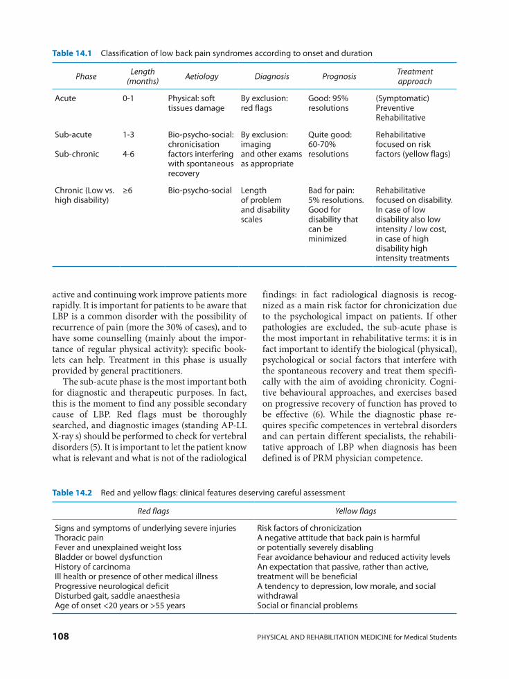
108 PHYSICAL AND REHABILITATION MEDICINE for Medical Students
findings: in fact radiological diagnosis is recog-nized as a main risk factor for chronicization due to the psychological impact on patients. If other pathologies are excluded, the sub-acute phase is the most important in rehabilitative terms: it is in fact important to identify the biological (physical), psychological or social factors that interfere with the spontaneous recovery and treat them specifi-cally with the aim of avoiding chronicity. Cogni-tive behavioural approaches, and exercises based on progressive recovery of function has proved to be effective (6). While the diagnostic phase re-quires specific competences in vertebral disorders and can pertain different specialists, the rehabili-tative approach of LBP when diagnosis has been defined is of PRM physician competence.
active and continuing work improve patients more rapidly. It is important for patients to be aware that LBP is a common disorder with the possibility of recurrence of pain (more the 30% of cases), and to have some counselling (mainly about the impor-tance of regular physical activity): specific book-lets can help. Treatment in this phase is usually provided by general practitioners.
The sub-acute phase is the most important both for diagnostic and therapeutic purposes. In fact, this is the moment to find any possible secondary cause of LBP. Red flags must be thoroughly searched, and diagnostic images (standing AP-LL X-ray s) should be performed to check for vertebral disorders (5). It is important to let the patient know what is relevant and what is not of the radiological
Table 14.1 Classification of low back pain syndromes according to onset and duration
Phase Length (months) Aetiology Diagnosis Prognosis Treatment
approach
Acute 0-1 Physical: soft tissues damage
By exclusion: red flags
Good: 95% resolutions
(Symptomatic) Preventive Rehabilitative
Sub-acute
Sub-chronic
1-3
4-6
Bio-psycho-social: chronicisation factors interfering with spontaneous recovery
By exclusion: imaging and other exams as appropriate
Quite good: 60-70% resolutions
Rehabilitative focused on risk factors (yellow flags)
Chronic (Low vs. high disability)
≥6 Bio-psycho-social Length of problem and disability scales
Bad for pain: 5% resolutions. Good for disability that can be minimized
Rehabilitative focused on disability. In case of low disability also low intensity / low cost, in case of high disability high intensity treatments
Table 14.2 Red and yellow flags: clinical features deserving careful assessment
Red flags Yellow flags
Signs and symptoms of underlying severe injuriesThoracic painFever and unexplained weight lossBladder or bowel dysfunctionHistory of carcinomaIll health or presence of other medical illnessProgressive neurological deficitDisturbed gait, saddle anaesthesiaAge of onset <20 years or >55 years
Risk factors of chronicizationA negative attitude that back pain is harmful or potentially severely disablingFear avoidance behaviour and reduced activity levelsAn expectation that passive, rather than active, treatment will be beneficialA tendency to depression, low morale, and social withdrawalSocial or financial problems
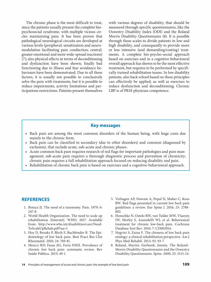
14 Principles of management of acute and chronic pain: the example of low back pain 109
with various degrees of disability, that should be measured through specific questionnaires, like the Oswestry Disability Index (ODI) and the Roland Morris Disability Questionnaire (8). It is possible through these scales to divide patients in low and high disability, and consequently to provide more or less intensive (and demanding/costing) treat-ments. A complete bio-psycho-social approach based on exercises and in a cognitive-behavioural overall approach has shown to be the most effective treatment, but requires to be performed by specifi-cally trained rehabilitation teams. In low disability patients, also back school based on these principles can effectively be applied, as well as exercises to reduce dysfunction and deconditioning. Chronic LBP is of PRM physician competence.
The chronic phase is the most difficult to treat, since the patients usually present the complete bio-psychosocial syndrome, with multiple vicious cir-cles maintaining pain. It has been proven that pathological neurological circuits are developed at various levels (peripheral: sensitization and neuro-modulation facilitating pain conduction; central: greater emotional and more wide-spread reactions) (7); also physical effects in terms of deconditioning and dysfunction have been shown; finally bad functioning due to illness and fear avoidance be-haviours have been demonstrated. Due to all these factors, it is usually not possible to conclusively solve the pain with treatments, but it is possible to reduce impairments, activity limitations and par-ticipations restrictions. Patients present themselves
Key messages
• Back pain are among the most common disorders of the human being, with huge costs due mainly to the chronic form.
• Back pain can be classified in secondary (due to other disorders) and common (diagnosed by exclusion), that include acute, sub-acute and chronic phases.
• Acute common back pain requires research of red flags for important pathologies and pain man-agement; sub-acute pain requires a thorough diagnostic process and prevention of chronicity; chronic pain requires a full rehabilitation approach focused on reducing disability and pain.
• Rehabilitation of chronic back pain is based on exercises and a cognitive-behavioural approach.
5. Verhagen AP, Downie A, Popal N, Maher C, Koes BW. Red flags presented in current low back pain guidelines: a review. Eur Spine J. 2016; 25: 2788-802.
6. Henschke N, Ostelo RW, van Tulder MW, Vlaeyen JW, Morley S, Assendelft WJ, et al. Behavioural treatment for chronic low-back pain. Cochrane Database Syst Rev. 2010; 7: CD002014.
7. Negrini S, Zaina F. The chimera of low back pain etiology: a clinical rehabilitation perspective. Am J Phys Med Rehabil. 2013; 92: 93-7.
8. Roland, Martin; Fairbank, Jeremy. The Roland–Morris Disability Questionnaire and the Oswestry Disability Questionnaire. Spine. 2000; 25: 3115-24.
REFERENCES
1. Bonica JJ. The need of a taxonomy. Pain. 1979; 6: 247-8.
2. World Health Organization. The need to scale up rehabilitation [Internet]. WHO; 2017. Available from: http://www.who.int/disabilities/care/Need-ToScaleUpRehab.pdf?ua=1
3. Hoy D, Brooks P, Blyth F, Buchbinder R. The Epi-demiology of low back pain. Best Pract Res Clin Rheumatol. 2010; 24: 769-81.
4. Meucci RD, Fassa AG, Faria NMX. Prevalence of chronic low back pain: systematic review. Rev Saúde Pública. 2015; 49-1.


Part IV
The disabling consequences of other common clinical
conditions during the life span

15 Balance troubles and the risk for falls in the elderly 113
In general, regular participation in physical activity or planned exercises has been shown to: 1) minimize the physiologic changes associated with typical aging, and improve functional sta-tus (e.g. muscle strength, bone density, physical and cognitive functioning, and activities of daily living); 2) contribute to psychological health (reduction of stress, anxiety, and depres-sion), well-being (improved sleep, self-esteem, and life satisfaction), and better quality of life; 3) increase longevity and decrease the risk of several of the most common diseases of indus-trialized societies (such as coronary heart dis-eases, diabetes, stroke, peripheral vascular dis-eases, colon and breast cancer, falls with hip fractures, and musculoskeletal symptoms); 4) be useful as primary or adjunctive treatment for certain chronic diseases (e.g. osteoporosis, obe-sity, hypertension, hyperlipidemia); and 5) assist in the prevention and treatment of disability, reducing institutionalization and favouring so-cial participation (5, 6). More in detail, in order to improve cardiorespiratory and muscular fit-ness, bone and functional health, and reduce many clinical risks related to non-communica-ble diseases, the main suggestions of the “Glob-al Recommendations on Physical Activity for Health” for people aged 65+ by World Health Organization (5), and “the Position Stand by the American College of Sports Medicine on exer-cise and physical activity for older adults” (6) are listed in the Table 15.1.
When adults of this age group cannot do the recommended amounts of physical activity due to health conditions, they should be as physically ac-tive as their abilities and conditions allow. In gen-eral, it would be appropriate to encourage a mod-erate start with gradual progress to higher levels of physical activity.
INTRODUCTION
More than one-fourth of older people (aged 65 years and above) fall each year; as such fall rates and the risk of consequent serious injuries in-crease with age (1). Falls are a major public health concern and represent a leading cause of injury (e.g. bone fractures, head injury), disability, early institutionalization, and deaths in older adult population (2). Frequently, falls and balance im-pairment also induce psychological reactions characterized by fear of future falling. This fear of falling (and related construct such as balance con-fidence) may be protective if it interferes only with hazardous activity and increases caution during performance in daily living tasks; however it can also be maladaptive, thus leading to further de-conditioning, functional decline, and poorer quality of life (1, 2).
In order to decrease falls and risk of falls among older people, multi-component fall-pre-vention programs have been proposed. They usu-ally include comprehensive risk factor assessment and reduction through tailored medical manage-ment approach (e.g. on medications, vision ability, etc.); education (on fall risk factors, use of assistive technology and modified techniques, environ-mental safety in the community); home safety as-sessment and household modifications; and phys-ical activity and exercise programs (3).
Current guidelines put emphasis on the impor-tance of a mix of different exercises for this popu-lation, including strengthening (resistance), flexibility and cardiovascular (endurance) exer-cises. They also comprise activities aiming to im-prove balance, gait and general movement ability, and reducing fall risks linked to age-related de-generative changes in sensorimotor and neuro-muscular systems (4).
15Balance troubles and the risk for falls in the elderly
Franco FRANCHIGNONI, Levent ÖZÇAKAR
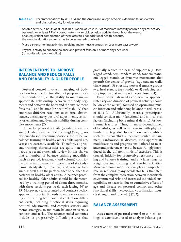
114 PHYSICAL AND REHABILITATION MEDICINE for Medical Students
gradually reduce the base of support (e.g., two-legged stand, semi-tandem stand, tandem stand, one-legged stand), 2) dynamic movements that perturb the centre of gravity (e.g., tandem walk, circle turns), 3) stressing postural muscle groups (e.g. heel stands, toe stands), or 4) reducing sen-sory input (e.g. standing with eyes closed) (4).
Frail individuals need a conservative approach (intensity and duration of physical activity should be low at the outset), focused on optimising mus-cle function and enhancing balance to reduce risk of falls (9). Additionally, exercise prescription should consider many functional and clinical risk factors (including bone mineral density) for low-trauma fractures. Thus, in most deconditioned older adults, as well as in persons with physical limitations (e.g. due to common comorbidities, such as osteoarthritis, neuromuscular impair-ment, cardiovascular diseases, etc.) appropriate modifications and progressions (tailored to toler-ance and preference) have to be accordingly intro-duced in the different kinds of exercises. This is crucial, initially for progressive resistance train-ing and balance training, and at a later stage for weight-bearing training and aerobic activities. Moreover, home modifications play an important role in reducing many accidental falls that stem from the complex interaction between identifiable environmental risks and increased individual sus-ceptibility to hazards (due to cumulative effects of age and disease on postural control and other functional skills, perception, coordination, mus-cle strength and tone, etc.) (2, 3).
BALANCE ASSESSMENT
Assessment of postural control in clinical set-tings is extensively used to analyze balance per-
INTERVENTIONS TO IMPROVE BALANCE AND REDUCE FALLS AND DISABILITY IN OLDER PEOPLE
Postural control involves managing of body position in space for two distinct purposes: pos-tural orientation (i.e. the ability to maintain an appropriate relationship between the body seg-ments and between the body and the environment for a task); and balance (or postural stability) that embraces different reactions to external distur-bances, anticipatory postural adjustments, senso-ry orientation, and dynamic stability during com-plex movements (7).
Unlike for physical activity (resistance, endur-ance, flexibility and aerobic training) (5, 6, 8), no evidence-based recommendations for effective balance training in healthy older adults (aged >65 years) are currently available. Therefore, at pres-ent, training characteristics are quite heteroge-neous. A recent systematic review (4) has shown that a number of balance training modalities (such as period, frequency, and volume) contrib-ute to the improvements in measures of static/dy-namic steady-state, proactive, and reactive bal-ance, as well as in the performance of balance test batteries in healthy older adults. A balance proto-col for healthy older adults should be character-ized by a training period of about three months, with three sessions per week, each lasting 30’ to 45’. Moreover, a task-oriented and context-specific approach is crucial. It needs to embrace examin-ing and training both postural control on differ-ent levels, including functional skills requiring postural adjustments, and complex sensory and motor strategies to maintain balance in various contexts and tasks. The recommended activities include: 1) progressively difficult postures that
Table 15.1 Recommendations by WHO (5) and the American College of Sports Medicine (6) on exercise and physical activity for older adults
• Aerobic activity in bouts of at least 10’ duration, at least 150’ of moderate-intensity aerobic physical activity per week, or at least 75’ of vigorous-intensity aerobic physical activity throughout the week, or an equivalent combination of these activities (for additional health benefits, the exercise duration/volume has to be increased/ doubled)
• Muscle-strengthening activities involving major muscle groups, on 2 or more days a week
• Physical activity to enhance balance and prevent falls, on 3 or more days per week (for adults with poor mobility)
15 Balance troubles and the risk for falls in the elderly 115
values depend on test protocol and technical characteristics of each stabilometric equipment.
ū Dynamic posturography, that allows a detailed quantification of many aspects of the sensory-motor organization of postural stability, but uses very expensive equipment and complex procedures, needs expert personnel, and has limited usefulness for training.
ū Equipment for assessment and treatment of bal-ance and mobility disorders. They offer quanti-tative assessment of many daily living tasks, and function-based training. Some of them are promising but need further refinements for an extensive clinical use, others (such as ceiling-mounted equipment using a protective harness and a dynamic body-weight support, while subjects perform exercises) are simple quite and useful in clinical settings.
CONCLUSION
The number of adults aged 65 years and older will double in the next 20-30 years (14). But, this demographic trend to increased longevity has to be coupled with the conservation of good physical and mental functioning. Conversely, the added years would be characterized by frailty and in-crease in chronic diseases, with risk for disability, and negative implications for both older people and society. Thus, a healthy/active aging is an es-sential public health goal, and must be supported by many appropriate environmental adaptations and by behavioural and lifestyle interventions (including physical activity covering balance training, as well as cardiorespiratory, resistance and flexibility exercises) (15).
formance and monitor its change. It can also help to determine both risk of falling and the most suitable interventions to improve postural control and safe mobility in patients with postural insta-bility. In general, outcome measures for assessing balance problems in clinical settings are quite simple, and have good validity, thus often avoid-ing the need for expensive instrumental analyses. They are usually divided into two groups:
ū Balance tests and multi-item rating scales – The best balance measures assess balance and mo-bility under task and environmental conditions of varying complexity, to challenge perfor-mance in activities that resemble as closely as possible those of daily living. The tools most widely used in elderly people are the Perfor-mance-Oriented Mobility Assessment, the Berg Balance Scale, the Dynamic Gait Index, the Balance Evaluation Systems Test (BESTest), and the Mini-BESTest (10-12).
ū Questionnaires on Fear of Falling and Balance-Confidence – These psychological reactions need to be analysed for a comprehensive clinical as-sessment of patients with balance disorders and a better understanding of reasons for possible restrictions of subject’s mobility, independence and social participation. The three most widely used scales in this area are the Fall Efficacy Scale (FES)-International, the Activities-Specific Bal-ance Confidence (ABC) Scale, and the Survey of Activities and Fear of falling in the Elderly (SAFFE scale) (13).
In addition, laboratory and instrumented as-sessments of balance can be performed using:
ū Static posturography, that analyses only quiet standing. The results of the trial and normative
116 PHYSICAL AND REHABILITATION MEDICINE for Medical Students
2009; 41: 1510-30. 7. Horak FB. Postural orientation and equilibrium:
what do we need to know about neural control of balance to prevent falls? Age Ageing. 2006; 35: ii7-ii11.
8. US Department of Health and Human Services (DHHS). 2008 Physical Activity Guidelines for Americans. Rockville (MD): DHHS; 2008. Avail-able from: https://health.gov/paguidelines/pdf/paguide.pdf
9. Beck BR, Daly RM, Singh MA, Taaffe DR. Exercise and Sports Science Australia (ESSA) position statement on exercise prescription for the preven-tion and management of osteoporosis. J Sci Med Sport. 2017; 20: 438-45.
10. Pardasaney PK, Slavin MD, Wagenaar RC, Latham NK, Ni P, Jette AM. Conceptual limitations of bal-ance measures for community-dwelling older adults. Phys Ther. 2013; 93: 1351-68.
11. Sibley KM, Howe T, Lamb SE, Lord SR, Maki BE, Rose DJ, Scott V, Stathokostas L, Straus SE, Jaglal SB. Recommendations for a core outcome set for measuring standing balance in adult populations: a consensus-based approach. PLoS One. 2015; 10: e0120568.
12. Di Carlo S, Bravini E, Vercelli S, Massazza G, Fer-riero G. The Mini-BESTest: a review of psycho-metric properties. Int J Rehabil Res. 2016; 39: 97-105.
13. Scheffer AC, Schuurmans MJ, van Dijk N, van der Hooft T, de Rooij SE. Fear of falling: measurement strategy, prevalence, risk factors and consequences among older persons. Age Ageing. 2008; 37: 19-24.
REFERENCES
1. Cameron K, Schneider E, Childress D, Gilchrist C. National Council on Aging - Falls Free®: 2015 Na-tional Falls Prevention Action Plan. Available from: https://www.ncoa.org/resources/2015-falls-free-national-falls-prevention-action-plan/
2. Centers for Disease Control and Prevention (CDC). Home and recreational safety – Older adults falls. Atlanta: CDC – National Center for Injury Preven-tion and Control, 2017. Available from: https://www.cdc.gov/HomeandRecreationalSafety/Falls/adultfalls.html
3. Chase CA, Mann K, Wasek S, Arbesman M. Sys-tematic review of the effect of home modification and fall prevention programs on falls and the per-formance of community-dwelling older adults. Am J Occup Ther. 2012; 66: 284-91.
4. Lesinski M, Hortobágyi T, Muehlbauer T, Goll-hofer A, Granacher U. Effects of balance training on balance performance in healthy older adults: a systematic review and meta-analysis. Sports Med. 2015; 45: 1721-38.
5. World Health Organization. Global Recommen-dations on Physical Activity for Health. Geneva: WHO. 2010. Available from: http://apps.who.int/iris/bitstream/10665/44399/1/9789241599979_eng.pdf
6. American College of Sports Medicine, Chodzko-Zajko WJ, Proctor DN, Fiatarone Singh MA, Min-son CT, Nigg CR, et al. American College of Sports Medicine position stand. Exercise and physical activity for older adults. Med Sci Sports Exerc.
Key messages
• In order to decrease falls and risk of falls among older people, multi-component fall-prevention programs should include: comprehensive assessment and reduction of risk factors (through tai-lored medical management approach); education; home safety assessment and household modi-fications; physical activity and exercise programs.
• No evidence-based recommendations for effective balance training in healthy older adults (aged >65 years) are currently available. But, a balance protocol for these subjects should be character-ized by a training period of about 3 months, with 3 sessions per week, each lasting 30’ to 45’, performed with a task-oriented and context-specific approach, examining and training postural control on different levels (including functional skills requiring postural control, and complex sensory and motor strategies to maintain balance in various contexts and tasks).
• Frail individuals need a conservative approach whereby intensity and duration of physical activ-ity should be tailored according to tolerance and preference. It should also optimize muscle func-tion and enhance balance, while considering many functional and clinical risk factors.
• A healthy and active aging is an essential public health goal which must be supported by environ-mental adaptations and behavioural and lifestyle interventions (including physical activity cover-ing balance training, as well as cardiorespiratory, resistance and flexibility exercises).
15 Balance troubles and the risk for falls in the elderly 117
atarone Singh MA. Updating the evidence for physical activity: summative reviews of the epide-miological evidence, prevalence, and interventions to promote “Active Aging”. Gerontologist. 2016; 56 Suppl 2: S268-80.
14. World Health Organization. World Report on Age-ing and Health. Geneva: WHO. 2015. Available from: http://apps.who.int/iris/bitstream/10665/ 186463/1/9789240694811_eng.pdf
15. Bauman A, Merom D, Bull FC, Buchner DM, Fi-

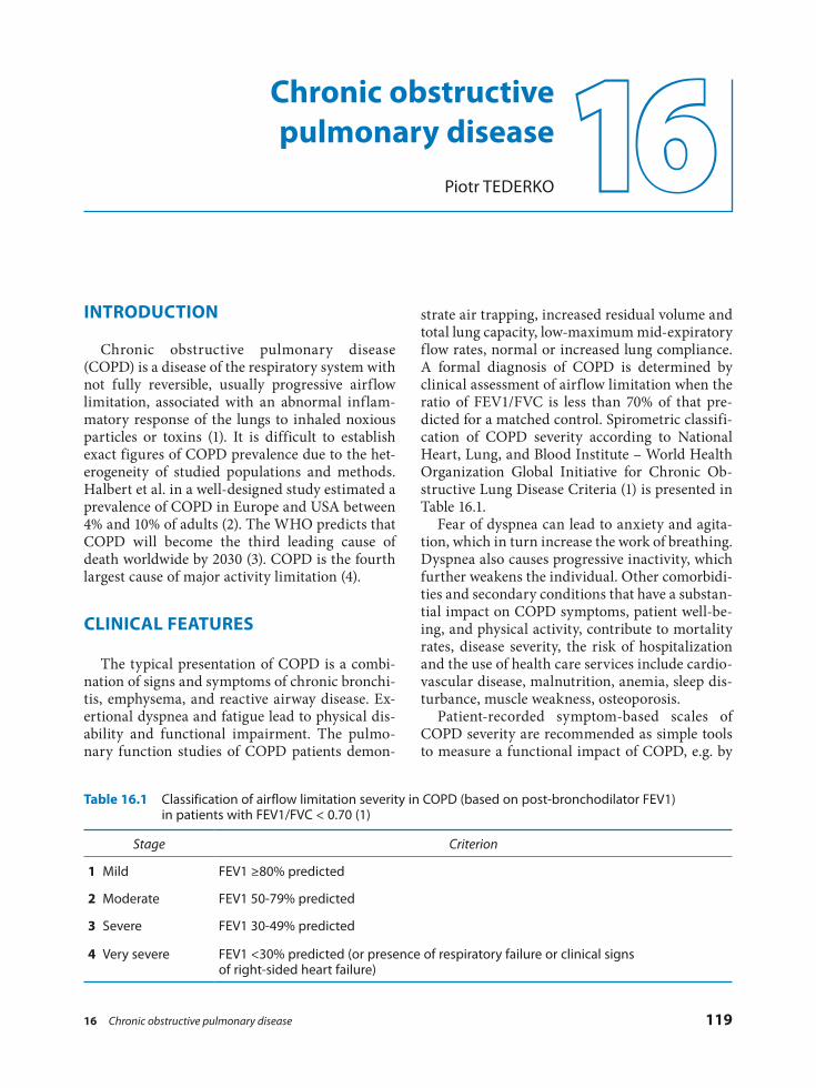
16 Chronic obstructive pulmonary disease 119
strate air trapping, increased residual volume and total lung capacity, low-maximum mid-expiratory flow rates, normal or increased lung compliance. A formal diagnosis of COPD is determined by clinical assessment of airflow limitation when the ratio of FEV1/FVC is less than 70% of that pre-dicted for a matched control. Spirometric classifi-cation of COPD severity according to National Heart, Lung, and Blood Institute – World Health Organization Global Initiative for Chronic Ob-structive Lung Disease Criteria (1) is presented in Table 16.1.
Fear of dyspnea can lead to anxiety and agita-tion, which in turn increase the work of breathing. Dyspnea also causes progressive inactivity, which further weakens the individual. Other comorbidi-ties and secondary conditions that have a substan-tial impact on COPD symptoms, patient well-be-ing, and physical activity, contribute to mortality rates, disease severity, the risk of hospitalization and the use of health care services include cardio-vascular disease, malnutrition, anemia, sleep dis-turbance, muscle weakness, osteoporosis.
Patient-recorded symptom-based scales of COPD severity are recommended as simple tools to measure a functional impact of COPD, e.g. by
INTRODUCTION
Chronic obstructive pulmonary disease (COPD) is a disease of the respiratory system with not fully reversible, usually progressive airflow limitation, associated with an abnormal inflam-matory response of the lungs to inhaled noxious particles or toxins (1). It is difficult to establish exact figures of COPD prevalence due to the het-erogeneity of studied populations and methods. Halbert et al. in a well-designed study estimated a prevalence of COPD in Europe and USA between 4% and 10% of adults (2). The WHO predicts that COPD will become the third leading cause of death worldwide by 2030 (3). COPD is the fourth largest cause of major activity limitation (4).
CLINICAL FEATURES
The typical presentation of COPD is a combi-nation of signs and symptoms of chronic bronchi-tis, emphysema, and reactive airway disease. Ex-ertional dyspnea and fatigue lead to physical dis-ability and functional impairment. The pulmo-nary function studies of COPD patients demon-
16Chronic obstructive pulmonary disease
Piotr TEDERKO
Table 16.1 Classification of airflow limitation severity in COPD (based on post-bronchodilator FEV1) in patients with FEV1/FVC < 0.70 (1)
Stage Criterion
1 Mild FEV1 ≥80% predicted
2 Moderate FEV1 50-79% predicted
3 Severe FEV1 30-49% predicted
4 Very severe FEV1 <30% predicted (or presence of respiratory failure or clinical signs of right-sided heart failure)
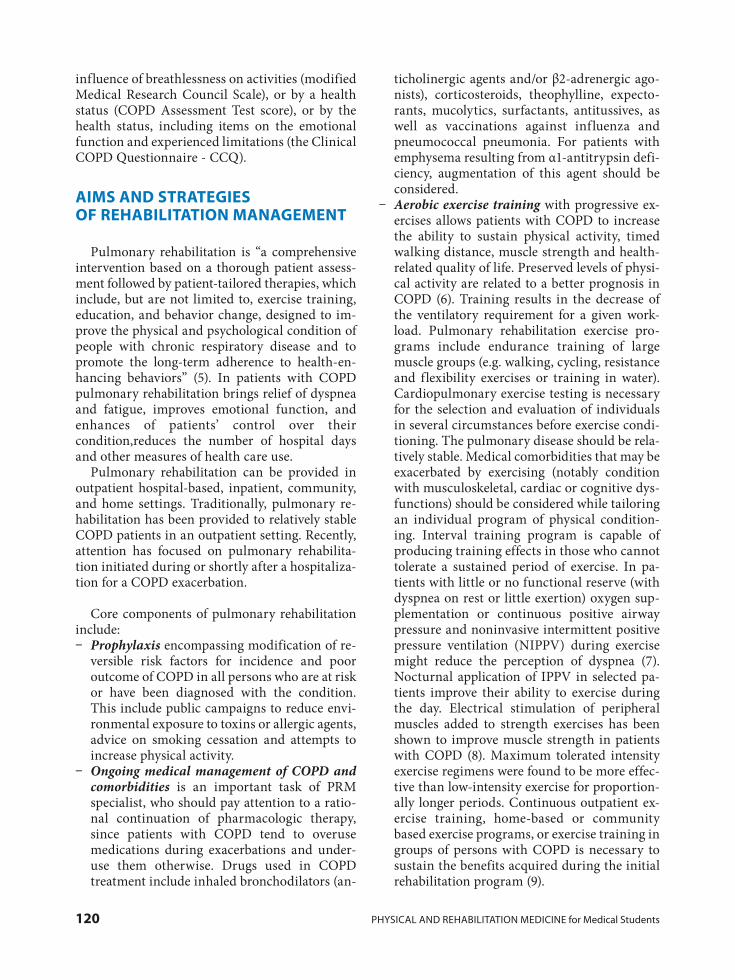
120 PHYSICAL AND REHABILITATION MEDICINE for Medical Students
ticholinergic agents and/or β2-adrenergic ago-nists), corticosteroids, theophylline, expecto-rants, mucolytics, surfactants, antitussives, as well as vaccinations against influenza and pneumococcal pneumonia. For patients with emphysema resulting from α1-antitrypsin defi-ciency, augmentation of this agent should be considered.
ū Aerobic exercise training with progressive ex-ercises allows patients with COPD to increase the ability to sustain physical activity, timed walking distance, muscle strength and health-related quality of life. Preserved levels of physi-cal activity are related to a better prognosis in COPD (6). Training results in the decrease of the ventilatory requirement for a given work-load. Pulmonary rehabilitation exercise pro-grams include endurance training of large muscle groups (e.g. walking, cycling, resistance and flexibility exercises or training in water). Cardiopulmonary exercise testing is necessary for the selection and evaluation of individuals in several circumstances before exercise condi-tioning. The pulmonary disease should be rela-tively stable. Medical comorbidities that may be exacerbated by exercising (notably condition with musculoskeletal, cardiac or cognitive dys-functions) should be considered while tailoring an individual program of physical condition-ing. Interval training program is capable of producing training effects in those who cannot tolerate a sustained period of exercise. In pa-tients with little or no functional reserve (with dyspnea on rest or little exertion) oxygen sup-plementation or continuous positive airway pressure and noninvasive intermittent positive pressure ventilation (NIPPV) during exercise might reduce the perception of dyspnea (7). Nocturnal application of IPPV in selected pa-tients improve their ability to exercise during the day. Electrical stimulation of peripheral muscles added to strength exercises has been shown to improve muscle strength in patients with COPD (8). Maximum tolerated intensity exercise regimens were found to be more effec-tive than low-intensity exercise for proportion-ally longer periods. Continuous outpatient ex-ercise training, home-based or community based exercise programs, or exercise training in groups of persons with COPD is necessary to sustain the benefits acquired during the initial rehabilitation program (9).
influence of breathlessness on activities (modified Medical Research Council Scale), or by a health status (COPD Assessment Test score), or by the health status, including items on the emotional function and experienced limitations (the Clinical COPD Questionnaire - CCQ).
AIMS AND STRATEGIES OF REHABILITATION MANAGEMENT
Pulmonary rehabilitation is “a comprehensive intervention based on a thorough patient assess-ment followed by patient-tailored therapies, which include, but are not limited to, exercise training, education, and behavior change, designed to im-prove the physical and psychological condition of people with chronic respiratory disease and to promote the long-term adherence to health-en-hancing behaviors” (5). In patients with COPD pulmonary rehabilitation brings relief of dyspnea and fatigue, improves emotional function, and enhances of patients’ control over their condition,reduces the number of hospital days and other measures of health care use.
Pulmonary rehabilitation can be provided in outpatient hospital-based, inpatient, community, and home settings. Traditionally, pulmonary re-habilitation has been provided to relatively stable COPD patients in an outpatient setting. Recently, attention has focused on pulmonary rehabilita-tion initiated during or shortly after a hospitaliza-tion for a COPD exacerbation.
Core components of pulmonary rehabilitation include:
ū Prophylaxis encompassing modification of re-versible risk factors for incidence and poor outcome of COPD in all persons who are at risk or have been diagnosed with the condition. This include public campaigns to reduce envi-ronmental exposure to toxins or allergic agents, advice on smoking cessation and attempts to increase physical activity.
ū Ongoing medical management of COPD and comorbidities is an important task of PRM specialist, who should pay attention to a ratio-nal continuation of pharmacologic therapy, since patients with COPD tend to overuse medications during exacerbations and under-use them otherwise. Drugs used in COPD treatment include inhaled bronchodilators (an-
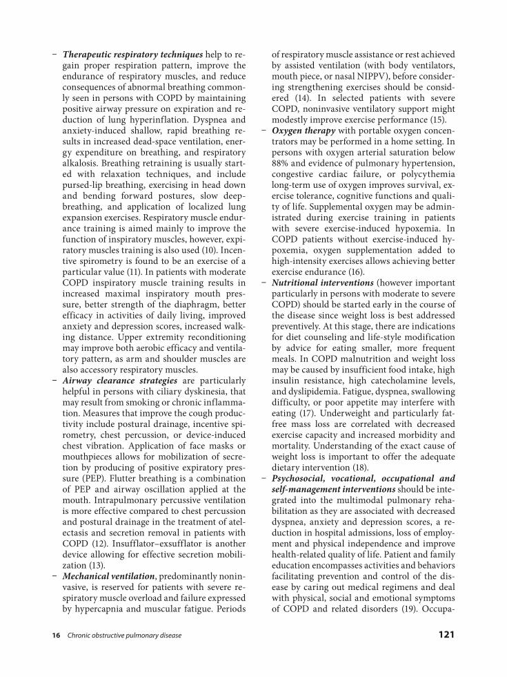
16 Chronic obstructive pulmonary disease 121
of respiratory muscle assistance or rest achieved by assisted ventilation (with body ventilators, mouth piece, or nasal NIPPV), before consider-ing strengthening exercises should be consid-ered (14). In selected patients with severe COPD, noninvasive ventilatory support might modestly improve exercise performance (15).
ū Oxygen therapy with portable oxygen concen-trators may be performed in a home setting. In persons with oxygen arterial saturation below 88% and evidence of pulmonary hypertension, congestive cardiac failure, or polycythemia long-term use of oxygen improves survival, ex-ercise tolerance, cognitive functions and quali-ty of life. Supplemental oxygen may be admin-istrated during exercise training in patients with severe exercise-induced hypoxemia. In COPD patients without exercise-induced hy-poxemia, oxygen supplementation added to high-intensity exercises allows achieving better exercise endurance (16).
ū Nutritional interventions (however important particularly in persons with moderate to severe COPD) should be started early in the course of the disease since weight loss is best addressed preventively. At this stage, there are indications for diet counseling and life-style modification by advice for eating smaller, more frequent meals. In COPD malnutrition and weight loss may be caused by insufficient food intake, high insulin resistance, high catecholamine levels, and dyslipidemia. Fatigue, dyspnea, swallowing difficulty, or poor appetite may interfere with eating (17). Underweight and particularly fat-free mass loss are correlated with decreased exercise capacity and increased morbidity and mortality. Understanding of the exact cause of weight loss is important to offer the adequate dietary intervention (18).
ū Psychosocial, vocational, occupational and self-management interventions should be inte-grated into the multimodal pulmonary reha-bilitation as they are associated with decreased dyspnea, anxiety and depression scores, a re-duction in hospital admissions, loss of employ-ment and physical independence and improve health-related quality of life. Patient and family education encompasses activities and behaviors facilitating prevention and control of the dis-ease by caring out medical regimens and deal with physical, social and emotional symptoms of COPD and related disorders (19). Occupa-
ū Therapeutic respiratory techniques help to re-gain proper respiration pattern, improve the endurance of respiratory muscles, and reduce consequences of abnormal breathing common-ly seen in persons with COPD by maintaining positive airway pressure on expiration and re-duction of lung hyperinflation. Dyspnea and anxiety-induced shallow, rapid breathing re-sults in increased dead-space ventilation, ener-gy expenditure on breathing, and respiratory alkalosis. Breathing retraining is usually start-ed with relaxation techniques, and include pursed-lip breathing, exercising in head down and bending forward postures, slow deep-breathing, and application of localized lung expansion exercises. Respiratory muscle endur-ance training is aimed mainly to improve the function of inspiratory muscles, however, expi-ratory muscles training is also used (10). Incen-tive spirometry is found to be an exercise of a particular value (11). In patients with moderate COPD inspiratory muscle training results in increased maximal inspiratory mouth pres-sure, better strength of the diaphragm, better efficacy in activities of daily living, improved anxiety and depression scores, increased walk-ing distance. Upper extremity reconditioning may improve both aerobic efficacy and ventila-tory pattern, as arm and shoulder muscles are also accessory respiratory muscles.
ū Airway clearance strategies are particularly helpful in persons with ciliary dyskinesia, that may result from smoking or chronic inflamma-tion. Measures that improve the cough produc-tivity include postural drainage, incentive spi-rometry, chest percussion, or device-induced chest vibration. Application of face masks or mouthpieces allows for mobilization of secre-tion by producing of positive expiratory pres-sure (PEP). Flutter breathing is a combination of PEP and airway oscillation applied at the mouth. Intrapulmonary percussive ventilation is more effective compared to chest percussion and postural drainage in the treatment of atel-ectasis and secretion removal in patients with COPD (12). Insufflator–exsufflator is another device allowing for effective secretion mobili-zation (13).
ū Mechanical ventilation, predominantly nonin-vasive, is reserved for patients with severe re-spiratory muscle overload and failure expressed by hypercapnia and muscular fatigue. Periods

122 PHYSICAL AND REHABILITATION MEDICINE for Medical Students
monary disease patients: a randomized controlled trial. Respir Res 2011; 12: 112.
8. Vivodtzev I, Lacasse Y, Maltais F. Neuromuscular electrical stimulation of the lower limbs in patients with chronic obstructive pulmonary disease. J Cardiopulm Rehabil Prev 2008; 28: 79-91.
9. Spruit MA, Pitta F, McAuley E, ZuWallack RL, Nici L. Pulmonary Rehabilitation and Physical Activity in Patients with Chronic Obstructive Pul-monary Disease. Am J Respir Crit Care Med 2015; 192: 924-33.
10. Mota S, Güell R, Barreiro E, et al: Clinical out-comes of expiratory muscle training in severe COPD patients, Respir Med 2007; 101: 516-24.
11. Heydari A, Farzad M, Ahmadi hosseini SH. Com-paring Inspiratory Resistive Muscle Training with Incentive Spirometry on Rehabilitation of COPD Patients. Rehabil Nurs 2015; 40: 243-8.
12. Testa A, Galeri S, Villafañe JH, et al. Efficacy of short-term intrapulmonary percussive ventilation in patients with chronic obstructive pulmonary disease. Disabil Rehabil 2015; 37: 899-903.
13. Homnick DN. Mechanical insufflation-exsuffla-tion for airway mucus clearance. Respir Care 2007; 52: 1296-305.
14. Dubé BP, Vermeulen F, Laveneziana P. Exertional Dyspnoea in Chronic Respiratory Diseases: From Physiology to Clinical Application. Arch Bronco-neumol 2017; 53: 62-70.
15. Ries AL, Bauldoff GS, Carlin BW, et al: Pulmonary rehabilitation: Joint ACCP/AACVPR evidence-
REFERENCES
1. Global Initiative for Chronic Obstructive Lung Disease (GOLD). Global strategy for the diagnosis, management, and prevention of COPD. Available at: http://goldcopd.org/gold-2017-global-strategy-diagnosis-management-prevention-copd/
2. Halbert RJ, Isonaka S, George D, et al. Interpreting COPD prevalence estimates: what is the true bur-den of disease. Chest 2003; 123: 1684-92.
3. World Health Organization. Burden of COPD. Available at: www.who.int/respiratory/copd/bur-den/en.
4. Higgins ITT. Epidemiology of bronchitis and em-physema. In: Fishman AP, ed. Pulmonary Diseases and Disorders. 2nd ed. New York: McGraw-Hill; 1988, pp. 70-90.
5. Spruit MA, Singh SJ, Garvey C, et al; ATS/ERS Task Force on Pulmonary Rehabilitation. An offi-cial American Thoracic Society/European Respi-ratory Society statement: key concepts and ad-vances in pulmonary rehabilitation. Am J Respir Crit Care Med 2013; 188(8): e13-e64.
6. Garcia-Rio F, Rojo B, Casitas R, et al. Prognostic value of the objective measurement of daily physi-cal activity in patients with COPD. Chest 2012; 142(2): 338-46.
7. Duiverman ML, Wempe JB, Bladder G, Vonk JM, Zijlstra JG, Kerstjens HA, Wijkstra PJ. Two-year home-based nocturnal noninvasive ventilation added to rehabilitation in chronic obstructive pul-
quality of life of COPD patients with advanced stable disease (21). Examples of useful aids in-clude motorized scooters and rolling walkers that help to increase distance of locomotion particularly in those who walk less than 300 m and who require a rest during an unaided 6-minute walk. Environmental modifications at home, such as beds with an overhead trapeze, grips, elevated toilet seats and hand rails in a toilets and bathrooms are also useful.
tional therapy can provide patients with severe COPD with skills of energy conservation and techniques for performing activities of daily living. Cognitive-behavioral therapy results in the decrease of anxiety and depression in per-sons with COPD. Patients unable to self-care may be more effectively managed in nursing facilities skilled in respiratory care (20).
ū Application of assistive devices can effectively decrease dyspnea and improve mobility and
Key messages
• Results of pulmonary function tests have no strict correlation with the functional status of the patient with COPD.
• Rehabilitation of a patient with COPD must include maintenance of adequate physical activity, patient’s education, nutritional counseling, effective control of anxiety.
• Increase of effort tolerance and quality of life improvement may be achieved despite the fact that pulmonary rehabilitation has no direct effect on lung function in COPD.

16 Chronic obstructive pulmonary disease 123
19. Ochmann U, Jörres RA, Nowak D. Long-term ef-ficacy of pulmonary rehabilitation: a state-of-the-art review. J Cardiopulm Rehabil Prev 2012; 32: 117-26.
20. Panagioti M, Scott C, Blakemore A, Coventry PA. Overview of the prevalence, impact, and manage-ment of depression and anxiety in chronic ob-structive pulmonary disease. Int J Chron Obstruct Pulmon Dis 2014; 9: 1289-306.
21. Gysels MH, Higginson IJ. Caring for a person in advanced illness and suffering from breathlessness at home: threats and resources. Palliat Support Care 2009; 7: 153-62.
based clinical practice guidelines, Chest 2007; 131: 4S-42S.
16. Mowery NT. Ventilator Strategies for Chronic Ob-structive Pulmonary Disease and Acute Respira-tory Distress Syndrome. Surg Clin North Am 2017; 97: 1381-97.
17. Akner G, Larsson K. Undernutrition state in pa-tients with chronic obstructive pulmonary dis-ease. A critical appraisal on diagnostics and treat-ment. Respir Med 2016; 117: 81-91.
18. Collins PF, Elia M, Stratton RJ. Nutritional sup-port and functional capacity in chronic obstruc-tive pulmonary disease: a systematic review and meta-analysis. Respirology 2013; 18: 616-29.


17 Cardiac rehabilitation for people with cardiovascular diseases 125
There is also data showing that regular exercise has considerable health benefit for people with type 1 diabetes (9).
PHYSIOPATHOLOGY
The pathophysiological conditions that underlie heart disease are hypertension, atherosclerosis within the coronary arteries, altered myocardial muscle mechanics, valvular dysfunction, and ar-rhythmias. The clinical presentation and symp-toms of heart diseases are diverse and depend on the source of the impairment. The main signs and symptoms are chest pressure/pain, dyspnea, fa-tigue, syncope, and palpitations. Whereas patients with the same pathology can experience very dif-ferent clinical presentation and activity limitations, appropriate examination, and evaluation are cru-cial for making a correct diagnosis, choosing the treatment and setting the goals of treatment (10).
TREATMENT
The treatment of heart diseases consists of ei-ther conservative or surgical treatment. There are different options for the treatment of ST-elevation and non-ST-elevation myocardial infarction in acute phase and the choice of treatment depends on various factors such as health condition of pa-tient, contraindications, time from first medical contact, mode of transportation of the patient and capabilities at the receiving hospital, but the main goal is to keep total ischemic time within 120 min-utes. The reperfusion in these patients can be ac-complished by catheter-based approaches, place-ment of a stent (bare-metal stent) or drug-eluting stent, manual aspiration thrombectomy, coronary artery bypass graft surgery, or in the absence of
INTRODUCTION
EpidemiologyCardiovascular diseases are leading causes of
global mortality and morbidity (1, 2) and in high-income countries the mortality rates range from 20-50% (3). In spite of the currently available treatments, deaths from ischaemic heart disease have increased by 16.6% globally over the last de-cade (1). Heart diseases also induce serious dam-age and often lead to physical and neurological consequences, which are often irreversible (2). That’s why primary prevention which can help to prevent coronary artery diseases and secondary prevention which can slow the progression and/or reverse disease progression are very important steps in the treatment of heart diseases (2, 4). Both can be achieved by determining modifiable risk factors and encouraging patients to change their lifestyle (4).
Risk factors for cardiovascular disorders and prevention
The main risk factors for ischaemic heart dis-ease are: smoking, drinking, obesity, low physical activity, hypertension, diabetes, dyslipidemia, psychosocial factors (5). Most of these risk factors we can control and/or reduce by adequate physical activity. Current guidelines recommend the phys-ical activity as a means to prevent hypertension as well as to treat hypertension (6, 7). There is also published data, demonstrating that regular aero-bic exercise raises high density lipoprotein choles-terol levels, decreases total cholesterol, low density lipoprotein cholesterol, triglyceride levels (8). Reg-ular exercise can prevent or delay type 2 diabetes development and improves blood glucose control in people already suffering from type 2 diabetes.
17Cardiac rehabilitation for people with cardiovascular diseases
Alvydas JUOCEVICIUS

126 PHYSICAL AND REHABILITATION MEDICINE for Medical Students
should always pay attention to the screening and treating the depression which is very common in cardiovascular diseases. It affects nearly half (~40%) of those with CHD (20) and even those with peripheral arterial disease with a prevalence ranging from 3 to 48% depending on study type (21). Depression is not only associated with mor-tality in CHD or heart failure but also with worse health-related quality of life (HRQoL), more fre-quent use of health services and costs/economical burden (21).
PRINCIPLES OF CARDIAC REHABILITATION
As a recent review of guidelines for cardiac re-habilitation exercise programmes summarized “strong, consistent positive evidence exists for ex-ercise-based cardiac rehabilitation for patients with stable angina pectoris, myocardial infarction and coronary revascularisation” (2). Cardiac Re-habilitation is also recommended for patients af-ter heart transplantation, valvular surgery and those suffering chronic heart failure (2).
There are three main phases of cardiac rehabilita-tion (10):
ū Phase 1 includes the beginning of rehabilitation while the patient is still in the acute phase in cardiology/cardio surgery service. It lasts until the patient stabilises and can be transferred to a rehabilitation service.
ū Phase 2 takes place in in-patient and/or out-pa-tient settings; the choice between these two de-pends on patient conditions and laws in differ-ent countries. The duration of stay also depends on different factors depending on countries.
ū Phase 3 includes out-patient setting and/or the community-based rehabilitation in the long-term requiring lifelong constitution.
As summarized in the 2014 overview of six Cochrane systematic reviews, exercise-based car-diac rehabilitation reduces hospitalization after the heart attack, percutaneous coronary interven-tion, or heart failure with improvements in HRQoL when compared with usual care (22). Despite the positive effect of cardiac rehabilita-tion, availability of cardiac rehabilitation pro-grammes globally is low (23). There are different reasons for this resulting from either patient or
contraindications, fibrinolytic therapy (11, 12). As soon as the patient clinical status stabilizes the rehabilitation team starts the mobilization of pa-tient, adapts respiratory exercises, etc.
One more patient group which requires conser-vative and/or surgical treatment and rehabilita-tion program afterward is patients suffering from valvular heart diseases. The most common form of valvular heart disease is degenerative valve dis-ease, whereas rheumatic heart disease accounts for most valve pathology in developing nations (13). At some point in the course of the disease, these patients require surgical valve replacement performed with mechanical or bioprosthetic valves (14). The choice between mechanical and biological prosthetic depends on different factors such as life expectancy, the age of a patient, surgi-cal risk, comorbidities, contraindications for anti-thrombotic therapy, patient’s preference (15). Even before the surgery when disease is still asymptom-atic these patients after careful examination and appropriate imagining studies can safely exercise after choosing adequate physical exercise program and even participate in sports as desired (16).
The principles of rehabilitation after the sur-gery remain the same in this patient group except there are some restrictions which are described later on in this chapter.
Coronary heart disease (CHD) is a chronic con-dition and patients who have recovered from a myocardial infarction are at high risk for new events and premature death (17). That’s why be-sides conservative and surgical heart diseases treatment, exercise-based cardiac rehabilitation is the cornerstone in the management of heart dis-eases because it is an essential component to influ-ence the underlying risk factors in order to stabi-lize, slow or reverse disease progression. Cardiac rehabilitation consists of multicomponent and in-terdisciplinary care of patients with a multisystem approach to the patient. Core components include medical and lifestyle risk factors management, patient’s education about physical activity, diet, smoking cessation (medications, behavioral thera-py, etc.), weight control, lipid and blood pressure management, psychosocial support, sexual coun-seling (6, 18). Patients should be informed about risk factors and their correction as soon as possi-ble. Secondary prevention (long-term rehabilita-tion therapies) have to start immediately after the patient’s condition becomes stable (19).
Physical medicine and rehabilitation physicians
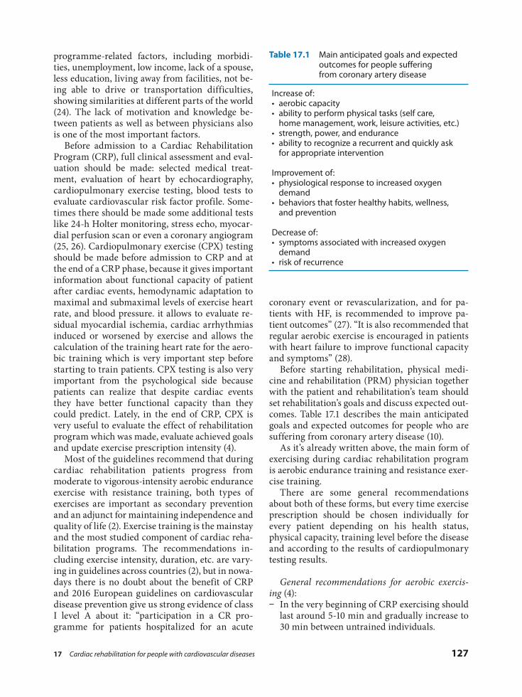
17 Cardiac rehabilitation for people with cardiovascular diseases 127
coronary event or revascularization, and for pa-tients with HF, is recommended to improve pa-tient outcomes” (27). “It is also recommended that regular aerobic exercise is encouraged in patients with heart failure to improve functional capacity and symptoms” (28).
Before starting rehabilitation, physical medi-cine and rehabilitation (PRM) physician together with the patient and rehabilitation’s team should set rehabilitation’s goals and discuss expected out-comes. Table 17.1 describes the main anticipated goals and expected outcomes for people who are suffering from coronary artery disease (10).
As it’s already written above, the main form of exercising during cardiac rehabilitation program is aerobic endurance training and resistance exer-cise training.
There are some general recommendations about both of these forms, but every time exercise prescription should be chosen individually for every patient depending on his health status, physical capacity, training level before the disease and according to the results of cardiopulmonary testing results.
General recommendations for aerobic exercis-ing (4):
ū In the very beginning of CRP exercising should last around 5-10 min and gradually increase to 30 min between untrained individuals.
programme-related factors, including morbidi-ties, unemployment, low income, lack of a spouse, less education, living away from facilities, not be-ing able to drive or transportation difficulties, showing similarities at different parts of the world (24). The lack of motivation and knowledge be-tween patients as well as between physicians also is one of the most important factors.
Before admission to a Cardiac Rehabilitation Program (CRP), full clinical assessment and eval-uation should be made: selected medical treat-ment, evaluation of heart by echocardiography, cardiopulmonary exercise testing, blood tests to evaluate cardiovascular risk factor profile. Some-times there should be made some additional tests like 24-h Holter monitoring, stress echo, myocar-dial perfusion scan or even a coronary angiogram (25, 26). Cardiopulmonary exercise (CPX) testing should be made before admission to CRP and at the end of a CRP phase, because it gives important information about functional capacity of patient after cardiac events, hemodynamic adaptation to maximal and submaximal levels of exercise heart rate, and blood pressure. it allows to evaluate re-sidual myocardial ischemia, cardiac arrhythmias induced or worsened by exercise and allows the calculation of the training heart rate for the aero-bic training which is very important step before starting to train patients. CPX testing is also very important from the psychological side because patients can realize that despite cardiac events they have better functional capacity than they could predict. Lately, in the end of CRP, CPX is very useful to evaluate the effect of rehabilitation program which was made, evaluate achieved goals and update exercise prescription intensity (4).
Most of the guidelines recommend that during cardiac rehabilitation patients progress from moderate to vigorous-intensity aerobic endurance exercise with resistance training, both types of exercises are important as secondary prevention and an adjunct for maintaining independence and quality of life (2). Exercise training is the mainstay and the most studied component of cardiac reha-bilitation programs. The recommendations in-cluding exercise intensity, duration, etc. are vary-ing in guidelines across countries (2), but in nowa-days there is no doubt about the benefit of CRP and 2016 European guidelines on cardiovascular disease prevention give us strong evidence of class I level A about it: “participation in a CR pro-gramme for patients hospitalized for an acute
Table 17.1 Main anticipated goals and expected outcomes for people suffering from coronary artery disease
Increase of:• aerobic capacity• ability to perform physical tasks (self care, home management, work, leisure activities, etc.)
• strength, power, and endurance• ability to recognize a recurrent and quickly ask
for appropriate intervention
Improvement of:• physiological response to increased oxygen
demand • behaviors that foster healthy habits, wellness,
and prevention
Decrease of:• symptoms associated with increased oxygen
demand• risk of recurrence

128 PHYSICAL AND REHABILITATION MEDICINE for Medical Students
resistance exercise training for the lower limbs can be carried out earlier provided a stable trunk positioning is ensured (4). Because of the risk of a restrictive pulmonary impairment and gas exchange abnormalities, deep breathing exercises should be implemented as a major component of postoperative care for these pa-tients and should also continue in a rehabilita-tion’s phase 2 (29).
There is one more form of exercising which is sometimes recommended but still not routinely specified as a component of the cardiac rehabilita-tion program – flexibility training. European guideline does not include it in CR recommenda-tions unlike World Health Organization (2).
As mentioned above in the text next to medica-tion as well as exercising, recommendations about the nutrition and psychological support should al-so be included in a cardiac rehabilitation program. There are more and more studies which prove that certain diet can influence cardiovascular health, that’s why patients should be taught about the im-portance of nutrition and other modifying risk factors as obesity, dyslipidemia, and hypertension.
Psychological care of cardiac patients should help people and their relatives adjust physically and emo-tionally to their illness. Improvement in physical capacity reduces depression and anxiety. Similarly, changes in psychological state may improve adher-ence to medication or exercise regimens (4). There is also established relation between cardiovascular disease and personality: the strongest evidence has emerged for depression, which seems to be both an independent predictor of future cardiovascular dis-ease and a determinant of morbidity, adaptation and quality of life after an acute coronary syndrome and coronary artery bypass surgery (30).
Cardiac rehabilitation is a multisystem approach to a cardiac patient which requires teamwork start-ing by a cardiologist and/or cardio surgeons and taking over the work by physical medicine and re-habilitation doctors and all the specialists from rehabilitation’s team: physiotherapist, occupational therapists, psychologist, nutritionist, nurse, etc. It’s a very important step to understand for both sides doctors and patients that treatment of heart dis-eases does not end up with a surgery and medica-ments, it must continue the rest of patient’s life, and applying adequate rehabilitation program, changing persons lifestyle, secondary preventing progression of their diseases.
ū Aerobic endurance training should be per-formed for ≥30 min 3-5 times per week, prefer-ably every day, resulting in a total exercise time of ≥150 min/week (or 2.5 h/week).
ū European and United States guidelines recom-mend that patients progress from moderate to vigorous-intensity aerobic exercise, increasing to 80% of the maximal aerobic capacity (VO2 max) or heart rate max over the rehabilitation course.
ū There could be used different forms of endur-ance training like ergometer training on a cycle or treadmill, walking, Nordic walking, biking, jogging, if tolerated swimming. For choosing a training form, the physician should consider individual’s baseline characteristics as well as preference and motivation by a patient.
ū Low-intensity physical activities, as for example level-ground walking, should be done on a daily basis.
The main goal of resistance exercise training is to increase the strength of muscles by performing static and/or dynamic muscle contractions by in-creasing muscle mass and/or improving coordina-tion and metabolism of muscle (4).
General recommendations for resistance exer-cise training (4):
ū This type of exercising should be included in phase II and phase III cardiac rehabilitation program and is contraindicated in phase I (acute phase in the hospital).
ū Resistance exercises should be started after 4-6 sessions of continuous endurance training.
ū Resistance load is prescribed as a percentage of patient’s maximum strength or based on the patient’s level of fatigue while exercising (2).
ū Low-intensity resistance exercise training should not be started earlier than 2 weeks post myocardial infarction and/or 7 days post-inter-ventional revascularization (4).
ū For patients recovering from coronary artery bypass surgery and other open heart surgery, there are more restrictions talking about resis-tance exercising, because the wound healing after thoracotomy takes around 4-6 weeks and physical exercise creates tangential vector forces in and around the sternum, that’s why resis-tance training should be avoided for at least 3 months.
ū If the patient after open heart surgery is stable and has a good exercise tolerance, low-intensity

17 Cardiac rehabilitation for people with cardiovascular diseases 129
lines of the European Society of Cardiology. Eur J Prev Cardiol 2014; 21: 664-81.
7. Diaz KM, Shimbo D. Physical activity and the prevention of hypertension. Curr Hypertens Rep 2013; 15: 659-68.
8. Halbert JA, Silagy CA, Finucane P, Withers RT, Hamdorf PA. Exercise training and blood lipids in hyperlipidemic and normolipidemic adults: a me-ta-analysis of randomized, controlled trials. Eur J Clin Nutr 1999; 53: 514-22.
9. Colberg SR, Sigal RJ, Yardley JE, Riddell MC, Dunstan DW, Dempsey PC et al. Physical Activity/Exercise and Diabetes: A Position Statement of the American Diabetes Association. Diabetes Care 2016; 39: 2065-79.
10. Dias KJ. Heart Disease. In: O‘Sullivan SB, Schmitz TJ, Fulk GD. Physical Rehabilitation. 6th ed. Phil-adelphia: F.A. Davis; 2014, p. 523-76.
11. O’Gara PT, Kushner FG, Ascheim DD, Casey DE Jr, Chung MK, de Lemos JA, et al. 2013 ACCF/AHA guideline for the management of ST-eleva-tion myocardial infarction: a report of the Ameri-can College of Cardiology Foundation/American Heart Association Task Force on Practice Guide-lines. Circulation 2013; 127: e362-425.
12. Amsterdam EA, Wenger NK, Brindis RG, Casey DE Jr, Ganiats TG, Holmes DR Jr, et al. 2014 AHA/ACC guideline for the management of patients with non-ST-elevation acute coronary syndromes: a report of the American College of Cardiology/American Heart Association Task Force on Prac-tice Guidelines. Circulation 2014; 130: e344-426.
13. Maganti K, Rigolin VH, Sarano ME, Bonow RO. Valvular Heart Disease: Diagnosis and Manage-ment. Mayo Clin Proc 2010; 85: 483-500.
REFERENCES
1. GBD 2015 Mortality and Causes of Death Collabo-rators. Global, regional, and national life expec-tancy, all-cause mortality, and cause-specific mor-tality for 249 causes of death, 1980-2015: a system-atic analysis for the Global Burden of Disease Study 2015. Lancet 2016; 388: 1459-544.
2. Price KJ, Gordon BA, Bird SR, Benson AC. A re-view of guidelines for cardiac rehabilitation exer-cise programmes: Is there an international con-sensus? Eur J Prev Cardiol 2016; 23: 1715-33.
3. Bjarnason-Wehrens B, McGee H, Zwisler AD. Car-diac rehabilitation in Europe: Results from the Eu-ropean Cardiac Rehabilitation Inventory Survey. Eur J Cardiovasc Prev Rehabil 2010; 17: 410-8.
4. Niebauer J, editor. Cardiac rehabilitation manual. London: Springer; 2011.
5. Task Force of the European Society of Cardiology and Other Societies on Cardiovascular Disease Prevention in Clinical Practice (constituted by representatives of nine societies and by invited experts)Developed with the special contribution of the European Association for Cardiovascular Pre-vention & Rehabilitation (EACPR). Eur Heart J 2012; 33: 1635-701.
6. Piepoli MF, Corrà U, Adamopoulos S, Benzer W, Bjarnason-Wehrens B, Cupples M. Secondary pre-vention in the clinical management of patients with cardiovascular diseases. Core components, standards and outcome measures for referral and delivery: a policy statement from the cardiac reha-bilitation section of the European Association for Cardiovascular Prevention & Rehabilitation. Endorsed by the Committee for Practice Guide-
Key messages
• Rehabilitation programmes for patients with cardiovascular conditions involve PRM physicians in leading exercise-based cardiac rehabilitation programmes jointly with cardiologists or other medical specialists trained in cardiac rehabilitation and with a competent team in in- and out-patient settings, home, and in the community to reduce impairments in function, activity limita-tions, and participation restrictions associated with these conditions.
• PRM physicians perform a complete bio-psycho-social evaluation of the individual patient with cardiovascular conditions, that also includes the identification of a series of emotional and social factors (such as -for example- psychological conditions, environmental factors, social support and relationships, attitudes of care providers and health professionals, as well as organization of local healthcare services) in relation to supported self-management, use of information technology for health promotion/lifestyle changes, well-being of family caregivers, and improved coordination among health professionals, in order to enhance motivation, exercise adherence and quality of cardiac rehabilitation programmes.
130 PHYSICAL AND REHABILITATION MEDICINE for Medical Students
systematic reviews. Cochrane Database Syst Rev 2014; 12: CD011273.
23. Turk-Adawi K, Sarrafzadegan N, Grace SL. Global availability of cardiac rehabilitation. Nat Rev Car-diol 2014; 11: 586-96.
24. Ruano-Ravina A, Pena-Gil C, Abu-Assi E, Ra-poseiras S, van ‚t Hof A, Meindersma E, et al. Participation and adherence to cardiac rehabilita-tion programs. A systematic review. Int J Cardiol 2016; 223: 436-43.
25. Giannuzzi P, Mezzani A, Saner H. Working Group on Cardiac Rehabilitation and Exercise Physiolo-gy. Physical activity for primary and secondary prevention. Position paper of the Working Group on Cardiac Rehabilitation and Exercise Physiology of the European Society of Cardiology. Eur J Car-diovasc Prev Rehabil. 2003; 10: 319-27.
26. American Association for Cardiovascular and Pul-monary Rehabilitation. Guidelines for Cardiac Rehabilitation and Secondary Prevention Pro-grams. 4th ed. Champaign: Human Kinetics; 2004.
27. Piepoli MF, Hoes AW, Agewall S, Albus C, Brotons C, Catapano AL, et al. 2016 European Guidelines on Cardiovascular Disease Prevention in Clinical Practice: The Sixth Joint Task Force of the Euro-pean Society of Cardiology and Other Societies on Cardiovascular Disease Prevention in Clinical Practice (constituted by Representatives of 10 Soci-eties and by Invited Experts) Developed with the Special Contribution of the European Association for Cardiovascular Prevention & Rehabilitation (EACPR). Eur Heart J. 2016; 37: 2315-81.
28. Ponikowski P, Voors AA, Anker SD, Bueno H, Cleland JG, Coats AJ, et al. 2016 ESC Guidelines for the Diagnosis and Treatment of Acute and Chronic Heart Failure: The Task Force for the Di-agnosis and Treatment of Acute and Chronic Heart Failure of the European Society of Cardiol-ogy (ESC) Developed with the Special Contribu-tion of the Heart Failure Association (HFA) of the ESC. Eur Heart J 2016; 37: 2129-200.
29. Westerdahl E. Optimal Technique for Deep Breathing Exercises after Cardiac Surgery. Miner-va Anestesiol 2015; 81: 678-83.
30. Steptoe A, Molloy GJ. Personality and Heart Dis-ease. Heart 2007; 93: 783-4.
14. Joint Task Force on the Management of Valvular Heart Disease of the European Society of Cardiol-ogy (ESC), European Association for Cardio-Tho-racic Surgery (EACTS), Vahanian A, et al. Guide-lines on the management of valvular heart disease (version 2012). Eur Heart J 2012; 33: 2451.
15. Nishimura RA, Otto CM, Bonow RO, Carabello BA, Erwin JP 3rd, Fleisher LA, et al. 2017 AHA/ACC Focused Update of the 2014 AHA/ACC Guideline for the Management of Patients With Valvular Heart Disease: A Report of the American College of Cardiology/American Heart Associa-tion Task Force on Clinical Practice Guidelines. Circulation 2017; 135: e1159-95.
16. Parker MW, Thompson PD. Exercise in Valvular Heart Disease: Risks and Benefits. Progr Cardio-vasc Dis 2011; 53: 437-46.
17. Fox KA, Carruthers KF, Dunbar DR, Graham C, Manning JR, De Raedt H, Buysschaert I, et al. Un-derestimated and underrecognized: the late conse-quences of acute coronary syndrome (GRACE UK-Belgian Study). Eur Heart J 2010; 31: 2755-64.
18. Steinke EE, Jaarsma T, Barnason SA, Byrne M, Doherty S, Dougherty CM, et al. Council on Car-diovascular and Stroke Nursing of the American Heart Association and the ESC Council on Car-diovascular Nursing and Allied Professions (CCNAP). Sexual counselling for individuals with cardiovascular disease and their partners: a con-sensus document from the American Heart Asso-ciation and the ESC Council on Cardiovascular Nursing and Allied Professions (CCNAP). Eur Heart J 2013; 34: 3217-35.
19. European Society of Cardiology. Guidelines & Scientific documents [Internet]. 2015 [cited 2017 May 14]. Available from: http://www.escardio.org/guidelines
20. Dickens C. Depression in people with coronary heart disease: prognostic significance and mecha-nisms. Curr Cardiol Rep 2015; 17: 83.
21. Brostow DP, Petrik ML, Starosta AJ, Waldo SW. Depression in patients with peripheral arterial disease: A systematic review. Eur J Cardiovasc Nurs 2017; 16: 181-93.
22. Anderson L, Taylor RS. Cardiac rehabilitation for people with heart disease: an overview of Cochrane
18 Cancer diseases 131
tion to the sequelae associated with cancer and its treatment (6). Cancer survivors may also experi-ence return to work difficulties due to chronic and persistent physical and emotional problems such as fatigue, pain, cognitive deficits, anxiety and depression. Eighteen months after diagnosis, a third of all cancer survivors did not succeed in returning to work (7) and overall cancer survivors have 1.4 times higher risk of being unemployed compared with healthy controls (8). It has been documented that cancer survivors’ health-related quality of life (HR-QoL) is much worse compared with population norms. HR-QoL is more often influenced by physical issues than emotional problems; approximately 25% of cancer patients report poor physical health whereas 10% report poor mental health (9).
THE ROLE OF REHABILITATION
Cancer rehabilitation is a coordinated, interdis-ciplinary medical care aiming to enable people with cancer to achieve optimal physical, psycho-logical, social and vocational functioning within the limits imposed by the disease and its treatment and engage in personally valued activities within their social contexts (10, 11). It should be integrat-ed throughout the oncology care continuum and delivered by trained rehabilitation professionals who have it within their scope of practice to diag-nose and treat patients’ physical, psychological and cognitive impairments in an effort to main-tain or restore function, reduce symptom burden, maximize independence and improve QoL (11). Evidence suggests that cancer patients have many unmet needs for rehabilitation services (9).
Rehabilitation interventions can be preventive, restorative, supportive or palliative depending on the phases of the cancer (11). Preventive rehabili-
INTRODUCTION
Cancer is a major public health issue, being the second leading cause of death worldwide. Global Burden of Disease Study results show that between 2005 and 2015, incident cancer cases increased by 33%, of which 12.6% were due to population growth, 16.4% due to an aging population, and 4.1% due to increasing age-specific incidence rates (1). Survival rates of cancer have been increasing as a result of several factors including successful early detection efforts, advances in diagnosis, treatment and supportive care, as well as life-style changes and the development of prevention vac-cines for some cancers (2). Five-year survival rate for all cancers has been increased to 67% (3).
EPIDEMIOLOGY
Despite increased survival rates, cancer survi-vors can experience various physical, cognitive and emotional impairments resulting from both the malignancy itself and from cancer-related treatment. Invasive surgery, powerful chemother-apy, and high doses of therapeutic radiation might cause long-term impairments of organ system functions, which in turn may limit physical per-formance and result in the inability to carry out routine daily activities. Over half of the cancer survivors report physical performance limita-tions; one third report participation restrictions. Limitations in activities and participation present many years following cancer diagnosis, even among survivors who are not elderly (4). Diffi-culty with ambulation and balance are the most frequently reported functional problems (5). Over half of all cancers occur in individuals over 65 years of age, and comorbidities of aging have sig-nificant negative impact on functioning in addi-
18Cancer diseasesAyşe A. KÜÇÜKDEVECI

132 PHYSICAL AND REHABILITATION MEDICINE for Medical Students
fection, fluid and electrolyte imbalances, anemia or cardiac dysfunction, cannot be identified and corrected, non-pharmacologic and pharmacologic treatment should be considered within the context of the patient’s clinical status (i.e., active cancer treat ment, post-treatment, end-of-life care). Non-pharmacologic interventions may include a mod-erate exercise program to improve functional ca-pacity and activity tolerance; psychosocial pro-grams to manage stress and increase support; implemen tation of energy conservation strategies; and nutri tional and sleep interventions as appro-priate. Pharmacologic therapy may include drugs, such as antidepressants for depression or levothy-roxine for hypothyroidism. The use of the psycho-stimulant methylpheni date may provide some benefit (15).
Pain: about half of cancer patients experience pain, which is usually due to their primary cancer as well as side effects of the treatment and other comorbidities (16). Pain might be moderately se-vere and affects functional status and QoL. Ade-quate pain control is a prerequisite for successful rehabilitation. Thorough evaluation requires as-sessment of all relevant pain etiologies and patho-physiologic processes. Pain control might require the integrated use of anticancer treatments, anal-gesic medication (non-steroidal anti-inflammato-ry drugs, opioids), physical modalities (e.g. TENS) and massage, interventional techniques (e.g. nerve blocks, spinal analgesia) and complementary ther-apies (e.g. acupuncture) (14).
Bony lesions due to metastases: bony meta-static disease is an important impairment which requires the active involvement of the rehabilita-tion team. Insidious pain, intensifying at rest is the common symptom. Fracture risk is increased and pathological fractures are quite likely to hap-pen. Median survival for patients with bone me-tastases is nearly 2-3 years. During this time, ap-propriate measures to decrease morbidity and pain, and improve function should be employed. Multidisciplinary collaboration including physi-cal and rehabilitation medicine, orthopaedic and spine surgery, and medical and radiation oncolo-gy should aim systematic disease management, pain control, skeletal stabilisation and rehabilita-tion (9, 12). Systemic management includes che-motherapy, hormonal therapy, monoclonal anti-bodies, anti-angiogenesis agents, biphosphonates
tation which is also called “prehabilitation” occurs between the time of cancer diagnosis and the be-ginning of acute cancer treatment and aims to prevent and reduce the severity of existing and anticipated treatment-related impairments, such as preoperative strengthening and endurance ex-ercises to buffer the potentially detrimental effects of immobility, respiratory exercises and training before lung resection surgery, or pelvic floor muscle training before prostatectomy (9).
Heterogeneity of cancer types creates very di-verse rehabilitation needs for cancer survivors (12). Rehabilitation needs should be determined after a thorough assessment of the patient regard-ing impairments, activity limitations, and partici-pation restrictions as well as personal and envi-ronmental factors. The impact of impairments on disability should be addressed and the phase of the cancer should be taken into consideration for making the appropriate rehabilitation plan. Reha-bilitative interventions should be patient-centred and impairment-driven aiming to improve func-tioning and QoL of the individual (11).
REHABILITATION AND MANAGEMENT OF COMMON CANCER-RELATED IMPAIRMENTS OF BODY FUNCTIONS /BODY STRUCTURES
Fatigue: cancer-related fatigue (CRF) is a dis-tressing, persistent, subjective sense of physical, emotional and/or cognitive tiredness related to cancer or cancer treatment that is not proportional to recent activity and interferes with usual func-tioning. CRF is experienced by the vast majority of patients (80%) during active cancer treatment and may last for a long time even after completion of the therapy. Clinically significant levels of CRF have been shown to exist in 1/3 of cancer survivors up to 6 years post-treatment, being associated with high levels of disability (13). Responsible mecha-nisms are multifactorial and underlying factors are pain, anemia, sleep disturbance, emotional dis-tress, deconditioning, nutritional deficits/imbal-ance, medical comorbidities and centrally acting medications (12, 14). Cancer patients should be screened routinely for the presence of fatigue and underlying factors. Management of CRF is cause-specific when underlying factors can be identified and treated (15). When specific causes, such as in-
18 Cancer diseases 133
and spasticity (12). Radiotherapy-induced neuro-logical impairments include myelopathy, plexopa-thy, radiculopathy, peripheral nerve injury and encephalopathy. These impairments usually pres-ent as late adverse effects of the radiation. Medical treatment of radiation-induced neural compro-mise can include short-term steroids, anticoagula-tion and/or hyperbaric oxygen therapy. Rehabili-tative interventions to improve functioning and QoL are administered depending on patient’s problems and needs in daily activities (14).
Radiotherapy-induced musculoskeletal im-pairments: radiation-induced fibrosis can affect skin and musculoskeletal tissues and results in dermal fibrosis, musculo-tendinous contraction, loss of joint range of motion and function, and loss of muscle mass depending on the treatment site. Most commonly affected joints are the gleno-humeral joint after axillary radiotherapy for breast cancer, the neck after radiation for head & neck cancer, and the femoro-acetabular joint after radiotherapy of the pelvic region. Conservative management with meticulous skin care, soft-tis-sue mobilisation and stretching exercises may help to increase range of motion. Pentoxifylline may help to restore microvascular supply of the tissues. Physical and occupational therapy, and the use of appropriate orthoses will help to im-prove the function of the affected body part as well activities of daily living (9, 12, 17).
Lymphedema: lymphedema is an abnormal col-lection of protein-rich fluid in the interstitium due to the obstruction of lymphatic drainage, resulting in swelling typically in an extremity but it may also affect face, neck and the torso. It frequently complicates cancer therapy after resection or irra-diation of axillary, inguinal and pelvic lymph nodes and vessels. Most common malignancies associated with lymphedema are breast, melano-ma, gynecological malignancies and lymphoma (12). Lymphedema progresses in stages: The first is the fluid stage, the second with subcutaneous fi-brosis, and the third with cutaneous fibrosis and verrucous hyperplastic changes of the skin. All interventions for lymphedema must have the goals of inducing and maintaining volume reduction, preventing medical complications, improving skin condition, reducing infection, enhancing patient adherence, and improving and maintaining func-tion and QoL (18). Complete (complex) deconges-
and radiation therapy. Pathological fractures are generally managed through well-established sur-gical algorithms (14). Rehabilitation focuses on protection, energy conservation and pain control through the use of orthoses and assistive devices as well as environmental modifications. Exercise prescriptions should aim increasing strength, en-durance and function with minimal loading or torsion of the affected skeleton (12).
Neurological impairments: neurological im-pairments, which have been identified in 30-46% of cancer patients may result from either direct effect of the primary or metastatic cancer or as a consequence of chemotherapy, radiation or sur-gery (12). Neurological deficits due to brain tu-mors or metastases include headaches, seizures, paresis, ataxia, cognitive dysfunction and visual impairment (9). Rehabilitation programme is planned according to patient’s impairments and needs, and may focus on enhancement of mobili-ty, transfers and self-care activities. Orthoses and assistive devices should be prescribed where nec-essary. Neuropsychological assessment and neu-rocognitive interventions to improve memory, at-tention and executive functions might be needed for patients with cognitive dysfunction resulting from either brain tumors or chemotherapy-in-duced mild cognitive impairment. Polyneuropa-thy is a prevalent neurological condition among cancer patients, resulting commonly from chemo-therapy as well as nutritional deficiencies and paraneoplastic disorders (12, 9). Clinical features consist of dysesthesia, loss of sensation and pro-prioception, and motor dysfunction. Rehabilita-tion principles include education, safety aware-ness, gait, balance and proprioceptive training, prescription of orthoses and assistive devices. Spinal cord injury due to metastatic disease oc-curs in 5% to 14% of all cancer patients. Thoracic spine is the most frequently involved site with clinical features of paraparesis/paraplegia, senso-ry level deficit, and bladder and bowel dysfunc-tion. Chemotherapy, radiotherapy, surgical de-compression and stabilisation might be required. Rehabilitation principles of spinal cord injury medicine is applied including physical and occu-pational therapy to enhance physical functioning, as well as rehabilitation of neurogenic bladder and bowel, and prevention and management of com-plications such as pulmonary and urinary tract infections, pressure ulcers, autonomic dysreflexia
134 PHYSICAL AND REHABILITATION MEDICINE for Medical Students
lenging impairments within the scope of cancer rehabilitation. Common rehabilitation problems include spinal accessory nerve palsy, shoulder dysfunction, cervical dystonia/contracture, com-munication disorders, dysphagia, and lymphede-ma of the neck and face (14). These impairments evolve during the course of treatment and reha-bilitative interventions must be adjusted accord-ingly. Spinal accessory nerve palsy affects the function of trapezius and results in shoulder dys-function. Rehabilitation interventions include range of motion and flexibility exercises to keep the mobility of shoulder and anterior chest wall, neuromuscular retraining and strengthening, and postural modification. For cervical dystonia/con-tracture/neuropathy, nerve stabilizing agents such as pregabalin, gabapentin and duloxetine, or bot-ulinum toxin injections must be required in addi-tion to physical therapy and exercises (21). Com-munication disorders include dysartria, dyspho-nia, and aphonia; and speech and language thera-py is needed to improve communication. Instru-mental evaluation of swallowing is needed for patients presenting with complaints of dysphagia. Pharyngeal phase of swallowing is usually affect-ed by surgical disruption or radiation fibrosis. Primary management for pharyngeal dysphagia is airway protection techniques and dietary modifi-cation (12, 21). Other cancer-related impairments of head and neck cancer survivors should be man-aged as explained above.
Prostate cancer: prostate cancer is the most common malignancy in men. Disabilities from bone metastases and complications of androgen deprivation are major factors in morbidity. Com-plications from androgen deprivation include loss of lean body mass, fatigue, and osteoporosis. Loss of lean body mass can be diminished by resistance training. About one third of patients develop an osteoporotic fracture within five years of initiat-ing androgen deprivation therapy. Preventive management strategies include bisphosphonates, exercises and, bone protection techniques (12). Other cancer-related impairments of prostate cancer survivors are managed as explained above.
Lung cancer: lung malignancies are common and might present with challenging problems rel-evant to rehabilitation, such as fatigue, decon-ditioning, pain, neurological complications (e.g. peripheral polyneuropathy, brachial plexopathy),
tive therapy (CDT) is the current gold standard therapy for the management of lymphedema that has been shown to be safe and effective. CDT is a 2-phase multi-modal method that incorporates manual lymphatic drainage (MLD); multi-layer, short-stretch compression bandaging; lymphatic remedial exercises; skin care; and education in lymphedema self-management, and elastic com-pression garments. CDT consists of an initial re-ductive phase (Phase I) followed by a maintenance phase (Phase II). Phase I includes a 3-8 week ther-apy of daily sessions of MLD and compression bandaging with remedial exercises, aiming to re-duce the size of the affected limb and improve the skin. After Phase I, the person needs to continue into Phase II, an ongoing, individualized self-management phase to maintain the gains of Phase I in long term, including daily use of compression garments and overnight bandaging with exercises (18). Weight loss is recommended as well as Inter-mittent Pneumatic Compression as an adjunct to CDT in Phase I (18, 19).
REHABILITATION APPROACHES IN SPECIFIC CANCER POPULATIONS
Breast cancer: breast cancer is the most com-mon malignancy in women and still leads to sig-nificant morbidity despite improvement in early diagnosis and management methods. The most common problems requiring rehabilitation in-clude postsurgical and other pain syndromes, re-gional shoulder dysfunction, and lymphedema of the upper limb. Shoulder dysfunction manifests itself as rotator cuff dysfunction, frozen shoulder, or myofascial pain resulting from immobilisation, muscle weakness and atrophy, mechanical altera-tions in the shoulder girdle musculature, radia-tion therapy and/or neurological impairment (12). Rehabilitative interventions should ideally begin preoperatively with home exercises for mobility and shoulder muscles strengthening. Postopera-tive early mobilisation of the upper extremity, followed by active range of motion and stretching exercises, and finally progressive resistive exer-cises are recommended (20). Other cancer-related impairments of breast cancer survivors should be managed as explained above.
Head and neck cancer: treatment of head and neck cancer might produce some of the most chal-

18 Cancer diseases 135
activity appears to exert a protective effect against the development of some type of cancers, most notably colon and breast cancers. Cancer preven-tion recommendations include at least 30 minutes of moderately vigorous physical activity on 5 or more days of the week for adults (12). Available guidelines for cancer survivors broadly focus on the well-being of cancer survivors, encouraging 150 minutes/week of aerobic exercise, 2days/week of strength training, and flexibility exercise on days when aerobic or resistance exercise is not per-formed. Exercise has been shown to be safe for cancer survivors and exercise programs should be adapted for the individual based on the health sta-tus, cancer treatment type, targeted health out-comes and disease trajectory (22).
brain and spine metastases, or myastenic syn-drome. Postoperative pulmonary rehabilitation interventions are needed both at the acute and post-acute stage to improve patients’ impaired pulmonary function (12). Other cancer-related impairments of lung cancer survivors are man-aged as explained above.
EXERCISE FOR CANCER
Evidence shows that exercise interventions for cancer patients have consistently substantiated gains in numerous parameters, including cardio-pulmonary fitness, fatigue, depression, anxiety, physical functioning, and QoL (10, 12, 14). Physical
Key messages
• Despite increased survival rates of all cancers, cancer survivors can experience various physical, cognitive and emotional impairments resulting from both the malignancy itself and from cancer-related treatment.
• The aim of cancer rehabilitation is to enable people with cancer to achieve optimal physical, psy-chological, social and vocational functioning within the limits imposed by the disease and its treatment, and engage in personally valued activities within their social contexts.
• Various therapeutic and rehabilitative interventions might be needed in order to manage com-mon cancer-related impairments such as pain, fatigue, lymphedema or neurological/musculo-skeletal problems, and to optimise activities and participation.
• Individually tailored physical activity and exercise programme will be of great benefit for cancer survivors to improve physical and cognitive functioning, as well as to enhance quality of life.
REFERENCES
1. Global Burden of Disease Cancer Collabora-tion, Fitzmaurice C, Allen C, Barber RM, Barre-gard L, Bhutta ZA, et al. Global, regional, and na-tional cancer incidence, mortality, years of life lost, years lived with disability, and disability-ad-justed life-years for 32 cancer groups, 1990 to 2015: A systematic analysis for the global burden of dis-ease study. JAMA Oncol. 2017; 3: 524-48.
2. Tiwari AK, Roy HK.Progress against cancer (1971-2011): how far have we come? J Intern Med. 2012; 271: 392-9.
3. Howlader N, Noone AM, Krapcho M, Miller D, Bishop K, Kosary CL, et al. (eds). SEER Cancer Statistics Review, 1975-2014. Bethesda, MD (USA): National Cancer Institute. Available from: https://seer.cancer.gov/csr/1975_2014
4. Ness KK, Wall MM, Oakes JM, Robison LL, Gur-ney JG. Physical performance limitations and participation restrictions among cancer survivors: A population-based study. Ann Epidemiol. 2006; 16: 197-205.
5. Cheville AL, Beck LA, Petersen TL, Marks RS, Gamble GL. The detection and treatment of can-cer-related functional problems in an outpatient setting. Support Care Cancer. 2009; 17: 61-7.
6. Deimling GT, Sterns S, Bowman KF, Kahana B. Functioning and activity participation restrictions among older adult, long-term cancer survivors. Cancer Invest 2007; 25: 106-16.
7. Mehnert A. Employment and work-related issues in cancer survivors. Crit Rev Oncol Hematol. 2011; 77: 109-30.
8. de Boer AG, Taskila T, OjajärviA, vanDijk FJ, Verbeek JH. Cancer survivors and unemployment:
136 PHYSICAL AND REHABILITATION MEDICINE for Medical Students
Netw 2015; 13: 1012-39. 16. Marcus DA. Epidemiology of cancer pain.
Curr Pain Headache Rep. 2011; 15: 231-4. 17. Hojan K, Milecki P. Opportunities for rehabilita-
tion of patients with radiation fibrosis syndrome. Rep Pract Oncol Radiother. 2013; 19: 1-6.
18. NLN Medical Advisory Committee. Position Statement of the National Lymphedema Network - The Diagnosis and Treatment of Lymphedema, 2011 February. Available from: https://www.lym-phnet.org/pdfDocs/position.papers/Diagnosis.Treatment.pdf
19. Rogan S, Taeymans J, Luginbuehl H, Aebi M, Mahnig S, Gebruers N. Therapy modalities to re-duce lymphoedema in female breast cancer pa-tients: a systematic review and meta-analysis. Breast Cancer Res Treat. 2016; 159: 1-14.
20. Harris SR, Schmitz KH, Campbell KL, McNeely ML.Clinical practice guidelines for breast can-cer rehabilitation: syntheses of guideline recom-mendations and qualitative appraisals. Cancer 2012; 118 Suppl S8: 2312-24.
21. Cohen EE, LaMonte SJ, Erb NL, Beckman KL, Sadeghi N, Hutcheson KA, et al American Cancer Society Head and Neck Cancer Survivorship Care Guideline. CA Cancer J Clin. 2016; 66: 203-39.
22. Schmitz KH,Courneya KS, Matthews C, Demark-Wahnefried W, Galvão DA, Pinto BM, et al. American College of Sports Medicine roundtable on exercise guidelines for cancer survivors. Med Sci Sports Exerc. 2010; 42: 1409-26.
a meta-analysis and meta-regression. JAMA 2009; 301: 753-62.
9. Silver JK, Baima J, Mayer S. Impairment-driven cancer rehabilitation: an essential component of quality care and survivorship.CA Cancer J Clin. 2013; 63: 295-317.
10. Egan MY, McEwen S, Sikora L, Chasen M, Fitch M, Eldred S. Rehabilitation following cancer treat-ment. Disabil Rehabil 2013; 35: 2245-58.
11. Silver JK, Raj VS, Fu JB, Wisotzky EM, Smith SR, Kirch RA. Cancer rehabilitation and palliative care: critical components in the delivery of high-quality oncology services. Support Care Cancer 2015; 23: 3633-43.
12. Vargo MM, Riutta JC, Franklin DJ. Rehabilitation for patients with cancer diagnosis. In: Frontera WR, Gans BM, Walsh NE, Robinson LR, editors. DeLi-sa’s Physical Medicine and Rehabilitation. Princi-ples and Practice. 5th Edition. Philadelphia: Lip-pincott Williams and Wilkins; 2010, pp. 1151-78.
13. Jones JM, Olson K, Catton P, Catton CN, Fleshner NE, Krzyzanowska MK, et al. Cancer-related fa-tigue and associated disability in post-treatment cancer survivors .J Cancer Surviv. 2016; 10: 51-61.
14. Cheville AL. Cancer rehabilitation. In: Braddom RL, editor. Physical Medicine and Rehabilitation. 4th Edition. Philadelphia: Elsevier Saunders; 2011, pp. 1371-401.
15. Berger AM, Mooney K, Alvarez-Perez A, Breitbart WS, Carpenter KM, Cella D, et al. Cancer-Related Fatigue, Version 2.2015. J Natl Compr Canc
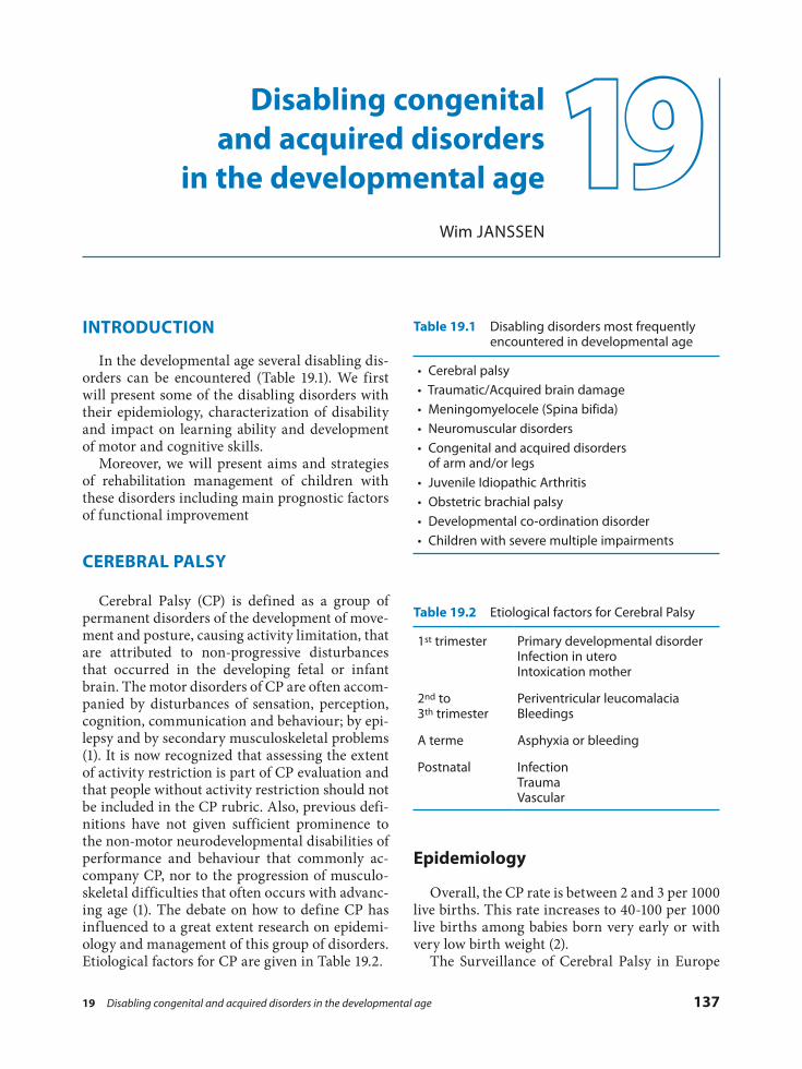
19 Disabling congenital and acquired disorders in the developmental age 137
Epidemiology
Overall, the CP rate is between 2 and 3 per 1000 live births. This rate increases to 40-100 per 1000 live births among babies born very early or with very low birth weight (2).
The Surveillance of Cerebral Palsy in Europe
INTRODUCTION
In the developmental age several disabling dis-orders can be encountered (Table 19.1). We first will present some of the disabling disorders with their epidemiology, characterization of disability and impact on learning ability and development of motor and cognitive skills.
Moreover, we will present aims and strategies of rehabilitation management of children with these disorders including main prognostic factors of functional improvement
CEREBRAL PALSY
Cerebral Palsy (CP) is defined as a group of permanent disorders of the development of move-ment and posture, causing activity limitation, that are attributed to non-progressive disturbances that occurred in the developing fetal or infant brain. The motor disorders of CP are often accom-panied by disturbances of sensation, perception, cognition, communication and behaviour; by epi-lepsy and by secondary musculoskeletal problems (1). It is now recognized that assessing the extent of activity restriction is part of CP evaluation and that people without activity restriction should not be included in the CP rubric. Also, previous defi-nitions have not given sufficient prominence to the non-motor neurodevelopmental disabilities of performance and behaviour that commonly ac-company CP, nor to the progression of musculo-skeletal difficulties that often occurs with advanc-ing age (1). The debate on how to define CP has influenced to a great extent research on epidemi-ology and management of this group of disorders. Etiological factors for CP are given in Table 19.2.
19 Disabling congenital and acquired disorders
in the developmental ageWim JANSSEN
Table 19.1 Disabling disorders most frequently encountered in developmental age
• Cerebral palsy• Traumatic/Acquired brain damage• Meningomyelocele (Spina bifida)• Neuromuscular disorders• Congenital and acquired disorders of arm and/or legs
• Juvenile Idiopathic Arthritis• Obstetric brachial palsy• Developmental co-ordination disorder• Children with severe multiple impairments
Table 19.2 Etiological factors for Cerebral Palsy
1st trimester Primary developmental disorder Infection in uteroIntoxication mother
2nd to 3th trimester
Periventricular leucomalacia Bleedings
A terme Asphyxia or bleeding
Postnatal Infection Trauma Vascular

138 PHYSICAL AND REHABILITATION MEDICINE for Medical Students
motor development will be assessed using several instruments that will change according to age and developmental phase. Muscular strength, spastic-ity and walking capacity are assessed. As a result hip (sub)luxation, scoliosis and contractures can occur. Early diagnosis and treatment are manda-tory so regular check-ups are needed. Detailed information on gait can be acquired using gait analysis to clarify walking pattern and select the proper treatment. Several treatment option are available for spasticity control to decrease the ef-fect of spasticity on the development of contrac-tures and walking disturbances. These interven-tions change during growth from physical thera-py, oral medication, botulinum toxin injections, intrathecal baclofen to multilevel surgery in teen-agers. In severe cases additional measures are needed such as providing a wheelchair or changes to the environment/house.
Adjunct to this the effects of cognition and speech development needs to be assessed and treated. Swallowing disorders occur quite fre-quently resulting in drooling that can be treated using different techniques (speech therapy, botuli-num toxin, surgery, etc.). There needs to be an advice on feasibility of education and professional training to present the child the most appropriate education level. Goal setting is done in agreement with parents and child. Adequate communication skills are mandatory for the rehabilitation team to define adequate and realistic goals.
PrognosisConcerning prognostic factors for future (long
term) functioning the gravity of the CP, the ability of the child to learn both on cognitive and motor level, the family, resources and other contextual factors.
TRAUMATIC BRAIN INJURY (TBI)/ ACQUIRED BRAIN INJURY
Traumatic and acquired brain injuries are here presented together, as they have a lot in common although there are differences which will be men-tioned. These disorders arise due to a trauma of the brain or can result from other causes that can be seen in Table 19.3. We have to be aware that these conditions occur in a developing brain in a child that is still growing, where the extent and
started in 1998, now involving 21 centres to pro-vide data in Europe.
Characterization of disability and impact on learning ability and development of motor/cognitive skills
CP is characterized primarily by disturbances of posture and movement, adjunct to the sensory sys-tem, cognition and behaviour. Also other disorders such as epilepsy, respiratory disorder, gastro-intes-tinal disorders can occur. These can be primary or secondary such as contractures, hip luxation.
The movement disorder is caused by a cerebral dysfunction and can be classified according type, localisation and gravity of the disorder. We distin-guish three groups of movement disorder in CP: spastic, dyskinetic and atactic. The spastic CP is most frequent: 77-93%, dyskinetic 2-15% and atac-tic 2-8% (3), localisation can be unilateral or bilat-eral.
Describing the severity of the movement disor-der is a method of describing the severity of the CP disorder. Mostly this is done by describing the level of activity using Gross Motor Function Clas-sification System (GMFCS, on ambulation) (4), the Manual Ability Classification System (MACS scale) (5), and Communication Function Classifi-cation System (CFCS). Cognitive and intellectual function can be disturbed and influence the de-velopment of a child to a great extent. Despite the disorder there is a growth of functioning accord-ing to GMFCS and MACS in childhood.
The extent of the disorders can be very severe in time, we also have to realize that survival in chil-dren with CP is influenced according to severity.
Strategies of rehabilitation management
By definition CP is a non-progressive distur-bances of the developing brain, which is not syn-onym with a stable medical condition. Due to the development of the brain and the motor system combined with the growth of the child changes occur which need to be assessed and if possible to be treated. The effect of the disorder on the com-munication, education and training has to be as-sessed and evaluated by a comprehensive team to cover all aspects of the sequelae. First of all the
19 Disabling congenital and acquired disorders in the developmental age 139
tion and activities is broad including moving, concentration, cognitive function, communica-tion, behaviour, social contacts, etc.
Prognosis Prognosis in TBI strongly depends on the se-
verity of trauma, as described above. In mild inju-ries complete recovery can occur, in severe cases the chance for persistent problems is high. For non traumatic brain injury the underlying cause is relevant, course can be similar to traumatic brain injury with (partly) recovery. However in other cases course can be progressive with loss of function i.e. in degenerative disorders.
SPINA BIFIDA (MENINGOMYELOCELE)
Spina Bifida (SB) is a complex disorder that is caused by a non-closure of the neural tube in the first month after conception. There is a distinc-tion between a closed and open form. When after birth the myelum is exposed: we speak of a Me-ningomyelocele. It often is accompanied by other malformations such as hydrocephalus, Chiari malformation or defect of the corpus callosum. After closure of the myelum defect after birth fre-quently a progressive hydrocephalus occurs with ventriculo-peritoneal drainage indicated.
EpidemiologyIncidence and prevalence vary per country and
is estimated to be 1 per 1000 live birth (6). Inci-dence can be influenced by differences in strate-gies after prenatal ultrasound screening and also
sequelae of the damage can be related to phase of brain development.
EpidemiologyRegistration for these disorders differ much per
country so no exact details can be given. We are aware that these disorders are frequent encoun-tered in PRM in childhood.
Characterization of disability and impact on learning ability and development of motor/cognitive skills
In traumatic brain injury there are three global levels of severity: the level of unconsciousness (to be assessed with Glasgow Coma Scale (GCS), the duration of unconsciousness and the duration of post traumatic amnesia. The course of TBI can be divided in three phases: acute, subacute and chronic. Main part of the recovery occurs gener-ally in the first six months after the trauma. The TBI can be accompanied by many other injuries who need their specific treatment. Care in acute and subacute phase are complex and need to be in given in a specialized centre.
Strategies of rehabilitation management
As written several forms are present so clinical picture can vary much giving both motor impair-ments as well as cognitive and behavioural prob-lems, combined with speech and communication disorders. Motor problems generally are less pre-dominant than in CP, so the assessment of func-
Table 19.3 Main causes of traumatic and non-traumatic (acquired) brain injuries
Traumatic Non-traumatic
Without skull injury• (Traffic) accident• Fall• Heavy object against head• Shaken baby syndrome
With skull injury• Bony impression due to fracture• Penetration of object
InfectionHypoxia/Anoxia (near drowning)Cerebrovascular accidentTumorIntoxication
Metabolic disordersDegenerative disordersEpilepsia Hydrocephalus
140 PHYSICAL AND REHABILITATION MEDICINE for Medical Students
ments related to hydrocephalus or structural brain defects.
Strategies of rehabilitation management
An appropriate assessment of motor and sen-sory level of SB is difficult to perform in a new-born so in the first and second year an adequate observation and reassessment will give the level which is of importance for advice on the level of gait that can be reached in future. Yearly follow-up of function is indicated to check for possible tethered cord problem in which an adhesion be-tween myelum and spinal column causes a dete-rioration of neurological functioning. Also con-trol of contractures is needed, especially when these can negatively influence gait.
Urological checks are needed for bladder and kidney control to assure if possible continence and prevention of kidney damage. Orthopaedic follow-up is needed for spinal column and devel-opment of hip joints, hip luxation occurs often in cases of high level SB.
Prognosis Prognosis for overall functioning depends on
the level of Spina Bifida, the presence of a hydro-cephalus, the occurrence of complications, and the level self-care that can be reached. Inconti-nence has a strong social impact. Sexual function-ing is impeded for several reasons amongst others the loss of sensation (8).
NEUROMUSCULAR DISORDERS
Neuromuscular disorder are disorders that originate from conditions of the anterior horn of the spinal cord, the nerve roots, neural plexus, nerves, the motor end plate and muscle. These conditions are rare.
EpidemiologyFor an overview of some of the most frequently
seen disorders and their incidence see Table 19.5, where not all figures can be given due to a lack of adequate registration. The prevalence of these con-ditions increases as a result of better diagnostics and care for these patients so survival is increasing.
the use of folium acid before conception is of in-fluence on incidence.
Characterization of disability and impact on learning ability and development of motor/cognitive skills
The clinical presentation varies according to the extent of myelum dysfunction, the level of malformations of the spine and accompanying defects as hydrocephalus. In general a higher (more proximal lesion i.e. thoracic) lesion leads to a higher impact than a lower lesion. Both mo-tor and sensory function can be impeded with consequences for posture and movement, blad-der and bowel control can be impeded, several disorders of pelvis and hips can occur. Due to motor nerve problems a disorder of gait and con-tractures of ankle/knee/hip occur. Congenital talipes equinovarus occur frequent as a conse-quence of muscular disbalance at the level of the ankle. A disbalance of muscle function leads in time to well-known contractures of ankle/knee/hip. A scoliosis and/or kyphosis can develop and be progressive during growth. The level of gait (classified by using the Hoffer classification) var-ies according to level and accompanying con-tractures (Table 19.4). The level of cognitive function can impede education: intelligence, hy-drocephalus and being wheelchair-bound influ-ences the possibilities for education. Also social skills can be influenced.
Due to sensory dysfunction skin problems can occur resulting in decubital ulcers. Most children with SB become independent for their daily self-care including bladder and bowel control, such as self-catheterisation and bowel lavage.
Education and training can be influenced both by mobility impairments and cognitive impair-
Table 19.4 Levels of ambulatory function in children and youth according to Hoffer et al. (7) criteria
• Non-ambulation• Non-functional ambulation• Household ambulation• Community ambulation• Normal ambulation
19 Disabling congenital and acquired disorders in the developmental age 141
lay of reaching motor milestones and slowing of motor development. In Duchenne muscular dys-trophy a typical gait pattern (equinus and wad-dling) develops with Gowers sign during rising.
Becker muscular dystrophy is milder and devel-ops later, with a debut at the age of 12.
Myotonic dystrophy (Morbus Steinert) can also occur in childhood, because there is the phenom-ena of anticipation, which means that in each fol-lowing generation the disorder is more pronounced due to the increase of the CTG-repeats contribut-ing to the clinical picture. The congenital form of myotonic dystrophy is a severe disorder.
Muscular dystrophies are very frequently ac-companied by cardiac problems; a cardiomyopa-thy as well as cardiac rhythm disorders. Some-times these cardiac problems are the presenting clinical problems before gait disorders occur. Furthermore is the respiratory system involved in neuromuscular disorders as a results of a decline of respiratory muscle strength, an increase of re-spiratory distress due to scoliosis development or both. Night time hypoventilation can occur with their complaints. Due to the decrease of muscle strength and impediment of motor development and skeletal load an osteoporosis can occur.
In some neuromuscular disorders cognitive disorders occur, for example in Duchenne and Becker Muscular Dystrophy. In general cognitive functions are not disturbed.
Strategies of rehabilitation management
Due to the diversity of neuromuscular disor-ders there is not one guideline for all. Manage-ment strongly depends on the character of the neuromuscular disorder, slow or fast progressive, cardiac and respiratory system involvement, cog-nitive impairments, etc. The rehabilitation man-
Characterization of disability and impact on learning ability and development of motor/cognitive skills
These conditions show a great variety as there are many different types (several hundreds); also the age of presentation, inheritance and severity can differ much. For more information on differ-ent types you can consult the homepage of Neuro-muscular Disease Center of Washington Univer-sity, USA (9) or of TREAT-NMD (10). For some disorders the genetic background and inheritance mode is quite clear, for others it still needs to be elucidated. For these disorders arising in child-hood they mostly are congenital. Only a few of these disorders are acquired such as Guillain-Barré syndrome or myositis, on infectious or im-munological base.
A thorough neurological or neuropediatric in-vestigation is needed for definite diagnosis. This is relevant in relation to prognosis, treatment and survival. As written due to the variety of the dis-orders there is a great variety of clinical presenta-tion but predominantly the loss of muscular strength is present. This loss can be mild or very severe, also depending on the type of the disorder. The spread of the muscular strength loss can be typical for some conditions. The course of the disorder can be slow or very progressive concern-ing the loss of muscular strength. In SMA there are 4 types, in type I the child does not reach the functional level to be seated independently, while in type IV there are only minor complaints with a normal life expectancy. In CMT the disorder starts predominantly distally in the extremities related to nerve length. The progression generally is slow, resulting in pes equinovarus due to the disbalance of muscle force and gait impairments.
Duchenne muscular dystrophy starts with a de-
Table 19.5 Epidemiology of the main neuromuscular disorders grouped by lesion level
Localisation Name Incidence
Anterior horn Spinal Muscular Atrophy (SMA) 1/10,000
Nerve Charcot Marie Tooth neuropathy (CMT)Guillan Barre Syndrome (GBS)
2/10,0001/10,000
Muscle Duchenne and Becker Muscular Dystrophy (DMD/BMD)Limb Girdle Muscular Dystrophy (LGMD)Myotonic Dystrophy (MD)
25/100,000
142 PHYSICAL AND REHABILITATION MEDICINE for Medical Students
Characterization of disability and impact on learning ability and development of motor/cognitive skills
The reduction deficiencies are mostly described using the ISPO nomenclature based on presence or absence of skeletal structures Day (12). We need to realize this is accompanied by a disturbance of growth plates, muscular structure and joint in-volvement giving rise to a complex disorder that will change during growth. Due to the underde-veloped limb there is an impact on ambulatory function or the development of grasping and ma-nipulation. Dedicated teams consisting of an or-thopaedic or plastic surgery team together with geneticist and rehab team provide care. Early as-sessment are performed to determine treatment goals i.e. a surgical approach, a prosthetic ap-proach or a mix of both. For legs and ambulatory function prosthetic fitting is self-evident. For the upper extremity prosthetic fitting is not self-evi-dent but the result of an evaluation together with parents on goal setting in each specific case.
Strategies of rehabilitation management
Close cooperation with an orthopaedic or plas-tic surgery team is needed for appropriate counsel-ling. Also after birth there is a need for support due to the unexpected ‘loss’, with provision of informa-tion on future functioning of the child. The care can be provided by a comprehensive rehab team in cooperation with a prosthetist to provide informa-tion on the possibilities a prosthesis. If needed a psychologist or social work can do counselling with family. There are no cognitive impairments but social functioning can be influenced. During the development of a child several periods of social ‘problems’ can occur during primary or secondary school which rises the need for support. The devel-opment of self-image can be disturbed giving rise to a low self-esteem (13). For upper extremity this can be the reason to provide a youngster with a passive prosthesis for their self-image.
Prognosis Prognosis for functioning are in general good,
depending on the extent of the deficiency and the resulting capabilities for walking and manipulation.
agement is most comprehensive for the group of Duchenne Muscular Dystrophy and SMA types I to III. Regular check-ups with a neuromuscular team consisting of a rehab team together with neurologist and orthopaedic surgeon is the obvi-ous way to act. Knowing that care standards differ from country to country but also even within in-dividual countries the TREAT-NMD was started to improve care and research in neuromuscular disorders (11). This is done by stimulating consen-sus guidelines, both academic and “family friend-ly” guides.
Prognosis Prognosis primarily depends on the character
of the neuromuscular disorder. We need to realize that due to changes in health care and of manage-ment of the disorders by the teams there is an in-creased life expectancy. This is obvious for Duch-enne muscular dystrophy, also ventilator support has extended life expectancy for this group. Fur-thermore nowadays recombinant therapy such as in Morbus Pompe can change life expectancy significantly.
CONGENITAL AND ACQUIRED DISORDERS OF ARM AND/OR LEGS
Congenital and acquired disorders of arm and/or legs represent a broad spectrum of disorders. We will only discuss reduction deficiencies of up-per and lower extremity. These disorders are con-genital disorders in which a part of arm or leg misses due to a developmental disorder. Reduc-tion deficiencies can be transversal or longitudi-nal. Also proximal deficiencies occur such as proximal focal femur dysplasia. The presentation is at birth although nowadays most of these disor-ders are known prenatally due to the use of ultra-sound during pregnancy. The developmental dis-order can be small or extended, so very distal re-duction deficiencies are not seen in the ultrasound examination.
EpidemiologyThese disorders can be isolated or part of a
syndrome. These disorders are relatively rare, 6.6/ 10,000 live birth. For legs the most frequent disor-der is fibula aplasia, sometimes bilateral.
19 Disabling congenital and acquired disorders in the developmental age 143
preventable spina bifida and anencephaly by 2010. Community Genet. 2002; 5: 70-7.
7. Hoffer MM, Feiwell E, Perry R, Perry J, Bonnett C. Functional ambulation in patients with myelome-ningocele. J Bone Joint Surg Am. 1973; 55: 137-48.
8. Verhoef M, Barf HA, Post MW, van Asbeck FW, Gooskens RH, Prevo AJ. Functional independence among young adults with spina bifida, in relation to hydrocephalus and level of lesion. Dev Med Child Neurol. 2006; 48: 114-9.
9. Neuromuscular Disease Center. Washington Uni-versity, St. Louis, MO, USA. Available from: http://neuromuscular.wustl.edu
10. Treat-NMD - Neuromuscular Network. Homepage. Available from: http://www.treat-nmd.eu/
11. Treat-NMD - Neuromuscular Network. Care over-view. Available from: http://www.treat-nmd.eu/care/overview
12. Day HJ. The ISO/ISPO classification of congenital limb deficiency. Prosthet Orthot Int. 1991; 15: 67-9.
13. Lankhorst IMF, Baars ECT, Wijk Iv, Janssen WGM, Poelma MJ, van der Sluis CK. Living with transversal upper limb reduction deficiency: limi-tations experienced by young adults during their transition to adulthood. Disabil Rehabil. 2017: 39: 1623-30.
REFERENCES
1. Rosenbaum P, Paneth N, Leviton A, Goldstein M, Bax M, Damiano D, et al. A report: the definition and classification of cerebral palsy April 2006. Dev Med Child Neurol Suppl. 2007; 109: 8-14.
2. Surveillance of Cerebral Palsy in Europe. © 2018 SCPE. Available from: http://www.scpenetwork.eu/en/cerebral-palsy/
3. Himmelmann K, Uvebrant P. The panorama of cerebral palsy in Sweden. XI. Changing patterns in the birth-year period 2003-2006. Acta Paediatr. 2014; 103: 618-24.
4. Hidecker MJ, Ho NT, Dodge N, Hurvitz EA, Slaughter J, Workinger MS, et al. Inter-relation-ships of functional status in cerebral palsy: analyz-ing gross motor function, manual ability, and communication function classification systems in children. Dev Med Child Neurol. 2012; 54: 737-42.
5. Eliasson AC, Krumlinde-Sundholm L, Rosblad B, Beckung E, Arner M, Ohrvall AM, et al. The Manual Ability Classification System (MACS) for children with cerebral palsy: scale development and evidence of validity and reliability. Dev Med Child Neurol. 2006; 48: 549-54.
6. Oakley GP Jr. Global prevention of all folic acid-
Key messages
• Disabling congenital and acquired disorders in the developmental age pose a challenge for phys-iatrist to maximise development of motor and cognitive skills and learning ability. Furthermore the physiatrist needs to minimise consequences of restraining societal and social mechanisms. The influence of growth and motor/cognitive development is integrated in rehabilitation treatment.


Part V
The importance of the PRM physician role in the healthcare system

20 Essential methods of assessing patient’s needs 147
ment (identifying patients’ needs), 2. Assignment (assigning intervention targets to required inter-ventions and relevant health professionals), 3. In-tervention, and 4. Evaluation (determining effec-tiveness of interventions) and the cycle continues with repetitive assessments until the patient’s needs are met (4, 5).
Assessment, the first and very important step to document patient’s needs relevant to function-ing before determining interventions, is per-formed from both the patient’s perspective ob-tained by history taking and also from the health professional’s perspective which includes special-ized tests or measures with the aim of goal-setting and ascertaining which functioning problems needs to be targeted by appropriate interventions (5). ICF based rehabilitation tools including ICF Core Sets (6, 7), ICF Assessment Sheet, and ICF Categorical Profile are recommended for use for documentation of functioning (5, 8).
It is important to note that when using the ICF for assessment, four principles are needed to be taken into account as described by Stucki et al. (9) and as shown in Table 20.1.
STEP-BY-STEP ASSESSMENT OF PATIENTS’ NEEDS IN A PATIENT WITH LOW BACK PAIN AS AN EXAMPLE
Physicians in many disciplines may encounter patients with low back pain (LBP) in their clinical practice. LBP ranks the first regarding ‘Years Lived with Disability’, an important indicator of functioning, with 60.1 million years in 2015 as reported in Global Burden of Disease Study (10). LBP-associated significant disability which results in activity limitations and participation restric-tions can be addressed with a variety of evidence-
INTRODUCTION
Assessing patients’ needs is the starting point for treatment decisions and for designing physical and rehabilitation medicine (PRM) programmes for a specific patient.
Terminologically, “assessment” incorporates procedures and techniques for classifying and measuring a variable relevant to an individual as a whole (1). Assessment allows us to reach a final decision regarding patients’ needs through classi-fication and measurement which act on each oth-er. While classification concerns assignment of an individual to a predefined category according to a qualitative feature (e.g. gender), measurement is quantification of a feature ascribed to an individ-ual variable (1).
From the perspective of PRM, described as “medicine of functioning” (2), the variables to be assessed relate to functioning. At this point, the International Classification of Functioning, Dis-ability and Health (ICF) (3) provides an excellent means of history taking and assessment to iden-tify patient’s needs. The ICF views health from a holistic perspective taking into consideration functioning properties of an individual with a disease or injury (a health condition) in interac-tion with the environment. The ICF describes functioning in three dimensions including body functions and structures, activities, and participa-tion with a contextual factors component includ-ing environmental and personal factors which may have influences on functioning. The ICF is an international standard and a common language for describing and measuring health and func-tioning/disability (3). (Please see Chapter 2 for more information on the ICF).
In PRM, “rehabilitation cycle”, describing the steps to be taken to find solutions to problems of the patients based on the ICF, includes 1. Assess-
20Essential methods of assessing patient’s needs
Aydan ORAL, Elena M. ILIEVA
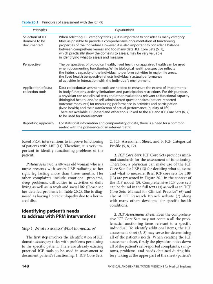
148 PHYSICAL AND REHABILITATION MEDICINE for Medical Students
2. ICF Assessment Sheet, and 3. ICF Categorical Profile (5, 8, 12).
1. ICF Core Sets: ICF Core Sets provides mini-mal standards for the assessment of functioning. Therefore, a physician can make use of the ICF Core Sets for LBP (13) for deciding what to assess and what to measure. Brief ICF core sets for LBP (13) are presented in Figure 20.1 in the context of the ICF model (3). Comprehensive ICF core sets can be found in the full text (13) as well as in “ICF Core Sets: Manual for Clinical Practice” (6) and also at ICF Research Branch website (7) along with many others developed for specific health conditions.
2. ICF Assessment Sheet: Even the comprehen-sive ICF Core Sets may not contain all the prob-lematic functioning items relevant to a specific individual. To identify additional items, the ICF assessment sheet (5, 8) may serve for determining all of the patient’s needs. When creating the ICF assessment sheet, firstly the physician notes down all of the patient’s self-reported complaints, symp-toms, problems, and needs obtained during his-tory taking at the upper part of the sheet (patient’s
based PRM interventions to improve functioning of patients with LBP (11). Therefore, it is very im-portant to identify functioning problems of the patient.
Patient scenario: a 40-year old woman who is a nurse presents with severe LBP radiating to her right leg lasting more than three months. Her other complaints include emotional problems, sleep problems, difficulties in activities of daily living as well as in work and social life (Please see her detailed problems in Table 20.2). She is diag-nosed as having L 5 radiculopathy due to a herni-ated disc.
Identifying patient’s needs to address with PRM interventions
Step 1. What to assess? What to measure?
The first step involves the identification of ICF domains/category titles with problems pertaining to the specific patient. There are already existing practical ICF tools to be used in assessment to document patient’s functioning: 1. ICF Core Sets,
Table 20.1 Principles of assessment with the ICF (9)
Principles Explanations
Selection of ICF domains to be documented
When selecting ICF category titles (3), it is important to consider as many category titles as possible to provide a comprehensive documentation of functioning properties of the individual. However, it is also important to consider a balance between comprehensiveness and too many data. ICF Core Sets (6, 7), which practically show the domains to assess, may be very valuable in identifying what to assess and measure
Perspective The perspectives of biological health, lived health, or appraised health can be used when documenting functioning. While biological health perspective reflects the intrinsic capacity of the individual to perform activities in major life areas, the lived health perspective reflects individual’s actual performance of activities in interaction with the individual’s environment
Application of data collection tools
Data collection/assessment tools are needed to measure the extent of impairments in body functions, activity limitations and participation restrictions. For this purpose, a physician can use clinical tests and other evaluations relevant to functional capacity (biological health) and/or self-administered questionnaires (patient-reported outcome measures) for measuring performance in activities and participation (lived health) and their satisfaction of actual performance (quality of life). There are available ICF-based and other tools linked to the ICF and ICF Core Sets (6, 7) to be used for measurement
Reporting approach For statistical information and comparability of data, there is a need for a common metric with the preference of an interval metric
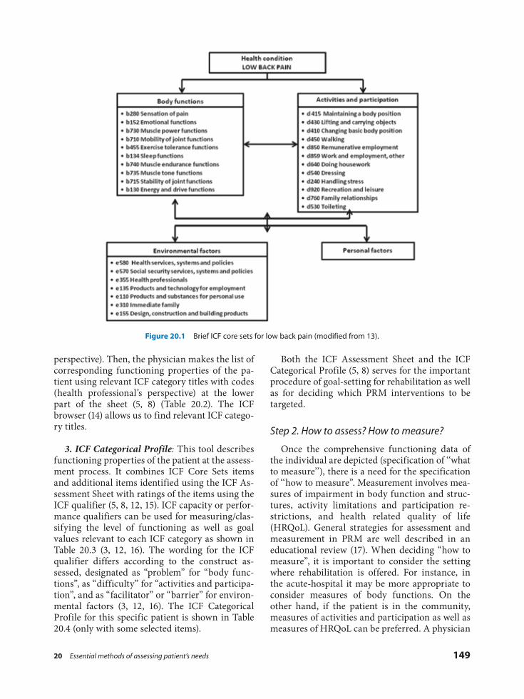
20 Essential methods of assessing patient’s needs 149
Both the ICF Assessment Sheet and the ICF Categorical Profile (5, 8) serves for the important procedure of goal-setting for rehabilitation as well as for deciding which PRM interventions to be targeted.
Step 2. How to assess? How to measure?
Once the comprehensive functioning data of the individual are depicted (specification of ‘‘what to measure’’), there is a need for the specification of ‘‘how to measure”. Measurement involves mea-sures of impairment in body function and struc-tures, activity limitations and participation re-strictions, and health related quality of life (HRQoL). General strategies for assessment and measurement in PRM are well described in an educational review (17). When deciding “how to measure”, it is important to consider the setting where rehabilitation is offered. For instance, in the acute-hospital it may be more appropriate to consider measures of body functions. On the other hand, if the patient is in the community, measures of activities and participation as well as measures of HRQoL can be preferred. A physician
perspective). Then, the physician makes the list of corresponding functioning properties of the pa-tient using relevant ICF category titles with codes (health professional’s perspective) at the lower part of the sheet (5, 8) (Table 20.2). The ICF browser (14) allows us to find relevant ICF catego-ry titles.
3. ICF Categorical Profile: This tool describes functioning properties of the patient at the assess-ment process. It combines ICF Core Sets items and additional items identified using the ICF As-sessment Sheet with ratings of the items using the ICF qualifier (5, 8, 12, 15). ICF capacity or perfor-mance qualifiers can be used for measuring/clas-sifying the level of functioning as well as goal values relevant to each ICF category as shown in Table 20.3 (3, 12, 16). The wording for the ICF qualifier differs according to the construct as-sessed, designated as “problem” for “body func-tions”, as “difficulty” for “activities and participa-tion”, and as “facilitator” or “barrier” for environ-mental factors (3, 12, 16). The ICF Categorical Profile for this specific patient is shown in Table 20.4 (only with some selected items).
Figure 20.1 Brief ICF core sets for low back pain (modified from 13).
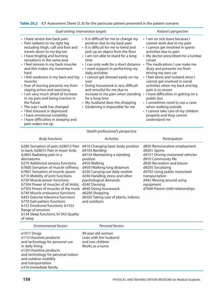
150 PHYSICAL AND REHABILITATION MEDICINE for Medical Students
Table 20.2 ICF Assessment Sheet (5, 8) for the particular patient presented in the patient scenario
Goal-setting: Intervention targets Patient’s perspective
• I have severe low back pain• Pain radiates to my right leg
including thigh, calf and foot and travels down to my big toe
• I have tingling and burning sensations in the same area
• I feel tension in my back muscles and this makes my movement hard
• I feel weakness in my back and leg muscles
• Fear of moving prevents me from staying active and exercising
• I am very much afraid of increase in my pain and being inactive in the future
• The way I walk has changed • I feel stressed or depressed• I have emotional instability• I have difficulties in sleeping and
pain wakes me up
• It is difficult for me to change my position due to my back pain
• It is difficult for me to bend and pick up an object from the floor
• I am not able to stand for a long time
• I can only walk for a short distance• I need support in performing my
daily activities • I cannot get dressed easily on my
own• Doing housework is very difficult
and stressful for me due to increase in my pain when standing for a long time
• My husband does the shopping• Gardening is impossible for me
• I am on sick leave because I cannot work due to my pain
• I cannot get involved in sports activities due to pain
• My doctor prescribed me a lumbar corset
• The medications I use make me dizzy and prevents me from driving my own car
• I feel alone and isolated since I cannot get involved in social activities when my back and leg pain is so severe
• I have difficulties in getting on a public bus
• I sometimes need to use a cane when walking outside
• I cannot take care of my children properly and they cannot understand me
Health professional’s perspective
Body functions Activities Participation
b280 Sensation of pain; b28013 Pain in back; b28015 Pain in lower limb; b2803 Radiating pain in a dermatomeb279 Additional sensory functionsb7800 Sensation of muscle stiffness; b7801 Sensation of muscle spasmb710 Mobility of joint functionsb730 Muscle power functions; b7304 Power of muscles of all limbs; b7305 Power of muscles of the trunkb740 Muscle endurance functionsb455 Exercise tolerance functionsb770 Gait pattern functionsb152 Emotional functions; b1522 Range of emotionb134 Sleep functions; b1343 Quality of sleep
d410 Changing basic body positiond4105 Bendingd4154 Maintaining a standing positiond450 Walkingd4501Walking long distancesd230 Carrying out daily routined240 Handling stress and other psychological demandsd540 Dressingd640 Doing houseworkd6200 Shoppingd6505 Taking care of plants, indoors and outdoors
d850 Remunerative employmentd9201 Sportsd4751 Driving motorized vehiclesd910 Community lifed920 Recreation and leisured9205 Socializingd4702 Using public motorized transportationd465 Moving around using equipment d7600 Parent-child relationships
Environmental factors Personal factors
e1011 Drugse1151Assistive products and technology for personal use in daily livinge1201Assistive products and technology for personal indoor and outdoor mobility and transportatione310 Immediate family
40-year old womanLives with her husband and two childrenWorks as a nurse
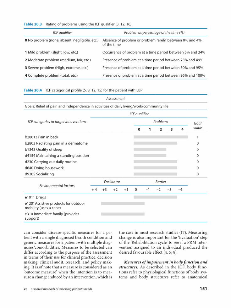
20 Essential methods of assessing patient’s needs 151
the case in most research studies (17). Measuring change is also important for the ‘Evaluation’ step of the ‘Rehabilitation cycle’ to see if a PRM inter-vention assigned to an individual produced the desired favourable effect (4, 5, 8).
Measures of impairment in body function and structures: As described in the ICF, body func-tions refer to physiological functions of body sys-tems and body structures refer to anatomical
can consider disease-specific measures for a pa-tient with a single diagnosed health condition and generic measures for a patient with multiple diag-noses/comorbidities. Measures to be selected can differ according to the purpose of the assessment in terms of their use for clinical practice, decision making, clinical audit, research, and policy mak-ing. It is of note that a measure is considered as an ‘outcome measure’ when the intention is to mea-sure a change induced by an intervention, which is
Table 20.3 Rating of problems using the ICF qualifier (3, 12, 16)
ICF qualifier Problem as percentage of the time (%)
0 No problem (none, absent, negligible, etc.) Absence of problem or problem rarely, between 0% and 4% of the time
1 Mild problem (slight, low, etc.) Occurrence of problem at a time period between 5% and 24%
2 Moderate problem (medium, fair, etc.) Presence of problem at a time period between 25% and 49%
3 Severe problem (High, extreme, etc.) Presence of problem at a time period between 50% and 95%
4 Complete problem (total, etc.) Presence of problem at a time period between 96% and 100%
Table 20.4 ICF categorical profile (5, 8, 12, 15) for the patient with LBP
Assessment
Goals: Relief of pain and independence in activities of daily living/work/community life
ICF categories to target interventions
ICF qualifier
Problems Goal value0 1 2 3 4
b28013 Pain in back
b2803 Radiating pain in a dermatome
b1343 Quality of sleep
d4154 Maintaining a standing position
d230 Carrying out daily routine
d640 Doing housework
d9205 Socializing
1
0
0
0
0
0
0
Environmental factorsFacilitator Barrier
+ 4 +3 +2 +1 0 –1 –2 –3 –4
e1011 Drugs
e1201Assistive products for outdoor mobility (uses a cane)
e310 Immediate family (provides support)

152 PHYSICAL AND REHABILITATION MEDICINE for Medical Students
pass the majority of the items of the activities and participation component of the ICF (22). Recently, Patient-Generated Health Data (PGHD), with a working definition of “health-related data – in-cluding health history, symptoms, biometric data, treatment history, lifestyle choices, and other in-formation – created, recorded, gathered, or in-ferred by or from patients or their designees (i.e., care partners or those who assist them) to help address a health concern” (23, p. 2) is introduced. PGHD combines PROMs with other PGHD gath-ered usually through electronic systems (such as sensors or smart phone applications) (23), which may be of efficient use in the future. Additionally, there are other PROMs allowing patients to rate their appraisal of the improvement or change in their health after an administered intervention, known as “global rating of change scales” (24) as well as those rating their satisfaction with treat-ment and/or health care (25).
An important issue is which measure to select. First of all, the selected measure (self-adminis-tered questionnaires/PROMs) needs to be valid (measuring what is planned to measure), reliable (referring to reproducibility and internal consis-tency), and responsive (ability to detect meaning-ful changes over time) as well as appropriate (matching the purpose of measuring) and precise (sensitive to make distinctions). Furthermore, is-sues of acceptableness in terms of obtaining re-sponses from the responders, completion time, adaptation to a language, cultural appropriate-ness, and feasibility in terms of requirement of expertise for administration or ease of adminis-tration as well as costs if the instrument is not free to use are also important (17, 26). Secondly, assess-ment instruments linked to the ICF comprehen-sively following linkage rules (27) should be of preference for comprehensive documentation of functioning and comparability of the data (9). Examples of measures regarding the presented LBP patient are given in Table 20.5.
The last issue is the need for the transformation of ordinal metrics (indicating a rank, a hierarchi-cal order based on a score obtained from a ques-tion with unequal distances between scores) to interval metrics (fundamental measurement on an interval level with equal distances between se-quential units), which can be achieved by Rasch model (17, 28). This issue is also important for precise measurement of change in outcomes achieved by PRM interventions (29).
parts of the body (i.e. organs, limbs and their parts). Impairments refer to problems in body functions or structures (3, 18). Measurement of body functions and structures requires a careful and thorough clinical examination and specific clinical tests including imaging techniques, labo-ratory tests, or functional capacity evaluations and other reliable assessment tools including self-administered questionnaires, also called as pa-tient reported outcome measures (PROMs) (17).
Measures of activity limitation and participa-tion restriction: Activity refers to the execution of a task or action by an individual and participation refers to involvement in a life situation. Activity limitations are defined as difficulties encountered by an individual when executing activities. Partici-pation restrictions describe problems encountered by an individual in involvement in life situations. Environmental factors described as the physical and social environment in which the individual lives may have a significant influence in activity limitations and participation restrictions (3, 18).
Measures of quality of life and other PROMs: “HRQoL refers to the individual’s psychosocial well-being and general satisfaction with physical, psychological and social domains of life and how this is affected by diseases, accidents, and treat-ments from the patient’s point of view” (19, p. 191). HRQoL can be measured either using generic in-struments or using specific instruments designed for a specific health condition, groups of patients, or functioning areas (20). Additionally, individu-alized and needs-based HRQoL measures are available which focus on the prioritization of life areas according to their importance and subjec-tive appraisal of the impact of the health condi-tion on the patient’s QoL, respectively (17). HRQoL measures not only gather information on impair-ments (symptoms and complaints) and function-ing properties (activity limitations and/or partici-pation restrictions), but they also gather informa-tion on patient’s own health perceptions (her/his well-being and satisfaction with her/his health) (19). Other PROMs include item bank generated PROs. One example is the Patient Reported Out-comes Measurement Information System (PRO-MIS®), created by the US National Institutes of Health which uses the data regarding health gath-ered from the patients through a reporting system (21). PROMIS® instruments were found to encom-
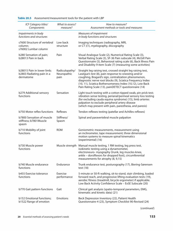
20 Essential methods of assessing patient’s needs 153
Table 20.5 Assessment/measurement tools for the patient with LBP
ICF Category titles/ Components
What to assess? measure?
How to measure? Assessment methods or tools and measures
Impairments in body functions and structures
Measures of impairment in body functions and structures
s7600 Structure of vertebral column;s76002 Lumbar column
Low back structure
Imaging techniques (radiography, MRI, or CT )(1), myelography, discography
b280 Sensation of pain; b28013 Pain in back
Pain Visual Analogue Scale (2), Numerical Rating Scale (3), Verbal Rating Scale (3), SF-36 Pain subscale (4), McGill Pain Questionnaire (5), Behavioral rating scale (6), Back Illness Pain and Disability 9-item Scale (7) (measuring some activities)
b28015 Pain in lower limb; b2803 Radiating pain in a dermatome
Radiculopathy/Neuropathic pain
Straight leg raising test, crossed straight leg raising test, Lasègue’s test (8), pain response to sneezing and/or coughing, Bragard’s sign, centralization phenomenon, diagnostic nerve root blocks (9), Sciatica Frequency Index (10, 11), Sciatica Bothersomeness Index (10,12), Low Back Pain Rating Scale (13), painDETECT questionnaire (14)
b279 Additional sensory functions
Sensation Light touch testing with a cotton-tipped swab, pin prick test; vibration sense testing, perianal/perineal sensory loss testing (for excluding cauda equina syndrome) (15), limb arteries palpation to exclude peripheral artery disease (which may present with pain, paresthesia, and paresis)
b750 Motor reflex functions Reflexes Tendon reflexes testing (patellar and Achilles reflexes)
b7800 Sensation of muscle stiffness; b7801Muscle spasm
Stiffness/Spasm
Spinal and paravertebral muscle palpation
b710 Mobility of joint functions
ROM Goniometric measurements, measurement using an inclinometer, tape measurement; three dimensional motion systems to measure spinal kinematics (experimental) (16)
b730 Muscle power functions
Muscle strength Manual muscle testing, 1 RM testing, leg press test, isokinetic testing using a dynamometer, electroneuro- myography (trunk, leg muscles-knee, ankle – dorsiflexors for dropped foot), circumferential measurements for atrophy (8, 9,15)
b740 Muscle endurance functions
Endurance Trunk endurance test, posturography (17), Biering-Sørensen test (18)
b455 Exercise tolerance functions
Exercise performance
5-minute or 50-ft walking, sit-to-stand, stair climbing, loaded forward reach, and progressive lifting evaluation tests (19), aerobic fitness (treadmill, bicycle ergometer) if applicable. Low Back Activity Confidence Scale – ExSE Subscale (20)
b770 Gait pattern functions Gait Clinical gait analysis (spatio-temporal parameters, EMG, kinematic and kinetic data) (21)
b152 Emotional functions; b1522 Range of emotion
Emotions Beck Depression Inventory (22), Patient Health Questionnaire-4 (23), Symptom Checklist 90-Revised (24)
(continued)
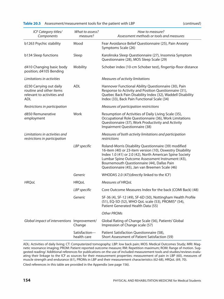
154 PHYSICAL AND REHABILITATION MEDICINE for Medical Students
Table 20.5 Assessment/measurement tools for the patient with LBP
ICF Category titles/ Components
What to assess? measure?
How to measure? Assessment methods or tools and measures
b1263 Psychic stability Mood Fear Avoidance Belief Questionnaire (25), Pain Anxiety Symptoms Scale (26)
b134 Sleep functions Sleep Karolinska Sleep Questionnaire (27), Insomnia Symptom Questionnaire (28), MOS Sleep Scale (29)
d410 Changing basic body position; d4105 Bending
Mobility Schober index (10-cm Schober test), fingertip-floor distance
Limitations in activities Measures of activity limitations
d230 Carrying out daily routine and other items relevant to activities and ADL
ADL Hannover Functional Ability Questionnaire (30), Pain Response to Activity and Position Questionnaire (31), Quebec Back Pain Disability Index (32), Waddell Disability Index (33), Back Pain Functional Scale (34)
Restrictions in participation Measures of participation restrictions
d850 Remunerative employment
Work Resumption of Activities of Daily Living Scale (35), Occupational Role Questionnaire (36), Work Limitations Questionnaire (37), Work Productivity and Activity Impairment Questionnaire (38)
Limitations in activities and restrictions in participation
Measures of both activity limitations and participation restrictions
LBP specific Roland-Morris Disability Questionnaire (39) modified 16-item (40) or 23-item version (10), Oswestry Disability Index 1.0 (41) or 2.0 (42), North American Spine Society Lumbar Spine Outcome Assessment Instrument (43), Bournemouth Questionnaire (44), Dallas Pain Questionnaire (45), Jan van Breemen Scale (46)
Generic WHODAS 2.0 (47)(directly linked to the ICF)
HRQoL HRQoL Measures of HRQoL
LBP specific Core Outcome Measures Index for the back (COMI Back) (48)
Generic SF-36 (4), SF-12 (49), SF-6D (50), Nottingham Health Profile (51), EQ-5D (52), WHO QoL scale (53), PROMIS® (54), Patient Generated Health Data (55)
Other PROMs
Global impact of interventions Improvement/ Change
Global Rating of Change Scale (56), Patients’ Global Impression of Change scale (57)
Satisfaction--- health care
Patient Satisfaction Questionnaire (58), Short Assessment of Patient Satisfaction (59)
ADL: Activities of daily living; CT: Computerized tomography; LBP: low back pain; MOS: Medical Outcomes Study; MRI: Mag-netic resonance imaging; PROM: Patient reported outcome measure; RM: Repetition maximum; ROM: Range of motion. Sug-gested reading/ Additional references for publications on the use of included measurement tools and studies/reviews evalu-ating their linkage to the ICF as sources for their measurement properties: measurement of pain in LBP (60), measures of muscle strength and endurance (61), PROMs in LBP and their measurement characteristics (62-68), HRQoL (69, 70). Cited references in this table are provided in the Appendix (see page 156).
(continued)

20 Essential methods of assessing patient’s needs 155
studies/191-introduction-to-the-icf-case-studies 9. Stucki G, Prodinger B, Bickenbach J. Four steps to
follow when documenting functioning with the International Classification of Functioning, Dis-ability and Health. Eur J Phys Rehabil Med. 2017; 53: 144-9.
10. GBD 2015 Disease and Injury Incidence and Prevalence Collaborators. Global, regional, and national incidence, prevalence, and years lived with disability for 310 diseases and injuries, 1990-2015: a systematic analysis for the Global Burden of Disease Study 2015. Lancet 2016; 388: 1545-602.
11. Valero R, Varela E, Küçükdeveci AA, Oral A, Ilieva E, Berteanu M, et al.; UEMS-PRM Section Professional Practice Committee. Spinal pain management. The role of physical and rehabilita-tion medicine physicians. The European perspec-tive based on the best evidence. A paper by the UEMS-PRM Section Professional Practice Com-mittee. Eur J Phys Rehabil Med. 2013; 49: 715-25.
12. Stucki G, Kostanjsek N, Üstün B, Ewert T, Cieza A. Applying the ICF in Rehabilitation Medicine. In: Frontera WR, editor-in-chief. DeLisa’s Physical Medicine and Rehabilitation: Principles and Prac-tice. Philadelphia: Lippincott Williams & Wilkins; 2010, Chapter 11, p. 301-24.
13. Cieza A, Stucki G, Weigl M, Disler P, Jäckel W, van der Linden S, et al. ICF Core Sets for low back pain. J Rehabil Med. 2004; 44 Suppl: S69-S74.
14. ICF browser [Internet]. World Health Organiza-tion: Geneva. Available from: http://apps.who.int/classifications/icfbrowser/
15. Stier-Jarmer M, Cieza A, Borchers M, Stucki G; World Health Organization. How to apply the ICF and ICF core sets for low back pain. Clin J Pain. 2009; 25: 29-38.
16. World Health Organization. How to use the ICF: A practical manual for using the International Clas-sification of Functioning, Disability and Health
REFERENCES
1. Tesio L. Functional assessment in rehabilitative medicine: principles and methods. Eura Medico-phys. 2007; 43: 515-23.
2. Stucki G, Melvin J. The International Classifica-tion of Functioning, Disability and Health: a uni-fying model for the conceptual description of physical and rehabilitation medicine. J Rehabil Med. 2007; 39: 286-92.
3. World Health Organization. International Classi-fication of Functioning, Disability and Health (ICF). WHO: Geneva; 2001. Available from: http://www.who.int/classifications/icf/en/
4. Steiner WA, Ryser L, Huber E, Uebelhart D, Ae-schlimann A, Stucki G. Use of the ICF model as a clinical problem-solving tool in physical therapy and rehabilitation medicine. Phys Ther. 2002; 82: 1098-107.
5. Rauch A, Cieza A, Stucki G. How to apply the In-ternational Classification of Functioning, Disabil-ity and Health (ICF) for rehabilitation manage-ment in clinical practice. Eur J Phys Rehabil Med 2008; 44: 329-42.
6. Bickenbach J, Cieza A, Rauch A, Stucki C, editors.[ICF Research Branch in cooperation with the WHO Collaborating Centre for the Family of In-ternational Classifications in Germany (at DIM-DI)]. ICF Core Sets: Manual for Clinical Practice. Göttingen: Hogrefe Publishing; 2012.
7. ICF Research Branch. ICF Core Sets Available from: https://www.icf-research-branch.org/down-load/category/4-icf-core-sets
8. Swiss Paraplegic Research, ICF Research Branch, Swiss Paraplegic Centre. Introduction to the ICF Case Studies: Translating interventions into real-life gains – A Rehab-Cycle approach. 2nd Edition 2015, Available from: https://www.icf-research-branch.org/download/send/17-icf-based-case-
Key messages
• Assessing patients’ needs is the starting point for treatment decisions and for designing physical and rehabilitation medicine programmes.
• ICF-based rehabilitation tools serve ideally for the documentation of functioning of individuals with a health condition and allow physicians to comprehensively identify patients’ needs to target appropriate PRM interventions.
• The practical ICF tools for identification of ICF domains/category titles to document patient’s functioning are ICF Core Sets, ICF Assessment Sheet, and ICF Categorical Profile.
• It is important to select proper patient-centred measurement tools when measuring impairment in body function and structures, activity limitations and participation restrictions, and HRQoL.
156 PHYSICAL AND REHABILITATION MEDICINE for Medical Students
sessment in Physical Medicine and Rehabilitation: Views and Perspectives. Chapter 2. Maugeri Foun-dation Books. Pavia: PIME, 2004, p. 35-43.
29. Franchignoni F, Ring H. Measuring change in re-habilitation medicine. Eura Medicophys. 2006; 42: 1-3.
APPENDIX
References for assessments/assessment tools mentioned in Table 20.5:
1. Chou R, Fu R, Carrino JA, Deyo RA. Imaging strategies for low-back pain: systematic review and meta-analysis. Lancet 2009; 373: 463-72.
2. Carlsson AM. Assessment of chronic pain. I. As-pects of the reliability and validity of the visual analogue scale. Pain 1983; 16: 87-101.
3. Von Korff M, Jensen MP, Karoly P. Assessing global pain severity by self-report in clinical and health services research. Spine (Phila Pa 1976) 2000; 25: 3140-51.
4. Ware JE Jr, Sherbourne CD. The MOS 36-item short form health survey (SF-36). 1. Conceptual frame-work and item selection. Med Care 1992; 30: 473-81.
5. Melzack R. The McGill Pain Questionnaire: ma-jor properties and scoring methods. Pain 1975; 1: 277-99.
6. Haefeli M, Elfering A. Pain assessment. Eur Spine J 2006; 15 Suppl 1: S17-S24.
7. Tesio L, Granger CV, Fiedler RC. A unidimen-sional pain/disability measure for low-back pain syndromes. Pain 1997; 69: 269-78.
8. van der Windt DA, Simons E, Riphagen II, Am-mendolia C, Verhagen AP, Laslett M, et al. Physi-cal examination for lumbar radiculopathy due to disc herniation in patients with low-back pain. Cochrane Database Syst Rev 2010; 2: CD007431.
9. Rubinstein SM, van Tulder M. A best-evidence review of diagnostic procedures for neck and low-back pain. Best Pract Res Clin Rheumatol 2008; 22: 471-82.
10. Patrick DL, Deyo RA, Atlas SJ, Singer DE, Chapin A, Keller RB. Assessing health-related quality of life in patients with sciatica. Spine (Phila Pa 1976) 1995; 20: 1899-908.
11. Deyo RA, Battie M, Beurskens AJ, Bombardier C, Croft P, Koes B, et al. Outcome measures for low back pain research. A proposal for standardized use. Spine (Phila Pa 1976) 1998; 23: 2003-13.
12. Grøvle L, Haugen AJ, Keller A, Natvig B, Brox JI, Grotle M. The bothersomeness of sciatica: pa-tients’ self-report of paresthesia, weakness and leg pain. Eur Spine J 2010; 19: 263-9.
(ICF). Exposure draft for comment. WHO: Gene-va, 2013. Available from:http://www.who.int/clas-sifications/drafticfpracticalmanual.pdf
17. Küçükdeveci AA, Tennant A, Grimby G, Franchi-gnoni F. Strategies for assessment and outcome measurement in physical and rehabilitation medi-cine: an educational review. J Rehabil Med. 2011; 43: 661-72.
18. World Health Organization. ICF training Begin-ner’s Guide. Towards a Common Language for Functioning, Disability and Health: ICF. WHO: Geneva; 2002. Available from: http://www.who.int/classifications/icf/icfbeginnersguide.pdf?ua=1
19. Franchignoni F, Salaffi F. Quality of life assess-ment in rehabilitation medicine. Eura Medico-phys. 2003; 39: 191-8.
20. Guyatt GH, Feeny DH, Patrick DL. Measuring health-related quality of life. Ann Intern Med. 1993; 118: 622-9.
21. National Institutes of Health. PROMIS® (Patient Reported Outcomes Measurement Information System Instrument). Development and Psycho-metric Evaluation Scientific Standards Available from: http://www.nihpromis.org/Documents/PROMIS_Standards_ 050212.pdf
22. Tucker CA, Escorpizo R, Cieza A, Lai JS, Stucki G, Ustun TB, et al. Mapping the content of the Pa-tient-Reported Outcomes Measurement Informa-tion System (PROMIS®) using the International Classification of Functioning, Health and Disabil-ity. Qual Life Res. 2014; 23: 2431-8.
23. Shapiro M, Johnston D., Wald J, Mon D. Patient-generated Health Data: White Paper. Prepared for the Office of the National Coordinator for Health it by RTI International. RTI international, NC, USA, April 2012. Available from:http://www.rti.org/sites/default/files/resources/patientgenerated-healthdata.pdf
24. Kamper SJ, Maher CG, Mackay G. Global rating of change scales: a review of strengths and weak-nesses and considerations for design. J Man Manip Ther. 2009; 17: 163-70.
25. Almeida RS, Bourliataux-Lajoinie S, Martins M. Satisfaction measurement instruments for health-care service users: a systematic review. Cad Saude Publica 2015; 31: 11-25.
26. Franchignoni F, Michail X. Selecting an outcome measure in rehabilitation medicine. Eura Medico-phys. 2003; 39: 67-8.
27. Cieza A, Fayed N, Bickenbach J, Prodinger B. Re-finements of the ICF linking rules to strengthen their potential for establishing comparability of health information. Disabil Rehabil. 2016 Mar: 1-10 [Epub ahead of print]
28. Tennant A. Principals and practice of measuring outcome. In: Barat M, Franchignoni F, editors. As-
20 Essential methods of assessing patient’s needs 157
erties of the Karolinska Sleep Questionnaire. J Sleep Res 1992; 1 Suppl 1: S113.
28. Okun ML, Kravitz HM, Sowers MF, Moul DE, Buysse DJ, Hall M. Psychometric evaluation of the Insomnia Symptom Questionnaire: a self-report measure to identify chronic insomnia. J Clin Sleep Med 2009; 5: 41-51.
29. Hays RD, Stewart AL. Sleep measures. In Stewart AL, Ware JE, editors, Measuring functioning and well-being: The Medical Outcomes Study ap-proach, Durham, NC: Duke University Press, 1992. p. 235-59.
30. Kohlmann T, Raspe H. Hannover Functional Questionnaire in ambulatory diagnosis of func-tional disability caused by backache. Rehabilita-tion (Stuttg) 1996; 35: I-VIII. [Article in Ger-man]
31. Roach KE, Brown MD, Albin RD, Delaney KG, Lipprandi HM, Rangelli D. The sensitivity and specificity of pain response to activity and posi-tion in categorizing patients with low back pain. Phys Ther 1997; 77: 730-8.
32. Kopec JA, Esdaile JM, Abrahamowicz M, Aben-haim L, Wood-Dauphinee S, Lamping DL, et al. The Quebec Back Pain Disability Scale. Measure-ment properties. Spine (Phila Pa 1976) 1995; 20: 341-52.
33. Waddell G, Main CJ. Assessment of severity in low back disorders. Spine 1984; 9: 204-7.
34. Stratford PW, Binkley JM, Riddle DL. Develop-ment and initial validation of the Back Pain Func-tional Scale. Spine 2000; 25: 2095-102.
35. Williams RM, Myers AM. A new approach to measuring recovery in injured workers with acute low back pain: Resumption of Activities of Daily Living Scale. Phys Ther 1998; 78: 613-23.
36. Kopec JA, Esdaile JM. Occupational role perfor-mance in persons with back pain. Disabil Rehabil 1998; 20: 373-9.
37. Lerner D, Amick BC 3rd, Rogers WH, Malspeis S, Bungay K, Cynn D. The Work Limitations Ques-tionnaire. Med Care 2001; 39: 72-85.
38. Reilly MC, Zbrozek AS, Dukes EM. The validity and reproducibility of a work productivity and activity impairment instrument. Pharmacoeco-nomics 1993; 4: 353-65.
39. Roland M, Morris R. A study of the natural history of back pain. Part I: development of a reliable and sensitive measure of disability in low-back pain. Spine (Phila Pa 1976) 1983; 8: 141-4.
40. Dionne CE, Koepsell TD, Von Korff M, Deyo RA, Barlow WE, Checkoway H. Predicting long-term functional limitations among back pain patients in primary care settings. J Clin Epidemiol 1997; 50: 31-43.
41. Fairbank JC, Couper J, Davies JB, O’Brien JP. The
13. Manniche C, Asmussen K, Lauritsen B, Vinterberg H, Kreiner S, Jordan A. Low Back Pain Rating scale: validation of a tool for assessment of low back pain. Pain 1994; 57: 317-26.
14. Freynhagen R, Baron R, Gockel U, Tölle TR. pain-DETECT: a new screening questionnaire to iden-tify neuropathic components in patients with back pain. Curr Med Res Opin 2006; 22: 1911-20.
15. O’Dell MW, Lin CD, Singh JR, Christolas GC. The physiatric history and physical examination. In: Cifu DX, editor. Braddom’s Physical Medicine and Rehabilitation. Philadelphia: Elsevier, Inc.; 2016, p. 3-40.
16. Van Herp G, Rowe P, Salter P, Paul JP. Three-di-mensional lumbar spinal kinematics: a study of range of movement in 100 healthy subjects aged 20 to 60+ years. Rheumatology (Oxford) 2000; 39: 1337-40.
17. Paul B, Leitner C, Vacariu G, Wick F, Zehetmayer S, Matzner M, et al. Low-back pain assessment based on the Brief ICF Core Sets: diagnostic rele-vance of motor performance and psychological tests. Am J Phys Med Rehabil 2008; 87: 452-60.
18. Biering-Sørensen F. Physical measurements as risk indicators for low-back trouble over a one-year period. Spine 1984; 9: 106-19.
19. Andersson EI, Lin CC, Smeets RJ. Performance tests in people with chronic low back pain: respon-siveness and minimal clinically important change. Spine (Phila Pa 1976) 2010; 35: E1559-63.
20. Yamada KA, Lewthwaite R, Popovich JM Jr, Be-neck GJ, Kulig K; Physical Therapy Clinical Re-search Network. The Low Back Activity Confi-dence Scale (LoBACS): preliminary validity and reliability. Phys Ther 2011; 91: 1592-603.
21. Baker R. Gait analysis methods in rehabilitation. J Neuroeng Rehabil 2006; 3: 4.
22. Beck AT, Ward CH, Mendelson M, Mock J, Er-baugh J. An inventory for measuring depression. Arch Gen Psychiatry 1961; 4: 561-71.
23. Kroenke K, Spitzer R L, Williams JB. The Patient Health Questionnaire-2: Validity of a two-item de-pression screener. Med Care 2003; 41: 1284-92.
24. Derogatis LR, Rickels K, Rock AF. The SCL-90 and the MMPI: a step in the validation of a new self-report scale. Br J Psychiatry 1976; 128: 280-9.
25. Waddell G, Newton M, Henderson I, Somerville D, Main CJ. A Fear-Avoidance Beliefs Question-naire (FABQ) and the role of fear-avoidance beliefs in chronic low back pain and disability. Pain 1993; 52: 157-68.
26. McCracken LM, Zayfert C, Gross RT. The Pain Anxiety Symptoms Scale: development and vali-dation of a scale to measure fear of pain. Pain 1992; 50: 67-73.
27. Kecklund G, Akerstedt T. The psychometric prop-

158 PHYSICAL AND REHABILITATION MEDICINE for Medical Students
healthmeasures.net/explore-measurement-sys-tems/promis
55. Cohen DJ, Keller SR, Hayes GR, Dorr DA, Ash JS, Sittig DF. Integrating patient-generated health da-ta into clinical care settings or clinical decision-making: lessons learned from project HealthDe-sign. JMIR Hum Factors 2016; 3: e26.
56. Jaeschke R, Singer J, Guyatt GH. Measurement of health status. Ascertaining the minimal clinically important difference. Control Clin Trials 1989; 10: 407-15.
57. Hurst H, Bolton J. Assessing the clinical signifi-cance of change scores recorded on subjective outcome measures. J Manipulative Physiol Ther 2004; 27: 26-35.
58. Ware JE Jr, Snyder MK, Wright WR, Davies AR. Defining and measuring patient satisfaction with medical care. Eval Program Plann 1983; 6: 247-63.
59. Hawthorne G, Sansoni J, Hayes L, Marosszeky N, Sansoni E. Measuring patient satisfaction with health care treatment using the Short Assessment of Patient Satisfaction measure delivered superior and robust satisfaction estimates. J Clin Epidemiol 2014; 67: 527-37.
60. Mannion AF, Balagué F, Pellisé F, Cedraschi C. Pain measurement in patients with low back pain. Nat Clin Pract Rheumatol 2007; 3: 610-8.
61. Gruther W, Wick F, Paul B, Leitner C, Posch M, Matzner M, et al. Diagnostic accuracy and reli-ability of muscle strength and endurance measure-ments in patients with chronic low back pain. J Rehabil Med 2009; 41: 613-9.
62. Grotle M, Brox JI, Vøllestad NK. Functional status and disability questionnaires: what do they assess? A systematic review of back-specific outcome questionnaires. Spine (Phila Pa 1976) 2005; 30: 130-40.
63. Mannion AF, Elfering A, Staerkle R, Junge A, Grob D, Semmer NK, et al. Outcome assessment in low back pain: how low can you go? Eur Spine J 2005; 14: 1014-26.
64. Maughan EF, Lewis JS. Outcome measures in chronic low back pain. Eur Spine J 2010; 19: 1484-94.
65. Ramasamy A, Martin ML, Blum SI, Liedgens H, Argoff C, Freynhagen R, et al. Assessment of pa-tient-reported outcome instruments to assess chronic low back pain. Pain Med 2017; 18: 1098-110.
66. Sigl T, Cieza A, Brockow T, Chatterji S, Kostanjsek N, Stucki G. Content comparison of low back pain-specific measures based on the International Classification of Functioning, Disability and Health (ICF). Clin J Pain 2006; 22: 147-53.
67. Ibsen C, Schiøttz-Christensen B, Melchiorsen H,
Oswestry low back pain disability questionnaire. Physiotherapy 1980; 66: 271-3.
42. Baker DJ, Pynsent PB, Fairbank JCT. The Oswes-try Disability Index revisited: its reliability, repeat-ability and validity, and a comparison with the St Thomas Disability Index. In: Roland M, Jenner JR, editors. Back pain: new approaches to rehabilita-tion and education. Manchester: Manchester Uni-versity Press; 1989, p. 174-86.
43. Daltroy LH, Cats-Baril WL, Katz JN, Fossel AH, Liang MH. The North American spine society lumbar spine outcome assessment Instrument: re-liability and validity tests. Spine (Phila Pa 1976) 1996; 21: 741-9.
44. Bolton JE, Breen AC. The Bournemouth Ques-tionnaire: a short-form comprehensive outcome measure. I. Psychometric properties in back pain patients. J Manipulative Physiol Ther 1999; 22: 503-10.
45. Lawlis GF, Cuencas R, Selby D, McCoy CE. The development of the Dallas Pain Questionnaire. An assessment of the impact of spinal pain on behav-ior. Spine (Phila Pa 1976). 1989; 14: 511-6.
46. Lankhorst GJ, van de Stadt RJ, Vogelaar TW, van der Korst JK, Prevo AJ. Objectivity and repeatabil-ity of measurements in low back pain. Scand J Re-habil Med 1982; 14: 21-6.
47. Üstün TB, Kostanjsek N, Chatterji S, Rehm J, editors. Measuring Health and Disability: Manual for WHO Disability Assessment Schedule 2.0 (WHODAS 2.0). World Health Organization; 2010 [Internet] [cited 2017 June 1]. Available from: http://apps.who.int/iris/bitstre m/10665/43974/1/9789241547598_eng.pdf
48. Ferrer M, Pellisé F, Escudero O, Alvarez L, Pont A, Alonso J, et al. Validation of a minimum outcome core set in the evaluation of patients with back pain. Spine (Phila Pa 1976) 2006; 31: 1372-9.
49. Ware J Jr, Kosinski M, Keller SD. A 12-Item Short-Form Health Survey: construction of scales and preliminary tests of reliability and validity. Med Care 1996; 34: 220-33.
50. Brazier J, Roberts J, Deverill M. The estimation of a preference-based measure of health from the SF-36. J Health Econ 2002; 21: 271-92.
51. Hunt SM, McEwen J, McKenna SP. Measuring health status: a new tool for clinicians and epide-miologists. J R Coll Gen Pract 1985; 35: 185-8.
52. EuroQol Group. EuroQol--a new facility for the measurement of health-related quality of life. Health Policy 1990; 16: 199-208.
53. WHOQOL Group. Development of the World Health Organization WHOQOL-BREF quality of life assessment. The WHOQOL Group. Psychol Med 1998; 28: 551-8.
54. National Institutes of Health. PROMIS®.[Internet] [cited 2017 June 5] Available from: http://www.
20 Essential methods of assessing patient’s needs 159
Functioning, Disability and Health. Disabil Reha-bil 2016; 38: 1089-96.
69. Lurie J. A review of generic health status measures in patients with low back pain. Spine (Phila Pa 1976) 2000; 25: 3125-9.
70. Finch AP, Dritsaki M, Jommi C. Generic prefer-ence-based measures for low back pain: which of them should be used? Spine (Phila Pa 1976) 2016; 41: E364-74.
Nielsen CV, Maribo T. Do patient-reported out-come measures describe functioning in patients with low back pain, using the Brief International Classification of Functioning, Disability and Health Core Set as a reference? J Rehabil Med 2016; 48: 618-24.
68. Nicol R, Robinson Nicol M, Hopfe M, Newell D. Linking the Bournemouth Questionnaire for low back pain to the International Classification of


21 Rehabilitation settings and the concept of interdisciplinary care 161
THE PERSON-CENTRED APPROACH OF PRM
PRM covers a broad range of disorders and in-cludes the consequences of trauma, surgery, dis-eases and congenital conditions. Moreover, PRM is not primarily focused on the disorder itself, but focuses on the consequences in terms of activity limitations and restrictions in participation.
PRM typically acts from a patient- or person-centred approach, that includes the personal char-acteristics of the patient, in contrast with the dis-ease-oriented approach of the organ-based spe-cialties. A person-centred approach covers several meanings (3):
ū addressing the person’s specific and holistic properties
ū addressing the person’s difficulties in everyday life
ū the person as an expert: participation and em-powerment
ū respect the person ‘behind’ the impairment or the disease.
INTERDISCIPLINARY TEAMWORK IN PRM
The consequence of this “holistic” approach is that PRM physicians do not work alone, but need to involve a large number of other healthcare pro-fessionals. The healthcare professionals operate with an interdisciplinary approach in a multi-professional team, which also includes the patient and/or his caregivers as well as peer counsellors.
PRM AS THE SPECIALTY OF THE REHABILITATION HEALTH STRATEGY WITH FUNCTIONING AS KEY OUTCOME
Physical and Rehabilitation Medicine (PRM) aims at the achievement of the best possible “functioning” of a person with a long-term health condition. It is clear that the classical biomedical model is not applicable to PRM. In the more ap-propriate “bio-psycho-social model” of treat-ment, therapy focuses on the care of the whole person, as described in the International Classifi-cation of Functioning, Disability and Health (ICF) (1). So the core of PRM is not merely single body structure or function, but the total person and human being, including his psychology and motivation (“personal factors”) and social envi-ronment (“participation” and “environmental factors”). PRM focuses in general on functioning and disability; PRM aims at reducing “activity limitations”, and improving “impairments”, while addressing also “participation restrictions” at a micro-level of healthcare (patient interac-tions). At both meso-level (hospitals, rehabilita-tion services, etc.) and macro-level (healthcare policy), participation restrictions of persons with disabilities or chronic health conditions should be addressed, with the expert help of PRM physi-cians, by those who work on society at large, in-cluding educators and politicians or other deci-sion makers. These levels of health care refer re-spectively to healthcare policy, healthcare orga-nization and community and the level of patient interaction (2).
21 Rehabilitation settings and the concept
of interdisciplinary careCarlotte KIEKENS
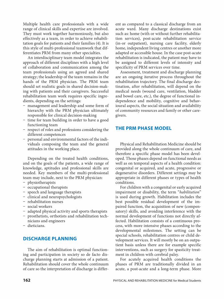
162 PHYSICAL AND REHABILITATION MEDICINE for Medical Students
ent as compared to a classical discharge from an acute ward. Many discharge destinations exist such as: home (with or without further rehabilita-tion services), post-acute rehabilitation service (in-or outpatient), nursing care facility, elderly home, independent living centres or another more adapted or accessible house. In the case post-acute rehabilitation is indicated, the patient may have to be assigned to different levels of intensity and specificity of PRM services over time.
Assessment, treatment and discharge planning are an ongoing iterative process throughout the rehabilitation trajectory. The final discharge des-tination, after rehabilitation, will depend on the medical needs (wound care, ventilation, bladder and bowel care, etc.), the degree of functional in-dependence and mobility, cognitive and behav-ioural aspects, the social situation and availability of community resources and family or other care-givers.
THE PRM PHASE MODEL
Physical and Rehabilitation Medicine should be provided along the whole continuum of care, and therefore a specific phase model has been devel-oped. Those phases depend on functional needs as well as on temporal aspects of a health condition: congenital or acquired, and acute, progressive or degenerative disorders. Different settings may be appropriate in different phases or types of health conditions.
For children with a congenital or early acquired impairment or disability, the term “habilitation” is used during growth. Habilitation includes the best possible residual development of the im-paired function, the acquisition of new (compen-satory) skills, and avoiding interference with the normal development of functions not directly af-fected. Habilitation consists of a continuous pro-cess, with more intensive phases according to the developmental milestones. The setting can be special schools, rehabilitation centres or child de-velopment services. It will mostly be on an outpa-tient basis unless there are for example specific interventions, such as surgery for spasticity treat-ment in children with cerebral palsy.
For acutely acquired health conditions the phases of PRM are traditionally divided in an acute, a post-acute and a long-term phase. More
Multiple health care professionals with a wide range of clinical skills and expertise are involved. They must work together harmoniously, but also effectively as a team, in order to achieve rehabili-tation goals for patients and their families (4). It is this style of multi-professional teamwork that dif-ferentiates PRM from many other specialties.
An interdisciplinary team model integrates the approach of different disciplines with a high level of collaboration and communication among the team professionals using an agreed and shared strategy; the leadership of the team remains in the hands of the PRM physician. The PRM team should set realistic goals in shared decision-mak-ing with patients and their caregivers. Successful rehabilitation team work requires specific ingre-dients, depending on the settings:
ū management and leadership and some form of hierarchy with the PRM physician ultimately responsible for clinical decision-making
ū time for team building in order to have a good functioning team
ū respect of roles and professions considering the different competences
ū personal and environmental factors of the indi-viduals composing the team and the general attitudes in the working place.
Depending on the treated health conditions, and on the goals of the patients, a wide range of knowledge, aptitudes and professional skills are needed. Key members of the multi-professional team may include, next to the PRM physician:
ū physiotherapists ū occupational therapists ū speech and language therapists ū clinical and neuropsychologists ū rehabilitation nurses ū social workers ū adapted physical activity and sports therapists ū prosthetists, orthotists and rehabilitation tech-
nicians and engineers ū dieticians.
DISCHARGE PLANNING
The aim of rehabilitation is optimal function-ing and participation in society so de facto dis-charge planning starts at admission of a patient. Rehabilitation should cover the whole continuum of care so the interpretation of discharge is differ-
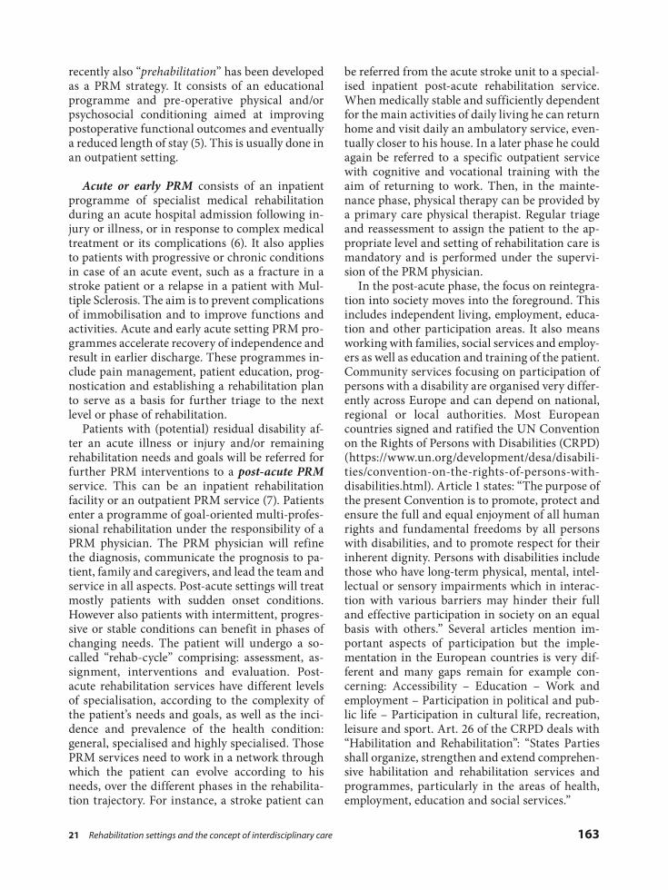
21 Rehabilitation settings and the concept of interdisciplinary care 163
be referred from the acute stroke unit to a special-ised inpatient post-acute rehabilitation service. When medically stable and sufficiently dependent for the main activities of daily living he can return home and visit daily an ambulatory service, even-tually closer to his house. In a later phase he could again be referred to a specific outpatient service with cognitive and vocational training with the aim of returning to work. Then, in the mainte-nance phase, physical therapy can be provided by a primary care physical therapist. Regular triage and reassessment to assign the patient to the ap-propriate level and setting of rehabilitation care is mandatory and is performed under the supervi-sion of the PRM physician.
In the post-acute phase, the focus on reintegra-tion into society moves into the foreground. This includes independent living, employment, educa-tion and other participation areas. It also means working with families, social services and employ-ers as well as education and training of the patient. Community services focusing on participation of persons with a disability are organised very differ-ently across Europe and can depend on national, regional or local authorities. Most European countries signed and ratified the UN Convention on the Rights of Persons with Disabilities (CRPD) (https://www.un.org/development/desa/disabili-ties/convention-on-the-rights-of-persons-with-disabilities.html). Article 1 states: “The purpose of the present Convention is to promote, protect and ensure the full and equal enjoyment of all human rights and fundamental freedoms by all persons with disabilities, and to promote respect for their inherent dignity. Persons with disabilities include those who have long-term physical, mental, intel-lectual or sensory impairments which in interac-tion with various barriers may hinder their full and effective participation in society on an equal basis with others.” Several articles mention im-portant aspects of participation but the imple-mentation in the European countries is very dif-ferent and many gaps remain for example con-cerning: Accessibility – Education – Work and employment – Participation in political and pub-lic life – Participation in cultural life, recreation, leisure and sport. Art. 26 of the CRPD deals with “Habilitation and Rehabilitation”: “States Parties shall organize, strengthen and extend comprehen-sive habilitation and rehabilitation services and programmes, particularly in the areas of health, employment, education and social services.”
recently also “prehabilitation” has been developed as a PRM strategy. It consists of an educational programme and pre-operative physical and/or psychosocial conditioning aimed at improving postoperative functional outcomes and eventually a reduced length of stay (5). This is usually done in an outpatient setting.
Acute or early PRM consists of an inpatient programme of specialist medical rehabilitation during an acute hospital admission following in-jury or illness, or in response to complex medical treatment or its complications (6). It also applies to patients with progressive or chronic conditions in case of an acute event, such as a fracture in a stroke patient or a relapse in a patient with Mul-tiple Sclerosis. The aim is to prevent complications of immobilisation and to improve functions and activities. Acute and early acute setting PRM pro-grammes accelerate recovery of independence and result in earlier discharge. These programmes in-clude pain management, patient education, prog-nostication and establishing a rehabilitation plan to serve as a basis for further triage to the next level or phase of rehabilitation.
Patients with (potential) residual disability af-ter an acute illness or injury and/or remaining rehabilitation needs and goals will be referred for further PRM interventions to a post-acute PRM service. This can be an inpatient rehabilitation facility or an outpatient PRM service (7). Patients enter a programme of goal-oriented multi-profes-sional rehabilitation under the responsibility of a PRM physician. The PRM physician will refine the diagnosis, communicate the prognosis to pa-tient, family and caregivers, and lead the team and service in all aspects. Post-acute settings will treat mostly patients with sudden onset conditions. However also patients with intermittent, progres-sive or stable conditions can benefit in phases of changing needs. The patient will undergo a so-called “rehab-cycle” comprising: assessment, as-signment, interventions and evaluation. Post-acute rehabilitation services have different levels of specialisation, according to the complexity of the patient’s needs and goals, as well as the inci-dence and prevalence of the health condition: general, specialised and highly specialised. Those PRM services need to work in a network through which the patient can evolve according to his needs, over the different phases in the rehabilita-tion trajectory. For instance, a stroke patient can

164 PHYSICAL AND REHABILITATION MEDICINE for Medical Students
need long-term rehabilitation care: assistance given over a long-term period of time to people who are experiencing long-term disabilities, chronic disease or difficulties in functioning (8). Long-term rehabilitation services can be provid-ed in the form of intermittent inpatient care, or continuous outpatient/community/home based rehabilitation. PRM should participate in Com-munity-Based Rehabilitation Programs, e.g. as advisor and/or trainer of community rehabilita-tion workers. PRM can support General Practi-tioners, other primary care health professionals and other medical specialists by giving advice and/or coordination of rehabilitation networks. In the long-term phase of PRM care special em-phasis lays on maintenance and secondary pre-vention activities.
Rehabilitation services over the different phas-es and settings should be stratified and organised in networks in order to allow for the best possible care adapted to the individual’s needs and goals, over the continuum of care.
Therefore PRM teams, together with Associa-tions of People with Disabilities should advocate for full inclusion and participation in society. This can be achieved through collaboration and inte-gration of PRM services with schools and univer-sities, employment services or sports and leisure services. A crucial aspect of functioning is acces-sibility and mobility in the large sense of the word: personal mobility (transfers, gait or wheelchair use), biking or car driving, public transport com-prising bus, tram, train, planes. In Art. 2 of the CRPD therefore “universal design” is defined as follows: the design of products, environments, programmes and services to be usable by all peo-ple, to the greatest extent possible, without the need for adaptation or specialized design. “Uni-versal design” shall not exclude assistive devices for particular groups of persons with disabilities where this is needed.
After a period of post-acute care, whether in-patient or outpatient based, some patients may
Key messages
• Physical and Rehabilitation Medicine (PRM) aims at the achievement of the best possible “func-tioning” of a person with a long-term health condition. PRM aims at reducing “activity limita-tions”, and improving “impairments”, while addressing also “participation restrictions”.
• PRM acts from a patient-centred approach, including the personal characteristics of the patient, in contrast with the disease-oriented approach of the organ-based specialties.
• Multiple health care professionals with a wide range of clinical skills and expertise are involved. They operate with an interdisciplinary approach, including also the patient and/or his caregivers as well as peer counsellors.
• PRM should be provided along the whole continuum of care. The phases depend on functional needs as well as on temporal aspects of a health condition: congenital or acquired, and acute, progressive or degenerative disorders. Different settings may be appropriate in different phases or types of health conditions: habilitation, prehabilitation, acute, post-acute and long-term rehabili-tation.
21 Rehabilitation settings and the concept of interdisciplinary care 165
review and meta-analysis. Physiotherapy. 2014; 100: 196-207.
6. Ward AB, Gutenbrunner C, Damjan H, Giustini A, Delarque H. UEMS Section of Physical and Rehabilitation Medicine: a position paper on Phys-ical and Rehabilitation Medicine in acute settings. J Rehabil Med. 2010; 42: 417-24.
7. Ward AB, Gutenbrunner C, Giustini A, Delarque A, Fialka-Moser V, Kiekens C, Berteanu M, Christ-odoulou N. A position paper on Physical & Reha-bilitation Medicine programmes in post-acute settings. Union of European Medical Specialists Section of Physical & Rehabilitation Medicine (in conjunction with the European Society of Physical & Rehabilitation Medicine). J Rehabil Med. 2012; 44: 289-98.
8. Takáč P, Petrovičová J, Delarque A, Stibrant Sun-nerhagen K, Neumann V, Vetra A, Berteanu M, Christodoulou N. Position paper on PRM and persons with long term disabilities. Eur J Phys Rehabil Med. 2014; 50: 453-64.
REFERENCES
1. WHO. The International Classification of Func-tioning, Disability and Health (ICF). Geneva: WHO Press; 2001.
2. Innovative care for chronic conditions: building blocks for action: global report, WHO 2002. Avail-able from:http://www.who.int/chp/knowledge/publications/icccglobalreport.pdf
3. Leplege A, Gzil F, Cammelli M, Lefeve C, Pachoud B, Ville I. Person-centredness: Conceptual and historical perspectives. Disabil Rehabil. 2007; 29: 20-1.
4. Neumann V, Gutenbrunner C, Fialka-Moser V, Christodoulou N, Varela E, Giustini A, Delarque A. Interdisciplinary team working in physical and rehabilitation medicine. J Rehabil Med. 2010; 42: 4-8.
5. Santa Mina D, Clarke H, Ritvo P, Leung YW, Mat-thew AG, Katz J, et al. Effect of total-body preha-bilitation on postoperative outcomes: a systematic


22 Effectiveness of rehabilitation interventions 167
erally, NSAID are used to reduce acute inflamma-tion, but are unlikely to treat the underlying cause of inflammation. They are efficient analgesics for a variety of arthritic disorders and for many other forms of soft tissue pain and injury (1).
Analgesics – Analgesics, particularly opioids, opioid-related compounds, local anesthetics, an-algesic adjunctive agents and anticonvulsants are useful for the treatment of peripheral neuropathic pain, complex regional pain syndromes and cen-trally mediated pain arising from central nervous system disorders.
Antispasticity medications – In patients with upper motor neuron syndrome, the treatment of spasticity is an important medical issue. The oc-currence of spasticity is not an absolute indication to prescript medication because an increase in lower limb muscle tone may be useful, for exam-ple, for patients with diminished voluntary mus-cle strength by facilitating the upright position with assistance, transfers, dressing and other ac-tivities of daily life (2). However, spasticity can be considered as irritating, painful, causing loss of sleep and aggravating quality of life. In these cases antispasticity medication is necessary. Baclofen, tizanidine, benzodiazepines, sodium dantrolene and botulinum toxin, are some of medications used in the management of spasticity.
Corticosteroids – They are profoundly effective anti-inflammatory drugs. They were initially used in the management of rheumatoid arthritis and since then their usage has been extended to a va-riety of other conditions.
Antiepileptic drugs – These medications are an important component of the physiatrist s arma-mentarium for the management of a variety of
INTRODUCTION
Rehabilitation interventions are effective in preventing complications associated with many diseases. This general perception is based on strong evidence, but there is need for more evi-dence to support the efficacy of each intervention or a combination of them for specific medical is-sues that a physiatrist is involved in. The abun-dance of these different interventions complicates this procedure. However physiatrists are also concerned with issues as disability, impairment, functioning, activity, environmental factors, par-ticipation and quality of life, and have to adjust their interventions on the principles of the Inter-national Classification of Functioning, Disability and Health (ICF). Interventions can be profiled in terms of the clinical conditions where they are used, their targets and the underlying theory guiding the therapies. This chapter summarizes the main interventions used in modern Physical and Rehabilitation Medicine.
MEDICATIONS
Physicians involved in the practice of dimin-ishing disability and handicap utilize physical medicine and rehabilitative techniques. However, Physical and Rehabilitation Medicine (PRM) doc-tors, during either inpatient management or pri-mary care for patients with disabilities, must be fully conversant with pharmacologic treatments. It is necessary for physiatrists to be familiar with effective new medications, which are useful for the population of their patients.
Nonsteroidal anti-inf lammatory drugs (NSAID) – It is a category of medications most commonly used in musculoskeletal diseases. Gen-
22Effectiveness of rehabilitation interventions
Markos SGANTZOS, Ioannis-Alexandros TZANOS

168 PHYSICAL AND REHABILITATION MEDICINE for Medical Students
function, coordination, balance, muscle power, endurance, to decrease rigidity and to enhance relaxation. Physical activities as well as exer-cise are interventions with limited adverse events. They can improve quality of life, by reducing pain severity and enhancing physical function (6).
Before subscribing therapeutic exercise, the physiatrist performs an evaluation of functional capacity based on medical history, physical exami-nation and laboratory exams. In patients with heart disorders, it is necessary to exclude patients with valvular heart disease, ventricular hypertrophy, severe arrhythmias and malignant hypertension.
The active assisted range of motion exercises are performed when the patient has very low muscle strength or when movement is limited by joint pain. During the exercises, it is crucial not to force tissues beyond the point of pain.
The passive range of motion exercises are per-formed by patients with lack of muscle strength. They include stretching of motionless muscles and joint capsules to prevent joint stiffness and muscle contracture. Joint extensibility is attained with constant and slow manual stretching of large muscles and joint capsules or with the assistance of mechanical equipment.
The three main types of therapeutic exercises are: muscle strength exercises; endurance exercis-es; and flexibility exercises.
Muscle strength exercises – The capacity of the muscle to generate force can be improved in many ways, including increasing the load, number and velocity of contractions and better technique. Re-sistance training increases power, walking veloci-ty, stair-climbing capacity and balance and also decreases topical and total fat tissue. There are three types of resistance training: isotonic, iso-metric, and isokinetic.
Resistance training has been proven to reduce risk factors for coronary artery disease, osteopo-rosis, diabetes mellitus and cancer. For example, resistance training has been shown to decrease systolic blood pressure and to increase bone min-eral density glucose tolerance, insulin resistance and bowel transit time. Cervical, low back pain and work-related back injuries have also been proven to improve with resistance training.
Endurance exercises – An endurance exercise regimen has three parameters: frequency, intensi-ty, and duration.
clinical features. These embrace the treatment of seizures following a multiplicity of congenital and acquired brain lesions, as well as a number of dif-ferent conditions, such as behavioral and psychi-atric disorders. They are also used in the manage-ment of neurogenic pain and irregular muscle tone (3).
Antidepressants – These drugs commonly are used in a variety of rehabilitation settings. They are employed to treat depression and sleep distur-bance and they are also involved in the manage-ment of pain (4). They can be distinguished into four general groups: the tricyclic antidepressants, the selective serotonin reuptake inhibitors, the monoamine oxidase inhibitors and others. Re-garding their use for pain management, a chronic treatment is needed for alleviation of neuropathic pain, implicating the activation of secondary downstream mechanisms and long-term neuro-plasticity (5).
Antihypertensives – Management of hyperten-sion demands consideration of diverse factors; blood pressure, risk factors, other associated dis-eases. Decreasing blood pressure reduces cardio-vascular mortality and morbidity, preventing from stroke, coronary disease and renal failure.
Intrathecal drug therapy – The direct infusion of medication into the epidural or intrathecal space, overcomes some of the frequent problems linked with orally administered centrally active agents. Implantable medication administration devices have been utilized to deliver drugs for the management of spasticity and pain.
THERAPEUTIC EXERCISE
Therapeutic exercise is defined as “bodily movement prescribed to correct an impairment, improve musculoskeletal function, or maintain a state of well-being”. It has a broad spectrum from highly specific activities restricted to particular muscles or body parts, to general and energetic activities that can return a recovering patient to high levels of physical condition.
The main aims of therapeutic exercise are to enhance ambulation, to release stiff muscles, ten-dons, and fascia, to maintain and increase range of motion, to improve blood circulation, pulmonary

22 Effectiveness of rehabilitation interventions 169
focus on different deep structures (8). Heat should be used very carefully in patients with sensory deficits and ischemic tissues and it is contraindi-cated in the presence of bleeding disorders, tu-mors and on developing fetus during pregnancy.
Cold application is commonly performed with ice, ice massage or cold packs. This modality has the ability to diminish edema, to cause vasocon-striction, to minimize some components of the inflammatory reaction, to reduce muscle tone and to affect pain physiology.
ELECTRICAL MODALITIES
Electrical stimulation is an old physiatric mo-dality, but a development of new interest in this domain has occurred since the mid-1970. It has been applied for functional and therapeutic pur-poses. The neuromuscular electrical stimulation has been used in normal muscle combined with voluntary exercise to increase muscle force, in se-lected cases of patients with myopathy to preserve motor capacity and in denervated muscles to sus-tain muscle fiber area and girth until the reinner-vation occurs.
Electrical stimulation of muscle following brain or spinal cord injury can have beneficial effects on the muscular atrophy presented after the immobi-lization related with these disorders. It can also prevent complications of disuse, such as deep ve-nous thrombosis, cardiovascular deconditioning and osteoporosis.
Therapeutic functional electrical stimulation (FES) has been applied in selected patients with urinary incontinence, ejaculatory failure, hemi-plegic upper limb to prevent shoulder subluxation, high-level quadriplegia for stimulation of phrenic nerve to circumvent the need of mechanical ven-tilation, and in the management of spasticity. FES has also been applied as an orthotic device in pa-tients with scoliosis or hemiplegic gait. In recent years, functional neuromuscular stimulation has been used for standing and gait in patients with spinal cord injury and for improvement of upper limb function in patients with cervical spinal cord injury.
Shortly after the description of the gate control theory of pain in 1965, transcutaneous electrical nerve stimulation (TENS) has been developed for pain treatment. TENS units have setting options for several parameters (intensity, frequency, pulse
The optimum level of these parameters is: ū Frequency: Aerobic exercise 3-5 days per week. ū Intensity: 64/70-94% of maximum heart rate
(HR max), or 40/50-85% of maximum oxygen uptake reserve (VO2 R) or heart rate reserve (HRR). At-risk patients, especially those with cardiac or respiratory disease, undergo a less intense training program, with the heart rate not exceeding that reached at 50-60% of maxi-mum O2 uptake (VO2 max).
ū Duration: Persistent or intermittent aerobic exercise for 20-60 minutes.
Flexibility exercises – They contribute in main-taining and increasing range of motion in a joint or a group of joints. This category of exercises lengthens muscle and connective tissue while re-ducing the risk of injury to those tissues. They should be done slowly and carefully, with a gradu-al increase in ranges of motion. There are 3 main forms of stretching techniques: static, dynamic, and Proprioceptive Neuromuscular Facilitation (PNF). In static technique the muscle is stretched until mild discomfort occurs and then held at that point for 15-30 s. In dynamic technique a momen-tum generated by recurrent bouncing movements causes a muscle stretch. However, dynamic flexi-bility exercises may result to muscular pain or damage. The PNF comprises of consecutive con-traction and relaxation of agonists and antagonists through an indicated sequence of movements.
THERAPEUTIC HEAT AND COLD
One of the most historic types of therapy is the application of heat or cold to relieve the symptoms of musculoskeletal disorders. These modalities are an important supplement to other therapies. Heat increases the flexibility of collagen tissue, so it can be stretched with less force and with lower possibility of mechanical injury than when it is stretched at normal tissue temperature. It also decreases joint stiffness, pain perception, muscle spasms and it increases blood flow. It is consid-ered as a first line treatment for increasing range of motion in patients and athletes (7).
Heat can be applied in various ways, fluctuat-ing from moist or dry heat, to ultrasound or dia-thermy treatments. Theoretically these different modalities have the ability to heat different types of tissue, to penetrate to different depths of and to
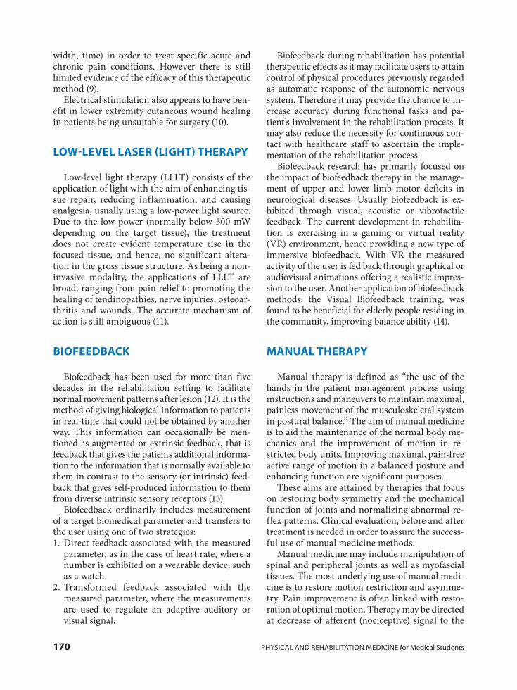
170 PHYSICAL AND REHABILITATION MEDICINE for Medical Students
Biofeedback during rehabilitation has potential therapeutic effects as it may facilitate users to attain control of physical procedures previously regarded as automatic response of the autonomic nervous system. Therefore it may provide the chance to in-crease accuracy during functional tasks and pa-tient’s involvement in the rehabilitation process. It may also reduce the necessity for continuous con-tact with healthcare staff to ascertain the imple-mentation of the rehabilitation process.
Biofeedback research has primarily focused on the impact of biofeedback therapy in the manage-ment of upper and lower limb motor deficits in neurological diseases. Usually biofeedback is ex-hibited through visual, acoustic or vibrotactile feedback. The current development in rehabilita-tion is exercising in a gaming or virtual reality (VR) environment, hence providing a new type of immersive biofeedback. With VR the measured activity of the user is fed back through graphical or audiovisual animations offering a realistic impres-sion to the user. Another application of biofeedback methods, the Visual Biofeedback training, was found to be beneficial for elderly people residing in the community, improving balance ability (14).
MANUAL THERAPY
Manual therapy is defined as “the use of the hands in the patient management process using instructions and maneuvers to maintain maximal, painless movement of the musculoskeletal system in postural balance.” The aim of manual medicine is to aid the maintenance of the normal body me-chanics and the improvement of motion in re-stricted body units. Improving maximal, pain-free active range of motion in a balanced posture and enhancing function are significant purposes.
These aims are attained by therapies that focus on restoring body symmetry and the mechanical function of joints and normalizing abnormal re-flex patterns. Clinical evaluation, before and after treatment is needed in order to assure the success-ful use of manual medicine methods.
Manual medicine may include manipulation of spinal and peripheral joints as well as myofascial tissues. The most underlying use of manual medi-cine is to restore motion restriction and asymme-try. Pain improvement is often linked with resto-ration of optimal motion. Therapy may be directed at decrease of afferent (nociceptive) signal to the
width, time) in order to treat specific acute and chronic pain conditions. However there is still limited evidence of the efficacy of this therapeutic method (9).
Electrical stimulation also appears to have ben-efit in lower extremity cutaneous wound healing in patients being unsuitable for surgery (10).
LOW-LEVEL LASER (LIGHT) THERAPY
Low-level light therapy (LLLT) consists of the application of light with the aim of enhancing tis-sue repair, reducing inflammation, and causing analgesia, usually using a low-power light source. Due to the low power (normally below 500 mW depending on the target tissue), the treatment does not create evident temperature rise in the focused tissue, and hence, no significant altera-tion in the gross tissue structure. As being a non-invasive modality, the applications of LLLT are broad, ranging from pain relief to promoting the healing of tendinopathies, nerve injuries, osteoar-thritis and wounds. The accurate mechanism of action is still ambiguous (11).
BIOFEEDBACK
Biofeedback has been used for more than five decades in the rehabilitation setting to facilitate normal movement patterns after lesion (12). It is the method of giving biological information to patients in real-time that could not be obtained by another way. This information can occasionally be men-tioned as augmented or extrinsic feedback, that is feedback that gives the patients additional informa-tion to the information that is normally available to them in contrast to the sensory (or intrinsic) feed-back that gives self-produced information to them from diverse intrinsic sensory receptors (13).
Biofeedback ordinarily includes measurement of a target biomedical parameter and transfers to the user using one of two strategies:1. Direct feedback associated with the measured
parameter, as in the case of heart rate, where a number is exhibited on a wearable device, such as a watch.
2. Transformed feedback associated with the measured parameter, where the measurements are used to regulate an adaptive auditory or visual signal.

22 Effectiveness of rehabilitation interventions 171
face, with several indications: 1) pain alleviation; 2) protection of weak or recovering musculoskeletal structures; 3) prevention or restoration of deformi-ties; and 4) functional recovery. Concerning mus-culoskeletal disorders, pain alleviation is a matter of significant importance, e.g. this can be attained by restricting the motion irritating a painful joint using a rigid splint. Protection of a weak structure or a fracture healing is attained by bracing. An ex-ample of orthotics’ role for the prevention and res-toration of deformities are children with scoliosis. Restoration often mandates either applying dy-namic force such as a spring or casting used to at-tenuate a joint contracture. All orthoses are pat-terned to regain function by relieving pain, reduc-ing weight bearing or correcting deformities, but some orthoses target mainly to functional improve-ment. These orthoses aim to restore a functional activity by assisting or facilitating a weak motion.
Terminology of orthoses is determined by the joints they involve (knee-ankle- foot orthosis), by their anticipated function (static, dynamic, func-tional) and by the material being manufactured (plaster, thermoplastic, etc.).
Prosthesis is defined as a manufactured device that substitutes a missing body segment, which has been lost after injury, disease or congenital disorders. The design of the prosthesis is based on the structural features and functional necessities of the patient.
An upper limb prosthesis is directed to replace very complicated functions executed with the hand. A standard prosthetic replacement for the upper limb can substitute many grasping and ma-nipulating hand functions and allow motion of proximal prosthetic joints to place the terminal part in space. However, it does not provide sen-sory feedback straight from the terminal part. The use of prosthesis in unilateral amputee usually does not reach a level of function and dexterity similar to that of the extremity it has replaced. Upper limb prostheses can be separated into three categories: body-powered or conventional, exter-nally powered or electric and passive or cosmetic.
A lower limb prosthesis can replace functions as weight bearing, locomotion, and cosmesis. These functions are less complicated and more equal among patients than those of an upper limb prosthesis. Since weight bearing is a primary issue in a lower limb prosthesis, distribution of the pressures between the skin and the socket of the prosthesis is crucial. The area of contact between
spinal cord. Endorphin release rises pain thresh-old and enhances pain relief. The reduction of pain improves ambulatory ability and efficiency of biomechanical motion. Manipulation has also beneficial effects by enhancing lymphatic return and improving circulation to the tissues (15). Re-garding pain in patients suffering from osteoar-thritis, manual therapy has beneficial effects in comparison to no treatment, but is less effective than pharmaceutical interventions or placebo (16).
COGNITIVE REHABILITATION
Cognitive Rehabilitation Therapy (CRT) is de-fined as the procedure of learning again cognitive skills that have been lost or changed as a result of brain lesion. If the retraining of the skills is impos-sible, the person must be taught new ones in order to compensate for the lost cognitive functions. The procedure of CRT consists of four components: 1. Education about cognitive deficits and capaci-
ties, focusing on the self-awareness of the prob-lem.
2. Process Training. This includes the acquisition of skills through direct re-education or prac-tice, focusing on the resolution of the problem.
3. Strategy Training. This refers to the use of en-vironmental, internal and external approaches, focusing is on compensating rather than solv-ing the problem.
4. Functional Activities Training. This includes the utilization of the other three components in everyday life, focusing on real life improve-ments.
CRT Services aim to attain functional changes by: 1. Promoting or re-establishing previously
learned behavioural models. 2. Establishing new forms of cognitive activity or
methods to compensate for neurological defi-cit.
3. Aiding the person be as autonomous as possible in the management of the everyday life as a family and society member (17).
ORTHOTICS AND PROSTHETICS
An orthosis is defined as a device (splint and brace), externally attached or applied to a body sur-
172 PHYSICAL AND REHABILITATION MEDICINE for Medical Students
during sitting, standing, stepping and running. Recent research focuses on the technological
progression of hardware and software in order to promote the practical and safe use of bionic upper and lower limb prosthetics/orthotics. This would signify a valuable and historical achievement, be-ing able to substantially improve the quality of life for disabled people (18).
skin and socket is the main place of lower limb prosthetic fitting difficulties and the cause of lower limb prosthetic modifications. The second main function, locomotion, should provide the amputee the ability to walk with a gait that simu-lates as much as possible normal gait. The cos-metic impact of a lower extremity prosthesis should be regarded as a function of prosthetic aid
Key messages
• Controversy still remains on the effectiveness of the above rehabilitation interventions. The ac-curate diagnosis of the underlying pathology is always necessary before a clinical decision.
• Further research is needed to determine the indications, the appropriate time and the parameters of the application of these therapeutic methods.
REFERENCES
1. Atkinson TJ, Fudin J, Jahn HL, Kubotera N, Ren-nick AL, Rhorer M. What’s new in NSAID phar-macotherapy: oral agents to injectables. Pain Med. 2013; 14: 11-7.
2. Simon O, Yelnik AP. Managing spasticity with drugs. Eur J Phys Rehabil Med. 2010; 46: 401-10.
3. Moore A, Wiffen P, Kalso E. Antiepileptic drugs for neuropathic pain and fibromyalgia. JAMA. 2014; 312: 182-3.
4. Dharmshaktu P, Tayal V, Kalra BS. Efficacy of an-tidepressants as analgesics: a review. J Clin Phar-macol. 2012; 52: 6-17
5. Kremer M, Salvat E, Muller A, Yalcin I, Barrot M. Antidepressants and gabapentinoids in neuro-pathic pain: mechanistic insights. Neuroscience 2016; 338: 183-206.
6. Geneen LJ, Moore RA, Clarke C, et al. Physical activity and exercise for chronic pain in adults: an overview of Cochrane Reviews. Cochrane Data-base Syst Rev 2017 Jan 14; 1: CD011279.
7. Bleakley CM, Costello JT. Do thermal agents affect range of movement and mechanical properties in soft tissues? A systematic review. Arch Phys Med Rehabil. 2013; 94: 149-63.
8. Swenson RS. Therapeutic modalities in the man-agement of nonspecific neck pain. Phys Med Reha-bil Clin N Am. 2003; 14: 605-27.
9. Samuel SR, Maiya GA. Application of low fre-quency and medium frequency currents in the management of acute and chronic pain-a narrative review. Indian J Palliat Care 2015; 21: 116-20.
10. Ashrafi M, Alonso-Rasgado T, Baguneid M, Bayat
A. The efficacy of electrical stimulation in lower extremity cutaneous wound healing: A systematic review. Exp Dermatol. 2017; 26: 171-8.
11. de Freitas LF, Hamblin MR. Proposed mecha-nisms of photobiomodulation or low-level light therapy. IEEE J Sel Top Quantum Electron. 2016; 22(3). pii: 7000417.
12. Tate JJ, Milner CE. Real-time kinematic, temporo-spatial, and kinetic biofeedback during gait re-training in patients: a systematic review. Phys Ther. 2010; 10: 1123-34.
13. Onate J, Guskiewicz K, Sullivan R. Augmented feedback reduces jump landing forces. J Orthop Sports Phys Ther. 2001; 10: 511-7.
14. Alhasan H, Hood V, Mainwaring F. The effect of visual biofeedback on balance in elderly popula-tion: a systematic review. Clin Interv Aging 2017; 12: 487-97.
15. Brault JS, Robert E. Kappler RE, Grogg BE. Ma-nipulation, Traction, and Massage. In: Braddom RL (ed) Physical Medicine and Rehabilitation. Philadelphia, PA: Elsevier Saunders; 2011.
16. French HP, Brennan A, White B, Cusack T. Manu-al therapy for osteoarthritis of the hip or knee - a systematic review. Man Ther 2011; 16: 109-17.
17. The Society for Cognitive Rehabilitation. What is cognitive rehabilitation therapy? [Internet] © 2010 - 2013. Avaialble from:https://www.societyforcog-nitiverehab.org/patient-family-resources/what-is-cognitive-rehab.php
18. Tucker MR, Olivier J, Pagel A, Bleuler H, Bouri M, Lambercy O, et al. Control strategies for active lower extremity prosthetics and orthotics: a re-view. J Neuroeng Rehabil. 2015 Jan 5; 12: 1.

23 Ethical implications of working with people with disabilities 173
aging of the population and the use of high-tech therapies, creates indirect disputes of Ηippocratian Ethics.
Clinical ethics is a practical discipline that pro-vides a structured approach to assist physicians in identifying, analyzing and resolving ethical issues in clinical medicine. The practice of good clinical medicine requires some working knowledge about ethical issues such as informed consent, truth-telling, confidentiality, end-of-life care, pain re-lief, and patient rights. Medicine, even at its most technical and scientific, is an encounter between human beings, and the physician’s work of diag-nosing disease, offering advice, and providing treatment is embedded in a moral context. Usu-ally, moral values such as mutual respect, honesty, trustworthiness, compassion, and a commitment to pursue shared goals, make a clinical encounter between physician and patient morally unprob-lematic. Occasionally, physicians and patients may disagree about values or face choices that challenge their values. It is then that ethical prob-lems arise. Clinical ethics is both about the ethical features that are present in every clinical encoun-ter and about the ethical problems that occasion-ally arise in those encounters (2).
Within the last decades there has been a vast increase in the awareness of ethical issues and di-lemmas within the medical profession, such as life and death decisions during an acute illness or trauma. A rich medical ethics literature, accumu-lating since the 1960s, has focused largely on top-ics encountered in the acute medical setting, for example, intensity of treatment decisions, with-drawing and withholding life-sustaining treat-ment, transplantation, scarce resource allocation
INTRODUCTION
Ethics, derived from the Greek “ethos” (behav-ior), is concerned with questions about right ver-sus wrong conduct and what constitutes a good or bad life, as well as the justificatory basis for such questions, the situations in which values conflict (e.g. ethical dilemmas), and the systematic analy-sis and resolution of these conflicts. Health ethics is the interdisciplinary field of study and practice that seeks specifically to understand the values undergirding decisions and actions in health care, health research and health policy, and to provide guidance for action when these values conflict. It is distinguishable from the narrower medical eth-ics, which is concerned with ethical issues that arise in the clinical context related to the care of specific patients, as well as the broader bioethics, which refers to ethical issues arising from the cre-ation and maintenance of the health of all living things (1).
Medicine from the Classical Age is based on the Hippocratic Ethics, the demand of which is the assumption that the Doctor is accountable solely to the diseased person. Hippocratic ethics, al-though it seems paradoxical, has not been the only Medical Ethics in the course of humanity. Prior to this, there were medical ethical views based on assumptions that the Doctor is primarily accountable to the Society and not to the diseased person. Throughout the history of Medicine both physicians and allied health professionals have reflected on the ethical aspect of their actions. Ethics refers to theoretical descriptions of values whilst morality describes conduct, that is whether behaviors are right or wrong.
Increasing the cost of health services, due to the
23 Ethical implications of working with people
with disabilitiesXanthi MICHAIL, Nikolaos BAROTSIS
174 PHYSICAL AND REHABILITATION MEDICINE for Medical Students
means the patient has the right to refuse or choose their treatment. We have an obligation to respect the autonomy of other persons, which is to respect the decisions made by other people concerning their own lives. This is also called the principle of human dignity. Dignity means the patient (and the person treating the patient) has the right to be treated with respect and honor. It gives us a nega-tive duty not to interfere with the decisions of competent adults, and a positive duty to empower others for whom we are responsible.
b) The Principle of Beneficence – Beneficence means a practitioner should act in the best interest of the patient. We have an obligation to bring about good in all our actions, and corollary we must take positive steps to prevent harm. However, adopting this corollary principle frequently places the physi-cian in direct conflict with respecting the autono-my of other persons. Persons are treated in an ethical manner not only by respecting their deci-sions and protecting them from harm, but also by making efforts to secure their well-being. Such treatment falls under the principle of beneficence.
c) The Principle of non-maleficence – Two general rules have been formulated as comple-mentary expressions of beneficent actions in this sense: (1) do not harm and (2) maximize possible benefits and minimize possible harms. We have an obligation not to harm others: “First, do no harm” and corollary where harm cannot be avoid-ed, we are obligated to minimize the harm we do. Combining beneficence and non-maleficence: each action must produce more good than harm.
d) The Principle of justice – The principle of justice concerns the question of what is due to whom, and how to distribute the costs and bene-fits of living in a society. We have an obligation to provide others with whatever they are owed or deserve. In public life, we have an obligation to treat all people equally, fairly, and impartially (7).
Combining beneficence and justice: we are ob-ligated to work for the benefit of those who are unfairly treated.
Keeping in mind the four ethical principles just mentioned, ethical issues in three settings com-monly encountered in rehabilitation medicine will be discussed: resource allocation and patient selection, the ethics of team care and ethical issues in goal setting.
decisions, assessment of decision-making capaci-ty, advance directives, etc. Less has been written about the nature of ethical issues clinicians are facing in the rehabilitation setting. However, ethi-cal and moral decisions are made on a daily basis in the field of rehabilitation medicine (3).
The first major examination of ethics in the setting of rehabilitation medicine, according to Haas & MacKenzie (4), probably occurred from 1985 through 1987, when the Hastings Center as-sembled a task force to examine ethical dimen-sions of rehabilitation. In 1987, the Hastings Cen-ter published a seminal article outlining themes identified by a working group, which included re-habilitation professionals, ethicists and philoso-phers, that met to explore the landscape of reha-bilitation ethics. The identified issues included the family’s role in decision making and caregiving, confidentiality issues within a caregiving team, goal setting, decision making in the context of a “new self” and paternalism, and access to reha-bilitation services (4).
Ethical issues within the rehabilitation setting have a slant different from those in the acute care setting, reflecting the complexities of life with a transformative disability, the interrelationships among the environment, families, and patient, the team model of care, and the values inherent in disability and rehabilitative medicine (5).
Healthcare providers and others tend to rate the quality of life of a person with a disability or chronic illness as lower than their own and lower than the person with the lived experience of dis-ability would rate their own quality of life.
The expression “basic ethical principles” refers to those general judgments that serve as a justifi-cation for particular ethical prescriptions and evaluations of human actions (6).
As a very simple introduction the four funda-mental ethical principles are presented:
a) The Principle of Respect for autonomy – Re-spect for persons incorporates at least two ethical convictions: first, that individuals should be treat-ed as autonomous agents, and second, that per-sons with diminished autonomy are entitled to protection. The principle of respect for persons thus divides into two separate moral require-ments: the requirement to acknowledge autonomy and the requirement to protect those with dimin-ished autonomy.
Autonomy is Latin for “self-rule”. Autonomy
23 Ethical implications of working with people with disabilities 175
Each team member has his or her own special-ized training and responsibilities, although there is often some overlap. The emphasis within reha-bilitation is to try and develop interdisciplinary or transdisciplinary rather than multidisciplinary teams, which means that each person functions within the context of the team, rather than as an isolated individual. The team generally consists of a physiatrist, nurses specialized in care of the re-habilitation patient, a social worker and multiple therapists, although team makeup may vary de-pending on the rehabilitation centre and the focus of the team. The patient should also be included as a member of the team, and whenever possible, should be involved in discussions and decision-making. There are two areas of possible conflict: within the team itself, and between the team and the patient. Conflicts between the team and the patient most often arise over the issue of goal set-ting, when the patient’s goals and desires are not always consistent with those of the other members of the team. Conflicts between two members should be dealt with and solved within the team context. It is important that the team provide con-sistent information to the patient and his or her family (3).
Thomasma (11) suggests that in order “to bring about a concert of moral interests within a team”, five steps must be followed:1. The team must develop a common moral lan-
guage for discussion of moral issues.2. Team members must have cognitive and practi-
cal training in articulating their feelings about issues rationally.
3. Value clarification exercises are needed.4. The team must have common experiences up-
on which to base workable moral policies.5. The team must develop a moral decision-mak-
ing method for all to use.
GOAL SETTING IN REHABILITATION
Goals are the functional outcomes that the pa-tient and team strive to achieve, and as such help to define and focus the team’s entire rehabilitative treatment plan. Thus, goals can be used as out-come criteria for evaluating the efficacy of care. In fact, systems of goal setting have become so widely accepted in the rehabilitation industry that quality assurance examiners sometimes use them as indicators.
PATIENT SELECTION AND RESOURCE ALLOCATION
The selection of patients who are to be admit-ted to a rehabilitation medicine ward is generally made by the physiatrist. Because in many centers demand for admission exceeds the number of available beds, difficult decisions often have to be made. In some cases there may be a clearly de-fined set of guidelines available, but selection is often more subjective.
Haas points out that patient selection requires consideration of both medical and non-medical factors (8). Medical factors include diagnosis, prognosis, secondary complications, functional performance and prognosis and ability to learn. Nonmedical factors can be social, vocational, per-sonal and financial (8).
The practice of selecting patients for rehabilita-tion can raise various ethical concerns. The rights, duties and responsibilities of both patients and practitioners must be considered. As mentioned, there is the potential that the process will be too subjective – thus the potential for injustice. The principles of beneficence and utilitarian justice must be considered, and may sometimes be in conflict.
When physicians must consider resources on a larger scale, without firm guidelines to help them with their decisions, there is a concern that they might be asked to put aside their basic commit-ment and compassion for individual patients. Physicians are therefore placed in an awkward situation: they must try and do their best for indi-vidual patients (be beneficent) while also control-ling costs and assuring maximum productivity for money spent (9).
THE ETHICS OF TEAM CARE
Comprehensive health care requires the col-laboration of several health care disciplines and perspectives. Physical medicine and rehabilita-tion, with its explicit emphasis on the candidate’s physical, psychosocial, and emotional well-being, places a premium on teamwork to help a patient achieve his or her goals. In spite of its shortcom-ings, a team approach, guided by well-formulated moral policies, provides ample opportunities for the rehabilitation candidate’s best interests to be served (10).
176 PHYSICAL AND REHABILITATION MEDICINE for Medical Students
HEALTH ETHICS, HUMAN RIGHTS AND RIGHTS OF PERSONS WITH DISABILITY
Human rights are “those rights which are in-herent to the human being” (18).
The modern human rights movement devel-oped after the Second World War and the adop-tion of the Universal Declaration of Human Rights in 1948, led to the adoption of treaties and other sources of law “protecting individuals and groups against actions which interfere with fundamental freedoms and human dignity” (19).
The United Nation’s Convention on the Rights of Persons with Disability, adopted in December 2006, sets out what recognized human rights principles mean in respect of people with disabili-ties. Central to the UN Convention’s understand-ing of human rights are respect for the inherent dignity, individual autonomy – including the freedom to make one’s own choices – and inde-pendence of persons.
Ethical questions about the duties and respon-sibilities of individuals and institutions include questions about the actions required to ensure the protection and promotion of human rights. Ad-ditional ethical questions related to human rights include questions about what should be done in cases where limited resources make it impossible to satisfy everyone’s human right to health care, and therefore ethical analysis is necessary to es-tablish priorities.
DISABILITY RESEARCH AND ETHICS
The rapid advances in science and at the same time the ethical rules, legislation and modern eco-nomic frameworks that define medical research and clinical practice are confronted by researchers and clinicians with considerable dilemmas.
Among the generally accepted basic ethical principles in our cultural tradition, three are par-ticularly relevant to the ethics of research involv-ing human subjects: the principles of respect of persons, beneficence and justice. These are based on the Belmont Report. In most cases of research involving human subjects, respect for persons demands that subjects enter into the research vol-untarily and with adequate information.
Research that involves people with disabilities is important in uncovering issues requiring atten-
The best way to resolve conflict in goal setting between patients and other team members is usu-ally through clear and open communication. If the patient is competent, his or her wishes must prevail. However, it may be reasonable to encour-age the patient to consider the values of his or her social framework and the impact of his or her decision on family members (12).
SHARED DECISION MAKING
A common source of ethical challenge for health care professionals in rehabilitation is situa-tions when patients wish to make risky choices not related or contrary to rehabilitation goals (13).
Sharing decisions, as opposed to clinicians making decisions on behalf of patients, is gaining increasing prominence in health care policy (14).
Shared decision making (SDM) has been de-fined as: “an approach where clinicians and pa-tients share the best available evidence when faced with the task of making decisions, and where pa-tients are supported to consider options, to achieve informed preferences” (15).
Three distinct, but related aspects of shared decision making are: 1) information and prepara-tion for making a decision; 2) the interactive pro-cess of discussing and generating a shared deci-sion; and 3) systematic opportunities to review and revise decisions after they are made. Shared decision making “reflects the values and processes of client-centered care, evidence-based medicine, and the recovery movement” (16).
SDM is an increasingly promoted approach for patients, families and clinicians to partner to make the best medical decisions for each individ-ual in a particular moment by using the best medical evidence. SDM and tools to promote SDM can improve patients’ knowledge/under-standing, participation in the decision-making process, satisfaction and trust in the healthcare team. SDM has also proposed long-term benefits to patients, clinicians, organizations and health-care systems.
Although long-term benefits have yet to be ex-plored, SDM has known benefits on decision-making and satisfaction and has the potential for improving other outcomes as well. To successfully perform SDM, clinicians must know their patients’ values and goals and the evidence underlying dif-ferent diagnostic and treatment options (17).
23 Ethical implications of working with people with disabilities 177
tients and to do what they can in the interest of their health. Doctors therefore advocate at differ-ent levels whether it is to secure the most appropri-ate care for individual patients, to ensure the provi-sion of adequate health services at a local or na-tional level, to promote healthy lifestyles or to ad-dress broader societal issues that impact on health.
Because of their professional qualities and at-tributes, doctors are often best placed to advocate on behalf of patients and the public.
Advocacy is “Bringing professionalism, knowl-edge, skills and compassion to the fore to give patients the voice and right to opportunities that they deserve.”
Doctors are charged to be altruistic and trusted to act in the best interests of their patients without political, economic, commercial or organizational influence. They not only have a duty to advocate on behalf of their patients but patients must be able to trust that their physicians are looking after their best interests.
PRM doctors have a unique understanding of their patient’s needs. The trust which is central to the doctor-patient relationship allows them to gain a unique insight into their patient’s personal and family history as well as their symptoms and expectations about their illness and their treat-ment. A PRM doctor has the clinical knowledge and familiarity with the health system and knows what is the best treatment for their patient or when their patient is not receiving optimal care. They also have a knowledge of the socio-econom-ic and environmental factors that affect the health of a patient, a community or a population.
tion; in informing policy; in evaluating programs and services; and in tracking how social and eco-nomic change affects people with disabilities. The importance of research and data related to dis-ability has been underscored in the Report of the Commission on the Status of People with Dis-abilities (1996), and in the United Nation’s Con-vention on the Rights of Persons with Disability (2006), adopted in December 2006 (18).
Other core principles spelled out in Article 3 of the UN Convention and central to the ethical evaluation of research include:
ū Equality ū Full and effective participation and inclusion in
society ū Respect for difference ū Accessibility.
ΤHE ROLE OF DOCTOR AS ADVOCATE
Advocacy was defined by Earnest et al. in the January 2010 issue of Academic Medicine as an “action by the physician to promote those social, economic, educational and political changes that ameliorate the suffering and threats to human health and well-being that he or she identifies through his or her professional work and exper-tise” (20).
The Role of Advocate has been identified as one of the most important duties and responsibilities of each and every doctor. Doctors as professionals are expected to provide the best possible care to pa-
Key messages
• Basic ethical principles which should be applied in the Rehabilitation Medicine clinical practice and research are: a) The Principle of Respect for autonomy, b) The Principle of Beneficence, c) The Principle of non-maleficence, d) The Principle of justice.
• Ethical issues commonly encountered in rehabilitation medicine clinical practice are: resource allocation and patient selection, the ethics of team care and ethical issues in goal setting.
• Sharing decisions, between clinicians and patients is an increasingly promoted approach in Re-habilitation Medicine with long-term benefits to patients, clinicians, organizations and health-care systems.
• Rehabilitation physicians have a duty to advocate on behalf of their patients, since they are trusted to act in the best interests of their patients.
178 PHYSICAL AND REHABILITATION MEDICINE for Medical Students
ary teams. Surg Technol. 1982; 2: 17. 12. Haas J. Ethical considerations of goal setting for
patient care in rehabilitation medicine. Am J Phys Med Rehabil. 1993; 72: 228-32.
13. Elwyn G, Frosch D, Thomson R, Joseph-Williams N, Lloyd A, Kinnersley P, et al. Shared Decision Making: A model for clinical practice. J Gen In-tern Med. 2012; 27: 1361-7.
14. O’Connor AM, Wennberg JE, Legare F, Llewellyn-Thomas HA, Moulton BW, Sepucha KR, et al. To-ward the “tipping point”: decision aids and in-formed patient choice. Health Aff. 2007; 26: 716-25.
15. Elwyn G, Laitner S, Coulter A, Walker E, Watson P, Thomson R. Implementing shared decision making in the NHS. BMJ. 2010; 341: c5146.
16. Adams JR, Drake RE. Shared decision-making and evidence-based practice. Community Ment Health J. 2006; 42: 87-105.
17. Armstrong MJ. Shared decision-making in stroke: an evolving approach to improved patient care. Stroke Vasc Neurol. 2017; 2(2): 84-7.
18. UN Office of the High Commissioner for Human Rights (OHCHR). Human Rights: A basic Hand-book for UN Staff [Internet]. © UNHCR 2017. [cited 2017 Jun 28]. Available from: http://www.refworld.org/docid/483eac7b2.html
19. UN Documents: Gathering a body of global agree-ments. 217 A (III) Universal Declaration of Hu-man Rights [Internet]. [cited 2017 Jun 28]. Avail-able from: http://www.un-documents.net/a3r217a.htm
20. Kanter SL. On physician advocacy. Acad Med. 2011; 86: 1059-60.
REFERENCES
1. Dawson A. The future of Bioethics: three dogmas and a cup of hemlock. Bioethics. 2010; 24: 218-25.
2. Jonsen AR, Siegler M, Winslade WJ. Clinical eth-ics: a practical approach to ethical decisions in clinical medicine. 5th ed. New York: McGraw Hill, Medical Pub. Division; 2002. 202 p.
3. Blackmer J. Ethical issues in rehabilitation medi-cine. Scand J Rehabil Med. 2000; 32: 51-5.
4. Haas JF, Mackenzie CA. The role of ethics in reha-bilitation medicine. Introduction to a series. Am J Phys Med Rehabil. 1993; 72: 48-51.
5. Kirschner KL, Stocking C, Wagner LB, Foye SJ, Siegler M. Ethical issues identified by rehabilita-tion clinicians. Arch Phys Med Rehabil. 2001; 82(12 Suppl 2): S2-8.
6. Beauchamp TL, McCullough LB. Medical ethics: the moral responsibilities of physicians. Engle-wood Cliffs, N.J: Prentice-Hall; 1984. 172 p.
7. Beauchamp TL, Walters L, editors. Contemporary issues in bioethics. 3rd ed. Belmont, Calif: Wad-sworth Pub. Co; 1989. 655 p.
8. Haas JF. Admission to rehabilitation centers: selec-tion of patients. Arch Phys Med Rehabil. 1988; 69: 329-32.
9. Beauchamp TL, Childress JF. Principles of bio-medical ethics. 3rd ed. New York: Oxford Univer-sity Press; 1989. 470 p.
10. Purtilo RB. Ethical issues in teamwork: the con-text of rehabilitation. Arch Phys Med Rehabil. 1988; 69: 318-22.
11. Thomasma D. Moral education in interdisciplin-

Books from the same Publisher, published under the auspices of the UEMS Physical & Rehabilitation Medicine Section & Board
and other international PRM bodies
Levent Özçakar, Martine De Muynck (eds)
Musculoskeletal Ultrasound in Physical and Rehabilitation Medicine
In recent years, musculoskeletal ultrasound (MSUS) has achieved an important role not only in clinical applications, but also in research, due to its several advantages (being convenient, inexpen-sive, non-invasive, repeatable, providing high-resolution dynamic and comparative imaging, and not requiring any exposure to radiation). As such, MSUS has increasingly become a valuable tool in the daily clinical practice of Physical and Rehabilitation Medicine physicians, and it can significantly contribute to the diagnostic and therapeutic algorithm of rehabilitation patients. For these reasons, the MSUS probe can be now thought of as synonymous with the physician’s stethoscope. In this book, aside from drawing attention to the growing issues in the agenda of PRM physicians using MSUS, the authors also discuss basic technical fea-tures of the technique, and focus on the both diagnostic and interventional utility of MSUS in different health conditions involving muscles, tendons, liga-ments, nerves, and joint lesions.
Volume 19.5 x 26 cm - Paperback - Pages 286 - Richly illustrated in colour ISBN 978-88-7051-420-9 - Cover price € 60,00
Levent Özçakar, Martine De Muynck (eds)
Ecografía musculoesqueléticaen medicina física y rehabilitación
Luis Baerga-Varela (Editor de la Edición en Español)Volume 19.5 x 26 cm - Paperback - Pages 286 - Richly illustrated in colour
ISBN 978-88-7051-553-4 - Cover price € 60,00
Levent Özçakar (ed)
Sonographic Atlas for Common Musculoskeletal Pathologies
Musculoskeletal ultrasound is user-dependent, thus it strongly needs training with a mentor and ‘maintenance’ education. Accordingly, the main purpose of this second book is to provide plenty of examples concerning the most commonly scanned musculoskeletal pathologies, and thus to fill the gap of long-term supervised education.
The sections of the book are organized as regions in an anatomy atlas. There are 500 images and 20 related videos, and the text is based mainly on the long figure legends, which also include clinical/physical examination findings. Importantly, while reading the book, you will see parts referred as “Expert Note” at the bottom of many pages. These comments are, for sure, the most distin-guishing part of the book, because they provide valuable, practical explanations, making the readers feel as if they are revising and discussing that particular US image with an expert sitting next to them.
Volume 19.5 x 26 cm - Paperback - Pages 576 - Richly illustrated in colour ISBN 978-88-7051-576-3 - Cover price € 100,00
LEVENT ÖZÇAKARMARTINE DE MUYNCK Editors
FRANCO FRANCHIGNONI Project Manager
Under the auspices of
MUSCULOSKELETALULTRASOUNDin Physical andRehabilitation Medicine
edi·ermes
copertina_ULTRASOUND_17mm.indd 1 29/09/17 15:42
ENGLISH
EDITION
ENGLISH
EDITION
LEVENT ÖZÇAKARMARTINE DE MUYNCK Editores
LUIS BAERGA-VARELA Editor de la Edición en Español
FRANCO FRANCHIGNONI Gerente de Proyecto
ECOGRAFÍAMUSCULOESQUELÉTICA
en Medicina Físicay Rehabilitación
edi·ermes
Bajo los auspicios de
SPANISH
EDITION