The different steps of skin formation in vertebrates
9
The different steps of skin formation in vertebrates ISABEL OLIVERA-MARTINEZ # , JEAN P. VIALLET, FREDERIC MICHON, DAVID J. PEARTON and DANIELLE DHOUAILLY* Equipe Biologie de la Différenciation Epithéliale, UMR CNRS 5538, Institut Albert Bonniot, Université Joseph Fourier, Grenoble, France ABSTRACT Skin morphogenesis occurs following a continuous series of cell-cell interactions which can be subdivided into three main stages: 1- the formation of a dense dermis and its overlying epidermis in the future appendage fields (macropattern); 2- the organization of these primary homogeneous fields into heterogeneous ones by the appearance of cutaneous appendage primor- dia (micropattern) and 3- cutaneous appendage organogenesis itself. In this review, we will first show, by synthesizing novel and previously published data from our laboratory, how heterogenetic and heterospecific dermal/epidermal recombinations have allowed us to distinguish between the respective roles of the dermis and the epidermis. We will then summarize what is known from the work of many different research groups about the molecular signaling which mediates these interactions in order to introduce the following articles of this Special Issue and to highlight what remains to done. KEY WORDS: chick, mouse, lizard, Ottawa naked, scaleless Int. J. Dev. Biol. 48: 107-115 (2004) 0214-6282/2004/$25.00 © UBC Press Printed in Spain www.ijdb.ehu.es *Address correspondence to: Dr. Danielle Dhouailly. BDE-LEDAC, UMR CNRS 5538, Institut Albert Bonniot, Domaine de la Merci, 38706, La Tronche Cedex, France. Fax: +33-4-76-54-94-25. e-mail: [email protected] # Current address: Division of Cell and Developmental Biology, School of Life Sciences, University of Dundee, Wellcome Trust Biocentre, Dow St., Dundee, DD1 5EH, United Kingdom. Introduction The integument, that is, the skin and cornea, is the only organ that is immediately visible to external examination. Any deviations from wild type are immediately detectable, which explains the large number of studies that have appeared in the last few years using transgenic and K.O. mice. In amniotes, cutaneous append- ages are exclusively composed of epidermal cells and during many years by the past, it was generally considered that epider- mis is the effector tissue and that its morphogenesis depends to a large extent upon dermal influence (Sengel, 1976). Dhouailly (1977, 1984) first pinpointed that both components of the skin should be considered as donors and receptors of information: skin morphogenesis depends on a continuous dialogue between its two components. At each step of this dialogue, attention needs to be paid not only to the responses of one tissue to the other by the way of diffusible signaling factors, but also primarily to the activa- tion of transcription factors and intratissue interactions, as has been beautifully shown for teeth formation (Pispa and Thesleff, 2003). The cellular interactions, as we saw in the previous chapters of this issue, start before the formation of an embryonic skin. Indeed, before skin morphogenesis, various cellular interactions occur, which specify first the formation of dermal progenitors (Olivera- Martinez et al.; Fliniaux et al., 2004) and then their densification Abbreviations used in this paper: OT, Ottawa naked. within the sub-ectodermal space, i.e. the establishment of the future cutaneous appendage fields or macropattern. These two first steps lead to the formation of a homogeneous embryonic skin, composed of an epidermis overlying a dense dermis. The next step, i.e. the initiation and organization of regular repetitive appendage primordia, or micropattern, is one of the most fasci- nating problems in development (Jiang et al., 1999, 2004; Bardot et al., 2004). The final step, the organogenesis of the epidermal primordia (placode) in a complete, mature appendage, is the most complex to elucidate and this has recently been done beautifully in the case of feather (Yu et al., 2004), the most complicated epidermal structure yet evolved (Wu et al., 2004). Many results have been obtained from experiments where dermis and epidermis were separated by enzymatic or chemical (EDTA) methods and recombined in various conformations. The recombination of tissues that have varying degrees of difference allows us to dissect out the successive steps and cell interactions involved. The comparison of results obtained in heterospecific recombinants from species belonging to the same class or to two different classes of amniotes led to the classic concept of the two steps in dermal induction (Dhouailly, 1977), firstly to initiate placode formation and secondly to direct appendage organogen- esis. Other information, derived from heterogenetic recombina-
-
Upload
independent -
Category
Documents
-
view
3 -
download
0
Transcript of The different steps of skin formation in vertebrates
The different steps of skin formation in vertebrates
ISABEL OLIVERA-MARTINEZ#, JEAN P. VIALLET, FREDERIC MICHON, DAVID J. PEARTONand DANIELLE DHOUAILLY*
Equipe Biologie de la Différenciation Epithéliale, UMR CNRS 5538, Institut Albert Bonniot, Université Joseph Fourier, Grenoble, France
ABSTRACT Skin morphogenesis occurs following a continuous series of cell-cell interactions
which can be subdivided into three main stages: 1- the formation of a dense dermis and its overlying
epidermis in the future appendage fields (macropattern); 2- the organization of these primary
homogeneous fields into heterogeneous ones by the appearance of cutaneous appendage primor-
dia (micropattern) and 3- cutaneous appendage organogenesis itself. In this review, we will first
show, by synthesizing novel and previously published data from our laboratory, how heterogenetic
and heterospecific dermal/epidermal recombinations have allowed us to distinguish between the
respective roles of the dermis and the epidermis. We will then summarize what is known from the
work of many different research groups about the molecular signaling which mediates these
interactions in order to introduce the following articles of this Special Issue and to highlight what
remains to done.
KEY WORDS: chick, mouse, lizard, Ottawa naked, scaleless
Int. J. Dev. Biol. 48: 107-115 (2004)
0214-6282/2004/$25.00© UBC PressPrinted in Spainwww.ijdb.ehu.es
*Address correspondence to: Dr. Danielle Dhouailly. BDE-LEDAC, UMR CNRS 5538, Institut Albert Bonniot, Domaine de la Merci, 38706, La Tronche Cedex,France. Fax: +33-4-76-54-94-25. e-mail: [email protected]
# Current address: Division of Cell and Developmental Biology, School of Life Sciences, University of Dundee, Wellcome Trust Biocentre, Dow St., Dundee, DD15EH, United Kingdom.
Introduction
The integument, that is, the skin and cornea, is the only organthat is immediately visible to external examination. Any deviationsfrom wild type are immediately detectable, which explains thelarge number of studies that have appeared in the last few yearsusing transgenic and K.O. mice. In amniotes, cutaneous append-ages are exclusively composed of epidermal cells and duringmany years by the past, it was generally considered that epider-mis is the effector tissue and that its morphogenesis depends toa large extent upon dermal influence (Sengel, 1976). Dhouailly(1977, 1984) first pinpointed that both components of the skinshould be considered as donors and receptors of information: skinmorphogenesis depends on a continuous dialogue between itstwo components. At each step of this dialogue, attention needs tobe paid not only to the responses of one tissue to the other by theway of diffusible signaling factors, but also primarily to the activa-tion of transcription factors and intratissue interactions, as hasbeen beautifully shown for teeth formation (Pispa and Thesleff,2003).
The cellular interactions, as we saw in the previous chapters ofthis issue, start before the formation of an embryonic skin. Indeed,before skin morphogenesis, various cellular interactions occur,which specify first the formation of dermal progenitors (Olivera-Martinez et al.; Fliniaux et al., 2004) and then their densification Abbreviations used in this paper: OT, Ottawa naked.
within the sub-ectodermal space, i.e. the establishment of thefuture cutaneous appendage fields or macropattern. These twofirst steps lead to the formation of a homogeneous embryonicskin, composed of an epidermis overlying a dense dermis. Thenext step, i.e. the initiation and organization of regular repetitiveappendage primordia, or micropattern, is one of the most fasci-nating problems in development (Jiang et al., 1999, 2004; Bardotet al., 2004). The final step, the organogenesis of the epidermalprimordia (placode) in a complete, mature appendage, is the mostcomplex to elucidate and this has recently been done beautifullyin the case of feather (Yu et al., 2004), the most complicatedepidermal structure yet evolved (Wu et al., 2004).
Many results have been obtained from experiments wheredermis and epidermis were separated by enzymatic or chemical(EDTA) methods and recombined in various conformations. Therecombination of tissues that have varying degrees of differenceallows us to dissect out the successive steps and cell interactionsinvolved. The comparison of results obtained in heterospecificrecombinants from species belonging to the same class or to twodifferent classes of amniotes led to the classic concept of the twosteps in dermal induction (Dhouailly, 1977), firstly to initiateplacode formation and secondly to direct appendage organogen-esis. Other information, derived from heterogenetic recombina-
108 I. Olivera-Martinez et al.
tions between the spontaneous chickscaleless mutant and wild type, firstdrew attention to the inability of themutant epidermis to respond to dermalinduction (Goetinck and Abbott, 1963;Sengel and Abbott, 1963). In fact thescaleless epidermis was not only un-able to form a placode, but the maindefect was its inability to transmit sig-nals that are required for dermal orga-nogenesis (Dhouailly and Sawyer,1984).
Here we will present these pioneeringcontributions obtained by using hetero-typic, heterogenetic and heterospecificdermal/epidermal recombinants, includ-ing the older and more recent results ofour laboratory, which have allowed usto determine the origin of most of thedifferent informative or permissive sig-nals from one or the other of the two skincomponents. We will then discuss whatare the molecular signals that couldmediate these interactions from the re-sults obtained by many different re-search groups, in particular from Dr.Chuong’s laboratory, in order to high-light what remains to done. In particular,we want to draw attention to the under-studied problem of how the cells main-tain the memory of their developmentalsettings, for example how skin cells areable to maintain their ability to establisha micropattern even if this is disrupted.
When the dense dermis forma-tion is perturbed: the chick Ot-tawa naked mutant
The Ottawa naked (OT) is an autoso-mal recessive mutation that has notbeen genetically characterized. Chicksare almost totally naked at hatching.Frequent webbing on toes II and III isassociated with the naked skin condi-tion. Moreover, embryos examined at 6days have frequent neural tube abnor-malities or even a total absence of thecaudal region. The chicks rarely reachthe adult stage. When they survive, theadults might develop a few down feath-ers but for the most part are totallynaked. The eggs from heterozygoteswere obtained from Dr. J. L. Pierro (Cen-ter for Environmental Health, Universityof Connecticut, Storrs). At E6.75 (stageHH 30) the dermis formation is veryirregular in the dorsal region and, inmost parts, the subectodermal mesen-
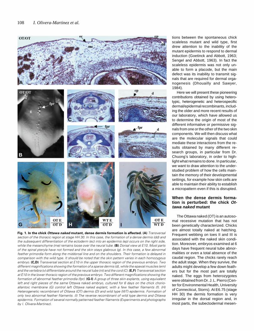
Fig. 1. In the chick Ottawa naked mutant, dense dermis formation is affected. (A) Transversalsection of the thoracic region at stage HH 30. In this case, the formation of a dense dermis (dd) andthe subsequent differentiation of the ectoderm (ec) into an epidermis (ep) occurs on the right side,while the mesenchyme (me) remains loose over the neural tube. (B) Dorsal view at E10. Most partsof the spinal pteryla have not formed and the skin stays glabrous (g). In this case, a few abnormalfeather primordia form along the middorsal line and on the shoulders. Their formation is delayed incomparison with the wild type. It should be noted that the skin pattern varies in each homozygousembryo. (C,D) Transversal section at E10 in the upper thoracic region of the previous embryo. Twodifferent magnifications showing the formation of a sparse dermis (d), while the epaxial muscles (em)and the vertebra (v) differentiate around the neural tube (nt) and the cord (C). (E,F) Transversal sectionat E10 in the lower thoracic region of the previous embryo. Two different magnifications showing theformation of abnormal feather primordia (fpr). (G-I) A group of three skin explants, using equivalentleft and right pieces of the same Ottawa naked embryo, cultured for 6 days on the chick chorio-allantoic membrane (G) control left Ottawa naked explant, with a few feather filaments (f). (H)Heterogenetic recombinant of Ottawa (OT) dermis (D) and wild type (WT) epidermis. Formation ofonly two abnormal feather filaments. (I) The reverse recombinant of wild type dermis and Ottawaepidermis. Formation of several normally patterned feather filaments (Experiments and photographsby I. Olivera-Martinez).
A
I
B
C D
E F
G H

Different steps of skin formation 109
chyme remains loose (Fig. 1A). At E8.5 a few feather primordiasometimes occur, albeit delayed two days with respect to the wildtype. At E10 (Fig. 1B), when a few feather buds are formed, theirnumber, diameter, location in the dorsal field (macropattern) andtheir arrangement (micropattern) are very variable, but are al-most symmetrical on each side of the middorsal line. A densedermis is not present in most dorsal regions (Fig. 1 C,D), althoughit forms and subsequently gives rise to abnormally sized dermalcondensations in some (Fig. 1 E,F). Heterogenetic dermal/epi-
Fig. 2. The chick scaleless mutant: the formation of dermal condensations is
affected due to a deficiency in epidermal signalling. (A) Dorsal view at E20. Notethe formation of feathers on the humeral, crural and caudal tracts. The spinal tractis entirely devoid of feathers. (B,C) At E7, the dorsal scaleless dermis reaches thestage of dense dermis (dd) formation (B) and this dense dermis homogeneouslyexpresses Delta-1 transcripts. (D-G) Heterogenetic dermal/epidermal recombi-nants. After 36 hours, WT epidermis/ SC dermis recombination (D) leads to therestriction of Delta-1 expression to the fibroblasts forming the dermal condensation(dc) of the feather primordia and, after 6 days on the chorioallantoic membrane (E),to the differentiation of feather filaments (f). The converse recombinant of SCepidermis and WT dermis leads to a homogeneous distribution of Delta-1 transcriptsin the superficial dense dermis (dd) after 36 hours (F) and after 6 days (G) to a bareexplant. d, dermis; ep, epidermis; ms, muscles; p, placode; sp, spare dermis. (A,B)Photographed by I. Olivera-Martinez; (C-G) courtesy of Elsevier (Viallet et al., 1998).
dermal recombinants were performed at stage HH 31between these mutant (OT/OT) and wild type (WT/WT)embryos and then grafted for 6 days on the chick chorio-allantoic membrane. The skin was dissected on each sideof the middorsal line and for each mutant embryo, a groupof three grafts was done: one control; one OT/OT dermis/WT/WT epidermis; and one WT/WT dermis/OT/OT epi-dermis. The results were consistent for each group ofthree (n=6) that survived. The controls and the recombi-nants involving an Ottawa naked dermis were featherlessor poorly feathered. In addition, in those rare cases wherea few feathers formed (Fig. 1 G,H), they were delayed by2-3 days with respect to the recombinants involving awild-type dermis. The recombinants involving a wild-typedermis produced large numbers of feathers (Fig. 1I), withthe corresponding primordia differentiating rapidly, assoon as the day after the recombination.
The Ottawa naked defect thus affects the formation ofa dense dermis, while the Ottawa naked epidermis func-tions normally. The formation of the dermis is howeverperturbed whatever the origin of the dermal progenitors,i.e. the neural crest (Couly and Le Douarin, 1988) forfacial dermis, the dermomyotome (Mauger, 1972) fordorsal dermis, or the somatopleural mesoderm (Christ etal., 1983) for ventral and limb skin. This is despite the factthat the molecular mechanisms which result in the speci-fication of the dermal progenitors appear to differ inregions where it has been studied in detail, i.e. the back(Olivera-Martinez et al., 2000, 2001, 2002 and 2004) andabdomen (Fliniaux et al., 2004). Moreover, as the sub-ectodermal mesenchyme formed, we can therefore sur-mise that only the densification of the dermis is affected.This implies that either the mesenchymal cells are im-peded in their proliferation/migration close to the ecto-derm, or that they do not receive specific signals from theectoderm, or that the signals are not at a sufficient level.The answer might come from studies currently in processin our laboratory on the respective roles of the ectodermand mesoderm in dermal densification in wild type em-bryos.
When the dense dermis is not redistributed toform dermal condensation: the chick Scalelessmutant
Scaleless is an autosomal recessive mutation that hasnot been genetically characterized, but has been thesubject of several scientific studies since the sixties.Scaleless chicks have smooth skin largely free of downfeathers and their tarsometatarsus and feet lack scales.
Scattered feathers are present in the head, humeral, crural andcaudal pterylae (Fig. 2A). The eggs from homozygous SC/SCmutants were obtained from J.L. Pierro (Center for EnvironmentalHealth, University of Connecticut, Storrs). Histological observa-tion of embryos at stage HH 30 shows that the dorsal dermisformed normally and is composed of a dense dermis overlying asparse dermis (Fig. 2B) (Viallet et al., 1998). Thus, in scalelessembryos (Viallet et al.,1998; Dhouailly et al., 1998; Widelitz et al.,2000; M. Harris, personal communication), as in wild type em-
A
B
C
D E
F G

110 I. Olivera-Martinez et al.
B
bryos (Wessells, 1965; see also review by Dhouailly et al., 2004),the formation of a dermis in areas which correspond to thepterylae is characterized by an increase in the cellular density ofthe fibroblasts. In scaleless embryos, however, the next step doesnot occur. Feather formation involves the segregation of at leasttwo types of dermal cells, via a redistribution of the cell populationthat forms the dense dermis (F. Michon, unpublished data). Thefirst type will form the dermal condensation, will then be endowedwith morphogenetic properties and participate in feather forma-tion. The second type will form the ordinary sparse dermisunderlying the inter-feather epidermis. DiI experiments (Jiang etal., 1999) show that the two fates of dermal fibroblasts could stillbe reassigned at E8 (HH 33), i. e. when the dermal condensationshave already formed. The Notch pathway is known to play a rolein binary choices (among others: Artavanis-Tsakonas et al., 1995;Simpson, 1997) and in situ hybridization of wild type chickembryos showed that Delta-1 transcripts are heterogeneouslydistributed in the forming dorsal dermal condensations at E7/E8(Viallet et al., 1998). In contrast, in scaleless embryos of the samestages, Delta-1 transcripts are homogeneously distributed in thedense dermis (Fig. 2C) (Viallet et al., 1998). Moreover, Delta-1over-expression using a retroviral infection in wild type embryosled to formation of large, ectopic secondary apteria (Viallet et al.,1998). Pioneering experiments at the beginning of the sixties(Goetinck and Abbott, 1963; Sengel and Abbott, 1963) showedthat the scaleless defect is expressed by the epidermis while the
scaleless dermis functions normally. More precisely, heterochronicheterogenetic recombinants demonstrate that the scaleless der-mis is endowed with appendage-inducing abilities at an earlystage and will rapidly lose them due to a lack of interaction with awild type epidermis (Dhouailly and Sawyer, 1984; Song andSawyer, 1996). Heterogenetic recombination of scaleless dermiswith a wild type epidermis leads, after 36 hours, to the repatterningof Delta-1 expression in the dermis (Fig. 2D), followed, after 6days of culture on chorioallantoic membrane, by the emergenceof feathers (Fig. 2E) (Viallet et al., 1998). The converse recombi-nant of a dorsal scaleless epidermis with a wild type dermis leadsto a homogeneous distribution of Delta-1 transcripts in the super-ficial dermis after 36 hours (Fig. 2F) and, 6 days later, to a smoothexplant (Fig. 2G) (Viallet et al., 1998). The group of P. Goetinck(Song et al., 1996) showed that the defect in scaleless embryosis a lack of expression of FGF4 by the epidermis. Their experi-ments were done in vitro, by adding beads overloaded with eitherFGF2 or 4 on scaleless chick embryo skin explants in vitro. Theyobtained abnormal feather buds which had an abnormalmicropattern and some fusions. The same year, our group wasengaged in a similar type of experiment, but the pieces of dorsalscaleless embryonic skin, overlaid with FGF2 beads, were graftedon the chick chorioallantoic membrane. Perfectly differentiatedfeathers appeared (Viallet et al., 1998) and moreover, the featherbuds arose sequentially in the vicinity of the loaded beads,suggesting that the beads do not replace an epidermal placode assuggested by Song et al. (1996), but instead gave and expandeda general permissive message to the dermis. The endogenousFGF4 signalling of the epidermis to the dermis in wild typeembryos is thus a permissive signal, which might interact indi-rectly with Delta-1 expression, to allow (Viallet et al., 1998;Dhouailly et al., 1998) the formation of dermal condensations infeather- or scale-forming regions. Dermal scale condensations,as well as the formation of placodes which precedes the organi-zation of the dense dermis are clearly distinguishable in wholepieces of dermis and epidermis after their separation (Dhouailly,1984). FGF4 does not constitute a “feather-message”, as sug-gested by a third group (Widelitz et al., 1996). Whereas theyobtained supernumerary feather formation in the sub-wing semi-apterium by using FGF2 beads, we did not in the midventralapterium (Dhouailly et al., 1998). The difference between theupper dermis of a semi-apterium and that of an apterium is that theformer normally forms a dense dermis, albeit with a delay, as wellas a few randomly distributed feathers, while the latter remainsloose and totally bare (Sengel et al., 1969; Sengel, 1976). More-over, FGF2 beads allow the scaleless foot skin to form scutatescales (Dhouailly et al., 1998; Prin and Dhouailly, 2004).
The first dermal induction and the initiation/patterningof appendages
Heterospecific recombinations between dermis and epidermiswere performed thirty years ago by using skin tissues from lizard,chick and mouse embryos, thus from the three different classesof amniotes (Dhouailly, 1973, 1975, 1977). They all yieldedconsistent results. In brief, class specificity of cutaneous append-ages, i.e. the formation of scale- feather- or hair-type buds isepidermis dependant, whereas their initiation/ patterning andoutlines are dermis dependant. The dermis, as it transmits its
Fig. 3. Formation of scale buds in a hair-pelage or a hair-vibrissa
pattern by a lizard embryo dorsal epidermis. (A)With dorsal mousedermis: formation of large and small scale buds corresponding to thecentral and lateral primary hair follicle pattern (B). (C) With upper-lip mousedermis: formation of large scale buds arranged in a whisker pattern (D),surrounded by small scale buds corresponding to pelage hair pattern. Thedermis is thus responsible for the pattern, while the epidermis respondsaccording to its genetic potential (Dhouailly, 1975). pg, peg; pl, placode.
A C
B D
Different steps of skin formation 111
initial triggering influence to initiate appendage morphogenesis inthe overlying epidermis, also specifies their size and distributionpattern and the epidermis responds according to its geneticpotential. These buds do not, however, give rise to matureappendages. For example, mouse pelage hair-forming dermisinduces a large quantity of two size classes of scale buds in alizard epidermis (Fig. 3A) and feather buds in a chick epidermis,corresponding to central large and lateral small primary follicles ofthe mouse pelage (Fig. 3B). Likewise, mouse vibrissa-formingdermis induces a small number of giant scale buds in a lizardepidermis (Fig. 3C) and giant feather buds in a chick epidermis,according to the mouse upper-lip pattern (Fig. 3D). A number ofsignaling factors have been shown to be expressed in the placodeas well as in the dermal condensation. Bmp 2, 4 and 7 andfollistatin (for a review: Chuong, 1998) are expressed in thefeather primordia during pattern formation while gremlin is onlyexpressed in the interbud dermis (Bardot et al., 2004). The ectopicexpression of Bmp2 and 4 leads to the inhibition of the featherprimordia and, reciprocally, to the over-expression of noggin,follistatin and gremlin induce the formation of ectopic or enlargedfeathers (Noramly and Morgan, 1998; Patel et al., 1999). Interest-ingly all the feather activators (BMP antagonists, FGF, Shh) andmost of the inhibitors (BMP2, 4) are expressed in the primordia.This has led to the proposal of a model based on diffusion ofrepressor/activator signals (Chuong, 1998).
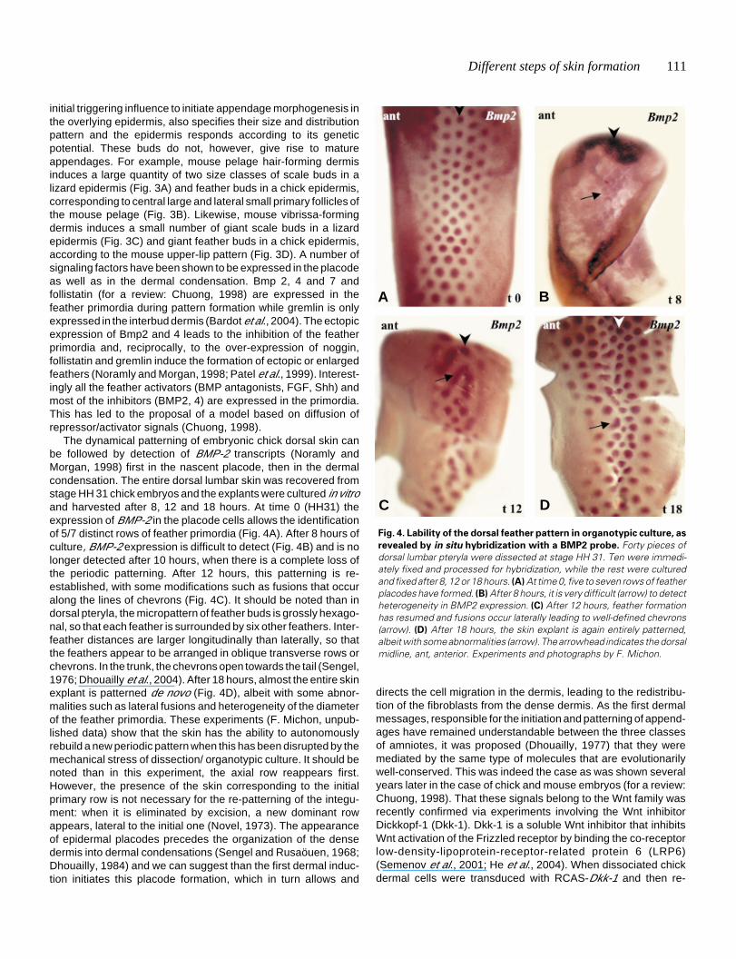
The dynamical patterning of embryonic chick dorsal skin canbe followed by detection of BMP-2 transcripts (Noramly andMorgan, 1998) first in the nascent placode, then in the dermalcondensation. The entire dorsal lumbar skin was recovered fromstage HH 31 chick embryos and the explants were cultured in vitroand harvested after 8, 12 and 18 hours. At time 0 (HH31) theexpression of BMP-2 in the placode cells allows the identificationof 5/7 distinct rows of feather primordia (Fig. 4A). After 8 hours ofculture, BMP-2 expression is difficult to detect (Fig. 4B) and is nolonger detected after 10 hours, when there is a complete loss ofthe periodic patterning. After 12 hours, this patterning is re-established, with some modifications such as fusions that occuralong the lines of chevrons (Fig. 4C). It should be noted than indorsal pteryla, the micropattern of feather buds is grossly hexago-nal, so that each feather is surrounded by six other feathers. Inter-feather distances are larger longitudinally than laterally, so thatthe feathers appear to be arranged in oblique transverse rows orchevrons. In the trunk, the chevrons open towards the tail (Sengel,1976; Dhouailly et al., 2004). After 18 hours, almost the entire skinexplant is patterned de novo (Fig. 4D), albeit with some abnor-malities such as lateral fusions and heterogeneity of the diameterof the feather primordia. These experiments (F. Michon, unpub-lished data) show that the skin has the ability to autonomouslyrebuild a new periodic pattern when this has been disrupted by themechanical stress of dissection/ organotypic culture. It should benoted than in this experiment, the axial row reappears first.However, the presence of the skin corresponding to the initialprimary row is not necessary for the re-patterning of the integu-ment: when it is eliminated by excision, a new dominant rowappears, lateral to the initial one (Novel, 1973). The appearanceof epidermal placodes precedes the organization of the densedermis into dermal condensations (Sengel and Rusaöuen, 1968;Dhouailly, 1984) and we can suggest than the first dermal induc-tion initiates this placode formation, which in turn allows and
directs the cell migration in the dermis, leading to the redistribu-tion of the fibroblasts from the dense dermis. As the first dermalmessages, responsible for the initiation and patterning of append-ages have remained understandable between the three classesof amniotes, it was proposed (Dhouailly, 1977) that they weremediated by the same type of molecules that are evolutionarilywell-conserved. This was indeed the case as was shown severalyears later in the case of chick and mouse embryos (for a review:Chuong, 1998). That these signals belong to the Wnt family wasrecently confirmed via experiments involving the Wnt inhibitorDickkopf-1 (Dkk-1). Dkk-1 is a soluble Wnt inhibitor that inhibitsWnt activation of the Frizzled receptor by binding the co-receptorlow-density-lipoprotein-receptor-related protein 6 (LRP6)(Semenov et al., 2001; He et al., 2004). When dissociated chickdermal cells were transduced with RCAS-Dkk-1 and then re-
Fig. 4. Lability of the dorsal feather pattern in organotypic culture, as
revealed by in situ hybridization with a BMP2 probe. Forty pieces ofdorsal lumbar pteryla were dissected at stage HH 31. Ten were immedi-ately fixed and processed for hybridization, while the rest were culturedand fixed after 8, 12 or 18 hours. (A) At time 0, five to seven rows of featherplacodes have formed. (B) After 8 hours, it is very difficult (arrow) to detectheterogeneity in BMP2 expression. (C) After 12 hours, feather formationhas resumed and fusions occur laterally leading to well-defined chevrons(arrow). (D) After 18 hours, the skin explant is again entirely patterned,albeit with some abnormalities (arrow). The arrowhead indicates the dorsalmidline, ant, anterior. Experiments and photographs by F. Michon.
A B
C D

112 I. Olivera-Martinez et al.
aggregated and overlaid by an epidermis, feather formation isinhibited (Chang et al., 2004). Dkk does not, however, differenti-ate between different Wnts and specific antagonists for eachmember of this large family do not exist yet.
The second dermal induction and feather organogen-esis
The association of chick epidermis with hair-forming mousedermis produces arrested feather buds, the epidermis of whichforms ingrowing thickenings which can be recognized as abnor-mal barb ridges (Dhouailly, 1973). These hypomorphic barb-ridges are formed by stacking of cells corresponding to future barband barbule cells. Nevertheless, although the cytodifferentiationof feather cells is independent from dermal signaling once featherformation has been triggered by the first dermal messagesoriginating from the mouse dermis, the outgrowth and typicalarchitectural organization of the feather filament did not occur.
In chick neoptile feathers the number of barbs varies from 11to 16 and the rachis, formed by the fusion of the anterior barbs, isrudimentary. In contrast, in duck neoptile feathers, the number ofbarbs varies from 18 to 26 and the anterior rachis is well devel-oped. Moreover, the teleoptile remiges appear soon after birth inchick but later in duck. The number of barb ridges in the embryonicfeather filament, which corresponds to that of barbs in the neoptilefeather; their partial fusion at the base of the feather to form arachis; the timing of the formation of the second generation, thejuvenile feathers and their shape, are species variable charac-ters, which are all governed by the second dermal messages(Dhouailly, 1970). The experiments to show this consisted of
heterospecific recombinations of wing bud ectodem and meso-derm between chick and duck, grafted on chick or duck hosts. Theresults were clear-cut. The morphological characters of the feath-ers were all determined by the dermis, except the number andshape of barbule cells, which conformed to the species origin ofthe epidermis (Fig. 5 A-C). Moreover, the time of renewal of theappendages was also dependent on the dermis (Fig. 5 E,F). Thecorresponding dermal/epidermal interactions must occur in thefeather follicle, between the dermal papilla and the ring (wall) ofundifferentiated epidermal cells. The later proliferate and subdi-vide into barb ridges, which fuse with the anterior part of the ringto give rise to the rachis, with additional barb ridges forming in theposterior part of the ring. Rachis formation, as well as the additionof new barb ridges, is more pronounced in case of the duck thanchick neoptile feather and occurs through the entire outgrowth ofthe teleoptile feather in both species.
Several signaling factors are expressed in the developingfeather, including BMP2, BMP4, Noggin and Shh (Yu et al., 2002;and 2004). BMP4 is expressed mainly in the dermal papilla, whileNoggin is expressed in its posterior region, where barb ridgesstart to form. Shh is expressed in the internal sheath and isessential for inducing apoptosis and thus the splitting of barbridges. The Chuong group, in a series of elegant experimentsshowed that, by adding RCAS viruses carrying these genes ordominant negative genes to the follicles of plucked teleoptilefeathers, abnormal feathers were formed during regeneration.Over-expression of Noggin, a BMP antagonist, increases the barbnumber and even causes them to become split, while over-expression of BMP2 and BMP4 caused the formation of a giantrachis and barb fusions. Suppression of Shh leads to the forma-
Fig. 5. Chimeric neoptile feathers produced by heterospecic duck/chick forelimb ectoderm/ mesodermal pulp recombinants. The dermis isresponsible for the feather architecture and the time of replacement of the neoptile by the teleoptile feather, while the shape of epidermal barbule cellsis conferred by the epidermal species.(A) A typical duck-type neoptile feather obtained from the association of chick epidermis and duck dermis. Notethe formation of 26 barbs, most of them being attached to a well developed rachis. (B) Detail of the barbules of the feather shown in (A), showing a typicalchick-type morphology, i.e. a succession of cylindrical cells, slightly swollen at their distal tip. (C) Typical chick-type neoptile feather obtained from theassociation of chick dermis and duck epidermis. Note the formation of 12 barbs. (D) Detail of the barbules of the feather shown in (C), showing a typicalduck-type morphology, i.e. a succession of cylindrical cells, with two spiny protrusions at their distal tip. (E) A chick host, two weeks old, bearing a chimericright wing composed of a duck mesenchymal pulp associated with a chick ectoderm. The chimeric wing is perfectly developed, but is covered only withneoptile duck-type feathers, while the left host wing is covered by the second generation, the juvenile teleoptile feathers. (F) A duck host, two weeksold, bearing a chimeric right wing composed of a chick mesenchymal pulp associated to a duck ectoderm. The chimeric wing is in this case poorlydeveloped, because the microsurgery was not perfect, but in the second generation, the juvenile teleoptile chick-type feathers had formed (Dhouailly,1970).
A
B
E
D
F
C

Different steps of skin formation 113
tion of a remnant membrane between the barbs. It is thus veryprobable that a difference in the level of expression of Noggin andBMP4/2 exists between the dermal papillae of the neoptilefeather follicle in chick and duck, but this would be difficult toquantify.
Conclusion: the molecular events which mediate der-mal/epidermal interactions
The molecular events underlying the cellular interactionsduring skin morphogenesis have thus been partially documentedby several laboratories using expressions pattern studies andectopic treatment using loaded beads or over-expression usingretroviruses. Many of the recently described developmentalsignalling pathways have been implicated in one or all of theseinteractions (summarised in figure 6). Among these signals, theWnt pathway is associated first in the formation of the dorsaldermis. Wnt1 from the dorsal neural tube has been shown toinduce the specification of dermal progenitors from the dorsaldermomyotome (Olivera-Martinez et al., 2001, 2002 and 2004).The dermal precursors express Wnt 11, which might be impli-cated in their migration to the subectodermal space (Olivera-Martinez et al., 2002, 2004). Wnt 1, 3a and 5a in the featherprimordia and Wnt 11 in the interbud dermis region control theshape of the feather bud while Wnt7a is involved in the anterior-posterior orientation during the outgrowth of the feather bud(Chang et al., 2004; Widelitz et al., 1999; Chuong et al., 1996).At early stage the level of β-catenin is homogeneous throughoutfeather field epidermis and then is restricted to placodes (Widelitzet al., 2000). We can thus propose that this general β-cateninexpression is the response of the epidermis to a general Wntmessage originating from the dermis. Then, this message ap-
the abdomen (Fliniaux and Viallet unpublished data). Duringpteryla formation transient BMP2 expression is observed in theepidermis while the BMP antagonists gremlin and follistatin areexpressed in the underlying dermis and the epidermis, respec-tively. (Noramly et al., 1998; Bardot et al., 2004; Patel et al., 1999).When the patterning occurs, BMP2, 4 and 7 and the BMPantagonists excepted gremlin are expressed in the primordia.This observation leads to a model based on activation via differ-ential diffusion of activators and inhibitors for the formation of theperiodic patterning (Chuong, 1998). Later, at the step of featherorganogenesis, the BMP/antagonist pair (in this case Noggin) isalso involved in the patterning of the barb ridges (Yu et al., 2002,2004). What is the role of Shh? It is expressed at 2 days by thechick endoderm (Watanabe et al., 1998) and could play a role ininhibiting BMP4 expression in the somatopleure in the abdomenand thus in allowing feather field formation. At the early stage ofpattern formation Shh is expressed in the placode while itsreceptor Ptc is expressed both in the placode and the dermalcondensation (Ting-Berreth and Chuong, 1996; Jung et al., 1998;Morgan et al., 1998). In our point of view, Shh is involved inactivating cell proliferation both in the dermis and epidermis.Forced expression of Shh (or Shh treatment) causes ectopicfeather formation in pteryla, semi-apteria and even in the midventralapterium (Fliniaux et al., 2004) and appears to be sufficient toenhance dermis density over a critical thresholds. In fact, Shh isstrongly expressed in feather buds that elongate into featherfilaments, is slightly expressed in overlapping scutate scales andis barely detectable and short lived in reticulate scales that did notoverlap (Prin et al., 2004). Inhibition of Shh signaling in chick (Prinand Dhouailly, 2004) can lead to feather growth arrest. Likewise,Shh knock-out mice (Chiang et al., 1999) have arrested hair buds.Together all these results lead to the conclusion that Shh is a
Fig. 6. Diagrammatic representation of the different
steps of dorsal skin formation in chick. For details, seetext.
pears to become restricted tothe primordia in wild-type em-bryo, whereas it remains as asmear over the tract fields inthe scaleless embryo (Widelitzet al., 2000). During pattern for-mation nuclear β-catenin stain-ing increases in the placodeand is lost in the ectoderm thatadopts interfollicular fate, in ad-dition the forced expression ofβ- catenin induces the forma-tion of ectopic feathers(Noramly et al., 1999; Widelitzet al., 2000). The restriction ofβ-catenin expression, as wellas that of Delta-1 expressionmight be a consequence ofFGF4 expression in the epider-mis (Viallet et al., 1998; Song etal., 1996). The Notch pathwaymay serve to stabilize the pat-terning of feather primordia(Viallet et al., 1998).
The coupled BMP4/BMP-antagonist is first observed dur-ing feather field specification in
114 I. Olivera-Martinez et al.
general growth activator of cutaneous appendages. MoreoverShh also intervenes later, to allow apoptosis of the internalepidermal sheath and consequently the splitting of barb ridgesduring feather organogenesis (Yu et al., 2002, 2004).
Thus, after more than thirty years of research, the cellularinteractions and their mediators become clearer. However, theformation of a dense dermis and particularly the nature of thesignal originating from the ectoderm, as well as the migration ofdermal cells to from the dermal condensations deserve furtherresearch work. Moreover, we need to complete our preliminarydiagram (Fig. 6), by adding which transcription factors and whattype of intra-tissular interactions are triggered at each step. Onlythen we will obtain an overview of feather- and of course, hair-forming skin morphogenesis as complete as that already detailedfor tooth organogenesis (for a review, see Pispa and Thesleff,2003).
AcknowledgmentsThe authors thank Dr. Thélu and all the different ex-PhD students of the
laboratory, in particular Dr. S. Blanchet-Rethore; Dr. C. Ferraris; Dr. I.Fliniaux;Dr. B. Kanzler and Dr. F. Prin, for continuous discussion leading to theelaboration of the present idea, as well as Mrs. B. Peyrusse for all theiconography and Mrs. G. Chevalier and Miss. P. Betton for technical help.
References
ARTAVANIS-TSAKONAS, S., MATSUNO, K. and FORTINI, M. E. (1995). Notchsignaling. Science. 268: 225-232.
BARDOT, B., LECOIN, L., FLINIAU, I., HUILLARD, E., MARX, M. and VIALLET, J. P.(2004). Drm/Gremlin, a BMP antagonist, defines the interbud region during featherdevelopment.Int. J. Dev. Biol. 48: 149-156.
CHIANG, C., SWAN, R. Z., GRACHTCHOUK, M., BOLINGER, M., LITINGTUNG, Y.,ROBERTSON, E. K., COOPER, M. K., GAFFIELD, W., WESTPHAL, H., BEACHY,P. A. and DLUGOSZ, A. A. (1999). Essential role for Sonic hedgehog during hairfollicle morphogenesis. Dev. Biol. 205: 1-9.
CHRIST, B., JACOB, M. and JACOB, H. J. (1983). On the origin and development ofthe ventrolateral abdominal muscles in the avian embryo. An experimental andultrastructural study. Anat Embryol (Berl). 166: 87-101.
CHANG, C. H., JIANG, T. X., LIN, C. M., BURRUS, L. W., CHUONG, C. M. andWIDELITZ, R. (2004). Distinct Wnt members regulate the hierarchical morphogen-esis of skin regions (spinal tract) and individual feathers. Mech. Dev. 121: 157-71.
CHUONG, C. M. (1998). Feather morphogenesis: A model of the formation ofepithelial appendages. In Molecular Basis of epithelial appendage morphogen-esis (C.M. Chuong, Ed.), pp. 57-74. R.G. Landes Co., Georgetown, Texas.
COULY, G. and LE DOUARIN, N. M. (1988). The fate map of the cephalic neuralprimordium at the presomitic ta the 3-somite stage in the avian embryo. Develop-ment Suppl. 103: 101-113.
DHOUAILLY, D. (1970). Déterminisme de la différenciation spécifique des plumesnéoptiles et téléoptiles chez le poulet et le canard. J. Embryol. Exp. Morphol. 18:389-400.
DHOUAILLY, D. (1973). Dermo-epidermal interactions between birds and mammals:differenciation of cutaneous appendages. J. Embryol. Exp. Morphol. 30: 587-603.
DHOUAILLY, D. (1975). Formation of cutaneous appendages in dermoepidermalrecombinations between reptiles, birds and mammals. Wilhelm, Roux Arch. Dev.Biol. 177: 323-340.
DHOUAILLY, D. (1977). Dermo-epidermal interactions during morphogenesis ofcutaneous appendages in amniotes. In Frontier Matrix Biology (ed. L. Robert),Creteil. 4: 86-121.
DHOUAILLY, D. (1984). Specification of feather and scale patterns. In PatternFormation. (Ed. G.M. Malacinski and S.V. Bryant). Macmillan Pub. Comp. NewYork, London. pp. 581-601.
DHOUAILLY, D. and SAWYER, R. H. (1984). Avian scale development. XI. Initialappearance of the dermal defect in scaless skin. Dev. Biol. 105: 343-350.
DHOUAILLY, D., PRIN, F., KANZLER, B. and VIALLET, J. P. (1998). Variation ofcutaneous appendages: Regional specification and cross-species signals. InMolecular basis of Epithelial appendage morphogenesis Vol. 1 (Ed. C.M.Chuong), R. G. Landes Company, Georgetown, Texa, USA pp. 45-56.
DHOUAILLY, D., OLIVERA-MARTINEZ, I., FLINIAUX, I., MISSIER, S., VIALLET, J.P.and THÉLU, J. (2004). Skin field formation: morphogenetic events. Int. J. Dev.Biol. 48: 85-91.
FLINIAUX, I., VIALLET, J. P. and DHOUAILLY, D. (2004). Ventral vs. dorsal chickdermal progenitor specification. Int. J. Dev. Biol. 48: 103-106.
GOETINCK, P. F. and ABBOT, U. K. (1963). Tissue interactions in the scalelessmutant and the use of scaleless as an ectodermal marker in studies of normal limbdifferentiation. J. Exp. Zool. 154: 7-19.
HE, X., SEMENOV, M., TAMAI, K. and ZENG, X. (2004). LDL receptor-relatedproteins 5 and 6 in Wnt/beta-catenin signaling: Arrows point the way. Develop-ment 131: 1663-1677
JIANG, TX., JUNG, HS., WIDELITZ, R. B. and CHUONG C. M. (1999). Self-organization of periodic patterns by dissociated feather mesenchymal cells andthe regulation of size, number and spacing of primordia. Development. 126: 4997-5009.
JIANG, T.-X., WIDELITZ, R.B., SHEN, W.-M., WILL, P., WU, D.-Y., LIN, C.-M.,JUNG, H.-S. and CHUONG, C.-M. (2004). Integument pattern formationinvolves genetic and epigenetic controls: feather arrays simulated by digitalhormones. Int. J. Dev. Biol. 48: 117-136.
JUNG, H. S., FRANCIS-WERST, P. H., WIDELITZ, R. B., JIANG, T. X., TING-BERRETH, S. A., TICKLE, C., WOLPERT, L. and CHUONG, C. M. (1998). Localinhibitory action of BMP’s and their relationships with activators in featherformation: implications for periodic patterning. Dev. Biol. 196: 11-23.
MAUGER, A. (1972). The role of somitic mesoderm in the development of dorsalplumage in chick embryos. I. Origin, regulative capacity and determination of theplumage-forming mesoderm. J Embryol Exp Morphol 28: 313-41.
MORGAN, B. A., ORKIN, R. W., NORAMLY, S. and PEREZ, A. (1998). Stage-specificeffects of sonic hedgehog expression in the epidermis. Dev. Biol. 201: 1-12.
NORAMLY, S. and MORGAN, B. A. (1998). BMP’s mediate lateral inhibition atsuccessive stage in the feather tract development. Development. 125: 3775-3787.
NORAMLY, S., FREMAN, A. and MORGAN, B. A. (1999). Beta-catenin signaling caninitiate feather bud development. Development. 126: 3509-3521.
NOVEL, G. (1973). Feather pattern stability and reorganization in cultured skin. J.Embryol. Exp. Morphol. 30: 605-633.
OLIVERA-MARTINEZ, I., COLTEY, M., DHOUAILLY, D. and POURQUIE, O. (2000).Mediolateral somitic origin of ribs and dermis determined by quail-chick chimeras.Development 127: 4611-4617.
OLIVERA-MARTINEZ, I., THELU, J., TEILLET, M. A. and DHOUAILLY, D. (2001).Dorsal dermis development depends on signal from the dorsal neural tube, whichcan be substituted by Wnt-1. Mech Dev 100: 233-44.
OLIVERA-MARTINEZ, I., MISSIER, S., FRABOULET, S., THELU, J. and DHOUAILLYD. (2002). Differential regulation of the chick dorsal thoracic dermal progenitorsfrom the medial dermomyotome. Development 129: 4763-72.
OLIVERA-MARTINEZ, I., THELU, J. and DHOUAILLY D. (2004). Molecular mecha-nisms controlling dorsal dermis generation from the somatic dermomyotome. Int.J. Dev. Biol. 48: 93-101.
PATEL, K., MAKARENKOVA, H. and JUNG, H. S. (1999). The role of long range, localand direct signalling molecules during chick feather bud development involving theBMPs, follistatin and the Eph receptor tyrosine kinase Eph-A4. Mech Dev 86: 51-62.
PISPA, J. and THESLEFF, I. (2003). Mechanisms of ectodermal organogenesis. Dev.Biol. 262: 195-205.
PRIN, F. and DHOUAILLY, D. (2004). How and when the regional competence ofchick epidermis is established: feather vs. scutate and reticulate scales, a problemen route to a solution. Int. J. Dev. Biol. 48: 137-148.
PRIN, F., LOGAN, C., D’SOUZA, D., ENSINI, M. and DHOUAILLY, D. (2004). Dorsalversus ventral scales and the dorsoventral patterning of chick foot epidermis. Dev.Dyn. 229: 564-78.
SEMENOV, M. K., TAMAI, B. K., BROTT, M., KUHL, S., SOKOL, S. and HE, X.(2001). Head inducer Dickkopf-1 is a ligand for Wnt co-receptor LRP6. Curr.Biol. 11: 951-961.
Different steps of skin formation 115
SENGEL, P. and ABBOTT, U. K. (1963). In vitro studies with the scaleless mutant:interaction during feather and scale differentiation. J. Herd. 54: 254-262.
SENGEL, P. and RUSAOUEN, M. (1968). Aspects histologiques de la différenciationprécoces des ébauches plumaires chez le poulet. C. R. Acad. Sci. 266: 795-797.
SENGEL, P., DHOUAILLY, D. and KIENY, M. (1969). Aptitude of the skin constitu-ents of the mid-ventral apterium of the chicken for forming feathers. Dev. Biol.19: 436-46.
SENGEL, P. (1976). Morphogenesis of the skin. In Developmental and Cell Biology,(Eds. Abercrombie, M., Neweth, D.R. and Torrey, J.G.). Vol. 1. CambridgeUniversity Press, Cambridge, MA, pp. 1-277.
SIMPSON, P. (1997). Notch signaling in development. Perspect. Dev. Neurobiol. 4:297-304.
SONG, H. K. and SAWYER, R. H. (1996). Dorsal dermis of the scaleless (sc/sc)embryo directs normal feather pattern formation until day 8 of development. Dev.Dyn. 205: 82-91.
SONG, H. K., WANG, Y. and COETINCK, P. F. (1996). fibroblast growth factor 2 canreplace ectodermal signaling for feather development. Proc. Natl. Acad. Sci. USA93: 10246-10249.
TING-BERRETH, S. A. and CHUONG, C. M. (1996). Sonic Hedgehog in feathermorphogenesis: induction of mesenchymal condensation and association withcell death. Dev. Dyn. 207: 157-170.
VIALLET, J. P., PRIN, F., OLIVERA-MARTINEZ, I., HIRSINGER, E., POURQUIÉ, O.and DHOUAILLY, D. (1998). Chick Delta-I gene expression and the formation ofthe feather primordia. Mech. Dev. 72: 159-168.
WESSELLS, N. K. (1965). Morphology and proliferation during early feather develop-ment. Dev. Biol. 12: 131-53.
WATANABE Y., DUPREZ D., MONSORO-BURQ A. H., VINCENT C. and LEDOUARIN N. M. (1998). Two domains in vertebral development: antagonisticregulation by SHH and BMP4 proteins. Development. 125: 2631-9.
WIDELITZ, R. B., JIANG, T., NOVEEN, A., CHEN, C. J. and CHUONG, C. M. (1996).FGF induces new feather buds form developing avian skin. J. Inv. Derm. 107: 797-803.
WIDELITZ, R. B., JIANG, T. X., LU, J. and CHUONG, C. M. (2000). beta-catenin inepithelial morphogenesis: conversion of part of avian foot scales into feather budswith a mutated beta-catenin. Dev. Biol. 219: 98-114.
WU, P., HOU, L., PLIKUS, M., HUGHES, M., SCEHNET, J., SUKSAWEANG, S.,WIDELITZ, R. B., JIANG, T.X. and CHUONG, C.M. (2004). Evo-Devo of amnioteinteguments and appendages.Int. J. Dev. Biol. 48: 248-267.
YU, M., WU, P., WIDELITZ, R. B. and CHUONG, C. M. (2002). The morphogenesisof feathers. Nature. 420: 308-12.
YU, M., UUE, Z., WU, P., CHUONG, C.M. (2004). The developmental biology offeather follicles. Int. J. Dev. Biol. 48: 181-191.