The Crystal Structures of Human Calpains 1 and 9 Imply Diverse Mechanisms of Action and...
14
The Crystal Structures of Human Calpains 1 and 9 Imply Diverse Mechanisms of Action and Auto-inhibition Tara L. Davis, John R. Walker, Patrick J. Finerty Jr, Farrell Mackenzie Elena M. Newman and Sirano Dhe-Paganon⁎ Structural Genomics Consortium and the Department of Physiology , University of Toronto, 100 College St, Toronto, ON, Canada M5G 1L5 Calpains are calcium activated cysteine proteases found throughout the animal, plant, and fungi kingdoms; 14 isoforms have been described in the human genome. Calpains have been implicated in multiple models of human disease; for instance, calpain 1 is activated in the brains of individuals with Alzheimer's disease, and the digestive tract specific calpain 9 is down- regulated in gastric cancer cell lines. We have solved the structures of human calpain 1 and calpain 9 protease cores using crystallographic methods; both structures have clear implications for the function of non-catalytic domains of full-length calpains in the calcium-mediated activation of the enzyme. The structure of minicalpain 1 is similar to previously solved structures of the protease core. Auto-inhibition in this system is most likely through rearrangements of a central helical/loop region near the active site cysteine, which occlude the substrate binding site. However, the structure of minicalpain 9 indicates that auto-inhibition in this enzyme is mediated through large intra-domain movements that misalign the catalytic triad. This disruption is reminiscent of the full-length inactive calpain conformation. The structures of the highly conserved, ubiquitously expressed human calpain 1 and the more tissue specific human calpain 9 indicate that although there are high levels of sequence conservation throughout the calpain family, isolated structures of family members are insufficient to explain the molecular mechanism of activation for this group of proteins. Crown Copyright © 2006 Published by Elsevier Ltd. All rights reserved. *Corresponding author Keywords: cysteine protease; structure; peptidase; active site; autoinhibition Introduction Calpains (EC 3.4.22.17) are calcium activated cysteine proteases containing a catalytic domain most closely related to that of papain. Calpains are found throughout the animal, plant, and fungi kingdoms; 14 isoforms have been described in the human genome to date. 1–3 There are two classes of calpains; those expressed ubiquitously and those which are expressed in a tissue-specific fashion. Of the ubiquitous calpains, the μ-calpain (calpain 1) and m-calpain (calpain 2) isoforms have been extensively studied with respect to structure and function in vitro and in vivo. 2 These “classical” calpains exist in cells as heterodimers of a large subunit comprised of four domains associated with a small subunit. Activation of calpain is a calcium dependent process and involves multiple structural changes followed by limited autoproteolysis to form active calpain enzyme. 2,4–6 Extended activation of calpains leads to further autoproteolysis, finally leading to stable protease-resistant cores of 50, 40, and 26 kDa for proteolysis of calpain 2 by calpain 1. 2,4,5 Calpains are not proenzymes, however; the unproteolyzed heterodimer of calpain 1 and calpain 2 is enzymatically active in the presence of calcium, although in the cell and under physiological condi- tions it is thought that activation will be concomitant with autoproteolysis. 5,7 The expression levels and activity states of both ubiquitous and tissue-specific calpains have been implicated in several models of human disease. Calpain 1 is activated in the brains of individuals with Alzheimer’s disease 8,9 and the proteolytic cleavage of beta-amyloid precursor protein and tau protein by calpain 1 might be involved in plaque formation. 8,10–12 In the eye, calpain 2 and a lens- specific variant of calpain 3 are responsible for Abbreviations used: β-ME, β-mercaptoethanol; sLY-AMC, succinyl-Leu-Tyr-(7-amino-4-methylcoumarin). E-mail address of the corresponding author: [email protected] doi:10.1016/j.jmb.2006.11.037 J. Mol. Biol. (2007) 366, 216–229 0022-2836/$ - see front matter. Crown Copyright © 2006 Published by Elsevier Ltd. All rights reserved.
-
Upload
independent -
Category
Documents
-
view
0 -
download
0
Transcript of The Crystal Structures of Human Calpains 1 and 9 Imply Diverse Mechanisms of Action and...
doi:10.1016/j.jmb.2006.11.037 J. Mol. Biol. (2007) 366, 216–229
The Crystal Structures of Human Calpains 1 and 9 ImplyDiverse Mechanisms of Action and Auto-inhibition
Tara L. Davis, John R. Walker, Patrick J. Finerty Jr, Farrell MackenzieElena M. Newman and Sirano Dhe-Paganon⁎
Structural GenomicsConsortium and theDepartment of Physiology,University of Toronto,100 College St, Toronto, ON,Canada M5G 1L5
Abbreviations used: β-ME, β-mersLY-AMC, succinyl-Leu-Tyr-(7-aminE-mail address of the correspondi
0022-2836/$ - see front matter. Crown C
Calpains are calcium activated cysteine proteases found throughout theanimal, plant, and fungi kingdoms; 14 isoforms have been described in thehuman genome. Calpains have been implicated in multiple models ofhuman disease; for instance, calpain 1 is activated in the brains of individualswith Alzheimer's disease, and the digestive tract specific calpain 9 is down-regulated in gastric cancer cell lines.We have solved the structures of humancalpain 1 and calpain 9 protease cores using crystallographic methods; bothstructures have clear implications for the function of non-catalytic domainsof full-length calpains in the calcium-mediated activation of the enzyme. Thestructure of minicalpain 1 is similar to previously solved structures of theprotease core. Auto-inhibition in this system is most likely throughrearrangements of a central helical/loop region near the active site cysteine,which occlude the substrate binding site. However, the structure ofminicalpain 9 indicates that auto-inhibition in this enzyme is mediatedthrough large intra-domainmovements thatmisalign the catalytic triad. Thisdisruption is reminiscent of the full-length inactive calpain conformation.The structures of the highly conserved, ubiquitously expressed humancalpain 1 and themore tissue specific human calpain 9 indicate that althoughthere are high levels of sequence conservation throughout the calpain family,isolated structures of family members are insufficient to explain themolecular mechanism of activation for this group of proteins.
Crown Copyright © 2006 Published by Elsevier Ltd. All rights reserved.
*Corresponding author
Keywords: cysteine protease; structure; peptidase; active site; autoinhibitionIntroduction
Calpains (EC 3.4.22.17) are calcium activatedcysteine proteases containing a catalytic domainmost closely related to that of papain. Calpains arefound throughout the animal, plant, and fungikingdoms; 14 isoforms have been described in thehuman genome to date.1–3 There are two classes ofcalpains; those expressed ubiquitously and thosewhich are expressed in a tissue-specific fashion. Ofthe ubiquitous calpains, the μ-calpain (calpain 1)and m-calpain (calpain 2) isoforms have beenextensively studied with respect to structure andfunction in vitro and in vivo.2 These “classical”calpains exist in cells as heterodimers of a largesubunit comprised of four domains associated with
captoethanol;o-4-methylcoumarin).ng author:
opyright © 2006 Published b
a small subunit. Activation of calpain is a calciumdependent process and involves multiple structuralchanges followed by limited autoproteolysis to formactive calpain enzyme.2,4–6 Extended activation ofcalpains leads to further autoproteolysis, finallyleading to stable protease-resistant cores of 50, 40,and 26 kDa for proteolysis of calpain 2 by calpain1.2,4,5 Calpains are not proenzymes, however; theunproteolyzed heterodimer of calpain 1 and calpain2 is enzymatically active in the presence of calcium,although in the cell and under physiological condi-tions it is thought that activation will be concomitantwith autoproteolysis.5,7
The expression levels and activity states of bothubiquitous and tissue-specific calpains have beenimplicated in several models of human disease.Calpain 1 is activated in the brains of individualswith Alzheimer’s disease8,9 and the proteolyticcleavage of beta-amyloid precursor protein and tauprotein by calpain 1 might be involved in plaqueformation.8,10–12 In the eye, calpain 2 and a lens-specific variant of calpain 3 are responsible for
y Elsevier Ltd. All rights reserved.

217Minicalpain Action and Auto-inhibition
proteolytic cleavages of α and β-crystallin whichwhen overactivated can lead to cataract forma-tion.1,8,13,14 Over 100 independent mutants of themuscle-specific isoforms of calpain 3 have beenreported to be pathologically associated with limbgirdle muscular dystrophy type 2A,15–17 and posi-tional cloning has identified calpain 10 gene variantsassociated with type 2 diabetes.18–21 Interestingly,the digestive-tract-specific calpain 9 is down-regu-lated in gastric cancer cell lines, and depletion ofcalpain 9 using antisense RNA resulted in transfor-mation and tumorigenesis in fibroblasts, suggestingcalpain 9 is acting as a tumor suppressor in thosesystems.22,23 In addition, down-regulation of cal-pain 9 has recently been linked to hypertensive heartand kidney disease in salt-sensitive Dahl rats.24
Therefore, understanding the molecular mechan-isms of both activation and inactivation of calpainsare research topics of interest, since both states of thecalpain enzyme are implicated in disease.The domain architecture of both calpain 1 and 9
consists of a short N-terminal extension (domain Ior dI), the catalytic core (dII), the C2-like domaindIII, and a penta-EF hand domain dIV, whichinteracts with the small subunit domains dV anddVI. A construct containing the isolated catalyticdomains IIa and IIb of calpain has been termed the“minicalpain”. The minicalpains of rat calpain 1and rat calpain 2 have previously been character-ized both enzymatically and structurally.25–28 Theminicalpain of rat calpain 1 shows similar enzy-matic characteristics compared to the intactenzyme26 and the crystal structure of an inactivemutant of rat minicalpain 1 shows that thedomains IIa and IIb are conformationally compe-tent to perform catalysis.25 Rat minicalpain 2, incontrast, was found to be 10,000 times less activethan the full-length calpain 2 protein. Based on thecrystal structure of rat minicalpain 2, this loss ofactivity was attributed to auto-inhibition of theminicalpain construct via dynamic instability of theα7 helix and blockage of the active site by aconserved tryptophan residue found next to theactive site cysteine.26 Structural genomic studies ofthe human minicalpains are expected to yield awealth of information about the active sites of thesebiologically relevant molecules, including structuralanalysis of the substrate binding surface and theregions surrounding the catalytic triad. Presentedhere are the X-ray crystallographic structures of twohuman minicalpains: calpain 1 and calpain 9.Although the study of these structures to aid indrug design is ongoing and expected to be fruitful, itis also clear that both of these structures raisequestions about the fundamental molecular mech-anism of minicalpain auto-inactivation.
Results
The overall architecture of human minicalpain 1,comprising the catalytic IIa and IIb domains of theenzyme (residues 29–360), retains the classical
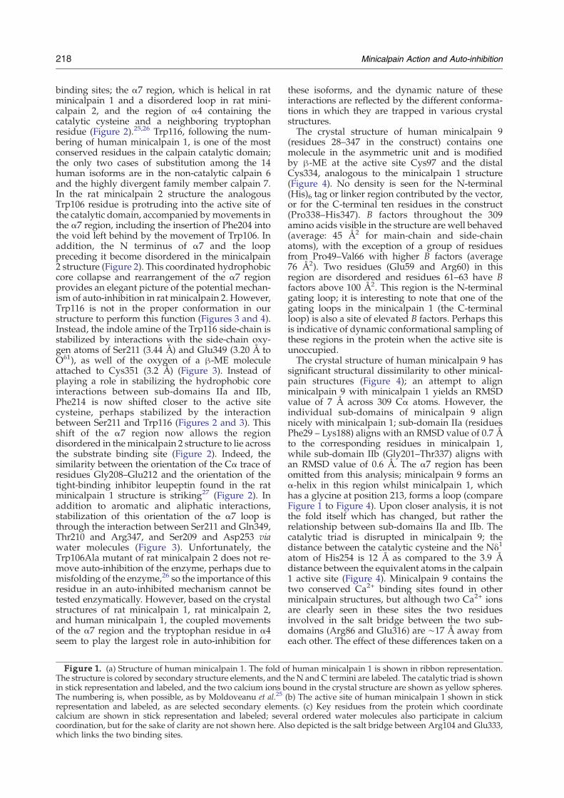
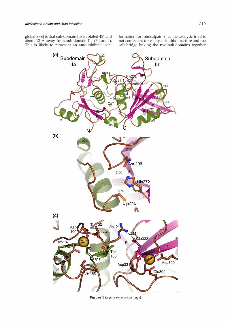
mixed α/β structure typical of these papain-likeproteins,2,29–31 (Figure 1). There are two moleculesof human minicalpain 1 in the asymmetric unit;included in the models are three sites of modifica-tion by β-mercaptoethanol (β-ME), including theactive site cysteine in one of the two molecules.The other β-ME modification is identical in bothmolecules of the asymmetric unit and is on Cys351,removed from the active site. The two protein chainsare superimposable with an RMSD value of 0.4 Å. Ingeneral, B factors throughout the core of themolecule are well-behaved, with an average B factorof 44 Å2 for main-chain and side-chain atoms; twoexceptions are the region around residues 206–224and the region including residues 253–267, whichhave average B factors around 80 Å2. These regionscorrespond to the dynamic α7 region, which ishelical in some minicalpain structures and a loop inothers (including the structure of human minical-pain 1), and the second gating loop found C-terminal to the α7 region. No density is observedfor the N-terminal 23 residues of the construct,including the (His)6 tag and residues 29–32 ofcalpain 1, or for the C-terminal six residues of theconstruct (Pro355–Ser360).The crystal structure of the protease core of human
calpain 1, solved in absence of substrate or inhibitor,is quite similar to the structures of rat minicalpain 1and rat minicalpain 2 described previously.25,26
Human minicalpain 1 aligns with an RMSD valueof 0.9 Å to rat minicalpain 1 (PDB ID 1KXR;alignment performed over 322 Cα atoms), andwith an RMSD value of 0.6 Å to rat minicalpain 2(PDB ID 1MDW, 321 Cα atoms used for thealignment).25,26 This is not completely surprising;the sequence identity across the amino acids in thecrystal structures is 87% for human and rat mini-calpain 1, and 71% for human minicalpain 1 and ratminicalpain 2. The catalytic triads of all threestructures are in identical conformations (Figures1(b) and 2). The network of acidic amino acids andordered water molecules which form coordinationsites for calcium ions in both the IIa and IIb sub-domains are intact, as is the Arg104–Glu333 saltbridge that connects the two calcium binding sites(Figure 1(c)). However, there were two regions inthe human minicalpain 1 structure which wereclearly in novel conformations: a conserved Trp116residue in the α4 helix just C-terminal to the catalyticcysteine residue and the residues from Ser206–Gly220 which make up the α6–α7 loop and the α7region (Figure 2).Rat minicalpain 1 was the first minicalpain
construct to be analyzed structurally; the mainmechanism of auto-inhibition was found to be thecalcium binding sites in sub-domains IIa and IIb,and indeed in the presence of calcium rat mini-calpain 1 is much more active than the otherminicalpains subsequently studied, although stillseveral orders of magnitude less active than full-length enzyme.25,26 The rat minicalpain 2 structure,by comparison with rat minicalpain 1, identified twosites of auto-inhibition in addition to the calcium
218 Minicalpain Action and Auto-inhibition
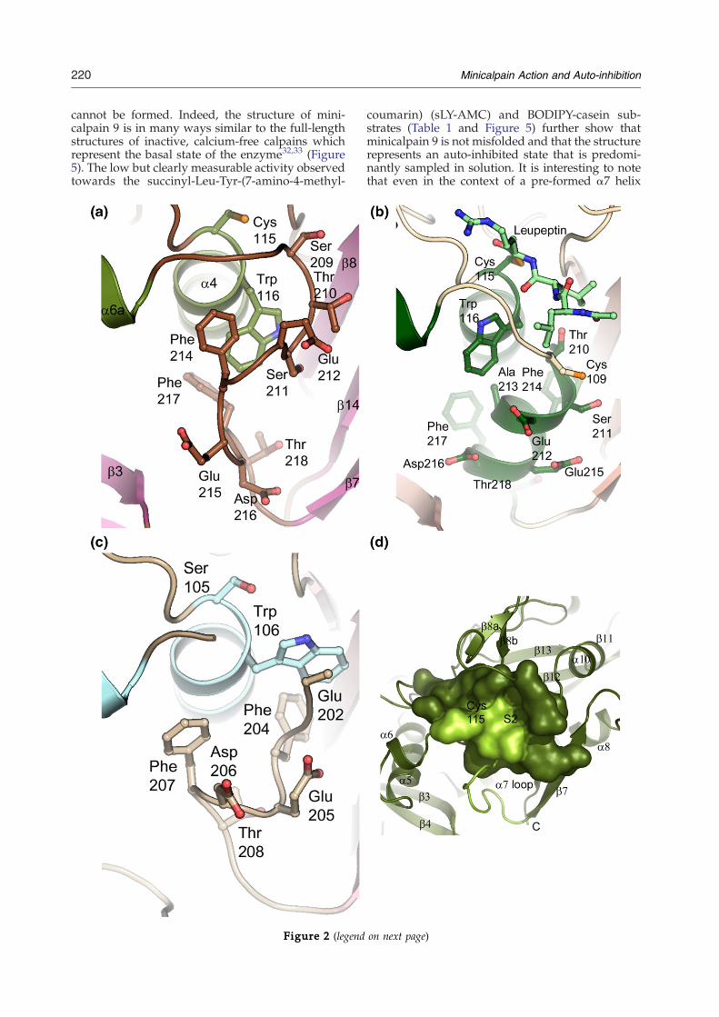
binding sites; the α7 region, which is helical in ratminicalpain 1 and a disordered loop in rat mini-calpain 2, and the region of α4 containing thecatalytic cysteine and a neighboring tryptophanresidue (Figure 2).25,26 Trp116, following the num-bering of human minicalpain 1, is one of the mostconserved residues in the calpain catalytic domain;the only two cases of substitution among the 14human isoforms are in the non-catalytic calpain 6and the highly divergent family member calpain 7.In the rat minicalpain 2 structure the analogousTrp106 residue is protruding into the active site ofthe catalytic domain, accompanied bymovements inthe α7 region, including the insertion of Phe204 intothe void left behind by the movement of Trp106. Inaddition, the N terminus of α7 and the looppreceding it become disordered in the minicalpain2 structure (Figure 2). This coordinated hydrophobiccore collapse and rearrangement of the α7 regionprovides an elegant picture of the potential mechan-ism of auto-inhibition in rat minicalpain 2. However,Trp116 is not in the proper conformation in ourstructure to perform this function (Figures 3 and 4).Instead, the indole amine of the Trp116 side-chain isstabilized by interactions with the side-chain oxy-gen atoms of Ser211 (3.44 Å) and Glu349 (3.20 Å toOδ1), as well of the oxygen of a β-ME moleculeattached to Cys351 (3.2 Å) (Figure 3). Instead ofplaying a role in stabilizing the hydrophobic coreinteractions between sub-domains IIa and IIb,Phe214 is now shifted closer to the active sitecysteine, perhaps stabilized by the interactionbetween Ser211 and Trp116 (Figures 2 and 3). Thisshift of the α7 region now allows the regiondisordered in theminicalpain 2 structure to lie acrossthe substrate binding site (Figure 2). Indeed, thesimilarity between the orientation of the Cα trace ofresidues Gly208–Glu212 and the orientation of thetight-binding inhibitor leupeptin found in the ratminicalpain 1 structure is striking27 (Figure 2). Inaddition to aromatic and aliphatic interactions,stabilization of this orientation of the α7 loop isthrough the interaction between Ser211 and Gln349,Thr210 and Arg347, and Ser209 and Asp253 viawater molecules (Figure 3). Unfortunately, theTrp106Ala mutant of rat minicalpain 2 does not re-move auto-inhibition of the enzyme, perhaps due tomisfolding of the enzyme,26 so the importance of thisresidue in an auto-inhibited mechanism cannot betested enzymatically. However, based on the crystalstructures of rat minicalpain 1, rat minicalpain 2,and human minicalpain 1, the coupled movementsof the α7 region and the tryptophan residue in α4seem to play the largest role in auto-inhibition for
Figure 1. (a) Structure of human minicalpain 1. The fold oThe structure is colored by secondary structure elements, and tin stick representation and labeled, and the two calcium ions bThe numbering is, when possible, as by Moldoveanu et al.25 (representation and labeled, as are selected secondary elemecalcium are shown in stick representation and labeled; sevecoordination, but for the sake of clarity are not shown here. Alwhich links the two binding sites.
these isoforms, and the dynamic nature of theseinteractions are reflected by the different conforma-tions in which they are trapped in various crystalstructures.The crystal structure of human minicalpain 9
(residues 28–347 in the construct) contains onemolecule in the asymmetric unit and is modifiedby β-ME at the active site Cys97 and the distalCys334, analogous to the minicalpain 1 structure(Figure 4). No density is seen for the N-terminal(His)6 tag or linker region contributed by the vector,or for the C-terminal ten residues in the construct(Pro338–His347). B factors throughout the 309amino acids visible in the structure are well behaved(average: 45 Å2 for main-chain and side-chainatoms), with the exception of a group of residuesfrom Pro49–Val66 with higher B factors (average76 Å2). Two residues (Glu59 and Arg60) in thisregion are disordered and residues 61–63 have Bfactors above 100 Å2. This region is the N-terminalgating loop; it is interesting to note that one of thegating loops in the minicalpain 1 (the C-terminalloop) is also a site of elevated B factors. Perhaps thisis indicative of dynamic conformational sampling ofthese regions in the protein when the active site isunoccupied.The crystal structure of human minicalpain 9 has
significant structural dissimilarity to other minical-pain structures (Figure 4); an attempt to alignminicalpain 9 with minicalpain 1 yields an RMSDvalue of 7 Å across 309 Cα atoms. However, theindividual sub-domains of minicalpain 9 alignnicely with minicalpain 1; sub-domain IIa (residuesPhe29 – Lys188) aligns with an RMSD value of 0.7 Åto the corresponding residues in minicalpain 1,while sub-domain IIb (Gly201–Thr337) aligns withan RMSD value of 0.6 Å. The α7 region has beenomitted from this analysis; minicalpain 9 forms anα-helix in this region whilst minicalpain 1, whichhas a glycine at position 213, forms a loop (compareFigure 1 to Figure 4). Upon closer analysis, it is notthe fold itself which has changed, but rather therelationship between sub-domains IIa and IIb. Thecatalytic triad is disrupted in minicalpain 9; thedistance between the catalytic cysteine and the Nδ1
atom of His254 is 12 Å as compared to the 3.9 Ådistance between the equivalent atoms in the calpain1 active site (Figure 4). Minicalpain 9 contains thetwo conserved Ca2+ binding sites found in otherminicalpain structures, but although two Ca2+ ionsare clearly seen in these sites the two residuesinvolved in the salt bridge between the two sub-domains (Arg86 and Glu316) are ∼17 Å away fromeach other. The effect of these differences taken on a
f human minicalpain 1 is shown in ribbon representation.he N and C termini are labeled. The catalytic triad is shownound in the crystal structure are shown as yellow spheres.b) The active site of human minicalpain 1 shown in sticknts. (c) Key residues from the protein which coordinateral ordered water molecules also participate in calciumso depicted is the salt bridge between Arg104 and Glu333,
219Minicalpain Action and Auto-inhibition
global level is that sub-domain IIb is rotated 45° andabout 12 Å away from sub-domain IIa (Figure 4).This is likely to represent an auto-inhibited con-
Figure 1 (legend o
formation for minicalpain 9, as the catalytic triad isnot competent for catalysis in this structure and thesalt bridge linking the two sub-domains together
n previous page)
220 Minicalpain Action and Auto-inhibition
cannot be formed. Indeed, the structure of mini-calpain 9 is in many ways similar to the full-lengthstructures of inactive, calcium-free calpains whichrepresent the basal state of the enzyme32,33 (Figure5). The low but clearly measurable activity observedtowards the succinyl-Leu-Tyr-(7-amino-4-methyl-
Figure 2 (legend
coumarin) (sLY-AMC) and BODIPY-casein sub-strates (Table 1 and Figure 5) further show thatminicalpain 9 is not misfolded and that the structurerepresents an auto-inhibited state that is predomi-nantly sampled in solution. It is interesting to notethat even in the context of a pre-formed α7 helix
on next page)
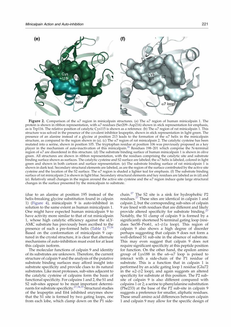
Figure 2. Comparison of the α7 region in minicalpain structures. (a) The α7 region of human minicalpain 1. Theprotein is shown in ribbon representation, with α7 residues (Ser209–Asp216) shown in stick representation for emphasis,as is Trp116. The relative position of catalytic Cys115 is shown as a reference. (b) The α7 region of rat minicalpain 1. Thisstructure was solved in the presence of the covalent inhibitor leupeptin, shown in stick representation in light green. Thepresence of an alanine instead of a glycine at position 213 leads to the formation of the α7 helix in the minicalpainstructure, as compared to the region shown in (a). (c) The α7 region of rat minicalpain 2. The catalytic cysteine has beenmutated into a serine, shown in position 105. The tryptophan residue at position 106 was previously proposed as a keyplayer in the mechanism of auto-inactivation of this minicalpain.26 Residues 198–201 which comprise the N-terminalregion of α7 are disordered in this structure. (d) The substrate binding surface of human minicalpain 1 is shown in olivegreen. All structures are shown in ribbon representation, with the residues comprising the catalytic site and substratebinding surface shown as surfaces. The catalytic cysteine and S2 surface are labeled; the α7 helix is labeled, colored in lightgreen and shown in both cartoon and surface representation. (e) The substrate binding surface of rat minicalpain 1 isshown in dark teal. Secondary structural elements are labeled, as are the region of the surface contributed by the active sitecysteine and the location of the S2 surface. The α7 region is shaded a lighter teal for emphasis. (f) The substrate bindingsurface of rat minicalpain 2 is shown in light blue. Secondary structural elements and key residues are labeled as in (d) and(e). Relatively small changes in the region around the active site cysteine and the α7 region induce quite large structuralchanges in the surface presented by the minicalpain to substrate.
221Minicalpain Action and Auto-inhibition
(due to an alanine at position 195 instead of thehelix-breaking glycine substitution found in calpain1) (Figure 4), minicalpain 9 is auto-inhibited insolution to the same extent as human minicalpain 1.One might have expected human minicalpain 9 tohave activity more similar to that of rat minicalpain1, whose high catalytic efficiency against the sLY-AMC substrate has previously been ascribed to thepresence of such a pre-formed helix (Table 1).25,26
Based on the conformation of minicalpain 9 cap-tured in the crystal structure, it is clear that alternatemechanisms of auto-inhibition must exist for at leastthis calpain isoform.The molecular functions of calpain 9 and identity
of its substrates are unknown. Therefore, the currentstructure of calpain 9 and the analysis of the putativesubstrate binding surfaces may afford insight intosubstrate specificity and provide clues to its naturalsubstrates. Like most proteases, sub-sites adjacent tothe catalytic cysteine of calpains form the basis offunctional specificity. For calpains 1 and 2, the S1 andS2 sub-sites appear to be most important determi-nants for substrate specificity.27,34,35 Structural studiesof the leupeptin and E64 inhibited enzymes showthat the S1 site is formed by two gating loops, onefrom each lobe, which clamp down on the P1 side-
chain.27 The S2 site is a sink for hydrophobic P2residues.27 These sites are identical in calpain 1 andcalpain 2, but the corresponding sub-sites of calpain9 are lined with residues that are different and likelyprovide altered specificity for substrate binding.27Notably, the S1 clamp of calpain 9 is formed by asignificantly shortened N-terminal gating loop (resi-dues Ser58–Pro61, α1–β1a loop). This region ofcalpain 9 also shows a high degree of disorderperhaps suggesting that calpain 9 does not form awell-defined S1 sub-site in the absence of substrate.This may even suggest that calpain 9 does notrequire significant specificity at this peptide positionfor function. On the other hand, the epsilon aminogroup of Lys188 in the α6–α7 loop is poised tointeract with a side-chain of the P1 residue ofsubstrate. This is a function that in calpain 1 isperformed by an acidic gating loop 1 residue (Glu72in the α2–β2 loop), and again suggests an alteredspecificity for substrate at this position. The P2 sub-site of calpain 9 is also different compared withcalpains 1 or 2; a serine to phenylalanine substitution(Phe233) at the base of the P2 sub-site in calpain 9suggests a preference for smaller, aliphatic residues.These small amino acid differences between calpain1 and calpain 9 may allow for the specific design of
Figure 3. The structural environment around Trp116 and the spatial relationship between α7 and α4 in humanminicalpain 1. The β-ME molecule modifying the distal Cys351 residue is shown in stick representation; distancesbetween either electrostatic pairs or water molecules are given in Angstrom units.
222 Minicalpain Action and Auto-inhibition
therapeutic inhibitors as well as diagnostic reagentsagainst the tissue-specific calpain 9.
Discussion
The current model of calpain activation includescalcium dependent release of the catalytic domainfrom a repressed conformation. In the full-lengthenzyme, non-catalytic supporting domains dI, dIII,dIV, and dVI provide a foundation such that the twolobes of the papain-like catalytic domain are keptapart until calciumbinding triggers activation.33,36,37In the absenceof calcium, the carboxy-terminal endofthe catalytic domain is covalently fixed to one cornerof the foundation (leading into theC2-likedomain III)and the amino-terminal peptide (domain I) is non-covalently fixed to the opposing corner. Calciumbinding to dIV and dVI releases their grip on theamino-terminalpeptide, freeing thecatalytic lobes forassemblyandcatalyticcompetency.The latterprocessalso requires binding of two calcium atoms in each ofthe catalytic lobes, leading to the stabilization of a saltbridgeandorientationof the two lobes.25,32,33,36Asanadded level of complexity, the non-catalytic domainsmay also provide auxiliary interactions needed for
full catalytic activity. Notably, dIII interacts andstabilizes the helical form of region α7 of calpain2, which abuts the active site.32,33,36,37 In theabsence of this interaction, α7 (as in the structure ofthe minicalpain 2 construct) can adopt a random coilconformation that subsequently allows rotation ofthe aromatic side-chain of a neighboring conservedtryptophan (Trp116) into the S2 sub-site, interferingwith binding of calpain substrates. The conforma-tional flexibility in α7 has been attributed to acentrally located glycine residue found in abouthalf of calpain isoforms, including rat calpain 2 andhuman calpain 1.26 The rat homolog of calpain 1 hasan alanine at that position in α7, adopts the helicalconformation even in absence of regulatory domains,and is more active in solution, thereby validating therequirement of a helical α7 for catalysis for cal-pains.25,26 Stabilization of the α7 region in order toobtain a catalytically competent enzyme is clearlynecessary for human calpain 1 as well, based on ourcrystal structure. As in the case of rat minicalpain 2,human minicalpain 1 encodes an α7 region which isfound in a random coil conformation. Presumably atleast one of the functions of the non-catalytic do-mains for human calpain 1 is to stabilize the α7region into a productive conformation for catalysis.
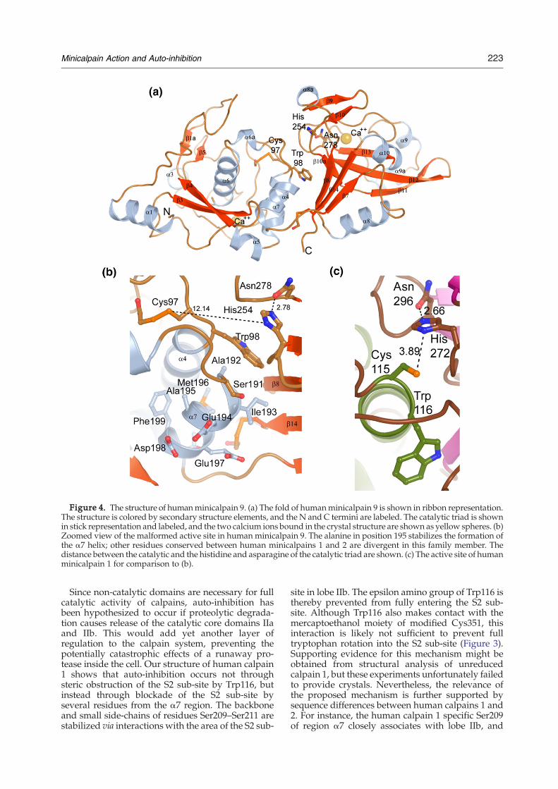
Figure 4. The structure of humanminicalpain 9. (a) The fold of humanminicalpain 9 is shown in ribbon representation.The structure is colored by secondary structure elements, and the N and C termini are labeled. The catalytic triad is shownin stick representation and labeled, and the two calcium ions bound in the crystal structure are shown as yellow spheres. (b)Zoomed view of the malformed active site in human minicalpain 9. The alanine in position 195 stabilizes the formation ofthe α7 helix; other residues conserved between human minicalpains 1 and 2 are divergent in this family member. Thedistance between the catalytic and the histidine and asparagine of the catalytic triad are shown. (c) The active site of humanminicalpain 1 for comparison to (b).
223Minicalpain Action and Auto-inhibition
Since non-catalytic domains are necessary for fullcatalytic activity of calpains, auto-inhibition hasbeen hypothesized to occur if proteolytic degrada-tion causes release of the catalytic core domains IIaand IIb. This would add yet another layer ofregulation to the calpain system, preventing thepotentially catastrophic effects of a runaway pro-tease inside the cell. Our structure of human calpain1 shows that auto-inhibition occurs not throughsteric obstruction of the S2 sub-site by Trp116, butinstead through blockade of the S2 sub-site byseveral residues from the α7 region. The backboneand small side-chains of residues Ser209–Ser211 arestabilized via interactions with the area of the S2 sub-
site in lobe IIb. The epsilon amino group of Trp116 isthereby prevented from fully entering the S2 sub-site. Although Trp116 also makes contact with themercaptoethanol moiety of modified Cys351, thisinteraction is likely not sufficient to prevent fulltryptophan rotation into the S2 sub-site (Figure 3).Supporting evidence for this mechanism might beobtained from structural analysis of unreducedcalpain 1, but these experiments unfortunately failedto provide crystals. Nevertheless, the relevance ofthe proposed mechanism is further supported bysequence differences between human calpains 1 and2. For instance, the human calpain 1 specific Ser209of region α7 closely associates with lobe IIb, and

Figure 5 (legend on opposite page)
224 Minicalpain Action and Auto-inhibition

Table 1. Kinetic constants for hydrolysis of sLY-AMC and BODIPY-casein by minicalpains
Human minicalpain 1 Human minicalpain 1 G213A Human minicalpain 9 Rat minicalpain 1
sLY-AMC 1.39×10⁻⁶±4.2×10⁻⁷ 2.16×10⁻⁵±7.1×10⁻⁸ 1.67×10⁻⁶±8.4×10⁻⁸ 42.7×10⁻⁵±4.7×10⁻⁵
kcat (s−1)
Km (μM) 57±6 83±0.1 38±9 335±6kcat/Km (M−1s−1) 0.024 0.259 0.035 1.275Relative catalytic efficiency 1 11 1.5 53BODIPY-casein (%) 100 586 105 n/aRelative activity(%)
Parameters for rat minicalpain 1 are from Moldoveanu et al.25
225Minicalpain Action and Auto-inhibition
Ser211 is also involved in interactions whichstabilize this arrangement of α7 (Figure 3). Incontrast, a novel mechanism of auto-inhibition canbe clearly seen in the minicalpain 9 structure,namely the misalignment of the catalytic lobesrelative to that found in other minicalpain struc-tures. This conformation reveals a novel aspect ofcalpain biochemistry.Although intriguing, the proteolyzed catalytic core
is most likely not the predominant species in cells.Regulation of activity in the full-length calpain is acomplex and multi-step process. For full-lengthcalpains 1 and 2, whose catalytic lobes are mis-aligned relative to each other in the inactive calcium-free state, activation requires reorientation of theselobes as well as stabilization of α7 to acquire fullactivity.6,33 Minicalpains have previously only beenseen in active conformations with respect to lobeorientation, best exemplified by catalytic triads thatare in the proper orientation and distance to performcatalysis and by a salt bridge which connects the twolobes of the catalytic core (see e.g. Figure 1). There-fore, it has been possible when looking at theseminicalpain structures to consider them almostcompletely competent for catalysis; the assumptionmade when looking at the isolated minicalpainstructures solved to date (including the structure ofhuman minicalpain 1) is that only small changesinvolving structural stabilization of the α7 regionwould be enough to recapitulate the active form ofthe calpain enzyme. The minicalpain 9 structure ismisaligned with respect to these other minicalpainstructures; however, if one independently modelsthe two sub-domains of calpain 9 onto those ofcalpain 1, each lobe superimposes with an RMSDvalue of less than 1 Å (Figure 5(a) and (b)). Moreimportantly, domain IIa and IIb in the minialpcain 9
Figure 5. Misalignment of catalytic subdomains IIa and IIb9. (a) The catalytic cores of human minicalpain 1 (green) anrepresentation and in stereo. In this view, sub-domains IIa arethe respective IIb sub-domains. The calcium ions for calpain 1in calpain 9 are in gold. The arrow points in the direction ofcalpain 9 were superimposed independently upon their coun0.87 Å for sub-domain IIa, and 0.9 Å for sub-domain IIb. The cyellow. (c) The active site region of human minicalpain 9 (mafull-length, calcium-free human calpain 2 (black) are shown in cIIb sub-domain, as shown by the histidine and asparagine ofcatalytic cysteine is labeled, but only the histidine and aspminicalpain 1 represents the active, enzymatically competentminicalpain 9 and calcium-free full-length calpain 2 structures,a way as to disrupt catalysis.
structure have a relationship reminiscent of thosefound in the calcium-free full length calpainstructures32,33(Figure 5(c)). We therefore proposethat the structure of human minicalpain 9 indicatesthat domains dIII and dIV of full-length calpain 9must contribute to catalysis through proper align-ment of the two catalytic lobes, rather than throughstabilization of the helical α7 conformation. Ourmodel is supported by the relative inactivity ofminicalpain 9 despite the presence of an alanine, andthereby helical conformation, in α7. Its specificactivity is 50-fold less than either the Gly213Alamutant of human calpain 1 or wild-type rat calpain 1(Table 1). It is possible that minicalpain 9 is simplynot equipped to bind the sLY-AMC or BODIPY-casein substrates; indeed, our own structure analysisof the N-terminal gating loop and substrate bindingsub-sites could support this idea. However, boththese substrates have repeatedly been used assubstrates for full-length calpains, even when laterstudies have shown lesser activities for isolatedminicalpains compared with their full-length coun-terparts; and full-length calpain 9 has previouslybeen shown in at least one study to cleave casein as asubstrate.23 CD and thermal denaturation studies ofminicalpain 9 in solution indicate that the protein iswell folded, contains secondary structure, andundergoes cooperative melting, all characteristicsof a well-behaved protein, and consistent with thecurrent structural determination (data not shown).The coordination of calcium ions by the isolatedminicalpain core is essentially identical to theequivalent regions in both theminicalpain structuresand also in the structures of the active full-lengthcalpin structures solved previously. Taken together,this evidence indicates that the functions of calpain 9domains dIII and dIV differ from their function in the
as a mechanism of auto-inhibition in human minicalpaind human minicalpain 9 (magenta) are shown in ribbonsuperimposed, to highlight the rotation and translation ofare shown in light yellow; the corresponding calcium ionsrotation/translation. (b) The sub-domains IIa and IIb ofterparts in calpain 1. The RMSD value of each domain isatalytic triad is shown as sticks; calcium ions are shown ingenta), human minicalpain 1 (green), and the structure ofartoon representation. The structures are aligned using thethe respective catalytic triads in stick representation. Eacharagine of calpain 9 are labeled. In this Figure, humanrelationship between the catalytic triad; in both the nativesub-domains IIa and IIb are rotated and translated in such
226 Minicalpain Action and Auto-inhibition
ubiquitous calpains, and activation by calcium willinvolve the formation of a catalytically competentlobe orientation by a novel mechanism. Furtherelucidation of the mechanism of calpain 9 activation,therefore, will almost certainly require studies of full-length protein.The highly conserved nature of calpain cores
would tend to imply a universal mechanism ofcatalysis and regulation. However, we present heretwo structures that suggest that the catalytic sub-units are more complicated and undergo subtle setsof structural changes that the primary sequencecannot provide insight into. In addition, it is clearthat the supporting domains in the full-lengthenzyme contribute more directly to full enzymaticactivity than can be explained by analysis of theamino acid sequence alone. Minicalpain structures,although not directly pertinent to the discussion ofthe full-length enzyme, allow the analysis of asnapshot of the active site, which can providedetailed information concerning the catalytic region,the substrate binding surface, and the relationshipbetween the catalytic lobes. These studies areimportant for the study and treatment of calpain-related diseases because the minicalpain region ofthe human calpains clearly possesses a greaterdegree of structural plasticity than has been pre-viously described, making modeling of the activesite inappropriate for design of specific calpaininhibitors. These studies can also allow for extra-polation to the function of the catalytic core in thecontext of the full-length enzyme, thereby pro-viding potentially valuable insight into the functionof the physiologically relevant form of the nativeenzyme.
Materials and Methods
Cloning and expression
cDNA encoding the full-length human calpain 1(accession number NM005186) and full-length humancalpain 9 (accession number NM006615) were obtainedfrom the Mammalian Gene Collection. Ligation indepen-dent cloning was utilized to clone into the p28a-LICvector, which contains an N-terminal (His)6 tag andthrombin cleavage site. Resulting plasmids were trans-formed into BL21(DE3) cells (Invitrogen) for large scaleprotein expression. Cells were grown in a supplementedTerrific Broth media at 37 °C to an A600 of ∼6 andinduced by adding 50 μM IPTG to the cultures. Cellswere induced overnight at 15 °C, then harvested andstored at −80 °C.
Purification and concentration
All chemicals were of high quality grade and purchasedthrough Sigma unless otherwise noted. Cell pellets from2–4 l culture were resuspended in 20 ml of lysis buffer(50 mM Tris (pH 8.0), 500 mM NaCl, 1 mM EDTA, 1 mMPMSF, 2 mM CaCl2 and 0.1 ml of general proteaseinhibitor Sigma P2714) and then homogenized for 30–60 s per pellet. Cell lysis was accomplished by sonication
(Virtis408912, Virsonic) at 4 °C; lysed cells were centri-fuged in a JA25.50 rotor in an Avanti J-20 XPI centrifuge(Beckman Coulter) for 20 min at 69,673g. The supernatantwas loaded by gravity onto 2 ml of Ni-NTA resin (Qiagen30450). Five column volumes of lysis buffer were used towash the column, followed by five column volumes oflow imidazole buffer (lysis buffer + 10 mM imidazole (pH8)). Samples were eluted from the Ni-NTA resin in 10 mlof elution buffer (lysis buffer + 250 mM imidazole and10%(v/v) glycerol). For crystals obtained from proteinwithout the (His)6 tag, one unit of thrombin (SigmaT9681) per milligram of protein was then added and theprotein was stored without shaking overnight at 4 °C,and all proteins were further purified using size exclusionchromatography. For gel filtration, an XK 16x65 column(18-1031-47 and 18-6488-01; GE Healthcare) packed withHiLoad Superdex 200 resin (10-1043-04; GE Healthcare)was pre-equilibrated with gel filtration buffer (lysisbuffer + 5 mM β-ME, 1 mM EDTA, and 1 mM CaCl2)and then samples were loaded onto the column at1.5 ml/min, and 2 ml fractions were collected usingpeak fractionation protocols. Fractions were pooled andconcentrated using Amicon concentrators with anappropriate molecular mass cut-off (Millipore). Theprotein was either used at 20 mg/ml for crystallizationin the case of calpain 9, or diluted tenfold into low saltcrystallization buffer (50 mM Tris (pH 8.0), 100 mMNaCl, 1 mM EDTA, and 2 mM CaCl2) then re-concentrated to 20 mg/ml for crystallographic screeningof calpain 1.
Crystallization, data collection, and structure solution
Calpain 1 and calpain 9 constructs were screened usingin-house conditions developed by the Structural GenomicsConsortium in 96 well sitting drop format using dropscontaining 1 μl of protein and 1 μl of reservoir. Multiplecrystallization conditions were observed to produce smallcrystals for two constructs of calpain 1, requiring furtheroptimization. Optimization involved, in part, elucidationof optimal pH and salt conditions for protein stabilityusing a high-throughput differential scanning light scat-tering system (StarGazer) as described.38 After optimiza-tion of the initial crystallization conditions and constructboundaries, purified calpain 1 (residues 29–360) was crys-tallized using the hanging drop vapor diffusion method.Diffracting crystals leading to the structure grew whenthe protein was mixed with the reservoir solutioncontaining 1.5 M ammonium formate, 0.1 M Tris (pH7.75) in a 1:1 volume ratio. Crystals typically appeared24–36 h after incubation at 18 °C; the typical size ofcalpain 1 crystals was 150 μm×30 μm× 10 μm. Crystalswere harvested into cryo-protection buffer (1:1 mixture ofglycerol and mother liquor; final concentration of glycerolwas 15%) and frozen in liquid nitrogen. In the case ofcalpain 9, optimization of initial crystallization conditionswas also performed by the hanging drop vapor diffusionmethod on a construct containing residues 28–347.Diffraction quality crystals were obtained by mixing1 μl of concentrated protein stock with 1 μl of reservoirsolution containing 18–20% (w/v) PEG 3350, 0.1 M Hepes(pH 7.7), 0.2 M CaCl2. Crystals typically appeared 36–72 hafter incubation at 18 °C; the typical size of calpain 1crystals was 75 μm×20 μm×10 μm. Crystals were har-vested into cryo-protection buffer (1:1 mixture of glyceroland mother liquor; final concentration of glycerol was15%) and frozen in liquid nitrogen. Diffraction data fromcrystals of calpain 1 were collected at beamline 19BM and
227Minicalpain Action and Auto-inhibition
from calpain 9 at beamline 32ID, both at the AdvancedPhoton Source (Argonne National Laboratory), andintegrated and scaled using the HKL2000 programpackage.39,40 Statistics of data collection and processingare provided in Table 2.For structure solution and refinement of calpain 1, the
program PHASER41 was used as part of the CCP4 suite42
to find the molecular replacement solution in theresolution range between 20 Å and 2.8 Å. Initial attemptsto find a molecular replacement solution with a model ofthe entire catalytic domain was unsuccessful, so thesearch model (1TLO) was subdivided into two domains.Following the successful molecular replacement searchand identification of the correct space group withPHASER, a round of maximum-likelihood refinementand phase extension to 2.4 Å was carried out withREFMAC5,42–44 and the phases were then input intoARP/wARP42,45,46 for automatic model building anditerative refinement. The models were completed usingthe graphics program O47 and further rounds of refine-ment using REFMAC5 resulted in an R-factor of 22.2%(Rfree 26.4%) for data from 44.32 Å to 2.40 Å. The finalmodel comprises 5142 protein atoms, 229 water mole-cules, four calcium ions, and two β-ME molecules for twomolecules in the asymmetric unit.For structure solution and refinement of calpain 9, the
program PHASER was again used with 1MDW as asearch model (subdivided into two sub-domains) in theresolution range between 20 Å and 2.5 Å. Then a round ofmaximum-likelihood refinement and phase extension to2.31 Å was carried out with REFMAC5, and ARP/wARPused for automatic model building and iterative refine-ment. The models were completed using the graphicsprogram O and further rounds of refinement usingREFMAC5 resulted in an R-factor of 20.3% (Rfree 27.2%)for data from 19.83 Å to 2.31 Å. The final modelcomprises 2465 protein atoms, 77 water molecules, twocalcium ions, and two β-ME molecules for one proteinmolecule in the asymmetric unit. Both models haveexcellent stereochemistry as judged by PROCHECK,48
Table 2. Summary of crystallographic analysis
Humanminicalpain 9
Humanminicalpain 1
A. Data collectionBeamline APS 32IDB APS 19BMWavelength 1 0.97889Resolution (Dmin, Å) 19.83–2.31 44.32–2.40Unique reflections 15,919 42,153Redundancy
(high resolution shell)3.7(3.7) 8.4(3.1)
Completeness (%) 99.7(100) 98.9(94.6)<I/σ> (high resolution shell) 19.2(3.4) 20.56(1.4)Rsym (high resolution shell) 0.069(0.34) 0.089(0.569)
B. RefinementResolution (Å) 19.83–2.31 44.32–2.40Reflections 15,919 39,890Number of atoms (solvent) 2552(77) 5387(229)Rwork (Rfree) (%) 20.3(27.2) 22.2(26.4)RMSD bond lengths (Å) 0.014 0.009RMSD bond angles (deg.) 1.413 1.127
C. Ramachandran plotMost favored (%) 86.6 87.5Additionally allowed (%) 13.4 11.9Generously allowed (%) 0 0Disallowed (%) 0 0
with no residues in disallowed or unfavorable regions ofRamachandran space.
Protease activity measurements using the sLY-AMCand BODIPY-casein substrates
Activities of calpain proteases were assayed using theEnzChek kit (Invitrogen, E6638) and with a succinyl-Leu-Tyr-(7-amino-4-methylcoumarin) (sLY-AMC) peptide sub-strate (BACHEM, I-1335) based on previously publishedprotocols25,26,49,50 with modifications for use in a 384 wellformat. Both assays are based on the increase influorescence that occurs upon proteolysis of the substrate;for the BODIPY FL casein substrate, proteolysis liberateslabeled casein fragments thus relieving the quenchedfluorescence resulting from hyper-labeling of the caseinproteins with BODIPY FL.50 In the case of the sLY-AMCpeptide the fluorescence signal increases when the AMCmoiety is liberated from the peptide.25 Briefly, 20 μM ofpurified protein was added to a mixture of substrate andactivity buffer (100 mM Hepes (pH 7), 100 mM NaCl,1 mM EDTA, 10 mM CaCl2, and 14 mM β-ME) on ice,aliquoted into 384 well plates, and fluorescence wasmeasured in a FluoDia T70 (Photon Technology Interna-tional) at room temperature. For the sLY-AMC assay,substrate concentrations from 15 μM to 300 μMwere usedto ensure substrate was not limiting over the time courseof the experiment; the amount of AMC released during theexperiment was calculated based on a standard curve ofAMC concentrations from 2 nm to 1 μM. For the BODIPY-casein assay, the total amount of label incorporated permolecule of substrate is unknown, and the activity assaywas done under limiting conditions of substrate, therebylimiting analysis of the data to simple activity per timemeasurements. All experiments were performed in quad-ruplicate to reduce well-to-well variability; in addition,each protein was tested in at least three differentexperiments to obtain standard deviations for the para-meters tested.
Protein Data Bank accession codes
The coordinates and structure factors for the structure ofthe protease core of human calpain 1 have been depositedinto the RCSB PDBwith ID 2ARY; the PDB ID for calpain 9is 1ZIV.
Acknowledgements
This research was supported by the StructuralGenomics Consortium with funds from GenomeCanada through the Ontario Genomics Institute, theCanadian Institutes for Health Research, the CanadaFoundation for Innovation, the Ontario Researchand Development Challenge Fund, the OntarioInnovation Trust, the Wellcome Trust, Glaxo-SmithKline, the Knut and Alice Wallenberg Founda-tion, Vinnova and the Swedish Foundation forStrategic Research. Use of the Advanced PhotonSource was supported by the U. S. Department ofEnergy, Office of Science, Office of Basic EnergySciences, under Contract No. DE-AC02-06CH11357.
228 Minicalpain Action and Auto-inhibition
References1. Huang, Y. & Wang, K. K. (2001). The calpain family
and human disease. Trends Mol. Med. 7, 355–362.2. Goll, D. E., Thompson, V. F., Li, H., Wei, W. & Cong, J.
(2003). The calpain system. Physiol. Rev. 83, 731–801.3. Khorchid, A. & Ikura, M. (2002). How calpain is
activated by calcium. Nature Struct. Biol. 9, 239–241.4. Cong, J. Y., Goll, D. E., Peterson, A.M. &Kapprell, H. P.
(1989). The role of autolysis in activity of the Ca-2+-dependent proteinases (Mu-Calpain and M-Calpain).J. Biol. Chem. 264, 10096–10103.
5. Cottin, P., Thompson, V. F., Sathe, S. K., Szpacenko, A.& Goll, D. E. (2001). Autolysis of mu- and m-calpainfrom bovine skeletal muscle. Biol. Chem. 382, 767–776.
6. Reverter, D., Strobl, S., Fernandez-Catalan, C., Sor-imachi, H., Suzuki, K. & Bode, W. (2001). Structuralbasis for possible calcium-induced activation mechan-isms of calpains. Biol. Chem. 382, 753–766.
7. Elce, J. S., Hegadorn, C., Simon, J. & Arthur, C. (1997).Autolysis, Ca2+ requirement, and heterodimer stabi-lity in m-calpain. J. Biol. Chem. 272, 11268–11275.
8. Nixon, R. A. (2003). The calpains in aging and aging-related diseases. Ageing Res. Rev. 2, 407–418.
9. Saito, K. I., Elce, J. S., Hamos, J. E. & Nixon, R. A.(1993). Widespread activation of calcium-activatedneutral proteinase (calpain) in the brain in Alzheimer-disease – a potential molecular-basis for neuronaldegeneration. Proc. Natl Acad. Sci. USA, 2628–2632.
10. Adamec, E., Mohan, P., Vonsattel, J. P. & Nixon, R. A.(2002). Calpain activation in neurodegenerative dis-eases: confocal immunofluorescence study with anti-bodies specifically recognizing the active form ofcalpain 2. Acta Neuropathol. 104, 92–104.
11. Mercken, M., Grynspan, F. & Nixon, R. A. (1995).Differential sensitivity proteolysis by brain calpain ofadult human-Tau, fetal human-Tau and Phf-Tau. FEBSLetters, 368, 10–14.
12. Nixon, R. A. (2000). A “protease activation cascade” inthe pathogenesis of Alzheimer's disease. AlzheimersDisease: Compend. Curr. Theor. 924, 117–131.
13. Azuma, M., Tamada, Y., Kanaami, S., Nakajima, E.,Nakamura, Y., Fukiage, C., Forsberg, N. E. et al. (2003).Differential influence of proteolysis by calpain 2 andLp82 on in vitro precipitation of mouse lens crystal-lins. Biochem. Biophys. Res. Commun. 307, 558–563.
14. Biswas, S., Harris, F., Dennison, S., Singh, J. P. &Phoenix, D. (2005). Calpains: enzymes of vision?Medical Science Monitor, 11, RA301–RA310.
15. Jia, Z. C., Petrounevitch, V., Wong, A., Moldoveanu,T., Davies, P. L., Elce, J. S. & Beckmann, J. S. (2001).Mutations in calpain 3 associated with limb girdle mus-cular dystrophy: Analysis by molecular modeling andby mutation in m-calpain. Biophys. J. 80, 2590–2596.
16. Laval, S. H. & Bushby, K. M. D. (2004). Limb-girdlemuscular dystrophies – from genetics to molecularpathology. Neuropath. Appl. Neurobiol. 30, 91–105.
17. Richard, I., Broux, O., Allamand, V., Fougerousse, F.,Chiannilkulchai, N., Bourg, N. et al. (1995). Mutationsin the proteolytic-enzyme calpain-3 cause limb-girdlemuscular-dystrophy type-2A. Cell, 81, 27–40.
18. Horikawa, Y., Oda, N., Cox, N. J., Li, X., Orho-Melander, M., Hara, M. et al. (2000). Genetic variationin the gene encoding calpain-10 is associated withtype 2 diabetes mellitus. Nature Genet. 26, 163–175.
19. Cox, N. J. (2002). Calpain 10 and genetics of type 2diabetes. Curr. Diab. Rep. 2, 186–190.
20. Paul, D. S., Harmon, A.W.,Winston, C. P. & Patel, Y. M.(2003). Calpain facilitates GLUT4 vesicle translocation
during insulin-stimulated glucose uptake in adipo-cytes. Biochem. J. 376, 625–632.
21. Cox, N. J., Hayes, M. G., Roe, C. A., Tsuchiya, T. & Bell,G. I. (2004). Linkage of calpain 10 to type 2 diabetes: thebiological rationale. Diabetes, 53, S19–S25.
22. Yoshikawa, Y., Mukai, H., Hino, F., Asada, K. & Kato,I. (2000). Isolation of two novel genes, down-regulatedin gastric cancer. Japan J. Cancer Res. 91, 459–463.
23. Lee, H. J., Tomioka, S., Kinbara, K., Masumoto, H.,Jeong, S. Y., Sorimachi, H. et al. (1999). Characteriza-tion of a human digestive tract-specific calpain, nCL-4,expressed in the baculovirus system. Arch. Biochem.Biophy. 362, 22–31.
24. Markmann, A., Schafer, S., Linz, W., Lohn, M., Busch,A. E. & Wohlfart, P. (2005). Down-regulation ofcalpain 9 is linked to hypertensive heart and kidneydisease. Cell. Physiol. Biochem. 15, 109–116.
25. Moldoveanu, T., Hosfield, C. M., Lim, D., Elce, J. S.,Jia, Z. & Davies, P. L. (2002). A Ca(2+) switch alignsthe active site of calpain. Cell, 108, 649–660.
26. Moldoveanu, T., Hosfield, C. M., Lim, D., Jia, Z. &Davies, P. L. (2003). Calpain silencing by a reversibleintrinsic mechanism. Nature Struct. Biol. 10, 371–378.
27. Moldoveanu, T., Campbell, R. L., Cuerrier, D. &Davies, P. L. (2004). Crystal structures of calpain-E64and -leupeptin inhibitor complexes reveal mobileloops gating the active site. J. Mol. Biol. 343, 1313–1326.
28. Li, Q. S., Hanzlik, R. P., Weaver, R. F. & Schonbrunn, E.(2006). Molecular mode of action of a covalentlyinhibiting peptidomimetic on the human calpainprotease core. Biochemistry, 45, 701–708.
29. Sorimachi, H., Ishiura, S. & Suzuki, K. (1997).Structure and physiological function of calpains.Biochem. J. 328, 721–732.
30. Hosfield, C. M., Ye, Q., Arthur, J. S., Hegadorn, C.,Croall, D. E., Elce, J. S. & Jia, Z. (1999). Crystallizationand X-ray crystallographic analysis of m-calpain, aCa2+-dependent protease. Acta Crystallog. sect. D, Biol.55, 1484–1486.
31. Sorimachi, H. & Suzuki, K. (2001). The structure ofcalpain. J. Biochem. (Tokyo), 129, 653–664.
32. Reverter, D., Sorimachi, H. & Bode, W. (2001). Thestructure of calcium-free human m-calpain: implica-tions for calcium activation and function. TrendsCardiovasc. Med. 11, 222–229.
33. Strobl, S., Fernandez-Catalan, C., Braun, M., Huber,R., Masumoto, H., Nakagawa, K. et al. (2000). Thecrystal structure of calcium-free human m-calpainsuggests an electrostatic switch mechanism for activa-tion by calcium. Proc. Natl Acad. Sci. USA, 97, 588–592.
34. Cuerrier, D., Moldoveanu, T. & Davies, P. L. (2005).Determination of peptide substrate specificity for mu-calpain by a peptide library-based approach: theimportance of primed side interactions. J. Biol. Chem.280, 40632–40641.
35. Guttmann, R. P., Day, G. A., III, Wang, X. & Bottiggi,K. A. (2005). Identification of a novel calpain inhibitorusing phage display. Biochem. Biophys. Res. Commun.333, 1087–1092.
36. Hosfield, C. M., Elce, J. S., Davies, P. L. & Jia, Z. (1999).Crystal structure of calpain reveals the structural basisfor Ca(2+)-dependent protease activity and a novelmode of enzyme activation. EMBO J. 18, 6880–6889.
37. Reverter, D., Braun, M., Fernandez-Catalan, C., Strobl,S., Sorimachi, H. & Bode, W. (2002). Flexibilityanalysis and structure comparison of two crystalforms of calcium-free human m-calpain. Biolog. Chem.383, 1415–1422.
38. Reinking, J., Lam, M. M. S., Pardee, K., Sampson,
229Minicalpain Action and Auto-inhibition
H. M., Liu, S. Y., Yang, P. et al. (2005). The Drosophilanuclear receptor E75 contains heme and is gasresponsive. Cell, 122, 195–207.
39. Minor, W., Cymborowski, M. & Otwinowski, Z.(2002). Automatic system for crystallographic datacollection and analysis. Acta Physica. Polonica. A, 101,613–619.
40. Otwinowski, Z. & Minor, W. (1997). Processing of X-ray diffraction data collected in oscillation mode.Macromol. Crystallog. pt A, 276, 307–326.
41. Read, R. J. (2001). Pushing the boundaries ofmolecular replacement with maximum likelihood.Acta Crystallog. sect. D, 57, 1373–1382.
42. Collaborative Computational Project, Number 4.(1994). The CCP4 suite: programs for protein crystal-lography. Acta Crystallog. sect. D, 50, 760–763.
43. Murshudov, G. N., Vagin, A. A. & Dodson, E. J. (1997).Refinement of macromolecular structures by themaximum-likelihood method. Acta Crystallog. sect. D,53, 240–255.
44. Winn, M. D., Isupov, M. N. & Murshudov, G. N.(2001). Use of TLS parameters to model anisotropicdisplacements in macromolecular refinement. ActaCrystallog. sect. D, 57, 122–133.
45. Lamzin, V. S. & Wilson, K. S. (1993). Automatedrefinement of protein models. Acta Crystallog. sect. D,49, 129–147.
46. Morris, R. J., Perrakis, A. & Lamzin, V. S. (2003). ARP/wARP and automatic interpretation of protein elec-tron density maps. Methods Enzymol. 374, 229–244.
47. Jones, T. A., Zou, J. Y., Cowan, S. W. & Kjeldgaard(1991). Improved methods for building proteinmodels in electron density maps and the location oferrors in these models. Acta Crystallog. sect. A, 47,110–119.
48. Laskowski, R. A., Macarthur, M. W., Moss, D. S. &Thornton, J. M. (1993). Procheck – a program tocheck the stereochemical quality of protein structures.J. Appl. Crystallog. 26, 283–291.
49. Harper, J. W., Hemmi, K. & Powers, J. C. (1985).Reaction of serine proteases with substituted isocou-marins – discovery of 3,4-dichloroisocoumarin, a newgeneral mechanism based serine protease inhibitor.Biochemistry, 24, 1831–1841.
50. Thompson, V. F., Saldana, S., Cong, J. Y. & Goll, D. E.(2000). A BODIPY fluorescent microplate assay formeasuring activity of calpains and other proteases.Anal. Biochem. 279, 170–178.
Edited by R. Huber
(Received 12 August 2006; received in revised form 6 November 2006)Available online 14 November 2006