The Characterization of the Repertoire of Wheat Antigens and Peptides Involved in the Humoral Immune...
13
International Scholarly Research Network ISRN Allergy Volume 2011, Article ID 950104, 12 pages doi:10.5402/2011/950104 Research Article The Characterization of the Repertoire of Wheat Antigens and Peptides Involved in the Humoral Immune Responses in Patients with Gluten Sensitivity and Crohn’s Disease Aristo Vojdani Immunosciences Laboratory, Inc., 822 S. Robertson Boulevard, Suite 312, Los Angeles, CA 90035, USA Correspondence should be addressed to Aristo Vojdani, [email protected] Received 20 July 2011; Accepted 21 August 2011 Academic Editors: B. M. Stadler and P. E. Taylor Copyright © 2011 Aristo Vojdani. This is an open access article distributed under the Creative Commons Attribution License, which permits unrestricted use, distribution, and reproduction in any medium, provided the original work is properly cited. Intestinal T cells from gluten sensitivity/celiac disease patients respond to a heterogeneous array of peptides. Our study extended this heterogeneity to humoral immune response to various wheat proteins and peptides in patients with gluten sensitivity or Crohn’s disease. IgG and IgA antibodies in sera from those patients and healthy control subjects were measured against an array of wheat antigens and peptides. In gluten-sensitive patients, IgG reacted most against transglutaminase, prodynorphin, wheat extract, and α-, γ-, and ω-gliadin; IgA reacted most against wheat then transglutaminase, glutenin, and other peptides. In the sera of Crohn’s disease patients, IgG reacted most against wheat and wheat germ agglutinin then transglutaminase, prodynorphin, α-, and γ-gliadin; IgA reacted foremost against prodynorphin then transglutaminase and α-gliadin. These results showed a substantial heterogeneity in the magnitude of IgG and IgA response against various wheat antigens and peptides. Measurements of IgG and IgA antibodies against such an array of wheat peptides and antigens can enhance the sensitivity and specificity of serological assays for gluten sensitivity and celiac disease and may also detect silent celiac disease or its overlap with inflammatory bowel disease. 1. Introduction Despite the efforts of several laboratories to define relevant gluten epitopes, the characterization of the complete reper- toire of peptides involved in the pathogenesis of celiac disease and associated disorders remains a daunting task because of the great heterogeneity of gluten proteins [1–4]. So far, several T cell stimulatory peptides from α-gliadin, γ-gliadin, and glutenins have been identified [1–4]. In a very recent study [5], intestinal T cells were isolated from 14 adults with celiac disease (CD) for recognition of 21 peptides derived from α-, γ-, ω-gliadins and glutenin. Results demonstrated that patients respond to a wide heterogeneous array of peptides; some recognized many peptides from sin- gle or multiple gliadin families, while others reacted to only one peptide. These results confirmed that a large number of gluten epitopes may be implicated in the development of gluten sensitivity, CD, and associated diseases. Indeed, a T cell line from a patient failed to recognize any of the 21 tested peptides. This suggests that other gliadin peptides and proteins are involved in the pathogenesis of gluten- sensitive enteropathy or CD [5]. T-cell responses of adult CD patients toward the overall 10 α-gliadin-derived peptides assayed indicated that they mainly focused on the 33-mer and its shorter forms, with the 17, 18, and 25-mer being the most frequently recognized by the T cell. In contrast, responses elicited by γ-gliadin-derived pep- tides were less focused than those induced by α-gliadin- derived peptides, most likely reflecting their more diverse sequences. Furthermore, the great majority of patients reacted to at least one γ-gliadin peptide and an overall half-recognized DQ2-γ-I. This frequent recognition of γ- gliadin peptides by intestinal T cells from individuals with CD suggests that their contribution to CD pathogenesis may be greater than what we had thought. They also found that intestinal T cell lines were frequently and strongly stimulated by the ω-gliadin-derived peptide, DQ2ω-I [4–6]. Understanding the hierarchy and consistency of epitopes is important, as recent studies have shown that immun- odominant epitopes not only can aid in a better diagnosis,
-
Upload
infordomain -
Category
Documents
-
view
1 -
download
0
Transcript of The Characterization of the Repertoire of Wheat Antigens and Peptides Involved in the Humoral Immune...

International Scholarly Research NetworkISRN AllergyVolume 2011, Article ID 950104, 12 pagesdoi:10.5402/2011/950104
Research Article
The Characterization of the Repertoire of Wheat Antigens andPeptides Involved in the Humoral Immune Responses in Patientswith Gluten Sensitivity and Crohn’s Disease
Aristo Vojdani
Immunosciences Laboratory, Inc., 822 S. Robertson Boulevard, Suite 312, Los Angeles, CA 90035, USA
Correspondence should be addressed to Aristo Vojdani, [email protected]
Received 20 July 2011; Accepted 21 August 2011
Academic Editors: B. M. Stadler and P. E. Taylor
Copyright © 2011 Aristo Vojdani. This is an open access article distributed under the Creative Commons Attribution License,which permits unrestricted use, distribution, and reproduction in any medium, provided the original work is properly cited.
Intestinal T cells from gluten sensitivity/celiac disease patients respond to a heterogeneous array of peptides. Our study extendedthis heterogeneity to humoral immune response to various wheat proteins and peptides in patients with gluten sensitivity orCrohn’s disease. IgG and IgA antibodies in sera from those patients and healthy control subjects were measured against an arrayof wheat antigens and peptides. In gluten-sensitive patients, IgG reacted most against transglutaminase, prodynorphin, wheatextract, and α-, γ-, and ω-gliadin; IgA reacted most against wheat then transglutaminase, glutenin, and other peptides. In the seraof Crohn’s disease patients, IgG reacted most against wheat and wheat germ agglutinin then transglutaminase, prodynorphin, α-,and γ-gliadin; IgA reacted foremost against prodynorphin then transglutaminase and α-gliadin. These results showed a substantialheterogeneity in the magnitude of IgG and IgA response against various wheat antigens and peptides. Measurements of IgG andIgA antibodies against such an array of wheat peptides and antigens can enhance the sensitivity and specificity of serological assaysfor gluten sensitivity and celiac disease and may also detect silent celiac disease or its overlap with inflammatory bowel disease.
1. Introduction
Despite the efforts of several laboratories to define relevantgluten epitopes, the characterization of the complete reper-toire of peptides involved in the pathogenesis of celiac diseaseand associated disorders remains a daunting task becauseof the great heterogeneity of gluten proteins [1–4]. So far,several T cell stimulatory peptides from α-gliadin, γ-gliadin,and glutenins have been identified [1–4].
In a very recent study [5], intestinal T cells were isolatedfrom 14 adults with celiac disease (CD) for recognition of 21peptides derived from α-, γ-, ω-gliadins and glutenin. Resultsdemonstrated that patients respond to a wide heterogeneousarray of peptides; some recognized many peptides from sin-gle or multiple gliadin families, while others reacted to onlyone peptide. These results confirmed that a large numberof gluten epitopes may be implicated in the developmentof gluten sensitivity, CD, and associated diseases. Indeed,a T cell line from a patient failed to recognize any of the21 tested peptides. This suggests that other gliadin peptides
and proteins are involved in the pathogenesis of gluten-sensitive enteropathy or CD [5]. T-cell responses of adultCD patients toward the overall 10 α-gliadin-derived peptidesassayed indicated that they mainly focused on the 33-merand its shorter forms, with the 17, 18, and 25-mer being themost frequently recognized by the T cell.
In contrast, responses elicited by γ-gliadin-derived pep-tides were less focused than those induced by α-gliadin-derived peptides, most likely reflecting their more diversesequences. Furthermore, the great majority of patientsreacted to at least one γ-gliadin peptide and an overallhalf-recognized DQ2-γ-I. This frequent recognition of γ-gliadin peptides by intestinal T cells from individuals withCD suggests that their contribution to CD pathogenesis maybe greater than what we had thought. They also found thatintestinal T cell lines were frequently and strongly stimulatedby the ω-gliadin-derived peptide, DQ2ω-I [4–6].
Understanding the hierarchy and consistency of epitopesis important, as recent studies have shown that immun-odominant epitopes not only can aid in a better diagnosis,

2 ISRN Allergy
but also can have therapeutic applications for the inductionof tolerance in several T cell-mediated diseases [7–9].
Conflicting data have been reported regarding theimmunodominance of gluten peptides. For example, 50%of T cell lines derived from Dutch children and adultswere reactive to peptides 33-mer and 13-mer [10]. Thiswas consistent with the findings of Camarca et al., whoalso showed that 50% of T cell lines derived from CDpatients recognized 33-mer of α-gliadin [5]. In contrast, 33-mer was universally recognized by HLA-DQ2+ in NorwegianCD patients [3, 11]. Overall, however, Camarca et al.’sstudy showed that there is a substantial heterogeneity in theintestinal T cell responses to α-, γ-, ω-gliadin and gluteninpeptides and these peptides are the most active peptides thatplay a significant role in the pathogenesis of CD.
Having observed the heterogeneity of intestinal T-cellresponses to gluten peptides, we wanted to see whether or notthis immune reaction could similarly be extended to humoralimmune responses, in particular IgG and IgA antibodyproduction against the repertoire of antigens and peptidesassociated with gliadin in patients with gluten sensitivity aswell as in patients with Crohn’s disease.
Crohn’s disease and ulcerative colitis fall under theclassification of inflammatory bowel disease (IBD). Theyare triggered by environmental factors including food andmicrobial antigens [12]. The serologic response in Crohn’sdisease includes antibodies against specific componentsof Saccharomyces cerevisiae, mycobacteria, bacteroides, andEscherichia coli [12–16]. In fact, the measurement of anti-bodies to baker’s and brewer’s yeasts directed against cellwall oligomannoside epitope (ASCA) have been proposedas a serological marker for Crohn’s disease [17]. Theseantibodies have a sensitivity of 60%–70% for differentiatingCrohn’s disease from controls and a specificity of 80%–95% [12–18]. Due to overlapping symptomatology betweenceliac and Crohn’s disease, ASCA antibodies were alsomeasured in a group of patients with CD. High incidencesof ASCA were reported in patients with gluten sensitivityenteropathy (GSE). The IgG and IgA antibodies in thesera of GSE patients provided proof of a systemic responseagainst Saccharomyces cerevisiae that suggested a breakdownin oral tolerance against the yeast antigens [19, 20]. Thehigh prevalence of ASCA in patients with celiac diseaseencouraged us to expand the aim of this study from humoralimmune response against a repertoire of wheat antigensand peptides in celiac disease to patients with Crohn’sdisease.
2. Materials and Methods
A whole-wheat antigen was prepared by combining water-soluble and alcohol-soluble proteins. Different gliadin pep-tides including α-gliadin-33-mer, -17-mer, γ-gliadin-15-mer, ω-gliadin-17-mer, glutenin-21-mer, gluteomorphin-16-mer, prodynorphin, transglutaminase (TG), and gliadinbound to TG, HPLC grade were synthesized by Bio-SynthesisInc., (Lewisville, Tex, USA). Wheat germ agglutinin (WGA)was purchased from Sigma/Aldrich (Saint Louis, Mo, USA).
Forty-eight sera from healthy control subjects aged 18–65 were obtained from Innovative Research (Novi, Mich,USA). Commercially available sera of 24 patients with glutensensitivity/celiac disease and 24 patients with Crohn’s diseasewere purchased from The Binding Site (San Diego, Calif,USA), Inova (San Diego, Calif, USA), Trina InternationalNanikon (Switzerland), Diamedix (Fl, USA), and InnovativeResearch (Novi, Mich, USA).
2.1. Measurement of IgG and IgA by ELISA. Antigen andpeptides were dissolved in methanol at a concentrationof 1.0 mg/mL then diluted 1 : 100 in 0.1 M carbonate-bicarbonate buffer, pH 9.5, and 100 μL each of wheat, α-gliadin-33-mer, α-gliadin-17-mer, γ-gliadin-15-mer,ω-gliadin-17-mer,glutenin-21-mer, gluteomorphin-16-merand prodynorphin, gliadin-bound transglutaminase,transglutaminase (TG), and WGA were added to rows1–11 of a microtiter plate. Row no. 12 was coated with100 μL of 10 μg/mL of human serum albumin and usedas control. Plates were incubated overnight at 4◦C andthen washed three times with 200 μL Tris-buffered saline(TBS) containing 0.05% Tween 20 (pH 7.4). The non-specific binding of immunoglobulins was prevented byadding 200 μL of 2% bovine serum albumin (BSA) in TBS,and incubated overnight at 4◦C. Plates were washed asmentioned previously, and then serum samples diluted1 : 100 in 1% BSA in TBS containing 0.05% Tween 20 wereadded to duplicate wells and incubated for 1 hour at roomtemperature.
Plates were washed, and then alkaline phosphatase goatantihuman IgG or IgA F(ab′)2 fragments (KPI, Gaithersburg,MD) optimal dilution of 1 : 400 for IgA and 1 : 800 for IgGin 1% BSA-TBS were added to each appropriate well; plateswere incubated for an additional hour at room temperature.After washing five times with TBS-Tween buffer, the enzymereaction was started by adding 100 μL of 1 mg/mL paranitro-phenylphosphate in diethanolamine buffer containing 1 mMMgCl2 and sodium azide (pH 9.8). The reaction was stopped45 mins later with 50 μL of 2 N NaOH. The optical density(OD) was read at 405 nm by the means of a microtiter reader.To exclude nonspecific binding, the ODs of the control wellscoated with HSA (Row no. 12) were subtracted from all otherwells. Sera from patients with celiac disease with known hightiters of IgG and IgA against gliadin and transglutaminasepeptides were used as positive controls.
2.2. Ethics. All samples were obtained from regulated andcertified commercial providers who strictly maintain theanonymity of their sample donors and who are compliantwith all required appropriate ethical practices.
2.3. Statistics. Statistics on Software (S.O.S.) version 2 wasused for statistical analysis. Normal distribution was testedby the Kolmogorov-Smirnov one-sample test. One-wayanalysis of variance was performed by means of ANOVA.For post hoc analysis, the large sample Z-test was employed.Analysis of population variances was performed using the F-test. P values were used to determine levels of significance.

ISRN Allergy 3
3. Results
3.1. Number of Patients and Tests. The data for IgG andIgA antibodies against an array of wheat antigens andpeptides plus TG were derived from the sera of 48 healthycontrol subjects ages 18–65, 50% male and 50% female,with no history of GI disorder including gluten sensitivityand inflammatory bowel disease. For comparison, theseantibodies were also measured in 48 sera which, based onelevations in gliadin and transglutaminase IgG, IgA (24 sera)and anti-Saccharomyces IgA (24 sera) were classified with thepossibility of gluten sensitivity/celiac disease and Crohn’s dis-ease, respectively. The degree of positivity of these sera wereconfirmed using INOVA kits for gliadin, transglutaminaseIgG, IgA and Saccharomyces cerevisiae (ASCA) IgA. Of thetotal number of serological tests, the 24 sera from patientswith gluten sensitivity/celiac disease showed different degreesof antibody level with at least one out of four (gliadin IgG,IgA, transglutaminase IgG, IgA) tests being positive. Theother 24 patients with Crohn’s disease were ASCA-positiveto varying degrees.
3.2. Prevalence of IgG and IgA Antibodies against Wheat andVarious Gliadin Peptides in Sera of Healthy Control Subjects.We selected a large panel of peptides to represent α-, γ-, ω-gliadin, glutenin, gluteomorphin, dynorphin, TG, andgliadin bound to TG. In addition, since WGA has a capacityto bind to different cells, inducing production of anti-WGAantibody [21, 22], we included WGA in our antibody testing.
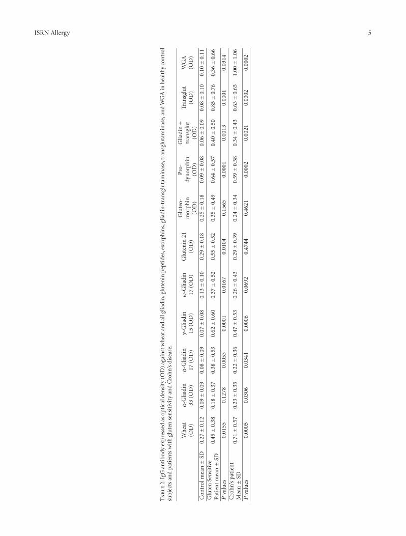
In healthy control subjects, we found moderate elevation(ELISA OD 0.4–1.0) of IgG antibody against glutenin-21-mer in 11/48, gluteomorphin 10/48, wheat in 9/48 speci-mens, and for α-gliadin-33-mer, ω-gliadin-17, gliadin-TG,and WGA 1/48. IgG was not detected against α-gliadin-17,γ-gliadin-15, prodynorphin, and TG in any of the 48 controlsera (see Table 1). The mean OD of IgG antibody againstwheat and other associated antigens in healthy controlsvaried from 0.07 ± 0.08 for γ-gliadin 15-mer to 0.29 ± 0.18for glutenin 21-mer (see Table 2).
The IgA antibody was also measured against this arrayof peptides and antigens in healthy controls. Moderateelevation in IgA antibody was detected against α-gliadin-17-mer and glutenin-21-mer in 9 out of 48 sera and againstwheat and gluteomorphin 5/48, prodynorphin 4/48, α-gliadin-33-mer, and ω-gliadin 2/48. The IgA antibody wasnot detected against γ-gliadin-15-mer, gliadin-TG, TG, andagainst WGA (see Table 1). The mean OD of IgA antibodyagainst this array of antigens and peptides in healthy controlswas as low as 0.06 ± 0.06 for gliadin + TG and as high as0.25± 0.26 for α-gliadin-17-mer (Table 3).
At the cutoff point of 0.39 OD or 3 SD above the ELISAbackground of wells coated with HSA in control sera, IgGantibody was detected in 23% against glutenin-21, 21%against gluteomorphin, and 19% against wheat. Against theother peptides or antigens, the IgG antibody was detected inonly 2% of the tested specimens or not at all (Table 1).
The pattern of IgA antibodies against these antigens andpeptides was different from IgG. The IgA antibody againstα-gliadin-17 and against glutenin-21 was detected in 19%,
followed by wheat and gluteomorphin (10%), prodynorphin(8%), (6%) and 4% against both α-gliadin-17-mer, and ω-gliadin-17. None of the sera from healthy controls showedelevation in IgA antibody against γ-gliadin-15, gliadin-TG,TG, and WGA (Table 1).
3.3. Detection of IgG and IgA Antibodies against Wheat andVarious Gliadin Peptides in the Sera of Patients with GlutenSensitivity/Celiac Disease. The IgG antibodies against theseantigens were measured in clinical specimens from patientswith gluten sensitivity/celiac disease who were positive forgliadin, TG, or their combination.
We found four different profiles of peptides and antigenrecognition by the sera of patients with CD. Results of thesepeptides and antigen recognition are illustrated in Figure 1and Table 1. At ELISA, OD of 0.39 or 3 SD above the blankvalue IgG antibody was most reactive against TG in 16/24specimens, then prodynorphin in 14/24, wheat in 13/24,glutenin in 12/24, γ-gliadin-15 in 11/24, ω-gliadin-17 in10/24, gluteomorphin, α-gliadin-17, and gliadin-TG in 8/24,and against α-gliadin-33-mer 5 out of 24 specimens. AgainstWGA 4 out of 24 specimens were positive for IgG antibody.
Twelve out of 24 specimens (50%) in various intensitiesthat showed a significant elevation of IgG antibody againstwheat also exhibited elevation in the levels of this antibodyagainst α, γ, ω-gliadins, glutenin, gluteomorphin, gliadin-TG, and WGA or their combinations (Figure 1).
Interestingly, the 12 specimens that reacted to wheatantigens and 3 or more different gliadin and gluteninpeptides all produced strong response against tissue TG,while one specimen that reacted to wheat did not react withany other antigen. Of the remaining 11 specimens (46%)that did not react to wheat antigens, 4 did not react to otherantigens, and the other 7 samples with various intensitieswere reactive against 1 to 8 different antigens or peptides(Figure 1). The mean OD of IgG antibody against 11 wheatand associated antigens varied from 0.18±0.37 to 0.85±0.76.Statistically, the differences between the mean ODs of IgGantibody against 9 out of 11 wheat-associated antigens inpatients with gluten sensitivity/celiac disease versus healthycontrols were significant (P < 0.0001 for TG to P < 0.0167for ω-gliadin-17), with P < 0.1565 for gluteomorphin theleast significant (Table 2).
The pattern of IgA antibodies against these same antigensand peptides was different from the pattern for IgG. All24 specimens showed reactivity to more than one antigenor peptide. The most prominent reactions were againstwheat and TG. Data summarized in Table 1 and Figure 2shows that 24/24 (100%) and 20/24 (83%) samples reactedwith IgA antibodies against wheat and TG, respectively,followed by prodynorphin with 17/24 (71%), glutenin-21with 15/24 (63%), gliadin-TG 14/24 (58%), WGA 13/24(54%), both gluteomorphin and γ-gliadin-15 with 12/24(50%), ω-gliadin-17 11/24 (46%), and then both α-gliadin-17 and α-gliadin-33 with 9/24 (38%). Statistically, thedifferences between the mean ODs of IgA antibody againstall of the wheat-associated antigens in patients with celiacdisease versus healthy controls were significant, with 6 having

4 ISRN Allergy
Table 1: Number of specimens with elevated antibodies against 11 tested antigens or peptides at the cutoff point of 0.39 OD.
IgG IgA
Healthy controls Gluten sensitive Crohn’s Healthy controls Gluten sensitive Crohn’s
n = 48 n = 24 n = 24 n = 48 n = 24 n = 24
# % # % # % # % # % # %
Wheat 9 19 13 54 16 67 5 10 24 100 6 25
α-Gliadin 33 1 2 5 21 7 29 2 4 9 38 3 13
α-Gliadin 17 0 0 8 33 4 17 9 19 9 38 3 13
γ-Gliadin 15 0 0 11 46 9 38 0 0 12 50 8 33
ω-Gliadin 17 1 2 10 42 5 21 2 4 11 46 3 13
Glutenin 21 11 23 12 50 6 25 9 19 15 63 3 13
Gluteomorphin 10 21 8 33 8 33 5 10 12 50 4 17
Prodynorphin 0 0 14 58 11 46 4 8 17 71 10 42
Gliadin + TG 1 2 8 33 8 33 0 0 14 58 2 8
TG 0 0 16 67 10 42 0 0 20 83 8 33
WGA 1 2 4 17 12 50 0 0 13 54 3 13
P values 0.0004 0.0017 0.0001 0.1262
TG = transglutaminase.
P values of P < 0.0001, with the least significant beingP < 0.0411 for α-gliadin-17 (Table 3).
3.4. Detection of IgG and IgA Antibodies against Wheat andVarious Gliadin Peptides in the Sera of Patients with Crohn’sDisease. IgG and IgA antibodies against different wheatantigens and peptides, TG, gliadin bound to TG, and WGAwere also measured in sera with IgA ASCA positive. ForIgG antibody, at the 0.39 OD cutoff, 16 out of 24 (67%) ofASCA-positive specimens reacted with wheat, 12 specimensout of 24 reacted very strongly with WGA (50%), 11 withprodynorphin (46%), 10 with TG (42%), 9 with γ-gliadin-15 (38%), 8 with gluteomorphin and gliadin-TG (33%),7 with α-gliadin-33 (29%), 6 with glutenin-21 (25%), 5with ω-gliadin-17 (21%), and 4 with α-gliadin-17 (17%).Interestingly, all 12 WGA-reactive specimens also reactedwith wheat antigens with or without the combination ofgliadin peptides (Figure 3).
The mean ODs for IgG antibodies against various wheatand associated peptides and antigens in healthy controlswere compared to those in patients with Crohn’s disease,obtaining the most significant P values with P < 0.0002 forprodynorphin, TG and WGA, as well as the least significantP values P < 0.4744 for glutenin, as are shown in Table 1.
In comparison to IgG, the prevalence of IgA-positivespecimens in IgA ASCA-positive samples was much lower.Overall, 10 out of 24 specimens (42%) reacted with pro-dynorphin, 8/24 (33%) against TG and γ-gliadin-15, 6/24(25%) against wheat, followed by gluteomorphin with 4/24(17%), α-gliadin-33, α-gliadin-17, ω-gliadin-17, glutenin-21, and WGA with 3/24 (13%), and 2/24 for gliadin-TG(8%). Six (25%) of the ASCA-positive samples did notexhibit any IgA antibody against the 11 tested wheat orassociated antigens and peptides (Figure 4).
The mean OD of IgA antibody against 11 tested wheat-associated antigens and peptides in the sera of patientswith Crohn’s disease are also shown in Table 3. Differencesbetween the mean ODs of IgA antibody against 3 outof 11 tested antigens in healthy controls versus patientswith Crohn’s disease were significant (P < 0.0035 forprodynorphin, P < 0.0044 for γ-gliadin-15, P < 0.0047 forTG (Table 2)).
The overall number and percentage of healthy controlsversus patients’ sera with elevated IgG and IgA antibodyagainst wheat antigens and associated peptides are shown inTable 1. As shown in this table, the difference in percentageof individuals with elevated antibodies in healthy controlsversus patients is very significant (P < 0.0004 for IgGantibody in the gluten-sensitive/celiac group, P < 0.0017for IgG in the Crohn’s group, and P < 0.0001 for IgAantibody in the gluten-sensitive group). While there is asignificant overlap between IgG and IgA antibodies in bothpatients’ groups, the percentage of IgA-reactive specimensagainst various tested antigens was the most significant inthe gluten-sensitive group, followed by IgG presence in boththe gluten-sensitive and Crohn’s disease groups, with IgAreactivity against these antigens being the least significant inpatients with Crohn’s disease (Table 1).
4. Discussion
A number of gluten peptides with a capacity to stimulateintestinal T-helper cells have been identified in CD patientsby many researchers (2–6, 10, and 23–26). In a very recentstudy T cells isolated from CD patients were screened forrecognition of 21 different peptides from α-, γ-, ω-gliadinsand glutenins [5]. It was demonstrated that intestinal T cellsfrom CD patients responded to a wide and heterogeneous
ISRN Allergy 5
Ta
ble
2:Ig
Gan
tibo
dyex
pres
sed
asop
tica
lden
sity
(OD
)ag
ain
stw
hea
tan
dal
lglia
din
,glu
ten
inpe
ptid
es,e
xorp
hin
s,gl
iadi
n-t
ran
sglu
tam
inas
e,tr
ansg
luta
min
ase,
and
WG
Ain
hea
lthy
con
trol
subj
ects
and
pati
ents
wit
hgl
ute
nse
nsi
tivi
tyan
dC
roh
n’s
dise
ase.
Wh
eat
(OD
)α
-Glia
din
33(O
D)
α-G
liadi
n17
(OD
)γ-
Glia
din
15(O
D)
ω-G
liadi
n17
(OD
)G
lute
nin
21(O
D)
Glu
teo-
mor
phin
(OD
)
Pro
-dy
nor
phin
(OD
)
Glia
din
+tr
ansg
lut
(OD
)
Tran
sglu
t(O
D)
WG
A(O
D)
Con
trol
mea
n±
SD0.
27±
0.12
0.09±
0.09
0.08±
0.09
0.07±
0.08
0.13±
0.10
0.29±
0.18
0.25±
0.18
0.09±
0.08
0.06±
0.09
0.08±
0.10
0.10±
0.11
Glu
ten
Sen
siti
vePa
tien
tm
ean±
SD0.
45±
0.38
0.18±
0.37
0.38±
0.53
0.62±
0.60
0.37±
0.52
0.55±
0.52
0.35±
0.49
0.64±
0.57
0.40±
0.50
0.85±
0.76
0.36±
0.66
Pva
lues
0.01
550.
1278
0.00
530.
0001
0.01
670.
0104
0.15
650.
0001
0.00
130.
0001
0.03
14C
roh
n’s
pati
ent
Mea
n±
SD0.
71±
0.57
0.23±
0.35
0.22±
0.36
0.47±
0.53
0.26±
0.43
0.29±
0.39
0.24±
0.34
0.59±
0.58
0.34±
0.43
0.63±
0.65
1.00±
1.06
Pva
lues
0.00
050.
0306
0.03
410.
0006
0.06
920.
4744
0.46
210.
0002
0.00
210.
0002
0.00
02

6 ISRN Allergy
Ta
ble
3:Ig
Aan
tibo
dyex
pres
sed
asop
tica
lden
sity
(OD
)ag
ain
stw
hea
tan
dal
lglia
din
,glu
ten
inpe
ptid
es,e
xorp
hin
s,gl
iadi
n-t
ran
sglu
tam
inas
e,tr
ansg
luta
min
ase,
and
WG
Ain
hea
lthy
con
trol
subj
ects
and
pati
ents
wit
hce
liac
and
Cro
hn’
sdi
seas
e.
Wh
eat
(OD
)α
-Glia
din
33(O
D)
α-G
liadi
n17
(OD
)γ-
Glia
din
15(O
D)
ω-G
liadi
n17
(OD
)G
lute
nin
21(O
D)
Glu
teo-
mor
phin
(OD
)
Pro
-dy
nor
phin
(OD
)
Glia
din
+tr
ansg
lut
(OD
)
Tran
sglu
t(O
D)
WG
A(O
D)
Con
trol
mea
n±
SD0.
23±
0.15
0.11±
0.09
0.25±
0.26
0.11±
0.06
0.13±
0.08
0.23±
0.23
0.19±
0.16
0.13±
0.10
0.06±
0.06
0.10±
0.07
0.11±
0.07
Glu
ten
sen
siti
vePa
tien
tm
ean±
SD0.
89±
0.30
0.28±
0.37
0.45±
0.51
0.71±
0.60
0.52±
0.54
0.66±
0.51
0.45±
0.48
0.84±
0.56
0.61±
0.51
1.01±
0.60
1.11±
0.95
Pva
lues
0.00
010.
0195
0.04
110.
0001
0.00
090.
0003
0.00
780.
0001
0.00
010.
0001
0.00
01C
roh
n’s
pati
ent
Mea
n±
SD0.
29±
0.50
0.13±
0.35
0.15±
0.33
0.39±
0.49
0.11±
0.29
0.15±
0.28
0.14±
0.28
0.44±
0.50
0.12±
0.25
0.52±
0.73
0.40±
0.81
Pva
lues
0.27
770.
3859
0.09
350.
0044
0.33
120.
0992
0.21
060.
0035
0.15
440.
0047
0.05
17

ISRN Allergy 7
No.Wheat
(OD)
α-Gliadin33
(OD)
α-Gliadin17
(OD)
γ-Gliadin15
(OD)
ω-Gliadin17
(OD)
Glutenin21
(OD)
Gluteo-morphin
(OD)
Pro-dynorphin
(OD)
Gliadin +transglut
(OD)
Transglut
(OD)
WGA
(OD)12 0.7 0.9 1.2 1.2 1 0.9 1.3 1.1 1.13 1.1 0.9 1.5 1.7 1.4 1.5 1.2 1.3 1.2 1.6 1.54 0.45 0.6 1.4 1.7 1.4 1.5 1.3 1.5 1.3 1.66 1.2 1.5 1.2 1 1 1.3 1.17 0.9 0.9 0.4 0.4 1.28 0.59
10 0.6 0.9 0.5 0.511 0.9 0.9 0.412 0.5 0.8 0.41 0.4 1.4 1.7 2.213 0.9 1.1 1.3 1.3 1.3 1.2 1.4 1.2 1.6 1.714 1.1 1.2 0.4 0.9 1.3 1 0.515 0.9 0.7 1 1 1.116 0.8 1 0.4 1.1 1.71718 1.319 1.420 0.6 0.42122 0.823 0.6 1.1 0.5 1 0.8 1.524 0.6 0.6 0.4 0.4 0.8 0.4
11
1 1
1
1
Figure 1: Prevalence of IgG expressed as optical density (OD) against wheat alone or in combination with gliadin, glutenin peptides,exorphins, gliadin-transglutaminase, transglutaminase, and WGA in sera of patients with gluten sensitivity/celiac disease.
No.Wheat
(OD)
α-Gliadin33
(OD)
α-Gliadin17
(OD)
γ-Gliadin15
(OD)
ω-Gliadin17
(OD)
Glutenin21
(OD)
Gluteo-morphin
(OD)
Pro-dynorphin
(OD)
Gliadin +transglut
(OD)
Transglut
(OD)
WGA
(OD)123456789
101112131415161718192021222324
0.8 1.3 1.60.6 0.40.9 1.3 2.61 1 0.4 1.2 1.7 1.5
0.9 1.1 1.2 1.2 0.9 1.2 1.2 1.1 2.20.7 0.9 1.2 1.2 0.9 1.3 1.1 1.11.1 0.9 1.5
11
11.51.7 1.4 1.51.61.2 1.3 1.2
0.6 1.4 1.7 1.4 1.5 1.3 1.5 1.3 1.60.6 0.9 0.5 0.50.9 0.9 1 0.40.5 0.8 0.4 0.4 1.4 1.7 2.20.9 1.1 1.3 1.3 1.3 1.2 1.4 1.2 1.6 1.7
1 1 1
1.1 1.2 0.4 0.9 1.3 0.50.9 0.7 1.10.8 0.4 1.1 1.70.6 1.1
11 11
0.5 0.8 1.50.6 0.6 0.4 0.4 0.8 0.40.6 0.90.9 0.6 1.1 1.5 1.3 1.2 1.1 1.7
1 1
1.5 1.6 2.21.2 0.6 0.5 0.5 0.4 0.4
0.4
0.4
0.9 0.6 1 1 1 1.3 0.6 1.8 2.21.2 0.9 1.3 2.61.1 0.9 1.2 1 0.9
0.91 1 1.8
1.9
Figure 2: Prevalence of IgA expressed as optical density (OD) against wheat alone or in combination with gliadin, glutenin peptides,exorphins, gliadin-transglutaminase, transglutaminase, and WGA in sera of patients with gluten sensitivity/celiac disease.
array of peptides [5]. In some patients, many peptides fromthe α-gliadin family were recognized, while in others, onlyone peptide caused lymphocyte stimulation and interferon-γ production. Furthermore, T-cell lines from one particularpatient did not recognize any of the 21 tested peptidesat all, while, overall, 86% of CD patients recognized adifferent array of peptides. It was concluded that other
gliadin peptides not tested in the study could be relevant insome CD patients [5].
Although all these findings showed great heterogene-ity in immunogenicity of gluten peptides for lympho-cyte proliferation and IFN-γ production [2–6, 23–26], noattempt was made to measure heterogeneity in antibodyresponse to various gluten and overall wheat proteins

8 ISRN Allergy
No.Wheat
(OD)
α-Gliadin33
(OD)
α-Gliadin17
(OD)
γ-Gliadin15
(OD)
ω-Gliadin17
(OD)
Glutenin21
(OD)
Gluteo-morphin
(OD)
Pro-dynorphin
(OD)
Gliadin +transglut
(OD)
Transglut
(OD)
WGA
(OD)123456789
101112131415161718192021222324
10.9 1.1 1.2 1.2 0.9 1.2 1.2 1.1 2.30.8 1.3 0.7 0.9 0.9 1.2 0.4 1.1
0.6 0.90.8 1.3 1.6
0.4 0.40.9 0.6 1.1 1.5 1.3 1.2 1.1 1.7 1.5 1.6 2.21.2 0.6 0.5 0.5 0.4 0.40.9 0.6 0.4 1.3 0.6 1.8 2.2
0.81.2 0.9 1.3 2.60.6 0.4
1.2 0.41.1
1 1 1
10.9 1.2 0.9 0.4 1.2 1.8
1.9 0.91.9 0.9 1.1 1.81.5 1.1 1.4 2.90.9 1.1 1.4 2.60.5
1 11
1
0.70.9 0.4 1.2 1.7 1.51 1
Figure 3: Prevalence of IgG expressed as optical density (OD) against wheat alone or in combination with gliadin, glutenin peptides,exorphins, gliadin-transglutaminase, transglutaminase, and WGA in sera of patients with Crohn’s disease.
No.Wheat
(OD)
α-Gliadin33
(OD)
α-Gliadin17
(OD)
γ-Gliadin15
(OD)
ω-Gliadin17
(OD)
Glutenin21
(OD)
Gluteo-morphin
(OD)
Pro-dynorphin
(OD)
Gliadin +transglut
(OD)
Transglut
(OD)
WGA
(OD)
123456789
101112131415161718192021222324
0.50.50.71.9 0.9 1.1 1.81.5 1.1 1.4 2.90.9 1.3 2.6
0.40.4 0.41.2 0.4
1
10.8
0.81.3
1.40.8 1.3 0.7 0.9 0.9 1.2 0.4 1.11.2 1.5 1.2 1.3 1.10.9 0.9
1 1 10.4 0.4 1.2
0.6 0.43
11
1 1
Figure 4: Prevalence of IgA expressed as optical density (OD) against wheat alone or in combination with gliadin, glutenin peptides,exorphins, gliadin-transglutaminase, transglutaminase, and WGA in sera of patients with Crohn’s disease.
and peptides. In the present study, we screened the seraof patients with gluten-sensitivity/celiac disease and Crohn’sdisease for the presence of IgG and IgA antibodies againstboth alcohol- and water-soluble components of wheat,α-gliadin-33-mer, -17-mer, γ-gliadin-15-mer, ω-gliadin-17-mer, and glutenin-21-mer.
The second category of peptides consisted of the opi-oids. Such peptides are called exorphins because of theirexogenous origin and morphine-like characteristics. In some
individuals, dietary exorphins are resistant to intestinaland enterobacterial proteinases; thus, gluteomorphins anddynorphins may be absorbed from the gut lumen into thebloodstream. Consequently, an immune response against theopioid peptides can result in peptide antibody productionand regulation of opioid receptor binding capability [27, 28].
Thirdly, lectins were incorporated into this antibodyarray. WGAs are lectins, or carbohydrate-binding proteins,with a capacity to bind to many cells and tissue antigens,

ISRN Allergy 9
including intestinal brush borders. Lectins, bound to intesti-nal cells and other cell membranes, are known to induce toxicdamage, inflammation, and autoimmunity [21, 22, 29].
Finally, earlier studies showed that gluten-sensitivepatients develop IgG and IgA antibodies to gliadin andto the autoantigen called transglutaminase [30, 31]. Thesearticles demonstrated that gliadin is the preferred substrateof transglutaminase and suggested that the interaction ofgliadin and TG may result in the creation of new antigeniccomplexes [31, 32]. Indeed, in a different study [33], it wasshown that at high molar excess gliadin peptides bind to sixlysine residues of TG, forming isopeptide bonds. However,despite this demonstration of the molecular characterizationof covalent complexes between tissue TG and gliadin pep-tides and discussion about its relevance in celiac disease, noattempt was made to measure antibodies against differentgliadin peptides and their complex formation with TG.Therefore, we extended the measurement of IgG and IgAantibodies against this gliadin and TG complex as well.
Similar to intestinal T-cell response, we demonstratedthat humoral immune response to various wheat antigensand associated peptides are largely heterogeneous [5, 23].Consistent with previous studies conducted with intestinal(T-cell) response against a heterogeneous array of wheatglutenin and α-, γ-, ω-gliadins, our results with IgG- andIgA-specific antibodies demonstrate that both sera withgluten-sensitivity/celiac disease and Crohn’s disease and, toa much lesser degree, sera from healthy controls respond to aheterogeneous array of peptides and antigens.
In some cases, IgG and IgA antibodies were detectedagainst wheat antigens alone or in combination with α-,γ-, and ω-gliadins and glutenin peptides, while in others,IgG or IgA were detected against one or more peptideswithout reacting to wheat antigens. This lack of humoralimmune response to water- and alcohol-soluble componentsof wheat indicates that digestion of wheat proteins into vari-ous peptides and their deamidation by TG plays a significantrole in their antigenicity. The selective deamidation of gliadinpeptides and their complex formation with TG make themmore specific B cell epitopes, which result in first IgA andthen IgG production [31–33]. Indeed, IgA was detected inthe great majority of patients with CD against wheat antigens(100%), followed by immune reaction against prodynorphin(71%), glutenin-21 (62%), gliadin-TG (58%), WGA (52%),and against other proteins and peptides between 37% and50%, as seen in Table 1.
In comparison with IgA, IgG was detected in the seraof celiac disease patients most prominently against TG,followed by prodynorphin, wheat extract, and then glutenin-21 mer (Table 1). The current methodology for diagnosingceliac disease is based on measuring IgG and IgA anti-bodies against gliadin and TG [30–38]. The specificity andsensitivity of these assays in patients with CD who exhibitabnormal histology (villous atrophy or flat mucosa) variesbetween 85% and 100% [34–38]. However, this specificityand sensitivity have not been established for patients withgluten sensitivity and patients with silent or atypical celiacdisease who may have GI symptoms but normal villi. Auto-antibodies can be detected in various diseases for a long
period during which no clinical symptoms are present[39, 40]. In fact, in many studies, a direct relationshiphas now been shown between antibody levels and severityof diseases [41–44]. Similar to these autoimmune diseases,in population screening for celiac disease, antibodies weredetected persistently over a 4-year period [45]. Interestingly,nine of the subjects with transient antibodies had villousatrophy, suggesting that this feature develops after chronicimmune activation including T-cell response, cytokinesand antibody production [39, 45, 46]. Thus, according tothese authors, as with type 1 diabetes and thyroiditis, asubstantial proportion have transient autoantibodies, butwhen the autoantibodies persist, the risk of progression toclinical celiac disease is high. As a result, celiac disease-associated autoantibodies are now widely used for diseaseprediction and diagnosis. Indeed, removal of the antigen,gluten, is currently the therapy of choice for celiac disease[34]. However, due to long-term immunoreactions andsevere tissue destruction, gluten-free diets do not result incomplete normalization of duodenal lesions in a majority ofpatients with celiac disease [47]. Therefore, early detection ofbiomarkers associated with chronic immune activation mayresult in timely intervention and the prevention of villousatrophy.
Since earlier studies performed on intestinal T cells [5,23–26] showed that response to various gliadin and otherassociated peptides is heterogeneous, we believe that theapplication of IgG and IgA antibodies against an array ofantigens and peptides that includes α-, γ-, and ω-gliadins,glutenin, WGA, gluteomorphin, prodynorphins, TG, andgliadin-bound TG can not only enhance the detection ofceliac disease but may also assist in the early detection ofatypical and silent celiac disease.
Atypical celiac disease, which presents with few or nosymptoms, is largely responsible for the increased prevalenceof CD today [48]. Celiac disease may be silent or atypical,but it is still a serious disorder [49]. It has been shown thatfor every recognized case of CD, there are 8 that remainundiagnosed [49], and undiagnosed CD can have veryserious consequences. The consequences of undiagnosed CDinclude not only underachievement [50] and a 5-fold higherrisk of non-Hodgkin’s lymphoma [51] but also a 4-foldincrease in all-cause mortality [52].
Due to some symptomatology overlap between Crohn’sdisease and CD [12], we applied IgG and IgA measurementsagainst various wheat antigens and associated peptides tothe sera of patients with Crohn’s disease who were positivefor ASCA to examine the occurrence of CD with IBD. Incomparison with healthy controls, IgG antibody in the seraof patients with Crohn’s disease was found to be highlyelevated, foremost against wheat extract (67%), secondlyagainst WGA (50%), prodynorphin third (46%), and thenTG (42%), with P values being significant against 9 outof 11 tested antigens (Tables 2 and 3). The differences inIgA antibody response against the same array of wheatantigens and peptides used in the study were less significant(Tables 2 and 3), only being significant against 4 of theantigens or peptides: prodynorphin, α-gliadin-15, TG, andWGA.

10 ISRN Allergy
Based on these findings, we propose that for the earlydetection of immune activation in atypical or silent celiacdisease and patients with IBD or Crohn’s disease, IgG andIgA antibodies be measured not only against α-gliadin andTG, but also against water- and alcohol-soluble componentsof wheat, WGA, ω- and γ-gliadin, glutenin, gluteomorphin,and gliadin bound to TG. This may increase the sensitivityand specificity of assays for sufferers not only of classicalceliac disease, but also atypical or silent CD as well as patientswith IBD who may suffer from gluten sensitivities.
It can be speculated that in addition to gluten-freediets for patients with CD who are ASCA positive, yeast-free diets may also be recommended. If the yeast-free dietalong with the gluten-free diet helps patients to get well,then this practice may become an acceptable alternativemethod of therapy. Further studies are needed in order tocompare and measure T-cell and antibody response to thesevarious antigens and peptides simultaneously in patientswith normal and abnormal villi.
Abbreviations
ASCA: Anti-Saccharomyces cerevisiae mannan antibodiesBSA: Bovine serum albuminCD: Celiac diseaseELISA: Enzyme-linked immunosorbent assayF(ab′): Fragment antigen-bindingHSA: Human serum albuminIBD: Inflammatory bowel diseaseIFN-γ : Interferon gammaIg: ImmunoglobulinOD: Optical densityTBS: Tris-buffered salineTG: TransglutaminaseWGA: Wheat germ agglutinin.
Disclosure
A. Vojdani is the co-owner and CEO of ImmunosciencesLab., Inc., Los Angeles, Calif, USA.
Acknowledgment
The author would like to thank Joel Bautista for thepreparation of this paper.
References
[1] M. Silano and M. De Vincenzi, “Bioactive antinutritional pep-tides derived from cereal prolamins: a review,” Die Nahrung,vol. 43, no. 3, pp. 175–184, 1999.
[2] H. Arentz-Hansen, R. Korner, Ø. Molberg et al., “Theintestinal T cell response to α-gliadin in adult celiac diseaseis focused on a single deamidated glutamine targeted by tissuetransglutaminase,” The Journal of Experimental Medicine, vol.191, no. 4, pp. 603–612, 2000.
[3] H. Arentz-Hansen, S. N. Mcadam, O. Molberg et al., “Celiaclesion T cells recognize epitopes that cluster in regions of
gliadins rich in proline residues,” Gastroenterology, vol. 123,no. 3, pp. 803–809, 2002.
[4] S. Tollefsen, H. Arentz-Hansen, B. Fleckenstein et al., “HLA-DQ2 and -DQ8 signatures of gluten T cell epitopes in celiacdisease,” The Journal of Clinical Investigation, vol. 116, no. 8,pp. 2226–2236, 2006.
[5] A. Camarca, R. P. Anderson, G. Mamone et al., “Intestinal Tcell responses to gluten peptides are largely heterogeneous:implications for a peptide-based therapy in celiac disease,”Journal of Immunology, vol. 182, no. 7, pp. 4158–4166, 2009.
[6] C. L. Vanderlugt and S. D. Miller, “Epitope spreading inimmune-mediated diseases: implications for immunother-apy,” Nature Reviews Immunology, vol. 2, no. 2, pp. 85–95,2002.
[7] W. L. G. Oldfield, A. B. Kay, and M. Larche, “Allergen-derivedT cell peptide-induced late asthmatic reactions precede theinduction of antigen-specific hyporesponsiveness in atopicallergic asthmatic subjects,” Journal of Immunology, vol. 167,no. 3, pp. 1734–1739, 2001.
[8] M. D. Leech, C. Y. Chung, A. Culshaw, and S. M. Anderton,“Peptide-based immunotherapy of experimental autoimmuneencephalomyelitis without anaphylaxis,” European Journal ofImmunology, vol. 37, no. 12, pp. 3576–3581, 2007.
[9] M. Larche and D. C. Wraith, “Peptide-based therapeutic vac-cines for allergic and autoimmune diseases,” Nature Medicine,vol. 11, no. 4, pp. S69–S76, 2005.
[10] W. Vader, Y. Kooy, P. Van Veelen et al., “The Gluten response inchildren with celiac disease is directed toward multiple gliadinand glutenin peptides,” Gastroenterology, vol. 122, no. 7, pp.1729–1737, 2002.
[11] L. Shan, Ø. Molberg, I. Parrot et al., “Structural basis for glutenintolerance in celiac sprue,” Science, vol. 297, no. 5590, pp.2275–2279, 2002.
[12] Z. Barta, I. Csıpo, G. G. Szabo, and G. Szegedi, “Seroreactivityagainst Saccharomyces cerevisiae in patient with Crohn’sdisease and celiac disease,” World Journal of Gastroenterology,vol. 9, no. 10, pp. 2308–2312, 2003.
[13] R. M. R. Barnes, S. Allan, C. H. Taylor-Robinson, R. Finn, andP. M. Johnson, “Serum antibodies reactive with Saccharomycescerevisiae in inflammatory bowel disease: is IgA antibody amarker for Crohn’s disease?” International Archives of Allergyand Applied Immunology, vol. 92, no. 1, pp. 9–15, 1990.
[14] P. Knoflach, B. H. Park, and R. Cunningham, “Serum anti-bodies to cow’s milk proteins in ulcerative colitis and Crohn’sdisease,” Gastroenterology, vol. 92, no. 2, pp. 479–485, 1987.
[15] J. Main, H. McKenzie, G. R. Yeaman et al., “Antibody toSaccharomyces cerevisiae (bakers’ yeast) in Crohn’s disease,”British Medical Journal, vol. 297, no. 6656, pp. 1105–1106,1988.
[16] M. H. Giaffer, A. Clark, and C. D. Holdsworth, “Antibodies toSaccharomyces cerevisiae in patients with Crohn’s disease andtheir possible pathogenic importance,” Gut, vol. 33, no. 8, pp.1071–1075, 1992.
[17] J. F. Quinton, B. Sendid, D. Reumaux et al., “Anti-Saccharo-myces cerevisiae manna antibodies combined with antineu-trophil cytoplasmic autoantibodies in inflammatory boweldisease: prevalence and diagnostic role,” Gut, vol. 42, no. 6,pp. 788–791, 1998.
[18] A. Yang, Y. Chen, E. Scherl, A. I. Neugut, G. Bhagat, and P. H.R. Green, “Inflammatory bowel disease in patients with celiacdisease,” Inflammatory Bowel Diseases, vol. 11, no. 6, pp. 528–532, 2005.

ISRN Allergy 11
[19] S. Strobel and A. M. Mowat, “Immune responses to dietaryantigens: oral tolerance,” Immunology Today, vol. 19, no. 4, pp.173–181, 1998.
[20] J. G. M. C. Damoiseaux, B. Bouten, A. M. L. W. Linderset al., “Diagnostic value of anti-Saccharomyces cerevisiaeand antineutrophil cytoplasmic antibodies for inflammatorybowel disease: high prevalence in patients with celiac disease,”Journal of Clinical Immunology, vol. 22, no. 5, pp. 281–288,2002.
[21] L. M. Sollid, J. Kolberg, H. Scott, J. Ek, O. Fausa, and P.Brantzaeg, “Antibodies to wheat germ agglutinin in celiacdisease,” Clinical and Experimental Immunology, vol. 63, no.1, pp. 95–100, 1986.
[22] N. Kitano, T. Taminato, T. Ida et al., “Detection of antibodiesagainst wheat germ agglutinin bound glycoproteins on theislet-cell membrane,” Diabetic Medicine, vol. 5, no. 2, pp. 139–144, 1988.
[23] J. A. Tye-Din, J. A. Stewart, J. A. Dromey et al., “Comprehen-sive, quantitative mapping of T cell epitopes in gluten in celiacdisease,” Science Translational Medicine, vol. 2, no. 41, ArticleID 41ra51, 2010.
[24] H. Sjostrom, K. E. A. Lundin, O. Molberg et al., “Identificationof a gliadin T-cell epitope in coeliac disease: general impor-tance of gliadin deamidation for intestinal T-cell recognition,”Scandinavian Journal of Immunology, vol. 48, no. 2, pp. 111–115, 1998.
[25] R. P. Anderson, D. A. van Heel, J. A. Tye-Din et al., “T cells inperipheral blood after gluten challenge in coeliac disease,” Gut,vol. 54, no. 9, pp. 1217–1223, 2005.
[26] Y. Van de Wal, Y. M. C. Kooy, P. van Veelen et al., “Gluteninis involved in the gluten-driven mucosal T cell response,”European Journal of Immunology, vol. 29, no. 10, pp. 3133–3139, 1999.
[27] G. Mace, M. Jaume, C. Blanpied et al., “Anti-μ-opioid-receptorIgG antibodies are commonly present in serum from healthyblood donors: evidence for a role in apoptotic immune celldeath,” Blood, vol. 100, no. 9, pp. 3261–3268, 2002.
[28] A. Vojdani, T. O’Bryan, J. A. Green et al., “Immune responseto dietary proteins, gliadin and cerebellar peptides in childrenwith autism,” Nutritional Neuroscience, vol. 7, no. 3, pp. 151–161, 2004.
[29] E. Kottgen, F. Kluge, B. Volk, and W. Gerok, “The lectinproperties of gluten as the basis of the pathomechanism ofgluten-sensitive enteropathy,” Klinische Wochenschrift, vol. 61,no. 2, pp. 111–112, 1983.
[30] S. A. McMillan, D. J. Haughton, J. D. Biggart, J. D. Edgar, K. G.Porter, and T. A. McNeill, “Predictive value for coeliac diseaseof antibodies to gliadin, endomysium, and jejunum in patientsattending for jejunal biopsy,” British Medical Journal, vol. 303,no. 6811, pp. 1163–1165, 1991.
[31] W. Dieterich, T. Ehnis, M. Bauer et al., “Identification of tissuetransglutaminase as the autoantigen of celiac disease,” NatureMedicine, vol. 3, no. 7, pp. 797–801, 1997.
[32] M. N. Marsh, “Transglutaminase, gluten and celiac disease:food for thought,” Nature Medicine, vol. 3, no. 7, pp. 725–726,1997.
[33] B. Fleckenstein, S. W. Qiao, M. R. Larsen, G. Jung, P.Roepstorff, and L. M. Sollid, “Molecular characterizationof covalent complexes between tissue transglutaminase andgliadin peptides,” Journal of Biological Chemistry, vol. 279, no.17, pp. 17607–17616, 2004.
[34] D. Agardh, K. Lynch, C. Brundin, S. A. Ivarsson, A. Lernmark,and C. M. Cilio, “Reduction of tissue transglutaminaseautoantibody levels by gluten-free diet is associated withchanges in subsets of peripheral blood lymphocytes inchildren with newly diagnosed coeliac disease,” Clinical andExperimental Immunology, vol. 144, no. 1, pp. 67–75, 2006.
[35] M. Ankelo, V. Kleimola, S. Simell et al., “Antibody responses todeamidated gliadin peptide show high specificity and parallelantibodies to tissue transglutaminase in developing coeliacdisease,” Clinical and Experimental Immunology, vol. 150, no.2, pp. 285–293, 2007.
[36] K. Kaukinen, P. Collin, K. Laurila, T. Kaartinen, J. Partanen,and M. Maki, “Resurrection of gliadin antibodies in coeliacdisease. Deamidated gliadin peptide antibody test providesadditional diagnostic benefit,” Scandinavian Journal of Gas-troenterology, vol. 42, no. 12, pp. 1428–1433, 2007.
[37] D. Basso, G. Guariso, P. Fogar et al., “Antibodies against syn-thetic deamidated gliadin peptides for celiac disease diagnosisand follow-up in children,” Clinical Chemistry, vol. 55, no. 1,pp. 150–157, 2009.
[38] P. Toftedal, C. Nielsen, J. T. Madsen, K. Titlestad, S. Husby,and S. T. Lillevang, “Positive predictive value of serologicaldiagnostic measures in celiac disease,” Clinical Chemistry andLaboratory Medicine, vol. 48, no. 5, pp. 685–691, 2010.
[39] D. Leslie, P. Lipsky, and A. Louis Notkins, “Autoantibodies aspredictors of disease,” The Journal of Clinical Investigation, vol.108, no. 10, pp. 1417–1422, 2001.
[40] A. L. Notkins, “New predictors of disease,” Scientific American,vol. 296, no. 3, pp. 72–79, 2007.
[41] R. D. G. Leslie, M. A. Atkinson, and A. L. Notkins, “Autoanti-gens IA-2 and GAD in type I (insulin-dependent) diabetes,”Diabetologia, vol. 42, no. 1, pp. 3–14, 1999.
[42] C. M. Dayan and G. H. Daniels, “Chronic autoimmunethyroiditis,” The New England Journal of Medicine, vol. 335, no.2, pp. 99–107, 1996.
[43] C. Betterle, M. Volpato, B. R. Smith et al., “Adrenal cortex andsteroid 21-hydroxylase autoantibodies in children with organ-specific autoimmune diseases: markers of high progression toclinical Addison’s disease,” Journal of Clinical Endocrinologyand Metabolism, vol. 82, no. 3, pp. 939–942, 1997.
[44] A. Vodjani, “Antibodies as predictors of autoimmune diseasesand cancer,” Expert Opinion on Medical Diagnostics, vol. 2, no.6, pp. 593–605, 2008.
[45] S. D. Johnston, R. G. P. Watson, S. A. McMillan, A. E. Evans,and A. H. G. Love, “Serological markers for coeliac disease:changes with time and relationship to enteropathy,” EuropeanJournal of Gastroenterology and Hepatology, vol. 10, no. 3, pp.259–264, 1998.
[46] W. Dickey, S. A. Mcmillan, and D. F. Hughes, “Identificationof coeliac disease in primary care,” Scandinavian Journal ofGastroenterology, vol. 33, no. 5, pp. 491–493, 1998.
[47] A. Lanzini, F. Lanzarotto, V. Villanacci et al., “Complete recov-ery of intestinal mucosa occurs very rarely in adult coeliacpatients despite adherence to gluten-free diet,” AlimentaryPharmacology & Therapeutics, vol. 29, no. 12, pp. 1299–1308,2009.
[48] E. W. Guthrie and M. A. Gettis, “Celiac disease: more commonthan once thought,” U.S. Pharmacist, vol. 33, no. 12, pp. 24–29,2008.
[49] M. Kumar and G. W. Rutecki, “Atypical celiac disease: couldyou be missing this common problem?” Consultant, vol. 50,no. 3, pp. 1–11, 2010.

12 ISRN Allergy
[50] M. A. Verkasalo, O. T. Raitakari, J. Viikari, J. Marniemi, andE. Savilahti, “Undiagnosed silent coeliac disease: a risk forunderachievement?” Scandinavian Journal of Gastroenterology,vol. 40, no. 12, pp. 1407–1412, 2005.
[51] Y. Gao, S. Y. Kristinsson, L. R. Goldin, M. Bjorkholm, N. E.Caporaso, and O. Landgren, “Increased risk for non-Hodgkinlymphoma in individuals with celiac disease and a potentialfamilial association,” Gastroenterology, vol. 136, no. 1, pp. 91–98, 2009.
[52] A. Rubio-Tapia, R. A. Kyle, E. L. Kaplan et al., “Increasedprevalence and mortality in undiagnosed celiac disease,”Gastroenterology, vol. 137, no. 1, pp. 88–93, 2009.

Submit your manuscripts athttp://www.hindawi.com
Stem CellsInternational
Hindawi Publishing Corporationhttp://www.hindawi.com Volume 2014
Hindawi Publishing Corporationhttp://www.hindawi.com Volume 2014
MEDIATORSINFLAMMATION
of
Hindawi Publishing Corporationhttp://www.hindawi.com Volume 2014
Behavioural Neurology
EndocrinologyInternational Journal of
Hindawi Publishing Corporationhttp://www.hindawi.com Volume 2014
Hindawi Publishing Corporationhttp://www.hindawi.com Volume 2014
Disease Markers
Hindawi Publishing Corporationhttp://www.hindawi.com Volume 2014
BioMed Research International
OncologyJournal of
Hindawi Publishing Corporationhttp://www.hindawi.com Volume 2014
Hindawi Publishing Corporationhttp://www.hindawi.com Volume 2014
Oxidative Medicine and Cellular Longevity
Hindawi Publishing Corporationhttp://www.hindawi.com Volume 2014
PPAR Research
The Scientific World JournalHindawi Publishing Corporation http://www.hindawi.com Volume 2014
Immunology ResearchHindawi Publishing Corporationhttp://www.hindawi.com Volume 2014
Journal of
ObesityJournal of
Hindawi Publishing Corporationhttp://www.hindawi.com Volume 2014
Hindawi Publishing Corporationhttp://www.hindawi.com Volume 2014
Computational and Mathematical Methods in Medicine
OphthalmologyJournal of
Hindawi Publishing Corporationhttp://www.hindawi.com Volume 2014
Diabetes ResearchJournal of
Hindawi Publishing Corporationhttp://www.hindawi.com Volume 2014
Hindawi Publishing Corporationhttp://www.hindawi.com Volume 2014
Research and TreatmentAIDS
Hindawi Publishing Corporationhttp://www.hindawi.com Volume 2014
Gastroenterology Research and Practice
Hindawi Publishing Corporationhttp://www.hindawi.com Volume 2014
Parkinson’s Disease
Evidence-Based Complementary and Alternative Medicine
Volume 2014Hindawi Publishing Corporationhttp://www.hindawi.com