Effective Degradation of Gluten and Its Fragments by ... - MDPI
22
pharmaceutics Review Effective Degradation of Gluten and Its Fragments by Gluten-Specific Peptidases: A Review on Application for the Treatment of Patients with Gluten Sensitivity Yakov E. Dunaevsky 1 , Valeriia F. Tereshchenkova 2 , Mikhail A. Belozersky 1 , Irina Y. Filippova 2 , Brenda Oppert 3, * and Elena N. Elpidina 1 Citation: Dunaevsky, Y.E.; Tereshchenkova, V.F.; Belozersky, M.A.; Filippova, I.Y.; Oppert, B.; Elpidina, E.N. Effective Degradation of Gluten and Its Fragments by Gluten-Specific Peptidases: A Review on Application for the Treatment of Patients with Gluten Sensitivity. Pharmaceutics 2021, 13, 1603. https://doi.org/10.3390/ pharmaceutics13101603 Academic Editors: Alexander V. Kurakov and Alexander A. Osmolovskiy Received: 29 August 2021 Accepted: 29 September 2021 Published: 2 October 2021 Publisher’s Note: MDPI stays neutral with regard to jurisdictional claims in published maps and institutional affil- iations. Copyright: © 2021 by the authors. Licensee MDPI, Basel, Switzerland. This article is an open access article distributed under the terms and conditions of the Creative Commons Attribution (CC BY) license (https:// creativecommons.org/licenses/by/ 4.0/). 1 A.N. Belozersky Institute of Physico-Chemical Biology, Lomonosov Moscow State University, 119991 Moscow, Russia; [email protected] (Y.E.D.); [email protected] (M.A.B.); [email protected] (E.N.E.) 2 Chemical Faculty, Lomonosov Moscow State University, 119991 Moscow, Russia; [email protected] (V.F.T.); irfi[email protected] (I.Y.F.) 3 USDA Agricultural Research Service, Center for Grain and Animal Health Research, Manhattan, KS 66502, USA * Correspondence: [email protected]; Tel.: +1-785-776-2780 Abstract: To date, there is no effective treatment for celiac disease (CD, gluten enteropathy), an autoimmune disease caused by gluten-containing food. Celiac patients are supported by a strict gluten-free diet (GFD). However, in some cases GFD does not negate gluten-induced symptoms. Many patients with CD, despite following such a diet, retain symptoms of active disease due to high sensitivity even to traces of gluten. In addition, strict adherence to GFD reduces the quality of life of patients, as often it is difficult to maintain in a professional or social environment. Various pharmacological treatments are being developed to complement GFD. One promising treatment is enzyme therapy, involving the intake of peptidases with food to digest immunogenic gluten peptides that are resistant to hydrolysis due to a high prevalence of proline and glutamine amino acids. This narrative review considers the features of the main proline/glutamine-rich proteins of cereals and the conditions that cause the symptoms of CD. In addition, we evaluate information about peptidases from various sources that can effectively break down these proteins and their immunogenic peptides, and analyze data on their activity and preliminary clinical trials. Thus far, the data suggest that enzyme therapy alone is not sufficient for the treatment of CD but can be used as a pharmacological supplement to GFD. Keywords: celiac disease; peptidases; gluten; enzyme therapy; gluten-free diet 1. Introduction Celiac disease (CD), or gluten enteropathy, is a hereditary predisposed disease, accom- panied by the atrophy of the small intestine mucosa, associated malabsorption syndrome, and the development of various deficiency conditions [1,2]. Celiac disease is caused by food containing gluten—the proteins of cereals that are the diet of the majority of the world population. Some immunogenic peptides of gluten proteins formed during digestion, mainly gliadins from wheat, rye, and barley, are resistant to proteolysis by human digestive peptidases [3,4] and cause CD in predisposed people. A strict gluten-free diet (GFD) is still the only means of supporting the health of CD patients, but efficacy is lacking in many that retain symptoms of the disease. In addition, a strict compliance to GFD reduces the quality of life of patients, as it is often difficult to maintain in a professional or social environment. In addition, a gluten-free diet is significantly more expensive than comparable conventional foods [5]. Peptidases that effectively cleave these difficult-to-hydrolyze peptides can be used as an enzyme therapy for patients with CD. The gluten proteins of cereals, and especially Pharmaceutics 2021, 13, 1603. https://doi.org/10.3390/pharmaceutics13101603 https://www.mdpi.com/journal/pharmaceutics
-
Upload
khangminh22 -
Category
Documents
-
view
0 -
download
0
Transcript of Effective Degradation of Gluten and Its Fragments by ... - MDPI

pharmaceutics
Review
Effective Degradation of Gluten and Its Fragments byGluten-Specific Peptidases: A Review on Application for theTreatment of Patients with Gluten Sensitivity
Yakov E. Dunaevsky 1, Valeriia F. Tereshchenkova 2, Mikhail A. Belozersky 1, Irina Y. Filippova 2, Brenda Oppert 3,*and Elena N. Elpidina 1
�����������������
Citation: Dunaevsky, Y.E.;
Tereshchenkova, V.F.; Belozersky,
M.A.; Filippova, I.Y.; Oppert, B.;
Elpidina, E.N. Effective Degradation
of Gluten and Its Fragments by
Gluten-Specific Peptidases: A Review
on Application for the Treatment of
Patients with Gluten Sensitivity.
Pharmaceutics 2021, 13, 1603.
https://doi.org/10.3390/
pharmaceutics13101603
Academic Editors: Alexander
V. Kurakov and Alexander
A. Osmolovskiy
Received: 29 August 2021
Accepted: 29 September 2021
Published: 2 October 2021
Publisher’s Note: MDPI stays neutral
with regard to jurisdictional claims in
published maps and institutional affil-
iations.
Copyright: © 2021 by the authors.
Licensee MDPI, Basel, Switzerland.
This article is an open access article
distributed under the terms and
conditions of the Creative Commons
Attribution (CC BY) license (https://
creativecommons.org/licenses/by/
4.0/).
1 A.N. Belozersky Institute of Physico-Chemical Biology, Lomonosov Moscow State University,119991 Moscow, Russia; [email protected] (Y.E.D.); [email protected] (M.A.B.);[email protected] (E.N.E.)
2 Chemical Faculty, Lomonosov Moscow State University, 119991 Moscow, Russia;[email protected] (V.F.T.); [email protected] (I.Y.F.)
3 USDA Agricultural Research Service, Center for Grain and Animal Health Research,Manhattan, KS 66502, USA
* Correspondence: [email protected]; Tel.: +1-785-776-2780
Abstract: To date, there is no effective treatment for celiac disease (CD, gluten enteropathy), anautoimmune disease caused by gluten-containing food. Celiac patients are supported by a strictgluten-free diet (GFD). However, in some cases GFD does not negate gluten-induced symptoms.Many patients with CD, despite following such a diet, retain symptoms of active disease due tohigh sensitivity even to traces of gluten. In addition, strict adherence to GFD reduces the quality oflife of patients, as often it is difficult to maintain in a professional or social environment. Variouspharmacological treatments are being developed to complement GFD. One promising treatment isenzyme therapy, involving the intake of peptidases with food to digest immunogenic gluten peptidesthat are resistant to hydrolysis due to a high prevalence of proline and glutamine amino acids. Thisnarrative review considers the features of the main proline/glutamine-rich proteins of cereals and theconditions that cause the symptoms of CD. In addition, we evaluate information about peptidasesfrom various sources that can effectively break down these proteins and their immunogenic peptides,and analyze data on their activity and preliminary clinical trials. Thus far, the data suggest thatenzyme therapy alone is not sufficient for the treatment of CD but can be used as a pharmacologicalsupplement to GFD.
Keywords: celiac disease; peptidases; gluten; enzyme therapy; gluten-free diet
1. Introduction
Celiac disease (CD), or gluten enteropathy, is a hereditary predisposed disease, accom-panied by the atrophy of the small intestine mucosa, associated malabsorption syndrome,and the development of various deficiency conditions [1,2]. Celiac disease is caused byfood containing gluten—the proteins of cereals that are the diet of the majority of the worldpopulation. Some immunogenic peptides of gluten proteins formed during digestion,mainly gliadins from wheat, rye, and barley, are resistant to proteolysis by human digestivepeptidases [3,4] and cause CD in predisposed people. A strict gluten-free diet (GFD) is stillthe only means of supporting the health of CD patients, but efficacy is lacking in many thatretain symptoms of the disease. In addition, a strict compliance to GFD reduces the qualityof life of patients, as it is often difficult to maintain in a professional or social environment.In addition, a gluten-free diet is significantly more expensive than comparable conventionalfoods [5].
Peptidases that effectively cleave these difficult-to-hydrolyze peptides can be usedas an enzyme therapy for patients with CD. The gluten proteins of cereals, and especially
Pharmaceutics 2021, 13, 1603. https://doi.org/10.3390/pharmaceutics13101603 https://www.mdpi.com/journal/pharmaceutics

Pharmaceutics 2021, 13, 1603 2 of 22
immunogenic peptides formed from them, are rich in proline, which is the only iminoacid among the twenty natural proteinogenic amino acids. The presence of a prolineresidue in the polypeptide chain changes its conformation, preventing the degradationof proline-rich proteins by broad-spectrum peptidases. In nature, hydrolysis of proline-containing proteins is carried out mainly by a special group of proteolytic enzymes—proline-specific peptidases (PSPs). Due to their unique specificity, PSPs are involved in the“fine” regulation of various metabolic processes, cell differentiation and maturation, theprocessing of biologically active peptides and proteins, and the formation of an immuneresponse. Therefore, the research regarding PSPs has been primarily directed to the study oftheir regulatory function with a strong medical focus, as PSPs are involved in a wide varietyof diseases, such as diabetes, cancer, Alzheimer’s and Parkinson’s diseases, hypertension,and neuropsychiatric diseases [6]. The ability of PSPs to neutralize the immunogenicpotential of proline-rich gluten peptides is a way to exploit their unique specificity andpresents an attractive option for patients with CD.
The immunogenic peptides of gluten are not only rich in proline but also glutamine.Therefore, peptidases are needed with activity towards glutamine that can either indepen-dently or in combination with PSPs partially or completely eliminate the immunogenicityof these peptides. Oral enzyme therapy using such gluten-destroying peptidases is apromising therapeutic approach. This narrative review describes the features and charac-teristics of the main proline/glutamine-rich proteins of cereals, analyzes peptidases fromvarious sources that can effectively break down these proteins, and discusses their potentialto produce gluten-free products, including the problems in incorporating these enzymes intherapies for CD.
2. Prolamins
Gluten proteins are the main storage proteins of cereal seeds, such as wheat, barley,and rye. Initially, the classification of these proteins was based on their solubility, andfractions included those that are alcohol-soluble or alcohol-insoluble [7]. Alcohol-solubleproteins were called prolamins according to their amino acid composition—prolamins arerich in proline (up to 30%) and glutamine (up to 50%) [7–10]. In wheat, prolamins arecalled gliadins, in barley—hordeins, and in rye—secalins. Alcohol-insoluble fractions werecalled glutelins, and in wheat—glutenins. Initially, prolamins and glutelins were assignedto different groups of proteins, but later it turned out that many glutelins are related toprolamins in their primary structure. The insolubility of glutelins in alcohol is due tothe fact that they form high-molecular polymers stabilized by disulfide bonds betweenindividual polypeptides. Since many glutelins are structurally related to prolamins, andsome reduced polymer subunits are alcohol soluble and rich in proline and glutamine, theyare now also considered prolamins [7–9].
Gliadins and glutenins in wheat grains are the main components of gluten. It is glutenthat provides wheat with the required dough-forming and baking qualities, as well as mostof the unique taste of wheat-based products. Gluten proteins are extremely heterogeneous.They can be divided into three main groups: high-molecular-weight prolamins (high-molecular-weight gluten subunits), S-poor prolamins (ω-gliadins), and S-rich prolamins,which include gliadins and low-molecular-weight prolamins (low-molecular-weight glutensubunits). Gliadins are divided into four discrete electrophoretic fractions, α-, β-, γ-, andω-gliadins, which differ both in molecular mass (Mr = 31,000−42,000 Da) and in aminoacid sequences [8,11].
There are individual sites or domains in the structure of prolamins that have both adiverse amino acid composition and possibly different origins, and amino acid sequencesconsisting either of repeating blocks based on one or more short peptide motifs, or enrichedwith certain amino acid residues, such as Gln, Pro, or Met [8]. The peptide bonds formedby these amino acids, primarily the cyclic imino acid residue Pro which disrupts thestructure of the protein helix, are not hydrolyzed by most known peptidases since they donot correspond to their specificity. That is, the enzymatic hydrolysis of prolamins can be
Pharmaceutics 2021, 13, 1603 3 of 22
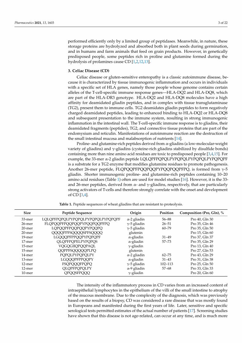
performed efficiently only by a limited group of peptidases. Meanwhile, in nature, thesestorage proteins are hydrolyzed and absorbed both in plant seeds during germination,and in humans and farm animals that feed on grain products. However, in geneticallypredisposed people, some peptides rich in proline and glutamine formed during thehydrolysis of prolamines cause CD [1,2,12,13].
3. Celiac Disease (CD)
Celiac disease or gluten-sensitive enteropathy is a classic autoimmune disease, be-cause it is characterized by tissue immunogenic inflammation and occurs in individualswith a specific set of HLA genes, namely those people whose genome contains certainalleles of the T-cell-specific immune response genes—HLA-DQ2 and HLA-DQ8, whichare part of the HLA-DR3 genotype. HLA-DQ2 and HLA-DQ8 molecules have a highaffinity for deamidated gliadin peptides, and in complex with tissue transglutaminase(TG2), present them to immune cells. TG2 deamidates gliadin peptides to form negativelycharged deamidated peptides, leading to enhanced binding to HLA-DQ2 or HLA-DQ8and subsequent presentation to the immune system, resulting in strong immunogenicinflammation in the intestinal wall. The T-cell-specific immune response is to gliadins, theirdeamidated fragments (peptides), TG2, and connective tissue proteins that are part of theendomysium and reticulin. Manifestations of autoimmune reaction are the destruction ofthe small intestinal mucosa and malabsorption of nutrients [14].
Proline- and glutamine-rich peptides derived from α-gliadins (a low-molecular-weightvariety of gliadins) and γ-gliadins (cysteine-rich gliadins stabilized by disulfide bonds)containing more than nine amino acid residues are toxic to predisposed people [3,4,15]. Forexample, the 33-mer α-2 gliadin peptide LQLQPFPQPQLPYPQPQLPYPQPQLPYPQPQPFis a substrate for a TG2 enzyme that modifies glutamine residues to promote pathogenesis.Another 26-mer peptide, FLQPQQPFPQQPQQPYPQQPQQPFPQ, is formed from γ-5gliadin. Shorter immunogenic proline- and glutamine-rich peptides containing 10–20amino acid residues (Table 1) often are used for model studies [16]. However, it is the 33-and 26-mer peptides, derived from α- and γ-gliadins, respectively, that are particularlystrong activators of T-cells and therefore strongly correlate with the onset and developmentof CD [3,4].
Table 1. Peptide sequences of wheat gliadins that are resistant to proteolysis.
Size Peptide Sequence Origin Position Composition (Pro, Gln), %
33-mer LQLQPFPQPQLPYPQPQLPYPQPQLPYPQPQPF α-2 gliadin 56–88 Pro 40, Gln 3026-mer FLQPQQPFPQQPQQPYPQQPQQPFPQ γ-5 gliadin 26–51 Pro 35, Gln 4620-mer LQPQQPFPQQPQQPYPQQPQ γ-5 gliadin 60–79 Pro 35, Gln 5020-mer QQQQPPFSQQQQSPFSQQQQ glutenin Pro 15, Gln 6019-mer LGQQQPFPPQQPYPQPQPF α-gliadin 31–49 Pro 37, Gln 3717-mer QLQPFPQPELPYPQPQS α-gliadin 57–73 Pro 35, Gln 2915-mer VQGQGIIQPQQPAQL γ-gliadin Pro 13, Gln 4015-mer QQPPFSQQQQQPLPQ glutenin Pro 27, Gln 5314-mer PQPQLPYPQPQLPY α-2 gliadin 62–75 Pro 43, Gln 2913-mer LGQQQPFPPQQPY α-gliadin 31–43 Pro 31, Gln 3812-mer FSQPQQQFPQPQ γ-5 gliadin 102–113 Pro 25, Gln 5012-mer QLQPFPQPQLPY α-9 gliadin 57–68 Pro 33, Gln 3310-mer QPQQSFPQQQ γ-gliadin Pro 20, Gln 60
The intensity of the inflammatory process in CD varies from an increased content ofintraepithelial lymphocytes in the epithelium of the villi of the small intestine to atrophyof the mucous membrane. Due to the complexity of the diagnosis, which was previouslybased on the results of a biopsy, CD was considered a rare disease that was mostly foundin Europeans and manifested during the first years of life. Later, sensitive and specificserological tests permitted estimates of the actual number of patients [17]. Screening studieshave shown that this disease is not age-related, can occur at any time, and is much more

Pharmaceutics 2021, 13, 1603 4 of 22
common than previously thought, namely 1% of the world’s population. In most patients,CD occurs with mild symptoms or has atypical clinical manifestations. A persistentepidemiological pattern is the steady increase in gluten intolerance in humans [18,19]. Thereasons for this can be the following factors: the increase in gluten consumption worldwide;early introduction of complementary foods containing cereals in children of the first yearof life against the background of a decrease in the duration of breastfeeding; the emergenceof new varieties of wheat with a high content of gluten; and accelerated methods in theproduction of bakery products (reducing the fermentation period) that increase the contentof toxic gluten peptides [20]. Malabsorption is the classic manifestation of CD. At the sametime, the following symptoms are observed: chronic diarrhea, flatulence, weight loss, andvitamin and microelement deficiencies. Over time, there is a high risk of developing cancerand other autoimmune diseases, as well as nervous disorders [21–23].
Currently, there is no cure for CD. A strict gluten-free diet (GFD) is the only effectiveway to maintain the health of CD patients. In most patients with gluten sensitivity, theintroduction of GFD leads to at least partial healing of the duodenal mucosa, improvementof most symptoms associated with gluten consumption, and a decrease in the titers ofspecific antibodies in gluten disease. However, in many patients, even with long-termstrict adherence to GFD, symptoms may persist, including inflammatory and architecturalchanges in the small intestine mucosa and positive antibody levels [24,25]. A number offactors may contribute to an incomplete response to a GFD. It is difficult to avoid cross-contamination during food production because gluten is widely used in the food industry.Food labeling may be inaccurate, misleading, or incorrect. In a double-blind clinical trial,patients with CD in remission who were given 50 mg of gluten daily experienced a 20%reduction in villus height/crypt depth compared to a daily placebo or 10 mg of gluten [26].This indicates that even traces of gluten can cause chronic mucosal damage. The acceptable(safe) limit of gluten may vary from patient to patient and may correspond to 10–100 mgper day, even though a slice of wheat bread contains approximately 3–4 g of gluten [27].Sticking to such a strict diet is difficult; generally, it is more expensive, less accessible,severely restricts food choice, may result in products with off-taste, and may lead toasocialized individuals (especially in adolescents) and depressive states [5]. Moreover,there is a lack of vitamins and minerals, as well as a tendency to anemia and osteoporosis,in patients on GFDs. In most cases, unintended gluten exposure can occur in patients as aresult of the consumption of 10–1000 mg of “hidden” gluten contained in common foodingredients such as sauces, salad dressings, food starches, malt extract thickeners, andother flavors, and sometimes simply as a result of cross-contamination during cooking.Thus, the complete elimination of gluten is, at best, a difficult task. Despite attempts toadhere to GFD, long-term treatment of patients with gluten disease often results in severeatrophy of the villi [25]. It is possible that many patients are inadvertently consuminghundreds of milligrams (or more) of gluten per day. Therefore, there is a need to develop anon-dietary (pharmacological) therapy that would either supplement or replace GFD andneutralize up to 1 g of gluten while the food is still in the stomach.
Various therapeutic strategies are being developed to combat CD. Enzyme therapy isespecially promising, as a supplement to food in the form of a peptidase preparation thatefficiently degrades prolamins peptides [28]. This approach is based on a direct effect onthe pathogenic substance, namely, uncleaved peptides with a large number of proline andglutamine residues that are not digested by typical stomach enzymes.
4. Peptidases that Effectively Hydrolyze Prolamins and Their Immunogenic(Toxic) Peptides
Since immunogenic gliadin peptides are rich in proline residues, PSPs can be usedto cleave bonds formed by the Pro residue in proteins and peptides [28]. PSPs character-ized thus far have different substrate specificity (Table 2). Most PSPs are exopeptidases:dipeptidyl peptidase (DPP) 2, DPP 4, DPP 8, DPP 9, prolyl carboxy peptidase (PRCP),aminopeptidase P (APP) 1, APP2, APP3, and prolidase. PSP endopeptidases, prolyl oligopeptidases (POP) and prolyl endo peptidases (PEP), usually have higher efficacy. Fibroblast

Pharmaceutics 2021, 13, 1603 5 of 22
activation protein (FAP) possesses both exo- and endopeptidase activity. All PSPs belongto one of two classes of peptidases—either serine or metallopeptidases. PSPs that areeffective in detoxifying the immunotoxic prolamin peptides are found in various organismsbelonging to different kingdoms of wildlife.
Table 2. Specificity of proline-specific peptidases.
Number Peptidase Class Enzymes Substrates 1
1
Serine peptidases
Prolyloligopeptidase (POP),prolylendopeptidase (РЕР),fibroblast activation protein
(FAP)
(Xaa)n-Xbb-Pro↓Xbb-(Xaa)n, n = 1–13(the length of the peptide is
approximately 30 amino acid residues)
2 Dipeptidylpeptidases (DPP) 2,DPP 4, DPP 8, DPP 9, FAP Xbb-Pro↓Xbb-(Xaa)n, n = 2–12
3 Prolylcarboxypeptidase (PRCP) (Xaa)n-Xbb-Pro↓Xbb, n—any number
4Metallopeptidases
Aminopeptidases P (APP) 1,APP2, APP3 Xbb↓Pro(Xaa)n, n = 1–9
5 Prolidase Xbb↓Pro1 Xaa—any amino acid; Xbb—any amino acid, except Pro.
In addition to proline, the other most common amino acid residue in cereal prolaminsis glutamine, so that peptidases with specificity toward this residue also are needed. Theactivity of cysteine post-glutamine cleaving peptidases (PGP) was detected in the larvalmidgut tissue of the Tenebrionidae beetles Tenebrio molitor and Tribolium castaneum usinghighly specific peptide substrates Z-Ala-Ala-Gln-pNA, Glp-Phe-Gln-pNA, and Glp-Phe-Gln-AMC, where Z is benzyloxycarbonyl, Glp—pyroglutamyl, pNa—p-nitroanilide, andAMC—4-amino-7-methylcoumaride [29–31]. Post-glutamine cleaving activity has also beenfound in studies of the hydrolysis of proline- and glutamine-rich immunogenic peptidesby subtilisin-like peptidases of bacteria [32] and cysteine peptidases of plants [33,34].
4.1. Hydrolysis of Gluten Proteins and Their Toxic Peptides by Bacterial Peptidases
A study of the hydrolytic properties of bacterial POP from Myxococcus xanthus (MX),Sphingomonas capsulata (SC), and Flavobacterium meningosepticum (FM) on two gliadin pep-tides that play a key role in the development of CD (from α-2 gliadin) revealed significantdifferences in the specificity of enzymes associated with the length of the substrate [35–38].SC-POP effectively hydrolyzed the shorter substrate PQPQLPYPQPQLP but had lowactivity against longer LQLQPFPQPQLPYPQPQLPYPQPQLPYPQPQPF. In contrast, FM-POP and MX-POP cleaved both substrates, although FM-POP had greater activity withthe longer substrate than MX-POP. Analysis of the hydrolysis products showed that SC-POP cleaved both Pro-Gln and Pro-Tyr bonds (PQPQLP↓YP↓QPQLP), FM-POP cleavedthe Pro-Gln bond better (PQPQLPYP↓QPQLP), and MX-POP preferred the Pro-Tyr site(PQPQLP↓YPQPQLP). MX-POP formed short fragments of 4–5 amino acid residues withLQLQPFPQPQLPYPQPQLPYPQPQLPYPQPQPF, while FM-POP formed long interme-diates, mainly due to the preferred cleavage of the central Pro-Gln bond of the substrate.Thus, there were clear differences in the specificity of POPs from different organisms, buttheir high hydrolytic activity with both immunogenic gliadin peptides make them potentialcandidates for enzyme therapy of CD [35]. Testing the therapeutic value of POP requiresa long-term study of CD patients who receive controlled amounts of POP-processed andunprocessed gluten.
SC- and MX-POP were obtained as recombinant proteins [39]. The tertiary structuresof the enzymes were determined by X-ray diffraction: the open structure of SC-POP witha resolution of 1.8 Å (Figure 1A), and the structure of MX-POP in a complex with theZ-Ala-prolinal inhibitor with a resolution of 1.5 Å (Figure 1B). Bacterial POPs consisted ofan α-catalytic domain with a catalytic triad of Ser, Asp, and His, and a β-propeller domain

Pharmaceutics 2021, 13, 1603 6 of 22
responsible for substrate specificity. Their active sites lay near the contact boundarybetween these two domains, so mutagenesis at the interdomain interface could be used tochange protein dynamics, which in turn would affect the change in substrate specificity.
Pharmaceutics 2021, 13, x FOR PEER REVIEW 6 of 22
substrate. Thus, there were clear differences in the specificity of POPs from different
organisms, but their high hydrolytic activity with both immunogenic gliadin peptides
make them potential candidates for enzyme therapy of CD [35]. Testing the therapeutic
value of POP requires a long‐term study of CD patients who receive controlled amounts
of POP‐processed and unprocessed gluten.
SC‐ and MX‐POP were obtained as recombinant proteins [39]. The tertiary structures
of the enzymes were determined by X‐ray diffraction: the open structure of SC‐POP with
a resolution of 1.8 Å (Figure 1A), and the structure of MX‐POP in a complex with the Z‐
Ala‐prolinal inhibitor with a resolution of 1.5 Å (Figure 1B). Bacterial POPs consisted of
an α‐catalytic domain with a catalytic triad of Ser, Asp, and His, and a β‐propeller domain
responsible for substrate specificity. Their active sites lay near the contact boundary
between these two domains, so mutagenesis at the interdomain interface could be used to
change protein dynamics, which in turn would affect the change in substrate specificity.
Figure 1. (a) Structure of SC‐POP (PDB 1YR2) with a resolution of 1.8 Å. The α‐helices of the catalytic
domain are green, β‐sheets of the β‐propeller domain are gray. (b) Structure of MX‐POP bound with
Z‐Ala‐prolinal inhibitor (PDB 2BKL). The α‐helices of the catalytic domain are teal, β‐sheets of the
β‐propeller domain are gray. Inhibitor Z‐Ala‐prolinal is shown as sticks and is purple. Structures
are visualized using PyMOL (https://pymol.org/2/, accessed on 25 May2021) (The PyMOL
Molecular Graphics System, Version 1.2r3pre, Schrödinger, LLC).
MX‐, SC‐, and FM‐POP cannot hydrolyze whole gliadins. However, they can
hydrolyze proline‐rich peptides that are formed by the action of human digestive
peptidases. The length of such peptides is from 9 to 33 amino acid residues, that is,
sufficient for the potential hydrolysis by these POP enzymes. Thus, the treatment of
gluten, pre‐hydrolyzed with a mixture of pepsin‐trypsin‐chymotrypsin, with
recombinant FM‐POP reduced the amount of potentially immunogenic peptides in vitro
and ex vivo, and also avoided the development of fat and carbohydrate malabsorption in
most patients with CD who developed the condition after two weeks of provocative
gluten intake [38]. The difficulties in using these enzymes are associated with their
optimal pH, which lies in the range of 7.0–8.5, so they cannot function in the stomach at
acidic pH values, and, moreover, they are unstable due to their degradation by pepsin
[40]. An enteric coating (decomposing in the intestine) on a gelatin capsule containing the
enzyme preparation can improve the protection of the enzyme from the gastric
Figure 1. (a) Structure of SC-POP (PDB 1YR2) with a resolution of 1.8 Å. The α-helices of the catalyticdomain are green, β-sheets of the β-propeller domain are gray. (b) Structure of MX-POP bound withZ-Ala-prolinal inhibitor (PDB 2BKL). The α-helices of the catalytic domain are teal, β-sheets of theβ-propeller domain are gray. Inhibitor Z-Ala-prolinal is shown as sticks and is purple. Structures arevisualized using PyMOL (https://pymol.org/2/, accessed on 25 May 2021) (The PyMOL MolecularGraphics System, Version 1.2r3pre, Schrödinger, LLC, New York, NY, USA).
MX-, SC-, and FM-POP cannot hydrolyze whole gliadins. However, they can hy-drolyze proline-rich peptides that are formed by the action of human digestive peptidases.The length of such peptides is from 9 to 33 amino acid residues, that is, sufficient for thepotential hydrolysis by these POP enzymes. Thus, the treatment of gluten, pre-hydrolyzedwith a mixture of pepsin-trypsin-chymotrypsin, with recombinant FM-POP reduced theamount of potentially immunogenic peptides in vitro and ex vivo, and also avoided thedevelopment of fat and carbohydrate malabsorption in most patients with CD who de-veloped the condition after two weeks of provocative gluten intake [38]. The difficultiesin using these enzymes are associated with their optimal pH, which lies in the range of7.0–8.5, so they cannot function in the stomach at acidic pH values, and, moreover, theyare unstable due to their degradation by pepsin [40]. An enteric coating (decomposingin the intestine) on a gelatin capsule containing the enzyme preparation can improve theprotection of the enzyme from the gastric environment (>1 h in 0.01 M HCl, pH 2 withpepsin), but may lead to a delay in the release of the POP enzyme in the duodenum (pH 6.0in the presence of trypsin and chymotrypsin) [36]. Therefore, it is necessary to optimize theamount and application of the enteric coating for successful delivery of the enzyme prepa-ration (protection and release). The possibility of the therapeutic use of enzymes requiresmore in-depth analysis of their specificity for other immunogenic gluten peptides [16],as well as determining their stability in the presence of bile salts and other substancesusually found in the intestine. Another important aspect to consider is the time it takesfor POP to digest the peptides, because intact peptides or immunogenic fragments cancross the small intestine mucosa before they undergo complete hydrolysis, and peptidaseactivity will be determined by the amount of POP. The theoretical dose of POP that willbe needed for a sufficiently complete and rapid digestion of gluten present in normalfood is difficult to calculate. It is a dynamic system, and the calculation of the relative

Pharmaceutics 2021, 13, 1603 7 of 22
rates of peptide transport and corresponding degradation of POP as it passes through thegastrointestinal system will be one of the key points in planning the future use of POP forenzyme therapy [41].
A recombinant strain of Lactobacillus casei, which produces the secreted activity ofMX-POP, has been proposed for the delivery of POP to the intestinal environment [42]. Thesecreted enzyme is able to destroy the immunotoxic 33-mer gliadin peptide, which plays akey role in the pathogenesis of CD. This strain survives a simulated gastrointestinal transitmaintaining the ability to produce and secrete MX-POP and thus may be a good system fordelivering MX-POP to the duodenum of patients with CD. The authors believe that thegenetically engineered food product may be useful as a vector for the production of POP insitu in the upper small intestine of CD patients after additional research and clinical trials.
A recombinant form of a new POP with a molecular mass of 77 kDa from the ther-mostable bacteria Sphaerobacter thermophiles was able to hydrolyze bonds after prolineresidues in the toxic peptide of α-gliadin, LGQQQP↓FPP↓QQP↓Y, PQPQLPYPQPQLP↓Y,and SQQQFP↓QPQQP↓FP↓QQP of γ-hordein. The enzyme was stable in the pH rangeof 5.0–8.0 with an optimum activity at pH 6.6. This POP was thermally stable withtemperature-optimum activity near 63 ◦C [43]. Due to these features (neutral pH optimumand high temperature optimum), this POP was not proposed as an enzymatic treatment ofCD, but instead can be useful for the production of gluten-free beverages and food. Theaddition of the enzyme to barley malt reduced the concentration of gluten. Thus, the S.thermophiles POP can be used for high-temperature mashing of barley malt in the brewingprocess [43].
The traditional process of producing different varieties of wheat and rye bread, aswell as improving its quality, is the fermentation of sourdough consisting of a symbioticculture of yeast and lactic acid bacteria (LAB) growing in a mixture of flour and water.Sourdough fermentation with LAB is used to make the flour suitable for baking, to controlthe development of characteristic flavor components, to achieve a yeast leaven of thedough, to suppress undesirable fermentation by other bacteria and yeast, and to increasethe shelf life of bread [44]. During the fermentation of the dough, proteolysis by LABreleases small peptides and free amino acids, which are important for the rapid growth ofmicrobes and acidification, as well as being precursors for the development of the flavorof yeast baked goods. In addition, this proteolytic activity can be used as a tool to reducethe amount of certain allergenic compounds derived from gluten (gliadin peptides), whichare often present in wheat baked goods and cause CD. A study of the hydrolysis of α-gliadin fragments by peptidases from sourdough of Lactobacillus plantarum and Pediococcuspentosaceus strains, among which proline-specific peptidases are prominent, showed that L.plantarum strains hydrolyzed more than 60% of α-gliadin peptides involved in the immuneresponse in CD patients, corresponding to fragments 31–43 (LGQQQPFPPQQPY) and62–75 (PQPQLPYPQPQSFP). None of the lactic acid bacteria strains cleaved fragment57–89 (LQLQPFPQPQLPYPQPQLPYPQPQLPYPQPQPF), but a mixture of L. plantarumCRL 775 and P. pentosaceus CRL 792 hydrolyzed this peptide by 57% in 8 h [45,46]. Furtherstudies are required to determine the optimal dose of LAB and their combinations, and thetime for fermentation to determine the practical application of these results.
Screening of 12 strains of LAB and yeast, based on their ability to hydrolyze wheatproteins and ferment dough, found that no unique strain of the LAB or yeast can completelydegrade wheat allergens [47]. However, Pediococcus acidilactici XZ 31, Torulaspora delbrueckiiJM1, and Saccharomyces cerevisiae JM4 demonstrated superiority over other strains in theirability to ferment dough and reduce the allergenicity of wheat products. Further in vivotesting of the anti-allergenic potential is required to confirm that selected LAB and yeaststrains can be considered as an effective starter culture for the preparation of hypoallergenicwheat products.
Controlled proteolysis in wheat dough also has been suggested to reduce the levelof gliadins to such an extent that the products obtained are tolerated by patients withCD. However, the expected difficulty from a technological point of view is related to

Pharmaceutics 2021, 13, 1603 8 of 22
the suitability of such wheat flour to provide the desired quality of bread, taking intoaccount the complete (or very deep) degradation of gluten. Products with a reduced glutencontent and an extended fermentation time may not be suitable for bread production.Low-gluten doughs can only be used as improvers for baking gluten-free bread with lowerquality compared to common (wheat) products [48,49] or used in the production of certainproducts, such as cookies, cakes, and pastries.
Hydrolysis of the immunogenic 33-mer proline-rich peptide (see Table 1) was per-formed with peptidases from Lactobacillus sanfranciscensis, L. alimentarius, L. brevis, and L.hilgardii [50]. Various peptidases were isolated from these LAB: PepN—aminopeptidase,PepI—proliniminopeptidase, PepX—X-prolyldipeptidylaminopeptidase (which sharessubstrate specificity with DPP 4), PepO—endopeptidase, POP—prolyloligopeptidase,PepT—tripeptidase, PepV—dipeptidase, PepQ—prolidase, and PepR—prolinase. Amongthe isolated PSPs, only POP hydrolyzed the Pro-Phe bond in the 33-mer, resulting in twopeptides, one of which is immunogenic. However, to achieve complete hydrolysis of the33-mer to free amino acids, it was necessary to use a combination of isolated peptidasesPepN, PepX, PepO, POP, PepT, PepV, and PepQ. It was concluded that LAB peptidasescan be safely used to modify the diet of CD patients because of their ability to hydrolyzeproline-rich peptides of gluten from varieties of Triticum turgidum L.
Screening of gliadin-cleaving proteolytic activity among 20 Lactobacillus strains showedthat the most active strain, L. casei, was able to hydrolyze the 33-mer immunogenic peptideof α-gliadin by 82% in 8 h, and completely in 12 h [51]. Other authors selected 11 strains ofLAB, belonging to the species L. curvatus, Pediococcus acidilactici, P. pentosaceus, L. coryni-formis, Weissella cibaria, L. plantarum, and L. helveticus, [52], as well as probiotic strains ofBifidobacterium bifidum, B. longum, B. breve, and B. animalis [53]. A probiotic mixture (twostrains of lactobacilli and three strains of bifidobacteria) reduced the toxicity of gliadinfragments remaining after peptic-tryptic digestion by degrading immunodominant gliadinpeptides [54].
A mixture of L. acidophilus 5e2 and Aspergillus niger peptidases was able to hydrolyzeboth gliadins and the toxic peptides. The concentrations of the immunogenic peptidesLGQQQPFPPQQPY and PQPQLPYPQPQLP decreased by 126 and 31 times, respectively,during 3 h of incubation at pH 4.0 and 37 ◦C. The authors believe that the technology doesnot guarantee the complete breakdown of cereal proteins but can be used for faster andmore efficient degradation of immunoreactive peptides than traditional processes [55].
A good source of peptidases that can effectively hydrolyze gliadins and their proline-and glutamine-rich peptides was found in bacteria from human salivary fluid [56–58].Using various synthetic substrates, a suspension containing bacteria from salivary fluidand oral plaque effectively hydrolyzed Z-Tyr-Pro-Gln-pNA after the glutamine residue.The authors also isolated and identified bacterial strains of Rothia aeria and R. mucilaginosathat effectively hydrolyzed gliadins and their toxic peptides at significantly shorter timescompared to the initial total suspension [59,60]. R. aeria effectively hydrolyzed the mainfraction of gliadins (in the region of 37 kDa) in a 30 min incubation, and after 2 h the bandof the target fraction was not detected. The enzyme from R. mucilaginosa was subsequentlyisolated and identified as belonging to the subtilisin family S8 of peptidases. The isolatedsubtilisin-like enzyme from R. mucilaginosa was highly effective in hydrolysis and neu-tralization of gluten epitopes involved in the provocation of CD [32]. Thus, subtilisinsrepresent another class of enzymes with great potential for enzymatic therapy of CD.
However, the problem is that subtilisins are weakly active in the acidic conditionsof the stomach, and thus instability and autodegradation are the main obstacles for theirtherapeutic use. There are two methods of pharmaceutical modification to protect andmaintain the activity of subtilisin (Sub): covalent or non-covalent attachment of polyethy-lene glycol (PEG) to the protected protein molecule (PEGylation), and coating with polymermicroparticles that partially resist enteral degradation and provide targeted delivery of thepharmacological agent based on copolymers of polylactic and glycolic acids (PLGA, mi-croencapsulation). The PEGylation of subtilisin by attaching methoxypolyethylene glycol

Pharmaceutics 2021, 13, 1603 9 of 22
(mPEG) protected the enzyme from autolysis at neutral pH [61]. The PEGylated enzyme(Sub-mPEG) was further encapsulated by PLGA. Microencapsulated Sub-mPEG-PLGAshowed significantly increased protection against acid exposure in vitro. In vivo, the im-munogenic gluten epitopes in the stomach of mice fed a diet containing Sub-mPEG-PLGAdecreased by 60% compared to 32% in mice fed a diet containing unmodified Sub. Theseresults show that pharmaceutical modification can protect Sub from self-digestion, as wellas from acid inactivation, which undoubtedly makes the enzyme more effective for usein vivo. Such modifications can be applied to other enzymes that effectively hydrolyzeimmunogenic peptides but are not stable enough under working conditions.
Comparison of salivary enzyme activity profiles from the oral cavity of healthy peopleand patients with CD showed that the activity of salivary glutenases was higher in patientswith CD [62]. The oral microbiomes of patients with CD differed significantly from healthyones, with higher levels of salivary lactobacilli in CD individuals, which may partiallyexplain the observed increased gluten-cleaving activity. This correlation between oral andintestinal microbiomes with CD requires further in-depth study. However, it suggeststhat the activity of salivary microbial glutenase is higher in patients with CD and mayaffect the processing of gluten and immunogenic epitopes before entering the stomachand small intestine. Considering that the rates of degradation of the gluten substrate insaliva are relatively low, enzymes from the endogenous microbiome of the oral cavity arelikely unable to fully digest food containing gluten. In this case, the activity of microbialenzymes in the oral cavity may contribute to the release of larger amounts of immunogenicgluten peptides. Consequently, an increase in incomplete digestion by the endogenousmicrobiomes of the oral cavity and duodenum may lead to an increase in transepithelialtransport of active immunogenic gluten epitopes and, thereby, promote CD activation. Thiswas confirmed in the mouse CD model, where as a result of partial digestion of gluten byPseudomonas aeruginosa, smaller peptides were then more easily transported through themouse intestinal barrier and fueled the activity of the disease [63,64]. Thus, the increase inincomplete digestion, which may be observed in the oral cavity of patients with CD, leadsnot to a decrease, but to an increase in the immunogenicity of gluten, due to the formationof more peptides with immunostimulating potential early in the digestion of food.
Most research described in this review is directed to the search and study of peptidasescapable of cleaving gliadins and their toxic peptides, based on their ability to hydrolyzethe bonds formed by proline or glutamine residues. Gordon et al. [65] took a differentapproach. Using computer simulations, they selected a peptidase with high cleavageactivity at low pH, satisfying specific requirements for enzymatic affinity. As a result,one enzyme, Kumamolysin-AS (KumaWT), from the acidophilic bacterium Alicyclobacillussendaiensis was selected. KumaWT is an endopeptidase with high activity at acidic pHvalues and with specificity for the dipeptide motifs PR and PK, close to the PQ motif foundin toxic gliadin peptides. KumaWT is a serine endopeptidase with a catalytic Ser-Glu-Asptriad, different from the Ser-His-Asp triad of classical serine peptidases. KumaWT is activeat low pH values with a maximum in the range from 2.0 to 4.0 due to a glutamic acidresidue with a pKa of 4.1, and specifically recognizes the PR and PK motifs. Spatial structureanalysis showed that the S1 binding subsite responsible for the coordination of amino acidresidues in the P1 position of the substrate (Arg, Lys) includes Asp358 and Asp368. Theproline residue in the P2 position of the substrate binds in the S2 subsite represented byTrp318. Possible mutations sites that may enhance the desired oligopeptide specificityof the enzyme were identified. As a result, the proposed modified enzyme, KumaMax,contained seven mutations: Val119Asp, Ser262Lys, Asn291Asp, Asp293Thr, Gly319Ser,Asp358Gly, and Asp368His, and exhibited maximum activity on a substrate including thePQPQLP fragment. Of these, the residues Gly319Ser, Asp358Gly, and Asp368His formednew hydrogen bonds with Gln at the P1 position. The simulated KumaMax enzymehydrolyzed the substrate containing the PQPQLP fragment 116 times more efficiently thanthe original KumaWT enzyme. The authors demonstrated the ability of KumaMax tocleave the immunogenic peptide QLQPFPQPQLPY of α9-gliadin.

Pharmaceutics 2021, 13, 1603 10 of 22
The stability of KumaMax in the presence of the main gastrointestinal peptidases wasinvestigated [65]. The enzyme was incubated for 30 min with pepsin at pH 4.0 and trypsinat pH 7.0, and KumaMax was stable with both enzymes. For comparison, the POP fromS. capsulate (SC-POP) was degraded by both enzymes, and an endopeptidase from barley(EP-B2) was stable only in the presence of pepsin under gastric digestion conditions andunderwent significant proteolysis under the trypsin treatment.
Later, a computer redesign of the active site of KumaMax led to the drug Kuma030,which specifically recognizes tripeptide sequences (PQL or PQQ) characteristic of theimmunogenic regions of gliadins, as well as homologous proteins of barley and rye [66].Treatment of gliadins with Kuma030 eliminated the T-cell response to gliadins. Kuma030was able to degrade more than 99% of the immunogenic gliadins fraction in laboratory-modeled gastric digestion in a physiologically significant time frame, to a level below thetoxic threshold for patients with CD, suggesting great potential for this enzyme as an oraltherapeutic agent. However, there are no data on the results of clinical trials of this enzyme,and it is not yet commercially available.
Another well-studied enzyme that breaks down gluten is latiglutenase (formerlyknown as ALV003), a mixture of two recombinant proteases that are in phases 1 and 2 ofclinical trials [67]. One of these peptidases is a modified recombinant version of proly-loligopeptidase from the bacterium Sphingomonas capsulata (SC-POP) with PSP cleavageactivity; the other is a cysteine peptidase from barley (EP-B2), which has post-glutaminecleavage activity. Together, these enzymes in vitro break down gluten significantly fasterthan any of the enzymes alone. This drug significantly reduced the mucosal damage foundin the placebo group (the ratio of villi height to crypt depth, the number of intraepitheliallymphocytes) when administered together with 2 g of gluten for 6 weeks in patients withCD [68]. At the same time, no statistically significant effects on serum levels of TG2-IgA, an-tibodies to deamidated gliadin peptides, or on symptoms were reported. When tested in alarge-dose-range study involving nearly 500 CD patients, there were no differences in histo-logical pattern or evaluation of serological markers between latiglutenase and placebo [69].However, there was a clinically and statistically significant improvement in the frequencyand severity of abdominal pain, and a reduction in bloating and fatigue. This observationmay indicate that latiglutenase treatment affects symptoms before clinically significanteffects on serological and histological endpoints appear. Studies by Syage et al. [70,71] alsoindicated an improvement in symptoms in patients identified as seropositive (positive forserum IgA antibodies to TG2 and/or IgA or IgG antibodies to deaminated gliadin peptides)after taking latiglutenase with a meal compared to placebo. The authors also speculatedthat the reason for symptom improvement without histological improvement may berelated to the time intervals required for histological improvement and improvement ofsymptoms, but also the poor relationship between the severity of symptoms and the degreeof villous atrophy and gluten intake.
ImmunogenX, a clinical biotherapy company, has published the results of phase 2 trialsof latiglutenase [72]. These results demonstrated that latiglutenase was safe and effectivein reducing the symptoms of seropositive CD patients on a gluten-free diet. Significantimprovements in both symptom severity and quality of life were observed in seropositivepatients who had CD-specific antibodies in their blood, but not in seronegative patientswho lacked CD markers in their blood but still had intestinal damage and had one ormore HLA genes for CD. A total number of 398 CD patients completed the 12-week study,taking either an oral dose of latiglutenase or a placebo three times a day. At 0, 6, and12 weeks of treatment, patients completed a daily symptom diary and numerous quality oflife questionnaires. Patients with seropositive CD experienced a reduction in abdominalpain, bloating, fatigue, and constipation. More symptomatic patients experienced a greaterreduction in these symptoms. The symptoms of nausea and diarrhea, however, didnot significantly improve. Thus, the results indicate that a statistically significant anddose-dependent improvement with latiglutenase is observed in seropositive, but not inseronegative patients with CD [71,73].
Pharmaceutics 2021, 13, 1603 11 of 22
In summary, although latiglutenase does not induce healing of mucous membranesand does not show clear benefits in seropositive patients, it does reduce the severity andfrequency of key symptoms in CD patients on GFD and improve their quality of life, andthis makes latiglutenase potentially promising for future treatment of CD.
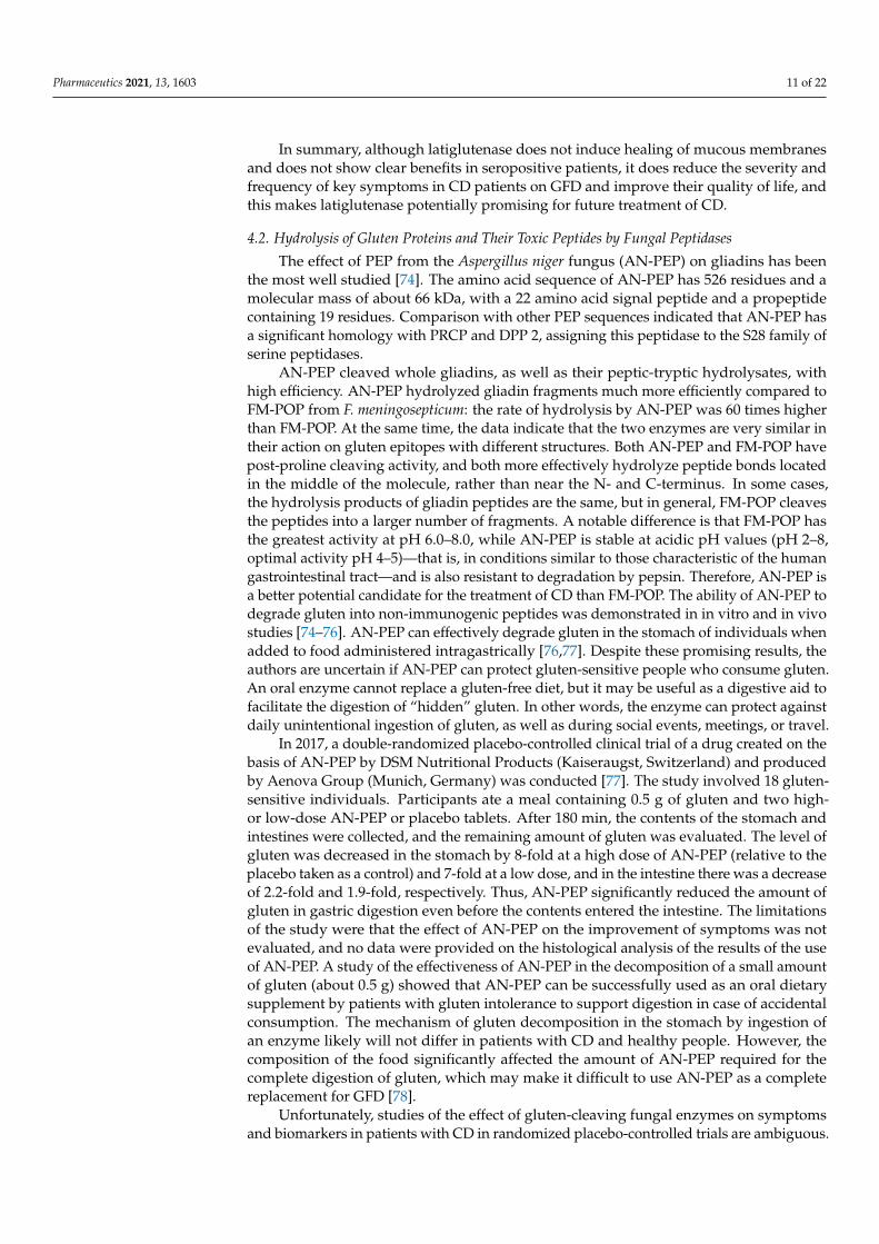
4.2. Hydrolysis of Gluten Proteins and Their Toxic Peptides by Fungal Peptidases
The effect of PEP from the Aspergillus niger fungus (AN-PEP) on gliadins has beenthe most well studied [74]. The amino acid sequence of AN-PEP has 526 residues and amolecular mass of about 66 kDa, with a 22 amino acid signal peptide and a propeptidecontaining 19 residues. Comparison with other PEP sequences indicated that AN-PEP hasa significant homology with PRCP and DPP 2, assigning this peptidase to the S28 family ofserine peptidases.
AN-PEP cleaved whole gliadins, as well as their peptic-tryptic hydrolysates, withhigh efficiency. AN-PEP hydrolyzed gliadin fragments much more efficiently compared toFM-POP from F. meningosepticum: the rate of hydrolysis by AN-PEP was 60 times higherthan FM-POP. At the same time, the data indicate that the two enzymes are very similar intheir action on gluten epitopes with different structures. Both AN-PEP and FM-POP havepost-proline cleaving activity, and both more effectively hydrolyze peptide bonds locatedin the middle of the molecule, rather than near the N- and C-terminus. In some cases,the hydrolysis products of gliadin peptides are the same, but in general, FM-POP cleavesthe peptides into a larger number of fragments. A notable difference is that FM-POP hasthe greatest activity at pH 6.0–8.0, while AN-PEP is stable at acidic pH values (pH 2–8,optimal activity pH 4–5)—that is, in conditions similar to those characteristic of the humangastrointestinal tract—and is also resistant to degradation by pepsin. Therefore, AN-PEP isa better potential candidate for the treatment of CD than FM-POP. The ability of AN-PEP todegrade gluten into non-immunogenic peptides was demonstrated in in vitro and in vivostudies [74–76]. AN-PEP can effectively degrade gluten in the stomach of individuals whenadded to food administered intragastrically [76,77]. Despite these promising results, theauthors are uncertain if AN-PEP can protect gluten-sensitive people who consume gluten.An oral enzyme cannot replace a gluten-free diet, but it may be useful as a digestive aid tofacilitate the digestion of “hidden” gluten. In other words, the enzyme can protect againstdaily unintentional ingestion of gluten, as well as during social events, meetings, or travel.
In 2017, a double-randomized placebo-controlled clinical trial of a drug created on thebasis of AN-PEP by DSM Nutritional Products (Kaiseraugst, Switzerland) and producedby Aenova Group (Munich, Germany) was conducted [77]. The study involved 18 gluten-sensitive individuals. Participants ate a meal containing 0.5 g of gluten and two high-or low-dose AN-PEP or placebo tablets. After 180 min, the contents of the stomach andintestines were collected, and the remaining amount of gluten was evaluated. The level ofgluten was decreased in the stomach by 8-fold at a high dose of AN-PEP (relative to theplacebo taken as a control) and 7-fold at a low dose, and in the intestine there was a decreaseof 2.2-fold and 1.9-fold, respectively. Thus, AN-PEP significantly reduced the amount ofgluten in gastric digestion even before the contents entered the intestine. The limitationsof the study were that the effect of AN-PEP on the improvement of symptoms was notevaluated, and no data were provided on the histological analysis of the results of the useof AN-PEP. A study of the effectiveness of AN-PEP in the decomposition of a small amountof gluten (about 0.5 g) showed that AN-PEP can be successfully used as an oral dietarysupplement by patients with gluten intolerance to support digestion in case of accidentalconsumption. The mechanism of gluten decomposition in the stomach by ingestion ofan enzyme likely will not differ in patients with CD and healthy people. However, thecomposition of the food significantly affected the amount of AN-PEP required for thecomplete digestion of gluten, which may make it difficult to use AN-PEP as a completereplacement for GFD [78].
Unfortunately, studies of the effect of gluten-cleaving fungal enzymes on symptomsand biomarkers in patients with CD in randomized placebo-controlled trials are ambiguous.

Pharmaceutics 2021, 13, 1603 12 of 22
Tack et al. [79] randomized 14 patients with CD to receive 7 g of gluten daily with AN-PEPor a placebo for 2 weeks. However, such a relatively high amount of gluten did not lead toclinical deterioration, even in the group of patients receiving placebo, so it was impossibleto show the effect of the enzyme on the change in clinical markers and symptoms. Quiteoften, patients with CD do not show symptoms when consuming gluten, especially aftera long period of abstinence, even if immunological reactions and damage of the smallintestine tissue have occurred [80].
Tolerase G, a dietary supplement offered by DSM (Kaiseraugst, Switzerland) alsocontaining AN-PEP, is currently positioned as an ideal product for gluten-sensitive peopletrying to follow a GFD, as it has the potential to digest hidden or residual gluten present ina wide range of products [81]. However, this product is not suitable for people with CDor gluten intolerance. Thus, Tolerase G is not an effective treatment for CD, since it doesnot completely break down gluten, and the resulting accumulation of gluten peptides inthe duodenum has not been determined. As mentioned above, AN-PEP is able to degradegluten proteins under acidic conditions in vitro and can apparently be considered as anoral supplement to reduce gluten exposure. However, the uses of glutenases, includingAN-PEP, are limited because of incomplete degradation of gluten [82].
PEP from the basidiomycete Flammulina velutipes (FV-PEP) is similar in specificity toAN-PEP, although the sequence similarity between them was only 39% [83]. The isolatedenzyme turned out to be a serine peptidase of the S28 family, with a molecular mass of50 kDa and the activity maximum at pH 4.5 and 45 ◦C. FV-PEP degraded the antigenicepitopes of α-gliadin that provoke CD. In the future, this enzyme may become an effectivetool for processing wheat and other gluten-containing cereals into more easily digestibleproducts for celiac patients.
Two PSPs from the S28 serine peptidase family—AoS28A and AoS28B—were foundin the acidic culture medium of the mold fungus A. oryzae when soy proteins or wheatgliadins were used as the sole source of nitrogen [84]. AoS28A is an orthologue of thepreviously characterized PEP from A. fumigatus (AfuS28) and A. niger (AN-PEP). AoS28Awas active in the pH range 2.0–6.0 with an optimum at pH 4.0 and using the substrateAla-Ala-Pro-pNA. AoS28B was active in the pH range 2.0–7.0 with an optimum at pH4.5. Both peptidases effectively degraded the immunotoxic proline-rich 33-mer gliadinpeptide, especially in acidic conditions. Both enzymes produced similar but not identicalpatterns of peptide fragments, with different kinetics and some difference in individualhydrolysis products. All peptides larger than nine amino acid residues were completelydigested by a combination of both enzymes for 20 min in acidic conditions, correspondingto conditions in the human stomach. Approximately 30–60 min were required for similardigestion when only one enzyme was used under the same experimental conditions, andAoS28B hydrolyzed the peptide more efficiently. Thus, the combination of both peptidasesis important for the development of an effective oral preparation in enzyme therapy forpatients with gluten intolerance.
The hydrolysis of peptides and some food proteins (in particular, gluten) by aminopep-tidase and X-prolyldipeptidylpeptidase from A. oryzae also was investigated [85,86]. Anon-specific aminopeptidase from A. oryzae was able to cleave the N-terminal glycine andproline residues with high efficiency, and X-prolyldipeptidylpeptidase hydrolyzed, with thespecificity of DPP 4, substrates that contain X-Pro at the N-terminus. The two peptidaseswere synergistic and effectively hydrolyzed proline-containing peptides.
A mixture of two commercially available food-grade enzymes, aspergilopepsin fromA. niger (ASP) and DPP 4 from A. oryzae, each widely used in the preparation of food,food additives, and feed, were evaluated for their use in detoxifying gluten [87]. Theactivity of ASP was tested against recombinant gluten proteins and synthetic gluten pep-tides. ASP, in contrast to mammalian pepsin, was able to hydrolyze gluten to mostlyshort peptides. However, unlike glutenases, such as PEP or EP-B2 of barley, whichhave high specificity for immunotoxic Pro- and Gln-rich gluten peptides, ASP does nothave such specificity. While it can hydrolyze these peptides, the presence of compet-

Pharmaceutics 2021, 13, 1603 13 of 22
ing substrates significantly slows down the hydrolysis. MS-MS analysis showed thatASP cleaves VQWPQ↓QQP↓V↓PQPHQPF of γ-gliadin, and also PFSQ↓Q↓Q↓QPV ofglutenin. In the absence of other protein substrates, ASP cleaved the 33-mer peptideLQLQPFPQPQLPYPQPQLPYPQPQPQF from α2-gliadin, as well as the 28-mer peptidePFPQPQLPYPQPQLPYPQPQLPYPQPQP (a shortened derivative of the 33-mer peptide).However, in the presence of more complex substrates, such as casein, ASP activity towardpeptides containing immunotoxic epitopes was slower. Although each enzyme was unableto detoxify gluten under simulated gastric conditions (0.01 M HCl was added periodicallyfor an hour), a combination of the two enzymes led to the detoxification of moderateamounts of dietary gluten. Since these enzymes have already been proven safe for humanconsumption, this enzyme therapy could provide at least short-term relief of the inflamma-tory bowel response in patients with gluten disease and suffering from accidental glutenexposure. In addition, ASP can be added to stronger and more specific glutenases, such asEP-B2 [33,88] and some microbial/fungal PEP [74], to further enhance their therapeuticeffect. Therefore, controlled clinical trials of these food enzymes are needed to assess theireffects on patients suffering from gluten immunity.
4.3. Hydrolysis of Gluten Proteins and Their Toxic Peptides by Insect Peptidases
The digestive peptidases of the yellow mealworm, Tenebrio molitor, a stored productpest, are of interest because the main food proteins for this insect are prolamins. Twopeptidases were isolated from a midgut extract of T. molitor larvae with post-prolinehydrolyzing activity: PPCP1 (post-proline cleaving peptidase) and PPCP2, with molecularmasses of 101 and 62 kDa, respectively [89,90]. PPCP1 was localized in the contents ofthe acidic anterior part of the gut with a maximum activity at pH 5.6, while PPCP2 was atissue-soluble enzyme evenly distributed along the midgut of the larvae, with a maximumactivity at pH 7.9. Inhibitory analysis indicated that both enzymes were serine peptidases.A specific inhibitor of POP, Z-Pro-prolinal, completely suppressed the activity of PPCP2 andonly partially inhibited PPCP1. The study of substrate specificity demonstrated that PPCP1preferentially hydrolyzed Z-Ala-Ala-Pro-pNA, and PPCP2 preferred a shorter peptideZ-Ala-Pro-pNA. In examining all data, PPCP2 was characterized as POP, and PPCP1 asPRCP [91]. In addition, recombinant DPP 4 of T. molitor was able to hydrolyze gliadins moreeffectively than human DPP 4, and T. molitor prolidase was proposed as a critical enzymefor the final stages of gliadins digestion, providing hydrolysis of imidodipeptides [92–94].
Proteolytic activities hydrolyzing the substrate Z-Ala-Ala-Gln-pNA after a glutamineresidue were detected in the extract of the midgut of T. molitor larvae [29]. There was onepost-glutamine cleaving peptidase (PGP2AM) in the anterior part of the midgut, and two(PGP1PM and PGP2PM) in the posterior part. All the isolated enzymes hydrolyzed Z-Ala-Ala-Gln-pNA only in the presence of dithiothreitol (DTT) and were completely inactivatedby E-64 (L-trans-epoxysuccinyl-leucylamido-(4-guanidino)-butane), which indicates thatthey are cysteine peptidases. An electrophoretic study of the dynamics of the hydrolysisof gliadins by PGP2AM and serine digestive peptidases showed that a cysteine peptidasewith PGP activity contributed significantly to their hydrolysis. Similarly, PGP activityof cysteine digestive peptidases with the selective substrate Glp-Phe-Gln-pNA was alsofound in the midgut of Tribolium castaneum, a related stored product pest also from theTenebrionidae family [30].
The efficiency of using individual PSPs is low because most are exopeptidases andcannot provide complete cleavage of gliadins. However, in the review [95], the combinationof T. molitor digestive enzymes, mainly cysteine cathepsin L and PSPs, are proposed todegrade gliadins in cooperation.
PSPs were found in extracts from the lesser grain borer, Rhyzopertha dominica [96]. Anisolated but uncharacterized enzyme was effective in the hydrolysis of CD-related peptidesof wheat and barley. The overall data suggests much can be learned through the study ofinsects that have become adapted to efficiently process cereal grains.
Pharmaceutics 2021, 13, 1603 14 of 22
4.4. Hydrolysis of Gluten Proteins and Their Toxic Peptides by Plant Peptidases
In plants, peptidases with PGP activity were the most active in the hydrolysis ofgluten proteins. A papain-like cysteine peptidase EP-B2 (EndoPeptidase from Barley)was purified and characterized from grains of sprouted barley, Hordeum vulgare, andthe recombinant proenzyme was expressed in E. coli [33,88]. The zymogen (proEP-B2)was rapidly autoactivated under acidic conditions at a rate independent of the proEP-B2 concentration, yielding a mature enzyme with a molecular mass of 27.7 kDa. Theability of the enzyme to hydrolyze toxic gliadin peptides was evaluated with the 33-mer peptide LQLQPFPQPQLPYPQPQLPYPQPQLPYPQPQP with a ratio of 1:10 (en-zyme:substrate) at pH 3 for 60 min (zymogen activation conditions). The analysis ofthe mass spectrum indicated that EP-B2 hydrolyzed bonds formed by glutamine residuesLQ↓LQPFPQPQ↓LLPYPQPQ↓LPYPQPQ↓LPYPQPQ.
The effect of EP-B2 on intact toxic peptides was studied in mice whose diet containedgluten. In the stomach and intestines of control mice, a number of toxic peptides, including11-mer PFPQPQLPYPQ, 14-mer PQPQLPYPQPQLPY, 28-mer PFPQPQLPYPQPQLPYPQPQLPQPQP, and 33-mer LQLQPFPQPQLPYPQPQLPYPQPQLPYPQPQPF, were detected byHPLC [97]. After EP-B2 was added to the mice’s food, the peptide content decreaseddepending on the time and concentration of the enzyme. Estimates were that the con-centration of the 33-mer peptide in the stomach could be reduced by more than 50-fold.EP-B2 was resistant to the action of pepsin, but lost activity in the presence of trypsin, andit was concluded that the potential for its use is limited to the gastric phase of digestion.Moreover, the activation of proEP-B2 occurs under acidic conditions and only at pH valuesless than 4.0. Thus, EP-B2 should remain active during the digestion of gluten in thestomach, but will quickly break down in the duodenum. For therapeutic use, the intestinalstability of recombinant EP-B2 can be increased through modification of trypsin-sensitivecleavage sites.
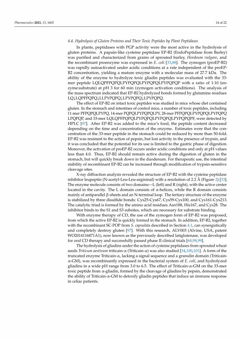
X-ray diffraction analysis revealed the structure of EP-B2 with the cysteine peptidaseinhibitor leupeptin (N-acetyl-Leu-Leu-argininal) with a resolution of 2.2 Å (Figure 2) [33].The enzyme molecule consists of two domains—L (left) and R (right), with the active centerlocated in the cavity. The L domain consists of α-helices, while the R domain consistsmainly of antiparallel β-sheets and an N-terminal loop. The tertiary structure of the enzymeis stabilized by three disulfide bonds: Cys25-Cys67, Cys59-Cys100, and Cys161-Cys213.The catalytic triad is formed by the amino acid residues Asn188, His167, and Cys28. Theinhibitor binds to the S1 and S3 subsites, which are necessary for substrate binding.
With enzyme therapy of CD, the use of the zymogen form of EP-B2 was proposed,from which the active EP-B2 is quickly formed in the stomach. In addition, EP-B2, togetherwith the recombinant SC-POP from S. capsulata described in Section 4.1, can synergisticallyand completely destroy gluten [97]. With this research, ALV003 (Alvine, USA, patentWO2014116871A1), now known as the previously described latiglutenase, was developedfor oral CD therapy and successfully passed phase II clinical trials [68,98,99].
The hydrolysis of gliadins under the action of cysteine peptidases from sprouted wheatseeds Triticum aestivum triticain-α (Triticain-α) was also studied [34,100,101]. A form of thetruncated enzyme Triticain-α, lacking a signal sequence and a granulin domain (Triticain-α-GM), was recombinantly expressed in the bacterial system of E. coli, and hydrolyzedgliadins in a wide pH range from 3.0 to 6.5. The effect of Triticain-α-GM on the 33-mertoxic peptide from α-gliadin, formed by the cleavage of gliadins by pepsin, demonstratedthe ability of Triticain-α-GM to detoxify gliadin peptides that induce an immune responsein celiac patients.
Pharmaceutics 2021, 13, 1603 15 of 22
Pharmaceutics 2021, 13, x FOR PEER REVIEW 15 of 22
the enzyme is stabilized by three disulfide bonds: Cys25‐Cys67, Cys59‐Cys100, and
Cys161‐Cys213. The catalytic triad is formed by the amino acid residues Asn188, His167,
and Cys28. The inhibitor binds to the S1 and S3 subsites, which are necessary for substrate
binding.
Figure 2. Structure of EP‐B2 with the cysteine peptidase inhibitor leupeptin (PDB 2FO5) with a
resolution of 2.2 Å. The α‐helices of the L domain are purple, β‐sheets of the R domain are gray.
Amino acid residues of the active center are shown as sticks and colored by elements (C—green,
N—blue, O—red). Leupeptin is shown as sticks and is teal. Structure is visualized using PyMOL
(https://pymol.org/2/, accessed on 25 May 2021) (The PyMOL Molecular Graphics System, Version
1.2r3pre, Schrödinger, LLC).
With enzyme therapy of CD, the use of the zymogen form of EP‐B2 was proposed,
from which the active EP‐B2 is quickly formed in the stomach. In addition, EP‐B2, together
with the recombinant SC‐POP from S. capsulata described in Section 4.1, can
synergistically and completely destroy gluten [97]. With this research, ALV003 (Alvine,
USA, patent WO2014116871A1), now known as the previously described latiglutenase,
was developed for oral CD therapy and successfully passed phase II clinical trials
[68,98,99].
The hydrolysis of gliadins under the action of cysteine peptidases from sprouted
wheat seeds Triticum aestivum triticain‐α (Triticain‐α) was also studied [34,100,101]. A
form of the truncated enzyme Triticain‐α, lacking a signal sequence and a granulin
domain (Triticain‐α‐GM), was recombinantly expressed in the bacterial system of E. coli,
and hydrolyzed gliadins in a wide pH range from 3.0 to 6.5. The effect of Triticain‐α‐GM
on the 33‐mer toxic peptide from α‐gliadin, formed by the cleavage of gliadins by pepsin,
demonstrated the ability of Triticain‐α‐GM to detoxify gliadin peptides that induce an
immune response in celiac patients.
Triticain‐α‐GM was incubated with the main gastrointestinal peptidases pepsin and
trypsin, at acidic and slightly alkaline pH values, respectively. The enzyme was relatively
resistant to pepsin cleavage when incubated at pH 3.0 at 37 °C for 30 min and maintained
its maximum glutenase activity under these conditions. In contrast, incubation of
Triticain‐α‐GM in the presence of trypsin at pH 8.0 and 37 °C resulted in complete
degradation within 15 min. Thus, Triticain‐α‐GM is stable under conditions of gastric
digestion, and has a significant ability to break down gluten [34].
Degradation of gliadins with high efficiency was also observed using peptidases of
raw papaya milky juice [102]. The test mixture was superior to individual plant enzymes
and fungal peptidases. The high activity of the mixture of enzymes from papaya latex was
Figure 2. Structure of EP-B2 with the cysteine peptidase inhibitor leupeptin (PDB 2FO5) with aresolution of 2.2 Å. The α-helices of the L domain are purple, β-sheets of the R domain are gray.Amino acid residues of the active center are shown as sticks and colored by elements (C—green,N—blue, O—red). Leupeptin is shown as sticks and is teal. Structure is visualized using PyMOL(https://pymol.org/2/, accessed on 25 May 2021) (The PyMOL Molecular Graphics System, Version1.2r3pre, Schrödinger, LLC).
Triticain-α-GM was incubated with the main gastrointestinal peptidases pepsin andtrypsin, at acidic and slightly alkaline pH values, respectively. The enzyme was relativelyresistant to pepsin cleavage when incubated at pH 3.0 at 37 ◦C for 30 min and maintainedits maximum glutenase activity under these conditions. In contrast, incubation of Triticain-α-GM in the presence of trypsin at pH 8.0 and 37 ◦C resulted in complete degradationwithin 15 min. Thus, Triticain-α-GM is stable under conditions of gastric digestion, andhas a significant ability to break down gluten [34].
Degradation of gliadins with high efficiency was also observed using peptidases ofraw papaya milky juice [102]. The test mixture was superior to individual plant enzymesand fungal peptidases. The high activity of the mixture of enzymes from papaya latex wasprimarily due to caricaine, the most alkaline among the cysteine peptidases of latex. Whenraw papaya milky juice was added to swine gut extract, a synergistic effect was observed,with an increase in the degree of cleavage of toxic peptides 11–19 (QNPSQQQPQ) and75–86 (RPQQPYQPQPQP) from α-gliadins. The synergistic effect was proposed to be dueto a difference in the specificity of the enzymes, that is, the hydrolysis products of oneenzyme can act as substrates for the second. These enzymes were used in the production ofbread with a low gluten content, treating the kneaded baking dough with caricaine isolatedfrom papaya latex extract [103,104]. The gluten level in the dough treated with purifiedenzyme was significantly lower compared to the crude extract treatment. As for the quality,bread made from caricaine-treated dough turned out to be looser and more porous, witha harder and darker crust, compared to the control sample. The bread obtained from thedough fermented for 7 h at 37 ◦C was considered the most optimal in terms of quality [103].
Thus, plant cysteine peptidases, along with bacterial and fungal peptidases, can beconsidered as candidates for use in the production of gluten-free products and in thetreatment of CD patients. However, more clinical data on the use of glutenase preparationsas a CD therapy will be necessary to validate effective treatments in gluten-dependentdisorders (Table 3).

Pharmaceutics 2021, 13, 1603 16 of 22
Table 3. Current data on the drugs for enzyme therapy of gluten sensitivity.
Product Company Base of the Drug (Origin) ClinicalTrial Phase References
KumaMax, Kuma030,Kuma062, TAK-062
Takeda PharmaceuticalCompany Limited, Tokyo,
Japan
Kumamolysine-AsAlicyclobacillus sendaiensis 1 [66,105,106]
Latiglutenase ALV003 Alvine Pharmaceuticals Inc.,San Carlos, CA, USA
Prolyl oligopeptidase (POP)Sphingomonas capsule + cysteinepeptidase from barley (EP-B2)
2 [67–69,71,73]
Tolerase G DSM Nutritional Products,Kaiseraugst, Switzerland
Prolylendopeptidase of themold fungus Aspergillus niger
(AN-PEP)
Dietarysupplement [77,81]
AMYRA’s enzymes,AMY01
AMYRA Biotech AGBasel,Switzerland
Combination of fungalexopeptidases
Dietarysupplement [107]
AMYRA’s enzymes,AMY02
AMYRA Biotech AGBasel,Switzerland
Combination of fungalexopeptidases Pre-clinical [107]
Nemysis E40 Nemysis Ltd., Dublin, Ireland Endopeptidase soilActinoallomurus strain Pre-clinical [108]
It should be noted that the DPP 4 enzyme often used in clinical testing is not activeat low pH and, therefore, is not very effective in the acidic environment of the stomach.In addition, DPP4 has no endopeptidase activity and, in the absence of other enzymes,can only act on the N-terminus of proteins, so large immunogenic fragments of gluten canremain uncleaved [109]. The role of gliadins in people with non-celiac gluten sensitivity(NCGS) remains unknown, and a rationale for the use of gluten-hydrolyzing enzymes insuch people is unclear [110]. Immuno-mediated enteropathy, CD or gluten disease, andNCGS are two different clinical conditions, although both are caused by wheat gliadin.People with NCGS suffer from the same symptoms as patients with CD (diarrhea, stomachproblems, fatigue, joint pain, etc.), but without the histological and serological markerscharacteristic of CD [111]. In patients with NCGS, gluten consumption also can lead topotentially harmful long-term effects [112]. A significant increase in markers of intestinalepithelial damage and systemic immune activation was found in this group of patientsreceiving food containing cereal proteins. In contrast to CD, the observed humoral immuneresponse with NCGS to gluten does not depend on the enzymatic activity of TG2 and thepresence of HLA-DQ2/DQ8, so hydrolysis products are assumed to target certain epitopesother than those in CD. Although NCGS individuals showed increased levels of antibodiesagainst native gliadin, the role of gluten in this condition is debated. While some studieshave shown that gluten consumption leads to gastrointestinal symptoms in people withNCGS [113,114], others have not confirmed the association [115]. The latest study indicatedthat bread pre-processed with AN-PEP had a 40% reduction in gluten [116]. However,eating this bread did not reduce the symptoms of gluten sensitivity in people with NCGS.The results could be due to an insufficient reduction in the gluten content, but instead ofgluten, other compounds present in wheat or related cereals may be involved in the onsetof symptoms. The later hypothesis is supported by the results of Skodje et al. [117], wherea randomized double-blind study using granola bars demonstrated that in NCGS people,symptoms were caused mainly by fructans, not gluten.
5. Conclusions
Hydrolysis of proline/glutamine-rich proteins is difficult because most broad-spectrumpeptidases are unable to cleave the peptide bonds formed by proline and glutamineresidues. However, proline/glutamine-rich proteins such as prolamins become pathogenicunder certain physiological conditions, but their proteolysis can provide a therapeuticeffect. Thus, prolamins and their immunogenic peptides in human food cause an autoim-mune response in predisposed people, leading to the development of CD. This reviewsummarized the use of various PSPs for the hydrolysis of these proteins. Among them, the

Pharmaceutics 2021, 13, 1603 17 of 22
greatest attention is paid to the study of POP and PEP, since these peptidases hydrolyzelong protein sequences into shorter fragments. However, a sufficiently complete hydrolysiswas possible only with the combined use of several different PSPs. Promising results arefound in studies of mixed complexes of PSPs and subtilisin-like or cysteine peptidases withPGP activity.
With that, a number of questions remain unanswered or insufficiently studied relatedto the effective use of peptidases to reduce the toxic effects of prolamins and their immuno-genic peptides. In addition, it is necessary to evaluate whether the enzymatic pretreatmentof wheat flour and the removal of harmful components for CD may lead to the loss ofcharacteristics that make gluten-containing products preferable for food production. Beforeincorporating commercially available enzyme preparations to reduce gluten sensitivity,such as those containing various glutenases derived from bacteria or fungi, it is importantto gather the available scientific data on their effectiveness and safety.
The use of these enzymes cannot be recommended to compensate for the intake oflarge quantities of gluten (consumed unintentionally or intentionally). Despite the fact thattheir effectiveness can be quite high, even a small amount of gluten or its peptides thatreach the duodenum can be harmful to CD patients. In addition, the effectiveness of theenzymes in vitro is affected by the composition of the food, and this effect has not yet beenproperly investigated in vivo.
The available biochemical data on a particular enzyme may help to select a promisingcandidate for possible enzyme preparations, but further clinical trials are needed to confirmthe therapeutic effectiveness of the selected enzyme in the treatment of gluten intolerance.So far, an analysis of the results suggests that enzyme therapy alone is not sufficient forthe treatment of CD. Such therapy is probably not able to neutralize the large amount ofgluten present on average in the human diet based on wheat or similar cereals. However,enzyme therapy can reduce the gluten-induced effects observed in the background of GFDpractices that occur by the unintentional consumption of small amounts of gluten, that is,to act as a pharmacological supplement to GFD.
Author Contributions: All authors contributed equally to this work. All authors have read andagreed to the published version of the manuscript.
Funding: This work was supported by Russian Foundation for Basic Research grant NO. 19-04-00852_a.
Institutional Review Board Statement: Not applicable.
Informed Consent Statement: Not applicable.
Data Availability Statement: Not applicable.
Acknowledgments: Mention of trade names or commercial products in this publication is solely forthe purpose of providing specific information and does not imply recommendation or endorsementby the U.S. Department of Agriculture. USDA is an equal opportunity provider and employer. Allindividuals included in this section have consented to the acknowledgement.
Conflicts of Interest: The authors declare no conflict of interest.
References1. Farrell, R.J.; Kelly, C.P. Celiac sprue. N. Engl. J. Med. 2002, 346, 180–188. [CrossRef] [PubMed]2. Cappello, M.; Morreale, G.C.; Licata, A. Elderly Onset Celiac Disease: A Narrative Review. Clin. Med. Insights Gastroenterol. 2016,
9, 41–49. [CrossRef] [PubMed]3. Shan, L.; Molberg, O.; Parrot, I.; Hausch, F.; Filiz, F.; Gray, G.M.; Sollid, L.M.; Khosla, C. Structural basis for gluten intolerance in
celiac sprue. Science 2002, 297, 2275–2279. [CrossRef]4. Shan, L.; Qiao, S.W.; Arentz-Hansen, H.; Molberg, O.; Gray, G.M.; Sollid, L.M.; Khosla, C.J. Identification and analysis of
multivalent proteolytically resistant peptides from gluten: Implications for celiac sprue. Proteome Res. 2005, 4, 1732–1741.[CrossRef] [PubMed]
5. Hopkins, S.; Soon, J.M. Nutritional quality, cost and availability of gluten-free food in England. Br. Food J. 2019, 121, 2867–2882.[CrossRef]

Pharmaceutics 2021, 13, 1603 18 of 22
6. Dunaevsky, Y.E.; Tereshchenkova, V.F.; Oppert, B.; Belozersky, M.A.; Filippova, I.Y.; Elpidina, E.N. Human proline specificpeptidases: A comprehensive analysis. Biochim. Biophys. Acta 2020, 1864, 129636. [CrossRef]
7. Shewry, P.R.; Tatham, A.S. The prolamin storage proteins of cereal seeds: Structure and evolution. Biochem. J. 1990, 267. [CrossRef]8. Shewry, P.R.; Halford, N.G. Cereal seed storage proteins: Structures, properties and role in grain utilization. J. Exp. Bot. 2002, 53,
947–958. [CrossRef]9. Barak, S.; Mudgil, D.; Khatkar, B.S. Biochemical and functional properties of wheat gliadins: A review. Crit. Rev. Food Sci. Nutr.
2015, 55, 357–368. [CrossRef]10. Biesiekierski, J.R. What is gluten? J. Gastroenterol. Hepatol. 2017, 32 (Suppl. 1), 78–81. [CrossRef]11. Shewry, P.R.; Tatham, A.S.; Forde, J.; Kreis, M.; Miflin, B.J. The classification and nomenclature of wheat gluten proteins: A
reassessment. J. Cereal Sci. 1986, 4, 97–106. [CrossRef]12. Haush, F.; Shan, L.; Santiago, N.A.; Gary, G.M.; Khosla, C. Intestinal digestive resistance of immunodominant gliadin peptides.
Am. J. Physiol. Gastrointest. Liver Physiol. 2002, 283, 996–1003. [CrossRef] [PubMed]13. Schuppan, D.; Junker, Y.; Barisani, D. Celiac disease: From pathogenesis to novel therapies. Gastroenterology 2009, 137, 1912–1933.
[CrossRef]14. Sollid, L.M. Molecular basis of celiac disease. Annu. Rev. Immunol. 2000, 18, 53–81. [CrossRef]15. Wieser, H. The precipitating factor in coeliac disease. Baillieres Clin. Gastroenterol. 1995, 9, 191–207. [CrossRef]16. Loponen, J. Prolamin Degradation in Sourdoughs. Ph.D. Thesis, University of Helsinki, Helsinki, Finland, 20 December 2006.17. Lionetti, E.; Catassi, C. New clues in celiac disease epidemiology, pathogenesis, clinical manifestations, and treatment. Int. Rev.
Immunol. 2011, 30, 219–231. [CrossRef]18. Lohi, S.; Mustalahti, K.; Kaukinen, K.; Laurila, K.; Collin, P.; Rissane, H.; Lohi, O.; Bravi, E.; Gasparin, M.; Reunanen, A.; et al.
Increasing prevalence of coeliac disease over time. Aliment. Pharmacol. Ther. 2007, 26, 1217–1225. [CrossRef] [PubMed]19. Catassi, C.; Kryszak, D.; Bhatti, B.; Sturgeon, C.; Helzlsouer, K.; Clipp, S.L.; Gelfond, D.; Puppa, E.; Sferruzza, A.; Fasano, A.
Natural history of celiac disease autoimmunity in a USA cohort followed since 1974. Ann. Med. 2010, 42, 530–538. [CrossRef]20. Shumilov, P.V.; Muhina, Y.G.; Netrebenko, O.K.; Ryazanova, O.V.; Schigaleva, N.E.; Kovalenko, A.A.; Levina, E.E.; Ponomareva,
A.P. Modern understanding of celiac disease pathogenetic mechanisms: Defining role in course clinical variants. Pediatriya 2016,95, 110–121.
21. Grossman, G. Neurological complications of coeliac disease: What is the evidence? Pract. Neurol. 2008, 8, 77–89. [CrossRef]22. Ford, R.P. The gluten syndrome: A neurological disease. Med. Hypotheses 2009, 73, 438–440. [CrossRef]23. Verdu, E.F.; Armstrong, D.; Murray, J.A. Between celiac disease and irritable bowel syndrome: The “no man’s land” of gluten
sensitivity. Am. J. Gastroenterol. 2009, 104, 1587–1594. [CrossRef] [PubMed]24. Midhagen, C.; Hallert, C. High rate of gastrointestinal symptoms in celiac disease patients living on a gluten-free diet: Controlled
study. Am J Gastroenterol. 2003, 98, 2023–2026. [CrossRef]25. Ilus, T.; Lähdeaho, M.L.; Salmi, T.; Katri, H.; Jukka, P.; Päivi, S.; Heini, H.; Markku, M.; Pekka, C.; Katri, K. Persistent duodenal
intraepithelial lymphocytosis despite a long-term strict gluten-free diet in celiac disease. Am. J. Gastroenterol. 2012, 107, 1563–1569.[CrossRef]
26. Catassi, C.; Fabiani, E.; Iacono, G.; D’Agate, C.; Francavilla, R.; Biagi, F.; Volta, U.; Accomando, S.; Picarelli, A.; De Vitis, I.; et al. Aprospective, double-blind, placebo-controlled trial to establish a safe gluten threshold for patients with celiac disease. Am. J. Clin.Nutr. 2007, 85, 160–166. [CrossRef] [PubMed]
27. Hischenhuber, C.; Crevel, R.; Jarry, B.; Maki, M.; Moneret-Vautrin, D.A.; Romano, A.; Troncone, R.; Ward, R. Review article: Safeamounts of gluten for patients with wheat allergy or coeliac disease. Aliment. Pharmacol. Ther. 2006, 23, 559–575. [CrossRef]
28. Wieser, H.; Koehler, P. Detoxification of gluten by means of enzymatic treatment. J. AOAC Int. 2012, 95, 356–363. [CrossRef][PubMed]
29. Goptar, I.A.; Semashko, T.A.; Danilenko, S.A.; Lysogorskaya, E.N.; Oksenoit, E.S.; Zhuzhikov, D.P.; Belozersky, M.A.; Dunaevsky,Y.E.; Oppert, B.; Filippova, I.Y.; et al. Cysteine digestive peptidases function as post-glutamine cleaving enzymes in tenebrionidstored-product pests. Comp. Biochem. Physiol. B Biochem. Mol. Biol. 2012, 161, 148–154. [CrossRef]
30. Filippova, I.Y.; Dvoryakova, E.A.; Sokolenko, N.I.; Simonyan, T.R.; Tereshchenkova, V.F.; Zhiganov, N.I.; Dunaevsky, Y.E.;Belozersky, M.A.; Oppert, B.; Elpidina, E.N. New glutamine-containing substrates for the assay of cysteine peptidases from theC1 papain family. Front. Mol. Biosci. 2020, 7, 578758. [CrossRef] [PubMed]
31. Dvoryakova, E.A.; Vinokurov, K.S.; Tereshchenkova, V.F.; Dunaevsky, Y.E.; Belozersky, M.A.; Oppert, B.; Filippova, I.Y.; Elpidina,E.N. Primary digestive cathepsins L of Tribolium castaneum larvae: Proteomic identification, properties, comparison with humanlysosomal cathepsin L. Insect Biochem. Mol. Biol. 2021, submitted.
32. Wei, G.; Tian, N.; Siezen, R.; Schuppa, D.; Helmerhorst, E.J. Identification of food-grade subtilisins as gluten-degrading enzymesto treat celiac disease. Am. J. Physiol. Gastrointest. Liver Physiol. 2016, 311, G571–G580. [CrossRef]
33. Bethune, M.T.; Strop, P.; Tang, Y.; Sollid, L.M.; Khosla, C. Heterologous expression, purification, refolding, and structural-functional characterization of EP-B2, a self-activating barley cysteine endoprotease. Chem. Biol. 2006, 13, 637–647. [CrossRef]
34. Savvateeva, L.V.; Gorokhovets, N.V.; Makarov, V.A.; Serebryakova, M.V.; Solovyev, A.G.; Morozov, S.Y.; Reddy, V.P.; Zernii,E.Y.; Zamyatnin, A.A., Jr.; Aliev, G. Glutenase and collagenase activities of wheat cysteine protease Triticain-α: Feasibility forenzymatic therapy assays. Int. J. Biochem. Cell. Biol. 2015, 62, 115–124. [CrossRef]
Pharmaceutics 2021, 13, 1603 19 of 22
35. Shan, L.; Marti, T.; Sollid, L.M.; Gray, G.M.; Khosla, C. Comparative biochemical analysis of three bacterial prolyl endopeptidases:Implications for coeliac sprue. Biochem. J. 2004, 383, 311–318. [CrossRef] [PubMed]
36. Gass, J.; Ehren, J.; Strohmeier, G.; Isaacs, I.; Khosla, C. Fermentation, purification, formulation, and pharmacological evaluationof a prolyl endopeptidase from Myxococcus xanthus: Implications for celiac sprue therapy. Biotechnol. Bioeng. 2005, 92, 674–684.[CrossRef] [PubMed]
37. Marti, T.; Molberg, O.; Li, Q.; Gray, G.M.; Khosla, C.; Sollid, L.M. Prolyl endopeptidase-mediated destruction of T cell epitopes inwhole gluten: Chemical and immunological characterization. J. Pharmacol. Exp. Ther. 2005, 312, 19–26. [CrossRef] [PubMed]
38. Pyle, G.G.; Paaso, B.; Anderson, B.E.; Allen, D.D.; Mart, T.; Li, Q.; Siegel, M.; Khosla, C.; Gray, G.M. Effect of pretreatment of foodgluten with prolyl endopeptidase on gluten induced malabsorption in celiac sprue. Clin. Gastroenterol. Hepatol. 2005, 3, 687–694.[CrossRef]
39. Shan, L.; Mathews, I.; Khosla, C. Structural and mechanistic analysis of two prolylendopeptidases: Role of interdomain dynamicsin catalysis and specificity. Proc. Natl. Acad. Sci. USA 2005, 102, 3599–3604. [CrossRef]
40. Piper, J.L.; Gray, G.M.; Khosla, C. Effect of Prolyl Endopeptidase on Digestive-Resistant Gliadin Peptides in vivo. J. Pharm. Exp.Ther. 2004, 311, 213–219. [CrossRef]
41. Matysiak-Budnik, T.; Candalh, C.; Cellier, C.; Dugave, C.; Namane, A.; Vidal-Martinez, T.; Cerf-Bensussan, N.; Heyman, M.Limited efficiency of prolyl-endopeptidase in the detoxification of gliadin peptides in celiac disease. Gastroenterology 2005, 129,786–796. [CrossRef]
42. Alvarez-Sieiro, P.; Martin, M.C.; Redruello, B.; Del Rio, B.; Ladero, V.; Palanski, B.A.; Khosla, C.; Fernandez, M.; Alvarez, M.A.Generation of food-grade recombinant Lactobacillus casei delivering Myxococcus xanthus prolyl endopeptidase. Appl. Microbiol.Biotechnol. 2014, 98, 6689–6700. [CrossRef] [PubMed]
43. Shetty, R.; Vestergaard, M.; Jessen, F.; Hagglund, P.; Knorr, V.; Koehler, P.; Prakash, H.S.; Hobley, T.J. Discovery, cloning andcharacterisation of proline specific prolyl endopeptidase, a gluten degrading thermo-stable enzyme from Sphaerobacter thermophiles.Enzyme Microb. Technol. 2017, 107, 57–63. [CrossRef]
44. Spicher, G.; Nierle, W. Proteolytic activity of sourdough bacteria. Appl. Microbiol. Biotechnol. 1988, 28, 487–492. [CrossRef]45. Gerez, C.L.; Font de Valdez, G.; Rollan, G.C. Functionality of lactic acid bacteria peptidase activities in the hydrolysis of
gliadin-like fragments. Lett. Appl. Microbiol. 2008, 47, 427–432. [CrossRef]46. Gerez, C.L.; Dallagnol, A.; Rollan, G.; Font de Valdez, G. A combination of two lactic acid bacteria improves the hydrolysis of
gliadin during wheat dough fermentation. Food Microbiol. 2012, 32, 427–430. [CrossRef] [PubMed]47. Fu, W.; Xue, W.; Liu, C.; Tian, Y.; Zhang, K.; Zhu, Z. Screening of Lactic Acid Bacteria and Yeasts from Sourdough as Starter
Cultures for Reduced Allergenicity Wheat Products. Foods 2020, 9, 751. [CrossRef]48. Katina, K.; Arendt, E.; Liukkonen, K.-H.; Autio, K.; Flander, L.; Poutanen, K. Potential of sourdough for healthier cereal products.
Trends Food Sci. Technol. 2005, 16, 104–112. [CrossRef]49. Rizzello, C.G.; De Angelis, M.; Di Cagno, R.; Camarca, A.; Silano, M.; Losito, I.; De Vincenzi, M.; De Bari, M.D.; Palmisano,
F.; Maurano, F.; et al. Highly efficient gluten degradation by lactobacilli and fungal proteases during food processing: Newperspectives for celiac disease. Appl. Environ. Microbiol. 2007, 73, 4499–4507. [CrossRef]
50. De Angelis, M.; Cassone, A.; Rizzello, C.; Gagliardi, F.; Minervini, F.; Calasso, M.; Di Cagno, R.; Francavilla, R.; Gobbetti, M.Mechanism of degradation of immunogenic gluten epitopes from Triticum turgidum L. var. durum by sourdough lactobacilli andfungal proteases. Appl. Enviroment. Microbiol. 2010, 76, 508–518. [CrossRef] [PubMed]
51. Alvarez-Sieiro, P.; Redruello, B.; Ladero, V.; Martin, M.C.; Fernandez, M.; Alvarez, M.A. Screening sourdough samples forgliadin-degrading activity revealed Lactobacillus casei strains able to individually metabolize the coeliac-disease-related 33-merpeptide. Can. J. Microbiol. 2016, 62, 422–430. [CrossRef]
52. Stefanska, I.; Piasecka-Jozwiak, K.; Kotyrba, D.; Kolenda, M.; Stecka, K.M. Selection of lactic acid bacteria strains for the hydrolysisof allergenic proteins of wheat flour. J. Sci. Food Agric. 2016, 96, 3897–3905. [CrossRef] [PubMed]
53. De Almeida, N.E.C.; Esteves, F.G.; dos Santos-Pinto, J.R.A.; De Paula, C.P.; Da Cunha, A.F.; Malavazi, I.; Palma, M.S.; Rodrigues-Filho, E. Digestion of intact gluten proteins by Bifidobacterium species: Reduction of cytotoxicity and proinflammatory responses.J. Agric. Food Chem. 2020, 68, 4485–4492. [CrossRef] [PubMed]
54. Giorgi, A.; Cerrone, R.; Capobianco, D.; Filardo, S.; Mancini, P.; Fanelli, S.; Mastromarino, P.; Mosca, L. A probiotic preparationhydrolyzes gliadin and protects intestinal cells from the toxicity of pro-inflammatory peptides. Nutrients 2020, 12, 495. [CrossRef][PubMed]
55. Brzozowski, B.; Stasiewicz, K.; Ostolski, M.; Adamczak, M. Reducing Immunoreactivity of Gliadins and Coeliac-Toxic PeptidesUsing Peptidases from L. acidophilus 5e2 and A. niger. Catalysts 2020, 10, 923. [CrossRef]
56. Helmerhorst, E.J.; Sun, X.; Salih, E.; Oppenheim, F.G. Identification of Lys-Pro-Gln as a novel cleavage site specificity ofsaliva-associated proteases. J. Biol. Chem. 2008, 283, 19957–19966. [CrossRef]
57. Messana, I.; Cabras, T.; Pisano, E.; Sanna, M.T.; Olianas, A.; Manconi, B.; Pellegrini, M.; Paludetti, G.; Scarano, E.; Fiorita, A.; et al.Trafficking and postsecretory events responsible for the formation of secreted human salivary peptides: A proteomics approach.Mol. Cell. Proteomics. 2008, 7, 911–926. [CrossRef]
58. Helmerhorst, E.J.; Zamakhchari, M.; Schuppan, D.; Oppenheim, F.G. Discovery of a novel and rich source of gluten-degradingmicrobial enzymes in the oral cavity. PLoS ONE 2010, 5, e13264. [CrossRef]
Pharmaceutics 2021, 13, 1603 20 of 22
59. Zamakhchari, M.; Wei, G.; Dewhirst, F.; Lee, J.; Schuppan, D.; Oppenheim, F.G.; Helmerhorst, E.J. Identification of Rothia bacteriaas gluten-degrading natural colonizers of the upper gastro-intestinal tract. PLoS ONE 2011, 6, e24455. [CrossRef]
60. Fernandez-Feo, M.; Wei, G.; Blumenkranz, G.; Dewhirst, F.E.; Schuppan, D.; Oppenheim, F.G.; Helmerhorst, E.J. The cultivablehuman oral gluten-degrading microbiome and its potential implications in coeliac disease and gluten sensitivity. Clin. Microbiol.Infect. 2013, 19, E386–E394. [CrossRef]
61. Darwish, G.; Helmerhorst, E.J.; Schuppan, D.; Oppenheim, F.G.; Wei, G. Pharmaceutically modified subtilisins withstand acidicconditions and effectively degrade gluten in vivo. Sci. Rep. 2019, 9, 7505. [CrossRef]
62. Tian, N.; Faller, L.; Leffler, D.A.; Kelly, C.P.; Hansen, J.; Bosch, J.A.; Wei, G.; Paster, B.J.; Schuppan, D.; Helmerhorst, E.J. Salivarygluten degradation and oral microbial profiles in healthy individuals and celiac disease patients. Appl. Environ. Microbiol. 2017,83, e03330–e03316. [CrossRef] [PubMed]
63. Caminero, A.; Galipeau, H.J.; McCarville, J.L.; Johnston, C.W.; Bernier, S.P.; Russell, A.K.; Jury, J.; Herran, A.R.; Casqueiro, J.;Tye-Din, J.A.; et al. Duodenal bacteria from patients with celiac disease and healthy subjects distinctly affect gluten breakdownand immunogenicity. Gastroenterology 2016, 151, 670–683. [CrossRef] [PubMed]
64. Caminero, A.; McCarville, J.L.; Galipeau, H.J.; Deraison, C.; Bernier, S.P.; Constante, M.; Rolland, C.; Meisel, M.; Murray, J.A.; Yu,X.B.; et al. Duodenal bacterial proteolytic activity determines sensitivity to dietary antigen through protease activated receptor-2.Nat. Commun. 2019, 10, 1198. [CrossRef] [PubMed]
65. Gordon, S.R.; Stanley, E.J.; Wolf, S.; Toland, A.; Wu, S.J.; Hadidi, D.; Mills, J.H.; Baker, D.; Pultz, I.S.; Siegel, J.B. Computationaldesign of an α-gliadin peptidase. J. Am. Chem. Soc. 2012, 134, 20513–20520. [CrossRef]
66. Wolf, C.; Siegel, J.B.; Tinberg, C.; Camarca, A.; Gianfrani, C.; Paski, S.; Guan, R.; Montelione, G.; Baker, D.; Pultz, I.S. Engineeringof Kuma030: A gliadin peptidase that rapidly degrades immunogenic gliadin peptides in gastric conditions. J. Am. Chem. Soc.2015, 137, 13106–13113. [CrossRef]
67. Gass, J.; Bethune, M.T.; Siegel, M.; Spencer, A.; Khosla, C. Combination enzyme therapy for gastric digestion of dietary gluten inpatients with celiac sprue. Gastroenterology 2007, 133, 472–480. [CrossRef]
68. Lahdeaho, M.L.; Kaukinen, K.; Laurila, K.; Vuotikka, P.; Koivurova, O.P.; Karja-Lahdensuu, T.; Marcantonio, A.; Adelman, D.C.;Mäki, M. Glutenase ALV003 attenuates gluten-induced mucosal injury in patients with celiac disease. Gastroenterology 2014, 146,1649–1658. [CrossRef]
69. Murray, J.A.; Kelly, C.P.; Green, P.H.R.; Marcantonio, A.; Wu, T.-T.; Mäki, M.; Adelman, D.C. No difference between latiglutenaseand placebo in reducing villous atrophy or improving symptoms in patients with symptomatic celiac disease. Gastroenterology2017, 152, 787–798.e2. [CrossRef]
70. Syage, J.A.; Murray, J.A.; Green, P.H.R.; Khosla, C. Latiglutenase improves symptoms in seropositive celiac disease patients whileon a gluten-free diet. Dig. Dis. Sci. 2017, 62, 2428–2432. [CrossRef]
71. Syage, J.A.; Green, P.H.R.; Khosla, C.; Adelman, D.C.; Sealey-Voyksner, J.A.; Murray, J.A. Latiglutenase treatment for celiacdisease: Symptom and quality of life improvement for seropositive patients on a gluten-free diet. GastroHep. 2019, 1, 293–301.[CrossRef]
72. ImmunogenX. Available online: https://immunogenx.com (accessed on 20 February 2021).73. Celiac Disease Foundation. Available online: https://celiac.org (accessed on 20 February 2021).74. Stepniak, D.; Spaenij-Dekking, L.; Mitea, C.; Moester, M.; de Ru, A.; Baak-Pablo, R.; van Veelen, P.; Edens, L.; Koning, F.
Highly efficient gluten degradation with a newly identified prolyl endoprotease: Implications for celiac disease. Am. J. Physiol.Gastrointest. Liver Physiol. 2006, 291, G621–G629. [CrossRef] [PubMed]
75. Mitea, C.; Havenaar, R.; Drijfhout, J.W.; Edens, L.; Dekking, L.; Koning, F. Efficient degradation of gluten by a prolyl endoproteasein a gastrointestinal model: Implications for coeliac disease. Gut 2008, 57, 25–32. [CrossRef] [PubMed]
76. Salden, B.N.; Monserrat, V.; Troost, F.J.; Bruins, M.J.; Edens, L.; Bartholome, R.; Haenen, G.R.; Winkens, B.; Koning, F.; Masclee,A.A. Randomised clinical study: Aspergillus niger-derived enzyme digests gluten in the stomach of healthy volunteers. Aliment.Pharmacol. Ther. 2015, 42, 273–285. [CrossRef] [PubMed]
77. Konig, J.; Holster, S.; Bruins, M.J.; Brummer, R.J. Randomized clinical trial: Effective gluten degradation by Aspergillus niger-derived enzyme in a complex meal setting. Sci. Rep. 2017, 7, 13100. [CrossRef] [PubMed]
78. Montserrat, V.; Bruins, M.J.; Edens, L.; Koning, F. Influence of dietary components on Aspergillus niger prolyl endoproteasemediated gluten degradation. Food Chem. 2015, 174, 440–445. [CrossRef] [PubMed]
79. Tack, G.J.; Van De Water, J.M.; Bruins, M.J.; Edens, L.; Kooy-Winkelaar, E.M.C.; van Bergen, J.; Koning, F.; Bonnet, P.; vonBlomberg, B.M.E.; Schreurs, M.W.J.; et al. Consumption of gluten with gluten-degrading enzyme by celiac patients: A pilot-study.World J. Gastroenterol. 2013, 19, 5837–5847. [CrossRef] [PubMed]
80. Katz, K.D.; Rashtak, S.; Lahr, B.D.; Melton, L.J.; Krause, P.K.; Maggi, K.; Talley, N.J.; Murray, J.A. Screening for celiac disease ina North American population: Sequential serology and gastrointestinal symptoms. Am. J. Gastroenterol. 2011, 106, 1333–1339.[CrossRef]
81. DSM. Available online: https://www.dsm.com (accessed on 20 February 2021).82. Krishnareddy, S.; Stier, K.; Recanati, M.; Lebwohl, B.; Green, P.H. Commercially available glutenases: A potential hazard in coeliac
disease. Ther. Adv. Gastroenterol. 2017, 10, 473–481. [CrossRef]83. Schulz, K.; Giesler, L.; Linke, D.; Berger, R.G. A prolyl endopeptidase from Flammulina velutipes for the possible degradation of
celiac disease provoking toxic peptides in cereal proteins. Process Biochem. 2018, 73, 47–55. [CrossRef]
Pharmaceutics 2021, 13, 1603 21 of 22
84. Eugster, P.J.; Salamin, K.; Grouzmann, E.; Monod, M. Production and characterization of two major Aspergillus oryzae secretedprolyl endopeptidases able to efficiently digest proline-rich peptides of gliadin. Microbiology 2015, 161, 2277–2288. [CrossRef]
85. Blinkovsky, A.; Byun, T.; Brown, K.; Golightly, E.; Klotz, A. A non-specific aminopeptidase from Aspergillus. Biochim. Biophys.Acta 2000, 1480, 171–181. [CrossRef]
86. Byun, T.; Kofod, L.; Blinkovsky, A. Synergistic action of an X-prolyl dipeptidyl aminopeptidase and a non-specific aminopeptidasein protein hydrolysis. J. Agric. Food Chem. 2001, 49, 2061–2063. [CrossRef]
87. Ehren, J.; Moron, B.; Martin, E.; Bethune, M.T.; Gray, G.M.; Khosla, C. A food-grade enzyme preparation with modest glutendetoxification properties. PLoS ONE 2009, 4, e6313. [CrossRef]
88. Vora, H.; McIntire, J.; Kumar, P.; Deshpande, M.; Khosla, C. A scalable manufacturing process for pro-EP-B2, a cysteine proteasefrom barley indicated for celiac sprue. Biotechnol. Bioeng. 2007, 98, 177–185. [CrossRef]
89. Goptar, I.A.; Filippova, I.Y.; Lysogorskaya, E.N.; Oksenoit, E.S.; Vinokurov, K.S.; Zhuzhikov, D.P.; Bulushova, N.V.; Zalunin,I.A.; Dunaevsky, Y.E.; Belozersky, M.A.; et al. Localization of post-proline cleaving peptidases in Tenebrio molitor larval midgut.Biochimie 2008, 90, 508–514. [CrossRef] [PubMed]
90. Goptar, A.; Koulemzina, I.A.; Filippova, I.Y.; Lysogorskaya, E.N.; Oksenoit, E.S.; Zhuzhikov, D.P.; Dunaevsky, Y.E.; Belozersky,M.A.; Elpidina, E.N. Properties of Post-Proline Cleaving Enzymes from Tenebrio molitor. Russ. J. Bioorg. Chem. 2008, 34, 310–316.[CrossRef] [PubMed]
91. Goptar, I.A.; Shagin, D.A.; Shagina, I.A.; Mudrik, E.S.; Smirnova, Y.A.; Zhuzhikov, D.P.; Belozersky, M.A.; Dunaevsky, Y.E.;Oppert, B.; Filippova, I.Y.; et al. A digestive prolyl carboxypeptidase in Tenebrio molitor larvae. Insect Biochem. Mol. Biol. 2013, 43,501–509. [CrossRef] [PubMed]
92. Tereshchenkova, V.F.; Goptar, I.A.; Kulemzina, I.A.; Zhuzhikov, D.P.; Serebryakova, M.V.; Belozersky, M.A.; Dunaevsky, Y.E.;Oppert, B.; Filippova, I.Y.; Elpidina, E.N. Dipeptidyl peptidase 4—An important digestive peptidase in Tenebrio molitor larvae.Insect Biochem. Mol. Biol. 2016, 76, 38–48. [CrossRef] [PubMed]
93. Tereshchenkova, V.F.; Goptar, I.A.; Zhuzhikov, D.P.; Belozersky, M.A.; Dunaevsky, Y.E.; Oppert, B.; Filippova, I.Y.; Elpidina, E.N.Prolidase is a critical enzyme for complete gliadin digestion in Tenebrio molitor larvae. Arch. Insect Biochem. Physiol. 2017, 95,e21395. [CrossRef] [PubMed]
94. Tereshchenkova, V.F.; Klyachko, Е.V.; Benevolensky, S.V.; Belozersky, М.A.; Dunaevsky, Y.Е.; Filippova, I.Y.; Elpidina, Е.N.Preparation and Purification of Recombinant Dipeptidyl Peptidase 4 Tenebrio molitor. Appl. Biochem. Microbiol. 2019, 55, 231–236.[CrossRef]
95. Elpidina, E.N.; Goptar, I.A. Digestive peptidases in Tenebrio molitor and possibility of use to treat celiac disease. Entomol. Res.2007, 37, 139–147. [CrossRef]
96. Mika, N.; Gorshkov, V.; Spengler, B.; Zorn, H.; Rühl, M. Characterization of novel insect associated peptidases for hydrolysis offood proteins. Eur. Food Res. Technol. 2015, 240, 431–439. [CrossRef]
97. Gass, J.; Vora, H.; Bethune, M.T.; Gray, G.M.; Khosla, C. Effect of barley endoprotease EP-B2 on gluten digestion in the intact rat. J.Pharmacol. Exp. Ther. 2006, 318, 1178–1186. [CrossRef]
98. Tye-Din, J.A.; Anderson, R.P.; Ffrench, R.A.; Brown, G.J.; Hodsman, P.; Siegel, M.; Botwick, W.; Shreeniwas, R. The effects ofALV003 pre-digestion of gluten on immune response and symptoms in celiac disease in vivo. Clin. Immunol. 2010, 134, 289–290.[CrossRef] [PubMed]
99. Siegel, M.; Garber, M.E.; Spencer, A.G.; Botwick, W.; Kumar, P.; Williams, R.N.; Kozuka, K.; Shreeniwas, R.; Pratha, V.; Adelman,D.C. Safety, tolerability, and activity of ALV003: Results from two phase 1 single, escalating-dose clinical trials. Dig. Dis. Sci. 2012,57, 440–450. [CrossRef]
100. Savvateeva, L.V.; Zamyatnin, A.A. Prospects of developing medicinal therapeutic strategies and pharmaceutical design foreffective gluten intolerance treatment. Curr. Pharm. Des. 2016, 22, 2439–2449. [CrossRef]
101. Gorokhovets, N.V.; Makarov, V.A.; Petushkova, A.I.; Prokopets, O.S.; Rubtsov, M.A.; Savvateeva, L.V.; Zernii, E.Y.; Zamyatnin,A.A. Rational design of recombinant papain-like cysteine protease: Optimal domain structure and expression conditions forwheat-derived enzyme triticain-α. Int. J. Mol. Sci. 2017, 18, 1395. [CrossRef] [PubMed]
102. Cornell, H.J.; Doherty, W.; Stelmasiak, T. Papaya latex enzymes capable of detoxification of gliadin. Amino Acids 2010, 38, 155–165.[CrossRef] [PubMed]
103. Buddrick, O.; Cornell, H.J.; Small, D.M. Reduction of toxic gliadin content of whole grain bread by the enzyme caricain. FoodChem. 2015, 170, 343–347. [CrossRef] [PubMed]
104. Cornell, H.J.; Stelmasiak, T.; Small, D.M.; Buddrick, O. Application of the rat liver lysosome assay to determining the reduction oftoxic gliadin content during breadmaking. Food Chem. 2016, 192, 924–927. [CrossRef] [PubMed]
105. Pultz, I.S.; Hill, M.; Vitanza, J.M.; Wolf, C.; Saaby, L.; Liu, T.; Winkle, P.; Leffler, D.A. Gluten Degradation, Pharmacokinetics,Safety, and Tolerability of TAK-062, an Engineered Enzyme to Treat Celiac Disease. Gastroenterology 2021, 161, 81–93. [CrossRef]
106. Takeda Pharmaceutical Company Limited. Available online: https://www.takeda.com (accessed on 20 February 2021).107. Amyra Biotech. Available online: https://www.amyra.com (accessed on 20 February 2021).108. NEMYSIS LIMITED. Available online: https://nemysisltd.com (accessed on 20 February 2021).109. Janssen, G.; Christis, C.; Kooy-Winkelaar, Y.; Edens, L.; Smith, D.; van Veelen, P.; Koning, F. Ineffective degradation of immuno-
genic gluten epitopes by currently available digestive enzyme supplements. PLoS ONE 2015, 10, e0128065. [CrossRef]
Pharmaceutics 2021, 13, 1603 22 of 22
110. König, J.; Brummer, R.J. Is an enzyme supplement for celiac disease finally on the cards? Expert Rev. Gastroenterol. Hepatol. 2018,12, 531–533. [CrossRef] [PubMed]
111. Sapone, A.; Lammers, K.M.; Mazzarella, G.; Mikhailenko, I.; Cartenì, M.; Casolaro, V.; Fasano, A. Differential mucosal IL-17expression in two gliadin-induced disorders: Gluten sensitivity and the autoimmune enteropathy celiac disease. Int. Arch. AllergyImmunol. 2010, 152, 75–80. [CrossRef]
112. Uhde, M.; Ajamian, M.; Caio, G.; De Giorgio, R.; Indart, A.; Green, P.H.; Verna, E.C.; Volta, U.; Alaedini, A. Intestinal cell damageand systemic immune activation in individuals reporting sensitivity to wheat in the absence of coeliac disease. Gut 2016, 65,1930–1937. [CrossRef] [PubMed]
113. Biesiekierski, J.R.; Newnham, E.D.; Irving, P.M.; Barrett, J.S.; Haines, M.; Doecke, J.D.; Shepherd, S.J.; Muir, J.G.; Gibson, P.R.Gluten causes gastrointestinal symptoms in subjects without celiac disease: A double-blind randomized placebo-controlled trial.Am. J. Gastroenterol. 2011, 106, 508–514. [CrossRef] [PubMed]
114. Di Sabatino, A.; Volta, U.; Salvatore, C.; Biancheri, P.; Caio, G.; De Giorgio, R.; Di Stefano, M.; Corazza, G.R. Small amounts ofgluten in subjects with suspected nonceliac gluten sensitivity: A randomized, double-blind, placebo-controlled, cross-over trial.Clin. Gastroenterol. Hepatol. 2015, 13, 1604–1612. [CrossRef]
115. Rees, D.; Holtrop, G.; Chope, G.; Moar, K.M.; Cruickshank, M.; Hoggard, N. A randomised, double-blind, cross-over trial toevaluate bread, in which gluten has been pre-digested by prolyl endoprotease treatment, in subjects self-reporting benefits ofadopting a gluten-free or low-gluten diet. Br. J. Nutr. 2018, 119, 496–506. [CrossRef] [PubMed]
116. Biesiekierski, J.R.; Peters, S.L.; Newnham, E.D.; Rosella, O.; Muir, J.G.; Gibson, P.R. No effects of gluten in patients withself-reported non-celiac gluten sensitivity after dietary reduction of fermentable, poorly absorbed, short-chain carbohydrates.Gastroenterology 2013, 145, 320–328. [CrossRef] [PubMed]
117. Skodje, G.I.; Sarna, V.K.; Minelle, I.H.; Rolfsen, K.L.; Muir, J.G.; Gibson, P.R.; Veierød, M.B.; Henriksen, C.; Lundin, K.E. Fructan,rather than gluten, induces symptoms in patients with self-reported non-celiac gluten sensitivity. Gastroenterology 2018, 154,529–539. [CrossRef] [PubMed]