The World's Largest FTA Satellite Magazine - TELE-audiovision
Upload
independentCategory
view
1download
0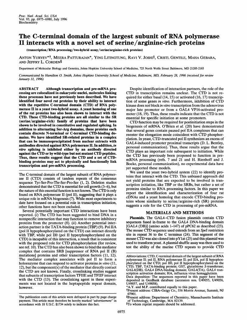
Proc. Natl. Acad. Sci. USAVol. 93, pp. 6975-6980, July 1996Biochemistry
The C-terminal domain of the largest subunit of RNA polymeraseII interacts with a novel set of serine/arginine-rich proteins
(transcription/RNA processing/two-hybrid assay/serine/arginine-rich proteins)
ANTON YURYEV*t, MEERA PATrURAJAN*, YING LITINGTUNG, RAVI V. JOSHIt, CRISTL GENTILE, MAHA GEBARA,AND JEFFRY L. CORDEN§Department of Molecular Biology and Genetics, Johns Hopkins University School of Medicine, 725 North Wolfe Street Baltimore, MD 21205-2185
Communicated by Hamilton 0. Smith, Johns Hopkins University School of Medicine, Baltimore, MD, February 28, 1996 (received for reviewJanuary 31, 1996)
ABSTRACT Although transcription and pre-mRNA pro-cessing are colocalized in eukaryotic nuclei, molecules linkingthese processes have not previously been described. We haveidentified four novel rat proteins by their ability to interactwith the repetitive C-terminal domain (CTD) of RNA poly-merase II in a yeast two-hybrid assay. A yeast homolog of oneof the rat proteins has also been shown to interact with theCTD. These CTD-binding proteins are all similar to the SR(serine/arginine-rich) family of proteins that have beenshown to be involved in constitutive and regulated splicing. Inaddition to alternating Ser-Arg domains, these proteins eachcontain discrete N-terminal or C-terminal CTD-binding do-mains. We have identified SR-related proteins in a complexthat can be immunoprecipitated from nuclear extracts withantibodies directed against RNA polymerase II. In addition, invitro splicing is inhibited either by an antibody directedagainst the CTD or by wild-type but not mutant CTD peptides.Thus, these results suggest that the CTD and a set of CTD-binding proteins may act to physically and functionally linktranscription and pre-mRNA processing.
The C-terminal domain of the largest subunit of RNA polymer-ase II (CTD) consists of tandem repeats of the consensussequence Tyr-Ser-Pro-Thr-Ser-Pro-Ser (1, 2). Deletion studiesdemonstrated that the ClD is essential for cell growth (3-6), butthe nature of this essential function is not known. The CID is onlyfound on RNA polymerase II (pol II), suggesting that it plays aunique role in mRNA biogenesis (7). While most experiments todate have focused on a potential role in transcription initiation,other functions have not been excluded.
Several potentially important CTD interactions have beenreported. (i) The CTD has been suggested to bind DNA in anonspecific interaction that may function to remove inhibitoryproteins from the promoter (8). (ii) Another potential inter-action partner is the TATA-binding protein (TBP) (9). Pol IIA(pol II hypophosphorylated on the CTD) can interact directlywith TBP, while pol IIO (pol II hyperphosphorylated on theCTD) is incapable of this interaction, a result that is consistentwith the proposed role for CTD phosphorylation (for review,see ref. 10). The CTD has also been shown to bind the mediatorcomplex that contains SRB [suppressor of RNA pol II (B)mutations] proteins and other transcription factors (11, 12).The mediator complex associates with pol II to form aholoenzyme that can respond to activator proteins in vitro (11,12), but the component(s) of the mediator that directly contactthe CTD are not known. Finally, crosslinking studies suggestthat subunits of transcription factors TFIIE and TFIIF interactwith the CTD (13). The crosslinking agent in these experi-ments was not located in the heptapeptide repeat domain,however.
Despite identification of interaction partners, the role of theCTD in transcription remains unclear. The CTD is not re-quired for either basalV(14, 15) or activated (16, 17) transcrip-tion of some genes in vitro. Furthermore, inhibition of CTDkinase does not block in vitro transcription from the adenovirusmajor late promoter or from a GAL4 VP16-activated pro-moter (18, 19). Thus, these results indicate that the CTD is notessential for specific initiation at some promoters.CTD function may be required for postinitiation steps in the
biogenesis of mRNA. O'Brien et al. (20) have demonstratedthat several genes contain paused pol IIA complexes that canreenter the elongation mode coincident with CTD phosphor-ylation. In yeast, CTD-truncated pol II synthesizes an excess ofGAL4-induced promoter proximal transcripts (D. L. Bentley,personal communication). Thus, these results argue that theCTD plays an important role subsequent to initiation. Whilethe CTD has previously been proposed to function in pre-mRNA processing (refs. 7 and 21 and H. Rienhoff and J.Boeke, personal communication), no experimental data haveyet supported these models.We used the yeast two-hybrid system (22) to identify pro-
teins that interact with the CTD. This unbiased approach didnot yield proteins that are expected to be involved in tran-scription initiation, like TBP or the SRBs, but rather a set ofproteins similar to RNA processing factors. In this paper wereport the identification and characterization of four ratcDNAs and a yeast homolog that encode CTD-binding pro-teins whose similarity to serine/arginine-rich (SR) proteinssuggests a role for the CTD in processing of pre-mRNA.
MATERIALS AND METHODSPlasmids. The GAL4-CTD fusion plasmids contain CTD
sequences fused in-frame to the GAL4 DNA-binding domain[GAL4 (DB)] (amino acids 1-147) of pPC62 as described (23).The mouse CTD sequence used extends from an SpeI restrictionsite in repeat 36 to the C terminus (24). This segment of themouse CTD was also cloned into pYlat (25) and this plasmid wasused to transform yeast. A plasmid shuffle assay was then used totest the ability of the murine CID repeats to provide CID
Abbreviations: CTD, C-terminal domain of the largest subunit ofRNApolymerase II; pol II, RNA polymerase II; pol IIA, pol II hypophos-phorylated on the CTD; pol IIO, pol II hyperphosphorylated on theCTD; TBP, TATA-binding protein; GST, glutathione S-transferase;GAL4(DB), GAL4 DNA-binding domain; GAL4(TA), GAL4 tran-scription activation domain; HA, influenza virus hemagglutinin.Data deposition: The sequences reported in this paper have beendeposited in GenBank database (accession nos. U49055, U49056,U49057, and U49058).*A.Y. and M.P. contributed equally to this paper.tPresent address: CIBA-Geigy Co., 556 Morris Avenue, Summit, NJ07901.tPresent address: Department of Chemistry, Massachusetts Instituteof Technology, Cambridge, MA 02139.§To whom reprint requests should be addressed.
6975
The publication costs of this article were defrayed in part by page chargepayment. This article must therefore be hereby marked "advertisement" inaccordance with 18 U.S.C. §1734 solely to indicate this fact.
Proc. Natl. Acad. Sci. USA 93 (1996)
function in yeast (25). Wild-type and mutant yeast CTD se-quences used to construct GAL4 fusions are as described (25).
Library Screening. The GAL4-mouse ClTD fusion was used toscreen a 14.5-day mouse embryo yeast two-hybrid library (23).Positive clones were isolated by two rounds of screening andretested by isolation of the library plasmid and retransformationinto yeast. Positive clones were further tested for their inability toactivate with a set of heterologous baits including RAG-1,RAG-2, DHL4, and yeast TYA or with the GAL4(DB) alone.RAG GAL4(DB) fusions were provided by Steve Desiderio(Johns Hopkins University) and expressed the RAG genes (26).The DHL4 fusion expresses a novel helix-loop-helix transcrip-tion factor and was provided by Greg Kato (Johns HopkinsUniversity). The yeast TYA fusion was obtained from C. B.Brachmann and J. D. Boeke (Johns Hopkins University).
Full-length cDNAs for the CMlD-binding proteins were isolatedfrom a rat hippocampus library [a gift from Anthony Lanahan(27)] using the short mouse clones as probe. Full-length or nearlyfull-length clones of rAi, rA4, rA8, and rA9 were sequenced.RNA Pol II Binding Assay. The CTD-binding domain of rAl
was cloned in pGEX (Pharmacia) and expressed as a glutathioneS-transferase (GST) fusion protein. Affinity beads containingeither the rAl-GST fusion or GST alone were prepared bypassing bacterial extracts over a glutathione-agarose column(Pharmacia). The columns retained rAl-GST at -1 mg/ml or
GST at 1.5 mg/ml. Yeast RNA pol11 (1.0 ,ug) was labeled withhemagglutinin (HA)-epitope-tagged cdc2 kinase and [,y-32P]ATP(28). The kinase in this reaction was retained on monoclonalantibody (mAb) 12CA5 beads during the reaction. Pol IIA wasprepared by labeling for 5 min with 10 ,uCi of ATP (3000Ci/mmol; 1 Ci = 37 GBq) to a stoichiometry of less than 1. In a
second reaction the labeled ATP was chased for an additional 5min with 0.2mM ATP. This chase is sufficient to shift the mobilityof the largest subunit to that expected for pol II0. Labeled pol IIwas removed from the kinase reaction by centrifugation of thekinase beads and incubated with 25 Al of rAl-GST or GST beadsin a final volume of 100 ,ul of binding buffer (0.1 M NaCl/10mMTris HCl, pH 7.9). After binding for 15 min at 23°C, the reactionmixture was centrifuged at 5000 x g for 30 s. The beads were thenwashed two more times with 1.0 ml of binding buffer. The finalpellet was resuspended in 2x SDS sample buffer, boiled for 90 s,electrophoresed on a 5% SDS gel, and autoradiographed. Vir-tually all of the labeled pol II was retained on the rAl-GSTcolumn. For peptide competition experiments, labeled yeast polIIA was incubated with rAl-GST or GST on beads but with theaddition of 10 ,tg of synthetic CTD peptide. After the bindingreaction, the reaction mixtures were diluted and washed as abovein the absence of peptide, and material was eluted and electro-phoresed on a 5% SDS gel.
Peptides used in this study are as follows: WT, (SPTSPSY)8;A2, (APTSPSY)8; A5, (SPTAPSY)8. Peptides were synthesizedfrom t-butoxycarbonyl-labeled amino acids and their concentra-tions were determined as described (29).
Immunoprecipitation of Pol II Complexes. Nuclear extractswere prepared from wild-type HeLa cells and from a HeLa straintransfected with an a-amanitin-resistant HA-epitope-taggedmouse pol II large subunit gene (M. L. West and J.L.C., unpub-lished results). Nuclear extract (50 ,ug) was incubated with 4 ,ugof purified anti-CUD mAb 8WG16 (14) or anti-HA-tag mAb12CA5 (30) in 500 ,ul of modified RIPA buffer (1 mM Tris HCl,pH 7.8/150 mM NaCl/2% Triton X-100/0.01% SDS/2 mMpepstatin/0.6 mM leupeptin/2 mM benzamidine hydrochlo-ride/i mM phenylmethylsulfonyl fluoride/2.5% BSA) at 4°C ona rotator for 4 hr. Protein A-Sepharose beads (20 ,ul, Pharmacia),equilibrated with modified RIPA buffer, was added to the abovemixture and further incubated at 4°C on a rotator for 1 hr. Beadswere then washed three times with 1 ml of ice-cold modifiedRIPA buffer for 30 min. The final bead pellet was extracted inSDS sample buffer and subjected to SDS/PAGE followed byWestern blot. The blots were probed with anti-SR protein mAb
16H3 (31) and the immunoreactive proteins were detected usingECL (Amersham).
In Viro Splicing. Capped and uniformly labeled [32P]E1-IVS-E2 RNA was synthesized from pPIP85 (32) using T7 RNApolymerase (Promega). The RNA product was separated on adenaturing 8% polyacrylamide gel and eluted by crushing the geland incubating with elution buffer containing 0.3 M sodiumacetate, 1/3 vol of phenol, and tRNA (20 ,ug/ml) for 2 hr at 30°Con a rotator. The eluted RNA was separated from gel pieces bya Millipore Ultrafree-MC filter unit (0.45 ,um), precipitated using2 vol of ethanol, and dissolved in nuclease-free water (20-50 ,ul).Splicing reactions (12.5 ul) containing uniformly labeled pre-mRNA (1 to 5 x 10⁵ cpm) were performed at 30°C in a solutionof 24% HeLa nuclear extract (Promega), 2 mM MgCl2, 60 mMKCl, 1 mM ATP, 20 mM creatine phosphate, and RNasin(Promega) at 1 unit/ml (32).. After incubation for 2 hr, RNAswere extracted and subjected to denaturing 15% polyacrylamidegel electrophoresis fQllowed by autoradiography.
RESULTSDesign of the Two-Hybrid Screen. Initial attempts at using the
CT'D as bait in the two-hybrid system failed because the CTDitself acts as an activator when fused to a DNA-binding domain.Several groups (33, 34) have reported similar observations andhave argued that the ability to activate transcription is an impor-tant CTD function. We constructed a series ofGAL4(DB)-CDfusions as potential bait, including a series of mutant CTDs thathad been tested for their ability to function in place of thewild-type CTD (25). Table 1 describes the C1TD fusions tested foractivation. The ability to activate transcription in a one-hybridassay does not correlate with the ability to sustain viability whenin the context of the pol II largest subunit gene in yeast. All of thephosphorylation site mutant CTDs activate, although they do notsupport viability. Transcription activation provided byGAL4(DB)-CTD fusion proteins is not an essential function ofthe CUlD because the 16 most distal repeats of the mouse CUDare functional in the context of yeast pol II but do not activatetranscription as a GAL4(DB) fusion. Phosphorylation of theC`1'D fusion proteins in vivo (data not shown) apparently createsan acidic activation domain. Multiple positively charged aminoacids in the nonconsensus part of mouse CTD may neutralize thisacidic activation potential. The failure of the distal repeats of themouse CTD to activate allowed us to use this domain as bait ina two-hybrid screen.
Isolation of CTD-Interacting Clones. We screened a mouseembryo cDNA two-hybrid library (23) and isolated 46 positiveclones out of 1.4 x 10⁶ colonies. None of these positive clonesinteracted with a series of control proteins described above.Positive clones were sequenced and placed into groups based on
Table 1. Activation and RNA pol II functions of CTD mutants
Activation in CTD functionCTD mutant one-hybrid assay in yeast pol II
WT yeast CTD + +(YAPTSPS)18 + -
(YEPTSPS)15 + -
(YSPTAPS)15 + -
(YSPTEPS)18 + -
(FSPTSPS)10 + -
(YAPTAPS)10 + -
(YPSTSPS)15 + -
Mouse CTD - +
Series of mutant CTDs shown were cloned and tested either for theirability to activate transcription as GAL4-(DB) fusions or for theirability to replace the wild-type CTD as part of the RPBI gene in yeast(25). The sequence of the repeated heptapeptide is given withinparentheses with the mutated residue(s) indicated in boldface type.The number of such repeats is noted outside the parentheses. Themouse CTD construct contains repeats 36-52.
6976 Biochemistry: Yuryev et al.
Proc. Natl. Acad. Sci. USA 93 (1996) 6977
sequence similarity. The largest group consists of 35 clonescontaining four related sequences (mAl, mA5, mA7, and mA9).The second group of positive clones consists of two relatedsequences (mA4 and mA8) that are distinct from the first group.Mouse A series clones were also tested by reversing the two-hybrid partners. In this arrangement the CID is fused to a GAL4transcription activation domain [GAL4(TA)] and the mouse Aseries clone is fused to the GAL4(DB). In this orientation boththe mAl and mA8 clones were shown to interact with both mouseand yeast CTDs, indicating that their CTD-binding domains alsorecognize the consensus C(LD.
Six of the 46 positive clones did not fall into the above groups.These clones contain three different sequences encoding proteinsthat interacted poorly with the CLTD when tested as GAL4(DB)fusions. We have not pursued these clones further. Notably, thisset does not contain TBP, SRBs, or any general transcriptionfactor. We tested GAL4(DB)-TBP fusions (full-length humanTBP or the N- or C-terminal halves) for their ability to interactwith the C1FD. No CTD interaction was observed (data notshown), suggesting that the ClTD and TBP do not interact in vivo.The positive mouse CFD-interacting clones that represent thefirst two interacting groups were selected for further study.
Full-Length CTD-Binding Proteins. Using the short mousetwo-hybrid clones as probes, we were able to isolate full-lengthclones for four different (CLD-interacting proteins and havenamed these rat genes rAl, rA4, rA8, and rA9. The rat cloneswere retested in the two-hybrid system and all four activated in aCLTD-dependent fashion (data not shown). We have sequencedall four clones and determined that they are novel cDNAsencoding proteins that are similar to SR proteins (35, 36).
Fig. 1A shows the sequence organization of four rat CTD-binding proteins. One common feature, the RS (Arg-Ser) do-main, contains a concentration of Arg-Ser diamino acid se-quences similar to that seen in SR proteins known to be involvedin splicing (Fig. IB). These RS domains also have related diaminoacid sequences conforming to the general consensus Arg(Lys)-Ser(Thr, Asp, Glu). When the Ser (or Thr) residues are phos-phorylated (35, 36), all of these diamino acid sequences displayalternating positive and negative charges.Two different C(TD-binding domains are represented in this set
of four proteins. rAl and rA9 contain a very similar "80-aminoacid sequence that is shown in Fig. 1C. This conserved region liesat the C terminus and is nearly identical to the mouse clones(mAl and mA9, respectively) used as the probe to identify the ratclones. We have not yet isolated a rat clone that is the equivalentof the mouse A5 clone. Several experiments suggested that theconserved "80-amino acid sequence is the CLT-binding domain.(i) Deletion of sequences to either side of this homology regionin the mouse mAl clone did not eliminate interaction with theC'LD in the two-hybrid assay (Fig. 1A, regions a and b). (ii) Wehave cloned a pair of rA9 splice variants that contain differentC-terminal sequences and no (ClD-binding homology region.These clones do not interact with the CID in the two-hybrid assay(data not shown).rA4 and rA8 contain similar N-terminal CLTD-interacting
domains that are different than the rAl/A9 CTD-binding se-quence. The conserved - 120-amino acid homology regions fromrA4 and rA8 that are shown in Fig. 1D aligned with the sequencesof the original mouse clones mA4 and mA8, respectively. Thesequence of the longest rA4 clone differs from its mouse homo-logue in that a 50-bp segment is deleted, causing a frame shift inthe CI'D-binding domain (Fig. ID). Conservation of the partialrA4 C1TD-binding sequence and the existence of a long openreading frame 3' of the deletion suggests that this cDNA derivesfrom an active gene. As was observed for rA9, we suspect that thedeletion of sequences in rA4 results from alternative splicing.rA4 and rA8 contain other sequence motifs that are common
to RNA-binding proteins. Immediately downstream of the RSdomain both proteins contain a similar RNA recognition motif(37). Database searches using the program BLAST (38) indicate
RS CTD-binding domain A[li i i i l l ~ -l i l i T -rr iI TI-IIIIIIIIIIIIIT 1 P1rAlA
\ RS\_k- vs- IL IL 1%1. rA9rCTD-binding domain - ,
rA8N QS F3RM RD
N- NzzXq-r---sy2z..r zz rA4B. Q (POPGM)16 RS RRM RDU2AF FMHLSKPMELRRELYRRRKXHRRt:RG<;GRGRGGGGGGGRERDRR=DRE32rAl GDPAPPDSPTWEAKKERLGSESTLASTARRRRRmG.RRGRrSHM EKRRRRRM-LA9 RASSES_XTKR TKYARCJLEHC*R3ST DGMR wMVSPFTEEH}TKRMHUXTCrA8 rHEPR KitSRK YSSER SRAREREKERQKKG;LPrA4 QEMEVEQPCVTEVXHP VPEKtPKRR0 RkH.SEMRDRRRHSPMQERRC.
n H EVJAK1.WHC KIi4iS 3IEI]VSQ NrAli5 TM.PEL- Q Y I Q 6~ialleSF>KDIM SVZS I R1Mrr.A5 .1 1s;s :rA9 3M e S I3 KM-D.
r,A4DISIi'l igV,11 I
^2361 nosse' FitD
rA4 5 TASESE]RJLsHPQrA4 TsEYL 8S'.-. A-SA ISDiFcGD SX4'IPPQS-AB I HD. I PP
E.rAs =LWMVDUUBQQtTeEE3P2BESIN3IP EhS3CAYVCMVHROQrS1.nLOKLSl-A4 LWMQ DCTaQQDVASLEE ES3IbrA P P3CAYIVHVHRQ 3YRMLQKLSUl 70K KLF&;VNYDTESReRREDEYYKRIHj2VYSlRSGKPlYAjIEYEHEB3MHSjYKHADSAP49A AKYES ->PL2WEMLQAVV jP DRVTGQHQYGzVEFLSEEMD. IKIMNNGR1 jVGVPKTMFQ RSrCP NVRNGK NCEFVKFEKRIDAZASIQGLQGFPUB1 A1YI3N1PHFAjAn&IPEUQtNMFB5LFK)SYP EK3CC5IKYDTHEQ AVCIVALAPABP I;N3YN-TE.TE3EFS e-GQgETSLSLVKDQ NDKPMEFG-HYlANHECnQKQVDELN
RNP-2 RNP-
FIG. 1. Sequence alignments of rat CTD-binding proteins. (A)Sequence organization of rat CTD-binding proteins rAl, rA9, rA4, andrA8. The following domains are indicated; RS, Arg/Ser rich; RRM,RNA recognition motif; RD, Arg/Asp rich; Q, Gln rich; (PQPGM),repeated pentapeptide in rA4. Lines under the rAl and rA8 clonesmarked a, b, and c represent the smallest mouse clones that interactwith the CTD in the two-hybrid assay. The sizes of the expected ratproteins are as follows: rAl, 129 kDa; rA9, 146 kDa; rA8, 144 kDa;rA4, 130 kDa. The rA4 protein size is based on a fusion of mouse andrat sequences as the longest rat cDNA clone contains a short deletionthat leads to a frame shift in the CTD-binding domain. (B) ConservedRS domains. The RS domains of rAl, rA9, rA4, and rA8 are comparedwith a previously identified SR protein (U2AF, GenBank accession no.X64044). Black-boxed residues represent Arg-Ser and Ser-Arg dipep-tides. The SR proteins alignments were taken from the output of BLASTsearches. Because of the repetitive nature of the homology, multiplealignments are possible. (C) rA1/A9 CTD-binding domain. The ratsequences are aligned with the homologous mouse clones and con-served residues are in black boxes. (D) rA4/A8 CTD-binding domain.The rat clones are aligned with the homologous mouse clones andconserved residues are -in black boxes. (E) rA4/A8 RNA recognitionmotifs. The RNA recognition motifs of rA4 and rA8 are aligned witha set of related RNA-binding proteins (accession nos. in parentheses)including the human Ul 70-kDa (S03048) and SAP49A (L35013)splicing factors and yeast RNA-binding proteins Ngrlp (P32831),Publp (P32588), and Pablp (P04147). Black-boxed residues areidentical among at least four of the sequences. In addition to theseresidues, there are many similarities that are not boxed. The positionsof the two motifs, RNP-1 and RNP-2 that make up the RRM areunderlined.
that rA4 and rA8 are related to a number of RNA-bindingproteins (Fig. 1E), some ofwhich, like the Ul 70-kDa protein, areinvolved in splicing (39). The strongest region of homology toother RNA-binding proteins is in the RNP-2 motif and adjacentsequences. The similarity to the RNP-1 motif is not as strong. Inaddition to the RS and RRM motifs, both rA4 and rA8 containglutamine-rich regions at their N termini and a motif rich inArg-Asp diamino acid sequences (RD) toward the C terminus.rA4 is unique in having a pentapeptide sequence (Pro-Gin-Pro-Gly-Met) that is repeated 16 times.AYeast CTD-Binding Protein Homolog. BLAST searches on the
National Center for Biotechnology Information network (37)
Biochemistry: Yuryev et al.
11
Proc. Natl. Acad. Sci. USA 93 (1996)
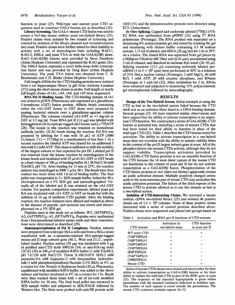
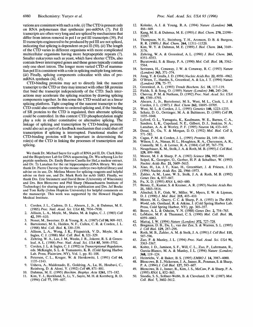
revealed a sequence related to rA4/A8. The yeast NRDI gene,identified as a suppressor of an intronic mutation, encodes aprotein that is involved in transcription elongation or the earlieststages of splicing (E. Steinmetz and D. Brow, personal commu-nication). NRDJ contains several regions of homology to the rA8protein (Fig. 2A) including the CTD-binding domain, alternatingArg motif, and RRM motif. In addition to RE repeats, NRD1contains several Ser-Arg diamino acid sequences.The N-terminal residues of NRDI were fused to the
GAL4(TA) and tested for interaction with the mouse CTlD in thetwo-hybrid assay. Fig. 2B shows that the NRD1 CTD-bindingfusion is able to interact with the CTD, although the signal is notas strong as seen with rA8. This weaker signal may be due tocompetition with endogenous Nrdlp or to a less efficient inter-action with the nonconsensus murine heptapeptides.rAl-CTD Direct Interaction. The CTD-binding domain ofrAl
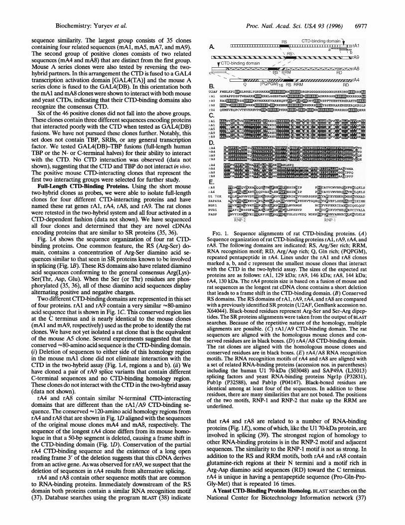
was expressed as a GST fusion protein and binding to pol II wasassayed. Fig. 3A demonstrates that the rAl-GST fusion proteinbinds quantitively to yeast pol IIA and pol II0. The interaction canbe substantially competed by a wild-type CTD peptide and lesseffectively by mutant CTD peptides (Fig. 3B). Thus, these resultsindicate that the rAl CTD-binding domain interacts directly withthe CTD. Although both IIA and II0 were bound by the rAl-GST fusion protein, we cannot rule out the possibility thatphosphorylation of the CTD is required for interaction as our IIAwas phosphorylated on the CTD to a substoichiometric level.RNA Pol II-SR Protein Complex. Pol II complexes from HeLa
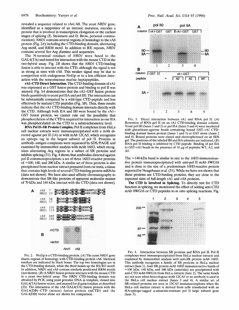
cell nuclear extracts were immunoprecipitated with a mAb di-rected against pol 11 (14) or with mAb 12CA5, which recognizesan epitope tag in the largest subunit of pol II. Proteins inantibody-antigen complexes were separated by SDS/PAGE andexamined by immunoblot analysis with mAb 16H3, which recog-nizes alternating Arg regions in a subset of SR proteins andinhibits splicing (31). Fig. 4 shows that antibodies directed againstpol11 coimmunoprecipitate a set of three 16H3-reactive proteinsof 100, 140, and 200 kDa. A similar set of three proteins is alsoprecipitated from nuclear extract prepared from rat testis, a tissuethat contains high levels of several CTD-binding protein mRNAs(data not shown). We have also used affinity chromatography todemonstrate that SR-like proteins of apparent molecular massesof 70 kDa and 140 kDa interact with the CTD (data not shown).
A rA8 7 JjNSEMYLNMY3PPEKAKMTQI&C.AQNRD1 10 VATKFKSLaTGSRIYCLJ3TYrA8NRD1
rA8NRDI
5 4 KCPEY3VPGRM57 3PDSHa3LGS 5IMM105 MD[SZ3ReLR119 SHE M IRSGL
A p° 110oolumn rA1-GST
*.......
110-
column GST IFpeptide
IIA-
pol IIAGS-II rAl-GST GST I
-150-IIA
42
rAl-GST- r2 n rwn
1 2 3 4 5
FIG. 3. Direct interaction between rAl and RNA pol II. (A)Retention of RNA pol II on an rAl CTD-binding domain column.Yeast pol 110 (lanes 1 and 2) or pol IIA (lanes 3 and 4) were incubatedwith glutathione-agarose beads containing bound GST-rAl CTD-binding domain fusion protein (lanes 1 and 3) or GST alone (lanes 2and 4). Bound proteins were eluted and electrophoresed on an SDSgel. The positions of the labeled IIO and IIA subunits are indicated. (B)RNA pol II binding is inhibited by CTD peptide. Binding of pol IIAto GST-rAl beads in the presence of 10 ,tg of peptides WT, A2, andA5.
The -140-kDa band is similar in size to the 16H3-immunoreac-tive protein immunoprecipitated with anti-pol II mAb 8WG16and is close to the size of a predominant 16H3-reactive proteinreported by Neugebauer et al. (31). While we have not shown thatthese proteins are CTD-binding proteins, they are close to theexpected sizes of full-length rAl and rA8 proteins.The CTD Is Involved in Splicing. To directly test for CTD
function in splicing, we monitored the effect of adding anti-CTDmAb 8WG16 or CTD peptides to in vitro splicing reactions. Fig.
1 2 3 4 5
200-
rA8 449 KKGLPPVMNRD1 246 -m:F- NSRRNRSZ
BmCTD e
68~
vector
Nrdl rA8
FIG. 2. Nrdlp is a CTD-binding protein. (A) The yeast NRD1 geneshares regions of homology with CTD-binding protein rA8. Identicalresidues are indicated by black boxes. The top two homologies are inthe CTD-binding domain, while the third makes up the RS/RE motif.In addition, NRDI and rA8 contain similarly positioned RRM motifs(not shown). (B) ANRD1 fusion protein interacts with the mouse CTDin a yeast two-hybrid assay. The NRDI CTD-binding domain wasobtained by PCR, using yeast genomic DNA as template, cloned intoGAL4(TA) fusion vector, and assayed for 13-galactosidase as described(23). The interaction of the rA8 GAL4(TA) fusion protein with theGAL4(DB)-CTD (mouse) fusion protein (mCTD) and theGAL4(DB) vector alone are shown for comparison.
FIG. 4. Interaction between SR proteins and RNA pol II. Pol IIcomplexes were immunoprecipitated from HeLa nuclear extracts andexamined by immunoblot analysis with anti-SR protein mAb 16H3.This antibody recognizes a family of SR proteins in HeLa nuclearextract (lane 1). Anti-SR protein mAb 16H3 immunoreactive bands of-100 kDa, 140 kDa, and 180 kDa (asterisks) are precipitated withanti-CTD mAb 8WG16 from HeLa extracts (lane 2). The same bandsare not seen when heterologous mAb 12CA5 or no antibody is used inthe HeLa cell nuclear extract (lanes 3 and 4). A similar set ofSR-related proteins are seen in 12CA5 immunocomplexes when theHeLa cell nuclear extract is derived from cells transfected with anHA-epitope-tagged a-amanitin-resistant pol II large subunit gene(lane 5).
6978 Biochemistry: Yuryev et al.
.t
i,IlllllkiW:97- .. II.
Proc. Natl. Acad. Sci. USA 93 (1996) 6979
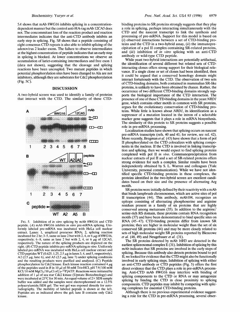
5A shows that mAb 8WG16 inhibits splicing in a concentration-dependent manner but the control anti-HA-tag mAb 12CA5 doesnot. The concommitant loss of the reaction product and reactionintermediates indicates that the anti-CTD antibody inhibits an
early step in splicing. Fig. 5B shows that a peptide consisting ofeight consensus CTD repeats is also able to inhibit splicing of theadenovirus 2 leader exons. The failure to observe intermediatesat the highest concentration of peptide indicates that an early stepin splicing is blocked. At lower concentrations we observe an
accumulation of lariet-containing intermediates and free exon 1(data not shown), suggesting that the cleavage and splicingreactions have been uncoupled. Two mutant peptides in whichpotential phosphorylation sites have been changed to Ala are notinhibitory, although they are substrates for Cdc2 phosphorylation(Fig. SC).
DISCUSSIONA two-hybrid screen was used to identify a family of proteinsthat interact with the CTD. The similarity of these CTD-
A
1 2 3 4 5 6 7 8
B Gl..J;J;] peptide
DJ-u*.i... .. .......... 40 W dl-
_m~~~~~CD -E
1 2 3 4 5 6 7
c
......
Wr A2 A5 B
FIG. 5. Inhibition of in vitro splicing by mAb 8WG16 and CTD
peptide. (A) mAb 8WG16 inhibits in vitro pre-mRNA splicing. Uni-
formly labeled pre-mRNA was incubated with HeLa cell nuclear
extract. Lanes: 1, unspliced precursor RNA; 2, splicing reaction
incubated for 2 hr; 3-5, same as lane 2 but with 2,4, or 6,ug of 8WG16,
respectively; 6-8, same as lane 2 but with 2, 4, or 6 jig of 12CA5,
respectively. The nature of the splicing products are depicted on the
right. (B) CTD peptide inhibits pre-mRNA splicing in vitro. Uniformly
labeled pre-mRNA was incubated with HeLa cell nuclear extract and
CTD peptides WT (0.625, 1.25,2.5,ug in lanes 3,4, and 5, respectively),
A2 (2.5 Aig, lane 6), and A5 (2.5,ug, lane 7) under splicing conditions
and the resulting products were purified and analyzed. (C) Peptide
phosphorylation by Cdc2 kinase. Each kinase reaction contained 4,ugof the peptides used in B in 10,ul of 50 mM Tris-HCl, pH 7.9/100 mM
KCl/10mM MgCl2/10,uCi of [,y-32P]ATP. Reactions were initiated by
addition of 1,ul of sea star Cdc2 kinase (Upstate Biotechnology) and
were incubated at23°C for 30 min. An equal volume of 2x SDS sample
buffer was added and the samples were electrophoresed on a 12.5%
polyacrylamide/SDS gel. The wet gel was exposed directly for auto-
radiography. The mobility of labeled peptide is shown at the left.
Peptides are as indicated above the gel; lane B contains only Cdc2
kinase.
binding proteins to SR proteins strongly suggests that they playa role in splicing, perhaps interacting simultaneously with theCTD and the nascent transcript to link the synthesis andprocessing of pre-mRNA. Support for this model is based on(i) observed interactions between a set of CTD-binding pro-teins and the CTD in a two-hybrid assay, (ii) the immunopre-cipitation of a pol II complex containing SR-related proteins,and (iii) inhibition of in vitro splicing with an anti-CTDantibody or wild-type CTD peptide.
While yeast two-hybrid interactions are potentially artifactual,the identification of several different but related sets of CTD-interacting clones offers strong support for their biological rele-vance. If a single clone or set of clones had been identified, thenit could be argued that a conserved homology domain mightinteract fortuitously with the CTD. The observation of two setsof CTD-binding domains, both contained in mammalian SR-likeproteins, is unlikely to have been obtained by chance. Rather, theoccurrence of two different CTD-binding domains strongly sup-ports the biological importance of the CTD interaction. Thepresence of one of these CTD-binding domains in the yeast NRD1gene, which contains other motifs in common with SR proteins,argues for the evolutionary conservation of CUD-binding pro-teins. While little is known about NRD1, its identification as asuppressor of a mutation located in the intron of a selectablemarker gene suggests that it plays a role in mRNA biosynthesis.The similarity of this protein to SR proteins suggests a possiblerole in pre-mRNA processing.
Localization studies have shown that splicing occurs on nascentpre-mRNA transcripts (refs. 40 and 41; for review, see ref. 42).More recently, Bregman et al. (43) have shown that a form of polII phosphorylated on the CTD colocalizes with splicing compo-nents in the nucleus. If the CTD is involved in linking transcrip-tion and splicing, then we would expect to find splicing proteinscomplexed with pol II in vivo. Coimmunoprecipitation fromnuclear extracts of polII and a set of SR-related proteins offersstrong evidence for such a complex. Similar results have beenindependently obtained by S. L. Warren and colleagues (YaleUniversity, personal communication). While we have not iden-tified specific CTD-binding proteins in these complexes, theproteins identified in the two-hybrid screen are excellent candi-dates based on their size and the presence of alternating Argmotifs.SR proteins were initially defined by their reactivitywith a mAb
that binds lampbrush chromosomes, which are active sites of polII transcription (44). This antibody, mAblO4, recognizes anepitope consisting of alternating phosphoserine and arginineresidues present in a family of six proteins that are highlyconserved among metazoans (35). In addition to the arginine/serine-rich RS domain, these proteins contain RNA recognitionmotifs (37) and have been demonstrated to bind specific sites onRNA (45-47). CTD-binding proteins differ from known SRproteins; they are higher in molecular weight than the set of sixconserved SR proteins (44) and may be more closely related tosets of high molecular weight SR proteins reported by Blencoweet al. (48, 49) and Neugebauer et al. (31).The SR proteins detected by mAb 16H3 are detected in the
earliest spliceosomal complexE (31). Inhibition of splicing by thismAb indicates that SR proteins are involved in the early steps ofsplicing. Because this antibody also detects proteins bound to polII, we looked for evidence that the CTD might also be functionallyinvolved in early splicing steps. Inhibition of splicing with eitheran anti-CTD antibody or CTD peptides (Fig. 5) offers the firstdirect evidence that the CTD plays a role in pre-mRNA process-ing. Anti-CTD mAb 8WG16 may interfere with binding ofsplicing components to theCTD or RNA or may antagonizesplicing by binding the CTD in close proximity to splicingcomponents. CTDpeptides may inhibit by competing with splic-ing complexes for essential CTD-binding proteins.Although there is no previous experimental evidence suggest-
ing a role for the CTD in pre-mRNA processing, several obser-
niAb16
.
bd
Biochemistry: Yuryev et al.
Proc. Natl. Acad. Sci. USA 93 (1996)
vations are consistent with such a role. (i) The CTD is present onlyon RNA polymerases that synthesize pre-mRNA (7). Pol IItranscripts are often very long and are spliced by mechanisms thatdiffer from intron removal in pol I or pol III transcripts (39). PolII transcripts engineered to be produced by pol III are not spliced,indicating that splicing is dependent on pol 11 (50). (ii) The lengthof the CTLD varies in different organisms with more complicatedmulticellular organisms having more heptapeptide repeats (7).Smaller eukaryotes such as yeast, which have shorter CTDs, alsocontain fewer interrupted genes and these genes typically containonly one short intron. The longer more varied CTD of mamma-lian pol II is consistent with a role in splicing multiple long introns.(iii) Finally, splicing components colocalize with sites of pre-mRNA synthesis (42, 43).
CT'D-binding proteins may act to directly link the nascenttranscript to the CTD or they may interact with other SR proteinsthat bind the transcript independently of the CTD. Such inter-actions may accelerate the splicing reaction by drawing splicingcomponents together. In this sense the CTD would act as a linearsplicing platform. Tight coupling of the nascent transcript to theCTD could also contribute to ordered splicing and, if the bindingof SR proteins to the CTD is regulated, then alternate splicingcould be controlled. In this context CTD phosphorylation mightplay a role in either constitutive or altemative splicing. Thelinkage of splicing and transcription by CID-binding proteinscould also act as part of a feedback mechanism that could shut offtranscription if splicing is interrupted. Functional studies ofCTlD-binding proteins will be important in determining therole(s) of the CTD in linking the processes of transcription andsplicing.
We thank Dr. Michael Sayre for a gift of RNA pol II, Dr. Clark Rileyand the Biopolymer Lab for DNA sequencing, Dr. Wu-schyong Liu forpeptide synthesis, Dr. Emily Barron-Casella for HeLa nuclear extract,and Dr. Ty Lanahan for the rat hippocampus cDNA library. We alsothank Dr. Pierre Chevray for the mouse embryo two-hybrid library andadvise on its use, Dr. Melissa Moore for splicing reagents and helpfuladvise on their use, and Dr. Mark Roth for mAb 16H3. Finally, wethank Drs. Eric Steinmetz and David Brow (University of Wisconsin)and Drs. Ben Blencowe and Phil Sharp (Massachusetts Institute ofTechnology) for sharing data prior to publication and Drs. Jef Boekeand Tom Kelly (Johns Hopkins University) for helpful comments onthe manuscript. This work was supported by the Howard HughesMedical Institute.
1. Corden, J. L., Cadena, D. L., Ahearn, J., Jr., & Dahmus, M. E.(1985) Proc. Natl. Acad. Sci. USA 82, 7934-7938.
2. Allison, L. A., Moyle, M., Shales, M. & Ingles, C. J. (1985) Cell42, 599-610.
3. Nonet, M., Sweetser, D. & Young, R. A. (1987) Cell 50,909-915.4. Bartolomei, M. S., Halden, N. F., Cullen, C. R. & Corden, J. L.
(1988) Mol. Cell. Biol. 8, 330-339.5. Allison, L. A., Wong, J. K., Fitzpatrick, V. D., Moyle, M. &
Ingles, C. J. (1988) Mol. Cell. Biol. 8, 321-329.6. Zehring, W. A., Lee, J. M., Weeks, J. R., Jokerst, R. S. & Green-
leaf, A. L. (1988) Proc. Natl. Acad. Sci. USA 85, 3698-3702.7. Corden, J. L. & Ingles, C. J. (1992) in Transcriptional Regulation,
eds. McKnight, S. L. & Yamamoto, K. R. (Cold Spring HarborLab. Press, Plainview, NY), Vol. 1, pp. 81-108.
8. Peterson, C. L., Kruger, W. & Herskowitz, I. (1991) Cell 64,1135-1143.
9. Usheva, A., Maldonado, E., Goldring, A., Lu, H., Houbavi, C.,Reinberg, D. & Aloni, Y. (1992) Cell 69, 871-881.
10. Dahmus, M. E. (1995) Biochim. Biophys. Acta 1261, 171-182.11. Kim, Y. J., Bjorklund, S., Li, Y., Sayre, M. H. & Kornberg, R. D.
(1994) Cell 77, 599-607.
12. Koleske, A. J. & Young, R. A. (1994) Nature (London) 368,466-469.
13. Kang, M. E. & Dahmus, M. E. (1995) J. Biol. Chem. 270, 23390-23397.
14. Thompson, N. E., Steinberg, T. H., Aronson, D. B. & Burgess,R. R. (1989) J. Biol. Chem. 264, 11511-11520.
15. Kim, W. Y. & Dahmus, M. E. (1989) J. Biol. Chem. 264, 3169-3176.
16. Zehring, W. A. & Greenleaf, A. L. (1990) J. Biol. Chem. 265,8351-8353.
17. Buratowski, S. & Sharp, P. A. (1990) Mol. Cell. Biol. 10, 5562-5564.
18. Serizawa, H., Conaway, J. W. & Conaway, R. C. (1993) Nature(London) 363, 371-374.
19. Jiang, Y. & Gralla, J. D. (1994) NucleicAcids Res. 22, 4958-4962.20. O'Brien, T., Hardin, S., Greenleaf, A. & Lis, J. T. (1994) Nature
(London) 370, 75-77.21. Greenleaf, A. L. (1993) Trends Biochem. Sci. 18, 117-119.22. Fields, S. & Song, 0. (1989) Nature (London) 340, 245-246.23. Chevray, P. M. & Nathans, D. (1992) Proc. Natl. Acad. Sci. USA
89, 5789-5793.24. Ahearn, J., Jr., Bartolomei, M. S., West, M. L., Cisek, L. J. &
Corden, J. L. (1987) J. Biol. Chem 262, 10695-10705.25. West, M. L. & Corden, J. L. (1995) Genetics 140, 1223-1233.26. Schatz, D. G., Oettinger, M. A. & Baltimore, D. (1989) Cell 59,
1035-1048.27. Lyford, G. L., Yamagata, K., Kaufmann, W. E., Barnes, C. A.,
Sanders, L. K., Copeland, N. E., Gilbert, D. J., Jenkins, N. A.,Lanahan, A. A. & Worley, P. F. (1995) Neuron 14, 433-445.
28. Desai, D., Gu, Y. & Morgan, D. 0. (1992) Mol. Biol. Cell 3,571-582.
29. Cagas, P. M. & Corden, J. L. (1995) Proteins 21, 149-160.30. Wilson, I. A., Niman, H. L., Houghten, R. A., Cherenson, A. R.,
Connolly, M. L. & Lerner, R. A. (1984) Cell 37, 767-778.31. Neugebauer, K. M., Stolk, J. A. & Roth, M. B. (1995) J. Cell Biol.
129, 899-908.32. Moore, M. J. & Sharp, P. A. (1992) Science 256, 992-994.33. Seipel, K., Georgiev, O., Gerber, H. P. & Schaffner, W. (1993)
Nucleic Acids Res. 21, 5609-5615.34. Xiao, H., Lis, J. T., Xiao, H., Greenblatt, J. & Friesen, J. D.
(1994) Nucleic Acids Res. 22, 1966-1973.35. Zahler, A. M., Lane, W. S., Stolk, J. A. & Roth, M. B. (1992)
Genes Dev. 6, 837-847.36. Fu, X. D. (1995) RNA 1, 663-680.37. Birney, E., Kumar, S. & Krainer, A. R. (1993) Nucleic Acids Res.
21, 5803-5816.38. Altschul, S. F., Gish, W., Miller, W., Myers, E. W. & Lipman,
D. J. (1990) J. Mol. Biol. 215, 403-410.39. Moore, M. J., Query, C. C. & Sharp, P. A. (1993) in The RNA
World, eds. Gestland, R. & Atkins, J. (Cold Spring Harbor Lab.Press, Cold Spring Harbor, NY), pp. 303-357.
40. Beyer, A. L. & Osheim, Y. N. (1988) Genes Dev. 2, 754-765.41. LeMaire, M. F. & Thummel, C. S. (1990) Mol. Cell. Biol. 10,
6059-6063.42. Mattaj, I. W. (1994) Nature (London) 372, 727-728.43. Bregman, D. B., Du, L., van der Zee, S. & Warren, S. L. (1995)
J. Cell Biol. 129, 287-298.44. Roth, M. B., Zahler, A. M. & Stolk, J. A. (1991) J. Cell Biol. 115,
587-596.45. Zuo, P. & Manley, J. L. (1994) Proc. Natl. Acad. Sci. USA 91,
3363-3367.46. Kohtz, J. D., Jamison, S. F., Will, C. L., Zuo, P., Luhrmann, R.,
Garcia-Blanco, M. A. & Manley, J. L. (1994) Nature (London)368, 119-124.
47. Heinrichs, V. & Baker, B. S. (1995) EMBO J. 14, 3987-4000.48. Blencowe, B. J., Nickerson, J. A., Issner, R., Penman, S. & Sharp,
P. A. (1994) J. Cell Biol. 127, 593-607.49. Blencowe,-B. J., Issner, R., Kim, J. S., McCaw, P. & Sharp, P. A.
(1995) RNA 1, 852-865.50. Sisodia, S. S., Sollner-Webb, B. & Cleveland, D. W. (1987) Mol.
Cell. Biol. 7, 3602-3612.
6980 Biochemistry: Yuryev et al.
Copyright © 2022 FDOKUMEN