The Bulletin of BISMiS
57
The Bulletin of BISMiS Published by Bergey’s International Society for Microbial Systematics Volume 7, part 2 – December 2018
-
Upload
khangminh22 -
Category
Documents
-
view
3 -
download
0
Transcript of The Bulletin of BISMiS

The Bulletin of BISMiS
Published by Bergey’s International Society for Microbial Systematics
Volume 7, part 2 – December 2018

Published by Bergey’s International Society for Microbial Systematics
ISSN 2159-287X
On the cover
Picture of delegates at the BISMiS 2018 International Conference
Editorial Board
Editor-in-Chief: Paul A. Lawson, University of Oklahoma, Norman, OK, USA
Associate Editors: Vartul Sangal, Northumbria University, United Kingdom
Amanda L. Jones, Northumbria University, United Kingdom
Jang-Cheon Cho, Inha University, Republic of Korea
Managing Editor: Shannon R. Fulton, University of Oklahoma, Norman, OK, USA
Officers of BISMiS
President:IainSutcliffe
President-Elect: Kamlesh Jangid
Secretary: Wen-Jun Li
Treasurer: Brian P. Hedlund
Copyright
The copyright in this publication belongs to Bergey’s International Society for Microbial Systematics (BISMiS). All rights reserved. This work may not be translated or copied in whole or in part without the written permission of the publisher (BISMiS), except for brief excerpts in connection with reviews or scholarly analysis. Use in con-nection with any form of information storage and retrieval, electronic adaptation, computer software, or by similar or dissimilar methodology now known or hereafter developed is forbidden. The use in this publication of tradenames,trademarks,servicemarks,andsimilarterms,eveniftheyarenotidentifiedassuch,isnottobetaken as an expression of opinion as to whether or not they are subject to proprietary rights.
© 2018 Bergey’s International Society for Microbial Systematics
Publisher and Editorial Office
Bergey’s International Society for Microbial SystematicsDepartment of Microbiology and Plant Biology770 Van Vleet OvalUniversity of OklahomaNorman, OK 73019-0390 USAEmail: [email protected]
The Bulletin of BISMiS

Contents of Volume 7, part 2
Bulletin Editorial 4 Paul A. Lawson BISMiS 2018, the Microbial Systematics Indaba 6 Fanus Venter
AutobiographySeeking Truth in the Microbial Cosmos - Part 2 10 James Staley The History of the National Collection of Type Cultures (NCTC) and the Collection 22as a Resource for Systematists and the Wider Scientific Community Barry Holmes Importance of Microbial Culture Collection in Pakistan: Challenges and Opportunities 44 Iftikhar Ahmed, Saira Abbas and Hamza Tariq
Halophilic Archaea from saline environments in India 49 Pradnya P. Kanekar and Snehal O. Kulkarni
My life with the genus Frankia: an ongoing love affair 55 Imen Nouioui
The Bulletin of BISMiS

Bulletin of the BISMiS (2018), Volume 7, part 2, pp 4-5
This issue of the Bulletin is globally diverse with articles from authors from the UK, US, Pakistan, India and South Africa. In Vol 7 part 1, we had a report from Kamlesh Janjid on the 3rd meeting of Bergey’s International Society for Microbial Systematics in Pune, India, likewise in this issue Fanus Venter brings us a report from the 4th meeting hosted by the University of Pretoria. I can personally attest what a wonderful meeting this was with very active sessions and discussions in the spectacular setting of South Africa.
The second article is part 2 of Jim Staley’s autobiography. In the last issue, Jim left us with his introduction to Marine microbiology and this is where his engrossing story contin-ues. His journey is truly inspiring not only to the younger members of our community but also to many of us who know Jim yet were unaware of the details of his wonder-ful life and all his contributions to microbial systematics. Again, I have received many positive comments from our members on the motivational nature of the article. Such comments and feedback reinforce the interest in this type of article and is the goal of these autobiographical articles to al-low insights into the individuals behind the familiar names.
The third and fourth articles focus on Culture Collections that represent invaluable global resources; the first is by Barry Holmes who provides a comprehensive review of the National Collection of Type Cultures (NCTC) one of the world’s first and foremost collections that became a model for many that were later established. The second is by If-tikhar Ahmed Saira Abbas and Hamza Tariq providing in-formation on the Culture Collections located in Pakistan and describes some of the history and challenges encountered in establishing these important resources for the scientific community. We close with two articles that provide updates and reviews on two groups of organisms; the first focuses on halophilic archea from saline environments in India by Pradnya P. Kanekar and Snehal O. Kulkarni, the second is by Imen Nouioui and her “love affair” with the genus Frankia.
Finally, as usual I wish to encourage readers or solicit friends and colleagues to submit articles for publication in future issues of the Bulletin, from original articles, re-
views, and autobiographies. I especially ask students and postdoctoral scientists to submit contributions of their ex-periences (good and bad!) that help mold their early careers.
Bulletin EditorialPaul A. Lawson
Contact Details
Microbiology and Plant Biology, University of Oklahoma, Norman, OK
© BISMiS 2018 4

The Bulletin of BISMiS5
Bulletin of the BISMiS (2018), Volume 7, part 2, pp 6-9
The University of Pretoria had the privilege of hosting the 4th BISMiS meeting in South Africa during April this year. The meeting of the Bergey’s International Society for Mi-crobial Systematics has become known as the foremost meeting for microbiologists sharing a passion for bacte-rial systematics and this conference lived up to this notion. The conference was attended by delegates from 14 differ-ent countries, representative of several regions of the world.
Having this meeting for the first time in Africa provided the opportunity to give the conference a true South Africa identity by organising it around the idea of an “indaba”. An indaba, in the traditional African culture of Zulu and Xhosa speaking people, is a gathering where people get together to sort out the problems that affect them all. At these gatherings, everyone has a voice and an attempt is always made to find common ground and to collectively decide how to proceed. Formal and informal discussions on the future of bacterial systematics therefore formed an integral part of the conference. One such session dealt with the future direction of species descriptions and was facilitated by the editors of the three main journals in the field, i.e. Martha Trujillo (IJSEM), Iain Sutcliffe (Antonie van Leeuwenhoek) and Ramon Rossello-Mora (Systematic and Applied Microbiology). The conference was concluded with a final discussion “Bacterial systematics: What lies ahead?” lead by Iain Sutcliffe the current BISMiS president.
The scientific programme focused on various relevant as-pects related to bacterial systematics. The opening address was given by Ramon Rossello-Mora from the Mediterra-nean Institute for Advanced Studies in Mallorca, Spain. In his presentation, “A need for taxonomists to take action be-fore it’s too late” Ramon discussed the urgent need to create a stable taxonomy with validated names for the uncultured bacteria. Other invited speakers included well-known tax-onomists such as Barny Whitman from the University of Georgia (USA), Brian Hedlund from the University of Ne-vada Las Vegas (USA), Svetlana Dedysh, Winogradsky In-stitute of Microbiology, Moscow (Russia), Wilhelm de Beer from the University of Pretoria (South Africa) and Wen-Jun Li from the Sun Yat-Sen University, Guangzhou, (China).
The Bergey’s Manual Trust was one of the main sponsors of the conference. They supported Jongsik Chun from the Bioinformatics Institute at Seoul National Univer-sity (Korea), to attend the meeting in order to receive the 2018 Bergey Award. Prof Chun also had the opportunity to reflect on the use of genomic data in bacterial taxonomy and metagenomics during the special award lecture. They also sponsored a number of travel awards for students and other young investigators to attend the meeting and used the opportunity to have a meeting of the Board of Trustees.
BISMiS 2018, the Microbial Systematics IndabaFanus Venter
Contact Details
Department of Biochemistry, Genetics and Microbiology, University of Pretoria, South Africa.
© BISMiS 2018 6
Figure 1. Delegates of BISMiS 2018
Figure 2. Delegates enjoying the BISMiS 2018 Banquet Foreground (L-R):FanusVenter,IainSutcliffe,MarthaTru-jillo, Fred Rainey Background (L-R): Paul de Vos, Vartul Sangal, Pierr Edm, Svetlana Dedysh, Ramon Rossello-Mora, Jongsik Chun

BISMiS 2018, the Microbial Systematics Indaba
Sponsorship was also received from IJSEM, Antonie van Leeuwenhoek and Systematic and Applied Micro-biology. IJSEM sponsored three student prizes. Marike Palmer, University of Pretoria (South Africa) won the prize for the best postdoc oral presentation, Raul Riesco, University of Salamanca (Spain) for the best student oral presentation and Kgothatso Chauke, Agricultural Research Council, (South Africa) for the best poster (Figure 3).
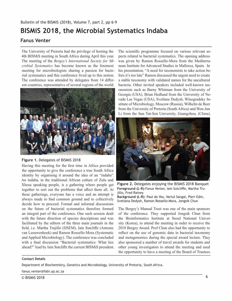
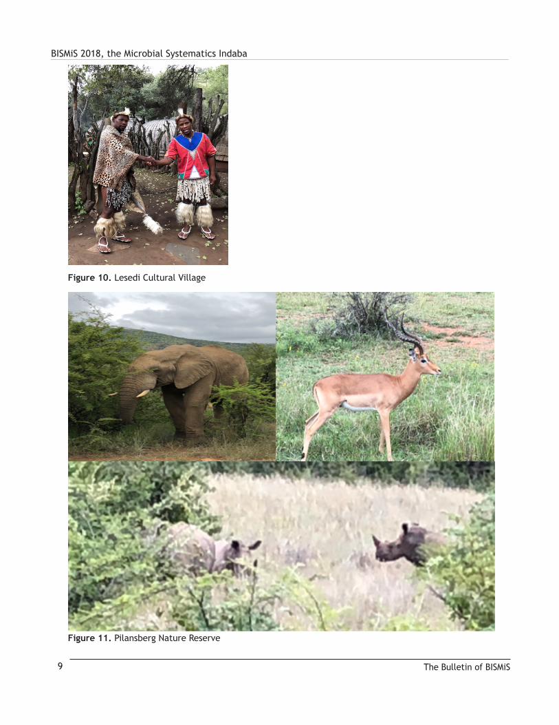
Delegates were treated to real African experiences such as the traditional braai (barbeque) and a drumming session which formed part of the welcoming function (Figure 4). In addition, delegates had plenty of opportunities to meet old friends and continue the discussion from the formal sessions (Figure 5 and 6). They also had a truly authentic African dining experience where they could feast on a variety of meat, including game at the Carnivore restaurant (Figure 7). Several of the delegates also joined the tours to the Sterfontein Caves (Figure 8), the Lion park (Figure 9) as well as the Lesedi Cultural Village (Figure 10) and Pilansberg nature reserve (Figure 11).
The Bulletin of BISMiS7
Figure 3. Prize awardees (L-R): Raul Riesco, Martha Trujillo (Editor of IJSEM), Kgothatso Chauke, and Marike Palmer.
Figure 4. Delegates enjoying the drumming session that formed part of the welcoming function
Figure 5. (L-R) Fred Rainey, Fanus Venter and Iain Sut-cliffeenjoyafter-dinnerdrinksandfineconversation
Figure 6. Paul Lawson and Mike Goodfellow putting the microbial world to rights!

BISMiS 2018, the Microbial Systematics Indaba
The Bulletin of BISMiS 8
Figure 7. Food and good conversation at the Carnivore restaurant
Figure 8. Sterfontein Caves
Figure 9. Lion park (L-R): cubs at play; adults chilling out; Ramon with a new friend!; Krithi Sanka-ranarayanan, now that’s a big lens
BISMiS 2018, the Microbial Systematics Indaba
The Bulletin of BISMiS9
Figure 10. Lesedi Cultural Village
Figure 11. Pilansberg Nature Reserve

Bulletin of the BISMiS (2018), Volume 7, part 2, pp 10-21
Marine Microbiology
I was first introduced to marine microbiology in North Carolina where I was invited to be an assistant on a short marine cruise on a Duke University boat with John Hobbie from North Carolina State, Professor Law-rence Pomeroy from U of Georgia and others who were comparing procedures for studying biological com-munity biomass and activity - a wonderful experience.
Later Thomas Odum, a Professor in our Department at UNC in Chapel Hill was conducting a study of ponds that con-tained 50% sea water mixed with 50% wastewater in More-head City on the Carolina coast. I set up the immersed micro-scope system to study the growth of algae in these lagoons and quickly realized that the algae in this system behaved entirely differently from the freshwater habitats in Michigan. They did not remain attached in one place on the glass slide surface! Clearly, I could not re-locate individual clones to study their development - the study had to be abandoned.
An early UW study entailed the use of microbial plating us-ing 0.01% peptone to assess microbial diversity. Samples were spread plated from various habitats and morphological colony types were identified with a dissecting microscope. One sample was from an incoming tide at Deception Pass in Washington State. The results were surprising. Almost every colony type on the marine medium was different! It is the most diverse viable sample I have ever seen. In 2015 reports were published that Puget Sound’s ocean influent was highly enriched in nutrients which helps explain the high diversity.
Whale microbiology and chitin degradation
Russell Herwig received his PhD from John Liston and became a postdoc. Russ and I decided to study the micro-biota of baleen whales. Coming from Hungate’s lab I knew something about the cattle forestomach fermentation. We hypothesized that, since whales are descended from land mammals and had similar digestive systems to ruminants, they might have a forestomach fermentation akin to cattle. That is how we came to study whales.
Our hypothesis was, if baleen whales were like ruminants, their fermentation products might be the same, in particu-lar they may produce the volatile fatty acids (VFAs), acetic, propionic and butyric. The most daunting aspect of study-ing the intestinal microbiota of whales, is obtaining fresh samples. We contacted several labs that had stored samples from whale forestomachs. Having assembled a small collec-tion of samples we subjected them to VFA analyses. Much to our delight, all the samples contained VFAs providing the first evidence for a forestomach fermentation (Herwig et al. 1984 AEM 47:421). Importantly, the substrate land ruminants use to produce VFAs is cellulose, comprised of glucose subunits, which is not in the diet of marine mam-mals. The marine counterpart is chitin, and its subunit is N-acetyl glucosamine. We wondered whether chitin was being degraded to form the VFAs by marine mammals that relied largely on krill, whose exoskeletons are largely chitin.
NSF Polar Programs supported this work. The best whale samples we obtained came from Iceland. Because our son, Greg, became ill during this period, Russ led the Iceland effort. Jay Stemmler, a technician, and Russ col-lected the freshest material possible, not only to analyze for chitin degradation, but also for forestomach acids. This approach alleviated concern about post-mortem fermenta-tions. Fin whales were sampled shortly after the whales were ‘landed’ on ship. The chitin degradation work in-volved taking samples from several positions in the diges-tive tract, incubating them anaerobically and testing for chitin concentration over time. If they degraded chitin, we should see a decline in chitin concentration (chitin degrada-tion could be due either to microbial or whale chitinases).
In Antarctica we used the same approach for test-ing chitin degradation in crab-eater seals. Penguins were captured and held in a container and fed con-trolled amounts of chitin (labeled with inert beads to be-gin and end the experiment) for a week before release.
The animal experiments gave similar results, i.e. about 25 % of the chitin was degraded through animal passage.
Seeking Truth in the Microbial Cosmos (cont’d)James Staley
Contact DetailsDepartment of Microbiology
University of Washington, Seattle, 98195
© BISMiS 2018 10

Seeking Truth in the Microbial Cosmos (cont’d)The most likely explanation is that animals play little if any role in chitin degradation in the animal digestive tract but instead rely on the chitinolytic bacteria in their intes-tines. If the animals play a degradation role, it is likely minor when one considers the entire chitin pathway in the marine environment. Russ led the chitin degradation work in marine waters and sediments with Nancy Pel-lerin, a technician, Roar Irgens and James Maki (Herwig et al., 1988 FEMS Micro Ecol 45:21). Analyses we made of the chitin cycle illustrate (Staley and Herwig, 1993 In Antarctic Microbiology E. I. Friedmann, ed Wiley-Liss, Inc New York, pp 241-264) most chitin degra-dation occurs in sediments where chitinolytic bacteria reside.
In retrospect, the dominant role of bacteria in chitin degrada-tion seems perfectly reasonable in light of the much later evo-lution of mammals and birds in comparison with zooplankton and krill with chitin exoskeletons. Like cellulolytic activi-ties in the ruminant, the chitinolytic activities seem largely if not exclusively microbial. Regrettably, we lacked suffi-cient resources to identify and name the chitinolytic bacteria.
Polycyclic Aromatic Hydrocarbon (PAH) degradation and the Dilution to Extinction Most Probable Number (MPN) Procedure
For an extended period Russ and I collaborated with civil engineer professors John Ferguson, David Stensel and Stu-art Strand. In the 1990’s Professor Jody Deming directed a five year program on marine bioremediation in which we all participated. My lab developed enrichment cultures for marine polycyclic aromatic hydrocarbon(PAH) - degrad-ing bacteria. Our approach was straight-forward: marine sediments were collected, mostly from Puget Sound, in particular from an EPA Superfund Site located in Winslow Harbor on Bainbridge Island contaminated by a defunct privately owned creosote wood treatment facility.
This combination of selective enrichment procedures with the quantification of specific groups of organisms as-sessed using MPNs (Dilution to Extinction MPN) became standard operating procedure in my lab. When important activities were discovered, the most numerous organisms were quantified by MPN from habitat samples. Enrich-ments with MPN dilutions retrieve the most numerous and environmentally significant organisms that may not dominate in undiluted enrichments. In MPN the highest dilutions that showed degradation were used to isolate, describe, name and place in culture collections the most numerous active organisms from an environment. If ordinary enrichment techniques are used the predominant organism in an enrichment culture would be one favored
in that enrichment, not the most numerous organism and important organism from the environment.
After collection, sediments were diluted in a series of ten-fold dilutions in half-strength seawater medium amended with either naphthalene or phenanthrene, two common low MW PAH compounds. Enrichments were incubated at 15oC and examined daily for the telltale appearance of yellow to red-colored degradation intermediates that are signs of PAH degradation. The highest dilution enrichment cultures that were positive were streaked to isolate the responsible bacteria.
Sheryl Dykesterhouse was an MS student who led the first studies and two PhD students, Allison Geiselbrecht and Brian Hedlund continued work on this topic, which also included studies in the Anacortes area near the oil refineries. The result was the isolation of several previ-ously unknown genera and species. The most remarkable example appeared in the highest dilutions of our PAH en-richments. We characterized and named the predominant novel genus and species Cycloclasticus (‘ring-breaking cuss’) pugetii after Peter Puget for whom Puget Sound was named (Dyksterhouse et al 1995 IJSEM 45:116).
Allison led the study of the incidence of PAH-degrading or-ganisms from contaminated marine sediments near Winslow (Geiselbrecht et al 1996 AEM 62:3344). Allison and Brian also discovered and named other PAH degraders including a Marinobacter strain (Hedlund, Geiselbrecht, Staley 2001 FEMS Micro Ltr 9992:1) and Vibrio cyclotrophicus from Puget Sound (Hedlund, Staley 2001 IJSEM 51:61). An addi-tional marine genus and species, Neptunomonas naphthovo-rans was also described (Hedlund et al 1999 AEM 65:251). Allison furthered our understanding of Cycloclasticus puget-ii biogeography by isolating other strains from the Gulf of Mexico off the coast of Texas (Geiselbrecht et al. 1998 AEM 64:4703) before the British Petroleum (BP) oil disaster.
Subsequently others confirmed that C. pugetii and other Cycloclasticus species are the most numerous and domi-nant marine PAH degraders on a global scale (Staley 2010 Handbook of Hydrocarbon and Lipid Microbiology, K. N. Timmis Ed-in-Chief). I was also invited to Xianmen, China to give a seminar on Cycloclasticus and PAH degradation.
After the marine PAH-degradation program ended, we sub-mitted a proposal to a new bioremediation initiative to con-tinue our work. Regrettably, after submitting our full pro-posal, the panel decided they would not consider funding any marine proposals on HC degradation! This seemed es-pecially odd because marine hydrocarbon spills pose some of the most significant problems in bioremediation whereas
The Bulletin of BISMiS11

Seeking Truth in the Microbial Cosmos (cont’d)contaminated soils are much better contained and were already well funded.
We were able to limp along with much-needed Sea Grant money for a time before our studies were deemed ‘too basic’ by a critical reviewer. This ended our efforts on marine bioremediation. I sadly shook my head in 2009 when the BP oilrig disaster occurred in the Gulf of Mex-ico. I knew then, as later reported, that Cycloclasticus spp would become predominant in the Gulf due to the release of all the uncontained PAHs during the spill. The result was unnecessary and caused incomprehensible damage to the marine biota and the livelihoods of mil-lions of innocent civilians who lived on, near or from the Gulf’s violated ecosystem which is still affected today.
Sea ice microbiology
My lab became involved in sea ice microbiology indirectly. The chitin degradation work led to the isolation of chitin-de-grading bacteria from marine waters of the Antarctic Penin-sula. Professor Roar Irgens, on sabbatical leave with me, had also studied and named gas vacuolate bacteria, suggested we look at our chitin plates to see if there were any chalky colo-nies indicative of gas vacuolate bacteria. Eureka - there were! This was most surprising as no one had previously reported cultures of gas vacuolate bacteria from the marine environ-ment (Irgens, Suzuki, Staley 1989 Curr Microbiol 18:262).
We pondered why they were present in sea ice. Know-ing that gas vacuolate bacteria are associated with gradi-ents, I hypothesized they might stratify within the sea ice microbial community (SIMCO). Unfortunately, by then, the annual sea ice on the peninsula had melted. Therefore, we proposed to NSF Polar Programs to go to the main McMurdo base to sample the following year.
We were funded and returned to McMurdo the follow-ing year obtaining several cores from McMurdo Sound near the ice airstrip and also across the Sound at Marble Point (Fig. 7). The upshot was that gas vacuolate bacteria were found in all ice cores suggesting they use the vesi-cles to rise in the water column where they could freeze in the SIMCO (Gosink, Irgens, Staley, 1993 FEMS Mi-cro Ecol 102:85; Gosink, Staley 1995 AEM 61:3486).
This discovery led me to hypothesize that the sea ice bacteria in the South Pole might be different from those at the North Pole because of the great separa-tion in distance and low temperature of viability of these bacteria may support their independent evolu-tion at the two poles (discussed in Evolution section).
Low temperature growth and psychrophile genomics
I became curious about the lowest growth temperature of the sea ice psychrophile we named Psychromonas ingrahamii (Auman et al 2006 IJSEM 56:1001). To determine this, we used a low temperature water bath set at -12 C. Full strength marine water broth was inoculated, incubated and observed daily to follow turbidity. Some test tubes froze early and no growth occurred in them. In the other replicates, turbid-ity was measured as growth proceeded. The doubling time calculated for several replicates was 240 hours (10 days).
Although this species might grow at a lower temperature, all tubes incubated at -15 C froze so our experiments could not be completed. Jennifer Breezee an undergradu-ate and Nate Cady a rotating graduate student aided with this work (Breezee, Cady, Staley 2004 Micro Ecol 47:300).
John Ingraham expressed an interest in having the genome of P. ingrahamii sequenced. Under Monica Riley’s leadership (Riley et al. 2008 BMC Genomics 9: 210) I was delighted to help because Professor Riley taught me microbial genetics while I was at UC Davis - it was wonderful to pursue this with her. The results indicated that P. ingrahamii contains six classes of proteins, at least one more than other bacteria, their membranes may have a lower hydrophobicity with excess asparagines and other features that enhance psychrophily.
The Bulletin of BISMiS 12
Fig 7. Jim Staley obtaining ice core from McMurdo Sound near sea ice air strip.

Seeking Truth in the Microbial Cosmos (cont’d)Black Sea Studies
Beginning about 2002, my lab collaborated with Profes-sor James Murray in Oceanography. As a chemical ocean-ographer, Jim was interested in the chemical and microbial activities, particularly those in the nitrogen cycle in the sub-oxic zone of the Black Sea. He had two PhD students, Clara Fuchsman and John Kirkpatrick with whom we worked.
Two postdocs in my lab, Sujatha Srinivasan and Brian Oak-ley, led our studies. John and Brian were lead authors on a paper based on using PCR procedures to identify Planctomy-cetes in the Black Sea (Kirkpatrick et al 2006 AEM 72:3078). This phylum contains the anammox (anaerobic ammonia oxidation) bacteria which carry out this process known to occur in the Black Sea. Interestingly, our studies showed that Planctomycetes and related organisms occur through-out the water column even deeply into the sulfidic zone.
We later named a new un-isolated anammox Candi-datus species “Scalindua richardsii” after Francis A. Richards, a UW chemical oceanographer who hypoth-esized the existence of anaerobic ammonium oxida-tion from chemical fluxes (Fuchsman et al. 2012 FEMS Micro Ecol 1-15). We also studied denitrification, ni-trogen fixation, CO2 fixation, etc in a series of papers.
I was especially impressed by Clara’s ability to relate the bacteriology to the chemical activities using tech-niques such as TRFLP and chemical isomers. Her work was regarded as the best example of interdisciplinary work in Chemical Oceanography by her PhD Committee.
Evolution
I have always been interested in investigating bacterial evo-lution, which was made possible by Carl Woese. His Tree of Life was constructed by analyzing the ssu rRNA sequences of representatives of all organisms. I had known Carl since I first corresponded with him in 1980 and provided him with samples of unusual bacteria for his studies. Later, Jerome Perry and I visited and interviewed him for the Saunders edi-tion of our textbook Microbial Life where he was one of the featured micro-biologists. He was also a co-author on our paper describing Polaribacter as a new genus with four spe-cies, which were the first gas vacuolate members of the Bacte-riodetes phylum (Gosink, Woese, Staley 1998 IJSB 48:223).
I was surprised during our last communications in late No-vember, 2011, a year before Carl died, that he was totally un-aware that Bergey’s Trust had completely adopted his phylo-genetic approach for bacterial classification. When I learned this, I immediately sent him a pdf file showing that the entire
classification of the Bacteria and Archaea was based on 16S rRNA gene sequences from Domain to Genus. This made his day! In his last correspondence he stated: “You I trust to do it right! All the best for the holidays old friend. Carl”
Woese’s incomparable research contributions provided bacte-riologists with 16S rRNA sequence analyses and the prospect of using other gene and protein sequences to explore impor-tant and unanswered evolutionary questions about micro-bial biogeography, speciation and the origin of the Domains. The ability to study microbial evolution was one of the most exciting opportunities during the culmination of my career.
With the help of Evgeni Sokorenko and Roger Buick I initiated a graduate level course in Microbial Evolution, which Evgeni is still teaching as one of the most popu-lar advanced courses in the Department of Microbiology.
Phylogenomic Species Concept and Universal Species Concept
As a taxonomist, I am interested in speciation, the evolu-tionary process whereby new species evolve from existing species and all species eventually become extinct. The ssu rRNA is too highly conserved to identify species. For spe-cies, the traditional method uses DNA – DNA hybridization (DDH) between two strains. An artificial cut-off of >70% for species was established by the work of John Johnson and Don Brenner, largely from the well-studied enteric bacteria.
Unfortunately, DDH is not an evolutionary method but the multilocus sequence analysis (MLSA) devel-oped in Professor Brian Spratt’s laboratory in London is. Using MLSA, the sequences of less highly conserved housekeeping genes can be concatenated to permit the phylogenetic identification of Bacteria and Archaea species and subspecies and is officially accepted by bacte-rial taxonomists (Stackebrandt et al 2002 IJSEM 52:1043).
I invited Brian to participate in an ASM symposium I or-ganized. While there he suggested we propose a meeting of the Royal Society of London on the topic of microbial speciation. The meeting, organized with the help of Mat-thew Fisher, was an excellent opportunity for taxonomists and evolutionary microbiologists to discuss the applica-tion of MLSA for studies of microbial speciation, bioge-ography and biodiversity. At the meeting, I proposed the Phylogenomic Species Concept (Staley 2006 Phil Trans R Soc B 361:1899; Staley 2010 Microbe 4:361). By using 16S rRNA sequences for higher taxonomic levels, i.e. from the Domain to Genus and combining that with MLSA for the species and subspecies, the complete classification of Bacteria and Archaea can be determined phylogenetically.
The Bulletin of BISMiS13

Seeking Truth in the Microbial Cosmos (cont’d)The Phylogenomic Species Concept (PSC) can be applied not only to the Bacteria and Archaea, but to all organisms as a Universal Species Concept (Staley 2009 Ind Micro Biotech 36:1331 and Staley 2013 in The Species Problem – Ongoing Issues Igor Pavlinov ed, InTech Europe, Rijeka, Croatia). For example, a recent article in Science states that one cannot de-termine the species of birds (or yeast) by using a single gene’s sequence (Zhang et al. 2014 Science 346:1311). Instead ad-ditional genes are necessary as with MLSA as stated by PSC.
Recently, we (Hedlund, Dodsworth, Staley 2015 Syst App Micro 38:231) explored considering the use of sin-gle cell and other genomic-PSC approaches for iden-tification and tentative naming of un-isolated bacte-rial and archaeal species to expedite our understanding of microbial diversity without the need for cultivation.
Biogeography of Sea ice bacteria
The sea ice bacterial work led me to postulate that it would be important to know whether the same species of bacteria could be found in the North Polar SIMCO (Sea Ice Microbi-al Community) as the South Polar SIMCO. This hypothesis is based on the long distances between the North and South polar sea ice caps. Also, since we were studying extreme psychrophilic bacteria, it seemed reasonable it would be difficult, or perhaps impossible, for them to survive trans-fer across the equator by birds and other vectors because of warm equatorial temperatures. Each pole might serve as an independent place for evolutionary divergence. Understand-ing biogeography is especially important for Bacteria and Ar-chaea because the traditional view was that the same species of bacteria will be found anywhere that environmental con-ditions are the same because of their rapid global dispersal.
The NSF ecology program funded us. We arranged to col-lect sea ice samples at Point Barrow as well as McMurdo, Antarctica. John Gosink studied this topic for his PhD. We collected sea ice samples from both poles and identified psy-chrophilic strains by fatty acid analyses and 16S rRNA gene sequences. At the time it was not known at what taxonomic level bi-polar bacterial endemism might occur and the bac-terial species definition is based on an arbitrary cut-off point.
Several new genera and species of gas vacuolate bacte-ria were named including Polaromonas vacuolata (Irgens, Gosink, Staley 1996 IJSB 46:822) from the South Pole, Oc-tadecobacter arcticus and O. antarcticus from each pole, re-spectively (Gosink, Herwig, Staley 1997 SAB 20:356) and Polaribacter with four species (Gosink, Woese, Staley 1998 IJSB 48:223) (Table 1) and summarized in our ‘Poles Apart’ paper (Staley, Gosink 1999 Ann Rev Microbiol 66:4104).
Our results found the same genus, but not the same spe-cies at both poles. Interestingly, one of the polar genera we named, Polaramonas, has species indigenous in gla-ciers. A recent, more intensive study supports the view that different species of Polaromonas occur in the North and South polar glaciers (Gowar et al 2016 Extremophiles 20:403), which is completely consistent with our findings.
I was invited to present our results at UC Berkeley in the mid-1990s. Following my seminar I met with Professor Norman Pace who vociferously disapproved of even consid-ering conducting studies on biogeography. I was shocked by his adamant denouncement. In my mind the question is not whether bacterial endemism exists, but at what taxonomic level might it occur? I reported we found the same genera at both poles, which I believe is important, but not the same species. Also I did not claim that my data were definitive.
In retrospect, it appears Norman Pace believes every-thing you need to know about evolution and taxonomy is revealed by 16S rRNA gene sequences. All microbial taxonomists know 16S does not typically identify a spe-cies, let alone subspecies and MLSA, as indicated previ-ously, is officially accepted (cf Stackebrandt et al 2002 above) for identifying bacterial species and subspecies.
Rachel Whitaker et al (2003 Science 301:976) subse-quently published a definitive paper showing endemism of “Sulfolobus islandicus” strains isolated from hot springs in North America, Iceland and the Kamchatka Penin-sula in Russia. She noted in her paper that 16S was un-able to distinguish among the 70+ strains she studied, but MLSA of less highly conserved genes did. More re-cently, we (Zuo, Hao, Staley 2013 Ant van Leeuw DOI 10.1007/s10482-013-0081-4) confirmed Rachel’s find-ings that strains were subspecies using electronic DNA-DNA hybridization of several “S. islandicus” genomes.
Co-Speciation of Simonsiella
Professor Daisy Kuhn (daughter of chemist, Thomas S. Kuhn) conducted beautiful work on Simonsiella, a gliding bacterium that is a bacterial inhabitant of the oral cavity of mammals. Daisy, who was a PhD student at UC Davis be-fore I arrived, assembled a collection of Simonsiella strains isolated from cats, dogs, sheep and humans. She sus-pected that they co-speciated with their mammalian hosts.
Brian Hedlund shared my curiosity, and we decided to test the hypothesis that co-speciation accounts for the evolution of separate species for each animal host. 16 strains were ob-tained from ATCC, four of each from the four host species.
The Bulletin of BISMiS 14
Seeking Truth in the Microbial Cosmos (cont’d)Brian completed the 16S sequences, and, as predicted, each Simonsiella species had its separate cluster of strains with the host animals so their phylogenies mirrored that of their host. Thus, the cats and dogs, as carnivores, formed two subgroups of one branch, and the human and sheep strains were on sep-arate branches of the mirror image (Staley 2006 Phil Trans R Soc B 361: 1899). This raises questions about the taxonomy of bacteria versus that of animals because the single genus Si-monsiella co-evolves with different families of animal hosts.
However, when other oral bacteria were included in the Trees, we found a more mixed picture: strains of closely related bacteria especially Neisseria spp. were interspersed within the human, dog, cat and sheep strains (Hedlund, Staley 2002 IJSEM 52:1377).
We infer the most likely explanation is that Neisseria and related species must have obtained 16S sequences from lysed Simonsiella cells in the environment, but, in contrast, Simonsiella must have a mechanism to re-ject or degrade these foreign sequences; an interest-ing question that could be experimentally addressed.
The Planctomycetes, Verrucomicrobia, Chlamydia (PVC) Superphylum
Some of the most remarkable bacteria known are found in the PVC Superphylum (Wagner, Horn 2006 Curr Opin Biotech 17:241). Unfortunately, they are very poorly stud-ied yet harbor unusual features and carry out activities not known elsewhere in nature. I was fortunate to have studied members of the PVC throughout my career, yet only more recently learning of their exciting importance to evolution.
Unwittingly, we isolated the first representative of the Verrucomicrobia in the 1960s (Prosthecobacter fusifor-mis) and also the first two species of the Planctomycetes in the 1970s (Pirellula staleyi and Planctomyces maris).
The Planctomycetes
While a PhD student at UC Davis, I isolated a pear-shaped budding bacterium from Putah Creek. A sec-ond isolate was obtained from Lake Lansing while at MSU. Because of other activities, I was not able to return to work on these organisms until UW in 1971. By then, only the Lake Lansing strain remained.
Following Arthur Henrici, who did not isolate it, I described it as a neotype strain of Pasteuria ramosa ala Metchnikoff (Staley 1973 Can J Micro 19:609). The subsequent isola-tion of a true Pasteuria ramosa, a pear-shaped bacterium containing endospores, indicated Henrici and I were mis-
taken. Nonetheless, the strain was clearly a member of the Planctomycetes, indeed, the first Planctomycete ever isolat-ed in pure culture. That organism was subsequently named Pirella staleyi by Mortimer Starr and Jean Schmidt and later by Hirsch’s lab, Pirellula staleyi, the name it still bears.
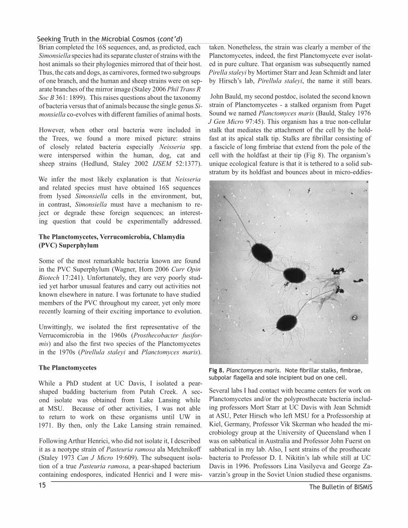
John Bauld, my second postdoc, isolated the second known strain of Planctomycetes - a stalked organism from Puget Sound we named Planctomyces maris (Bauld, Staley 1976 J Gen Micro 97:45). This organism has a true non-cellular stalk that mediates the attachment of the cell by the hold-fast at its apical stalk tip. Stalks are fibrillar consisting of a fascicle of long fimbriae that extend from the pole of the cell with the holdfast at their tip (Fig 8). The organism’s unique ecological feature is that it is tethered to a solid sub-stratum by its holdfast and bounces about in micro-eddies-
Several labs I had contact with became centers for work on Planctomycetes and/or the polyprosthecate bacteria includ-ing professors Mort Starr at UC Davis with Jean Schmidt at ASU, Peter Hirsch who left MSU for a Professorship at Kiel, Germany, Professor Vik Skerman who headed the mi-crobiology group at the University of Queensland when I was on sabbatical in Australia and Professor John Fuerst on sabbatical in my lab. Also, I sent strains of the prosthecate bacteria to Professor D. I. Nikitin’s lab while still at UC Davis in 1996. Professors Lina Vasilyeva and George Za-varzin’s group in the Soviet Union studied these organisms.
The Bulletin of BISMiS15
Fig 8. Planctomyces maris.Notefibrillarstalks,fimbrae,subpolarflagellaandsoleincipientbudononecell.

Seeking Truth in the Microbial Cosmos (cont’d)Those labs became international centers for the study of the Planctomycetes and Prosthecomicrobia including all fac-ets of their biology. Likewise, we continued to study them throughout my career although not as a primary research focus.
Verrrucomicrobia, bacterial tubulin and evolutionary implications
Carl Woese became quite interested in the PVC Super-phylum after publishing a paper with James Moulder’s group at the Univeristy of Chicago showing that Chla-mydia are members of the PVC group. In a 2008 E-mail, Carl told me he thought they should comprise a Kingdom.
The Verrucomicrobia
In about 2000, Carl Woese asked if I had any suggestions for bacteria to be sequenced at Integrated Genomics (IG), a startup in Chicago headed by Professor Robert Haselkorn. I provided one Verrucomicrobium (Prosthecobacter de-jongeii) as well as a Planctomycetes soil strain (Gemmata Wa-1). Material was sent and sequences became available soon thereafter. I received a phone call from Ross Over-beek at IG when the Prosthecobacter sequence was com-plete. He said he was happy to do a search right then, if we had genes of interest. I called out to the lab members and Cheryl Jenkins, a post-doc from John Fuerst’s lab said,
“Ask him about tubulin.” I knew immediately this was an excellent suggestion because Karl Schleifer’s lab had pre-liminary evidence that an unisolated Verrucomicrobia spe-cies might have tubulin genes. So I asked Ross, who is not a microbiologist, and after a moment’s delay he calmly re-sponded, “Yes, they do.” I responded incredulously, “Are you sure?” “Absolutely!” he replied. We were stunned.
With that, I immediately put Cheryl in charge of follow-ing up, and, as the data trickled in, it was one of the most gratifying discoveries of our lab. We first submitted the paper to Science for publication and were surprised they were not even interested in reviewing it - after all it was the first genetic evidence of tubulin genes in a prokary-ote. I knew that Professor Lynn Margulis had published a paper in Science some time earlier in which she provided
‘EM evidence for tubulin’ in spirochetes that supported her hypothesis that spirochetes were the evolutionary fore-runners of the eukaryotic flagellum which contains tubu-lin. But no spirochetes are known to have tubulin genes.
Carl Woese agreed to help with the review process at PNAS (Jenkins et al. 2002 PNAS 99:17049). Two tubu-lin genes were discovered that were homologs for α- and β-tubulin. We named them bacterial tubulin BtubA and
BtubB representing their relatedness to α- and β-tubulin, respectively. Using PCR we verified that all four species of Prosthecobacter from our lab contained both tubu-lin genes. No other Verrucomicrobia or other bacterial or archaeal species is currently known to contain tubulin.
I knew Lynn since her sabbatical leave at UW in the early 1970s. She was a good colleague, and I later favorably re-viewed her Five Kingdom book. Some time after our tubulin paper was published and shortly before her death, I received a phone call from her. We had a friendly exchange about a number of issues relating to photosynthetic bacteria she was studying. Incidentally, she mentioned how she had evidence from the fossil record of tubulin in a spirochete! It reminded me of a talk she presented at a FEMS meeting in Barce-lona where she derided Carl Woese’s evolutionary work on the Tree of Life. In retrospect, I believe she was reacting to confronting real scientific evidence that cast doubt on her Five Kingdom classification. I believe her talk was a tirade against the messenger because the message was too painful to accept. Paradigm switches are difficult for many scien-tists especially when their own contributions are at stake.
Nuclear Compartment Commonality (NuCom) Hypothesis
Regrettably, some scientists have recently attacked Woese’s Three-Domain Tree of Life as being ‘incorrect’ because it does not fit their view of the evolution of the Eu-karya from two prokaryotic lineages (a ‘Prokaryotes First’ hypothesis), the Bacteria and the Archaea. One group (Williams, Embley 2015 Phil Trans R Soc Lond B Biol Sci 370:20140318) replaced Carl’s tree by a two-pronged ‘stick figure’ of two Domains Bacteria and Archaea, which give rise to the Eukarya, by invoking ‘massive horizon-tal gene transfer’! In my view, one cannot discover the Eukarya if you are ‘barking up the wrong Tree of Life’. Also, like Carl Woese, I believe it is highly unlikely that a bacterium and an archaeon could fuse together.
The Bulletin of BISMiS 16
Fig 9. Prosthecobacter fusiformis bar is 1 micrometer – note prostheca and timbre

Seeking Truth in the Microbial Cosmos (cont’d)Further, it is scientifically impossible to test such a singu-lar event – some philosophers regard an untestable hypoth-esis as being invalid scientifically for that reason (Rich-ard A. Muller: Now The Physics of Time Norton 2016).
At the time we published the last edition of our textbook (2nd edition of Microbial Life with Sinauer) in 2007, I had doubts about having included a figure illustrat-ing a fusion event for the evolution of the Eukaryotes.
About that time CG Kurland presented a seminar at UW. He espoused a “Eukaryotes First” hypothesis. Although I doubt-ed that eukaryotes gave rise to the Bacteria and Archaea, Kurland’s intriguing point to me was: it is simpler to pro-duce a prokaryote from a eukaryote by reductive evolution, than a eukaryote by the fusion of two prokaryotes. I agree!
My NuCom hypothesis concerns the evolution of the Bacteria and Eukarya (Staley 2013 Astrobiol Outreach 1:105). I had one of those Eureka moments when I real-ized that the Planctomycetes and other members of the PVC Superphylum are nucleated. NuCom posits that the ancestors of the Bacteria and the Eukarya, which com-prise disparate, independent branches of the TofL were both nucleated from the time DNA replication evolved.
According to NuCom, the Eukarya have always been nucle-ated and the ancestors of the Bacteria are the nucleated an-cestors of the PVC Superphylum. Unfortunately, most mi-crobiologists know little about the PVC Superphylum which is key to understanding the NuCom hypothesis. Support for NuCom comes from the fact that Planctomycetes are known to be the most ancient members of the Bacteria by two inde-pendent phylogenetic approaches, careful 16S and proteomic analyses (Brochier, Phillipe 2002 Nature 417:244; Jun et al 2010 PNAS 107:133). Other support for NuCom is that the cell (and hence nuclear) membranes of the Bacteria and Eu-karya are essentially identical in composition, and, impor-tantly, homologous enzymes synthesize them. Common Bac-teria, such as E. coli are enucleate descendants of the PVC Superphylum that have lost their nucleus through reductive evolution. Other evidence of homologous proteins has been submitted (Staley, Fuerst. 2017, in press Res in Micro).
It is important to recognize that NuCom does not propose that the PVC gave rise to the Eukarya, a misguided view by some PVC adherents. The first genomics paper published on two PVC species Prosthecobacter dejongei and Gemmata Wa1-1 (Staley, Bozek, Jenkins 2005 FEMS Micro Ltrs 243: 9) concluded that it was doubtful that the PVC gave rise to the Eukarya but they shared important ancient proteins such as tubulin from LUCA (Last Universal Common Ancestor) as explained by NuCom (see Staley, Fuerst paper above).
I believe there is more evidence to support NuCom than any other hypothesis about the origin of the Eukarya. However, without providing evidence to the contrary, Norm Pace and W. Ford Doolittle do not agree. In contrast, I believe my good friend, Carl Woese would. When confronted with evi-dence for NuCom, doubters are inexplicably unconvinced.
From an aesthetic viewpoint of Truth (J W McAllister Beau-ty and Revolution in Science 1996 Cornell University Press, Ithaca), NuCom provides the simplest, most beautiful expla-nation for the evolution of the Bacteria and the Eukarya. No one needs to explain how an Archaeon evolved tubulin from FtsZ or artubulin, why the ribosomes of Archaea and Bacteria are different from the Eukarya or how the Eukayotes evolved their PLFA ester-linked membranes from an Archaeon, or in-voke ‘massive horizontal gene transfer’ of ancient proteins. And most importantly a fantastic fusion event is unnecessary.
In my view, those who believe in “Prokaryotes First” are constrained by a false paradigm. However, the NuCom hypothesis is a testable hypothesis and suggestions for test-ing it were included in Staley and Fuerst (2017). After I re-tired, I conducted theoretical work. One article proposes Domain Cell Theory on the evolution of the Bacteria, Ar-chaea and Eukarya (Open Biol. 7: 170041 2017). Domain Cell Theory states that each of the Domains of life in Carl Woese’s Tree of Life, i.e. the Bacteria, Archaea and Eukarya formed independent cellular lineages so that Bacteria—> Bacteria, Archaea—> Archaea and Eukarya—> Eukarya The Eukarya have always been nucleated and the earliest Bacteria (i.e. PVC phyla) were also nucleated (Figure 10).
The Bulletin of BISMiS17
Figure 10. Tree of Life based on co-evolutionary pro-cesses. The two main lines of descent are the Bacteria and Eukarya. During the pre-cellular evolutionary stage the Euryarhaeaota evolved with the Bacteria and the Crenarchaeota (TACK) evolved with the Eukarya. After the Bacteria and Eukarya became nucleated this process ceased because their DNA was inaccessible.

Seeking Truth in the Microbial Cosmos (cont’d)
In contrast the Archaea formed two independent cel-lular lineages, the Euryarchaeota and the Crenarchaeo-ta, neither of which became nucleated. I have more recently collaborated with Gustavo Caetano-Annoles who introduced me to the importance of protein fam-ily-folding patterns and their evolutionary implica-tions. We have two papers, one in press in Bioessays and another in preparation about the Tree of Life (Figure 10). This work confirms Domain Cell Theory.
Other Scientific Activities
Colleagues In addition to my mentors, graduate students, technicians, postodocs and those on sabbatical leave who have been discussed, other students included Karen Junge, Ivy Su-zuki, Gary Oertli, David Tison and Matt Stoecker. I have had the good fortune of supportive colleagues throughout my career. Many are at UW including John Sherris, Gene Nester, John Leigh, Charles Evans, Helen and Arthur Whiteley, Erling Ordal, Mary Lidstrom, Evgeni Sokurenko, Woody Sullivan, Jim Lara, Beth Traxler, Jim Champoux, Peter Greenberg, Steve Lory, Steve Moseley, Jody Dem-ing, Roger Buick, Matt Parsek, Edward Haskins, Thomas Quinn, Beth Traxler, John Baross, Ron Merrill, Charles Ev-ans, Neal Groman, Milt Gordon, Steve Lory, David Catling, Larry Corey, Julie Overbaugh, Tom Fritsche, David Stahl, Brian Lewis, Ludmilla Chistoserdova, Tom Lie, Frank and Ruth Harold, Kelly Hughes, Cary Hartman, Jim Murray, Al Devol, Marie Coyle, Clem Furlong (from UC Davis), Bob Waaland, Sharon Doty, Ben Hall, Joe Felsenstein, Claire-Horner Devine, Joseph Ammirati, Gordon Orians, Conway Leovy, Roger Buick, David Catling, Eric Cheney, Deborah Kelly, Don Brownlee, Steve Warren, Peter Ward, Woodruff Sullivan, Richard Smith, Dennis Kunkel, Rosanne Cat-tolico, Imre Friedmann, Joe Ammirati, Thomas and Yvette Edmonson, Karl Banse, John Adams, Gustavo Caetano-Annoles, Ramona Memmer, Janis Fulton, Dale Parkhurst, Dorothy Cramer, Mary Bicknell, Carol Laxson, Kendal Gray, Mark Chandler, Sarah Mears, Jane Halsey, Bonnie Hightower.
Other colleagues at UW are John Liston, Robert Wiss-mar, John Ferguson, Deborah Kelly, David Stensel, Eric Cheney, Rita Horner, Stuart Strand, ceramic engineers Il-han Aksay (now at Princeton) and Mehmet Sarikaya with Tao Ren, and Tao’s colleague Xian Ming Shi who hosted Sonja and me in Shanghai during the 2009 Exposition.
I also want to acknowledge Bergey Trustees and Associ-ates: Robert Murray, Noel Krieg, John Holt, Marvin Bryant, Richard Castenholz, David Boone, Don Brenner, William
Whitman, Fred Rainey, Michael Goodfellow, Karl Schle-ifer, Norbert Pfennig, Peter Sneath, Arnold Raven, Martha Trujillo, Peter Kämpfer, Jan Ursing, Stan Williams, James Moulder, Hans Lautrop, Paul De Vos, Michael Goodfel-low, Wolfgang Ludwig, Ken-Ichuro Suzuki, Karl Stet-ter. Later, Associate Editors were invited to join meetings while the taxa they had expertise in were being written. I know many others in systematics including Antonio Ven-tosa, Dorothy Jones, Erko Stackebrandt, Hans Trüper, Paul Lawson, Rita Colwell, Aiden Parte, Cletus Kurtzman.
Other international colleagues include Tony Walsby, Heinz Schlesner, Thomas McMeekin, Ramon Rosello-Mora, Brian Spratt, John Bowman Huub J. M. Op den Camp, Es-sam Ghanem.
I enjoyed sabbatical leaves with Professors Vic Skerman and Kevin Marshall in Australia, Ralph Berger in Hawaii and Peter Hirsch with Heinz Schlesner in Germany. Also, I have many Australian, European and Asian friends and colleagues, including Professors John Fuerst, Lindsay Sly, Brian Spratt, Lise Øvreas, Bailin Hao, Aharon Oren,
A number of Postdocs in the lab included Russell Herwig, James Maki, Cheryl Jenkins, Sujatha Srinivasan, Brian Oakley, Tom Moench, Judith Bland, Robert Ward, Alex Semenov, Benjamin Yee.
Several sabbatical leave individuals also worked in the lab including John Fuerst, Don Johnstone, JoAnne Chee-Sanford, Roar Irgens, R. A. Sanford, Liv Fiksdal, Lina Vasilyeva, Jeanne Poindexter.
Bergey’s and BISMiS
I became a Bergey’s Manual Trustee in 1976. When I re-tired at the mandatory age of 70 in 2008, I was the longest serving member. For me, Bergey’s was the ideal scientific organization with an honorable goal: working together to produce the most up-to-date, affordable and accurate list-ings of species and their characteristics along with a classi-fication of the Bacteria and Archaea. Trustees collaborated in a congenial manner and worked with a supporting group of several hundred invaluable and dedicated contributors to produce a detailed description of species and the most recent classification. It was and remains a wonderful or-ganization of scientists with an annual meeting held alter-nately in North America and Europe and more recently in Asia. John Liston was Chair of the Trust, and Professor Robert G. E. Murray followed him during the 1970s. Mur-ray remained Chair until after the first edition of Bergey’s Manual of Systematic Bacteriology (BMSB) was published.
The Bulletin of BISMiS 18
Seeking Truth in the Microbial Cosmos (cont’d)John Holt was head of the editorial office when I joined and maintained that position until his retirement. John Holt and I were invited to the USSR to meet with George Za-varzin and others at the Institute of Microbiology at the time of Glasnost because Soviet microbiologists were in-terested in producing a Russian translation of Bergey’s Manual of Determinative Bacteriology. While there, we met George Zavarzin, Lina Vasilyeva, D. I. Nikitin, and many other bacteriologists who were doing fascinating research at the Institute in Moscow as well as some at Moscow State University (E. N. Kondratieva) and in Len-ingrad (St. Petersburg) and also visited the Microbiology biotechnology center in Puschino led by Yuri Trotsenko where UW colleague, Ludmilla Chistoserdova studied.
Soon after I joined in 1976, John Holt and Bob Mur-ray proposed that the Trust consider publishing a more comprehensive taxonomy that contained more historical and descriptive information about the bacteria and sug-gested calling this Bergey’s Manual of Systematic Bacte-riology (BMSB). This was unanimously approved by the Trustees. The first edition of BMSB was based on phe-notypic properties of Bacteria except for the Archaea sec-tion led by Karl Stetter, which was based on 16S analyses.
Peter Sneath and I were particularly interested in producing a completely phylogenetic classification – at the time this con-sisted of computer print-outs of just a few pages from Carl Woese’s work that I brought to the annual meetings. Howev-er, at the time of the second edition a complete truly phyloge-netic classification based on 16S rRNA sequencing from Do-main to Genus became possible (Fig 10). For the 2nd edition of BMSB, the phylogenetic work was admirably performed by Wolfgang Ludwig and Professor Karl Heinz Schleifer.
I became Chair of Bergey’s Trust in 2000 amid a time of change after John Holt retired and George Garrity replaced him. The 2nd edition was behind schedule, and Michi-gan State University wanted to assert more control over the Trust. The upshot was that the Trust decided to move. The cordial move was made to the University of Georgia with William B. Whitman as head of the editorial office.
In addition to completing the 2nd edition, the other ma-jor accomplishment during my Chairmanship was the establishment of an international society for micro-bial taxonomists called Bergey’s International Society for Microbial Systematics (BISMiS). We also initiated a newsletter, The Microbial Taxonomist, and the BIS-MiS Bulletin with biographies, reviews, opinion articles and invited autobiographies that I edited for two years.
The inaugural meeting of BISMiS was held in Bei-jing in 2011. This was a spectacular event led by the new Bergey Chair, Michael Goodfellow along with current Trustees and several hundred mostly Chi-nese and other Asian taxonomists in attendance.
I was invited by Ken-Ichuro Suzuki to deliver a 30th an-niversary address at the Japanese Society of Microbial Sys-te-matics in Tokyo in 2012 where I discussed the history of Bergey’s Trust and the phylogenomic species concept. I met Professor Emeritus Kazuo Komagata and invited and edited his autobiography for the Bulletin of BISMiS.
American Society for Microbiology (ASM)
ASM is another organization with great leadership that serves the larger community of microbiologists. I’ve been a member for over 50 years and served one term on the Council Policy Committee. I received the US Feder-ation of Culture Collections J. Roger Porter Award, from ASM in 2008. My talk was entitled “Beyond 16S” which discussed speciation, MLSA analyses, biogeography and genomics. I have been honored to have two species named for me: Pirellula staleyi and Polaribacter staleyi.
Books
With Anna-Louise Reysenbach, I edited a book en-titled Biodiversity of Microbial Life: Foundation of Earth’s Biosphere (2002 John Wiley & Sons, New York).
In about 1990, Jerome Perry asked whether I would like to co-author a textbook for microbiology majors. After some consideration, I accepted. Our first edition was pub-lished with Saunders. Another two editions entitled Micro-bial Life were published by Sinauer Press, the last in 2007.
The Bulletin of BISMiS19
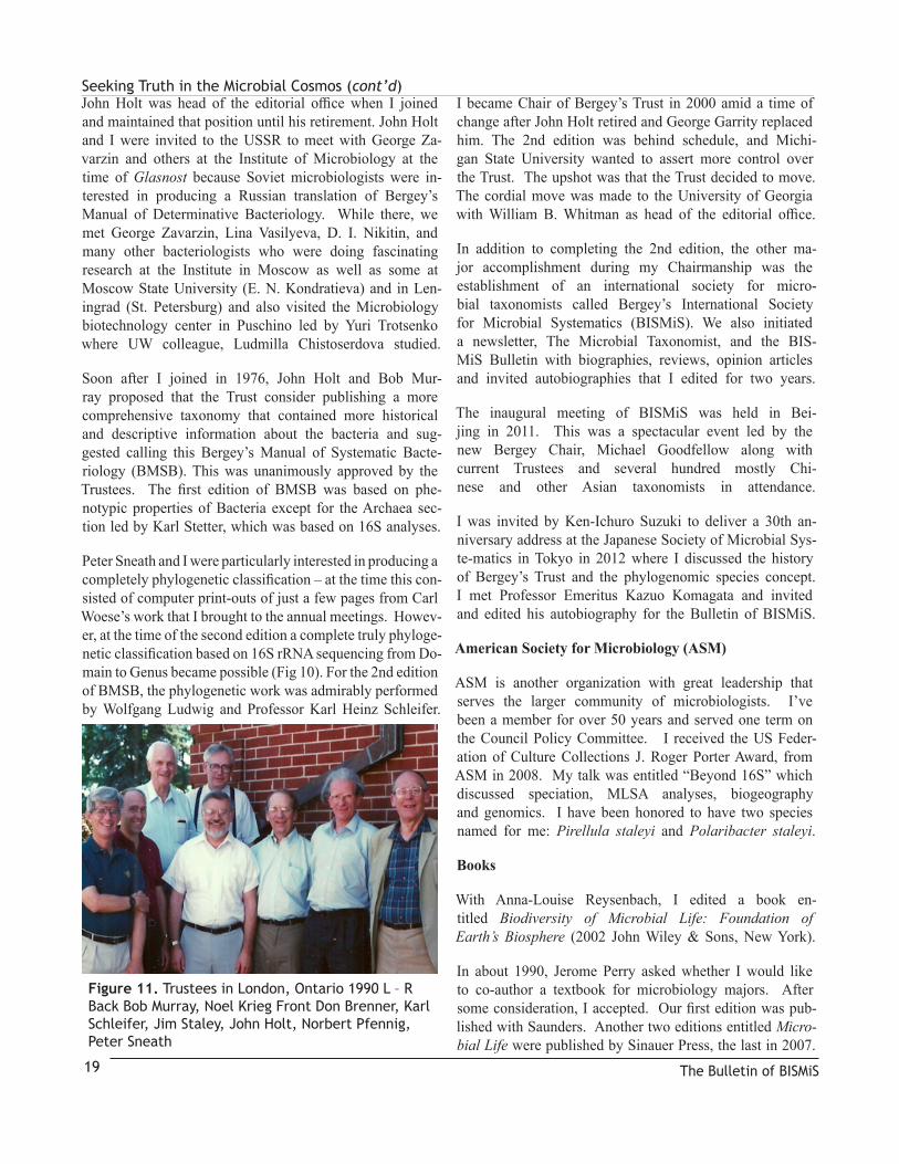
Figure 11. Trustees in London, Ontario 1990 L – R Back Bob Murray, Noel Krieg Front Don Brenner, Karl Schleifer, Jim Staley, John Holt, Norbert Pfennig, Peter Sneath
Seeking Truth in the Microbial Cosmos (cont’d)
I have not kept up with current major’s editions of textbooks that are available now. But, I must say, from what I know, I am concerned. Most disturbing to me is that there appears to be little if any mention of the PVC Superphylum in text-books. This group of organisms is key to understanding the evolution of life, but many microbiology graduate students do not even know they exist! Is it any wonder that the ‘Pro-karyotes First’ folks still have fertile ground to plant their myths about evolution? Also, there seems to be a tendency in texts to provide as many facts as possible. I believe much more needs to be done to put the facts in context with stories, particularly with respect to evolution. If ‘dumbing down’ occurs in advanced microbiology textbooks, it’s a pity.
Astrobiology Doctoral Program
One of the most fun chapters in my career was the astro-biology program. It all began early in 1997 with a semi-nar program, Planets and Life originated by Woodruff
‘Woody’ Sullivan III in the UW Astronomy department and John Baross in Oceanography. They asked me to contribute to the seminar series by reviewing the recent paper on the evidence for life on Mars based on the re-cent discovery of the Allan Hills Martian meteorite from Antarctica. The seminar course involved faculty from several different disciplines including atmospheric sci-ences, aeronautical engineering, geology, biology, microbi-ology, oceanography, astronomy, genetics and biochemistry.
That fall, NSF put out a call for proposals to their new In-tegrative Graduate Education and Research Traineeship (IGERT) program. I asked Woody Sullivan whether he would be interested in applying to it. He said he was, but he was too busy at the time and suggested he would help if I took the lead on it. So, I reluctantly agreed and placed him and Conway Leovy as Co-PIs of the proposal. I wrote the pro-posal, circulated it to all the Co-PIs for input and submitted.
Never have I received such rave reviews. One panelist on the review committee commented: “What are they putting in their coffee in Seattle!” And so the program began in 1998. I was Chair of the AB for several years and was re-placed by Woody Sullivan about the time I retired. The pro-gram is still active although the Department of Microbiol-ogy is less involved. Victoria Meadows is the current Chair.
I was invited to write a paper about the Astrobiology Pro-gram at UW that received input from all Co-PIs (Staley 2003 Cur Op Biotech 14: 347). We described astrobiology as a basic, integrative program for teaching science and engineering at all grade levels because astrobiology poses
some of the most basic, profound and intriguing ques-tions about life and human existence such as: “How did life originate and evolve?” “What is the future of life?”
“Does life occur elsewhere in the Universe?”
CODA
As a scientist I have rejoiced in the opportunity to search for and occasionally discover some scientific truths that are part of the complex tapestry of nature. I have had a thrilling career. I have enjoyed my interactions with all sorts of wonderful people from professors, men-tors, undergraduate, graduate and post-doctoral stu-dents, colleagues at UW and elsewhere in the global sci-ence community with whom I have enjoyed associating.
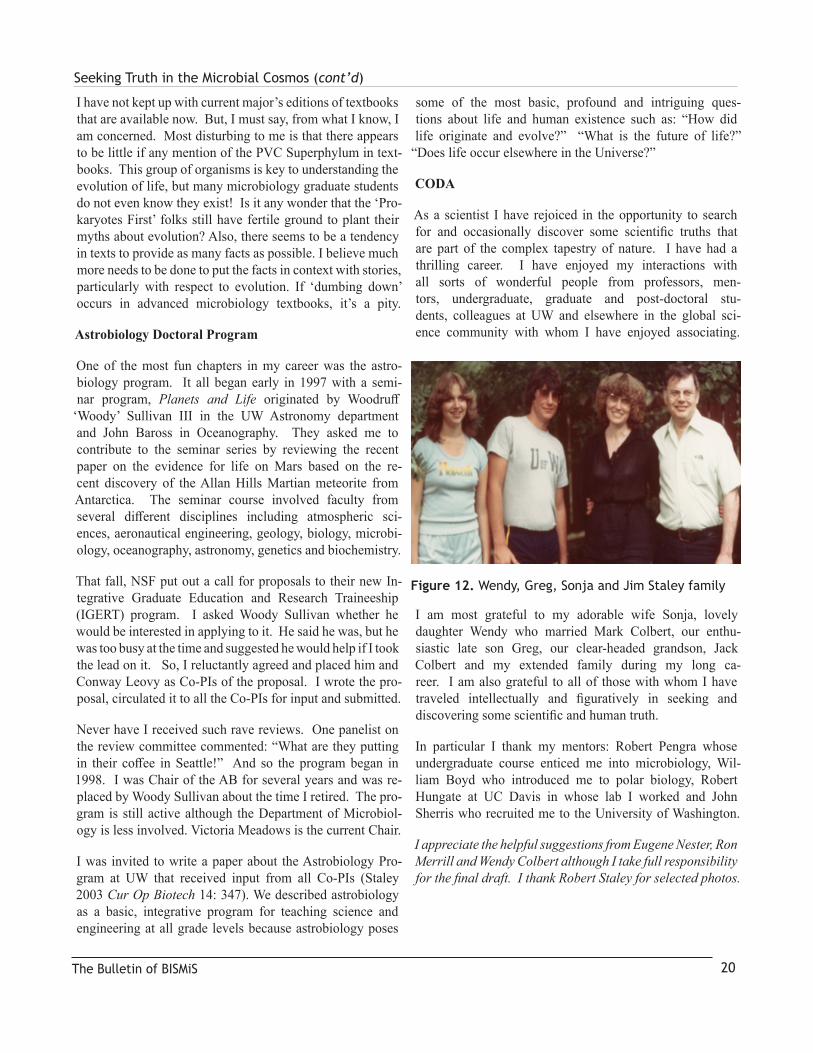
I am most grateful to my adorable wife Sonja, lovely daughter Wendy who married Mark Colbert, our enthu-siastic late son Greg, our clear-headed grandson, Jack Colbert and my extended family during my long ca-reer. I am also grateful to all of those with whom I have traveled intellectually and figuratively in seeking and discovering some scientific and human truth.
In particular I thank my mentors: Robert Pengra whose undergraduate course enticed me into microbiology, Wil-liam Boyd who introduced me to polar biology, Robert Hungate at UC Davis in whose lab I worked and John Sherris who recruited me to the University of Washington.
I appreciate the helpful suggestions from Eugene Nester, Ron Merrill and Wendy Colbert although I take full responsibility for the final draft. I thank Robert Staley for selected photos.
The Bulletin of BISMiS 20
Figure 12. Wendy, Greg, Sonja and Jim Staley family

Seeking Truth in the Microbial Cosmos (cont’d)
The Bulletin of BISMiS21
Phylum Genera and Species Current status Reference
Verrucomicrobia (Hedlund, Gosink, Staley 1996 IJSEM 46:960)
Prosthecobacter fusiformis (Staley, deBont, deJonge 1976 Ant van Leeu 42:333) Prosthecobacter dejongei (Hedlund, Gosink, Staley 1997 Ant van Leeu 72:29)Prosthecobacter debontii (“)Prosthecobacter vannervenii (“)
Planctomycetes “Pasteuria ramosa” (Staley 1973 Can J Micro 19:609) now Pirellula staleyi Planctomyces maris (Bauld, Staley 1976 J Gen Micro 97:45)“Scalindua richardsii“ (Fuchsman et al 2012 FEMS Micro Eco 1)
BacteroidetesPolaribacter irgensii (Gosink, Woese, Staley 1998 IJSB 48:223).Polaribacter franzmannii (“)Polaribacter filamentus (“)Polaribacter glomeratus (“)Gram + Planococcus mcmeekinii (Junge et al., 1998 Sys App Micro 21:306)Rhodococcus zopfii (Stoecker, Herwig, Staley 1994 IJSB 44: 106)
ProteobacteriaProsthecomicrobium pneumaticum (Staley 1968 J Bact 95:1921)“Prosthecomicrobium enhydrum” now Vasilyevaea enhydra (Yee et al 2010 IJSEM 60:2960)Vasilyevaea mishustinii (“)“Prosthecomicrobium litoralum” (Bauld, Bigford, Staley IJSB 33:613) now Bauldia litoralis (Yee et al 2010 IJSEM 60:2960,)Bauldia consociatum (“)Prosthecomicrobium hirschii (Staley, IJSB 1984 34:304) Proposed: “Prosthecodimorpha hirschii” submittedAncalomicrobium adetum (Staley 1968 J Bac 95:1921) Aquabacter spiritensis (Irgens et al 1990 Archives Microbiol 155:137)Octadecobacter arcticus (Gosink, Herwig 1997 Staley Sys App Bac 20:356)Octadecobacter antarcticus (“)Colwellia demingiae (Bowman et al 1998 IJSB 48:1171)Colwellia hornerae (“)Colwellia rossensis (“)Colwellia psychrotrophica (“)Cycloclasticus pugetii (Dyksterhouse et al 1995 IJSEM 45:116) Neptunomonas naphthovorans (Hedlund et al 1999 AEM 65:251)Vibrio cyclotrophicus (Hedlund, Staley 2001 IJSEM 51:61)Polaromonas vacuolata (Irgens, Gosink, Staley 1996 IJSB:46:822)Psychromonas ingrahamii (Auman et al 2006 IJSEM 56:1001)Psychromonas boydii (Auman et al 2010 IJSEM 60:84)Uncertain taxon: Enhydrobacter aerosaccus (Staley et al. 1987 IJSB 37:289)
Table 1. Names of bacterial taxa described by Staley lab

Bulletin of the BISMiS (2018), Volume 7, part 2, pp 22-43
Frantisek Král initiated the first bacterial culture collection in the world, in 1890 in Prague, though he did not offer a supply service. It was the NCTC, established in 1920, that was the first collection in the world to offer a supply service of bacterial cultures, with about 2,000 cultures distributed in the first year and the first catalogue published in 1922. The NCTCs Order Book from 1920 confirms that Dr (later Sir) Alexander Fleming was one of its earliest customers. The early history of the NCTC has been published previ-ously (St John-Brooks, 1944, 1945) and the history of the first 50 years of the NCTC was recorded in a publication celebrating that anniversary (Anon, 1971); this publica-tion is the source of some of what follows. The author was employed in the NCTC from 1971 to 2012, so this article consolidates 92 years of NCTC history in a single record.
The Medical Research Committee (later Council, MRC) had for some time considered the setting up of a type cul-ture collection from which authentic micro-organisms could be obtained from a trustworthy source for use in scientific work. Prior to World War I, the principal sources of sup-ply were outside the United Kingdom, including the Institut Pasteur in Paris, the Král collection (later moved to Vienna) and the American Museum of Natural History in New York (in 1925 to become the American Type Culture Collection, ATCC). The NCTC started on the 1st January 1920 under the directorship of Dr (later Sir) John C. G. Ledingham, who was Chief Bacteriologist to the Lister Institute of Pre-ventive Medicine in Chelsea, London (Figure 1). The Lister Institute provided the accommodation and the Medical Re-search Council provided the equipment and staff. The first Curator was Dr R. St John-Brooks, and he was supported by an Assistant Curator, Mabel Rhodes. The nucleus of the Collection comprised the some 200 cultures already held in the Lister Institute Private Collection; these included Esche-rich’s original strain of Escherichia coli. The first accessions were Sir Frederick Andrewes’ strains of Shigella flexneri.
Though unpublished, records of Mabel Rhodes (1945) sur-vive and paint an interesting picture. She records that when they first occupied the accommodation at the Lister Institute
it “was one of the largest and most pleasant in the build-ing” but “In the way of equipment it contained nothing but a telephone so until the necessary equipment could be ob-tained the fitting out was done by scrounging and borrowing from other departments though everyone was most helpful in setting up the Collection.” She records how “Paraffin wax was used almost from the first for sealing cultures but, ow-ing to its messiness, other methods were tried, sometimes with disastrous results.” She recalled how “The use of semi-solid serum agar under paraffin reduced the subculturing of the Neisseria from once a week to once in two months.”
In these early days, there were no special facilities for the handling of hazardous pathogens such as Shigella species. In 1922, a requested culture of Francisella tularensis was received with the “advice that it should be thrown down the sink as so many workers had gone down with the disease” (Rhodes, 1945). In subsequently handling it, the Curator, As-sistant Curator and another microbiologist, Dr H. Schütze, all became infected, probably from handling guinea pigs exper-imentally infected with the organism. Not surprisingly, ex-periments on animals with this organism were discontinued.
In 1930, Dr Ledingham became Director of the Lister In-stitute leaving Dr St John-Brooks in charge of the collec-tion. Initially, cultures were maintained and distributed by subculture but Dorset’s egg medium, sloped in bijou bottles, was soon adopted. In 1933, experiments began on the use of freeze-drying for the long-term preservation of bacterial cul-tures using the Homer Smith method, but the results were dis-appointing (Rhodes, 1945). However, in 1934, a technique was adopted as described to Dr St John-Brooks by Professor Alfredo Sordelli who was visiting from Buenos Aires at the time. Cultures were freeze-dried over phosphorous pentox-ide, and, from 1934 onwards, almost every strain in the col-lection was dried in duplicate by this method. Cultures were still, however, distributed in sub-culture, with the freeze-dried tubes held in reserve from which to prepare subcultures.
The NCTC received many visitors over these years, the most notable being “Queen Elizabeth (sic) of the Bel-gians and Professor Einstein” (Rhodes, 1945). Appar-ently the press was rather troublesome at times, writing frivolous articles about the Collection (Rhodes, 1945).
The History of the National Collection of Type Cultures (NCTC) and the Collection as a Resource for Systematists and the Wider Scientific CommunityBarry Holmes
Contact DetailsNCTC, Health Protection Agency, Colindale, London NW9 5EQ, UK; [email protected]
© BISMiS 2018 22

TheHistoryoftheNationalCollectionofTypeCultures(NCTC)andtheCollectionasaResourceforSystematistsandtheWiderScientificCommunity
Early on in the NCTC, it became apparent there was a significant demand in the United Kingdom (UK) for the supply of cultures of microfungi. The NCTC therefore col-laborated with the British Mycological Society for their assistance in the maintenance of microfungi, many strains of which were then held in the NCTC collection. Under a centralisation policy at the Lister Institute the NCTC col-lection, similar to that at ATCC, came to include microor-ganisms of medical, veterinary, agricultural and economic interest including diverse organisms as both pathogenic and saprophytic fungi, phytopathogenic bacteria and yeasts.
By the end of its first year of operation, the NCTC held about 800 cultures. Five years later the number held had increased to 1,500, after 10 and 15 years to 2,500 and 3,500, respectively, and by 1939 it numbered some 4,300 strains.Subsequent to publication of the first catalogue in 1922, succeeding editions were published in 1925, 1931 and 1936.In 1938, just prior to the advent of World War II, 6,397 cul-tures were distributed, of which 45% were for scientific re-search, 20% went to colleges and schools for teaching pur-poses and 35% to workers engaged in technology. Of those cultures, 46 were dispatched abroad (Africa 6, Asia 11, Aus-tralasia 2, Europe 23, North America 2, South America 2).
In 1939, the NCTC was transferred from Chelsea to the Lister Institute farm laboratories at Elstree, Hertfordshire (site subsequently occupied by Bio Products Laboratory, Dagger Lane), where the space available was limited (Fig-ure 2). The NCTC faced a difficult period, and, by 1944, held about 5,000 cultures, imposing a considerable strain on the staff and space available to maintain the cultures. The war conditions imposed other difficulties. Although formal export controls in the UK were not introduced until 1993 following the first Gulf war, during World War II the Postal and Telegraph Censorship Department in 1944 intercepted a culture of Penicillium notatum NCTC 4222 which had been dispatched to the Sociedade Industrial Farmaceutica in Lisbon. The culture was returned to the NCTC and Dr St. John-Brooks wrote to the censor advising that he was not aware of any embargo on the sending of cultures of Sir Alexander Fleming’s strain of P. notatum out of this coun-try. However, the Allies did not want the enemy to develop penicillin, for if they had done so then this may have had a profound effect on the course of the war. The interesting story of penicillin in Europe during World War II is recount-ed by Sharma (2009). There is a similar report of a culture being intercepted having been despatched to “the Argen-tine” to an institute whose director was on a government blacklist that the NCTC staff had no prior knowledge of.
Come 1945, the war ended, and the following year Dr St.
John-Brooks retired and Dr Samuel Tertius Cowan took his place as Curator from 1st January 1947. At this time, the MRC took the view that the NCTC should confine itself to bacteria of medical and veterinary interest. Characteri-sation of all the cultures in the collection was begun, and, during the period 1946-1950, groups of microorganisms not meeting the NCTCs new remit were transferred to relevant institutes where they formed the nuclei of sever-al of the UKs other national culture collections. In some cases, the original NCTC number was maintained, thus the National Collections of Industrial and Marine Bacte-ria (NCIMB) which were to form then in Teddington (now in Aberdeen) hold the same strain of Staphylococcus au-reus: NCTC 6571 ≡ NCIMB 6571. The NCTCs culture holding was reduced to about 3,000. Following a recom-mendation in 1947, the United Kingdom National Com-mittee of the British Commonwealth Collections of Mi-croorganisms (UKNC) was established to coordinate the activities of the then existing collections of microorganisms.
Events leading to the creation of the Emergency Public Health Laboratory Service in 1939 have been described by Wilson (1948). The Public Health Laboratory Service (PHLS) itself came into being in 1946 with its central laboratory in Colindale at what had previously been the Government Lymph Establishment (built to facilitate the production of smallpox vaccine). It is worth noting that the Government Lymph Establishment also began its work at the British Institute of Preventive Medicine at Chelsea (later known as the Lister Institute of Preventive Medi-cine) in 1898, but moved in 1907 to the newly constructed Government Lymph Establishment at Hendon (Fremlin, 1946). In July 1949, the NCTC moved from Elstree to the Central Public Health Laboratory (CPHL) of the PHLS in Colindale, London. At this time some 6,500 cultures were being supplied annually. The NCTC took up residence in a “temporary” building known affectionately as “the hut”. Here, in 1949 and with new equipment, large-scale freeze-drying commenced, one of the first national collections to do so, and a new policy was implemented for every strain in the collection: to check its purity, to test and record its characters (colony morphology and biochemical test re-sults were recorded on pre-printed cards measuring 13x6 inches ), to freeze-dry adequate stocks for distribution and to test at regular intervals the viability of the dried cultures and confirm their purity (a card was used for each strain to record details of successive batches and on another card the successive counts on an individual batch). This mam-moth task took 10 years to complete. The millionth ampoule was produced in 1970 and to this day the NCTC cultures are largely distributed as viable freeze-dried (lyophi-lised) cultures in glass ampoules sealed under vacuum.
The Bulletin of BISMiS23

TheHistoryoftheNationalCollectionofTypeCultures(NCTC)andtheCollectionasaResourceforSystematistsandtheWiderScientificCommunityIn 1960, the administration and finance of the NCTC passed from the MRC to the PHLS Board. Dr Kenneth John Steel joined the NCTC in 1959 and was soon running NCTC. This was for various reasons, not the least of which was the appointment of Dr Cowan as Administrative Director of CPHL in 1961. Subsequently, Dr Cowan also became Dep-uty Director of the PHLS, but then resigned as Curator of NCTC, to be replaced in 1965 by Dr Stephen Paget Lapage.
Bacterial Identification Service
Given the wide range of bacteria held and maintained in the NCTC, as in other collections, staff became well acquainted with their characteristics. Reference laboratories existed for organisms such as Salmonella and Shigella species submit-ted by sending laboratories to Colindale, but the NCTC pro-vided a service for the identification of bacteria for which no reference laboratory existed. Such organisms could be atypi-cal strains of commonly encountered species, rare species, or even new taxa. To aid medical laboratories worldwide in the practical identification of medical bacteria Drs S. T. Cowan and K. J. Steel drew on the NCTCs expertise and compiled a highly-acclaimed manual (Cowan and Steel 1965; with a second edition in 1974 and a third edition in 1993). With the formation of the NCTC Computer Laboratory (vide in-fra), the identification service operated in two sections with the Gram-positive and fastidious Gram-negative organisms continuing to be identified by a traditional approach and without the benefit of computer assistance. This national re-ferral service was first offered in 1965 and was provided by Mr. Jerry J. S. Snell using the biochemical test result tables in Cowan and Steel’s Manual as well as culture collection strain data. From the test results of isolates submitted for identification, Jerry improved the test result tables and pro-duced other tables for some of the more fastidious bacteria. These appeared in later editions of Cowan and Steel’s Man-ual. The two new species Kingella denitrificans and K. in-dologenes were published from this work (Snell and Lapage, 1976). Rita Legros took over managing the service in 1975, and was involved in publishing a proposal of a new species Haemophilus equigenitalis (Taylor et al. 1978). During this period the service started to use a computer matrix for the identification of Corynebacterium and closely related gen-era developed by Dr. I. S. Bowie and Dr. W. R. Willcox. From mid-1979 this identification service was headed by Dr Henry Malnick; he published several articles, particularly on Anaerobiospirillum (e.g. Malnick et al. 1989). In-house rapid enzyme/biochemical tests were introduced for fastidi-ous bacteria that could not be grown in standard test media so that an identification could be obtained in 4 – 18 hours. The gas chromatograph (vide infra) for the detection of end products of glucose fermentation proved a useful additional
tool for identifying Lactobacillus spp., anaerobic Actino-mycetes spp. and Propionibacterium spp. The service was transferred elsewhere in Colindale in 1994 (vide infra). By this time, there had been over 20 articles published by staff and many more published by referring laboratories who ac-knowledged named staff or the NCTC for the identification.
Computer Laboratory
The NCTC was awarded a research grant by the Depart-ment of Health and Social Security (DHSS) in 1965 to de-velop a computer-based identification system, the first such grant to be awarded to the PHLS. At this time no-one could foresee how much computers would develop over the next 50 years. This project is thought to be the first application of a computer in the PHLS, for identification of bacteria applying Bayesian probabilities to the results of a series of conventional biochemical tests. The group of organisms chosen as a model was Gram-negative aerobes able to grow on ordinary nutrient agar. The Atlas Computer used was a joint development between the University of Manchester, Ferranti, and Plessey. The first machine, officially commis-sioned in 1962, was one of the world’s first supercomputers, considered to be the most powerful computer in the world at that time, equivalent to four IBM 7094s. It was said that whenever Atlas went offline half of the United Kingdom’s computer capacity was lost. Two further machines were built. The results of the biochemical tests were punched onto 8-column paper tape which was read by a Flexowriter which converted the data to an electrical signal to be sent via a telephone link to London University Computer Ser-vices (LUCS) Atlas computer. Following the computations, printouts of the results were received by post. Subsequently the PHLS invested in a series of in-house ITL minicomput-ers onto which the identification programs were transferred. Data were input manually via terminals to the computer, sub-sequently by pre-printed Optical Mark Recognition (OMR) forms, and results were printed out in-house. Over succes-sive years probability matrices were published for Gram-negative fermentative organisms (Holmes et al. 1986a), non-fermentative Gram-negative organisms (Holmes et al. 1986b) and Campylobacters (On et al. 1996). As bacteria cannot be identified successfully unless they have been classified well, the Computer Laboratory became heavily involved in systematics, recognising and describing new taxa, often in collaboration with similar centres overseas (e.g., Ochrobactrum anthropi; Holmes et al. 1988). In later years, in collaboration with Dr M Costas, the use of SDS-PAGE of whole cell proteins and their computerised analy-sis was investigated as a means for not only identifying an organism to the species level, but assigning it to a particu-lar electrophoretype at the same time (Costas et al. 1993a).
The Bulletin of BISMiS 24
TheHistoryoftheNationalCollectionofTypeCultures(NCTC)andtheCollectionasaResourceforSystematistsandtheWiderScientificCommunity
The rise in interest in Clostridium difficile, due to some significant outbreaks, also lead to the application of elec-trophoretyping in the investigation of outbreaks in Canada (Costas et al. 1994a) and the UK (Costas et al. 1994b). The Computer Laboratory continued to provide a probabilistic identification service until 1994. In 1993, the last full year of operation, for all the NCTC identification services com-bined, 1,015 cultures were received of which 926 proved pure and viable; they fell into the following categories (laboratories assuming responsibility for these organisms post-1994 given in parentheses): 1. Gram-negative, non-fermentative, non-fastidious bacteria: 419 (45%; Labora-tory of Health Care Associated Infections [LHCAI]); 2. Gram-positive, fastidious and non-fastidious bacteria: 261 (28%; Respiratory and Systemic Infections Laboratory [RSIL]); 3. Gram-negative, fermentative, non-fastidious bacteria: 164 (18%; Laboratory of Enteric Pathogens [LEP]; 4. Gram-negative, fastidious bacteria: 82 (9%; retained briefly in the NCTC). Some 160 laboratories made use of the identification services in 1993.
Origins of UKNEQAS
As a condition of using the probabilistic identification service offered by the Computer Laboratory, from 1966 on-wards, sending laboratories had to complete an application form which included the biochemical test results they had obtained for the unknown organism. These results, together with further results determined in the Computer Labora-tory, were processed through the computer to hopefully achieve a satisfactory identification level. The identifica-tion might be achieved on the sender’s results alone, a combination of Computer Laboratory and sending labora-tory results or purely on Computer Laboratory results. The process frequently repeated tests carried out by the sender and differences were frequently found, often proving to be the reason the sender couldn’t identify the organism in their own laboratory. As examples, there were differences between sender and Computer Laboratory in 17% of cases of acid from sorbitol and 16% for nitrate reduction. The mean was 6.1% of the tests showing differences. Only for 19% of strains tested were no differences found and in 12% of strains there were as many as three test differences. The authors (Lapage et al., 1970) alluded to the desirability of standardised methods of testing. These observations were followed up by a questionnaire to the sending laborato-ries, the results from which revealed laboratories were using a wide range of methods for nominally the same test. To determine the effects of these varying methods on laboratory outcomes, freeze-dried disc cultures for qual-ity assessment of water testing for presumptive coliforms were developed in the NCTC during 1971-72. The discs
were prepared from Whatman’s glass fibre paper as these were easily broken up. Freeze-dried disc cultures for quality control surveys were developed during 1972-74. Whatman’s antibiotic assay discs were inoculated with a bacterial suspension and then freeze-dried. The discs were then sealed, in air, between layers of various materials of which heat-sealed, plastic-coated aluminium foil ultimately proved best for short-term preservation. Accordingly, a pilot external quality assessment (EQA) distribution was made comprising four cultures on discs sealed in foil. At that time the cultures could be put in the ordinary post in a plain brown envelope, with a survey sheet on which the recipient laboratory could report their findings. Following this initial trial, Dr SP Lapage discussed his concerns and results with PHLS senior management. Dr P B Crone from the Sunderland Public Health Laboratory then based him-self initially in the NCTC analysing the results of further distributions. Wider collaboration in an initial ad hoc man-ner took place between various interested parties, before a Steering committee was formed with the collaboration of Joan Stokes at University College Hospital (UCH), the PHLS, the Department of Health (DoH) and the Societies and Colleges. The Quality Control Laboratory (QCL) was subsequently established in Neasden Hospital with Dr Pe-ter Crone as its first Director. When Dr Crone retired, QCL was merged with the Standards Laboratory for Serologi-cal Reagents as the Division of Microbiological Reagents and Quality Control (DMRQC) with Dr Philip Gardner as Director, and moved to a building on the Colindale Hospital site. The QCL function was integrated across the Division. After Dr Gardner retired, Dr Tony Taylor became Director. The QCL functions were taken over by Mr Jerry JS Snell (a former NCTC medical laboratory scientific officer) as Scheme Organiser in 1985 and the laboratory moved into the new CPHL at this time. At the behest of DoH, QCL was subsequently split off as QAL (Quality As-surance Laboratory) with Jerry Snell made Director in 1991 (the rest of DMRQC briefly became DMR and then LMR [Laboratory of Microbiological Reagents] until closure in 1994). By 2010, the UK National External Quality As-sessment Scheme (UKNEQAS) had created some 22 new microbiology schemes in 10 years and some 52 countries were participating in one or more of these schemes.
Molecular biology
Toward the end of 1966, Robert J. Owen was appointed un-der a three-year MRC grant to study bacterial DNA and of-ficially became a member of the NCTC staff early in 1970. The work initiated under the MRC project continued with investigation into improved methods of DNA extraction and ways of calculating G+C content (Owen and Hill 1979).
The Bulletin of BISMiS25

TheHistoryoftheNationalCollectionofTypeCultures(NCTC)andtheCollectionasaResourceforSystematistsandtheWiderScientificCommunity
Base composition determinations were performed for in-house research projects and as a service provided to meet any outside requests, including training. Also DNA-DNA hybridisation by various methods and genome size deter-minations were on-going activities as part of the NCTC sys-tematics research - notably on Alteromonads, Flavobacteria, Pseudomonads, then increasingly on Campylobacters. The NCTC was fully equipped for radioisotopic labelling work with a dedicated laboratory. Joint projects included one with the PHLS Mycology Reference Laboratory, then at the London School of Hygiene and Tropical Medicine, which investigated the DNA contents of dermatophytes using the NCTC analytical ultracentrifuge. In the 1980s, the DNA work developed extensively with the availability of new procedures particularly for strain discrimination (molecular typing) e.g. restriction endonuclease analysis, Southern blot hybridisation, pulsed field gel electrophoresis, PCR Restric-tion Fragment Length Polymorphisms (RFLP) analysis and later on partial DNA sequencing (Owen 1983, 1989). This was important in identification as the concept of the spe-cies was refined and population genetics became a basic principle. Work on Campylobacter that included setting up phage- and sero-typing schemes, as well as molecular typ-ing, had significant funding from the DoH and Food Stan-dards Agency (FSA). On-going opportunities for visitors to learn the techniques used for all aspects of the work were available every year in the form of 12-month placements for students from UK universities to complete a project for their degree and for qualified graduates from EU regions to do a period of advanced study funded by the European Com-mission (mainly from Spain). Much developmental work was also carried out on rapid detection and identification by means of PCR assays for Campylobacter and Helicobacter species. Another notable scientist laying the groundwork in these developments was Dr. David Pitcher. He described DNA extraction using Chelex, and Guanidine thiocyanate (Pitcher et al. 1989) as well as DNA sequencing using the LKB Macrophor (Pharmacia Biotechnology Inc.). Dave Pitcher applied himself particularly to ribotyping, RFLP analysis and to Southern blotting and hybridisation using non-radioisotopic (biotinylated) probes. He used numerical analysis with the application of UPGMA and the Dice coef-ficient (Soto et al. 1991). In 1994 much of this work ceased and Robert Owen, Deputy Curator of NCTC at the time, was transferred to LEP to head a Helicobacter reference unit.
Gas-liquid chromatography Laboratory
In the early 1970s, there was promise in differentiating bac-teria by analysis of their metabolites, cellular fatty acids and other complex cellular macromolecules using gas-liquid
chromatography (GLC). This was because of the limits of identification of bacteria using the then ‘standard’ bio-chemical tests even where these were linked with sophis-ticated numerical analysis of the results. The demand for GLC analysis arose due to the occurrence of lesser known Gram-positive and Gram-negative bacteria in infections of immune-compromised patients. Accordingly, a Medical Re-search Council (MRC) grant was successfully acquired with which to purchase a GLC and to support a research scientist to develop the methodology. The instrument obtained was a Pye Unicam GCV (Gas Chromatograph Versatile) equipped with a flame ionisation detector and a DP 101 computing integrator recorder for data analysis. Traces were recorded on a Philips PM 8100 flat-bed recorder. The first research scientist appointed was Dr John E Hine who came from Brisbane, Australia, to take up the position from August 1975 to August 1978. The work done during this period fo-cused on metabolic by-products. Some useful results were yielded with Gram-positive coryneform bacteria but less useful results were obtained with pseudomonad-like bacte-ria, possibly as they are more oxidative and convert sugars to carbon dioxide and water, rather than short chain fatty acids. In retrospect, work on cellular fatty acids and other complex cellular macromolecules should have begun earlier. However, the GLC research in the NCTC was undertaken in a new area where the science and the scientific techniques were not fully familiar to those involved, all of whom were microbiologists and not chemists. The NCTC laboratories at this time were also not equipped for complex chemistry and work on cellular lipids would have required the use of flammable and possibly toxic solvents. Leaving the project with a sole researcher was also unhelpful; it could have been more fruitful had the work been carried out in collaboration with a laboratory that had more experience in such chemical analysis and the equipment required for such work, thereby facilitating a team approach. John Hine was followed by Mrs Jane H Barnard, who had joined the NCTC in March 1977 and would head the GLC Laboratory until September 1980. In January 1979 Mrs Susan M Leaper joined the NCTC un-til 1982. She developed an interest in using the NCTC GLC for the differentiation of Campylobacter species (Leaper and Owen 1981). Although further developmental stud-ies with the instrument were not pursued, the instrument proved a useful additional tool for identifying certain organ-isms, by detection of the end products of glucose fermenta-tion, in the bacterial identification service (vide supra). The machine fell into disuse once the NCTC started referring all the anaerobes to the Anaerobe Reference Laboratory then at Luton, for post freeze-drying authentication checks. Eventually the instrument was disposed of and the labo-ratory (on the ground floor of the Tower building) closed.
The Bulletin of BISMiS 26

TheHistoryoftheNationalCollectionofTypeCultures(NCTC)andtheCollectionasaResourceforSystematistsandtheWiderScientificCommunity
Molecular Identification Research Unit
The NCTC Molecular Identification Research Unit was established in 1983 under the direction of Dr. Menelaos Costas. The major research interest was centered on the de-velopment of molecular methods (both protein and nucleic acids) for the identification and typing of pathogenic bac-teria. The techniques used included high-resolution 1- and 2-D polyacrylamide gel electrophoresis for total bacterial protein separation and agarose gel electrophoresis for sepa-ration of nucleic acids. The resulting bacterial “fingerprints” were then analysed using an LKB Laser Scanner (Densi-tometer) linked to a sophisticated computerised numerical analysis package (developed in-house). The equipment was of the most modern and sophisticated then available and in-cluded two laser densitometers and powerful microcomput-ers. The Unit proved an international centre of excellence and attracted major grants from both the EEC and the USA. A number of scientists (both national and international) in-terested in furthering their technical competence and under-standing of these techniques were trained at the Unit. The analytical methods employed were at the leading edge of technology. The Unit provided electrophoretic and analyti-cal technology which was not available elsewhere. It was one of only a few centres specialising in the computer anal-ysis of complex electrophoretic patterns (both protein and DNA) of pathogenic bacteria. A four-year grant for protein research, partly provided through the EEC, enabled the Unit to make rapid progress especially in software for analysis of electrophoretic protein patterns. Collaborations were un-dertaken with other groups, primarily based in Belgium, in both 1- and 2-D protein research via this grant. Three further major grants were also awarded to the Unit, from the United States (from Norwich Eaton Pharmaceuticals and Proctor & Gamble [2 awards]). These grants were originally for electrophoretic protein analysis of Helicobacter pylori, but were then extended to both protein and nucleic acid analy-ses of this pathogen. Much of the work was undertaken in collaboration with Dr. R.J. Owen, who had a major interest in Campylobacter and Helicobacter species. The results of the research efforts of the Unit have provided the mate-rial for numerous research papers published in international journals (e.g. Costas et al. 1993b). The Unit closed in 1994.
Plasmid Laboratory
The NCTC Plasmid Collection was established in 1985 un-der the direction of Dr. Victoria Hughes. The nucleus of the collection was the considerable collection of medically im-portant plasmids and supporting strains amassed by Profes-sor Naomi Datta FRS and colleagues at Hammersmith Hos-pital between 1960 and 1984. The Datta collection, some
10,000 cultures, had been maintained in nutrient agar stabs at ambient temperature and comprised clinical isolates, mostly Enterobacteriaceae, but also pseudomonads and plasmids (mostly R plasmids) derived from those strains, held in Escherichia coli K12 of various genetic backgrounds. In ad-dition to Incompatibility (Inc) group reference plasmids, a wide range of plasmids of various Inc groups and plasmid variants (generated by in vivo recombination, gene insertion or deletion) facilitated the classification of the multiple re-sistance plasmids commonly isolated. Also included in the NCTC Collection are plasmids from many other laborato-ries, plasmid-free genetically marked strains, plasmid DNA molecular weight standards, strains with transposons in plasmids or the host chromosome, and plasmids whose re-sistance genes have been characterised. Preservation, qual-ity control and supply protocols were established for acces-sions and a database developed to generate a catalogue of the strains and their uses. The cultures were accessioned into the NCTC by their own set of reference numbers and were listed separately in the 1994 edition of the NCTC Cata-logue of Strains. Later characterisations included the Mur-ray Collection of pre-antibiotic era bacteria which facilitat-ed studies of the molecular evolution of resistance plasmids and their genes and the accession of Gram-positive genera and their resistance determinants when these infections gradually became more prevalent in both clinical and com-munity settings. The laboratory became the medical branch of the CEC-funded European Resource Centre for plasmid-bearing bacterial strains in collaboration with the Deutsche Sammlung von Mikroorganismen und Zellkulturen GmbH (DSMZ), which supplied plasmids of biotechnological relevance. Later the plasmid laboratory provided the lo-cation for, and contributed to, a BBC/Open University TV broadcast on microbial genetics and antibiotic resistance.
Dr. Victoria Hughes left in 1988. She was succeeded in 1990 by Dr John Stanley. With the agreement of the then Director of the CPHL, Dr Morag Timbury, the unit was renamed as the Molecular Genetics Unit within the NCTC, to focus on plasmids as molecular composites of transposable elements and extrachromosomal replicons, as well as on emerging molecular genetic approaches to the epidemiological typing of a variety of significant bacterial pathogens. Within the NCTC, Dr. Stanley pursued DNA-based studies in coopera-tion with the unit of Dr. Robert Owen, concentrating on spe-ciation and typing of members of the genera Campylobacter and Helicobacter. The techniques employed included high resolution genotyping, plasmid profiling, 16S rRNA gene typing, use of various species-specific DNA probes and oth-er studies of genetic diversity. Three new species were de-scribed: Campylobacter helveticus, Helicobacter canis and H. pullorum. Studies of the genus Salmonella were carried
The Bulletin of BISMiS27

TheHistoryoftheNationalCollectionofTypeCultures(NCTC)andtheCollectionasaResourceforSystematistsandtheWiderScientificCommunity
out in cooperation with the unit of Dr. John Threlfall in the Laboratory of Enteric Pathogens (LEP) at the CPHL and the Swiss National Reference Laboratory for Foodborne Dis-ease in Berne. Three members of the technical staff of the unit (Meeta Desai, Namoos Baquar and Dennis Linton) were awarded PhDs in recognition of their published contributions to Campylobacter, Helicobacter and Salmonella research. In 1994, Dr. Stanley and the above staff members were relo-cated to the Virus Reference Division under Dr. Philip Mor-timer, to form half of a new Molecular Biology Unit whose remit extended to CPHL as a whole. The NCTC was then left with maintenance of the plasmid collection, despite having no resources or special expertise with which to do so. The collection was maintained through the good offices of LEP at Colindale with advice on appropriate growth media and conditions to conserve the plasmids and then checking of new ampoule batches that the host strains contained plasmid DNA of the expected molecular weight(s). Cultures from the collection remained available to order from the NCTC.
Mycoplasma Reference Laboratory and NCTC
At its inception in 1966, the PHLS Mycoplasma Reference Laboratory (MRL; Director Dr. Basil Andrews) was housed within the Virus Reference Laboratory at Colindale and then, from 1976, at the Norwich Public Health Laboratory. The Reference Laboratory had a continuing close collabora-tion with the NCTC. Its collection of reference Mycoplasma cultures were lyophilised by, and supplied from, the NCTC. These cultures had their own set of reference numbers and were listed separately in the NCTC Catalogue of Strains. Culture suspensions would be prepared in the MRL and transferred to the NCTC to be freeze-dried. Ampoules of new batches would be sent back to MRL for checking that the correct organism had been freeze-dried and that the viability count and culture-purity were satisfactory. This arrangement was relatively simple during the period 1966-76, but became more difficult when MRL relocated to Norwich and culture suspensions had to be driven to the NCTC at Colindale. It was always intended that when the new CPHL was built the PHLS Mycoplasma Reference Laboratory would move there from Norwich. A suite of rooms had been dedicated for the Laboratory. However, such were the changes in needs from inception to completion of the new building that the suite of rooms was re-allocated to more pressing work. The reference function did indeed move to the new building in 1984, but as an NCTC unit, under Dr. Ronald Hubert Leach and renamed the Mycoplasma Reference Facility. It subse-quently became in 1990 a designated FAO/WHO Collabo-rating Centre for Reference and Research on Mycoplasmas.
Dr. Leach retired officially in July 1991 but was then re-appointed for three successive years as an honorary micro-biologist and retired fully in July 1994. Dr. David Pitcher gradually took on leadership of the Facility over those years. Like other areas of the NCTC, the Facility undertook stud-ies of systematics and described new taxa (e.g. Mycoplasma auris, M. cottewii and M. yeatsii; DaMassa et al. 1994). In October 1994 the Facility, then headed by Dr. Pitcher, was relocated within the then Respiratory and Systemic In-fections Laboratory (RSIL) at Colindale. Initially, it had been intended that the FAO/WHO Collaborating Centre functions were to be shared between RSIL (for curatorial functions) and the NCTC (for culture ordering and supply functions). However, it had become accepted that only a few mycoplasmas were of significance to human health so the Facility staff were gradually reassigned within RSIL to other specialities, deemed more pressing than mycoplasmas. Dr. Pitcher continued to provide some support, performing certain tests such as growth inhibition and G+C content cal-culation for new Mycoplasma species that were submitted to the NCTC for accession. However, the NCTC was left with maintenance of the Mycoplasma collection, despite be-ing left no resources or special expertise with which to do so. The collection was maintained through the good offices of Mycoplasma Experience Ltd., who would prepare the sus-pensions and send them to the NCTC by courier for freeze-drying, whilst a Liverpool University department per-formed the quality control checks on new ampoule batches.
Other NCTC activities
The NCTC scientific and technical staff were also fully in-volved in all the culture collection routine activities such as dealing with world-wide queries and orders for cultures, ac-cessioning, stock control and the re-drying programme, vi-ability checks and weekly quality control meetings (the so-called ‘plate party’ was held every Thursday afternoon where all senior staff would examine all culture plates). Research and development was also carried out to improve preserva-tion. Following trials carried out by Keith Redway, inositol replaced glucose for NCTC freeze-drying circa 1973, using either inositol broth or inositol serum. Serum was used rou-tinely for most organisms, and broth for enterobacteria to avoid possible immunological change or damage. The trials showed a general improvement in long-term survival using inositol for many cultures. Attempts were also made to im-prove the viability of problematic anaerobes and L-drying (Liquid drying, i.e. with no freezing of the culture) was developed for routine preservation of Helicobacter species. The L-drying method used in the NCTC was based on meth-ods developed by Annear and others, dating back to 1958.
The Bulletin of BISMiS 28

TheHistoryoftheNationalCollectionofTypeCultures(NCTC)andtheCollectionasaResourceforSystematistsandtheWiderScientificCommunity
NCTC national activities and relations with other UK collections
A little over 10 national microbial collections existed in the 1970s. Two subsequently closed: the National Collection of Wood-Rotting Fungi (NCWRF) and the National Collec-tion of Food Bacteria (NCFB; incorporating the National Collection of Dairy Organisms, NCDO). Their collections were re-homed respectively by CABI (Centre for Agricul-ture and Biosciences International) and the National Col-lections of Industrial and Marine Bacteria (NCIMB; at one time separate collections). The remaining eight collections are detailed in Table 1 with their full titles (NCPV did not come into being until 1999). As the various UK culture col-lections largely had different funding sources, collaboration between them was on an informal basis. For many years their joint forum was the UKFCC (United Kingdom Federa-tion for Culture Collections) with occasional meetings and a regular newsletter (Issue 29 was published in 1998). The NCTC and NCPF, however, were both part of the PHLS so there was particularly close collaboration between them. All NCPF cultures that could be preserved by freeze-drying were lyophilised by the NCTC. Some of the more popu-lar NCPF cultures were also held in stock by the NCTC to provide more of a ‘one-stop shop’ to several customers.
The need to further develop the collections was recognised by advisors to the UK Government (Whittenbury, 1994) re-sulting in the funding and establishment of the UK National Culture Collection (UKNCC), bringing together expertise and co-ordinating some essential activities (effectively re-placing UKFCC, which ceased to be). A critical mass was created to enable the member collections to achieve much more together. An on-line catalogue was created of the consolidated holdings of all the collections, manuals and hard copy catalogues were printed as well as promotional literature, and trade stands were manned in commercial exhibitions at international conferences; UKNCC even had its own logo adopted by all the constituent collections. Unfortunately, the UKNCC funding was limited and once used up the collections started to lose their interdependency.
The PHLS was subsumed within the Health Protection Agency (HPA) in 2003. Of the nine national collections in the UK at this time, four now belonged to the same organ-isation: ECACC and the NCPV, both at HPA Porton, the NCPF at Bristol and the NCTC at Colindale. Although they initially collaborated informally, all subsequently became consolidated as the Health Protection Agency Culture Col-lections (HPACC), with a single director and website etc. (but vide infra). ECACC was by far the largest of the col-lections and as the collections became more integrated, the
ECACC way of doing things prevailed. The NCTC ceased processing orders, responding to technical inquiries and having direct contact with customers as these activities be-came centralised at HPA Porton during 2010. By August 2010, all orders were being processed at Porton, but still be-ing despatched from Colindale. The NCTC at Colindale be-came a ‘production platform’. It was planned that the NCTC would leave Colindale altogether (away from the Colindale reference laboratories where lie expertise on the clinically important organisms of the NCTC collection) and relocate to HPA Porton in 2012. The NCTC space at Colindale was given up and plans made for its re-use once the NCTC had vacated it. It subsequently transpired that none of the NCTC staff would relocate to Porton, but it was by then too late for them to remain at Colindale as alternative plans had been made for the space. In the end, the NCTC was found a home at the National Institute of Biological Standards and Control (NIBSC; also part of the HPA at this time) not too far away at South Mimms. Transfer of production and the drying record cards to NIBSC took place in April 2011. Be-ing only one year away from retirement, the Head of NCTC remained as the sole member of the NCTC staff at Colin-dale. The 1950s secondary drier, now replaced by two new custom-built secondary driers (Figure 3), was abandoned at Colindale at this time (see Figure 4; the longevity of this machine resulted from the fact that even into the new Colin-dale building from 1984, the NCTC continued to have its own workshop, including a lathe, so that all machines were regularly serviced by the NCTC staff and replacement parts could be manufactured as required). Following the move to NIBSC, the ampoule stock was still at Colindale so the NCTC laboratory staff from NIBSC had to continue coming to Colindale to pack and despatch them to fulfil customer orders. They also brought newly-produced ampoules (af-ter passing quality control checks) from NIBSC to add to stock. The Head of NCTC was on hand to supervise packing of eventually all ampoules in preparation for their transfer to the warehouse at Porton, which was still planned to go ahead. Following commissioning of a new purpose-built warehouse at Porton, the ampoule stocks were transferred from HPA Colindale to HPA Porton in September 2011; af-ter this Porton took over packing and despatch of ampoules.
NCTC international activities
Senior NCTC staff long participated in framing the rules that governed bacterial nomenclature. R. St. John-Brooks belonged to the Commission on Nomenclature and Tax-onomy; he was one of two Permanent Secretaries of the International Committee on Bacteriological Nomencla-ture and a co-author in 1948 of the Bacteriological Code, now the International Code of Nomenclature of Bacteria.
The Bulletin of BISMiS29

TheHistoryoftheNationalCollectionofTypeCultures(NCTC)andtheCollectionasaResourceforSystematistsandtheWiderScientificCommunity
Sam Cowan was Secretary of the Editorial Board produc-ing the 1958 edition, whilst Stephen Lapage was principal author of both the 1975 and 1990 revisions of the Code.
Dr. Stephen Lapage, together with other noteworthies of the time, like Peter Sneath at the University of Leicester, engaged widely with international initiatives in bacterial systematics. A consequence of the development of stable microbial classifications was a requirement to rationalise bacterial nomenclature. Both Lapage and Sneath played significant roles in the work of the International Com-mittee on Systematic Bacteriology that resulted in a most important innovation, a new starting date for bacterial no-menclature (1st January 1980; previously it had been 1st May 1753). Names of bacteria with poor descriptions and no recognisable type strain would lose standing in nomen-clature. A committee was appointed to oversee the task of compiling the Approved Lists of Bacterial Names, retain-ing only those taxa which were adequately described and with type strains. This committee initially comprised just three people, one of whom was S. P. Lapage. The commit-tee produced an edited first draft in 1976 and the Approved Lists were published and came into effect in 1980 (Skerman et al. 1980). If one takes, as an example, the genus Pseu-domonas, in Bergey’s Manual of Determinative Bacteriol-ogy 8th edition (1974) 265 species are mentioned; only 29 species are fully described (only about 10% of named spe-cies, those extensively characterised), 74 species are incom-pletely described but with extant type strains, 112 species are incompletely described and without extant type strains, there are 20 species that might be more appropriately in-cluded in other genera and 30 species in other genera that might be more appropriately included in Pseudomonas. In 1980, just 87 species were retained in the genus, only those extensively characterised and with extant type strains.
Lapage (and Sneath) were also heavily involved with the Bergey Trust, the organisation responsible for Bergey’s Manual of Systematic Bacteriology, the definitive handbook of descriptions of all bacterial species at the time of publica-tion. Several NCTC staff contributed to various subcommit-tees of the now International Committee on Systematics of Prokaryotes (Table 2). Cowan (1978) compiled a diction-ary of microbial taxonomy, edited by Roland Hill, which has been widely used by microbial systematists and oth-ers throughout the world. NCTC staff members have also contributed widely to bacterial systematics (the author was the recipient of the 1999 Bergey Award) through numerous scientific publications, 129 in the first 50 years to 1970 and a grand total of 604 by 2012. The NCTC was extremely successful, in relation to its size, in obtaining significant grants and other funding input during its peak years from
1965 to 1995, mainly from the MRC, EU, DoH and FSA. It was this funding that was the basis for the high publication rate over those years, not only on research but also book chapters on methods. Many publications were written with overseas collaborators, in particular with the Centers for Disease Control in Atlanta and Laboratory of Microbiology of the University of Gent (LMG). There was also excellent collaboration between other culture collections, particularly the Culture Collection, University of Göteborg, Sweden (CCUG), the DSMZ and Japan Collection of Microorgan-isms (JCM). In the case of JCM, this collaboration extended to the Head of NCTC in 2010 climbing to the summit of Mount Fuji with two staff members from JCM (Figure 5). The NCTC is also a member of the European Culture Col-lections’ Organisation (ECCO) and the World Federation for Culture Collections (WFCC; the NCTC being a mem-ber since WFCC’s inception in 1970). The latter pioneered the development of an international database on culture re-sources worldwide, the result of which is the WFCC World Data Center for Microorganisms (WDCM), which also hosts the WDCM Reference Strain catalogue. The NCTC staff also joined the Task Force on Biological Resource Centres established by the OECD’s Working Party on Biotechnol-ogy, which in 2007 resulted in the publication of the OECD Best Practice Guidelines for Biological Resource Centres.
October 1994 and a change in fortunes
During the early 1990s, LMR had become increasingly un-der scrutiny as many of the reagents it produced were also produced commercially. Consideration was being given as to whether LMR should therefore continue with its remit. At this time also, the NCTC had reached a peak, from 16 staff in 1970 it had risen to 38 (including students: Molecu-lar Biology/Molecular Identification Research Unit [13], Identification Services [6], General Services [collection maintenance, 4.5], Mycoplasma Reference Facility [4.5], Plasmid Laboratory [5] plus Curator, Deputy Curator and 3 Office staff; Figure 6). For the financial year beginning in 1994, cuts in government spending in support of the Health Protection Agency (HPA) were announced. Accordingly, economies had to be identified and it was decided that the majority of the cuts would fall on LMR (to close altogether) and the NCTC. It will already have been noted above that several of the NCTC Units either closed or were transferred in 1994; posts were redundant and staff at risk. Some staff members lost their jobs, but most were found alternative positions and continued their scientific careers. The NCTC had been proud that not only did it have several of its staff with PhDs, but it also boasted three with DScs (Roland Hill, Barry Holmes and Ronald Leach). Now it found it-self reduced to only eight members of staff (Figure 7).
The Bulletin of BISMiS 30

TheHistoryoftheNationalCollectionofTypeCultures(NCTC)andtheCollectionasaResourceforSystematistsandtheWiderScientificCommunity
Menelaos Costas was appointed the new Curator of NCTC, but then immediately seconded to head the Media Pro-duction Department (where he would remain, ending his scientific career). The NCTC now had effectively seven staff and it began a new commercial era, maintaining the collection and promoting culture sales. All research in the NCTC came to an end at this time (but vide infra), although some long outstanding systematics projects begun in col-laboration with mostly overseas colleagues were even-tually written up and published (e.g. Foster et al. 2004; Holmes et al. 2006, 2013, Vandamme et al. 2006). In 1999, Dr. Costas became fully established in the Media Pro-duction Department and Barry Holmes became Head of NCTC. The past NCTC Curators are illustrated in Figure 8.
Decline in microbial taxonomy in the UK
The NCTC was not the only centre of systematic excel-lence to suffer. The MRC Microbial Systematics Unit at Leicester University, headed by such as Professor P.H.A. Sneath and Dr. Dorothy Jones, was dissolved in 1975. Sub-sequently, the Unit effectively became the Microbiology Department in the newly set up Leicester Medical School where some systematics work was continued (e.g. Barrett et al. 1994). Another centre of systematics excellence was the Department of Microbiology, AFRC Institute of Food Research, Reading Laboratory under Dr. Matthew David (Dave) Collins (home also of the National Collection of Food Bacteria, NCFB). They also described new taxa (e.g. Collins et al. 2004), but this laboratory too was to close in 2004. In evidence submitted by the Society for General Microbiology in 2008 to the House of Lords Science and Technology Committee’s Follow-up Inquiry on Systematic Biology Research and Taxonomy, it is reported that in the years 2000 - 2002, some 8 - 9% of papers in the Interna-tional Journal of Systematic and Evolutionary Microbiol-ogy (IJSEM) had corresponding authors from the UK. By 2007 this had fallen to 2%. Examination of the January 2008 issue of IJSEM, for example, showed that only two papers describing new species came from UK laboratories.
An electronic catalogue of culture data
The last hard copy catalogue had been published in 1936. By the late 1960s a new edition was planned with much of the revision to be undertaken by Dr. R.J. Owen. There was no alphabetical listing of the NCTC cultures before 1970. There were numerical lists but these were out of date and included numbers of cultures such as fungi, plant pathogens and strains of industrial significance that had been removed from the core NCTC holding and passed to other collections or discarded. Dr. Owen devoted a lot of time working on
the catalogue as did other scientific staff. It was a huge job in pre-computer days, although entries could be recorded on a ‘Flexowriter’ so that successive editions would not re-quire retyping of entries already made. Dr. Owen was par-ticularly tasked with drawing up an alphabetical listing with up-to-date nomenclature (there were a lot of Bacillus spp. in the old lists) and then compiling the numerical list to go at the end of the catalogue. There was much updating of individual strain entries in a standard format with the name details and type strain status and history and references to add as well. Checking the proofs from the printers was also a big job and then re-checking to ensure any corrections had been correctly incorporated (all those Salmonella serotypes proved a particular nightmare). This new edition, the fifth, was published in 1972. The next edition followed in 1983: this proved an easier task but there were a lot of additions and deletions. Also the correct type strains and name chang-es had to be incorporated as the nomenclature needed to be consistent with the Approved Lists of Bacterial Names that had then just been published. The updating of the catalogue was an on-going task with the constantly developing nomen-clature and new accessions being added. The seventh edition in 1989 included a new section on plasmids plus the NCPF holdings and the final hard copy catalogue in 1994 listed the NCTC cultures and plasmids, Mycoplasma cultures and specific antisera, Leptospira cultures and the NCPF cultures.
During the 1980s, it was appreciated that searching for key facts relating to microorganisms was an increasingly impor-tant aspect of biotechnology research. Apart from the drudg-ery involved, the task could be haphazard. The UK govern-ment sought to address this by providing a means of obtaining such information from remote computer terminals. Known as MiCIS, an acronym for Microbial Culture Information Service, the new service would enable subscribers to search its databank rapidly for information on microorganisms, or to pinpoint organisms with certain properties. MiCIS was to be developed by the Laboratory of the Government Chemist (LGC), an arm of the then Department of Trade and Industry. It would incorporate data on more than 30,000 microbial strains isolated, characterised, and stored largely by the nine national culture collections located in various parts of the UK. It was envisaged as the forerunner of a Europe-wide computer database system designed along similar lines, and eventually of a global service. It would become the first ser-vice of its kind to go live in Europe. However, before it could start, not all UK collections had their catalogue and strain data in an electronic form, including the NCTC. LGC there-fore assigned two members of staff to visit each collection in turn to code up data from hard copy records into a common electronic format. Although the task was completed and the service commenced operations in March 1987, the culture
The Bulletin of BISMiS31

TheHistoryoftheNationalCollectionofTypeCultures(NCTC)andtheCollectionasaResourceforSystematistsandtheWiderScientificCommunity
collections themselves did not use the database, so it was not kept current and eventually the service fell into disuse. In the late 1980s, international initiatives were also progressing between collections which had electronic catalogues of their collection holdings and associated data. One early initiative was the EEC sponsored Microbial Information Network Eu-rope (MINE) which it was hoped would be fully operational by 1989. In 1996, the EU funded an initiative Common Access to Biological Resources and Information (CABRI) which developed into an online service where users could search a number of European Biological Resource Centre cat-alogues. These catalogues could be searched independently, or as one, and the located materials ordered online or by post; constituent collections include CABI, ECACC and NCIMB. The lack of an electronic database of the NCTC culture data precluded the NCTC from taking part in such initiatives.
With its new, more commercial, remit from October 1994, the NCTC began designing and producing sales literature and a pop-up stand to attend commercial exhibitions at in-ternational conferences. What it needed desperately though was an electronic database of the NCTC culture data. The author discovered that the original MiCIS data still survived at the DSMZ. They kindly provided the NCTC data. The NCTC signed up to the BioloMICS database which had been developed specifically for culture collections. Vincent Robert, who developed the software, took the NCTC data, ran it against the DSMZ database to collect all the equiva-lent numbers of each NCTC culture in other culture collec-tions, then ran these numbers against other databases to draw in 16S, 23S ribosomal RNA and gyrB gene sequences. This at once gave the NCTC the electronic database it needed and from a scientific point of view was likely to be the best the NCTC would have for the foreseeable future. Cultures could be ordered directly through the BioloMICS database, but its drawback was a lack of compatibility with the HPA corporate ‘ORACLE’ finance system. Stocks of the NCTC cultures were maintained on a stand-alone ‘SAGE’ system; on this it was easy to manage the stock flow with issues/ sales invoices and calculate an accurate closing stock within SAGE; there was uncertainty as to whether or not the stock movements could be captured in the ‘ORACLE’ system. Thus whilst sales invoices were generated by ‘SAGE’, the information on them had to be manually uploaded on a monthly basis into the ‘ORACLE’ sales ledger system so as to ensure the sales income was captured within ‘ORACLE’. Within HPACC a new financial system was developed for sales from all four constituent collections. Once this came into operation and the NCTC invoicing was transferred to Porton, the stocks were migrated to the ‘ORACLE’ system with automatic sales invoices being generated in ‘ORACLE’. Use of the BioloMICS database was then discontinued.
Other NCTC developments post-1994
With the advent of molecular methods, divergence had been reported between cultures of nominally the same strain held in different culture collections. Concerned by this possibility, as each new batch of NCTC cultures had been prepared from the previous batch, from 1996 and for the more popular cultures, seed stocks were prepared from the earliest available freeze-dried batches. Seed stocks of the NCTC cultures were preserved in liquid nitrogen on coated glass beads in polypropylene vials (Hazard Group 2 cultures only). These cryopreserved seed stocks were generally prepared from ampoule cultures opened in preparation for batch re-drying. Since these seed stocks would ultimately be used up, for particularly popular cul-tures seed stocks were also prepared in ampoules to be used for the creation of future cryopreserved seed stocks.
Freeze-dried cultures were relatively easy to handle and post. Restrictions on hazardous bacteria in the postal system were not introduced until the Vienna Convention (1964). In the NCTC in the 1970s cultures for accession or identification were often received inadequately packed and it was no sur-prise that packaging eventually had to meet internationally recognised standards, which became even more stringent from 1st January 2005, with only micro-organisms consid-ered as diagnostic specimens accepted in the post. Not only was there a requirement to meet these new regulations in order to send cultures, but the increasing threat of hazardous pathogens falling into the wrong hands led to a whole raft of new legislation to be followed [Specified Animal Pathogens Order (SAPO) 1998; Specified Animal Pathogens (Amend-ment) (England) Order 2006; Anti-Terrorism, Crime and Security Act 2001; The Schedule 5 to the Anti-terrorism, Crime and Security Act 2001 (Modification) Order 2007; The Part 7 of the Anti-terrorism, Crime and Security Act 2001 (Extension to Animal Pathogens) Order 2007; The Bi-ological Agents and Genetically Modified Organisms (Con-tained Use) Regulations 2010; Export Control Act 2002]. As in other countries, facilities holding hazardous pathogens had to be registered, individual transfers to be controlled and cultures supplied only from and to registered facilities.
Although the NCTC had experimented with other methods of preservation, lyophilised cultures in glass ampoules un-der vacuum were the ideal for culture collections because of the longevity of the cultures (one example at a 55-year count showed no drop in viability). Other methods enabled cultures to survive for only a year or so but were suitable for quality control use or (as the NCTC had initiated. vide supra) for use in quality control distributions. Dr. Arthur Codd, at the then Newcastle Public Health Laboratory
The Bulletin of BISMiS 32

TheHistoryoftheNationalCollectionofTypeCultures(NCTC)andtheCollectionasaResourceforSystematistsandtheWiderScientificCommunity
developed the short-term preservation of microbial cultures in the format of discs comprising bacteria or fungi in a solid water soluble matrix. These LENTICULE discs were pro-duced in Newcastle and were quantitative, containing viable microorganisms in a certified and narrow defined quantity and were sold directly, abroad as well as in the UK. The NCTC produced a range of qualitative cultures in LENTI-CULE disc format. The NCTC was also supplied with them in quantitative format by Dr. Codd for resale. They were also employed in quality control distributions by the Food and Environmental Proficiency Testing Unit, who also devel-oped a range in the quantitative format as certified reference materials which were made available to scheme participants. All these producers used NCTC and NCPF cultures. Once HPACC was established, the NCTC stopped supplying LEN-TICULE discs whilst Newcastle ceased supplying them di-rectly and supplied them to the HPACC warehouse at Porton for onward distribution along with other HPACC products.
Molecular Identification Services Unit (MISU)
Although NCTC research came to an end in 1994 and the identification services had been re-located elsewhere in Colindale, there was a resurgence of research in June 1997 when Dr. Haroun N. Shah was appointed to head and initi-ate a new “Molecular Identification Services Unit” as part of the NCTC. MISU’s remit was to analyse and report on
“atypical, rarely isolated and emerging pathogens” so it began to explore, develop and implement new techniques that would provide new information other than the physi-ological tests that had long been the mainstay of microbi-ology laboratories. These new approaches might then find application in other laboratories. Initially, there were only two other staff; one of these, Dunstan Rajendram, was the recipient of the PHLS’s first MRC PhD studentship. The analysis of long-chained cellular fatty acids using capil-lary-gas chromatography was initiated in 1998 through a capital equipment grant and was maintained until 2008, utilising NCTC strains to develop and validate the system.
In 1998, the laboratory began to isolate DNA and worked on several platforms in collaboration with various compa-nies such as Applied Biosystems, MWG-Biotech, Li-Cor, Perkin Elmer, Pharmacia, GE-Healthcare and Roche to be-gin sequencing 16S rRNA. While 16S rRNA was used for studies on microbial evolution, its use as a diagnostic test was not done before. Sequencing was initially contracted through Cambridge Biosciences, while Blast searches were done in MISU. Eventually the method was confidentially applied. A new laboratory (not in NCTC) was established under Prof. Saheer Gharbia: the Genomics, Proteomics and Bioinformatics Unit (GPBU) in 2003 and DNA sequenc-
ing was established in Colindale. PCR products of isolates sent into MISU could now be sequenced in GPBU. The system worked efficiently, and in 2005, MISU became the first laboratory globally to achieve Clinical Pathol-ogy Accreditation to use 16S rRNA as a diagnostic tool.
In 1998, MISU also began to explore the potential of a very new and novel technology – Matrix-Assisted Laser Desorp-tion/Ionisation Time of Flight Mass Spectrometry (MALDI-TOF MS), initially with the mass spectrometry company, Kratos Analytical and subsequently Micromass (later taken over by Waters). A method was published (Shah et al., 2000), followed by its value in distinguishing poorly classified anaerobes (Shah et al., 2002) then publication of the first ever MALDI-TOF MS database (Keys et al., 2004) using NCTC strains, all of which had a huge impact in the field.
While GPBU continued to expand and consolidate in ge-nomics, so much of the proteomics were being done in MISU that GPBU changed its name to Applied and Func-tional Genomics (AFGU) to expand its role in genome sequencing. MISU stayed part of the NCTC until 2008 at which time it was amalgamated with AFGU as the Depart-ment for Bioanalysis and Horizon Technologies (DBHT). In November 2011, the HPA agreed to implement MAL-DI-TOF MS as a diagnostic tool in Colindale. Instruments were soon purchased for several other HPA laboratories in preparation for implementation during the London 2012 Olympics, if required. Currently, MALDI-TOF MS is used globally and is regarded as a ‘quantum leap for diagnostic microbiology’ and systematics. It was pioneered initially using NCTC strains and two books (Shah and Gharbia 2010, 2017) were subsequently published to highlight the extensive work done to drive the success of this technology into microbiology and also current and future perspectives.
From October 1998, MISU and AFGU (DBHT) organ-ised annual international conferences in genomics and proteomics. They were organised to showcase develop-ing technologies in these fields and to ensure that HPA was involved in its development. These conferences generated significant income and additionally, consid-erable funding was obtained both by MISU and AFGU in the form of several major grants to support research.
Retirement
On 1st April 2012, the Head of NCTC retired after 41 years at Colindale, marking the apparent end of the era of the NCTC at Colindale. In June 2012, all “13 x 6” strain record cards were moved from Colindale to NIBSC in their original cabinets (see Figure 9). Only a reserve culture store then remained in use at Colindale.
The Bulletin of BISMiS33

TheHistoryoftheNationalCollectionofTypeCultures(NCTC)andtheCollectionasaResourceforSystematistsandtheWiderScientificCommunity
By this time the NCTC held over 5,000 cultures of medi-cal, scientific and veterinary importance, over 100 myco-plasmas, over 500 plasmids, host strains, bacteriophages and transposons, the most popular fungal cultures from the NCPF collection, provided the services of an Interna-tional Depository Authority for patent strains and offered freeze-drying services (a major undertaking being the freeze-drying of cultures of Mycobacterium tuberculosis to create the TDR Tuberculosis Strain Bank in Belgium [Vincent et al., 2012]). Financially, the collection had been successful, in 1994/95 covering 84% of its costs. Much of this cost recovery had been the result of substantial price increases. In 1994, all ampoules were priced at £38.00. Un-der HPACC a banding structure was introduced such that in 2012 cultures could be £65.00, £150.00, or £300.00. In-creasing prices, however necessary to cover costs, does re-sult in fewer ampoules being provided (in 1992/93 6,294 ampoules, in 1993/94 7,297, in 2009/10 3,384 and in 2010/11 3,566). Striking an appropriate balance is not easy.
Acknowledgments
I am very grateful to the staff who documented the first 50 years of the NCTCs history and to the following former col-leagues who contributed to ensure this account is as accu-rate and complete as possible: Pauline Borman, Menelaos Costas, Geoff Cowburns, Norman Fry, John Hine, Vicky Hughes, Ron Leach, Rita Legros, Henry Malnick, Rob-ert Owen, Vincent Robert, Robert Rudge, Haroun Shah, Ian Sharp, David Smith, Jerry Snell and John Stanley.
ReferencesAnon. 1971. The National Collection of Type Cultures
– The first 50 years – 1920-1970. PHLS Publications, pp. 1-30.
Barrett, S.J., L.K. Schlater, R.J. Montali and P.H.A. Sneath. 1994. A new species of Neisseria from igua-nid lizards, Neisseria iguanae sp. nov. Lett. Appl. Microbiol., 18: 200-202.
Collins, M.D., T. Lundström, C. Welinder-Olsson, I. Hansson, O. Wattle, R.A. Hutson and E. Falsen. 2004. Streptococcus devriesei sp. nov., from equine teeth. Syst. Appl. Microbiol. 27: 146-150.
Costas, M., B. Holmes, K. Frith, C. Riddle and P.M. Hawkey. 1993a. Identification and typing of Pro-teus penneri and Proteus vulgaris biogroups 2 and 3, from clinical sources, by computerized analysis of electrophoretic protein patterns. J. Appl. Bacteriol., 75: 489-498.
Costas, M., B. Holmes, M. Ganner, S.L.W. On, P.N.
Hoffman, M.A. Worsley and H. Panigrahi. 1994b. Identification of outbreak-associated and other strains of Clostridium difficile by numerical analysis of SDS-PAGE protein patterns. Epidem. Infect. 113: 1-12.
Costas, M., B. Holmes, S.L.W. On, M. Ganner, M.C. Kelly and S.K. Nath. 1994a. Investigation of an out-break of Clostridium difficile infection in a general hospital by numerical analysis of protein patterns by sodium dodecyl sulfate-polyacrylamide gel electro-phoresis. J. Clin. Microbiol. 32: 759-765.
Costas, M., On, S.L.W., R.J. Owen, B. Lopez-Urquijo and A. J. Lastovica. 1993b. Differentiation of He-licobacter Species by Numerical Analysis of their One-Dimensional Electrophoretic Protein Patterns. Syst. Appl. Microbiol. 16: 396-404.
Cowan, S.T. and K.J. Steel. 1965. Cowan and Steel’s manual for the identification of medical bacteria, Cambridge University Press.
Cowan, S.T. 1978. A dictionary of microbial taxonomy, L.R. Hill ed. Cambridge University Press, 1st Edition.
DaMassa, A.J., J.G.Tully, D.L. Rose, D. Pitcher, R.H. Leach and G.S. Cottew. 1994. Mycoplasma auris sp. nov., Mycoplasma cottewii sp. nov., and Mycoplasma yeatsii sp. nov., new sterol-requiring mollicutes from the external ear canals of goats. Int. J. Syst. Bacteriol. 44: 479-484.
Foster, G., B. Holmes, A.G. Steigerwalt, P.A. Lawson, P. Thorne, D.E. Byrer, H.M. Ross, j. Xerry, P.M. Thompson and M.D. Collins. 2004. Campylobacter insulaenigrae sp. nov., isolated from marine mam-mals. Int. J. Syst. Evol. Microbiol. 54: 2369-2373.
Fremlin, H.S. 1946. Work of the government lymph es-tablishment July, 1898, to June, 1946. Brit. Med. J. 613-614.
Holmes, B., C.A. Dawson and C.A. Pinning. 1986a. A revised probability matrix for the identification of Gram-negative, aerobic, rod-shaped, fermentative bacteria. J. Gen. Microbiol. 132: 3113-3135.
Holmes, B., C.A. Pinning and C.A. Dawson 1986b. A probability matrix for the identification of Gram-neg-ative, aerobic, non-fermentative bacteria that grow on nutrient agar. J. Gen. Microbiol., 132: 1827-1842.
Holmes, B., M. Popoff, M. Kiredjian and K. Kersters. 1988. Ochrobactrum anthropi gen. nov., sp. nov. from human clinical specimens and previously known as Group Vd. Int. J. Syst. Bacteriol. 38: 406-416.
The Bulletin of BISMiS 34

TheHistoryoftheNationalCollectionofTypeCultures(NCTC)andtheCollectionasaResourceforSystematistsandtheWiderScientificCommunity
Holmes, B., P. Segers, T. Coenye, M. Vancanneyt and P. Vandamme. 2006. Pannonibacter phragmitetus, described from a Hungarian soda lake in 2003, had been recognized several decades earlier from human blood cultures as Achromobacter Groups B and E. Int. J. Syst. Evol. Microbiol. 56: 2945-2948.
Holmes, B., A.G. Steigerwalt and A. Nicholson. 2013. A DNA-DNA hybridization study of strains of Chry-seobacterium, Elizabethkingia, Empedobacter and of other usually indole-producing non-fermenters of CDC groups IIc, IIe, IIh and IIi, all from mostly hu-man clinical sources and proposals of Chryseobacte-rium bernardetii sp. nov., Chryseobacterium carnis sp. nov., Chryseobacterium lactis sp. nov., Chryseo-bacterium nakagawai sp. nov. and Chryseobacterium taklimakanense comb. nov. Int. J. Syst. Evol. Micro-biol. 63: 4639–4662.
Keys, C.J., D.J. Dare, H. Sutton, G. Wells, M. Lunt, T. McKenna, M. McDowall and H.N. Shah. 2004. Com-pilation of a MALDI-TOF mass spectral database for the rapid screening and characterisation of bacteria implicated in human infectious diseases. Infection, Genetics and Evolution 4: 221-242.
Lapage, S.P., S. Bascomb, W.R. Willcox and M.A. Cur-tis. 1970. Computer identification of bacteria. In Ann Baillie and R.J. Gilbert eds., Automation, Mechani-zation and Data Handling in Microbiology, Academic Press, pp.1-22.
Leaper, S. and R.J. Owen. 1981. Identification of cat-alase-producing Campylobacter species based on biochemical characteristics and on cellular fatty acid composition. Curr. Microbiol. 6: 31-35.
Malnick, H., A. Jones and J.C. Vickers. JC. 1989. An-aerobiospirillum: cause of a “new” zoonosis? Lancet May 20;1 (8647): 1145-1146.
On, S.L.W., B. Holmes and M.J. Sackin. 1996. A prob-ability matrix for the identification of Campylobac-ters, Helicobacters and allied taxa. J. Appl. Bacteriol. 81: 425-432.
Owen, R.J. (1983). Nucleic acids in the classification of campylobacters. Eur. J. Clin. Microbiol. 2: 367-377.
Owen,R.J. 1989. Chromosomal DNA fingerprinting: a new method of species and strain identification ap-plicable to microbial pathogens. J. Med. Microbiol. 30: 89-99.
Owen, R.J. and L.R.Hill. 1979. The estimation of base
compositions, base pairing and genome sizes of bac-terial deoxyribonucleic acid. In F.A. Skinner and D.W. Lovelock eds., Identification Methods for Mi-crobiologists, 2nd edition (SAB Technical Series No.14), Academic Press, London, pp. 227-296.
Pitcher, D.G., N.A. Saunders and R.J. Owen. 1989. Rap-id extraction of bacterial genomic DNA with guanid-ium thiocyanate. Lett. Appl. Microbiol. 8: 151-156.
Rhodes, M. 1945. Unpublished.Shah, H.N. and S.E. Gharbia (eds). 2010. Mass Spec-
trometry for Microbial Proteomics. Wiley-Blackwell [ISBN: 978-0470681992; 534 pages].
Shah, H.N. and S.E. Gharbia (eds). 2017. MALDI-TOF and Tandem MS for Clinical Microbiology. Wiley [ISBN: 978-1-118-96023-3; 648 pages].
Shah,H.N., C. Keys, S.E. Gharbia, K. Ralphson, F. Trundle, I. Brookhouse and M. Claydon. 2000. The application of MALDI-ToF Mass Spectrometry to profile the surface of intact bacterial cells. Microbial Ecology in Health and Disease. 12: 241-246
Shah,H.N., C.J. Keys, O. Schmid and S.E. Gharbia. 2002. Matrix-Assisted Laser Desorption/Ionisation Time of Flight Mass Spectrometry and Proteomics; a New Era in Anaerobic Microbiology. Clinical Infec-tious Diseases 35: 58-64
Shama, G. 2009. Zones of Inhibition? The transfer of Information relating to penicillin in Europe during World War II. In Allen I. Laskin, Sima Sariaslani and Geoffrey M. Gadd eds., Advances in Applied Microbiology, Vol. 69, Burlington: Academic Press, pp.133-158.
Skerman, V. B. D., V. McGowan and P. H. A. Sneath. 1980. Approved lists of bacterial names. American Society for Microbiology, Washington, D.C.
Snell, J.J.S. and S. P. Lapage. 1976. Transfer of some saccharolytic Moraxella species to Kingella Henrik-sen and Bøvre 1976, with descriptions of Kingella in-dologenes sp. nov. and Kingella denitrificans sp. nov. Int. J. Syst. Bacteriol. 26: 451-458.
Soto, A., D.G. Pitcher and F. Soriano. 1991. A numeri-cal analysis of ribosomal RNA gene patterns for typ-ing clinical isolates of Corynebacterium group D2. Epidemiol. Infect. 107: 263-272.
St John-Brooks, R. 1944. The National Collection of Type Cultures. British Medical Bulletin, Vol. 2, No. 12, 284-286.
The Bulletin of BISMiS35

TheHistoryoftheNationalCollectionofTypeCultures(NCTC)andtheCollectionasaResourceforSystematistsandtheWiderScientificCommunity
St John-Brooks, R. 1945. The National Collection of Type Cultures of micro-organisms. Proc. Soc. Appl. Bact. (Abstracts), 5-8.
Taylor,C.E., R.O. Rosenthal, D.F. Brown, S.P. Lapage, L.R. Hill and R.M. Legros. 1978. The causative or-ganism of contagious equine metritis 1977: proposal for a new species to be known as Haemophilus equi-genitalis. Equine Vet J. 10: 136-44.
Vandamme, P., B. Holmes, H. Bercovier and T. Coenye. 2006. Classification of Centers for Disease Control Group Eugonic Fermenter (EF)-4a and EF-4b as Neisseria animaloris sp.nov. and Neisseria zoodeg-matis sp. nov., respectively. Int. J. Syst. Evol. Micro-biol. 56: 1801-1805.
Vincent, V., L. Rigouts, E. Nduwamahoro, B. Holmes, J.Cunningham, M. Guillerm, C.-M. Nathanson, F. Moussy, B. De Jong, F. Portaels.and A. Ramsay. 2012. The TDR Tuberculosis Strain Bank: a resource for basic science, tool development and diagnostic services. Int. J. Tuberculosis and Lung Disease 16: 24-31.
Whittenbury, R. 1994. Review of UK microbial culture collections. H.M.S.O., London.
Wilson, G.S. 1948. The Public Health Laboratory Ser-vice* Origin and development of Public Health Lab-oratories. Brit. Med. J. 677-682.
The Bulletin of BISMiS 36

TheHistoryoftheNationalCollectionofTypeCultures(NCTC)andtheCollectionasaResourceforSystematistsandtheWiderScientificCommunity
The Bulletin of BISMiS37
CABI CentreforAgricultureandBiosciencesInternational(filamentousfungi)CCAPa Culture Collection of Algae and ProtozoaNCIMBa National Collections of Industrial and Marine Bacteria (also other environmental bacteria)NCPPB National Collection of Plant Pathogenic BacteriaNCYC National Collection of Yeast CulturesECACCb European Collection of Animal Cell CulturesNCPVb National Collection of Pathogenic VirusesNCPFb,c National Collection of Pathogenic Fungi (medically important fungi)NCTCb,c National Collection of Type Cultures (medically important bacteria)
Table 1. The UK National Microbiological Collections
a Separate collections at one timeb Consolidated within the Health Protection Agency Culture Collections (HPACC).c Originally in PHLS.ECACC, NCPV and NCTC were all planned to be co-located at Porton during 2012.
Subcommittee for the Taxonomy of Aerobic Bacteroidetes [formerly Flavobacterium- and Cytophaga-like Bacteria] (R. J. Owen, B. Holmes)Subcommittee on Taxonomy of Aeromonadaceae, Vibrionaceae and related organisms (B. Holmes)Subcommittee on the Taxonomy of Campylobacter and related bacteria (R. J. Owen, S. L. W. On)Subcommittee on the Taxonomy of Enterobacteriaceae (R. St. John-Brooks, B. Holmes)Subcommittee on the Taxonomy of Gram-negative Anaerobic Rods (Haroun N. Shah)Subcommittee on the Taxonomy of Micrococci and Staphylococci (Roland Hill)Subcommittee on the Taxonomy of Mollicutes (Ronald Leach)Subcommittee on Numerical Taxonomy (Roland Hill)
Table 2.RolesofNCTCstaffcontributingtotheInternationalCommitteeonSystematicsof Prokaryotes (and its forerunners)
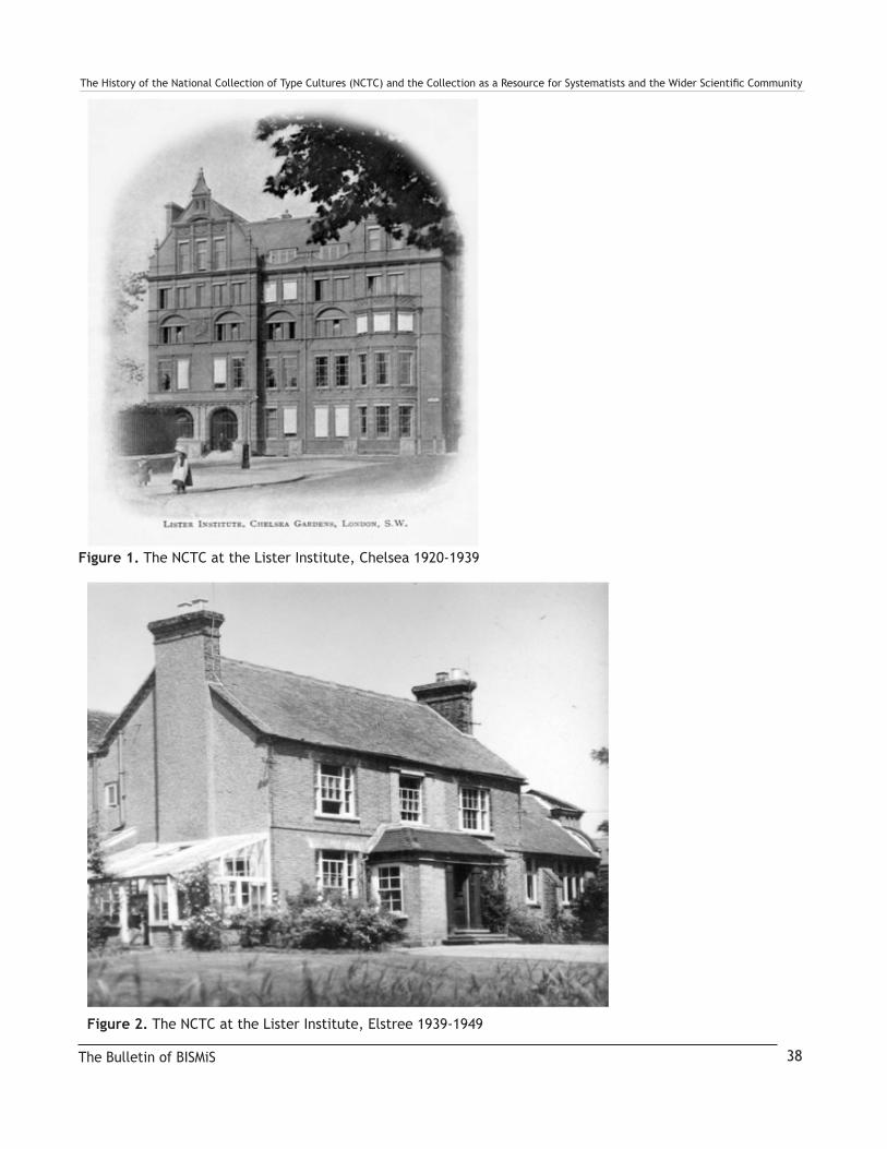
TheHistoryoftheNationalCollectionofTypeCultures(NCTC)andtheCollectionasaResourceforSystematistsandtheWiderScientificCommunity
The Bulletin of BISMiS 38
Figure 1. The NCTC at the Lister Institute, Chelsea 1920-1939
Figure 2. The NCTC at the Lister Institute, Elstree 1939-1949
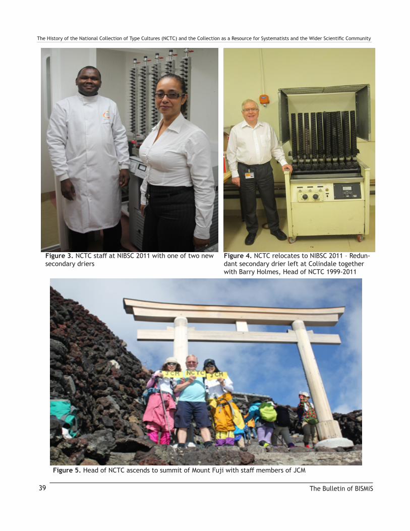
TheHistoryoftheNationalCollectionofTypeCultures(NCTC)andtheCollectionasaResourceforSystematistsandtheWiderScientificCommunity
The Bulletin of BISMiS39
Figure 3.NCTCstaffatNIBSC2011withoneoftwonewsecondary driers
Figure 4. NCTC relocates to NIBSC 2011 – Redun-dant secondary drier left at Colindale together with Barry Holmes, Head of NCTC 1999-2011
Figure 5.HeadofNCTCascendstosummitofMountFujiwithstaffmembersofJCM

TheHistoryoftheNationalCollectionofTypeCultures(NCTC)andtheCollectionasaResourceforSystematistsandtheWiderScientificCommunity
The Bulletin of BISMiS 40
Figure 6.NCTCstaffcirca 1990
Figure 7.NCTCstaffcirca 2000

TheHistoryoftheNationalCollectionofTypeCultures(NCTC)andtheCollectionasaResourceforSystematistsandtheWiderScientificCommunity
The Bulletin of BISMiS41
Figure 8. Curators of the NCTC
1920-1946 R. St John-Brooks 1947-1965 Samuel Tertius Cowan
1965-1978 Stephen Paget Lapage 1978-1994 Leslie Roland Hill 1995-1999 Menelaos Costas

TheHistoryoftheNationalCollectionofTypeCultures(NCTC)andtheCollectionasaResourceforSystematistsandtheWiderScientificCommunity
The Bulletin of BISMiS 43
Figure 9. 13x6 cards in original cabinets
Bulletin of the BISMiS (2018), Volume 7, part 2, pp 44-48
The importance of microbes in biotechnological, agricultur-al and industrial applications has inspired many Institutes to explore indigenous microbial diversity from various ecolog-ical niches of Pakistan. Among these, the most common ex-amples are the strains associated with plant growth-promot-ing activities that could be used as biofertilizers (Abbas et al. 2015a; Ahmed et al. 2014c; Bangash et al. 2015; Hayat et al. 2010); strains for bio-remediation of heavy-metals polluted soils/water systems (Abbas et al. 2015a; Abbas et al. 2014; Abbas et al. 2015b); strains useful in food processing indus-try (Schmidt et al. 2012); pathogenic strains responsible for the bacterial blight of rice (Nagarajkumar et al. 2004) and citrus canker; and other extremophilic (i.e. NaCl tolerant, heavy-metals tolerant) strains (Abbas et al. 2015a; Abbas et al. 2014; Abbas et al. 2015b; Bangash et al. 2015; Jamil et al. 2013; Roohi et al. 2012; Roohi et al. 2014a; Roohi et al. 2014b) for novel biotechnological applications. However, very few of these microbes have been confirmed at the spe-cies level based upon 16S rRNA gene sequence (Abbas et al. 2015a; Abbas et al. 2014; Abbas et al. 2015b; Ahmed et al. 2014a; Ahmed et al. 2014b; Ahmed et al. 2014c; Ahmed et al. 2014d; Amin et al. 2016a; Amin et al. 2016b; Amin et al. 2016c; Amin et al. 2018; Amin et al. 2017; Bangash et al. 2015) and many have been incorrectly identified being published based on morphological data alone (Fatima et al. 2009; Hasan et al. 2009; Ikram-ul-Haq et al. 2009). Most of the strains collected by various labs were either stocked in glycerol as unidentified strains or discarded over time due to contamination or loss of interest of researchers at end of the particular projects. There are many examples within vari-ous life sciences departments of Pakistani Universities or research organizations, where no old authentic isolated and purified strains have been retained. Despite tireless efforts of researchers from various institutes, new bacterial species are rarely published from Pakistan, except those reported recently by our group at National Agricultural Research Centre (NARC), Islamabad in addition to few others (Imran et al. 2010; Khalid et al. 2015). Major reasons include a lack of facilities and specialized equipment for chemotaxonomic
analyses as well as expertise required for the validation pro-cess of novel species. There is a big challenge to validly pub-lish any novel species from Pakistani ecology because only few efforts have been made systematically to recognize the strains at national and/or international level based upon sci-entific background and molecular tagging (gene sequence).
Microorganisms are classified in risk groups signifying their safety level; only strains from group risk 1 are considered as safe and can be used for product development. If the process of identification is not performed with the latest methods, it may be possible that any pathogenic strains may be missed and become part of the developed bio product that belongs to risk group 2 or higher. Since any environment / ecology is a natural reservoir for human opportunistic pathogens, the taxonomical identification of the beneficial isolates must be performed at an early stage in the selection process during development of any bio-product. This must be performed using the best available technology, for example the se-quencing of conserved markers such as 16S rRNA, 23S rRNA etc. Indeed, microbial strains should be identified by molecular biological methods, mainly at DNA level, which allows the identification of organisms and the delineaton of said organisms to the correct risk groups and recognize op-portunistic pathogens. Contamination in microbial culture is also another common problem, thus, identification of mi-crobial strains to be used in any product development which is going to be commercialized, its quality control should be managed over time with some standards. It’s a matter of fact that commercialized product should be registered or patented so identification of beneficial microbes should also be a prerequisite to patent or register any kind of bio product such as biofertilizer or microbial process. The identification process involves the use of partial sequence analysis of the 16S rRNA gene besides a basic characterization, the analy-sis of the cellular fatty acids, differentiating classical physi-ological and biochemical tests and/or ribotyping analysis etc. Thus, a taxonomically unassigned strain needs to be identified and characterized by a wide range of approaches
Importance of Microbial Culture Collection in Pakistan: Challenges and OpportunitiesIftikhar Ahmed1, Saira Abbas2 and Hamza Tariq1
Contact Details1Microbial Genetic Resources Program (MGRP), Bioresources Conservation Institute (BCI), National Agricultural Research Centre (NARC), Islamabad-45500, Pakistan.
2Department of Zoology, University of Science and Technology, Bannu, Pakistan
Correspondence Email: [email protected]
© BISMiS 2018 44

Importance of Microbial Culture Collection in Pakistan: Challenges and Opportunitiesto obtain a broad range of informative data from the ge-netic and epigenetic level, including morphology, physiol-ogy, chemistry, DNA patterns and sequences, and whole genome hybridization.
Microbial Culture Collections play an important role in of-fering services not only to preserve microorganisms using techniques that maintain viability, purity, and important characteristics of microorganisms, but also to supply high quality microorganisms for use in teaching, research and industrial applications. In order to meet modern demands for further advancement of bio-economy, a concept of bio-repository of biological materials and information in Paki-stan has been introduced recently: Pakistan Bio-Resources Centre (Pak-BRC) for conservation of bio-assets of Paki-stan. Pak-BRC is considered to be one of the key scientific infrastructures to support agriculture sector, the health sec-tor and the industrial sector or other sectors, and in turn en-sure that these advances help to drive economic growth at desired pace in the subjects of biotechnology, agriculture and life sciences industry. Thus, Pak-BRC is fundamental to harnessing and preserving our microbial biodiversity and serve as an essential element of the infrastructure for research and development. Pak-BRC is also necessary to protect the intellectual property rights of microbial assets of country through management of data bases and DNA tagging the biological material using molecular techniques. Consequently, it will improve usage of microbial materials while facilitating collaborative research and development programs without duplication of effort at the national level.
In Pakistan, extensive research work has been conducted on microflora and continues. However, only few labo-ratories in Research Institutes and Universities have maintained microbes for their specific research purposes such as NARC, Islamabad, University of Agriculture and NIBGE, Faisalabad; Punjab University, Lahore, etc.. Long term preservation of microbes is a neglected pro-cess in Pakistan. According to World Data Centre for Microorganisms (WDCM), there have been following five Culture Collection Centres registered from Pakistan (WFCC, 2018; http://www.wfcc.info/ccinfo) (Table 1).
Among these, PTCC and NCCP deal with bacteria, whereas ESBL-Pak mostly deals with pathogenic microbes, while other two mainly involved in the collection of fungus strains. However, since the establishment of PTCC for the last two decades, none of the strain has been characterized or vali-dated at species level. There are 15,625 species of bacteria (as of May 5 2017) identified globally (LPSN, www.bacte-rio.net), but from a rich ecolosystem of Pakistan, but to date, only 27 species have been reported, validated and published
(Abbas et al. 2015a; Abbas et al. 2014; Abbas et al. 2015b; Ahmed et al. 2014a; Ahmed et al. 2014c; Amin et al. 2016a; Amin et al. 2016b; Amin et al. 2016c; Amin et al. 2018; Amin et al. 2017; Bangash et al. 2015; Roohi et al. 2014b). In Pakistan due to diverse nature and due to limited taxo-nomic research on identification of microbial strains, there is a huge potential to explore our own microbial assets and identify novel species. Microbiota of any region or ecol-ogy may be specific to that environment, thus exploring the microbiota of Pakistan may furnish some incredibly use-ful and rarely explored taxa / strains that may revolutionize the biotech industry and be important in other applications.
Recently, Pakistan Agricultural Research Council (PARC) took an initiative to improve identification and preservation of economically important bacterial strains from Pakistani ecosystems. In this regard, PARC established microbial bio-resources repository: National Culture Collection of Paki-stan (NCCP) for preservation of this bio-asset of Pakistan. During our studies for collection and preservation of indig-enous beneficial strains many promising novel candidate strains were identified based upon 16S rRNA gene sequenc-ing, which can be delineated as novel species. However, to meet the minimum standards to describe any novel species of bacteria, many chemotaxonomic experiments need spe-cialized equipment not available in the majority of laborato-ries in Pakistan. This NCCP project was initiated in 2009 and later, a complete project that included the important infra-structure was approved in 2015 by the Ministry of National Food Security and Research. But unfortunately, no resourc-es were released to NCCP for the development of resources.
Limited lab facilities and meager resources led to the es-tablishment of international collaborative links with Ja-pan, China and Korea for delineation of our candidate strains as novel species. Now, for the first time in the history of Pakistani research, these productive collabora-tions have made it possible to validly publish the follow-ing novel species of bacteria from Pakistani ecosystems:
Continued on next page
The Bulletin of BISMiS45
◊ Novel genus Caldovatus sediminis gen. nov., sp. nov. (validated 2017)
◊ Tepidimonas alkaliphilus sp. nov., (published 2017 and validated 2018)
◊ Tepidimonas sediminis sp. nov. (published 2017 and validated 2018)
◊ Phenylobacterium tarrae sp. nov., (published and vali-dated 2018)

Importance of Microbial Culture Collection in Pakistan: Challenges and Opportunities
Since, identification and systematics of bacteria have not been extensively included in the main applied microbiologi-cal research work in Pakistan, the rich unexplored ecology of Pakistan may offer an opportunity to explore many ad-ditional novel species of bacteria by the application of poly-phasic taxonomic characterization and their preservation in the National Culture Collection of Pakistan (NCCP). The emphasis will be focused on the addition of novel beneficial microorganisms useful in agriculture and industry. There is a growing need to collect many more strains from vari-ous facilities for identification and preservation in NCCP.
The major gaols of NCCP would be:
I. A large number of economically important bacteria will be identified using molecular techniques and these will be available to biotechnology and industry. The identified (and characterized) strains will be utilized as plant growth-promoters and can be commercially made available to the farming community.
II. A large number of economically important strains preserved in NCCP as a resource for future scientific and academic research.
III. Changing climate conditions affects microbial diversity and may result in loss of many beneficial strains. NCCP will help to protect and restore these endangered micror-ganisms as a global resource.
IV. Human Resource development would be another output, as researchers of NCCP and students involved in these studies would be trained and work in collaboration with other Institutes on identification and chemotexonomic characterization of novel strains as a new species. Thus, a highly trained workforce would be established within Pakistan.
ReferencesAbbas S, Ahmed I, Iida T, Lee Y-J, Busse H-J, Fuji-
wara T, Ohkuma M (2015a) A heavy-metal tolerant novel bacterium, Alcaligenes pakistanensis sp. nov., isolated from industrial effluent in Pakistan. Antonie van Leeuwenhoek 108:859–870 doi:10.1007/s10482-015-0540-1
Abbas S, Ahmed I, Kudo T, Iida T, Ali GM, Fujiwara T, Ohkuma M (2014) Heavy metal-tolerant and psy-chrotolerant bacterium Acinetobacter pakistanensis sp. nov. isolated from a textile dyeing wastewater treatment pond. Pak J Agr Sci 51:595-608
Abbas S, Ahmed I, Kudo T, Iqbal M, Lee YJ, Fujiwara T, Ohkuma M (2015b) A heavy metal tolerant novel bacterium, Bacillus malikii sp. nov., isolated from tan-nery effluent wastewater. Antonie van Leeuwenhoek 108:1319-1330 doi:10.1007/s10482-015-0584-2
Ahmed I, Abbas S, Kudo T, Iqbal M, Fujiwara T, Oh-kuma M (2014a) Deinococcus citri sp. nov., isolated from citrus leaves canker lesion. Int J Syst Evol Mi-crobiol 64:4134-4140 doi:10.1099/ijs.0.066555-0
Ahmed I, Ehsan M, Sin Y, Paek J, Khalid N, Hayat R, Chang YH (2014b) Sphingobacterium pakistanen-sis sp. nov., a novel plant growth promoting rhizo-bacteria isolated from rhizosphere of Vigna mungo. Antonie van Leeuwenhoek 105:325-333 doi:10.1007/s10482-013-0077-0
The Bulletin of BISMiS 46
◊ Nocardioides thalensis sp. nov., (validated 2017)
◊ Thermus caldifontis sp. nov., (validated 2017)
◊ Streptomyces caldifontis sp. nov., (validated 2017)
◊ Microvirga pakistanensis sp. nov., (published 2016, validated 2017)
◊ Nocardioides pakistanensis sp. nov., (validated 2016)
◊ Bacillus malikii sp. nov. (validated 2016)
◊ Alcaligenes pakistanensis sp. nov., (validated 2016)
◊ Kushneria pakistanensis sp. nov., (validated 2015)
◊ Acinetobacter pakistanensis sp. nov., (validated 2015)
◊ Deinococcus citri sp. nov., (validated 2014)
◊ Lysinibacillus composti sp. nov., (validated 2014)
◊ Lysinibacillus pakistanensis sp. nov., (validated 2014)
◊ Bacillus pakistanensis sp. nov., (validated 2014)
◊ Sphingobacterium pakistanensis sp. nov., (validated 2014)
◊ Cellulomonas pakistanensis sp. nov., (validated 2014)

Importance of Microbial Culture Collection in Pakistan: Challenges and OpportunitiesAhmed I, Kudo T, Abbas S, Ehsan M, Iino T, Fujiwara T,
Ohkuma M (2014c) Cellulomonas pakistanensis sp. nov., a novel moderately halotolerant Actinobacteria. Int J Syst Evol Microbiol 64:2305-2311 doi:10.1099/ijs.0.059063-0
Ahmed I, Sin Y, Paek J, Ehsan M, Hayat R, Iqbal M, Chang YH (2014d) Description of Lysinibacillus pakistanensis. Int J Agri Biol 16:447-450
Amin A et al. (2016a) Microvirga pakistanensis sp. nov., a novel bacterium isolated from desert soil of Cho-listan, Pakistan. Archives of microbiology 198:933-939 doi:10.1007/s00203-016-1251-3
Amin A, Ahmed I, Habib N, Abbas S, Xiao M, Hozzein WN, Li WJ (2016b) Nocardioides pakistanensis sp. nov., isolated from a hot water spring of Tatta Pani in Pakistan. Antonie van Leeuwenhoek 109:1101-1109 doi:10.1007/s10482-016-0711-8
Amin A, Ahmed I, Khalid N, Osman G, Khan IU, Xiao M, Li WJ (2016c) Streptomyces caldifontis sp. nov., isolated from a hot water spring of Tatta Pani, Kotli, Pakistan. Antonie van Leeuwenhoek doi:10.1007/s10482-016-0778-2
Amin A, Ahmed I, Khalid N, Zhang Y, Xiao M, Li W-J (2018) Insights into the Thermophile Diversity in Hot Springs of Pakistan. In: Egamberdieva D, Birkeland N-K, Panosyan H, Li W-J (eds) Extremophiles in Eurasian Ecosystems: Ecology, Diversity, and Ap-plications. Springer Singapore, Singapore, pp 1-28. doi:10.1007/978-981-13-0329-6_1
Amin A et al. (2017) Diversity and distribution of ther-mophilic bacteria in hot springs of Pakistan. Micro-bial Ecol:1-12 doi:10.1007/s00248-017-0930-1
Bangash A, Ahmed I, Abbas S, Kudo T, Shahzad A, Fu-jiwara T, Ohkuma M (2015) Kushneria pakistanensis sp. nov., a novel moderately halophilic bacterium iso-lated from rhizosphere of a plant (Saccharum spon-taneum) growing in salt mines of the Karak area in Pakistan. Antonie van Leeuwenhoek 107:991-1000 doi:10.1007/s10482-015-0391-9
Fatima Z, Saleemi M, Zia M, Sultan T, Aslam M, Rehm-an R, Chaudhary MFJAJoB (2009) Antifungal activ-ity of plant growth-promoting rhizobacteria isolates against Rhizoctonia solani in wheat. 8
Hasan F, Khan S, Shah AA, Hameed AJPJB (2009) Pro-duction of antibacterial compounds by free and im-mobilized Bacillus pumilus SAF1. 41:1499-1510
Hayat R, Ali S, Amara U, Khalid R, Ahmed I (2010) Soil beneficial bacteria and their role in plant growth promotion: a review. Ann Microbiol 60:579-598 doi:10.1007/s13213-010-0117-1
Ikram-ul-Haq AS, Saleem A, Javed MJPJB (2009) Mutagenesis of Bacillus licheniformis through ethyl methane sulfonate for alpha amylase production. 41:1489-1498
Imran A, Hafeez FY, Fruhling A, Schumann P, Malik KA, Stackebrandt E (2010) Ochrobactrum ciceri sp. nov., isolated from nodules of Cicer arietinum. Int J Syst Evol Microbiol 60:1548-1553 doi:10.1099/ijs.0.013987-0
Jamil M, Zeb S, Anees M, Roohi A, Ahmed I, Rehman Su, Rhae Es (2013) Role of Bacillus licheniformis in phytoremediation of nickel contaminated soil culti-vated with rice. International Journal of Phytoreme-diation (http://dxdoiorg/101080/152265142013798621):Online Version
Khalid R et al. (2015) Rhizobium pakistanensis sp nov., isolated from groundnut (Arachis hypogaea) nodules grown in rainfed Pothwar, Pakistan. Anton Leeuw Int J G 107:281-290 doi:10.1007/s10482-014-0326-x
Nagarajkumar M, Bhaskaran R, Velazhahan R (2004) Involvement of secondary metabolites and extracel-lular lytic enzymes produced by Pseudomonas fluo-rescens in inhibition of Rhizoctonia solani, the rice sheath blight pathogen. Microbiol Res 159:73-81
Roohi A, Ahmed I, Iqbal M, Jamil M (2012) Preliminary isolation and characterization of halotolerant and halophilic bacteria from salt mines of Karak, Paki-stan. Pakistan Journal of Botany 44:365-370
Roohi A, Ahmed I, Khalid N, Iqbal M, Jamil M (2014a) Isolation and Phylogenetic Identification of Halo-tolerant/Halophilic Bacteria from the Salt Mines of Karak, Pakistan. International Journal of Agriculture and Biology 16:564-570
Roohi A, Ahmed I, Paek J, Sin Y, Abbas S, Jamil M, Chang YH (2014b) Bacillus pakistanensis sp. nov., a halotolerant bacterium isolated from salt mines of the Karak Area in Pakistan. Antonie van Leeuwenhoek 105:1163-1172 doi:10.1007/s10482-014-0177-5
Schmidt VS, Wenning M, Scherer S (2012) Sphingobac-terium lactis sp. nov. and Sphingobacterium alimen-tarium sp. nov., isolated from raw milk and a dairy environment. Int J Syst Evol Microbiol 62:1506-1511 doi:10.1099/ijs.0.036327-0.
The Bulletin of BISMiS47
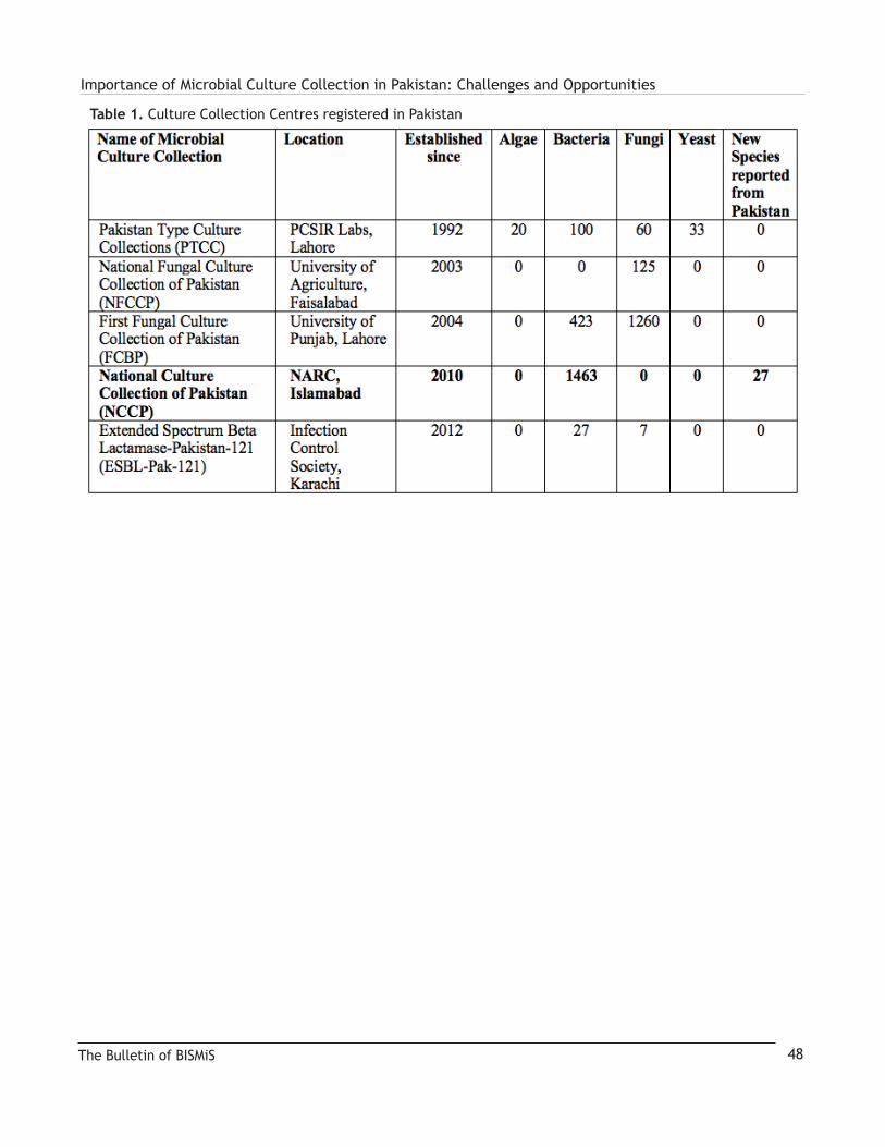
Importance of Microbial Culture Collection in Pakistan: Challenges and Opportunities
The Bulletin of BISMiS 48
Table 1. Culture Collection Centres registered in Pakistan

Bulletin of the BISMiS (2018), Volume 7, part 2, pp 49-54
Abstract
Halophilic archaea are salt loving microorganisms that usu-ally inhabit hypersaline environments like solar salterns, salt lakes, salt deposits, salt crystals etc. and rarely from low saline environments like sea water. India is rich in microbial diver-sity. The coastal regions so far explored for isolation of halo-archaea include Rajasthan, Gujarat, Maharashtra, Goa and Tamil Nadu, salt lake of Rajasthan, soda lake of Lonar, North Maharashtra, Andaman Islands. Haloarchaea from these re-gions have also been isolated and identified. Haloarchaeal genera namely Natronobacterium, Natronococcus, Halofer-ax, Halobacterium, Natrinema, Haloarcula, Halorubrum, Halogeometricum, Halococcus, Halolamina, Halosarcina, Halostagnicola, Haloterrigena, Natrialba, Natronoarchaeum, Haloadaptatus and Chromohalobacter have been described.
The beauty of our mother earth lies in its diversity of liv-ing species including microorganisms. Geological activi-ties have created a number of habitats that have shaped the evolution of microorganism leading to the establishment of a high diversity of different organisms. Some of these environments exhibit extreme conditions e.g. high tempera-ture in thermal springs, high salinity and alkalinity in soda lakes, high salinity in salt deposits etc. Man made activi-ties also lead to the formation of extreme environments e.g. acid mine drainage, solar saltpans, cement manufacturing, potato process waters etc. Microorganisms that survive and live in such extreme environments are termed as extremo-philes. Halophiles are one of these groups of organisms that thrive in saline environments. Compared to thermophilic, acidophilic and alkaliphilic microorganisms, less documen-tation has been made in case of halophilic microorganisms.
Halophilic microorganisms inhabit saline environments such salt lakes, salt deposits, solar salterns, salt pans, sa-line ponds, etc. Depending upon their requirement of salt for growth, they are classified as slight halophile (0.2-0.5 M NaCl), moderate halophile (0.5-2.5 M NaCl), border-line extreme halophile (1.5-4.0 M NaCl) and extreme halophile (> 2.5 M NaCl). Halotolerant organisms do not require NaCl for growth but can tolerate high content of
salt (0.0 to 5.2 M NaCl) (Grant et al., 2001). The haloar-chaea are classified under the domain Archaea, phylum Euryarchaeota, class Halobacteria, orders Halobacteriales, Haloferacales and Natrialbales, families Halobacteriaceae, Haloarculaceae, Halococcaceae, Haloferacaceae, Halo-rubraceae and Natrialbaceae (Gupta et al., 2015, 2016)
For isolation of halophilic archaea, different nutrient media with high concentration (3-4 M) NaCl have been devised e.g. Sehgal and Gibbons (SG) medium (Sehgal and Gib-bons, 1960); Tindall medium (Tindall and Truper, 1986) etc. Because of the high concentrations of NaCl in the nutrient media, selective isolation of halophilic archaea and elimina-tion of halotolerant microorganisms is achieved. Character-istic features of halophilic archaea include typical pink red/ orange red coloured colonies, pleomorphic nature of cells, requirement of 1.5 M NaCl for growth, ether lipids in cell membrane, resistance to antibiotic chloramphenicol, inhibi-tion of growth by sodium taurocholate, requirement of Mg2+, amino acids for growth in case of some species and more importantly lysis of cells in water and low saline solution. Globally, haloarchaea have been reported from different sa-line environments. A number of authros have reviewed these organisms; Rodriguez-Valera et al. (1979) reported isolation of extreme halophiles from low saline environment i.e. sea-water. The Dead Sea, a terminal lake at the border between Israel and Jordon, and the Great Salt Lake, Utah, USA hasve been extensively studied for 20 years for isolation of halo-archaeal strains. Oren et al. (1990) reported the isolation of novel haloarchaeal species viz. Halobacterium gomorrense, Haloarcula marismortui, Haloferax volcanii from Dead Sea. Oren et al (2000) also reviewed isolation of novel haloar-chaeal species from various eco niches such as salt pans, solar salts, crystallizer ponds of solar salterns, hypersaline lakes, salt mines as well as hypersaline soda lakes. Presence of halophilic microorganisms and haloarchaea from ancient rock salt and salt sediments have been reported by several re-searchers viz. (Stan-Lotter et al., 1999, 2002) as reviewed by Grant et al., (1998) and McGenity et al., (2000). Kanekar et al. (2012) also reviewed taxonomy, diversity and physiology of halophilic microorganisms including halophilic archaea.
Halophilic Archaea from saline environments in IndiaPradnya P. Kanekar and Snehal O. Kulkarni
Contact Details
Corresponding author: Pradnya P. Kanekar (retired)
Department of Biotechnology, Modern College of Arts, Science and Commerce,
Shivajinagar, Pune 411005, M. S. ,India. [email protected]
© BISMiS 2018 49

Halophilic Archaea from saline environments in IndiaIndia is a country endowed with rich biodiversity including of microorganisms isolated from natural saline environments include Sambhar salt lake in Rajasthan (Fig. 1), Great and Little Rann of Kutch, Gujarat, soda lake of Lonar, Buldhana District, North Mahatrashtra, low saline coastal sediments, sand and sea water of Arabian sea across West Coast of In-dia. Man-made habitats include mainly solar salterns from Mumbai, Goa, Kandla Port, Gujarat, Kanyakumari, Tamiln-adu, Shiroda, Sindhudurga District, etc. The West coast of India popularly known as Western Ghats are recognized as hot spots of Biodiversity. This coastal region is spread over from Gujarat to Tamilnadu in South India through Maharash-tra, Goa and Kerala and explored for halophilic biodiversity.
The first report of isolation of haloarchaea from India was from Sambhar salt lake, Rajasthan. The lake is both saline and alkaline and situated at 26° 58 ‘N and 75° 5’E in the middle of a closed depression in the Aravalli mountain ranges, approximately 65 Km northwest of Jaipur. Upasani and Desai (1990) reported chemical composition of brines of Sambhar lake and isolated six strains of haloalkaliphi-lic archaea from brines taken from the main lake as well as solar evaporation pans of Sambhar lake. The salt con-tent of the brines ranged from 12-30 % and pH 9.5-10.5. The authors used modified Brown medium having pH 9.5 and varied the content of NaCl and KCl. The strains were studied for their lipid profile and found to contain phos-phatidylglycerophosphate (PGP), phosphatidylglycerol (PG), and phosphatidic acid (PA).The haloarchaeal strains were identified as Natronobacterium and Natronococcus. Natronobacterium strains SSL 1 and SSL6 isolated from Sambhar Lake were found to contain novel glycolipid.
Gujarat coastal region has been extensively explored for halophilic microorganisms, the major salt producing sites in Gujarat are Saurashtra at Kandala, Jamnagar, Maliya, Mithapur, Porbander, and Bhavnagar which become the natural habitats for haloarchaea. Haloarchaea have been isolated from marine salterns at Mithapur, Gujarat (Upasani et al., 1994). The organisms were identified as Haloferax MSW1 (NCMB 2287) and Halobacterium MSW 5 (NCMB
2288). These along with the other haloarchaeal strains were studied for their lipids. Dave and Desai (2006) studied the microbial diversity of marine salterns near Bhavnagar. The sample sites i.e. marine salterns are located at 22° 10’ N and 72° 15 ‘E on the Western and Northern coasts of the Gulf of Khambhat, Arebian Sea, about 25-30 Km north of Bhavnagar. Out of 73 selected isolates of halophiles, six iso-lates were found to be highly pleomorphic and halophilic. They were identified as Natrinema thermotolerant. Halo-philic archaebacterium have been isolated from salt pan sit-uated in Kandla and identified as Halobacterium sp. (NCBI Gen Bank Accession No. DQ465014) (Akolkar et al.,2008).
Thomas et al (2012) investigated the distribution of halo-archaea in hypersaline marshy environment of Little and Great Rann of Kutch, Gujarat. The region cov-ered for collection of soil and water samples lies be-tween 23°10.727’N, 070°43.733’E of Little Rann and 23°57.7215’N, 069°43.7611’E of Great Rann of Kutch. The haloarchaeal population was represented by the genera Halobacterium, Natrinema, Haloferax, Haloarcula, Halo-rubrum, Halogeometricum, Halomicrobium, and some uncultured haloarchaeal clones. Phosphate solubilizing haloarchaea isolated from Rann of Kutch, Gujarat included genera Haloarcula, Halobacterium, Halococcus, Halofer-ax, Halolamina, Halosarcina, Halostagnicola, Haloter-rigena, Natrialba, Natrinema and Natronoarchaeum.
The coastal region of Western Maharashtra is popular for tourist destinations such as Mumbai Fort, Gate Way of In-dia, Shreewardhan and Harihareshwar of Raigad District, Sindhudurga Fort, Rock Garden Malvan, Deobag, Tarkar-li of Sindhudurga District, Bhagavati port of Ratnagiri, Thiba palace of Ratnagiri District and many other places. A number of solar salt pans are also in use. e.g. Mulund, Bhandup, Bhainder of Mumbai, Shiroda of Sindhudurga District, etc. All these saline environments are populated by halophiles but comparatively less explored. Phylogenetic analysis of archaeal Ribosomal DNA sequences from salt pan sediment of Mumbai have been documented (Ahmad et al., 2008). Upper sediment (2-10 cm distant from sur-face) samples were collected from salt pans of Mumbai, lo-cated geographically between 18°53’Nand 19°20’N latitude and 72°45’E and 73°00’E longitude. Total genomic DNA was extracted from samples and PCR amplification of 16S rRNA genes was performed using archaea specific primers. Cloning of environmental PCR products was carried out followed by Restriction Fragment Length Polymorphism (RFLP) screening of rDNA clones and nucleotide sequenc-ing of cloned SSU rRNA genes. Phylogenetic analysis of cloned SSU rRNA gene sequences was performed by comparing the closest database relatives of all sequences
The Bulletin of BISMiS50
Figure 1. Sambhar Lake Rajasthan showing pink red bloom of haloarchaea

Halophilic Archaea from saline environments in Indiagenerated to 16 S rRNA gene sequences available at NCBI. Based on the similarity values, the closest relatives among Euryarchaeota were found to be Haloarcula amylolytica, Haladaptatus pauchihalophilus and many uncultured Archaea including methanogenic species. Pathak and Sar-dar (2012) have isolated haloarchaea from solar saltern of Mulund (E) 19°10’12”N, 72°57’18’’E, Mumbai. Based on biochemical and physiological characterization, the halo-archaeal strains were found to belong to genera Chromo-halobacter, Haloferax, Halobacterium and Halorubrum.
Isolation of halophilic archaea from sea water and soil samples from salt production sites of Mumbai have been documented (Digaskar et al., 2015). The haloarchaeal genus Haloferax was found to be dominant in the saltern ecosys-tem. Thombre and Oke (2015) have reported the Studies on stress proteins induced by temperature stress in extremely halophilic archaeon Haloferax mediterranei RT 18 iso-lated from surface water samples of Marine Drive area of Mumbai. Soluble proteins were isolated, separated by SDS-PAGE and identified by MALDI-TOF MS-MS analysis. The stress protein identified as beta proteosomal subunit is found to play role in maintaining protein homeostatis.
Kanekar et al. (2015a) explored rock- pit sea water from Rock Garden, Malvan, Sindhudurga District, West Coast of Maharashtra for isolation of haloarchaea. Eleven halo-archaeal strains obtained were characterized and identified based on 16S rRNA gene sequencing as Haloastagnicola larsenii. This is presumably the first report of the isola-tion of Halostagnicola larsenii from Arabian sea water, West Coast of India. Kanekar et al. (2015b) have reported Halostagnicola sp. from sand and sea water of Andaman Islands. Kanekar et al. (2016a) have also investigated colo-nization of rocks of Rock Garden, Malvan, Sindhudurga District by the haloarchaea imparting red coloration to the rock, the organism was identified as Haloferax larse-nii. This is presumably the first report of isolation of halo-archaea from red coloured rocks of rocky beach. Patil et al. (2016) have reported haloalkaliphilic archaea namely Natrinema sp., Natrialba wudunaoensis, Natrialba chah-annaoensis, Natronobacterium innermongoliae from al-kaline and saline Lonar Lake, North Maharashtra, India.
Goa is a coastal state (15°34’60 N, 74°0’E) and one of the beautiful tourist places of India having natural beaches en-joyed by the tourists. Both the sea water and salt pans in and around Goa have been explored for isolation of halophiles. Branganca and Furtado (2009) isolated and characterized haloarchaea from low salinity coastal sediments and waters of Goa. Water and sediment samples were collected from coastal regions of Goa namely Talpona, Vasco, Dona Paula,
Miramur and Bainguinim. The haloarchaeal isolates ob-tained using nutrient media containing 20 % salt were tenta-tively identified as Halobacterium and Haloarcula sp. Mani et al (2012) reported isolation of halophilic archaea from natural solar saltern of Goa. Salt making is being practiced for many years in Goa. Salt pans are located in Pernem, Bar-dez, Tisawadi and Salcete talukas of Goa. The authors se-lected a single solar saltern at Ribandar (15°30’N, 73°51’E) to examine culturable haloarchaeal diversity. Water and sed-iment samples were collected 0-10 cm distance from surface twice during the two phases of salt production. In pre-salt harvesting phase /initial stage/salt dilute stage, the halo-archaeal strains of Halococcus salifodinae were obtained while in salt harvesting phase/ crystallization stage, haloar-chaeal strains of Halococcus salifodinae, Haloferax volcanii, Haloarcula argentinensis, Haloarcula japonica, Haloarcula sp., Halorubrum sp., and Haloferax alexandrinus were ob-tained. The carotenoid pigment was extracted from the or-ange red growth of the isolates and spectrophotometrically confirmed as bacterioruberin. Metal tolerance of these halo-archaeal strains was investigated by Chaudhary et al. (2014). The strains of Halococcus salifodinae, Haloferax volcanii, Haloarcula japonica and Halorubrum sp. could grow and tolerate cadmium concentration ranging from 0.5-4 mM.
The peninsular coast of India particularly the Kanyakumari coast is the beautiful tourist place in India. Haloarchaea were isolated from saltern soil samples (K.R.T. Asha et al., 2005). There are over 5000 salterns in the coastal re-gions of Kanyakumari, thus making the site a congenial environment for growth of haloarchaea. Based on cultural, morphological and biochemical characterization, the halo-archaeal isolates were tentatively identified as Haloar-cula vallismortis (HA3) and Haloarcula quadrata (HA9).
Manikandan et al. (2009) have studied microbial diversity of solar salterns of Tamil Nadu. Samples from the crys-tallizer ponds were collected from multipond solar salt-erns in Kelambakkam, Marakanam, Vedaranyam of Tamil Nadu. The haloarchaeal isolates belonged to Haloferax sp. Haloferax larsenii, Halorubrum sp., Halogeometricum borinquense, Halobacterium sp., Haloarcula sp., Haloru-brum sodomense, Halobacterium salinarum and Haloferax volcanii. Thus the microbial community of Tamil Nadu salterns was found to be dominated by haloarchaeal gen-era Haloferax and Halorubrum. Haloarchaea were isolated from soil samples collected from salt pans of Kanyaku-mari. The strain BTSH 10 was identified as Natrinema sp. Among the various saline environments, rock salt has been looked upon as a source of haloarchaea by researchers over the globe. The presence of viable microorganisms from an-cient rock salt was described by Reiser and Tasch (1960)
The Bulletin of BISMiS 51

Halophilic Archaea from saline environments in Indiaand Dombrowski (1963). Norton et al. (1993), Stan-Lotter et al.(1999), McGenity et al. (2000) ,Stan-Lotter et al. (2002) and Gruber et al. (2004) reported the presence of haloarchaeal strains of Haloarcula, Halorubrum, Halococ-cus salifodinae, Halococcus dombrowskii sp. nov., Halo-bacterium noricense from various rock salt deposits and mines (Fig.2). Black Salt is a type of rock salt commonly called as Himalayan Black salt. It is salty, pungent smelling condiment used in South Asia. The Black Salt contains sodi-um chloride, trace impurities of sodium sulphate, sodium bi-sulphate, sodium bisulphide, iron sulfide and hydrogen sul-phide. Due to presence of iron sulphide, it forms brownish pink to dark violet translucent crystals when whole. When ground into powder, it looks light purple to pink in colour (Fig.3). Black salt is considered as a cooling spice in Indian Ayurvedic preparations and reported to help in relieving in-testinal gas, heartburn, hysteria etc. (Ali, 1999 ; Sadhale and nene, 2004; Aggarwal and Kotwal,2009). Since there were many reports regarding occurrence of haloarchaeal strains in different rock salts and the commercial Indian black salt looks pink in colour, it was thought to be a source of haloar-chaea. Kanekar et al (2016 b) have isolated Halostagnicola larsenii IBS (MCC 2956) from Indian Black salt procured from local market by enrichment of the sample in Sehgal and Gibbons (SG) medium (Sehgal and Gibbon, 1960) con-taining 3.42 M NaCl. Pure culture obtained from well iso-lated pink-red colony was identified based on morphologi-cal, physiological and biochemical characterization and 16 S rRNA gene sequencing (Fig. 4). This is first report of iso-lation of Halostagnicola larsenii from commercial Indian black salt. Earlier Henriet et al. (2014) reported occurrence of two haloarchaeal genera viz. Halarchaeum acidiphilum and Halobacterium noricense from Himalayan pink salt.
Conclusion
Halophilic archaea are unique microorganisms endowed with the property to live and grow in high saline environments like salt lakes, solar salterns, salt deposits, ancient rock salt mines, salt crystals and rarely from low saline environments like sand and sea water. Moderate attempts have been made to study their biodiversity mainly from West Coast of India viz. coastal regions of Gujarat, Maharashtra, Goa and Tamil Nadu, salt lake of Rajasthan, soda lake of Lonar, North Ma-harashtra, Andaman Islands. Thus in closing it is apparent that a limited number of saline sites have been explored from India for isolation of haloarchaea and there are important opportunities to study biodiversity of halophiles from India.
The Bulletin of BISMiS52
Figure 2. Rock salt mine in Himachal Pradesh, India
Figure 3. Indian Black salt showing purplish black crystals and pink powder
Figure 4. Neighbor-joining phylogenetic tree based on 16 S rRNA gene sequences indicating the relatedness of the strain IBS to Halostagnicola larsenii XH-48T and some haloarchaeal species. Accession numbers are shown in parentheses. Boot-strap values (%) are based on 1000 replicates. Bar repre-sents expected changes per site.

Halophilic Archaea from saline environments in IndiaReferencesAggarwal H, Kotwal N (2009) Foods used as Ethno-
medicine in Jammu. Ethno-Med 3: 65-68Ahmad N, Sharma S, Khan FG, Kumar R, Johri S, Ab-
din MZ, Qazi GN (2008) Phylogenetic analyses of Archaeal ribosomal DNA sequences from salt pan sediments of Mumbai, India. Curr Microbiol DOI:10.1007/s00284-008-9167-z
Akolkar AV, Deshpande GM, Raval KN, Durai D, Nerurkar A & Desai AJ. (2008) Organic solvent tol-erance of Halobacterium sp.1(1) & its extracellular protease. J. Basic Microbiol. 48:421-425
Ali ZA, (1999) Folk veterinary medicine in Moradabad District (Uttar Pradesh), India. Fitoterapia 70: 340-347 (DOI:10.1016/S0367-326X(99)00039-8)
Braganca JM, Furtado I (2009) Isolation and character-ization of haloarchaea from low salinity coastal sedi-ments and waters of Goa. Curr. Sci. 96: 1182-1184
Chaudhary A, Imran Pasha M, Salgaonkar BB, Bra-gance JM (2014) Cadmium tolerance by haloarchaeal strains isolated from solar salterns of Goa, India. Int Jour Biosci, Biochem Bioinfo. 4: 1-6.
Dave SR and Desai HB. (2006) Microbial diversity at marine salterns near Bhavnagar, Gujarat, India. Curr. Sci. 90: 497-500
Digaskar V U, Thombre RS, Oke RS (2015) Screening of extremely halophilic archaea for its biotechnologi-cal potential. Int Jour of Pharma and Biosciences. 6 (B): 811-819.
Dombrowski H (1963) Bacteria from Paleozoic salt de-posits. Ann NY Acad Sci. 108: 453-460
Grant WD, Kamemkura M, McGenity TJ, Ventosa A (2001) Class III Halobacteria class, nov. In Bergey’s Manual of systematic Bacteriology. Ed. By D.R. Boone, R. W. Castenholz, G. M. Garrity, Vol. 1, 2nd edn. Springer, New York, pp.294-334.
Grant WD, Gemmell RT, McGenity TJ (1998) Extremo-philes 2: 279-287
Gruber C, Legat A, Pfaffenhuemer M, Radax C, Wei-dler G, Busse HJ, Stan-Lotter H(2004) Halobac-terium noricense sp. nov., an archaeal isolate from a bore core of an alpine Permo-Triassic salt deposit, classification of Halobacterium sp.NRC-1 as a strain of Halobacterium salinarum and emended descrip-tion of Halobacterium salinarum. Extremophiles, 8: 431-439.
Gupta RS, Naushad S, Baker S (2015) Phylogenomic analyses and molecular signatures for the class Halo-bacteria and its two major clades : a proposal for division of the class Halobacteria into an emended order Halobacteriales and two new orders, Halofera-cales ord. nov. and Natrialbales ord. nov., contain-ing the novel families Haloferacaceae fam.nov. and Natrialbaceae fam.nov. Int. J. Syst. Evol. Microbiol. 65: 1050-1069.
Gupta RS, Naushad S, Fabros R, Adeolu M (2016) A phylogenomic reappraisal of family-level divisions within the class Halobacteria: proposal to divide the order Halobacteriales into the families Halobacte-riaceae, Haloarculaceae fam. nov., and Halococca-ceae fam. Nov., and the order Haloferacales into the families. Haloferacaceae and Halorubraceae fam nov. Antonie Van Leeuwenhoek, 109:565-587
Henriet O, Fourmentin J, Delince B, Mahillon J (2014) Exploring the diversity of extremely halophilic ar-chaea in food grade salts. Int. Jour. Food. Microbial. 191: 36-44.
KRT Asha, Denslin AJ Vinitha, Sehgal G Kiran, WA Manjusha, N Sukumaran, J Selvin (2005) Isolation and cultivation of halophilic Archaea from solar salt-erns located in peninsular coast of India. The Int J Microbiol, 1.
Kanekar PP, Kanekar SP, Kelkar AS , Dhakephalkar PK. (2012) Halophiles- Taxonomy, Diversity, Physiology and Applications. In Microorganisms in Environ-mental Management. Editors, Satyanarayana, Johri and Anil Prakash, Publisher- Springer Business, B.V., pp 1-34.
Kanekar PP, Kulkarni SO, Kanekar SP, Shouche Y, Jani K, Sharma A (2015 a) Exploration of a haloarchaeon, Halostagnicola larsenii, isolated from rock pit sea water, West Coast of Maharashtra, India, for pro-duction of bacteriorhodopsin. J Appl Microbiol 118: 1345-1356
Kanekar SP, Saxena N, Pore SD, Arora P, Kanekar PP, Dhakephalkar PK (2015 b) Draft genome sequence of Halostagnicola sp. A56, an extremely halophilic archaeon isolated from Andaman Islands. Genome Announcements 3: e01332-15
The Bulletin of BISMiS 53

Halophilic Archaea from saline environments in IndiaKanekar PP, Kulkarni SO, Dhakephalkar PK, Kulkarni
KG, Saxena N. (2016 a) Isolation of an halophilic, bacteriorhodopsin-producing archeon, Haloferax larsenii RG3D.1 from the rocky beach of Malvan, West Coast of India. Geomicrobiol J. DOI: 10.1080/01490451.2016.1179365.
Kanekar PP, Kulkarni SO, Shouche Y, Jani K, Sharma A (2016 b) Isolation of bacteriorhodopsin producing haloarchaeon Halostagnicola larsenii IBS (MCC 2956) from Indian black. Bergey’s International So-ciety for Microbial Systematic (BISMiS) meeting proceeding, p 49.
Mani K, Salgaonkar BB, Branganca JM (2012) Cultur-able halophilic archaea at the initial crystallization stages of salt production in a natural solar saltern of Goa, India. Aquatic Biosystem 8: 15
Manikandan M, Kannan V, Pasic L (2009) Diversity of microorganisms in solar salterns of Tamil Nadu, In-dia. World J. Micro. Biotech. 25: 1001-1017.
McGenity TJ, Gemmell RT, Grant WD, Stan-Lotter H (2000) Origins of halophilic microorganisms in an-cient salt deposits. Env. Microbiol. 2: 243-250.
McGenity TJ, Gemmell RT, Grant WD, Stan-Lotter H (2000) Origins of halophilic microorganisms in an-cient salt deposits. Env. Microbiol. 2: 243-250.
Norton CF, McGenity TJ, Grant WD, (1993) Archaeal halophiles (halobacteria) from two British salt mines. J. Gen. Microbiol. 139: 1077-1081.
Oren A, Ginzburg M, Ginzburg Z, Hochstein I and Volcani BE (1990) Haloarcula marismortui (volcani) sp. nov.; nom. rev.; an extremely halophilic bacterium from the Dead Sea. Int J. Syst. Bacteriol. 40: 209-210
Pathak AP, Sardar AG (2012) Isolation and character-ization of carotenoid producing haloarchaea from solar saltern of Mulund, Mumbai, India. Ind Jour Natural products and Resources 3: 483-488.
Patil J, Suryawanshi P, Bajekal S (2016) Siderophores of haloalkaliphilic archaea from Lonar Lake, Maha-rashtra, India. Curr Sci. 111: 621-623
Reiser R, Tasch P (1960) Investigation of the viability of osmophile bacteria of great geological age. Trans Kans Acad Sci. 63: 31-34. (PubMed: 13740659)
Rodriguez-Valera F, Ruiz-Beeraquero F, Ramos-Cormenzana A (1979) Isolation of extreme halo-philes from sea water. Appl. Envoron. Microbiol 38:164-165.
Sadhale N, Nene YL (2004) On elephants in Manasol-lasa-2.Diseases and Treatment. Asian Agri- History 8: 115-127.
Sehgal SN, Gibbons NE. (1960) Effect of some metal ions on the growth of Halobacterium cutirubrum. Can. J. Microbiol. 6: 165-169.
Stan-Lotter H, McGenity TJ, Legat A, Denner EBM, Glaser K, Stetter KO, Wanner G (1999) Very similar strains of Halococcus salifodinae are found in geo-graphically separated Permo-Triassic salt deposits. Microbiol. 145: 3565-3574.
Stan-Lotter H, Pfaffenhuemer M, Legat A, Busse HJ, Radax C, Gruber C (2002) Halococcus dombrowskii sp. nov., an archaeal isolate from a Permo-Triassic alpine salt deposit. Int J Syst Evol Microbiol 52: 1807-1814.
Thomas M, Pal KK ,Dey R, Saxena AK, Dave SR (2012) A novel haloarchaeal lineage widely distrib-uted in the hypersaline marshy environment of Little and Great Rann of Kutch in India, Curr. Sci. 103: 1078-1084.
Thombre RS and Oke RS (2015) Study of stress proteins induced by temperature stress in extremely halophil-ic archaea, Haloferax mediterranei RT 18. Int J Curr Microbiol Appl Sci . Special issue-2:199-209.
Tindall BJ and Truper HG (1986) Ecophysiology of the aerobic halophilic archaebacteria . Syst. Appl. Mi-crobiol. 7: 202-212
Upasani V and Desai S (1990) Sambhar salt lake Chemical composition of the brines and studies on haloalkaliphilic archaebacteria. Arch. Microbiol. 154:589-593.
Upasani VN, Desai SG, Moldveanu N, Kates M (1994) Lipids of extremely halophilic archaeobacteria from saline environments in India: a novel glycolipid in Natronobacterium strains. Microbiol 140: 1959-1966.
The Bulletin of BISMiS54

Bulletin of the BISMiS (2018), Volume 7, part 2, pp 55-57
Since my undergraduate days, I have been fascinated by the nature of microbial plant interactions and how they might be used to improve agricultural and environmental practices. I am especially interested in nitrogen-fixing actinobacteria belonging to the genus Frankia as they form mutualistic as-sociations with dicotyledenous plants that might be used to stabilise sand dunes and decrease the spread of the Sahara Desert. Plants infected with Frankia are classified in over 200 dicotyledenous plants which represent 8 angiosperms families (Torrey and Tjepkema, 1979). These associations lead to the development of root nodules on host plants where the microsymbionts fix nitrogen for the benefit of the macrosymbionts (Magallon et al., 1999; Normand and Ben-son, 2012). The high rates of nitrogen-fixation achieved by actinorhizal plants allows them to colonise marginal soils thereby opening up the prospect of using such plants as pioneers in nitrogen-deficient soils, in land reclamation, as a source of timber and pulp (Benson and Silvester, 1993).
The ecological significance of actinorhizal association led to intensive studies on host plant ecology, mechanisms of infection and root nodule physiology (Benson and Silvester, 1993). In contrast, until recently, somewhat sporadic prog-ress was made unravelling the subgeneric classification of Frankia strains mainly due to difficulties in isolating and characterising such slow-growing actinobacteria, problems compounded by the limited availability of representative strains from culture collections. These limitations meant that different Frankia strains were used in molecular systematic studies (Akimov et al., 1991; Akimov and Dobritsa, 1992; An et al., 1983; Bloom et al., 1989; Fernandez et al., 1989; Lumini and Bosco, 1996), including comparative analyses of conserved gene sequences (Murry et al., 1995 ; Bautista et al., 2011 ). Nevertheless, it became apparent from phy-logenetic, morphological and physiological studies that the genus was markedly heterogeneous even though only a sin-gle species, Frankia alni (Woronin 1866; Von Tubeuf 1895) was recognised albeit without a recognised type strain. Fur-thermore, it also became clear from phylogenetic studies based on 16S rRNA (Normand et al., 1996), gyrB, glnII genes (Nouioui et al., 2011), 16S-23S rRNA internal tran-scribed spacers (Ghodhbane-Gtari et al., 2010), multi-locus gene sequences (atp1, ftsZ, dnaK, gyrA and sec) (Gtari et
al., 2015) and core genome studies (Tisa et al., 2016) that most Frankia strains could be assigned to four clusters, the members of which showed distinct host ranges. Cluster I strains infect host plants belonging to the Belutaceae and Casuarinaceae (except Gymnostoma); those assigned to cluster 2 infect Ceanothus (Rhamnaceae), the subfamily Dryadoideae (Rosaceae), Coriariaceae and Datiscaceae while the more genetically diverse strains of cluster 3 are infective Colletieae (all actinorhizal Rhamnaceae except Ceanothus), Myricaceae, Gymnostoma and occasionally on Alnus. The remaining lineage encompasses “atypical” strains which are unable to re-infect actinorhizal host plants or form infective root nodules, and /or unable to fix nitrogen.
My PhD project was designed to caste light on the phylog-eny and evolution of Frankia strains based on the anlaysis of glnII, gyrB, nifH and 16S-23S rRNA spacer sequences. It was evident from the resultant concatenated trees that the strains fell into four evolutionary lineages, as described above, which provided the basis for subsequent phylogenet-ic taxonomic studies. The variation of the average pairwise distance between strains assigned to the four clusters sug-gested that over evolutionary time Frankia had undergone a gradual shift from a saprophytic to a facultative and mu-tualistic lifestyle. In a corresponding study based on bacte-rial and plant gene sequences, it was shown that Frankia strains associated to Coriaria has evolved independently.
These critical developments opened up the possibility of un-dertaking comprehensive phylogenetic studies on Frankia strains to resolve outstanding taxonomic issues. In a semi-nal polyphasic taxonomic study (Nouioui et al., 2016), it was proposed that Frankia alni ACN14a, isolated in pure culture from Alnus viridis subspecies crispa with mor-phological properties matching the original description of F. alni, be recognised as the type strain of this species according to Rule 18f of the International Code of No-menclature of Prokaryote. Two additional species were proposed for strains CcI3 and BMG5.12 isolated from Casuarina cunninghamiana and Elaeagnus angustifolia, namely Frankia casuarinae and Frankia elaeagni, respec-tively. These isolates, like the F. alni strain, were based on the results of chemotaxonomic and phenotypic studies and on complementary molecular systematic data retrieved from full genome sequences. Subsequently, 10 additional Frankia species have been proposed for strains assigned
My life with the genus Frankia: an ongoing love affairImen Nouioui
Contact DetailsResearch Associate, Newcastle University, UK
© BISMiS 2018 55

My life with the genus Frankia:anongoingloveaffairto clusters 1 to 4, as exemplified by, Frankia canadensis (Nor-mand et al., 2018), Frankia torreyi (Nouioui et al., 2018a), Frankia coriariae (Nouioui et al., 2017a), Candidatus Fran-kia datiscae (Persson et al., 2011), Candidatus Frankia cali-forniensis (Normand et al., 2017), Frankia discariae (Noui-oui et al., 2017b), Frankia elaeagni (Nouioui et al., 2016), Frankia irregularis (Nouioui et al., 2018b), Frankia asym-biotica (Nouioui et al., 2017c), Frankia inefficax (Nouioui et al., 2017d), and Frankia saprophytica (Nouioui et al., 2018c).
The marked improvements in the classification of the ge-nus Frankia together with the availability of more than 35 genome sequences of representative strains provide an in-valuable platform not only for promoting our understanding of actinorhizal symbionts, but also for exploitable biology. It is, for instance, particularly interesting that the genome of Frankia strains are rich in natural product biosynthetic gene clusters that encode for the biosynthesis of novel and uncharacterized specialized metabolites, some of which may prove to be of value in the search for new antibiotics needed to control multi-drug resistant microbial pathogens.
In addition, the availability of strains representing well described species open up the prospect of selecting ap-propriate strains for the reclamation of polluted (Richards et al., 2002; Rehan et al., 2014a,b; Baker et al., 2015) and saline soils (Tani and Sasakawa, 2003) and in raising the fertility of marginal land (Diagne et al., 2013) and as bio-inoculants for enhancing the growth of specific host plants.
References
Akimov VN, Dobritsa SV. (1992). Grouping of Frankia strains on the basis of DNA relatedness. Syst Appl Microbiol 15; 372–379.
Akimov VN, Dobritsa SV, Stupar OS. (1991). Grouping of Frankia strains by DNA:DNA homology: How many genospecies are in the genus Frankia? Ni-trogen fixation. In Polsinelli M et al (eds) Develop-ments in Plant and Soil Sciences. Kluwer Academic Publisher, the Netherlands, pp. 635–636.
An CS, Wills JW, Riggsby WS, Mullin BC. (1983). De-oxyribonucleic acid base composition of 12 Frankia isolates. Can J Bot 61:2859–2862.
Baker E, Tang Y, Chu F, Tisa LS. (2015). Molecular responses of Frankia sp. strain QA3 to naphthalene. Can J Microbiol 61:281–29.
Bautista GHH, Cruzb HA, Nesme X, Valdés M, Men-doza HA, et al. (2011). Genomospecies identifica-
tion and phylogenomic relevance of AFLP analysis of isolated and non-isolated strains of Frankia spp. Syst Appl Microbiol 34:200–206.
Benson DR, Silvester WB. (1993). Biology of Fran-kia strains, actinomycete symbionts of actinorhizal plants. Microbiol Rev 57: 293–319.
Bloom RA, Lechevalier MP, Tate RL. (1989). Physi-ological, chemical, morphological, and plant infec-tivity characteristics of Frankia isolates from Myrica pennsylvanica: Correlation to DNA restriction pat-terns. Appl Environ Microbiol 55:2161–2166
Diagne N, Arumugam K, Ngom M, Nambiar-Veetil M, Franche C, et al. (2013). Use of Frankia and actinorhizal plants for degraded lands reclamation. BioMed Research International 2013:9.
Fernandez MP, Meugnier H, Grimont PAD, Bardin R. (1989). Deoxyribonucleic acid relatedness among members of the genus Frankia . Int J Syst Bacteriol 39:424–429.
Ghodhbane-Gtari F, Nouioui I, Chair M, Boudabous A, Gtari M. (2010). 16S-23S rRNA intergenic spacer region variability in the genus Frankia. Microb Ecol 60:487-495.
Gtari M, Ghodhbane-Gtari F, Nouioui I, Ktari A, Hez-bri K, et al (2015) Cultivating the uncultured:- grow-ing the recalcitrant cluster-2 Frankia strains. Sci Rep 5:13112.
Lumini E, Bosco M. (1996). PCR-restriction fragment length polymorphism identification and host range of single-spore isolates of the flexible Frankia sp. strain UFI 132715. Appl Environ Microbiol 62:3026–3029.
Magallon S, Crane PR, Herendeen PS. (1999). Phylo-genetic pattern, diversity, and diversification of eudi-cots. Ann Mo Bot Gard 86:297–372.
Murry MA, Zhang D, Schneider M, Bruijn DFJ. (1995). Use of repetitive sequences and the polymerase chain reaction to fingerprint the genomes of Frankia isolates. Symbiosis 19:223–240.
Normand N, Nouioui I, Pujic P, Fournier P, Dubost A, et al (2018) Frankia canadensis sp. nov.,isolated from root nodules of Alnus incana ssp rugosa growing in Canada. Int J Syst Evol Microbiol 68: 3001-3011.
Normand P, Benson DR. (2012). Order XVI Frankia-les. In: Goodfellow M et al (eds) Bergey’s Manual of Systematic Bacteriology, vol 5, 2nd edn, The Actino-bacteria. Springer, New York, pp 508–510.
The Bulletin of BISMiS56

My life with the genus Frankia:anongoingloveaffairNormand P, Nguyen TV, Battenberg K, Berry AM, Heu-
vel BV, et al. (2017). Proposal of ‘Candidatus Fran-kia californiensis’, the uncultured symbiont in nitro-gen-fixing root nodules of a phylogenetically broad group of hosts endemic to western North America. Int J Syst Evol Microbiol 67:3706–3715.
Normand P, Orso S, Cournoyer B, Jeannin P, Chapelon C, et al. (1996). Molecular phylogeny of the genus Frankia and related genera and emendation of the family Frankiaceae. Int J Syst Bacteriol 46:1–9.
Nouioui I, Ghodhbane-Gtari F, Beauchemin NJ, Tisa LS, Gtari M. (2011). Phylogeny of members of the Frankia genus based on gyrB, nifH and glnII se-quences. Antonie Van Leeuwenhoek 100:579–587.
Nouioui I, Ghodhbane-Gtari F, Klenk H-P, Gtari M. (2018c). Frankia saprophytica sp. nov. an atypical non-infective (Nod–) and non-nitrogen fixing (Fix–) actinobacterium isolated from Coriaria nepalensis root nodules. Int J Syst Evol Microbiol 68:1090-1095.
Nouioui I, Ghodhbane-Gtari F, Jando M, Tisa LS, Klenk H-P, et al. (2018a). Frankia torreyi sp. nov., the first actinobacterium of the genus Frankia Brunchorst 1886, 174AL isolated in axenic culture. Antonie van Leeuwenhoek. https://doi.org/10.1007/s10482-018-1131-8
Nouioui I, Ghodhbane-Gtari F, Montero-Calasanz MD, Göker M, Meier-Kolthoff JP, et al. (2016). Proposal of a type strain for Frankia alni (Woronin 1866) Von Tubeuf 1895, emended description of Frankia alni and recognition of Frankia casuarinae sp. nov. and Frankia elaeagni sp. nov. Int J Syst Evol Microbiol 66:5201–5210.
Nouioui I, Ghodhbane-Gtari F, Montero-Calasanz MDC, Rohde M, Tisa LS, et al. (2017d). Frankia in-efficax sp. nov., an actinobacterial endophyte induc-ing ineffective, non nitrogen-fixing, root nodules on its actinorhizal host plants. Antonie Van Leeuwen-hoek 110:313-320.
Nouioui I, Ghodhbane-Gtari F, Rhode M, Sangal V, Klenk H-P, et al. (2018b) Frankia irregularis sp. nov., an actinobacterium unable to nodulate its original host, Casuarina equisetifolia, but effectively nodu-late members of the actinorhizal Rhamnales. Int J Syst Evol Microbiol 68:2883-2914.
Nouioui I, Ghodhbane-Gtari F, Rohde M, Klenk H-P, Gtari M. (2017c). Frankia coriariae sp. nov., an infective and effective microsymbiont isolated
from Coriaria japonica. Int J Syst Evol Microbiol 67:1266-1270.
Nouioui I, Gueddou A, Ghodhbane-Gtari F, Rhode M, Gtari M, Klenk H-P (2017d) Frankia asymbiotica sp. nov., a non infective actinobacterium isolated from Morella californica root nodule. Int J Syst Evol Mi-crobiol 67:4897-4901.
Nouioui I, Montero-Calasanz MDC, Ghodhbane-Gtari F, Rohde M, Tisa LS, et al. (2017b) Frankia discari-ae sp. nov.: an infective and effective microsymbiont isolated from the root nodule of Discaria trinervis. Arch Microbiol 199:641-647.
Persson T, Benson DR, Normand P, Vanden Heuvel B, Pujic P, et al. Genome sequence of “Candidatus Frankia datiscae” Dg1, the uncultured microsym-biont from nitrogen-fixing root nodules of the dicot Datisca glomerata. J Bacteriol 193:7017-7018.
Rehan M, Furnholm T, Finethy RH, et al. (2014a) Cop-per tolerance in Frankia sp. strain EuI1c involves surface binding and copper transport. Appl Micro-biol Biotechnol 98:8005-8015.
Rehan M, Kluge M, Franzle S, Kellner H, Ullrich R, et al. (2014b). Degradation of atrazine by Frankia alni ACN14a: gene regulation, dealkyation, and dechlori-nation. Appl Microbiol Biotechnol 98:6125-6135.
Richards JW, Krumholz GD, Chval MS, Tisa LS. (2002). Heavy metal resistance patterns of Frankia strains. Appl Environ Microbiol 68:923-927.
Tani C, Sasakawa H. (2003). Salt tolerance of Casua-rina equisetifolia and Frankia Ceq1 strain isolated from the root nodules of C. equisetifolia. Soil Sci Plant Nutr 49:215–222.
Tisa LS, Oshone R, Sarkar I, Ktari A, Sen A, Gtari M. (2016). Genomic approaches toward understanding the actinorhizal symbiosis: an update on the status of the Frankia genomes. Symbiosis 70:5–1.
Torrey JG, Tjepkema JD. (1979). Symbiotic nitrogen fixation in actinomycete nodulated plants. Bot Gaz 140: i–ii
Von Tubeuf K. (1895). Pflanzenkrankheiten durch Kryptogame Parasiten verursacht. Verlag J Springer, Berlin, pp 1–599..
Woronin MS. (1866). Über die bei der Schwarzerle (Alnus glutinosa) und bei der gewöhnlichen Garten-Lupine (Lupinus mutabilis) auftretenden Wurzelan-schwellungen. Mem Acad Imp Sci St Petersbourg VII Series 10:1–13.
The Bulletin of BISMiS 57

Finally, we have some in-house news concerning the Bulletin. Since 2013, my graduate student, Nisha Patel (Fig 1), served as the Managing Editor. This summer after earning her PhD, she graduated and after a number of job offers took a position in Boston, MA with a Microbiome company. On behalf of BISMiS, I want to personally thank Dr. Nisha Patel for her tremendous efforts over the years and wish her well for her future in taking her taxonomic knowledge to our colleagues in industry. The Bulletin Managaing Editor position and duties have been assumed by one of my new graduate students, Shannon Fulton (Fig 2), and she has already performed a sterling job with this issue.
-Dr. Paul A. Lawson Editor-in-Chief
The Bulletin of BISMiS58
Figure 1. Dr. Nisha Patel
Figure 2. Shannon Fulton
Bulletin Editorial (cont’d)