The Antimicrobials Anacardic Acid - Docusalut
17
ORIGINAL RESEARCH published: 26 February 2019 doi: 10.3389/fmicb.2019.00326 Edited by: Octavio Luiz Franco, Universidade Católica de Brasília, Brazil Reviewed by: Jason John Paxman, La Trobe University, Australia Claus Schneider, Vanderbilt University, United States *Correspondence: Francisco J. Fernández [email protected] M. Cristina Vega [email protected] † Present address: Javier Querol-García, Instituto de Investigación Sanitaria del Hospital Clínico San Carlos, Madrid, Spain Gara Sánchez-Barrón and Francisco J. Fernández, Abvance Biotech srl, Madrid, Spain ‡ These authors have contributed equally to this work Specialty section: This article was submitted to Antimicrobials, Resistance and Chemotherapy, a section of the journal Frontiers in Microbiology Received: 13 August 2018 Accepted: 07 February 2019 Published: 26 February 2019 Citation: Gómez S, Querol-García J, Sánchez-Barrón G, Subias M, González-Alsina À, Franco-Hidalgo V, Albertí S, Rodríguez de Córdoba S, Fernández FJ and Vega MC (2019) The Antimicrobials Anacardic Acid and Curcumin Are Not-Competitive Inhibitors of Gram-Positive Bacterial Pathogenic Glyceraldehyde-3-Phosphate Dehydrogenase by a Mechanism Unrelated to Human C5a Anaphylatoxin Binding. Front. Microbiol. 10:326. doi: 10.3389/fmicb.2019.00326 The Antimicrobials Anacardic Acid and Curcumin Are Not-Competitive Inhibitors of Gram-Positive Bacterial Pathogenic Glyceraldehyde-3-Phosphate Dehydrogenase by a Mechanism Unrelated to Human C5a Anaphylatoxin Binding Sara Gómez 1‡ , Javier Querol-García 1†‡ , Gara Sánchez-Barrón 1† , Marta Subias 1,2 , Àlex González-Alsina 3 , Virginia Franco-Hidalgo 1 , Sebastián Albertí 3 , Santiago Rodríguez de Córdoba 1,2 , Francisco J. Fernández 1 * † and M. Cristina Vega 1 * 1 Center for Biological Research, Spanish National Research Council, Madrid, Spain, 2 CIBER de Enfermedades Raras, Madrid, Spain, 3 Institut Universitari d’Investigació en Ciències de la Salut, University of the Balearic Islands, Mallorca, Spain The ubiquitous and highly abundant glycolytic enzyme D-glyceraldehyde-3-phosphate dehydrogenase (GAPDH) is pivotal for the energy and carbon metabolism of most organisms, including human pathogenic bacteria. For bacteria that depend mostly on glycolysis for survival, GAPDH is an attractive target for inhibitor discovery. The availability of high-resolution structures of GAPDH from various pathogenic bacteria is central to the discovery of new antibacterial compounds. We have determined the X-ray crystal structures of two new GAPDH enzymes from Gram-positive bacterial pathogens, Streptococcus pyogenes and Clostridium perfringens. These two structures, and the recent structure of Atopobium vaginae GAPDH, reveal details in the active site that can be exploited for the design of novel inhibitors based on naturally occurring molecules. Two such molecules, anacardic acid and curcumin, have been found to counter bacterial infection in clinical settings, although the cellular targets responsible for their antimicrobial properties remain unknown. We show that both anacardic acid and curcumin inhibit GAPDH from two bacterial pathogens through uncompetitive and non-competitive mechanisms, suggesting GAPDH as a relevant pharmaceutical target for antibacterial development. Inhibition of GAPDH by anacardic acid and curcumin seems to be unrelated to the immune evasion function of pathogenic bacterial GAPDH, since neither natural compound interfere with binding to the human C5a anaphylatoxin. Keywords: GAPDH – glyceraldehyde-3-phosphate dehydrogenase, Streptococcus pyogenes, Clostridium perfringens, anacardic acid, curcumin, complement – immunological term, enzyme inhibition, X-ray crystallography Frontiers in Microbiology | www.frontiersin.org 1 February 2019 | Volume 10 | Article 326
-
Upload
khangminh22 -
Category
Documents
-
view
1 -
download
0
Transcript of The Antimicrobials Anacardic Acid - Docusalut

fmicb-10-00326 February 26, 2019 Time: 13:0 # 1
ORIGINAL RESEARCHpublished: 26 February 2019
doi: 10.3389/fmicb.2019.00326
Edited by:Octavio Luiz Franco,
Universidade Católica de Brasília,Brazil
Reviewed by:Jason John Paxman,
La Trobe University, AustraliaClaus Schneider,
Vanderbilt University, United States
*Correspondence:Francisco J. Fernández
[email protected]. Cristina Vega
†Present address:Javier Querol-García,
Instituto de Investigación Sanitaria delHospital Clínico San Carlos,
Madrid, SpainGara Sánchez-Barrón and
Francisco J. Fernández,Abvance Biotech srl, Madrid, Spain
‡These authors have contributedequally to this work
Specialty section:This article was submitted to
Antimicrobials, Resistanceand Chemotherapy,
a section of the journalFrontiers in Microbiology
Received: 13 August 2018Accepted: 07 February 2019Published: 26 February 2019
Citation:Gómez S, Querol-García J,
Sánchez-Barrón G, Subias M,González-Alsina À, Franco-Hidalgo V,
Albertí S, Rodríguez de Córdoba S,Fernández FJ and Vega MC (2019)The Antimicrobials Anacardic Acid
and Curcumin Are Not-CompetitiveInhibitors of Gram-Positive Bacterial
PathogenicGlyceraldehyde-3-Phosphate
Dehydrogenase by a MechanismUnrelated to Human C5a
Anaphylatoxin Binding.Front. Microbiol. 10:326.
doi: 10.3389/fmicb.2019.00326
The Antimicrobials Anacardic Acidand Curcumin Are Not-CompetitiveInhibitors of Gram-Positive BacterialPathogenicGlyceraldehyde-3-PhosphateDehydrogenase by a MechanismUnrelated to Human C5aAnaphylatoxin BindingSara Gómez1‡, Javier Querol-García1†‡, Gara Sánchez-Barrón1†, Marta Subias1,2,Àlex González-Alsina3, Virginia Franco-Hidalgo1, Sebastián Albertí3,Santiago Rodríguez de Córdoba1,2, Francisco J. Fernández1*† and M. Cristina Vega1*
1 Center for Biological Research, Spanish National Research Council, Madrid, Spain, 2 CIBER de Enfermedades Raras,Madrid, Spain, 3 Institut Universitari d’Investigació en Ciències de la Salut, University of the Balearic Islands, Mallorca, Spain
The ubiquitous and highly abundant glycolytic enzyme D-glyceraldehyde-3-phosphatedehydrogenase (GAPDH) is pivotal for the energy and carbon metabolism of mostorganisms, including human pathogenic bacteria. For bacteria that depend mostlyon glycolysis for survival, GAPDH is an attractive target for inhibitor discovery. Theavailability of high-resolution structures of GAPDH from various pathogenic bacteria iscentral to the discovery of new antibacterial compounds. We have determined the X-raycrystal structures of two new GAPDH enzymes from Gram-positive bacterial pathogens,Streptococcus pyogenes and Clostridium perfringens. These two structures, and therecent structure of Atopobium vaginae GAPDH, reveal details in the active site thatcan be exploited for the design of novel inhibitors based on naturally occurringmolecules. Two such molecules, anacardic acid and curcumin, have been found tocounter bacterial infection in clinical settings, although the cellular targets responsiblefor their antimicrobial properties remain unknown. We show that both anacardic acidand curcumin inhibit GAPDH from two bacterial pathogens through uncompetitive andnon-competitive mechanisms, suggesting GAPDH as a relevant pharmaceutical targetfor antibacterial development. Inhibition of GAPDH by anacardic acid and curcuminseems to be unrelated to the immune evasion function of pathogenic bacterial GAPDH,since neither natural compound interfere with binding to the human C5a anaphylatoxin.
Keywords: GAPDH – glyceraldehyde-3-phosphate dehydrogenase, Streptococcus pyogenes, Clostridiumperfringens, anacardic acid, curcumin, complement – immunological term, enzyme inhibition, X-raycrystallography
Frontiers in Microbiology | www.frontiersin.org 1 February 2019 | Volume 10 | Article 326

fmicb-10-00326 February 26, 2019 Time: 13:0 # 2
Gómez et al. Bacterial Pathogenic GAPDH Structure and Inhibition
INTRODUCTION
D-Glyceraldehyde-3-phosphate dehydrogenase (GAPDH, E.C.1.2.1.12) catalyzes the sixth glycolytic reaction (SupplementaryFigure S1). Before GAPDH, the previous reactions of glycolysisconsume energy in the form of two ATP molecules; incontrast, the following reactions generate four ATP moleculesso that glycolysis has a net energy-producing output. Therefore,GAPDH appears as one of the most promising enzymes toinhibit in order to interfere with glycolysis, since blocking theGAPDH-catalyzed reaction would still allow the cells to consumeenergy without being able to regenerate it. For organisms thatrely on glycolysis for all or most of their energy generatingreactions, inhibiting GAPDH offers a unique opportunity. Forexample, trypanosomatids thrive exclusively on glycolysis as it isperformed in a specialized organelle and a multitude of inhibitorshave been developed that are efficient at combating leishmaniosis(Suresh et al., 2001), Chagas’ disease (Souza et al., 1998) andother trypanosomiases (Lambeir et al., 1991; Vellieux et al.,1993; Verlinde et al., 1994; Kim et al., 1995; Bressi et al., 2001).These inhibitors have been designed to be highly selective forthe pathogen’s GAPDH in order to avoid being trapped by theabundant liver GAPDH.
The lessons learned in these inhibitor development campaignscan be transferred to the search for better inhibitors towardGAPDH of bacterial pathogens, some of which are greatly relianton the glycolysis. GAPDH shares an evolutionarily conservedfold consisting in two juxtaposed domains, an N-terminalNAD-binding domain (NBD) and a C-terminal catalytic domain.At the boundary between the two domains lies a catalyticcysteine residue. The cleft formed by the domain interfacehelps to stabilize the cofactor binding pocket and providesinteractions with the substrates and products. GAPDH requiresthe nicotinamide adenine dinucleotide (NAD+) cofactor thatremains tightly bound in the active site. The active site of GAPDHcontains also two structurally distinct phosphate-binding sites,the Pi and Ps sites, where inorganic phosphate and the phosphategroup from the glyceraldehyde-3-phosphate (G3P) substratebind, respectively, during reaction (Skarzynski and Wonacott,1988; Yun et al., 2000). In fact, there are two different Pi sitesdepending on the precise location within the active site of theinorganic phosphate moiety, which are termed the “classical” and“new” Pi sites (Yun et al., 2000; Mukherjee et al., 2010).
Besides its glycolytic role, GAPDH assumes many diversemoonlighting functions both in eukaryotes and prokaryotes.In these non-glycolytic functions, GAPDH participates incritical cellular processes like apoptosis and replication andit has been involved in the development of human diseasesand tumorigenesis. In pathogenic bacteria, GAPDH takes onvirulence and immunoevasive roles that require GAPDH to beexported to the extracellular space, either associated with cell wallstructures or completely detached from the GAPDH exportingbacterial cells (Sirover, 1997, 2005, 2011, 2012, 2014; Chauhanet al., 2017; Raj et al., 2017). The interaction of pathogenicbacterial GAPDH with complement components has recentlyemerged as an immune evasive mechanism that may be relevantfor infection biology and for the therapeutic management of
infectious diseases (Terao et al., 2006; Querol-García et al.,2017). Inhibition of the glycolytic or the moonlighting functionsof GAPDH has been posited as a valuable strategy to curbtrypanosomatid and bacterial infections (e.g., Querol-Garcíaet al., 2017). Both synthetic ADP analogs and natural productshave been established as antimicrobial agents targeting GAPDH(Pereira et al., 2008; Freitas et al., 2009), which has paved the wayfor their development as pharmaceutical lead compounds in thequest for new therapies against infectious diseases.
The recent structural determination of Atopobium vaginaeGAPDH at 2.1-Å resolution has provided detailed informationabout the configuration of the active site that can be usedfor the discovery of novel inhibitors. In this work, wehave solved two new structures of GAPDH from pathogenicGram-positive bacteria, Streptococcus pyogenes and Clostridiumperfringens, to 1.55 and 2.55 Å resolution, respectively. Thestructural analysis of these three GAPDH structures and theircomparison with trypanosomatid and human liver GAPDHprovides insight into the structural and chemical features thatselective inhibitors should have.
In particular, we have investigated the inhibitory propertiesof two natural compounds, anacardic acid and curcumin, whichare known to have antimicrobial properties against severalGram-positive pathogenic bacteria but lack a validated target(Cheng et al., 2001; Mamidyala et al., 2013; Sharma et al., 2013;Hamad and Mubofu, 2015; Hollands et al., 2016). Curcumin,in particular, has also shown promise for the treatment ofprecancerous cervical lesions and advanced breast cancer,uses for which it is being tested in ongoing clinical trials(Cheng et al., 2001; Ko and Moon, 2015; Karibe et al., 2018;Norouzi et al., 2018). The characterization of the inhibitorymodality of anacardic acid and curcumin has revealed that theyact through uncompetitive and non-competitive mechanisms.The combination of enzymatic, structural and computationalchemistry approaches sheds light on the mechanism of inhibitionand raises the opportunity to develop stronger, more efficientGAPDH-targeted inhibitors for antimicrobial therapy.
MATERIALS AND METHODS
Cloning, Expression, and Purification ofGAPDHGenomic DNA from a Clostridium perfringens type strain(DSM-756) and a synthetic optimized cDNA for Streptococcuspyogenes were used as template for PCR amplification of therespective GAPDH genes, SpGAPDH (UniProt AccessionNo. P68777) and CpGAPDH (UniProt Accession No.Q0STD4/Q8XKT9). Plasmids for expression of CpGAPDHand SpGAPDH were constructed by restriction-ligation cloningas tobacco etch virus (TEV)-cleavable N-terminal hexahistidinetagged fusions into pETM11 (Gunter Stier, European MolecularBiology Laboratory) and pET21b (Novagen), respectively(Querol-García et al., 2017). The expression plasmids weretransformed into E. coli Rossetta(DE3)pLysS cells (Novagen) andtransformants isolated in selective LB-agar plates. Transformantsfrom a freshly prepared plate or from a bacterial glycerol stock
Frontiers in Microbiology | www.frontiersin.org 2 February 2019 | Volume 10 | Article 326
fmicb-10-00326 February 26, 2019 Time: 13:0 # 3
Gómez et al. Bacterial Pathogenic GAPDH Structure and Inhibition
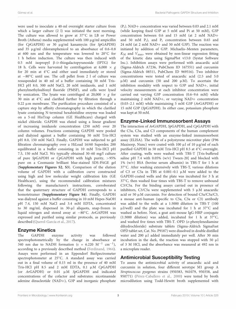
were used to inoculate a 40-ml overnight starter culture fromwhich a larger culture (2 l) was initiated the next morning.The culture was allowed to grow at 37◦C in LB or PowerBroth (Athena) media supplemented with 100 µg/ml ampicillin(for CpGAPDH) or 30 µg/ml kanamycin (for SpGAPDH)and 35 µg/ml chloramphenicol to an absorbance of 0.6–0.8at 600 nm and the temperature was lowered to 20◦C for1 h before induction. The culture was then induced with0.5 mM isopropyl β-D-thiogalactopyranoside (IPTG) for18 h. Cells were harvested by centrifugation at 6,000 × gfor 20 min at 4◦C and either used immediately or storedat −80◦C until use. The cell pellet from 2 l of culture wasresuspended in 40 ml of a buffer containing 50 mM Tris-HCl pH 8.0, 500 mM NaCl, 20 mM imidazole, and 1 mMphenylmethylsulfonyl fluoride (PMSF), and cells were lysedby sonication. The lysate was centrifuged at 20,000 × g for30 min at 4◦C and clarified further by filtration through a0.22 µm membrane. The purification procedure consisted of acapture step by affinity chromatography in which the clarifiedlysate containing N-terminal hexahistidine enzyme was loadedon a 5-ml HisTrap column (GE Healthcare) charged withnickel chloride. GAPDH was eluted using a linear gradientof increasing imidazole concentration (250 mM) over 20column volumes. Fractions containing GAPDH were pooledand dialyzed against a buffer containing 50 mM Tris-HClpH 8.0, 150 mM NaCl. Finally, GAPDH was subjected to gelfiltration chromatography over a HiLoad 16/60 Superdex 200equilibrated in a buffer consisting in 10 mM Tris-HCl pH7.5, 150 mM NaCl. We typically obtained 30–60 mg/l cultureof pure SpGAPDH or CpGAPDH with high purity, >95%pure on a Coomassie brilliant blue-stained SDS-PAGE gel(Supplementary Figures S2, S3). Comparison of the elutionvolume of GAPDH with a calibration curve constructedusing high and low molecular weight calibration kits (GEHealthcare), using the recommended calibration mixturesfollowing the manufacturer’s instructions, corroboratedthat the quaternary structure of GAPDH corresponds to ahomotetramer (Supplementary Figure S4). Finally, GAPDHwas dialyzed against a buffer consisting in 10 mM Hepes-NaOHpH 7.4, 150 mM NaCl and 3.4 mM EDTA, concentratedto 30 mg/ml, dispensed in 30-µl aliquots, snap-frozen inliquid nitrogen and stored away at −80◦C. AvGAPDH wasexpressed and purified using similar protocols, as previouslydescribed (Querol-García et al., 2017).
Enzyme KineticsThe GAPDH enzyme activity was followedspectrophotometrically by the change in absorbance at340 nm due to NADH formation (ε = 6.220 M−1 cm−1),according to a previously described method (Ferdinand, 1964).Assays were performed in an Eppendorf BioSpectrometerspectrophotometer at 25◦C. A standard assay was carriedout in a final volume of 0.15 ml in the presence of 40 mMTris-HCl pH 8.5 and 2 mM EDTA, 0.1 µM CpGAPDH(or AvGAPDH) or 0.01 µM SpGAPDH and indicatedconcentrations of the cofactor and substrates: nicotinamideadenine dinucleotide (NAD+), G3P and inorganic phosphate
(Pi). NAD+ concentration was varied between 0.03 and 2.1 mM(while keeping fixed G3P at 5 mM and Pi at 50 mM), G3Pconcentration between 0.6 and 15 mM (at 2 mM NAD+and 50 mM Pi), and Pi concentration between 0.65 and24 mM (at 2 mM NAD+ and 50 mM G3P). The reaction wasinitiated by addition of G3P. Michaelis–Menten parameters,KM and Vmax, were obtained by non-linear regression fittingof the kinetic data using SigmaPlot v13.0 (Systat SoftwareInc.). Inhibition assays were performed with anacardic acid(Sigma-Aldrich A7236, PubChem ID 167551) and curcumin(Sigma-Aldrich 08511, PubChem ID 969516). Two inhibitorconcentrations were tested of anacardic acid (2.5 and 5.0µM) and curcumin (50 and 200 µM). To ascertain theinhibition modality with respect to G3P and NAD+, initialvelocity measurements at each inhibitor concentration werecarried out varying G3P concentration (0.6–9.6 mM) whilemaintaining 2 mM NAD+, or varying NAD+ concentration(0.03–2.1 mM) while maintaining 5 mM G3P (AvGAPDH) or15 mM G3P (SpGAPDH). In either case, potassium phosphatewas kept at 50 mM.
Enzyme-Linked Immunosorbent AssaysThe interaction of AvGAPDH, SpGAPDH, and CpGAPDH withthe C5a, C3a, and C3 components of the human complementsystem was studied with an enzyme-linked immunosorbentassay (ELISA). The wells of a polystyrene microtiter plate (F96Maxisorp, Nunc) were coated with 100 µl of 10 µg/ml of eachpurified GAPDH in 50 mM Tris-HCl pH 8.5 at 4◦C overnight.After coating, wells were washed with TBS-T [Tris-bufferedsaline pH 7.4 with 0.05% (w/v) Tween-20] and blocked with1% (w/v) BSA (bovine serum albumin) in TBS-T for 1 h at37◦C. After washing extensively with TBS-T, various dilutionsof C3 or C5a in TBS at 0.001–0.1 µM were added to theGAPDH-coated wells and the plate was incubated for 3 h at37◦C, then washed four times with TBS-T to remove unboundC3/C5a. For the binding assays carried out in presence ofinhibitors, C3/C5a were supplemented with 5 µM anacardicacid or 50 µM curcumin. For detection of bound C5a/C3a/C3,a mouse anti-human (specific to C5a, C3a or C3) antibodywas added to the wells at a 1:3000 dilution in TBS-T (100µl/well) and the plate was incubated for 1 h at 37◦C andwashed as before. Next, a goat anti-mouse IgG-HRP conjugate(1:3000 dilution) was added, incubated for 1 h at 37◦C,and washed five times with TBS-T. OPD (o-phenylenediaminedihydrochloride) substrate tablets (Sigma-Aldrich SigmaFastOPD tablet set, Cat. No. P9187) were dissolved in double distilledwater and 200 µl added immediately per well. After 30 minincubation in the dark, the reaction was stopped with 50 µlof 3 M HCl, and the absorbance was measured at 492 nm ina microplate reader.
Antimicrobial Susceptibility TestingTo assess the antimicrobial activity of anacardic acid andcurcumin in solution, four different serotype M1 group AStreptococcus pyogenes strains (950383, 941079, 950358, and950771) (Pérez-Caballero et al., 2000) were tested by brothmicrodilution using Todd-Hewitt broth supplemented with
Frontiers in Microbiology | www.frontiersin.org 3 February 2019 | Volume 10 | Article 326

fmicb-10-00326 February 26, 2019 Time: 13:0 # 4
Gómez et al. Bacterial Pathogenic GAPDH Structure and Inhibition
0.5% (w/v) yeast extract (THY). Anacardic acid and curcuminwere obtained from Sigma-Aldrich (Catalog numbers A7236and 08511, respectively). Briefly, suspensions with a turbidityequivalent to that of a 0.5 McFarland standard were preparedby suspending the growth from overnight cultures on THY agarplates in 2 ml of sterile saline. The suspensions were furtherdiluted 1:10 to obtain a final inoculum of 5 × 10⁵ CFU/ml.The wells of microtiter plates containing 50 µl of the bacterialsuspension and 50 µl of anacardic acid or curcumin solutionsat different concentrations were incubated overnight in ambientair at 37◦C.
CrystallizationBefore attempting crystallization of GAPDH the requirednumber of aliquots was quickly thawed and centrifugedat 10,000 × g for 10 min at 4◦C to remove any potentialaggregates that might have resulted from a freezing-thawingcycle. A commercial Pre-Crystallization Test (HamptonResearch) was used to adjust the protein concentration toa suitable concentration for more extensive crystallizationscreenings, which was finally set to 7.5 mg/ml for SpGAPDHand 15 mg/ml for CpGAPDH. The full JCSG-plus sparsematrix and the PACT premier systematic PEG/Ion/pHscreenings (Molecular Dimensions) were performed by thesitting-drop vapor-diffusion method using drops containing 1µl either GAPDH stock supplemented with 0.6 mM NAD+and 1 µl crystallization condition at 20◦C. The optimumcrystallization conditions were 0.2 M potassium nitrate (whichcould be replaced by ammonium formate or ammoniumsulfate), 22% (w/v) PEG 3350 for SpGAPDH, and 0.2 Msodium acetate, 20% (w/v) PEG 3350 for CpGAPDH. Crystalswere then cryoprotected with 20% (v/v) sterile glycerol,mounted in standard MicroMount (MiTeGen) and flashfrozen in liquid nitrogen.
Data Collection and ProcessingDiffraction data were collected from flash-frozen crystals at 100K. Data for SpGAPDH crystals were collected on an ADSC CCDdetector at the ID232 beamline (ESRF, Grenoble, France) (Flotet al., 2010) to a resolution of 1.5 Å, and CpGAPDH crystalswere collected using a photon-counting Pilatus 6M (DECTRISLtd., Baden, Switzerland) at the BL13-XALOC beamline (ALBA,Barcelona, Spain) (Juanhuix et al., 2014) to 2.55 Å resolution.Complete data sets were then processed with XDS (Kabsch,2010a,b) and scaled and merged with Aimless (Evans andMurshudov, 2013) from the CCP4 software suite (Winn et al.,2011). Data collection and processing statistics are summarizedin Table 1.
Model Building and CrystallographicRefinementThe structures of SpGAPDH and CpGAPDH were determinedby the molecular replacement method using the programPHASER (McCoy et al., 2007) from the PHENIX programsuite (Adams et al., 2010). A dimer of the Staphylococcusaureus GAPDH devoid of NAD+ (SaGAPDH PDB ID 3LVF)
TABLE 1 | Crystallographic data collection and refinement statistics.
SpGAPDH CpGAPDH
Data collection
Wavelength (Å) 0.8726 0.9795
Space group P 21 21 21 P 21
Cell dimensions
a, b, c (Å) 79.27, 91.60, 106.27 73.28, 101.61, 92.82
α, β, γ (◦) 90, 90, 90 90, 107.07, 90
Resolution range (Å) 44.13–1.50 (1.55–1.50) 44.37–2.55 (2.64–2.55)
Total no. of reflections 423,801 (22,988) 177,859 (17,860)
No. of unique reflections 122,574 (10,880) 42,338 (4,183)
Completeness (%) 98–53 (88.69) 99.61 (99.71)
Redundancy 3.5 (2.1) 4.2 (4.3)
〈I〉/σ(〈I〉) 15.81 (1.66)# 12.66 (1.24)#
Rmeasa 0.0707 (0.642) 0.0825 (1.126)
CC1/2b 0.997 (0.527) 0.998 (0.604)
Refinement and validation
Reflections used 122,305 (10,879) 42,320 (4,183)
Reflections for Rfree 3030 (269) 2115 (209)
Rworkc 0.1486 (0.284) 0.1640 (0.313)
Rfreed 0.1633 (0.322) 0.2140 (0.378)
No. residues (chains) 672 (2) 1326 (4)
R.m.s. deviations
Bond lengths (Å) 0.008 0.010
Bond angles (◦) 1.28 1.41
B-factors (Å2)
Protein 14.87 74.12
Ligands 17.09 78.38
Water 31.02 60.97
Ramachandran plot
Favored (%) 96.7 96.7
Outliers (%) 0.0 0.0
Values for the highest resolution shell are given in parentheses. #The maximumresolution of the data set was chosen so that CC1/2 > 0.5 and the 〈I〉/σ(〈I〉) > 1.2.aRmeas =
∑h
( nn-1 )
12
n∑i|Ii(h)− 〈I(h)〉| /
∑h
∑i
Ii(h), where n is the number of
independent observations of I(h). b CC1/2 is the Pearson correlation coefficientcalculated between two random half data sets. cRwork =
∑h|FO − FC| /
∑h
FO,
where FO and FC are the observed and calculated structure factor amplitudes ofreflection h. d Rfree is as Rwork, but calculated with 5–10% of randomly chosenreflections omitted from refinement.
(Mukherjee et al., 2010) was used as search model aftermutation of the PDB file according to a sequence alignment(61% identity) with CHAINSAW (Stein, 2008). The fullyrefined structure of SpGAPDH was then used as searchmodel to solve CpGAPDH crystal structure after sequenceadjustment. Similar model building and refinement protocolswere applied to solve the crystal structures of SpGAPDHand CpGAPDH. An omit map calculated from the modelphases before (rigid) refinement or model building showedelectron density corresponding to four NAD+ molecules,one per protomer. The complete homotetramer was thenused for rigid-body and maximum likelihood refinementwithin phenix.refine (Afonine et al., 2012) setting aside2.5% (SpGAPDH) or 5.0% (CpGAPDH) of the reflections(selected randomly) to create a data set of test reflections
Frontiers in Microbiology | www.frontiersin.org 4 February 2019 | Volume 10 | Article 326
fmicb-10-00326 February 26, 2019 Time: 13:0 # 5
Gómez et al. Bacterial Pathogenic GAPDH Structure and Inhibition
FIGURE 1 | Crystal structure of SpGAPDH. (A) Overall structure of SpGAPDH (PDB 6FZH) in cartoon representation with chain colors (O in green, P in cyan, Q inviolet, and R in orange). Dashed lines indicate the directions of the two molecular symmetry axes that lie on the plane of the figure. The NAD+ is shown in spacefillingrepresentation and in CPK colors. Two views of the SpGAPDH homotetramer are shown, with either the Q-axis or the R-axis oriented along an axis perpendicular tothe paper. (B) NAD+-binding pocket. The NAD+ cofactor is shown in stick representation (carbon atoms in gray, other atoms in CPK colors). Residues that interactwith NAD+ via direct hydrogen bonds are colored lime green and connected by black dashed lines; those residues that make indirect contacts with the cofactor viawater-mediated hydrogen bonds are colored cyan and are connected by yellow dashed lines. Residues that interact with NAD+ through van der Waals interactionsare shown in salmon. Cofactor-binding residues donated by the S loop of the adjacent subunit.
for cross-validation of the refinement procedure. Refinementcycles were interspersed with cycles of manual building(first, placing NAD+ and then solvent molecules) andvalidation with Coot (Emsley et al., 2010). Torsion-anglenon-crystallographic symmetry restraints were applied duringthe initial refinement but were removed during the finalrefinement stages. Crystallographic refinement statistics aresummarized in Table 1.
The coordinates and structure factors have been depositedin the Protein Data Bank (PDB) with accession codes6FZH (SpGAPDH) and 6FZI (CpGAPDH). Authors will
release the atomic coordinates and experimental data uponarticle publication.
RESULTS AND DISCUSSION
Crystal Structures of SpGAPDH andCpGAPDHWe have determined the first crystal structures of SpGAPDHat 1.50 Å resolution (Figure 1) and CpGAPDH at 2.55 Åresolution (Figure 2). Both crystal structures correspond
Frontiers in Microbiology | www.frontiersin.org 5 February 2019 | Volume 10 | Article 326

fmicb-10-00326 February 26, 2019 Time: 13:0 # 6
Gómez et al. Bacterial Pathogenic GAPDH Structure and Inhibition
FIGURE 2 | Crystal structure of CpGAPDH. (A) Overall structure of CpGAPDH (PDB 6FZI) in cartoon representation with chain colors (O in violet, P in orange, Q insalmon, and R in green). Dashed lines indicate the directions of the two molecular symmetry axes that lie on the plane of the figure. The NAD+ is shown inspacefilling representation and in CPK colors. Two views of the CpGAPDH homotetramer are shown, with either the Q-axis or the R-axis oriented along an axisperpendicular to the paper. (B) NAD+-binding pocket. The NAD+ cofactor is shown in stick representation (carbon atoms in grey, other atoms in CPK colors).Residues that interact with NAD+ via direct hydrogen bonds are colored lime green and connected by black dashed lines; those residues that make indirect contactswith the cofactor via water-mediated hydrogen bonds are colored cyan and are connected by yellow dashed lines. Residues that interact with NAD+ through van derWaals interactions are shown in salmon. Cofactor-binding residues donated by the S loop of the adjacent subunit.
to the holoenzymes with the NAD+ cofactor deeplyburied into the active site (Figures 1B, 2B). The twoGAPDH enzymes are sequence and structural homologsto A. vaginae (Av) GAPDH, whose structure was recentlypublished (Querol-García et al., 2017). We solved themby molecular replacement using Staphylococcus aureusGAPDH devoid of NAD+ as a model (SaGAPDH; PDBID 3LVF) (Mukherjee et al., 2010). Crystallographic dataprocessing and refinement and validation statistics arereported in Table 1.
The global architecture of CpGAPDH and SpGAPDH consistsin a D2 homotetramer with O, P, Q, and R subunits,with three non-equivalent interfaces related by three mutuallyperpendicular axes (P, Q, and R) (Figures 1A, 2A). TheN-terminal NAD+ (or cofactor) binding domain is an α/β/αRossmann fold spanning residues 1–149 in CpGAPDH and1–151 in SpGAPDH, which usually contains a central 7-strandedparallel β-sheet. Consecutive β-sheets in the cofactor bindingdomain are connected via short α-helices. The C-terminalcatalytic domain spans residues 150–332 in CpGAPDH and
Frontiers in Microbiology | www.frontiersin.org 6 February 2019 | Volume 10 | Article 326

fmicb-10-00326 February 26, 2019 Time: 13:0 # 7
Gómez et al. Bacterial Pathogenic GAPDH Structure and Inhibition
FIGURE 3 | Structural homology. (A) Multiple sequence alignment of bacterial GAPDH sequences. Sequences are colored according to their secondary structure:α-helices in light blue, 310 helices in orange and β-sheets in pink. (B) Superposition of CpGAPDH (PDB 6FZI) (tetramer colored in wheat) and SpGAPDH (PDB 6FZH)(blue) with AvGAPDH (PDB 5LD5) (gray). The largest differences are indicated by red ovals in the superposition between the corresponding monomeric structures.
152–336 in SpGAPDH. The structure of the catalytic domaincontains an 8-stranded parallel β-sheet with several α-helicesand 310 helices packed on both sides of the β-sheet. The twodomains are connected at the catalytic cysteine residue (Cys150 inCpGAPDH and Cys152 in SpGAPDH), which is located betweenthe last β-strand in the cofactor binding domain and the firstα-helix of the catalytic domain. The pKa-lowering catalytic triadin GAPDH is completed by two absolutely conserved residuesthat can be identified by sequence comparisons and/or structuresuperposition, His177 and Arg233 in CpGAPDH and His179 and
Arg235 in SpGAPDH. The conformation adopted by Arg233 inCpGAPDH allows it to establish interactions with the nearbyThr180, Asp182, and Gln183 residues. Likewise, for SpGAPDH,Arg235 interacts with Thr182, Asp184, and Gln185.
Although the so-called Pi and Ps sites cannot be visualizeddirectly in our crystal structures because they were notco-crystallized with phosphate anions (or sulfate anions), theycan be defined on the basis of a S. aureus (Sa) GAPDH structurecrystallized with inorganic phosphate and NAD+ (PDB ID3K73). The Ps site in CpGAPDH (SpGAPDH), which defines the
Frontiers in Microbiology | www.frontiersin.org 7 February 2019 | Volume 10 | Article 326

fmicb-10-00326 February 26, 2019 Time: 13:0 # 8
Gómez et al. Bacterial Pathogenic GAPDH Structure and Inhibition
FIGURE 4 | Adenine pocket. Close-up on the adenine-binding pockets of GAPDH from A. vaginae, C. perfringens, and S. pyogenes. The NAD+ cofactor andsurrounding amino acid residues are shown in sticks. Residues that interact with the cofactor via direct hydrogen bonds are colored in lime green (with black dottedlines representing the bond) and those that are mediated by water molecules are colored in aquamarine (with yellow dotted lines as bonds). Residues makinghydrophobic contacts with NAD+ are in pink. An asterisk (∗) denotes residues belonging to the adjacent S loop. (A) AvGAPDH (PDB 5LD5). (B) CpGAPDH (PDB6FZI). (C) SpGAPDH (6FZH).
FIGURE 5 | Substrate phosphate sites. Residues that contribute to the Ps are shown in sticks and in chain color. In all cases, NAD+ is shown in CPK colors.(A) AvGAPDH (PDB 5LD5). (B) CpGAPDH (PDB 6FZI). (C) SpGAPDH (PDB 6FZH).
place where the phosphate moiety of G3P is located, is formedby Thr180 (Thr182) Oγ, the carboxylate of Asp182 (Asp184),the guanidinium group of Arg233 (Arg235) and the 2′ hydroxylof the nicotinamide-ribose (O2D) of NAD+. The Pi holdingthe inorganic phosphate substrate is instead formed by Thr210(Thr212) and Gly211 (Gly213) plus the side chains of Ser149(Ser151), His177 (His179) and Arg233 (Arg235), beside the mainand side chains of Thr151 (Thr153).
The last important feature of GAPDH active site is the Sloop, a long, winding segment of GAPDH that is found roughlymidway between the cofactor-binding sites of its same subunitand the neighboring subunit. In CpGAPDH (SpGAPDH), theS loop comprises residues Ala178-Ile205 (Ala180-Ile207). Therespective S loops provide part of the bridging region between thecatalytic His177 and Arg233, and they contain residues Leu186(Leu188), Asp187 (Asp189), and Pro189 (Pro190), which interactwith the neighboring subunit related through rotation around the
molecular R axis. Furthermore, Pro189 (Pro190) is part of theadenine-ribose binding pocket.
The C-terminal helix of the catalytic domain docks intoa complementary groove in the N-terminal domain, therebycreating an extensive interdomain interface that stabilizes thebilobal structure of GAPDH and also contributes to the bindingpocket for the nicotinamide ring of the NAD+ cofactor.The amino acids contributing to the NAD+ binding pocketof CpGAPDH (SpGAPDH) include direct hydrogen-bondingresidues like Arg11 (Arg12), Ile12 (Ile13), and Ser77 (Arg78and Asn316) (main-chain) and Asp33 (Asp34), Ser119 (Thr121)and Asn181 (side-chain), and Asn314 (both main-chain andside-chain), residues that interact through water-mediatedhydrogen bonds, main chains of Gly10 (Gly11, Glu77, Ala96,Gly98, and Gly183) and Cys95 and side chains of Asn32 (Asn33)and Glu315 of the same monomer, plus Asp187 (Leu188 andAsp189) from the S loop of the adjacent monomer. The NAD+
Frontiers in Microbiology | www.frontiersin.org 8 February 2019 | Volume 10 | Article 326

fmicb-10-00326 February 26, 2019 Time: 13:0 # 9
Gómez et al. Bacterial Pathogenic GAPDH Structure and Inhibition
binding pocket is completed by an array of hydrophobic residuesfrom the same subunit (Leu34, Thr96, Phe98, Phe99, Ala120, andTyr318 in CpGAPDH, or Leu35, Thr97, Phe99, Phe100, Ala122,and Tyr320 in SpGAPDH) and from the S loop of the adjacentmonomer (Pro189 in CpGAPDH or Pro191 in SpGAPDH).
Structural Homology Between GAPDHFrom Pathogenic BacteriaThe three enzymes analyzed here (CpGAPDH, SpGAPDH,and AvGAPDH) have pairwise sequence identities greater than60% (Figure 3A) and, correspondingly, the root-mean-squaredifferences (r.m.s.d.) upon superposition of the Cα atoms ofthe complete homotetramers vary from 1.3 to 1.4 Å. Ther.m.s.d. values reduce to about 1.0 Å when only monomersare superimposed. Most differences cluster around connectingregions and loops on the surface of GAPDH (Figure 3B),although more specific structural differences can be notedbetween each specific pair of GAPDH enzymes at the S loops andin the vicinity of 310 helices, as well as more limited discrepanciesin the secondary structure adopted by short sequence segments.
More important are the differences that occur at the activesite, in particular those found in the adenine and nicotinamidebinding subsites. In AvGAPDH, two changes occur with respectto SaGAPDH around the adenine-binding subsite and the Sloop, respectively (Querol-García et al., 2017). Ala95 replacesa Pro residue, and Leu204 replaces a Gln residue (Figure 4A).Since the interactions contributed by Pro and the water-mediatedhydrogen bond by Gln only involve main-chain atoms, thestrength of NAD+ binding is essentially unaltered while theunique features provided for by Ala95 and Leu204 remainaccessible for drug differential binding.
When AvGAPDH and CpGAPDH are compared (Figure 4B),differences around the adenine binding pocket become evident.One difference is found in the loop spanning residues 93–97 ofAvGAPDH, where Ala95 is substituted for Ser in CpGAPDH.Since the relevant interaction is water-mediated, the substitutiondoes not affect the main-chain carbonyl hydrogen bond. Anotherchange concerns the substitution of Tyr117 (AvGAPDH) by Phein CpGAPDH, thereby losing the water-mediated hydrogen bondmade between Tyr117 hydroxyl with the N1 and N6 atomsof adenine. Finally, the last difference between AvGAPDH andCpGAPDH in the NAD+ binding pocket is the substitution ofGly199 (AvGAPDH) by Asn181 (CpGAPDH). The net effect ofthis substitution is that the interaction between Leu186 from theS loop of a neighboring subunit with the opposite NAD+ cofactoroccurs via Asn181 in CpGAPDH rather than through a watermolecule in AvGAPDH.
Comparing the adenine-binding subsites of AvGAPDH withSpGAPDH, there are two important substitutions (Figure 4C):Ala95 to Arg (although the interaction with NAD+ occursthrough main-chain hydrogen bonds) and, analogously toCpGAPDH, Tyr117 is a Phe in SpGAPDH.
The phosphate binding sites (Pi and Ps) are very conservedamong the GAPDH enzymes examined. In particular, the Pssites (Figure 5) deviate only from the consensus in that residues182, 184, and 200 of AvGAPDH, CpGAPDH, and SpGAPDH,
FIGURE 6 | Structural homology between S loops. Pairwise structuralsuperposition of the S loop of AvGAPDH (PDB 5LD5) (gray) with(A) SaGAPDH (PDB 3K73) (violet), (B) CpGAPDH (PDB 6FZI) (orange), and(C) SpGAPDH (PDB 6FZH) (blue). The side chains of residues 209–217 of theS loop (AvGAPDH numbering) are depicted in sticks.
respectively, are negatively charged Asp residues instead of aSer/Thr residue as present in other GAPDH enzymes. To stabilizethe phosphate moiety from the substrate at Ps, these enzymeshave an Arg residue at position 215, 197, and 199 of AvGAPDH,CpGAPDH, and SpGAPDH, respectively, which also interactwith the previous Asp residue. Other bacterial GAPDH enzymeslike B. stearothermophilus and S. aureus also share a similarmechanism for the stabilization of the substrate phosphate group,lending support to the notion that the tandem Asp–Arg substitutefor the single Ser/Thr residue observed in human and protozoanGAPDH enzymes.
The inorganic phosphate subsite, Pi, is also largely conservedacross all the analyzed bacterial GAPDH enzymes. Thecontroversy about the existence of two types of Pi sites, a “classic”(Skarzynski et al., 1987) and a “new” Pi (Korndörfer et al., 1995)site, cannot be resolved with our structures since they were notcrystallized with either phosphate or sulfate, nor do they containcovalent intermediates bound to the catalytic Cys residue. Thestructure of SaGAPDH crystallized with phosphate anions butwithout NAD+ (PDB ID 3L6O) or with a covalently boundthioacylated intermediate (PDB ID 3LC2) (Mukherjee et al.,2010) exhibit a “new” Pi site, whereas other SaGAPDH structures
Frontiers in Microbiology | www.frontiersin.org 9 February 2019 | Volume 10 | Article 326

fmicb-10-00326 February 26, 2019 Time: 13:0 # 10
Gómez et al. Bacterial Pathogenic GAPDH Structure and Inhibition
FIGURE 7 | Comparison of bacterial and human liver GAPDH active site structure. (A) Comparison with CpGAPDH (PDB 6FZI). (B) Comparison with SpGAPDH(PDB 6FZH). In both cases, HsGAPDH (PDB 1ZNQ) is shown in yellow color. NAD+ molecules are depicted as spacefilling models. Regions where structuraldifferences are more noticeable are boxed with a blue outline (adenine pocket) or a red outline (S loop). A pairwise sequence alignment is shown underneath thecorresponding superimposed structures. Structural elements are color coded (α-helices in cyan, 310 helices in orange and β-sheets in cyan). Residues in thealignment that contribute to the adenine-binding pocket or to the S loop are denoted by blue or red segments, respectively.
TABLE 2 | GAPDH enzyme kinetic parameters.
Enzyme Substrate Kinetic model KM (mM) Vmax (mM s−1 mg−1) kcat (s−1) kcat/KM (mM s−1)
SpGAPDH G3P Michaelis 2.8 ± 0.3 61.1 ± 2.6 6110 ± 260 2182 ± 320
NAD+ Michaelis 0.28 ± 0.03 37.3 ± 1.1 3730 ± 100 13, 321 ± 1800
Pi Michaelis 2.9 ± 0.2 34.5 ± 0.8 3450 ± 80 1189 ± 109
CpGAPDH G3P Hill (n = 2.1 ± 0.3) 2.8 ± 0.2 10.6 ± 0.4 107 ± 4 38 ± 4
NAD+ Hill (n = 2.0 ± 0.2) 0.18 ± 0.01 6.8 ± 0.1 68 ± 1 378 ± 29
Pi Hill (n = 1.6 ± 0.1) 8.4 ± 0.4 8.7 ± 0.2 87 ± 4 10 ± 1
AvGAPDHa G3P Michaelis 2.6 ± 0.5 5.7 ± 0.3 57 ± 6 22 ± 5
NAD+ Michaelis 0.08 ± 0.03 3.1 ± 0.2 31 ± 3 348 ± 150
Pi Michaelis 3.4 ± 0.6 2.9 ± 0.2 29 ± 3 8 ± 2
aKinetic parameters for AvGAPDH were taken from Querol-García et al. (2017).
show more “classic” Pi sites. The movement of the active-sitesegment 209–215 (in SaGAPDH numbering) toward the frontwould define the “new” Pi site configuration. Therefore, we cansafely assume, giving the sequence and structural homology, thatAvGAPDH, CpGAPDH, and SpGAPDH should also experiencea conformational change leading to the formation of “new”Pi sites under the same conditions where a “new” Pi site isobserved in SaGAPDH.
The structure of the S loop is essentially conserved acrossAvGAPDH, CpGAPDH, and SpGAPDH (Figure 6), thuscontributing equally to stabilizing the interaction betweenneighboring subunits. The major difference is seen betweenAvGAPDH (and SaGAPDH) and CpGAPDH/SpGAPDH.Whereas in the S loop of AvGAPDH (209–217), Arg209, andLys210 stick out toward the NAD+ bound in the same subunit,in CpGAPDH a Lys residue substitutes Arg209 of AvGAPDH
Frontiers in Microbiology | www.frontiersin.org 10 February 2019 | Volume 10 | Article 326

fmicb-10-00326 February 26, 2019 Time: 13:0 # 11
Gómez et al. Bacterial Pathogenic GAPDH Structure and Inhibition
that points toward the protein–protein interface rather thantoward the NAD+ cofactor. Furthermore, in SpGAPDH a Glyresidue substitutes Lys210 of AvGAPDH, completely eliminatingthe positive charge.
Comparison With Human Liver GAPDHComparison with the structure of human liver GAPDH(HsGAPDH) (PDB ID 1ZNQ) (Ismail and Park, 2005) isinformative because structural and chemical features that aredistinctive between the GAPDH of bacterial pathogens andHsGAPDH can be exploited for the discovery of selectiveinhibitors. Selective inhibitors avoiding the first pass metabolismin the liver have in principle two advantages: (1) theirconcentrations in systemic circulation will probably be greater,and (2) less adverse effects by not interfering with liverHsGAPDH’s physiological function.
Critical active site residues in the adenine-binding pocketare poorly conserved between CpGAPDH (75AKSN78) andHsGAPDH (78QERD81) (Figure 7A) while nearly perfectlyconserved in SpGAPDH (76AERD79) (Figure 7B). The loss ofconservation in CpGAPDH might have functional consequences.Besides the overall alteration in charge distribution aroundthe adenine pocket, the most significant change appears to bethe rejection of a positive side chain in CpGAPDH from theinside face of the adenine pocket. In contrast, in SpGAPDHand HsGAPDH, an arginine side chain seems firmly insertedinto the adenine pocket. This additional positive charge maycontribute to the overall binding energy for NAD+. As forthe orientation of the adenine ring, we found an equivalentpair of aliphatic/aromatic residues in CpGAPDH/SpGAPDH andHsGAPDH: Leu34/Leu35 and Phe98/Phe99 for Phe37 and Val101in HsGAPDH. Those interactions participate in anchoring theNAD+ while keeping the orientation of the adenine ring. Takinginto consideration that many known GAPDH inhibitors targetthe adenine subpocket, the non-conservative changes observedin CpGAPDH may have consequences for the binding strengthand selectivity of GAPDH-targeted drugs.
The S loop also contains several positively charged residuesin CpGAPDH and SpGAPDH that are different, neutral ornegatively charged in HsGAPDH. For example, Lys191 andArg197 (CpGAPDH) correspond, respectively, to Gly193 andAsp198 (HsGAPDH). Similarly, Arg193 and Gly194 (SpGAPDH)correspond to Gly193 and Lys194 (HsGAPDH). In addition,insertions into the S loop of CpGAPDH causes Lys191 sidechain to point toward the opposite neighbor surface, andthe same effect occurs for Arg199 in SpGAPDH (instead ofAsp198 in HsGAPDH). These observations suggest that the Sloop may represent a second site for targeting inhibitors withselectivity toward the bacterial enzymes. Depending on theamount and distribution of negative charge on the inhibitor, itmay be possible to further optimize inhibitor binding againstCpGAPDH (targeting the Lys191/Lys192 dipeptide) (Figure 7A)or SpGAPDH (focusing on Arg193) (Figure 7B).
Enzyme KineticsWe measured the steady-state kinetic parameters for SpGADPHand CpGAPDH with respect to the three substrates (NAD+,
FIGURE 8 | Chemical structures of GAPDH inhibitors. (A) Anacardic acid.(B) Curcumin.
TABLE 3 | Inhibition kinetics parameters.
Inhibitor Enzyme Substrate Inhibitionmodality a
Ki (µM)
Anacardic acid SpGAPDH G3P NC 3.3 ± 0.3
NAD+ NC 2.8 ± 0.9
AvGAPDH G3P NC 1.25 ± 0.08
NAD+ NC 6.3 ± 0.4
Curcumin SpGAPDH G3P UC 38 ± 4
NAD+ NC 32 ± 7
AvGAPDH G3P UC 39 ± 3
NAD+ NC 28 ± 5
aNC, non-competitive; UC, uncompetitive.
G3P, and Pi) by varying the concentration of each substrate ata time while maintaining fixed concentrations of the other twosubstrates. The complete set of kinetic parameters are shownin Table 2, along with those of A. vaginae GAPDH (Querol-García et al., 2017). The three GAPDH enzymes exhibitedroughly similar values for the kinetic parameters except for kcat(and kcat/KM), which was significantly greater for SpGAPDH.In contrast to AvGAPDH and SpGAPDH, CpGAPDH exhibitedcooperative behavior for all substrates. For CpGAPDH, fitting ofthe corresponding initial velocity curves to a Hill model yieldedan approximate value for the Hill constant of 2.0.
Inhibition by Anacardic Acid andCurcuminNatural products contain active principles with biologicalfunctions, including antimicrobial and antibiotic properties,which can be used to treat infections. The recent discoverythat both anacardic acid and curcumin, two small moleculesthat are isolated from plants used in human nutrition, havepotent antibacterial activities when administered to cell cultures,prompted us to evaluate whether GAPDH could be one
Frontiers in Microbiology | www.frontiersin.org 11 February 2019 | Volume 10 | Article 326

fmicb-10-00326 February 26, 2019 Time: 13:0 # 12
Gómez et al. Bacterial Pathogenic GAPDH Structure and Inhibition
FIGURE 9 | Inhibition of SpGAPDH and AvGAPDH by anacardic acid. Effect of increasing concentrations of anacardic acid on the kinetic parameters of SpGAPDHand AvGAPDH for G3P and NAD+ (CpGAPDH was not affected). Double-reciprocal representation of the inhibition kinetics, which is diagnostic of thenon-competitive model of inhibition.
of the intracellular targets of those compounds. Anacardicacid (6-pentadecylsalicylic acid) is purified from the oil ofAnacardium occidentale (Tyman and Kiong, 1978) and consistsin a salicylic acid moiety with a long unbranched side chainattached to position 6 of the phenol ring (Figure 8A). Thesecond compound, curcumin (Figure 8B), is the principalpolyphenolic phytochemical molecule found in the rhizomeof Curcuma longa and its antibacterial properties have longbeen known (Schraufstatter and Bernt, 1949). Both compoundsshare two main traits. Firstly, they contain substituted phenolrings in their structures, one in anacardic acid and two incurcumin. Secondly, one of the ring substituents is a long linearchain of carbon atoms. In curcumin, this linear substituentis shorter than in anacardic acid but it ends in a secondphenol moiety.
While anacardic acid has been previously shown to inhibitGAPDH from T. cruzi in a non-competitive fashion withrespect to G3P and NAD+ (Pereira et al., 2008), there wasno antecedent relating curcumin with GAPDH inhibition. Toassess the potential inhibitory activity of both compoundson GAPDH from Gram-positive bacterial pathogens, wetested the effect of two concentrations of anacardic acid(2.5 and 5.0 µM) and curcumin (50 and 200 µM) on the
steady-state kinetic parameters of SpGAPDH, CpGAPDH, andAvGAPDH. Despite the sequence and structural similarityand roughly similar kinetic parameters, CpGAPDH was notaffected by the concentrations used of either inhibitor. Table 3lists the kinetic parameters obtained from the inhibitionexperiments (Figures 9, 10).
Anacardic acid inhibited GAPDH through a non-competitivemechanism with respect to both G3P and NAD+ substrates.With nearly equal low micromolar K i values for bothsubstrates, anacardic acid is a potent GAPDH inhibitor. Itsnon-competitive inhibition mechanism allows anacardic acidto bind to both the free enzyme and the enzyme-substratecomplex, presumably in a surface pocket little affected bysubstrate binding or catalytic turnover. Since the inhibitionmodality and K i of anacardic acid was identical for SpGAPDHand AvGAPDH, they are likely to share a similar anacardicacid-binding pocket. It is remarkable that CpGAPDH couldnot be inhibited by anacardic acid at the concentrationstested despite the sequence and structural similarity withSpGAPDH and AvGAPDH.
Curcumin, in contrast, is a 10-fold less potent inhibitor andit acts through two different inhibitor modalities depending onthe substrate. While curcumin is an uncompetitive inhibitor
Frontiers in Microbiology | www.frontiersin.org 12 February 2019 | Volume 10 | Article 326

fmicb-10-00326 February 26, 2019 Time: 13:0 # 13
Gómez et al. Bacterial Pathogenic GAPDH Structure and Inhibition
FIGURE 10 | Inhibition of SpGAPDH and AvGAPDH by curcumin. Effect of increasing concentrations of curcumin on the kinetic parameters of SpGAPDH andAvGAPDH for G3P and NAD+ (CpGAPDH was not affected). Double-reciprocal representation of the inhibition kinetics, which is diagnostic of the uncompetitivemodel of inhibition.
with respect to G3P for SpGAPDH and AvGAPDH, it behavesas a non-competitive inhibitor with respect to NAD+. Takentogether, these observations indicate that curcumin binds eitherto the GAPDH-G3P complex or to an enzyme configurationcreated by G3P binding. The non-competitive inhibitor modalityof curcumin with respect to NAD+ demonstrates that thecofactor is not involved in, nor does its presence affect,curcumin binding.
The different inhibition mechanisms in operation foranacardic acid and curcumin expose the difference in GAPDHbinding modes between the two compounds. While anacardicacid most likely targets a preformed pocket on the enzyme’ssurface that is accessible in both the free enzyme and theMichaelis complex (non-competitive inhibition with a single K i),curcumin seems to prefer a direct attachment to a GAPDH-G3Pcomplex and consequently exhibits a pure uncompetitiveinhibition modality.
The observed differences between GAPDH from variouspathogenic bacteria with respect to their susceptibilityto inhibition by anacardic acid and curcumin (e.g.,CpGAPDH cannot be inhibited by either of them), theinhibition potency (K i for anacardic acid is 10-foldsmaller than for curcumin for SpGAPDH and AvGAPDH),
TABLE 4 | Minimum inhibitory concentration (MIC) of curcumin and anacardic acidagainst evaluated strains.
MIC anacardic acid MIC curcumin
Strains (µg/ml) (µM) (µg/ml) (µM)
S. pyogenes 950383 0.5 1.4 128 347
S. pyogenes 941079 0.5 1.4 128 347
S. pyogenes 950358 0.5 1.4 128 347
S. pyogenes 950771 2.0 5.7 128 347
and the inhibition modality (non-competitive versusuncompetitive), all reflect the underlying structural andchemical disparities between these otherwise highly conservedenzymes. Even in the absence of crystal structures for theGAPDH-inhibitor complexes, the differential behaviorof SpGAPDH, CpGAPDH, and AvGAPDH to anacardicacid and curcumin, despite their considerable sequenceand structural conservation, points to the existence ofsufficient differences to afford selective inhibition throughlead optimization efforts. Given the greater disparities thatseparate bacterial and human liver GAPDH, the identificationof un- and non-competitive inhibitors and then the judicious
Frontiers in Microbiology | www.frontiersin.org 13 February 2019 | Volume 10 | Article 326
fmicb-10-00326 February 26, 2019 Time: 13:0 # 14
Gómez et al. Bacterial Pathogenic GAPDH Structure and Inhibition
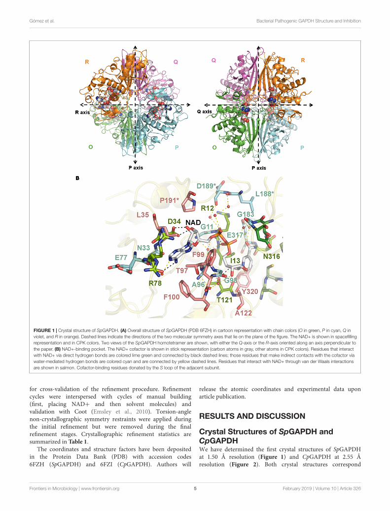
FIGURE 11 | Interaction of human complement factors C5a, C3a, or C3 with surface-immobilized AvGAPDH, SpGAPDH, and CpGAPDH measured by an ELISAassay. Absorbance at 492 nm is plotted along the y-axis against increasing C5a, C3a, or C3 concentration on the x-axis. Data points and error bars representmean ± SD (standard deviation; N = 3). Interaction with C5a (A) or C3 (B) in absence and at fixed concentration of anacardic acid (5 µM) or curcumin (50 µM).
exploitation of these inhibitor-binding pockets will openthe avenue for the advent of more potent, highly selectivebacterial GAPDH inhibitors.
Anacardic Acid and Curcumin InhibitGrowth of S. pyogenesSince anacardic acid and curcumin are both capable of inhibitingGAPDH, we decided to assay their effect when added directlyto dividing cells of clinically relevant bacterial strains byusing standard antimicrobial susceptibility tests. We choseserotype M1 group A S. pyogenes strains 950383, 941079,950358, and 950771 previously characterized in our laboratorieswhich had been isolated from clinical cases of necrotizingfasciitis (Pérez-Caballero et al., 2000). Of those, only strain950383 had been observed to interact with human complementFHL-1 (Pérez-Caballero et al., 2000). Both anacardic acid andcurcumin were indeed capable of halting microbial growth
when used at sub-millimolar concentrations. Our antimicrobialsusceptibility tests demonstrated that anacardic acid exhibiteda greater antimicrobial activity than curcumin (Table 4).Thus, all four S. pyogenes strains were susceptible to lowconcentrations of anacardic acid (0.5–2 µg/ml, 1.4–5.7 µM)with minimal inhibitory concentration (MIC) of 1.4 µM (0.5µg/ml), while at least 128 µg/ml of curcumin (347 µM)were required to inhibit bacterial growth. This result is inagreement with a recent report on the growth inhibitoryproperties of anacardic acid over S. pyogenes and S. agalactiaethat reported MIC values of 5 µM (Hamad and Mubofu,2015; Hollands et al., 2016). While anacardic acid was ableto inhibit growth at concentrations close to its determinedK i, the curcumin concentration necessary to elicit minimalgrowth inhibition was significantly greater than its K i (38 vs.347 µM). The latter discrepancy indicates that the biologicalactivity of curcumin is reduced by one or possibly many factors
Frontiers in Microbiology | www.frontiersin.org 14 February 2019 | Volume 10 | Article 326

fmicb-10-00326 February 26, 2019 Time: 13:0 # 15
Gómez et al. Bacterial Pathogenic GAPDH Structure and Inhibition
including lower membrane permeability, off-target effects, andlow-level metabolic turnover.
Interaction of GAPDH With HumanComplement FactorsBesides the pivotal role that GAPDH plays in bacterialcatabolism, GAPDH can also act as an effective virulence factorby targeting and sequestering human complement factors like C3,C1q, and the anaphylatoxin C5a (Terao et al., 2006; Terrasse et al.,2012; Querol-García et al., 2017). A prerequisite for bacterialGAPDH to display such complement immunoevasive functionsis the colocalization of GAPDH and the targeted complementproteins. Colocalization can be achieved by exporting GAPDHto the extracellular side of the cell wall, where it can remainassociated with cell-wall structures or be released into themedium. GAPDH export has already been demonstrated forvarious bacterial and eukaryotic organisms through a variety ofmolecular mechanisms (Terao et al., 2006; Matta et al., 2010;Aguilera et al., 2012; Seidler, 2013; Sahoo et al., 2013).
Having previously analyzed the C5a binding activity ofSpGAPDH, CpGAPDH, and AvGAPDH (Querol-García et al.,2017), we resorted to enzyme-linked immunosorbent assays(ELISA) to test whether GAPDH from these bacteria mightbe able to bind not only C5a but also the closely relatedanaphylatoxin C3a and its precursor C3. C3 was chosen becauseit has been reported to be a target for extracellular GAPDHfrom a parasitic nematode (Sahoo et al., 2013). C5a binding wasassayed for SpGAPDH and CpGAPDH, extending the previouslypublished results on AvGAPDH (Querol-García et al., 2017). Allthree GAPDH were capable of binding C5a and, albeit with loweraffinity, also C3 in these assays in a dose-dependent manner(Figure 11). This is an important observation that is in line withrecent observations concerning GAPDH from S. pneumoniaeand from the parasite nematode Haemonchus contortus, both ofwhich simultaneously target more than one complement factor(Terrasse et al., 2012; Sahoo et al., 2013; Vedamurthy et al.,2015). However, we could not detect binding of C3a to GAPDHabove background level, indicating a high degree of selectivity ofGAPDH for C5a (Figure 11).
Next, we tested whether anacardic acid and/or curcumincould prevent GAPDH binding to C5a or C3 using the sameELISA assay described above. Since the N-terminus of bothSpGAPDH and AvGAPDH has been implicated in C5a bindingand this region includes the catalytic Cys residue and an extendedloop that closes the active site cavity, we speculated whetherchanges in or near the active site cavity could disrupt C5arecognition. We also probed the effect that anacardic acid orcurcumin might have on C3 binding, even though it is lesslikely that a small-molecule inhibitor could prevent GAPDHfrom binding to a far greater protein like C3. Indeed, the resultsshow that the presence of either inhibitor at concentrationsthat achieve nearly full inhibition of GAPDH catalytic activitywere not effective at disrupting the interaction with C5a andonly poorly effective with C3 (Supplementary Figure S5). Takentogether, these observations demonstrate that the surface areasthat GAPDH uses to interact with C5a and C3 are either distinct(non-overlapping or only partially overlapping) or are not
significantly altered by binding of anacardic acid or curcumin.These results point to the functional segregation between the C5aand C3 interaction surfaces and the GAPDH binding pockets forthe two natural compounds in the assayed systems. Given that themoonlighting functions of GAPDH as an immune evasive factortake place in the extracellular space, they are likely decoupledfrom the enzymatic activity.
Relevance of GAPDH-TargetingAntimicrobialsBacterial infections are a significant medical and economic threatto human societies. Antibiotics have long been used to controlbacterial infections successfully. However, their widespread usehas had the undesired effect of promoting the emergenceof multidrug resistant bacteria, which represent a seriousdanger for human health, especially for immunocompromisedpatients in the hospital environment. In this work, we havecharacterized the inhibitory potency and modality of twonatural products, anacardic acid and curcumin, toward theubiquitous and highly conserved glycolytic enzyme GAPDHfrom several Gram-positive bacterial pathogens. Owing tothe vital role it plays in secondary metabolism, GAPDHmay be an interesting target for drug discovery providedthat inhibitors can be proved safe, efficacious and selectivetoward the bacterial enzymes. Low micromolar concentrationsof anacardic acid and, to a lesser degree, curcumin caninhibit growth of clinically isolated bacterial cultures andinhibit GAPDH in vitro, thereby suggesting that GAPDH mightbe the relevant physiological target for their antimicrobialactivities. Since pathogenic bacterial GAPDH can also interferewith the human C5a anaphylatoxin-mediated signaling, it isimportant to understand whether the antimicrobial activity ofthese compounds could also interfere with GAPDH’s immuneevasion activity. Our results indicate that these two functionsare unrelated. Deciphering the high-resolution structure ofS. pyogenes and C. perfringens GAPDH will pave the wayfor the discovery of selective inhibitors based on anacardicacid and curcumin.
AUTHOR CONTRIBUTIONS
MV, FF, SA, and SRC conceived the experimental study, designedthe experiments, analyzed the data, and wrote the manuscript.JQ-G and SG expressed and purified SpGAPDH, CpGAPDH,AvGAPDH, and C5a, and GS-B expressed and purified C3a.VF-H helped with the initial expression and purification ofSpGAPDH and CpGAPDH. JQ-G, FF, and MV crystallizedSpGAPDH and CpGAPDH. FF and MV collected the X-raydata set from both crystals, determined, and analyzed the crystalstructures. SG measured enzyme kinetics and the inhibitionby anacardic acid and curcumin. MS and SRC provided anti-C5a, anti-C3a, and anti-C3 antibodies for ELISA assays. SG andGS-B performed the ELISA assays. ÀG-A and SA measuredthe MIC of anacardic acid and curcumin with clinical isolatesof S. pyogenes. SA provided materials and helped design theexperiments and analyzed the data. All authors contributedcritically to the manuscript.
Frontiers in Microbiology | www.frontiersin.org 15 February 2019 | Volume 10 | Article 326

fmicb-10-00326 February 26, 2019 Time: 13:0 # 16
Gómez et al. Bacterial Pathogenic GAPDH Structure and Inhibition
FUNDING
This study was supported by Spanish Instituto de Salud CarlosIII (PI12/01667 to MV), Spanish Ministerio de Economíay Competitividad (CTQ2015-66206-C2-2-R and SAF2015-72961-EXP to MV), the Spanish Network of Excellenceon Complement (SAF2016-81876-REDT to MV, SA, andSRC), the Regional Government of Madrid (S2010/BD-2316 and S2017/BMD-3673 to MV and SRC), and theEuropean Commission [Framework Programme 7 (FP7)] projectComplexINC (Contract No. 279039 to MV). All grants wereco-funded with European Union ERDF funds (EuropeanRegional Development Fund). The funders had no role instudy design, data collection and analysis, decision to publish,or preparation of the manuscript. We acknowledge supportof the publication fee by the CSIC Open Access PublicationSupport Initiative through its Unit of Information Resourcesfor Research (URICI).
ACKNOWLEDGMENTS
We gratefully acknowledge access to the ALBA SynchrotronLight Source, Barcelona, Spain, for provision of synchrotronradiation facilities at BL13-XALOC, and the EuropeanSynchrotron Radiation Facility, Grenoble, France, at theID23-2 beamline; and the staff for excellent support atthe two synchrotrons. GS-B acknowledges the supportof the Ph.D. program in Biochemistry, MolecularBiology, and Biomedicine of the Universidad Complutensede Madrid (UCM).
SUPPLEMENTARY MATERIAL
The Supplementary Material for this article can be foundonline at: https://www.frontiersin.org/articles/10.3389/fmicb.2019.00326/full#supplementary-material
REFERENCESAdams, P. D., Afonine, P. V., Bunkóczi, G., Chen, V. B., Davis, I. W.,
Echols, N., et al. (2010). PHENIX: a comprehensive python-based system formacromolecular structure solution. Acta Crystallogr. D Biol. Crystallogr. 66,213–221. doi: 10.1107/S0907444909052925
Afonine, P. V., Grosse-Kunstleve, R. W., Echols, N., Headd, J. J., Moriarty, N. W.,Mustyakimov, M., et al. (2012). Towards automated crystallographic structurerefinement with phenix.refine. Acta Crystallogr. D Biol. Crystallogr. 68, 352–367.doi: 10.1107/S0907444912001308
Aguilera, L., Ferreira, E., Giménez, R., Fernández, F. J., Taulés, M., Aguilar, J.,et al. (2012). Secretion of the housekeeping protein glyceraldehyde-3-phosphate dehydrogenase by the LEE-encoded type III secretion system inenteropathogenic Escherichia coli. Int. J. Biochem. Cell Biol. 44, 955–962.doi: 10.1016/j.biocel.2012.03.002
Bressi, J. C., Verlinde, C. L., Aronov, A. M., Shaw, M. L., Shin, S. S., Nguyen, L. N.,et al. (2001). Adenosine analogues as selective inhibitors of glyceraldehyde-3-phosphate dehydrogenase of trypanosomatidae via structure-based drug design.J. Med. Chem. 44, 2080–2093.
Chauhan, A. S., Kumar, M., Chaudhary, S., Patidar, A., Dhiman, A., Sheokand, N.,et al. (2017). Moonlighting glycolytic protein glyceraldehyde-3-phosphatedehydrogenase (GAPDH): an evolutionarily conserved plasminogen receptoron mammalian cells. FASEB J. 31, 2638–2648. doi: 10.1096/fj.201600982R
Cheng, A. L., Hsu, C. H., Lin, J. K., Hsu, M. M., Ho, Y. F., Shen, T. S.,et al. (2001). Phase I clinical trial of curcumin, a chemopreventive agent,in patients with high-risk or pre-malignant lesions. Anticancer Res. 21,2895–2900.
Emsley, P., Lohkamp, B., Scott, W. G., and Cowtan, K. (2010). Features anddevelopment of Coot. Acta Crystallogr. D Biol. Crystallogr. 66, 486–501.doi: 10.1107/S0907444910007493
Evans, P. R., and Murshudov, G. N. (2013). How good are my data and what is theresolution? Acta Crystallogr. D Biol. Crystallogr. 69, 1204–1214. doi: 10.1107/S0907444913000061
Ferdinand, W. (1964). The isolation and specific activity of rabbit-muscleglyceraldehyde phosphate dehydrogenase. Biochem. J. 92, 578–585. doi: 10.1042/bj0920578
Flot, D., Mairs, T., Giraud, T., Guijarro, M., Lesourd, M., Rey, V., et al. (2010).The ID23-2 structural biology microfocus beamline at the ESRF. J. SynchrotronRadiat. 17, 107–118. doi: 10.1107/S0909049509041168
Freitas, R. F., Prokopczyk, I. M., Zottis, A., Oliva, G., Andricopulo, A. D., Trevisan,M. T. S., et al. (2009). Discovery of novel Trypanosoma cruzi glyceraldehyde-3-phosphate dehydrogenase inhibitors. Bioorg. Med. Chem. 17, 2476–2482.doi: 10.1016/j.bmc.2009.01.079
Hamad, F. B., and Mubofu, E. B. (2015). Potential biological applications of bio-based anacardic acids and their derivatives. Int. J. Mol. Sci. 16, 8569–8590.doi: 10.3390/ijms16048569
Hollands, A., Corriden, R., Gysler, G., Dahesh, S., Olson, J., Raza Ali, S.,et al. (2016). Natural product anacardic acid from cashew nut shellsstimulates neutrophil extracellular trap production and bactericidalactivity. J. Biol. Chem. 291, 13964–13973. doi: 10.1074/jbc.M115.695866
Ismail, S. A., and Park, H. W. (2005). Structural analysis of humanliver glyceraldehyde-3-phosphate dehydrogenase. Acta Crystallogr. D Biol.Crystallogr. 61, 1508–1513. doi: 10.1107/S0907444905026740
Juanhuix, J., Gil-Ortiz, F., Cuní, G., Colldelram, C., Nicolás, J., Lidón, J.,et al. (2014). Developments in optics and performance at BL13-XALOC, the macromolecular crystallography beamline at the ALBAsynchrotron. J. Synchrotron Radiat. 21, 679–689. doi: 10.1107/S160057751400825X
Kabsch, W. (2010a). Integration, scaling, space-group assignment and post-refinement. Acta Crystallogr. D Biol. Crystallogr. 66, 133–144. doi: 10.1107/S0907444909047374
Kabsch, W. (2010b). XDS. Acta Crystallogr. D Biol. Crystallogr. 66, 125–132.doi: 10.1107/S0907444909047337
Karibe, T., Imaoka, T., Abe, K., and Ando, O. (2018). Curcumin as anin vivo selective intestinal breast cancer resistance protein inhibitor incynomolgus monkeys. Drug Metab. Dispos. 46, 667–679. doi: 10.1124/dmd.117.078931
Kim, H., Feil, I. K., Verlinde, C. L., Petra, P. H., and Hol, W. G. (1995).Crystal structure of glycosomal glyceraldehyde-3-phosphate dehydrogenasefrom Leishmania mexicana: implications for structure-based drug design anda new position for the inorganic phosphate binding site. Biochemistry 34,14975–14986. doi: 10.1021/bi00046a004
Ko, E.-Y., and Moon, A. (2015). Natural products for chemoprevention ofbreast cancer. J. Cancer Prev. 20, 223–231. doi: 10.15430/JCP.2015.20.4.223
Korndörfer, I., Steipe, B., Huber, R., Tomschy, A., and Jaenicke, R. (1995). Thecrystal structure of holo-glyceraldehyde-3-phosphate dehydrogenase from thehyperthermophilic bacterium Thermotoga maritima at 2.5 A resolution. J. Mol.Biol. 246, 511–521.
Lambeir, A. M., Loiseau, A. M., Kuntz, D. A., Vellieux, F. M., Michels, P. A.,and Opperdoes, F. R. (1991). The cytosolic and glycosomal glyceraldehyde-3-phosphate dehydrogenase from Trypanosoma brucei. Kinetic properties andcomparison with homologous enzymes. . Eur. J. Biochem. 198, 429–435.doi: 10.1111/j.1432-1033.1991.tb16032.x
Mamidyala, S. K., Ramu, S., Huang, J. X., Robertson, A. A. B., andCooper, M. A. (2013). Efficient synthesis of anacardic acid analogues
Frontiers in Microbiology | www.frontiersin.org 16 February 2019 | Volume 10 | Article 326

fmicb-10-00326 February 26, 2019 Time: 13:0 # 17
Gómez et al. Bacterial Pathogenic GAPDH Structure and Inhibition
and their antibacterial activities. Bioorg. Med. Chem. Lett. 23, 1667–1670.doi: 10.1016/j.bmcl.2013.01.074
Matta, S. K., Agarwal, S., and Bhatnagar, R. (2010). Surface localized andextracellular glyceraldehyde-3-phosphate dehydrogenase of Bacillus anthracisis a plasminogen binding protein. Biochim. Biophys. Acta 1804, 2111–2120.doi: 10.1016/j.bbapap.2010.08.004
McCoy, A. J., Grosse-Kunstleve, R. W., Adams, P. D., Winn, M. D., Storoni, L. C.,and Read, R. J. (2007). Phaser crystallographic software. J. Appl. Crystallogr. 40,658–674. doi: 10.1107/S0021889807021206
Mukherjee, S., Dutta, D., Saha, B., and Das, A. K. (2010). Crystal structureof glyceraldehyde-3-phosphate dehydrogenase 1 from methicillin-resistant Staphylococcus aureus MRSA252 provides novel insights intosubstrate binding and catalytic mechanism. J. Mol. Biol. 401, 949–968.doi: 10.1016/j.jmb.2010.07.002
Norouzi, S., Majeed, M., Pirro, M., Generali, D., and Sahebkar, A. (2018). Curcuminas an adjunct therapy and microRNA modulator in breast cancer. Curr. Pharm.Des. 24, 171–177. doi: 10.2174/1381612824666171129203506
Pereira, J. M., Severino, R. P., Vieira, P. C., Fernandes, J. B., da Silva,M. F., Zottis, A., et al. (2008). Anacardic acid derivatives as inhibitors ofglyceraldehyde-3-phosphate dehydrogenase from Trypanosoma cruzi. Bioorg.Med. Chem. 16, 8889–8895. doi: 10.1016/j.bmc.2008.08.057
Pérez-Caballero, D., Albertí, S., Vivanco, F., Sánchez-Corral, P., and Rodríguezde Córdoba, S. (2000). Assessment of the interaction of human complementregulatory proteins with group a streptococcus. Identification of a high-affinitygroup a streptococcus binding site in FHL-1. Eur. J. Immunol. 30, 1243–1253.
Querol-García, J., Fernández, F. J., Marin, A. V., Gómez, S., Fullà, D., Melchor-Tafur, C., et al. (2017). Crystal structure of glyceraldehyde-3-phosphatedehydrogenase from the gram-positive bacterial pathogen a. vaginae, animmunoevasive factor that interacts with the human C5a anaphylatoxin. Front.Microbiol. 8:541. doi: 10.3389/fmicb.2017.00541
Raj, M., Langley, M., McArthur, S. J., and Jean, F. (2017). Moonlighting glycolyticenzyme glyceraldehyde-3-phosphate dehydrogenase (GAPDH) is required forefficient hepatitis C virus and dengue virus infections in human Huh-7.5.1 cells.J. Gen. Virol. 98, 977–991. doi: 10.1099/jgv.0.000754
Sahoo, S., Murugavel, S., Devi, I. K., Vedamurthy, G. V., Gupta, S. C., Singh,B. P., et al. (2013). Glyceraldehyde-3-phosphate dehydrogenase of the parasiticnematode Haemonchus contortus binds to complement C3 and inhibits itsactivity. Parasite Immunol. 35, 457–467. doi: 10.1111/pim.12058
Schraufstatter, E., and Bernt, H. (1949). Antibacterial action of curcumin andrelated compounds. Nature 164:456.
Seidler, N. W. (2013). Basic biology of GAPDH. Adv. Exp. Med. Biol. 985, 1–36.doi: 10.1007/978-94-007-4716-6_1
Sharma, R., Kishore, N., Hussein, A., and Lall, N. (2013). Antibacterial and anti-inflammatory effects of Syzygium jambos L. (Alston) and isolated compoundson acne vulgaris. BMC Compl. Altern. Med. 13:292. doi: 10.1186/1472-6882-13-292
Sirover, M. A. (1997). Role of the glycolytic protein, glyceraldehyde-3-phosphatedehydrogenase, in normal cell function and in cell pathology. J. Cell Biochem.66, 133–140.
Sirover, M. A. (2005). New nuclear functions of the glycolytic protein,glyceraldehyde-3-phosphate dehydrogenase, in mammalian cells. J. CellBiochem. 95, 45–52. doi: 10.1002/jcb.20399
Sirover, M. A. (2011). On the functional diversity of glyceraldehyde-3-phosphate dehydrogenase: biochemical mechanisms and regulatorycontrol. Biochim. Biophys. Acta 1810, 741–751. doi: 10.1016/j.bbagen.2011.05.010
Sirover, M. A. (2012). Subcellular dynamics of multifunctional protein regulation:mechanisms of GAPDH intracellular translocation. J. Cell Biochem. 113,2193–2200. doi: 10.1002/jcb.24113
Sirover, M. A. (2014). Structural analysis of glyceraldehyde-3-phosphatedehydrogenase functional diversity. Int. J. Biochem. Cell Biol. 57, 20–26.doi: 10.1016/j.biocel.2014.09.026
Skarzynski, T., Moody, P. C., and Wonacott, A. J. (1987). Structure of holo-glyceraldehyde-3-phosphate dehydrogenase from Bacillus stearothermophilus
at 1.8 A resolution. J. Mol. Biol. 193, 171–187. doi: 10.1016/0022-2836(87)90635-8
Skarzynski, T., and Wonacott, A. J. (1988). Coenzyme-induced conformationalchanges in glyceraldehyde-3-phosphate dehydrogenase from Bacillusstearothermophilus. J. Mol. Biol. 203, 1097–1118. doi: 10.1016/0022-2836(88)90130-1
Souza, D. H., Garratt, R. C., Araújo, A. P., Guimarães, B. G., Jesus, W. D., Michels,P. A., et al. (1998). Trypanosoma cruzi glycosomal glyceraldehyde-3-phosphatedehydrogenase: structure, catalytic mechanism and targeted inhibitor design.FEBS Lett. 424, 131–135. doi: 10.1016/S0014-5793(98)00154-9
Stein, N. (2008). CHAINSAW: a program for mutating pdb files used as templatesin molecular replacement. J. Appl. Crystallogr. 41, 641–643. doi: 10.1107/S0021889808006985
Suresh, S., Bressi, J. C., Kennedy, K. J., Verlinde, C. L., Gelb, M. H., and Hol, W. G.(2001). Conformational changes in Leishmania mexicana glyceraldehyde-3-phosphate dehydrogenase induced by designed inhibitors. J. Mol. Biol. 309,423–435. doi: 10.1006/jmbi.2001.4588
Terao, Y., Yamaguchi, M., Hamada, S., and Kawabata, S. (2006). Multifunctionalglyceraldehyde-3-phosphate dehydrogenase of Streptococcus pyogenes isessential for evasion from neutrophils. J. Biol. Chem. 281, 14215–14223.doi: 10.1074/jbc.M513408200
Terrasse, R., Tacnet-Delorme, P., Moriscot, C., Pérard, J., Schoehn, G., Vernet, T.,et al. (2012). Human and pneumococcal cell surface glyceraldehyde-3-phosphate dehydrogenase (GAPDH) proteins are both ligands of humanC1q protein. J. Biol. Chem. 287, 42620–42633. doi: 10.1074/jbc.M112.423731
Tyman, J. H., and Kiong, L. S. (1978). Long chain phenols: Part XI. Composition ofnatural cashew nutshell liquid (Anacardium occidentale) from various sources.Lipids 13, 525–532. doi: 10.1007/BF02533591
Vedamurthy, G. V., Sahoo, S., Devi, I. K., Murugavel, S., and Joshi, P.(2015). The N-terminal segment of glyceraldehyde-3-phosphatedehydrogenase of Haemonchus contortus interacts with complementsC1q and C3. Parasite Immunol. 37, 568–578. doi: 10.1111/pim.12273
Vellieux, F. M., Hajdu, J., Verlinde, C. L., Groendijk, H., Read, R. J., Greenhough,T. J., et al. (1993). Structure of glycosomal glyceraldehyde-3-phosphatedehydrogenase from Trypanosoma brucei determined from Laue data. Proc.Natl. Acad. Sci. U.S.A. 90, 2355–2359.
Verlinde, C. L., Callens, M., Van Calenbergh, S., Van Aerschot, A., Herdewijn, P.,Hannaert, V., et al. (1994). Selective inhibition of trypanosomal glyceraldehyde-3-phosphate dehydrogenase by protein structure-based design: towardnew drugs for the treatment of sleeping sickness. J. Med. Chem. 37,3605–3613.
Winn, M. D., Ballard, C. C., Cowtan, K. D., Dodson, E. J., Emsley, P., Evans,P. R., et al. (2011). Overview of the CCP4 suite and current developments.Acta Crystallogr. D Biol. Crystallogr. 67, 235–242. doi: 10.1107/S0907444910045749
Yun, M., Park, C. G., Kim, J. Y., and Park, H. W. (2000). Structural analysisof glyceraldehyde 3-phosphate dehydrogenase from Escherichia coli: directevidence of substrate binding and cofactor-induced conformational changes.Biochemistry 39, 10702–10710.
Conflict of Interest Statement: The authors declare that the research wasconducted in the absence of any commercial or financial relationships that couldbe construed as a potential conflict of interest.
Copyright © 2019 Gómez, Querol-García, Sánchez-Barrón, Subias, González-Alsina,Franco-Hidalgo, Albertí, Rodríguez de Córdoba, Fernández and Vega. This is anopen-access article distributed under the terms of the Creative Commons AttributionLicense (CC BY). The use, distribution or reproduction in other forums is permitted,provided the original author(s) and the copyright owner(s) are credited and that theoriginal publication in this journal is cited, in accordance with accepted academicpractice. No use, distribution or reproduction is permitted which does not complywith these terms.
Frontiers in Microbiology | www.frontiersin.org 17 February 2019 | Volume 10 | Article 326