Isolates of Staphylococcus aureus and saprophyticus resistant to antimicrobials isolated from the...
8
Isolates of Staphylococcus aureus and saprophyticus resistant to antimicrobials isolated from the Lebanese aquatic environment Steve Harakeh a, * , Hadi Yassine a , Shady Hajjar a , Mutasem El-Fadel b a Department of Biology, American University of Beirut, P.O. Box 11-0236, Beirut, Lebanon b Department of Civil and Environmental Engineering, American University of Beirut, P.O. Box 11-0236, Beirut, Lebanon Abstract The indiscriminate use of antimicrobials especially in developing countries has evoked serious bacterial resistance and led to the emer- gence of new and highly resistant strains of bacteria to commonly used antimicrobials. In Lebanon, pollution levels and bacterial infec- tions are increasing at a high rate as a result of inadequate control measures to limit untreated effluent discharges into the sea or freshwater resources. The aim of this study was to isolate and molecularly characterize various Staphylococcus strains isolated from sea water, fresh water, sediments, and crab samples collected from representative communities along the coast of Lebanon. The results on the antimicrobial resistance indicated that the level of resistance of Staphylococcus aureus varied with various antimicrobials tested. The resistance patterns ranged between 45% in freshwater isolates and 54.8% in seawater ones. Fifty one percent of the tested isolates have shown resistance to at least one of the five tested antimicrobials; with seawater isolates exhibiting the highest rates of antimicrobial resistance. Ó 2006 Elsevier Ltd. All rights reserved. Keywords: Staphylococcus aureus; Staphylococcus saprophyticus; Antimicrobial resistance; Aquatic environment 1. Introduction In Lebanon, the aquatic environment is polluted in many parts of the country leading to many water-borne diseases. Horizontal transfer of plasmid-encoded genes is the primary reason for the dissemination of resistant genes in the environment (Chandrasekaran et al., 2003; Son et al., 1997). In Lebanon, the antimicrobial susceptibility of Gram negative bacteria has been determined largely for clinical isolates (Araj and Kanj, 2000). However, anti- microbial resistance patterns of environmental isolates have not been evaluated. Bacterial contamination in particular could result in many chronic and acute diseases like food poisoning, intes- tinal diarrhea, cholera, typhoid fever, dysentery and others (Griffin and Tauxe, 1991; Brook et al., 1994; Ashbolt, 2004). Coupled with the indiscriminate and uncontrolled use of antimicrobials, it could lead to a sharp rise in bacte- rial resistance to commonly used antimicrobials. Infection by these resistant strains is more threatening than by non-resistant ones (Holmberg et al., 1984). Based on the importance of Staphylococcus aureus as a public health hazard to humans (Aly, 1990; Slobodnikova et al., 1995; Martineau et al., 2000; Palomares et al., 2003; Chen et al., 2004), the aim of the present study was to isolate and identify Staphylococci spp. present in samples collected from the aquatic environment in Lebanon using, biochemical tests and molecular biology based techniques such as polymerase chain reaction (PCR). The latter is a more reliable technique than the commonly used conven- tional procedures like biochemical tests which yield results that may be misleading (Palomares et al., 2003). Further- more, the resistance of the bacteria was evaluated using different antimicrobial agents. 0025-326X/$ - see front matter Ó 2006 Elsevier Ltd. All rights reserved. doi:10.1016/j.marpolbul.2005.12.008 * Corresponding author. Tel.: +961 3570 119. E-mail address: [email protected] (S. Harakeh). www.elsevier.com/locate/marpolbul Marine Pollution Bulletin 52 (2006) 912–919
-
Upload
independent -
Category
Documents
-
view
1 -
download
0
Transcript of Isolates of Staphylococcus aureus and saprophyticus resistant to antimicrobials isolated from the...

www.elsevier.com/locate/marpolbul
Marine Pollution Bulletin 52 (2006) 912–919
Isolates of Staphylococcus aureus and saprophyticus resistantto antimicrobials isolated from the Lebanese aquatic environment
Steve Harakeh a,*, Hadi Yassine a, Shady Hajjar a, Mutasem El-Fadel b
a Department of Biology, American University of Beirut, P.O. Box 11-0236, Beirut, Lebanonb Department of Civil and Environmental Engineering, American University of Beirut, P.O. Box 11-0236, Beirut, Lebanon
Abstract
The indiscriminate use of antimicrobials especially in developing countries has evoked serious bacterial resistance and led to the emer-gence of new and highly resistant strains of bacteria to commonly used antimicrobials. In Lebanon, pollution levels and bacterial infec-tions are increasing at a high rate as a result of inadequate control measures to limit untreated effluent discharges into the sea orfreshwater resources. The aim of this study was to isolate and molecularly characterize various Staphylococcus strains isolated fromsea water, fresh water, sediments, and crab samples collected from representative communities along the coast of Lebanon. The resultson the antimicrobial resistance indicated that the level of resistance of Staphylococcus aureus varied with various antimicrobials tested.The resistance patterns ranged between 45% in freshwater isolates and 54.8% in seawater ones. Fifty one percent of the tested isolateshave shown resistance to at least one of the five tested antimicrobials; with seawater isolates exhibiting the highest rates of antimicrobialresistance.� 2006 Elsevier Ltd. All rights reserved.
Keywords: Staphylococcus aureus; Staphylococcus saprophyticus; Antimicrobial resistance; Aquatic environment
1. Introduction
In Lebanon, the aquatic environment is polluted inmany parts of the country leading to many water-bornediseases. Horizontal transfer of plasmid-encoded genes isthe primary reason for the dissemination of resistant genesin the environment (Chandrasekaran et al., 2003; Sonet al., 1997). In Lebanon, the antimicrobial susceptibilityof Gram negative bacteria has been determined largelyfor clinical isolates (Araj and Kanj, 2000). However, anti-microbial resistance patterns of environmental isolateshave not been evaluated.
Bacterial contamination in particular could result inmany chronic and acute diseases like food poisoning, intes-tinal diarrhea, cholera, typhoid fever, dysentery and others
0025-326X/$ - see front matter � 2006 Elsevier Ltd. All rights reserved.
doi:10.1016/j.marpolbul.2005.12.008
* Corresponding author. Tel.: +961 3570 119.E-mail address: [email protected] (S. Harakeh).
(Griffin and Tauxe, 1991; Brook et al., 1994; Ashbolt,2004). Coupled with the indiscriminate and uncontrolleduse of antimicrobials, it could lead to a sharp rise in bacte-rial resistance to commonly used antimicrobials. Infectionby these resistant strains is more threatening than bynon-resistant ones (Holmberg et al., 1984).
Based on the importance of Staphylococcus aureus as apublic health hazard to humans (Aly, 1990; Slobodnikovaet al., 1995; Martineau et al., 2000; Palomares et al., 2003;Chen et al., 2004), the aim of the present study was toisolate and identify Staphylococci spp. present in samplescollected from the aquatic environment in Lebanon using,biochemical tests and molecular biology based techniquessuch as polymerase chain reaction (PCR). The latter is amore reliable technique than the commonly used conven-tional procedures like biochemical tests which yield resultsthat may be misleading (Palomares et al., 2003). Further-more, the resistance of the bacteria was evaluated usingdifferent antimicrobial agents.
S. Harakeh et al. / Marine Pollution Bulletin 52 (2006) 912–919 913
2. Materials and methods
2.1. Sample collection
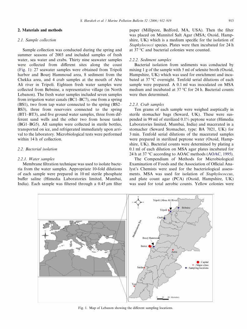
Sample collection was conducted during the spring andsummer seasons of 2003 and included samples of freshwater, sea water and crabs. Thirty nine seawater sampleswere collected from different sites along the coast(Fig. 1): 27 seawater samples were obtained from Tripoliharbor and Bourj Hammoud area, 8 sediment from theChekka area, and 4 crab samples at the mouth of AbuAli river in Tripoli. Eighteen fresh water samples werecollected from Bebnine, a representative village (in NorthLebanon). The fresh water samples included seven samplesfrom irrigation water canals (BC1–BC7), one from a spring(BS1), two from tap water connected to the spring (BS2–BS3), three from reservoirs connected to the spring(BT1–BT3), and five ground water samples, three from dif-ferent sand wells and the other two from house tanks(BG1–BG5). All samples were collected in sterile bottles,transported on ice, and refrigerated immediately upon arri-val to the laboratory. Microbiological tests were performedwithin 14 h of collection.
2.2. Bacterial isolation
2.2.1. Water samples
Membrane filtration technique was used to isolate bacte-ria from the water samples. Appropriate 10-fold dilutionsof each sample were prepared in 10 ml sterile phosphatebuffer saline (Himedia Laboratories limited, Mumbai,India). Each sample was filtered through a 0.45 lm filter
Fig. 1. Map of Lebanon showing t
paper (Millipore, Bedford, MA, USA). Then the filterwas placed on Mannitol Salt Agar (MSA; Oxoid, Hamp-shire, UK) which is a medium specific for the isolation ofStaphylococci species. Plates were then incubated for 24 hat 37 �C and bacterial colonies were counted.
2.2.2. Sediment samples
Bacterial isolation from sediments was conducted bymixing 1 g of the sample with 5 ml of selenite broth (Oxoid,Hampshire, UK) which was used for enrichment and incu-bated at 37 �C overnight. Tenfold serial dilutions of eachsample were prepared. A 0.1 ml was inoculated on MSAmedium and incubated at 37 �C for 24 h. Bacterial countswere then determined.
2.2.3. Crab samples
Ten grams of each sample were weighed aseptically insterile stomacher bags (Seward, UK). These were sus-pended in 99 ml of sterilized 0.1% peptone water (HimediaLaboratories limited, Mumbai, India) and macerated in astomacher (Seward Stomacher, type: BA 7021, UK) for3 min. Tenfold serial dilutions of the macerated sampleswere prepared in sterilized peptone water (Oxoid, Hamp-shire, UK). Bacterial counts were determined by plating a0.1 ml of each dilution on MSA agar plates incubated for24 h at 37 �C according to AOAC methods (AOAC, 1995).
The Compendium of Methods for MicrobiologicalExamination of Foods and the Association of Official Ana-lyst’s Chemists were used for the bacteriological assess-ments. MSA was used for isolation of Staphylococcus,and plate count agar (PCA) (Oxoid, Hampshire, UK)was used for total aerobic counts. Yellow colonies were
he different sampling locations.

914 S. Harakeh et al. / Marine Pollution Bulletin 52 (2006) 912–919
selected as suspected S. aureus. However, colonies withother colors like white or cream were also included andwere subjected to PCR analysis for identification.
2.3. DNA extraction
A loop full of bacteria from each colony was suspended in5 ml of sterile Brain Heart Infusion broth (BHI) (Oxoid,Hampshire, UK) and grown overnight at 37 �C on a shaker.The next day, 1 ml of the culture was used to extract totalgenomic DNA using the GFX genomic blood DNA purifi-cation kit from Amersham Biosciences (Little Chalfont,Buckinghamshire, UK) (Ausubel et al., 1989; Sambrooket al., 1989). Double the volume of the proteinase K bufferwas used and the incubation time was increased to 25 min.After the extraction, the concentrations of the DNA sampleswere measured using the spectrophotometer (NC 9423 Bio100 2E, Thermo Electron Corporation, UK). Moreover,1 lg of several DNAs was run on a 1% agarose gel (Amresco,OH, USA) to check the quality of the genomic DNA.
2.4. PCR assay
PCR was used with two pairs of primers (ABgene Surrey,UK) for the molecular detection of S. aureus. FA1 and RA2,were used to amplify S. aureus aroA-gene, leading to anexpected product of 1153 base pair fragment representingmost of the aroA-gene sequence (Marcos et al., 1999). Thesecond pair of primers was designed to amplify asequence-specific 279 base pair fragment within the nuc-gene. nucA F sequence and nucA R sequence were selectedas depicted in Table 1 (Palomares et al., 2003). Furthermore,a pair of primers specific for Staphylococcus saprophyticus
were used (Martineau et al., 2000). The PCR amplificationreaction was conducted in a 25 ll volume containing0.5 lM of each primer, 150 ng of the DNA template, 1· ofreaction buffer (ABgene Surrey, UK), 0.2 mM each ofdATP, dGTP, dCTP, and dTTP (ABgene, Surrey, UK),1 U of Thermos aquaticus (Taq) DNA polymerase (AB geneproducts). Amplification was conducted in a DNA thermalcycler (BioRad, Hercules, CA, USA) using the followingprogram: denaturation at 94 �C for 5 min, 35 cycles at94 �C for 3 min, 55 �C annealing for 30 s, 72 �C extensionfor 3 min and a final extension at 72 �C for 10 min.
Negative control was included in each PCR reaction,positive controls included DNA of identified clinical iso-lates S. aureus with the two sets of primers FA1, RA2
Table 1The oligonucleotide primers used in the detection of Staphylococci isolates
Target gene Primers Sequence
aroA FA1 50-AAGGGCGAAATAGAAGTGCCGGaroA RA2 50-CACAAGCAACTGCAAGCAT-3 0
nuc nuc AF 50-AGCCAAGCCTTGACGAACTAA-30
Nuc nuc AR 50-GCGATTGATGGTGATACGGTT-3 0
AF144088a st 50-TCAAAAAGTTTTCTAAAAAATTTAF144088a st 50-ACGGGCGTCCACAAAATCAATAG
a Amplicon for S. saprophyticus.
and nucA F, nucA R. Ten microliters of the PCR productwere mixed with 2 ll 6X loading dye (ABgene Surrey, UK)and ran on a 1% agarose gel at 90 V for 80 min.
2.5. Antimicrobial resistance
Strains grown on MSA media with different morpholo-gies were tested for their resistance to five antimicrobials(oxacillin, teicoplanin, gentamicin, clindamycin, and van-comycin) using the disc diffusion method as set by theNational Committee for Clinical Laboratory Standards(NCCLS) (NCCLS, 2000, 2002). Brain Infusion broth(BHI) (Oxoid, Hampshire, UK) was inoculated with iso-lates grown overnight at 37 �C with continuous shaking,until a 0.5 McFarland turbidity standard was obtained.A 0.1 ml of the culture was inoculated on BHI Agar plates(Oxoid, Hampshire, UK). Antimicrobial discs (Bio-Merieux�, Sa Marcy l’Etoile, France) impregnated withoxacillin (1 lg), teicoplanin (30 lg), gentamicin (10 lg),clindamycin (2 lg) or vancomycin (30 lg) were placed onthe inoculated plates which were incubated at 37 �C over-night. After incubation, zones of inhibition around eachantimicrobial disc were measured. Each sample was classi-fied as either resistant or susceptible to the mentioned anti-microbial using the NCCLS guidelines.
2.6. Statistical analysis
Data analysis was completed using SPSS 11 statisticalsoftware. Counts were analyzed in terms of mean and stan-dard variation and were then compared among sites for allbacteria using non-parametric tests for K independentsamples. Concerning resistance, univariate analysis wasconducted on all variables. Also, bivariate analysis exam-ined the influence of the antibacterial agent used on thebehavior of the bacteria.
3. Results
3.1. Bacteriological counts
A summary of bacteriological counts and their rankaccording to the site of collection are summarized in Table2. All seawater samples collected from the Bourj Ham-moud area were contaminated with Staphylococcus specieswith an average count of 7.2 · 103 CFU/100 ml. Ninetytwo percent of seawater samples collected from Tripoli
Resource PCR product size (bp)
GC-3 0 Marcos et al. (1999) 1153Marcos et al. (1999) 1153Palomares et al. (2003) 279Palomares et al. (2003) 279
A-30 Martineau et al. (2000) 210GA-30 Martineau et al. (2000) 210
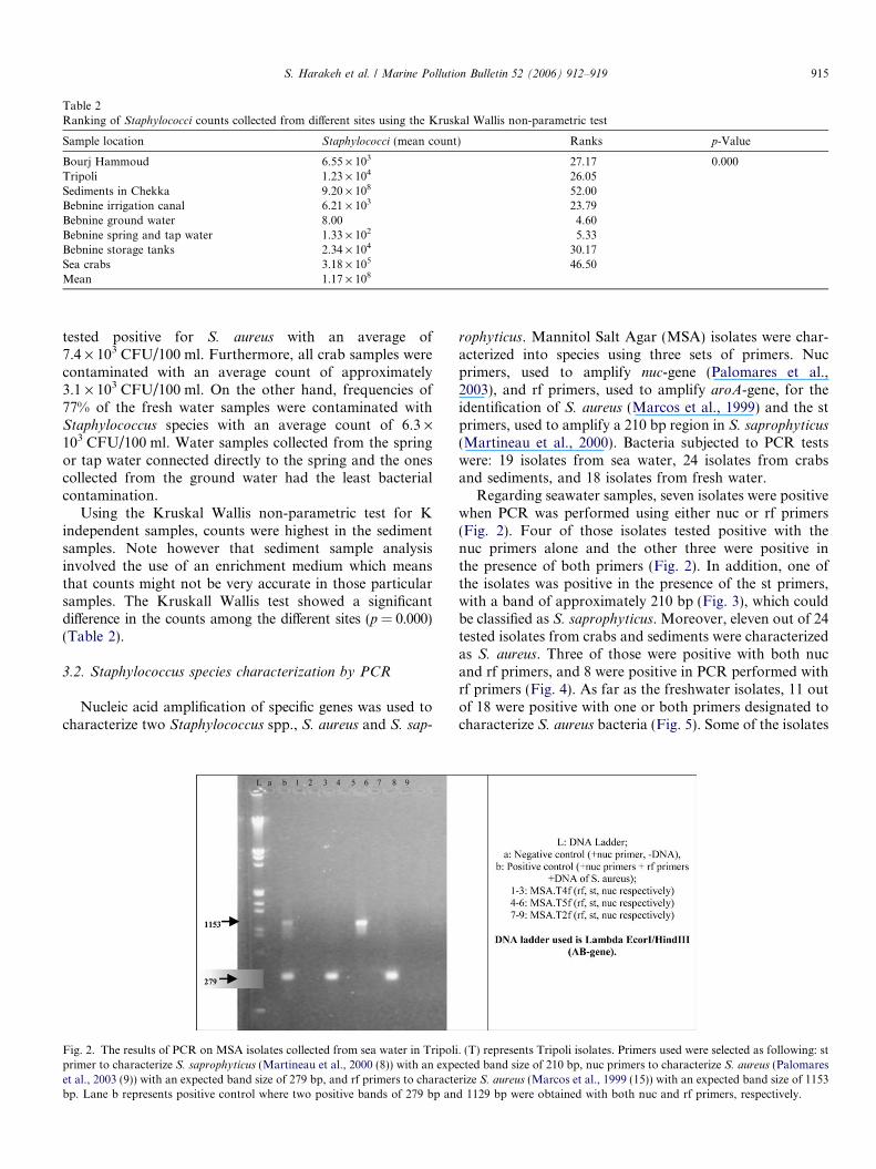
Table 2Ranking of Staphylococci counts collected from different sites using the Kruskal Wallis non-parametric test
Sample location Staphylococci (mean count) Ranks p-Value
Bourj Hammoud 6.55 · 103 27.17 0.000Tripoli 1.23 · 104 26.05Sediments in Chekka 9.20 · 108 52.00Bebnine irrigation canal 6.21 · 103 23.79Bebnine ground water 8.00 4.60Bebnine spring and tap water 1.33 · 102 5.33Bebnine storage tanks 2.34 · 104 30.17Sea crabs 3.18 · 105 46.50Mean 1.17 · 108
S. Harakeh et al. / Marine Pollution Bulletin 52 (2006) 912–919 915
tested positive for S. aureus with an average of7.4 · 103 CFU/100 ml. Furthermore, all crab samples werecontaminated with an average count of approximately3.1 · 103 CFU/100 ml. On the other hand, frequencies of77% of the fresh water samples were contaminated withStaphylococcus species with an average count of 6.3 ·103 CFU/100 ml. Water samples collected from the springor tap water connected directly to the spring and the onescollected from the ground water had the least bacterialcontamination.
Using the Kruskal Wallis non-parametric test for Kindependent samples, counts were highest in the sedimentsamples. Note however that sediment sample analysisinvolved the use of an enrichment medium which meansthat counts might not be very accurate in those particularsamples. The Kruskall Wallis test showed a significantdifference in the counts among the different sites (p = 0.000)(Table 2).
3.2. Staphylococcus species characterization by PCR
Nucleic acid amplification of specific genes was used tocharacterize two Staphylococcus spp., S. aureus and S. sap-
Fig. 2. The results of PCR on MSA isolates collected from sea water in Tripoliprimer to characterize S. saprophyticus (Martineau et al., 2000 (8)) with an expeet al., 2003 (9)) with an expected band size of 279 bp, and rf primers to charactebp. Lane b represents positive control where two positive bands of 279 bp an
rophyticus. Mannitol Salt Agar (MSA) isolates were char-acterized into species using three sets of primers. Nucprimers, used to amplify nuc-gene (Palomares et al.,2003), and rf primers, used to amplify aroA-gene, for theidentification of S. aureus (Marcos et al., 1999) and the stprimers, used to amplify a 210 bp region in S. saprophyticus
(Martineau et al., 2000). Bacteria subjected to PCR testswere: 19 isolates from sea water, 24 isolates from crabsand sediments, and 18 isolates from fresh water.
Regarding seawater samples, seven isolates were positivewhen PCR was performed using either nuc or rf primers(Fig. 2). Four of those isolates tested positive with thenuc primers alone and the other three were positive inthe presence of both primers (Fig. 2). In addition, one ofthe isolates was positive in the presence of the st primers,with a band of approximately 210 bp (Fig. 3), which couldbe classified as S. saprophyticus. Moreover, eleven out of 24tested isolates from crabs and sediments were characterizedas S. aureus. Three of those were positive with both nucand rf primers, and 8 were positive in PCR performed withrf primers (Fig. 4). As far as the freshwater isolates, 11 outof 18 were positive with one or both primers designated tocharacterize S. aureus bacteria (Fig. 5). Some of the isolates
. (T) represents Tripoli isolates. Primers used were selected as following: stcted band size of 210 bp, nuc primers to characterize S. aureus (Palomaresrize S. aureus (Marcos et al., 1999 (15)) with an expected band size of 1153d 1129 bp were obtained with both nuc and rf primers, respectively.

Fig. 3. The results of PCR-based detection of S. saprophyticus in MSA isolates from sea water revealed as positive bands with st primers. (S) representsBourj Hammoud isolates.
Fig. 4. The results of the PCR-based detection of S. aureus in MSA isolates from sea water revealed as positive bands with either nuc or rf primers. (BH)represents sediment isolates, and (C) represents crab ones. Lane b presents positive bands of approximately 279 bp and 1129 bp with nuc and rf primers,respectively.
Fig. 5. The results of PCR-based detection of S. aureus in MSA isolates from sea water in Bebnine revealed as positive bands with either st or rf primers.(B) represents Bebnine isolates. Lane b shows positive bands of approximately 279 bp and 1129 bp with nuc and rf primers, respectively.
916 S. Harakeh et al. / Marine Pollution Bulletin 52 (2006) 912–919
S. Harakeh et al. / Marine Pollution Bulletin 52 (2006) 912–919 917
that were not characterized by PCR were characterized byAPI (BioMerieux�, Sa Marcy l’Etoile, France) Staphylo-
coccus biochemical tests revealing that most unidentifiedbacteria belong to the species S. xylosus.
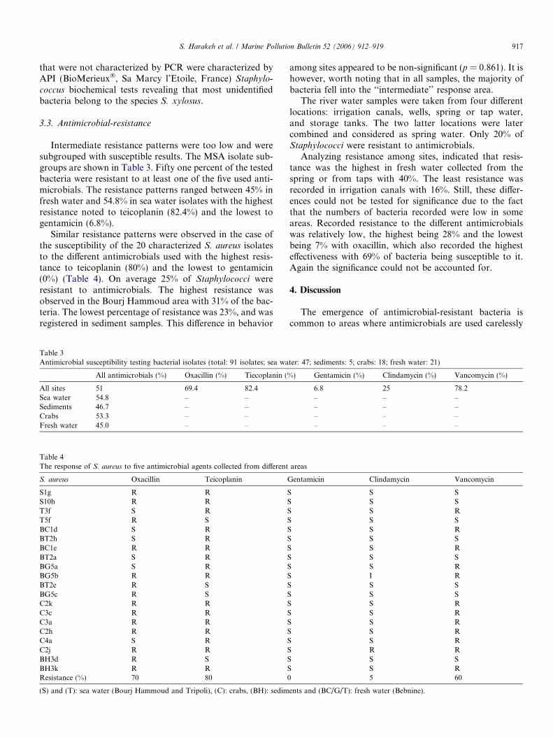
3.3. Antimicrobial-resistance
Intermediate resistance patterns were too low and weresubgrouped with susceptible results. The MSA isolate sub-groups are shown in Table 3. Fifty one percent of the testedbacteria were resistant to at least one of the five used anti-microbials. The resistance patterns ranged between 45% infresh water and 54.8% in sea water isolates with the highestresistance noted to teicoplanin (82.4%) and the lowest togentamicin (6.8%).
Similar resistance patterns were observed in the case ofthe susceptibility of the 20 characterized S. aureus isolatesto the different antimicrobials used with the highest resis-tance to teicoplanin (80%) and the lowest to gentamicin(0%) (Table 4). On average 25% of Staphylococci wereresistant to antimicrobials. The highest resistance wasobserved in the Bourj Hammoud area with 31% of the bac-teria. The lowest percentage of resistance was 23%, and wasregistered in sediment samples. This difference in behavior
Table 3Antimicrobial susceptibility testing bacterial isolates (total: 91 isolates; sea wa
All antimicrobials (%) Oxacillin (%) Tiecoplanin (%
All sites 51 69.4 82.4Sea water 54.8 – –Sediments 46.7 – –Crabs 53.3 – –Fresh water 45.0 – –
Table 4The response of S. aureus to five antimicrobial agents collected from different
S. aureus Oxacillin Teicoplanin G
S1g R R SS10h R R ST3f S R ST5f R S SBC1d S R SBT2h S R SBC1e R R SBT2a S R SBG5a S R SBG5b R R SBT2e R S SBG5c R S SC2k R R SC3c R R SC3a R R SC2h R R SC4a S R SC2j R R SBH3d R S SBH3k R R SResistance (%) 70 80 0
(S) and (T): sea water (Bourj Hammoud and Tripoli), (C): crabs, (BH): sedim
among sites appeared to be non-significant (p = 0.861). It ishowever, worth noting that in all samples, the majority ofbacteria fell into the ‘‘intermediate’’ response area.
The river water samples were taken from four differentlocations: irrigation canals, wells, spring or tap water,and storage tanks. The two latter locations were latercombined and considered as spring water. Only 20% ofStaphylococci were resistant to antimicrobials.
Analyzing resistance among sites, indicated that resis-tance was the highest in fresh water collected from thespring or from taps with 40%. The least resistance wasrecorded in irrigation canals with 16%. Still, these differ-ences could not be tested for significance due to the factthat the numbers of bacteria recorded were low in someareas. Recorded resistance to the different antimicrobialswas relatively low, the highest being 28% and the lowestbeing 7% with oxacillin, which also recorded the highesteffectiveness with 69% of bacteria being susceptible to it.Again the significance could not be accounted for.
4. Discussion
The emergence of antimicrobial-resistant bacteria iscommon to areas where antimicrobials are used carelessly
ter: 47; sediments: 5; crabs: 18; fresh water: 21)
) Gentamicin (%) Clindamycin (%) Vancomycin (%)
6.8 25 78.2– – –– – –– – –– – –
areas
entamicin Clindamycin Vancomycin
S SS SS RS SS RS SS RS SS RI RS SS SS RS RS RS RS RR RS SS R5 60
ents and (BC/G/T): fresh water (Bebnine).

918 S. Harakeh et al. / Marine Pollution Bulletin 52 (2006) 912–919
in medicine and in intensive animal husbandry. Because ofthe emergence of such bacteria, the effectiveness of antimi-crobials in fighting infections is decreasing leading to thefatal spread of diseases particularly S. aureus which is animportant cause of nosocomial infections and have beenisolated from intensive care units, general wards, and veter-inary teaching hospital environment and from the hands ofnurses (Chi et al., 2004; Murdoch, 2004; Fujimura et al.,2004; Cimiotti et al., 2004; Szewezyk et al., 2004; Weeseet al., 2004; Nimri and Batchoun, 2004; Lemmen et al.,2004).
Coastal development in addition to freshwater and sea-water abuse have led to the release of enteric human patho-gens and other pollutants at sampled locations along thecoast of Lebanon. This study isolated Staphylococci spe-cies, which are involved in many opportunistic infectionsof humans and animals (Aly, 1990), and assessed the levelsof contamination by those organisms, identified and char-acterized their different strains using PCR, and tested theirresistance to commonly used antimicrobials.
Some levels of bacterial contamination along the Leba-nese coast are relatively high and can render the water apotential vehicle for the transmission of many diseases.Most samples were contaminated with Staphylococci spe-cies. The contamination at the Bourj Hammoud area canbe attributed to the presence of an abandoned waste dis-posal facility along the seashore. The high level of Staphy-
lococci contamination in most samples ascertains theability of this microorganism to survive in harsh environ-mental conditions. Regarding freshwater, as expected, thesamples collected from the water spring were less contam-inated. However, high contamination was observed in sam-ples collected from irrigation water and reservoir tanks duemostly to unsafe practices of disposing untreated domesticwastewater in irrigation canals. The crab samples were alsocontaminated with Staphylococci species indicating thatother filter feeders might be equally contaminated, raisingpotential exposure and infectious diseases through directhuman consumption, especially when consumed raw. Notehowever, that the number of tested crab samples is toosmall and may not be representative of the aquatic faunaalong the entire coast of Lebanon.
In the PCR analysis for S. aureus, samples that exhibitedpositive resistance with one primer but not with two, indi-cates that environmental strains are highly prone to muta-tions occurring continuously and frequently in nature.
Seawater isolates from the Tripoli harbor showed thehighest rates in antimicrobial resistance. Such differentialexpression in antimicrobial resistance between Tripoli iso-lates and other ones might be due to the higher level of con-tamination with sewage-containing antimicrobials in thatarea, noting that this area has shown higher levels of con-tamination than the other sites. Resistance to teicoplaninand vancomycin were lower in the environmental isolates(80% and 60%, respectively) in comparison to the clinicalisolates (91% and 92%, respectively) (Araj and Kanj,2000). This differential expression of antimicrobial resis-
tance between environmental isolates and clinical onesrefers to the imprudent use of teicoplanin and vancomycinfor the treatment of different infectious diseases. In con-trast, resistance to oxacillin appeared higher in environ-mental isolates. Since oxacillin might be either genomicor plasmid encoded, resistance to it could be easily trans-ferred in nature by horizontal gene transfer (Fluit et al.,2001).
The total sensitivity of S. aureus to gentamicin and clin-damycin is likely due to stopping their use as either thera-peutic antimicrobials in veterinary medicine, or as growthpromoters in conventional animal fattening (Peters et al.,2003).
The results of this study emphasize the need for contin-uous monitoring of the contamination levels and antimi-crobial resistance patterns of bacteria isolated from theenvironment to limit the excessive use of antimicrobialsand thus reduce resistance. The results will also help shedlight on the patterns of resistance encountered in environ-mental isolates of S. aureus. Solid waste and wastewaterdisposal provide ample opportunity for their growth, andthe indiscriminate use of antimicrobials increases theirresistance to commonly used antimicrobials. Additionalanalysis is needed to better define the ecology of resistanceto antimicrobial agents in the environment as a whole. Theemergence of drug-resistant bacteria which could havedrastic effects on public health at large, increases the needfor proper management options.
Acknowledgements
Special thanks are extended to the US Agency for Inter-national Development for its continuous support to theWater Resources Center, the Environmental and WaterResources Engineering Program, and the InterfacultyGraduate Environmental Sciences Program at the Ameri-can University of Beirut. We extend our gratitude to theUniversity Research Board at the American University ofBeirut for partial financial support for this project, toMr. Shadi Hajjar and Mr. Omar Zouhairi for technicalhelp, and to Ms. Ama Saddaka and Mr. Joe El Khouryfor typing and editing support.
References
Aly, R., 1990. The pathogenic staphylococci. Semin. Dermatol. 9, 292–299,Review.
AOAC, 1995. Official Methods of Analysis of AOAC International, 16thed., Gaithersburg, MD, USA.
Araj, G.F., Kanj, S.S., 2000. Current Status and changing trends ofantimicrobial resistance in Lebanon. J. Med. Liban. 48, 221–226.
Ashbolt, N.J., 2004. Microbial contamination of drinking water anddisease outcomes in developing regions. Toxicology 198, 229–238,Review.
Ausubel, F.M., Brent, R., Kingdtone, R.E., 1989. Curr. Protocols Mol.Biol. 2, 11.5.1.
Brook, M.O., Smith, R., Bannister, B.A., McConnel, M., Chart, H.,Scotland, S.M., Sawyer, A., Smith, M.J., Rowe, B., 1994. Prospectivestudy of verocytotoxin-producing, enteroaggregative and diffusely
S. Harakeh et al. / Marine Pollution Bulletin 52 (2006) 912–919 919
adherent Escherichia coli in different diarrhoeal states. Epidemiol.Infect. 112, 63–67.
Chandrasekaran, S., Venkatesh, B., Lalithakumari, D., 2003. Transfer andexpression of a multiple antimicrobial resistance plasmid in marinebacteria. Curr. Microb. 37, 347–351.
Chen, T.R., Chiou, C.S., Tsen, H.Y., 2004. Use of novel PCR primersspecific to the genes of staphylococcal enterotoxin G, H, I for thesurvey of Staphylococcus aureus strains isolated from food-poisoningcases and food samples in Taiwan. Int. J. Food Microb. 92, 189–197.
Chi, C.Y., Wong, W.W., Fung, C.P., YU, K.W., Liu, C.Y., 2004.Epidemiology of community-acquired Staphylococcus aureus bactere-mia. Microbiol. Immunol. Infect. 37 (1), 16–23.
Cimiotti, J.P., Wu, F., Della-Latta, P., Nesin, M., Larson, E., 2004.Emergence of resistant staphylococci on the hands of new graduatenurses. Infect. Control Hosp. Epidemiol. 25 (5), 431–435.
Fluit, A.C., Visser, M.R., Schmitz, F.J., 2001. Molecular detection ofantimicrobial resistance. Clin. Microbiol. Rev. 14, 836–871.
Fujimura, S., Kato, S., Hashimoto, M., Takeda, H., Maki, F., Watanabe,A., 2004. Survey of methicillin-resistant Staphylococcus aureus fromneonates and the environment in the NICU. J. Infect. Chemother. 10(2), 131–132.
Griffin, P.M., Tauxe, R.V., 1991. The epidemiology of infections causedby Escherichia coli 0157:H7, other enterohemorrhagic E. coli, and theassociated hemolytic uremic syndrome. Epidemiol. Rev. 13, 60–98.
Holmberg, S.D., Wells, J.G., Cohen, M.L., 1984. Animal to-mantransmission of antimicrobial-resistant Salmonella: investigation ofUS outbreaks. Science 225, 833–835.
Lemmen, S., Hafner, H., Zolldann, D., Stanzel, A., Lutticken, R., 2004.Distribution of multi-resistant Gram-negative versus Gram-positivebacteria in the hospital inanimate environment. J. Hosp. Infect. 56,191–197.
Marcos, J.Y., Soriano, A.C., Salazar, M.S., Moral, C.H., Ramos, S.S.,Smeltzer, M.S., Carrasco, G.N., 1999. Rapid identification and typingof Staphylococcus aureus by PCR-restriction fragment lengthpolymorphism analysis of the aroA gene. J. Clin. Microbiol. 37,570–574.
Martineau, F., Picard, F.J., Menard, C., Roy, P.H., Ouellette, M.,Bergeron, M.G., 2000. Development of a rapid PCR assay specific for
Staphylococcus saprophyticus and application to direct detection fromurine samples. J. Clin. Microbiol. 38, 3280–3284.
Murdoch, F.E., Smmons, R.L., Chapple, I.L., 2004. Isolation andcharacterization of subgingival staphylococci fr periodontitis patientsand controls. Oral Dis. 10 (3), 155–162.
National Committee for Clinical Laboratory Standards, 2000. Methodsfor dilution antimicrobial susceptibility tests for bacteria that growaerobically. NCCLS approved standard M7-A5. National Committeefor laboratory Standards, Wayns, PA.
National Committee for Clinical Laboratory Standards, 2002. Perfor-mance standards for antimicrobial susceptibility testing. Supplementaltables, M100-S12. NCCLS, Wayne, PA.
Nimri, L.F., Batchoun, R., 2004. Community-acquired bacteraemia in arural area: predomin bacterial species and antibiotic resistance. J Med.Microbiol. 53 (10), 1045–1049.
Palomares, C., Torres, M.J., Torres, A., Aznar, J., Palomares, J.C., 2003.Rapid detection and identification of Staphylococcus aureus fromblood culture specimens using real-time fluorescence PCR. Diagnos.Microbiol. Infect. Dis. 45, 183–189.
Peters, J., Mac, K., Wichmann-Schauer, H., Klein, G., Ellerbroek, L.,2003. Species distribution and antimicrobial resistance patterns ofenterococci isolated from food of animal origin in Germany. Int. J.Food Microb. 88, 311–314.
Sambrook, J., Fritsch, E.F., Maniatis, T., 1989. Molecular Cloning: ALaboratory Manual, p. B.17.
Slobodnikova, L., Kotulova, D., Zahradnikova, I., 1995. Staphylococcus
aureus in chronic and recurrent infections. Folia Microb. (Praha) 40,655–658.
Son, R., Rusu, G., Karim, M.I., 1997. Conjugal transfer of plasmids andantimicrobial resistance among Escherichia coli isolated from animalsin a rural area in Sarawak (Malaysia). J. Appl. Microbiol. 82, 240–244.
Szewezyk, E.M., Rozalska, M., Cieslikowski, T., Nowak, T., 2004.Plasmids of Staphylococcus cohnii isolated from the intensive unit.Folia Microb. (Praha) 49 (2), 123–131.
Weese, J.S., DaCosta, T., Button, L., Goth, K., Ethier, M., Boehnke, K.,2004. Isolation of methicillin-resistant Staphylococcus aureus fromenvironment in a veterinary teaching hospital. J. Vet. Int. Med. 18 (4),468–470.