Validation of a Method for Measuring the Position of Pick ...
Upload
independentCategory
view
4download
0
doi:10.1093/brain/awl260 Brain (2006) Page 1 of 14
The adult form of Niemann–Pick disease type C
Mathieu Sevin,1 Gaetan Lesca,4,5,6 Nicole Baumann,2 Gilles Millat,4,5 Olivier Lyon-Caen,1
Marie T. Vanier4,5 and Frederic Sedel1,3
1Assistance Publique-Hopitaux de Paris, Federation des maladies du systeme nerveux, 2Unite mixte de recherche INSERMU-711, UPMC, Hopital de la Salpetriere, 3Centre de reference, Maladies Lysosomales a Expression Neurologique, Paris,4Hospices Civils de Lyon, Laboratoire Gillet-Merieux, Centre Hospitalier Lyon-Sud, Pierre-Benite and 5INSERMUnite 499, Faculte de Medecine Laennec, Lyon, France6Permanent address: Consultation de Genetique, Hopital Debrousse, Lyon, France
Correspondence to: Marie T. Vanier, MD PhD, Laboratoire Gillet-Merieux, Batiment 3B, Centre HospitalierLyon-Sud, 69495 Pierre-Benite cedex, FranceE-mail: [email protected]
Niemann–Pick disease type C (NPC) is a fatal neurovisceral lipid storage disease of autosomal inheritanceresulting from mutations in either the NPC1 (95% of families) or NPC2 gene. The encoded proteins appear to beinvolved in lysosomal/late endosomal transport of cholesterol, glycolipids and other molecules but their exactfunction is still unknown. The clinical spectrum of the disease ranges from a neonatal rapidly fatal disorder to anadult-onset chronic neurodegenerative disease. Based upon a comprehensive study of 13 unrelated adultpatients diagnosed in France over the past 20 years as well as the analysis of the 55 other cases publishedsince 1969, we have attempted to delineate the major clinical, radiological, biochemical and genotypic char-acteristics of adult NPC. Overall, mean age at onset (6SD) of neuropsychiatric symptoms was 25 6 9.7 years.The diagnosis of NPC was established after a mean delay of 6.2 6 6.4 years and the mean age at death(calculated from 20 cases) was 38 6 10.2 years. Major clinical features included cerebellar ataxia (76%), verticalsupranuclear ophthalmoplegia (VSO, 75%), dysarthria, (63%), cognitive troubles (61%), movement disorders(58%), splenomegaly (54%), psychiatric disorders (45%) and dysphagia (37%). Less frequent signs were epilepsyand cataplexy. During the course of the disease, clinical features could be subdivided into (i) visceral signs(hepatomegaly or splenomegaly), (ii) cortical signs (psychiatric cognitive disorders and epilepsy); and (iii)deep brain signs (VSO, ataxia, movement disorders, dysarthria, dysphagia, cataplexy) which exhibited differentevolution patterns. Asymptomatic and non-evolutive visceral signs were often noticed since early childhood(38.5% of our patients), followed by mild cortical signs in childhood (learning difficulties) and early adulthood(62% of cases among which 38% were psychiatric disorders). Deep brain signs were observed in 96% of patients andwere usually responsible for death. In general, there was a good correlation between clinical signs and thelocalization of brain atrophy on MRI. The ‘variant’ biochemical phenotype characterized by mild abnormalitiesof the cellular trafficking of endocytosed cholesterol was over-represented in the adult form of NPC and seemedassociated with less frequent splenomegaly in childhood and lesser psychiatric signs. Involvement of the NPC1gene was shown in 33 families and of the NPC2 gene in one. Improving the knowledge of the disease amongpsychiatrists and neurologists appears essential since emerging treatments should be more efficient at the visceralor cognitive/psychiatric stages of the disease, before the occurrence of widespread deep brain neurological lesions.
Keywords: adult; inborn errors of metabolism; lysosome; Niemann–Pick C
Abbreviations: NPC ¼ Niemann–Pick disease type C; VSO ¼ vertical supranuclear ophthalmoplegia
Received July 16, 2006. Revised August 16, 2006. Accepted August 18, 2006
IntroductionNiemann–Pick disease type C (NPC, MIM 257220) is a
neurovisceral lysosomal lipid storage disorder of autosomal
recessive inheritance characterized at the cellular level by
accumulation of unesterified cholesterol and glycolipids in
the endosomal/lysosomal system (Patterson et al., 2001;
Vanier and Millat, 2003). The pattern of accumulating
lipids, however, is different in the brain and in non-neural
organs. Approximately 95% of patients have mutations in
the NPC1 gene (mapped at 18q11; MIM 607623) which
encodes a large membrane glycoprotein with late endosomal
# The Author (2006). Published by Oxford University Press on behalf of the Guarantors of Brain. All rights reserved. For Permissions, please email: [email protected]
Brain Advance Access published September 26, 2006 by guest on June 1, 2013
http://brain.oxfordjournals.org/D
ownloaded from

localization. The remainder have mutations in the NPC2 gene
(mapped at 14q24.3) (MIM 601015), encoding a small soluble
lysosomal protein that binds cholesterol with high affinity.
The finding of a non-redundant functional cooperativity
between NPC1 and NPC2 confirmed the early concept that
the two proteins function in tandem or in sequence (Vanier
et al., 1996; Sleat et al., 2004). They appear to be involved in
post-lysosomal/late endosomal transport of cholesterol, gly-
colipids and other molecules, but their precise functions and
relationship remain unclear, and their primary substrate
unknown (for review see Vanier and Millat, 2003; Vincent
et al., 2003; Vanier and Millat, 2004). The biochemical
diagnosis of NPC is currently based on the demonstration of
impaired LDL-cholesterol trafficking in cultured fibroblasts of
patients, by cytochemical visualization of accumulated free
cholesterol after filipin staining and study of LDL-induced
cholesterol ester formation (Vanier et al., 1991a).
NPC shows an extreme clinical heterogeneity (Vanier and
Suzuki, 1996; Patterson et al., 2001; Patterson, 2003; Vanier
and Millat 2003). The age of presentation is ranging from
the perinatal period to adults over 50 years. The initial
manifestations may also vary, being hepatic, pulmonary,
neurological or psychiatric in nature. The systemic (includ-
ing hepatic, splenic and pulmonary) and neurological
diseases follow independent courses. NPC is now recognized
as a relatively common cause of liver disease in early life,
including foetal hydrops, and in nearly half of the cases,
a neonatal cholestatic icterus with hepatosplenomegaly. In
childhood, isolated hepatosplenomegaly is often the pre-
senting symptom. Nevertheless, apart from infants with
early death from liver or respiratory failure and exceedingly
rare adults with isolated splenomegaly, all NPC patients
develop neurological or psychiatric symptoms. Indeed, the
most common classification of the disease is based on the
age of onset of the neuropsychiatric symptoms, which to
a large extent, correlates with the life span of the patients.
In the severe infantile neurological form, delayed motor
development and hypotonia become apparent between 1 and
2 years of age and most of these patients die before the age of
5 years. In the classical phenotype (60–70% of all cases),
which includes the late infantile and the juvenile forms,
neurological symptoms appear between 3 and 15 years, with
death occurring in most cases between 8 and 25 years.
Cerebellar ataxia, dysarthria, dysphagia, cataplexy, seizures,
dystonia, progressive dementia and a characteristic vertical
supranuclear ophthalmoplegia (VSO) are prominent signs.
The adult neurological onset form is infrequent and the
majority of publications have reported single cases. Previous
attempts to delineate the major clinical features of the adult
form of NPC were based on the review of a limited number
of published cases (i.e. 16 cases in Schulman et al., 1995
and 27 cases in Lossos et al., 1997) and data concerning
genotypes, biochemical phenotypes, brain imaging and
disease course were lacking. Based on the detailed study
of 13 adult patients diagnosed in France during the past
25 years and a literature review of 55 previously published
cases, the aim of the present study is to provide an
updated description of the adult form of the disease. This
clarification is now becoming particularly important, since
emerging therapies are in sight.
Patients and methodsBetween 1985 and 2005, from the data of a national reference
biochemical diagnostic centre (M.T.V.), 133 patients, belonging
to 99 unrelated families, were definitively diagnosed as having NPC
in France. Thirteen unrelated patients from this cohort with an age
at onset of neurological or psychiatric signs at or after 15 years were
included in the present study. Age at onset was defined here as the
age of first obvious neurological or psychiatric symptoms, which
excluded clumsiness, learning difficulties in childhood or reading
difficulties which could retrospectively be related to mild VSO. For
eight patients (numbered 1, 2, 4, 7, 8, 11, 12 and 13), neurological
examination and medical data recording were performed by one
of us (F.S.). In other cases, the clinical history was based on the
detailed patient’s medical records. MRI analysis was included in the
present study only when recent data were available, which excluded
patients for whom MRI was performed only at the beginning of the
disease, or patients for whom only MRI reports were available. The
diagnosis of NPC was biochemically confirmed in cultured skin
fibroblasts by combined studies of filipin staining and LDL-induced
cholesteryl ester formation, and patients were classified into either
a classic or a variant biochemical phenotype (Vanier et al., 1991a).
The classic phenotype refers to patients whose fibroblasts show
a striking cholesterol accumulation and a severe block in LDL-
induced cholesteryl ester formation. In cells from biochemically
variant patients, demonstration of cholesterol storage requires
challenge with pure LDL, and the rate of cholesteryl ester formation
may not be affected. Methods used for mutation analysis have been
described previously (Millat et al., 2001, 2005).
Dependence was defined as one or more of the following
outcomes: (i) unable to walk alone; (ii) assistance required
for simple activities of daily living; (iii) institutionalization; or
(iv) gastrostomy tube required for feeding. Cognitive disorder was
defined as any cognitive dysfunction (excluding psychiatric
troubles) interfering with activities of daily living. Cases number
4 and 5 have been previously published as single case reports (Philit
et al., 2002; Tyvaert et al., 2005), but were included in our 13 cases
since additional clinical data and longer follow-up were available.
These two cases were excluded from the literature review. Review of
the literature was performed using the NIH Pubmed database.
Several cases published more than once were excluded by careful
perusal of publications or personal communications. Fifty-five
cases of NPC disease in adult life were finally selected from
27 reports. Only cases for which detailed clinical data were available
were included in the statistical analysis. For MRI analysis, only
publications including MRI pictures of sufficient quality were
included.
ResultsCase reports (Table 1)Cases with prominent psychiatric or cognitive signsCase 1: This male patient had no familial or personal history.
Childhood acquisitions were normal, but school perfor-
mances were noted to be poor, because of slow learning,
Page 2 of 14 Brain (2006) M. Sevin et al.
by guest on June 1, 2013http://brain.oxfordjournals.org/
Dow
nloaded from

Tab
le1
Sum
mar
yof
clin
ical
,bio
chem
ical
and
genet
icdat
ain
13
Fren
chpat
ients
with
adult
neu
rolo
gica
lonse
tof
NPC
Cas
eG
ender
Pre
monitory
sign
sin
child
hood
Age
atneu
rol.
onse
t(y
ears
)
Dia
gnosi
sdel
ay(y
ears
)
Firs
tneu
ropsy
.si
gnin
adulthood
Maj
or
clin
ical
sign
sduri
ng
dis
ease
cours
eD
epen
den
cedel
ay(y
ears
)M
RI
(atr
ophy
)A
geat
dea
th(y
ears
)
Age
atla
stfo
llow
-up
(yea
rs)
Bio
ch.
phen
oty
pe
NPC
1m
uta
tions
Mea
n6
SD20.7
66.7
5.2
64.8
8.9
64
386
10.2
30.3
67
1M
Cog
16
9Psy
Psy
,C
og,
At,
VSO
,M
d7
Cort
ical
,su
bco
rtic
al26
Cl
R615L/
?a
2F
none
30
10
Psy
Psy
,C
og,
At,
Md,V
SO,Sm
10
Cort
ical
,su
bco
rtic
al,
corp
us
callo
sum
42
Cl
D1097N
/F283fs
a
3M
hm
,Sm
15
8Psy
Psy
,C
og,
At,
VSO
,Sm
9C
orp
us
callo
sum
,su
bco
rtic
al,co
rtic
al31
31
Cl
Y871C
/F76
3La
4F
none
18
13
Psy
Psy
,C
og,
At,
VSO
,Sm
11
NA
36
Cl
R372W
/?5
MSm
,hea
34
1Psy
Psy
,C
og,
Sm4
Norm
al39
39
Cl
Y825C
/?6
MSm
,C
og
28
NA
Cog
Cog,
At
4C
ort
ical
and
cere
bella
r38
32
Cl
I1061T
/A605V
b
7M
VSO
?16
12
Md
Md,A
t,V
SO,Sm
16
Cer
ebel
lar
and
bra
inst
em,
mild
WM
hig
hin
tensi
ty36
Var
G992R
/N452fs
8F
17
2A
tA
t,M
d,V
SO,Sm
none
NA
23
Var
G992A
/G993fs
a
9F
hea
28
0A
tA
t,V
SO,Sm
none
NA
49
28
Var
V950M
/V950M
b
10
Mhm
,Sm
17
1M
dM
d,A
t,V
SO,Sm
13
Cort
ical
and
cere
bella
r32
32
Cl
R1059Q
/L724P
11
FV
SO?,
Cog,
18
2A
tPsy
,C
og,
At,
Md,V
SO,Sm
nC
ereb
ella
ran
dbra
inst
em,
Mild
WM
hig
hin
tensi
ty22
Var
V950M
/I1061T
12
MC
og,
ca15
2V
SOPsy
,C
og,
At,
VSO
,M
d,Sm
6N
A27
Var
V950M
/I1061T
13
Msm
17
2V
SOC
og,
Md,V
SO,Sm
nM
ildW
Mhig
hin
tensi
ty20
Cl
I1061T
/G538R
At
=at
axia
;bio
chem
.=
bio
chem
ical
;ca
=ca
taple
xy;
Cl
=cl
assi
calbio
chem
ical
phen
oty
pe
(sev
ere
chole
ster
oltr
affic
king
abnorm
alitie
s);C
og
=co
gnitiv
edis
ord
er(e
xcl
udin
gpsy
chia
tric
sym
pto
ms)
;F
=fe
mal
e;hm
=hep
atom
egal
y;hea
=hea
ring
loss
;M
=m
ale;
Md
=m
ove
men
tdis
ord
ers
(incl
udin
gdys
tonia
,ch
ore
aor
par
kinso
nis
m);
NA
=not
avai
lable
;neu
rol.
=neu
rolo
gica
l;Psy
=psy
chia
tric
trouble
s;sm
=sp
lenom
egal
y;V
ar=
vari
ant
bio
chem
ical
phen
oty
pe
(mild
chole
ster
oltr
affic
king
abnorm
alitie
s);W
M=
white
mat
ter.
a Mill
atet
al.(2
005).
bM
illat
etal
.(2
001).
Adult form of Niemann–Pick C disease Brain (2006) Page 3 of 14
by guest on June 1, 2013http://brain.oxfordjournals.org/
Dow
nloaded from

mild difficulties in writing, reading and speaking. However,
he could follow a normal educational training. When he
was 16 years old, he was hospitalized for an acute paranoid
delirium, which resolved after 2 months. Three years later
he experienced another acute episode but with persistence
of psychotic features suggestive of paranoid schizophrenia.
During the following years, he became clumsy with poor
upper limbs coordination. Cognitive troubles appeared,
including apathy, memory impairment and a dysexecutive
syndrome which progressively worsened. At 21 years of
age, neurological examination revealed a tetra pyramidal
syndrome, gait and limbs cerebellar ataxia, dysarthric
speech, dysphagia and VSO. At the age of 24 years, he had
lost gait and speech. Dystonic postures involving hands
and legs and myclonic jerks appeared. Gastrostomy was
performed at 26 years because of severe deglutition
problems. Mild cortical and subcortical atrophy were
reported on brain MRI. Electroneuromyography, electro-
retinogram, visual evoked potentials and abdominal ultra-
sonography were normal. EEG performed at 23 years of age
revealed generalized slowing.
Case 2: This 40-year-old female had normal psychomotor
development, with normal schooling, and worked as an
employee in a supermarket. She had two normal pregnan-
cies. When 30 years old, she suffered from an acute
persecutive delusion with acoustic hallucinations. Persistent
delusion led to the diagnosis of dysthymic schizophrenia.
Despite chronic therapy with neuroleptics and mood
stabilizers, she experienced eight delusion relapses in
6 years requiring hospitalizations at each time. She refused
medical follow-up for the next 4 years. During this period, in
addition to psychotic features, she developed slow dysarthric
speech, attention and memory impairment, dysphagia and
ataxic gait. At the age of 40 years, neurological examination
revealed VSO, gait cerebellar ataxia, pyramidal signs, left
upper limb rest tremor and rigidity. Neuropsychological
evaluation showed dementia with non-fluent aphasia,
apragmatism, anosognosia, perseverations, memory impair-
ment, visuoconstructive apraxia and prosopagnosia. Her gait
progressively worsened and at the age of 42 years, she was
unable to walk alone. Abdominal ultrasonography disclosed
hepatomegaly and splenomegaly. Brain MRI revealed
moderate atrophy affecting the cerebral cortex, the cerebel-
lum and the corpus callosum, without signal abnormalities.
EEG showed generalized slowing.
Case 3: This patient had no familial history. His personal
history was remarkable for thalassaemia and thrombopenia
with recurrent epistaxis in early childhood. At 24 months
of age, hepatosplenomegaly was found and bone marrow
aspiration revealed foam cells accumulation suggestive of
NPC. Partial splenectomy was performed at 12 years because
of an acute abdominal pain. Lipid analysis of frozen spleen
tissue performed by one of us (M.T.V.) revealed a typical
NPC pattern and the diagnosis was definitely confirmed
later on skin fibroblasts. His psychomotor development was
normal and he attended normal school until 15 years old,
when his parents and teachers first noticed behavioural
disturbances including social isolation, self mutilations,
aggressiveness and hyperphagia. A psychiatric evaluation
revealed a depressive syndrome. An insulin dependent
diabetes mellitus developed at the age of 16 years. Gait
abnormalities with falls appeared when he was 19 years old
and progressively worsened. At the age of 24 years, beha-
vioural disturbances led to institutionalization. Neurological
examination found gait and upper limbs cerebellar syn-
drome and VSO. Neuropsychological evaluation was not
possible. Brain MRI showed mild cortical, subcortical and
corpus callosum atrophy with relative sparing of the
posterior fossae. He experienced one generalized tonic–
clonic seizure and frequent sudden falls without loss of
consciousness, suggestive of cataplexy. He died at 30 years of
age in a bedridden state. Electroneuromyography, EEG and
brain MRI were normal.
Case 4: This patient was born premature at 32 weeks, but
her psychomotor development was normal. She attended
normal school until the age of 16, but stayed unemployed
thereafter. Dissociative symptoms progressively appeared.
When 29 years old, she was not autonomous anymore,
because of disorientation in time and space and severe
behaviour disturbances. She refused neuropsychological
evaluation and brain MRI. Clinical examination revealed
cerebellar ataxia, dysarthric speech, VSO and hepatospleno-
megaly. Her motor skills progressively worsened and
increasing dysphagia led to gastrostomy at the age of 35.
At 36, she was not able to walk alone anymore, she was
mute, with episodes of agitation and had tonico–clonic
generalized seizures.
Case 5: This male patient had no familial history. He
attended normal school and was employed in a gas
company. Splenomegaly was noticed at the age of 3 years,
for which a spleen biopsy revealed foam cells. A diagnosis of
probable NPC was then strongly suspected. He was also
treated for hypercholesterolaemia and, depression and
presented deafness which was considered as a sequel of
head trauma. He had been followed up for a transient
glomerular nephropathy (membranoproliferative glomeru-
lonephritis type II) with renal failure. At the age of 34 years,
he developed acute delirium with paranoid delusion and
dissociative features. Neuroleptic therapy led to partial
improvement. On examination, he was alert and well
oriented but a pyramidal syndrome was noted. Over the next
4 years, attention deficit and difficulties in finding words
worsened. Neuropsychological evaluation, at the age of 38,
showed diffuse cognitive alterations, with predominant
frontal lobe dysfunction, memory impairment and visuo-
constructive apraxia. He rapidly deteriorated within the next
months with acute renal failure, acute delirium, dysphagia,
cachexia and dehydration, requiring a combination of
dialysis, sedative drugs and gastrostomy. He subsequently
became catatonic and bedridden with total dependence.
Routine laboratory tests, chest radiography and brain MRI
were reported to be normal. He died at 39 years of age.
Page 4 of 14 Brain (2006) M. Sevin et al.
by guest on June 1, 2013http://brain.oxfordjournals.org/
Dow
nloaded from

Case 6: This male patient had no familial history. Isolated
splenomegaly was found during the first days of life, and a
splenectomy was performed at 10 years of age revealing
a storage process consistent with the diagnosis of NPC.
Learning difficulties were noticed during late childhood. At
28 years of age he became slow, apathic and had difficulties
in finding words. He had to stop working the same year.
From the age of 30, he exhibited an unsteady gait and
falls. Neurological examination 2 years later revealed manual
grasping, cerebellar ataxia, pyramidal syndrome, severe
dysarthria and dysphagia. No VSO was noticed. EEG
revealed diffuse slow waves. On brain MRI, cerebral and
cerebellar atrophy was reported. He died at the age of 38.
Cases with predominant movement andgait impairmentCase 7: This male patient had no personal or familial history.
His psychomotor development was normal and schooling
was unremarkable except that he reported difficulties to
direct his gaze downward when reading. At 16 years of age,
he noticed difficulties to perform fine left hand movements.
Neurological examination, at 25 years, revealed VSO and
mild generalized dystonia involving the four limbs, assoc-
iated with myoclonic jerks that were both spontaneous and
evoked by nociceptive stimulations. He became progressively
ataxic and dysarthric, with a pyramidal syndrome including
brisk tendon reflexes, legs spasticity and a bilateral
Babinski sign. A neuropsychological evaluation disclosed a
mild dysexecutive syndrome. Abdominal ultrasonography
revealed a homogeneous splenomegaly. At 32 years, he could
not walk alone anymore and from the age of 35, he became
dependent for most daily activities. Brain MRI showed
marked atrophy of the pons and cerebellum, mild atrophy of
cortical and subcortical areas. Electroneuromyography
showed only a bilateral carpal tunnel syndrome and EEG
revealed generalized slowing.
Case 8: This female patient with no personal or familial
medical history was noticed to be mildly clumsy since the
age of 14, often breaking glasses. However, she attended
normal school, obtained the French ‘baccalaureat’ and
started a professional training course. When 17 years old,
neurological abnormalities were first seen including dys-
arthric speech, gait and limb cerebellar ataxia and mild
dysphagia. At 19 years, VSO was also found, together with
brisk deep tendon reflexes and dystonia of the right lower
and upper limbs. She also experienced several sudden falls
while standing, without loss of consciousness, that were
described as a tonus loss and were suggestive of cataplexy.
General examination did not show splenomegaly but this
was found with ultrasonography. Gait difficulties progres-
sively increased but when 23 years old, she was still
autonomous for activities of daily living. CT-scan of the
brain was normal. She refused MRI. Electroneuromyography
was normal and EEG showed non-specific bilateral slow
waves.
Case 9: Age at onset could not be determined with
precision in this female patient. She had hearing loss since
the age of 3, and her familial history was remarkable for
deafness also affecting two of her brothers as well as three
nephews. Her physical, motor and intellectual developments
were otherwise normal, but she was sometimes clumsy.
She attended normal school until the age of 17. She worked
successfully in several offices and had two normal preg-
nancies. When 28 years old, she consulted for deafness but
did not complain of other symptoms. A careful neurological
evaluation revealed cerebellar dysfunction with ataxic gait,
dysmetria, dysarthric speech and VSO. Abdominal ultra-
sonography showed hepatosplenomegaly. No MRI of the
brain was performed. With time, ataxia and dysarthria
became more and more invalidating, but her mental capa-
cities remained quite well preserved. Progressive dysphagia
led to gastrostomy at the age of 45. She died from
septicaemia at the age of 49.
Case 10: This male patient had no familial history.
Following a transient documented episode of neonatal
cholestatic icterus, hepatosplenomegaly was found on
systematic clinical examination when he was 5 weeks old.
During childhood, he presented numerous, spontaneously
resolutive infections affecting skin and parotid glands,
associated with interstitial neuropathy and persistent
elevation of sedimentation rate, suggestive of immunodefi-
ciency. A spleen biopsy revealed sea blue histiocytes but no
definite diagnosis was made. When 17 years old, he
exhibited abnormal, fixed postures of his hands, progres-
sively followed by increasing gait difficulties. Neurological
examination at the age of 18 showed generalized cervical and
upper limbs dystonia, gait cerebellar ataxia and VSO.
Splenomegaly was present. Cortical and cerebellar atrophy
were noticed on a brain CT-scan (no MRI was performed).
EEG showed diffuse slow waves. The biochemical examina-
tion of a liver biopsy (M.T.V.) showed a lipid storage profile
typical of NPC and the diagnosis was confirmed on
fibroblasts. Chorea and myoclonus affecting all body parts
subsequently appeared, leading to a progressive deteriora-
tion of his motor skills. In addition, the cerebellar syndrome
increased, and finally he became unable to walk alone at
the age of 30. A gastrostomy tube was placed because of
increasing dysphagia. He died when 32 years old after a post-
traumatic cerebral haemorrhage.
Case 11: This female patient had learning difficulties since
childhood because of poor concentration capacities. She also
had a tendency to flex her head rapidly to see downward
which was interpreted as a tic but which can in retrospect
be interpreted as a sign of VSO. At 12 years, she benefited
from orthoptic re-education. She could achieve a sales
person professional position but with difficulties because of
calculation impairment. From the age of 19 years she
progressively exhibited dysarthria, cerebellar ataxia, dystonic
postures involving fingers and VSO. Abdominal ultrasono-
graphy disclosed an isolated splenomegaly. At 20 years of
age, the diagnosis of NPC was made. She was hospitalized
Adult form of Niemann–Pick C disease Brain (2006) Page 5 of 14
by guest on June 1, 2013http://brain.oxfordjournals.org/
Dow
nloaded from

in a psychiatric department for 6 months because of social
withdrawal, aggressiveness, persecutive delusion and suicide
attempts. These psychiatric troubles were controlled with
neuroleptics and at the age of 22 years, the patient is still
autonomous for most activities of daily living. A brain
MRI showed mild atrophy of the pons and cerebellum but
without discernible cortical atrophy. Electroneuromyogra-
phy was considered normal.
Case 12: This male patient experienced reading difficulties
in childhood. These were not due to VSO, but rather to
a problem in understanding the syllabus system. He could
follow normal schooling with help and a professional
training course in floriculture. When 14 years old, he had
falls due to abrupt loss of muscular tone without loss
of consciousness, which were retrospectively suggestive of
cataplexy. When 15 years old, a teacher noticed he had
difficulties to look downwards. At 17 years he first visited a
neurologist who noted a cerebellar syndrome and VSO.
Abdominal echography showed splenomegaly. The diagnosis
of NPC was made. Ataxia worsened and at 21 years, he
had to use a wheelchair. From 23 years, he also exhibited
dysarthria, hand dystonic postures and choreic movements.
From 20 years, he started to present acute psychiatric
troubles with agitation, aggressiveness, visual and auditive
hallucinations and delusion. These episodes occurred at an
average rate of once per year, lasting �15 days each time and
were well controlled with treatment. MRI could not be
performed because of choreic movements. Electroneuro-
myography did not show any abnormalities.
Case 13: This male patient had an asymptomatic
splenomegaly which was first noticed at 2 years of age.
He did not exhibit any intellectual or motor impairment
until 17 years old when he started to experience learning
difficulties together with oculomotor troubles. Progressively,
dysarthria and dystonic postures involving hands and feet
appeared. He also complained of mild hypoacousia.
Neurological examination found dystonic hands and feet
movements while walking, vertical and horizontal VSO, mild
cerebellar ataxia and hypoacousia. Neuropsychological
evaluation disclosed a mild dysexecutive syndrome. At
20 years of age, the patient is still autonomous in most
activities and he currently undergoes a professional training
course in geothermics. Brain MRI did not show
abnormalities and electroneuromyography was normal.
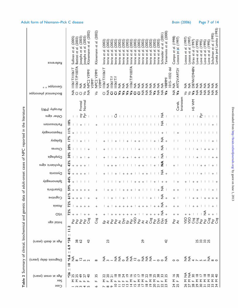
Global analysis of our case reports andreview of the literatureData collected from our 13 patients are summarized
in Tables 1, 3 and 4. Clinical, biochemical and genetic
characteristics of published cases are summarized in Tables 2,
3 and 4. From the compilation of our cases and 55 case
reports for which sufficient data were available, first pre-
senting signs in adulthood were psychiatric troubles in 38%
of cases; cognitive troubles (excluding psychiatric features)
in 23%; ataxia in 20%; movement disorders (dystonia) in
11%; VSO in 8%; dysarthria in 8% and epilepsy in 3% of
cases. Overall, the mean age at onset (6SD) of neuropsy-
chiatric symptoms was 25 6 9.7 years. The diagnosis of NPC
was established after a mean delay of 6.2 6 6.4 years and
the mean age at death (calculated from 20 cases) was 38 6
10.2 years.
DiscussionNPC is pan ethnic, with an estimated incidence in Western
European countries of �1/120 000 to 1/150 000 living births
(Vanier and Millat, 2003). From published data, it is
not possible to estimate the exact proportion of adult
forms among NPC patients in general. This may also vary
according to the ethnic background, due to different
mutation patterns. From our study, a first evaluation can
be made at the scale of one country. The adult form
accounted for 10% of the patients diagnosed in France
between 1985 and 2005. Among these patients, two were
diagnosed between 1985 and 1995 and the remaining
between 1995 and 2005, with an approximate constant rate
of one patient per year during the past 10 years in a
population of 60 million inhabitants. It is however difficult
to provide a reliable estimation of the incidence of NPC in
adults, as an unknown proportion of these patients is
probably undiagnosed or misdiagnosed.
Two main explanations can be given to this situation.
On one hand, most neurologists are not aware of this
infrequent diagnosis as suggested by the main delay for
diagnosis, which is �6 years. On the other hand, NPC is
particularly difficult to diagnose when presenting as pure
psychiatric disorder, frontal dementia, ataxia or dystonia.
In addition, the wide range of neurological signs seen in
NPC can mimic other, more familiar, neurological and
psychiatric diseases. Thus, in adults with NPC reported in
the literature, some initial diagnoses were Alzheimer’s
disease, Parkinson’s disease, schizophrenia, Wilson’s disease,
multiple sclerosis, Creutzfeldt–Jakob disease or Gayet–
Wernicke encephalopathy. Finally, the biological confir-
mation of NPC requires a skin fibroblast culture, and only a
limited number of laboratories offer reliable diagnosis tests.
From data collected from our own experience and the
exhaustive review of published cases, the major clinical,
biological and molecular aspects of adult NPC can be
defined as follows.
Disease courseFirst neurological symptoms occurred within the second or
third decades in most patients, but onset as late as 54 years
has been reported (Vanier et al., 1991b; Trendelenburg,
2006). We defined here the onset of the adult disease as the
age at onset of obvious neurological or psychiatric signs.
Overall, age at onset was 25 years, but 11 of our 13 patients
exhibited premonitory signs or symptoms in childhood
that preceded neurological deterioration in adulthood.
These signs included splenomegaly (5 cases), hepatomegaly
Page 6 of 14 Brain (2006) M. Sevin et al.
by guest on June 1, 2013http://brain.oxfordjournals.org/
Dow
nloaded from
Tab
le2
Sum
mar
yof
clin
ical
,bio
chem
ical
and
genet
icdat
aof
adult-o
nse
tca
ses
of
NPC
report
edin
the
liter
ature
Case
Sex
Ageatonset(years)
Diagnosisdelay(years)
Ageatdeath(years)
Initialsign
VSO
Ataxia
Cognitive
Dysarthria
Splenomegaly
Dystonia
Psychiatricsigns
Dysphagia
Chorea
Epilepsy
Hepatomegaly
Parkinsonism
Othersigns
Atrophy(MRI)
Biochemicalphenotype
Genotype**
Reference
*266
10
*6.6
66.9
*386
11.2
72%
72%
61%
59%
44%
41%
43%
30%
20%
17%
11%
10%
1M
25
NA
Psy
+�
++
++
+�
��
+�
�N
AI1
061T
/I1061T
Sulli
van
etal
.(2
005)
2M
25
838
Psy
++
++
�+
+�
��
��
�C
lI1
061T
/P1007A
Bat
tist
iet
al.(2
003)
3F
49
12
62
Psy
�+
++
�+
++
�+
�+
my
Fronta
lC
lN
AJo
sephs
etal
.(2
003)
4F
27
5Psy
++
�+
�+
+�
��
��
Pyr
Norm
alV
aN
AJo
sephs
etal
.(2
003)
5F
40
243
Cog
++
++
��
�+
+�
��
�N
AN
PC
2V
39M
/V
39M
*K
lunem
ann
etal
.(2
002)
6F
35
8C
og
++
++
��
��
��
�+
�V
aN
PC
2V
39M
/V
39M
*K
lunem
ann
etal
.(2
002)
7M
22
NA
Ep
+�
��
�+
�+
++
��
�C
lI1
061T
/I1061T
Imri
eet
al.(2
002)
8F
20
323
At
�+
�+
��
+�
��
��
�N
AN
AIm
rie
etal
.(2
002)
9M
21
1Psy
+�
++
��
+�
��
��
�C
lI1
061T
/?Im
rie
etal
.(2
002)
10
F18
NA
Psy
++
��
�+
++
�+
��
Ca
Cl
I1061T
/?Im
rie
etal
.(2
002)
11
F18
NA
At
++
��
+�
��
��
��
�V
aN
AIm
rie
etal
.(2
002)
12
F24
0D
ys+
++
+�
+�
+�
��
��
Va
NA
Imri
eet
al.(2
002)
13
F19
NA
Ep
++
�+
�+
�+
�+
��
�V
aN
AIm
rie
etal
.(2
002)
14
M16
NA
Psy
++
�+
�+
��
��
��
�V
aI1
061T
/P1007A
Imri
eet
al.(2
002)
15
F15
12
VSO
+�
��
+�
�+
�+
��
�C
lN
AIm
rie
etal
.(2
002)
16
M17
0Psy
�+
�+
+�
++
++
��
�C
lN
AIm
rie
etal
.(2
002)
17
M18
NA
29
At
�+
��
++
+�
��
��
�N
AN
AIm
rie
etal
.(2
002)
18
F22
NA
At
++
��
�+
��
��
��
�N
AN
AIm
rie
etal
.(2
002)
19
M18
NA
Cog
+�
+�
��
��
��
��
�N
AN
AIm
rie
etal
.(2
002)
20
M25
1Psy
++
��
+�
++
�+
��
�V
aN
AIm
rie
etal
.(2
002)
21
F38
0D
ys+
+�
+�
��
��
��
��
NA
NA
Imri
eet
al.(2
002)
22
F33
0D
ysN
AN
AN
AN
A+
NA
NA
NA
NA
NA
+N
AN
AN
AN
ASc
hnei
der
etal
.(2
001)
23
M25
NA
42
NA
++
+�
++
��
�+
��
�V
aV
889M
/1015–1032
del
Yam
amoto
etal
.(2
000)
24
M16
0Psy
++
++
��
+�
��
��
�N
AN
AC
ampo
etal
.(1
998)
25
F28
0A
t+
+�
++
��
��
�+
��
Cer
eb,
bra
inst
emV
aA
972V
/A972V
Loss
os
etal
.(1
997)
26
MN
AN
AV
SO+
��
�+
��
��
��
��
Va
NA
Loss
os
etal
.(1
997)
27
MN
AN
AV
SO+
��
��
��
��
��
��
Va
NA
Loss
os
etal
.(1
997)
28
F30
4C
og
�+
+�
+�
��
��
��
�H
SW
MV
aD
874V
/D948N
Gra
uet
al.(1
997)
29
M18
725
Cog
�+
++
+�
�+
+�
��
�N
AN
ALo
veet
al.(1
995)
30
M17
533
Psy
��
��
�+
+�
��
��
Pyr
NA
NA
Love
etal
.(1
995)
31
M18
533
Psy
NA
�+
��
�+
�+
��
��
NA
NA
Love
etal
.(1
995)
32
M18
NA
25
Cog
+�
+�
��
��
+�
��
�N
AN
ALo
veet
al.(1
995)
33
M30
8Psy
++
++
�+
++
++
�+
�N
AN
ASc
hulm
anet
al.(1
995)
34
M40
0C
og
��
+�
+�
�+
��
��
�N
AN
ALa
nsk
aan
dLa
nsk
a(1
993)
Adult form of Niemann–Pick C disease Brain (2006) Page 7 of 14
by guest on June 1, 2013http://brain.oxfordjournals.org/
Dow
nloaded from
Tab
le2
Con
tinue
dCase
Sex
Ageatonset(years)
Diagnosisdelay(years)
Ageatdeath(years)
Initialsign
VSO
Ataxia
Cognitive
Dysarthria
Splenomegaly
Dystonia
Psychiatricsigns
Dysphagia
Chorea
Epilepsy
Hepatomegaly
Parkinsonism
Othersigns
Atrophy(MRI)
Biochemicalphenotype
Genotype**
Reference
35
F32
6C
og
++
++
��
��
��
�+
�N
AN
AH
ule
tte
etal
.(1
992)
36
M19
32
51
Psy
��
��
��
+�
��
��
�N
AN
AV
anie
ret
al.(1
991b)
37
M45
045
Psy
��
+�
��
+�
��
��
�N
AN
AV
anie
ret
al.(1
991b)
38
F34
NA
Psy
��
��
��
+�
��
��
�V
aN
AV
anie
ret
al.(1
991b)
39
F34
9C
og
�+
++
��
�+
+�
��
Pyr
Va
P1007A
/P1007A
Van
ier
etal
.(1
991b)
40
F36
NA
Cog
�+
++
+�
��
��
��
Pyr
NA
NA
Van
ier
etal
.(1
991b)
41
M18
NA
Dys
++
++
++
�+
��
��
�C
lN
AFi
nk
etal
.(1
989)
42
NA
28
NA
47
At
�+
++
��
+�
��
��
Pyr
Va
NA
Van
ier
etal
.(1
988)
43
NA
59
NA
At
++
++
��
��
��
��
�V
aN
AV
anie
ret
al.(1
988)
44
F18
3D
ys+
++
++
�+
�+
�+
+Pyr
NA
NA
Yan
-Go
etal
.(1
984)
45
F26
20
46
Cog
++
+�
++
��
+�
�+
�N
AN
AElle
der
etal
.(1
983)
46
M23
20
At
++
++
++
��
��
+�
�N
AN
ALo
ngs
tret
het
al.(1
982)
47
M20
3A
t+
+�
++
+�
��
�+
��
NA
NA
Bre
enet
al.(1
981)
48
F18
11
29
Cog
++
++
+�
�+
��
��
Pyr
NA
NA
Horo
upia
nan
dY
ang
(1978)
49
M6
30
10
NA
++
+�
+�
��
��
��
�N
AN
AW
her
ett
and
Rew
cast
le(1
969)
50
M6
30
10
NA
++
+�
+�
��
��
��
�N
AN
AW
her
ett
and
Rew
cast
le(1
969)
51
F6
25
NA
At
++
�+
�+
��
��
��
�V
aI1
061T
/?La
chm
ann
etal
.(2
004)
52
F53
15
Psy
++
++
�+
++
+�
��
Pyr
Norm
alV
aK
1206fs
/?T
rendel
enburg
etal
.(2
006)
53
M15
8Psy
++
++
++
+�
��
��
�Fr
onta
l,C
CV
aS9
40L/
S954L
Wal
terf
ang
etal
.(2
006)
54
M16
8Psy
++
++
��
+�
��
��
�V
aN
AW
alte
rfan
get
al.(2
006)
55
M18
0C
og
+�
++
++
+�
��
+�
�N
orm
alC
lI1
061T
/I1061T
Wal
terf
ang
etal
.(2
006)
At
=at
axia
;C
a=
cata
ple
xy;
CC
=co
rpus
callo
sum
;C
l=
clas
sica
lbio
chem
ical
phen
oty
pe
(bold
:co
nfir
med
inour
labora
tory
(MT
V);
Cog
=co
gnitiv
edis
ord
ers;
Dys
=dys
arth
ria;
my
=m
yocl
onus;
Ep
=ep
ilepsy
;H
SW
M=
hig
hsi
gnal
of
the
white
mat
ter;
NA
=not
avai
lable
;Psy
=psy
chia
tric
trouble
s;Pyr
=pyr
amid
alsy
ndro
me;
VSO
=ve
rtic
alsu
pra
nucl
ear
ophth
alm
ople
gia;
Va
=va
rian
tbio
chem
ical
phen
oty
pe
[bold
:confir
med
by
one
ofu
s(M
TV
)].*
Mea
n6
SD.*
*All
pat
ients
inw
hom
the
genoty
pe
was
studie
dhad
muta
tions
inth
eN
PC1
gene,
exce
ptPat
ients
5an
d6
who
had
am
uta
tion
oft
he
NPC
2ge
ne;
NPC
1co
mple
men
tation
group
was
furt
her
det
erm
ined
by
one
ofu
s(M
TV
)in
Cas
es11,1
3,1
5,1
8an
d20
.Note
that
Cas
es5
and
6,8
and
9,17
and
18,36–38,39
and
40,re
spec
tive
ly,ar
esi
blin
gs.C
ases
25–27
are
rela
ted.
Page 8 of 14 Brain (2006) M. Sevin et al.
by guest on June 1, 2013http://brain.oxfordjournals.org/
Dow
nloaded from

(2 cases), learning difficulties (4 cases), deafness (2 cases)
and difficulties to look downward (2 cases). However, even
if these patients could not be considered as completely
‘normal’ during the early phases of the illness, they all
followed normal schooling, and most of them had
professional training or had started to work, meaning that
their neurological involvement was not significant until
adulthood. Except acute psychotic episodes, the disease was
progressive. Our patients became dependent within a mean
delay of 8.9 years after the beginning of neurological signs.
In these and previous published cases, the age at death was
38 years which is 13 years after the beginning of main
neurological signs. Clinical signs of the disease could be
categorized into three categories: visceral signs (including
hepatomegaly and splenomegaly), cortical signs (including
psychiatric disorders, cognitive troubles and epilepsy) and
deep brain signs [including movement disorders (dystonia,
chorea or parkinsonism), cerebellar ataxia, VSO, dysarthria,
dysphagia, cataplexy and deafness]. Each of these three
categories of symptoms or signs exhibited sequential and
distinct courses: (i) visceral signs were sometimes present
since early childhood and generally remained stable or
regressed, (ii) cortical signs sometimes present in childhood
constituted the most frequent presenting features in early
adulthood but were rarely a late complication thereafter,
(iii) deep brain signs appeared quite late in the course of
the disease but constituted the major cause of disability and
death. In addition, from our personal series, some patients
displayed a relatively milder clinical picture characterized
by the predominance of deep brain signs without major
cortical signs and absence of symptomatic visceral signs in
childhood. As discussed later, these patients correspond
mostly with the variant biochemical phenotype.
Visceral signsHepatomegaly and splenomegaly were present in 53.8 and
92.3% of our 13 patients, respectively. These frequencies are
significantly higher than those calculated from previously
published cases (10.7% for hepatomegaly and 44.6% for
splenomegaly). In most of our cases, however, splenomegaly
was asymptomatic and was found only after abdominal
ultrasonography. Therefore, this sign should easily be
missed, possibly explaining the low frequencies reported in
the literature. As in other forms of NPC, visceral
involvement always preceded neurological involvement.
This ‘visceral period’ could last for decades as illustrated
by 38.5% of our cases in whom organomegaly was noticed
from early childhood and by published reports of adult
patients presenting with an isolated splenomegaly (Fensom
et al., 1999; Harzer et al., 2003; Dvorakova et al., 2006).
Better awareness that NPC is one of the possible diagnoses
for isolated splenomegaly or hepatosplenomegaly should
allow an earlier detection of patients with this presentation,
including some who will not develop neurological symptoms
until adulthood. The fact that visceral and neurological signs
follow independent courses suggests that the nervous system
involvement is caused by a different pathophysiological
mechanism. Although neonatal cholestatic jaundice is
frequent in classic NPC, this feature was apparently very
rare in the adult form, both in our cases (1 case) or in the
literature (none reported). Nevertheless, patient notes
concerning the neonatal period were probably seldom
available, and transient episodes might have been over-
looked.
Table 3 Clinical signs of NPC in adulthood
Clinical signs (%)
This seriesn = 13
Reviewn = 54*
CompilationN = 67**
Ataxia 92 72 76VSO 85 72 75Cognitive disorder 62 61 61Dysarthria 77 59 63Movement disorder 62 56 58Splenomegaly 92 44 54Psychiatric 53 43 45Dysphagia 69 30 37Pyramidal 39 15 19Hepatomegaly 54 11 19Epilepsy 15 17 16Deafness 23 0 4Cataplexy 23 0 4
*n = 55 for splenomegaly and hepatomegaly. **n = 68 for spleno-megaly and hepatomegaly.
Table 4 Proportion (%) of different clinical signs during disease course
Premonitory signsin childhood
First neuropsychiatric signs in adulthood Clinical signs during disease course
This seriesn = 13
This seriesn = 13
Reviewn = 52
Compilationn = 65
This seriesn = 13
Reviewn = 54*
Compilationn = 67**
Visceral 39 92 44 53Cortical 31 46 65 62 69 78 76Deep brain 0 54 35 38 100 94 96
Visceral: visceral signs (include hepatomegaly, splenomegaly); cortical: cortical signs (include psychiatric, cognitive signs and epilepsy); deepbrain signs include ataxia, VSO, dystonia. *n = 55 for splenomegaly and hepatomegaly. **n = 68 for splenomegaly and hepatomegaly.
Adult form of Niemann–Pick C disease Brain (2006) Page 9 of 14
by guest on June 1, 2013http://brain.oxfordjournals.org/
Dow
nloaded from

‘Cortical’ signsOverall, psychiatric, cognitive troubles or epilepsy con-
stituted the presenting feature in early adulthood in 62% of
cases, with 38% of patients exhibiting psychiatric disorders,
23% cognitive troubles and 3% epilepsy.
Psychiatric signsThese could remain isolated for several years and were
usually consistent with psychosis, including paranoid
delusions, auditory or visual hallucinations, interpretative
thoughts, behavioural disturbances with aggressiveness, self-
mutilation or social isolation. Other types of psychiatric
disturbances included depressive syndrome, transient iso-
lated visual hallucinations, bipolar disorders and obsessive-
compulsive disorders (Imrie et al., 2002; Battisti et al., 2003;
Sullivan et al., 2005). Onset could be progressive or acute,
with spontaneous remissions and relapses. Most of patients
who presented with psychosis as the initial manifestation of
the disease did not have abnormalities at neurological
examination, and therefore were diagnosed as having
schizophrenia or other forms of psychosis. Importantly,
psychiatric features rarely constituted a late complication,
with only two of our patients presenting psychiatric troubles
after an initial motor presentation.
Cognitive dysfunctionThis was highly variable, from patients affected by a
moderate dysexecutive syndrome, only detectable by specific
psychometric testing, to severely demented patients with
major apathy and mutism, requiring institutionalization. The
commonest feature was the presence of a frontal syndrome.
However, some patients exhibited other dysfunctions such as
aphasia, apraxia and memory impairment, consistent with a
widespread cortical dysfunction. Such cortical dysfunction is
in accordance with the Alzheimer-like lesions that have been
described in NPC (Horoupian and Yang, 1978; Love et al.,
1995; Yamazaki et al., 2001; Saito et al., 2002).
EpilepsyThis is frequent in late infantile or juvenile forms of the
disease but was rarely present in adults. Only two of our
patients experienced tonico–clonic generalized fits but this
was not a major feature of their neurological disease.
Epilepsy was found in 18% of published cases, but was
disabling, presenting as progressive myoclonic epilepsy, in
only two cases (Imrie et al., 2002).
‘Deep brain’ signsThese signs could be classified into three main categories
according to the rostro-caudal gradient of involved cerebral
structures: basal ganglia dysfunction (dystonia, chorea and
parkinsonism), cerebellar dysfunction (cerebellar ataxia)
and brainstem dysfunction (VSO, dysarthria, dysphagia,
cataplexy and deafness). They were presenting signs in
adulthood in 38% of cases. Thereafter, deep brain signs
constituted an almost constant feature during the course of
the disease (96% of patients) and represented the major
cause of death.
Cerebellar ataxiaThis was the most common sign during the course of the
disease (76% of all cases). It usually consisted of both a static
and kinetic cerebellar syndrome, which could involve the
trunk and the four limbs.
VSOThis was present in 75% of cases. However, it was rarely
a presenting sign (8% of cases). At the beginning, gaze
disturbances resulted in mild difficulties in reading or going
downstairs. Downward gaze was usually more affected than
upward gaze, and dissociation could be observed between early
impairment of saccades, and relative preservation of pursuit
movements. Most patients displayed a quite specific pattern of
abnormalities, consisting of (i) abolition of all vertical
voluntary saccades, (ii) paresis of downward pursuit move-
ments and (iii) preservation of full vertical oculocephalic
reflexes. This pattern did not differ from what has been
observed in juvenile cases (Vanier et al., 1988, 1991b; Fink
et al., 1989; Patterson et al., 2001). In later stages of the disease,
both vertical and horizontal movements became impaired,
with limitation of both voluntary and pursuit movements but
preservation of oculocephalic reflexes. In addition, VSO is seen
in a limited number of neurodegenerative diseases (such as
progressive supranuclear palsy, corticobasal degeneration,
Huntington disease, diffuse Lewy body disease, Creutzfeld–
Jacob disease). It is also observed in Whipple’s disease,
mesencephalic focal lesions or paraneoplastic syndromes. This
sign has also been reported in adults with inborn errors of
metabolism including GM2 gangliosidosis, non-ketotic hyper-
glycinaemia, glutaric aciduria type 1 and horizontal supra-
nuclear ophthalmoplegia, which is a common feature of type
III Gaucher disease. Thus, the finding of a supranuclear
ophthalmoplegia in a patient with an unclassified neurological
affection rapidly narrows the field of possible diagnoses.
Movement disordersMovement disorders (58%) included dystonia (40%), chorea
(19%) or parkinsonism (10%). Focal onset dystonia affecting
either upper or lower limbs, the trunk or the orofacial region
with secondary generalization, as well as an initial general-
ized presentation, were observed. Parkinsonism when noted
was mild, consisting in bradykinesia, axial rigidity, hypomi-
mia or isolated rest tremor. It was frequently found only
after systematic neurological examination, and did not
represent a key diagnostic feature. Chorea has been
described either as focal or generalized, sometimes leading
to severe functional impairment (Love et al., 1995; Shulman
et al., 1995; Imrie et al., 2002; Klunemann et al., 2002).
Dysarthria and dysphagiaDuring the course of the disease, dysarthria (63% of all
cases) and dysphagia (37% of cases) constituted major
Page 10 of 14 Brain (2006) M. Sevin et al.
by guest on June 1, 2013http://brain.oxfordjournals.org/
Dow
nloaded from

causes of disability in patients. They were of mixed origin,
due to combined cerebellar dysfunction, dystonia and
brainstem involvement.
CataplexyThis was observed in �20% of cases in the classical form but
has been described only in one case with adult onset (Imrie
et al., 2002). We found in our cases three patients who
actually had frequent falls without loss of consciousness,
which were compatible with cataplexy. Since cataplexy is not
a frequently recognized neurological sign in adults, it might
be underreported, and falls due to cataplexy may be
misdiagnosed as a consequence of cerebellar ataxia.
Perceptive deafnessThis was found in three of our patients. In one of them, it
had initially been regarded as a sequel of mild head trauma.
The second patient had a familial history of deafness,
compatible with a recessive mode of inheritance. The third
patient had unexplained perceptive hypoacousia. To our
knowledge, deafness has not been reported either in adults
or children with NPC. However, Fink et al. (1989) in a series
of 22 patients with various clinical forms of NPC, found
alterations of both central and peripheral auditory pathways
in more than half.
Other signsFew cases of documented peripheral neuropathy have been
reported in children (Hahn et al., 1994; Alvelius et al., 2001;
Zafeiriou et al., 2003). However, electroneuromyography
was considered normal in all our patients in whom it was
performed (Cases 1, 3, 7, 8, 11, 12 and 13) and peripheral
neuropathy has never been described in the 55 previously
published cases of adult NPC. Thus, peripheral nervous
system involvement is not a usual feature of adult NPC. One
of our patients developed diabetes mellitus when 16 years
old and another a severe nephropathy but this seems to be
coincidental.
NeuroradiologyDetailed MRI data were available for four of our patients
and eight published cases. MRI was normal or in most cases
at the beginning of the disease. Thereafter, there was a good
correlation between predominant clinical signs and radi-
ological abnormalities. Patients exhibiting predominant
psychiatric or cognitive signs displayed cortical atrophy
predominating in the frontal lobes, sometimes with corpus
callosum atrophy. In contrast, patients with predominant
gait and movement disorders had more pronounced
brainstem and cerebellar atrophy with relative sparing of
the cortical and subcortical areas. At late stages of the
disease, atrophy was diffuse, involving cortical, subcortical
areas and the posterior fossae. In addition, recent reports of
functional imaging in NPC, using different techniques such
as magnetic resonance spectroscopy, single-photon emission
computed tomography (SPECT) or PET, revealed more
diffuse abnormalities in thalamic and caudate nuclei, and
frontal and parieto-occipital regions (Tedeschi et al., 1998;
Battisti et al., 2003; Trendelenburg et al., 2006).
Correlations between biochemical andclinical phenotypes and genotypeTo-date �230 disease-causing mutations of the NPC1 gene
have been reported, with a majority (�70%) of missense
mutations. About one-third of the mutations are located in a
particular domain of the protein, the cysteine-rich luminal
loop (codons 855–1098) (Vanier and Millat, 2003; Millat
et al., 2005). From the available data (Tables 1 and
2), mutations in this loop appear more frequently involved
in adult patients than in the global population of NPC
patients. I1061T, systematically screened for, in our
13 patients and in 19 unrelated patients of the literature
(Table 2 and own unpublished data), constituted globally
23% of the mutated alleles and was the most recurrent
mutation. Homozygous I1061T is typically associated with a
slowly progressive juvenile neurological onset form (Millat
et al., 1999), but this genotype was also reported in three
adult-onset cases (Table 2, Cases 1, 7 and 55). It is now well
established that certain other mutations located in the
cysteine-rich luminal loop, among which is P1007A, underlie
the so called ‘variant’ biochemical phenotype, characterized
by lesser alterations of cellular LDL-cholesterol trafficking
than observed in the ‘classic’ phenotype (Millat et al., 2001;
Vanier and Millat, 2003). The biochemical phenotype was
precisely defined in our 13 cases and 26 additional unrelated
cases of the literature, among which 16 were also studied by
one of us (M.T.V.). The variant phenotype, which is
observed in 15–20% of the NPC1 families, was clearly over-
represented in adult-onset NPC, being found in 5 of our
13 patients and in 17 of the 26 published cases, i.e. globally
in 59% of the families. Although the finding of severe
cholesterol trafficking alterations in culture fibroblasts
(classic phenotype) does not exclude a late neurological
onset of the disease, these data indicate that a ‘variant’
phenotype tends to be more often associated with a later
onset form, which is in good agreement with our previous
statements (Vanier et al., 1991b; Vanier and Suzuki, 1996;
Vanier and Millat, 2003). Since biochemical testing of the
variant phenotype requires specific and complex conditions
(Vanier et al., 1991a), the frequent occurrence of this
phenotype may be a further cause for under diagnosis of
late-onset patients.
Interestingly, a classic phenotype was observed in all our
five patients who exhibited a symptomatic splenomegaly
in early childhood, as well as all our six patients with a
psychiatric or cognitive onset. In contrast, all our five
patients with a variant phenotype exhibited a more restricted
clinical picture consistent with an involvement of deep brain
structures, but without splenomegaly in childhood and no
major cognitive or psychiatric troubles. Overall (Tables 1
Adult form of Niemann–Pick C disease Brain (2006) Page 11 of 14
by guest on June 1, 2013http://brain.oxfordjournals.org/
Dow
nloaded from

and 2), among patients with a psychiatric onset biochemi-
cally studied by us, 75% (9 out of 12) showed a classical
phenotype while only 15% (3 out of 20) patients displayed a
variant phenotype. Although the limited number of patients
precludes further extrapolations, the variant phenotype
might represent a milder form of the disease relatively
limited to deep brain structures.
Mutation P1007A, the second most common NPC1
mutation, although not found in our series, is a ‘variant’
allele often associated with late-onset forms (literature Cases
2, 14 and 39). The V950M mutation was in our series as
frequent as I1061T (4 out of 26 alleles). It clearly appears as
an ‘adult-onset’ mutation, either in the homozygous state
(Case 9) but also in combination with I1061T (Cases 11 and
12). Interestingly, the three unrelated patients carrying this
allele are all from the French region of Brittany. Finally,
several mutations have been reported to affect codon 992.
G992W, characteristic of Nova Scotia patients (former type
D) has not been reported so far in adult-onset patients, but
G992R and G992A should be added to the list of NPC1
mutations correlated with an adult-onset form of the
disease. Only one family with NPC2 mutations has been
associated with an adult-onset form (Klunemann et al.,
2002).
Within a family, there is homogeneity in subtypes of
neurological presentation, with relatively good genotype–
phenotype correlations. On the other hand, a large
variability of visceral manifestation between sibs can occur,
especially regarding perinatal liver disease (Millat et al.,
2001; Vanier and Millat, 2003). Thus, the pathogenesis of
visceral involvement may be related to additional yet
unknown modifying factors.
Physiopathology and emerging treatmentsNeurological signs of NPC arise from both neurodegenera-
tion and neuronal dysfunction (Walkley and Suzuki, 2004).
Restoring cellular homeostasis by any therapeutic approach
could potentially reverse cellular dysfunction and then
provide significant clinical improvement. Since brain choles-
terol is essentially acquired from the endogenous pathway,
the pathogenesis of the neuronal dysfunction cannot be
explained by the impairment in processing and utilization
of endocytosed cholesterol, which constitutes the cellular
hallmark of the disease in fibroblasts. In neurons, cholesterol
accumulation is indeed minimal (Karten et al., 2002;
Gondre-Lewis et al., 2003), in contrast to what is observed
in peripheral tissues such as liver and spleen. Indeed, early
treatment strategies aiming at reducing cholesterol showed
no clinical benefit on the neurological disease in patients
(Schiffmann, 1996) or in the mouse model (Ericksson et al.,
2000). There is instead an obvious accumulation of several
glycolipids, essentially gangliosides GM2 and GM3, lacto-
sylceramide and glucosylceramide (Elleder et al., 1985;
Vanier, 1999; Zervas et al., 2001a; Gondre-Lewis et al.,
2003). Since the glycolipid storage appears to contribute to
some of the neuropathological features, substrate reduction
therapy using N-butyl-deoxynojirimycin (NB-DNJ, OGT
918), an inhibitor of glucosylceramide synthase, was tried in
the NPC1 mouse and cat mutants. It resulted in delayed
onset of neurological symptoms, and a 20% longer survival
of the mice (Zervas et al., 2001b). This compound has
recently been approved in Europe, USA and Israel for
treatment of type 1 Gaucher disease under the name of
miglustat. A controlled clinical trial was thus initiated in
neurologically symptomatic adult and juvenile patients with
encouraging interim results (Patterson et al., 2006). Other
approaches such as allopregnanolone tested on animal
models are promising (Griffin et al., 2004; Ahmad et al.,
2005). An early diagnosis of the disease, before the
occurrence of irreversible neurological lesions, is a challenge
for future years as these prospective therapies might prove
more efficient at early stages of the disease particularly in
late-onset, slowly evolutive forms.
AcknowledgementsThis work was supported by INSERM/AFM/French Ministry
of Research (Research Network on Rare Diseases, contract
4MR32F) and Vaincre les Maladies Lysosomales (grants to
M.T.V.). We thank Drs A. Durr, F. Bartolomei, C. Hommet,
J. P. Louboutin, C. Lubetzki, J. M. Mussini, C. Tranchant,
J. L. Virelizier for providing clinical data.
ReferencesAhmad I, Lope-Piedrafita S, Bi X, Hicks C, Yao Y, Yu C, et al.
Allopregnanolone treatment, both as a single injection or repetitively,
delays demyelination and enhances survival of Niemann-Pick C mice.
J Neurosci Res 2005; 82: 811–21.
Alvelius G, Hjalmarson O, Griffiths WJ, Bjorkhem I, Sjovall J. Identification of
unusual 7-oxygenated bile acid sulfates in a patient with Niemann-Pick
disease, type C. J Lipid Res 2001; 42: 1571–7.
Battisti C, Tarugi P, Dotti MT, De Stefano N, Vattimo A, Chierichetti F, et al.
Adult onset Niemann-Pick type C disease: a clinical, neuroimaging and
molecular genetic study. Mov Disord 2003; 18: 1405–9.
Breen L, Morris HH, Alperin JB, Schochet SS Jr. Juvenile Niemann-Pick
disease with vertical supranuclear ophthalmoplegia. Two cases reports and
review of the literature. Arch Neurol 1981; 38: 388–90.
Campo JV, Stowe R, Slomka G, Byler D, Gracious B. Psychosis as a
presentation of physical disease in adolescence: a case of Niemann-Pick
disease, type C. Dev Med Child Neurol 1998; 40: 126–9.
Dvorakova L, Sikora J, Hrebicek M, Hulkova H, Bouckova M, Stolnaja L, et al.
Subclinical course of adult visceral Niemann-Pick type C1 disease. A rare or
underdiagnosed disorder? J Inherit Metab Dis 2006; 29: 591.
Elleder M, Jirasek A, Vlk J. Adult neurovisceral lipidosis compatible with
Niemann-Pick disease type C. Virchows Arch A Pathol Anat Histopathol
1983; 401: 35–43.
Elleder M, Jirasek A, Smid F, Ledvinova J, Besley GT. Niemann-Pick disease
type C. Study on the nature of the cerebral storage process. Acta
Neuropathol (Berl) 1985; 66: 325–36.
Erickson RP, Garver WS, Camargo F, Hossain GS, Heidenreich RA.
Pharmacological and genetic modifications of somatic cholesterol do not
substantially alter the course of CNS disease in Niemann-Pick C mice.
J Inherit Metab Dis 2000; 23: 54–62.
Fensom AH, Grant AR, Steinberg SJ, Ward CP, Lake BD, Logan EC, et al. An
adult with a non-neuronopathic form of Niemann-Pick C disease. J Inherit
Metab Dis 1999; 22: 84–6.
Fink JK, Filling-Katz MR, Sokol J, Cogan DG, Pikus A, Sonies B, et al. Clinical
spectrum of Niemann-Pick disease type C. Neurology 1989; 39: 1040–9.
Page 12 of 14 Brain (2006) M. Sevin et al.
by guest on June 1, 2013http://brain.oxfordjournals.org/
Dow
nloaded from

Gondre-Lewis MC, McGlynn R, Walkley SU. Cholesterol accumulation in
NPC1-deficient neurons is ganglioside dependent. Curr Biol 2003;
13: 1324–9.
Grau AJ, Brandt T, Weisbrod M, Niethammer R, Forsting M, Cantz M, et al.
Adult Niemann-Pick disease type C mimicking features of multiple
sclerosis. J Neurol Neurosurg Psychiatry 1997; 63: 552.
Griffin LD, Gong W, Verot L, Mellon SH. Niemann-Pick type C disease
involves disrupted neurosteroidogenesis and responds to allopreg-
nanolone. Nat Med 2004; 10: 704–11.
Hahn AF, Gilbert JJ, Kwarciak C, Gillett J, Bolton CF, Rupar CA, et al. Nerve
biopsy findings in Niemann-Pick type II (NPC). Acta Neuropathol (Berl)
1994; 87: 149–54.
Harzer K, Massenkeil G, Frohlich E. Concurrent increase of cholesterol,
sphingomyelin and glucosylceramide in the spleen from non-neurologic
Niemann-Pick type C patients but also patients possibly affected with other
lipid trafficking disorders. FEBS Lett 2003; 537: 177–81.
Horoupian DS, Yang SS. Paired helical filaments in neurovisceral lipidosis
(juvenile dystonic lipidosis). Ann Neurol 1978; 4: 404–11.
Hulette CM, Earl NL, Anthony DC, Crain BJ. Adult onset Niemann-Pick
disease type C presenting with dementia and absent organomegaly. Clin
Neuropathol 1992; 11: 293–7.
Imrie J, Vijayaraghaven S, Whitehouse C, Harris S, Heptinstall L, Church H,
et al. Niemann-Pick disease type C in adults. J Inherit Metab Dis 2002;
25: 491–500.
Josephs KA, Van Gerpen MW, Van Gerpen JA. Adult onset Niemann-Pick
disease type C presenting with psychosis. J Neurol Neurosurg Psychiatry
2003; 74: 528–9.
Karten B, Vance DE, Campenot RB, Vance JE. Cholesterol accumulates in cell
bodies, but is decreased in distal axons, of Niemann-Pick C1-deficient
neurons. J Neurochem 2002; 83: 1154–63.
Klunemann HH, Elleder M, Kaminski WE, Snow K, Peyser JM, O’Brien JF,
et al. Frontal lobe atrophy due to a mutation in the cholesterol binding
protein HE1/NPC2. Ann Neurol 2002; 52: 743–9.
Lachmann RH, te Vruchte D, Lloyd-Evans E, Reinkensmeier G, Sillence DJ,
Fernandez-Guillen L, et al. Treatment with miglustat reverses the lipid-
trafficking defect in Niemann-Pick disease type C. Neurobiol Dis 2004;
16: 654–8.
Lanska DJ, Lanska MJ. Niemann-Pick disease type C in a middle-aged
woman. Neurology 1993; 43: 1435–6.
Longstreth WT. Jr, Daven JR, Farrell DF, Bolen JW, Bird TD. Adult dystonic
lipidosis: clinical, histologic, and biochemical findings of a neurovisceral
storage disease. Neurology 1982; 32: 1295–9.
Lossos A, Schlesinger I, Okon E, Abramsky O, Bargal R, Vanier MT, et al.
Adult-onset Niemann-Pick type C disease. Clinical, biochemical, and
genetic study. Arch Neurol 1997; 54: 1536–41.
Love S, Bridges LR, Case CP. Neurofibrillary tangles in Niemann-Pick disease
type C. Brain 1995; 118: 119–29.
Millat G, Marcais C, Rafi MA, Yamamoto T, Morris JA, Pentchev PG, et al.
Niemann-Pick C1 disease: the I1061T substitution is a frequent mutant
allele in patients of Western European descent and correlates with a classic
juvenile phenotype. Am J Hum Genet 1999; 65: 1321–9.
Millat G, Marcais C, Tomasetto C, Chikh K, Fensom AH, Harzer K, et al.
Niemann-Pick C1 disease: correlations between NPC1 mutations, levels of
NPC1 protein, and phenotypes emphasize the functional significance of
the putative sterol-sensing domain and of the cysteine-rich luminal loop.
Am J Hum Genet 2001; 68: 1373–85.
Millat G, Baılo N, Molinero S, Rodriguez C, Chikh K, Vanier MT. Niemann–
Pick C disease: use of denaturing high performance liquid chromatography
for the detection of NPC1 and NPC2 genetic variations and impact on
management of patients and families. Mol Genet Metab 2005; 86:
220–32.
Patterson MC. A riddle wrapped in a mystery: understanding Niemann-Pick
disease, type C. Neurologist 2003; 9: 301–10.
Patterson MC, Vanier MT, Suzuki K, Morris JA, Cartsea ED, Neufeld EB, et al.
Niemann-Pick disease type C: a lipid trafficking disorder. In: Scriver CR,
Beaudet AL, Sly WS, Valle D, editors. The metabolic and molecular bases of
inherited disease. New York: Mc Graw-Hill; 2001. p. 3611–34.
Patterson M, Vecchio D, Prady H, Abel L, Ait Aissa N, Wraith E. Oral
miglustat in Niemann-Pick type C (NPC) disease. Rev Neurol 2006;
43: 8.
Philit JB, Queffeulou G, Walker F, Gubler MC, Dupuis E, Vrtovsnik F, et al.
Membranoproliferative glomerulonephritis type II and Niemann-Pick
disease type C. Nephrol Dial Transplant 2002; 17: 1829–31.
Saito Y, Suzuki K, Nanba E, Yamamoto T, Ohno K, Murayama S. Niemann-
Pick type C disease: accelerated neurofibrillary tangle formation and
amyloid beta deposition associated with apolipoprotein E epsilon
4 homozygosity. Ann Neurol 2002; 52: 351–5.
Schiffmann R. Niemann-Pick disease type C: from bench to bedside. JAMA
1996; 276: 561–4.
Schneider AR, Stichling F, Hoffmann M, Scheler R, Arnold JC, Riemann JF.
Hepatosplenomegaly and progressive neurological symptoms: late mani-
festation of Niemann-Pick disease type C - a case report. Z Gastroenterol
2001; 39: 971–4.
Shulman LM, David NJ, Weiner WJ. Psychosis as the initial manifestation
of adult-onset Niemann-Pick disease type C. Neurology 1995; 45:
1739–43.
Sleat DE, Wiseman JA, El-Banna M, Price SM, Verot L, Shen MM, et al.
Genetic evidence for nonredundant functional cooperativity between
NPC1 and NPC2 in lipid transport. Proc Natl Acad Sci USA 2004;
101: 5886–91.
Sullivan D, Walterfang M, Velakoulis D. Bipolar disorder and Niemann-Pick
disease type C. Am J Psychiatry 2005; 162: 1021–2.
Tedeschi G, Bonavita S, Barton NW, Betolino A, Frank JA, Patronas NJ, et al.
Proton magnetic resonance spectroscopic imaging in the clinical evaluation
of patients with Niemann-Pick type C disease. J Neurol Neurosurg
Psychiatry 1998; 65: 72–9.
Trendelenburg G, Vanier MT, Maza S, Millat G, Bohner G, Munz DL, et al.
Niemann-Pick Type C disease in a 68-year-old patient. J Neurol Neurosurg
Psychiatry 2006; 77: 997–8.
Tyvaert L, Stojkovic T, Cuisset JM, Vanier MT, Turpin JC, De Seze J, et al.
Troubles psychiatriques revelateurs d’une maladie de Niemann-Pick type C
a l’age adulte. Rev Neurol (Paris) 2005; 161: 318–22.
Vanier MT. Lipid changes in Niemann-Pick disease type C brain: personal
experience and review of the literature. Neurochem Res 1999; 24: 471–9.
Vanier MT, Millat G. Niemann-Pick disease type C. Clin Genet 2003;
64: 269–81.
Vanier MT, Millat G. Structure and function of the NPC2 protein. Biochim
Biophys Acta 2004; 1685: 14–21.
Vanier MT, Suzuki K. Niemann-Pick diseases. In: Vinken PJ, Bruyn GW,
editors. Handbook of clinical neurology. Vol. 66. Amsterdam: Elsevier
Science; 1996: p. 133–62.
Vanier MT, Wenger DA, Comly ME, Rousson R, Brady RO, Pentchev PG.
Niemann-Pick disease group C: clinical variability and diagnosis based on
defective cholesterol esterification. A collaborative study on 70 patients.
Clin Genet 1988; 33: 331–48.
Vanier MT, Rodriguez-Lafrasse C, Rousson R, Gazzah N, Juge MC,
Pentchev PG, et al. Type C Niemann-Pick disease: spectrum of phenotypic
variation in disruption of intracellular LDL-derived cholesterol processing.
Biochim Biophys Acta 1991a; 1096: 328–37.
Vanier MT, Rodriguez-Lafrasse C, Rousson R, Duthel S, Harzer K,
Pentchev PG, et al. Type C Niemann-Pick disease: biochemical aspects
and phenotypic heterogeneity. Dev Neurosci 1991b; 13: 307–14.
Vanier MT, Duthel S, Rodriguez-Lafrasse C, Pentchev P, Carstea ED.
Genetic heterogeneity in Niemann-Pick C disease: a study using somatic
cell hybridization and linkage analysis. Am J Hum Genet 1996; 58: 118–25.
Vincent I, Bu B, Erickson RP. Understanding Niemann-Pick type C disease: a
fat problem. Curr Opin Neurol 2003; 16: 155–61.
Walkley SU, Suzuki K. Consequences of NPC1 and NPC2 loss of function in
mammalian neurons. Biochim Biophys Acta 2004; 1685: 48–62.
Walterfang M, Fietz M, Fahey M, Sullivan D, Leane P, Lubman DI,
Velakoulis D. The neuropsychiatry of Niemann-Pick type C disease in
adulthood. J Neuropsychiatry Clin Neurosci 2006; 18: 158–70.
Wherrett J, Rewcastle N. Adult neurovisceral lipidosis. Clin Res 1969;
17: 665.
Adult form of Niemann–Pick C disease Brain (2006) Page 13 of 14
by guest on June 1, 2013http://brain.oxfordjournals.org/
Dow
nloaded from

Yamamoto T, Ninomiya H, Matsumoto M, Ohta Y, Nanba E, Tsutsumi Y,
et al. Genotype-phenotype relationship of Niemann-Pick disease
type C: a possible correlation between clinical onset and levels of
NPC1 protein in isolated skin fibroblasts. J Med Genet 2000; 37: 707–12.
Yamazaki T, Chang TY, Haass C, Ihara Y. Accumulation and aggregation of
amyloid beta-protein in late endosomes of Niemann-Pick type C cells.
J Biol Chem 2001; 276: 4454–60.
Yan-Go FL, Yanagihara T, Pierre RV, Goldstein NP. A progressive neurologic
disorder with supranuclear vertical gaze paresis and distinctive bone
marrow cells. Mayo Clin Proc 1984; 59: 404–10.
Zafeiriou DI, Triantafyllou P, Gombakis NP, Vargiami E, Tsantali C,
Michelakaki E. Niemann-Pick type C disease associated with peripheral
neuropathy. Pediatr Neurol 2003; 29: 242–4.
Zervas M, Dobrenis K, Walkley SU. Neurons in Niemann-Pick disease type C
accumulate gangliosides as well as unesterified cholesterol and undergo
dendritic and axonal alterations. J Neuropathol Exp Neurol 2001a;
60: 49–64.
Zervas M, Somers KL, Thrall MA, Walkley SU. Critical role for
glycosphingolipids in Niemann-Pick disease type C. Curr Biol 2001b;
11: 1283–7.
Page 14 of 14 Brain (2006) M. Sevin et al.
by guest on June 1, 2013http://brain.oxfordjournals.org/
Dow
nloaded from
Copyright © 2022 FDOKUMEN