TF-ICTX160043-index 344..359 - EAPCCT
149
ABSTRACTS 36th International Congress of the European Association of Poisons Centres and Clinical Toxicologists (EAPCCT) 24–27 May, 2016, Madrid, Spain 1. Intramuscular and intravenous e-liquid injection: a new phenomenon? Eleri Thomas a , J. Allister Vale b , Michael Eddleston c , Simon HL Thomas d and John P Thompson a a NPIS Cardiff, University Hospital Llandough, Cardiff, UK; b NPIS Birmingham, City Hospital, Birmingham, UK; c NPIS Edinburgh, Scottish Poisons Information Bureau, Edinburgh, UK; d NPIS Newcastle, Regional Drug and Therapeutics Centre, Newcastle upon Tyne, UK Objective: Electronic cigarette design and shape have altered noticeably since their introduction to the UK market. Current e-cigarettes exist as disposable and re-chargeable devices. Cartridges of liquid nicotine solution can be placed inside these devices, or alternatively, a nicotine containing solution (known as e-liquid) can be used to replenish e-cigarette reser- voirs. The solution typically contains nicotine, propylene glycol or vegetable glycerine, flavouring and water.[1] Solutions may also contain unspecified ingredients such as methyl salicylate (oil of wintergreen) [2] and nitrosamine.[1] Previous National Poisons Information Service (NPIS) data suggest that these highly concentrated, toxic liquids can be misused by means of ingestion. Recently, the NPIS has received enquiries concerning the parenteral administration of e-cigarette refill liquid. We sought to determine if the pattern of enquiries made to the UK NPIS concerning exposure to e-liquids containing nicotine is changing. Methods: Telephone enquiries to the NPIS between 1 April 2014 and 31 October 2015 relating to exposures concern- ing injections of liquid nicotine solution were examined to deter- mine incidence and clinical features. Results: Of 379 enquiries identified relating to e-cigarettes and e-cigarette refill liquid, five were in relation to liquid nicotine solution administrated by injection. Cases involved individuals aged 39 to 59 years. All exposures were acute. Four patients were male, 2 patients injected e-liquid intramuscularly, 1 intravenously and 2 sub- cutaneously. E-liquid was injected both intentionally (n ¼ 2) and as a result of recreational abuse (n ¼ 2). One patient, who was accidentally exposed to the solution, remained asymptom- atic. Three patients developed mild features including a local- ised skin reaction, somnolence, fever and palpitations. This corresponded to a maximum poisons severity score (PSS) [3] of one. Chest pain and QT prolongation occurred in one case resulting in a maximum PSS score of two. Conclusion: Parenteral use of e-liquid, including recreational use, is occasion- ally encountered. Although local problems such as extravasation injury, skin necrosis and compartment syndrome may be expected after injection of this agent, as nicotine is extremely irri- tating to tissues, serious outcomes were not encountered in this small case series. References [1] Wollscheid KA, Kremzner ME. Electronic cigarettes: safety concerns and regulatory issues. Am J Health Syst Pharm. 2009;66:1740–1742. [2] Bassett RA, Osterhoudt K, Brabazon T. Nicotine poisoning in an infant. N Engl J Med. 2014;370:2249–50. [3] Persson H, Sj€ oberg G, Haines J, et al. Poisoning Severity Score: Grading of acute poisoning. J Toxicol Clin Toxicol. 1998; 36:205–13. 2. Fatal outcome after suicidal subcutaneous injection of E-cigarette liquid Justus Vasama a , Kalle Hoppu a , Mikko J. Parry a and Jari Kalliom€ aki b a Poison Information Centre, Helsinki University Central Hospital, Helsinki, Finland; b Tampere University Hospital, Tampere, Finland; Objective: The use of electronic cigarettes is a growing trend worldwide as well as in Finland. Easy access and high nicotine concentration make these products a potent health hazard. Accidental or intentional misuse of e-cigarette liquids can lead to severe poisonings or even death. Nicotine poisonings are well known, however, there are only few case reports in the literature with serious poisonings involving e-cigarettes. We describe a case of intentional subcutaneous injection with fatal outcome. Case report: A 29-year-old woman with a history of severe depression called an ambulance after admittedly taking 75mg of diazepam and subcutaneously injecting 10ml of e-cigarette liquid bought from the Internet (unknown strength, liquids usually contain 10–40mg/mL of nicotine) into her abdomen. On arrival at the emergency ward 1hour after injection she was very agitated, hys- terical, hyperventilating continuously and mildly tachycardic. Otherwise her vital signs where unremarkable. She was given 30mg oxazepam orally to help calm her. Two hours after the injection she became drowsy and had a seizure followed by asys- tole. Prompt cardiopulmonary resuscitation resulted in return of spontaneous circulation (ROSC 12 þ 2 minutes). The patient was intubated and transferred to the ICU. After initial stabilization she was hypertensive, tachycardic, lactatemic and developed respira- tory acidosis. She had also aspirated the activated charcoal which had been given in the ambulance. Symptomatic treatment with fluids, norepinephrine and cefuroxime was continued. Her pupils remained dilated and unresponsive to light. All sedatives were discontinued in hope of recovery of consciousness. Muscle trem- ors were treated with clonidine, pethidine and levetiracetam with- out response. Her level of consciousness (Glasgow Coma Scale 3) remained unchanged during follow up. Initial brain computerised tomography (CT) scan showed diffuse parenchymal swelling. Brain death and cessation of cerebral blood flow was confirmed by neurological examination, and CT angiography 35hours after hos- pital admission. As the patient had previously been somatically CLINICAL TOXICOLOGY, 2016 VOL. 54, NO. 4, 371–519 http://dx.doi.org/10.3109/15563650.2016.1165952 Downloaded by [Kings College London] at 07:31 21 April 2016
-
Upload
khangminh22 -
Category
Documents
-
view
3 -
download
0
Transcript of TF-ICTX160043-index 344..359 - EAPCCT
ABSTRACTS
36th International Congress of the European Association of Poisons Centres andClinical Toxicologists (EAPCCT) 24–27 May, 2016, Madrid, Spain
1. Intramuscular and intravenouse-liquid injection: a newphenomenon?
Eleri Thomasa, J. Allister Valeb, Michael Eddlestonc,Simon HL Thomasd and John P Thompsona
aNPIS Cardiff, University Hospital Llandough, Cardiff, UK; bNPISBirmingham, City Hospital, Birmingham, UK; cNPIS Edinburgh,Scottish Poisons Information Bureau, Edinburgh, UK; dNPISNewcastle, Regional Drug and Therapeutics Centre, Newcastle uponTyne, UK
Objective: Electronic cigarette design and shape have alterednoticeably since their introduction to the UK market. Currente-cigarettes exist as disposable and re-chargeable devices.Cartridges of liquid nicotine solution can be placed insidethese devices, or alternatively, a nicotine containing solution(known as e-liquid) can be used to replenish e-cigarette reser-voirs. The solution typically contains nicotine, propylene glycolor vegetable glycerine, flavouring and water.[1] Solutions mayalso contain unspecified ingredients such as methyl salicylate(oil of wintergreen) [2] and nitrosamine.[1] Previous NationalPoisons Information Service (NPIS) data suggest that thesehighly concentrated, toxic liquids can be misused by means ofingestion. Recently, the NPIS has received enquiries concerningthe parenteral administration of e-cigarette refill liquid. Wesought to determine if the pattern of enquiries made to theUK NPIS concerning exposure to e-liquids containing nicotineis changing. Methods: Telephone enquiries to the NPIS between1 April 2014 and 31 October 2015 relating to exposures concern-ing injections of liquid nicotine solution were examined to deter-mine incidence and clinical features. Results: Of 379 enquiriesidentified relating to e-cigarettes and e-cigarette refill liquid,five were in relation to liquid nicotine solution administratedby injection. Cases involved individuals aged 39 to 59 years.All exposures were acute. Four patients were male, 2 patientsinjected e-liquid intramuscularly, 1 intravenously and 2 sub-cutaneously. E-liquid was injected both intentionally (n¼ 2)and as a result of recreational abuse (n¼ 2). One patient, whowas accidentally exposed to the solution, remained asymptom-atic. Three patients developed mild features including a local-ised skin reaction, somnolence, fever and palpitations. Thiscorresponded to a maximum poisons severity score (PSS) [3]of one. Chest pain and QT prolongation occurred in one caseresulting in a maximum PSS score of two. Conclusion:Parenteral use of e-liquid, including recreational use, is occasion-ally encountered. Although local problems such as extravasationinjury, skin necrosis and compartment syndrome may beexpected after injection of this agent, as nicotine is extremely irri-tating to tissues, serious outcomes were not encountered in thissmall case series.
References
[1] Wollscheid KA, Kremzner ME. Electronic cigarettes: safety concernsand regulatory issues. Am J Health Syst Pharm.2009;66:1740–1742.
[2] Bassett RA, Osterhoudt K, Brabazon T. Nicotine poisoning in aninfant. N Engl J Med. 2014;370:2249–50.
[3] Persson H, Sj€oberg G, Haines J, et al. Poisoning Severity Score:Grading of acute poisoning. J Toxicol Clin Toxicol. 1998;36:205–13.
2. Fatal outcome after suicidalsubcutaneous injection of E-cigaretteliquid
Justus Vasamaa, Kalle Hoppua, Mikko J. Parrya andJari Kalliom€akibaPoison Information Centre, Helsinki University Central Hospital,Helsinki, Finland; bTampere University Hospital, Tampere, Finland;
Objective: The use of electronic cigarettes is a growing trendworldwide as well as in Finland. Easy access and high nicotineconcentration make these products a potent health hazard.Accidental or intentional misuse of e-cigarette liquids can lead tosevere poisonings or even death. Nicotine poisonings are wellknown, however, there are only few case reports in the literaturewith serious poisonings involving e-cigarettes. We describe a caseof intentional subcutaneous injection with fatal outcome. Casereport: A 29-year-old woman with a history of severe depressioncalled an ambulance after admittedly taking 75 mg of diazepamand subcutaneously injecting 10 ml of e-cigarette liquid boughtfrom the Internet (unknown strength, liquids usually contain10–40 mg/mL of nicotine) into her abdomen. On arrival at theemergency ward 1 hour after injection she was very agitated, hys-terical, hyperventilating continuously and mildly tachycardic.Otherwise her vital signs where unremarkable. She was given30 mg oxazepam orally to help calm her. Two hours after theinjection she became drowsy and had a seizure followed by asys-tole. Prompt cardiopulmonary resuscitation resulted in return ofspontaneous circulation (ROSC 12þ 2 minutes). The patient wasintubated and transferred to the ICU. After initial stabilization shewas hypertensive, tachycardic, lactatemic and developed respira-tory acidosis. She had also aspirated the activated charcoal whichhad been given in the ambulance. Symptomatic treatment withfluids, norepinephrine and cefuroxime was continued. Her pupilsremained dilated and unresponsive to light. All sedatives werediscontinued in hope of recovery of consciousness. Muscle trem-ors were treated with clonidine, pethidine and levetiracetam with-out response. Her level of consciousness (Glasgow Coma Scale 3)remained unchanged during follow up. Initial brain computerisedtomography (CT) scan showed diffuse parenchymal swelling. Braindeath and cessation of cerebral blood flow was confirmed byneurological examination, and CT angiography 35 hours after hos-pital admission. As the patient had previously been somatically
CLINICAL TOXICOLOGY, 2016VOL. 54, NO. 4, 371–519http://dx.doi.org/10.3109/15563650.2016.1165952
Dow
nloa
ded
by [
Kin
gs C
olle
ge L
ondo
n] a
t 07:
31 2
1 A
pril
2016
healthy and was not known to have objected, the patient wasconsidered a potential organ donor. The kidneys were subse-quently used successfully for organ transplantation without anyspecific problems related to the intoxication known to date.Conclusion: E-cigarette liquids are potentially fatal if misused. Ourcase further supports this opinion.
3. Household product safetyevaluation over a 15-year periodbased on systematic follow up
Monique Mathieu NolfPoison Centre, Lille, France
Objective: To evaluate the safety of household products by ana-lyzing unintentional human exposures to household products(UHEHP) over the past 15 years. Methods: The Lille Poison Centre(LPC) routinely follows all human cases throughout the year what-ever the origin of call, source of exposure, type of patient, severityof symptoms or decisions taken during the initial call. The followup is done by telephone for cases left at home by contacting thepatient or their family and by telephone and letter to the phys-ician if the patient is hospitalized. Symptoms reported during theinitial call and follow up are collected in the case database CIGUE(CDC) and the severity is evaluated using the Poison SeverityScore (PSS). If after two attempts, no response is obtained, thecase is classified as ‘‘lost to follow up’’ and evolution as‘‘unknown’’. Cases are followed until recovery even on a long-term if needed. All UHEHP cases (2000–2014) were extracted fromthe LPC CDC. Data analysis concerned: type of patient (adult,child), presence of symptoms at initial call and follow up, knownfollow up, evolution (recovery, death) and severity of symptoms(PSS1, PSS2, PSS3).[1] Results: From 2000 to 2014, there were199,194 cases collected by the LPC; of these 97,545 cases (49%)were related to UHEHP. Most cases involved children (73%).During the initial call the LPC staff advised patients to attend hos-pital in 23% of cases (22,426) or advised them to stay at home in77% (75,119). All 97,545 cases were contacted for follow up andcomplete follow up was obtained for 78,123 cases (80%). Amongthem, 11 patients died, 137 (0.2%) developed symptoms of severeseverity (PSS3), 1259 (1.6%) of moderate severity (PSS2), 31,099(40%) of low severity (PSS1) and 45,614 (58%) remained asymp-tomatic. Conclusion: Without systematic follow up, the true con-sequences of UHEHP cannot be appreciated and unsuspectedseverity at the initial call may be missed.
Reference
[1] Persson H, Sj€oberg G, Haines J, et al. Poisoning Severity Score.Grading of acute poisoning. J Toxicol Clin Toxicol.1998;36:205–213.
4. MAGAM II DISC: eye exposurescaused by cleaning products inDenmark, Italy, Slovakia and CzechRepublic
Elke F€arbera, Fabrizio Sesanab, Anna Celentanob,Ellen B Pedersenc, Niels E Ebbehøjc, Silvia Plackov�ad,Bla�zena Cag�a�nov�ad, Daniela Pelclovae,Sergey Zakharove and Herbert DeselfaGIZ-Nord Poisons Centre, University Medical Centre, G€ottingen,Germany; bPoisons Information Centre, Milan, Italy; cThe DanishPoisons Information Center, Copenhagen University HospitalBispebjerg, Copenhagen, Denmark; dNational ToxicologicalInformation Centre, Department of Occupational Medicine andToxicology, University Hospital Bratislava, Bratislava, Slovakia;eDepartment of Occupational Medicine, Charles University, Prague,Czech Republic; fFederal Institute for Risk Assessment (BfR), Berlin,Germany
Objective: Local effects following cleaning product eye exposuresare frequently reported to European poisons centres. The object-ive of this study is to quantify the risk of irreversible eye damagecaused by exposure to detergents and cleaning products basedon human exposure data from European poison centres (PC).Methods: From June 2013 to February 2015 PCs of Denmark,Italy, Slovakia and Czech Republic (DISC) recorded and followedup all human eye exposures to detergents or maintenance prod-ucts in a harmonised manner (including telephone interviews andcollection of medical reports). Poisoning severity grading was per-formed according to PSS. Results: In total details on 657 expo-sures were collected and analysed (338 children <14 years, 2adolescent <17 years, 311 adults, 8 unknown age; 341 females,310 males, 5 unknown gender). All exposures were accidental,including 13 occupational; 6% of exposures involved products forprofessional use. Follow up was successful for 598 cases (91%),and 5 cases had incomplete follow up; 30 ophthalmologists’ writ-ten reports (5%), 14 other written medical reports (2.3%) and 3medical reports transmitted by a layperson (0.5%) were obtained.Severity of injury was classified as minor in 483/598 cases (81%)with completed follow up, 71 as moderate (12%), 2 as severe(0.3%) while 59 cases could not be graded based on the data col-lected. Frequent symptoms notified were signs of irritation. Eyeirrigation was performed in 95% of cases. In 17% of cases symp-toms lasted more than 24 hours (minor 8%; moderate 8%; severe0.3%). Healing was reported in 86%, with healing expected inanother 8%. Two cases with residual eye damage after 21 dayswere recorded: a child suffered from persistent sensitivity to light,a female had photophobia and reduced vision. Conclusion: Mostpatients with ocular exposure to cleaning products had minorsymptoms and were treated with eye irrigation. Healing withinhours or days was reported in most cases but two patients suf-fered from residual eye symptoms after 20 days. This is in con-trast to results of the MAGAM II Study [1] based on reports fromGermany and Austria, where no cases with residual damage after20 days were recorded. Acknowledgements: Financing by theAISE (International Association for Soaps, Detergents andMaintenance Products) in Brussels covers the running costs of thestudy. The fund provider has no influence on the research work.
Reference
[1] Hermanns-Clausen M, Desel H, F€arber E, et al. MAGAM II:Prospective observational multicentre poisons centre study on eyeexposures caused by cleaning products. Clin Toxicol (Phila).2015;53:315–316.
372 ABSTRACTS
Dow
nloa
ded
by [
Kin
gs C
olle
ge L
ondo
n] a
t 07:
31 2
1 A
pril
2016
5. Evaluation of medical outcomesassociated with exposures to liquidlaundry detergent packets reported tothe US National Poison Data System
Ryan Lucas, Kate Reynolds and Jody GreenRocky Mountain Poison and Drug Center, Denver Health, Denver,USA
Objective: Since becoming available in the US in 2012, the popu-larity of liquid laundry detergent packets (LDPs) has increased.Along with growing popularity and availability, reported expo-sures to LDPs to US Poison Centers (PCs) have also increased. TheNational Poison Data System (NPDS) captures data from thesereports and provides a near real-time snapshot of exposuresreported in the US at any time. This analysis serves to describeLDP exposures reported to US PCs via the NPDS, with focus onmedical outcomes associated with these exposures. Methods: TheNPDS was searched for exposures involving 1 or more LDP prod-ucts (no other substances) from 1 January 2013 through 30 June2014. Only cases followed to known outcome were included tofacilitate analysis of the most detailed records. Descriptive statis-tics were used to describe demographics, exposure characteristics,and outcome-related variables. Results: During the study period17,857 exposures to LDPs were reported, of which 13,307 (74.5%)were followed to a known outcome and involved only a LDP. Theslight majority of cases involved male patients (51.3%; n¼ 6825).Most exposures occurred in children aged <6 years (93.9%;n¼ 12,497), with the highest incidence in children aged 2–4 years(42.9%; n¼ 5704). Treatment in a healthcare facility (HCF) was rec-ommended or received in 51.7% (n¼ 6876) of cases with 11.3%(n¼ 778) of those involving HCF admission. Most cases involved aminor effect (66.0%; n¼ 8781) or no effect (22.6%; n¼ 3002), but4 deaths (<0.1%) were reported. All deaths involved unintentionalexposure reasons, but demographic and exposure characteristicswere otherwise dissimilar. Two deaths involved children (ages 7and 18 months) and 2 involved adults (ages 72 and >89 years).The route of exposure was ingestion in all 4 cases, with aspirationreported in both adults. Causality information was only availablefor the fatality involving the 7 month old; the LDP exposure wasdetermined to be undoubtedly responsible for the outcome.Conclusion: NPDS data can be used to evaluate risks associatedwith new household products and allow federal agencies to edu-cate the public and improve product safety. Recent packagingchanges and warning labels to deter unintended exposures havebeen implemented although the effectiveness of these changeshas yet to be systematically evaluated. With further labeling andpackaging standards designed to limit ingestions of LDPs pend-ing, ongoing evaluation of NPDS data will be useful in monitoringexposure to these products.
6. Accidental childhood exposures tosingle use laundry detergent packs:3-year analysis of an on-goingprospective study
Shan Yina, Jonathan Colvina, Gerard Stijntjesb,Leslie Rylanderb and Kersi Vasuniab
aCincinnati Children’s Drug & Poison Information Center, Cincinnati,USA; bProcter & Gamble (P&G), Brussels, Belgium
Objective: Prior to introduction of single-use liquid laundrydetergent (LLD) packs in North America (February 2012), a pro-spective observational study was initiated among 12 US PoisonControl Centers (PCCs) to evaluate the reporting rates, situ-ational variables and biological response to this product cat-egory. Methods: An analysis of LLD pack exposures involvingchildren (age �5 years) reported during the first three years ofpeak market experience, July 2012 through June 2015, usingdata from PCCs participating in the ongoing prospective study(serving 25% of US population). The complete PCC record wasobtained to evaluate key parameters including demographics,morbidity and exposure scenario. The case narrative wasreviewed to verify coding accuracy and to isolate situationaland packaging characteristics associated with each exposure.Trend analysis was performed and normalized using Nielsenconsumption data. Results: There were 8520 childhood expo-sures (age �5 years) reported during the three year period.Children age �3 years represented 90.7% (n¼ 7730) of allcases, and ingestion (84%) and ocular (14%) were the majorroutes of exposure. The total case count was similar for Year1 (n¼ 2723) and Year 2 (n¼ 2718), however increased in Year3 (n¼ 3079) due to the introduction of new products.Moderate/major outcome represented 10.2%, 7.6% and 6.5% oftotal cases in Y1, Y2 and Y3 respectively. Review of evaluablecase narratives (n¼ 3145) indicated that exposures were morelikely to be facilitated by another adult/child for children age<1 year (16.9%) versus older children (9.3%). Among childrenwho accessed the LLD pack directly, 38.5% accessed the prod-uct outside of the original packaging. Exposure frequency waslowest when the LLD pack was accessed from a location thatwas out of sight and out of reach (3.6%) versus out of reachalone (17.9%). In total 68.7% of exposures involved accessfrom a location that was within sight and within reach.Among cases with a specific product coded (n¼ 8144, 95.6%),multi-colored products did not demonstrate a disproportion-ately higher rate of exposure. Additionally, the exposure rateper 100,000 units purchased peaked in May 2013 for the mar-ket leader (P&G), followed by a sustained reduction that coin-cided with the timing of packaging changes andimplementation of educational initiatives. Conclusion:Childhood exposure to LLD packs continues to be an importantfocus of US PCCs. Data for the market leader demonstrated areduction in exposure rates, which coincided with the implemen-tation of prevention strategies.
7. Paediatric eye exposures tohousehold products
Anna Celentano, Fabrizio Sesana, Franca Davanzo,Joannhe Georgatos, Paolo Severgnini,Rossana Borghini, Valeria Dimasi, Antonella Pirina andMarcello FerruzziNational Milan Poison Centre, Ospedale Niguarda Ca’ Granda,Milano, Italy
Objective: Ocular exposure to household products in children is apublic health event. The risk of corneal damage due to accidentalexposure to liquid laundry detergents capsules (LLDC) has beendocumented [1] and we sought to examine ocular exposure tohousehold products reported to the Milan Poison Centre.Methods: We analyzed our paediatric eye exposures from 1March 2014 to 28 February 2015. The type of data collectedincluded: child identification, call site, route of exposure, dosevalue and unit, agent name, active ingredients, category code,field of application, packaging, product diluted, eye exposition,circumstances of exposure, impacted eye, symptoms present,
CLINICAL TOXICOLOGY 373
Dow
nloa
ded
by [
Kin
gs C
olle
ge L
ondo
n] a
t 07:
31 2
1 A
pril
2016
therapy, medical assessment, symptom duration, outcome, degreeof severity according to the Poison Severity Score (PSS). Results:During the study period 247 paediatric eye exposures werereported. The age group distribution was infants (n¼ 8), school-children (n¼ 16) and toddlers (n¼ 223), involving 157 males and90 females. Of these, 150 children were taken to hospital, 86stayed at home and 12 were unknown. The category code distri-bution involved: all purpose and neutral cleaners (n¼ 71), laundrydetergents (n¼ 49), unknown (n¼ 28), textile bleaches (n¼ 25),glass cleaners (n¼ 15), dishwashing detergents (n¼ 13), bathroomcleaners (n¼ 10), cleaners not specified (n¼ 4) and others(n¼ 32). The circumstances of exposure were: children sprayedthe product in the face (n¼ 61), squeezing a liquid detergent cap-sule (LDC) that leaked (n¼ 31), playing with the product (n¼ 10)and biting the LDC (n¼ 6). Clinical effects included inflammation(n¼ 47), irritation (n¼ 40), lacrimation (n¼ 21), severe pain(n¼ 19), mild palpebral oedema (n¼ 11), irritation (n¼ 9), cornealabrasion (n¼ 7), conjunctivitis (n¼ 7); photophobia (n¼ 6), aggra-vated conjunctivitis (n¼ 3), purulent conjunctivitis (n¼ 1),impaired vision (n¼ 1), mild sensation of foreign body (n¼ 1) andmarginal corneal ulcers (n¼ 1). Irrigation was performed in allcases. No symptoms were present in 29 cases; minor symptomswere present in 183 cases and moderate symptoms in 18 cases.Cases with symptom duration more than 24 hours were minor(n¼ 4) or moderate (n¼ 10). PSS was not assignable in 17 cases.Full recovery was reported in 182 cases with healing expected in19 cases. Conclusion: No severe cases were reported. LLDCs wereinvolved in 4 cases of corneal abrasion. One child with eye expos-ure to a LDC described yellow vision for few minutes and onechild exposed to an oven and grill cleaner had a corneal ulcerthat healed rapidly.
Reference
[1] Whitney RE, Baum CR, Aronson PL. Diffuse corneal abrasion afterocular exposure to laundry detergent pod. Pediatr Emerg Care.2015;31:127–128.
8. Rate estimates and trends ofpediatric exposures to liquid laundrydetergent capsules in Italy
Laura Settimia, Laura Lauriaa, Anna Celentanob,Felice Giordanoc, Fabrizio Sesanab andFranca Davanzob
aNational Institute of Health, Rome, Italy; bNational Poison ControlCenter in Milan, Niguarda C�a Granda Hospital, Milan, Italy;cDepartment of Public Health and Infectious Diseases, SapienzaUniversity of Rome, Rome, Italy
Objective: In Italy, liquid laundry detergents capsules (LLDCs)were initially launched in August 2010. Immediately after, theNational Poison Control Centre in Milan (NPCCM) documented anincreased number of incidents involving young children [1] anddifferent preventive actions had been undertaken by industry,including precautionary statements on outer packaging and tele-vision advertisements, and information campaigns (since January2011), changes of the lid of outer packaging (in February 2012),and opaque outer packaging (in August 2012). In the presentstudy, LLDC exposure rates during a five year period are reported.These measures are proposed as a basis for evaluating the impactof these preventive measures. Methods: The database of NPCCMwas searched to identify all cases of exposure to laundry deter-gents between 1 September 2010 and 31 December 2014 in
children aged <5 years. The quantity of LLDCs sold by monthand company, i.e. a major company (MC-LLDCs) and other com-panies (OCs-LLDCs) was used to calculate exposure rates, i.e. num-ber of cases exposed to LLDCs/millions of units sold/month byyear and company. Changes in exposure rates were identifiedusing change-point analysis. A change was considered significantwhen the confidence interval (CI) was 95% or higher, as estimatedby bootstrapping techniques. The mean numbers of cases ofexposure to MC-LLDCs and OCs-LLDCs/month observed in theidentified pre- and post-change point periods, adjusted by quan-tity sold, were compared using analyses of variance (ANOVA).Results: There were 1041 patients exposed to MC-LLDCs and 511to OCs-LLDCs. The average rate of MC-LLDCs exposures changedabruptly in December 2012 (CI for change: 100%), four monthsafter the introduction of opaque outer packaging: in September2010–November 2012 (pre-change point period) the average ratewas 1.9 cases/million units sold, while in December 2012-December 2014 (post-change period) it was 0.9 cases/million unitssold. No significant changes were observed for rates of exposureto OCs-LLDCs (average rate 1.0 cases/million units sold). TheANOVA analysis indicated that in the post-change period therewas a statistically significant reduction in the mean number ofcases exposed to MC-LLDCs, accounting for �19.6 cases/month(95% CI: �23.2 to �16.1, p< 0.0001). Conclusion: These observa-tions indicate that reducing visibility of LLDCs can lower exposurerates by 50%. According to the present data, precautionary state-ments and informative campaigns had no impact when productswere sold in transparent outer packaging.
Reference
[1] Celentano A, Sesana F, Settimi L, et al. Accidental exposures toliquid detergent capsules. Clin Toxicol (Phila). 2012;50:353.
9. Liquid detergent capsules: how tomake the product and its use safer
Anja P. G. Wijnandsa, Jan Meulenbelta,b,c,Antoinette J. H. P. van Riela and Irma De Vriesa
aNational Poisons Information Center, University Medical Center,Utrecht, The Netherlands; bIntensive Care Center, UniversityMedical Center, Utrecht, The Netherlands; cInstitute for RiskAssessment Sciences, Utrecht University, Utrecht, The Netherlands
Objective: In the Netherlands the number of accidental exposuresto liquid detergent capsules (LDCs) is increasing. As exposure tolaundry LDCs more often results in more serious effects thanexposure to traditional laundry detergent products, informationabout the circumstances of exposure is valuable for developingpreventive measures. The Dutch Poisons Information Center par-ticipated in a follow up study of the International Association forSoaps, Detergents and Maintenance Products (AISE), studying thecircumstances of exposure to all LDCs. Methods: In the studyperiod from 1 October 2014 to 31 March 2015, all cases of expos-ure to LDCs used for laundry, automatic dishwashing or othercleaning purposes, were prospectively followed up. A standar-dized questionnaire was used to interview the parents (of children<16 years) or the patients. The study protocol was approved bythe medical ethics committee. Results: In total 148 cases met theinclusion criteria and 101 could be followed up, almost allinvolved children (average age 2.7 years). Cases concerned 84laundry, 12 automatic dishwashing and 5 general cleaning prod-ucts. Ingestion was the predominant route of exposure (94%).Symptoms occurred in 82% of patients (66% minor, 16% moder-ate effects). Vomiting was reported in 54% of patients after inges-tion. The occurrence of vomiting increases the risk of aspiration
374 ABSTRACTS
Dow
nloa
ded
by [
Kin
gs C
olle
ge L
ondo
n] a
t 07:
31 2
1 A
pril
2016
and chemical pneumonia. However, chemical pneumonia was notreported. Most of the incidents happened at home. In 41% of thecases the child took the LDC from the closed original packaging.Only 12% of the boxes were stored in a high cabinet at that time.In 12% the LDC was ready for use and found by the child on topof or inside the (dish)washing machine. In 7% parents gave thecapsule to the child to play with or to help while doing the laun-dry. In 8% of cases the capsules were lost by the parents andfound by the child. Conclusion: An important aspect of accidentalexposures to LDCs is their appearance. One third of the inter-viewed parents suggested a change to the appearance of LDCs todiminish their attractiveness to children. As many children took aLDC from the closed original packaging, the current packaging isclearly not very child resistant. Improving the child safety for stor-age boxes might help to reduce the number of childhood expo-sures. Continuing education of parents with a focus on how tostore these products and keep them out of sight and out of reachof children, as well as raising awareness about the potentialhealth risks, remains important.
10. Hot-cold packs: trifle or threat?
Jutta Trompelt, Heidemarie Zeimentz, Oliver Sauerand Andreas St€urerPoison Centre Mainz, Mainz, Germany
Objective: Oral ingestions of hot/cold gels are challenging inproduct identification and risk evaluation. A single case of severeintoxication has been reported.[1] We analysed cases reported tothe Poisons Centre (PC). Methods: A retrospective analysis(January 1995 to June 2015) of the PC database (search terms:cool pack, hot/cold compress and comparable product names).Cases involving multiple agents were excluded. Results: In total787 cases of oral gel exposure (0.17% of all human PC cases)were retrieved; 781 mono-ingestions (99%) with a follow up rateof 30% (n¼ 235) were analyzed. Most cases were accidental (777,99.5%); 3 were suicidal and 1 with unknown circumstance. Themajority of patients were infants (73%). Most children ingestedsmall amounts compared to the elderly who predominantlyingested a larger quantity. Initially (n¼ 781) 93.5% of patientswere asymptomatic, 6.1% with minor and 2 patients with moder-ate symptoms. For patients with follow up (n¼ 235) mostremained asymptomatic (86.4%); 13% had minor and 2 had mod-erate symptoms. There were no severe cases. Symptoms (n¼ 47)were mostly gastrointestinal (79%) and non-specific (72%). Therewas one case of dyspnoea without causality and one report ofpronounced acidosis. Of the cases with follow up 99.1% made afull recovery (0.9% unknown). The patient with acidosis was anelderly patient with dementia who ingested one whole hot coldpack (unknown ingredients). Acidosis occurred after 6–8 hours (pH7.174 minimum, base excess maximum -16.7). Repetitive infusionsof sodium bicarbonate and ethanol therapy (started after21 hours) were given. No ethylene glycol was detected and therewas no crystalluria. The patient recovered fully. In total 116 differ-ent names were documented, with the product name identifiedin 28%; the ingredients were determined in 36% of identifiedproducts. None of these contained ethylene glycol. Conclusion:No severe intoxications were identified. Nevertheless, infants areat risk if the product contains glycols or ammonium nitrate. Thereis one report of renal failure in a child after ingestion of hot/coldgel in Australia.[1] Many different and poorly labelled productsare available. Though most patients had mild or no symptoms,one developed pronounced acidosis. Correlation of ingestion andsymptoms is probable, even without detection of a toxic agent.Therefore a global all-clear is not possible. Obligatory labellingwould simplify risk evaluation. Further investigations could detect
severe symptomatic cases to underline the need for reliable label-ing of ingredients.
Reference
[1] [cited 2015 Nov 12] http://www.bfarm.de/SharedDocs/Risikoinformationen/Medizinprodukte/DE/anwendungsempfeh-lung_Kalt-WarmKompressen.html [Website in German].
11. Toxicity resulting from automotivescreenwash exposures reported to theUK’s National Poisons InformationService from 2012 to 2014
Rachael C. Daya, Sally M. Bradberrya,Michael Eddlestonb, Simon H. L. Thomasc,John P. Thompsond and J. Allister Valea
aNPIS Birmingham, City Hospital, Birmingham, UK; bNPIS Edinburgh,Scottish Poisons Information Bureau, Edinburgh, UK; cNPISNewcastle, Regional Drug and Therapeutics Centre, Newcastle uponTyne, UK; dNPIS Cardiff, University Hospital Llandough, Cardiff, UK
Objective: Automotive screenwashes may contain ethylene glycol,and/or methanol and/or isopropanol, or ethanol alone or in com-bination with the other ingredients. The concentrations and com-binations of each constituent can vary considerably betweenproducts. Some products are sold ‘‘ready-to-use’’ off the shelfwhile others require dilution in water at various ratios dependenton season. This study investigated the toxicity resulting fromexposure as reported to the UK’s National Poisons InformationService by doctors and other healthcare workers. Methods:Enquiries were analysed retrospectively for the 3 year periodJanuary 2012 to December 2014. Results: There were 208 enqui-ries involving 181 exposures. The majority of exposures followedingestion (n¼ 171, 94%), 6 of which also involved skin contact.The remainder were due to dermal exposure alone (n¼ 4), eyeexposure alone (n¼ 2), inhalation alone (n¼ 2), exposure to theear (n¼ 1) and multiple routes (n¼ 1). Of those exposed 24%were children below 5 years of age and 37% were under 18 yearsof age. The composition of the screenwash ingested was knownwith certainty in only 99 cases. Of these 27 of the ingestedscreenwashes contained methanol alone, 36 were combined withisopropanol, 14 with ethylene glycol and 6 with ethylene glycoland isopropanol. In addition, 7 screenwashes contained ethyleneglycol alone, 3 contained isopropanol, 4 contained ethanol aloneand 2 contained other ingredients. Ethanol was present in 71% of90 methanol or ethylene glycol containing products and 82% of45 products containing isopropanol. Most patients who ingestedscreenwash were asymptomatic (n¼ 126), but 36 developedminor features (PSS 1), 3 developed moderate features (PSS 2)and one elderly man developed severe features (PSS 3) and laterdied after having ingested screenwash containing ethylene glycoland an iron-containing fertiliser. Abdominal pain (n¼ 7), nausea(n¼ 6), vomiting (n¼ 6), metabolic acidosis (n¼ 6), headache(n¼ 5), somnolence (n¼ 5) and raised osmolar gap (n¼ 2) werereported most commonly after ingestion. Conclusion: Mostpatients ingesting automotive screenwash did not develop theanticipated features of toxicity. The implication is that the amountof screenwash ingested was very small or that the presence ofethanol (present in 71% of products) protected against potentialtoxicity from methanol and ethylene glycol-containing products.Ethanol was present in 82% of products containing isopropanol(n¼ 45) and may have increased toxicity.
CLINICAL TOXICOLOGY 375
Dow
nloa
ded
by [
Kin
gs C
olle
ge L
ondo
n] a
t 07:
31 2
1 A
pril
2016

12. Eucalyptus oil poisoning inAustralia: do we need koala-proofpackaging?
Rose Cairns, Jared A. Brown and Nicholas A BuckleyNew South Wales Poisons Information Centre, The Children’sHospital at Westmead, Sydney, Australia
Objective: Eucalyptus oil is a popular household product inAustralia. It is an essential oil marketed as a discrete product butis also found in inhalant solutions (including vaporiser fluids), top-ical preparations and cleaning products. Ingestion of as little as5 mL of concentrated oil can cause toxicity in children. This studyaimed to describe cases of eucalyptus oil exposures reported toAustralia’s largest poisons centre over an 11 year period.Methods: The New South Wales Poisons Information Centre(NSWPIC) receives approximately 100,000 calls per year fromhealthcare professionals and the general public. The NSWPIC data-base was retrospectively searched for exposures to eucalyptus oilfrom 1 January 2004 to 31 December 2014. Results: Over the 11-year study period there were 6085 unique exposures to eucalyp-tus oil reported to the NSWPIC. Of these, 77% (n¼ 4847) wereaccidental, 15% (903) were therapeutic errors, and 5% (n¼ 273)were intentional. The vast majority (87% n¼ 5294) involved inges-tion. At least 792 cases were reported ingestions of �5 mL, 239ingested �20 mL, and 53 had reportedly ingested �100 mL. Mostpatients (57%, n¼ 3452) were aged 0–14 years, 33% (n¼ 1982)were aged 14–74 years, 4% were aged 75 years and over.Patients were treated in hospital in 20% (n¼ 1253) of cases.Overall 28% (n¼ 1714) of patients were symptomatic at the timeof call. Clinical features reported included vomiting, ataxia, seiz-ures, miosis, sedation, hyperactivity, respiratory depression requir-ing intubation, and aspiration pneumonitis. In addition to these6085 exposures, there was a further 5334 records of exposure tovaporiser fluid combinations, which typically contain eucalyptusoil at various concentrations. Conclusion: This study summarisesa large number of cases of eucalyptus oil exposure reported tothe NSWPIC. This is a relatively common enquiry, and althoughsmall ingestions can often be managed at home, one fifth ofexposures required hospitalisation. Common reasons for ingestioninclude children accessing the product from a vaporiser unit ordirectly from the bottle; or administration of eucalyptus oil inerror for medication. Given the frequency of these exposures andthe potential toxicity, more preventative measures are needed toimprove product safety. In Australia, eucalyptus oil does not needto be in child-resistant packaging if in a container of �15 mL.Given that toxicity can result at doses lower than this, this ruleseems inadequate. Further preventative measures could includechanging packaging to reduce the likelihood of therapeutic errors,and re-design of vaporiser wells.
13. Surveillance of pediatric exposureto laundry detergents: comparisonbetween cases exposed to liquidcapsules and traditional products
Laura Settimia, Felice Giordanob, Anna Celentanoc,Laura Lauriaa, Fabrizio Sesanac and Franca Davanzoc
aNational Institute of Health, Rome, Italy; bDepartment of PublicHealth and Infectious Diseases, Sapienza University of Rome, Rome,Italy; cNational Poison Control Center in Milan, Niguarda C�a GrandaHospital, Milan, Italy
Objective: Previous investigations have shown that liquid laundrydetergent capsules (LLDCs) have the potential to cause corrosiveeye damage, pulmonary toxicity and serious laryngopharyngealinjuries in young children.[1] The present paper is aimed at pro-viding a comparison between the main characteristics of casesexposed to LLDCs and traditional laundry detergents (TLDs) and ameasure of the effect of LLDCs exposure as a risk factor for mod-erate/high severity. Methods: The database of the Italian NationalPoison Control Centre in Milan was searched to identify all casesof unintentional exposure to laundry detergents in children aged<5 years between 1 September 2010 and 31 December 2014.Severity of poisoning was classified according to the PoisoningSeverity Score.[2] Cases of exposure to LLDCs and TLDs werecompared by means of Pearson’s v2 test or Fisher’s exact test. Alogistic regression model was used to measure the strength ofthe associations between different types of laundry detergentsand severity of poisoning by maximum likelihood estimate of theodds ratios (ORs) and the related 95% confidence intervals (CIs),adjusted by exposure period. Results: A total of 2748 cases wereidentified; 1551 (56%) LLDCs exposures and 1042 (38%) TLDsexposures, including 719 (26%) cases involving liquids, 275 (10%)granules, and 48 tablets (2%). In comparison to patients exposedto TLDs, those exposed to LLDCs were characterized by a pre-dominance of cases treated in hospital (69% versus 41%,p< 0.001), suffering clinical effects (75% versus 21%, p< 0.001),exposed via multiple routes (12% versus 6%, p< 0.001), and pre-senting with moderate/severe poisoning (10% versus <1%,p< 0.0001). Seven cases exposed to LLDCs developed severe poi-soning. Among symptomatic cases, those exposed to LLDCs morefrequently developed gastrointestinal (76% versus 69%, p< 0.05)oropharyngeal (27% versus 12%, p< 0.001) and respiratory (19%versus 11%, p< 0.01) effects in comparison to those exposed toTLDs. The OR estimate showed that the risk of moderate/severepoisoning was 21.5 times higher in children exposed to LLDCs ofone major company (OR 21.5; 95% CI 5.3–88.0) and 12 timeshigher if exposed to LLDCs of other companies (OR 12.0; 95% CI2.8–50.7). Conclusion: These observations underscore the need toprevent hazardous exposure LLDCs and to reduce their intrinsictoxicity.
References
[1] Forrester MB. Comparison of pediatric exposures to concentrated"pack" and traditional laundry detergents. Pediatr Emerg Care.2013;29:482–486.
[2] Persson HE, Sj€oberg GK, Haines JA, et al. Poisoning severity score.Grading of acute poisoning. J Toxicol Clin Toxicol.1998;36:205–213.
14. Acute and chronic oesophagealinjury following caustic ingestions in a25-year cohort
Timothy Cowana, Robert Fosterb andGeoffrey K. Isbistera,c
aDepartment of Clinical Toxicology, Calvary Mater Newcastle,Newcastle, NSW, Australia; bDepartment of Gastroenterology, JohnHunter Hospital, Newcastle, NSW, Australia; cClinical ToxicologyResearch Group, University of Newcastle, Newcastle, NSW, Australia
Objective: Caustic ingestions can cause life-threatening injurieswithin hours. In survivors with significant oesophageal injury thereis a risk of oesophageal stricture but the incidence and risk factorsare unclear. This study aimed to determine the incidence of
376 ABSTRACTS
Dow
nloa
ded
by [
Kin
gs C
olle
ge L
ondo
n] a
t 07:
31 2
1 A
pril
2016
oesophageal strictures in caustic ingestions and potential risk fac-tors. Methods: All exposures to caustic substances (acids, alkalisand other corrosives) were identified from a 25 year database ofpoisonings. Cases involving caustic ingestion were included. Allpatients at least one year post-ingestion were followed up. Chartreview was completed from hospital records and patients wereinterviewed by telephone (up to 5 attempts), including a Mayodysphagia questionnaire. The primary outcome was confirmedoesophageal stricture. Other outcomes included in-hospital mor-tality, subsequent mortality, inpatient endoscopy results andlength of stay (LOS). Results: From 120 exposures 31 involvedother routes, leaving 89 caustic ingestions in 88 patients. The 88patients had a median age of 31 years (1–87 years) and 42 (48%)were male. There were 13 cases involving strong alkalis (pH >12),8 strong acids (pH <2), 29 domestic bleaches, 30 other domesticproducts, 6 non-domestic products and three unknown substan-ces. One patient developed a tracheoesophageal fistula (3Binjury), treated with a colonic conduit. Another developed a stric-ture, which was diagnosed 25 days following 2A injury and wasdilated endoscopically. Both these patients developed stricturesafter ingestion of a strong alkali. Median LOS was 1 day (0–66).Inpatient endoscopies were performed in 29 patients: 5 normal, 5grade 1, 16 grade 2 and 3 grade 3. Of 88 patients, 12 died (3inpatients died within 24 hours [phenol, sodium azide, hydro-chloric acid], 9 subsequently from unrelated causes), 28 could notbe contacted (one had two ingestions during the study period; 2had normal oesophageal investigations within a month) and 48were contacted (1.7–24 years later). Of the 48 patients 41 wereinterviewed. Four reported dysphagia on the questionnaire; onehad normal endoscopy, one awaits endoscopy, one was psychoticand one choked on fluids but not solids. Five could not be inter-viewed (normal endoscopy [1], no dysphagia per carer [3] andstroke [1]). Conclusion: In a Western country there was a broadmixture of caustic substances ingested. Although there were anumber of deaths and severe complications, these were apparentwithin hours, and occurred with highly caustic substances. Onlyone delayed stricture occurred and this was not predicted byinpatient endoscopy.
15. A review of the toxicity ofpicaridin-containing insect repellentsreported to the National Poison DataSystem
J. Priyanka Vakkalankaa, Lauren T. Murphyb,Jennifer L. Parker Cotea and Nathan P. Charltona
aUniversity of Virginia, Charlottesville, USA; bCooper UniversityHospital, Camden, USA
Objective: The insect repellent picaridin, available in Europe since2001 and the US since 2005, is reported to be of low toxicity. Thepurpose of this study was to review poison center data regardingingestion of insect repellents containing picaridin and comparethose to insect repellents containing diethyltoluamide (DEET) andother non-DEET-containing insect repellents through the NationalPoison Data System (NPDS). Methods: The NPDS was queried forall human exposure cases reported to US poison centers involvingsingle agent ingestions of insect repellents (both intentional andunintentional) between 2000 and 2014. Records were retrievedusing the American Association of Poison Control Center genericcategories 201048 (Insect Repellents with DEET) and 201049(Insect Repellents without DEET). A subset of picaridin exposureswere assessed using PoisindexVR product ID 6744589. Insect repel-lents of unknown type were not included in this analysis.
Data analyzed included demographic, exposure characteristics,clinical effects, and medical management of these exposures.Results: There were 67,927 insect repellent exposures reported, ofwhich 76% included products containing DEET and 24% of prod-ucts that did not contain DEET. After inclusion of picaridin toPoisindexVR in 2006, there were a total of 276 picaridin exposures(max: 42 in 2007; min: 3 in 2006). Patients were predominantlyunder 5 years of age (77.2%) and reported primarily as uninten-tional exposures (97.3%). Overall, the majority of patients werenot followed for outcome as they were expected to have no clin-ical effects (n¼ 10,954; 16.1%) or only minimal clinical effects(n¼ 31,086; 45.8%). Of all patients followed for outcome, themajority of experienced no effect (n¼ 16,727; 24.6%) or minoreffect (n¼ 6343; 9.3). In the picaridin group, the majority experi-enced no effect (n¼ 63; 22.8%) or minor effect (n¼ 14; 5.1%).After picaridin ingestion, only one patient experienced a moder-ate effect and no patients experienced major effects or death.One death was reported with ingestion of a DEET-containingproduct. Clinical effects were fairly consistent across each cat-egory, and most frequent effects reported included oral irritation(4%) and vomiting (3%). Conclusion: The majority of patientsreported to ingest picaridin-containing insect repellents had no oronly minor effects. This is consistent with the majority of out-comes generally reported with DEET or non-DEET containinginsect repellents. Although these data are limited, unintentionalingestion of picaridin-containing insect repellent is unlikely tocause more than minor toxicity and can generally be managedoutside of a healthcare facility.
16. Single dose activated charcoal(AC): application by medical non-professionals – a prospective singlecentre study on availability andquality of administration
Rudolf Pfaba, Sabrina Schmolla, Jochen Stenzela,Alexander Hapfelmeierb and Florian Eyera
aDepartment of Clinical Toxicology, Klinikum rechts der Isar,Technische Universit€at M€unchen, Mnchen, Germany; bDepartmentof Medical Statistics and Epidemiology, Klinikum rechts der Isar,Technische Universit€at M€unchen, M€unchen, Germany
Objective: After toxin ingestion, oral AC reduces toxin absorptionbut delayed administration markedly decreases efficiency. Timelydiscussion with a Poisons Information Centre (PIC) may favour on-site application by laypersons. A PIC can further allocate caseseither necessitating medical professional care (MedProfC) or caseswhere AC administration and observation by laypersons appearsufficient (LayC). Additional time can be saved by storing ACprophylactically at sites at risk of toxin ingestions and suitable forAC storage (SiteAptAC). However, AC application by LayC may beinferior to MedProfC regarding AC dose, time to application andincidence of side effects. Methods: A prospective single centrestudy between February 2013 and July 2014 by PIC Munich, serv-ing a population of approximately 10,000,000. The PIC advised ACadministration according to EAPCCT guidelines. LayC was recom-mended whenever toxin and circumstances allowed. Afterinformed consent, study-relevant items were collected using astandardized telephone interview within 1–2 days. Ingestion siteswere classified as SiteAptAC and not SiteAptAC. Questionsaddressed were timesaving by LayC and storage of AC; availability(proportion of SiteAptAC with stored AC); quality (influence of thefollowing factors: LayC/MedProfC); patients’ age, recommendedAC dose, type of preparation (tablets/powder) on the quality of
CLINICAL TOXICOLOGY 377
Dow
nloa
ded
by [
Kin
gs C
olle
ge L
ondo
n] a
t 07:
31 2
1 A
pril
2016
AC administration, defined as: AC dose administered compared tothe recommended dose, time needed for administration, inci-dence of AC-related side effects. A multiple linear regressionmodel or appropriate statistical inter-group testing was used foranalysis. Results: AC was recommended in 548 cases. For ques-tions on timesaving and availability 213 cases were eligible, 137received AC, 113 at site. In 30/113, AC was stored prophylactically.Here, median time between the call and AC administration was5 minutes and at least 14 minutes earlier compared with all othermodalities of AC acquisition/application. For quality 176 caseswere eligible, 140 LayC, 36 MedProfC. The median applied ACdose was 0.38 g/kg (66.7% of recommended AC-dose).Significantly more AC was given with AC powder than with tab-lets. The administered AC dose was significantly inversely corre-lated with the recommended dose. The person giving the AC orthe patient’s age had no significant influence (Lay/MedProf). Theonly side effect reported was nausea (n¼ 5, all laypersons).Conclusion: Storage of AC for prophylactic use at sites with riskof toxin ingestions saves time to administration. AC can be givensafely by laypersons without relevant negative influence on ACdose, duration or severe side effects. AC powder appears superiorto tablets. Storage for prophylactic use as powder should beencouraged.
17. Acute side effects afterconsumption of the novel syntheticcannabinoids AB-CHMINACA andMDMB-CHMICA
Maren Hermanns-Clausena, Dieter M€ullerb,Josephine Kithinjia, Verena Angererc, Florian Franzc,Florian Eyerd, Hartmud Neurathb, Gesine Liebetraue
and Volker Auw€arterc
aPoisons Information Center, Center for Pediatrics and AdolescentMedicine, Medical Center - University of Freiburg, Freiburg,Germany; bGIZ-Nord Poisons Center and Toxicology Lab., UniversityMedical Center, G€ottingen, Germany; cInstitute of ForensicMedicine, Forensic Toxicology, Medical Center – University ofFreiburg, Freiburg, Germany; dDepartment of Clinical Toxicology,Klinikum rechts der Isar, Technical University of Munich, Munich,Germany; ePoisons Information Center, Erfurt, Germany
Objective: In 2014, the European Monitoring Center for Drugs andDrug Addiction (EMCDDA) reported about 30 novel synthetic can-nabinoids (SC), including indole and indazole-based valine deriva-tives carrying a cyclohexylmethyl side chain such as AB-CHMINACAand MDMB-CHMICA (also misleadingly sold as MMB-CHMINACA),which represent a new class of SC. They are full agonists with a sig-nificantly higher affinity and activity at the cannabinoid receptortype 1 (CB1) compared to JWH-018. Methods: Prospective observa-tional studies of patients treated in emergency departments (ED)after recreational use of SC, in cooperation with the Poison Centre(PC) and the Institute of Forensic Medicine Freiburg. Serum undurine samples were analysed using liquid chromatography-electro-spray ionization-tandem mass spectrometry (LC-ESI-MS/MS) for SCand their metabolites. Only those cases with analytically con-firmed intake of AB-CHMINACA or MDMB-CHMICA and follow upwere included. One case was excluded because of a high serumconcentration of 3-methylmethcathinone (3-MMC). Severity wasevaluated according to the Poisoning Severity Score (PSS).Results: In total 45 patients (40 male, 5 female, 12–48 years) wereincluded. AB-CHMINACA was identified in 21 serum and 21 urinesamples, and MDMB-CHMICA in 18 serum and 24 urine samples.In 21 cases more than one SC was present. Amphetamine
derivatives were detected in 6 cases. AB-CHMINACA was detectedfor the first time in July and MDMB-CHMICA in October 2014.Severity of poisoning was minor (n¼ 8), moderate (n¼ 30) orsevere (n¼ 7). Most frequent clinical symptoms were somnolence(n¼ 25), tachycardia (n¼ 23), disorientation (n¼ 19), hallucinations(n¼ 16), vomiting (n¼ 16), generalized seizures (n¼ 13), aggres-sive behaviour (n¼ 11) and hypokalaemia (n¼ 11). Less frequentsymptoms were psychosis (n¼ 6), syncope (n¼ 6), muscle weak-ness/loss of control, and amnesia (5 each). Impairment of short-term memory lasted for several weeks in one patient. Psychosiswas associated with self-mutilating behaviour (n¼ 1) resulting inpneumomediastinum. Rigor (n¼ 2), moderate rhabdomyolysis(n¼ 2), minor elevation of creatinine (n¼ 3), acute renal failure(n¼ 1) with increased creatinine (10.8 mg/dL), hypothermia(n¼ 2), dysarthria (n¼ 2), and intermittent apnoea (n¼ 1)occurred less often. Electrocardiogram (ECG) changes (n¼ 4) andsevere sinus bradycardia (n¼ 1) were also reported. All patientssurvived. Conclusion: The consumption of AB-CHMINACA andMDMB-CHMICA has increased since July 2014. This is alarmingbecause of the unexpected high frequency of neuropsychiatricsymptoms such as generalized seizures (29%), aggressive behav-iour (25%), psychosis (13%) and syncope (13%), and the relativelylarge number of life-threatening courses (7 out of 45, 16%). Thenew SCs seem to have a higher potential for toxicity compared tofirst generation SCs, such as JWH-018.
18. Intoxications involving3-fluorophenmetrazine (3-FPM):Results from the STRIDA project
Jenny Westerbergha, Matilda B€ackberga, Olof Beckb
and Anders Helanderb
aSwedish Poisons Information Centre, Stockholm, Sweden;bKarolinska University Laboratory, Clinical Pharmacology, andKarolinska Institutet, Department of Laboratory Medicine,Stockholm, Sweden
Objective: Many novel psychoactive substances (NPS) are deriva-tives of classic drugs of abuse. Phenmetrazine was formerly pre-scribed for weight management in Sweden, but was withdrawnbecause of the risk of abuse. 3-Fluorophenmetrazine (3-FPM) is afluorinated analogue of phenmetrazine sold as a central stimulantthrough the NPS market. This report presents a case series of ana-lytically confirmed intoxications involving 3-FPM identified withinthe Swedish STRIDA project. Methods: In STRIDA, blood and urinesamples from intoxicated patients presenting in emergencydepartments and intensive care units throughout the country areanalysed for new and traditional psychoactive substances bymulti-component liquid chromatography-mass spectrometry. Themethod currently covers approximately 225 parent substances ormetabolites and is continuously updated, as new substances areintroduced on the NPS market and reference material becomesavailable. Data on the associated clinical features are collectedfrom initial phone consultations with the Swedish PoisonsInformation Centre (PIC) and medical records. Cases were gradedaccording to the Poisoning Severity Score (PSS). Results: BetweenNovember 2014 and September 2015, 3-FPM was analytically con-firmed in serum and/or urine samples from 21 cases in the pro-ject. The mean age of patients was 32.6 (range 22–54) years and81% were men. Common clinical signs reported were tachycardia(28%), agitation (22%), hallucinations (22%), seizures (17%) anddilated pupils (17%). Treatment included sedation with benzodia-zepines and/or propofol. All except one case also tested positivefor other NPS (e.g. isopropylphenidate, diphenidine, hexedrone,and designer benzodiazepines) and/or traditional drugs
378 ABSTRACTS
Dow
nloa
ded
by [
Kin
gs C
olle
ge L
ondo
n] a
t 07:
31 2
1 A
pril
2016

(e.g. amphetamine, cannabis, and ethanol). The 3-FPM cases weregraded as either moderate (PSS 2, 67%) or mild (PSS 1, 33%) poi-sonings, but none as severe or lethal (PSS 3 or 4). At the PIC,3-FPM was first recorded in January 2015, and there were fourconsultations on 3-FPM and another three on ‘‘phenmetrazine’’during the study period. Conclusion: These results emphasize theimportance of bioanalytical investigation in cases of NPS intoxica-tion. If based on statistics from consultations with the PIC, theoccurrence of intoxications involving 3-FPM would have beenmuch underestimated. The clinical signs reported in this case ser-ies resembled those of other stimulants, and the high incidenceof co-exposure with other psychoactive substances makes it diffi-cult to relate 3-FPM to a unique clinical picture. In August 2015,3-FPM was put under legal control as a narcotic substance inSweden. However, on the NPS market, it has already beenreplaced by another structural analogue of phenmetrazine, 4-FPM.
19. An analytically-confirmed case ofbenzylglycinamide consumption
Antonella Vallia, Pietro Papaa, Loretta Rocchia,Laura MA Rolandia, Massimo Serrab, Davide Lonatic
and Carlo A LocatellidaLaboratory of Clinical and Analytical Toxicology, IRCCS PoliclinicoS. Matteo Foundation, Pavia, Italy; bDepartment of Drug Sciences,Medicinal Chemistry and Pharmaceutical Technology Section,University of Pavia, Pavia, Italy; cPoison Control Centre and NationalToxicology Information Centre, IRCCS Maugeri Foundation Hospitaland University of Pavia, Pavia, Italy; dPoison Control Centre,National Toxicology Information Centre and Clinico-ToxicologicalCoordinating Centre of the National Early Warning System, IRCCSMaugeri Foundation Hospital, Pavia, Italy
Objective: We present a case of analytically-confirmed consump-tion of benzylglycinamide, a phenyl derivative of glycinamide andan analogue of milacemide,[1,2] in a patient with suspected novelpsychoactive substance (NPS) intoxication. Case report: A 21-year-old female with a history of addiction was admitted to thepsychiatric ward with suspected NPS intoxication. She showedreduced emotional expression, dysphoria, and restlessness anddenied any consumption of drugs of abuse. Her mother men-tioned some episodes of ‘‘absence’’ during the previous month.Standard toxicological screening in urine, neurological examina-tions, standard and sleep-deprived electroencephalogram (EEG)and magnetic resonance neurography (MRN) were negative. Aurine sample and a powder found by the patient’s mother weresent to our laboratory for NPS screening. The analysis of the pow-der with gas chromatography-mass spectrometry (GC-MS), fullscan mode and proton nuclear magnetic resonance (1H-NMR)proved it was N-benzylglycinamide. The benzylglycinamide transi-tions (precursor ion 165.05; quantifier transition 90.95, qualifier120) used for the analysis in liquid chromatography-tandem massspectrometry (LC-MS/MS), multiple reaction monitoring mode,were obtained through the tuning of the powder, being a certi-fied standard of benzylglycinamide difficult to purchase from oursupplier companies. Urine 1 ml with dosulepin added as internalstandard, was extracted with a mixture of exane:ethylacetate (3:1)at pH 14 and analysed by LC-MS/MS was positive for benzylglyci-namide (230 ng/mL). Urine was also submitted for NPS screening[3] that involved: a generic analysis for basic, non-volatile substan-ces by GC-MS; a screening by LC-MS/MS for NPS belonging to theclasses of cathinones, benzofurans, 2C-family drugs, otheramphetamine-like substances, dissociative anaesthetic and twoimmunoassays for synthetic cannabinoids in urine. None weredetected. Samples were negative for buprenorphine, LSD,
and ecstasy. Conclusion: Benzylglycinamide was never commer-cialized and no data about its pharmacokinetic and pharmacody-namic properties in humans are available. The evidencepresented here warns that N-benzylglycinamide is a possible NPSwhose effects are almost completely unknown.
References
[1] Sussan S, Dagan A, Blotnik S, et al. The structural requirements forthe design of antiepileptic-glycine derivatives. Epilepsy Res.1999;34:207–220.
[2] O’Brien EM, Dosterrt P, Pevarello P, et al. Interactions of some ana-logues of the anticonvulsant milacemide with monoamine oxi-dase. Biochem Pharmacol. 1994;48:905–914.
[3] Papa P, Rocchi L, Rolandi L, Valli A, et al. [Research and identifica-tion of new psychoactive substances in cases of suspected intoxi-cation in Italy.] [Article in Italian] Italian Journal of Addiction.2014;4:50–56.
20. Ocular ischemic syndromeassociated with repeated intravenousinjection of cocaine andmethamphetamine
Rita G. McKeever, Jason Lange, David Vearrier,David J. Goldberger and Michael GreenbergDrexel University College of Medicine, Philadelphia, USA
Objective: Methamphetamine and cocaine are commonly useddrugs of abuse. We report the first case of chronic ocular ischemicsyndrome secondary to intravenous (IV) injection of cocaine andmethamphetamine. Case report: A 26-year-old female with a pastmedical history of IV drug abuse presented to the emergencydepartment complaining of anxiety, palpitations, blurred and‘‘tunnel" vision. The visual changes had been ongoing for 3months. She admitted to a history of IV cocaine and metham-phetamine use. She reported injecting herself intravenously30 minutes prior to arrival with cocaine into a right neck bloodvessel and methamphetamine into a left neck blood vessel. Thepatient indicated she has been doing this repeatedly for 4months and had unintentionally punctured her carotid artery onseveral occasions. She reported daily tobacco cigarette use, occa-sional marijuana use, and denied use of other illicit drugs besidesmethamphetamine and cocaine. Presenting vital signs included ablood pressure of 126/85 mmHg, heart rate of 111 beats/min,respiratory rate 18, oral temperature of 36.2 �C, and pulse oxim-etry of 97% on room air. Pertinent physical findings included avisual acuity of 20/30 (OD) and 20/30 (OS). Confrontational visualfield testing revealed diminished peripheral vision in both eyes.Ophthalmologic examination demonstrated no pupillary defect ineither eye, the retinal arteries were markedly narrowed andbeaded in appearance, and retinal veins were mildly dilated.Optic neuropathy was noted in both eyes with marked pallor ofthe optic discs bilaterally. In addition, pinpoint non-tender punc-tures were noted at the injection sites and ultrasound of the bilat-eral neck vessels and soft tissue were normal. The urine drugimmunoassay was positive for amphetamines, 3,4-methylenediox-ymethamphetamine (MDMA), benzodiazepines, buprenorphine,marijuana, cocaine, and opiates. Conclusion: Ocular ischemic syn-drome is an uncommon and vision-threatening diagnosis typicallyassociated with chronic arterial hypoperfusion to the eye.[1] Wepresent a case of ocular ischemic syndrome we believe to beassociated with the IV injection of methamphetamine and cocaineinto blood vessels in the neck. It is not possible to definitively
CLINICAL TOXICOLOGY 379
Dow
nloa
ded
by [
Kin
gs C
olle
ge L
ondo
n] a
t 07:
31 2
1 A
pril
2016
state whether the symptoms in this patient were secondary tointracarotid administration with thromboembolic phenomenon orto systemic vasoconstrictive properties of the administered drugs.Clinicians should be alert to the practice of illicit drugs injectedinto neck vessels and for the potential for vascular occlusive phe-nomenon to impair vision and damage the optic nerve.
Reference
[1] Biousse V, Newman NJ. Ischemic optic neuropathies. N Engl JMed. 2015;372:2428–2436.
21. Identification of novelpsychoactive substances in biologicalsamples from patients with severeclinical toxicity in the UK: preliminaryresults from the Identification OfNovel psychoActive substances (IONA)study
Simon L. Hilla, Michael Dunna, Javad Najafib,Rachelle Abouchedidc, Paul I. Darganc,David M. Woodc and Simon H. L. Thomasa
aHealth Protection Research Unit, Medical Toxicology Centre,Institute of Cellular Medicine, Newcastle University, Newcastle uponTyne, UK; bNewcastle Hospitals NHS Foundation Trust, Newcastleupon Tyne, UK; cClinical Toxicology, Guy’s and St Thomas’ NHSFoundation Trust and King’s Health Partners and Faculty of LifeSciences and Medicine, King’s College London, London, UK
Objective: Recreational use of novel psychoactive substances(NPS) presents a challenge for health services because of thelarge numbers of substances emerging and a lack of informationabout their pharmacology and toxicology. The UK IdentificationOf Novel psychoActive substances (IONA) study aims to identifythe NPS involved in episodes of severe acute toxicity presentingto hospital and to link detected substances with the clinical fea-tures of severe toxicity experienced by users. Here we describethe methodology of the IONA study and results from the first 21participants recruited from two hospitals in Newcastle andLondon. Methods: Following appropriate ethical and researchgovernance approvals, adults (�16 years) with severe acute tox-icity (according to specific definitions) presenting to participatinghospitals after suspected recreational drug exposure can berecruited with informed consent; for those individuals withoutcapacity at the time of presentation, the support/consent of anappropriate relative/representative can be obtained. Individualconsent is then sought when the patient has regained capacity.Blood, urine and/or oral fluid samples are collected and clinicalfeatures recorded using a structured data collection sheet.Samples are transferred for analysis in Newcastle together withthe associated clinical data in linked anonymised format with thecode held by the local clinical team. Samples are analysed byliquid chromatography-tandem mass spectrometry. Results:Samples were analysed from 21 patients (16 male, 5 female;median age 28, range 16–58 years) presenting between Marchand October 2015. The clinical and laboratory features most com-monly recorded were reduced level of consciousness (14, 67%),confusion (14, 67%), agitation (10, 48%), tachycardia >140 bpm(13, 62%), acidosis (9, 43%), elevated creatine kinase (8, 38%), hal-lucinations (7, 33%), hypertension (6, 29%) and seizures (5, 24%);8 patients (38%) required intubation and ventilation.
Sample analysis identified NPS in 17 (81%) patients, with multipleNPS detected in 7 (33%). Detected NPS included synthetic canna-binoid receptor agonists (SCRAs) (n¼ 11), 25I-NBOMe (n¼ 6),methiopropamine (n¼ 3), ethylphenidate (n¼ 2) and mephedrone(n¼ 1). Other recreational substances identified included mor-phine, methamphetamine, amphetamine and methadone.Conclusion: The feasibility of consenting NPS users and collectingand analysing samples has been established. These preliminarydata demonstrate the NPS currently involved in episodes ofsevere toxicity in London and Newcastle. Five further hospitalsare now participating in the study with others in set up, increas-ing the geographic spread of the study and the rate of participantrecruitment.
22. Profile variations of drugs of abusecases attending EmergencyDepartment (ED) in a ten year period:The rise of cannabis
Carmen Lahuerta Pueyoa, Judith Prieto Labianob,Ana Serrano Ferrerc, Sebastian Menao Guillena andAna Ferrer DufolaaUnit of Clinical Toxicology, HCU Lozano Blesa, Zaragoza, Spain;bBiochemistry Department, Hospital Arnau Vilanova, Lerida, Spain;cPsychiatry Service, Principe de Asturias University Hospital, Alcalade Henares, Spain
Objective: The main agents involved in drug abuse cases seen inthe emergency department (ED) have been steadily changing inthe last decades. As a result there are radical changes in the clin-ical and prognosis profile. We aim to describe these variations inour ED stressing the rise of cannabis cases which have reachedsecond place in frequency following ethanol, as a consequence ofits generalized abuse among young population in Spain.Methods: A descriptive retrospective study of cannabis-associatedcases seen in the ED from 2003 to 2012. Cases were extractedfrom a prospective database held by the Unit of ClinicalToxicology containing all ED acute poisonings treated in thisperiod. It allowed us to verify the change of etiological profileand the characteristics of cannabis-related cases. Results: Casesinvolving drugs of abuse attending the ED make up approxi-mately 50% of the total acute poisonings, which represent anaverage of 600 cases a year. The outstanding main agent is etha-nol as a constant pattern accounting for 80% of cases. Up to2008 the main illegal drug was cocaine but since 2009 this placehas been occupied by cannabis. The total number of cannabis-associated cases over the study period was 612. The rising trendof cannabis associated cases is as follows: it was present in 4%and 15% of the study group in 2003 and 2012, respectively. Thereis a significant higher prevalence of cases in males (79%). The agerange remains constant with a mean of 24 years (SD 2).Additional drugs were involved in most cases (71%). The clinicalsymptoms of the 177 cases in which cannabis was consumedalone were as follows: anxiety (20.5%), agitation (18.2%), tachycar-dia (18.6%) and delirium (17.1%). A few patients developed severecomplications with convulsions (n¼ 10), unconsciousness (n¼ 3),cerebral hemorrhage (n¼ 1) and stroke (n¼ 1). Conclusion: Wehave found a steady rise in the number of patients presenting tothe ED with exposure to cannabis. The cases are more frequent inmales and patients under 26 years of age. The symptoms weremainly psychiatric and of low severity although some severe caseswere reported.
380 ABSTRACTS
Dow
nloa
ded
by [
Kin
gs C
olle
ge L
ondo
n] a
t 07:
31 2
1 A
pril
2016
23. Phenethylamines – they haveknown, but have they loved? Massintoxication with 2C-E in northernGermany
Adrienne Kilian, Brenda Huppke, Annette Groeneveldand Andreas SchaperGIZ-Nord Poisons Centre, University Medical Centre, G€ottingen,Germany
Objective: In recent years the recreational use of novel psycho-active substances (NPS) has grown. The 2,5-dimethoxy-phenyle-thylamine series, commonly known as the 2C series, represents aspecific class of ring substituted phenethylamines with potent hal-lucinogenic properties.[1] The first of many 2C compounds wassynthesized in 1974 by Alexander Shulgin, author of "PIHKAL(Phenethylamines I Have Known And Loved): a chemical lovestory".[2] Literature reports involving 2C intoxication are limited.Nevertheless, fatalities have been reported.3We report the firstmass intoxication with 2C-E, which occurred during a conferencefor alternative and homeopathic practitioners in northernGermany in September 2015. All 29 participants voluntarily tookan unknown mind-altering substance during a seminar. Caseseries: Of the 29 patients, 21 presented to hospital following theoral exposure. There were 11 female, 8 male and 2 genderunknown patients, age range 30–70 years. All patients experi-enced hallucinations/agitation, mild to moderate tachycardia andsomnolence (unclear if sedative related). Single episode seizureand creatine kinase (CK) elevation were observed in one third ofpatients. A minority displayed mild hyperthermia (23.1%, 3/13).Rapid bedside toxicology screening was negative for amphet-amines in all tested patients (n¼ 10). Twenty patients had a mod-erate Poisoning Severity Score (PSS) (95.2%) and one severe(4.8%). Sedation was achieved with benzodiazepines, other seda-tives were not required. Sixteen patients (76.2%) were dischargedfrom hospital within 24 hours and 5 (23.8%) within 48 hours.Capsules found at the scene were confirmed as 2C-E by a policelaboratory. Biological samples were not available pending possiblelegal action. Conclusion: The increasing popularity of NPS poses achallenge for poisons centres. 2C-E reportedly displays both hallu-cinogenic and stimulating effects.[3] The observed clinical featuresin our patients were consistent with previous reports. Adequatesedation was achieved with benzodiazepines. All patients weredischarged from hospital within 48 hours. No fatalities wereobserved. In the words of Alexander Shulgin, father of the 2C ser-ies, ‘‘Someday, the full character of 2C-E will be understood, butfor the moment, let it rest as being a difficult and worth-whilematerial. A very much worth-while material.’’
References
[1] Dean BV, Stellpflug SJ, Burnett AM, et al. 2C or not 2C: phenethyl-amine designer drug review. J Med Toxicol. 2013;9:172–178.
[2] Shulgin A, Shulgin A. PIHKAL: a chemical love story. Berkeley, USA:Transform Press, 1991.
[3] Srisuma S, Bronstein AC, Hoyte CO. NBOMe and 2C substitute phe-nylethylamine exposures reported to the National Poison DataSystem. Clin Toxicol (Phila). 2015;53:624–628.
24. Cross-reactivity of selected old andnovel psychoactive substances (NPS)in an amphetamine and ecstasyimmunoassay
Isabel Gomilaa, Loreto Quesadab, Ana G�omezc,Sonia Avellaa and Bernardino Barceloa
aServicio de An�alisis Cl�ınicos, Unidad de Toxicolog�ıa Cl�ınica,Hospital Universitario Son Espases, Palma de Mallorca, Spain;bUnidad de Toxicolog�ıa Cl�ınica, Hospital Universitario Son Espases,Palma de Mallorca, Spain; cServicio de Biqu�ımica Cl�ınica, HospitalUniversitario Ram�on y Cajal, Madrid, Spain
Objective: The increasing use and abuse of NPS has created ananalytical challenge for clinical toxicology laboratories aiming tomaintain a high detection rate, as well as trying to identify causesfor ‘‘false’’ positive test results. We evaluated the cross-reactivityof selected NPS on two immunoassays (amphetamine andecstasy). Methods: The drugs tested were: para-methoxymetham-phetamine (PMMA), meta-chlorophenylpiperazine (m-CPP), cathi-nones (methylone; mephedrone; methylenedioxypyrovalerone[MDPV]); alpha-pyrrolidinovalerophenone, (a-PVP), ketamine,methoxetamine and 2-oxo-3-hydroxy-lysergic acid diethylamide(2-oxo-3-hydroxy-LSD). The DRIVR Amphetamines and EcstasyAssays were used with the applications recommended by themanufacturer on an C16000 Architect instrument (AbbottDiagnostics). The cut off values were 1000 ng/mL and 500 ng/mL,respectively. The cross-reactivity was determined for pure stand-ard solutions of drugs prepared by diluting the pure standards inblank urine at 6–1000 ng/mL CerilliantVR (Sigma-Aldrich, Barcelona).Cross-reactivity (%) was calculated using: cut off concentration/drug concentration � 100. If cross-reactivity was observed, theresults were plotted as milliabsorbance/minute versus concentra-tions. The concentration of drug that produces an absorbancereading equivalent to the cut off was calculated by fitting a quad-ratic equation. Results: Little cross-reactivity was observed formost drugs except PMMA with both assays and m-CPP, to a lesserextent, with the amphetamine assay (Table 1). Conclusion: PMMAand m-CPP demonstrated cross-reactivity towards the immuno-assay evaluated. The manufacturer only cites cross-reactivity ofPMMA on the DRIVR Ecstasy Assay (29% versus 35% in our study).Recognizing cross-reactivity can help to determine analytical con-firmation strategies for individual patients.
Table 1. The cross-reactivity of selected drugs on amphetamine and ecstasyimmunoassays.
Drug
DRIVR AmphetaminesAssay DRIVR Ecstasy Assay
Cross-reactivity
(%) Concentration
Cross-reactivity
(%) Concentration
PMMA 127 785 ng/mL* 35 1413 ng/mL*m-CPP 6.7 15 mg/mL* 0.05 1000 mg/mL*Methylone <1 100 mg/mL§ 0.63 80 mg/mL*Mephedrone <1 100 mg/mL§ <0.5 100 mg/mL§MDPV <1 100 mg/mL§ <0.5 100 mg/mL§a-PVP <1 100 mg/mL§ <0.5 100 mg/mL§Ketamine <3 30 mg/mL§ <1.7 30 mg/mL§Methoxetamine <3 30 mg/mL§ <1.7 30 mg/mL§2-Oxo-3-hydroxy-LSD <3 30 mg/mL§ <1.7 30 mg/mL§
*Concentration of drug that produces an absorbance reading equivalent to thecut off of amphetamines/ecstasy assay.§Maximum concentration that tested negative in relation to the calibrator valuecut off.
CLINICAL TOXICOLOGY 381
Dow
nloa
ded
by [
Kin
gs C
olle
ge L
ondo
n] a
t 07:
31 2
1 A
pril
2016
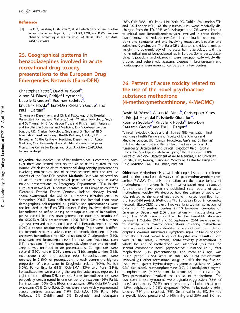
Reference
[1] Beck O, Rausberg L, Al-Saffar Y, et al. Detectability of new psycho-active substances, ’legal highs’, in CEDIA, EMIT, and KIMS immuno-chemical screening assays for drugs of abuse. Drug Test Anal.2014;6:492–499.
25. Geographical patterns inbenzodiazepines involved in acuterecreational drug toxicitypresentations to the European DrugEmergencies Network (Euro-DEN)
Christopher Yatesa, David M. Woodb,Alison M. Dinesc, Fridtjof Heyerdahld,Isabelle Giraudone, Roumen Sedefove,Knut Erik Hovdad, Euro-Den Research Groupc andPaul I. Darganb
aEmergency Department and Clinical Toxicology Unit, HospitalUniversitari Son Espases, Mallorca, Spain; bClinical Toxicology, Guy’sand St Thomas’ NHS Foundation Trust and King’s Health Partnersand Faculty Life Sciences and Medicine, King’s College London,London, UK; cClinical Toxicology, Guy’s and St Thomas’ NHSFoundation Trust and King’s Health Partners, London, UK; dTheNorwegian CBRNe Centre of Medicine, Department of AcuteMedicine, Oslo University Hospital, Oslo, Norway; eEuropeanMonitoring Centre for Drugs and Drug Addiction (EMCDDA),Lisbon, Portugal
Objective: Non-medical use of benzodiazepines is common; how-ever there are limited data on the acute harms related to thismisuse. We describe acute recreational drug toxicity presentationsinvolving non-medical use of benzodiazepines over the first 12months of the Euro-DEN project. Methods: Data was collected onall acute recreational drug/novel psychoactive substance (NPS)toxicity presentations to Emergency Departments (EDs) in theEuro-DEN network of 16 sentinel centres in 10 European countries(Denmark, Estonia, France, Germany, Ireland, Norway, Poland,Spain, Switzerland, the UK) for 12 months (October 2013 toSeptember 2014). Data collected from the hospital chart was:demographics, self-reported drugs/NPS used (presentations werenot included in the Euro-DEN dataset if they involved deliberatebenzodiazepine self-poisoning or therapeutic use of benzodiaze-pines), clinical features, management and outcome. Results: Ofthe 5529 Euro-DEN presentations, 1006 (18%) (73% males, meanage 36) involved non-medical use of a benzodiazepine; in 196(19%) a benzodiazepine was the only drug. There were 18 differ-ent benzodiazepines involved, most commonly clonazepam (315),unknown benzodiazepine (259), diazepam (219), alprazolam (140),oxazepam (59), bromazepam (33), flunitrazepam (20), nitrazepam(15), lorazepam (7) and tetrazepam (3). More than one benzodi-azepine was recorded in 80 presentations. Co-ingestions wereethanol (380), heroin (326), cannabis (140), amphetamine (118),methadone (109) and cocaine (93). Benzodiazepines werereported in 2–50% of presentations to each centre; the highestproportion of cases were in Drogheda, Ireland (50%), Munich(42%), Parnu, Estonia (33%), Oslo-EMA (33%) and Paris (33%).Benzodiazepines were among the top five substances reported ineight of the 16 Euro-DEN centres. Some benzodiazepines wereparticularly concentrated in one centre: bromazepam (94% Paris),flunitrazepam (90% Oslo-EMA), clonazepam (89% Oslo-EMA) andoxazepam (73% Oslo-EMA). Others were more widely representedacross centres: alprazolam (48% Oslo-EMA, 19% Paris, 14%Mallorca, 5% Dublin and 5% Drogheda) and diazepam
(38% Oslo-EMA, 18% Paris, 11% York, 9% Dublin, 8% London-STHand 8% London-KCH). Of the patients, 51% were medically dis-charged from the ED, 19% self-discharged and 7% were admittedto critical care. Benzodiazepines were involved in three deaths;two unknown benzodiazepines (one in combination with metha-done and cannabis) and one involving oxazepam, baclofen andzolpidem. Conclusion: The Euro-DEN dataset provides a uniqueinsight into epidemiology of the acute harms associated with thenon-medical use of benzodiazepines in Europe. Some benzodiaze-pines (alprazolam and diazepam) were geographically widely dis-tributed and others (clonazepam, oxazepam, bromazepam andflunitrazepam) were more concentrated in a few centres.
26. Pattern of acute toxicity related tothe use of the novel psychoactivesubstance methedrone(4-methoxymethcathinone, 4-MeOMC)
David M. Wooda, Alison M. Dinesb, Christopher Yatesc, Fridtjof Heyerdahld, Isabelle Giraudone,Roumen Sedefove, Knut Erik Hovdad, Euro-DenResearch Groupb and Paul I. Dargana
aClinical Toxicology, Guy’s and St Thomas’ NHS Foundation Trustand King’s Health Partners and Faculty of Life Sciences andMedicine, London, UK; bClinical Toxicology, Guy’s and St Thomas’NHS Foundation Trust and King’s Health Partners, London, UK;cEmergency Department and Clinical Toxicology Unit, HospitalUniversitari Son Espases, Mallorca, Spain; dThe Norwegian CBRNeCentre of Medicine, Department of Acute Medicine, Oslo UniversityHospital, Oslo, Norway; eEuropean Monitoring Centre for Drugs andDrug Addiction (EMCDDA), Lisbon, Portugal
Objective: Methedrone is a synthetic ring-substituted cathinone,and is the beta-keto derivative of para-methoxymethamphet-amine (PMMA). The only information on the acute toxicity ofmethedrone in humans is from Internet-based user discussionforums; there have been no published case reports of acutemethedrone toxicity. We describe here the pattern of acute tox-icity related to the use of methedrone in cases identified fromthe Euro-DEN project. Methods: The European Drug EmergenciesNetwork (Euro-DEN) project involves longitudinal collection ofdata from 16 sentinel centres in 10 European countries onEmergency Department (ED) presentations with acute drug tox-icity. The 5529 cases submitted to the Euro-DEN databasebetween 1 October 2013 and 30 September 2014 were searchedto identify acute toxicity presentations involving methedrone.Data was extracted from identified cases included: basic demo-graphics, co-used substances, symptoms/signs, initial dispositionfrom the ED and overall length of hospital stay. Results: Therewere 92 (87 male, 5 female) acute toxicity presentations inwhich the use of methedrone was identified (this was thesecond commonest novel psychoactive substance (NPS) aftermephedrone (245 presentations)). The mean ± SD age was31 ± 7 (range 17–55) years. In total 65 (71%) presentationsinvolved �1 other recreational drugs or NPS; the top five co-used were gammahydroxybutyrate/gammabutyrolactone (GBH/GBL) (39 cases), methamphetamine (19), 3,4-methylenedioxyme-thamphetamine (MDMA) (10), ketamine (8) and cocaine (6).Two presentations involved the co-use of mephedrone. Thetwo commonest symptoms were agitation/aggression (33% ofcases) and anxiety (32%); other symptoms included chest pain(13%), palpitations (12%), dyspnoea (10%), hallucinations (9%),seizures (7%) and psychosis (3%). On arrival in the ED, 3% hada systolic blood pressure of >160 mmHg and 30% and 1% had
382 ABSTRACTS
Dow
nloa
ded
by [
Kin
gs C
olle
ge L
ondo
n] a
t 07:
31 2
1 A
pril
2016
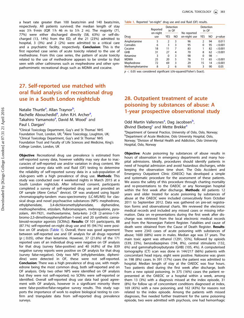
a heart rate greater than 100 beats/min and 140 beats/min,respectively. All patients survived; the median length of staywas 3 h 9 min (IQR 1 h 46 m to 5 h 2 m). The majority (71,77%) were either discharged directly (58, 63%) or self-dis-charged (13, 14%) from the ED; of the 21 (23%) admitted tohospital, 3 (3%) and 2 (2%) were admitted to a critical careand a psychiatric facility, respectively. Conclusion: This is thefirst reported case series of acute toxicity related to the use ofmethedrone. From this case series, the pattern of acute toxicityrelated to the use of methedrone appears to be similar to thatseen with other cathinones such as mephedrone and other sym-pathomimetic recreational drugs such as MDMA and cocaine.
27. Self-reported use matched withoral fluid analysis of recreational druguse in a South London nightclub
Natalie Thurtlea, Allan Traynorb,Rachelle Abouchedida, John R.H. Archera,Takahiro Yamamotoa, David M. Woodc andPaul I. Darganc
aClinical Toxicology Department, Guy’s and St Thomas’ NHSFoundation Trust, London, UK; bAlere Toxicology, Loughton, UK;cClinical Toxicology Department, Guy’s and St Thomas’ NHSFoundation Trust and Faculty of Life Sciences and Medicine, King’sCollege London, London, UK
Objective: Recreational drug use prevalence is estimated fromself-reported survey data, however validity may vary due to inac-curacies of self-reported use and/or variation in drug content. Wecombined survey data with oral fluid (OF) testing to determinethe reliability of self-reported survey data in a sub-population ofclub-goers with a high prevalence of drug use. Methods: Thisstudy was conducted over 4 weekend nights in March 2015 at aSouth London nightclub. After informed consent, participantscompleted a survey of self-reported drug use and provided anOF sample (Alere CertusVR device). OF was analysed using liquid-chromatography-tandem mass-spectrometry (LC-MS/MS) for clas-sical drugs and novel psychoactive substances (NPS: mephedrone,ethylphenidate, 3,4-dichloromethylphenidate, diphenidine,methoxphenidine, etizolam, flubromazepam, mebroqualone, pyra-zolam, AH-7921, methoxetamine, beta-keto 2-CB [2-amino-1-(4-bromo-2,5-dimethoxyphenyl)ethan-1-one] and 20 synthetic canna-binoid-receptor agonists [SCRAs]). Results: Of 101 participants, 88(87.1%) self-reported on-night drug use and 95 (94.1%) were posi-tive on OF analysis (Table 1). Overall, there was good agreementbetween self-reported use and OF analysis for all drugs reported(p� 0.05), other than ketamine. However, 37 (21.6%) of the 171reported uses of an individual drug were negative on OF analysisfor that drug (survey false-positive) and 40 (4.8%) of the 839negative survey reports were positive on OF analysis for that drug(survey false-negative). Only two NPS (ethylphenidate, dipheni-dine) were detected in OF, these were not self-reported.Conclusion: There was a high prevalence of drug use, with meph-edrone being the most common drug, both self-reported and onOF analysis. Only two other NPS were identified on OF analysisbut they were not self-reported; no SCRAs were self-reported oridentified. Overall self-reported drug use data had good agree-ment with OF analysis, however in a significant minority therewere false-positive/false-negative survey results. This study sup-ports the importance of work to explore biological testing to con-firm and triangulate data from self-reported drug prevalencesurveys.
28. Outpatient treatment of acutepoisoning by substances of abuse: A1-year prospective observational study
Odd Martin Vallersnesa, Dag Jacobsenb,Øivind Ekebergc and Mette Brekkea
aDepartment of General Practice, University of Oslo, Oslo, Norway;bDepartment of Acute Medicine, Oslo University Hospital, Oslo,Norway; cDivision of Mental Health and Addiction, Oslo UniversityHospital, Oslo, Norway
Objective: Acute poisoning by substances of abuse results inhours of observation in emergency departments and many hos-pital admissions. Ideally, procedures should identify patients inneed of hospital admission and avoid hazardous discharges, whilekeeping the observation time short. The Oslo Accident andEmergency Outpatient Clinic (OAEOC) has developed a simpleand systematic procedure for the assessment of these patients.We assess the safety of this procedure through charting mortalityand re-presentations to the OAEOC or any Norwegian hospitalwithin the first week after discharge. Methods: All patients 12years and older treated for acute poisoning by substances ofabuse at the OAEOC were included consecutively from October2011 to September 2012. Data was gathered on pre-set registra-tion forms and observational charts. We reviewed the electronicmedical records and included any missed cases or missing infor-mation. Data on re-presentations during the first week after dis-charge was retrieved from the local electronic medical recordsand from the Norwegian Patient Register. Fatalities and cause ofdeath were obtained from the Cause of Death Register. Results:There were 2343 cases of acute poisoning with substances ofabuse; 1600 (68%) were in males. Median age was 37 years. Themain toxic agent was ethanol (1291, 55%), followed by opioids(539, 23%), benzodiazepines (194, 8%), central stimulants (132,6%) and gammahydroxybutyrate (GHB) (105, 4%). A computerisedtomography (CT) scan was done in 144/217 (66%) patients withconcomitant head injury, eight were positive. Naloxone was givenin 198 (8%) cases. In 391 (17%) cases the patient was admitted tohospital. Median length of stay at the OAEOC was four hours.Two patients died during the first week after discharge, bothfrom a new opioid poisoning. In 375 (16%) cases the patient re-presented at the OAEOC or a hospital within a week, amongthem 13 (3%) with a diagnosis missed at the index episode, 31(8%) for follow up of concomitant conditions diagnosed at index,169 (45%) with a new poisoning, and 162 (43%) for reasons notrelated to the index episode. Among the patients with misseddiagnoses, five needed further treatment for the same poisoningepisode, two were admitted with psychosis, one had hemorrhagic
Table 1. Reported ‘‘on-night’’ drug use and oral fluid (OF) results.
Drug
Reportedon-night
use
Detectionin OF
No reportedon-night use
Detectionin OF
p-valueYES NO YES NO
Amphetamine 5 2 3 96 2 94 0.011Cannabis 6 3 3 95 0 95 <0.001Cocaine 18 11 7 83 1 82 <0.001GHB 31 27 4 70 8 62 <0.001Ketamine 3 0 3 98 0 98 –MDMA 25 20 5 76 11 65 <0.001Mephedrone 72 69 3 29 15 14 <0.001Methamphetamine 8 2 6 93 3 90 0.05
p < 0.05 was considered significant (chi-squared/Fisher’s Exact).
CLINICAL TOXICOLOGY 383
Dow
nloa
ded
by [
Kin
gs C
olle
ge L
ondo
n] a
t 07:
31 2
1 A
pril
2016
gastritis, one had fractures in need of operative surgery, and fourhad minor injuries. Conclusion: The procedure in use at theOAEOC can be considered safe and could be implemented else-where. The high re-presentation rate during the first week callsfor better follow up procedures.
30. Increase in unintentional pediatricexposures in a recreational marijuanastate
George Wanga, Marie-Claire Lelaitb, Sara Deakynec,Alvin Bronsteinb, Lalit Bajaja and Genie Rooseveltd
aUniversity of Colorado Anschutz Medical Campus, Rocky MountainPoison and Drug Center, Aurora, CO, USA; bRocky Mountain Poisonand Drug Center, Denver, CO, USA; cChildren’s Hospital Colorado,Aurora, CO, USA; dDenver Health Hospital, Denver, CO, USA
Objective: Despite US Federal Schedule I status, 27 states havedecriminalized medical marijuana and 4 states have passed legis-lation legalizing recreational marijuana.[1] In Colorado, the recre-ational marijuana industry became operational in January 2014.Child resistant packaging (CRP) requirements were included inmarijuana legalization. Although medical marijuana decriminaliza-tion was associated with increased unintentional pediatric expo-sures,[2] the additional impact of recreational marijuanalegalization is unclear. Methods: A retrospective study of casespresenting to a children’s hospital (CH) and calls to a regional poi-son center (PC) for children 0–9 years with a marijuana exposurefrom 2009 to 2014 in a state with medical and recreational mari-juana legalization. Rates for the PC data were calculated by divid-ing the number of cases in the state and the rest of the US bythe corresponding populations, obtained from the US Census, andPoisson Regression was used to evaluate trends. Results: At theCH 46 patients presented with median age of 2.7 (1.4, 3.7) years.Of these 18 exposures (39%) involved medical marijuana; 7 of the16 cases (44%) in 2014 involved recreational products. Infusededible products were involved in 22 cases (48%). Most patients(28, 61%) were observed in the emergency or urgent care depart-ment with inpatient admission for 11 (24%). There were 116 mari-juana exposure calls to the PC with a median age of 2.0 years(IQR 1.3, 4.0). In 34 exposures (29%) the product was inappropri-ately stored or left in sight of the child. The majority of childrenhad either no (24%) or minor effects (46%); there were 13 (11%)moderate and 4 (3%) major effects. The state had a 34% averageannual increase in PC calls (95% CI: 17%, 54%; p< 0.01) while therest of the US had a 14% average annual increase in calls (95%CI: 10%, 18%, p< 0.01); the difference was statistically significant(p¼ 0.03). When compared to 2013, the annual call rate to PC formarijuana exposures significantly increased in 2014 (p¼ 0.05).Conclusion: There was an increase in PC calls for pediatric expo-sures during the first year of recreational marijuana legalization,and edible products were a significant source. Identifying success-ful preventive strategies for pediatric marijuana exposuresrequires further investigation as the effectiveness of CRP remainsunclear.
References
[1] [cited 2015 Nov 12] http://medicalmarijuana.procon.org/view.resource.php?resourceID¼000881
[2] Wang GS, Roosevelt G, Le Lait MC, et al. Association of uninten-tional pediatric exposures with decriminalization of marijuana inthe USA. Ann Emerg Med. 2014;63:684–689.
31. ADB-Fubinaca in the real world:a case series of 15 poisonings
Csaba PapDepartment of Toxicology, P�eterfy Hospital, Budapest, Hungary
Objective: ADB-Fubinaca is a synthetic cannabinoid that hasrecently been identified in herbal blends. The aim of the presentstudy was to describe toxicity following ADB-Fubinaca poisoningin Hungary. Case series: At the beginning of May 2015 during atwo-day period 15 patients were admitted to our departmentafter consuming a new recreational drug. There were 8 males and7 females. Based on the history, all patients had taken one blue-coloured tablet never used before. Each tablet was marked withthe sign of a very popular Internet social networking site. Theonly co-ingestant was ethanol in 2 patients (13.3%). The averageage was 30.3 years, only two patients were younger than 21(range 18–48 years). The mean time to admission was 2.1 hours.We took blood and urine samples for toxicological analysis at thetime of admission. Symptoms that developed were central ner-vous system (CNS) depression (73.3%), confusion (60%) mildtachycardia (53.3%), hypertension (33.3%), mild acidosis (26.7%),hallucinations (26.7%), anxiety (20%), agitation (20%), aggression(20%), mydriasis (13.3%) vomiting (6.7%), hypotension (6.7%) andblurred vision (6.7%). There were no cases of bradycardia, seizure,elevated temperature or extrapyramidal symptoms. There wereno fatalities; one patient was intubated and ventilated mechanic-ally because of deep coma and respiratory insufficiency. Using thePoison Severity Score, 20% of patients had mild, 73.3% moderateand 6.7% severe intoxication. All patients required hospitalization;the length of stay averaged 45.78 hours (range 11–187 hours).Therapy included IV fluids, low molecular weight heparins forthrombosis prophylaxis and pantoprazole for stress ulcer prophy-laxis. Naloxone proved to be ineffective in improving the level ofconsciousness in the first 4 patients, so was not used in the othercases. Administration of IV benzodiazepine (midazolam) wasrequired in 6 patients because of confusion, agitation, aggressivebehaviour or hallucinations. All but 3 patients were given oxygenintranasally. As a consequence of transient agitation, 3 patientsdeveloped moderate rhabdomyolysis. The patient that requiredmechanical ventilation developed pneumonia. There was no othercomplication. One patient was moved to psychiatry because ofprolonged hallucinations and psychosis. Toxicological analysis per-formed within 2 days revealed ADB-Fubinaca in all samples. Wereported the cases to the Hungarian National Focal Point.Conclusion: Toxicity following use of novel cannabinoid-typeddesigner drugs is unpredictable. ADB-Fubinaca can cause a shortperiod of confusion and agitation followed by long-lasting CNSdepression, hypoxaemia and fluctuating changing in level ofconsciousness.
32. Specific issues in acute cannabispoisoning in adolescents
Viorela Nitescu, Dora Boghitoiu,Alexandru Ulmeanu, Alina Antohe, Andreea Fulgeanuand Coriolan UlmeanuPediatric Poisoning Center Emergency Clinical Hospital for Children‘‘Grigore Alexandrescu’’, Bucharest, Romania
Objective: To assess the specific clinical aspects of acute canna-binoid poisoning in adolescents admitted in a pediatric poisoncenter. Methods: We performed a five year retrospective study
384 ABSTRACTS
Dow
nloa
ded
by [
Kin
gs C
olle
ge L
ondo
n] a
t 07:
31 2
1 A
pril
2016
(2010–2014) of poisoned patients with drugs of abuse in whomurinary toxicological analysis revealed tetrahydrocannabinol (THC).The following parameters were analyzed: age, sex, place of con-sumption, clinical picture, length of stay, treatment and evolution.Results: Out of the total of 214 patients with acute drug of abusepoisoning, 41 had a urine sample positive for THC and diagnosisof acute cannabis poisoning. The majority of patients were boys(n¼ 28), with a median age of 15.9 years. Of the 41 cases, 36 poi-soning occurred in a public place: in the street (n¼ 16), in school(n¼ 12) or in a nightclub (n¼ 8). Regarding the clinical picturethe following symptoms were noted: decreased coordination inthe majority of cases (n¼ 36), headache (n¼ 19), emesis (n¼ 16),somnolence (n¼ 11), chest pain (n¼ 6), agitation (n¼ 4), tremorof the extremities (n¼ 4), slurred speech (n¼ 4), lightheadedness(n¼ 3), aggression (n¼ 1), hallucinations (n¼ 1) and seizures(n¼ 1). Of the 6 patients with chest pain in one increased cardiacenzymes and terminal phase changes on the electrocardiogramwere noted. The average length of stay was 1.2 days with a min-imum of one and a maximum of 3 days. All the patients receivedsymptomatic treatment. No deaths occurred. Conclusion: Themajority of adolescents with acute cannabis poisoning presentedwith mild symptoms, requiring only symptomatic treatment and ashort period of hospitalization.[1] Even rare, severe manifestationssuch as cardiac damage can occur.[2] One of the most commonplaces where acute cannabis exposure occurred was school and itis necessary to develop educational activity to raise awareness ofthe risks of cannabis abuse among students.
References
[1] Annual Report to the European Early Warning System. In: 2014National Report on Drugs to the EMCDDA by Reitox NationalFocal Point. Romania New Developments, Trends and In-depthInformation on Selected Issues; [cited 2016 Feb 24]. Available at:http://www.emcdda.europa.eu/publications/national-reports/romania-2014
[2] Ulmeanu C. Drugs of Abuse poisoning. In: Acute poisoning in chil-dren and adolescents. Romania: Tridona. 2015:249–52.
33. Novel psychoactive substance(NPS) consumption in binge drinkers:a new and potentially dangeroushealth risk
Marta Crevania, Francesca Chiaraa, Pietro Papab,Elisa Rodac, Davide Lonatia, Andrea Giampretia andCarlo A. LocatellidaPoison Control Centre and National Toxicology Information Centre,IRCCS Maugeri Foundation Hospital and University of Pavia, Pavia,Italy; bLaboratory of Analytical Toxicology, Clinical ChemistryService, IRCCS Policlinico San Matteo Foundation, Pavia, Italy;cToxicology Unit, Laboratory of Clinical & Experimental Toxicology,IRCCS Maugeri Foundation, Pavia, Italy; dPoison Control Centre,National Toxicology Information Centre and Clinico-toxicologicalCoordinating Centre of the National Early Warning System, IRCCSMaugeri Foundation Hospital, Pavia, Italy
Objective: Binge drinking is an increasingly common dangerouspractice involving mainly children or adolescents and, in some cir-cumstances (e.g. rave party, college drinking), is possibly associ-ated with the co-consumption of substances of abuse.[1] Weanalyzed all cases of suspected consumption of novel psycho-active substances (NPS) in binge drinkers referred to the Pavia
Poison Control Centre. Methods: Cases of binge drinking inpatients <25 years with blood alcohol concentration(BAC)> 0.5 g/L were retrospectively reviewed (January 2010–July2015) and assessed for: age, history, substances of abuse declared,circumstances of exposure, clinical manifestations, outcome, BAC,NPS-identified, association with classic drugs of abuse (cocaine,opiates, cannabis, amphetamine/methamphetamine), correspond-ence between history and the analytically identified-NPS. Caseswith incomplete data were excluded. Results: In total 105 patientsmet the inclusion criteria. The lowest age recorded was 13 years(65% <18 years); 69% of cases occurred during the weekend.Substances of abuse co-consumed were: ketamine (n¼ 10), 3,4-methylenedioxymethamphetamine (MDMA) (n¼ 5), cannabis(n¼ 4), amphetamine (n¼ 5), cocaine (n¼ 3), energy drink (n¼ 2),lysergic acid diethylamide (LSD) (n¼ 2), gammabutyrolactone(GBL) (n¼ 1) and heroin (n¼ 1). Co-consumption of more thanone substance of abuse was registered in 38% of cases. Main clin-ical manifestations were agitation (55%), tachycardia (38%), coma(34%), delirium/hallucinations (31%), drowsiness (28%), mentalconfusion (17%) and seizures (10%); no fatal cases were regis-tered. In 65% of patients the BAC was higher than 1.5 g/L (highestBAC 3.26 g/L). Seventy-six cases were negative for NPS, while 36%of these were positive for classic drugs. In 29 patients (28%) atleast one NPS was analytically confirmed. The NPS detected inbiological samples (urine/blood) were MDMA (n¼ 14), ketamine(n¼ 7), methoxetamine (n¼ 6), gammahydroxybutyrate (GHB)/GBL(n¼ 2), 2C-series (n¼ 2), synthetic cathinones, 4-methylethcathi-none (4-MEC) (n¼ 1), synthetic cannabinoids, JWH-073 (n¼ 1),atropine (n¼ 1) and benzofurans, APB (n¼ 1). Correspondencebetween substance declared and laboratory results was registeredin 10% of cases and complete discrepancy in 45% of cases.Conclusion: Binge drinking is a relevant and increasing socialproblem related to acute and late toxic effects (e.g. brain develop-ment) especially during adolescence. Association between bingedrinking and NPS may be particularly insidious. In our experience3 out of 10 binge drinkers consumed at least one NPS. Specialistadvanced laboratory support is essential to make the correct diag-nosis and guide the treatment. Acknowledgements: Study sup-ported by the Department for Antidrug Policies-Presidency of theCouncil of Ministers.
Reference
[1] Siqueira L, Smith VC, Committee on Substance Abuse. Binge drink-ing. Pediatrics. 2015;136:e718–e726.
34. Prospective cohort study todetermine low risk acute ethanolintoxications risk factors
Christopher Yatesa, Jordi Puiguriguera,Catalina Homara and Guillermo Fronterab
aClinical Toxicology Unit, Emergency Department, HospitalUniversitari Son Espases, Palma de Mallorca, Spain; bClinical TrialsUnit, IDISPA, Palma de Mallorca, Spain
Objective: The management of acute ethanol intoxication isbased on evaluation and observation with the rare need for ther-apy or hospital facilities in the case of complications (i.e. trauma).However, these patients are often indiscriminately transferred byambulance to busy emergency departments (EDs). Identifying lowrisk subjects that could be treated and observed at a lower levelof care would optimize the use of resources. Our objective was toidentify ethanol intoxicated patients at low risk for complications
CLINICAL TOXICOLOGY 385
Dow
nloa
ded
by [
Kin
gs C
olle
ge L
ondo
n] a
t 07:
31 2
1 A
pril
2016
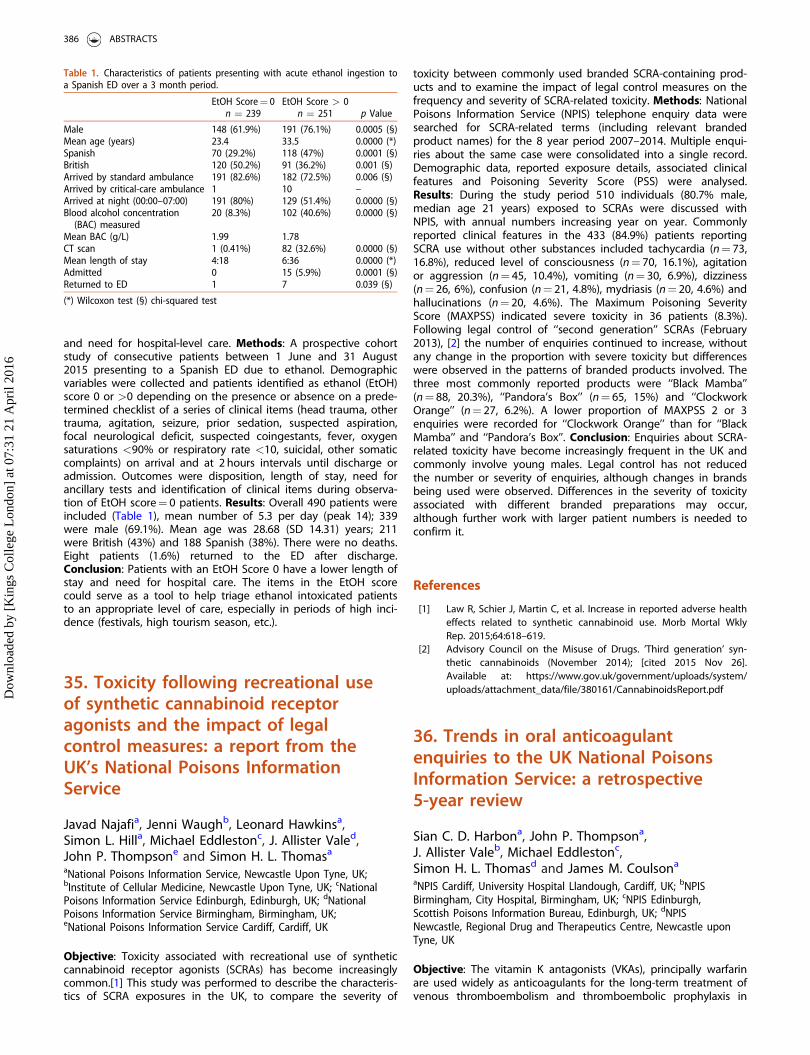
and need for hospital-level care. Methods: A prospective cohortstudy of consecutive patients between 1 June and 31 August2015 presenting to a Spanish ED due to ethanol. Demographicvariables were collected and patients identified as ethanol (EtOH)score 0 or >0 depending on the presence or absence on a prede-termined checklist of a series of clinical items (head trauma, othertrauma, agitation, seizure, prior sedation, suspected aspiration,focal neurological deficit, suspected coingestants, fever, oxygensaturations <90% or respiratory rate <10, suicidal, other somaticcomplaints) on arrival and at 2 hours intervals until discharge oradmission. Outcomes were disposition, length of stay, need forancillary tests and identification of clinical items during observa-tion of EtOH score¼ 0 patients. Results: Overall 490 patients wereincluded (Table 1), mean number of 5.3 per day (peak 14); 339were male (69.1%). Mean age was 28.68 (SD 14.31) years; 211were British (43%) and 188 Spanish (38%). There were no deaths.Eight patients (1.6%) returned to the ED after discharge.Conclusion: Patients with an EtOH Score 0 have a lower length ofstay and need for hospital care. The items in the EtOH scorecould serve as a tool to help triage ethanol intoxicated patientsto an appropriate level of care, especially in periods of high inci-dence (festivals, high tourism season, etc.).
35. Toxicity following recreational useof synthetic cannabinoid receptoragonists and the impact of legalcontrol measures: a report from theUK’s National Poisons InformationService
Javad Najafia, Jenni Waughb, Leonard Hawkinsa,Simon L. Hilla, Michael Eddlestonc, J. Allister Valed,John P. Thompsone and Simon H. L. Thomasa
aNational Poisons Information Service, Newcastle Upon Tyne, UK;bInstitute of Cellular Medicine, Newcastle Upon Tyne, UK; cNationalPoisons Information Service Edinburgh, Edinburgh, UK; dNationalPoisons Information Service Birmingham, Birmingham, UK;eNational Poisons Information Service Cardiff, Cardiff, UK
Objective: Toxicity associated with recreational use of syntheticcannabinoid receptor agonists (SCRAs) has become increasinglycommon.[1] This study was performed to describe the characteris-tics of SCRA exposures in the UK, to compare the severity of
toxicity between commonly used branded SCRA-containing prod-ucts and to examine the impact of legal control measures on thefrequency and severity of SCRA-related toxicity. Methods: NationalPoisons Information Service (NPIS) telephone enquiry data weresearched for SCRA-related terms (including relevant brandedproduct names) for the 8 year period 2007–2014. Multiple enqui-ries about the same case were consolidated into a single record.Demographic data, reported exposure details, associated clinicalfeatures and Poisoning Severity Score (PSS) were analysed.Results: During the study period 510 individuals (80.7% male,median age 21 years) exposed to SCRAs were discussed withNPIS, with annual numbers increasing year on year. Commonlyreported clinical features in the 433 (84.9%) patients reportingSCRA use without other substances included tachycardia (n¼ 73,16.8%), reduced level of consciousness (n¼ 70, 16.1%), agitationor aggression (n¼ 45, 10.4%), vomiting (n¼ 30, 6.9%), dizziness(n¼ 26, 6%), confusion (n¼ 21, 4.8%), mydriasis (n¼ 20, 4.6%) andhallucinations (n¼ 20, 4.6%). The Maximum Poisoning SeverityScore (MAXPSS) indicated severe toxicity in 36 patients (8.3%).Following legal control of ‘‘second generation’’ SCRAs (February2013), [2] the number of enquiries continued to increase, withoutany change in the proportion with severe toxicity but differenceswere observed in the patterns of branded products involved. Thethree most commonly reported products were ‘‘Black Mamba’’(n¼ 88, 20.3%), ‘‘Pandora’s Box’’ (n¼ 65, 15%) and ‘‘ClockworkOrange’’ (n¼ 27, 6.2%). A lower proportion of MAXPSS 2 or 3enquiries were recorded for ‘‘Clockwork Orange’’ than for ‘‘BlackMamba’’ and ‘‘Pandora’s Box’’. Conclusion: Enquiries about SCRA-related toxicity have become increasingly frequent in the UK andcommonly involve young males. Legal control has not reducedthe number or severity of enquiries, although changes in brandsbeing used were observed. Differences in the severity of toxicityassociated with different branded preparations may occur,although further work with larger patient numbers is needed toconfirm it.
References
[1] Law R, Schier J, Martin C, et al. Increase in reported adverse healtheffects related to synthetic cannabinoid use. Morb Mortal WklyRep. 2015;64:618–619.
[2] Advisory Council on the Misuse of Drugs. ’Third generation’ syn-thetic cannabinoids (November 2014); [cited 2015 Nov 26].Available at: https://www.gov.uk/government/uploads/system/uploads/attachment_data/file/380161/CannabinoidsReport.pdf
36. Trends in oral anticoagulantenquiries to the UK National PoisonsInformation Service: a retrospective5-year review
Sian C. D. Harbona, John P. Thompsona,J. Allister Valeb, Michael Eddlestonc,Simon H. L. Thomasd and James M. Coulsona
aNPIS Cardiff, University Hospital Llandough, Cardiff, UK; bNPISBirmingham, City Hospital, Birmingham, UK; cNPIS Edinburgh,Scottish Poisons Information Bureau, Edinburgh, UK; dNPISNewcastle, Regional Drug and Therapeutics Centre, Newcastle uponTyne, UK
Objective: The vitamin K antagonists (VKAs), principally warfarinare used widely as anticoagulants for the long-term treatment ofvenous thromboembolism and thromboembolic prophylaxis in
Table 1. Characteristics of patients presenting with acute ethanol ingestion toa Spanish ED over a 3 month period.
EtOH Score¼ 0n ¼ 239
EtOH Score > 0n ¼ 251 p Value
Male 148 (61.9%) 191 (76.1%) 0.0005 (§)Mean age (years) 23.4 33.5 0.0000 (*)Spanish 70 (29.2%) 118 (47%) 0.0001 (§)British 120 (50.2%) 91 (36.2%) 0.001 (§)Arrived by standard ambulance 191 (82.6%) 182 (72.5%) 0.006 (§)Arrived by critical-care ambulance 1 10 –Arrived at night (00:00–07:00) 191 (80%) 129 (51.4%) 0.0000 (§)Blood alcohol concentration
(BAC) measured20 (8.3%) 102 (40.6%) 0.0000 (§)
Mean BAC (g/L) 1.99 1.78CT scan 1 (0.41%) 82 (32.6%) 0.0000 (§)Mean length of stay 4:18 6:36 0.0000 (*)Admitted 0 15 (5.9%) 0.0001 (§)Returned to ED 1 7 0.039 (§)
(*) Wilcoxon test (§) chi-squared test
386 ABSTRACTS
Dow
nloa
ded
by [
Kin
gs C
olle
ge L
ondo
n] a
t 07:
31 2
1 A
pril
2016
atrial fibrillation. Their use is complicated by a narrow therapeuticindex, extensive interactions and a requirement for routine moni-toring. The novel oral anticoagulants (NOACs) provide effectiveanticoagulation without the need for coagulation monitoring.Concern has been expressed regarding the safety of the NOACsafter overdose because of the previous lack of licensed antidotesto counter excessive anticoagulation in the haemorrhagingpatient. We examine patterns of poisoning from newer oral anti-coagulant medicines. Methods: Data concerning telephone enqui-ries to the National Poisons Information Service were sourcedfrom the UK Poisons Information Database (UKPID) for a retro-spective examination of enquiries received between the 2008 and2013. The database was searched for: acenocoumarol, apixaban,dabigatran, phenindione, rivaroxaban and warfarin. Data wereanalysed by the chi-squared test. Fisher’s exact test was used incomparisons which contained a numerical value less than five. Ap-value of less than 0.05 was considered to be statistically signifi-cant. Results: A total of 1390 enquiries regarding oral anticoagu-lants were received during the time period, including 1280 (92%)related to warfarin, 13 (1%) to other VKAs and 97 (7%) to NOACs(rivaroxaban 65, dagibatran 29, apixaban 3). Enquiries concerningNOACs increased as a proportion of all oral anticoagulants from0.5% in 2009 to 19% in 2013. Eighty-three percent were reportedas asymptomatic exposures, 15% were symptomatic and in 2%the clinical features were unknown. The number of cases thatwere symptomatic was 193 (14%) for warfarin, 7 (24%) for dabiga-tran and 4 (6%) for rivaroxaban. A significantly higher proportionof dabigatran cases were symptomatic compared to rivaroxaban,p¼ 0.04, but not compared to warfarin, p¼ 0.3. Conclusion: Theproportion of enquiries involving NOACs has increased to almosta fifth of oral anticoagulant enquiries. The majority of exposuresto both warfarin and NOACs were asymptomatic. There appearedto be variation in the proportions of symptomatic cases betweenthe most commonly encountered NOACs. Further data, includingdemographic and prescription data, are required to investigatethis preliminary finding.
37. AfriToxVR TeleLog: the developmentof a novel web-based data collectiontool for poison centre call data inSouth Africa
Cindy R. Stephena, Selig Leysera, J. Clare Robertsa,Farahnaz Mohameda, Kate H. Balmea,Linda E. Curlinga, Carine Marksb andCherylynn Wiumb
aPoisons Information Centre, Department of Paediatrics and ChildHealth, Red Cross War Memorial Children’s Hospital and Universityof Cape Town, Cape Town, South Africa; bTygerberg PoisonsInformation Centre, Department of Medicine, Faculty of HealthSciences, Stellenbosch University and Tygerberg Academic Hospital,Parow, South Africa
Objective: Poison centre call data describing poisoning exposuresis an important source of information for administrative, medico-legal and research purposes, and is essential to the toxicovigi-lance role of poisons centres. Such call data is scarce worldwide,particularly in the developing world. In 2013, the PoisonsInformation Centre (PIC) at Red Cross War Memorial Children’sHospital (RCWMCH) set out to develop the AfriToxVR TeleLog as asimple and reliable data collection tool for its poison call data.The system is linked to the AfriToxVR poisons information databaseused widely throughout Southern Africa. Methods: The RCWMCHPIC received poisons calls from 2011, recording call data in a
book-based system. During 2013, a real-time database was devel-oped enabling call data to be entered directly into the databaseby the Specialist in Poisons Information (SPI) on duty. FileMakerVR
software was used and there was extensive collaboration betweenthe software designer and RCWMCH SPIs to create an appropriateand user-friendly tool from which accurate reports could beextracted. Sequential tabbed pages with drop-down menusprompt specific information to be entered including details of thesubject, nature of the exposure and characteristics of the substan-ces, as well as free text fields for clinical details (includingPoisoning Severity score), advice given and follow up. Results:The AfriToxVR TeleLog became operational in 2014 at theRCWMCH PIC with 1858 calls recorded during the first year.Retrospective data from 6155 calls received between 2011 and2014 was transcribed into the database. During 2015, the AfriToxVR
TeleLog was further developed into a web-based system to sup-port the creation of a single 24/7 national poisons helpline. Thishelpline is available to the public and healthcare professionals,and is staffed by members of the RCWMCH and TygerbergAcademic Hospital PICs, the two major poison centres in SouthAfrica. Overall the AfriToxVR TeleLog has functioned well, althoughissues of connectivity can hamper consistent real-time data cap-ture at remote sites. It has become an excellent source of poisonsdata for South Africa containing details on over 12,000 calls. Sincethe introduction of the national helpline, call numbers haveincreased significantly with an average 775 calls per month.Conclusion: The AfriToxVR TeleLog database is achieving its goalof facilitating real-time collection of good quality poison centrecall data. While ongoing refinement of the AfriToxVR TeleLog isnecessary, as well as mentoring of SPIs using it, it is becoming anational asset to South Africa’s health service.
38. Antidotes network: is it possible toconnect stocks between centers?
Edurne Fernandez De Gamarraa, Raquel Aguilarb,Antoni Brotoc, Milagros Garcia-Pelaezd,Lidia Martineze and Santiago Noguef
aPharmacy Department, Hospital de la Santa Creu i Sant Pau,Barcelona, Spain; bPharmacy Department, Hospital Universitari Dr.Josep Trueta, Girona, Spain; cPharmacy Department, ConsorciSanitari de Terrassa, Terrassa, Spain; dPharmacy Department,Hospital de Sabadell, Institut Universitari Parc Taul�ı - UniversidadAut�onoma de Barcelona, Sabadell, Spain; ePediatrics EmergencyDepartment, Hospital Sant Joan de D�eu, Barcelona, Spain; fClinicalToxicology Unit, Hospital Cl�ınic, Barcelona, Spain
Objective: Antidotes are essential drugs for management of acuteintoxications. The lapse of time from the contact with the toxicagent until antidote administration can determine patient survival.Pharmacy Departments are responsible for warranting an optimalqualitative and quantitative stock of these drugs. However, anational regulation about stocking of antidotes in hospitals is lack-ing, so availability problems can be an important concern. In thiscontext, an ‘‘Antidotes network’’ has been proposed in our region(Xarxa d’ant�ıdots de Catalunya [Catalan Antidote Network]).Methods: A working group, including pharmacists and physicianswith experience in the field of Clinical Toxicology, was establishedunder the framework of the Catalan Society of Clinical Pharmacyto develop the network. Firstly, the group produced a documentwith recommendations about stocking of antidotes according tohospital complexity and location. Then an online application wasdesigned to be used as a tool for communication between cen-ters. The application gathers information about 15 antidotes,selected according to criteria such as difficulties in availability,
CLINICAL TOXICOLOGY 387
Dow
nloa
ded
by [
Kin
gs C
olle
ge L
ondo
n] a
t 07:
31 2
1 A
pril
2016
urgency, frequency of use or cost (digoxin antibody fragments,methylene blue, deferoxamine, dimercaprol, sodium calcium ede-tate, ethanol, physostigmine, fomepizole, glucagon, hydroxocoba-lamin, pyridoxine, pralidoxime, silibinin, botulinum antitoxin andsnake venom antisera). This tool makes information about thestock in each center (including expiry date) available and facili-tates antidote borrowing between hospitals. Results: The onlineapplication (accessible through www.xarxaantidots.org) waslaunched in July 2015. It has an open zone with informationabout the project and offers the option to ask non-urgent toxico-logical questions to the group experts, and a private zone access-ible with username and password for those centers that havejoined the network. So far 34 Catalan hospitals that provide emer-gency care have joined. In each center there is a pharmacist anda physician from the Emergency Department responsible for thenetwork. These figures are denominated ‘‘farmatox’’ and ‘‘urge-tox’’. The ‘‘farmatox’’ is in charge of the stock maintenance,updates the drug movements and helps with borrowing andlending antidotes between hospitals. The ‘‘urgetox’’ develops thefunctions of toxicology referent from the Emergency Department.Conclusion: The Antidote Network could allow improved commu-nication between centers involved in the management of pois-oned patients, help in adjusting and harmonizing antidotes stockand accelerate antidote borrowing, if required.
39. Extraordinary mobilizations ofantidotes from the National Stockpileto hospital emergency departments:an example of versatility andintegration of national functions andsystems
Eleonora Buscagliaa, Valeria M. Petrolinia,Virgilio Costanzob, Loredana Velluccib,Olha Maystrovaa, Giulia Scaravaggia,Emanuela Cortinia and Carlo A. LocatelliaaPoison Control Centre and National Toxicology Information Centre,Toxicology Unit, IRCCS Maugeri Foundation and University of Pavia,Pavia, Italy; bMinistry of Health, National Health System, Rome, Italy
Objective: Since 2005, the Italian State has established an extraor-dinary endowment of antidotes for terrorist chemical and radio-nuclear events (Scorta Nazionale Antidoti [SNA]). Charged by theMinistry of Health, the Pavia Poison Control Centre (PPCC) is theclinical unit responsible for (i) the diagnostic-therapeutic specialistconsultation for non-conventional attacks, (ii) SNA operationalmanagement (e.g. upgrade, distribution, planning), and (iii) thecontinuous training of the Italian NHS. SNA is organized on anational scale (regional and national stockpiles, located in hospi-tals and in State deposits, respectively), and is an intangible stock-pile whose integrity is essential to fulfil its functions. However,when an absolute shortage of an antidote occurs in NHS hospitalsand the antidotal treatment of intoxicated patients is necessary, aquota of the SNA stockpile can be extraordinarily mobilized.Operational procedures need a clinical evaluation by the PPCCfirst, and then an on-time authorization by the Ministry of Health.Rapid replacement of the mobilized amounts by the requiringhospital is a procedural obligation. We evaluated the SNA’s extra-ordinary mobilizations (SNA-EM) in a seven-year period. Methods:We investigated all SNA-EMs authorized or made in the period2008–2014. For each mobilization (i) the cause of the extraordin-ary request (clinical indication, antidote availability/shortage inneighboring hospital and Poison Centers), (ii) the time required
for antidote arrival to the requiring hospital and (iii) the SNAstockpiles involved were assessed. Results: Exceptional mobiliza-tions from the SNA to NHS hospitals were performed 25 times(for 28 patients), always linked to single/multiple poisoning fromconventional causes/events. The mobilized antidotes were prali-doxime (n¼ 17), succimer (DMSA) (n¼ 3), unithiol (DMPS) (n¼ 2),hydroxocobalamin (n¼ 1), methylene blue (n¼ 1) and Prussianblue (n¼ 1). In 21 cases, SNA-EM occurred to hospitals located inthe same region as the SNA deposit and in 4 cases in differentregions. In some cases, the mobilized antidotes (unithiol, Prussianblue and succimer) are rarely used and difficult to find in NHShospitals. Conclusion: SNA is an essential facility in order to havethe necessary antidotes in case of exceptional events. The currentorganization of SNA, considered highly important in the EU, com-bines clinical toxicological expertise and antidote supply in orderto obtain diagnostic and therapeutic appropriateness. This organ-ization has proven useful and able to overcome hospital short-comings of normal/rare antidotes in cases where toxic agents areunusual or the need for antidotes exceeds normal hospital avail-ability. Acknowledgements: Support of Ministry of Health (4393/2013-CCM).
40. Profile and risk assessment ofhousehold cleaners as evaluated fromEmergency Department (ED) cases inthe Spanish Toxic Surveillance System(STSS) in the last 5 years
Ana Ferrer Dufola, Roman Royo Hernandezb,Clara Serrano Ferrerc and Santiago Nogue Xaraud
aUnit of Clinical Toxicology, Clinic University Hospital, Zaragoza,Spain; bEmergency Dept., Clinical University Hospital, Zaragoza,Spain; cMedical School, Zaragoza University, Zaragoza, Spain; dUnitof Clinical Toxicology, Clinic Hospital, Barcelona, Spain
Objective: The chemical substances marketed as householdcleaning agents are a toxic hazard which is closely in contact withthe general population. We aimed to evaluate the profile and riskof exposure to household cleaning agents in patients thatattended the ED collaborating with the STSS. Methods: We ana-lysed the cases attending the ED of 21 public hospitals due toexposure to irritant gases, caustics, solvents and detergents from2010 to 2014. Results: There were 2175 cases which represent49% of the total cases included in the STSS over the study period.Mean age was 36 years (SD 25). Sex distribution was even with49% male (mean age 35 years) and 51% female (mean age 41years). Domestic accidents accounted for 1415 cases (65%) with amean age of 36 years and higher numbers of females (57%). Themain substances involved were caustics (44%) followed by deter-gents (21%), irritant gases (21%) and solvents (12%). There were10 severe cases and 5 deaths (0.35%). Occupational accidentsaccounted for 318 cases (15%) with a mean age of 39 years andhigher numbers of men (64%). The main substances involvedwere caustics (35%), irritant gases (34%), solvents (24%) anddetergents (7%) producing one severe case and no deaths.Suicidal gestures accounted for 304 cases (14%) with a mean ageof 47 years with even numbers by gender. The main substancesinvolved were caustics (75%) followed by solvents (12%) anddetergents (12%) and just one case involving irritant gases. Therewere 34 severe cases and 24 deaths (8%). Overall there were 435cases involving children (up to 16 years old) with a significantlyhigher number of males (57%). The main substances involvedwere caustics (38%) and detergents (34%) followed by solvents(16%) and irritant gases (7%). There are no severe or lethal cases
388 ABSTRACTS
Dow
nloa
ded
by [
Kin
gs C
olle
ge L
ondo
n] a
t 07:
31 2
1 A
pril
2016
involving children. Conclusion: The global group of householdcleaning products has a profile of low risk with a proportion ofsevere cases of 2.34% and a mortality of 1.51%. The severe andlethal cases were mainly due to suicide attempt involving caustics.It is worth stressing that one-third of the lethal cases were due toingestion of salfuman (hydrochloric acid marketed for domesticuse). No severe cases occurred in children.
41. Adverse drugs reactions (ADR)collected by medical-staffedambulances: a pilot study with theSAMU de Paris
Agnes Lillo-Leloueta, Frederic J. Baudb, Benoit Vivienc
and Pierre CarlicaAssistance Publique – Hospitaux de Paris, Paris, France; bPoisonControl Centre, Paris, France; cSAMU de Paris, Paris, France
Objective: In Paris, emergency medical assistance is run by the‘‘SAMU de Paris’’. Medical-staffed ambulances go directly to anypatient with an acute medical problem after evaluation of theclinical situation over the telephone. The aim of the study was todetermine whether the SAMU de Paris allowed collection ofadverse drug reactions (ADR). Methods: Every month, forms fromall medical interventions performed by the SAMU de Paris, wereanalysed retrospectively by a physician trained in pharmacovigi-lance. The following information is available: patient characteris-tics, description of the medical event, diagnosis and outcome. Ifan ADR is present or even suspected, a copy of the form is made.The pharmacovigilance centres follow up with medical staff toretrieve more information about the final diagnosis and druginvolvement. If an ADR is confirmed, the case is anonymously reg-istered in the national database. Results: From January to March2015, 100 cases of possible ADRs were collected while only 8cases were spontaneously declared to the Centre Regional dePharmacovigilance (CRPV). Of the 100 cases 68 ADRs were imme-diately diagnosed and registered (Table 1). Causality is pendingfor 23 cases. The main effects observed were cardiovascular (car-diac arrest, bradycardia) (n¼ 9) and neurological (coma, confu-sion) (n¼ 9); other effects were malaise, skin reaction,gastrointestinal perforation, hypoglycaemia and renal failure. Forthe remaining 9 cases, drug causality has been definitely ruledout, with no drug intake (n¼ 5), and another causative agent for4 cases (illicit substances n¼ 2, food n¼ 2). Conclusion: To ourknowledge, this is the first time that serious ADRs have been col-lected directly from first responders, before hospitalization. Except
intentional drug overdose for suicide attempts, the main ADRsobserved are expected, such as bleeding with anticoagulants orhypoglycaemia with insulin. Further analysis on cardiovasculareffects is pending the late phase of poisoning. This limitation canbe corrected at least by prospective follow up studies.
42. Improper use of a medicine bottleto prepare an e-cigarette refill liquid
Maria Laura Zuccolia, Marco Piteab, Marcello Ferruzzia,Anna Celentanoa, Fabrizio Sesanaa,Giscardo Panzavoltaa, Adriana Tomoiagaa,Beatrice Giliottia, Angelo Travagliaa,Rossana Borghinia, Laura Settimic andFranca Davanzoa
aNational Milan Poison Control Centre – Ospedale Niguarda Ca’Granda, Milano, Italy; bPediatric Department, Presidio Ospedaliro diSaronno, Milano, Italy; cNational Institute of Health (ISS), Rome,Italy
Objective: Over the last few years, electronic cigarettes (e-ciga-rettes) have become increasingly popular. Nicotine concentrationsin the refill liquid (e-liquid) vary between 0 and 26 mg/mL, poten-tially reaching high-risk concentrations. This is a particular dangerfor children, in whom the absorption of only 2 mg orally or0.1 mg dermally can produce signs and symptoms of acute nico-tine intoxication.[1] Here we report a case in which a vial of e-liquid was mistakenly taken for a bottle of a mucolytic drug solu-tion. Case report: A 3-year-old child (weight 15 kg) was admittedto the Emergency Department (ED) for nicotine intoxication. Thechild had mistakenly been administered 10 ml of e-liquid insteadof the same amount of carbocisteine. The total amount of nico-tine ingested was 80 mg (concentration of nicotine in the do-it-yourself e-liquid: 8 mg/mL, 0.8%), with an unknown amount ofglycerine and propylene glycol. The child’s mother reported thattwo vomiting events occurred within a few minutes after inges-tion. In the ED, the child presented with drowsiness and paleness.An electrocardiogram (ECG) did not show any alterations exceptfor a high heart rate (130 beats/minute); blood pressure was 102/70 mmHg and the percutaneous oxygen saturation was 99% onroom air. Activated charcoal was administered. The state of con-sciousness improved within 40 minutes, and tachycardia resolvedin a few hours. Conclusion: Despite the significant amount ofnicotine ingested (5.3 mg/kg) and the fast absorption through allroutes of exposure of liquid nicotine, the symptoms were mildand the amount of nicotine absorbed may have been reduced bythe two episodes of vomiting and by the administration of acti-vated charcoal. Notwithstanding the ingestion of e-liquid contain-ing nicotine could lead to serious poisoning especially in children.Regulations concerning packaging and product design for refillliquids are still lacking. We deem it important and necessary thatusers, manufacturers, and healthcare providers consider thesepreparations as a potential danger.
Reference
[1] Smolinske SC, Spoerke DG, Spiller SK, et al. Cigarette and nicotinechewing gum toxicity in children. Hum Toxicol. 1988; 7:27–31.
Table 1 Main characteristics of ADRs collected by the SAMU de Paris.
Event Number (% total)Drugs suspected (more than
one can be suspected)
Intentional drug overdose 27 (40%) Psychotropic (24), paracetamol(2), potassium (1), digoxin (1),methadone (1)
Bleeding 14 (20%) Oral anticoagulant (8), lowmolecular weight heparins (4),antiplatelet (2)
Hypoglycaemia 12 (18%) Insulin (12)Anaphylaxis 6 (9%) Antibiotics (4), non-steroidal
anti-inflammatory (2), neuro-muscular blocker (1)
Miscellaneous 9 (13%) Psychotropic (5), antitubercu-losis agents (2), analgesic(2), antibiotic (1),corticosteroid (1)
CLINICAL TOXICOLOGY 389
Dow
nloa
ded
by [
Kin
gs C
olle
ge L
ondo
n] a
t 07:
31 2
1 A
pril
2016
43. E-cigarettes and syntheticcannabinoids: a new trend
Kristin McCloskey, David Vearrier, Rita G McKeeverand Michael GreenbergDrexel University, Philadelphia, USA
Objective: Electronic cigarettes (e-cigarettes) have becomeincreasingly popular both for using nicotine and, more recently,for drugs of abuse. We present what we believe to be the firstreported case of a patient using an e-cigarette to vaporize (‘‘vap-ing’’) synthetic cannabinoids (K2) for intoxication. Case report: A36-year-old male with a history of alcohol and drug abuse wasbrought to the emergency department (ED) by ambulance foraltered mental status after being found ‘‘kicking’’ and ‘‘rollingaround on the sidewalk’’. At the scene, the patient was holdingan e-cigarette which he explained to paramedics he had filledwith K2 to get high. In the ED, the patient’s vital signs were bloodpressure 151/56 mmHg, heart rate 106 beats/min, respiratory rate20/min, oxygen saturations 94% and temperature 37.1 �C (axillary).A finger-stick blood glucose was 116 mg/dL. His physical examin-ation was remarkable for severe agitation, which spontaneouslyresolved. Laboratory studies were significant for creatine kinase3936 U/L, negative serum ethanol, and a negative urine drugimmunoassay. Electrocardiogram (EKG) showed normal sinusrhythm with a rate of 100 and non-specific T wave and ST seg-ment changes. The patient was given intravenous fluids for hisrhabdomyolysis. Serial neurological examinations demonstratedno improvement over a 4 hour period. A computed tomography(CT) scan of the head was normal. The patient was admitted forfurther treatment of his rhabdomyolysis and serial neurologicalexaminations. Over the next 36 hours, the patient’s mental statusand creatine kinase normalized, and he left against medicaladvice on hospital day three. Conclusion: There has been a resur-gence of synthetic cannabinoid use in the US beginning in April2015. As this case illustrates, vaping synthetic cannabinoids via e-cigarettes can lead to serious toxicity. Whether this method ofuse leads to greater or lesser toxicity then traditional smoking hasyet to be determined. Regardless, it is important for physicians tobe aware of emerging trends of abuse.
44. Dermal and ocular exposure tovinegar essence in Austria, 2002–2014
Tara Arif, Angelika Holzer, Helmut Schiel,Dieter Genser and Kinga Bartecka-MinoPoisons Information Centre, Vienna, Austria
Objective: Household vinegar contains up to 25% acetic acid, butindustrially used vinegar may contain up to 80% acetic acid.Unfortunately, this high percentage acetic acid can be purchasedat wholesale and via the Internet. Vinegar essence is used inhouseholds as a descaling agent, cleaning agent and stronglydiluted as a salad dressing. We performed a retrospective analysisof calls regarding dermal and ocular exposure of vinegar essence.Methods: Cases from the Poison Information Centre databaseinvolving acute dermal and ocular exposures to vinegar essencewere evaluated for the period of 2002–2014. All exposures wereaccidental and in most cases the amount of vinegar essenceinvolved was unclear. Results: Over the 13 year period, 25 casesof dermal, 2 cases of ocular and 2 of dermal and ocular exposureto vinegar essence were documented (see Table 1). Of these, 24patients developed chemical burns of the skin or eyes. Three
patients showed only irritation of the skin. In two cases therewere no symptoms at all, so the exposure was classified as ques-tionable. In three well documented paediatric cases (16 months,22 months old and a child of unknown age) vinegar essence wasapplied topically in order to lower fever. All these childrenshowed clinical signs of infection due to their illness and chemicalskin burns on both legs, but no systemic complications occurred.Ocular exposure resulted in conjunctivitis without permanentdamage. Conclusion: Topical exposure to organic acids has beenreported to cause chemical skin burns resulting in systemiceffects,[1] and is a particular risk after prolonged contact involvinga large area of skin. Topical applications of acetic acid have beenreported to result in chemical burns,1 but systemic effects havenot been described so far. In our small series of well documentedcases, dermal exposure to vinegar essence caused chemical skinburns without systemic toxicity. Awareness regarding the risks ofsystemic effects after chemical burns is indicated, however therisk is low.
Reference
[1] Korkmaz A, Sahiner U, Yurdak€ok M. Chemical burn caused by top-ical vinegar application in a newborn infant. Pediatr Dermatol.2000;17:34–36.
45. Household cleaning productexposures: data from the MoroccanPoison Control Centre (2010–2014)
Ghyslaine Jalal, Maria Windy, Narjis Badrane,Naima Rhalem and Rachida SoulaymaniPoison Control Centre and Pharmacovigilance, Rabat, Morocco
Objective: Household cleaning products are heterogeneous.These are chemical compounds for multiple uses (cleaningclothes, floor, drains, dishwasher products, bleach, etc). The pipedrain cleaners, concentrated bleach and dishwasher salt, areresponsible for very serious accidents.[1] We sought to describethe epidemiological profile of household cleaning product expo-sures, and particularly the circumstances of intoxication in casesreported to poison control centre of Morocco. Methods: A retro-spective study of cases of household cleaning product exposurereported to the Centre anti-poison du Maroc (CAPM) over a 5year period (2010 to 2014). All the cases were analyzed for demo-graphic features, circumstances, clinical consequences, productsand evolution. Results: Information on 2353 cases was collected;these cases accounted for 4.7% of all poisoning cases over thisperiod. The average age of our patients was 17 ± 9.45 years (range1 to 98 years). Sex ratio was 0.7. Adults and children (<5 years ofage) were most commonly involved, 65.8% and 9%, respectively.Accidental poisoning was most common in children (62%).Suicide attempts occurred in 38% of cases. The home was themost common location where the poisoning occurred (94%).
Table 1. Accidental dermal and ocular exposure in adults and children exposedto vinegar essence.
Symptoms
>15 years11 Months–10 years
Dermal Ocular Dermalþ ocular Dermaln ¼ 13 n ¼ 2 n ¼ 2 n ¼ 12
None 1 0 0 1Irritation 1 0 0 2Chemical burn 11 2 2 9
390 ABSTRACTS
Dow
nloa
ded
by [
Kin
gs C
olle
ge L
ondo
n] a
t 07:
31 2
1 A
pril
2016
The most incriminating household cleaning products were bleach(69.3%) followed by spirit of salts (hydrochloric acid) (11.6%). Themost frequent exposure routes were ingestion (93.6%) followingby inhalation (55.1%) and eye contact (1.3%). Patients were symp-tomatic in 71.1% of cases. The most commonly reported symp-toms were gastroesophageal (96%). The outcome was favourablein 94.9% of the cases. Sequelae occurred in 4.6% of cases anddeath in 0.5% (64.2% of deaths were in adults). Conclusion: Thepoison control centre of Morocco has implemented a strategy toreduce deaths by cleaning products. This has involved variousstrategies including a specific review of cleaning product expos-ure, legal restriction of the sale of hazardous household products,safety packaging and prevention awareness campaigns.
Reference
[1] Rauber-L€uthy C, Kupferschmidt H. Household chemicals: manage-ment of intoxication and antidotes. EXS. 2010;100:339–363.
46. A fatal human exposure to sodiumdichloroisocyanurate tablets
Rachel Vickery and John P. ThompsonNPIS, Cardiff, UK
Objective: Sodium dichloroisocyanurate in contact with stomachacid produces an exothermic reaction which can liberate chlorinegas, which can result in respiratory distress if liberated chlorine isinhaled.[1] Toxic pulmonary oedema and cardiovascular failurecan occur if the poisoning takes a fulminating course.[1] Wedescribe a fatal ingestion of household sterilising tablets contain-ing sodium dichloroisocyanurate. Case report: A 73-year-old malepatient presented to the Emergency Department following a sus-pected ingestion of two Diversity Titan Chlor tabletsVR containing20–30% sodium dichloroisocyanurate and 3–10% sodium carbon-ate. They were thought to have been chewed as he was foundwith white residue in and around his mouth. He was dischargedafter a brief period of observation but he re-presented 24 hoursafter ingestion with dysphagia and clinical symptoms of bilateralpneumonia, which was later confirmed on a chest X-ray.Investigations taken on re-presentation revealed metabolic acid-osis (lactate 5.3 mmol/L, bicarbonate 18 mmol/L), blood glucose19.6 mmol/L, haemoglobin 138 g/L, white cell count 12.8� 109/L,platelets 143� 109 /L, prothrombin time 12 s, activated partialthromboplastin time 26 s, fibrinogen 5.4 g/L, albumin 47 g/L, alka-line phosphatase 73 U/L, alanine transaminase 17 U/L, bilirubin 29mmol/L, sodium 144 mmol/L, potassium 4.4 mmol/L, urea9.0 mmol/L, chloride 77 mmol/L and C-reactive protein 194 mg/L.In view of frailty and dementia and his deteriorating condition hewas deemed unfit for aggressive intervention and was transferredto the intensive care unit and treated with intravenous piperacil-lin/tazobactam, fluids and oxygen. Despite supportive treatment,his condition deteriorated, treatment was withdrawn and he diedsix days post-ingestion. The post-mortem examination report didnot mention corrosive injuries. Conclusion: There are only a fewsimilar cases reported. The characteristic symptoms upon presen-tation to the emergency department include vomiting and stri-dor.[2] All previously reported cases involved ingestion of onetablet and were treated successfully without sequelae. In thiscase, the patient ingested two tablets, had delayed onset of fea-tures, pre-existing co-morbidities and a delay in treatment. Thesemay have been contributing factors to the fatal outcome of thispatient. Ingestion of two tablets containing 20–30% sodiumdichloroisocyanurate may be fatal.
References
[1] Okonek S, Reinecke HJ, Krienke EG, et al. [Poisoning by hypochlor-ite-containing disinfectants. A retrospective analysis of 594 casesof poisoning.] [Article in German] Dtsch Med Wochenschr. 1984;109:1874–1877.
[2] Siodlak MZ, Gleeson MJ, Wengraf CL. Accidental ingestion of steri-lising tablets by children. Br Med J (Clin Res Ed). 1985;290:1707–8.
47. Accidental poisonings by ingestionof decanted liquid chemicals:Retrospective analysis of casesreported to a Poisons Centre1997–2011
Cornelia Reichert, Anja Dessauvagie,Alessandro Ceschiv, Hugo Kupferschmidt andChristine Rauber-L€uthyNational Poisons Centre, Tox Info Suisse, Associated Institute of theUniversity of Zurich, Zurich, Switzerland
Objective: Poisonings by ingestion of decanted chemicals areregularly observed by Poisons Centres, however data describingsuch incidents are limited.[1,2] Methods: A retrospective reviewof cases with accidental ingestion of liquid chemicals, stored inanother container than the original one, which were reported toa Poisons Centre from 1997 to 2011, was conducted. The clinicalcourse was compared to all cases with accidental ingestion ofchemicals. Results: In total 1121 exposures to decanted chemicalswere reported, corresponding to 0.34% of all calls regarding expo-sures during the same period. In 818 cases adults (�16 years)were affected and 303 involved children (<16 years). In 754 casesthe chemical was stored in a soft-drink bottle, in 361 in anotherbottle, in 6 in another container. In 669 (59.7%) cases a householdproduct was involved, in 338 (30.1%) an industrial, in 95 (8.5%) anagricultural and in 19 (1.7%) an unknown product. In 538 of thecases treatment by a physician was documented, including 431 inhospital. The most frequently reported effects at initial call weremild gastrointestinal, neurological and respiratory symptoms. Finaloutcome was known in 382 cases where a follow up by a phys-ician was available. It was classified as no effect in 56, mild in282, moderate in 37 and severe in 7 cases; no deaths occurred.Severe effects were caustic injuries of the gastrointestinal tract,epiglottic oedema, coma, aspiration pneumonia and acidosis. In27 (61.4%) of the moderate and severe cases industrial products,often containing corrosives or hydrocarbons, were involved. In thesame period 5432 cases with follow up and oral exposure to achemical stored in the original container were reported. A symp-tomatic course was significantly more frequent with decantedchemicals (85.3% and 59.1%, respectively; odds ratio 4.02; 95% CI3.01–5.37; p< 0.0001). Ingestion of an industrial product wasmore frequent in cases with decanted products (38.5% and25.4%, respectively). Conclusion: Poisonings with decanted chem-icals are rare in Switzerland, but they cause clinical signs in a highpercentage of cases, often requiring treatment. Moderate andsevere symptoms were more frequent in cases with ingestion ofindustrial products, especially products containing hydrocarbonsor corrosives.
CLINICAL TOXICOLOGY 391
Dow
nloa
ded
by [
Kin
gs C
olle
ge L
ondo
n] a
t 07:
31 2
1 A
pril
2016
References
[1] Cassidy N, Tracey JA. Morbidity and mortality following inadvert-ent poisoning with decanted chemicals. Ir Med J.2005;98:175–178.
[2] Geller RJ, Kezirian R, Bangar P, et al. Epidemiology of accidentalpoisoning caused by storage of non-food substances in food con-tainers and unmarked bottles/containers. Clin Toxicol (Phila). 2009;47:705.
48. Laundry detergents capsules:experience of the Spanish PoisonsCenter
Susana De La Oliva Urieta, Emilio Menc�ıas Rodr�ıguez,Jes�us Agudo Ord�o~nez and Jos�e Luis Conejo MenorINTCF-SIT, Madrid, Spain
Objective: Use of laundry detergent capsules (CDR, its Spanishacronym) is widespread. Anionic and non-ionic surfactants are themain ingredients in standard laundry detergents.[1] Since toxicityis directly correlated to surfactant concentration, capsules (50%anionic surfactant) are more dangerous than powder (20%). In thecapsules the concentration range is 2.5–5% higher. CDRs appealto children because of their vivid colour, and they can play withthem and may bite them. The main clinical effects are gastrointes-tinal symptoms and respiratory problems. The objective is toreport CDR exposure cases recorded by the ToxicologyInformation Service (SIT). Methods: A retrospective study ofreported exposures to CDRs between 1 January 2008 and 30 June2015 recorded in the SIT database. SIT manages a database withover 179,000 files of potentially poisonous products marketed inSpain with its corresponding toxicological profile, which enablesaccurate and quick medical response in poisoning cases.Notification of product composition to SIT is mandatory in agree-ment with current regulations.[2,3] Results: In total 994 casesinvolving CDRs were recorded. The representative profile corre-sponds to a male aged 19–24 months, ingesting part of a capsule.Adverse health effects were gastrointestinal, respiratory and cuta-neous. Conclusion: It is critical to strictly follow hazard advices forcleaning products, particularly with CDRs, considering the higherrisk in the youngest members of the population. Preventive meas-ures against exposure in children (especially below 6 years)should focus on safe packaging and clearly displayed warningson the label of all potential risks. SIT collaborates with ADELMA(Spanish Association of related cleaning products) and theEuropean detergent industry in its campaign to promote safe useof these products. As a result of several reports provided by poi-son centers, manufacturers are implementing specific safetyactions for chemicals marketed in Europe.
References
[1] Mart�ınez R, Menc�ıas E. Productos de limpieza dom�esticos. In:Menc�ıas Rodr�ıguez E, Mayero Franco LM, eds. Manual deToxicolog�ıa b�asica. Madrid, Spain: Ed. D�ıaz de Santos; 2000:452–4.
[2] Real Decreto 770/1999, de 7 de mayo, por el que se aprueba laReglamentaci�on t�ecnico-sanitaria para la elaboraci�on, circulaci�on ycomercio de detergentes y limpiadores. Bolet�ın Oficial del Estado,18 de mayo de 1999, n�um. 118, de 18 de mayo de 1999,18545–51.
[3] Commission Regulation (EU) No 453/2010 of 20 May 2010 amend-ing Regulation (EC) No 1907/2006 of the European Parliament and
of the Council on the Registration, Evaluation, Authorisation andRestriction of Chemicals (REACH). OJEU 2010;L133/1–43.
49. Exposure to liquid laundrydetergent capsules: experience of theDanish Poisons Information Centre
Ellen B. Pedersen and Lotte C. G. HoegbergThe Danish Poisons Information Center, Copenhagen UniversityHospital Bispebjerg, Copenhagen, Denmark
Objective: In 2012 liquid laundry detergent capsules (LLDCs) wereintroduced in Denmark as a more convenient method to packagesoap intended for washing machines. LLDCs can be mistaken fortoys, and severe poisonings in toddlers have been reported inseveral case studies.[1,2] The objective of this study was to evalu-ate the estimated health risk caused by exposure to these prod-ucts based on exposure data from the Danish Poisons InformationCentre (DPIC). Methods: A retrospective study based on the sys-tematic registration of enquiries to the DPIC regarding exposureto LLDCs. The observation period was August 2012 to October2015. All registrations on the cases, including poisoning severitygrading, were performed systematically and immediately follow-ing the call. Results: In total 168 cases were registered (121 callsfrom citizens, 47 calls from healthcare services); 4 cases in 2012,49 cases in 2013; 59 cases in 2014 and 56 cases in 2015. Themedian age of the patients was 2 years (range 8 months to 67years). Most exposures took place at home (n¼ 160) by accidentduring play (n¼ 121). The route of exposure was mainly oral(n¼ 136), followed by eye (n¼ 25) and skin (n¼ 4). Most fre-quently reported symptoms were vomiting (n¼ 52) and rednessand irritation of the eyes (n¼ 21). In 82 cases no symptoms werereported. Risk assessment overall was no or mild risk (n¼ 125);moderate (n¼ 35) and severe risk (n¼ 1). About 30% of exposedpatients were recommended further examination at a healthcarefacility. Conclusion: The reported effects observed after exposureto LLDCs were very similar to those resulting from exposure toother detergents in the DPIC. Only one case was assessed as asevere poisoning. The number of registered exposures per yearincreased in the observation period. A greater awareness frommanufacturers and consumers of the dangers and risks of theliquid laundry detergent capsules would be desirable.
References
[1] Lauren G. Pick your poison: what’s new in poison control for thepreschooler. J Pediatr Nurs. 2015;30:395–401.
[2] Beuhler M, Gala P, Wolfe H, et al. Laundry detergent "pod" inges-tions: a case series and discussion of recent literature. PediatrEmerg Care. 2013;29:743–747.
50. Composition of automotivescreenwashes sold in the UK: howmany contain �3% w/w methanol?
Rachael C. Daya, Emma Moynsa, Gary Doughertyb,Michael Eddlestonc, Simon H. L. Thomasd,John P. Thompsone and J. Allister Valea
aNational Poisons Information Service, Birmingham, UK; bHealthand Safety Executive, UK; cNational Poisons Information Service,
392 ABSTRACTS
Dow
nloa
ded
by [
Kin
gs C
olle
ge L
ondo
n] a
t 07:
31 2
1 A
pril
2016
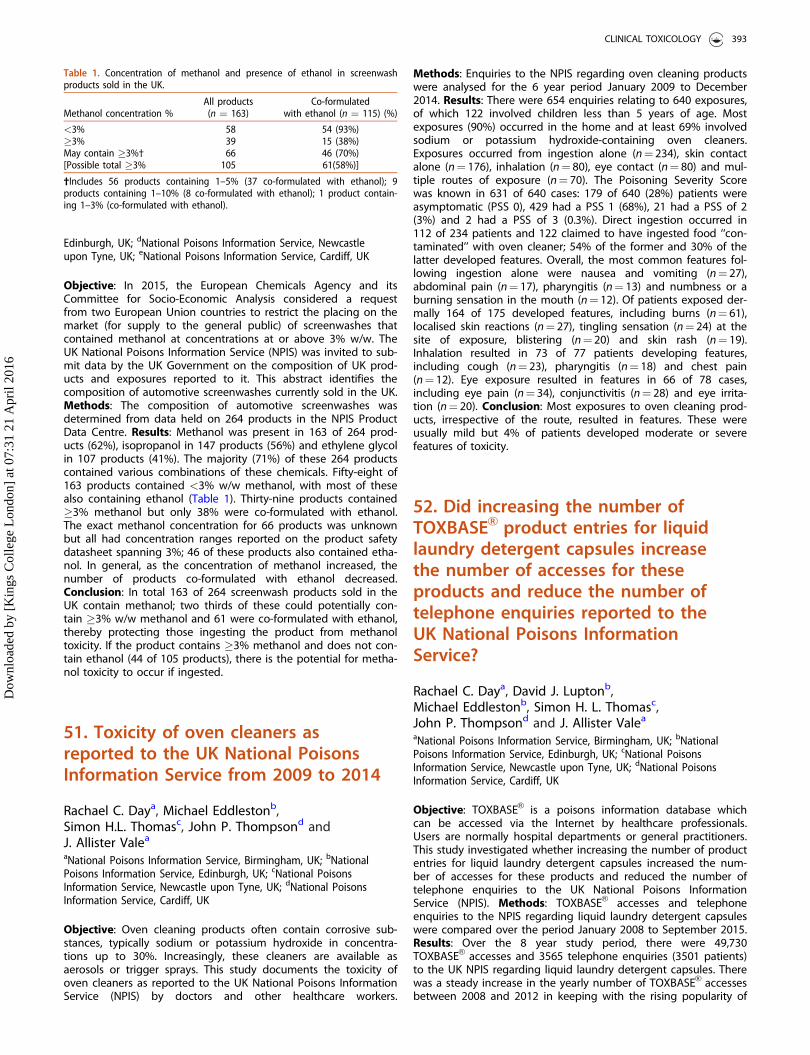
Edinburgh, UK; dNational Poisons Information Service, Newcastleupon Tyne, UK; eNational Poisons Information Service, Cardiff, UK
Objective: In 2015, the European Chemicals Agency and itsCommittee for Socio-Economic Analysis considered a requestfrom two European Union countries to restrict the placing on themarket (for supply to the general public) of screenwashes thatcontained methanol at concentrations at or above 3% w/w. TheUK National Poisons Information Service (NPIS) was invited to sub-mit data by the UK Government on the composition of UK prod-ucts and exposures reported to it. This abstract identifies thecomposition of automotive screenwashes currently sold in the UK.Methods: The composition of automotive screenwashes wasdetermined from data held on 264 products in the NPIS ProductData Centre. Results: Methanol was present in 163 of 264 prod-ucts (62%), isopropanol in 147 products (56%) and ethylene glycolin 107 products (41%). The majority (71%) of these 264 productscontained various combinations of these chemicals. Fifty-eight of163 products contained <3% w/w methanol, with most of thesealso containing ethanol (Table 1). Thirty-nine products contained�3% methanol but only 38% were co-formulated with ethanol.The exact methanol concentration for 66 products was unknownbut all had concentration ranges reported on the product safetydatasheet spanning 3%; 46 of these products also contained etha-nol. In general, as the concentration of methanol increased, thenumber of products co-formulated with ethanol decreased.Conclusion: In total 163 of 264 screenwash products sold in theUK contain methanol; two thirds of these could potentially con-tain �3% w/w methanol and 61 were co-formulated with ethanol,thereby protecting those ingesting the product from methanoltoxicity. If the product contains �3% methanol and does not con-tain ethanol (44 of 105 products), there is the potential for metha-nol toxicity to occur if ingested.
51. Toxicity of oven cleaners asreported to the UK National PoisonsInformation Service from 2009 to 2014
Rachael C. Daya, Michael Eddlestonb,Simon H.L. Thomasc, John P. Thompsond andJ. Allister Valea
aNational Poisons Information Service, Birmingham, UK; bNationalPoisons Information Service, Edinburgh, UK; cNational PoisonsInformation Service, Newcastle upon Tyne, UK; dNational PoisonsInformation Service, Cardiff, UK
Objective: Oven cleaning products often contain corrosive sub-stances, typically sodium or potassium hydroxide in concentra-tions up to 30%. Increasingly, these cleaners are available asaerosols or trigger sprays. This study documents the toxicity ofoven cleaners as reported to the UK National Poisons InformationService (NPIS) by doctors and other healthcare workers.
Methods: Enquiries to the NPIS regarding oven cleaning productswere analysed for the 6 year period January 2009 to December2014. Results: There were 654 enquiries relating to 640 exposures,of which 122 involved children less than 5 years of age. Mostexposures (90%) occurred in the home and at least 69% involvedsodium or potassium hydroxide-containing oven cleaners.Exposures occurred from ingestion alone (n¼ 234), skin contactalone (n¼ 176), inhalation (n¼ 80), eye contact (n¼ 80) and mul-tiple routes of exposure (n¼ 70). The Poisoning Severity Scorewas known in 631 of 640 cases: 179 of 640 (28%) patients wereasymptomatic (PSS 0), 429 had a PSS 1 (68%), 21 had a PSS of 2(3%) and 2 had a PSS of 3 (0.3%). Direct ingestion occurred in112 of 234 patients and 122 claimed to have ingested food ‘‘con-taminated’’ with oven cleaner; 54% of the former and 30% of thelatter developed features. Overall, the most common features fol-lowing ingestion alone were nausea and vomiting (n¼ 27),abdominal pain (n¼ 17), pharyngitis (n¼ 13) and numbness or aburning sensation in the mouth (n¼ 12). Of patients exposed der-mally 164 of 175 developed features, including burns (n¼ 61),localised skin reactions (n¼ 27), tingling sensation (n¼ 24) at thesite of exposure, blistering (n¼ 20) and skin rash (n¼ 19).Inhalation resulted in 73 of 77 patients developing features,including cough (n¼ 23), pharyngitis (n¼ 18) and chest pain(n¼ 12). Eye exposure resulted in features in 66 of 78 cases,including eye pain (n¼ 34), conjunctivitis (n¼ 28) and eye irrita-tion (n¼ 20). Conclusion: Most exposures to oven cleaning prod-ucts, irrespective of the route, resulted in features. These wereusually mild but 4% of patients developed moderate or severefeatures of toxicity.
52. Did increasing the number ofTOXBASEVR product entries for liquidlaundry detergent capsules increasethe number of accesses for theseproducts and reduce the number oftelephone enquiries reported to theUK National Poisons InformationService?
Rachael C. Daya, David J. Luptonb,Michael Eddlestonb, Simon H. L. Thomasc,John P. Thompsond and J. Allister Valea
aNational Poisons Information Service, Birmingham, UK; bNationalPoisons Information Service, Edinburgh, UK; cNational PoisonsInformation Service, Newcastle upon Tyne, UK; dNational PoisonsInformation Service, Cardiff, UK
Objective: TOXBASEVR is a poisons information database whichcan be accessed via the Internet by healthcare professionals.Users are normally hospital departments or general practitioners.This study investigated whether increasing the number of productentries for liquid laundry detergent capsules increased the num-ber of accesses for these products and reduced the number oftelephone enquiries to the UK National Poisons InformationService (NPIS). Methods: TOXBASEVR accesses and telephoneenquiries to the NPIS regarding liquid laundry detergent capsuleswere compared over the period January 2008 to September 2015.Results: Over the 8 year study period, there were 49,730TOXBASEVR accesses and 3565 telephone enquiries (3501 patients)to the UK NPIS regarding liquid laundry detergent capsules. Therewas a steady increase in the yearly number of TOXBASEVR accessesbetween 2008 and 2012 in keeping with the rising popularity of
Table 1. Concentration of methanol and presence of ethanol in screenwashproducts sold in the UK.
Methanol concentration %All products
(n ¼ 163)Co-formulated
with ethanol (n ¼ 115) (%)
<3% 58 54 (93%)�3% 39 15 (38%)May contain �3%† 66 46 (70%)[Possible total �3% 105 61(58%)]
†Includes 56 products containing 1–5% (37 co-formulated with ethanol); 9products containing 1–10% (8 co-formulated with ethanol); 1 product contain-ing 1–3% (co-formulated with ethanol).
CLINICAL TOXICOLOGY 393
Dow
nloa
ded
by [
Kin
gs C
olle
ge L
ondo
n] a
t 07:
31 2
1 A
pril
2016
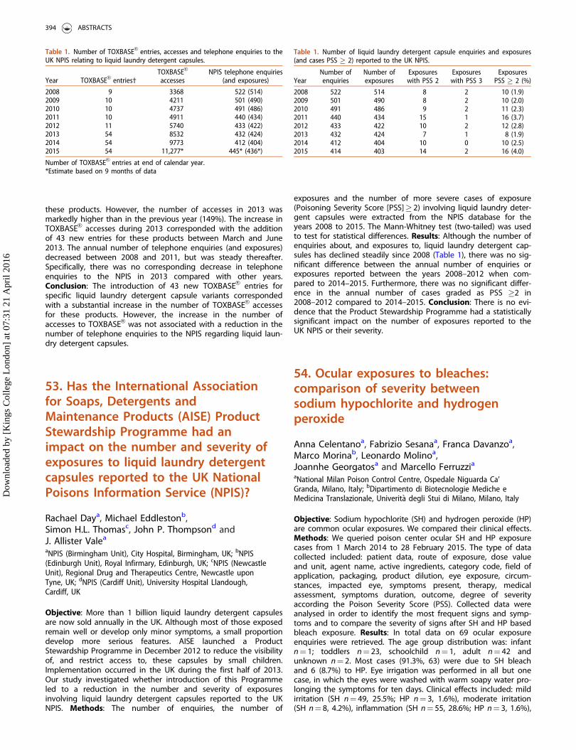
these products. However, the number of accesses in 2013 wasmarkedly higher than in the previous year (149%). The increase inTOXBASEVR accesses during 2013 corresponded with the additionof 43 new entries for these products between March and June2013. The annual number of telephone enquiries (and exposures)decreased between 2008 and 2011, but was steady thereafter.Specifically, there was no corresponding decrease in telephoneenquiries to the NPIS in 2013 compared with other years.Conclusion: The introduction of 43 new TOXBASEVR entries forspecific liquid laundry detergent capsule variants correspondedwith a substantial increase in the number of TOXBASEVR accessesfor these products. However, the increase in the number ofaccesses to TOXBASEVR was not associated with a reduction in thenumber of telephone enquiries to the NPIS regarding liquid laun-dry detergent capsules.
53. Has the International Associationfor Soaps, Detergents andMaintenance Products (AISE) ProductStewardship Programme had animpact on the number and severity ofexposures to liquid laundry detergentcapsules reported to the UK NationalPoisons Information Service (NPIS)?
Rachael Daya, Michael Eddlestonb,Simon H.L. Thomasc, John P. Thompsond andJ. Allister Valea
aNPIS (Birmingham Unit), City Hospital, Birmingham, UK; bNPIS(Edinburgh Unit), Royal Infirmary, Edinburgh, UK; cNPIS (NewcastleUnit), Regional Drug and Therapeutics Centre, Newcastle uponTyne, UK; dNPIS (Cardiff Unit), University Hospital Llandough,Cardiff, UK
Objective: More than 1 billion liquid laundry detergent capsulesare now sold annually in the UK. Although most of those exposedremain well or develop only minor symptoms, a small proportiondevelop more serious features. AISE launched a ProductStewardship Programme in December 2012 to reduce the visibilityof, and restrict access to, these capsules by small children.Implementation occurred in the UK during the first half of 2013.Our study investigated whether introduction of this Programmeled to a reduction in the number and severity of exposuresinvolving liquid laundry detergent capsules reported to the UKNPIS. Methods: The number of enquiries, the number of
exposures and the number of more severe cases of exposure(Poisoning Severity Score [PSS]� 2) involving liquid laundry deter-gent capsules were extracted from the NPIS database for theyears 2008 to 2015. The Mann-Whitney test (two-tailed) was usedto test for statistical differences. Results: Although the number ofenquiries about, and exposures to, liquid laundry detergent cap-sules has declined steadily since 2008 (Table 1), there was no sig-nificant difference between the annual number of enquiries orexposures reported between the years 2008–2012 when com-pared to 2014–2015. Furthermore, there was no significant differ-ence in the annual number of cases graded as PSS �2 in2008–2012 compared to 2014–2015. Conclusion: There is no evi-dence that the Product Stewardship Programme had a statisticallysignificant impact on the number of exposures reported to theUK NPIS or their severity.
54. Ocular exposures to bleaches:comparison of severity betweensodium hypochlorite and hydrogenperoxide
Anna Celentanoa, Fabrizio Sesanaa, Franca Davanzoa,Marco Morinab, Leonardo Molinoa,Joannhe Georgatosa and Marcello FerruzziaaNational Milan Poison Control Centre, Ospedale Niguarda Ca’Granda, Milano, Italy; bDipartimento di Biotecnologie Mediche eMedicina Translazionale, Univerit�a degli Stui di Milano, Milano, Italy
Objective: Sodium hypochlorite (SH) and hydrogen peroxide (HP)are common ocular exposures. We compared their clinical effects.Methods: We queried poison center ocular SH and HP exposurecases from 1 March 2014 to 28 February 2015. The type of datacollected included: patient data, route of exposure, dose valueand unit, agent name, active ingredients, category code, field ofapplication, packaging, product dilution, eye exposure, circum-stances, impacted eye, symptoms present, therapy, medicalassessment, symptoms duration, outcome, degree of severityaccording the Poison Severity Score (PSS). Collected data wereanalysed in order to identify the most frequent signs and symp-toms and to compare the severity of signs after SH and HP basedbleach exposure. Results: In total data on 69 ocular exposureenquiries were retrieved. The age group distribution was: infantn¼ 1; toddlers n¼ 23, schoolchild n¼ 1, adult n¼ 42 andunknown n¼ 2. Most cases (91.3%, 63) were due to SH bleachand 6 (8.7%) to HP. Eye irrigation was performed in all but onecase, in which the eyes were washed with warm soapy water pro-longing the symptoms for ten days. Clinical effects included: mildirritation (SH n¼ 49, 25.5%; HP n¼ 3, 1.6%), moderate irritation(SH n¼ 8, 4.2%), inflammation (SH n¼ 55, 28.6%; HP n¼ 3, 1.6%),
Table 1. Number of TOXBASEVR entries, accesses and telephone enquiries to theUK NPIS relating to liquid laundry detergent capsules.
Year TOXBASEVR entries†TOXBASEVR
accessesNPIS telephone enquiries
(and exposures)
2008 9 3368 522 (514)2009 10 4211 501 (490)2010 10 4737 491 (486)2011 10 4911 440 (434)2012 11 5740 433 (422)2013 54 8532 432 (424)2014 54 9773 412 (404)2015 54 11,277* 445* (436*)
Number of TOXBASEVR entries at end of calendar year.*Estimate based on 9 months of data
Table 1. Number of liquid laundry detergent capsule enquiries and exposures(and cases PSS � 2) reported to the UK NPIS.
YearNumber ofenquiries
Number ofexposures
Exposureswith PSS 2
Exposureswith PSS 3
ExposuresPSS � 2 (%)
2008 522 514 8 2 10 (1.9)2009 501 490 8 2 10 (2.0)2010 491 486 9 2 11 (2.3)2011 440 434 15 1 16 (3.7)2012 433 422 10 2 12 (2.8)2013 432 424 7 1 8 (1.9)2014 412 404 10 0 10 (2.5)2015 414 403 14 2 16 (4.0)
394 ABSTRACTS
Dow
nloa
ded
by [
Kin
gs C
olle
ge L
ondo
n] a
t 07:
31 2
1 A
pril
2016
conjunctivitis (SH n¼ 6, 3.1%; HP n¼ 1, 0.5%), conjunctivitisaggravated (SH n¼ 1, 0.5%), pain (SH n¼ 18, 9.4%; HP n¼ 1,0.5%), lacrimation (SH n¼ 13, 6.8%; HP n¼ 1, 0.5%), mildlyimpaired vision (SH n¼ 6, 3.1%), mild sensation of foreign body(SH n¼ 9, 4.7%), moderate sensation of foreign body (SH n¼ 3,1.6%), severe impaired vision (SH n¼ 1, 0,5%), mild palpebraloedema (SH n¼ 4, 2.1%), photophobia (SH n¼ 3, 1.6%) and cor-neal abrasion (SH n¼ 2, 1.0%). PSS ranged from no symptoms in3 cases (SH n¼ 2, 2.9%; HP n¼ 1, 1.5%), minor in 45 cases (SHn¼ 40, 58%; HP n¼ 5, 7.2%), moderate in 12 (SH n¼ 12, 17.4%).PSS was not assignable in 9 cases (SH 13.04%). One adultexposed to a SH bleach reported feeling heaviness of the eyeand discomfort for 2 months. Another adult had ocular ulcerationfollowing SH bleach exposure; however the eyewash was per-formed for only one minute. Conclusion: The comparison of thePSS assigned in the HP and in the SH groups gave the followingresults: asymptomatic or minor cases account for the total of HPgroup without any moderate cases, while 17.4% of the caseswere rated as moderate in the SH group. No severe cases wereobserved in either group.
55. Unintended severe poisoning by amodern toilet cleaner: analysis of casereports and product databases forearly risk detection
Kathrin Begemann, Esther Feistkorn, Nina Glaser andHerbert DeselBundesinstitut f€ur Risikobewertung (BfR), Berlin, Germany
Objective: Given the great variety of household products avail-able, it is difficult to maintain an up-to-date overview of theircompositions, particularly with regard to toxicological aspectsand their respective poisoning risk. In the field of householdchemicals there are increasing numbers of product groups withchanging formulations placed on the market. Household prod-ucts should be safe to use and with respect to foreseeablemisuse. A regular evaluation of well-documented cases of poi-soning, with specific regard to severe individual cases, as wellas a continuously updated overview of the wide range of prod-ucts available may help to identify risks early and take precau-tionary measures. Methods: Based on the regulations of theGerman Chemicals Act, physicians who are consulted for treat-ment or evaluation of sequelae of diseases caused by chemicalsare requested to report to the Bundesinstitut f€ur Risikobewertung(BfR; Federal Institute for Risk Assessment) health effects from realor suspected exposures to chemical substances. In addition, sub-mitters of products are obliged to notify formulations of chemicalproducts. A case of severe poisoning in an elderly, dementedpatient who accidentally drank a strongly acidic toilet cleaner ispresented. This is the first reported case of severe poisoning withthe newer product group of toilet cleaners for domestic use. Thiscase was the reason to search the product database for newproduct groups with new risks. Results: An 83-year-old patientsuffering from severe dementia developed 3rd degree burns tohis gastrointestinal tract after ingesting 100 mL of a toilet powercleaner containing 10% hydrochloric acid. There was prolongedhematemesis and necrosis throughout the oesophagus and thestomach. Laboratory tests showed a significant increase in C-react-ive protein, metabolic acidosis and a severe decrease in haemo-globin concentration. After parenteral nutrition and symptomatictherapy he stabilized and was discharged in a nursing home/resi-dential care home on day 31. Overview search in the productdatabase revealed that the first power cleaners for consumers
with strongly acidic pH appeared on the German market in 2011.Conclusion: Patients with dementia represent a high-risk-groupfor poisonings. Analysis of reported cases of poisoning can helpto early identify special risks. For comprehensive early risk identifi-cation, a complete overview of the poisoning situation is required.Product databases containing a wide range of information on thecomposition and toxicological aspects and their analysis can sup-port identification of risks at an early stage and facilitate improve-ment of risk management measures.
56. Addition of a bittering agent inliquid laundry detergent capsulemembranes: is there a tangibleingestion risk decrease? An ItalianPoison Control Centre’s point of view
Anna Celentano, Franca Davanzo, Fabrizio Sesana,Marco Morina, Leonardo Molino, Rossana Borghiniand Marcello FerruzziNational Milan Poison Control Centre, Ospedale Niguarda Ca’Granda, Milano, Italy
Objective: EU 1297/2014 Regulation point 3.3, concerns theaddition of a safe concentration of a bittering agent (BA) toliquid laundry detergent capsule (LLDC) hydrosoluble mem-branes as a deterrent to oral exposure within a maximum timeof six seconds. We aimed to verify if the addition of a BA inLLDC membranes can really decrease the risk of paediatric acci-dental exposures. Methods: Three factors were considered con-cerning the use of a BA within the LLDC’s outer membrane: thetime required for the membrane’s dissolution, the time requiredfor bitter taste perception and the necessary concentration ofdenatonium benzoate [1] in order to avoid accidents. Results:The time required for the LLDC to dissolve when in contact withsaliva or wet hands (the most common setting in paediatricexposure cases) is currently still unknown. Therefore, it should bethe subject of experimental tests. Bitterness is perceived in theposterior third of the tongue, innervated by the IX cranial nerveterminations. Therefore, bitter taste perception is not as immedi-ate as it is for sweet taste, which is felt on the tip of tonguethrough the corda tympani. Typically children bite down on theLLDC using their canines, thus piercing the outer membranealmost immediately and causing the detergent to hit the oralmucosa, eyes, and skin causing vomiting, irritation, cough, andother signs and symptoms. Therefore, it seems unlikely that anyamount of denatonium benzoate would be adequate to induce arapid rejection of the LLDC. Conclusion: It is widely accepted [2]that adding a BA to a detergent only worsens possible signs andsymptoms following ingestion. Therefore, it would be opportuneto reconsider whether adding a substance that facilitates vomitingto LLDCs is at all prudent. The addition of a BA does not seem auseful precaution in the absence of adequate experimental stud-ies. Additional data could emerge from the study currently in pro-gress by the European Union in collaboration with PoisonCentres.
References
[1] Rodgers Jr GC, Tenenbein M. The role of aversive bitteringagents in the prevention of pediatric poisonings. Pediatrics.1994;93:68.
CLINICAL TOXICOLOGY 395
Dow
nloa
ded
by [
Kin
gs C
olle
ge L
ondo
n] a
t 07:
31 2
1 A
pril
2016
[2] SCCS/1359/10 Scientific Committee on Consumer Safety. Opinionon the potential health risks posed by chemical consumer prod-ucts resembling food and/or having child-appealing properties. 8.3pages 21–22.
57. Inhalation injury by chlorinegenerated by mixturesof hypochlorite
Uwe Stedtler and Maren Hermanns-ClausenPoisons Center Freiburg, Freiburg, Germany
Objective: Hypochlorite-containing cleaners may liberate chlorinegas when acidified e.g. by adding an acid sanitary cleaner. Usuallythese cleaners contain sodium hydroxide or other strong alkalis tostabilise the sodium hypochlorite. We analysed reports to our poi-sons center concerning inhalation exposure to chlorine liberatedfrom hypochlorite-containing mixtures. Methods: A retrospectivesearch of the case database of the poisons center. Cases ofhuman inhalation exposures to chlorine released from mixinghypochlorite as well as human inhalation hypochlorite evapor-ation alone were analysed. Frequency and symptoms were com-pared. Results: In total 85 cases of human exposures to chlorinefrom mixtures of hypochlorite and acids (0.8 of 1000 cases) wereregistered from 2010 to 2015. Of these 94% of exposed patientsreported symptoms. In two cases the symptoms were not consid-ered to be caused by the inhalation accident. The most frequentsymptoms reported were (percent of symptomatic patients):cough (45%), dyspnea (33%), irritated upper airway (26%), abdom-inal discomfort (pain, nausea, vomiting) (21%), thoracic pain(20%), irritated eyes (11%), dizziness (8%), and bronchospasm(6%). Further symptoms were malaise, headache, irritated nose,sweating, muscle pain, and others. In 12 patients (14%) the symp-toms were graded as moderate severe. The main symptoms inthis group were dyspnea (83%), cough, and irritated airway. Onethird of the patients experienced bronchial obstruction. All symp-tomatic patients developed symptoms while exposed or shortlyafter exposure. There were no severe or fatal cases and all symp-toms were expected to resolve completely. As hypochlorite-con-taining cleaners release chlorine-like smelling gases, weadditionally analysed inhalation exposures to hypochlorite solu-tions alone in the same period. There were 42 patients exposedto hypochlorite evaporation alone. Of these 36 of them (86%) hadsymptoms and in 30 cases these were considered to be causedor possibly be caused by the hypochlorite. The most frequentsymptoms were irritated upper airway (33%), nausea or vomiting(30%), cough (23%), irritated eyes (20%), dizziness (13%), dyspnea(10%), headache (8%) and irritated nose (8%). All symptoms wereconsidered mild. There was no bronchospasm or thoracic discom-fort. Conclusion: Despite clear warning on the labels hypochlor-ite-containing solutions are sometimes mixed with acids. Thesemixtures cause more pronounced injuries than vapors of hypo-chlorite solutions alone.
58. Acute health problems due torecreational drug use in patientspresenting to an urban emergencydepartment in Switzerland
Evangelia Liakonia, Patrick C. Dolderb,Katharina Rentschb and Matthias E. Liechtia
aDivision of Clinical Pharmacology and Toxicology, UniversityHospital Basel and University of Basel, Basel, Switzerland;bLaboratory Medicine, University Hospital Basel and University ofBasel, Basel, Switzerland
Objective: We describe the frequency and the acute medicalproblems due to recreational drug use in patients presenting toan emergency department in Switzerland during one year with afocus on novel psychoactive substances. The results were com-pared with data from the previous year. Methods: The study wasconducted at the University Hospital of Basel, Switzerland,between October 2014 and September 2015 and within theEuro-DEN project. All cases presenting with acute toxicity due toself-reported acute recreational drug use or with symptoms/signsconsistent with acute toxicity were included. Cases of isolatedethanol intoxication were excluded. Drug tests were performedusing immunoassays and liquid chromatography-tandem massspectrometry (LC-MS/MS) to detect novel psychoactive substan-ces. Results: During the study period there were 50,624 emer-gency department attendances of which 210 were directly relatedto acute toxicity of drugs of abuse. The mean patient age was33 years and 73% were male. Analytical drug confirmation wasavailable in most (136) cases. Alcohol co-use was reported in 48%of the cases. Use of more than one recreational drug wasreported in 24% of the cases and in 34% more than one sub-stance was analytically confirmed. Most presentations wererelated to cocaine (33%), cannabis (32%), heroin (14%), 3,4-meth-ylenedioxymethamphetamine (MDMA) (7%) and other amphet-amines (7%). The most common analytically detected substanceswere cannabis (33%), cocaine (27%), opioids excluding metha-done (19%), benzodiazepines (18%) and amphetamines includingMDMA (10%). There were only 2 cases of relatively novel substan-ces (a severe intoxication with para-methoxymethamphetamine(PMMA) in combination with other substances and an intoxicationof minor severity with 2,5-dimethoxy-4-propylphenethylamine(2C-P), both self-reported without analytical confirmation). Themost frequent symptoms were tachycardia (28%), anxiety (23%),nausea or vomiting (18%) and agitation (17%). Severe complica-tions included 2 fatalities (1 case with MDMA and 1 with heroin),2 acute myocardial infarctions, seizures (13 cases), and psychosis(6 cases). Most patients (76%) were discharged home, 10% wereadmitted to intensive care, and 2% were referred to psychiatriccare. Conclusion: Similar to the prior year, most medical problemsrelated to illicit drugs concerned cocaine and cannabis and mainlyincluded sympathomimetic toxicity and/or psychiatric disorders.We confirm that novel psychoactive substances rarely lead toemergency department consultations (with only two presentationsduring the study period, similar to the previous year).
59. Synthetic cannabinoid receptoragonist (SCRA) toxicity reported to theVictorian Poisons Information Centre
Anselm Y. Wong, Dawson Macleod andShaun L. GreeneVictorian Poisons Information Centre, Victoria, Australia
Objective: SCRAs have gained prevalence over the last few yearsdue to their marketing as ‘‘legal highs’’, online availability andchallenge to regulate. SCRA toxicosis often results in more severeoutcomes than phytocannabinoids and have evolved over time.Exposures and symptoms from SCRAs reported to an AustralianPoisons Information Centre (PIC) have not been previously pub-lished. We aimed to describe the enquiries made to the Victorian
396 ABSTRACTS
Dow
nloa
ded
by [
Kin
gs C
olle
ge L
ondo
n] a
t 07:
31 2
1 A
pril
2016
Poisons Information Centre (PIC) regarding SCRA toxicity.Methods: A retrospective review of the Victorian PIC electronicenquiry database, using search terms including ‘‘synthetic canna-binoids’’ AND ‘‘marijuana’’, was performed for the period April2011 to October 2015. Data collected included patient demo-graphics, reported symptoms, type of caller, intentional or acci-dental exposure, route of administration, poisoning severity scoreand product name. Results: There were 57 exposures regardingSCRAs reported to the Victorian PIC over the study period. Seven(12%) of these involved adolescents (15–19 years old), 44 (77%)were adults (age 20–74) and six (11%) were unrecorded. Fifty-six(99%) cases were intentional and one (1%) accidental use. Themajority of exposures were inhalational (n¼ 53, 93%) and lesswere ingestions (n¼ 4, 7%). The poisoning severity scores rangedfrom none (n¼ 9, 16%) to mild (40, 70%), moderate (n¼ 6, 11%)and severe (n¼ 2, 3%). There were 21 different brands of SCRAproducts recorded, the commonest being Kronic, Blue Lotus andK2. One of these products, K2, was associated with multiple seiz-ures, decrease in conscious state and cardiac arrest requiring car-diopulmonary resuscitation (CPR) and intubation. The commonesteffect was decreased conscious state (n¼ 14, 25%). Other symp-toms included vomiting (n¼ 7), agitation (n¼ 6), seizures (n ¼6),tachycardia (n¼ 4), delirium (n¼ 3), dilated pupils (n¼ 2), chestpain (n¼ 2) and paraesthesia (n¼ 1). Symptoms experienced typ-ically manifested within 6 hours. Conclusion: SCRA-associatedsymptoms reported to an Australian PIC were variable but moresevere than symptoms previously described with phytocannabi-noid use. Continued work is needed to limit harms associatedwith these products.
60. Clinical signs and symptoms inpatients poisoned with processedcannabis (Majoon Birjandi)in Eastern Iran
Reza Afsharia, Ali Heydaria and Omid Mehrpourb
aAddiction Research Centre, Mashhad University of MedialToxicology, Mashhad, Iran; bMedical Toxicology and Drug AbuseResearch Center, Birjand University of Medical Science, Birjand, Iran
Objective: The pattern of drug abuse varies in different parts ofthe world.[1–3] Youngsters in the east of Iran abuse processedcannabis which is an ingredient of a traditional pie called MajoonBirjandi (MB).[4] The aim of this study was to evaluate the clinicaland paraclinical signs and symptoms in patients poisoned withMB. Methods: We designed a cross-sectional descriptive studyusing a standardized questionnaire administered to all patientswith MB poisoning admitted to the toxicology emergency depart-ment of the Imam Reza Hospital in Mashhad, Iran from March2010 to 2011. Information on demographic, medical, psychiatricand electrocardiographic indices and vital signs on admissionwere collected. Results: The study included 37 patients with anaverage age of 23 years, 34 of whom were men and threewomen. Patients had consumed a mean (SD, minimum-maximum)1.3 (0.8, 0.5–3) pies. All patients were positive for tetrahydrocanna-binol (THC) by urinary immunoassay. Common symptomsreported according to frequency were sore throat (92%) followedby dry mouth (91%), flushing (81%), panic (73%), vertigo (73%),conjunctivitis (60%) and agitation (62%). Most patients hadmydriasis and this was statistically related to QRS widening(p¼ 0.04). In addition ten patients presented with decreased levelof consciousness. Onset of signs after ingestion of MB was longerthan expected with cannabis and effects were of longer duration.Conclusion: It appears that the major ingredient of MB is canna-bis. In comparison to the literature related to smoking cannabis,
clinical manifestations of ingesting MB appear later and lastlonger.
References
[1] Mehrpour O, Sezavar SV. Diagnostic imaging in body packers.Mayo Clin Proc. 2012;87:e53–e54.
[2] Karrari P, Mehrpour O, Balali-Mood M. Iranian Crystal: a misunder-standing of the crystal-meth. J Res Med Sci. 2012;17:203–204.
[3] Zavar A, Afshari R, Alidoust M, et al. Curriculum development inregard to illicit drug abuse. FMEJ. 2012;2:31–36.
[4] Mehrpour O, Karrari P, Afshari R. Recreational use and overdose ofingested processed cannabis (Majoon Birjandi) in the eastern Iran.Hum Exp Toxicol. 2012;31:1188–1189.
61. An unhappy end for a partyinvolving Datura stramonium andother abuse drugs
F�atima Ram�ona, Elena Almarzab, Oscar Quintelab andAntonio Ortegab
aSpanish Poison Control Center, National Institute of Toxicologyand Forensic Sciences, Las Rozas, Madrid, Spain; bChemical andDrug Service, National Institute of Toxicology and ForensicSciences, Las Rozas, Madrid, Spain
Objective: Cases involving Datura stramonium comprise 1.8% ofall toxic plant exposures reported to our center. In summer 2011we registered three cases involving Datura stramonium and drugsof abuse which occurred at a party in an abandoned farmhouse.Three friends assumed they had ingested amphetamines(‘‘speed’’), ethanol and an unknown dose of a beverage madefrom Datura stramonium seeds offered at the party. Case report:Case 1: A 20-year-old man was admitted to an emergency depart-ment at 6 am, with a Glasgow Coma Scale of 9, agitation, hyper-thermia, generalized rigidity, tremors and bilateral mydriasis.Neostigmine and diazepam were administered, and agitation andtremors decreased. Hospital screening urine drug tests were nega-tive, and blood ethanol was 1.67 g/L. He presented with sinustachycardia, hypertension, dry mouth and hot flushed skin.Haematology, renal function and creatine phosphokinase werenormal. Treatment was symptomatic and supportive. His signsresolved and he was discharged on day 2. Comprehensive toxico-logical screening for psychoactive substances and drugs of abusewas performed including ethanol and volatile analysis by head-space-gas chromatography with flame ionization detector (HS-GC-FID), enzyme immunoassay, and an extensive analysis in blood byhigh-performance liquid chromatography with diode-array detec-tion (HPLC-DAD), gas chromatography-mass spectrometry (GC-MS), and liquid chromatography-tandem mass spectrometry (LC-MS/MS). Results were negative. Cases 2 and 3: Two males, 17 and18-years-old, were found dead on waste ground after attendingthe same party. Their bodies showed obvious signs of dehydra-tion. Forensic examination determined the time of death as 5 pmon the same day patient 1 was admitted. The maximum tempera-ture that day was around 40 �C. The 18-year-old was seen naked,walking aimlessly at 4 pm and refused any help. At 6 pm he wasfound dead. Autopsies were performed the following day. Nosigns of violence were found. Urine, blood, gastric contents, liver,brain and kidney were submitted for analysis. Toxicological resultsin case 2 were: atropine detected in brain, stomach, liver, bloodand urine, while scopolamine was only detected in urine.Amphetamine, 3,4-methylenedioxymethamphetamine (MDMA),and alcohol were found in different biological matrices. In case 3
CLINICAL TOXICOLOGY 397
Dow
nloa
ded
by [
Kin
gs C
olle
ge L
ondo
n] a
t 07:
31 2
1 A
pril
2016
atropine and scopolamine were found in stomach contents, whileonly atropine was found in liver and urine. Amphetamine, MDMA,tetrahydrocannabinol (THC) and alcohol were found in differentbiological matrices. Conclusion: Despite the low percentage ofDatura stramonium consults recorded by our PCC, poisonings andfatalities related to this plant have been detected.
62. Not your regular high: Potentiallylethal cardiac dysrhythmias caused byloperamide
Rachel S. Wightmana, Robert S. Hoffmana,Mary Ann Howlandb, Daniel M. Lugassya andRana Biarya
aNew York University School of Medicine, New York, USA; bSt JohnsUniversity, New York, USA
Objective: Loperamide, a non-prescription anti-diarrheal agent, isa peripheral mu-opioid receptor agonist that is excluded from theblood-brain barrier by P-glycoprotein at therapeutic doses, but inoverdose it penetrates the central nervous system (CNS). Wereport cardiac conduction abnormalities and dysrhythmias afteringestion of a recreational supra-therapeutic dose of loperamidewith laboratory confirmation. Case report: A 48-year-old womanwith a history of alcohol and benzodiazepine abuse presentedwith somnolence, weakness and slurred speech. She had beentaking 20–40 tablets of 2 mg loperamide 1–2 times/day for weekswith clonazepam and whiskey. Vital signs were blood pressure(BP) 124/90 mmHg, pulse 88/min, respiratory rate 20/min, tem-perature 36.9 �C and oxygen saturations 100% on room air.Glucose was 6.4 mmol/L. An electrocardiogram (ECG) approxi-mately ten minutes later showed a ventricular rate of 56/min,QRS 164 ms, QTc 571 ms with no discernible P waves. Lactate was3.5 mmol/L and potassium was 6.2 mEq/L. Laboratory findingswere an anion gap of 20, ethanol concentration 3.9 mmol/L, cre-atinine 203 mmol/L, and loperamide concentration 210 ng/mL(average therapeutic <10 ng/mL). She became hypotensive, butresponded to fluids. Following treatment for hyperkalemia with2 g calcium gluconate, insulin 10 Units, dextrose 50%, and hyper-tonic sodium bicarbonate, a repeat ECG showed a ventricular rateof 66/min, QRS 156 ms, QTc 603 ms. Magnesium was given andpacer pads were placed. During the infusion of magnesium herBP fell to 92/58 mmHg with pulse 54/min, respiration rate 14/min,and saturations 97% so the infusion was stopped. The ECG afterthe magnesium infusion had a ventricular rate of 51/min, QRS of134 ms, and QTc 570 ms. In the ICU she had multiple runs of non-sustained ventricular tachycardia. She improved over the next48 hours, was transferred to a floor bed but on day four of hospi-talization left against medical advice. At that time her ECGshowed sinus tachycardia, QRS 82 ms and QTc 460 ms.Conclusion: Loperamide produces both QRS and QT prolongationat supra-therapeutic dosing. A blood loperamide concentration of210 ng/mL is among the highest reported. Supra-therapeutic dos-ing of loperamide is promoted on multiple drug-use websites andonline forums as a treatment for opioid withdrawal and a possiblemethadone alternative. With the current epidemic of prescriptionopioid abuse, toxicity related to loperamide, an opioid agonistthat is readily available over-the-counter, is occurring more fre-quently. It is important for clinicians to be aware of the potentiallylife-threatening toxicity related to loperamide abuse in order toprovide proper diagnosis, management and patient education.
63. Inhalational heroin resulting inacute cardiomyopathy andsensorineural hearing loss
David J. Goldberger and David VearrierDepartment of Emergency Medicine, Drexel University College ofMedicine, Philadelphia, USA
Objective: To describe what we believe to be the first reportedcase of inhalational heroin use resulting in both opioid-inducedsensorineural hearing loss and heroin-induced acute cardiomyop-athy. Opioid-induced sensorineural hearing loss has been reportedin numerous case reports. The mechanism of ototoxicity isunknown and most patients make a complete neurologic recov-ery. Heroin-induced acute cardiomyopathy has been rarelyreported and may feature near global myocardial hypokinesis andreduced ejection fraction. Patients may recover some heart func-tion but recovery is typically incomplete. Case report: A 15-year-old male was admitted to the intensive care unit (ICU) after anoverdose involving both oral alprazolam and inhalation of heroin.The patient’s mother found him unresponsive the morning ofpresentation. On arrival the patient was given naloxone withoutresponse and upon arrival at the Emergency Department (ED) hewas endotracheally intubated for airway protection. Initial vitalsigns were temperature 36.3 �C, heart rate 123 beats/min, respira-tory rate 8/min, blood pressure (BP) 109/64 mmHg and oxygensaturations 99% on room air. Shortly after intubation heart ratenormalized to 89 and BP dropped to 77/48 mmHg. Physical exam-ination was remarkable only for decreased mentation, pinpointpupils and bradypnea. Initial laboratory work up showed an ele-vated creatinine of 2.3 mg/dL, lactate 8 mmol/L, aspartate trans-aminase (AST) 1103 IU/L with negative acetaminophen, creatinekinase (CK) 533 IU/L and troponin 1.56 ng/mL. Urine drug screenwas positive for benzodiazepines, tetrahydrocannabinol (THC) andopiates. Electrocardiogram (ECG) showed a right bundle brunchblock and Brugada-like patterns. CK and troponin peaked thenext day at 21,800 IU/L and 12.9 ng/mL, respectively. An echocar-diogram the same day showed hypokinesis of the entire myocar-dium except for the basal and mid-posteriolateral wall, theejection fraction (EF) was about 30%. On hospital day 2 thepatient was extubated and complained of hearing difficulty whichwas confirmed as moderate bilateral sensorineural hearing loss onaudiology testing. Over the course of the next 48 hours there wasresolution of cardiomyopathy as evident by an echocardiogramwith an EF of 63% without dyskinesia apparent. As for the hearingloss, the patient was placed on a course of oral steroids and hadsignificant improvement by the time of discharge. Conclusion:We report what we believe to be the first case of simultaneousopioid-induced sensorineural hearing loss and heroin-inducedacute cardiomyopathy. Both the hearing loss and cardiomyopathyimproved over a period of days.
64. A fatal case of acute clenbuterolpoisoning
Ann-Charlott Svanhagen and Mark PersonneSwedish Poisons Information Centre, Stockholm, Sweden
Objective: Clenbuterol is a long acting beta-2-adrenergic agonistwith anabolic, lipolytic and sympathomimetic properties, usedmostly in veterinary practice. Its effects are also coveted by somebody builders. We report the death of an adult after intake of an
398 ABSTRACTS
Dow
nloa
ded
by [
Kin
gs C
olle
ge L
ondo
n] a
t 07:
31 2
1 A
pril
2016
acute overdose. Case report: A 36-year-old body builder usingvarious anabolic substances ingested up to 50 tablets of clenbu-terol of an unknown strength. His past medical history included amechanical aortic valve inserted during his youth, subsequentwarfarin treatment with low compliance and medication with ser-traline and gabapentin. Shortly after the drug intake he wasfound in general convulsions without spontaneous breathing.Cardiopulmonary resuscitation was started immediately by hisgirlfriend. Upon arrival of the ambulance electrocardiogramshowed ventricular fibrillations. He was intubated by the ambu-lance personnel and received multiple defibrillations during trans-port to hospital. On admission to the emergency roomresuscitation was continued including repeated defibrillations,bolus doses of amiodarone (450 mg in total), repeated doses ofadrenaline, magnesium and norepinephrine infusion. The patientwas temporally stabilized and forced cooling was initiatedprophylactically against hypoxic brain injury. Laboratory testsshowed an elevated troponin T concentration of 84 ng/L. Lactatewas 7.6 mmol/L, glucose 9.4 mmol/L and leucocytes 26.5� 109/L.The potassium value was not interpretable due to test tube hem-olysis. During the following hour the patient had reoccurring epi-sodes of ventricular tachycardia including suspected torsades depointes. Cardiac index was 2.63 L/min/m2 (normal range 2.8–4.2 L/min/m2) and blood pressure 60/40 mmHg. Treatment with intra-venous lipid emulsion was tried without positive effect. Furtherresuscitation efforts were considered futile and were withheldapproximately four hours after the debut of symptoms. Autopsyrevealed myocardial ischemia with a pale anterior wall of the leftchamber. The patient’s mechanical aortic valve was without anyflaws and the suture line intact. Clenbuterol ingestion was verifiedby post mortem analysis (2 ng/g blood). The cause of death wasstated as acute cardiac ischemia. Conclusion: Clenbuterol over-dose is known to cause massive catecholamine release and subse-quent risk of myocardial ischemia and arrhythmias, symptoms ourpatient presented in a pronounced and malignant way.[1]Takotsubo cardiomyopathy may also be a mechanism for acuteheart failure in such cases; an echocardiogram may confirm thiscondition. Treatment attempts with catecholamines intravenouslywill probably exacerbate the situation.
Reference
[1] Huckins DS, Lemons MF. Myocardial ischemia associated with clen-buterol abuse: report of two cases. J Emerg Med.2013;44:444–449.
65. Clinical features and outcomes in acase series of 549 MDMA-relatedacute toxicity presentations reportedto the European Drug EmergenciesNetwork (Euro-DEN) project over15 months
Cathelijne Lyphouta, Paul I. Darganb, Alison M. Dinesa,Christopher Yatesa, Fridtjof Heyerdahlc,Knut Erik Hovdac, Isabelle Giraudond,Roumen Sedefovd, Euro-Den Research Groupa andDavid M. Woodb
aClinical Toxicology, Guy’s and St Thomas’ NHS Foundation Trustand King’s Health Partners, London, UK; bClinical Toxicology, Guy’sand St Thomas’ NHS Foundation Trust and King’s Health Partnersand Faculty of Life Sciences and Medicine, King’s College London,
London, UK; cThe Norwegian CBRNe Centre of Medicine,Department of Acute Medicine, Oslo University Hospital, Oslo,Norway; dEuropean Monitoring Centre for Drugs and DrugAddiction (EMCDDA), Lisbon, Portugal
Objective: Although MDMA (3,4-methylenedioxymethamphet-amine, ecstasy) is a familiar drug to clinical toxicologists and poi-sons centres, there are only small published series of acuteMDMA toxicity. The aim of this study is to describe the demo-graphics and clinical features of acute MDMA toxicity presenta-tions to the Euro-DEN project over a 15-month period. Methods:Data was collected from 16 sentinel centres in 10 European coun-tries (Denmark, Estonia, France, Germany, Ireland, Norway, Poland,Spain, Switzerland, the UK) using the Euro-DEN minimum datasetfor 15 months (1 October 2013 to 31 December 2014).[1] The fol-lowing data was extracted: age and gender, self-reported drugsused, clinical features, initial disposition from the emergencydepartment (ED), length of hospital stay and outcome. Results: Ofthe 6747 presentations to Euro-DEN over this 15 month period,549 (8.1%) involved the use of MDMA prior to presentation. Theproportion of presentations to each Euro-DEN centre thatinvolved MDMA varied from 0 to 16.7%. The majority of presenta-tions involving MDMA were male (70.5%) and the mean ± SD agewas 24.7 ± 6.6 years. The mean ± SD number of drugs (excludingethanol) per presentation was 1.9 ± 0.7; the top 3 co-ingestantswere cocaine (124, 23%), cannabis (71, 13%) and gammahydroxy-butyrate/gammabutyrolactone (GBH/GBL) (44, 8%); these variedacross sites reflecting the different recreational drug use patterns.The most commonly reported clinical features were agitation/aggression (174 presentations, 31.7%), anxiety (131, 23.9%), palpi-tations (69, 12.6%) and chest pain (55, 10.0%). Hyperthermia (tem-perature �39 �C) was recorded in 16 (2.9%) patients. There were2 deaths and both occurred in individuals with hyperthermia(temperatures on presentation of 40.5 �C and 41.8 �C). The medianlength of stay was 3 hours 43 minutes (IQR 1h 57m to 5h 37m).The majority (323, 58.8%) of patients were medically dischargedfrom the ED, 91 patients (16.6%) self-discharged, 25 (4.6%) wereadmitted to critical care, 14 (2.6%) were admitted to a psychiatricward and 89 (16.2%) to another ward. Conclusion: In this largeseries of presentations with acute MDMA toxicity, clinical featureswere stimulant/sympathomimetic in nature. There were twodeaths and these occurred in patients with hyperthermia on pres-entation. The overall outcome was good with the majority ofpatients discharged from the ED.
Reference
[1] Wood DM, Heyerdahl F, Yates CB, et al. The European DrugEmergencies Network (Euro-DEN). Clin Toxicol (Phila).2014;52:239–241.
66. The impact of legislative control ofmethylphenidate-based novelpsychoactive substances onrecreational drug-related admissionsto the Royal Infirmary of Edinburgh
Janice M. Pettiea, Margaret A. Dowa, Robert Greigb,Michael Eddlestona and James W. Deara
aClinical Toxicology Unit, Royal Infirmary of Edinburgh and NPISEdinburgh, Scottish Poisons Information Bureau, Edinburgh, UK;bThe University of Edinburgh, Edinburgh, UK
CLINICAL TOXICOLOGY 399
Dow
nloa
ded
by [
Kin
gs C
olle
ge L
ondo
n] a
t 07:
31 2
1 A
pril
2016
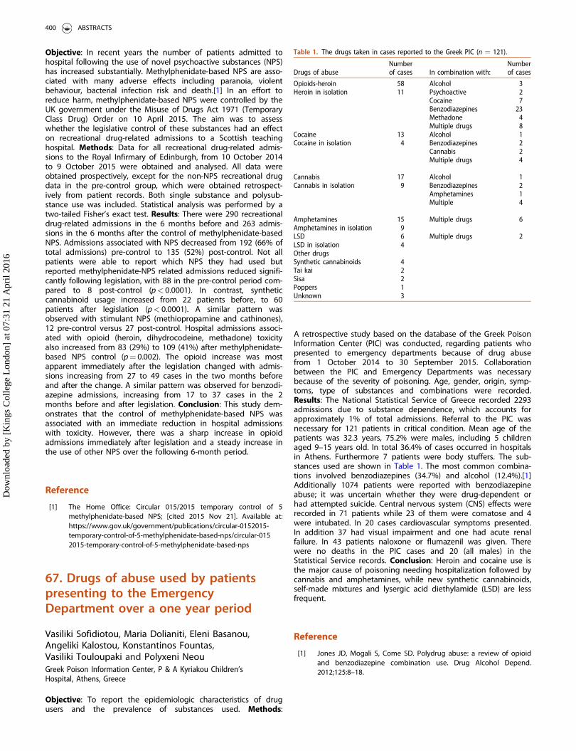
Objective: In recent years the number of patients admitted tohospital following the use of novel psychoactive substances (NPS)has increased substantially. Methylphenidate-based NPS are asso-ciated with many adverse effects including paranoia, violentbehaviour, bacterial infection risk and death.[1] In an effort toreduce harm, methylphenidate-based NPS were controlled by theUK government under the Misuse of Drugs Act 1971 (TemporaryClass Drug) Order on 10 April 2015. The aim was to assesswhether the legislative control of these substances had an effecton recreational drug-related admissions to a Scottish teachinghospital. Methods: Data for all recreational drug-related admis-sions to the Royal Infirmary of Edinburgh, from 10 October 2014to 9 October 2015 were obtained and analysed. All data wereobtained prospectively, except for the non-NPS recreational drugdata in the pre-control group, which were obtained retrospect-ively from patient records. Both single substance and polysub-stance use was included. Statistical analysis was performed by atwo-tailed Fisher’s exact test. Results: There were 290 recreationaldrug-related admissions in the 6 months before and 263 admis-sions in the 6 months after the control of methylphenidate-basedNPS. Admissions associated with NPS decreased from 192 (66% oftotal admissions) pre-control to 135 (52%) post-control. Not allpatients were able to report which NPS they had used butreported methylphenidate-NPS related admissions reduced signifi-cantly following legislation, with 88 in the pre-control period com-pared to 8 post-control (p< 0.0001). In contrast, syntheticcannabinoid usage increased from 22 patients before, to 60patients after legislation (p< 0.0001). A similar pattern wasobserved with stimulant NPS (methiopropamine and cathinones),12 pre-control versus 27 post-control. Hospital admissions associ-ated with opioid (heroin, dihydrocodeine, methadone) toxicityalso increased from 83 (29%) to 109 (41%) after methylphenidate-based NPS control (p¼ 0.002). The opioid increase was mostapparent immediately after the legislation changed with admis-sions increasing from 27 to 49 cases in the two months beforeand after the change. A similar pattern was observed for benzodi-azepine admissions, increasing from 17 to 37 cases in the 2months before and after legislation. Conclusion: This study dem-onstrates that the control of methylphenidate-based NPS wasassociated with an immediate reduction in hospital admissionswith toxicity. However, there was a sharp increase in opioidadmissions immediately after legislation and a steady increase inthe use of other NPS over the following 6-month period.
Reference
[1] The Home Office: Circular 015/2015 temporary control of 5methylphenidate-based NPS; [cited 2015 Nov 21]. Available at:https://www.gov.uk/government/publications/circular-0152015-temporary-control-of-5-methylphenidate-based-nps/circular-0152015-temporary-control-of-5-methylphenidate-based-nps
67. Drugs of abuse used by patientspresenting to the EmergencyDepartment over a one year period
Vasiliki Sofidiotou, Maria Dolianiti, Eleni Basanou,Angeliki Kalostou, Konstantinos Fountas,Vasiliki Touloupaki and Polyxeni NeouGreek Poison Information Center, P & A Kyriakou Children’sHospital, Athens, Greece
Objective: To report the epidemiologic characteristics of drugusers and the prevalence of substances used. Methods:
A retrospective study based on the database of the Greek PoisonInformation Center (PIC) was conducted, regarding patients whopresented to emergency departments because of drug abusefrom 1 October 2014 to 30 September 2015. Collaborationbetween the PIC and Emergency Departments was necessarybecause of the severity of poisoning. Age, gender, origin, symp-toms, type of substances and combinations were recorded.Results: The National Statistical Service of Greece recorded 2293admissions due to substance dependence, which accounts forapproximately 1% of total admissions. Referral to the PIC wasnecessary for 121 patients in critical condition. Mean age of thepatients was 32.3 years, 75.2% were males, including 5 childrenaged 9–15 years old. In total 36.4% of cases occurred in hospitalsin Athens. Furthermore 7 patients were body stuffers. The sub-stances used are shown in Table 1. The most common combina-tions involved benzodiazepines (34.7%) and alcohol (12.4%).[1]Additionally 1074 patients were reported with benzodiazepineabuse; it was uncertain whether they were drug-dependent orhad attempted suicide. Central nervous system (CNS) effects wererecorded in 71 patients while 23 of them were comatose and 4were intubated. In 20 cases cardiovascular symptoms presented.In addition 37 had visual impairment and one had acute renalfailure. In 43 patients naloxone or flumazenil was given. Therewere no deaths in the PIC cases and 20 (all males) in theStatistical Service records. Conclusion: Heroin and cocaine use isthe major cause of poisoning needing hospitalization followed bycannabis and amphetamines, while new synthetic cannabinoids,self-made mixtures and lysergic acid diethylamide (LSD) are lessfrequent.
Reference
[1] Jones JD, Mogali S, Come SD. Polydrug abuse: a review of opioidand benzodiazepine combination use. Drug Alcohol Depend.2012;125:8–18.
Table 1. The drugs taken in cases reported to the Greek PIC (n ¼ 121).
Drugs of abuseNumberof cases In combination with:
Numberof cases
Opioids-heroin 58 Alcohol 3Heroin in isolation 11 Psychoactive 2
Cocaine 7Benzodiazepines 23Methadone 4Multiple drugs 8
Cocaine 13 Alcohol 1Cocaine in isolation 4 Benzodiazepines 2
Cannabis 2Multiple drugs 4
Cannabis 17 Alcohol 1Cannabis in isolation 9 Benzodiazepines 2
Amphetamines 1Multiple 4
Amphetamines 15 Multiple drugs 6Amphetamines in isolation 9LSD 6 Multiple drugs 2LSD in isolation 4Other drugsSynthetic cannabinoids 4Tai kai 2Sisa 2Poppers 1Unknown 3
400 ABSTRACTS
Dow
nloa
ded
by [
Kin
gs C
olle
ge L
ondo
n] a
t 07:
31 2
1 A
pril
2016
68. Chest pain associated withrecreational use of cocaine andmephedrone: should we be askingpatients about use?
Takahiro Yamamotoa, Shwetha Raob,Christopher Walkerc, Andrew Kicmanc,David M. Woodd and Paul I. Dargand
aClinical Toxicology, Guy’s and St Thomas’ NHS Foundation Trustand King’s Health Partners, London, UK; bEmergency Department,King’s College Hospital NHS Foundation Trust and King’s HealthPartners, London, UK; cFaculty of Life Sciences and Medicine, King’sCollege London, London, UK; dClinical Toxicology, Guy’s and StThomas’ NHS Foundation Trust and King’s Health Partners andFaculty of Life Sciences and Medicine, King’s College London,London, UK
Objective: Previous studies from the US and Spain involving ana-lysis of urine samples have found cocaine metabolites in up to25% of all patients with acute coronary syndrome (ACS).[1] Theaim of this study was to determine the prevalence of cocaine andmephedrone use in patients presenting with suspected ACS byanalysis of blood samples and whether a history of drug use wasobtained by the attending clinician. Methods: Potential ACS caseswere prospectively identified from the hospital computerisedblood test system of one inner-city London teaching hospital.Patients over 18 years old presenting to the EmergencyDepartment during September 2014 who had a blood test fortroponin T were included. The blood samples taken were alsosubsequently analysed by liquid chromatography-tandem mass-spectrometry (LC-MS/MS) for the presence of cocaine, mephe-drone and their metabolites. Data on patient demographics, clin-ical presentation and troponin/creatinine results were obtainedfrom the medical records. Results: In total 384 patients met theinclusion criteria (267 males, 117 females); the mean ± SD (range)age was 59.2 ± 17.1 (19–98) years. Of these, 7 (2%) male patientshad detectable drugs in the blood sample (5 cocaine and metab-olites, 1 cocaine metabolites, 1 mephedrone); their mean age waslower than those where drugs were not detected (37.9 ± 11.8years compared to 59.6 ± 17.0 years, p< 0.001). A history ofcocaine/mephedrone use was documented by the attending clin-ician in 4 (57%) of these 7 cases; 1 (14%) denied drug use and 2(29%) had no documentation of use. Conclusion: A small minorityof patients presenting with suspected ACS had analytical confirm-ation of cocaine or mephedrone use prior to the onset of chestpain and a history of drug use was only documented in just overhalf of the patients who had drugs detected. It is important thatclinicians ask patients presenting with chest pain about drug useprior to presentation. Patients with positive samples wereyounger, in keeping with past studies but the overall proportionof positive samples were much lower, this is likely to be becauseprevious studies have looked at cocaine metabolites rather thanparent drug.[1] Further work needs to be done to determine thetrue incidence of sympathomimetic drug-related ACS in largercohorts.
Reference
[1] Hollander JE, Todd KH, Green G, et al. Chest pain associated withcocaine: an assessment of prevalence in suburban and urbanemergency departments. Ann Emerg Med. 1995;26:671–676.
69. ‘‘Maajoune’’ preparations: themost legendary confections ofcannabis with other substances ofabuse in Morocco
Narjis Badrane, Hanane Chaoui, Naima Rhalem andRachida Soulaymani BencheikhPoison Control Centre of Morocco, Rabat, Morocco
Objective: Traditional abuse of cannabis as a preparation called‘‘Maajoune’’ in Morocco is common. It contains cannabis, as themain ingredient, and other substances such as atropine hallucino-genic plants, poppy seeds and psychotropic drugs depending onthe region where it is made. It was the most common substanceof abuse incriminated in the drug poisoning cases reported tothe Poison Control and Pharmacovigilance Centre of Morocco(CAPM).[1] The aim of our study was to analyze the aspects ofconsumption of this mixture over the past two years. Methods: Aretrospective study from January 2013 to December 2014 of allcases of poisoning by ‘‘Maajoune’’ reported to the CAPM. Thedata included circumstances of poisoning, sex, age distributionand symptomatology. The age classification used was theInternational Programme on Chemical Safety classification. Weused a logistic regression model to determine the risk factors ofhospitalization. Results: ‘‘Maajoune’’ was the most drug of abuseresponsible for poisoning (49.9% of all substance abuse intoxica-tions). The average age was 19.6 ± 8.4 years. The most affectedage group was adults (45.6%) followed by adolescents (32.6%)and patients aged less than 15 years (infant, toddler and children22.1%). The sex ratio (M/F) was 3.6. Addictive circumstance wasthe most frequent (66.4%) followed by accidents (30.4%). In85.6% of the cases, patients were symptomatic. The dominatingclinical signs were gastrointestinal (40.3%) and neurological(38.4%). The risk factor for hospitalization was age less than 15years (p¼ 0.01, OR 1.6). Conclusion: The consequence of the trivi-alization of ‘‘Maajoune’’ consumption in Morocco is the increasein poisoning cases in young addicts but also accidental poisoningin children. An awareness program in schools and through themedia is needed to address this scourge.
Reference
[1] Chaoui H, Rhalem N, Semlali I, et al. Poisoning by substances ofabuse in Morocco: Moroccan poison control center data (1980 to2009) and risks factors for death. Rev Res J. 2014;3:1–8.
70. Epidemiology and clinical pictureof hashish oil poisoning in Slovenia
Vid Leban, Mojca Dobaja and Miran BrvarCentre for Clinical Toxicology and Pharmacology, University MedicalCentre Ljubljana, Ljubljana, Slovenia
Objective: Hashish oil is a dark, viscous organic substance pro-duced from the resin collected from the flowering tops ofCannabis sativa plants. It contains varying concentrations of tetra-hydrocannabinol (THC) up to 50%. Despite the prohibition, abuseof THC is widespread among the Slovenian population. The useof hashish oil is propagated for medicinal purposes, as it isbelieved to be generally beneficial. The aim of the study was toevaluate the epidemiology of hashish oil poisoning in Ljubljana,
CLINICAL TOXICOLOGY 401
Dow
nloa
ded
by [
Kin
gs C
olle
ge L
ondo
n] a
t 07:
31 2
1 A
pril
2016
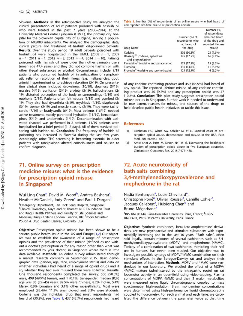
Slovenia. Methods: In this retrospective study we analyzed theclinical presentation of adult patients poisoned with hashish oilwho were treated in the past 7 years (2008–2014) at theUniversity Medical Centre Ljubljana (UMCL), the primary city hos-pital for the Slovenian capital city of Ljubljana, serving a popula-tion of 600,000 inhabitants. We analysed the demographic data,clinical picture and treatment of hashish oil-poisoned patients.Results: Over the study period 19 adult patients poisoned withhashish oil were hospitalized in the UMCL (2008 n¼ 1, 2009n¼ 1, 2011 n¼ 1, 2012 n¼ 2, 2013 n¼ 4, 2014 n¼ 10). Patientspoisoned with hashish oil were older than other cannabis users(mean age 47.4 years) and they did not combine hashish oil withother illegal substances or alcohol. Circumstances include 9/19patients who consumed hashish oil in anticipation of symptom-atic relief or resolution of their illness (e.g. malignancies, gout,arterial hypertension) or to achieve relaxation (5/19). On presenta-tion clinical signs included drowsiness (10/19), slowness (5/19),malaise (4/19), confusion (3/19), anxiety (3/19), hallucinations (2/19), distorted perception of the body or surroundings (2/19) andeuphoria (2/19). Patients often felt nausea (9/19) and vomited (3/19). They also had dysarthria (5/19), mydriasis (4/19), diaphoresis(3/19), tremor (2/19) and muscle spasms (2/19). They were tachy-cardic (7/19) or bradycardic (6/19). Most patients (16/19) neededactive treatment, mostly parenteral hydration (11/19), benzodiaze-pines (5/19) and antiemetics (1/19). Decontamination with acti-vated charcoal was performed in 2 patients; 11/19 patients werehospitalized for a brief period (1–2 days). All of them survived poi-soning with hashish oil. Conclusion: The frequency of hashish oilpoisoning has increased in Slovenia during the last few years.Accordingly, urine THC screening is becoming essential in olderpatients with unexplained altered consciousness and nausea toconfirm diagnosis.
71. Online survey on prescriptionmedicine misuse: what is the evidencefor prescription opioid misusein Singapore?
Wui Ling Chana, David M. Woodb, Andrea Besharatc,Heather McDanielc, Jody Greenc and Paul I. Darganb
aEmergency Department, Tan Tock Seng Hospital, Singapore;bClinical Toxicology, Guy’s and St Thomas’ NHS Foundation Trustand King’s Health Partners and Faculty of Life Sciences andMedicine, King’s College London, London, UK; cRocky MountainPoison & Drug Center, Denver, Colorado, USA
Objective: Prescription opioid misuse has been shown to be aserious public health issue in the US and Europe.[1,2] Our object-ive was to establish the awareness of a range of prescriptionopioids and the prevalence of their misuse (defined as use with-out a doctor’s prescription or for any reason other than what wasrecommended by your doctor) in Singapore where there is littledata available. Methods: An online survey administered througha market research company in September 2015. Basic demo-graphic data (gender, age, race, employment status) and data onwhether individuals had heard of a range of opioid drugs and ifso, whether they had ever misused them were collected. Results:One thousand respondents completed the survey: 500 (50.0%)male, 499 (49.9%) female, and 1 (0.1%) transgender; median (IQR)age was 35 (29–45) years; 82.5% were Chinese, 8.2% Indian, 5.4%Malay, 0.8% Eurasian and 3.1% other race/ethnicity. Most wereemployed (85.4%), 11.3% unemployed and 3.3% were students.Codeine was the individual drug that most respondents hadheard of (30.2%), see Table 1; 437 (43.7%) respondents had heard
of any codeine containing product and 459 (45.9%) had heard ofany opioid. The reported lifetime misuse of any codeine-contain-ing product was 40 (9.2%) and any prescription opioid was 47(10.2%). Conclusion: This pilot study suggests prescription opioidmisuse occurs in Singapore. Further work is needed to understandits true extent, reasons for misuse, and sources of the drugs tohelp develop public health initiatives to tackle this issue.
References
[1] Birnbaum HG, White AG, Schiller M, et al. Societal costs of pre-scription opioid abuse, dependence, and misuse in the USA. PainMed. 2011;12:657–667.
[2] Amie Shei A, Hirst M, Kirson NY, et al. Estimating the healthcareburden of prescription opioid abuse in five European countries.Clinicoecon Outcomes Res. 2015;7:477–488.
72. Acute neurotoxicity ofbath salts combining3,4-methylenedioxypyrovalerone andmephedrone in the rat
Nadia Benturquiaa, Lucie Chevillarda,Christophe Poir�ea, Olivier Roussela, Camille Cohiera,Jacques Calleberta, Huixiong Chenb andBruno M�egarbanea
aINSERM U1144, Paris-Descartes University, Paris, France; bCNRSUMR8601, Paris-Descartes University, Paris, France
Objective: Synthetic cathinones, beta-keto-amphetamine deriva-tives, are new psychoactive and stimulant substances with expo-nentially increasing use in the last 10 years. ‘‘Bath salts’’, oftensold legally, contain mixtures of several cathinones such as 3,4-methylenedioxypyrovalerone (MDPV) and mephedrone (4MMC).Toxicity of a combination of two cathinones, mimicking their realuse in humans, has never been studied. Our objective was toinvestigate possible synergy of MDPV/4MMC combination on theirstimulant effects in the Sprague-Dawley rat and analyze theirmechanisms of interaction. Methods: MDPV and 4MMC were syn-thesized in our laboratory. We studied the effect of a MDPV/4MMC mixture (administered by the intragastric route) on ratlocomotor activity in an open-field using video-tapping. Plasmaconcentrations of MDPV, 4MMC and their 3 major metaboliteswere measured using liquid chromatography coupled to massspectrometry high-resolution. Brain monoamine concentrationswere determined using high-performance liquid chromatographycoupled to fluorometry. For each animal and each time, we calcu-lated the difference between the parameter value at that time
Table 1. Number (%) of respondents of an online survey who had heard ofand reported life-time misuse of prescription opioids.
Drug Name
Number (%) ofrespondents who
had heard ofthe drug
Number (%)of respondentswho had heardof the drug andreported lifetime
misuse
Codeine 302 (30.2%) 23 (7.6%)DhasedylVR (codeine, ephedrine
and promethazine)175 (17.5%) 16 (9.1%)
PanadeineVR (codeine and paracetamol) 175 (17.5%) 15 (8.6%)Tramadol 136 (13.6%) 11 (8.1%)ProcodinVR (codeine and promethazine) 125 (12.5%) 4 (3.2%)
402 ABSTRACTS
Dow
nloa
ded
by [
Kin
gs C
olle
ge L
ondo
n] a
t 07:
31 2
1 A
pril
2016
and baseline and the area under the curve of its time course.Comparisons were performed using two-way ANOVA followed bypost-tests using Bonferroni correction. Pharmacokinetics (PK) wasmodeled and parameters calculated using WinNonlinVR software.Results: MDPV (3 mg/kg)/4MMC (30 mg/kg) combination wasresponsible for a significant increase in rat locomotor activity incomparison to saline, MDPV and 4MMC alone (p¼ 0.02), with asynergic interaction between the two drugs. 4MMC PK was notaltered in the presence of MDPV while MDPV absorption wasreduced in the presence of 4MMC without significant modifica-tions in its half-life and clearance. The neurochemical studyrevealed significant increase in dopamine and serotonin but notin norepinephrine concentrations in the prefrontal cortex, whencombining both drugs in comparison to each drug administeredseparately. Conclusion: A MDPV/4MMC mixture as found in ‘‘bathsalts’’ significantly increases locomotor activity in rats in compari-son to each substance administered alone. Mechanisms of inter-action include significant decrease in MDPV gastrointestinalabsorption and significant increase in monoamine reuptake inhib-ition in the brain with reinforcement of both the dopaminergicand serotoninergic profiles. However, further investigations at thelevel of gastrointestinal transporters and monoamine transportersare required.
73. Substance abuse among medicalstudents in Maribor, Slovenia
�Ziga Volgemut and Polonca FerkFaculty of Medicine, University of Maribor, Maribor, Slovenia
Objective: There is little information about how familiar futurephysicians in Slovenia are with the abuse of substances. Our aimwas to investigate lifetime abuse of drugs and illicit drugs amongmedical students at the Faculty of Medicine in Maribor, Slovenia.Methods: An online anonymous self-administered questionnairewas developed. Through email and the faculty website, 645 stu-dents (from 1st to 6th year of study) were invited to participate incompleting the questionnaire. Questions were related to age, gen-der, year of study, other basic demographic information as well asto consumption of alcohol, cigarettes, cannabis, synthetic cannabi-noids, alkyl nitrites, inhalants, amphetamines, cocaine, lysergicacid diethylamide (LSD), magic mushrooms, heroin, gammahy-droxybutyrate (GHB) and mephedrone. The data was anonym-ously collected and stored on servers of an independent surveyprovider. The results were descriptively evaluated. Results: In total288 out of 645 (45%) students responded. From the respondents,69% were females and 31% males; which closely represents theratio of female to male students at our faculty. The mean age ofstudents participating in the study was 22.0 ± 2.2 years. Of therespondents 70% had used a substance for abuse at some point.Most frequent drugs of abuse were alcohol and cannabis, with alifetime prevalence of 67% and 40%, respectively; 8% of studentswere smokers, 11% reported smoking occasionally, 65% werenon-smokers and 16% did not answer the question on smoking.Magic mushrooms had been used by 8%, whereas 7% of studentsused amphetamines and 3% cocaine. There was a higher preva-lence of consumption among male compared to female students(75% versus 68%, respectively). Also, a higher substance use in6th compared to 1st year students was noted (88% versus 66%,respectively). Conclusion: In comparison with similar studies onmedical students, we report lower alcohol consumption, lowertobacco usage and lower cannabis consumption in our students;however, higher consumption of hallucinogens wasobserved.[1–3] Further investigations on frequency and reasonsfor abusing substances are required.
References
[1] Roncero C, Egido A, Rodr�ıguez-Cintas L, et al. Substance useamong medical students: a Literature Review 1988–2013. ActasEsp Psiquiatr. 2015;43:109–121.
[2] Vi�snjic A, Jovic S, Grbe�sa G. Alcohol consumption amongstudents – a cross-sectional study at three largest universities inSerbia. Srp Arh Celok Lek. 2015;143:301–308.
[3] Voigt K, Twork S, Mittag D, et al. Consumption of alcohol, ciga-rettes and illegal substances among physicians and medical stu-dents in Brandenburg and Saxony (Germany). BMC Health ServRes. 2009;9:219.
74. Abuse of synthetic cannabinoids inIsrael: reports to the National PoisonInformation Center 2010–2014
Yael Lurie, Gal Neuman, Iris Levdov-Avital,Daniel Kurnik and Yedidia BenturClinical Pharmacology & Toxicology Section, Haifa, Israel
Objective: Synthetic cannabinoid receptor agonists comprise alarge family of structurally unrelated substances marketed asabusable designer drugs. Since 2011, many synthetic cannabi-noids have been included in Israel’s controlled substance act, butthere is little information about the epidemiology of syntheticcannabinoid abuse in Israel. We therefore surveyed demographicand clinical characteristics of synthetic cannabinoid users reportedto the Israel Poison Information Center. Methods: We searchedthe Israel Poison Information Center electronic database for casesof synthetic cannabinoid use and withdrawal syndrome betweenJanuary 2010 (first report of synthetic cannabinoid use) andDecember 2014. Callers’ data and patients’ demographic and clin-ical data were extracted and subjected to descriptive analysis.Laboratory identification was not done since at present there isno analytical laboratory in Israel performing analyses of syntheticcannabinoids in body fluids. Therefore, the identification of theculprit was based on self-report by the users or their escortsaccording to the substance description and common streetnames. Results: Over the study period 107 cases of synthetic can-nabinoid use were reported to the Israel Poison InformationCenter. The number of reports increased consistently each year,starting with 10 in 2010 and reaching 67 in 2014. Most of the call-ers (93%) were healthcare professionals. Users were predomin-antly young males (median age 22 years, range 12–51 years; 84%males). In most cases (93%), the substance was smoked. The mostfrequent clinical manifestations were gastrointestinal (nausea orvomiting 35%, diarrhea 10%), neuro-psychiatric (agitation 23%,psychoses or hallucinations 15%, headache 12.5%, drowsiness 9%,seizures 5%) and cardiovascular (sinus tachycardia 22%, supra-ventricular tachyarrhythmias 3%, chest pain 10%, bradycardia 9%,hypertension 4%). Two percent had acute kidney injury.Symptoms were of moderate severity in 21%, and severe in 3%of cases. No fatalities were reported. Withdrawal syndrome wasreported in 11 patients (10.3%) and included gastrointestinalsymptoms (nausea or vomiting 64%, diarrhea 27%), agitation(36%), tremor (27%), and acute kidney injury (18%). Conclusion:The rising number of reports to the Israel Poison InformationCenter of synthetic cannabinoid use and withdrawal syndromeduring the past 5 years suggests increasing use in the populationat large. Users had a typical profile (predominantly young males).While most symptoms were mild, considerable morbidity wasreported in some cases. Understanding the short- and long-termconsequences of synthetic cannabinoid use and law enforcementare ongoing challenges.
CLINICAL TOXICOLOGY 403
Dow
nloa
ded
by [
Kin
gs C
olle
ge L
ondo
n] a
t 07:
31 2
1 A
pril
2016
75. Alcoholic poisonings inadolescents treated in an emergencyroom in the summer of 2015
Jordi Puiguriguera, Christopher Yatesa,Amparo Fraileb, Monica Guerrab, Catalina Homara andIsabel Ramosb
aClinical Toxicology Unit, Emergency Department, HospitalUniversitari Son Espases, Palma De Mallorca, Spain; bEmergencyDepartment, Hospital Universitari Son Espases, Palma De Mallorca,Spain
Objective: Although the sale and consumption of alcoholic bever-ages is forbidden to minors (under 18 years of age) in Spain, alco-hol is the most prevalent cause of intoxication in adolescents. Asin neighboring countries, there is an increasing trend in bingedrinking. This phenomenon in our environment is seen primarilyin summer, presumably related to tourism. This study examinedthe incidence and characteristics of acute alcohol intoxication(AAI) in summer in a tertiary care emergency department (ED)serving an area with high density of tourism. Methods: An obser-vational retrospective analysis of alcohol intoxication presentingto the ED over three months (1 June to 31 August 2015), analyz-ing epidemiological data (sex, country of origin and age, time ofarrival) and care variables (arrival by ambulance, symptoms, ancil-lary tests, disposition, length of stay in the ED, documentedwhether released to adult tutor or guardian and if a legal reportwas filed). Results: In total 42 patients were treated (8.57% of allAAI in the same period), 22 were males (52.4%), 19 were Spanish(45.2%) and 17 were British (40.4%). Most were 17-years-old (69%)with one 13 and one 14-year-old. All patients arrived between22:00 and 10:00, with the range between 02:00 and 2:59 the mostcommon (21.4%). Most arrived by ambulance (29 cases, 69%). Themain symptoms reported were decreased level of consciousness(31, 73.8%), suspected multiple toxins in 7 (16.8%), agitation in 4,other injuries and palpitations in 2, and one patient had headtrauma. Quantification of ethanol in blood was performed in 11cases (26.1%), with an average blood ethanol concentration of2.01 g/L. In one case, a computerised tomography (CT) scan wasperformed. The average stay in the ED was 4.29 hours (range0:56–12:27 hours). All patients were discharged from the ED. Therewere no deaths or subsequent readmissions, although affiliationdata was missing in 5 cases (11.9%). The presence of an adultguardian or tutor was reflected in the discharge report; in ninecases (21.4%), a legal report was filed in 11.9%. Conclusion: AAIin minors presenting in summer in the ED accounted for 8.57% oftotal AAI cases, patients were mainly Spanish and British. Mostwere seen for decreased level of consciousness with a short aver-age stay and without serious acute complications. Dischargerecords were poor in reporting to whom the minors were dis-charged and in filing mandatory legal reports.
76. Gamma-butyrolactone overdosewith metabolic acidosis treatedwithout dialysis
James Boyda, Ilkka Ojanper€ab and Mika Paloheimoa
aDepartment of Emergency Medicine, Helsinki University Hospital,Helsinki, Finland; bDepartment of Forensic Medicine, HelsinkiUniversity, Helsinki, Finland
Objective: Gamma-hydroxybutyrate (GHB) and its prodruggamma-butyrolactone (GBL) are used as drugs of abuse.[1] A largedose of GBL, rapidly metabolized to GHB, has caused severemetabolic acidosis requiring treatment in an intensive care unitand dialysis in previously published cases.[2,3] We present twocases of GBL poisoning. Case series: In separate incidents two 34-year-old patients, a male and a female, were intubated byEmergency Medical Services because of a Glasgow Coma Scale of3/15. GBL use was suspected on scene in both cases. Both wereadministered vasoactive drugs (ephedrine, epinephrine and/ornorepinephrine) due to hypotension. Their first measured arterialblood gases were pH 6.90, PaCO2 6.38 kPa, PaO2 15.4 kPa, HCO3
9.5 mmol/L, base excess (BE) �23 mmol/L and pH 6.99, PaO2
20.5 kPa, PaCO2 5.3 kPa, HCO3 8 mmol/L, BE �21.3 mmol/L,respectively. In addition to mechanical ventilation and norepin-ephrine-infusions, they were also administered Ringer’s acetate,sodium bicarbonate and fentanyl intravenously. Their acidosissubsided within 10 and six hours, respectively, and neither patientrequired dialysis. Both recovered without sequelae. The GHB con-centrations measured later from blood samples were 1500 and1400 mg/L, respectively, and were determined by gas chromatog-raphy/mass spectrometry according to a previously reportedmethod.[4] Comprehensive toxicological analysis revealed also thefollowing blood drug concentrations: 0.5 mcg/L of fentanyl and0.37 mg/L of 3,4-methylenedioxymethamphetamine (MDMA) inthe male patient and alpha-pyrrolidinovalerophenone (alpha-PVP)0.05 mg/L, amphetamine 0.05 mg/L, ephedrine 0.13 mg/L, fentanyl0.3 mcg/L, clonazepam 0.22 mg/L, diazepam 0.52 mg/L, nordiaze-pam 0.17 mg/L, and alprazolam 0.013 mg/L in the female.Conclusion: GBL overdose with metabolic acidosis can be suc-cessfully treated with supportive care only.
References
[1] Schep LJ, Knudsen K, Slaughter RJ, et al. The clinical toxicology ofgamma-hydroxybutyrate, gamma-butyrolactone and 1,4-butane-diol. Clin Toxicol (Phila). 2012;50:458–470.
[2] Roberts DM, Smith MW, Gopalakrishnan M, et al. Extreme gamma-butyrolactone overdose with severe metabolic acidosis requiringhemodialysis. Ann Emerg Med. 2011;58:83–85.
[3] Connors NJ, Hoffman RS, Nelson LS. Increased serum osmolalityand metabolic acidosis following massive gamma-hydroxybutyrateconsumption. Clin Toxicol (Phila). 2013;51:367.
[4] Elian AA. A novel method for GHB detection in urine and its appli-cation in drug-facilitated sexual assaults. Forensic Sci Int.2000;109:183–187.
77. All that glitters is not LSD!
Gael Le Rouxa, Severine Ferecb, Benedicte Lelievreb,Marie Bretaudeau-Deguignea, Chadi Abbarab,Alain Turcantb and David Boelsa
aPoison Control Centre, University Hospital, Angers, France;bLaboratory of Pharmacology and Toxicology, University Hospital,Angers, France
Objective: Among the recreational drugs, 2,5-dimethoxy-derivatedphenethylamines have hallucinogenic properties as lysergic aciddiethylamide (LSD). Recently, 2,5-dimethoxy-4-chloroamphetamine(DOC) appeared on the drug market and was responsible forsporadic deaths reported in the literature. We present five clinicalcases reported to our Poison Control Centre from March 2014 toMarch 2015 that could contribute to a better knowledge of thisproduct. Cases report: Five men aged 16 to 23 years consumeddrugs during a private party (2 cases) or a rave party (3 cases).
404 ABSTRACTS
Dow
nloa
ded
by [
Kin
gs C
olle
ge L
ondo
n] a
t 07:
31 2
1 A
pril
2016
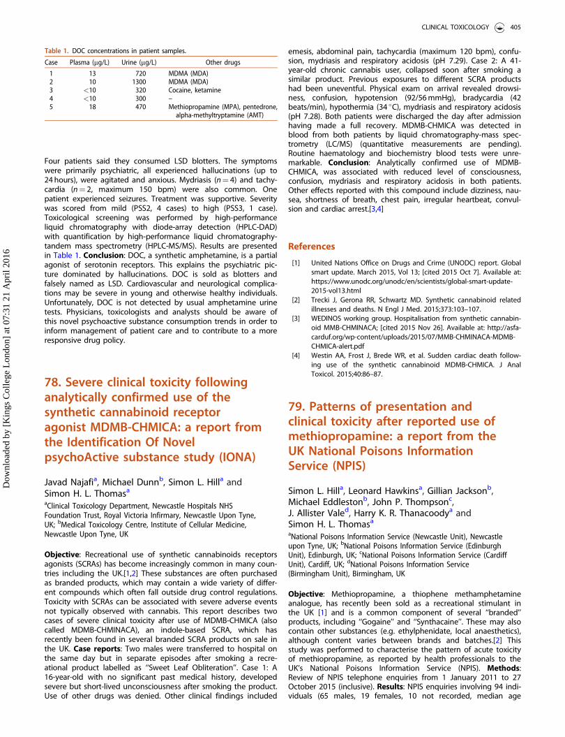
Four patients said they consumed LSD blotters. The symptomswere primarily psychiatric, all experienced hallucinations (up to24 hours), were agitated and anxious. Mydriasis (n¼ 4) and tachy-cardia (n¼ 2, maximum 150 bpm) were also common. Onepatient experienced seizures. Treatment was supportive. Severitywas scored from mild (PSS2, 4 cases) to high (PSS3, 1 case).Toxicological screening was performed by high-performanceliquid chromatography with diode-array detection (HPLC-DAD)with quantification by high-performance liquid chromatography-tandem mass spectrometry (HPLC-MS/MS). Results are presentedin Table 1. Conclusion: DOC, a synthetic amphetamine, is a partialagonist of serotonin receptors. This explains the psychiatric pic-ture dominated by hallucinations. DOC is sold as blotters andfalsely named as LSD. Cardiovascular and neurological complica-tions may be severe in young and otherwise healthy individuals.Unfortunately, DOC is not detected by usual amphetamine urinetests. Physicians, toxicologists and analysts should be aware ofthis novel psychoactive substance consumption trends in order toinform management of patient care and to contribute to a moreresponsive drug policy.
78. Severe clinical toxicity followinganalytically confirmed use of thesynthetic cannabinoid receptoragonist MDMB-CHMICA: a report fromthe Identification Of NovelpsychoActive substance study (IONA)
Javad Najafia, Michael Dunnb, Simon L. Hilla andSimon H. L. Thomasa
aClinical Toxicology Department, Newcastle Hospitals NHSFoundation Trust, Royal Victoria Infirmary, Newcastle Upon Tyne,UK; bMedical Toxicology Centre, Institute of Cellular Medicine,Newcastle Upon Tyne, UK
Objective: Recreational use of synthetic cannabinoids receptorsagonists (SCRAs) has become increasingly common in many coun-tries including the UK.[1,2] These substances are often purchasedas branded products, which may contain a wide variety of differ-ent compounds which often fall outside drug control regulations.Toxicity with SCRAs can be associated with severe adverse eventsnot typically observed with cannabis. This report describes twocases of severe clinical toxicity after use of MDMB-CHMICA (alsocalled MDMB-CHMINACA), an indole-based SCRA, which hasrecently been found in several branded SCRA products on sale inthe UK. Case reports: Two males were transferred to hospital onthe same day but in separate episodes after smoking a recre-ational product labelled as ‘‘Sweet Leaf Obliteration’’. Case 1: A16-year-old with no significant past medical history, developedsevere but short-lived unconsciousness after smoking the product.Use of other drugs was denied. Other clinical findings included
emesis, abdominal pain, tachycardia (maximum 120 bpm), confu-sion, mydriasis and respiratory acidosis (pH 7.29). Case 2: A 41-year-old chronic cannabis user, collapsed soon after smoking asimilar product. Previous exposures to different SCRA productshad been uneventful. Physical exam on arrival revealed drowsi-ness, confusion, hypotension (92/56 mmHg), bradycardia (42beats/min), hypothermia (34 �C), mydriasis and respiratory acidosis(pH 7.28). Both patients were discharged the day after admissionhaving made a full recovery. MDMB-CHMICA was detected inblood from both patients by liquid chromatography-mass spec-trometry (LC/MS) (quantitative measurements are pending).Routine haematology and biochemistry blood tests were unre-markable. Conclusion: Analytically confirmed use of MDMB-CHMICA, was associated with reduced level of consciousness,confusion, mydriasis and respiratory acidosis in both patients.Other effects reported with this compound include dizziness, nau-sea, shortness of breath, chest pain, irregular heartbeat, convul-sion and cardiac arrest.[3,4]
References
[1] United Nations Office on Drugs and Crime (UNODC) report. Globalsmart update. March 2015, Vol 13; [cited 2015 Oct 7]. Available at:https://www.unodc.org/unodc/en/scientists/global-smart-update-2015-vol13.html
[2] Trecki J, Gerona RR, Schwartz MD. Synthetic cannabinoid relatedillnesses and deaths. N Engl J Med. 2015;373:103–107.
[3] WEDINOS working group. Hospitalisation from synthetic cannabin-oid MMB-CHMINACA; [cited 2015 Nov 26]. Available at: http://asfa-carduf.org/wp-content/uploads/2015/07/MMB-CHMINACA-MDMB-CHMICA-alert.pdf
[4] Westin AA, Frost J, Brede WR, et al. Sudden cardiac death follow-ing use of the synthetic cannabinoid MDMB-CHMICA. J AnalToxicol. 2015;40:86–87.
79. Patterns of presentation andclinical toxicity after reported use ofmethiopropamine: a report from theUK National Poisons InformationService (NPIS)
Simon L. Hilla, Leonard Hawkinsa, Gillian Jacksonb,Michael Eddlestonb, John P. Thompsonc,J. Allister Valed, Harry K. R. Thanacoodya andSimon H. L. Thomasa
aNational Poisons Information Service (Newcastle Unit), Newcastleupon Tyne, UK; bNational Poisons Information Service (EdinburghUnit), Edinburgh, UK; cNational Poisons Information Service (CardiffUnit), Cardiff, UK; dNational Poisons Information Service(Birmingham Unit), Birmingham, UK
Objective: Methiopropamine, a thiophene methamphetamineanalogue, has recently been sold as a recreational stimulant inthe UK [1] and is a common component of several ‘‘branded’’products, including ‘‘Gogaine’’ and ‘‘Synthacaine’’. These may alsocontain other substances (e.g. ethylphenidate, local anaesthetics),although content varies between brands and batches.[2] Thisstudy was performed to characterise the pattern of acute toxicityof methiopropamine, as reported by health professionals to theUK’s National Poisons Information Service (NPIS). Methods:Review of NPIS telephone enquiries from 1 January 2011 to 27October 2015 (inclusive). Results: NPIS enquiries involving 94 indi-viduals (65 males, 19 females, 10 not recorded, median age
Table 1. DOC concentrations in patient samples.
Case Plasma (mg/L) Urine (mg/L) Other drugs
1 13 720 MDMA (MDA)2 10 1300 MDMA (MDA)3 <10 320 Cocaine, ketamine4 <10 300 –5 18 470 Methiopropamine (MPA), pentedrone,
alpha-methyltryptamine (AMT)
CLINICAL TOXICOLOGY 405
Dow
nloa
ded
by [
Kin
gs C
olle
ge L
ondo
n] a
t 07:
31 2
1 A
pril
2016

31 years, range 17–64 years) were identified with exposure tomethiopropamine or a product previously shown to containmethiopropamine as a major constituent by sample analysis (7methiopropamine alone; 41 a single methiopropamine-containingproduct; 46 used more than one substance/branded product).Numbers of cases by year were 15 (2011), 22 (2012), 24 (2013), 19(2014) and 14 (2015 up to 17 March). Methiopropamine-contain-ing products most commonly involved were ‘‘Synthacaine’’(n¼ 15), ‘‘Gogaine’’ (n¼ 13) and ‘‘Pink Panther’’ (n¼ 13). Commonclinical features reported overall were tachycardia/palpitations(32%), agitation/confusion/anxiety (29%), mydriasis (12%), chestpain (8%), nausea/vomiting (7%), acidosis (6%) and convulsions(6%). There were 4 (4%) cases classified as severe with clinical fea-tures including hyperpyrexia, agitation and a reduced level ofconsciousness requiring ventilation. Methiopropamine exposurewas confirmed by analysis of urine in one severe case.Conclusion: Interpretation of these data is challenging because ofthe variable presence of the drug in different branded products,which may also contain other substances. Our results suggest thatmethiopropamine use predominantly involves younger adults andmales and that there were no major changes in presentation ratesover the period of study until 2015. Clinical features are consistentwith an amphetamine-related substance and with those previ-ously reported in an analytically confirmed case,[3] but other co-used substances are likely to make an important contribution.
References
[1] Angelov D, O’Brien J, Kavanagh P. The syntheses of 1-(2-thienyl)-2-(methylamino) propane (methiopropamine) and its 3-thienyl iso-mer for use as reference standards. Drug Test Anal.2013;5:145–149.
[2] Welsh Emerging Drugs and Identification of Novel SubstancesProject (WEDINOS); [cited 2015 Nov 2]. Available at: http://www.wedinos.org/
[3] Lee HM, Wood DM, Hudson S, et al. Acute toxicity associated withanalytically confirmed recreational use of methiopropamine (1-(thi-ophen-2-yl)-2-methylaminopropane). J Med Toxicol. 2014;10:299–302.
81. An unusual case of cannabinoidhyperemesis syndrome
Mariapina Gallo, Georgios Eleftheriou, Raffaella Buteraand Giuseppe BacisBergamo Poison Center, AO Papa Giovanni XXIII, Bergamo, Italy
Objective: A growing number of cases of cannabinoid hypereme-sis syndrome (CHS) have been published worldwide. CHS is char-acterized by repeated vomiting, nausea, flushing and diaphoresisin patients with chronic cannabis use. Hot showers or baths canrelieve symptoms. Case report: An 18-year-old male presented tothe emergency department (ED) with nausea, vomiting, andabdominal pain. In the last two years the patient had been eval-uated about ten times in the ED with similar complaints. The yearbefore, he had been admitted to hospital for evaluation but noorganic cause was found. Previous investigations, including esoph-agogastroduodenoscopy and colonoscopy, were normal. Onadmission he was suffering with hyperemesis, bradycardia anddiaphoresis. An electrocardiogram (EKG) showed sinus bradycardiaand a chest radiograph was unremarkable. Abdominal ultrasoundshowed increased parietal colon thickness and free peritonealliquid. Treatment with intravenous normal saline and high dosemetoclopramide was ineffective. Bradycardia was reversed withatropine 0.5 mg IV bolus, but recurred after one hour.
He admitted cannabis consumption a few times in the past.Urinary screening for substances of abuse was positive for tetra-hydrocannabinol (THC). After investigations the patient declaredthat a recent cigarette in reality was tobacco mixed with hashish.All symptoms resolved after 24 hours. To confirm the diagnosis ofCHS, the patient admitted that on previous occurrences the hotshower had improved symptoms. Conclusion: Cannabinoid hyper-emesis is a paradoxical reaction that occurs with long-term canna-bis use, resulting in severe nausea, cyclic vomiting, chronicabdominal pain, and compulsive bathing behavior. It often can bemissed and confused with various disorders. No clear mechanismhas been identified to explain the pathophysiology. One potentialmechanism may involve cannabinoid receptor type 1 (CB1) recep-tors in the gastrointestinal mucosa. Regular cannabis use is con-sidered essential for the diagnosis of CHS. In contrast our patienthad an occasional use and passive smoke inhalation. Abstinencefrom cannabis resolves the symptoms completely. Cannabinoidhyperemesis syndrome can result in repeated visits to the ED andshould be considered in younger patients with cannabis use andrecurrent nausea, vomiting, and abdominal pain. The timing, loca-tion, and characteristics of symptoms can be helpful in determin-ing the diagnosis of CHS, and patients should be asked about therelief of symptoms with hot water bathing. Physicians should beaware of this syndrome.
82. Clinical features ofparamethoxymethamphetamine(PMMA) poisoning in lethal andnon-lethal cases
Carlo A. Locatellia, Eleonora Buscagliab,Sarah Vecchiob, Carolina Prevaldic, Giulia Scaravaggib,Pietro Papad, Davide Lonatib and Teresa CoccinieaPoison Control Centre, National Toxicology Information Centre andClinico-Toxicological Coordinating Centre of the National EarlyWarning System, IRCCS Maugeri Foundation Hospital, Pavia, Italy;bPoison Control Centre and National Toxicology Information Centre,IRCCS Maugeri Foundation Hospital and University of Pavia, Pavia,Italy; cEmergency Department, San Don�a di Piave Hospital, ULSS10, San Don�a di Piave, Italy; dLaboratory of Analytical Toxicology,Clinical Chemistry Service, IRCCS Policlinico San Matteo Foundation,Pavia, Pavia, Italy; eToxicology Unit, Laboratory of Clinical &Experimental Toxicology, IRCCS Maugeri Foundation, Pavia, Italy
Objective: Paramethoxymethamphetamine (PMMA), and itsmetabolite paramethoxyamphetamine (PMA), cause central ner-vous system stimulation and hallucinogenic effects, like 3,4-meth-ylenedioxymethamphetamine (MDMA). PMMA/PMA poisoningresulting in fatalities has been described.[1] We report 5 cases ofPMMA/PMA severe intoxication collected in 2 years. Case series:Five cases referred to Pavia Poison Control Centre (PPCC) wereevaluated (age ranging from 16 to 32 years). Patients did notadmit PMMA/PMA consumption: one patient referred to MDMAintake, one to patient amphetamine and cannabis and threepatients were unable to report the substance taken. The mostcommon clinical manifestations were severe psychomotor agita-tion (100%), tachycardia (80%), mydriasis (60%), sweating (60%),hallucinations (40%) and hyperthermia (20%). Immunoenzymaticurinary tests were positive for amphetamine/methamphetamine,MDMA, cocaine and tetrahydrocannabinol (THC).Chromatographic second level laboratory investigations were per-formed and all patients were positive for PMMA/PMA but also toMDMA (n¼ 4), amphetamine (n¼ 3), THC (n¼ 3), cocaine (n¼ 2),ketamine (n¼ 1) and methoxetamine (n¼ 1). Treatment consisted
406 ABSTRACTS
Dow
nloa
ded
by [
Kin
gs C
olle
ge L
ondo
n] a
t 07:
31 2
1 A
pril
2016
of benzodiazepines (n¼ 5) and oro-tracheal intubation andrespiratory support (n¼ 2). Hospital stay ranged from 24 to96 hours for the patients that needed intensive care treatment.One fatal case was registered; the clinical picture worsened rap-idly with metabolic acidosis, hypoglycemia, hyperkalemia, severehyperthermia, multi-organ failure and severe disseminated intra-vascular coagulation. Although pharmacological treatments andintensive care support (including depurative techniques) weregiven, the patient died in 20 hours. The results of blood analysiswere: PMMA 615 ng/mL, PMA 91 ng/mL, tramadol 88 ng/mL,MDMA 192 ng/mL, methylenedioxyamphetamine (MDA)< 20 ng/mL, cocaine 28 ng/mL and benzoylecgonine 529 ng/mL. Analysisof a residual part of the tablet taken by the patient revealedPMMA 25 mg and MDMA 11 mg. Conclusion: PMMA/PMA intoxi-cation may result in a rapid and fatal outcome. Hyperthermiashould be interpreted as a potentially severe risk factor.Nevertheless, the national epidemiological data of abuse ofPMMA/PMA cannot be quantified, as the PPCC (with advancedtoxicological analysis facilities) and the National Early WarningSystem detect only patients with acute and severe intoxication.Urine positivity for ecstasy and amphetamines should be consid-ered as cross-positivity to PMMA/PMA (not easily detectable incommon laboratories). All the cases were reported to the NationalEarly Warning System. Acknowledgements: Study carried outwith the support of DPA-Presidency of the Council of Ministers.
Reference
[1] Lurie Y, Gopher A, Lavon O, et al. Severe paramethoxymetham-phetamine (PMMA) and paramethoxyamphetamine (PMA) out-break in Israel. Clin Toxicol (Phila). 2012;50:39–43.
83. Clinical features of intoxicationwith 2C-series phenethylamines
Carlo A. Locatellia, Davide Lonatib, Andrea Giampretib,Valeria M. Petrolinib, Pietro Papac,Eleonora Buscagliab, Elisa Rodad and Teresa CoccinidaPoison Control Centre, National Toxicology Information Centre andClinico-Toxicological Coordinating Centre of the National EarlyWarning System, IRCCS Maugeri Foundation Hospital, Pavia, Italy;bPoison Control Centre and National Toxicology Information Centre,IRCCS Maugeri Foundation Hospital and University of Pavia, Pavia,Italy; cLaboratory of Analytical Toxicology, Clinical ChemistryService, IRCCS Policlinico San Matteo Foundation, Pavia, Italy;dLaboratory of Clinical & Experimental Toxicology, Toxicology Unit,IRCCS Maugeri Foundation Hospital, Pavia, Italy
Objective: 2C-series phenethylamines intoxication may result inadmission to an emergency department (ED). Early diagnosiscould ameliorate the clinical management of these patients, butstandard urine toxicological screening may detect only a genericpositivity to amphetamines or ecstasy. A study was conductedthrough the national ED network referring to the Pavia PoisonControl Centre (PPCC) in order to evaluate the clinical featuresand prevalence of analytically confirmed 2C-series phenethyl-amine intoxications in 2014. Case series: Among the consecutivecases referred to the PPCC in 2014 for suspected/confirmed poi-soning by novel substances of abuse (NPS), intoxications by 2C-series phenethylamines were evaluated (n¼ 10). Cases wereassessed for age, history, acute clinical manifestations, outcome,treatment and toxicological-analytical investigations. Among 10cases of 2C-series phenethylamine intoxication, 8 involved 2-CIand 2 the 2-CE series. Patients did not admit to use of 2C-seriesdrugs; 3 patients were unable to report the substance(s) taken,
and 7 admitted to use of another NPS. The most common clinicalmanifestations were severe psychomotor agitation (60%), tachy-cardia (50%), hallucinations (30%), mydriasis (30%), gastrointestinaldiscomfort, drowsiness, confusion, coma, seizures and hyperther-mia (20% each); no lethal cases were registered. Second levellaboratory investigations were performed in all the cases (non-urgent analysis). All urine samples were positive for 2C-seriesdrugs but also for tetrahydrocannabinol (THC) (n¼ 8), 3,4-methyle-nedioxymethamphetamine (MDMA) (n¼ 4), amphetamines (n¼ 3)and ketamine (n¼ 1). The treatment consisted of benzodiazepines(n¼ 10), intubation and respiratory support (n¼ 4). Hospital stayranged from 10 to 96 hours for the patients that needed intensivecare treatment. Conclusion: The use of 2C-series phenethylaminescannot be quantified in Italy, as the PPCC (with advanced toxico-logical analysis facilities) and the National Early Warning Systemdetect only patients with acute and severe intoxication. Urinepositivity for ecstasy and amphetamines can be associated with2-C series phenethylamines intoxication (not easily detectable incommon laboratories). All the cases described here were reportedto the National Early Warning System. Acknowledgements: Studycarried out with the support of DPA - Presidency of the Councilof Ministers.
84. Accidental intoxication withcannabis after mistaking it for frozenfood
Anne-Marie Patata, Alexandra Bouchera,Margarita Martin-Delgadob, Maxime Arnaudc,S�ebastien Beroudc and Corine Pulcea
aPoison Control Centre, Lyon, France; bDepartment of Pediatrics,Macon, France; cEmergency Care Unit, Macon, France
Objective: In France, cannabis is the most commonly used illicitdrug among the general population, mainly by smoking. It canalso be ingested, especially as pastries (e.g. space cake, etc).Accidental ingestion occurs more often in young children whoeat cannabis left within their reach, however, a few cases of acci-dental ingestion of cannabis have been reported in adults aftermistaking it for food or being unaware the food was made withcannabis. We describe two outbreaks of cannabis exposure involv-ing three patients in 2014 and one in 2015. This occurred inunusual but similar circumstances: a mix up with food stored inthe freezer. Case series: The first three patients shared the samedishes while the other one ate alone. All four patients experi-enced vomiting and neurological symptoms within one hour fol-lowing ingestion. The clinical presentation, poorly suggestive oftypical food poisoning, was characterized by acute alteration ofconsciousness, dizziness, ataxia, nausea and vomiting. The anam-nesis and the circumstances of occurrence were secondarily clari-fied. The first three patients ingested pasta seasoned with a saucefound in the freezer, which turned out to be marijuana butter,unlabelled and prepared by a third party. The other patient acci-dentally ate frozen cannabis leaves which had been mistaken forfrozen spinach. All four patients tested positive for cannabis onurine toxicology screen. They all recovered within 36 hours follow-ing ingestion. Conclusion: Accidental cannabis poisoning in adultscan occur in unusual circumstances, through ingestion ofunlabelled material mistaken for frozen food, facilitated by thestorage place. An alleged food poisoning with neurological symp-toms such as psychomotor impairment, drowsiness and dizzinessmay suggest food confusion with marijuana. A routine urinescreening test for 11-nor-9-carboxy-D9-tetrahydrocannnabinol(THCCOOH) can help to confirm the diagnosis.
CLINICAL TOXICOLOGY 407
Dow
nloa
ded
by [
Kin
gs C
olle
ge L
ondo
n] a
t 07:
31 2
1 A
pril
2016
85. Futile fight – trends in poisoningwith drugs of abuse in Hungary
Csaba PapDepartment of Toxicology, P�eterfy Hospital, Budapest, Hungary
Objective: To determine trends and specific features in the clin-ical course and epidemiology of acute poisonings of differenttypes of novel psychoactive substances (NPS). Methods:Retrospective evaluation of clinical records of patients admittedto our department between 1 December 2013 and 30 November2014. We processed the data of the patients who were admittedto our department after consumption of any drug, who showedsymptoms related to their drug taking and had no other cause oftheir symptoms. Results: There were 2036 cases of acute poison-ing with NPS involving 1611 patients (1459 patients had one epi-sode and 154 had >1). Length of hospital stay was <6 hours in170 patients, 6 to 24 hours in 1262 patients and >24 hours in 604patients. There were a total of 359 acute poisonings caused byclassic drugs of abuse and 1677 acute poisonings caused by NPS.Three new designer drugs have become widespread (causing1642 intoxications): ‘‘penta crystal’’ (typically containing pente-drone) in 527 patients, ‘‘music’’ (typically containing alpha-pyrroli-dinovalerophenone [alpha-PVP] or PV8) in 362 patients and ‘‘bio-grass" or "herbal smoke’’ (containing various synthetic cannabi-noids and often cathinones) in 753 patients. There were 1216males and 461 females intoxicated by NPS; mean age was 25.2years, but in the autumn a third of patients (132/439) wereyounger than 18 with regard to the use of herbal smoke. The useof classic amphetamine-like stimulants resulted in increased psy-chomotor activity (82.3%), increased heart rate (76.6%) and bloodpressure (71.2%), dilated pupils (70.8%) and chest complaints(28.1%). With cathinones the most characteristic symptoms wereagitation (78.9%), confusion (72.1%), hallucinations (45.4%), para-noid psychosis (42.5%), aggression (34.7%) and trauma (27.6%).Cannabis typically caused dizziness (86.9%), nausea/vomiting(80.6%) and mild hypotension (29.1%). After using herbal smokecentral nervous system depression (55.3%), confusion (53.25%)and hallucinations (24.3%) occurred frequently. According to thePoison Severity Score intoxication caused by the classic stimulantswas predominantly mild (40.6%) or moderate (51.1%); however,the new cathinone-type drugs typically caused moderate (48.1%)and severe (34.9%) poisoning. We identified a great number ofmoderate (55%) or severe (13.8%) intoxications due to the use of"bio-grass". The symptoms caused by the use of synthetic canna-binoids changed season by season. There were 3 fatal cases.Conclusion: The recreational drug market has changed pro-foundly, including type of substances available and their conse-quences. NPS more often result in serious psychosis, aggressivestate and traumatic injuries than classic drugs of abuse.
86. A synthetic cannabinoid receptoragonist (AB-FUBINACA)-associatedfatality
Denise Fernandeza, Jennifer Hammerb,Jonathan De Olanoc, Lewis S. Nelsonc,Robert S. Hoffmanc and Mark K. Sua
aNew York City Poison Control Center, New York, USA; bNew YorkCity Office of Chief Medical Examiner, New York, USA; cEmergencyMedicine Department, NYU Langone Medical Center, New York,USA
Objective: Synthetic cannabinoid receptor agonists (SCRAs) arecurrently popular novel drugs of abuse.[1] Increased use of SCRAshas occurred despite their ban by the US Drug EnforcementAgency and some local jurisdictions. Few confirmed SCRA-associ-ated deaths are reported.[2,3] In 2015, the number of SCRA-related emergency department (ED) visits in our area skyrocketed.Despite increased use, this is the first confirmed SCRA-associatedfatality in our area. Case report: A 35-year-old man with a historyof polysubstance abuse was reported to have agitated behaviorafter suspected cocaine and ‘‘K2’’ (SCRA) use. Upon arrival, he wasseverely agitated but verbal with equal and reactive pupils. Dueto persistent agitation, he was physically restrained and given10 mg of intramuscular midazolam. A computerised tomography(CT) scan of the brain was unremarkable and a chest radiographsuggested aspiration pneumonia. The patient was endotracheallyintubated and subsequently developed a pulseless electrical activ-ity (PEA) arrest. Resuscitative measures were unsuccessful and hewas pronounced dead. On autopsy, the medical examiner notedinsignificant minor traumatic injuries that were consistent withcombative behavior and physical restraints and did not contributeto death. He was noted to have cardiac hypertrophy with concen-tric left ventricular hypertrophy and abundant white frothy mater-ial in the bronchi without other structural abnormalities.Toxicology testing identified the presence of AB-FUBINACA,cocaine, and benzoylecgonine. In femoral blood, the AB-FUBINACA concentration was >2.0 ng/mL, benzoylecgonine con-centration <0.05 mg/L and cocaine was not detected suggestingthat cocaine use was more remote. Cocaine and benzoylecgoninewere detected in the urine. Urine was not tested for SCRAs. In vit-reous humour, the cocaine concentration was <0.05 mg/L andbenzoylecgonine was 0.08 mg/L; SCRA was not tested. The causeof death was determined to be ‘‘acute mixed drug intoxication(cocaine and synthetic cannabinoid receptor agonist (AB-FUBINACA)).’’ Conclusion: During the surge of reported SCRA usein our area, this is a single confirmed SCRA-(AB-FUBINACA)-associ-ated fatality.
References
[1] Su MK, Seely KA, Moran JH, et al. Metabolism of classical cannabi-noids and the synthetic cannabinoid JWH-018. Clin PharmacolTher. 2015;97:562–564.
[2] Trecki J, Gerona RR, Schwartz MD. Synthetic cannabinoid-relatedillnesses and deaths. N Engl J Med. 2015;373:103–107.
[3] Shanks KG, Winston D, Heidingsfelder J, et al. Case reports of syn-thetic cannabinoid XLR-11 associated fatalities. Forensic Sci Int.2015;252:e6–e9.
87. Cases of suspected drink spikingpresenting to a Danish emergencydepartment
Katrine E M€oller Mortensena, Carsten Boe Pedersenb
and Gesche J€urgensc
aDepartment of Anesthesiology, Roskilde Hospital, Roskilde,Denmark; bDepartment of Anesthesiology, Køge Hospital, Køge,Denmark; cUnit of Clinical Pharmacology, Roskilde Hospital,Roskilde, Denmark
Objective: The number of presentations to emergency depart-ments in Denmark due to suspicion of forced drug exposure bydrink spiking is unknown. This study describes the number andpattern of presentations on suspicion of ingestion of a spikeddrink to a Danish emergency department (ED).
408 ABSTRACTS
Dow
nloa
ded
by [
Kin
gs C
olle
ge L
ondo
n] a
t 07:
31 2
1 A
pril
2016
Methods: We reviewed medical records from presentation to theED at the Bispebjerg Hospital, Copenhagen, between 10 January2013 and 30 September 2014, to identify patients who hadingested a spiked drink. We extracted clinical and anamnesticdata and rated patients’ clinical condition retrospectively usingthe Poison Severity Score (PSS). Results: Twenty-three out of 571referrals with suspected intoxicants or drug intoxication over thestudy period were suspected victims of drink spiking. This corre-sponds to 0.27 out of 1000 presentations to the emergency ward.The mean age was 22 years (range 15–35 years). Nineteen werewomen. All presented to the emergency ward within 8 hours ofsuspected exposure, 16 within 4 hours and 5 within 2 hours. Fivepatients were admitted by ambulance, while the rest presentedthemselves. Four patients suspected 3,4-methylenedioxymetham-phetamine (MDMA) as the possible substance and one suspectedgammahydroxybutyrate (GHB). In seven cases, the staff suspectedethanol to be the only exposure; however, serum ethanol wasonly measured in six cases (mean 1.4, range 0.9–2.0). Five patientswere screened for paracetamol and acetylsalicylate poisoning (allnegative). No further toxicological screening in biospecimens wasdone, although routinely collected in all cases. Five patientsreported rapidly developing severe cerebral symptoms or blackout. Seven patients showed no symptoms at presentation(PSS¼ 0), six had signs of mild intoxication (PSS¼ 1), eightshowed symptoms of severe acute poisoning (PSS¼ 2) and twopatients had life-threatening symptoms (PSS¼ 3). All survived.Conclusion: The clinical picture of patients presenting to the EDdue to possible exposure to a spiked drink ranged from asymp-tomatic to severe intoxication. Incomplete information, concomi-tant alcohol consumption, a wide range of possible exposuresand random use of toxicological screening, impede uniform clin-ical management. Stays at the ED were usually short and withoutfollow up care.
88. Trend analysis of novelpsychoactive substances (NPS)detected in pooled urine samplesfrom street urinals in London, UK over18 months
John R. H. Archera, Simon Hudsonb,Takahiro Yamamotoa, Rachelle Abouchedida,Natalie Thurtlea, James Hoa, George Baileya,David M. Woodc and Paul I. Darganc
aClinical Toxicology, Guy’s and St Thomas’ NHS Foundation Trustand King’s Health Partners, London, UK; bHFL Sport Science, LGCHealth Sciences, Fordham, UK; cClinical Toxicology, Guy’s and StThomas’ NHS Foundation Trust and King’s Health Partner’s andFaculty of Life Sciences and Medicine, King’s College London,London, UK
Objective: Current methods for determining use of recreationaldrugs including novel psychoactive drugs (NPS) rely on popula-tion/subpopulation self-reported surveys. The reliability of thesemay vary due to the inaccuracies of self-reported use and/or vari-ation of drug content. Therefore analysis of biological samplescan be helpful in confirming the drugs actually used. This studyaimed to determine changes in patterns of NPS detected in streeturinals in London over an 18 month period. Methods:Anonymised pooled urine samples were collected using 12 stand-alone four-person urinals in the same geographical locations incentral London, UK on the first Saturday night each monthfrom July 2013 to December 2014. Samples were analysed using
full-scan accurate mass high-resolution liquid chromatographytandem mass-spectrometry against databases containing morethan 1700 drug compounds and their metabolites. Data were ana-lysed and we report the number of months the NPS wasdetected, the range of urinals positive per month and the per-centage detection rate (PDR: urinals positive for the NPS as a per-centage of the total urinals screened in the months the NPS wasdetected). Results: In total 18 NPS were detected with a median(IQR) of 5 (4–5.5) NPS detected per month. Four NPS weredetected consistently over the 18 months: mephedrone wasdetected every month (2–9 urinals/month, PDR 52%); methioprop-amine in 16 months (1–4 urinals/month, PDR 16%); methylhexa-neamine in 14 months (1–4 urinals/month, PDR 19%); andmethylone in 11 months (1–8 urinals/month, PDR 30%). Theremaining 14 NPS were detected in 6 or fewer months: ethylonein 6 months (range 1–4 urinals/month, PDR 24%); N,alpha-diethyl-phenethylamine in 4 months (range 1–3 urinals/month, PDR15%); ethylamphetamine in 2 months (8 urinals in one month, 10urinals the other [PDR 75%]); 1-(3-chlorophenyl)piperazine and 5F-AKB-48 both detected in 2 months (1 urinal/month, PDR 8%);AKB48 detected in 1 month in 5 urinals (PDR 42%); and 1,4-tri-fluoromethylpiperazine, N-ethylphentermine, 1-(benzofuran-5-yl)-N-ethylpropan-2-amine, 1-benzylpiperazine, ethylphenidate,methylphenidate, phenylisobutylamine and 5F-APICA/STS-135each detected in one urinal in one month only (PDR 8%).Conclusion: NPS were commonly detected in this study, in par-ticular mephedrone which was seen throughout the whole of thestudy. There was variation in many other NPS with short-termdetection of some across the 18 months. Analysis of anonymisedpool urine samples from portable urinals can be used to monitortrends in NPS use over time and be triangulated with other indi-cators to build a fuller picture of the prevalence of NPS use.
89. Adverse events associated withtianeptine use and abuse
Lindsay M. Foxa, Daira Falkowitzb, Payal Sudb,Mark K. Suc and Daniel M Lugassya
aDivision of Medical Toxicology, Ronald O Perelman Department ofEmergency Medicine, New York University School of Medicine, NewYork, NY, USA; bDepartment of Emergency Medicine, North ShoreUniversity Hospital, Manhasset, NY, USA; cNew York City PoisonControl Center, New York, NY, USA
Objective: To describe two cases of adverse events occurringfrom tianeptine use. Tianeptine is an atypical antidepressant withabuse potential [1] that is unregulated and available for purchasewithout a prescription in the US. Case report: Case 1: A 23-year-old male medical student presented to the emergency depart-ment (ED) for altered mental status and reported several episodesof ‘‘blacking out’’ recently. He admitted to using tianeptine pur-chased on the Internet without a prescription to treat anxiety forthe past 3 months. His vitals were temperature 36.9 �C, heart rate86/min, blood pressure 130/79 mmHg, respiratory rate 16 andoxygen saturation 100%. On physical exam he was alert butappeared anxious and easily agitated. His pupils were 6 mm bilat-erally and reactive, with dry mucus membranes, and hypoactivebowel sounds. His electrocardiogram (ECG) had normal QRS andQTc intervals. His laboratory findings were unremarkable and thepatient was observed in the ED for 8 hours before being seen bypsychiatry and discharged home. Case 2: A 27 year-old-malelaboratory researcher presented to the ED unresponsive afterabusing tianeptine and alcohol. His vital signs were temperature37 �C, heart rate 78/min, blood pressure 129/63 mmHg and oxy-gen saturation 47% with infrequent shallow respirations. He wascyanotic, with pinpoint pupils and horizontal nystagmus. He was
CLINICAL TOXICOLOGY 409
Dow
nloa
ded
by [
Kin
gs C
olle
ge L
ondo
n] a
t 07:
31 2
1 A
pril
2016
administered 0.4 mg naloxone IV, with dramatic improvement inmental status and increase in respiratory rate. His ECG was normalsinus rhythm of 85/min, with QTc of 442 msec and QRS durationof 102 msec. The serum ethanol concentration was 238 mg/dL(51.67 mmol/L). Urine drug screen was negative for opiates. Hewas administered another 0.4 mg naloxone IV for recurrentrespiratory depression, and admitted to the medical intensivecare unit (MICU) on a naloxone drip. Conclusion: We describetwo cases of patients experiencing adverse effects from tianeptinepurchased over the Internet. Tianeptine is structurally similar totricyclic antidepressants, and was recently described as having muopioid receptor agonism.[2] Tianeptine is available without pre-scription in the US, has high abuse potential, and can causerespiratory depression. Reversal of tianeptine-related respiratorydepression with naloxone is not well-documented prior to thesecase reports.
References
[1] Gassaway MM, Rives ML, Kruegel AC, et al. The atypical anti-depressant and neurorestorative agent tianeptine is a mu-opioidreceptor agonist. Transl Psychiatry. 2014; 4:e411.
[2] Vadachkoria D, Gabunia L, Gambashidze K, et al. Addictive poten-tial of tianeptine – the threatening reality. Georgian Med News.2009;174:92–94.
91. A review of enquiries received bythe UK National Poisons InformationService (NPIS) from England and Walesinvolving calcium channel blockingdrugs 2009–2013
Gillian Coopera, John P. Thompsona, J. Allister Valea,Michael Eddlestonb, Simon H. L. Thomasa andJames M. Coulsona
aNPIS, Cardiff, UK; bNPIS, Edinburgh, UK
Objective: To report the incidence of enquiries to the NPIS con-cerning calcium channel blocking (CCB) drugs in England andWales. Methods: Records of telephone enquiries received fromEngland and Wales by the NPIS between 1 January 2009 and 31December 2013 were reviewed. Enquiries involving calcium chan-nel blocking (CCB) drugs were examined and those from hospitalswere analysed further for details regarding admission to theintensive care unit (ICU), features, treatment modalities and out-come. Results: The NPIS received 226,940 telephone enquiriesduring this period. Of these 2865 (1%) related to CCB drugs, aloneor in combination with other medications. There was no signifi-cant change in rate over time. Except for a small peak of acciden-tal ingestions by children under 5 years, enquiries regarding CCBsincreased with age. Patients over 70 years accounted for 42% ofenquiries. Amlodipine was most commonly taken (52% of CCBs)(diltiazem 20%, nifedipine 9%, felodipine 8%, verapamil 6%, lerca-nidipine 4%, and lacidipine 1%). There were 510 enquiries (con-cerning 427 patients) from hospitals regarding symptomatic cases.Of these, 185 were admitted to ICU. The majority of exposureswere intentional (n¼ 358); 24 were accidental, 23 due to thera-peutic error and 22 due to other circumstances. CCBs taken aloneaccounted for 65 cases. In two cases it was unclear if other drugswere taken, while 360 cases involved multiple drugs. Symptomsdocumented at time of enquiry or follow up included hypoten-sion (275 cases), bradycardia (125), acidosis (125), central nervoussystem (CNS) depression (168), other CNS features (158); abnormal
renal function (68) and pulmonary oedema (6). Treatments usedincluded IV fluids (297 cases), calcium gluconate or chloride (165),ionotropes (127), glucagon (112), insulin dextrose infusion (87)and Intralipid (37). Of the 29 deaths reported (17 male, 12female), 25 were intentional overdoses. Five related to CCBs takenalone, all of which involved verapamil. Other deaths involvedamlodipine (15), diltiazem (3), nifedipine (2), lercanidipine (1) andnimodipine (1). Conclusion: CCB drugs account for a small pro-portion of deaths reported in cases discussed with the NPIS, andcommonly involve multiple drug overdose. Amlodipine is themost frequent agent reported but a much higher proportion ofverapamil enquiries result in death and accounted for all thedeaths where only one agent was taken. This may reflect inherentdifferences in toxicity, but may also reflect different demographicsand underlying medical conditions. Linking these findings to pre-scribing and demographic data would allow consideration of rela-tive toxicity.
92. Central nervous system toxicityfollowing mefenamic acid overdose:an analysis of UK National PoisonsInformation Service Data
Ashraf Kamoura, Siobhan Crichtonb, Gillian Cooperc,David J. Luptond, Michael Eddlestond, J. Allister Valee,John P Thompsonc and Simon H.L. Thomasf
aMedical Assessment Unit, North Manchester General Hospital,Manchester, UK; bDepartment of Primary Care and Public Health,King’s College London, London, UK; cNational Poisons InformationService, Cardiff Unit, University Hospital Llandough, Vale ofGlamorgan, UK; dNational Poisons Information Service, EdinburghUnit, Royal Infirmary of Edinburgh, Edinburgh, UK; eNationalPoisons Information Service, Birmingham Unit, City Hospital,Birmingham, UK; fNational Poisons Information Service, NewcastleUnit, Newcastle upon Tyne Hospitals NHS Foundation Trust,Newcastle upon Tyne, UK
Objective: Mefenamic acid overdose has been linked with agreater risk of central nervous system (CNS) toxicity, especiallyconvulsions, than other non-steroidal anti-inflammatory drugs(NSAIDs).[1] This is of concern because mefenamic acid has beenprescribed mainly for menstrual symptoms,[2] although recentevidence does not demonstrate efficacy or safety advantagesover other NSAIDs [3,4] and also because of the relatively highrisk of self-harm, including drug overdose, in young females.[5]We compare the CNS toxicity profile of mefenamic acid followingoverdose with that of ibuprofen, diclofenac and naproxen, usingdata collected routinely by poisons centres in the UK. Methods:National Poisons Information Service (NPIS) telephone enquiriesrelated to mefenamic acid, ibuprofen, diclofenac and naproxenwere analysed for the 7 year period January 2007 to December2013. Results: There were 22,937 NPIS patient-specific telephoneenquiries related to the NSAIDs studied, including mefenamic acid(925), ibuprofen (17,302), diclofenac (3385) and naproxen (1325).Patients taking mefenamic acid were younger and more com-monly female. CNS toxicity was reported more frequently follow-ing overdose with mefenamic acid than the other NSAIDs (OR7.77, 95% CI 5.68 to 10.62, p< 0.001). Convulsions were substan-tially more common after overdose with mefenamic acid thanwith the other NSAIDs combined (OR 81.5, 95% CI 27.8 to 238.8,p< 0.001). Ingested dose and age (highest risk in 20–30 year agegroup) were significant predictors of CNS toxicity (p< 0.001 and<0.05, respectively), but not sex. Conclusion: Overdose withmefenamic acid is associated with a significantly increased risk of
410 ABSTRACTS
Dow
nloa
ded
by [
Kin
gs C
olle
ge L
ondo
n] a
t 07:
31 2
1 A
pril
2016
neurological toxicity, especially convulsions, compared with otherNSAIDs. As mefenamic acid has no therapeutic superiority overother NSAIDs, and because of the increased risk of self-harm inteenagers and young adults, to whom the drug is most com-monly prescribed, it should only be prescribed when safer alterna-tives are contraindicated or not tolerated.
References
[1] Balali-Mood M, Proudfoot A, Critchley J, et al. Mefenamic acidoverdosage. Lancet. 1981;1:1354–1356.
[2] Baum C, Kennedy D, Forbes M. Utilization of nonsteroidal antiin-flammatory drugs. Arthritis Rheum. 1985;28:686–692.
[3] Marjoribanks J, Proctor M, Farquhar C, et al. Nonsteroidal anti-inflammatory drugs for dysmenorrhoea. Cochrane Database SystRev. 2010;CD001751.
[4] Lethaby A, Duckitt K, Farquhar C. Non-steroidal anti-inflammatorydrugs for heavy menstrual bleeding. Cochrane Database Syst Rev.2013;CD000400.
[5] Hawton K, Saunders K, O’Connor R. Self-harm and suicide in ado-lescents. Lancet 2012;379:2373–2382.
93. Improving the management of apoisoned patient: how complete is theinitial information provided byemergency physicians to poisonscenters?
Lise Gautiera, Anne-C�eline Faucherb,Coralie Bragancab, Herv�e Laborde-Casterotb andMagali Labadieb
aSAMU-SMUR, Bordeaux, France; bBordeaux Poison Control Center,Bordeaux, France
Objective: One of the missions of Poison Control Centers (PCC) isto be consulted on the ‘‘diagnosis, prognosis and treatment ofpoisonings’’.[1] Enquiries are frequently received from emergencyphysicians.[2] As with any call center, the toxicologist needs toprovide a prompt and appropriate response and in order toachieve this the information received needs to be as accurate aspossible. The objective of this study was to evaluate the relevanceof the information that emergency physicians are able to givewhen they call the PCC for the first time and to determine if theinformation provided makes it possible to propose an appropriateresponse for the best management of the patient. Methods: Aprospective study was conducted between 1 January and 31August 2015 analyzing the first call received by the territorial PCCfrom an emergency physician for a poisoned patient. On receiptof an enquiry the toxicologist completes a specific questionnairerecording all the information the emergency physician provideswithout reference to the patient’s record including the nature ofthe toxin, the (ingested) dose, the time, the route and the dur-ation of exposure, the age, the weight, the medical history of thepatient, the clinical signs, the electrocardiogram (ECG), the suicidalor accidental nature, and any prior decontamination. The primaryendpoint was the rate of questionnaires completed. Results: Intotal 300 calls were included. Of these only 7% were completedto the maximum and 59% gave at least 80% of the expectedinformation. Body weight and ECG (if deemed appropriate) weremissing in 50% of cases. The nature of the toxin was missing in28% of cases and 8% did not have information on the dose.Information on prior decontamination was lacking in a third ofcases. The information collected was not influenced by the
institution where the enquiry originated from. Conclusion: Someessential information is often lacking on the first call. Emergencyphysicians need to be better informed about the information theyneed to provide to the toxicologist when they call the PCC. Thiswill facilitate a more complete and specific answer and save timefor both emergency physicians and toxicologists.
References
[1] Mowry JB, Spyker DA, Cantilena LR, et al. 2013 Annual Report ofthe American Association of Poison Control Centers’ NationalPoison Data System (NPDS): 31st Annual Report. Clin Toxicol(Phila). 2014;52:1032–1283.
[2] Labadie M, Penouil F, Capaldo L. Rapport d’Activit�e et deToxicovigilance. Centre antipoison et de toxicovigilanced’Aquitaine et de Poitou-Charentes; 2014.
94. Poisoning by carbon monoxide inMorocco: 1991–2013
Rachida AghandousCentre Antipoison et de Pharmacovigilance du Maroc, Rabat,Morocco
Objective: The goal of this study is to describe the details relatedto patient statements, and to evaluate the spatiotemporal evolu-tion of carbon monoxide poisoning reported by all regionalhealth directorates to the Centre Anti Poison et dePharmacovigilance du Maroc (CAPM). Methods: A retrospectivestudy of all cases of carbon monoxide reported to the CAPM dur-ing the period 1991 to 2013. The CAPM has a database of casesderived from two sources: Intoxication declaration forms of poi-soning cases received from health delegations throughout thekingdom and toxicological forms completed by doctors duringcalls received by the center from the public and health professio-nals working in public facilities. In all cases, the CAPM physicianmakes a risk assessment and follows the information to comple-tion by regular telephone contacts until the final evolution ofpatient. The data collected includes the variables: date, time ofpoisoning, the person who reported the case, origin, patient (sex,age, weight, pregnancy), toxin/substance suspected, intoxication(isolated or collective circumstances, place, route, symptoms,treatment and evolution). Age groups adopted were those of theInternational Programme on Chemical Safety (IPCS) of the WorldHealth Organization (WHO). The assessment of the poisoningseverity was made using the Poisoning Severity Score (PSS). Thedescriptive analysis focused on the demographic characteristicsand the clinical signs and evolution of carbon monoxide poison-ing in Morocco. Data analysis was performed in EpiInfo softwareand Excel. Results: Between 1991 and 2013, there were 25,363cases involving carbon monoxide poisoning reported to theCAPM. The average age of patients was 26.1 ± 15.8 years and themale:female sex ratio was 0.48. Poisoning was accidental in 99.2%of cases, mostly in private residences (96.8%) and during winter.Most cases (89.31%) occurred in urban areas. The region ofMeknes-Tafilalet, in north-central Morocco, was the most affectedwith 17.3% of cases. The most common symptoms reported werecentral and peripheral nervous system disorders (38.8%), gastro-intestinal disorders (35.9%) and respiratory signs (21.7%). Deathoccurred in 1.1% of cases. Conclusion: This qualitative and quan-titative study allows us to highlight the dangers and risks of car-bon monoxide poisoning. Also, it provides us with theopportunity to plan a strategy against the harmful effects of car-bon monoxide poisoning in Morocco.
CLINICAL TOXICOLOGY 411
Dow
nloa
ded
by [
Kin
gs C
olle
ge L
ondo
n] a
t 07:
31 2
1 A
pril
2016
Reference
[1] Aghandous R, Chaoui H, Rhalem N, et al. Poisoning by carbonmonoxide in Morocco from 1991 to 2008. J Pak Med Assoc.2012;62:335–338.
95. Toxicovigilance of poisoning casesin the Meknes Tafilalet region,Morocco during 2013–2014
Rachida AghandousCentre Antipoison et de Pharmacovigilance du Maroc, Rabat,Morocco
Objective: In Morocco, toxicovigilance is a public health sur-veillance system which sometimes requires supplemental datafrom specific studies targeting a region or a product group inorder to develop a more representative epidemiologic profile.This study aimed to collect poisoning cases directly from hos-pital records. Methods: The Meknes Tafilalet (MT) region ofMorocco includes 6 cities: the prefecture of Meknes, being theadministrative center of the region, and 5 other provinces (ElHajeb, Ifrane, Kh�enifra, Errachidia and Midelt). We conducted aretrospective, cross-sectional study on poisoning cases admittedto emergency medical facilities in the region from 2013 to 2014.All patients with poisoning were included. All the data was ana-lysed using Epi Info software as well as MS Excel (statistical tooland cross tables). Results: There were 2810 poisoning cases inthe Meknes Tafilalet region over the 2-year period. The highestnumber of poisoning cases occurred in December. The occurrenceof poisoning cases was more important in urban areas; the high-est number of poisoning cases occurred in Meknes, followed byIfrane. The sex ratio (M/F) was 0.64. The mean age of poisonedpatients was 26.7 ± 16 years. Over half the cases (52.5%) wereattributed to gases. The exposure was non-intentional in 90.3% ofcases and the majority of cases occurred at home (89.9%), fol-lowed by exposures in public areas (8.5%). Symptoms occurred in29% of cases. The admission grade was level 2 in 96.2% of thecases and 1.3% of cases were fatal. Conclusion: The toxicovigi-lance system, elaborated by the Centre Anti Poison et dePharmacovigilance du Maroc (CAPM), provides information onpoisoning cases and enables the construction of an epidemio-logical profile on poisoning and follow up within a spatio-tem-poral framework in Morocco.[1] In order to improve thesystem’s quality and performance, the commitment and willing-ness of the people involved are essential, with coordination at thenational level, rationalization of information inside/betweenregions with the CAPM and institutionalization of its activities areessential.
Reference
[1] Rhalem N, Chaoui H, Semlali I, et al. Exhaustivit�e des donn�ees detoxicovigilance au Maroc. Implication des services de sant�e publi-que et de surveillance �epid�emiologique. Toxicologie Maroc.2012;15:8–15.
96. Paediatric battery ingestion: theexperience of the UK National PoisonsInformation Service (NPIS)
Leonard Hawkinsa, Nathan C. Georgea, Simon L. Hilla,Simon H. L. Thomasa, Michael Eddlestonb,John P. Thompsonc and J. Allister Valed
aNPIS Newcastle, Regional Drug and Therapeutics Centre,Newcastle upon Tyne, UK; bNPIS Edinburgh, Scottish PoisonsInformation Bureau, Edinburgh, UK; cNPIS Cardiff, UniversityHospital Llandough, Cardiff, UK; dNPIS Birmingham, City Hospital,Birmingham, UK
Objective: Button battery ingestion by children has recently beenthe subject of a patient safety alert issued by National HealthService (NHS) England. The alert was issued to raise awareness ofthe risk of death and serious harm from delays in recognisingand treating ingestion of button batteries.[1] This study was per-formed to characterise the patterns of toxicity associated withingestion of batteries (regardless of type) by children reported tothe UK National Poisons Information Service (NPIS) and the char-acteristics of those involved. Methods: The NPIS national tele-phone enquiry database (UKPID) was interrogated for enquiriesinvolving ingestion of any type of battery by a child (0–12 years)for a 6-year period (2009–2014). Results: Of 650 enquiries (603unique cases) identified, 499 (82.7%) involved patients reportedto be asymptomatic at the time of presentation. The most com-mon features reported were abdominal pain (3.7%), vomiting(1.5%), melaena (1.3%) and diarrhoea (1.1%). Cases were reportedto the NPIS with a median of 1.5 hours post-ingestion, with 31%reported within 0.5 hours. Ingestion of batteries occurred mostoften within the 1–3 year age group (58%) and was more com-mon in boys (60.3%).The majority of exposures occurred at home(98.6%). The maximum WHO/IPCS/EC/EAPCCT Poisoning SeverityScore (PSS) [2] was available for 510 (78.4%) enquiries and wasrecorded as 0 (no effect, 76%), 1 (mild effects, 19.8%), 2 (moder-ate, 1.7%) or 3 (severe, 0.59%). When results of imaging wereavailable (79 enquiries) batteries were most commonly found tobe in the stomach (61.3%) or at/past the pylorus (12.7%). In 3cases (3.8%) the battery was located in the oesophagus.Conclusion: Paediatric battery ingestion can occasionally result insevere toxicity, but this experience suggests most cases areasymptomatic at first point of contact with a healthcare profes-sional. Lodging of batteries in the oesophagus, which may causesevere local necrosis, is not common, although information onbattery location is often unavailable at the time of enquiry.Further and more complete follow up of NPIS enquiries is neededto evaluate long-term sequelae.
References
[1] NHS England. Patient safety alert – risk of death and serious harmfrom delays in recognising and treating ingestion of button bat-teries (NHS/PSA/W/2014/0172014); [cited 2015 Oct 15]. Availableat: https://www.england.nhs.uk/2014/12/19/psa-button-batteries/
[2] Persson HE, Sj€oberg GK, Haines JA, et al. Poisoning Severity Score:Grading of acute poisoning. J Toxicol Clin Toxicol.1998;36:205–213.
412 ABSTRACTS
Dow
nloa
ded
by [
Kin
gs C
olle
ge L
ondo
n] a
t 07:
31 2
1 A
pril
2016
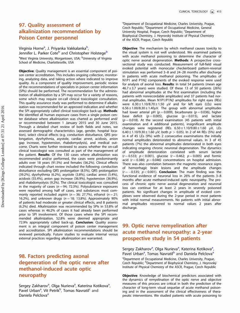
97. Quality assessment ofalkalinization recommendation byPoison Center personnel
Virginia Hornea, J. Priyanka Vakkalankab,Jennifer L. Parker Coteb and Christopher Holstegeb
aWest Virginia University, Morgantown, USA; bUniversity of VirginiaSchool of Medicine, Charlottesville, USA
Objective: Quality management is an essential component of poi-son center accreditation. This includes ongoing collection, monitor-ing, analyzing data, and taking action where indicated to improvequality. As a component of quality improvement, periodic reviewof the recommendations of specialists in poison center information(SPIs) should be performed. The recommendation for the adminis-tration of alkalinization by a SPI may occur for a variety of reasons,some which may require on-call clinical toxicologist consultation.This quality assurance study was performed to determine if alkalin-ization was recommended for an approved indication and whetherthe SPI appropriately called clinical toxicologist back-up. Methods:We identified all human exposure cases from a single poison cen-ter database where alkalinization was charted as performed and/or recommended between 1 January 2015 and 30 June 2015.Through a detailed chart review of both fields and notes, weassessed demographic characteristics (age, gender, hospital loca-tion), select clinical effects (e.g. conduction disturbance, QRS pro-longation, dysrhythmia, asystole, cardiac arrest, acidosis, aniongap increase, hypotension, rhabdomyolysis), and medical out-come. Charts were further reviewed to assess whether the on-callclinical toxicologist was consulted as part of the management ofthe patient. Results: Of the 130 cases where alkalinization wasrecommended and/or performed, the cases were predominantlyadults over 18 years (91.5%) and females (56.2%). Clinical effectsindicated through chart review included the following: conductiondisturbance excluding QRS prolongation (8.5%), QRS prolongation(39.2%), dysrhythmia (6.2%), asystole (3.8%), cardiac arrest (5.4%),acidosis (55.4%), anion gap increase (36.9%), hypotension (36.9%),and rhabdomyolysis (9.2%). The clinical toxicologist was consultedin the majority of cases (n¼ 94; 72.3%). Polysubstance exposureswere reported among half of cases, and substances most com-monly reported included aspirin (n¼ 36; 27.7%), ethanol (n¼ 21;16.2%), and unknown drugs (n¼ 18; 13.8%). Approximately 90%of patients had moderate or greater clinical effects, and 8 patients(6.2%) died. Alkalinization was recommended by SPIs in 53.8% ofcases whereas in 46.2% of cases it had already been performedprior to SPI involvement. Of those cases where the SPI recom-mended alkalinization, 52.8% were deemed appropriate and72.9% appropriately called back-up. Conclusion: Quality assess-ment is an integral component of poison center managementand accreditation. SPI alkalinization recommendations should bereviewed periodically. Future studies to evaluate internal versusexternal practices regarding alkalinization are warranted.
98. Factors predicting axonaldegeneration of the optic nerve aftermethanol-induced acute opticneuropathy
Sergey Zakharova, Olga Nurievaa, Katerina Kotikovaa,Pavel Urbana, Vit Petrikb, Tomas Navratilc andDaniela Pelclovaa
aDepartment of Occupational Medicine, Charles University, Prague,Czech Republic; bDepartment of Occupational Medicine, GeneralUniversity Hospital, Prague, Czech Republic; cDepartment ofBiophysical Chemistry, J. Heyrovsk�y Institute of Physical Chemistryof the ASCR, Prague, Czech Republic
Objective: The mechanism by which methanol causes toxicity tothe visual system is not well understood. We examined patientswith acute methanol poisoning to determine the character ofoptic nerve axonal degeneration. Methods: A prospective cross-sectional study was conducted. Measurement of full-field visualevoked potential with monocular checkerboard pattern-reversalstimulation was performed 3–8 and 24–28 months after dischargein patients with acute methanol poisoning. The amplitudes ofN1P1 and P1N2 components of the evoked response were usedfor analysis of axonal loss. Results: In total 54 patients (mean age46.7 ± 3.7 years) were studied. Of these 13 of 50 patients (26%)had abnormal amplitudes at the first examination (including thepatients with nonrecordable amplitudes) and 37 patients had nor-mal amplitudes. Mean N1P1/P1N2 amplitudes for right eyes (REs)were 6.30 ± 1.10/8.70 ± 1.50 mV and for left eyes (LEs) were6.56 ± 1.00/8.30 ± 1.40mV. The group with abnormal amplitudeshad lower arterial pH (p¼ 0.009), bicarbonate (p¼ 0.036), higherbase deficit (p¼ 0.005), glucose (p¼ 0.015), and lactate(p¼ 0.018). At the second examination (45 patients with initialexamination and 4 additional patients), insignificant amplitudechanges were registered (REs 6.50 ± 1.10/9.80 ± 1.60 mV, LEs6.40 ± 1.10/9.30 ± 1.60 mV; both p > 0.05). In 2 of 44 REs (5%) andin 4 of 45 LEs (9%) with 2 consecutive examinations the initiallynormal amplitudes deteriorated to abnormal values. In 3 of 45patients (7%) the abnormal amplitudes deteriorated in both eyesindicating ongoing chronic neuronal degeneration. The dynamicsof amplitude deterioration correlated with serum lactate(r¼ 0.533; p< 0.001), glucose (r¼ 0.462; p¼ 0.005) and formicacid (r¼ 0.380; p¼ 0.046) concentrations on hospital admission.There was also correlation between the magnetic resonance signsof hemorrhagic brain lesions and the amplitude changes(r¼�0.535; p< 0.001). Conclusion: The main finding was thefunctional evidence of neuronal loss in 26% of the patients 3–8months after acute methanol-induced optic neuropathy. The pro-cess of chronic optic nerve axonal degeneration and neuronalloss can continue for at least 2 years in severely poisonedpatients. No significant changes in amplitude of evoked com-plexes were observed during the 2-year period in most patientswith initial normal measurements. No patients with initial abnor-mal amplitudes recovered to normal values 2 years afterdischarge.
99. Optic nerve remyelination afteracute methanol neuropathy: a 2-yearprospective study in 54 patients
Sergey Zakharova, Olga Nurievaa, Katerina Kotikovaa,Pavel Urbana, Tomas Navratilb and Daniela Pelclovaa
aDepartment of Occupational Medicine, Charles University, Prague,Czech Republic; bDepartment of Biophysical Chemistry, J. Heyrovsk�yInstitute of Physical Chemistry of the ASCR, Prague, Czech Republic
Objective: Knowledge of biochemical predictors associated withthe dynamics of remyelination of the optic nerve and objectivemeasures of this process are critical in both the prediction of thecharacter of long-term visual sequelae of acute methanol poison-ing and in the assessment of the clinical effectiveness of thera-peutic interventions. We studied patients with acute poisoning to
CLINICAL TOXICOLOGY 413
Dow
nloa
ded
by [
Kin
gs C
olle
ge L
ondo
n] a
t 07:
31 2
1 A
pril
2016
determine the prevalence and dynamics of optic nerve remyelina-tion after methanol-induced optic neuropathy. Methods: A pro-spective study in patients with acute methanol poisoning.Measurement of full-field visual evoked potential with monocularcheckerboard pattern-reversal stimulation was performed 3–8 and24–28 months after discharge from hospital. The latency of thepositive peak (P1) was used for the analysis of remyelinationdynamics. Results: In total 54 patients (mean age 46.7 ± 3.7 years)were studied. Of these, 27 patients had abnormal P1 latencies.Mean P1 latency for right eyes (REs) was 115.3 ± 2.1 ms and forleft eyes (LEs) was 117.4 ± 3.2 ms. The group with abnormallatency had lower arterial pH (p¼ 0.017), higher anion gap(p¼ 0.013), methanol concentration (p¼ 0.027), base deficit(p¼ 0.033) and lactate (p¼ 0.048). At the second examination,shortening of P1 latencies was registered (REs/LEs 98.3 ± 2.4/102.7 ± 4.5 ms; p< 0.001). The dynamics of latency shortening forREs/LEs were 17 ± 1.3/15.1 ± 3.1 ms, with insignificant inter-eye dif-ference (p¼ 0.271). The dynamics of remyelination correlated withserum methanol (r¼�0.588; p< 0.001), arterial pH (r¼ 0.339;p¼ 0.040), and concentration of carbohydrate-deficient transferrin(r¼�0.411; p¼ 0.011). Remyelination occurred in cases of mild ormoderate damage of myelin sheaths; no improvement of conduc-tion was found in severe cases. Conclusion: The dynamics ofremyelination correlated with the degree of acidosis and severityof poisoning. Chronic alcohol abuse had a negative effect onremyelination dynamics.
101. Efficacy of isosorbide dinitrate asan antidote for cyanide poisoning inrabbits depending on the timing oftreatment
Ophir Lavona, Amit Avrahamib and Arik Eisenkraftc
aClinical Pharmacology and Toxicology Unit, Carmel Medical Center,Haifa, Israel; bPre Clinical Research Authority, Technion-IsraelInstitute of Technology, Haifa, Israel; cFaculty of Medicine, HebrewUniversity, Jerusalem, Israel
Objective: To evaluate the efficacy of isosorbide dinitrate as anantidote for cyanide poisoning in rabbits and the effect of thetiming of treatment. Methods: A comparative in vivo study usingmale 3000 ± 200 g New Zealand White rabbits, randomly dividedinto 4 groups. The rabbits were poisoned by intravenous lethaldoses of potassium cyanide (1 mg/kg); in a previous study wedemonstrated that these cyanide doses caused a high mortality(5/6) within minutes (median 10 min). The animals in the differentgroups were treated intravenously with isosorbide dinitrate at adose of 50 mcg/kg at 1, 3, 5 or 7 minutes after the poisoning,respectively. The primary outcome was short-term survival up to30 minutes. Other outcomes that were examined included time todeath, clinical score, mean blood pressure, pulse, blood pH, andblood lactate concentrations. Results: There were 24 rabbits in 4groups of 6. All animals (6/6) in the group treated 1 minute afterpoisoning survived. Five rabbits out of 6 from each of the othergroups survived. The time to death of rabbits that died (one ineach group treated after 3, 5 and 7 minutes) was within the rangeof 8–13 minutes after poisoning. All animals collapsed shortly afterpoisoning accompanied by disturbed vital signs (blood pressureincreased and heart rate decreased). Lactic metabolic acidosisdeveloped further; average peak blood lactate concentrationswere 15.5–19.0 mmol/L at 10 minutes after poisoning. The treatedrabbits gradually improved in all measured parameters. Recoverywas faster in the animals that were treated 1 and 3 minutes afterpoisoning. Blood pH and lactate concentrations improved slowlycompared to the clinical score and the hemodynamic parameters.
Conclusion: Isosorbide dinitrate improves survival of rabbits pois-oned with lethal doses of potassium cyanide when given even upto 7 minutes after poisoning. Recovery of the animals was fasterwhen treated early after poisoning (1–3 minutes). Delayed recov-ery of the animals treated later is probably secondary to the accu-mulated metabolic injury, mainly lactic acidosis. Isosorbidedinitrate shows potential as an effective antidote for cyanide poi-soning in a realistic time window.
102. Hydrofluoric acid: an in vitroexperiment in a 3D human airwayreconstructed epithelia
Laurence Mathieua, Francois Burghera,Samuel Constantb, Song Huangb, Alan Hallc andJoel Blometa
aPrevor Laboratory, Valmondois, France; bEpithelix Sarl, Geneva,Switzerland; cTCMTS, Denver, USA
Objective: Hydrofluoric acid (HF) is very corrosive and hazardousfor human exposure.[1] Inhalation of hydrofluoric acid can be fatalbut few data are available to understand how hydrofluoric aciddamages lung. Methods: We designed an experiment to explorethe effect of HF on MucilAirTM a unique 3D Human AirwayEpithelia reconstructed in vitro.[2] For the study 100 ml of HF wasapplied on the epithelia for 20 minutes; the concentrations testedwere 0.15, 0.75, 1.5, 7.5, 15, 75 and 150 mM. The following end-points were analyzed: morphology, tissue integrity monitoring(TEER), lactate dehydrogenase (LDH) release quantification, CiliaBeating Frequency (CBF) and histology. Results: For concentra-tions up to 1.5 mM of HF, no toxic effects were detected by allthe endpoints measured. At concentrations of 7.5 mM and above,damage to epithelia were observed. The TEER dropped signifi-cantly, the pseudo-stratified structure was altered and only thebasal cell layer was present. Cilia beating frequency was reduced,however, based on TEER analysis, damage caused by HF below7.5 mM could be repaired 7 days after exposure, and the CBF val-ues recovered despite some variations at earlier time-points,except when HF was used above 75 mM. HF at 75 mM and abovecaused severe damage to epithelia, which was not reversible.Histology clearly showed that the pseudo-laminate structureincluding the three cell types was well maintained for inspectionand treatment with HF at 1.5 mM. HF 15 mM contact induced anabnormal structure with the presence of a single layer of basalcells. This is perfectly in line with the high value of TEER found toDay 7. From 15 to 150 mM strong and non-reversible toxicity wasobserved. Conclusion: The MucilAir
TM
model allows characteristicdamages induced by HF and confirms that a low concentration ofhydrofluoric acid can induce irreversible cellular damage. Themodel could be of interest to evaluate the benefit of early decon-tamination and treatments in further studies.
References
[1] Tsonis L, Hantsch-Bardsley C, Gamelli RL. Hydrofluoric acid inhal-ation injury. J Burn Care Res. 2008;29:852–855.
[2] Baxter A, Thain S, Banerjee A, et al. Targeted omics analyses, andmetabolic enzyme activity assays demonstrate maintenance of keymucociliary characteristics in long term cultures of reconstitutedhuman airway epithelia. Toxicol In Vitro. 2015;29:864–875.
414 ABSTRACTS
Dow
nloa
ded
by [
Kin
gs C
olle
ge L
ondo
n] a
t 07:
31 2
1 A
pril
2016
103. Hyperbaric oxygen protectsneurotrophic activity of carbonmonoxide-exposed astrocytes
Jure Levstika, Miran Brvarb, Dusan Suputc andDamijana M. Jurica
aInstitute of Pharmacology and Experimental Toxicology, Faculty ofMedicine, Ljubljana, Slovenia; bCentre for Clinical Toxicology andPharmacology, University Medical Centre Ljubljana, Ljubljana,Slovenia; cInstitute of Pathology, Faculty of Medicine, Ljubljana,Slovenia
Objective: Carbon monoxide (CO) causes neuronal and glialapoptosis that can result in delayed neurological symptoms.Recently we reported that CO/normoxia caused a progressivedecline of viability and mitochondrial function accompanied bycaspase and calpain activation.[1] Impairment in astrocyte func-tion was reduced time-dependently by hyperbaric, not normo-baric, oxygen. Due to the central role of astrocytes in maintainingneuronal function by offering neurotrophic support we investi-gated the toxic effects of CO/normoxia on intrinsic neurotrophicactivity in these cells and evaluated possible protective influenceof oxygen treatment against CO poisoning. Methods: Cultured ratastrocytes were exposed to 3000 ppm CO in air for different timeperiods (0.5–24 hours) followed by 24 hours of normoxia.Following an 8 hour exposure to CO that significantly affectedastrocytic cellular function the cultured cells were exposed during24 hours of normoxia for 1 hour in different time periods(0–7 hours) after CO to 100% normobaric oxygen (NBO) or 100%oxygen at a pressure of 3 bar (HBO). Real-time polymerase chainreaction (PCR) was performed to examine the expression of nervegrowth factor (NGF), brain-derived neurotrophic factor (BDNF) andneurotrophin-3 (NT-3). Specific two-site enzyme immunoassayswere utilized to determine protein synthesis and secretion of theexamined neurotrophins. Results: CO/normoxia caused a progres-sive decline of gene expression, synthesis and secretion of NGF,BDNF and NT-3 with different intensity. Maximal responseoccurred after 8 hours in CO. Subsequent 1 hour treatment withoxygen disclosed pressure- and time-dependent efficacy in restor-ing astrocytic neurotrophic activity. The protective effect was evi-dent when cells were exposed to HBO 1–5 hours after CO but notwhen exposed to HBO immediately after CO exposure. A dimin-ished efficiency of HBO in enhancement of neurotrophin synthesiswas observed 7 hours after CO exposure. In contrast, NBO had noprotective influence on CO-poisoned cells. Conclusion: The neuro-protective role of oxygen therapy in CO-exposed astrocytes ispressure- and time-dependent. In addition to preventing mito-chondrial dysfunction and apoptotic processes our results indicatethat HBO, but not NBO, restores astrocytic neurotrophic supportthat may dictate the short- and long-term neuronal survival andthe maintenance and retraction of synaptic connections. To pre-vent the occurrence of late neuropsychological sequelae ourstudy opens the way to consider time and pressure regimen ofoxygen therapy in clinical management of CO poisoning.
References
[1] Juric DM, Finderle �Z, �Suput D, et al. The effectiveness of oxygentherapy in CO poisoning is pressure- and time-dependent.Toxicol Lett. 2015;233:16–23.
104. A novel, fluorescence-basedmethod to detect effects of novelpsychoactive substances (NPS) onneurotransmitter re-uptaketransporters
Laura Hondebrinka, Anouk Verbovenb,Gina GDM Van Kleefb, Jan Meulenbelta,b,c andRemco HS Westerinkb
aNational Poisons Information Center, University Medical CenterUtrecht, Utrecht, The Netherlands; bNeurotoxicology ResearchGroup, Institute for Risk Assessment Sciences, Utrecht University,Utrecht, The Netherlands; cDepartment of Intensive Care Medicine,University Medical Center Utrecht, Utrecht, The Netherlands
Objective: Prevalence of drug use, including novel psychoactivesubstances (NPS), is high and new substances are emergingyearly. Most drugs are well-known to affect the function of neuro-transmitter reuptake transporters. Often, this is investigated usingradioactive assays, requiring specific laboratory facilities. As thedemand for rapid methods to investigate the potency of substan-ces affecting neurotransmission is increasing, we used a novelfluorescence-based high-throughput method to investigate effectsof NPS on re-uptake transporters for dopamine (DAT), norepin-ephrine (NET), and serotonin (SERT). Methods: Human embryonickidney cells (HEK 293, kindly provided by Roche) expressinghuman neurotransmitter re-uptake transporters (hDAT, hNET orhSERT) were used to determine the effect on neurotransmitteruptake during a 30 minute exposure to several drugs and meas-ure their half maximal inhibitory concentration (IC50). Frequentlyused common drugs and NPS were selected: cocaine, amphet-amine, 4-fluoroamphetamine, methoxetamine, para-methoxyme-thamphetamine (PMMA), 2C-B, 2C-B-NBOMe and 2C-I-NBOMe.Uptake was determined using a fluorescent substrate that mimicsdopamine, norepinephrine and serotonin. Uptake of the substrateincreases intracellular fluorescence, which was measured overtime with a microplate reader. Results: All drugs inhibited mono-amine transporter function in a concentration-dependent manner.hDAT and hNET were most potently inhibited by cocaine,amphetamine and 4-fluoroamphetamine (IC50 �1–5 mM), while2C-B was a weak inhibitor (IC50 >100 mM). hSERT was mostpotently inhibited by cocaine, methoxetamine and the NBOMes(IC50 �2–5 mM), while amphetamine and 4-fluoroamphetaminewere weak inhibitors. PMMA strongly inhibited hNET (IC50 6 mM)and less potently inhibited hDAT and hSERT (IC50 43 and >100mM). Conclusion: Our data show that the fluorescence-basedassay can be used to rapidly determine effects of substances onthe function of neurotransmitter reuptake transporters, withoutthe need of a specially equipped laboratory. In addition, thismethod allows for measurements over time, in contrast to end-point measurements. This can be useful to investigate the effectof successive exposures, for example, when examining a candi-date drug for undue drug-induced transporter effects.
CLINICAL TOXICOLOGY 415
Dow
nloa
ded
by [
Kin
gs C
olle
ge L
ondo
n] a
t 07:
31 2
1 A
pril
2016
105. Variability of neurobehavioraltoxicity of naphyrone, a new syntheticcathinone, with acute or bingeadministration
Nadia Benturquiaa, Camille Gamblina,Lucie Chevillarda, Olivier Roussela, Patricia Risedea,Jacques Calleberta, Matthias E. Liechtib andBruno M�egarbanea
aINSERM U1144, Paris-Descartes University, Paris, France; bDivisionof Clinical Pharmacology and Toxicology, University Hospital Baseland University of Basel, Basel, Switzerland
Objective: Naphyrone is a new psychoactive substance and acathinone derivate. It was reported to be used by approximately2% of recreational drug users and is frequently present in ‘‘bathsalts’’. Scarce data are available regarding its toxicity, pharmaco-kinetics and effects on brain monoamines. Our objectives were tostudy naphyrone-related effects on behavior and brain mono-amine content in mice according to two administration modalities(acute and binge) mimicking its use in humans. Methods: Anexperimental study in Swiss mice of naphyrone-induced effectsafter acute and repeated administration (binge) on the locomotoractivity, anxiety (openfield), resignation (forced swimming), mem-ory (Y-maze), hedonic status (sucrose consumption) and investiga-tion of naphyrone-induced effects on monoamines in theprefrontal cortex. Plasma naphyrone concentrations were meas-ured using high performance liquid chromatography-mass spec-trometry (HPLC-MS) and brain monoamine concentrations usingHPLC coupled to fluorometry. For each animal and each time, wecalculated the difference between the parameter value at thattime and baseline and the area under the curve of its timecourse. Comparisons were performed using two-way ANOVA fol-lowed by post-tests using Bonferroni correction. Pharmacokinetics(PK) was modeled and parameters calculated using WinNonlinVR
software. Results: Naphyrone-induced dose-dependent stimula-tion of locomotor activity that appeared more marked and pro-longed than methylenedioxypyrovalerone (MDPV)- and cocaine-related effects (used as positive controls), additionally increasingafter binge administration (p< 0.001). Significant increase in thedistance walked at the periphery of the openfield was observedup to 24 hours post-injection (p< 0.001), corresponding to thebehavior compensation of enhanced fear. During binge adminis-tration (3 times per day, 3 successive days), increased locomotoreffects at day 3 (after the 9th injection) in comparison to day 1(after the 1st injection) supported naphyrone-induced hyper-sensi-tization process. Similarly, significant effects were observed onmice depression (p< 0.0001) and memory (p< 0.005) but not onhedonic status. Significant dose-dependent increase in aggres-sively social relationships was also reported among naphyrone-treated mice (p< 0.001). The neurochemical study revealed signifi-cant increase in dopamine and norepinephrine concentrations inthe prefrontal cortex, without significant modifications in sero-tonin concentrations. This monoamine profile was similar afterrepeated naphyrone administration. The naphyrone pharmacoki-netic profile was described after acute and binge administrationand effects correlated to plasma concentrations. Conclusion:Neurobehavioral disorders induced by acute and repeated naph-yrone administration mainly consist of the stimulation of loco-motor activity. The increase in the cortical dopamineconcentrations may suggest an addictive potential that should befurther investigated.
106. Acute behavioral toxicity andaddictive liability of the syntheticcathinone3,4-methylenedioxypyrovalerone(MDPV) in the rat
Nadia Benturquia, Cyril Masniere, Lucie Chevillard,Olivier Roussel, Patricia Risede, Jacques Callebert andBruno M�egarbaneINSERM U1144, Paris-Descartes University, Paris, France
Objective: 3,4-Methylenedioxypyrovalerone (MDPV), a syntheticcathinone with toxicity and high abuse potential, has beenincreasingly used over the last five years. To date, few experimen-tal studies have investigated MDPV-related behavioral effects. Westudied MDPV-related neurobehavioral effects in rats in variousmodalities of administration, mimicking human use. Methods: Anexperimental study in Sprague-Dawley rats investigating MDPV-induced effects on the locomotor activity, anxiety (openfield), res-ignation (forced swimming), memory (Y-maze), hedonic status(sucrose consumption) and brain neurochemistry (monoaminescontent in the prefrontal cortex), using three different administra-tion patterns including acute (3 mg/kg intraperitoneal injection[IP]), binge-like (MDPV 3 mg/kg IP, 3 times per day, 3 days) andprolonged treatment (MDPV 1 mg/kg IP, 10 days). Plasma concen-trations of MDPV and its two major metabolites were measuredusing liquid chromatography coupled to high-resolution massspectrometry. Brain monoamine concentrations were measuredusing high-performance liquid chromatography (HPLC) coupled tofluorometry. For each animal and each time, we calculated thedifference between the parameter value at that time and baselineand the area under the curve of its time course. Comparisonswere performed using two-way ANOVA followed by post-testsusing Bonferroni correction. Results: Acute MDPV administrationwas responsible for locomotor hyperactivity with onset of stereo-typies, significant decrease in anxiety, anhedonic status, andmemory impairment (p< 0.05 for each neurobehavioral measure-ment). Following binge-like MDPV administration, tolerance toimmediate locomotor effects after drug withdrawal and significantdecrease in anxiety, hedonic status and food consumption wereobserved (p< 0.05 for each neurobehavioral measurement).Increase in the prefrontal cortex serotonin concentrationsoccurred, in accordance with the observed behavioral effects.Prolonged MDPV administration resulted in tolerance develop-ment after one week of drug withdrawal. Conclusion: Our resultssuggest MDPV-induced marked neurobehavioral effects in the ratwith the development of tolerance after binge-like and prolongedrepeated administration.
107. A bifunctional and peptide-basedopioid agonist-nociceptin antagonistligand for treatment of pain: is thebenefit-to-neurorespiratory toxicityratio improved compared to the usualopioids?
Camille Lagarda, Lucie Chevillarda, Patricia Risedea,Bruno M�egarbanea and Steven Balletb
416 ABSTRACTS
Dow
nloa
ded
by [
Kin
gs C
olle
ge L
ondo
n] a
t 07:
31 2
1 A
pril
2016
aINSERM U1144, Pars-Descartes University, Paris, France;bDepartment of Organic Chemistry, Vrije Universiteit Brussel,Brussels, Belgium
Objective: Pain is a key public health concern, affecting one infive adults across Europe (ca. 75 million people). To date, mor-phine and tramadol are the two major agents used to treat acuteand neuropathic pain, respectively. In order to simultaneouslyaddress these two types of pain, a bifunctional hybrid (KGNOP1)was developed associating a peptide-based opioid agonist(KGOP1) to a nociceptin antagonist. Our objective was to investi-gate KGNOP1-induced anti-nociceptive and anti-hyperalgesiceffects in comparison to its related respiratory effects in Sprague-Dawley rats, focusing on determining their time-course and dose-effect relationships in comparison to the reference treatments.Methods: The anti-nociceptive effects were determined using thehot plate (kept at a temperature of 52 �C, maximum latencyperiod to paw lick or paw brisk shaking set at 60 s), the anti-hyperalgesic effects using the cold plate (kept at a temperature of4 �C, number of paw withdrawal in rats with chronic constrictioninjury-induced neuropathic pain) and the respiratory effects usingplethysmography (measurement of inspiratory and expiratorytimes and tidal volume). We determined the dose-effect relation-ships for each tested drug (KGNOP1, KGOP1, morphine, tramadol,and solvent) according to the investigated effect. To permit thesimultaneous analysis of the effect of time and treatments on thedifferent parameters, we calculated for each animal and for eachstudied parameter, the area under the curve (AUC) from T0 to thecompletion of the measurement. For each parameter, we com-pared the AUCs using Kruskal-Wallis tests between the studiedgroups. The effective doses 50% (ED50) were determined usingsigmoidal modeling. Results: KGNOP1 was responsible for dose-dependent anti-nociceptive effects more marked and lasting lon-ger than morphine (p< 0.05), initial and prolonged anti-hyper-algesic effects in comparison to tramadol (p< 0.05), but dose-dependent increase in inspiratory time greater than morphine(p< 0.05). Additionally, the respiratory depression induced byKGNOP1 was more marked than with its opioid part, KGOP1,alone (p< 0.05). The ED50 regarding the anti-nociceptive effectswere calculated at 0.35 mmol/kg with KGNOP1 and 14.7 mmol/kgwith morphine while ED50 regarding the respiratory effects wereat 0.43 mmol/kg with KGNOP1 and 16.2 mmol/kg with morphine,allowing attribution to KGNOP1 of a poor therapeutic margin of1.23, similar to morphine. Conclusion: KGNOP1-induced anti-noci-ceptive and anti-hyperalgesic activities are greater than those ofthe tested reference treatments; however KGNOP1-related respira-tory toxicity appears too important to allow its consideration forany therapeutic application.
108. The disposition of cocaine and itsmetabolites in biological matricesfollowing controlled administrationin volunteers
Paul I. Dargana, Eleanor L. Menziesb,John R. H. Archerc, Takahiro Yamamotoc,Simon Elliotd, Dene Baldwine, Mark C. Parkinb,David M. Wooda and Andrew Kicmanb
aClinical Toxicology, Guy’s and St Thomas’ NHS Foundation Trustand King’s Health Partner’s and Faculty of Life Sciences andMedicine, King’s College London, London, UK; bFaculty of LifeSciences and Medicine, King’s College London, London, UK;cClinical Toxicology, Guy’s and St Thomas’ NHS Foundation Trust
and King’s Health Partner’s, London, UK; dROAR Forensics, Malvern,UK; eAlere Toxicology, Loughton, UK
Objective: Previous studies describing the pharmacokinetics ofcocaine and its metabolites focus on plasma and urine sampleanalysis. There are limited data on cocaine/metabolite pharmaco-kinetics and disposition in whole blood, oral fluid, sweat and/orhair. Analysis of these matrices is particularly relevant to forensicinvestigations and workplace drug-testing as they are easy tostore and transport. The study aimed to describe the dispositionand pharmacokinetics of cocaine and its metabolites in thesematrices. Methods: After informed consent, 7 healthy drug-naivemale volunteers (25–40 years) insufflated a single 100 mg dose ofpharmaceutical-grade cocaine hydrochloride. Whole blood,plasma, oral fluid and palm sweat were sampled regularly overthe first 6 hours and on days 2 and 3. Head hair samples weretaken at baseline and 30, 60 and 90 days. Samples were stored at-20 �C until analysis using ultra-performance liquid chromatog-raphy tandem mass spectrometry (UPLC-MS/MS) for cocaine, ben-zoylecognine (BZE), ecgonine methyl ester (EME); hair sampleswere also analysed for norcocaine. Results: Cocaine was detectedin whole blood, plasma and oral fluid samples at the first collec-tion (5 mins) and BZE/EME in samples collected at the second col-lection (10 mins). There was a significant correlation betweenwhole blood and plasma cocaine for BZE and EME (r2
0.860–0.963, p< 0.0001). Calculated maximum serum concentra-tion (Cmax) for cocaine was 417.1 ng/mL in whole blood and450.3 ng/mL in plasma. The time to reach the maximum serumconcentration (Tmax) was 21 and 27 minutes, respectively, withhalf-life 1.3 and 1.5 hours, respectively. Calculated Cmax for BZEwas 386.1 ng/mL (whole blood) and 414.1 ng/mL (plasma); Tmaxwas 145 and 180 minutes, respectively. Calculated Cmax for EMEwas 135.1 ng/mL (whole blood), 118.8 ng/mL (plasma); Tmax 137and 120 minutes, respectively. Mean Cmax whole blood:plasmaratios were 0.90 for cocaine, 0.94 for BZE and 1.13 for EME.Cocaine and BZE were first detected in palm sweat 1 hour aftercocaine administration and EME at 2 hours; they remained detect-able for 24 and 48 hours for EME and cocaine/BZE, respectively.Cocaine, BZE, EME and norcocaine were detected in hair samplesat all time points. Conclusion: The whole blood:plasma cocaineand metabolite ratios neared unity. Whole blood/plasma cocaineand metabolite kinetics suggest that both matrices are suitablefor forensic and clinical sampling. The time course of detection ofcocaine/metabolites in oral fluid and palm sweat will aid interpret-ation of forensic and work-place testing. Detection of norcocainein hair samples confirms endogenous cocaine/metabolite inclu-sion into hair rather than surface contamination and ex vivometabolism.
109. Polyvinylpyrrolidone-coated ironoxide nanoparticles (PVP-Fe3O4NPs):toxicological profile evaluation inhuman brain cells
Uliana De Simonea, Carlo A Locatellib, Davide Lonatib,Lenin Ram�ırez-Candoc and Teresa CocciniaaLaboratory of Experimental and Clinical Toxicology, ToxicologyUnit, IRCCS Maugeri Foundation Hospital of Pavia, Pavia, Italy;bPoison Control Centre and National Toxicology Information Centre,IRCCS Maugeri Foundation Hospital and University of Pavia, Pavia,Italy; cCentro De Investigacion Y Valoracion De La Biodiversidad(Civabi), Universidad Politecnica Salesiana, Quito, Ecuador
CLINICAL TOXICOLOGY 417
Dow
nloa
ded
by [
Kin
gs C
olle
ge L
ondo
n] a
t 07:
31 2
1 A
pril
2016
Objective: Magnetic iron oxide nanoparticles (IONPs) as magnet-ite (Fe3O4NPs) have attracted extensive interest due to theirsuperparamagnetic physicochemical properties in biomedical (i.e.brain-targeted drug, magnetic resonance imaging, contrastagents) and industrial fields (i.e. audio speakers, position sensing,water purification). Fe3O4NPs can reach the central nervous sys-tem (CNS) independent of administration route. Considering therole of astrocytes in iron (Fe) homeostasis and protection of braincells against metal toxicity, exposure to Fe3O4NPs may be a riskdue to Fe ion release leading to a disruption of normal ironmetabolism/homeostasis in the brain - a characteristic hallmarkresembling that of several neurodegenerative disorders. Methods:Effects induced by polyvinylpyrrolidone (PVP)-Fe3O4NPs (Ø 20 nm)were assessed on D384 astrocyte and SH-SY5Y neuronal cells after4, 24 and 48 hours at increasing concentrations (1–100 lg/mL) byevaluating the following biological functions: mitochondrial activ-ity (MTT assay); cell membrane integrity (calcein-AM/PI-staining);cell morphology (phase-contrast microscopy); and intracellularPVP-Fe3O4NP accumulation (Prussian blue-staining). Results: PVP-Fe3O4NPs induced dose- and time-dependent alterations of mito-chondrial function in D384; 25–30% cell viability decrease wasobserved early (4 h) from 25 to 100 mg/mL. Cytotoxicity was morepronounced after prolonged exposure with 35–55% cell deathafter 24 h (10–100 lg/mL) and 25–75% after 48 h (1–100 mg/mL).SH-SY5Y were less susceptible than D384. Cytotoxicity wasobserved after 48 h only with 35–45% viability decrease (10–100mg/mL). No effect on membrane integrity was observed in bothCNS cell types at all doses and time-points considered. D384showed morphological alterations (roundish cells) at the highestdoses (50–100 lg/ml) after 48 h, while no effect was observed inSH-SY5Y. Dose- and time-dependent PVP-Fe3O4NP accumulationwas detected in both cell types. Blue spots in D384 were visibleat �10 lg/mL after 4 h and increased at higher doses (25–100 lg/mL), and in SH-SY5Y they were detected at �25 lg/mL.Conclusion: PVP-Fe3O4NPs alter mitochondrial function only, withastrocytes being more susceptible than neurons. Critical doses are25 lg/mL after 4 h and 1 lg/mL after 48 h for D384 and 10 lg/mLafter 48 h for SH-SY5Y. Fe accumulates in a dose- and time-dependent manner mainly in astrocytes. In summary, mitochon-drial effects of PVP-F3O4NPs in cerebral cells could raise healthconcerns based on the possibility that cellular IONP overload maytrigger adverse cellular responses by releasing Fe ions (especiallyin astrocytes) compromising the normal functions of the CNS.Acknowledgements: Italian Ministries of Health, Research andEducation; and ENI S.p.A. Div-R&M.
110. Lack of symptoms following alarge combined carbamazepine andquetiapine overdose: the role ofenzyme induction by carbamazepine
Søren Bøgevig and Tonny S. PetersenDepartment of Clinical Pharmacology, Copenhagen UniversityHospital Bispebjerg and Frederiksberg, Copenhagen, Denmark
Objective: Carbamazepine is a potent inducer of cytochromeP450 (CYP) 3A4 and P-glycoprotein. Induction is known to reduceplasma concentrations of other CYP3A4 metabolized drugs whenthese are taken in normal therapeutic doses. We report the sameeffect following an acute overdose of two CYP3A4 metabolizeddrugs taken in large quantities. Case report: A 32-year-old femalewith a year-long history of anorexia presented to the emergencydepartment 2 hours after an acute overdose of 14 g carbamaze-pine and 10.8 g quetiapine. Past medical history included myx-edema, epilepsy and depression and medications were
levothyroxine (150 mcg/day), carbamazepine (300 mg/day) andquetiapine (600 mg/day). Overdose was reported by the patientand confirmed by psychiatric contact personnel. At the time ofpresentation she was without symptoms, including normal elec-trocardiogram (ECG). She was treated with one dose of activatedcharcoal and admitted to the cardiology department for cardiacmonitoring and close observation. The Danish Poison InformationCenter was consulted, and advice was given to expect severesymptoms and to administer multiple doses of activated charcoal(MDAC). No symptoms developed within 48 hours of observation.Plasma quetiapine concentrations 6.5, 14.5 and 20 hours afteringestion were 230, 27 and <13 nmol/L (therapeutic range50–650 nmol/L), respectively, giving an estimated half-life of2.5 hours (normal range 4.9–8.4 hours).[1] Serum carbamazepineconcentrations 6.5, 10.5, 14.5, 20 and 35 hours after ingestionwere 37, 33, 21, 16 and 7 mmol/L (therapeutic range 20–50 mmol/L), respectively, giving an estimated half-life of 11.5 hours (normalrange 10–20 hours). Conclusion: The carbamazepine-inducedCYP3A4 activity may have markedly reduced the intoxicationsymptoms in this case of a confirmed large overdose of carba-mazepine and quetiapine. Both peak concentration and the half-life of quetiapine were markedly reduced and quetiapine neverreached toxic plasma concentrations, even with this large dose.The MDAC may have contributed to the lower peak concentrationof both drugs, but the rapid elimination, low bioavailability andlack of symptoms following the large quetiapine ingestion is likelyto be a consequence of carbamazepine CYP3A4 induction.[2]
References
[1] Isbister GK, Friberg LE, Hackett LP, et al. Pharmacokinetics of que-tiapine in overdose and the effect of activated charcoal. ClinPharmacol Ther. 2007;81:821–827.
[2] Grimm SW, Richtand NM, Winter HR, et al. Effects of cytochromeP450 3A modulators ketoconazole and carbamazepine on quetia-pine pharmacokinetics. Br J Clin Pharmacol. 2006;61:58–69.
111. Acrylamide poisoning withlong-term neurological consequences
Antonio Due~nas-Laitaa, Jose Luis Perez-Castrillona,Pere Sanz-Gallenb, Beatriz Mart�ın-Pereza andMarta Ruiz-Mambrillac
aRio Hortega University Hospital and University of Valladolid,Valladolid, Spain; bUniversity of Barcelona, Barcelona, Spain;cUniversity of Valladolid, Valladolid, Spain
Objective: Acrylamide and its polymer, polyacrylamide, are widelyused in the water treatment, papermaking, electrophoresis gels,dyes, cosmetics and textile industries. International organizationshave classified acrylamide as a potential carcinogenic and neuro-toxic agent.[1,2] Case report: A 31-year-old woman with no rele-vant personal history or toxic habits, accidentally inhaledacrylamide at work. Immediately after exposure, she suffered a45-minute episode of dizziness, loss of strength, tingling in thearms and feet, blurred vision, slurred speech, drowsiness andrespiratory difficulties. The patient was treated at the emergencydepartment of a nearby hospital, remaining under observation for48 hours with monitoring, fluids and oxygen therapy. After dis-charge, she continued to suffer from progressive deterioration fortwo years, with asthenia, headache, cognitive disorders, dizziness,hypoesthesia, paresthesia, dry eye and visual accommodationproblems. Study by the Clinical Toxicology Unit, University ofValladolid found symptoms compatible with cerebellar ataxiaassociated with bitemporal hemianopsia. The panel of tests
418 ABSTRACTS
Dow
nloa
ded
by [
Kin
gs C
olle
ge L
ondo
n] a
t 07:
31 2
1 A
pril
2016
performed included: standard biochemistry, uric acid, lactatedehydrogenase, creatine kinase, total proteins, C-reactive protein,rheumatoid factor, ammonia, glutamic acid, hormones (PTH, TSH,T3, T4, cortisol, FSH, LH, ACTH, prolactin, progesterone, testoster-one), antibodies (ANA, anti-SSA, anti-SS, anti-transglutaminaseantibodies, ANCA, anti-Yo, anti-Hu, anti-Tr, anti CCDI antibodies),vitamins (25-hydroxyvitamin D, vitamin B902, folic acid, vitamin E),serology (herpes simplex, cytomegalovirus, Epstein Barr, Borrelia,human immunodeficiency virus, human T-lymphotropic virus type1), tumor markers (CEA, CA12.5, CA 19.9, alpha fetoprotein), met-als (copper, lead), ceruloplasmin, angiotensin converting enzyme(ACE) and urine (fractionated porphyrins, mercury, and lead). Allresults were negative or within normal parameters. Brain andspine computerised tomography and magnetic resonance imag-ing with and without contrast, magnetic resonance myelographyand electromyography showed no significant alterations.Conclusion: The cause-effect relationship between exposure toacrylamide and the onset of symptoms and the absence of anidentifiable organic cause suggests the neurological damage inthis case was due to acrylamide poisoning.
References
[1] Exon JH. A review of the toxicology of acrylamide. J ToxicolEnviron Health B Crit Rev. 2006;9:397–412.
[2] Pennisi M, Malaguarnera G, Puglisi V, et al. Neurotoxicity of acryl-amide in exposed workers. Int J Environ Res Public Health.2013;10:3843–3854.
112. The challenge of unknownchemical exposure
Dong-Zong Hung and Kai-Wei YangDivision of Toxicology, China Medical University Hospital, ChinaMedical University, Taichung, Taiwan
Objective: Chemical injury on skin is not always obvious. Unlikestrong acids, hydrofluoric acid burns do not present with appar-ent coagulum, but the acid penetrates deeply into tissues tocause severe injuries.[1,2] Case report: A 35-year-old male pre-sented with a painful skin rash on the legs two hours after beingsplashed with a clear fluid while cycling past a street populatedby small-sized factories. The pH of the unknown liquid on hisjeans was measured to be 1 using litmus paper. He was managedas for an ordinary acid corrosive injury and the burn was dressedwith antibiotic ointment, however he later developed severe localpain, deep into bone, and after toxicology consultation it wasthought that the fluid could have been hydrofluoric acid. Thepatient had some improvement after local application of calciumgluconate, but the wound required repeated surgical debridementand skin grafting due to extensive necrotic lesions. Fluoride ionwas detected in the jeans by ion exchange chromatography.Conclusion: Emergency physicians should be aware that concen-trated hydrofluoric acid can induce deep tissue destruction dueto penetration of fluoride ion, but does not burn clothes due toits weak acidity. These combined characters might be the clue fordifferential diagnosis of acid corrosive injury.
References
[1] Dinis-Oliveira RJ, Carvalho F, Moreira R, et al. Clinical and forensicsigns related to chemical burns: a mechanistic approach. Burns.2015;41:658–679.
[2] Stuke LE, Arnoldo BD, Hunt JL, et al. Hydrofluoric acid burns: a 15-year experience. J Burn Care Res. 2008;29:893–896.
113. Risks related to mineral oils afteraccidental injection of veterinaryvaccines: prospective study at thePoison Control Centre of Angers
David Boelsa, Gael Le Rouxa, Jean-Francois Hamelb
and Florence Jegoua
aPoison Control Centre, University Hospital, Angers, France; bCinicalResearch Centre, University Hospital, Angers, France
Objective: The accidental injection of veterinary vaccines (VV)causes septic complications and persistent functional impair-ment.[1,2] The presence of mineral oil (MO) used as an excipientappears to result in more frequent complications. The objective ofthis prospective study was to demonstrate that the presence ofMO in VVs increases the risk of septic and functional complica-tions. Methods: A prospective study of cases of injection of VVscompiled by the Poison Control Centre of Angers between 2007and 2014. The assessment criteria selected were: septic complica-tions, functional impairment at one month and surgery. Theexplanatory variables were: age, sex, contaminated syringe, pres-sure injection, site of injury and presence of mineral oil. The statis-tical study is based on a multivariate analysis of the data. Results:In total 266 cases of VV injection were identified between 2007and 2014. In 89% of cases the circumstances were professionaland in 58% of cases occurred while vaccinating poultry. VVs con-tained MO in 62% of cases. The site of the injury was the hand in77% of cases. Out of the 266 patients, 72 (i.e. 27%) developedcomplications such as abscess, phlegmon, tenosynovitis, arthritis,lymphangitis, and 46 required surgery. At the one month followup, 45 patients had a persistent functional impairment. The pres-ence of MO in the VVs increases the risk of septic complications(adjusted OR at 4.4; p< 0.002). MOs were also associated with asignificantly greater number of surgical procedures and persistentfunctional impairment. Conclusion: This prospective study revealsa high frequency of septic complications with VV injections. Thepresence of MO significantly increases the septic complicationsand accrues the surgical procedures and the persistence of afunctional impairment. An accidental injection of this type of vac-cine could require surgery despite appropriate treatment andearly antibiotic therapy. A national prospective protocol organisedin 2015 by a group of experts (Poison Centres, VeterinaryMedicines Agency and the French Agency for Food,Environmental and Occupational Health & Safety [ANSES]) shouldultimately allow the proposal of preventive and correctivemeasures.
References
[1] Woodward KN. Adverse reactions in humans to veterinary medi-cinal products. J Vet Pharmacol Ther. 2005;28:185–201.
[2] Couzens G, Burke D. Veterinary high pressure injection injurieswith inoculation for larger animals. J Hand Surg Br.1995;4:497–499.
CLINICAL TOXICOLOGY 419
Dow
nloa
ded
by [
Kin
gs C
olle
ge L
ondo
n] a
t 07:
31 2
1 A
pril
2016
114. Severe poisoning after ingestionof chloroform
Dushan S. Jayaweeraa, Shawkat A. K. M. Islama,Naren Gunjaa, Chris Cowieb, Latesh Poojarac,Michael S. Robertsd and Geoffrey K. Isbistere
aDepartment of Clinical Pharmacology & Toxicology, WesternSydney Health, Sydney, Australia; bShimadzu Australasia, Sydney,Australia; cIntensive Care Unit, Blacktown Hospital, Sydney,Australia; dTherapeutics Research Centre, University of Queensland,Brisbane, Australia; eClinical Toxicology Research Group, Universityof Newcastle, Newcastle, Australia
Objective: Poisoning due to chloroform ingestion is rare and fewcases have been reported. The classic features of chloroform tox-icity include rapid loss of consciousness, respiratory depressionand delayed hepatotoxicity. We describe a case of ingestion withtransaminitis, severe gastrointestinal inflammation and hypogly-caemia. Case report: A 30-year-old female ingested 20–30 mL of99% chloroform. At the scene, vital signs were Glasgow ComaScale (GCS) 3, heart rate (HR) 100 bpm, unrecordable blood pres-sure (BP) and respiratory rate (RR) 20/min. On arrival to the emer-gency department she was intubated for decreased GCS,bradypnoea and desaturation. Laboratory investigations showedblood glucose 9.7 mmol/L, bilirubin 16 mmol/L (<15), aspartatetransaminase (AST) 62 IU/L (<35), alanine transaminase (ALT)24 IU/L (<30) and white cell count of 34� 109/L. The ALT beganto rise at 36 hours and peaked at 1283 IU/L at 72 hours.International Normalized Ratio (INR) began to rise at 14 hours,peaking at 2.3 by 36 hours. The bilirubin and AST peaked at67 hours post-ingestion at 64 mmol/L and 734 IU/L, respectively.Intravenous acetylcysteine was commenced at 42 hours, as perthe protocol for paracetamol toxicity, and was continued for72 hours until liver function tests (LFTs) peaked and were declin-ing.[1] Electrolytes, renal function, chest X-ray and electrocardio-gram remained normal throughout. She had one episode ofhypoglycaemia at 16 hours (3.7 mmol/L), which resolved withintravenous dextrose. Contact dermatitis was noted on the faceand upper back, caused by excoriation from drooling saliva andvomitus. A chest and abdominal computerised tomography (CT)scan showed extensive small and large bowel oedema and thick-ening consistent with extensive toxin-induced enteritis and colitis.An upper gastrointestinal endoscopy confirmed oesophageal ero-sions and severe erosive gastritis. She was kept nil by mouth andtreated with proton pump inhibitors, sucralfate and total paren-teral nutrition for 4 days. The patient was discharged home onday 9, following psychiatric assessment, by which time the derma-titis had resolved, she was tolerating oral intake and her LFTswere near normal. Chloroform was detected in blood using head-space gas chromatograph mass spectrometry. Conclusion: Ourcase demonstrates that chloroform-induced hepatotoxicity occursabout 36 hours post-ingestion, similar to paracetamol hepatotox-icity. Chloroform can also lead to contact dermatitis, severe gastri-tis and entero-colitis. Recovery occurred with good supportivecare.
Reference
[1] Dell’Aglio DM, Sutter ME, Schwartz MD, et al. Acute chloroformingestion successfully treated with intravenously administeredN-acetylcysteine. J Med Toxicol. 2010;6:143–146.
115. Methyl ethyl ketone peroxidetoxicity treated with acetylcysteine
Michael Downesa, Michael Bastickb, Chris Cowiec,Michael S. Robertsa and Geoffrey K. Isbisterd
aNew South Wales Poisons Information Centre, The Children’sHospital Westmead, Sydney, Australia; bIntensive Care Unit, GosfordHospital, Gosford, Australia; cShimadzu Australasia, Sydney,Australia; dClinical Toxicology Research Group, University ofNewcastle, Newcastle, Australia
Objective: Methyl ethyl ketone peroxide (MEKP) is a highly react-ive substance, often used as a fiberglass fixing agent. Although arare, mainly unintentional, poisoning it is highly toxic causing cor-rosive effects in the gastrointestinal tract, hepatic necrosis, renalimpairment and cardiovascular collapse. We report a case ofMEKP toxicity managed with acetylcysteine infusion and support-ive care. Case report: A 57-year-old male presented to the emer-gency department of a metropolitan hospital with profuse, waterydiarrhoea and diaphoresis. He had ingested a mouthful of liquid,whilst working on his boat, which he believed to be water butwas in fact MEKP. Vital signs on arrival were heart rate 120 bpm,blood pressure 170/90 mmHg, respiratory rate 30/min and oxygensaturations 100% on a non-rebreather mask. His initial venousblood gas showed a compensated metabolic acidosis with a lac-tate of 6.0 mmol/L. His haemoglobin was 210 g/L which wasthought to reflect haemoconcentration from intravascular volumedepletion. Initial liver function tests (LFTs) were alanine transamin-ase (ALT) 55 U/L, aspartate transaminase (AST) 91 U/L andInternational Normalized Ratio (INR) 1.0. Approximately 12 hourspost-ingestion his LFTs worsened with ALT 637 U/L, AST 551 U/Land INR 1.3. Following consultation with the poisons informationcentre, acetylcysteine was commenced. Over the next 24 hourstransaminases continued to rise with ALT peaking at 761 U/L andAST 576 U/L; INR remained slightly elevated at 1.3. The patientalso complained of throat pain and his oral intake decreased. Achest computerised tomography (CT) scan was performed whichexcluded oesophageal perforation but suggested some colonicthickening based on the small section of large bowel visible onthe scans. He continued to have diarrhoea for 48 hours post-ingestion while his transaminases were decreasing and INR was1.1. At 72 hours post-ingestion acetylcysteine was stopped. Ontelephone follow up at 14 days post-ingestion he was clinicallywell. AST was within normal limits but ALT remained slightly ele-vated at 89 U/L (reference <51). MEKP was confirmed in bloodwith headspace gas chromatography-mass spectrometry.Conclusion: The case confirms the unintentional ingestion of thishighly toxic volatile substance. Despite a small ingestion it causedhepatotoxicity and gastrointestinal injury.
116. Risks from and concerns aboutsodium fluoroacetate (1080): a reviewof enquiries to the New ZealandPoisons Centre
Michael G. Beasley and Lucy M. ShieffelbienUniversity of Otago, Dunedin, New Zealand
Objective: The rodenticide sodium fluoroacetate (1080; usually as0.15% baits) has been used for decades in New Zealand for con-trolling possums and other pests to protect forests and nativebird species. The aim of this study was to review enquiries to the
420 ABSTRACTS
Dow
nloa
ded
by [
Kin
gs C
olle
ge L
ondo
n] a
t 07:
31 2
1 A
pril
2016
National Poisons Centre (NPC) regarding 1080, to help clarify pub-lic concerns and possible risks from its continuing use. Methods:A review was conducted of all telephone enquiries to the NPCregarding 1080 for the five year period between January 2010and December 2014. The enquiries were classified in terms ofrelevant features such as caller background, ‘‘victim’’ (human oranimal), and site and route of exposure where relevant. Ourdescriptions and assessments of the incidents were also reviewed.Results: There were 102 enquiries, only 25 of which related toactual, probable or possible exposure, 11 involving humans and14 animals, the latter mainly dogs. These human exposure enqui-ries mainly involved possible contact via aerial drops (4 incidents,6 enquiries), but also included one occupational exposure and apossible intentional ingestion. None involved children. Ten furtherenquiries were from callers concerned about potential 1080exposure but the description of events suggested this wouldhave been very unlikely. The majority (n¼ 67) were requests forinformation only. For the 77 calls in the latter categories the mostfrequent enquiries related to the toxicity and toxic profile of 1080(n¼ 13), the location and timing of aerial drops (n¼ 11), potentialfor water contamination (n¼ 8), risks of secondary poisoning(n¼ 8), and risks to dogs (n¼ 8). Five enquiries related to avail-ability of analytical testing. Some incidents or concerns provokedmore than one enquiry. Conclusion: These data suggest that des-pite few proven human exposures to 1080, there are still publicconcerns over its use, particularly around inadvertent exposurevia aerial drops, possible water contamination, risks of secondarypoisoning via consumption of game species poisoned by 1080,and dangers to dogs (a very susceptible species). This concernwas despite a favourable review by the ParliamentaryCommissioner for the Environment (2011), mandatory notificationof impending aerial drops, the rarity of water contamination, andthe unlikelihood of human secondary poisoning.[1] This suggeststhat perceptions of risk from its use often exceed actual risks,requiring further public education. The dearth of occupationalcalls and lack of reported child ingestions was reassuring.
Reference
[1] Eason C. Sodium monofluoroacetate (1080) risk assessment andrisk communication. Toxicology. 2002;181–182:523–530.
117. Severe toxicity with triclopyroverdose: a case report
Colin B. Pagea, Tomas Rosekb, Michael S. Robertsb
and Geoffrey K. Isbistera
aUniversity of Newcastle, Newcastle, Australia; bUniversity of SouthAustralia, Adelaide, Australia
Objective: A case series of synthetic auxin-like herbicides includ-ing triclopyr suggests that their human toxicity is relatively benign[1] with only one case report describing severe toxicity with hypo-tension, metabolic acidosis and central nervous system depres-sion.[2] Its pharmacokinetics in a low ingestion volunteer studyreport a half-life of 5.1 hours with 80% recovered unchanged inthe urine.[3] We report a case of triclopyr ingestion and its treat-ment including dialysis. Case report: A 79-year-old male with apast history of hypertension and atrial fibrillation presented twohours after ingesting 250 ml of a weed killer containing 50 g/L oftriclopyr (12.5 g). His Glasgow coma scale was 11 (motor 6, verbal2, eyes 3) with heart rate 80 beats/min and blood pressure 132/87 mmHg. Respiratory rate was 19 and oxygen saturation on air95%. A venous blood gas (VBG) taken on arrival demonstrated asevere acidaemia largely from a metabolic acidosis (pH 7.13, pCO2
58, base excess �11.0 and lactate 6.1). Subsequently his oxygensaturations fell to 84% on air and a chest X-ray demonstratedaspiration pneumonitis. Over the next three hours his conditiondeteriorated with an increasing oxygen requirement and a wor-sening metabolic and respiratory acidosis (pH 6.97, pCO2 78, baseexcess -16 and lactate 5.6) on VBG. He was subsequently intu-bated and ventilated, given 100 mmol of 8.4% sodium bicarbon-ate and transferred to an intensive care unit for supportive careincluding dialysis. Dialysis in the form of slow low-efficiency dialy-sis (SLED) was commenced at approximately 10 hours post-inges-tion for 10 hours. Serial arterial blood gases showed animprovement in pH (7.21 to 7.41) and base excess (�12.8 to�2.1). His intensive care admission was complicated by deliriumand aspiration pneumonia but he was discharged home well aftera 33-day admission. Triclopyr was detected in blood using liquidchromatography mass spectrometry. Conclusion: Triclopyr inges-tion can cause severe toxicity in deliberate ingestion and dialysisappears to have a role in its treatment, particularly in reversingthe acidosis.
References
[1] Park JS, Seok SJ, Gil HW, et al. Clinical outcome of acute intoxica-tion due to ingestion of auxin-like herbicides. Clin Toxicol (Phila).2011;49:815–819.
[2] Kyong YY, Lee KU, Choi KH. Severe systemic intoxication followingtriclopyr-TEA ingestion. Clin Toxicol (Phila). 2010;48:942–944.
[3] Carmichael NG, Nolan RJ, Perkins JM, et al. Oral and dermalpharmacokinetics of triclopyr in human volunteers. Hum Toxicol.1989; 8:431–437.
118. Clinical characteristics of zincphosphide poisoning in Thailand
Satariya Trakulsrichaia, Natcha Kosanyawata,Charuwan Sriaphab, Achara Tongpoob,Sunun Wongvisawakornb, Panee Rittilertb andWinai WananukulbaDepartment of Emergency Medicine, Ramathibodi Hospital,Bangkok, Thailand; bRamathibodi Poison Center, RamathibodiHospital, Bangkok, Thailand
Objective: To describe clinical characteristics and outcomes ofzinc phosphide poisoning which is a common rodenticide and toevaluate whether prognosis can be determined by clinical presen-tation. Methods: We performed a retrospective study with datafrom the Ramathibodi Poison Center Toxic Surveillance System,during a three-year period. Results: There were 455 poisoningcases involving zinc phosphide. Most were male (60.5%) and fromthe central region (71%). The mean age was 39.9 ± 19.2 years. Themost common route was oral exposure (99.3%). Most patientsshowed normal vital signs, oxygen saturation and consciousnessat the first presentation. The three most common clinical presen-tations were gastrointestinal (68.8%), cardiovascular (22%) andrespiratory signs and symptoms (13.8%). Most patients had nor-mal blood chemistry laboratory results and chest X-ray findingson presentation. The median hospital stay was 2 days and themortality rate was 7%. About 70% of patients received gastro-intestinal decontamination including gastric lavage and singledose activated charcoal. Thirty-one patients were intubated andrequired ventilator support. Inotropic drugs were given to 4.2%.Four moribund patients also received hyperinsulinemia-euglyce-mia therapy and intravenous hydrocortisone, however they alldied. We found that age, time to treatment, vital signs,
CLINICAL TOXICOLOGY 421
Dow
nloa
ded
by [
Kin
gs C
olle
ge L
ondo
n] a
t 07:
31 2
1 A
pril
2016
electrolytes and creatinine at presentation, intubation and ino-tropic drug therapy during the hospital stay were statistically sig-nificantly different between the patients that died and those thatsurvived (p< 0.05). Conclusion: Zinc phosphide poisoning stillresults in fatalities. From our study, patients presenting with shockor tachypnea or tachycardia or abnormal biochemical changes(especially acidosis) or acute kidney injury might be more at riskof severe toxicity and should be closely monitored along withaggressive treatment.
119. Surveillance of acute plantprotection pesticide-relatedpoisonings in Italy
Laura Settimia, Franca Davanzob, Luciana Cossaa,Elisabetta Urbania and Felice Giordanoc
aNational Institute of Health, Rome, Italy; bNational Poison ControlCenter in Milan, Niguarda C�a Granda Hospital, Milan, Italy;cDepartment of Public Health and Infectious Diseases, SapienzaUniversity of Rome, Rome, Italy
Objective: Regulation 2009/1185/EU on sustainable use of pesti-cides requires reporting from European Member States on plantprotection pesticide (PPP)-related poisonings. These data can pro-vide an informative basis to identify emerging problems, supportdevelopment of preventive and regulatory measures and evaluatetheir impact. In Italy, surveillance of acute PPP-related poisonings(SAPReP), based on Poison Control Centre data, has been imple-mented since 2001 by the Italian National Institute of Health andthe National Poison Control Centre in Milan. The present contribu-tion is aimed at characterizing PPP-related poisonings identifiedin Italy. Methods: Information on cases notified to SAPReP during2007–2012 were reviewed and classified according to standardrules: the reported exposures were grouped according to maincategory of use and chemical class, as indicated by Regulation(EC) No 1185/2009 concerning statistics on pesticides. Each casewas reviewed to evaluate the association between exposure andclinical effects. Severity of poisonings was assessed according toPoisoning Severity Score.[1] Results: There were 2108 cases ofaccidental PPP-related poisonings over the study period. Mostinvolved males (1442, 68%; females 442, 20%; gender unknown224, 12%). About 50% of poisonings occurred at work, in agricul-tural settings, and 36% at home. Some 70% of exposuresoccurred between May and September. Severity of poisoning waslow in 84% of cases (1774), moderate in 14% (305) and high in1% (28). One fatal case was identified. Clinical effects most fre-quently reported included gastrointestinal (1316; 28%), ocular(933; 20%), dermatological (585; 13%), and respiratory (562; 12%)signs/symptoms. Insecticides/acaricides were involved in 42% ofpoisonings, fungicides/bactericides in 16%, herbicides in 15%, andsoil sterilants in 13%. Five mass exposures were identified: twoincidents were caused by off-site drift of metam sodium, a soilsterilant, and involved 86 and 103 by-standers, respectively; twoincidents were caused by chlorpyrifos methyl, an organophos-phate insecticide/acaricide (one occurred in a hospital, 10 cases;one occurred in agricultural setting, 20 agricultural workers); oneincident was caused by phenthoate and involved 40 agriculturalworkers. Conclusion: Our findings indicate that acute pesticides-related poisonings continue to be an important health factor inItaly. Furthermore, it provides an example of how reporting PPP-related poisoning could be standardised in order to support com-parability between data collected by Member States.
Reference
[1] Persson HE, Sj€oberg GK, Haines JA, et al. Poisoning severity score.Grading of acute poisoning. J Toxicol Clin Toxicol.1998;36:205–213.
121. Death cap mass poisoning ofrefugees and reporting channels foracute poisoning cases
Nina Glasera, Esther Feistkorna, Kathrin Begemanna,Daniela Acquaroneb, Martin Ebbeckec, Florian Eyerd,Gesine Liebetraue, Andreas Schaperc, Carola Seidelf,Uwe Stedtlerg, Andreas St€urerh, Erol Tutdibii andHerbert DeselaaBundesinstitut f€ur Risikobewertung, Berlin, Germany; bGiftnotrufCharit�e, Berlin, Germany; cGIZ Nord, G€ottingen, Germany;dGiftnotruf M€unchen, Germany; eGIZ Erfurt, Germany;fInformationszentrale gegen Vergiftungen, Bonn, Germany; gVIZ,Freiburg, Germany; hGIZ, Mainz, Germany; iInformations- undBehandlungszentrum f€ur Vergiftungen, Homburg, Germany
Objective: Arrival of a high number of refugees from Syria,Albania and other countries during summer/autumn 2015 led toan unusual health problem in Germany when several groups ofmigrants collected and consumed death cap mushrooms(Amanita phalloides). A strong increase in case numbers of deathcap poisonings has been recorded in Germany within a shortperiod of time. Using national and European health threat report-ing channels, the emerging risk was rapidly communicated. Anoverview of the situation regarding poisonings with death cap inrefugees in Germany was obtained. Methods: Analysis of reportsfrom poisons centres (PC) and hospitals, analysis of media reportsand exploration of email messages exchanged between nationalinstitutions. Results: The Hannover Medical School published apress release on a local outbreak of death cap poisonings of refu-gees on 16 September 2015. The Federal Institute for RiskAssessment (BfR) received information on the first case series ofpoisoning by PC G€ottingen on 17 September. On the same day,BfR in its capacity as a European Food Safety Authority (EFSA)Focal Point received early and urgent information regarding anoutbreak of poisoning of refugees due to ingestion of death captreated in three hospitals in northern Germany sent by the LowerSaxony Federal State Office of Health via the Robert KochInstitute. On the 18 September the information was submitted byBfR to all German PCs. On a European level, the outbreak wasreported to the Rapid Alert System for Food and Feed (RASFF)and to the Early Warning Response System (EWRS), the EU institu-tion responsible for acute, cross-border health threats (DecisionNo 1082/2013/EU). BfR started a survey involving PCs and hospi-tals mentioned in media reports. At least 40 cases were registeredin August and September 2015, including at least six cases withliver transplantations and three lethal outcomes. Only three deathcap poisonings of refugees were notified after September 2015.Whether an early widespread distribution of information on theevent (a poster had been designed, translated into eight lan-guages and successfully distributed by Hannover Medical Schoolvia their refugee aid network) or changing weather conditionshas led to a termination of the outbreak remains unclear.Conclusion: This acute poisoning outbreak has demonstrated theimportance of networking among authorities, institutions andfacilities. Many parties involved were successful in rapidlyexchanging important information. Early risk communication, inparticular early risk identification and assessment enabledadequate action at all levels throughout Germany.
422 ABSTRACTS
Dow
nloa
ded
by [
Kin
gs C
olle
ge L
ondo
n] a
t 07:
31 2
1 A
pril
2016
122. Clinical outcomes and predictivefactors in ‘‘massive’’ paracetamoloverdose
Daniel Marksa, Alison M. Dinesa, Melvin Lipia,Aja Thapab, David M. Woodc, Paul I. Darganc andShaun L. Greeneb
aClinical Toxicology, Guy’s and St Thomas’ NHS Foundation Trustand King’s Health Partners, London, UK; bAustin Toxicology Serviceand Victorian Poisons Information Centre, Austin Hospital,Melbourne, Australia; cClinical Toxicology, Guy’s and St Thomas’NHS Foundation Trust and King’s Health Partners and Faculty ofLife Sciences and Medicine, King’s College London, London, UK
Objective: There is currently debate as to whether standard ace-tylcysteine regimes are adequate for "massive" paracetamol over-doses.[1] This study evaluated determinants of outcome in"massive" paracetamol overdose. Methods: Retrospective analysisof data prospectively collected on a purpose-designed clinicaltoxicology database on presentations to two UK teaching hospi-tals 2005–2013. Patients with single acute overdoses wereincluded if they had ingested >30 g or an initial plasma paraceta-mol concentration >2-fold the 150 mg/L paracetamol nomogramtreatment line. Paracetamol dose, time to presentation/acetylcys-teine administration, and ratio of measured plasma paracetamolconcentration (APAPpl) to the 150 mg/L treatment line at the cor-responding time (APAPt) were compared to peak InternationalNormalized Ratio (INR) and ALT/AST. Results: Of 127 cases identi-fied 88 (69.2%) were female and median (IQR) age was 32 (23–42)years. Ingested dose was known in 91.3% (116), with median(IQR) of 24 g (16–36 g). Time to presentation was 5.5 h (3.5–13.3 h);53 (41.7%) presented >8 h post-ingestion. Time to acetylcysteinewas known in 78 (61.4%). Median (IQR) APAPpl was 182 mg/L(79–256); 28 (22.0%) patients had a rise in ALT/AST >5-fold theupper limit of normal (ULN), of whom 17 (13.4%)> 1000 IU/L and4 (3.1%)> 10,000. INR was >2 in 12 (9.4%) patients, and >6.5 in2 (1.6%) cases. There were significant correlations between serumALT/AST and time to presentation (r¼ 0.21, p¼ 0.02) and to ace-tylcysteine (r¼ .30, p¼ 0.008); and INR and time to acetylcysteine(r¼ 0.27, p¼ 0.02). Overall, higher APAPpl:APAPt were associatedwith greater peak ALT/AST and INR (r¼ 0.33, p¼ 0.0002). Therewas no correlation between reported dose nor unadjustedAPAPpl, and either ALT/AST or INR. Patients who presented >10 hpost-overdose had higher APAPpl:APAPt (p¼ 0.001) and increasedrisk of ALT/AST >5x ULN (p¼ 0.02). Of the 29 (37.1%) receivingacetylcysteine within 8 h, 2 (7%) developed INR >2 and 4 (17.2%)had AST/ALT >5x ULN (of whom 2 had AST/ALT >1000IU/L). Liverinjury was predicted by a high APAPpl:APAPt (p¼ 0.03). Amongstpatients receiving acetylcysteine >8 h, 10 (20.4%) developed anINR >2 and 17 (34.7%) AST/ALT >5-fold ULN. Mean APAPpl:APAPt
was similar between liver injury patients who received acetylcys-teine before compared to after 8 h. Conclusion: ‘‘Massive’’ para-cetamol overdose is associated with high rates of liver injurydespite treatment, particularly in patients presenting late; even inthose presenting at <8 hours a significant minority develop liverinjury. High APAPpl:APAPt is a key predictor of injury, rather thanreported dose ingested or absolute plasma paracetamolconcentration.
Reference
[1] Chiew AL, Isbister GK, Duffull SB, et al. Evidence for the changingregimens of acetylcysteine. Br J Clin Pharmacol. 2016;81:471–481
123. Pharmacokinetic modelling of ahigh dose acetylcysteine regimenbased on the SNAP trial
Harry K. R. Thanacoody and Simon H. L. ThomasNational Poisons Information Service (Newcastle Unit), Newcastleupon Tyne Hospitals NHS Foundation Trust and Medical ToxicologyCentre, Newcastle University, Newcastle-upon-Tyne, UK
Objective: A novel acetylcysteine (‘‘SNAP’’) regimen (100 mg/kgover 2 hours then 200 mg/kg over 10 hours) causes fewer adversereactions [1] and is likely to be effective in most patients, butmore prolonged therapy may be needed in those with substantialoverdose. This can be achieved by administration of a further200 mg/kg infusion over 10 hours (extended SNAP regimen). Weaimed to compare predicted plasma acetylcysteine concentrationsof two extended 22 hour SNAP regimens (with and without a2 hour break between the first and second 10 hours infusions)with the current UK acetylcysteine regimen (150 mg/kg over1 hour, 50 mg/kg over 4 hours then 100 mg/kg over 16 hours).Methods: Pharmacokinetic modelling using a 3-compartmentalmodel was performed using published pharmacokinetic parame-ters following an intravenous bolus of acetylcysteine [2] compar-ing the two extended SNAP regimens with the current standardacetylcysteine regimen. Results: Mean simulated peak plasmaacetylcysteine concentration with the current regimen was568 mg/L at 1 hour compared to 222 mg/L at 2 hours with theextended SNAP regimens. After peaking, the mean simulatedplasma acetylcysteine concentration declined slowly to around120 mg/L at the end of the extended SNAP regimen but declinedrapidly to a concentration below that of the SNAP regimen within1.5 hours with the current regimen. Following discontinuation ofthe 12 hour SNAP regimen, plasma concentrations rapidlydeclined to the level achieved using the current regimen within2 hours but rapidly increased within 1.5 hours to around 200 mg/Lwhen a second 200 mg/kg 10-hourly bag is started after 2 hours.Conclusion: The pharmacokinetic model suggests that a further10-hour 200 mg/kg infusion at the end of the 12 hour infusion(total 500 mg/kg over 22 hours), provides plasma acetylcysteineconcentrations of 120–220 mg/L, which are unlikely to causedelayed adverse reactions. Further studies are needed to deter-mine the efficacy of this regimen for patients with massive para-cetamol overdoses or early hepatotoxicity.
References
[1] Bateman DN, Dear JW, Thanacoody HKR, et al. Reduction ofadverse effects from intravenous acetylcysteine treatment forparacetamol poisoning: a randomized controlled trial. Lancet.2014;383:697–704.
[2] Brown M, Bjorksten A, Medved I, et al. Pharmacokinetics of intra-venous N-acetylcysteine in men at rest and during exercise. Eur JClin Pharmacol. 2004;60:717–723.
CLINICAL TOXICOLOGY 423
Dow
nloa
ded
by [
Kin
gs C
olle
ge L
ondo
n] a
t 07:
31 2
1 A
pril
2016
124. The standard treatment protocolis inadequate following overdose ofextended release paracetamol: apharmacokinetic and clinical analysisof 53 cases
Hel�ene Salmonsona, Gunilla Sj€oberga, Jacob Brogrenb
and Emma Hanssonb
aSwedish Poisons Information Centre, Stockholm, Sweden;bMedical Products Agency, Uppsala, Sweden
Objective: Experience shows that treatment of overdose withextended release (ER) paracetamol formulations poses challengesbeyond those encountered with immediate release (IR) prepara-tions. This may be linked to differences in the exposure profilebetween the products. The aim of the study was to describe thepharmacokinetics of paracetamol following acute overdoses of anER formulation (Alvedon 665VR ), containing 2/3 of extendedrelease, 1/3 immediate release, and assess whether present treat-ment recommendations are adequate. Methods: Hospital casereports including laboratory values concerning acute intake of ERparacetamol in toxic doses (�10 g or 140 mg/kg) were collectedretrospectively between 2009 and 2015. Inclusion criteria were areported dose, estimated time of intake and determined serumconcentrations. Graphical analysis and descriptive statistics (R3.1.3 and Excel) as well as population pharmacokinetic modeling(NONMEM 7) were used to describe the observed data. Results:In total 53 cases were identified. The mean age was 30.5 years(range 13–68), median reported dose 23 g (range 10–166) and76% were females. The number of serum paracetamol concentra-tions per individual ranged from 1 to 10. Forty-three patientswere treated with N-acetylcysteine (NAC). Liver impairmentoccurred in 21%. The pharmacokinetic analysis showed saturableabsorption as the duration of the absorption was correlated toincreasing amounts of ingested drug. A plateau with high serumconcentrations for 24 hours or more were observed in 21%, and 5patients had a second peak approximately 12 hours (range 8–19)after intake. Late crossing of the standard treatment nomogram(Campbell line) were seen in 19% of the cases. Seven patientsdeveloped liver impairment despite timely treatment with NAC,the majority with high serum concentration for a prolonged time.Conclusion: Treatment recommendations based on IR formula-tions are inadequate after intake of toxic doses of this ER prepar-ation. Risk assessment using initial serum samples 4–8 hours afterestimated intake cannot be trusted. Serial paracetamol measure-ments, initially and up to at least 18 hours after intake, are essen-tial to determine if NAC is required and to what extent. As liverimpairment developed in patients treated with the currently rec-ommended NAC regime, we suggest that a higher maintenancedose of NAC should be given in cases with persistently high con-centrations of paracetamol. Treatment should be continuedbeyond 20 hours in patients with detectable concentrations and/or sign of significant liver impairment. Further studies are requiredto better determine the optimal management for this ERformulation.
125. Citalopram overdose in childrenand adolescents
Dagmar Prasaa, Sonja Grosb, Simone Justa,Elke F€arberc, Sabine Stoletzkic, Uwe Stedtlerd,
Carola Seidele, Andreas Vagtf, Elja Heistermannf,Dieter Genserg and Gabriele DostalhaPoisons Information Centre, Erfurt, Germany; bPoisons InformationCentre, Mainz, Germany; cPoisons Information Centre, G€ottingen,Germany; dPoisons Information Centre, Freiburg, Germany; ePoisonsInformation Centre, Bonn, Germany; fPoisons Information Centre,Berlin, Germany; gPoisons Information Centre, Vienna, Austria;hPoisons Information Centre, Munich, Germany
Objective: Information on the toxicity of citalopram in paediatricpatients is limited, especially in children >6 years. Furthermore,studies are needed to reconfirm the suggested triage guidelinefor the treatment of children <6 years as safe and reliable.[1] Theaim of this study is to provide more data on this topic. Methods:A multicentre retrospective analysis of acute overdoses of citalo-pram in children and adolescents. Inclusion criteria were singlesubstance ingestion, defined dose, and documented follow up forat least 10 hours. Severity of symptoms was assessed according tothe Poisoning Severity Score. Results: A total of 109 cases metthe inclusion criteria. Patients involved were 51 babies/toddlers(0.08–5 years), 18 schoolchildren (6–13 years), and 40 adolescents(14–17 years). Doses ranged between 5–100 mg (0.4–6.7 mg/kg) inbabies/toddlers, 10–1600 mg (0.5–22.7 mg/kg) in schoolchildren,and 60–1600 mg in adolescents. Overall 36.7% of patientsremained asymptomatic. More than half the children and adoles-cents (52.3%) developed only mild symptoms. The lowest dosecausing mild symptoms was 10 mg (0.67 mg/kg), 20 mg (0.49 mg/kg), and 60 mg in babies/toddlers, schoolchildren and adolescents,respectively. In children <6 years no moderate toxicity wasobserved. However, a 13-year-old child and 11 adolescents suf-fered from moderate symptoms after ingestion of 260 mg(3.3 mg/kg) and 100–1600 mg, respectively. Otherwise, adolescentstolerated up to 400 mg without any toxicity. The clinical feature ofpoisoning is particularly characterised by neurologic symptomslike fatigue (17.4%), dizziness (17.4%), tremor (11.6%), somnolence(8.7%), tachycardia (18.8%), and nausea/vomiting (33.3%).Infrequently, QT prolongation (5.8%) was observed. Seizures(11.6%) only developed in adolescents at doses from 400 mg.Conclusion: Most cases of citalopram overdose in this studyresulted in no or only mild effects (89%). Severe symptoms werenot observed. There is no correlation between dose and severityof symptoms. Results of the present study confirm the assump-tion that citalopram <5 mg/kg is not likely to cause serious tox-icity in children <6 years.[1] Nevertheless, further investigationsare necessary to assess the toxicity of citalopram especially in chil-dren >6 years.
Reference
[1] Herrington L, Miller JL, Geller RJ, et al. Pediatric ingestion of citalo-pram: What is a safe dose for home management? Clin Toxicol(Phila). 2015;53:723–724.
126. Examining methotrexateexposures reported to the UK NationalPoisons Information Service2004–2015: highlighting adversereactions and avoidable therapeuticerrors
Richard D. Adamsa, Gillian Cooperb, Gillian Jacksona,Victoria A. Eaglinga, David Stewarta,
424 ABSTRACTS
Dow
nloa
ded
by [
Kin
gs C
olle
ge L
ondo
n] a
t 07:
31 2
1 A
pril
2016
Simon H.L. Thomasc, John P. Thompsonb,J. Allister Valed and Michael Eddlestona
aNPIS Edinburgh, Scottish Poisons Information Bureau, Edinburgh,UK; bNPIS Cardiff, University Hospital Llandough, Cardiff, UK; cNPISNewcastle, Regional Drug and Therapeutics Centre, Newcastle uponTyne, UK; dNPIS Birmingham, City Hospital, Birmingham, UK
Objective: To investigate the circumstances surrounding metho-trexate overdose reported to the UK National Poisons InformationService (NPIS). Methods: Using data extracted from UK PoisonsInformation Database (UKPID), a centralised NPIS telephoneenquiry database, we reviewed the number and nature of inci-dents involving methotrexate from 1 April 2004 to 31 March2015. Results: Of 1469 enquiries the most common cause ofexposure was therapeutic error (n¼ 770, 52.4%), compared to17% of total NPIS enquiries related to therapeutic error. Other cir-cumstances were accidental (n¼ 340, 23.1%), intentional (n¼ 261,17.8%) and adverse reactions (n¼ 33, 2.2%). Most exposuresoccurred at home (n¼ 1333, 90.7%); 513 (34.9%) patients weremale. Of the 1285 enquiries where poisoning severity score (PSS)was reported 948 (73.8%) were asymptomatic. Minor symptomswere reported in 8.1% of accidental exposures, 10.8% of thera-peutic errors, 25.8% of intentional exposures and 14.3% ofadverse reactions. Moderate and severe symptoms (n¼ 82) werereported in 2% of accidental exposures, 3% of therapeutic errors,8% of intentional exposures and 75% of adverse reactions. Of theadverse reactions 11 (33%) developed in hospital; 51.5% weremale. Symptoms included pancytopenia (n¼ 8), abnormal hepaticfunction (n¼ 8), acute renal failure (n¼ 7), abnormal renal func-tion (n¼ 4), anaemia (n¼ 4), granulocytopenia (n¼ 3), diarrhoea(n¼ 3), ulceration (n¼ 3) and thrombocytopenia (n¼ 3). Themean dose was 30 mg (1–300 mg) for accidental exposures,82.9 mg (2.5–9800 mg) in therapeutic errors, 105.6 mg (5–2800 mg)in intentional exposures and 2860 mg (10–25000 mg) in adversereactions. Of the 770 therapeutic errors 416 (54%) were acute ontherapeutic, 64 (8.3%) staggered, 112 (14.5%) sub-acute and 21(2.7%) chronic. Most intentional overdoses (n¼ 96, 36.8%) tookplace on a background of therapeutic use. Of 1110 accidental ortherapeutic error exposures methotrexate was mistaken for folicacid (n¼ 28, 2.5%), prednisolone (n¼ 4) or another medication(n¼ 5). The 10 mg tablets were mistaken for 2.5 mg tablets in 53(4.8%) cases. In 275 (24.8%) cases, patients took a repeat of theironce weekly dose. Actions were required in 60.6% (n¼ 890).Actions recommended were investigations (n¼ 428, 29.1%), sup-portive care (n¼ 228, 15.5%), antidote (n¼ 186, 12.6%), monitor-ing vital signs (n¼ 65), referral to hospital (n¼ 19), referral togeneral practitioner (n¼ 8), cardiac monitoring (n¼ 6) andhaemodialysis (n¼ 3). Conclusion: Enquiries about methotrexatemore frequently involve therapeutic errors than overall NPISenquiries (52.4% versus 17%). Therapeutic errors commonlyinvolve acute on therapeutic ingestions (54%), producing nosymptoms (83.2%). Potentially avoidable errors included repeatingonce weekly doses (24.8%) and tablet identification errors (8.1%).
127. Calcium channel blockerpoisonings in Denmark: 5 years’ datafrom the Danish Poison InformationCenter
Salam Al-Gibouri, Jon T. Andersen,Espen Jimenez-Solem, Kasper M. Petersen,Søren Bøgevig, Tonny S. Petersen, Kim Dalhoff andMikkel Christensen
Department of Clinical Pharmacology, Copenhagen UniversityHospital Bispebjerg and Frederiksberg, Copenhagen, Denmark
Objective: Poisoning with a calcium channel blocker (CCB) is aserious medical event that often requires treatment in a hospitaland can be fatal. The objective of this study was to describe thepopulation poisoned with CCBs and the frequency and conse-quences of CCB poisonings in Denmark during the past 5 years.Methods: All enquiries concerning CCBs reported to the DanishPoison Information Center (DPIC) from January 2009 to February2015 were paired with outcome data from the Danish NationalPatient Register and analysed retrospectively. Results: Of a totalof 126,987 enquiries, 350 (0.3%) concerned CCBs. When dividedin age decades, children from zero to ten years of age were themost frequently represented (22%) followed by the age groups:41–50 (15%), 51–60 (14%), 61–70 (14%) and 71–80 (12%) years.The age groups: 11–40 and >80 years were least exposed (24%in total). Amlodipine was involved in 58% of all cases. Enquiriesconcerning females (56%) were more frequent than males (44%),but there were no gender differences in the proportion of inten-tional compared to accidental poisonings (the latter comprised43% of cases). Of all enquiries, 24% involved children (<16 years),and these were almost exclusively due to accidental poisonings(93%). The severity of the poisoning (by risk assessment of DPIC)were most often moderate (52%) and severe (27%). Most of thepatients required hospitalisation (79%) including all children.Unintentional poisonings most commonly involved only one drug(78%), whereas most intentional poisonings involved multipledrugs (84%). Patients with psychiatric diagnoses were more fre-quently involved in intentional ingestion (70%) than patients with-out psychiatric diagnosis (57%). In total, seven patients (2%) died;all were intentional exposures. All but one of the fatal poisoningsdied within the first two days of hospital admission. The CCBsinvolved in multidrug fatal poisoning were amlodipine (n¼ 3),verapamil (n¼ 1), and felodipine (n¼ 1). Verapamil was the onlycause of fatal CCB single-drug poisonings (n¼ 2). Conclusion:From 2009 to 2015, enquiries to the DPIC concerning CCBs wereinfrequent, but a quarter involved children and most casesresulted in hospitalisation. Mortality occurred only in adults withintentional exposures, and only verapamil led to death in single-drug poisoning.
128. Severe hypercalcemia secondaryto chronic vitamin D overdose in aninfant
Kristin McCloskey, David Vearrier, Rita G. McKeeverand Michael GreenbergDrexel University, Philadelphia, USA
Objective: The American Academy of Pediatrics recommends400 IU of vitamin D supplement per day for children <1 year. Wereport a case of accidental vitamin D overdose resulting from par-ental confusion over concentration differences in two differentbrands of supplement. Case report: A full-term, breast-fed infantborn while on vacation in Canada was recommended to start onedropper full (20 mL) of an unknown Canadian brand vitamin Dsupplement. Two months later, the family returned to the USwhere they purchased a new bottle of vitamin D drops unawareof a difference in concentration. The mother continued to admin-ister an entire dropper full of drops instead of the recommendedone drop intermittently over the next eight months. The motherbrought the patient to the ED at 10 months of age for progres-sively worsening lethargy, emesis, poor weight gain, and failure to
CLINICAL TOXICOLOGY 425
Dow
nloa
ded
by [
Kin
gs C
olle
ge L
ondo
n] a
t 07:
31 2
1 A
pril
2016
meet developmental milestones. In the ED, the patient was som-nolent but rousable. Her physical examination was remarkable foran inability to roll, crawl or pull to stand. Her initial vital signswere blood pressure 101/45 mmHg, heart rate 138, temperature37 �C, respiratory rate 24/min, oxygen saturations 97% on roomair and weight 7.3 kg (7th percentile). Laboratory results were sig-nificant for serum calcium 16 mg/dL, ionized calcium 2.01 mmol/L,phosphorus 3.3 mg/dL, intact parathyroid hormone 5.6 pg/mL,parathyroid hormone-related protein (PTHrP) 9.5 pmol/L (ARUPlabs), 1,25-vitamin D 339 pg/mL, 25-vitamin D >300 ng/mL, andleukocytosis (17.4 mcL). Ultrasound of the kidney showed bilateraldiffusely increased echogenicity of renal medulla consistent withnephrocalcinosis. Electrocardiogram (EKG) did not show shortenedQT or J waves. She was admitted and treated with aggressiveintravenous fluid hydration, furosemide, and calcitonin. Sheremained hypercalcemic throughout her hospital stay and wasdischarged on hospital day 13 with a serum calcium of 11.3 mg/dL. One month after discharge serum calcium was 10.8 mg/dLand 25,OH-vitamin D 87 ng/mL. Conclusion: Chronic overdose ofvitamin D can result in serious toxicity including hypercalcemia,cardiac arrhythmias, and renal dysfunction. We present a case ofinadvertent vitamin D overdose secondary to differences in sup-plement concentrations. As public use of supplements and herbalremedies continues to increase and remains largely unregulated,physicians must be diligent about dosing instructions and patienteducation regarding their risks.
129. Salicylate poisoning: risk factorsfor severe outcome
Rachel M. Shivelya, Robert S. Hoffmanb andAlex F. ManiniaaIcahn School of Medicine at Mount Sinai, New York, USA;bNew York University School of Medicine, New York, USA
Objective: Acetylsalicylic acid (ASA; aspirin) poisoning remains asignificant public health threat with upwards of 20,000 exposuresannually in the US and morbidity/mortality rates of up to 25%.[1]Identifying predictors of severe outcome facilitates targeted treat-ment to lower these rates. In this study, we aimed to establishearly predictors of severe in-hospital outcomes in EmergencyDepartment (ED) patients presenting with ASA poisoning.Methods: This was a secondary data analysis of ASA overdosesfrom a prospective cohort study of suspected acute drug overdo-ses at two urban university teaching hospitals from 2009–2013.Patients were enrolled consecutively and were considered eligiblefor inclusion based on clinical suspicion of ASA ingestion.Children (<18 years) and alternate diagnoses were excluded.Demographics, clinical parameters, serum ASA concentrations,treatment modalities and death/admission rates were collectedfrom the medical record. Severe outcome was defined as a com-posite occurrence of any of the following: acidemia (pH <7.3 orbicarbonate <16 mEq/L), hemodialysis, or death. Results: In total48 patients met inclusion criteria, with 43.8% male, median age32 years, mean initial ASA concentration 28.1 mg/dL, and 10(21%) were classified as severe outcome. There were two deaths,neither of whom received hemodialysis. Patients were treatedwith sodium bicarbonate in one-third of cases, while 54.2%received activated charcoal and 64.6% were admitted. Univariateanalysis indicated that age (p¼ 0.04, t-test), respiratory rate (RR)(p¼ 0.04, t-test), creatinine (p¼ 0.05, t-test), lactate (p¼ 0.002, t-test), coma (p¼ 0.05, chi-square), and presence of co-ingestants(p¼ 0.04, chi-square) were significantly associated with severeoutcome, while ASA alone had no association. However, whenadjusted for serum ASA concentration, only age (OR 1.02 per add-itional year, CI 1.0–1.1), RR (1.09 per additional breath/min,
CI 1.03–1.15), creatinine (2.8 per additional mg/dL CI 1.1–7.1), andco-ingestions (OR 6.4, CI 2.3–17.8) were independent predictors ofsevere outcome. Conclusion: We have derived independent pre-dictors of severe outcome from acute ASA poisoning, which canaid in identifying patients who require aggressive treatment. Age,RR, creatinine, and co-ingestants are predictive of severe outcomein ED patients with acute ASA poisoning, while serum ASA con-centration alone is not. Despite the severity of these cases, onlyone-third received sodium bicarbonate, suggesting potential bar-riers to administration which require further study.
Reference
[1] Kreplick LW. Toxicity, salicylate. eMedicine J. 2001;2:6.
130. Accidental epidural or intrathecaladministration of thiocolchicoside canbe lethal
Davide Lonatia, Azzurra Schicchia, Livio Paulinb,Cesare Bonezzib, Sarah Vecchioa, Laura Demartinib
and Carlo A LocatelliaaPoison Control Centre and National Toxicology Information Centre,IRCCS Maugeri Foundation Hospital and University of Pavia, Pavia,Italy; bPain Unit, IRCCS Maugeri Foundation Hospital, Pavia, Italy
Objective: Thiocolchicoside, a glycosulfurate analogue of colchi-cine, is a muscle relaxant (claimed to possess GABA-mimetic andglycinergic actions) promoted in some EU and South Americacountries, and in Mexico, as symptomatic treatment of painfulmuscle spasm, especially if associated with acute lower back pain.After intramuscular administration, minimal adverse effects(gastrointestinal discomfort, hypotension, agitation) have beenreported occasionally. No data are available in the medical litera-ture about erroneous intrathecal administration and consequentadverse effects. We report seven cases of erroneous epidural/intrathecal administration of thiocolchicoside. Case series: In a20-year period 7 patients were identified and studied (42–65years; M/F 5/2). In all cases, except one (in which erroneous self-administration in an epidural catheter occurred) thiocolchicoside(1 vial; 4 mg/2 ml) was erroneously given by epidural or intra-thecal injection during an outpatient lumbar infiltration proced-ure. Local anesthetics (lidocaine/xylocaine/bupivacaine) associatedwith ketoprofen or corticosteroids (triamcinolone/methylpredniso-lone) were co-administered. Severe lumbar pain (n¼ 5) associatedwith perineal or saddle paresthesia or anesthesia (n¼ 3) appearedwithin 30 minutes. Clinical manifestations worsened during thefollowing 2 hours in all cases, with repeated and unresponsivetonic-clonic seizures (100%), coma (86%), persistent lumbar/pelvicpain (86%), rectal tenesmus (57%), repeated spontaneous ejacula-tion (40%), hyperthermia and severe hypertension (57%). Braincomputerised tomography (CT) scan evidenced air microembolismin one case. Four patients died (50% within 10 hours), and in twoof them thiocolchicoside was detected in cerebrospinal fluid.Conclusion: Intrathecal administration of a xenobiotic may berelated with potentially lethal adverse effects, and it can occurfrom preparation/dosing error or inadvertent penetration in thedura. Inadvertent and erroneous administration via pump deliverysystems results in massive overdose. Some factors influence thechemical toxic effects (e.g. dosage, osmolality, lipophilicity, baric-ity).[1] Some drugs (local anesthetics, corticosteroids) are usuallygiven by epidural/intrathecal in anesthesia/analgesia procedures.Conversely, thiocolchicoside epidural/intrathecal injection causessevere neurotoxic effects (autonomic instability, pain, pelvic
426 ABSTRACTS
Dow
nloa
ded
by [
Kin
gs C
olle
ge L
ondo
n] a
t 07:
31 2
1 A
pril
2016
anesthesia, seizures and coma) with rapid lethal outcome in 60%of our cases. No particular predictive factors for fatal evolutionhave been identified. Considering the potential and unpredictabledramatic effects, these cases should be treated immediately inhigh intensity care units, and recovery from cerebrospinal fluiddrainage should be evaluated. Outpatient lumbar infiltrationshould be considered a high risk procedure.
Reference
[1] Rama BR. Intrathecal administration of xenobiotic. In: Hoffman RS,Howland MA, Lewin NA, et al, eds. Goldfrank’s ToxicologicEmergencies. 10th ed. New York, USA: McGraw-Hill Education,2015:703–710.
131. Co-administration of methadoneand ondansetron associated withtorsades de pointes
Lindsay M. Foxa, Rana Biarya, Robert S. Hoffmana,Mary Ann Howlanda,b, Lewis S. Nelsona andSilas W. Smitha
aDivision of Medical Toxicology, Ronald O Perelman Department ofEmergency Medicine, New York University School of Medicine,New York, USA; bSt. John’s University College of Pharmacy andHealth Sciences, New York, USA
Objective: To describe two patients treated with methadone andondansetron who developed QT prolongation leading to torsadesde pointes (TdP). Case report: Case 1: A 55-year-old man with apast medical history of hypertension and heroin abuse presentedto the emergency department (ED) in police custody with a chiefcomplaint of ‘‘I am in withdrawal’’. The patient had last used her-oin several hours prior to presentation. He denied chest pain.Initial vital signs were: pulse 49 beats/min, blood pressure 224/113 mmHg, respiratory rate 16/min and oxygen saturations nor-mal. Electrocardiogram (ECG) showed sinus bradycardia, QT476 ms, rate of 52 beats/min, small ST elevations in V1–V3, and Twave inversions in leads I, V5, and V6. Serum potassium was3.5 mmol/L. Hypertension and ECG changes prompted admission.Nine hours later, he received ondansetron 4 mg IV to treat nauseaand vomiting, and methadone 10 mg IM to treat opioid with-drawal. Two hours later he received an additional dose of ondan-setron 4 mg IV and 30 minutes later he became pulseless, withTdP on the monitor. The patient returned to sinus rhythm follow-ing defibrillation. ECG after defibrillation showed QT 640 ms witha rate of 53 beats/min. Case 2: A 58-year-old man with a pastmedical history of polysubstance abuse on methadone presentedto the ED with nausea and vomiting. He last took methadone80 mg by mouth the night prior to arrival and had used heroinand smoked a synthetic cannabinoid receptor agonist two hoursprior to arrival. Initial vital signs were: pulse 47 beats/min, bloodpressure 144/69 mmHg, respiratory rate 14/min and oxygen satu-rations 99%. The initial potassium was 4.3 mmol/L with moderatehemolysis. The patient received ondansetron 4 mg IV one hourafter arrival. Thirty minutes later, the patient became diaphoreticand ill-appearing. An ECG demonstrated a QT 528 ms and heartrate 54 beats/min. A repeat ECG 10 minutes later showed QT596 ms and hear rate 52 beats/min. Repeat potassium was3.5 mmol/L. The patient was admitted to the coronary care unit,where he had multiple episodes of TdP requiring defibrillation.Conclusion: Methadone is commonly used to treat opioiddependence and withdrawal. Patients on methadone may receiveondansetron to treat nausea and emesis. Both medications
prolong the QT interval, and methadone contributes to bradycar-dia, increasing the risk for TdP. When administering QT-prolong-ing medications, providers should be aware of additional riskfactors for developing TdP, such as bradycardia and hypokalemia.
132. Precipitated opioid withdrawalfrom naltrexone injection
Rachel S. Wightman, Nathan Kunzler andLewis S. NelsonNew York University School of Medicine, New York, USA
Objective: To describe the clinical effects and management ofsevere precipitated opioid withdrawal (POW) after injection ofintramuscular naltrexone (Vivitrol). Case report: A 25-year-oldmale with a history of polysubstance abuse presented to theEmergency Department (ED) with agitation and altered mentalstatus. A few hours earlier he had received extended release intra-muscular naltrexone (ERIN) 380 mg intramuscularly at the conclu-sion of a 12-day detoxification program for heroin. The patientreported to the ED provider surreptitious heroin use multipletimes during the detoxification period including 2–3 bags of her-oin on the day prior to the naltrexone injection. The patientreported the onset of withdrawal shortly after the naltrexoneinjection and denied receipt of a preceding test dose of opioidantagonist. In the ED his vital signs were blood pressure 139/72 mmHg, heart rate 111/min, respiratory rate 22/min, tempera-ture afebrile and oxygen saturation 96% on room air. An electro-cardiogram showed sinus tachycardia. The physical examinationwas notable for bilaterally dilated reactive pupils, diaphoresis, agi-tation and delirium. The patient received lorazepam 6 mg, mida-zolam 2 mg, and haloperidol 10 mg without effect. He wasintubated and placed on propofol, lorazepam and dexmedetomi-dine infusions. The following day the patient was extubated andweaned from sedation. On day three of hospitalization he was ori-ented to person and year, and the vital signs were normal. By thenext day his mental status improved to baseline and he electedto leave against medical advice. Conclusion: IM extended releasenaltrexone is an approved therapy to prevent relapse of opioidand alcohol abuse. POW after ERIN can be life-threatening thoughrelatively short-lived compared to the duration of action of thedrug. Documented POW after ERIN is limited to one case report.In that case a 17-year-old opioid-dependent girl had POW soonafter receiving her third dose of ERIN in the setting of continuedoxycodone use throughout antagonist therapy.[1] To our know-ledge this is the first reported case of iatrogenic POW induced byERIN after recent heroin use. Our patient recovered fully after 3days, which is consistent with the course of resolution of opioidwithdrawal and much more rapid than the fall in naltrexone con-centrations. Adequate detoxification and antagonist testing shouldbe accomplished prior to initiation of ERIN therapy.
Reference
[1] Fishman M. Precipitated withdrawal during maintenance opioidblockade with extended release naltrexone. Addiction.2008;103:1399–1401.
CLINICAL TOXICOLOGY 427
Dow
nloa
ded
by [
Kin
gs C
olle
ge L
ondo
n] a
t 07:
31 2
1 A
pril
2016
134. Comparison of atypicalantipsychotic exposures in youngchildren reported to US poison centers
Wendy Klein-Schwartz, Gina Stassinos, Larry Gonzalesand Bruce AndersonMaryland Poison Center, University of Maryland School ofPharmacy, Baltimore, USA
Objective: With increasing use of atypical antipsychotics therehas been an increase in the number of exposures reported to poi-son centers. Previous reports are limited to small case series, datafrom one poison center and/or encompass relatively short timeperiods. The objectives of this study were to evaluate nationalpoison center data involving atypical antipsychotic exposures inyoung children and to compare outcomes amongst selected atyp-ical antipsychotic agents. Methods: A retrospective study of USNational Poison Data System from 2005–2013 of single substanceexposures to five atypical antipsychotics in children <6 years ofage and followed to known outcome was performed. Data wereevaluated for circumstance, clinical effects, management sites andoutcomes. Results: There were 16,935 cases that met the inclu-sion criteria: 5018 aripiprazole, 1735 olanzapine, 3904 quetiapine,4778 risperidone and 1500 ziprasidone. The majority of exposureswere unintentional general (i.e. exploratory in nature) (90.6%).Therapeutic error occurred more often with risperidone (19.9%)than with the other four drugs (2.7%). Most frequently reportedclinical effects were drowsiness/lethargy (35.6%), tachycardia(6.9%), agitation (4.0%), ataxia (3.3%), vomiting (3.0%) and dys-tonia (2.2%). Clinical effects occurred in 29.3% of quetiapine,40.1% of risperidone, 56.6% of ziprasidone, 57.9% of olanzapineand 59.5% of aripiprazole. Drowsiness/lethargy occurred mostoften with aripiprazole (47.6%), ziprasidone (46.5%) and olanza-pine (45.1%) and less commonly with quetiapine (20.5%) and ris-peridone (28.6%). Olanzapine had the highest frequency ofagitation (12.6%). Tachycardia occurred most often with olanza-pine (11.4%) and least often with quetiapine (4.4%). Managementsites were non-healthcare facility (28.0%), treated/discharged fromemergency department (48.9%), admitted to noncritical care(11.4%), admitted to critical care (9.5%) and other/unknown(2.2%). Admission was least likely for risperidone (14.0%) and que-tiapine (12.0%) and most likely for olanzapine (33.0%). Coded out-comes were no effect (53.3%), minor effect (33.7%), moderate(12.1%) and major (0.9%). More serious toxicity (moderate/major)occurred most often with aripiprazole (16.5%), olanzapine (19.1%)and ziprasidone (20.6%) and least often with quetiapine (5.4%)and risperidone (10.9%). Conclusion: Overall outcomes weregood, with major toxicity in less than 1% of cases. Risperidoneand quetiapine exposures resulted in less toxicity than the otheratypical antipsychotics. Possible reasons for these findings includehigher frequency of therapeutic errors which usually involve lowerdoses with risperidone and possibly lower inherent toxicity withquetiapine. Further study is warranted to confirm these findings.
135. Torsades de pointes due toco-administration of methadone andmoxifloxacin
Evangelia Liakonia, Xaver Huberb, Mihaela Stegertb,S�everine Crettolc, Chin B. Eapc,d andMatthias E. Liechtia
aDivision of Clinical Pharmacology and Toxicology, UniversityHospital of Basel, Basel, Switzerland; bClinic for Internal Medicine,University Hospital of Basel, Basel, Switzerland; cUnit ofPharmacogenetics and Clinical Psychopharmacology, Center forPsychiatric Neurosciences, Department of Psychiatry, LausanneUniversity Hospital, Lausanne, Switzerland; dSchool of Pharmacy,University of Geneva and University of Lausanne, Geneva,Switzerland
Objective: Combinations of drugs that block hERG channels pro-long the QT interval and increase the risk of life-threatening ven-tricular tachyarrhythmias. This is a predictable and preventableadverse reaction. Methadone is a chiral drug; levomethadone pro-duces the opioid effect and dextromethadone is a more potentblocker of the hERG-channel.[1,2] Methadone metabolism is medi-ated mainly by the cytochrome P450 enzymes CYP3A4 andCYP2B6, whereby dextromethadone is metabolized mostly byCYP2B6. About 7% of Caucasians are CYP2B6 slow metabolizersresulting in a higher risk for drug-induced QT prolongation.[1]Here we explore the clinical context of an important adverse drugeffect of methadone and moxifloxacin. Case report: A 55-year-oldman with methadone substitution (220 mg/day) presented withfever (39.4 �C) and cough. The medical history included hepatitisC and HIV infection, currently without treatment. The electrocar-diogram (ECG) showed sinus bradycardia (47 bpm) with a QTcinterval of 460 ms (normal value <450 ms). The patient wastreated with moxifloxacin 400 mg/day for suspected pneumonia.The following day the patient collapsed and a polymorphic ven-tricular tachycardia (torsades de pointes, TdP) was noted.Methadone and moxifloxacin were identified as potential causesand he was switched to levomethadone (at half the methadonedose) and amoxicillin/clavulanic acid. No further arrhythmiasoccurred and 4 days later the QTc interval was normal (420 ms).Genotyping showed the patient was not a CYP2B6 slow metabol-izer. Conclusion: In this case, the QT prolongation and TdP couldhave been avoided by using another antibiotic and/or levometha-done instead of methadone. A clinical trial showed that switchingmethadone to levomethadone resulted in a significant QTc reduc-tion.[2] A search of the WHO Global Database of Safety Reportsrevealed 263 cases of TdP associated with methadone(2000–2015), 17 with morphine (1997–2015) and 3 with levome-thadone (2010–2015). Of note, the number of prescriptions wasnot considered. Drugs that cause QT prolongation should be usedwith caution and combinations of such drugs should be avoided.Alerts should be included in electronic prescription systems. Useof levomethadone instead of methadone likely reduces the risk ofQT prolongation in particular in patients receiving high dosemethadone.
References
[1] Eap CB, Crettol S, Rougier JS, et al. Stereoselective block of hERGchannel by (S)-methadone and QT interval prolongation inCYP2B6 slow metabolizers. Clin Pharmacol Ther. 2007;81:719–728.
[2] Ansermot N, Albayrak O, Schl€apfer J, et al. Substitution of (R,S)-methadone by (R)-methadone: impact on QTc interval. Arch InternMed. 2010;170:529–536.
136. Methadone-inducedhypoglycemia in an 11-month-oldchild
Michael Toce and Michele BurnsHarvard Medical Toxicology Program, Boston, USA
428 ABSTRACTS
Dow
nloa
ded
by [
Kin
gs C
olle
ge L
ondo
n] a
t 07:
31 2
1 A
pril
2016

Objective: Unintentional methadone exposures can result in cen-tral nervous system (CNS) and respiratory depression. Metabolicderangements have also been reported. We report a case of aninfant male who developed hypoglycemia after methadoneexposure. Case report: An otherwise healthy 11-month-old childpresented in respiratory failure. Initial glucose was 160 mg/dL. Hewas intubated and transferred to a large, tertiary children’s hos-pital. Upon arrival, he was hypotensive and received dopamineand epinephrine. Laboratory evaluation was notable for a glucoseof 17 mg/dL. He was given a 4 mL/kg dextrose 10% in water boluswith a subsequent glucose of 53 mg/dL. Physical exam was not-able for 4 mm pupils that became pin-point and he received IVnaloxone (0.1 mg/kg2) with delayed improvement in miosis andhemodynamics. The parents had a history of IV opioid abuse andwere on methadone maintenance therapy. A quantitative metha-done concentration was 123 ng/mL. Endocrinologic evaluationshowed an elevated insulin concentration and a low serum beta-hydroxybutyric acid concentration. Metabolic evaluation, includingplasma amino acids and urine organic acids, was negative.Toxicologic testing for sulfonylureas was negative. He was extu-bated on hospital day 6 and was transferred to the Neurologyservice on hospital day 9. Methadone-induced hypoglycemia hasbeen reported with therapeutic use in cancer patients,[1,2] andrecent research has shown an association between opioids andhypoglycaemia.[3] We observed hypoketotic, hyperinsulinemichypoglycemia after methadone exposure. Alternative explanationsfor hypoglycemia, namely sulfonylurea exposure, fatty oxidationdisorders, and hyperinsulinism, were negative. A proposed mech-anism of opioid-induced insulin secretion is suspected.Conclusion: Methadone is a synthetic opioid that is used in thetreatment of pain as well as opioid dependence. Pediatric patientsare especially vulnerable to serious adverse outcomes with unin-tentional exposure, including hypoglycemia. Methadone exposureshould be in the differential of hypoglycemia in the poisonedpatient.
References
[1] Flory JH, Wiesenthal AC, Thaler HT, et al. Methadone use and therisk of hypoglycemia for inpatients with cancer pain. J PainSymptom Manage. 2015;51:79–87.
[2] Gjedsted J, Dall R. Severe hypoglycemia during methadone escal-ation in an 8-year-old child. Acta Anaesthesiol Scand.2015;59:1394–1396.
[3] Fournier J-P, Azoulay L, Yin H, et al. Tramadol use and the risk ofhospitalization for hypoglycemia in patients with noncancer pain.JAMA Intern Med. 2015;175:186–193.
137. Massive paracetamol overdose:an observational study
Angela L. Chiewa, Geoffrey K. Isbisterb, Colin B. Pagec
and Nicholas A. Buckleyd
aClinical Toxicology Unit/ Emergency Department, Prince of WalesHospital, Randwick, New South Wales, Australia; bDepartment ofClinical Toxicology and Pharmacology, Calvary Mater Newcastle andClinical Toxicology Research Group, University of Newcastle, NewSouth Wales, Australia; cClinical Toxicology Unit, Princess AlexandraHospital, Brisbane, Queensland, Australia; dNew South WalesPoisons Information Centre, The Children’s Hospital, WestmeadNew South Wales, Australia
Objective: Describe clinical characteristics and outcomes of ‘‘mas-sive’’ paracetamol overdose. Methods: Observational study, overvarying time periods from two toxicology units (5 and 20 years)and calls to NSW (3 years) and Queensland (1 year) PoisonsInformation Centre (PIC). Included were acute ingestions of imme-diate-release paracetamol �40 g, ingested over <8 h. Toxicologyunit data was extracted from clinical databases and medicalrecords. Clinical data from PIC cases was collected prospectivelyas part of the Australian Paracetamol Project. To compare para-cetamol concentrations, the ratio of the first paracetamol concen-tration taken >4 to <15 h post-ingestion to the standard(150 mg/L at 4 h) nomogram line, at that time point was calcu-lated. Results: Overall 145 paracetamol overdoses were studiedwith reported median dose ingested 50 g (range 40–150 g). Themedian age was 28 years (range 13–78 years) and median presen-tation time 3.7 h (0.5–144 h). Activated charcoal was administeredto 37 (25%), at a median time of 2 h post-ingestion (0.4–32 h).Two patients received charcoal after 24 h because of a secondparacetamol peak. Acetylcysteine was given to 126 (87%), themedian time to treatment was 6.5 h. In Australia, 300 mg/kg ofintravenous acetylcysteine is given over 21 h; 50/126 (40%) requir-ing acetylcysteine received greater than 21 h of treatment. Ofthese 26 (21%) received additional acetylcysteine because para-cetamol was still detected near the completion of the standardacetylcysteine infusion (paracetamol concentration range3–1145 mg/L). Furthermore, 33 (26%) received an increased doseof acetylcysteine, most commonly a doubling of the 100 mg/kgover 16 h infusion. Hepatotoxicity occurred in 22 (15%), 5 treatedwithin 8 h of ingestion; one received a liver transplant. A further11 developed ALT >50 IU/L, 6 treated within 8 h. Median time toacetylcysteine was significantly longer in those developing anabnormal ALT (5.8 h versus 13.5 h, p< 0.001). There was a signifi-cantly lower initial median paracetamol ratio, comparing thosewho received charcoal and those who did not (1.44 [n¼ 35] ver-sus 2.19 [n¼ 91], p¼ 0.0002). Only one patient receiving charcoalbefore 24 h developed an abnormal ALT (136 IU/L). Patientstreated within 8 h of ingestion, who developed ALT >50 IU/L alsohad a significantly higher ratio (3.30 [n¼ 11] versus 1.75 [n¼ 92],p¼ 0.0003). Overall there was no significant difference betweengroups in median dose ingested (50 g, p¼NS). Those requiringfurther acetylcysteine for detectable paracetamol, had a medianparacetamol ratio of 3.02 (range 1.14–12.1). Conclusion: TheAustralian paracetamol guidelines recommend increasing thedose of acetylcysteine in patients with a paracetamol concentra-tion double the nomogram line,[1] which was less likely in thosegiven charcoal. A fifth required prolonged acetylcysteine treat-ment, because of detectable paracetamol at the completion ofthe standard infusion. This has implications for proposed shorteracetylcysteine regimens.
Reference
[1] Chiew AL, Fountain JS, Graudins A, et al. Summary Statement:New guidelines for management paracetamol poisoning inAustralia and New Zealand. Med J Aust. 2015;203:215–218.
CLINICAL TOXICOLOGY 429
Dow
nloa
ded
by [
Kin
gs C
olle
ge L
ondo
n] a
t 07:
31 2
1 A
pril
2016
138. A multi-center, retrospectivestudy to evaluate the safety andefficacy of a three-bag acetylcysteinedosing regimen compared to a two-bag acetylcysteine dosing regimen forthe treatment of paracetamoloverdose: The Danish IntravenousAcetylcysteine Study (DIVAS)
Ditlev Rasmussena, Kim Dalhoffa, Richard Dartb,Byron Kaelinc and Amy Rockc
aBispebjerg University Hospital, Copenhagen, Denmark; bRockyMountain Poison and Drug Center, Denver, USA; cCumberlandPharmaceuticals Inc., Nashville, USA
Objective: The acetylcysteine regimen, although slightly variableworldwide, is universally complex; it includes three, separate,weight-determined infusions over different timeframes, with aresultant high risk of medication error.[1–3] In 2012, Denmarkadopted a two-bag dosing regimen and it is recommended thatall patients suspected of paracetamol poisoning are treated withacetylcysteine. The intent of this retrospective chart review is tocollect efficacy and safety data of a two-bag dosing regimen foracetylcysteine infusion. Methods: A medical chart review ofpatients who received intravenous acetylcysteine for treatment ofparacetamol overdose was conducted in two Danish medical cen-ters from January 2012 through December 2014. Patients wereidentified using ICD-10 and Anatomical Therapeutic Chemical(ATC) Classification System codes. The REDCap system was usedfor electronic data acquisition and storage. Results: Of 490 cases,147 were male (30%) with a mean age of 42 ± 18.7 years and 343were female (70%) with a mean age of 33 ± 18.1 years. Acuteingestions comprised 82%, repeated ingestions were 16.7% and1% were unknown. The three-bag regimen was administered to133 (27.1%) cases and 357 cases (72.9%) received the two-bagregimen. Overall, 21 cases (4.3%) developed hepatotoxicity. Of thecases that received the two-bag regimen 15/357 (4.2%) developedhepatotoxicity while 8/133 (6.0%) of cases that received thethree-bag regimen developed hepatotoxicity. The overall inci-dence of anaphylactoid reactions was 5.7% with all being mild tomoderate in intensity. Conclusion: These data suggest compar-able efficacy and safety between the two-bag and three-bag dos-ing regimens of acetylcysteine in paracetamol poisoning.
References
[1] Bateman DN, Dear JW, Thanacoody HK, et al. Reduction of adverseeffects from intravenous acetylcysteine treatment for paracetamolpoisoning: a randomised controlled trial. Lancet.2014;383:697–704.
[2] Ferner RE, Langford NJ, Anton C, et al. Random and systematicmedication errors in routine clinical practice: a multicenter studyof infusions, using acetylcysteine as an example. Br J ClinPharmacol. 2001;52:573–577.
[3] Hayes BD, Klein-Schwartz W, Doyon S. Frequency of medicationerrors with intravenous acetylcysteine for acetaminophen over-dose. Ann Pharmacother. 2008;42:766–770.
139. Metal-on-metal hip jointprostheses: a retrospective case seriesinvestigating association of systemictoxicity with serum cobalt andchromium concentrations
James Hoa, Paul Waxb, Jerrold B. Leikinc,John R. H. Archera, David M. Woodd, Paul I. Dargand
and Jeffrey Brente
aClinical Toxicology, Guy’s and St Thomas’ NHS Foundation Trustand King’s Health Partners, London, UK; bSouthwestern School ofMedicine, University of Texas, Dallas, USA; cMedical Toxicology,Pritzker School of Medicine, Chicago, USA; dClinical Toxicology,Guy’s and St Thomas’ NHS Foundation Trust and King’s HealthPartners and Faculty of Life Sciences and Medicine, King’s CollegeLondon, London, UK; eToxicology Associates, University of Colorado,Denver, USA
Objective: There has been increasing interest in the potential fortoxicity associated with metal-on-metal (MoM) hip prostheses. Wedescribe patients referred for outpatient clinical toxicology assess-ment of potential toxicity related to MoM prostheses. Case series:A retrospective review of patients with MoM hip joint prosthesesfrom a specialist outpatient clinical toxicology service in London,UK and, the US Toxicology Investigator’s Consortium (ToxIC) data-base. Thirty-one cases were identified (17 US, 14 UK); 8 (25.8%)had bilateral MoM prostheses; 3 (9.7%) had bilateral prostheses,of which one was MoM; 20 had unilateral MoM prostheses. All 31had cobalt concentrations recorded (median peak cobalt concen-tration 10.0 [IQR 3.8–32.8] mcg/L); chromium concentration wasrecorded in 25 cases (median peak chromium concentration 6.9[IQR 3.7–18.7] mcg/L). There was no difference in median concen-tration between those with unilateral and bilateral MoM for cobalt(10.0 [IQR 2.5–51.4] versus 10.2 [IQR 5.9–18.1] mcg/L; p¼ 0.73) orchromium (9.1 [IQR 3.4–22.0] versus 6.7 [IQR 5.1–7.2] mcg/L;p¼ 0.47). Twelve had joint magnetic resonance imaging (MRIs), ofwhom two (16.7%) had metallosis without correlation with cobalt/chromium concentrations (Fisher’s exact test; p¼ 0.45 andp¼ 0.18, respectively). The most commonly reported symptomswere lethargy/malaise and hearing loss (both reported by 9(29.0%) individuals) (Table 1); the presence of symptoms did notcorrelate with cobalt/chromium concentrations. Three (9.7%)patients were diagnosed with significant systemic cobalt toxicity:median peak serum cobalt concentration (164.8 [IQR 87.6–630.4]mcg/L) was greater than those without this diagnosis (8.7 [IQR2.8–18.1] mcg/L), but was not statistically significant (p¼ 0.056).
Table 1. Frequency of clinical features by cobalt/chromium concentration inpatients with metal-on-metal hip joint prostheses.
Clinical features Number
Below medianconcentration*
Above medianconcentration*
p-value(Fisher’s
exact test)
Co Cr Co Cr Co Cr
Numbness/paraesthesia
5 (16.1%) 1 0 4 2 0.33 0.48
Weakness/paralysis
3 (9.7%) 0 0 3 2 0.22 0.48
Peripheralneuropathy
2 (6.5%) 1 1 1 0 1.00 1.00
Lethargy/malaise
9 (29.0%) 4 5 5 2 1.00 0.37
Hearing loss 9 (29.0%) 5 5 4 1 1.00 0.15Tinnitus 8 (25.8%) 4 1 4 2 1.00 1.00
*Median peak cobalt (Co) concentration 10.0 mcg/L; median peak chromium(Cr) concentration 6.9 mcg/L
430 ABSTRACTS
Dow
nloa
ded
by [
Kin
gs C
olle
ge L
ondo
n] a
t 07:
31 2
1 A
pril
2016
Conclusion: In these patients with potential toxicity related toMoM prostheses, although there was a high prevalence ofreported symptoms, only three (9.7%) had significant cobalt tox-icity. Symptoms did not correlate with peak cobalt/chromium con-centrations and whilst cobalt/chromium concentrations werehigher in those with systemic toxicity this difference was not stat-istically significant.
140. Metal release from hip implant:Clinical experience withN-acetylcysteine as potentialcomplexing agent in two cases
Andrea Giampreti, Marta Crevani, Sarah Vecchio,Valeria M. Petrolini, Anna Ronchi andCarlo A. LocatelliPoison Control Centre and National Toxicology Information Centre,IRCCS Maugeri Foundation Hospital and University of Pavia, Pavia,Italy
Objective: Local and systemic toxicity from cobalt (Co) and chro-mium (Cr) from metal-on-metal hip replacement may result inneuropathy, cardiomyopathy and hypothyroidism. However, con-sensus statements and evidence-based data on management ofthese patients are focused on hip implant-related problems withlittle information on toxicological management, including chela-tion. We describe two cases treated with N-acetylcysteine. Casereports: Case 1: In March 2012 a 67-year-old was male referred toour Poison Centre with high Co (16.06 mcg/L) and Cr (7.22 mcg/L)blood concentrations (normal values 0.05–1.1 mcg/L, Cr0.1–0.5 mcg/L, respectively). In September 2009 he underwent Co/Cr hip implantation for coxarthrosis. Except for magnetic reson-ance that evidenced a little fluid collection near the acetabulum,orthopaedic evaluation and echography were normal. Oral N-ace-tylcysteine (300 mg/kg/day for 10 days) was given due to persist-ent elevation of blood concentrations. The Co/Cr bloodconcentrations dropped by 86% and 87%, respectively, after ther-apy and remained low during the 3 years’ follow up. Case 2: InNovember 2013, an 81-year-old female was referred because ofhigh Co (20.24 mcg/L) and Cr (4.25 mcg/L) blood concentrations.In January 2007 she had total hip metal arthroplasty. Clinicalcourse was characterized by hip pain and peri-prosthetic fluid col-lection. No hip revision was indicated. Oral N-acetylcysteine(300 mg/kg/day for 9 days) decreased Co/Cr blood concentrationsto 45% and 24% of the pre-chelation concentrations, respectively.In both cases increased urinary Co and Cr concentrations wereobserved during therapy. Conclusion: Toxicological managementof patients with a metal prosthesis is complex and Co/Cr bloodconcentrations need to be evaluated. In patients with increasedconcentrations continuous exposure cannot be excluded and che-lation could be considered, although described in few cases theefficacy of the chelating agents in these patients remains anec-dotal.[1,2] N-Acetylcysteine with its thiol groups may providecomplexing sites for metals,[3] and in our experience, was toler-ated and reasonably increased Co/Cr elimination in two metalhip-implanted patients.
References
[1] Hannemann F, Hartmann A, Schmitt J, et al. European multidiscip-linary consensus statement on the use and monitoring of
metal-on-metal bearings for total hip replacement and hip resur-facing. Orthop Traumatol Surg Res. 2013;99:263–271.
[2] Bradberry SM, Wilkinson JM, Ferner RE. Systemic toxicity related tometal hip prostheses. Clin Toxicol (Phila). 2014;52:837–847.
[3] Llobet JM, Domingo JL, Corbella J. Comparative effects ofrepeated parenteral administration of several chelators on the dis-tribution and excretion of cobalt. Res Commun Chem PatholPharmacol. 1988;60:225–233.
141. Comparison of blood lead levels(BLL) in drug users, individuals undermethadone maintenance therapy andhealthy individuals
Omid Mehrpour, Atefeh Ghoreishi andShiva ShabrangMedical Toxicology and Drug Abuse Research Center (MTDRC),Birjand University of Medical Sciences, Birjand, Iran
Objective: Adulteration of opium with lead is a new health threatin Iran. There is widespread abuse of drugs in Iran and increasingreports of lead poisoning.[1–5] The aim of this study was to inves-tigate the blood lead level (BLLs) in opium addicts and to com-pare it with the BLL in healthy individuals and those undermethadone therapy. To our knowledge, it is the first study toinvestigate BLLs in patients under methadone maintenance ther-apy (MMT) in the literature. Methods: This case-control study wasconducted in 99 participants in Birjand, Iran, consisting of 33opium addicts, 33 patients under methadone therapy and 33healthy individuals as controls. The BLL was measured in all par-ticipants and data analysis was done using SPSS 15. Results:There were 53 men and 46 women with a mean age of55.4 ± 12.8 years. The mean BLL in the opium addicts, methadonetherapy group, and controls was 80.3 ± 34.5 lg/L, 67.9 ± 25.4 lg/Land 57.3 ± 27.4 lg/L, respectively. There was a significant differ-ence between the opium addicts and healthy individuals(p¼ 0.006); but there was no significant difference in BLL betweenopium addicts and the methadone therapy group or between themethadone therapy group and the healthy group. Moreover themethod of drug abuse (oral versus inhalational) had a significanteffect on BLL (p¼ 0.036), and a significant association was foundbetween drug abuse duration and BLL. Conclusion: Overall, thisstudy demonstrated a significant increase in BLL in opium addicts,particularly oral users, in comparison with healthy individuals.
References
[1] Malekinejad M, Vazirian M. Transition to injection amongst opioidusers in Iran: Implications for harm reduction. Int J Drug Policy.2012; 23:333–337.
[2] Salehi H, Sayadi AR, Tashakori M, et al. Comparison of serum leadlevel in oral opium addicts with healthy control group. Arch IranMed. 2009;12:555–558.
[3] Farzin L, Amiri M, Shams H, et al. Blood levels of lead, cadmium,and mercury in residents of Tehran. Biol Trace Elem Res.2008;123:14–26.
[4] Hayatbakhsh-Abbasi M, Ansari M, Shahesmaeili A, et al. Leadserum levels in opium-dependent individuals. Addict Health.2009;1:106–109.
[5] Domeneh BH, Tavakoli N, Jafari N. Blood lead level in opiumdependents and its association with anemia: A cross-sectionalstudy from the capital of Iran. J Res Med Sci. 2014;19:939–943.
CLINICAL TOXICOLOGY 431
Dow
nloa
ded
by [
Kin
gs C
olle
ge L
ondo
n] a
t 07:
31 2
1 A
pril
2016
142. Human accidental poisoning in arural and fishing occupational settingin Italy: a 5-year Poison Control Centerstudy
Sarah Vecchio, Mara Garbi, Andrea Giampreti,Marta Crevani, Giulia Scaravaggi and CarloA LocatelliPoison Control Centre and National Toxicology Information Centre,IRCCS Maugeri Foundation Hospital and University of Pavia, Pavia,Italy
Objective: To describe clinical aspects and medical managementof human poisoning in a rural and fishing occupational setting, inorder to identify preventive measures. Methods: A 5-year(2010–2014) retrospective study of data from the Pavia PoisonCentre (PPC), which receives enquiries from all over Italy, was per-formed. Cases were evaluated for: circumstances of poisoning,clinical manifestations, management, risks factors and outcome.Results: Farmers, veterinarians, herpetologists, those involved infishing and aquarists were identified as professionals at risk foraccidental poisoning in a rural setting. In detail, 581 occupationalpoisoning cases were identified. These were: farmers (494; 87%)[pesticides/herbicides/rodenticides (439; 89%), insect/spider bites(9; 1.6%), Vipera envenomation (22; 4%) and lethal incident inconfined spaces (8; 2%)]; veterinarians (64; 11.3%) [accidental self-administration of drugs/vaccine/antibiotics/sedatives (63; 98%),wound management for potential rabies (1, 2%)]; herpetologists(4; 0.7%) [Agkistrodon bilineatus, Crotalus atrox, Bitis parviocula,Bothriechis schlegelii envenomation]; fishing/aquarist (19; 3.4%)[stings, algal toxins intoxications, ciguatoxin ingestion, palytoxininhalation]. The inappropriate and/or incorrect use of protectiveequipment was associated with toxic pesticide exposures.Envenomation by exotic snakes was rare and a particular risk forcertain categories (e.g. herpetologists/veterinarians) and mayrequire antivenom administration not always promptly available.In these cases specific antidote was obtained from abroad afteractivation of extraordinary procedures. All cases fully recoveredexcept one (Crotalus bite) that had a mild permanent dysfunctionof the hand. Among veterinarians, occupational exposures aremainly characterized by accidental self-injection of veterinarymedicines. No cases of systemic toxic effects were registered. Theveterinarian managed for a potentially rabid fox bite requiredonly active immunization. In the PPC experience fishermen maybe exposed to stings from venomous marine organisms(Trachinidae 50%, Dasyatidae 25%, moray eel 13%, sea anemone/coral 12%). Aquarists were exposed to palytoxin inhalation afterboiling a pot of water containing a piece of rock polluted by thecnidarian zoanthids Palythoa species, removed from a 300 Laquarium. The patients (father 36-years-old and daughter 18-months) manifested gastrointestinal symptoms, fever (39 �C), sorethroat, cough and dyspnea. They had recovered by 48 hours.Conclusion: Besides playing a fundamental role in the diagnosisof intoxication, the PPC also provides specialized advice to evalu-ate correct indications for antidote administration and to supplyantidote in an adequate quantity. In occupational settings, a cor-rect preventive information program may be essential and con-tinuous training of physicians and workers is required.
143. Validation of a prediction rule foradverse cardiovascular events fromdrug overdose
Alex F. Maninia, Lynne D. Richardsona, David Vlahovb
and Robert S. Hoffmanc
aThe Icahn School of Medicine at Mount Sinai, New York, USA;bUniversity of California at San Francisco, San Francisco, USA; cNewYork University/Bellevue Hospital, New York, USA
Objective: Adverse cardiovascular events (ACVE) complicate up to16.9% of hospitalizations in acute drug overdose patients.[1] Wepreviously derived a risk prediction rule for ACVE in patients withacute drug overdose that had a 97.1% negative predictivevalue,[2] and sought to internally validate that rule. Methods: Aprospective cohort study conducted over 17 months (2012–2014)at two urban university hospitals. Patients were adults with sus-pected acute drug overdose enrolled from the emergency depart-ment. The composite study outcome, ACVE, was defined as anyof the following: myocardial injury (elevated cardiac troponin I),shock (requiring vasopressors), ventricular dysrhythmia (tachycar-dia or fibrillations, torsades de pointes), or cardiac arrest (pulse-lessness requiring cardiopulmonary resuscitation [CPR]). The riskprediction rule included any of these 3 factors: prior cardiac dis-ease (coronary artery disease or congestive heart failure); QTc�500 ms; initial serum bicarbonate �20 mmol/L. Sample size waspredetermined in order to calculate the rule test characteristicswith 95% confidence interval (CI) widths <5%; we calculated theneed to analyze 900 patients. Results: There were 1457 suspectedacute drug overdose patients screened, of whom 552 wereexcluded (185 non-drug overdose, 145 pediatrics, 111 missingdata, 110 alternate diagnosis, 1 chronic), leaving 905 for analysis(mean age 41 years; female, 44%; suicidal, 40%). ACVE occurred in65 patients (7.2%, CI 5.6–9.1) (myocardial injury 44; shock 31; dys-rhythmia 16; cardiac arrest 17) and there were 16 deaths (1.8%, CI0.9–2.6). The multivariable model adjusting for the previouslyderived risk factors, controlling for age, confirmed the followingindependent predictors of ACVE: QTc �500 ms (OR 5.5, CI2.8–10.9), bicarbonate �20 mmol/L (OR 2.7, CI 1.5–4.9), and priorcardiac disease (OR 39.5, CI 17.9–87). The validated prediction rulehad 75.4% (CI 63.1–85.2) sensitivity, 82.3% specificity (CI79.9–85.1), and 97.8% negative predictive value (CI 96.4–98.7). Thepresence of 2 or more risk factors had 51.5% positive predictivevalue (CI 34.5–68.6). Conclusion: Validation of the previously-derived risk prediction rule for ACVE in patients with acute drugoverdose demonstrated slightly improved sensitivity and negativepredictive value in the validation cohort. External validation in dis-tinct patient populations and clinical settings remains warranted.
References
[1] Manini AF, Nelson LS, Stimmel B, et al. Incidence of adverse car-diovascular events in adults following drug overdose. Acad EmergMed. 2012;19:843–849.
[2] Manini AF, Hoffman RS, Stimmel B, et al. Clinical risk factors for in-hospital adverse cardiovascular events after acute drug overdose.Acad Emerg Med. 2015;22:499–507.
432 ABSTRACTS
Dow
nloa
ded
by [
Kin
gs C
olle
ge L
ondo
n] a
t 07:
31 2
1 A
pril
2016

144. Antagonizing the errors ofhistory: bedside experience withflumazenil
Joseph J. Rasimasa, Violetta Kivovichb,Kamal K. Sachdevac and Joseph Ward Donovand
aPsychiatry & Medical Toxicology, Regions Hospital, St. Paul, MN,USA; bInternal Medicine, New York Presbyterian Hospital, New York,NY, USA; cPinnacleHealth Toxicology Center, Harrisburg, PA, USA;dEmergency Medicine, Penn State College of Medicine, Hershey,PA, USA
Objective: Toxicity from sedatives negatively affects cognition,behavior, and physiologic status. Although a direct antidote isavailable, it is rarely used due to fears of withdrawal and seizures.Flumazenil is approved for treatment of sedation and/or comasecondary to GABA-ergic substances. It has been proposed forcases of unresponsiveness of unknown etiology and has alsobeen effective in reversing paradoxical reactions to benzodiaze-pines,[1] but flumazenil remains underutilized in clinical practicedue to clinical lore and case reports of adverse events. At onetoxicology center, however, flumazenil is routinely employed inthe emergency department and acute hospital settings. It isadministered 0.5 mg IV over 30 seconds, repeated every 1–2 hoursas required to sedated and/or confused patients with relaxedautonomic indices and peripheral neurologic status. This studywas designed to systematically report on the safety and efficacyof that practice. Methods: A six-year retrospective review of anti-dote use and a one-year close observational study of critical caretoxicology practice in a toxicology center. Retrospective caseswere identified through keyword searches of electronic medicaland pharmacy records, correlated with written service records forall 5063 patients seen from 2003–2010. Prospective cases werecollected as part of standard clinical practice by study investiga-tors from 2010–2011, with intentional gathering of data on toxico-logic diagnoses, medical histories, antidotal responses andadverse effects. Results: Flumazenil was used to treat 731 patientsover the two phases of study. The overall positive response ratewas over 80%. No major adverse events were documented forthe entire retrospective period. In the prospective year, therewere 12 instances of minor side effects out of 212 patientstreated. No seizures, arrhythmias, or episodes of emesis wereobserved. Three patients experienced drooling, 7 had transientanxiety, and there were 2 separate episodes of odd behaviorupon awakening from coma in a patient with central nervous sys-tem (CNS) disease and Cluster B personality disorder. Comorbidanxiety disorders were associated with anxiety after flumazeniltreatment, but no patient required medical intervention for thiseffect. Conclusion: Flumazenil is a safe diagnostic and therapeuticantidote for cases of suspected toxic sedation. Concerns abouttreating patients with seizure disorders and/or chronic use of ben-zodiazepines are unfounded based on our data. Side effects arerare and mild, and can be managed with caregiver presence andbehavioral interventions.
Reference
[1] Weinbroum AA, Flaishon R, Sorkine P, et al. A risk-benefit assess-ment of flumazenil in the management of benzodiazepine over-dose. Drug Saf. 1997;17:181–196.
145. Effectiveness of correction ofmetabolic acidosis on intermittentversus continuous modes ofhemodialysis in acute methanolpoisoning
Sergey Zakharova, Olga Nurievaa, Katerina Kotikovaa,Jiri Hlusickaa, Tomas Navratila, Jaromir Belacekb,Martin Komarcb and Daniela Pelclovaa
aToxicological Information Center, Department of OccupationalMedicine, First Faculty of Medicine, Charles University and GeneralUniversity Hospital, Prague, Czech Republic; bInstitute of Biophysicsand Informatics, First Faculty of Medicine of Charles University,Prague, Czech Republic
Objective: Severity of metabolic acidosis is a well-known prognos-tic parameter in acute methanol poisoning. A strong associationexists between arterial blood pH and mortality and probability oflong-term sequelae of poisoning. During an outbreak of methanolpoisonings in the Czech Republic in 2012, we studied the effect-iveness of acidemia correction with intermittent/extended dailyhemodialysis (IHD/EDD) versus continuous veno-venous hemofil-tration/hemodialysis/hemodiafiltration (CVVH/HD/HDF). Methods:Data were obtained from a prospective study on 31 patientstreated with dialysis: IHD was used in 13, EDD in 5, and CVVH/HD/HDF in 13 patients. The mean time needed for a 0.01 increasein arterial blood pH, a 1 mmol/L increase of standard bicarbonate,and the total time to standard bicarbonate correction (�22 mmol/L) during dialysis session, were measured. Results: The CVVH/HD/HDF group was more acidotic than IHD/EDD group (mean pH6.79 ± 0.1 versus 7.05 ± 0.1, p¼ 0.001). No correlation betweenmeasured parameters of acidemia correction and age, weight,and serum methanol concentration on admission was found. Thetotal time to standard bicarbonate correction correlated witharterial blood pH (r¼�0.511; p¼ 0.003), serum glucose (r¼ 0.399;p¼ 0.026), and lactate on admission (r¼ 0.383; p¼ 0.040). Themean time to 1 mmol/L-increase of standard bicarbonate was0.193 ± 0.033 hours for IHD/EDD and 0.570 ± 0.140 hours for CVVH/HD/HDF (p< 0.001), and the mean time needed to 0.01 increaseof arterial blood pH was 0.113 ± 0.019 hours for IHD/EDD and0.189 ± 0.061 hours for CVVH/HD/HDF (p¼ 0.024). The mean rateof increase of standard bicarbonate was 5.67 ± 0.90 mmol/L/hourfor IHD/EDD and 2.17 ± 0.74 mmol/L/hour for CVVH/HD/HDF(p< 0.001). The mean time needed for 1 mmol/L increase ofstandard bicarbonate correlated with dialysate flow rate(r¼�0.657; p< 0.001) and blood flow rate (r¼�0.460; p¼ 0.009).The total time to standard bicarbonate correction correlated withdialysate flow rate (r¼�0.738; p< 0.001) and blood flow rate(r¼�0.602; p< 0.001). The mean time needed for 0.01 increaseof arterial blood pH was 40% shorter and the mean time to1 mmol/L increase of standard bicarbonate was more than 60%shorter with IHD/EDD compared to CVVH/HD/HDF, and resultedfrom the higher blood and dialysate flow rates on intermittentmode of dialysis. Conclusion: Our study supports the superiorityof IHD/EDD over CVVH/HD/HDF in terms of rate of metabolic acid-osis correction. We recommend optimizing dialysis by increasingthe blood and dialysate flow as much as possible given the limita-tions of the apparatus and the patient’s clinical conditions.Acknowledgements: Charles University projects P25/1LF/2, P26/1LF/2, and P28/1LF/6.
CLINICAL TOXICOLOGY 433
Dow
nloa
ded
by [
Kin
gs C
olle
ge L
ondo
n] a
t 07:
31 2
1 A
pril
2016
146. Outbreak of facio-tronculardystonia in central Africa due tofalsified diazepam containinghaloperidol
Nicolas Peyraud, Frederic J. Baud, Micaela Serafiniand Annette HeinzelmannDoctors Without Borders, Geneva, Switzerland
Objective: In December 2014, patients with suspected meningitiswere reported by the Ituri Health District, in the northeastDemocratic Republic of Congo. In January 2015, M�edecins sansFrontieres (MSF) was approached by the Ministry of Health (MoH)to support outbreak investigation and response. Methods: At MoHand MSF case-management sites, information for each patientwas recorded in a standardized Excel line-list. Patients’ demo-graphic characteristics, clinical features, and outcome aredescribed. Cerebrospinal fluid (CSF) was analysed for evidence ofmeningitis, and urine and 39 medicine samples underwent toxico-logical investigations. Results: Initial investigations suggested thatbacterial meningitis was not the aetiology. The epidemiologicalpattern of the outbreak (curve, age distribution, evolution), thenegative clinical symptoms/signs for meningitis (few with fever,neck stiffness, Kernig’s or Brudzinski’s signs), the fact that onlyfour patients had CSF evidence for Neisseria meningitidis, the clin-ical features of acute dystonia, the very low mortality and absenceof severe sequelae, the clinical improvement with diazepam andthe good outcome without antibiotics suggested, on 4 February2015, that bacterial meningitis was not the cause of this outbreak.Patients presented with acute dystonic reactions affecting themuscles of the face, eyes, neck, tongue, and upper limbs withParkinsonism and oculogyric crises. Over eight months, therewere 1029 hospitalisations (931 patients with at least one admis-sion). When videos of patients were reviewed by paediatric neu-rologists, they suggested ‘‘facial-truncal dystonic reaction’’. Thisinsight changed the strategy of investigation from an outbreak ofsuspected meningitis to an outbreak of unknown origin resultingin acute dystonic reactions, probably linked to a toxic agent caus-ing extrapyramidal reactions. The urine from all patient samplestested positive for haloperidol. Analysis of medicine samples dem-onstrated that one type of tablet sold as ‘‘diazepam’’ containedhaloperidol as the sole active pharmaceutical ingredient, suggest-ing that this large outbreak was due to haloperidol toxicity fromfalsified diazepam. How diazepam came to contain haloperidol isstill under investigation by national/international authorities.Conclusion: This outbreak emphasizes the need to consider tox-icity resulting from falsified medicines when facing collective atyp-ical signs and symptoms. To address such outbreaks, aninternational multidisciplinary collaboration, including clinical toxi-cologists, working in collaboration with medicine regulatoryauthorities are mandatory to address the major challenge of falsi-fied drugs in developing countries.
147. To what extent doeshaloperidol-induced facio-tronculardystonic syndrome mimic infectiousdiseases? The Congo experience
Frederic J. Baud, Nicolas Peyraud, Micaela Serafiniand Annette HeinzelmannDoctors Without Borders, Paris, France
Objective: From week 52 in 2014 to August 2015 there was anoutbreak of atypical meningitis in the North-East of theDemocratic Republic of Congo, an area included in the ‘‘meningi-tis belt’’ of Africa. M�edecins Sans Frontieres (MSF) was asked toprovide evidence that clinical findings did not result from aninfectious disease but rather from a toxicological origin, actually afacio-troncular dystonic syndrome (FTDS) resulting from haloperi-dol replacing diazepam in medications. Methods: To address thismajor issue, data were collected at the outbreak epicentre inorder to allow comparison of FTDS and infectious diseases usingindividual chart file records, examination of patients at the site,and administration of biperiden. In patients without sepsis, antibi-otics were withdrawn. FTDS was evidenced by positive urine toxi-cological analysis. Results: From weeks 1–13, 2015, there were158 consecutive cases of FTDS, the ratio of patients of < 5 yearsversus greater was 20/80%. There were no deaths. The weeklyincrease in cases was hectic. In the MSF centre at Atsinia, 11 casesof FTDS were collected meanwhile 10 patients were admitted forinfectious diseases, including meningitis, malaria or both. In theFTDS and infectious groups, the median of age (range) were 16years (0.5–45) and 10 years (0.5–42), respectively. The M/F sexratio were 2/9 and 5/5. The median delay in onset of signs andsymptoms were 1 day (0.5–3) and 2 days (1–3), respectively. Thefrequencies of signs and symptoms in the FTDS and infectiousgroups were: muscle stiffness (91%/70%), neck stiffness (64%/70%), protrusion of the tongue (64%/20%), headache (45%/9%),facial distortion (36/0%), upright deviation of the eyes (27%/30%)and seizures (18%/20%). Regarding signs of sepsis, the mediantemperature on admission was 37 �C (36–38) and 38 �C(35.5–38.9). Systemic inflammatory response syndrome (SIRS)indexed by age was noted in 1/11 (9%) and 4/10 (40%) of casesin the FTDS and infectious groups, respectively. Finally, biperidenwas consistently efficient in cases of FTDS who recovered withoutantibiotics. Conclusion: These results suggest great similarity ofsigns and symptoms between FTDS and meningitis including,delay in onset, headache (more frequent in FTDS than meningitis),neck stiffness, upright deviation of the eyes, and seizures.However, there were also major differences including rapidincrease in the weekly number of cases, sensitivity of females, thenumber of adults presenting with FTDS, protrusion of tongue andfacial distortion. FTDS presented without SIRS in the majority ofcases and was reversed by biperiden.
148. Abuse of immediate releaseopioid analgesics as compared toextended release formulations inthe US
Janetta Iwanicki, Heather McDaniel, Geoff Severtson,Jody Green and Richard DartRocky Mountain Poison and Drug Center, Denver, USA
Objective: Therapeutic use and subsequent abuse of prescriptionopioids in the US increased substantially between 1990 and2010.[1] However, promising data show multiple recent interven-tions may contribute to a decrease in prescription opioidabuse.[2] The US market share of opioid analgesics is 10%extended release (ER) and 90% immediate release (IR). However,most interventions have focused on decreasing ER abuse. Ourobjective was to compare rates of opioid analgesic abuse for ERand IR formulations in the US using poison center data. Methods:We compared rates of abuse of ER to IR opioid formulations usingdata from the Research, Abuse, Diversion and Addiction RelatedSurveillance (RADARSVR ) system Poison Center Program. Data for
434 ABSTRACTS
Dow
nloa
ded
by [
Kin
gs C
olle
ge L
ondo
n] a
t 07:
31 2
1 A
pril
2016
opioid intentional abuse were evaluated from 2009 through 2014,and Poisson regression was used to compare IR opioid casecounts and ER opioid counts over time. Results: From 2009 to2014, the rate of IR opioid abuse adjusted for population wasmarkedly higher than the rate of ER abuse, with an IR rate in2014 of 0.179 (0.165–0.194) per 100,000 population, and an ERrate of 0.023 (95% CI 0.019–0.029). The population-adjusted ratesfor both ER and IR abuse declined, with ER decreasing by 7.4%and IR decreasing by 5.2% by 2014. When adjusted for prescrip-tions dispensed, the rate of ER abuse was higher than the rate ofIR abuse, with an ER rate of 0.213 (0.176–0.257) per 10,000 pre-scriptions, and an IR rate of 0.107 (0.099–0.115). However, the rateof ER abuse declined significantly more rapidly than the IR rate,with an 8.3% decrease by 2014 for ER abuse and a 3.8% decreasefor IR abuse. Conclusion: Between 2009 and 2014, rates of pre-scription opioid abuse per population were higher for IR medica-tions than ER medications, with the rates for both decliningduring that time. However, when adjusted for prescriptions dis-pensed, the rates for ER medications were higher than those forIR medications, but with the rates for ER medications decliningmore rapidly. For a larger public health impact, interventions todecrease prescription opioid abuse in the US should include bothIR and ER formulations.
References
[1] The DAWN Report: Highlights of the 2011 Drug Abuse WarningNetwork (DAWN) Findings on Drug-Related EmergencyDepartment Visits. SAMHSA 2014; [cited 2016 Fedb 24]. Availableat: http://archive.samhsa.gov/data/2k13/DAWN127/sr127-DAWN-highlights.htm
[2] Dart RC, Surrat HL, Cicero TJ, et al. Trends in opioid analgesicabuse and mortality in the USA. N Engl J Med. 2015;372:241–248.
149. Ingestion of codeine syrup inchildren: a 10-year retrospective study
Tara Arif, Kinga Bartecka-Mino, Angelika Holzer,Helmut Schiel and Dieter GenserPoisons Information Centre, Vienna, Austria
Objective: Codeine bound to polystyrene sulfonate in syrup hasbeen prescribed frequently for children to relieve cough. SinceJuly 2015 codeine is contraindicated for the treatment of coughin paediatric patients younger than 12 years in Austria, becausemetabolism is variable and unpredictable in this age group,increasing the risk of side effects. Methods: We evaluated tele-phone enquiries to the Poisons Information Centre (PIC) in Austriaregarding intake of codeine bound to polystyrene sulfonate bychildren under the age of 14 years from 2005 to 2014. Results:The PIC received 300 calls related to 260 children (aged between45 days and 14 years); 40 of the enquiries were repeated calls. In41 cases the children’s weight or the amount suspected couldnot be defined. The remaining 219 children ingested codeineeither unintentionally on their own (n¼ 186; 85%) or a wrongdose was given by a parent (n¼ 33; 15%). According to the litera-ture1 and our own data a threshold value of 5 mg/kg was chosenfor analysis as an estimated limit for the probability of a relevantintoxication. In 125 cases (93 accidental, 32 mistakenly given by aparent) the amount of codeine ingested was under 5 mg/kg. In123 (98.4%) of these cases there were no signs of intoxication. Inone case the risk of intoxication could not be estimated at thetime of consultation. One child (9 months, 6.8 kg) became somno-lent after a calculated dosage of 1.7 mg/kg, however in this casethe syrup was old, not properly closed and had thickened. In
94 cases (93 accidental, 1 mistakenly given by a parent) theamount of codeine ingested was over 5 mg/kg. In 27 (28.7%) ofthese cases intoxication could be excluded, in 8 (8.5%) patientsthe risk could not be estimated. In 59 (62.8%) cases hospitaliza-tion was recommended. In children that ingested >5 mg/kg 13(13.8%) developed symptoms such as fatigue (n¼ 6), vomiting(n¼ 6), nausea (n¼ 1), miosis (n¼ 1), somnolence (n¼ 1), and agi-tation (n¼ 1). Conclusion: The recommendation for medical mon-itoring after ingestion of >5 mg/kg of codeine bound topolystyrene sulfonate is confirmed by our evaluation. However,special care is necessary if correct storage and usage cannot beconfirmed.
Reference
[1] Schlatter-Lanz I, Schneider L. [Vergiftungen mit opioidhaltigenHustenmitteln]. Schweiz Apotheker Zeitung. 1989;127:645–648.
150. Rebound hepatotoxicity followingsuccessful treatment ofacetaminophen overdose
William Rushton and Justin ArnoldUniversity of Alabama at Birmingham, Birmingham, USA
Objective: To report a case of clinically significant rebound hep-atotoxicity following appropriate treatment for an acetaminophenoverdose. Case report: A 14-year-old female presented withsevere vomiting 8 hours after she reportedly ingested 94 tabletsof 325 mg acetaminophen (30.55 g) and six tablets of 200 mg ibu-profen. Her initial vital signs were stable and her acetaminophenconcentration upon arrival to the emergency department was262.7 mcg/mL, aspartate aminotransferase (AST) was 34 IU/L, andalanine aminotransferase (ALT) 108 IU/L. Other laboratory valueswere not consistent with coagulopathy, renal injury, or acidosis.She was immediately started on a 21 hour treatment course ofintravenous N-acetylcysteine (150 mg/kg intravenous bolus over1 hour followed by 50 mg/kg intravenous over 4 hours and then100 mg/kg intravenous over 16 hours). Following treatment, repeatlaboratory values demonstrated an acetaminophen concentrationless than 10 mcg/mL, AST 32 IU/L, and ALT 40 IU/L and N-acetyl-cysteine was subsequently discontinued. The next morning whileawaiting psychiatric consult, the patient’s vomiting returned.Repeat laboratory tests obtained 24 hours after N-acetylcysteinehad been discontinued showed ALT 146 IU/L, AST 135 IU/L, N-acetaminophen was undetectable, and bilirubin was 0.3 mg/dL.Due to rebound hepatotoxicity, a repeat course of N-acetylcys-teine was commenced (with the same regimen as previously).AST peaked at 205 IU/L and ALT peaked at 395 IU/L. The patientnever had any coagulopathy, acidosis, or renal insufficiency.Repeat liver function tests were checked after the second treat-ment round of N-acetylcysteine was completed and were withinthe normal reference range. Conclusion: This case is unique inthat it demonstrates an initial mild hepatic injury as demonstratedby the admission ALT of 108 IU/L with subsequent resolution ofsymptoms, successful metabolism of acetaminophen, and returnof hepatic function markers to normal, followed 48 hours afteringestion, with laboratory and clinical symptoms consistent with arebound hepatic injury. There was no exposure to other hepato-toxic agents and her acetaminophen concentration remainedundetectable. A second course of N-acetylcysteine was startedwith successful resolution of the patient’s hepatic injury. Furtherresearch may be necessary to determine if prolonged surveillanceof hepatic function is required following acetaminophen toxicity.
CLINICAL TOXICOLOGY 435
Dow
nloa
ded
by [
Kin
gs C
olle
ge L
ondo
n] a
t 07:
31 2
1 A
pril
2016
151. Paracetamol poisonings treatedwith two complete courses ofN-acetylcysteine
Markus Tellerup, Sune Forsberg and Jonas H€ojerSwedish Poisons Information Centre, Stockholm, Sweden
Objective: Peak paracetamol concentrations sometimes occurextremely late in the course of exposure after a single overdose.In some of these cases, the question arises whether the regulardosing schedule of N-acetylcysteine (NAC), which after the first41=4 hours of treatment is reduced to 6.25 mg/kg/h, is inadequate.We report two cases with very late peak paracetamol concentra-tions where the NAC treatment schedule was restarted. Casereports: Case 1: A 48-year-old male was admitted to hospitalshortly after ingestion of 105 g paracetamol and high doses ofopioids (6.3 g codeine, 550 mg ketobemidone). He presentedunconscious with pronounced opioid symptoms. NAC infusionwas started immediately. Liver function tests were normal. The ini-tial plasma paracetamol concentration was 1197 mmol/L, decreas-ing to 308 mmol/L 16 hours after admission. However, at 32 hoursthere was a second peak concentration of 1315 mmol/L. It waspresumed that the absorption of paracetamol had been delayeddue to reduced gastrointestinal motility. At 34 hours, it was there-fore decided to restart the NAC treatment schedule (ongoing at6.25 mg/kg/h) with the regular initial bolus dose (150 mg/kg/15 min) followed by 50 mg/kg/4 h and 100 mg/kg/16 h. Thepatient developed significant liver damage with a peak ALT of34.3 mkat/L (2060 U/L) at 120 hours (INR 1.2). Case 2: Another 48-year-old male ingested 66.5 g paracetamol in the form of modi-fied-release tablets. He also presented to hospital about one hourlater and the initial paracetamol concentration was 1032 mmol/L.Liver function tests were normal. NAC was started immediately.Subsequent paracetamol concentrations rose to 2871 mmol/L18 hours after admission. At that point, it was decided to restartthe NAC treatment with a second full course as in case 1. Thepatient developed liver damage with a peak ALT of 111 mkat/L(6660 U/L) at 113 hours (INR 2.0). Conclusion: These two casesillustrate the problem with delayed peak paracetamol concentra-tions. Both patients had a long period of high paracetamol con-centrations during which they only received a NAC dose of6.25 mg/kg/h (prolonged third bag). This fact may reasonablyexplain the development of liver damage despite the rigoroustreatment. Serial paracetamol measurements and timely appropri-ate dosage adjustment of NAC are suggested in selected cases.
152. Pregabalin overdose: a review ofcases reported to a poisons centre
Patricia Caseya, Rawan Al-Ansarib, David Williamsc
and Edel Duggana
aNational Poisons Information Centre of Ireland, Beaumont Hospital,Dublin, Ireland; bRoyal College of Surgeons in Ireland, Dublin,Ireland; cDepartment of Geriatric and Stroke Medicine, RCSI andBeaumont Hospital, Dublin, Ireland
Objective: Pregabalin is a gamma-aminobutyric acid (GABA) ana-logue approved for the treatment of partial seizures, neuropathicpain and generalised anxiety disorder. It has also been used topotentiate the effects of recreational drugs.[1] The aim of thisstudy was to review the circumstances of exposure and the sever-ity of poisoning following pregabalin overdose. Methods: A retro-spective review of enquiries to the National Poisons Information
Centre (NPIC) concerning pregabalin in patients 16 years andolder from 2009 to 2013 inclusive was conducted. Data extractedincluded gender, age, coingestants, route of exposure, circumstan-ces (intent), poisoning severity score (PSS), and features of poison-ing. Results: In total 482 cases were reviewed (286 female, 196male). The mean age of patients was 38 years with 67.3%between the age of 20 and 49 years. Most enquiries (78.8%) con-cerned intentional overdose of pregabalin, with 15.3% accidentaloverdose/therapeutic errors, 1.5% recreational abuse and 4.4%other/unknown intent. Pregabalin was the only substance takenin 103 cases (21.4%) while the majority of patients (78.6%) hadtaken multiple agents. The most common coingestants were ben-zodiazepines (41.1%), antidepressants (32.2%), antipsychotics(26.6%) and alcohol (15.8%); 64 patients (13.3%) had also takenopiates, including 11 who had taken heroin. Also 8 (1.7%) patientshad taken CNS stimulants and 3 (0.6%) cannabis. Intentional over-dose of pregabalin alone occurred in 57 patients; 24 of thesecases were asymptomatic, 23 had minor features, 4 moderate and2 severe features of poisoning. Features were not known in twocases and two patients had symptoms attributed to their psychi-atric illness. Drowsiness was the most common feature reported(n¼ 17). Other features were tachycardia (n¼ 5), vomiting (n¼ 3),dizziness (n¼ 2) and coma (n¼ 2). Abdominal pain, nausea, diar-rhoea, mild haematemesis, tremor, ataxia, confusion, hallucina-tions, hypertension, neck stiffness and sweating were alsoreported (1 case each). Convulsions were reported in two patientsbut were unwitnessed in one case and pseudoseizures were sus-pected in the second. Two fatalities were reported following pre-gabalin overdose and both were associated with othercoingestants. Conclusion: Recreational use of pregabalin wasrarely reported (1.5%) and only a small number of patients hadalso taken illegal drugs. Drowsiness was the most common fea-ture reported following intentional overdose of pregabalin alone.Severe toxicity was rare and the two fatalities reported followedmixed overdoses.
Reference
[1] Schifano F. Misuse and abuse of pregabalin and gabapentin: causefor concern? CNS Drugs. 2014;28:491–496.
153. Attacks with self-defense sprays
Santiago Nogu�ea, Montserrat Amig�ob,Ver�onica Velascoc and Emilio Salgadoa
aClinical Toxicology Unit, Hospital Clinic, Barcelona, Spain;bEmergency Department, Hospital Clinic, Barcelona, Spain;cChemical School, Barcelona University, Barcelona, Spain
Objective: The use of defense sprays, most of which contain CS,CN or oleoresin capsicum (OC) as the active agent, is common inmany countries.[1] However, these sprays may be used as offen-sive weapons during a fight or a robbery.[2] The aim of this studywas to describe the characteristics of patients treated by ouremergency department after an assault with a self-defense spray.Methods: We reviewed all patients who attended the ChemicalDecontamination Unit (CDU), Emergency Department, HospitalClinic of Barcelona, in 2010–2014. Patients assaulted by a self-defense spray were selected. Epidemiological, clinical, and thera-peutic features and the evolution were evaluated. Results: Fifteenpatients, representing 13% of cases that attended the CDU, wereincluded. The mean age was 25 years, 13 (87%) were male and53% of cases were foreigners in Barcelona for work, family ortourism. In all cases, the assault occurred during a fight or anattempted robbery. Sixty-seven per cent of cases occurred
436 ABSTRACTS
Dow
nloa
ded
by [
Kin
gs C
olle
ge L
ondo
n] a
t 07:
31 2
1 A
pril
2016
between 04:30 and 06:30 hours, and in 60% of cases �2 patientsattacked in the same episode were treated. The location of theattack was in the street (67%), inside a nightclub (20%) or a com-mercial property (7%). The most-affected areas were the face andeyes. The predominant symptoms were itching, irritation, burningsensation and pain. Treatment was an amphoteric, osmotic solu-tion in 80% of cases, and water or saline solution in other cases,achieving a significant symptomatic improvement. Reported painwas measured using a visual analog scale, and decreased from amean of 7 points at admission to 3 points after decontamination(p< 0.01). Eleven cases required ophthalmological care consistingof topical symptomatic treatment. There were no sequelae in anycase. Conclusion: The use of self-defense sprays in assaults orrobberies is a reality in our environment. Decontamination meas-ures help relieve symptoms and the prognosis is good.
References
[1] Due~nas A, Felipe S, Ruiz-Mambrilla M, et al. CO poisoning causedby inhalation of CH3Cl contained in personal defense spray. Am JEmerg Med. 2000;18:120–121.
[2] Breakell A, Bodwala GG. CS gas exposure in a crowded night club:The consequences for an accident and emergency department. JAccid Emerg Med. 1988;15:56–57.
154. Death after imidaclopridingestion: a case report
Ahmad G. Khorasani, Zahra F. Danesh andShahin ShadniaLoghman Hakim Hospital, Shahid Behesht University of MedicalScience, Tehran, Iran
Objective: Imidacloprid belongs to a relatively new class of insec-ticides, the chloronicotinyl neonicotinoid compounds. Animalstudies indicate relatively low toxicity to mammals.[1]Imidacloprid, the commonest neonicotinoid used in South Asia,was recently reported to have a 0% case fatality in a series of 68patients presenting with poisoning.[2] Here we report death afteringestion of imidacloprid. Case report: A 28-year-old male wasbrought to our emergency department with a history of inten-tional ingestion of about 250 ml of 14% imidacloprid two hoursprior to arrival. He was agitated and developed nausea, vomiting,dilated reactive pupils with muscle jerking movements. He hadno significant co-morbid medical illness or any addiction. Onphysical examination his temperature was 36.7 �C with heart rate98/min, blood pressure 110/60 mmHg, respiratory rate 18/min andoxygen saturation of 92%. His oxygen saturation and heart ratedecreased 90 minutes after admission and he developed apnoea.After intubation, administration of 0.5 mg atropine, and transfer tothe intensive care unit he had tachycardia 160/minute and hyper-thermia 38.9 �C. Supportive care was initiated. Investigationsshowed that he had leukocytosis with normal hemoglobin, anderythrocyte and platelet counts. His electrocardiogram showednormal sinus tachycardia and serum electrolytes, blood sugar, cre-atine phosphokinase (CPK), lactate dehydrogenase (LDH), amino-transferases, arterial blood gas, kidney function tests andcholinesterase activity were normal. Intravenous midazolam wasgiven but failed to control jerking movements of the lower andupper limbs and a propofol infusion was started. Approximately30 minutes later the patient had a cardiac arrest. He responded to10 minutes of cardiopulmonary resuscitation (CPR) with anadequate pulse rate but unfortunately two hours later hadanother arrest and died. Conclusion: This patient manifestedneurogenic and cardiopulmonary dysfunction probably due to
central nicotinic stimulation, but these symptoms are not consid-ered characteristic and specific for imidacloprid poisoning. In thispatient progressive deterioration and eventual demise could nothave been predicted. A serum imidacloprid concentration wouldhave helped confirm exposure but was unavailable. This report ofimidacloprid toxicity sensitizes clinicians to an emerging cause ofpoisoning and highlights the need for a careful review of its tox-icity profile.
References
[1] Shadnia S, Moghaddam HH. Fatal Intoxication with imidaclopridinsecticide. Am J Emerg Med. 2008;26:634.
[2] Mohamed F, Gawarammana I, Robertson TA, et al. Acute humanself poisoning with imidacloprid compound: a neonicotinoidinsecticide. PLoS One. 4:e5127.
155. Fatal accidental ingestion ofalphachloralose rodenticide in twosiblings
Maria Windya, Ghyslaine Jalala, Naima Rhalema,Abdelmajid Soulaymanib and Rachida SoulaymaniaaPoison Centre and Pharmacovigilance, Rabat, Morocco; bIbn TofailUniversity, Faculty of Science and Technology, Kenitra, Morocco
Objective: Alphachloralose is used as a rodenticide. The com-pound is formed from the chemical combination of equimolarchloral and glucose and was first synthesized in 1889 byHeffter.[1] In Morocco various products are available as grains,paste, powder or blocks ’packaged in packs of 3 g, 7 g or as 9 g(three packs of 3 g). Product names include Raticide 50, Raticide70, Black Pearl Grains and Alfa Ratone Block. Case report: Wereport the case of two siblings, a girl of 6-years-old and her 3-year-old brother. The children were brought to the emergencyroom of a provincial hospital with suspected food poisoning. Thefather reported that the offending food was a dairy product thatthe children had consumed an hour before. On admission bothchildren had intractable vomiting and abdominal pain. The girlalso had severe diarrhea. Neurologically they were very agitatedwith impaired consciousness, bradycardia (50 beat by minutes)and cyanotic lips. They were given an intravenous infusion ofsedative and antiemetic drugs but they died a few hours afteradmission. In view of this event, the Ministry of Health decided toopen an investigation after taking toxicology samples from thechildren. An analysis of the remains of the offending food wasnegative, however, after examination and inspection of the familyhome, the investigation revealed the use of a rodenticide"Raticide 50". The previous day the mother had deposited thebait on pieces of bread and tomato slices behind a closet thatwas easily accessible to children. The conclusion was that the chil-dren were victims of accidental poisoning by rat poison.Conclusion: We report the fatal poisoning of two siblings by analphachloralose rodenticide. This product is widely used at homeby the Moroccan population and is responsible for a significantnumber of poisoning cases, mostly in infants. Unrestricted sale,ease of access, cheap price and lack of awareness means that thenumber of poisoning cases is increasing and currently poses ahealth problem requiring a multi-strategic response.
CLINICAL TOXICOLOGY 437
Dow
nloa
ded
by [
Kin
gs C
olle
ge L
ondo
n] a
t 07:
31 2
1 A
pril
2016
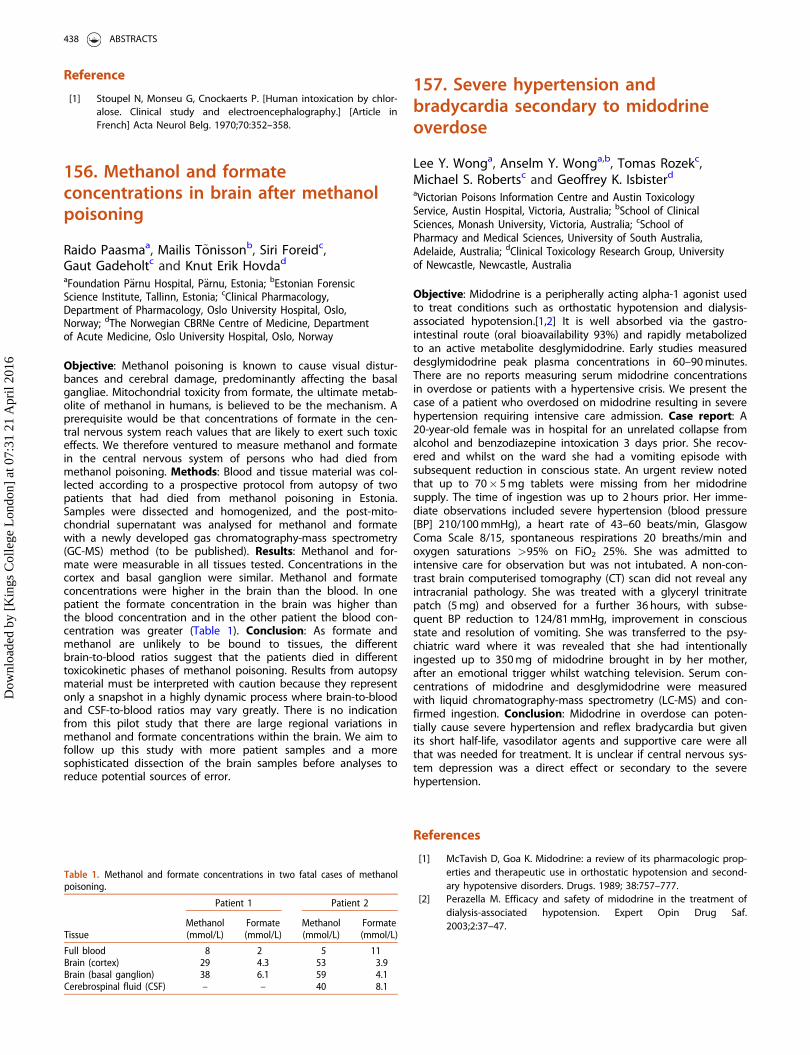
Reference
[1] Stoupel N, Monseu G, Cnockaerts P. [Human intoxication by chlor-alose. Clinical study and electroencephalography.] [Article inFrench] Acta Neurol Belg. 1970;70:352–358.
156. Methanol and formateconcentrations in brain after methanolpoisoning
Raido Paasmaa, Mailis T~onissonb, Siri Foreidc,Gaut Gadeholtc and Knut Erik Hovdad
aFoundation P€arnu Hospital, P€arnu, Estonia; bEstonian ForensicScience Institute, Tallinn, Estonia; cClinical Pharmacology,Department of Pharmacology, Oslo University Hospital, Oslo,Norway; dThe Norwegian CBRNe Centre of Medicine, Departmentof Acute Medicine, Oslo University Hospital, Oslo, Norway
Objective: Methanol poisoning is known to cause visual distur-bances and cerebral damage, predominantly affecting the basalgangliae. Mitochondrial toxicity from formate, the ultimate metab-olite of methanol in humans, is believed to be the mechanism. Aprerequisite would be that concentrations of formate in the cen-tral nervous system reach values that are likely to exert such toxiceffects. We therefore ventured to measure methanol and formatein the central nervous system of persons who had died frommethanol poisoning. Methods: Blood and tissue material was col-lected according to a prospective protocol from autopsy of twopatients that had died from methanol poisoning in Estonia.Samples were dissected and homogenized, and the post-mito-chondrial supernatant was analysed for methanol and formatewith a newly developed gas chromatography-mass spectrometry(GC-MS) method (to be published). Results: Methanol and for-mate were measurable in all tissues tested. Concentrations in thecortex and basal ganglion were similar. Methanol and formateconcentrations were higher in the brain than the blood. In onepatient the formate concentration in the brain was higher thanthe blood concentration and in the other patient the blood con-centration was greater (Table 1). Conclusion: As formate andmethanol are unlikely to be bound to tissues, the differentbrain-to-blood ratios suggest that the patients died in differenttoxicokinetic phases of methanol poisoning. Results from autopsymaterial must be interpreted with caution because they representonly a snapshot in a highly dynamic process where brain-to-bloodand CSF-to-blood ratios may vary greatly. There is no indicationfrom this pilot study that there are large regional variations inmethanol and formate concentrations within the brain. We aim tofollow up this study with more patient samples and a moresophisticated dissection of the brain samples before analyses toreduce potential sources of error.
157. Severe hypertension andbradycardia secondary to midodrineoverdose
Lee Y. Wonga, Anselm Y. Wonga,b, Tomas Rozekc,Michael S. Robertsc and Geoffrey K. Isbisterd
aVictorian Poisons Information Centre and Austin ToxicologyService, Austin Hospital, Victoria, Australia; bSchool of ClinicalSciences, Monash University, Victoria, Australia; cSchool ofPharmacy and Medical Sciences, University of South Australia,Adelaide, Australia; dClinical Toxicology Research Group, Universityof Newcastle, Newcastle, Australia
Objective: Midodrine is a peripherally acting alpha-1 agonist usedto treat conditions such as orthostatic hypotension and dialysis-associated hypotension.[1,2] It is well absorbed via the gastro-intestinal route (oral bioavailability 93%) and rapidly metabolizedto an active metabolite desglymidodrine. Early studies measureddesglymidodrine peak plasma concentrations in 60–90 minutes.There are no reports measuring serum midodrine concentrationsin overdose or patients with a hypertensive crisis. We present thecase of a patient who overdosed on midodrine resulting in severehypertension requiring intensive care admission. Case report: A20-year-old female was in hospital for an unrelated collapse fromalcohol and benzodiazepine intoxication 3 days prior. She recov-ered and whilst on the ward she had a vomiting episode withsubsequent reduction in conscious state. An urgent review notedthat up to 70� 5 mg tablets were missing from her midodrinesupply. The time of ingestion was up to 2 hours prior. Her imme-diate observations included severe hypertension (blood pressure[BP] 210/100 mmHg), a heart rate of 43–60 beats/min, GlasgowComa Scale 8/15, spontaneous respirations 20 breaths/min andoxygen saturations >95% on FiO2 25%. She was admitted tointensive care for observation but was not intubated. A non-con-trast brain computerised tomography (CT) scan did not reveal anyintracranial pathology. She was treated with a glyceryl trinitratepatch (5 mg) and observed for a further 36 hours, with subse-quent BP reduction to 124/81 mmHg, improvement in consciousstate and resolution of vomiting. She was transferred to the psy-chiatric ward where it was revealed that she had intentionallyingested up to 350 mg of midodrine brought in by her mother,after an emotional trigger whilst watching television. Serum con-centrations of midodrine and desglymidodrine were measuredwith liquid chromatography-mass spectrometry (LC-MS) and con-firmed ingestion. Conclusion: Midodrine in overdose can poten-tially cause severe hypertension and reflex bradycardia but givenits short half-life, vasodilator agents and supportive care were allthat was needed for treatment. It is unclear if central nervous sys-tem depression was a direct effect or secondary to the severehypertension.
References
[1] McTavish D, Goa K. Midodrine: a review of its pharmacologic prop-erties and therapeutic use in orthostatic hypotension and second-ary hypotensive disorders. Drugs. 1989; 38:757–777.
[2] Perazella M. Efficacy and safety of midodrine in the treatment ofdialysis-associated hypotension. Expert Opin Drug Saf.2003;2:37–47.
Table 1. Methanol and formate concentrations in two fatal cases of methanolpoisoning.
Tissue
Patient 1 Patient 2
Methanol(mmol/L)
Formate(mmol/L)
Methanol(mmol/L)
Formate(mmol/L)
Full blood 8 2 5 11Brain (cortex) 29 4.3 53 3.9Brain (basal ganglion) 38 6.1 59 4.1Cerebrospinal fluid (CSF) – – 40 8.1
438 ABSTRACTS
Dow
nloa
ded
by [
Kin
gs C
olle
ge L
ondo
n] a
t 07:
31 2
1 A
pril
2016
158. Paracetamol toxicity: a survey ofawareness in the Eastern province ofSaudi Arabia
Mohammed Al Jumaan, Bader Alossaim andMohye AlwahasUniversity of Dammam, Alkhobar, Saudi Arabia
Objective: Paracetamol is the most widely used antipyretic as it iscommonly available over-the-counter. It is generally safe; how-ever, hepatotoxicity may occur in overdose or in patients with riskfactors. Paracetamol toxicity accounts for 50% of all cases of acuteliver failure (ALF) in the US and carries a 30% mortality.[1]Unintentional toxicity has been estimated to account for 1000 to2000 cases of ALF, and 100 deaths annually.[2] Poisoning is dueto lack of patient awareness and misconceptions regarding thedangers of paracetamol-containing medication leading to misuse.We sought to assess the awareness of the population in theEastern Province of Saudi Arabia regarding paracetamol.Methods: A questionnaire was distributed during an educationalcampaign for 1 week to individuals over the age of 12, containingmultiple choice questions relating to demographic data, level ofeducation, paracetamol usage, side effects, paracetamol-contain-ing products and toxic dose. Results: Of the 432 subjects whoparticipated, 217 (50%) were male; 209 (48%) were aged 21–30years, 118 (27%) 12–20 years and only 11 (2.5%) were aged51–65 years; 18 (4%) did not give their age. College level educa-tion was recorded in 254 (56%) respondents. Paracetamol wasused for the relief of headaches (344, 49%) and high temperature(231, 33%). Potential side effects were recorded as damage tointernal organs in 243 respondents (48%) and 85 (17%) believedit had no side effects. When asked the amount in a single doseto cause toxicity, 143 (33%) answered 5 tablets, 64 (15%) and 62(15%) stated 15 and 20 tablets, respectively and 13 (3%) stated50 tablets. In this population 39% were aware of drugs listed thatcontained paracetamol, while 9% were not; 66% of respondentswere aware that Adol, Panadol Night and Fevadol contained para-cetamol, although 13% were not aware that Tempera, Relaxonand Parafon contained paracetamol. In addition, 68% did not rec-ognise that Tylenol contains paracetamol. Conclusion: Awarenessof paracetamol toxicity in the Eastern Province of Saudi Arabia isbelow expectations and there is need for more awarenesscampaigns.
References
[1] Ostapowicz GA, Fontana RJ, Schiodt FV, et al. Results of a pro-spective study of acute liver failure at 17 tertiary care centers inthe USA. Ann Intern Med. 2002;137:945–954.
[2] Department of Health and Human Services, Food and DrugAdministration. Organ-specific warnings: internal analgesic, anti-pyretic, and antirheumatic drug products for over-the-counterhuman use; final monograph. Federal Register.2009;74:19385–19409.
159. Appropriate medicationadministration by medical traineeswhen treating serotonin syndrome
Nathan P. Charltona, J. Priyanka Vakkalankaa,William Rushtonb and Calvin J. Simmonsa
aUniversity of Virginia, Charlottesville, USA; bUniversity of AlabamaBirmingham, Birmingham, USA
Objective: Medication safety events are an important cause ofmorbidity and mortality in healthcare. Despite the frequent pre-scription of serotonergic medications, medical students andhousestaff (HS) at the authors’ institution are often unable toidentify serotonergic drug interactions during their toxicologyrotation. The objective of this study was to determine if senior(4th year) medical students (MS-4) and HS are cognizant of medi-cations that have serotonergic activity that could potentiate sero-tonin syndrome. Methods: A clinical vignette regarding anadolescent male who presents with fulminant serotonin syndromeafter abusing dextromethorphan was distributed via email listservto a MS-4 class and HS across all departments at the authors’institution. Participants were given a list of 10 medications com-monly used in the intensive care unit (ICU) and asked to identifywhich medications were known to increase serotonergic activityand should be avoided in the management of this patient, aswell as those medications that are acceptable to administer.Study personnel agreed on five medications in each category,though this distribution was not shared with participants. Chi-squared analyses were used to identify differences in the correctselection of medication by training level (MS-4 versus HS).Results: There were 277 respondents to this survey (120 MS-4,157 HS; overall response rate of 30.3%). Only 2.5% of participantscorrectly identified all 10 medications, with an overall mean scoreof 64.9%. There were no differences in mean overall score ofmedication identification by training level (MS-4 versus HS) oryear in training (HS). There was no statistical difference betweenMS-4s and HS in the proportion that correctly identified all medi-cations that were acceptable to administer (p¼ 0.980), as well asthe five medications to avoid (p¼ 0.169). MS-4s were more likelyto identify all five medications that were acceptable to administerthan HS (49.2% versus 34.4%; p¼ 0.013). HS departments requir-ing an elective in medical toxicology had a greater overall aver-age score compared to departments not requiring the elective(71.8% versus 63.7%, p¼ 0.008). Conclusion: In this study, MS-4sand HS often failed to recognize the serotonergic effects of com-monly used medications in the ICU, which could result in anadverse drug event. Although training level did not improve over-all outcome, HS departments requiring a medical toxicology elect-ive had a statistically significant improvement in overall score.While many factors contribute to ensuring patient safety regard-ing medication administration, further education is needed formedical trainees regarding medication safety and adverse drugreactions.
160. Methadone intoxication treatedat an emergency centre in Serbia from2011 to 2014
Vesna Mijatovica, Zorka Drvend�zijaa, Isidora Samojlika,Stojan Petkovicb and Vesna Pajticc
aDepartment of Pharmacology, Toxicology and ClinicalPharmacology, Faculty of Medicine, University of Novi Sad, NoviSad, Serbia; bDepartment of Forensic Medicine, Faculty of Medicine,University of Novi Sad, Novi Sad, Serbia; cDepartment ofAnaesthesia and Intensive Care, Emergency Centre, Clinical Centreof Vojvodina, Novi Sad, Serbia
Objective: Methadone is a long-acting synthetic opioid as well asa full agonist of l opioid receptors.[1] It is not only used formethadone maintenance treatment (MMT) of opiate addicts but
CLINICAL TOXICOLOGY 439
Dow
nloa
ded
by [
Kin
gs C
olle
ge L
ondo
n] a
t 07:
31 2
1 A
pril
2016

also in the treatment of cancer pain and chronic pain resultingfrom other etiologies.[2] Methadone intoxication usually occurs asa result of drug overdose in opiate addicts or accidental poison-ing in children and the elderly.[3] The objective of this study wasto determine the characteristics of methadone intoxication inpatients treated at the Emergency Centre, Clinical Centre ofVojvodina, Serbia. Methods: Data were collected from medicalrecords of patients hospitalized for suspicion of opioid intoxica-tion at the Emergency Centre between 2011 and 2014. Results:During this time period, there were 278 patients hospitalized forsuspicion of opioid intoxication. In 47.0% of them methadonewas the main etiological factor. The majority of patients were hos-pitalized during 2014. Most of them (77.78%) were male. Themean age of patients was 33.5 ± 8.0 years. Analyzing anamnesticdata, 37.0% of patients reported that they had taken methadone,while 14.8% were patients receiving MMT. The mean methadonedose was 154.0 ± 162.7 mg. The majority of patients (83.7%) hadtaken methadone orally. In the toxicological reports of 15.6%patients, only methadone was present; while in others methadonehad been taken with other drugs. A small number of patients(4.4%) were admitted to the Intensive Care Unit of the EmergencyCentre; one patient died. Conclusion: The number of intoxicationcases increased during the study period; it is therefore importantto advise caution not only when prescribing methadone topatients in MMT but also when treating methadone-intoxicatedpatients. Particular attention should be paid to the prevention ofmethadone abuse and its availability on the illicit market.
References
[1] Mijatovic V, Samojlik I, Ajdukovic N, et al. Methadone-relateddeaths – epidemiological, pathohistological, and toxicological traitsin 10-year retrospective study in Vojvodina, Serbia. J Forensic Sci.2014;59:1280–1285.
[2] Mijatovic V, Dickov A, Petkovic S, et al. Safety assessment of lowdoses of methadone in combination with benzodiazepines in realoccasions during methadone maintenance treatment. HeroinAddict Relat Clin Probl. 2013;15:29–34.
[3] Mijatovic V, Samojlik I, Petkovic S, et al. Consequences of cardiactoxicity in patients on low methadone doses during methadonemaintenance treatment. Heroin Addict Relat Clin Probl.2015;17:73–78.
161. Rhabdomyolysis associated withsynephrine and yohimbe containingnutritional supplements and weightlifting
William J. Meggs, Kathleen Bryant, Susan Miller,Mohan Punja and Juan MarchEast Carolina University, Greenville, USA
Objective: To report a case of rhabdomyolysis with acute renalinjury associated with the use of nutritional supplements toenhance body building. Case report: A 22-year-old male in priorgood health reported to an emergency department with general-ized fatigue, muscle soreness, nausea, anorexia, and dark urine.For the two days prior to arrival, he had supplemented exercisewith the nutritional supplement HyperFX Concentrated EnergyFormula. The ingredient list stated the product contained caffeineand bitter orange extract (30% synephrine). He also used MegaMen Energy and Metabolism Supplement containing yohimbe.Social history was positive for cigarette smoking, moderate alco-hol use and occasional marijuana use. Vital signs on arrival were
temperature 36.5 �C, pulse 72/minute, respirations 18/minute,blood pressure 160/86 and oxygen saturation 99% on room air.Mucus membranes were dry. There was no muscle tenderness.No other examination findings were reported. Creatine kinase(CK) was 295,040 U/L and peaked at 791,520 U/L (reference range32 to 294 U/L). Creatinine peaked at 386.5 lmol/L (reference range53 to 106 lmol/L) on hospital day 3. Urine was red with 3þ pro-tein. Treatment with IV hydration with normal saline, urine alkalin-isation and furosemide resulted in normalization of CK andcreatinine, and he recovered without sequelae. Conclusion:Sympathomimetic-containing nutritional supplements, when com-bined with exercise can cause rhabdomyolysis and significantimpairment of renal function.[1,2] The HyperFX ConcentratedEnergy Formula supplement is specifically marketed to be ‘‘well-suited to long sessions of aerobic activity or high intensity work-outs".[3] Physicians should obtain a detailed history aboutsupplement use in patients presenting with muscle injury or newrenal injury. While association does not establish causation, basedon other experiences, workouts augmented by supplements con-taining smpathomimetics may be associated with health risks andshould be used with caution.
References
[1] Scroggie DA, Harris M, Sakai L. Rhabdomyolysis associated withnutritional supplement use. J Clin Rheumatol. 2000;6:328–332.
[2] Dehoney S, Wellein M Rhabdomyolysis associated with the nutri-tional supplement Hydroxycut. Am J Health Syst Pharm.2009;66:142–148.
[3] BSN Product Website, 2014; [cited 2015 Nov 2]. http://www.gobsn.com/product/hperflx.html
162. Diabetic medication enquiries tothe New Zealand National PoisonsCentre
Yvette C. MillardDepartment of Preventive and Social Medicine, Dunedin,New Zealand
Objective: To characterize diabetic medication exposures in NewZealand using data reported to the New Zealand PoisonsInformation Centre. Methods: Telephone enquiries involvinghuman exposures to diabetic medications (anti-diabetics and insu-lin) between January 2005 and December 2014 were retrospect-ively reviewed. Data on patient age, reason for exposure,substances involved, route of exposure, symptomatology and rec-ommended treatment were collated and assessed. Results: Overthe study period 81,249 human therapeutic exposure enquirieswere received, 634 involving diabetic medications (388 single dia-betic medications, 75 multiple diabetic medications, and theremainder involving diabetic medications in combination withother medications). Metformin was the most commonly involvedsubstance (53%), followed by insulin formulations (17%), gliclazide(14%), glipizide (13%) and pioglitazone (1%). Acarbose, exenatide,rosiglitazone and glibenclamide were involved in <1% of cases.Adults accounted for 58% of incidents, with 36% occurring in chil-dren <5 years. This is notably different from total pharmaceuticalpoisonings for the period where 32% involved adults and 57%involved children <5 years. Regarding specific diabetic medica-tions, insulin exposures occurred mainly in adults (93%), while57% of metformin exposures occurred in adults and 36% in chil-dren <5 years. Sulfonylurea incidents were spread 40% in adultsand 52% in children <5 years. Poisonings were mainly therapeutic
440 ABSTRACTS
Dow
nloa
ded
by [
Kin
gs C
olle
ge L
ondo
n] a
t 07:
31 2
1 A
pril
2016
errors (41%) and child exploratory behavior (39%). Ingestion wasthe main route of exposure (83%), followed by injections (17%).One incident involved the insufflation of a ground up glipizidetablet. Diabetic medication exposures increased from the begin-ning of the study period; there were 43 cases in 2005 with a peakof 90 incidents in 2013. Medical referral for monitoring and/ortreatment was recommended in 74% of cases. This is considerablyhigher compared to all pharmaceutical poisonings where only31% of patients required medical referral. Home observation wasrecommended in 23% of diabetic poisonings, typically followingaccidental metformin exposures. Symptoms were reported in 38of the patients who took hypoglycemic medications only. Themost common symptoms were central nervous system depres-sion, dizziness/light-headedness, hypoglycemia and gastrointes-tinal upset. Conclusion: Poisoning exposure to diabeticmedications is a fairly infrequent occurrence in New Zealand.However, medical assessment is recommended more frequently,and adults are more commonly involved, compared to otherpharmaceutical overdoses. As most cases involved therapeuticerrors or child exploratory exposures, secure storage of medica-tions away from children and ensuring the correct medicationsare given to the correct patient at the correct time could help toreduce the incidence of poisonings.
163. Delirium following loperamideoverdose: a case report
Felix Hammann and Matthias E. LiechtiDivision of Clinical Pharmacology and Toxicology, Department ofInternal Medicine, University and University Hospital Basel, Basel,Switzerland
Objective: Loperamide is an effective oral anti-diarrheal agent inadults and older children. It acts as an agonist at the l-opioidreceptor (MOR) but has very low systemic availability and crossesthe blood brain barrier (BBB) poorly. Hence, opioid action is usu-ally restricted to the intestinal tract with no central effects.However, bioavailability and central nervous system (CNS) pene-tration of loperamide may increase in overdose or if other permis-sive factors are present. We describe a case of agitated deliriumduring treatment with high dose loperamide. Case report: An 80-year-old woman presented with a high-output stoma after sigmaresection due to endometrial carcinoma with post-actinic stenosisof the small intestine and sigmoid. The patient was treated withloperamide 30 mg once daily and colestyramine. Her other medi-cation was unremarkable. Despite the already high daily dose,loperamide was increased to 40 mg once daily and colestyraminewas stopped. The following day she became somnolent and con-fused. With antipsychotic therapy and discontinuation of lopera-mide, somnolence and delirium fully resolved. Conclusion:Loperamide is a lipophilic compound that is mostly absorbed inthe small intestine. P-glycoprotein (P-gp) efflux and cytochromeP450 3A4 metabolism in the intestine and the liver reduce its sys-temic availability to 0.3%.[1] Loperamide shows highly potentMOR affinity similar to fentanyl,[2] so even modest increases inplasma concentrations could cause central effects. Due to itsover-the-counter availability and potential for recreational abuse,loperamide has previously been described as ‘‘the poor man’smethadone’’ and is misused in combination with medicationswhich increase its bioavailability.[3] High-throughput stomaincreases intestinal transit time, thereby impairing the capacity forloperamide absorption. In this patient, colestyramine had likelyfurther decreased loperamide absorption by unspecific binding inthe gut lumen. We propose that the combination of the highdose of loperamide and discontinuation of colestyramine resultedin a sudden and sharp rise in availability, possibly overwhelming
intestinal and BBB P-gp efflux capacities. Clinical managementincludes withdrawal of drug and symptomatic management.Naloxone can be given as a specific antidote.
References
[1] Janssen-Cilag AG Zug, Switzerland. Product Information ImodiumDuo(R). 2011; [cited 2015 Nov 4]. Available at: http://www.imo-dium.ch/sites/default/files/product/Imodium_duo_gebrauchsinfo.pdf
[2] Chen Z, Davies E, Miller W, et al. Design and synthesis of 4-phenylpiperidine compounds targeting the mu receptor. Bioorg MedChem Lett. 2004;14:5275–5279.
[3] Dierksen J, Gonsoulin M, Walterscheid JP. Poor man’s methadone:A case report of loperamide toxicity. Am J Forensic Med Pathol.2015;36:268–270.
164. Accidental intravenousadministration of a Lactobacillusreuteri preparation in a neonate
Noora Heiskanena, Pirjo Tynj€al€aa, Kalle Hoppua,Anna-Kaisa Eronena, Kirsi M. Salonenb andHeidi Sandstr€omc
aPoison Information Centre, HUCH Emergency Care, University ofHelsinki and Helsinki University Hospital, Helsinki, Finland;bChildren’s Hospital, University of Helsinki and Helsinki UniversityHospital, Helsinki, Finland; cPorvoo Hospital, Helsinki UniversityHospital, Porvoo, Finland
Objective: We describe a case of accidental intravenous (IV) dos-ing of Lactobacillus reuteri in a neonate. Case report: A maleinfant born at 38þ 5 gestational weeks by Cesarean section(3.022 g, Apgar 9/9/10) was transferred to the neonatal care unitat the age of three days because of poor sucking, drowsiness anddecreasing weight (�13%). IV benzylpenicillin, netilmycin andaciclovir were started because the symptoms and marginal leuco-penia (8.9� 109/L) suggested infection. Blood culture and herpesvirus samples were later found to be negative. Enteral nutritionvia nasogastric tube was initiated, and resulted in weight increase.At 7 days old the infant still had a nasogastric tube and an IVcannula. A nurse, after administering the IV antimicrobials askedthe mother to give Rela DropsVR (Lactobacillus reuteri, vegetableoils and colloidal anhydrous silica) to the infant. In error themother administered the probiotic drops into the IV cannula, butthis was not discovered until the evening of the same day, whenthe infant started to grunt. The IV cannula was immediatelyremoved. The infant seemed otherwise normal, except for tender-ness at the infusion site and elevated C-reactive protein (CRP)44 mg/L. After placement of a new IV cannula and blood culture,penicillin and netilmycin doses were increased. IV paracetamolwas used for analgesia. The infant became tachypnoeic, was rest-less and developed respiratory acidosis (pH 7.27, pCO2 7.8 kPa).Therefore nasal continuous positive airway pressure (CPAP) with-out additional oxygen was initiated. On the first day after the inci-dent (day 2) chest X-ray revealed a large left-sided atelectasis andpneumonia was suspected. CRP peaked at 102 mg/L. In the even-ing episodes of tremors, rigidity (opisthotonus) and bradycardia(80/min) without hypoxia were observed and he was transferredto a neonatal intensive care unit. G-penicillin was switched tomeropenem due to neurologic symptoms (liquor leucocytosis upto 50� 106/L) and suspected meningitis. All cultures remainednegative and the CRP and the child’s clinical state stabilized.Neurological status, brain ultrasound and magnetic resonance
CLINICAL TOXICOLOGY 441
Dow
nloa
ded
by [
Kin
gs C
olle
ge L
ondo
n] a
t 07:
31 2
1 A
pril
2016
imaging (MRI) were normal and no specific abnormalities wereseen on electroencephalogram. Bacteria cultured from the RelaDropsVR preparation proved sensitive to netilmycin. Antimicrobialtreatment was continued until day 11 and the infant was dis-charged on day 13 without sequelae. Conclusion: Accidentalintravenous administration of probiotic drops led to a seriousadverse reaction in a neonate. It could not be determinedwhether symptoms were caused by infection or microemboli orboth.
165. Severe prolonged tachycardiaafter massive overdose ofpaliperidone
Lee Wonga, Morris Odellb, Katherine Wongb andShaun L. Greenea
aAustin Health, Melbourne, Australia; bVictorian Institute of ForensicMedicine, Melbourne, Australia
Objective: Paliperidone (9-hydroxyrisperidone) is a second gener-ation atypical antipsychotic. The recommended dose for schizo-phrenia is 6 mg/day. We report a massive paliperidone ingestiondemonstrating prolonged significant cardiovascular toxicity, withserum concentrations. Case report: A 23-year-old female pre-sented three hours after ingestion of extended release paliperi-done 504 mg, quetiapine 400 mg and zopiclone 15 mg. She wasdrowsy with heart rate 96/min, blood pressure 130/70 mmHg,respiratory rate 14/min and pulse oximetry 98% on room air.Renal function, electrolytes, liver function tests, calcium, magne-sium and phosphate were normal. A 12-lead electrocardiogram(ECG) three hours post-ingestion revealed sinus tachycardia (110beats/min), with normal conduction intervals. Ten hours post-ingestion she was tachycardic on ambulation with a heart rate of140–180 beats/min, and a resting heart rate of 90–110 beats/minand systolic blood pressure 95 mmHg. Tachycardia developedwithin seconds of minimal exertion, including small posturalchanges while supine. One liter of intravenous crystalloids wasadministered and blood pressure normalized. Serial ECGs showedsinus tachycardia (up to 184 beats/min) with absolute QT intervalof 320 msec (which remained constant). She was admitted to thecoronary care unit for monitoring and bed rest. Intravenous mag-nesium and potassium (low normal serum potassium) wereadministered. Tachycardia persisted for 72 hours peaking at 190beats/min on any exertion 36–40 hours post-overdose. ECG ondischarge showed sinus rhythm of 100 beats/min, PR interval 100msec, and an absolute QT interval of 320 msec. We were unableto obtain a transthoracic echocardiogram to assess cardiac func-tion. Serum paliperidone concentrations 4 and 40 hours post-ingestion were 29 ng/mL and 883 ng/mL respectively. A meanplasma concentration of 10.7 ng/mL is reported following daily3 mg dosing.[1] Quetiapine was not detected in serum at 40 hourspost-ingestion. Conclusion: Paliperidone in overdose can causeprolonged cardiovascular dysfunction characterised by restingsinus tachycardia with intermittent episodes of posture-evokedextreme tachycardia. Its availability as an extended release prepar-ation results in high blood concentrations and effects that canpersist for several days. This case is the largest recorded paliperi-done overdose, with confirmatory serum concentrations. We rec-ommend a period of prolonged cardiac monitoring and minimalexertion in patients with significant overdose. Prescribers shouldbe aware of the potential for significant prolonged paliperidonetoxicity when taken in overdose.
Reference
[1] Boom S, Talluri K, Janssens L, et al. Single and multiple-dosepharmacokinetics and dose proportionality of the psychotropicagent paliperidone extended release. J Clin Pharmacol.2009;49:1318–1330.
166. Overdoses of ropinirole reportedto the UK National PoisonsInformation Service
Alison M. Gooda, Gillian Jacksona,Caroline E. McGrorya, Victoria Eaglinga,Simon H. L. Thomasb, John P. Thompsonc,J. Allister Valed and Michael Eddlestona
aNPIS Edinburgh, Scottish Poisons Information Bureau, Edinburgh,UK; bNPIS Newcastle, Regional Drug and Therapeutics Centre,Newcastle upon Tyne, UK; cNPIS Cardiff, University HospitalLlandough, Cardiff, UK; dNPIS Birmingham, City Hospital,Birmingham, UK
Objective: Ropinirole is a dopamine D2/D3 receptor agonist forthe treatment of Parkinson’s disease and restless legs syndrome.As there is little information on overdose this study was carriedout to review UK National Poisons Information Service (NPIS)enquiries. Methods: Information was extracted from telephoneenquiries received by the NPIS and recorded on the UK PoisonsInformation Database from 1 January 2008 to 31 August 2015.Results: There were 113 enquiries concerning 104 exposures (2tablet identification requests and 7 duplicate calls). Where agewas known the mean was 59.7 years (median 63; range 1–100).There were 43 females and 58 males, remainder unknown. Eightenquiries concerned children (1–3 years) who had taken 1–8 mg.One vomited, one was described as ‘‘sleepy and grumpy’’, in onefeatures were unknown and 5 were asymptomatic. For the 96adults (mean age 65 years; 41 females, 53 males, remainderunknown), 11 were described as deliberate overdoses. Twopatients had taken ropinirole alone (24.5 mg and 58 mg) and bothwere asymptomatic (5 hours and 30 minutes post-ingestion). Ofthe other 9, one had a poisons severity score of 2 (PSS2) [1] (29-year-old female patient with Parkinson’s disease who had taken112 mg ropinirole and 10 mg rasagiline, and had dyskinesia, agita-tion and hallucinations) and one PSS3 (33-year-old male with ahistory of ingesting a variety of drugs including a beta-blocker,presented with coma, twitching, mydriasis, tachycardia and hallu-cinations, developed respiratory arrest and convulsions but recov-ered and then claimed only to have taken alcohol). Theremaining patients scored PSS0 or 1. The non-intentional inges-tions (85) were therapeutic errors (65), accidental overdoses (12),adverse reactions (3), or circumstances unknown (5). In the 41cases where ropinirole was taken alone, no features greater thanPSS1 were reported and 23 were asymptomatic. Features reportedwere (frequency >1): vomiting (7), nausea (6), dizziness (4) andsomnolence (2). Of the adverse effects reported during thera-peutic use, the following were reported in this series: abdominalpain (1), confusion (3), dizziness (6), hallucinations (4), hypotension(1), nausea (6) and vomiting (6). Conclusion: Most poisons centrereferrals concerning ropinirole were therapeutic errors or acciden-tal overdoses. Most patients who take excess ropinirole acciden-tally do not have serious effects and features reported are similarto those found in therapeutic use.
442 ABSTRACTS
Dow
nloa
ded
by [
Kin
gs C
olle
ge L
ondo
n] a
t 07:
31 2
1 A
pril
2016
Reference
[1] Persson HE, Sj€oberg GK, Haines JA, et al. Poisoning severity score.Grading of acute poisoning. J Toxicol Clin Toxicol.1998;36:205–213.
167. Safety of antipsychotic drugs:a 16-year review
Bla�zena Cag�a�nov�aa, Silvia Plackov�aa,Jaroslav Kresanekb, Elena Ondriasovac andNatalia Luptakovac
aNational Toxicological Information Centre, Department ofOccupational Medicine and Toxicology, University HospitalBratislava, Bratislava, Slovakia; bSlovak Medical University,Bratislava, Slovakia; cFaculty of Pharmacy, Comenius University,Bratislava, Slovakia
Objective: Evaluation of the safety profile of traditional and atyp-ical antipsychotics in overdose, causes of intoxications and demo-graphic indicators. Methods: Intoxications were analysed on thebasis of data gathered from telephone consultations and medicalreports forwarded to the National Toxicological InformationCentre (NTIC) in Bratislava from the whole of Slovakia during theperiod 1999–2014. All cases involving the non-prescribed use oroverdose of antipsychotics were reviewed. Only coingestion ofethanol was accepted. The severity of poisoning was classified inaccordance with the Poisoning Severity Score. Results: Over the16-year period 2339 antipsychotic exposures were the subject ofenquiries to the NTIC of which 1150 involved single agent anti-psychotic ingestions. We registered 642 (55.8%) atypical anti-psychotic overdoses and 508 (44.2%) incidents with firstgeneration antipsychotics. Since 1999 there has been a 3.5 foldincrease in antipsychotic overdoses. The median age of patientswas 31 years (1 to 92 years), 53.1% of cases involved females.Suicidal intoxications were more frequent (80.3%) and had amore serious clinical course. Accidental poisonings (16.6%)occurred mostly in children under 6 years of age. Risperidone,quetiapine and tiapride made up most of the atypical anti-psychotic overdoses. Levomepromazine and chlorprothixene werethe most frequently involved in the traditional antipsychotic poi-sonings. Clinical symptoms of intoxication were manifested in 920patients (79.9%) of the observed group. We registered just 152cases (13.2%) in which no symptoms were manifested after thenon-prescribed use or overdose. The most frequent (717 cases,62.3%) were minor symptoms (sleepiness, dizziness, gastrointes-tinal distress, mild extrapyramidal symptoms, hypotension) thatsubsided in 24 hours. In 152 (13.2%) patients we observed symp-toms of moderate and in 49 (4.2%) patients symptoms of severeintoxication. Severe intoxications in 2 (0.17%) patients overdosedwith clozapine resulted in death. In total 788 (68.5%) patientswere hospitalised. The median length of hospital stay was 2 days(1–28 days). Conclusion: Atypical antipsychotic overdoses dramat-ically increased over the 16-year period, together with their risingprescription. During this time, there was no decline in severity orthe length of hospital stay, despite the reduction in first gener-ation antipsychotic overdoses. According to our findings, there isno significant difference in safety profile between atypical andtraditional antipsychotics.
168. Severe taxane skin reaction dueto a drug interaction withamiodarone: a case report
Felix Hammanna, Katrin Conenb, Michael Medingerc,Philipp Cesanad, Christoph Rochlitzb andAnne B Taegtmeyera
aDivision of Clinical Pharmacology and Toxicology, Department ofInternal Medicine, University and University Hospital Basel, Basel,Switzerland; bDivision of Medical Oncology, Department of InternalMedicine, University and University Hospital Basel, Basel,Switzerland; cDepartment of Internal Medicine, University andUniversity Hospital Basel, Basel, Switzerland; dDepartment ofDermatology and Allergology, University and University HospitalBasel, Basel, Switzerland
Objective: Paclitaxel and docetaxel are taxanes that are used inchemotherapy of several solid tumors. They are eliminated fromthe cell by P-glycoprotein (P-gp) and inactivated by CYP 3A4(docetaxel, paclitaxel) and CYP 2C8 (paclitaxel). We report a caseof severe taxane toxicity due to co-medication with the antiar-rhythmic drug amiodarone. Case report: A 77-year-old womanwith invasive ductal breast cancer was referred to our hospitalwith blistering of the skin, inability to swallow solids or liquids,and dehydration. She was undergoing chemotherapy as anadjunct to surgery. Her medication included amiodarone whichshe was taking for her ‘‘palpitations’’. During the first four cyclesof outpatient paclitaxel therapy she developed increasing abdom-inal discomfort and skin lesions. Therapy was switched to doce-taxel because of paclitaxel’s known potential for skin toxicity,however symptoms worsened, mucositis ensued, and chemother-apy was discontinued after only one cycle of docetaxel. Over thenext days, however, symptoms progressed, eventually making oralintake impossible. On admission, we began intravenous hydration,symptomatic treatment of the skin reaction, and placed a naso-gastric tube to allow for administration of medication and nutri-tion. Analysis of two routine blood samples taken 9 and 10 daysafter docetaxel revealed serum concentrations of 4.73 and4.09 ng/mL, respectively, corresponding to a severely prolongedterminal half-life of 114 hours (reference 11.1 hours). Paclitaxel wasalso present in these blood samples (20 and 21 days after the lastadministration, respectively), but concentrations were below thevalidated lower limit of detection.[1] Under normal conditions,paclitaxel should not be detectable 11 days after the last adminis-tration. The patient was discharged two weeks later with nearresolution of the skin and mucosal lesions. There were no recur-rences of dermatological or gastrointestinal toxicity on follow up.Conclusion: Amiodarone is a strong P-gp inhibitor and a moder-ate CYP 3A4 inhibitor. There is evidence for CYP 2C8 inhibition.[2]We show the inhibitory effects of amiodarone on taxane clear-ance. We also suggest caution when combining other CYP 2C8inhibitors with paclitaxel.
References
[1] Hendrikx JJ, Rosing H, Schinkel AH, et al. Combined quantificationof paclitaxel, docetaxel and ritonavir in human feces and urineusing LC-MS/MS. Biomed Chromatogr. 2014;28:302–310.
[2] Polasek TM, Elliot DJ, Lewis BC, et al. Mechanism-based inactiva-tion of human cytochrome P4502C8 by drugs in vitro. J PharmacolExp Ther. 2004;311:996–1007.
CLINICAL TOXICOLOGY 443
Dow
nloa
ded
by [
Kin
gs C
olle
ge L
ondo
n] a
t 07:
31 2
1 A
pril
2016
169. Acute iatrogenic parenteralvancomycin overdose and associatednephrotoxicity: a case report
Morgan A. A. Riggana, Mary Ann Howlandb,Elizabeth Q. Hinesa, David S. Goldfarbc andLewis S. Nelsona
aNew York University School of Medicine, New York, USA;bSt. John’s University College of Pharmacy and Health Sciences,New York, USA; cNYU Langone Medical Center, New York, USA
Objective: Vancomycin-associated nephrotoxicity, first reported in1958, was initially thought to be due to the presence of impur-ities. Newer preparations are devoid of such impurities, yetnephrotoxicity still occurs. The majority of cases involve treatmentwith vancomycin for >48 hours, high trough concentrations, pre-existing chronic kidney disease, or concurrent use of othernephrotoxic drugs. We present the first reported case of acute iat-rogenic vancomycin-associated nephrotoxicity occurring withinhours of drug administration. Case report: A 13-year-old, 60 kggirl with scoliosis was admitted to the pediatric intensive careunit after undergoing an uncomplicated spinal fusion. Only acet-aminophen 800 mg IV was prescribed. On post-operative day oneshe received a dose and immediately developed flushing withoutshortness of breath or urticaria. Vital signs were blood pressure112/59 mmHg, heart rate 130/minute, respiratory rate 27/minuteand temperature 38.9 �C. Diphenhydramine IV and a fluid boluswere administered. Thirty minutes later, pharmacy called reportinga dispensing error; the patient had received 8 g vancomycin. Aserum vancomycin concentration one hour after administrationwas 93 lmol/L (139 lg/mL). Two hours later her creatinine (Cr)was 89 lmol/L (1.01 mg/dL), a 100% increase from her baseline44 lmol/L (0.5 mg/dL). Five hours later, her Cr was 121 lmol/L(1.37 mg/dL) and vancomycin concentration was 96 lmol/L(144 lg/mL). Nephrology performed hemodialysis (HD) for 5 hours.Post-dialysis, her Cr was 17 lmol/L (0.2 mg/dL) and vancomycinwas undetectable. However, two hours later, her Cr and vanco-mycin concentration increased to 165 lmol/L (1.87mg/dL) and42 lmol/L (63 lg/mL), respectively. Her urine output decreased to0.7 ml/kg/h. A second session of HD was performed 13 hours afterthe initial error. Over the next 24 hours, Cr continued to rise, buther vancomycin concentration declined without further HD. Onday 8, Cr returned to baseline and she was discharged on day 9stable with mild hypertension. Conclusion: The mechanism foracute vancomycin-associated nephrotoxicity remains unclear.Proposed mechanisms include oxidative injury and ischemia lead-ing to tubulo-interstitial damage and mitochondrial dysfunctionwithin proximal renal tubule cells. The etiology of nephrotoxicityin our patient is unknown, however the single drug exposure andclose temporal relationship suggests vancomycin played a signifi-cant role. This case suggests a significant increase in serum Crmay occur following a single acute parenteral vancomycin over-dose. Further research is needed to determine if this rise in Cr isdue to laboratory interference, increased Cr production, decreasedCr secretion, or acute tubular injury.
170. What are the causes of and howto prevent medication errors bylaypersons?
Michal Urban, Daniela Pelclova, Roman Lesso andSergey Zakharov
Toxicological Information Centre, Department of OccupationalMedicine, First Faculty of Medicine, Charles University and GeneralUniversity Hospital, Prague, Czech Republic
Objective: The causes of unintentional medications errors by laypersons are rarely studied. The identification of the frequency,sources, reasons and consequences could help to reduce theiroverall rate. Methods: Records of medication error enquiries in2013–2014 were extracted from the electronic database of theToxicological Information Centre (TIC) and following variableswere reviewed: drug class, dosage form, dose, age of the subject,cause of the error, time interval from ingestion to the call, symp-toms, prognosis evaluation at the time of the call and first aid rec-ommended. Results: Overall 1354 calls met the inclusion criteria,including central nervous system (CNS) drugs (23.6%), respiratorysystem drugs (18.5%) and gastrointestinal drugs (16.2%) were themost common drug classes involved in the medication errors.Symptoms at the time of the call were mostly absent (73.0%),minor (18.3%), irrelevant/not related (6.5%), moderate (1.7%), andrarely severe (0.5%). The ingested dose, as evaluated by TIC toxi-cologists was mostly mild (42.6%), less frequently low/therapeutic(35.1%), causing only mild gastrointestinal discomfort/irritation(12.8%), toxic (9.4%), or lethal (0.1%) effects. Most patients werein the youngest age subgroup 0–5 years (46%), followed by20–59 years (29%), 6–12 years (11%), �60 years (9%) and 13–19years (5%). Most errors were caused by parents (59.0%), less bypatients themselves (35.9%). The recommendations of the TICincluded oral fluids (16%) or charcoal (8%) administration. Othermeasures (total 5%) were less frequent, such as superficial decon-tamination, vomiting induction, gastric lavage, symptomatic treat-ment or administration of antidotes (N-acetylcysteine or digitalisantidote in the most severe cases). The most frequent reasons forthe errors involved leaflet misinterpretation and/or mistaken dose(53.6%), mixing up medications (19.2%), attempt to reduce painwith repeated dose (6.4%), confusion in psychiatric/elderlypatients (2.7%), wrong route of administration (2.2%), accidentwith the closure of the bottle and other (9%) or unknown cause(6.9%). Conclusion: The high proportion of children among thepatients is worrying and small children are rather surprisingly themost vulnerable group. The reason may be that children’s dos-ages vary by weight. In addition, many drugs come in severalconcentrations and a proper dose adjustment is more difficult.Most overdoses could be prevented by safer labelling, proper capclosures for liquid products and medication reconciliation by bothphysicians and pharmacists teaching patients how to use medi-cines properly. Acknowledgements: Charles University projectsP25/1LF/2 and P28/1LF/6.
171. A case of flecainide overdoseresponding to lipid emulsion therapy
Susan Miller, Mohan Punja and William J MeggsEast Carolina University, Greenville, USA
Objective: Lipid emulsion therapy is considered for cardiotoxicdrug overdoses unresponsive to other therapies.[1] Two casesreport flecainide overdoses responding to fat emulsion ther-apy.[2,3] We report a similar case. Case report: A 69-year-old manwith history of depression, atrial fibrillation, and restless leg syn-drome informed his spouse that he had ingested 1 g of flecainide,12 mg of clonazepam, and 1 mg of ropinirole. She drove him tothe hospital. On arrival, he complained of dyspnea and dry cough,but had no other complaints. Initial vital signs one hour afteringestion were pulse 75/minute, respiratory rate 16/minute, bloodpressure (BP) 119/62 mmHg, and oxygen saturation 98% on room
444 ABSTRACTS
Dow
nloa
ded
by [
Kin
gs C
olle
ge L
ondo
n] a
t 07:
31 2
1 A
pril
2016
air. Initial electrocardiogram 70 minutes after ingestion demon-strated a broad complex irregular tracing with heart rate of 76/minute, QRS duration 268 msec, and QTc interval 644 msec. At79 minutes after ingestion his BP fell to 66/29 mmHg. He wasgiven a bolus of 150 mEq of sodium bicarbonate followed by acontinuous infusion, with no improvement in hypotension orshortening of QRS duration. Coughing, rales, and hypoxia devel-oped two hours after ingestion. Chest roentgenogram confirmedpulmonary edema. Rapid sequence intubation was performedwithout incident. Central and arterial lines were placed. A 135 mLbolus of a 20% lipid emulsion therapy was followed by a continu-ous infusion of 1350 mL over two hours. BP rose to 100/66 mmHgat 2.5 hours after ingestion, then to 152/94 mmHg. There were nofurther hypotensive episodes. The patient was transferred to thecardiac intensive care unit (CICU). His hospital course was compli-cated by atrial fibrillation requiring medication adjustment andpulmonary edema requiring diuresis. He made an uneventfulrecovery and was transferred to the psychiatric service two weeksafter admission. Conclusion: In this case of flecainide overdose,there was a rapid reversal of hypotension after lipid emulsiontherapy was instituted after sodium bicarbonate infusion wasineffective.
References
[1] American College of Medical Toxicology: ACMT position statement:interim guidance for the use of lipid resuscitation therapy. J MedToxicol. 2011;7:81–82.
[2] Moussot PE, Marhar F, Minville V, et al. Use of intravenous lipid20% emulsion for the treatment of a voluntary intoxication of fle-cainide with refractory shock. Clin Toxicol (Phila). 2011;49:514.
[3] Sivalingam SK, Gadiraju VT, Hariharan MV, et al. Flecainide toxicity– treatment with intravenous fat emulsion and extra corporeal lifesupport. Acute Card Care. 2013;15:90–92.
172. Paracetamol overdose in Slovenia
Tina Antolica, Lucija Peterlin Masica and Miran Brvarb
aFaculty of Pharmacy, University of Ljubljana, Ljubljana, Slovenia;bCentre for Clinical Toxicology and Pharmacology, UniversityMedical Centre Ljubljana, Ljubljana, Slovenia
Objective: The incidence of paracetamol overdose and paraceta-mol-induced liver failure varies widely across Europe and the US.In Slovenia paracetamol is an over-the-counter drug. However, itcan be bought only in pharmacies with a limited amount of para-cetamol in these products. Maximum pack size is 20 tablets inblister packs and up to three packs can be supplied in one trans-action. The aim of the study was to evaluate epidemiology ofparacetamol overdose in Ljubljana, Slovenia. Methods: In thisretrospective study we analysed the epidemiology and clinicalpresentation of adult patients poisoned with paracetamol whowere treated in the University Medical Center, Ljubljana (UMCL),the primary city hospital in the capital, serving a population of600,000, in the past 25 years. Results: In total 90 adult patientswith paracetamol overdose were hospitalized in UMCL during thepast 25 years. The overall incidence of paracetamol overdose was0.6/100,000 population in this period. The number of paracetamoloverdosed patients steadily increased from three patients in 1990(0.5/100,000) to nine patients in 2014 (1.5/100,000). The mean ageof the study cohort was 34 years (range 16–88 years). There were29 women and 61 men. Of the 90 patients, 78 ingested paraceta-mol with suicidal intent and 12 paracetamol overdoses occurredby mistake. The ingested dose of paracetamol was known in 71patient and 56 of them ingested a toxic dose of paracetamol.Nevertheless, a toxic serum concentration was detected in
17 patients only; of these 14 patients ingested paracetamol in asuicide attempt. Overall 40 patients were treated with acetylcys-teine and 6 patients developed acute liver failure. No caseresulted in liver transplantation or death. Conclusion: In Sloveniathe incidence of inpatient admissions due to paracetamol over-dose is about 10-times lower than in the US.[1] The main reasonmay be that only pharmacies are allowed to sell paracetamol anda pharmacist must make or supervise the sale. Patients are askedabout other medical conditions and other medicines they are tak-ing. So, misunderstanding of the active ingredient is uncommon.Patients can obtain only up to three packs in one transaction andmaximum pack size is 20 tablets. In addition, tablets are packagedin blister packs which probably reduce the risk of deliberateingestion of a larger number of tablets at once.
Reference
[1] Altyar A, Kordi L, Skrepnek G. Clinical and economic characteristicsof emergency department visits due to acetaminophen toxicity inthe USA. BMJ Open. 2015;5:e007368.
173. Supplementation of vitamin Dcan be hazardous when exaggerated:a case report
Eva K. Olssona, Henry Lindberga andTomas Forsbergb
aSwedish Poisons Information Centre, Stockholm, Sweden;bDepartment of Internal Medicine, Capio St Gorans Hospital,Stockholm, Sweden
Objective: Vitamin D supplements have become popular and canbe purchased easily over the counter in diverse formulations anddosages. They are also marketed on the Internet as a cure forvarious conditions like depression, tiredness and strengthening ofthe immune system. We present a case of severe vitamin D poi-soning with persistent hypercalcemia and renal failure afterrepeated intake of colecalciferol intended for horses. Case report:A 21-year-old man with Asperger’s syndrome presented at theemergency department with severe headache, photophobia, nau-sea and vomiting after a night of computer gaming. He was pre-viously healthy and denied all kinds of substance intake. Initialsuspicion of subarachnoid hemorrhage was excluded with braincomputerised tomography (CT) scan and lumbar puncture withnormal results. Incidental findings of severe hypercalcemia of3.64 mmol/L (ionized 1.87) and renal failure with creatinine153 mmol/L were noted. Extended sampling showed an extremelylow concentration of parathyroid hormone (<0.3 pmol/L), and ahigh 25(OH)-vitamin D, concentration (>350 nmol/L). Treatmentwas started with intravenous fluids and steroids. Calcitonin wasgiven for 3 days and on day 3 he was given denosumab. Steroidtreatment continued for 3 weeks and was then slowly weaned.He developed atrial fibrillation (probably due to treatment withextremely large volumes of fluids) which resolved with diuretics.The high calcium concentration persisted for 2 weeks and contin-ued treatment with large volumes of fluids was necessary to keepcalcium at an acceptable concentration. After 3 weeks in hospitalthe calcium concentration and renal function normalized but the25(OH)-vitamin D was still 1050 nmol/L and the 1,25(OH)2-vitaminD 391 pmol/L. After confrontation, the patient finally admittedtwo months daily intake of spoonful of an animal product con-taining 500 000 IU/g vitamin D3 for revitalizing purposes.Conclusion: There is ongoing debate on vitamin D supplementa-tion. Fairly high doses are considered safe but the toxic upper
CLINICAL TOXICOLOGY 445
Dow
nloa
ded
by [
Kin
gs C
olle
ge L
ondo
n] a
t 07:
31 2
1 A
pril
2016
limit is unknown. There is also a difference in tolerated dose inacute poisoning and cumulative exposure. There are previouscase reports of severe vitamin D toxicity, but more data needs tobe collected regarding toxic doses.[1] There is also a need for eas-ily accessible information on the subject to the general public toavoid hazardous intake.
Reference
[1] Vogiazi MG, Jacobsen-Dickman E, Deboer MD, et al. Vitamin Dsupplementation and risk of toxicity in pediatrics: a review of cur-rent literature. J Clin Endocrinol Metab. 2014;99:1132–1141.
174. QT prolongation after accidentaltamoxifen ingestion in a 5-year-oldchild
Arianna Dilaghia, Lara Bertieria, Francesco Gambassib,Silvia Tamburelloc, Guido Mannaionid,Antonino Santacrocee and Alessandra PistellifaDipartimento di NEUROFARBA, Sezione di Farmacologia eTossicologia, Universit�a degli Studi di Firenze, Florence, Italy;bSOD Centro Antiveleni, AOU Careggi, Florence, Italy; cU.O.C. diPediatria, Azienda Ospedaliera Universitaria Senese, Siena, Italy;dDipartimento di NEUROFARBA, Sezione di Farmacologia eTossicologia, Universit�a degli Studi di Firenze, Florence, Italy;eDipartimento di Medicina Molecolare e dello Sviluppo, Sezione diClinica Pediatrica, Universit�a degli Studi di Siena, Siena, Italy;fSOD Tossicologia Medica, AOU Careggi, Florence, Italy
Objective: Five percent of all hospital admissions in Europe arecaused by adverse drug reactions (ADRs) which are the fifth causeof death in hospital.[1] Therapeutic errors account for 18.7–56%of all ADRs.[2] Tamoxifen is a nonsteroidal antiestrogen thatantagonizes estrogen receptors in breast cancer cells thereby pre-venting their growth.[3] Although tamoxifen overdose isextremely rare, neurotoxicity and prolonged QT interval aredescribed as side effects and the drug is not labeled for pediatricuse. Case report: Tamoxifen citrate (20 mg) was accidentallyadministered to a 5-year-old child instead of a leukotriene recep-tor antagonist used for the maintenance treatment of asthma.After consulting the Poison Center of Florence Careggi UniversityHospital, the child was admitted to the Emergency Room of SienaHospital. On arrival, electrocardiogram (ECG) was performed andactivated charcoal was administered. Intravenous fluid therapywith saline was started and considering tamoxifen’s long half-life(5–7 days), continuous ECG recording was performed. QTc pro-longation was observed 9 and 47 hours after ingestion (0.47 s and0.46 s, respectively). No other symptoms were reported. The childremained in hospital for five days and was discharged after QTcnormalization. Conclusion: In this case, a single dose of tamoxifencaused the development of QTc prolongation in a pediatricpatient. Unintentional therapeutic errors are frequent and cancause severe ADRs and patient hospitalization. The AmericanAssociation of Poison Control Centers reported that medicationerrors continue to be a source of preventable injury with increas-ing incidence across the out-of-hospital population.[4] In 2014,the Poison Center of Florence received 4331 calls and 188 (4.3%)were related to therapeutic errors. Safe and appropriate drug use,risk reduction and error prevention must be promoted.
References
[1] Commission Staff Working Document accompanying document tothe Proposal for a Regulation of the European Parliament and ofThe Council amending, as regards pharmacovigilance of medicinalproducts for human use, dated 10 December 2008; [cited 2015Nov 26]. Available at: http://ec.europa.eu/health/files/pharmacos/pharmpack_12_2008/pharmacovigilance-ia-vol2_en.pdf
[2] Medication Errors Workshop 28 February – 1 March 2013 ReportEuropean Medicines Agency, London, UK; [cited 2015 Nov 26].Available at: http://www.ema.europa.eu/docs/en_GB/document_library/Report/2013/05/WC500143163.pdf
[3] Product Information: Nolvadex oral tablet. AstraZenecaPharmaceuticals LP, Wilmington, Delaware, 2004; [cited 2015 Nov26]. Available at: http://www.accessdata.fda.gov/drugsatfda_docs/label/2005/17970s053lbl.pdf
[4] Brophy TJ, Spiller HA, Casavant MJ, et al. Medication errorsreported to U.S. Poison Control Centers, 2000–2012. Clin Toxicol(Phila). 2014;52:880–888.
175. Use of octreotide as a noveltreatment for insulin overdose in anon-diabetic patient
Scott Twaddella, Ian M. Whyteb andGeoffrey K. Isbisterc
aDepartment of Medicine, Calvary Mater Newcastle, Newcastle,Australia; bDepartment of Clinical Toxicology and Pharmacology,Calvary Mater Newcastle, Newcastle, Australia; cClinical ToxicologyResearch Group, University of Newcastle, Newcastle, Australia
Objective: Insulin overdose in a non-diabetic patient is uncom-mon and often difficult to treat. Standard treatment consists ofadministration of large amounts of intravenous glucose. Diabeticpatients who overdose on insulin are relatively easy to manage asblood glucose levels (BGLs) increase once exogenous insulin iseliminated. In contrast, non-diabetics with insulin overdose arestill able to produce endogenous insulin, making titration of glu-cose problematic and the end point of management with glucosedifficult to determine. We describe two cases of insulin overdosein non-diabetics where octreotide was used in the second toinhibit endogenous insulin. Case report: Case 1: A 28-year-oldnon-diabetic female presented after injecting 5000 U of 30% neu-tral insulin/70% isophane insulin into multiple subcutaneous siteson her abdomen. Her initial BGL was 2.6 mmol/L and she wasdrowsy and diaphoretic. After 50 mL of 50% glucose intravenouslyher BGL increased to 5.9 mmol/L. Glucose 50% was commencedas an infusion and she received 600 g over 9.5 hours to maintainher BGL >4 mmol/L. On Day 4 the infusion rate was halved withno change in BGL, and then ceased. Following this her BGLincreased to >10 mmol/L and then normalised. On admission herinsulin was 2769 mU/L and C-peptide 0.7 mcg/L. On day 5 insulinwas 29.3 mU/L and C-peptide 3.3 mcg/L. Case 2: A 17-year-oldfemale presented on multiple occasions following large overdosesof combinations of short- and intermediate-acting insulin withhypoglycaemia and undetectable C-peptide concentrations. Onthe first admission BGLs were erratic and the rates of glucoseinfusions were difficult to predict. On subsequent admissionsoctreotide was commenced at 25 U/h with the glucose infusion.Glucose was titrated to maintain the BGL between 4 and12 mmol/L. In each case an increasing BGL lead to the glucosebeing decreased and subsequently ceased. Octreotide was ceasedonce glucose was no longer required. Conclusion: These casesdemonstrate that non-diabetic insulin overdoses are difficult tomanage and the body’s ability to maintain euglycaemia makes itimpossible to determine when glucose treatment can be weaned
446 ABSTRACTS
Dow
nloa
ded
by [
Kin
gs C
olle
ge L
ondo
n] a
t 07:
31 2
1 A
pril
2016
and ceased. The series of admissions in the second patient dem-onstrates that the administration of octreotide removes the effectof endogenous insulin secretion on BGL. This makes it easier todetermine when to stop glucose treatment.
176. Erroneous intravenous injections
Henk Van Pelt, Marijke Fortuin, Yves Haerden andMartine MostinBelgian Poison Centre, Brussels, Belgium
Objective: Erroneous intravenous injection of the wrong drug orof drugs not intended for intravenous use is a rare event. Poisoncenters are sometimes asked to perform a risk evaluation of suchevents. We present an overview of calls to the Belgian PoisonCentre received over a 10 year period. Methods: A search of thePoison Centre data of the last ten years (2005–2014) was under-taken together with a search of the drugs involved. Results:Overall there were 130 calls for advice concerning erroneousintravenous injections. This accounted for 0.025% of all calls.There were 74 calls concerning adults, 52 children, 3 dogs and 1cat. Drugs not intended for intravenous use (mostly oral dosageforms) were involved in 111 cases and 19 cases concerned admin-istration of the wrong intravenous drug. The most frequentlyencountered drugs were promethazine (n¼ 9), ranitidine (n¼ 7),methylprednisolone (n¼ 6), amoxicillin ± clavulanate (n¼ 6), tran-examic acid (n¼ 4), lidocaine (n¼ 4), paracetamol (n¼ 4), olanza-pine (n¼ 3) and ceftriaxone (n¼ 3). Follow up was available for28 of the 130 injections. The severity of poisoning was none(n¼ 17), minor (n¼ 7), moderate (n¼ 2) and major (n¼ 2). Thefirst case with major symptoms was a child of 2 years whoreceived amoxicillin/clavulanate syrup intravenously and devel-oped hypotensive shock with blood pressure 50/30 mmHg. Thechildren recovered with symptomatic treatment. The second casewith major symptoms was a 71-year-old man who received vita-min D intravenously. He developed chest pain, dyspnoea, cyanosisand hypotension. He was treated symptomatically and had nomore symptoms the next day. As the vitamin D product con-tained arachis oil a thoracic scan was performed but no emboliwere visible. There were no lethal cases. Conclusion: Problemswhich can be encountered with erroneous intravenous injectionsare a rapid rise of serum concentrations of the involved drug,effects of excipients/drug additives not intended for intravenoususe, oil-based medication (fat emboli), suspension (formation ofmicro emboli) and infection risk. Suitable animal data or compar-able case reports are generally not available. This makes riskevaluation difficult. For 70 of the 84 different drugs involved wecould find no comparable cases in the literature. Although a rareevent, erroneous intravenous injections can have serious conse-quences. To help Poison Centres to provide quick and adequateadvice, sharing of experience in an international database wouldbe a welcome tool for poison control centers.
177. Analysis of homeopathic productexposures reported to US PoisonCenters
Kate Reynolds, Edward Krenzelok and Jody GreenRocky Mountain Poison and Drug Center – Denver Health, Denver,CO, USA
Objective: Homeopathic product sales have increased in the USand European Union.[1,2] Despite their popularity and the generalbelief in their safety, the US Food and Drug Administration raisedquestions about homeopathic product regulation and about datasources available to understand homeopathic product safety. Thisanalysis aims to describe homeopathic product exposuresreported to US poison centers (PCs) and to discuss how PC datacan be used to understand safety concerns related to homeo-pathic products. Methods: The National Poison Data System(NPDS) of the American Association of Poison Control Centerswas searched from 2005–2014 for all human exposures to homeo-pathic products. Age, gender, exposure reason, level of care, andmedical outcome variables were analyzed using descriptive statis-tics. Results: In total 101,851 homeopathic product exposureswere reported from 2005–2014. Most exposures involved children<12 years old (92%), with children <4 years old representing(83%) of all exposures. Unintentional general exposure reasonswere most common (86%; the exposure reason most commonlyassociated with accidental unsupervised ingestions [AUIs]). Mostexposures followed to a known medical outcome resulted in noeffect or an unrelated effect (86%) and were managed outside ahealthcare facility (HCF; 91%). A medical outcome of major ormoderate effect was recorded in 0.9% of exposures. Five deathswere reported, but homeopathic products were determined notto be contributory or responsible for the outcome in any of thesecases. Conclusion: As reported to US PCs, homeopathic productsare relatively safe, with most exposures managed outside a HCF(92%) and few involving major or moderate effect (0.9%).Comparing homeopathic product and all pharmaceutical expo-sures reported to US PCs in 2013, pharmaceutical exposuresinvolved more management in a HCF (30% versus 8%) and moremajor or moderate effect (7% versus 0.9%).[3] Safety effortsshould target pediatric unintentional exposures, as most homeo-pathic product exposures involved AUIs (86%). Understandingcharacteristics and outcomes of PC exposures is useful in evaluat-ing product safety trends.
References
[1] Nutrition Business Journal. Supplement Business Report 2015;[cited 2015 Nov 26]. Available at: http://newhope360.com/nbj-2015-supplement-business-report [10.18374/JIMS-15-3.6]
[2] European Coalition on Homeopathic and AnthroposophicMedicinal Products E.E.I.G. Homeopathic and AnthroposophicMedicine in Europe. Facts and Figures. 2007; [cited 2015 Nov 26].Available at: http://www.echamp.eu/fileadmin/user_upload/Brochures/ECHAMP-FlyerF_F-4p.pdf
[3] Mowry JB, Spyker DA, Cantilena LR Jr, et al. 2013 Annual Report ofthe American Association of Poison Control Centers’ NationalPoison Data System (NPDS): 31st Annual Report. Clin Toxicol(Phila). 2014;52:1032–1283.
178. Vincristine fatality after accidentalintrathecal administration: analyticaland therapeutic challenges
Davide Lonatia, Sarah Vecchioa, Stefano Zonab,Sara Negric, Andrea Giampretia, Cristina Mussinib andCarlo A. LocatelliaaPoison Control Centre and National Toxicology Information Centre,IRCCS Maugeri Foundation Hospital and University of Pavia, Pavia,Italy; bInfectious Disease Clinic, AOU Policlinico di Modena,Modena, Italy; cEnvironmental Research Center, IRCCS MaugeriFoundation Hospital and University of Pavia, Pavia, Italy
CLINICAL TOXICOLOGY 447
Dow
nloa
ded
by [
Kin
gs C
olle
ge L
ondo
n] a
t 07:
31 2
1 A
pril
2016
Objective: Intravenous administration of vincristine (VCR) is usedin the treatment of several malignancies. Inadvertent intrathecaladministration produces ascending paralysis and severe neurotox-icity that can progress to respiratory failure and coma. This med-ical error is usually fatal, and occurs when VCR is confused withother drugs, mainly methotrexate. No specific treatment has beenreported. Immediate cerebrospinal fluid (CSF) drainage and earlyirrigation may be useful to prolong survival.[1] We report a fatalcase of analytically confirmed intrathecal accidental administrationof vincristine. Case report: VCR (1.3 mg) was accidentally adminis-tered intrathecally instead of methotrexate to a 56-year-old malepatient with a positive history for leukemia, hepatitis C virus (HCV)and human immunodeficiency virus (HIV). Considering the severepotential toxicity and although the patient was asymptomatic, anearly (4 hours later) continuous external ventricular drainage (EVD)was placed and continued for 5 days. Cerebrospinal fluid, bloodand urine samples were stored during EVD procedures.Pyridoxine, folinic acid and cyanocobalamin were also adminis-tered. The clinical condition worsened progressively and he devel-oped paralysis (with level at T10) at day 8, respiratory failure andadult respiratory distress syndrome (ARDS) requiring respiratorysupport. The patient died on day 31. To guide the invasive treat-ment (EVD), an analytical method for VCR determination waspromptly developed and quantitative determinations were per-formed using ultrahigh performance liquid chromatography tan-dem mass spectrometry (UHPLC-MS/MS) (Acquity-TQD, Waters).VCR concentrations in CSF were 4.9 mg/L (before EVD) and0.01 mg/L (11 hours after EVD started). VCR was undetectablefrom the second day, and was negative (limit of detection0.004 mg/L) in all serum and urine samples. Conclusion:Accidental intrathecal administration of vincristine is a clinicalemergency with dramatic evolution. Urgent and continuous EVD(for at least 48 hours) decreases the concentration of VCR in CSFand seems to be sufficient to significantly and rapidly reduce theVCR concentration, however, in our case this was not sufficient toreduce neurotoxicity that occurred one week later. The patient’sclinical condition worsened and the severe pre-existing diseasesmay have contributed. The method in UPLC-MS/MS is suitable forthe rapid detection of VCR in CSF and essential to guide the dur-ation of continuous EVD.
Reference
[1] Pongudom S, Chinthammitr Y. Inadvertent intrathecal vincristineadministration: report of a fatal case despite cerebrospinal fluidlavage and a review of the literature. J Med Assoc Thai.2011;94:S258–S263.
179. Convulsions associated withanalytically confirmed phenibutingestion
Muhammad E. M. O. Elamina, Michael Dunnb,Simon L. Hilla,b and Simon H. L. Thomasa,b
aNational Poisons Information Service (Newcastle Unit), Newcastleupon Tyne Hospitals NHS Foundation Trust, Newcastle upon Tyne,UK; bMedical Toxicology Centre, Newcastle University, Newcastleupon Tyne, UK
Objective: Phenibut is a neuropsychotropic drug structurally simi-lar to c-aminobutyric acid (GABA) used for its anxiolytic and cog-nition enhancing effects. We report a case of analyticallyconfirmed phenibut ingestion associated with toxicity includingconvulsions. Case report: A 71-year-old male with a history of
myalgic encephalopathy and depression was admitted with vom-iting, agitation, hallucinations and reduced conscious level(Glasgow Coma Scale (GCS) 10/15). Despite administration of lor-azepam 1 mg intravenously for agitation, he developed a general-ised tonic-clonic seizure terminated with a further 3 mg ofintravenous lorazepam. He remained haemodynamically stablewith a blood pressure of 103/52 mmHg and heart rate of 67 bpm.He had a 2 day intensive care unit admission for observation fol-lowing seizures and reduced GCS without requiring circulatory orventilation support. His confusion slowly improved with normal-isation of his conscious level over 48 hours. Initial hyponatraemia(123 mmol/L) self-corrected over the first 24 hours; other bloodtests were unremarkable. A 12-lead electrocardiogram (ECG) fol-lowing the seizure revealed a borderline prolonged QTc (475 msat 75 bpm). The following day T wave inversion in V3 and QTc of509 ms at 87 bpm were noted; these normalised by discharge. Onrecovery he confirmed ingesting a ‘‘teaspoonful’’ of phenibut toimprove his motivation and energy; despite ‘‘online instructions’’recommending taking a ‘‘1=4 spoonful’’. His regular medicationsincluded gabapentin for back pain (started 6 years previously),which he denied taking in excess. Phenibut was identified from aserum sample by liquid chromatography-mass spectrometry.Conclusion: Limited information has been published in Englishon adverse effects following therapeutic dose or overdose of phe-nibut. Tonic-clonic seizures have been described in presumedphenibut use without analytical confirmation,[1] whereas analytic-ally confirmed recreational use of phenibut has been associatedwith fluctuating levels of consciousness, agitation, delirium, som-nolence, dystonia and dilated pupils; with some cases requiringintubation and critical care admission.[2,3] This case indicates thatphenibut toxicity may be associated with convulsions, but furtherinformation on the clinical features of phenibut toxicity isrequired.
References
[1] Marraffa JM, Nacca NE, Stork CM, et al. Phenibut: One poison cen-ter’s experience. Clin Toxicol (Phila). 2014;52:736–737.
[2] Wong A, Little M, Caldicott D, et al. Analytically confirmed recre-ational use of phenibut (b-phenyl-c-aminobutyric acid) boughtover the Internet. Clin Toxicol (Phila). 2015;53:783–784.
[3] Downes MA, Berling IL, Mostafa A, et al. Acute behavioural dis-turbance associated with phenibut purchased via an Internet sup-plier. Clin Toxicol (Phila). 2015;53:636–638.
180. Peridural accidentaladministration of xenobiotics:an unpredictable outcome
Davide Lonatia, Azzurra Schicchia,Valeria M. Petrolinia, Francesca Chiaraa,Eleonora Buscagliaa, Raffaella Sorrentinob,Enrico M. Visettib and Carlo A. LocatelliaaPoison Control Centre and National Toxicology Information Centre,IRCCS Maugeri Foundation Hospital and University of Pavia, Pavia,Italy; bIntensive Care Unit, Umberto Parini Hospital, Aosta, Italy
Objective: Peridural or intrathecal administration of xenobioticsmay result in potentially severe adverse effects depending on fac-tors that influence the toxicity (e.g. dosage, osmolarity, lipophilic-ity, baricity). Overdose or erroneous administration of a xenobioticunintended for peridural administration may not follow predict-able pharmacokinetic models.[1] We describe two cases of peri-dural erroneous injection for which, regards this specific route of
448 ABSTRACTS
Dow
nloa
ded
by [
Kin
gs C
olle
ge L
ondo
n] a
t 07:
31 2
1 A
pril
2016
administration, data in medical literature are lacking. Case report:Case 1: A 72-year-old female, underwent a surgical hysterectomy,and erroneously received via peridural catheter 100 ml of sodiumchloride solution 0.9% with ketorolac 30 mg, esomeprazole 40 mgand cefotaxime 500 mg infused over 9 hours. At the end of infu-sion she experienced severe burning lumbar pain, hypertensionand mild lower limb edema. Clinical manifestations resolved dur-ing the next 2 days and no specific treatment was been applied.No sequelae were reported at one month follow up. Case 2: A 59-year-old male, underwent gastrectomy, and mistakenly received aparenteral nutrition mix (CLINIMIX N9VR ) (estimated amount 50 ml)in 30 minutes when the intravenous delivery was inadvertentlyconnected to the peridural catheter. The patient manifestedimmediate abdominal pain and a liquor lavage with sodium chlor-ide solution 0.9% at 5 mL/h for 48 hours was performed. No otherclinical effects were reported and he was discharge after twoweeks. Conclusion: Erroneous administration of a xenobiotic notregistered or routinely used via the peridural (and intrathecal)route may require prompt and urgent intervention aimed atimmediate withdrawal of cerebrospinal fluid. Cases with uncertainoutcome and not previously described should be considered andmanaged as potentially fatal. With beta-lactam antibiotics neuro-excitatory features (convulsions due to their c-aminobutyric acidantagonist properties) have been reported.[1] Our data, even ifsingle experiences, describe a favorable clinical evolution of cases.Medical error is possible despite standardization of procedures,and confusion can arise with specialized and exclusive productsavailable for peridural/intrathecal injection. Several factors influ-ence the clinical toxicity of xenobiotics given peridurally, and amultidisciplinary approach is necessary in order to correctly evalu-ate and manage such cases.
Reference
[1] Rama BR. Intrathecal administration of xenobiotic. In: Hoffman RS,Howland MA, Lewin NA, et al, eds. Goldfrank’s ToxicologicEmergencies. 10th ed. New York, USA: McGraw-Hill Education,2015:703–10.
181. Adverse events related to theadministration of two differentformulation of long-acting injectionantipsychotics
Valeria M. Petrolini, Giulia Scaravaggi, Sarah Vecchio,Marta Crevani, Eleonora Buscaglia, Mara Garbi andCarlo A. LocatelliPoison Control Centre and National Toxicology Information Centre,IRCCS Maugeri Foundation Hospital and University of Pavia, Pavia,Italy
Objective: Long-acting injections (LAI) of antipsychotic drugs areused in psychiatric practice for treating patients with non-adher-ence/partial adherence to oral pharmacotherapy. Most of thesedrugs are conjugated to fatty acids or other components thatconfer slow release properties when administered by intramuscu-lar (depot) injection. However, direct entry into the bloodstreamcan occur during administration. We describe a case series ofadverse events during administration of antipsychotic LAI in orderto identify differences in severity and risks with olanzapine andhaloperidol LAI. Case series: Olanzapine-LAI (OLA/LAI): Wedescribe four cases of PIDS (post-injection delirium/sedation syn-drome) after intramuscular administration of olanzapine-LAI.
These effects are probably due to entry of the drug into a bloodvessel during injection. Case 1: Thirty minutes after injection of300 mg a49-year-old man was lethargic (Glasgow Coma Scale8–9) with sinus tachycardia (150–160 beats/min) requiring beta-blockade. He improved 12 hours later and was completely recov-ered after 24 hours. Case 2: A 50-year-old woman in therapy withOLA/LAI for one year, developed paresthesia, confusion, disorien-tation and mood swings (aggressiveness and depression) afterthe 13th dose, She recovered with symptomatic treatment. Case3: During the first OLA-LAI dose (405 mg) a 38-year-old mandeveloped drowsiness that persisted for 12 hours. Case 4: Ninetyminutes after OLA-LAI 300 mg rapid intramuscular administration,a 63-year-old woman had agitation, akathisia and QT prolongation(492 ms) which resolved with symptomatic treatment. Haloperidol-LAI: Case 1: A 55-year-old man erroneously received a 50 mg vialof haloperidol decanoate in a 1 hour infusion. Serum haloperidolwas undetectable 8 hours later. Patient was asymptomatic andreturned to the psychiatric ward the day after. Case 2: A 89-year-old woman received 50 mg haloperidol decanoate by intravenousinstead of intramuscular injection. Serum concentration was1.1 ng/mL one hour after administration and was undetectable24 hours later. She was monitored for two days but had no neuro-logical impairment or rhythm disturbances. Haloperidol serumconcentrations were assayed using high performance liquid chro-matography (limit of detection 0.5 ng/mL). Conclusion: PIDS isrelated to the rapid dissolution of the olanzapine pamoate salt inblood vessels, while haloperidol decanoate requires an esteraseenzyme to act. Consistent with these findings, adverse events canoccur even after correct administration of olanzapine LAI, buteven if haloperidol decanoate is intravenously administered, nei-ther clinical effects nor elevation in serum concentrations areexpected.
182. Resurgence of propylhexedrineabuse as an inexpensive and legalalternative to methamphetamine use
Justin Arnold and William RushtonUniversity of Alabama at Birmingham, Birmingham, USA
Objective: To describe a case of toxicity related to a classicallyabused medication, propylhexedrine, and an uncommon methodof drug delivery. Case report: A 28-year-old male with a historyof bipolar disorder, anxiety, and polysubstance abuse presentedto an urban emergency department after ingesting one intact250 mg propylhexedrine-soaked cotton rod. The patient reportedsmoking methamphetamine earlier in the day but then ran out.He purchased a propylhexedrine nasal inhaler and disassembledit to obtain the propylhexedrine-soaked cotton rod inside. He pro-ceeded to ingest the entire cotton rod in an effort to achieve amethamphetamine-like high. He developed severe chest pain thatprompted him to go to the hospital. Upon arrival, he was anxious,tremulous, and agitated. His pulse was 120 beats per minute,respiratory rate 16/min, blood pressure 150/101 mmHg, and tem-perature 36.9 �C. An electrocardiogram (EKG) showed sinus tachy-cardia without any ST changes or abnormal QRS or QT intervals.Complete blood count and basic metabolic panel was normalexcept for sodium of 132 mmol/L and glucose 107 mg/dL. Urinedrug screen was negative. The patient improved with 4 mg loraze-pam, which resulted in normalization of vital signs and improve-ment in symptoms. The patient later stated that he frequentlyingests propylhexedrine-soaked cotton rods as a suitable and lessexpensive alternative when he cannot purchase methampheta-mine. Conclusion: Propylhexedrine is an over-the-counter sym-pathomimetic similar to methamphetamine that is packaged as a
CLINICAL TOXICOLOGY 449
Dow
nloa
ded
by [
Kin
gs C
olle
ge L
ondo
n] a
t 07:
31 2
1 A
pril
2016
nasal decongestant and weight loss supplement that was abusedfrom the 1960s to the 1980s for its stimulant effect.Propylhexedrine’s only structural difference from methampheta-mine is the substitution of an alicyclic cyclohexyl group in placeof the aromatic phenyl group of methamphetamine. It is not,therefore, classified as a cycloamine and not a phenylephrine oramphetamine. It is legal to purchase in the US and easily pur-chased through Internet sites such as eBay and Amazon. Thiscase is unique in that demonstrates a possible renewed interestin the abuse of a medication that has not been commonlyreported in several decades. Additionally, our patient chose toingest the entire propylhexedrine-soaked rod instead of a morecommonly accepted method of extracting the medication andthen ingesting or injecting the resulting liquid. Clinicians shouldbe aware of the resurgence and methods of abuse, includingingestion of the entire medication delivery rod, of propylhexe-drine as the prevalence of methamphetamine abuse continues torise globally.
183. QRS prolongation and ventriculartachycardia after citalopram overdosein a pediatric patient
Bradley L Demeter, Michael Toce and Michele BurnsHarvard Medical Toxicology Fellowship, Boston, USA
Objective: Citalopram and its active metabolites are known to bepotent QT-prolonging substances; however, their effect on theQRS interval in overdose is less clear. Case series have identifiedseizures and QT prolongation occurring in a dose-dependentmanner, but only a minority of patients develop QRS widening,with no note of ‘‘serious arrhythmias or clinically significant hypo-tension’’.[1] We report a case of severe cardiac toxicity following acitalopram overdose corroborated by serum drug concentration.Case report: A 17-year-old female with a history of depressionpresented to the Emergency Department after a syncopal eventat a party followed by generalized tonic-clonic movements. Heronly endorsed exposure was recreational marijuana use. She wastachycardic (103–124 beats per minute) and hypotensive (96/50 mmHg). Her electrocardiogram (ECG) showed a maximum QRSduration of 132 ms (baseline QRS 86 ms) and QTc of 636 ms. Shehad multiple premature ventricular contractions, which progressedto 30 beats of ventricular tachycardia. She was treated with anamiodarone bolus, followed by multiple boluses of sodium bicar-bonate and a continuous infusion. She had interval resolution ofthe abnormal QRS 7.5 hours from time of presentation, and herQT interval normalized by hour 79. Comprehensive urine drugscreen by gas chromatography-mass spectrometry (GC-MS) wasnegative except for a citalopram concentration of 1100 ng/mL(normal steady-state serum concentration 9–200 ng/mL).Conclusion: Toxic effects of citalopram include seizures and ECGabnormalities, namely QT prolongation, but also include QRS wid-ening with potential for malignant ventricular dysrhythmia.
Reference
[1] Personne M, Sj€oberg G, Persson H. Citalopram overdose—reviewof cases treated in Swedish hospitals. J Toxicol Clin Toxicol.1997;35:237–240.
184. Adult clonidine overdose:prolonged bradycardia and centralnervous system depression, but notsevere toxicity
Geoffrey K. Isbistera, Simon P. Heppellb,Colin B. Pagec and Nicole M. Ryana
aClinical Toxicology Research Group, University of Newcastle,Newcastle, Australia; bDepartment of Clinical Toxicology andPharmacology, Calvary Mater Newcastle, Newcastle, Australia;cEmergency Department, Princess Alexandra Hospital, Brisbane,Australia
Objective: Clonidine was originally used as an antihypertensive,but is now used in patients with substance abuse and in attentiondeficit hypersensitivity disorder. Most studies have been in chil-dren and there are limited reports of adult clonidine overdose.The study aimed to describe the clinical effects of clonidine over-dose in adults. Methods: A review of a prospective cohort ofpoisoned patients who took a clonidine overdose (>200 mcg).Demographic information, clinical effects, treatment, complica-tions (central nervous system effects, cardiovascular effects), andlength of stay were extracted from a clinical database. Results:There were 133 admissions for clonidine overdose from January1988 to September 2015. In 37 patients clonidine was ingestedalone and in the remainder there were 1 to 8 coingestants. Thecommonest coingestants were benzodiazepines, alcohol, antipsy-chotics, opiates and antidepressants. Of 133 patients 78 (59%)were female, and the median age of participants was 27 years(range 14–65 years). The median dose taken was 2000 mcg(400–15,000 mcg). There was a decreased level of consciousness(Glasgow Coma Scale [GCS]< 15) in 75 of 133 (56%) admissions,and coma (GCS <9) in 12 (9%). Miosis occurred in 34 (26%)patients. Bradycardia (heart rate <60 bpm) occurred in 78 (59%)patients and persisted for a median of 20 hours (3 to 58 hours;n¼ 57). Two patients had early severe hypertension (systolicblood pressure [BP]> 180 mmHg). Hypotension (BP< 90 mmHg)occurred in 23 of 133 (17%). Hypothermia (temperature <35 �C)was present in 10 patients. There were no deaths. The medianlength of stay was 21 hours (interquartile range 14–35 hours).Thirty one patients were admitted to the intensive care unit.Thirteen of these were intubated and ventilated, but noneingested clonidine alone. Treatments included activated charcoalin 14, atropine in 11 and naloxone in 13. None of the 13 patientsgiven naloxone responded (no change in GCS and no change inheart rate). The use of naloxone was associated with a longerlength of stay. Conclusion: Clonidine commonly causes centralnervous system depression and bradycardia, while severe effectssuch as hypotension and coma were uncommon. Clonidine aloneappears to rarely cause significant respiratory depression; nonerequired ventilatory support. Naloxone was not associated withimproved outcome.
185. Elevation of blood urea nitrogenwith initiation of arginine chloridesupplementation
Jennifer L. Parker Cote, Lipika Bhat,Jennifer R. Charlton and Nathan P. CharltonUniversity of Virginia, Charlottesville, USA
450 ABSTRACTS
Dow
nloa
ded
by [
Kin
gs C
olle
ge L
ondo
n] a
t 07:
31 2
1 A
pril
2016
Objective: Metabolic alkalosis is a common entity in the pediatriccardiac intensive care unit. In many instances contraction alkalosiscan be treated with chloride supplementation. We present a caseof contraction alkalosis treated with arginine chloride that devel-oped a markedly elevated blood urea nitrogen (BUN) promptingpediatric nephrology consultation for acute kidney injury. Casereport: A 5-month-old, 3.8 kg female, with a history of trisomy 21and ventricular septal defect (VSD) repair, was hospitalized formediastinitis. She was intubated for respiratory distress andtreated with intravenous (IV) fluids, along with IV vancomycin,cefepime and an epinephrine infusion. Pulmonary hypertensionand pulmonary edema prompted treatment with furosemide andchlorothiazide IV infusion, in addition to inhaled nitric oxide (iNO),milrinone and sildenafil. Due to developing hypochloremic meta-bolic alkalosis she was started on acetazolamide along with potas-sium chloride and sodium chloride supplementation in herenteral feeds. Hypochloremia and metabolic alkalosis persistedand the furosemide and chlorothiazide infusions were taperedand arginine chloride 20 mmol was given IV every 6 hours. Fourhours after the initial dose, the BUN rose from 26 mg/dL to44 mg/dL (reference 7–19 mg/dL). She received 3 subsequentdoses of arginine chloride over the next 18 hours; the BUNincreased further to 82 mg/dL and the arginine chloride was dis-continued. Aspartate aminotransferase (AST) was 55 U/L (reference<35 U/L), alanine aminotransferase (ALT) was 36 U/L (reference<55 U/L) and ammonia 49 mmol/L (reference 21–50 mmol/L). TheBUN peaked at 121 mg/dL 9 hours later and returned to baseline2 days later with supportive care. Creatinine rose from 0.5 mg/dLto 0.8 mg/dL (reference 0.6–1.1 mg/dL) during this time periodbut also returned to baseline within 2 days. Conclusion:Persistent metabolic alkalosis while on loop and thiazide diureticsprompted chloride supplementation for contraction alkalosis.Despite potassium chloride and sodium chloride supplementationin enteral feeds, metabolic alkalosis persisted, prompting supple-mentation with arginine chloride which is listed in standard pedi-atric clinical references as a potential treatment for metabolicalkalosis. Arginine is an amino acid and is metabolized to orni-thine and urea. Increases in BUN may occur primarily in patientswith renal impairment due to decreased elimination of urea. Withno prior reported cases, clinicians should be aware that argininesupplementation for the treatment of contraction metabolic alkal-osis may result in uremia with a normal creatinine, and considerthis side effect prior to implementation.
186. Ocular cyclopentolate: anothercause of toxic acute confusional state
Beatriz Mart�ına, Lorena Fernandeza, Ana Villanuevaa,Nuria L�opeza, Pablo Royuelaa andAntonio Due~nas-Laitab
aHospital Universitario R�ıo Hortega, Valladolid, Spain; bHospitalUniversitario R�ıo Hortega, Valladolid University, Valladolid, Spain
Objective: Cyclopentolate hydrochloride is a mydriatic, cyclople-gic, muscarinic antagonist. It is used during eye examinations.Since 1902, only 76 cases of neurotoxicity related to the adminis-tration of anticholinergic eye drops have been reported, 30involved cyclopentolate. It is estimated that only 1–5% of theactive ingredient of the eye drops penetrates the eye, although80% may pass to the general circulation through the nasolacrimal,conjunctival and uveoscleral routes, either directly or indirectlyafter absorption by the digestive tract.[1] From the general circu-lation, cyclopentolate may cross the blood-brain barrier to thecentral nervous system. Systemic anticholinergic clinical manifes-tations (dry skin and mucous membranes, facial flushing, acute
urinary retention, fever) are rare: cardiovascular (tachycardia andhypertensive crisis) and neurotoxic (acute delirium, psychomotoragitation, ataxia, abnormal language, hallucinations, convulsions,and coma) effects are more prevalent and normally abate whenthe active substance is eliminated (approximately 12 hours afterinstillation). We report a patient with acute confusion after admin-istration of cyclopentolate eye drops. Case report: A 75-year-oldwoman was transferred from an eye clinic to our emergencydepartment due to sudden disorientation and psychomotor agita-tion. The patient had attended the clinic for a retinal arteriog-raphy 90 minutes earlier. Pupillary dilation was provoked byinstillation of a couple of drops of cyclopentolate 1% in each eye.On admission, the patient was conscious, disoriented in time andspace, and had intense agitation, incoherent speech and dysarth-ria. The patient had mydriatic and reactive pupils and a murmurin the aortic area. Blood pressure was 143/56 mmHg, heart rate92 pm, temperature 37.7 �C, oxygen saturation 94%, and bloodglucose 163 mg/dL. Electrocardiogram (ECG) showed sinus rhythmwith Q wave in lead III. A computerised tomography (CT) scanshowed no significant alterations and all analytical tests werewithin normal parameters except for troponin I (0.13 ng/mL). Thepatient required sedation with intravenous midazolam for symp-tom control and was admitted to the intensive care unit forobservation. After an acute episode lasting 10 hours, she wasneurologically and cardiovascularly stable. At 24 hours, troponinpeaked at 2.7 ng/mL without ECG changes or cardiac signs.Conclusion: Topical anticholinergics may provoke neurotoxic andcardiotoxic manifestations. In addition, no previously reportedcase has described impaired markers of myocardial damage inpatients without underlying heart disease.
Reference
[1] Jim�enez-Jim�enez FJ, Alonso-Navarro H, Fern�andez-Diaz A, et al.[Neurotoxic effects induced by the topical administration of cyclo-plegics. A case report and review of the literature.] [Article inSpanish] Rev Neurol. 2006;43:603–609.
187. Adverse drug reactions reportedto the Poison Control Centre ofMorocco (2013–2014)
Narjis Badranea,b, Ahlam Meftaha, Hanane Chaouia,b,Naima Rhalema,b, Abdelmajid Soulaymanib andRachida Soulaymani Bencheikha,c
aPoison Control and Pharmacovigilance Centre of Morocco, Rabat,Morocco; bGenetics and Biometry Laboratory, Faculty of Sciences,University Ibn Tofail, Kenitra, Morocco; cFaculty of Medicine andPharmacy of Rabat, Rabat, Morocco
Objective: Poison information centres may be contacted byphysicians and the public for advice on both drug overdoses andadverse drugs reactions (ADR). In Morocco, this role of managingADRs by the poison control centre is facilitated by the existenceof the pharmacovigilance and toxicovigilance centres in the samepremises. The aim of this study was to describe the epidemio-logical features of human ADRs reported to the Moroccan PoisonControl and Pharmacovigilance Centre (CAPM) between 2013 and2014. Methods: We conducted a retrospective study including allcases of ADR reported by telephone to the CAPM from 1 January2013 to 31 December 2014. The data included age distribution,sex, symptomatology and the outcome. Results: The CAPMreceived enquiries on 352 ADRs (5.8% of medication poisoningcases). The public reported 128 of these. Of the notifications from
CLINICAL TOXICOLOGY 451
Dow
nloa
ded
by [
Kin
gs C
olle
ge L
ondo
n] a
t 07:
31 2
1 A
pril
2016
healthcare professionals, 90% came from emergency departments.The average age of patients was 30.4 ± 21.4 years and the sexratio (M/F) was 1.03. Two classes of drugs were incriminated in14.2% of adverse reactions. The drugs implicated in the largestnumber of ADRs were antidepressants (13.3% of cases) followedby antimicrobials (12.7% of cases). The main adverse reactionsreported were skin manifestations (19.5% of cases) and digestivesigns (15.2% of cases). Overall 8.2% of cases required hospitaliza-tion. One death was reported and the drug involved was midazo-lam. Conclusion: Poison control centres play an important role inthe reporting of the adverse drugs reactions and in theirmanagement.
188. Botulism cases in Austria,2006–2014
Kinga Bartecka-Minoa, Anton Hlavab,Christian Kornschoberc and Tara Arifa
aPoisons Information Centre, Vienna, Austria; bAustrian PublicHealth Institute, Vienna, Austria; cAustrian Agency for Health andFood Safety Ltd, Vienna, Austria
Objective: Botulism caused by toxins produced by Clostridiumbotulinum is a rare but life-threatening disease. We evaluatedhuman botulism cases in Austria from 2006 to 2014. Methods:Discharge diagnoses from Austrian hospitals, cases of humanbotulism registered by the Austrian Agency for Health and FoodSafety, and cases reported to the Poison Information Centre wereevaluated from 2006 to 2014. Results: In 32 cases (29 adults andthree children aged 3 months, 8 months and 2 years) botulismwas suspected based on their symptoms. In adult patients themost common primary symptoms reported were diplopia, ptosis,difficulty in accommodation, xerostomia, dysphagia and dysarth-ria. In 9 cases mouse bioassays were positive for botulinum toxin.In 4 cases serotypes were classified and serotype B was isolatedin 3 cases. In one case the causative source suspected was home-made smoked meat. In one patient who required mechanical ven-tilation for 9 days, serotype E was confirmed. In this case the con-sumption of a smoked, vacuum-packed trout was assumed to thesource. The 3-month-old infant was restless, refused to drink andsuffered from loss of head control, xerostomia and dysphagia. Astool sample in this patient was positive for subtype B toxin. Thecausative agent could not be found. This child recovered afterreceiving antitoxin and symptomatic treatment. This was the firstcase of infant botulism verified in Austria (2013). The 8-month-oldinfant developed muscle weakness, loss of head control, mydriasisand respiratory paralysis. Botulinum toxin type B was isolatedfrom a stool specimen. The infant recovered without sequelaeafter receiving antitoxin and mechanical ventilation for severalweeks. The source could not be identified. The 2-year-old childshowed several unspecific symptoms. All samples were negativeand he recovered after symptomatic treatment. Conclusion: Thediagnosis of human botulism can be challenging due to partiallyunspecific and diverse symptoms and the variable severity of thedisease. In most cases a differential diagnosis excluding otherneurologic diseases is necessary. As the laboratory confirmationvia mouse bioassay takes about five days in Austria, the mostimportant therapy is intensive care treatment. Antitoxin is givenvery rarely.
189. Human exposures to opioidanalgesics reported to the PoisonsInformation Centre Erfurt from 2005to 2014
Germaine Frimlova, Helmut Hentschel, Dagmar Prasa,Mandy Gollmann, Simone Just, Gesine Liebetrau,Bettina Plenert, Anne St€urzebecher andMichael DetersPoisons Information Centre, Erfurt, Germany
Objective: In Germany, the number of prescriptions of opioidanalgesics (OA) dispensed increased from the beginning of 2004to the end of 2013 by 45% and fell slightly in 2014.[1] Therefore,we asked whether a similar development could be observed incases of OA exposures registered by the Poisons InformationCentre (PIC) Erfurt. Methods: The changes in frequencies, circum-stances of exposure, symptoms, symptom severity, age groups,and substances involved in all OA-related enquiries to the PICErfurt were analysed retrospectively from the beginning of 2005to the end of 2014 and compared to non-OA (NOA) exposures.Results: In total, 1909 and 9526 cases of OA and NOA exposureswere registered. In 925 and 5225 cases, only one OA or NOA wasinvolved. Although OA and NOA exposures increased by almost50% and 32.8% from 166 and 832 in 2005 to 247 and 1105 in2014, the percentage of all cases of exposure remained almostconstant at 1.3% (1.1–1.5%) and 6.7% (6.5–7.1%), respectively. Themost frequent OA were tramadol (n¼ 718) and tilidine (n¼ 395).While tramadol exposures showed no certain trend (median: 73.5;range: 57 to 86), tilidine exposures doubled from 29 in 2005 to 60in 2014. OA exposures occurred commonly in adults (90.4%) andless frequently in children (9.5%; toddlers 4.1%) compared to NOAexposures (adults 67.6%; children 32.3% [toddlers 15.2%]). Theproportion of intentional abuse exposures was higher in OA(5.7%) than in NOA exposures (0.4%), whereas the proportion ofaccidental and suicidal exposures was lower (14.6% and 52.6%versus 20.9% and 58.5%). OA exposures were more often symp-tomatic (mild 48.4% versus 27.3%; moderate 13.4% versus 4.3%,severe 6.8% versus 1.2%) than NOA cases. Of 27 cases with seiz-ures, 24 involved tramadol. Conclusion: OA exposures correlatedonly partially with the number of prescriptions dispensed.Tramadol and tilidine were involved most often. While the abso-lute numbers of tramadol exposures remained almost constant,the cases with tilidine increased twofold. Compared to NOA expo-sures, OA exposures resulted more often in moderate and severesymptoms. The high potential of tramadol to cause seizures hasalready been described in the literature.[2]
References
[1] B€oger RH, Schmidt G. Analgetika. In: Schwabe U, Paffrath D, eds.Arzneiverordnungsreport 2014. Springer-Verlag Berlin-Heidelberg,Germany; 2014:301–319.
[2] Ryan NM, Isbister GK. Tramadol overdose causes seizures andrespiratory depression but serotonin toxicity appears unlikely. ClinToxicol (Phila). 2015;53:545–550.
452 ABSTRACTS
Dow
nloa
ded
by [
Kin
gs C
olle
ge L
ondo
n] a
t 07:
31 2
1 A
pril
2016
190. Palytoxin exposures reported tothe US National Poison Data System(NPDS)
Lauren T. Murphy, J. Priyanka Vakkalanka,Jennifer L. Parker Cote and Nathan P. CharltonUniversity of Virginia, Charlottesville, USA
Objective: Palytoxin is one of the most potent toxins known. It isproduced by the microalgae Ostreopsis and can result in deathafter consumption of fish containing the poison. While ingestioncan result in systemic toxicity and death, much less is knownabout clinical symptoms when exposed through dermal and inha-lational routes, typically from exposure to coral. The purpose ofthis study was to describe and compare the symptoms of dermaland inhalational palytoxin exposures reported to the US NationalPoison Data System (NPDS). Methods: The NPDS was queried forall dermal and inhalational palytoxin exposures reported to USpoison centers between 2003 and 2014. Data analyzed includeddemographic and exposure characteristics. Differences in selectclinical effects and therapies were evaluated for single route ofexposures through chi-squared analyses. Results: Of the 171exposures reported, 116 (67.8%) were dermal only, 44 (25.7%)were inhalational, and 11 (6.4%) were both. The majority of expo-sures occured in adults 20–39 years of age (n¼ 79, 46.2%), males(n¼ 137; 80.1%), occurred at a residence (n¼ 147; 86.0%, andwere accidental [e.g. accidents, occupational, bites/stings (n¼ 152;88.9%). Approximately one-third of cases were managed outsidea healthcare facility, while most cases managed at a healthcarefacility were treated, evaluated and released (n¼ 57; 33.3%). Casesthat were exposed dermally were more likely to have edema(11.2% versus 0.0%; p¼ 0.021) or irritation/pain (25.9% versus 0%;p< 0.001). Inhalational exposures were more likely to have cough-ing/choking (34.1% versus 0.9%; p< 0.001), dyspnea (54.5% versus4.3%; p< 0.001), fever/hyperthermia (61.4% versus 6.9%;p< 0.001), muscle weakness (18.2% versus 2.6%; p< 0.001), tachy-cardia (20.5% versus 6.9%; p¼ 0.013) and vomiting (18.2% versus6.0%; p¼ 0.019). Conclusion: Though palytoxin exposure is rare inthe US, dermal exposures were more frequently reported to NPDSand were associated with mostly benign clinical effects.Inhalational exposure was associated with more notable toxicity,although most symptoms were also relatively benign. Supportivecare and bronchodilator therapy is the mainstay of treatment forinhalational exposure, while dermal irritation is managed with irri-gation and washing. Given that most occurrences were docu-mented as unintentional exposure at a residence, there needs tobe better education about the dangers of palytoxin-containingcoral as well as preventative measures for aquarium techniciansand enthusiasts to prevent further exposures. As NPDS is a pas-sive surveillance system, exposures may be under-reported andthe data presented here may underestimate the true burden.
191. How can Poison Control Centersdetect new trends in health? VitaminD exposures as an example
Salom�e Ballesteros and F�atima Ram�onSpanish Poison Control Center, Instituto Nacional Toxicologia yCiencias Forenses, Madrid, Spain
Objective: The prescription of vitamin D supplements has risen inrecent years owing to increased awareness of vitamin D
deficiency. We hypothesize a change in the epidemiology of over-doses that could be detected by the Poison Control Center (PCC).Methods: This was a retrospective analysis of data from our PCCto evaluate trends of exposures to vitamin D over the period 1January 2005 to 30 June 2015. Cases were limited to humanexposures involving vitamin D as a single substance. Results:Over the study period 1869 consults related to vitamin D (vitaminD3 2000 UI/ml or 5-hydroxyvitamin D 0.266 mg or 3 mg/ampoule)met the inclusion criteria. An increase of cases per month wasobserved from 9.6 in 2005 to 35.8 in 2015 (a 3.7 times increase).The majority of patients were male (58.7%) and infants (66.1%).Mean age was 6.1 years. In 2005, 66.3% of cases were related toinfants (<1 year old) and 3.4% were adults. In 2015 the data were45.9% and 36%, respectively, an increase in the percentage ofadults of more than 10 times. The reasons for exposure were dou-ble or triple dosing 47.3%, intoxication 19.5%, wrong dose 14.4%,wrong route 2.5%, medication administered to the wrong patient0.4% and other 15.9%. In adults the predominant reason wasintoxication (29.3%) followed by double-triple dosing (28.6%). Ininfants and toddlers (<2 years) the percentages were 18.9 and69.3%, respectively. Only 8 cases were estimated as major out-come, with 34 moderate outcomes. Infants had proportionally lessrisk of having severe medical outcomes; 18 moderate or majoroutcomes (1.8% of infants), whereas the percentage within thisage group was 4.3% for children between 1–14 years, and 5.1%for adults. Clinical effects at the time of the consult were primarilygastrointestinal with vomiting (n¼ 12), nausea (n¼ 6), diarrhea(n¼ 6), abdominal pain (n¼ 3) and constipation (n¼ 1), astheniaand headache (8 cases each), hypertension and dizziness (3 each),hypercalcemia (n¼ 2), and others (myalgia, dry mouth, ataxia,tachycardia, agitation, delirium, anorexia [1 each]); however mostpatients were asymptomatic (96.5%). Most enquiries werereceived from the public (87.3%); of these 94.2% were managedat home. Conclusion: Our data demonstrate an emergence ofvitamin D overdoses in adults. Although the toxic exposure tothis vitamin carries a very small risk, awareness regarding a cau-tious use of supplements can prevent it.
192. Exposures to drugs used to treatattention deficit hyperactivity disorder(ADHD): a Poison Control Centerexperience
F�atima Ram�on and Salom�e BallesterosSpanish Poison Control Center, National Institute of Toxicology andForensic Sciences, Las Rozas, Madrid, Spain
Objective: Prevalence of attention deficit hyperactivity disorder(ADHD) among children and adolescents is approximately 5–12%in Spain. Currently, atomoxetine, lisdexamphetamine, modafinil,and methylphenidate are used to treat this psychiatric disorderbut these can also be diverted for recreational purposes. Thereare few statistical data on exposures to these drugs in our com-munity. Therefore our aim was to investigate the circumstances oftoxic exposures to ADHD therapeutic drugs. Methods: From April2005 to September 2015, the Spanish Poison Control Center regis-tered human toxic exposures to atomoxetine, lisdexamphetamine,modafinil, and methylphenidate. Consults on veterinary cases (28)and those related to pharmacological questions (282) wereexcluded. Data was collected retrospectively on a standardizeform including age and gender of each patient, causes of poison-ing, route of exposure and clinical manifestations. Results: Duringthe study period 2165 cases met the inclusion criteria. The mostfrequent drugs were methylphenidate (90.1%), atomoxetine(7.7%), lisdexamphetamine (1.4%), and modafinil (0.7%). Exposure
CLINICAL TOXICOLOGY 453
Dow
nloa
ded
by [
Kin
gs C
olle
ge L
ondo
n] a
t 07:
31 2
1 A
pril
2016
to more than one substance (poly-substance) was reported in7.6% of consults. Most patients were male (57.6% male, female24.5%, and 17.9% unknown). The age range of patients was 6months-75 years; of these 58.2% were children >2–14 years,26.3% were >14 years, 5.7% were �2 years and 9.8% unknown.Mode ages were 8 and 10 years (5.8% each). Calls were receivedmost frequently from the general public (60% of cases), followedby hospitals (25.3%), primary healthcare units (12.6%), other(1.3%) and unknown (0.6%). Oral was the main route of exposure(76%). The majority of exposures (66%) were accidental (non-intentional) and 11.2% were intentional. The circumstances ofcases involving intentional exposure were suicidal (66.5% of thisgroup), abuse (11.9%, of which 64.2% were male, range age15–45 years) and other (21.5%). At the time of the consult clinicalmanifestations were present in 11% of cases, 70.2% of them hadneurological signs and symptoms, 27.3% had cardiovascular signs,6.3% gastrointestinal, 3.4% dermal, 2.1% ocular and 2.9% other.The outcome estimation was asymptomatic 9.3% of consults, mild33.4%, moderate 18.2% and severe 1.7%. Conclusion: Our poisoncontrol center has received a significant percentage of moderateto severe exposures to drugs for ADHD. Although a limitation tothis study included its voluntary and retrospective nature, datashould alert paediatricians and psychiatrists to assess for the risksof ADHD drugs, which have a relatively narrow therapeutic index.
193. Risks of silica gel in medicinecontainers: the example of a vitaminD and Lactobacillus product for newborns
Tinie Van’t Veen, Nete Brandt Hansen andLotte CG HoegbergDepartment of Anesthesiology, The Danish Poisons InformationCentre, Copenhagen University Hospital Bispebjerg, Copenhagen,Denmark
Objective: Over a two month period in 2015 the Danish PoisonsInformation Center (DPIC) saw a sudden increase in the numberof enquiries from parents who had given their new born babies avitamin D/Lactobacillus combination product. They were con-cerned as they had inadvertently made up the mixture ready foruse by adding the silica gel that was placed in a small bag in thepackage into the vitamin D oil solution instead of theLactobacillus that was separately placed in the bottle cap. Theproduct description indicated that the oil solution would becloudy after adding Lactobacillus, which was only partly soluble.According to the Danish Health Authority the daily recommendedvitamin D dose in babies (>2-weeks-old) is 10 micrograms,equivalent to 5 drops.[1] There are no specific recommendationson Lactobacillus dosing for infants, but studies have shown thatLactobacillus may have a beneficial effect on atopic dermatitis [2]and colic.[3] Methods: Enquiries to the DPIC regarding the vita-min D drops/Lactobacillus product up to 31 October 2015 wereanalysed. Results: There were 19 enquiries from parents who pre-pared the mixture incorrectly. The first enquiries were received atthe beginning of September 2015. Often the parents were notaware of the mistake for some time (days). In two cases the prod-uct was used for 3 and 6 weeks, respectively, before the errorwas realised. Fortunately none of babies were unwell after beingfed the incorrectly made mixture, but they did not receive theLactobacillus. Conclusion: As a result of the many enquiriesreceived the manufacturer issued an alert to all pharmaciesencouraging them to provide information to parents on how tomake up the product correctly.[1] The vitamin D/Lactobacillus
product was first marketed in 2014, but the first batch containingsilica gel in the package was not available until mid-2015. From2016 there will be a warning sign on the silica gel bag statingthat it should be discarded. Moreover, a pre-mixed vitamin D/Lactobacillus product is being prepared.[4]
References
[1] Prevention, diagnosis and treatment of Vitamin D deficiency;[cited 2015 Nov 4]. [Website in Danish] https://sundhedsstyrelsen.dk/da/sundhed/ernaering/�/media/FA2FC43A29D146918C9695BEC2716A33.ashx
[2] Rosenfeldt V, Benfeldt E, Nielsen SD, et al. Effect of probiotic lacto-bacillus strains in children with atopic dermatitis. J Allergy ClinImmunol. 2003;111:389–395.
[3] Savino F, Bailo E, Oggero R, et al. Bacterial counts of intestinalLactobacillus species in infants with colic. Pediatr Allergy Immunol.2005;16:72–75.
[4] Personal communication, 2 November 2015. Chief ExecutiveOfficer, Søren Thomsen Biocare Copenhagen.
194. An epidemiological study ofacute intoxications treated in a secondlevel, recently-opened hospitalbetween 2010 and 2015
Vanesa Anton Vazquez, Francisca C�ordoba Ruiz,Isabel De Vall�es Garcia, Elena G€uell, Jessica Rey,Gemma Agudo and Blanca LosadaHospital Moises Broggi, Sant Joan Desp�ı-Barcelona, Spain
Objective: Acute intoxication (AI) is one of the most commonclinical situations in emergency departments. An increase in thenumber of toxicological events has been observed in recent years.Our study describes the epidemiological and toxicological charac-teristics of AI in patients admitted to a hospital emergency room.Methods: A descriptive and retrospective analysis of intoxicationsdiagnosed between 2010 and 2015 in a second-level hospitalwith a population zone of 350,000 inhabitants. Medical recordswere reviewed to collect demographic characteristics, intoxicationtype, treatment and outcome. The statistical analysis was carriedout using SPSS. Results: In total 2058 patients were included,with an average age of 36 ± 5 years (56% women). This figure rep-resents 1.1% of the total emergency admissions in this period.The incidence was maximal in November, December and January.The most frequent toxicological agents were drugs 943 (46%),most commonly psychiatric medication (811, 48%). Overall themost common drugs were benzodiazepines (n¼ 500, 53%), anti-depressants (n¼ 141, 15%) and antipsychotics (n¼ 94, 10%). Thesecond most common substances were ethanol (n¼ 665, 22%)and drugs of abuse (n¼ 205, 10%), of which cocaine (n¼ 98,48%), cannabis (n¼ 51, 25%) and heroin (n¼ 23, 11%) were mostcommon. Intentional exposure occurred in 71% (n¼ 1456) ofcases. A third of patients (n¼ 669, 33%) received gut decontamin-ation (most frequently activated charcoal 67% and gastric lavage33%) while a quarter (n¼ 504, 24%) received antidote treatment.Urine screening was carried out in 21% (n¼ 426) of patients.Admission to intensive care was required in 32 cases (6.5%). Ajudicial report was carried out in 478 (23%) of cases. The mortalityrate was 0.2% (n¼ 5). Conclusion: The number of detected intoxi-cations increased during the observational period. While medicaldrugs remain the main agents, drugs of abuse have decreased.The use of gastric lavage and urine screening has decreased as a
454 ABSTRACTS
Dow
nloa
ded
by [
Kin
gs C
olle
ge L
ondo
n] a
t 07:
31 2
1 A
pril
2016
result of improvement actions. The mortality rate from AI was lowin our population. This study establishes the importance of theprofile in acute intoxications in our environment, which is neces-sary to improve the quality of our department and more efficientprevention.
195. Severe accidental intoxication:a 10-year analysis
Sonja Gros, Hans J€urgen Reinecke, R€udiger Hillmann,Vimal Chada, Ingo Sagoschen, Oliver Sauer andAndreas St€urerPoison Center Mainz, Mainz, Germany
Objective: Accidental intoxications with severe clinical course arerare.[1] Knowledge of risk factors and agents is essential for treat-ment and prevention. This study presents analysis of 10 years’ ofdata from a single poison center to identify the risk factors forsevere poisoning. Methods: An explorative data analysis ofhuman intoxications reported to the Poison Center in Mainz from2005 until 2014. Inclusion criteria were defined as accidentalintoxications with a Poisoning Severity Score (PSS) of 3 (severe), avalidated exposure, possible causality and successful follow up.Results: Of 265,423 reported human cases during the 10 yearperiod 168,302 were accidental, 502 of those reached an PSS of 3,208 of those had a successful follow up and finally 168 cases(0.06% of all) remained for further investigation after selectingdata for validated exposure and possible causality. These included168 cases covered 140 survivors and 28 cases with a fatal out-come. Differentiation to age groups showed a peak in infants(median age 2 years) and a slight increase in patients over 65years. These age groups show a disposition for certain agents. Ininfants (n¼ 34) severe accidental intoxications was commonlyassociated with pharmaceutical products (32.4%), corrosive clean-ing agents (29.4%) and lamp oil (26.5%). Incidents in adults(n¼ 65) often involved inflammable gas inhalation (20%) and self-collected and misidentified food (especially herbs and mush-rooms) (20%). Risks for the elderly (age over 65 years) (n¼ 54)were self-administration of an incorrect medicine or overdosageof pharmaceutical products (38.9%). This age-group also pre-sented with a high number of intoxications with cosmetics, soapsand disinfectants/germicides (14.8%). Conclusion: Accidentalintoxications per se are numerous, but only a small number resultin severe clinical progression (PSS 3). For improved identificationof the substances involved, the circumstances of exposure andidentification of persons at risk (toxicovigilance) a national moni-toring of severe accidental intoxications is required as a basis fora prevention strategy. This could be provided by national PoisonCenters if adequate personnel resources are available.
Reference
[1] AAPCC. 2013 Annual Report of the NPDS [cited 2015 Nov 4].Available at: http://www.aapcc.org/annual-reports/
196. Acute poisoning in addictedpatients in the toxicology department
Julia V. Radenkova-Saeva and Anelia M. LoukovaToxicology Clinic, Emergency University Hospital ‘‘N.I.Pirogov’’,Sofia, Bulgaria
Objective: To analyse the characteristics of acute poisoning casesof both alcohol and other psychoactive drugs in addictedpatients, admitted to the Toxicology Department, EmergencyUniversity Hospital ‘‘Pirogov’’, between January 2012 andDecember 2014. Methods: A 3-year retrospective study was con-ducted. Patients over 18 years of age with acute alcohol and poi-soning with psychoactive drugs were studied. Data were retrievedfrom hospital records. We analysed the etiological and demo-graphic characteristics of the acutely poisoned patients. The sub-stances include alcohol and illicit drugs such as opiates,cannabinoids, cocaine, amphetamines and hallucinogens. Thepresence of alcohol and psychoactive substances was determinedby thin-layer chromatography. All patients with intoxication byillicit drugs had physical and psychological dependence. Theevaluation was carried out according to the criteria for depend-ence of DSM-IV (Diagnostic and Statistical Manual of MentalDisorders, 4th Edition). Of the patients with alcohol poisoning72% were addicted, as assessed by the Clinical Institute AlcoholWithdrawal Assessment Scoring Guidelines (CIWA-Ar). Results: Atotal of 1327 patients over 18 years of age, admitted to theToxicology Department were included; there were 339 (25.5%)females and 988 (74.5%) males. The frequency distribution ofacute intoxications was alcohol 1088 (82.0%; females 21.6%, males60.4%) and psychoactive substance 178 (13.4%, females 3.7%,males 10.5%). The combination of alcohol and illicit drugs wasobserved in 51 (3.8%) patients (females 0.3%, males 3.5%). Thehighest incidence of alcohol poisoning was found in 2013, involv-ing more males than females. The largest number of patients wasin the age group over 35 years, followed by those aged 26–35years. The highest incidence of monotoxic poisoning with psycho-active substances was found in 2013, again involving more malesthan females. In comparison with alcohol abusers, drug abuserswere younger. The largest number of patients was in the groupaged 26–35 years, followed by those aged 19–25 years. Thepatients with mixed poisoning were evenly distributed in all agegroups. A decrease in heroin poisoning was shown, however anincrease in marijuana, amphetamines and methadone poisoningwas observed during the 3-year study period. A fatal outcomewas recorded in 14 cases (1.1%) with acute poisoning.Conclusion: Alcohol, cannabinoids, opioids and amphetaminesare the most important drugs of abuse causing acute poisoningsand requiring medical intervention.
197. Evaluation of biocidal productenquiries to the Austrian PoisonInformation Centre 2014
Tara Arifa, Kinga Bartecka-Minoa, Helmut Schiela,Angelika Holzera, Marianne Keckb and Dieter Gensera
aPoison Information Centre, Vienna, Austria; bAustrian FederalMinistry of Agriculture, Forestry, Environment and WaterManagement, Vienna, Austria
Objective: A biocidal product is any substance or mixture intend-ing to destroy, deter, render harmless, prevent the action of, orexert a controlling effect on any harmful organism by any meansother than mere physical or mechanical action. Biocidal productsare divided into four main groups: disinfectants, preservatives,pest control and other biocidal products. The European Union hasset up strict rules and procedures to ensure a high level of pro-tection for human health, animal health and the environment.Methods: On behalf of and funded by the Austrian FederalMinistry of Agriculture, Forestry, Environment and WaterManagement the local Poison Information Centre (PIC) retrospect-ively evaluated enquiries regarding exposures to biocidal products
CLINICAL TOXICOLOGY 455
Dow
nloa
ded
by [
Kin
gs C
olle
ge L
ondo
n] a
t 07:
31 2
1 A
pril
2016
in 2014. Results: PIC Austria received a total 25,000 telephoneenquiries in the year 2014, including 529 regarding biocidal prod-uct exposure. Of these, 312 (59%) patients were under the age of15 years, and 217 (41%) over 15 years of age. Accidental exposureoccurred in most cases (n¼ 502); 15 cases were suicidal attemptsand in 12 cases the exposures were due to other causes e.g. mis-use. In most cases the product was taken orally (n¼ 313), in othercases it was inhaled (n¼ 133), in the patient�s eyes (n¼ 31) or onthe skin (n¼ 29). In 23 cases there was a combined exposure. In433 cases a poisoning could be excluded due to minor exposure.Due to lack of information, in 68 cases the risk of intoxicationcould not be estimated at the time of consultation. In 14 casesintoxication was suspected and medical observation was recom-mended. In only 14 patients was intoxication verified due to theseverity of the symptoms: 5 patients inhaled chlorine gas, 3ingested hand disinfectant, one patient ingested a chlorinatedsanitizer, one ingested copper sulfate, one had an ocular exposureto industrial disinfectant, one had chemical burns of the skincaused by an industrial disinfectant and two patients had chem-ical burns after dermal exposure with a sanitizing product forswimming pools. In nine cases the exposure took place at home;four were exposed at the work place and in one case in a hos-pital. Conclusion: In relation to the total number of calls, enqui-ries regarding biocidal products are relatively rare and thenumber of human intoxications seems to be small. Only 14 caseswith severe symptoms which required medical treatment wererecorded. No deaths were recorded in the cases reported to thelocal PIC.
198. Poison exposures in early teens:calls to the Finnish Poison InformationCentre in 2014
Pirjo Tynj€al€a and Kalle HoppuPoison Information Centre, Helsinki University Central Hospital,Helsinki, Finland
Objective: To study the poison exposures of adolescents aged10–15 years based on calls to the Finnish Poison InformationCentre (FPIC). Methods: A review of data from the Call Databaseof the FPIC and the National Population Register data for the year2014. Results: The FPIC received 27,909 calls related to humanpoison exposures in 2014. Of these, 954 (3.4%) concerned poisonexposures in adolescents aged 10–15 years (early teens), whocomprise 6.5% of the total Finnish population. The incidence wastherefore 270 calls/100,000 inhabitants in this age group. Olderadolescents �16 years and adults comprise 83.6% of the totalpopulation and the FPIC received 9243 calls (33.1% of all calls)about this group, an incidence of 202 calls /100,000 inhabitants.While only 2% of the exposed early teens made the telephonecall to the FPIC themselves, 43% of adults did. Concerning bothage groups, up to one third of the callers came from healthcareprofessionals. At the time of the call, 40% of the exposed earlyteens had symptoms; one third (32%) received advice to visit adoctor immediately or were already under treatment, and onefifth (21%) were advised to visit a doctor if the symptoms per-sisted or worsened. The most common route of exposure wasoral intake, but also eye, skin and airway exposures were docu-mented. Of all exposures, 31% were intentional. The most com-mon intoxicants were medicines, which comprised 55% of allexposures in the early teens. Also alcohols, petroleum productsand solvents, plus acids and bases were listed in the top ten ofagents. Of the exposure to medicines 50% were intentional. Themajority of drugs were central nervous system (CNS) agentsnamely antiemetics, antipsychotics, hypnotics, anxiolytics or
antiepileptics. Although only 3.4% of exposures occurred in earlyteens, the age-group-related proportion of calls due to medicineexposure was 11%. Exposure to fluoxetine, cyclizine, risperidoneand oxcarbazepine were particularly high, being respectively,36%, 30%, 22%, and 19% of exposures to medicines in all agegroups. Conclusion: FPIC calls due to suspected toxic exposure in10–15-year-olds might be more frequent than those occurring inolder teenagers or adulthood. One third of all exposures and ahalf of exposures to medicines were intentional. Only 2% of earlyteens made the call to the FPIC themselves, compared to 43% inadulthood. Drug exposure to CNS agents such as antipsychotics,anxiolytics and antiemetics seemed to be common.
199. Descriptive study of medicaltoxicology consultations at a teachinguniversity hospital in Bangkok
Rittirak Othonga and Ziad N. KazzibaVajira Hospital, Bangkok, Thailand; bEmory University, Atlanta, USA
Objective: A medical toxicologist became available at our teach-ing hospital in 2012 then joined the Toxicology Investigator’sConsortium (ToxIC) in August 2013. ToxIC is the American Collegeof Medical Toxicology’s research network mainly involved in theUS healthcare setting. Our hospital is one of few outside the USin the network. Data collection in the ToxIC Registry can be usefulin several ways such as research and resource management.Objectives were to describe toxicological cases seen at the bed-side in a hospital in Bangkok and include their presentations andtreatments provided. Methods: Data on epidemiology, toxic sub-stance type, venomous organism species, clinical presentation,laboratory results, and treatment provided were collected pro-spectively at the bedside and were entered into the database.The study period was 1 August 2013 to 31 March 2015. Results:Annually, the hospital received approximately 80,000 EmergencyDepartment visits. During the 20-month-study, 559 cases wereregistered in the toxicology logbook. This made our incidence oftoxicological cases approximately 0.42%. Thirty-five of these caseswere not recorded on ToxIC data sheets and not entered into theToxIC registry. Data from 524 cases were included for further ana-lysis. Of the 524 patients, 58% were male. The majority (59%)were between 19–65 years old, followed by 25% of cases whowere aged 13–18 years. The primary reasons for the encounterwere intentional pharmacological (41%), toxic animal (27%), andintentional non-pharmacological (12%) exposures. The major pri-mary substance encounters were opioids (84 cases; 94% were tra-madol). Of 143 toxic animal exposures, 52% and 21% involvedsnakes and centipedes, respectively. Regarding treatment, 11cases underwent gastric lavage, 13 received single-dose activatedcharcoal, 10 received multiple-dose activated charcoal, and 1underwent hemodialysis. The most common antidotes given wereacetylcysteine (20 cases), thiamine (13 cases), naloxone (10 cases)and snake-antivenom (9 cases). Conclusion: Intentional exposurewas the most common reason for toxicology encounters. Themajor substance was tramadol which is widely available over-the-counter in Thailand. On the other hand, envenomation was also amajor cause of toxicological visits at this hospital, even thoughBangkok is the most urbanized city in the country and this couldgive us a unique opportunity for envenomation research in anurban setting. Gastric lavage was still done, but in a small numberof patients. This information is crucial for resource managementand plans for establishing a formal and sustainable medical toxi-cology service at this teaching hospital.
456 ABSTRACTS
Dow
nloa
ded
by [
Kin
gs C
olle
ge L
ondo
n] a
t 07:
31 2
1 A
pril
2016
200. Increased paracetamol-relatedcalls to the Finnish PoisonsInformation Centre better reflectparacetamol sales than seriouspoisonings
Mikko J Parrya and Kalle Hoppub
aPoison Information Centre, Dept. Emergency Medicine, HelsinkiUniversity Hospital, Helsinki, Finland; bPoison Information Centre,Helsinki University Hospital, Helsinki, Finland
Objective: Recent studies have indicated that the number ofparacetamol poisonings has been increasing in some countries.[1]We studied the changes in paracetamol-related queries to theFinnish Poison Information Centre (FPIC), and serious intoxications,involving hepatotoxicity or death in 2001–2014. These data werecompared with paracetamol sales in Finland. Methods: A retro-spective analysis of the FPIC database on calls, the national causeof death registry, the registries of liver transplantations andMolecular Absorbent Recycling System (MARS)-treated patientsfrom the liver intensive care unit of the Helsinki UniversityHospital. Data on paracetamol prescription and over-the-counter(OTC) sales as defined daily doses (DDD) was obtained from theFinnish Medicines Agency. Results: Between 2001 and 2014 thenumber of calls/year to the FPIC related to human paracetamolexposures increased 5-fold from 227 to 1058. There was no sig-nificant change in the age distribution of the enquiries. Most ofthe calls involved minors: mean 58% (range 52–64%) for childrenunder 6 years old, and 9% (range 6–14%) for children of 6–15years. Paracetamol¼ related fatalities have gradually increasedfrom an average of 7/year (range 4–10) in 2000–2005 to an aver-age of 11/year (range 6–17) in 2010–2013, whereas the numberof liver transplantations remained low, average 0.6/year (range0–2). For patients in need of MARS-treatment a slight decreasewas seen. Simultaneously, a pronounced increase in the sales ofparacetamol from 5.6 (47% prescription, 53% OTC) to 29.7 (81%prescription, 19% OTC) DDD/1000 inhabitants/day from 2001 to2014 is recorded. The best linear relationship (R2¼0.97; p< 0.001)was observed between FPIC calls involving adults and prescriptionsales of paracetamol in 2001–2014. For fatalities and sales aweaker relationship is seen (R2¼0.317; p¼ 0.045). Also the rela-tionship between FPIC calls involving adults and paracetamol-related fatalities has a positive trend, although poorly describedby a linear fit (R2¼0.352; p¼ 0.032). Conclusion: The increase inFPIC queries are better explained by increased paracetamol salesrather than changes in the chosen indicators for serious poison-ings. Although there is an evident trend between sales and fatal-ities the relationship remains weak, possibly due to the smallnumber of fatalities.
Reference
[1] H€ojer J, Karlson-Stiber C, Landgren A, et al. Paracetamolf€orgiftningar allt vanligare. L€akaretidningen 2013; 110:CFW3; [cited2015 Nov 12]. Available at: http://www.lakartidningen.se/Klinik-och-vetenskap/Rapport/2013/10/Paracetamolforgiftningar-allt-vanli-gare/#eng
201. A 4-year analysis of callsanswered by the staff at Red CrossWar Memorial Children’s Hospital(RCWMCH) Poisons Information Centre(PIC) in South Africa
Farahnaz Mohameda, Brian Eleyb, J. Clare Robertsa,Kate H. Balmea, Linda E. Curlinga andCindy R. Stephena
aPoisons Information Centre, Department of Paediatrics and ChildHealth, Red Cross War Memorial Children’s Hospital and Universityof Cape Town, Cape Town, South Africa; bPaediatric InfectiousDiseases Unit, Department of Paediatrics and Child Health, RedCross War Memorial Children’s Hospital and University of CapeTown, Cape Town, South Africa
Objective: In South Africa information about exposure to poison-ous substances is very limited.[1,2] To address this gap a studywas undertaken to describe the experience of the poisons help-line at the RCWMCH PIC located in Cape Town, South Africa.Methods: A retrospective cross-sectional study of telephone callsto the RCWMCH PIC on human poison exposures during daytimeworking hours, from 20 January 2011 to 31 December 2014 wascompleted. Calls were recorded in AfriTox TeleLog, a purpose-designed database for collecting demographic, clinical and toxico-logical information from calls received by a poisons helpline.Results: Over the study period, 3896 calls relating to human poi-son exposures were received, involving 4279 agents or substan-ces. Of all exposures, children <13 years were involved in 62%,adolescents (13–19 years) in 5%, and adults in 33%. The numberof exposures remained relatively stable year on year. Of the nineprovinces in South Africa, 79% of all calls came from those thatinclude the major cities in South Africa, namely Gauteng,Kwazulu-Natal and the Western Cape. Ingestion was the mostcommon route of exposure, accounting for 83% of cases. Mostexposures were accidental (76%). Of these, 77.5% were children,60% of whom were aged 1–2 years. Of the intentional exposureswhere gender was recorded, 55% of adults and 81% of adoles-cents were female. Ingestion of two or more substancesaccounted for 248 exposures. The majority of these multipleingestions (60%) involved adults who had ingested pharmaceuti-cals in 72% of cases. Substances involved in all calls were pre-dominantly pharmaceuticals (35.8%), pesticides and repellents(14.5%), and household products (13.7%). Conclusion: Six of thenine provinces in South Africa were underrepresented in the pro-file of calls received suggesting that improved surveillance isrequired in these predominantly rural provinces to gain a morecomprehensive understanding of poisoning in the whole country.Since the majority of exposures involved children, considerationshould be given to preventing these exposures by implementinglegislation which encourages the wider use of child-resistant con-tainers, appropriate storage and labelling of potentially harmfulproducts.
References
[1] Balme K, Roberts JC, Glasstone M, et al. The changing trends ofchildhood poisoning at a tertiary children’s hospital in SouthAfrica. S Afr Med J. 2012;102:142–146.
[2] Veale DJH, Wium CA, M€uller GJ. Toxicovigilance I: a survey ofacute poisonings in South Africa based on Tygerberg PoisonInformation Centre data. S Afr Med J. 2013;103:293–297.
CLINICAL TOXICOLOGY 457
Dow
nloa
ded
by [
Kin
gs C
olle
ge L
ondo
n] a
t 07:
31 2
1 A
pril
2016
202. Referrals from UK schools to theNPIS: an 8-year study
David Stewarta, Caroline E. McGrorya, J. Allister Valeb,Simon H. L. Thomasc, John P. Thompsond,Michael Eddlestona and Euan A. Sandilandsa
aNPIS Edinburgh, Scottish Poisons Information Bureau, Edinburgh,UK; bNPIS Birmingham, City Hospital, Birmingham, UK; cNPISNewcastle, Regional Drug and Therapeutics Centre, Newcastle uponTyne, UK; dNPIS Cardiff, University Hospital Llandough, Cardiff, UK
Objective: Poisoning in children frequently occurs in the home,and is usually accidental and of low severity. Our objective was toanalyse the frequency of poisonings occurring in UK schools.Methods: We retrospectively analysed all enquiries to the UKNational Poisons Information Service (NPIS) involving childrenaged 4–19 years and originating from a school between January2008 and May 2015. Calls were analysed with respect to gender,age, agent involved, poisoning severity and intent. Results: Overthe study period, the UK NPIS received 1519 telephone enquiriesregarding children with potential poisons exposure whilst atschool. Males accounted for 60.4% of the total calls. Sixty-five per-cent of the calls involved secondary school-aged children (>12years old). The agents encountered included low toxicity agentsincluding poster paints and glue sticks (37%), pharmaceuticals(25%), chemicals used in science classes (20%) and plants (16%).Unknown agents, drugs of abuse and recreation accounted forthe final 2%. Poisoning severity was graded ‘‘none’’ or ‘‘mild’’(Poison Severity Score [PSS] 0 or 1) [1] for 95% of the callsreceived. Establishing intent was difficult. Accidental poisoningwas common in all age groups. Deliberate self-poisoningaccounted for a greater proportion of poisoning episodes in sec-ondary school-aged children (0.5% in <12 versus 10.1% in >12),especially teenage girls, who accounted for 79.2% of the inten-tional poisonings. Conclusion: The UK NPIS receives around 200enquiries per year originating from schools, the majority of whichare accidental and of low severity. Consistent with published data,the proportion of exposures due to deliberate self-poisoning ishigher in the teenage years, especially amongst girls.
Reference
[1] Persson HE, Sj€oberg GK, Haines JA, et al. Poisoning severity score.Grading of acute poisoning. J Toxicol Clin Toxicol.1998;36:205–213.
203. Study of acute atypicalantipsychotic poisonings in adultsin Bulgaria
Emilia V. Kirova, Margarita S. Gesheva,Anelia M. Loukova and Julia V. Radenkova-SaevaToxicology Clinic, Emergency University Hospital ‘‘N.I.Pirogov’’,Sofia, Bulgaria
Objective: The aim of the study was to analyze the epidemiologicfeatures of atypical antipsychotics poisonings in adults in Bulgaria.Methods: Patients over 18 years of age with acute atypical anti-psychotic poisonings hospitalized in the leading Clinic ofToxicology, Emergency University Hospital ‘‘N. I. Pirogov’’ for theperiod 2007–2014 were studied. Data were retrieved from the
medical documentation and were statistically analyzed. Results:There were 136 patients with atypical antipsychotic poisoning,which overall accounted for 6.2% of total medication poisonings(n¼ 2250); it varied from 1.5% to 9.7% of all medication poison-ings during the studied period. Conventional antipsychoticsaccounted for 5.3% (n¼ 119) and varied from 9.96% to 2.87%. Atendency of increasing frequency of atypical antipsychotics poi-sonings and decreasing conventional antipsychotics poisoningswas observed. The most frequent medication in atypical anti-psychotic overdose was quetiapine. In 73 cases only one drugwas taken (53.7%) and in 63 cases (46.3%) multiple drugs weretaken. The average age of patients was 40 years (95% CI 36–43);48 (35.3%) were male and 88 (64.71%) were female. A significantprevalence of women was demonstrated in patients over 40(p¼ 0.001). The most frequent reason for overdose was a suicideattempt (97.8%, n¼ 133). In 30 (22.6%) cases the intentional over-dose was serial. Most patients (n¼ 135, 99.3%) had psychiatric dis-orders and 125 (91.91%) were treated with atypical antipsychotics.Most of the patients (93.2%, 95% CI 88.9–97.6%) met the follow-ing criteria: psychiatric disorder, the poisoning was a result ofintentional overdose and the overdosed medication had beenprescribed for the patient’s psychiatric disorder. Conclusion: Thestudy shows a tendency of increasing frequency of atypical anti-psychotic poisonings in Bulgaria. Most patients had psychiatricdisorders and intentionally overdosed on their own medications.To reduce the frequency of these poisonings it is necessary toprevent the suicidal attempts of patients with psychiatricdisorders.
204. Epidemiology of cannabispoisoning: data from the toxicologylaboratory of the Poison Control andPharmacovigilance Centre of Morocco(2013–2015)
Narjis Badranea,b, Omaima Elbouazzia,b,Mouna Bentafrita, Mohamed Ghandia,Abdelmajid Soulaymanib, Naima Aitdaouda,b andRachida Soulaymani Bencheikha,c
aPoison Control and Pharmacovigilance Centre of Morocco, Rabat,Morocco; bGenetics and Biometry Laboratory, Faculty of Sciences,University Ibn Tofail, Kenitra, Morocco; cFaculty of Medicine andPharmacy of Rabat, Rabat, Morocco
Objective: To assess harm associated with cannabis use and todescribe the epidemiological features of cannabis poisoning cases.Methods: We analysed the requests of urine detection of canna-bis sent to the toxicology laboratory of the Poison Control andPharmacovigilance Centre of Morocco (CAPM-LAB) from January2013 to September 2015. The qualitative determination of 11-nor-D9-tetrahydrocannabinol-9-carboxylic acid (11-nor-D9-THC-9-COOH) in urine was performed by chromatographic rapid test(the cut-off level was 50 ng/mL or higher).The data included cir-cumstances of poisoning, sex, age distribution and symptomatol-ogy of the positive cases. Results: In total 224 requests for urinedetection of cannabis were received at CAPM-LAB, of these 54%of patients were positive for cannabis with an average age of21.1 ± 6.8 years old. The sex-ratio was 11 (110 males, 10 females).Most of the positive cases (80%) were chronic users and therequests for determination of cannabis were from the addictioncentres in these cases. Acute accidental exposures occurred in sixcases (patients age less than 13 years old). Acute overdose inaddictive circumstance occurred in 16 cases (17–28 years old).
458 ABSTRACTS
Dow
nloa
ded
by [
Kin
gs C
olle
ge L
ondo
n] a
t 07:
31 2
1 A
pril
2016
All acute poisoning cases required hospitalization because ofneurological symptoms. In 19.8% of cases, patients were positivealso for other drugs of abuse. No deaths were reported.Conclusion: Cannabis use is common in Morocco and affectsyoung people. Although chronic users were most common, therewere also cases of acute poisoning in children.
205. The burden of maliciouspoisoning in the EmergencyDepartment
Mara Pisania, Giorgia Bottarob, Valentina Ferroc,Francesco Paolo Rossia, Maria Alessia Mesturinoa andAnna Maria Musolinoa
aPediatric Emergency Department, Ospedale Pediatrico BambinoGes�u, IRCSS, Rome, Italy; bDepartment of Pediatrics, Tor VergataUniversity, Rome, Italy; cDepartment of Pediatrics, SapienzaUniversity of Rome, Rome, Italy
Objective: Child abuse is a significant cause of preventable mor-bidity and mortality. The non-therapeutic administration of phar-maceuticals and use of other substances for parental neglect mayrepresent a form of child maltreatment. The American Academyof Pediatrics guidelines do not mention malicious use of pharma-ceuticals and non-pharmaceuticals in the initial evaluation of sus-pected abuse.[1] We evaluated the frequency of suspectedmalicious abuse in patients referred to the EmergencyDepartment (ED) of Bambino Ges�u Pediatric Hospital (OPBG).Methods: A retrospective analysis of all children <5 years of agereferred to the ED from 2000 to 2015 with a diagnosis coded as‘‘poison exposure’’. Criteria for possible malicious poisoning were(1) repetitiveness with a trend leading to a worsening of condi-tions; (2) reticent parents, medical history incompatible with clin-ical examination; incongruity between exposure and the reportedevent; (3) child’s attitude depending on presence of caregiver ornot during clinical examination; (4) children <1 years and 5–11years; (5) a history of previous injures; (6) additional risk factors:siblings with the same history (poisoning or injuries), massivedoses of substances, multiple exposure, unusual or illegal drugexposure. Results: There were 779,314 ED visits over the studyperiod and 1794 cases met the inclusion criteria (0.23% of totalED visits). Median age was 2.7 years. Of these cases, 111 patients(6.18%) had more than two visits to the ED. In this group, the rea-son for visits to the ED was coded each time as ‘‘poisoning’’ for21 patients (18.9%) while for 12 patients (10.8%) it was coded as‘‘poisoning’’ with a positive history of trauma in previous visits.Furthermore, of the 111 cases, 32 patients (28.8%) were from thesame family. The most commonly ingested agents were cleaningproducts, personal care products, sedatives/hypnotics/antipsy-chotics, levothyroxine, cough and cold preparations, analgesics,tobacco and plants. Conclusion: Malicious use of pharmaceuticalsand non-pharmaceuticals may represent an under-recognizedform and/or component of child maltreatment.[2] It should beconsidered when multiple substances are involved, with recurrentepisodes and when the history is inconsistent with the clinicalpicture.[3] Continued toxicosurveillance is essential for awarenessof correlated dangers.
References
[1] Guidelines for the evaluation of sexual abuse of children: subjectreview. American Academy of Pediatrics Committee on ChildAbuse and Neglect. Pediatrics. 1999;103:186–191.
[2] Yin S. Malicious use of nonpharmaceuticals in children. ChildAbuse Negl. 2011;35:924–929.
[3] Calello DP, Henretig FM. Pediatric toxicology: specialized approachto the poisoned child. Emerg Med Clin North Am. 2014;32:29–52.
206. Costs of adult poisoned patientsadmitted to an adult emergencydepartment of a tertiary hospitalevaluated through a toxicovigilanceprogram
Raul Mu~noz, Alberto Borobia, Ana Martinez-Virto,Manuel Quintana, Jesus Frias and Antonio J CarcasHospital Universitario La Paz, Madrid, Spain
Objective: Toxicovigilance is the active process of identifying andevaluating the toxic risks existing in a community, and evaluatingthe measures taken to reduce or eliminate them.[1,2] Through avalidated toxicovigilance program (SAT-HULP) we examined thecharacteristics of acute poisoning cases (APC) attending theEmergency Department (ED) of the Hospital Universitario La Paz(HULP, Madrid, Spain) and assessed their economic impact on thehealth system.[3] Methods: An active poisoning surveillance sys-tem performs a daily search for cases in the hospital’s computer-ized case records. Cases retrieved are entered into a database forrecording of type of poisoning episode, reasons for exposure,causative agent, signs and symptoms, and treatment. We carriedout a cross-sectional epidemiological study with analytical projec-tion, based on an impact study on costs and cost per survivor.The data for the costs attributable to APC observed at HULP (out-patients and inpatients) was based on the information providedby the diagnosis-related groups (DRG) through the correspondinghospital discharge reports (available through SAT-HULP).[4]Results: During the first 3 years of SALT-HULP operation wefound a total of 3750 APC, a cumulative incidence rate of 1.75%of patients attending the ED. The mean (SD) patient age was 40.9(17.8) years and 51.2% were male. Drug abuse accounted for51.7% of the cases and overdose of pharmaceutical agents for34.8%. Suicide attempts were the second most frequent category(38.1%) and other causes accounted for 14.5% of APC. The totalcost of hospital care for our hospital rose to E1,825,263.24(approximately E730,105.30/year) resulting in a permanent occu-pation of 4 beds/year. Conclusion: SAT-HULP constitutes a vali-dated toxicovigilance tool, which continuously integrates availabledata sources in real-time and helps health services manage APCdata flexibly, including the consumption of resources from thehealth system.
References
[1] World Health Organisation. Poisoning prevention and manage-ment 2011; [cited 2015 Nov 15]. Available at: www.who.int/pcs/poisons/en
[2] Watson AW, Litovitz T, Belson MG, et al. The Toxic ExposureSurveillance System (TESS): risk assessment and real-time toxicovi-gilance across USA poison centers. Toxicol Appl Pharmacol.2005;207:604–610.
[3] Mu~noz R, Borobia A, Qunitana M, et al. Desarrollo y validaci�on deun programa de toxicovigilancia con detecci�on automatizada decasos en un hospital terciario (SAT-HULP). Emergencias.2013;25:423–429.
CLINICAL TOXICOLOGY 459
Dow
nloa
ded
by [
Kin
gs C
olle
ge L
ondo
n] a
t 07:
31 2
1 A
pril
2016
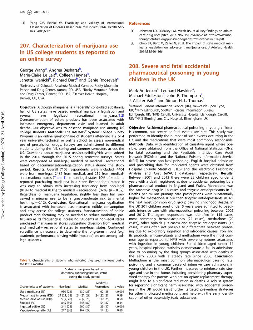
[4] Yang CM, Reinke W. Feasibility and validity of InternationalClassification of Diseases based case-mix indices. BMC Health ServRes. 2006;6:125.
207. Characterization of marijuana usein US college students as reported toan online survey
George Wanga, Andrea Besharatb,Marie-Claire Le Laitb, Colleen Haynesb,Janetta Iwanickib, Richard Dartb and Genie Rooseveltc
aUniversity of Colorado Anschutz Medical Campus, Rocky MountainPoison and Drug Center, Aurora, CO, USA; bRocky Mountain Poisonand Drug Center, Denver, CO, USA; cDenver Health Hospital,Denver, CO, USA
Objective: Although marijuana is a federally controlled substance,half of US states have passed medical marijuana legislation andseveral have legalized recreational marijuana.[1,2]Overconsumption of edible products has been associated withincreased emergency department visits and blamed in adultdeaths. Our objective was to describe marijuana use among UScollege students. Methods: The RADARSVR System College SurveyProgram is an online questionnaire of students attending a 2 or 4year university, technical or online school to assess non-medicaluse of prescription drugs. Surveys are administered to differentstudents during the fall, spring and summer semesters across theUS. Questions about marijuana use/accessibility/risk were addedin the 2014 through the 2015 spring semester surveys. Stateswere categorized as non-legal, medical or medicalþ recreationalbased on decriminalization/legalization status during the studyperiod. Results: Overall 7105 respondents were included: 4404were from non-legal, 2482 from medical, and 219 from medical-þ recreational states (Table 1). In non-legal states 10% of studentsreported purchasing marijuana in a store. Respondents stated itwas easy to obtain with increasing frequency from non-legal(81%) to medical (83%) to medicalþ recreational (87%) (p¼ 0.02).Regardless of marijuana legislation, slightly less than half per-ceived marijuana use to be a great-moderate risk to mentalhealth (p¼ 0.12). Conclusion: Recreational marijuana legalizationis associated with increased use, increased edible consumptionand easy access for college students. Standardization of edibleproduct manufacturing may be needed to reduce morbidity, par-ticularly as its frequency is increasing. Students in non-legal statespurchased marijuana in stores suggesting diversion from medicaland medicalþ recreational states to non-legal states. Continuedsurveillance is necessary to determine the long-term impact (e.g.academic performance, driving while impaired) on use among col-lege students.
References
[1] Johnston LD, O’Malley PM, Miech RA, et al. Key findings on adoles-cent drug use; [cited 2014 Nov 15]. Available at: http://www.moni-toringthefuture.org/pubs/monographs/mtf-overview2014.pdf
[2] Choo EK, Benz M, Zaller N, et al. The impact of state medical mari-juana legislation on adolescent marijuana use. J Adolesc Health.2014;55:160–166.
208. Severe and fatal accidentalpharmaceutical poisoning in youngchildren in the UK
Mark Andersona, Leonard Hawkinsa,Michael Eddlestonb, John P. Thompsonc,J. Allister Valed and Simon H. L. Thomasa
aNational Poisons Information Service (UK), Newcastle upon Tyne,UK; bNPIS Edinburgh, Scottish Poisons Information Bureau,Edinburgh, UK; cNPIS Cardiff, University Hospital Llandough, Cardiff,UK; dNPIS Birmingham, City Hospital, Birmingham, UK
Objective: Accidental pharmaceutical poisoning in young childrenis common, but severe or fatal events are rare. This study wasperformed to identify the number of such events occurring in theUK and the medications that were most commonly responsible.Methods: Data, with identification of causative agent where pos-sible, were obtained from the Office of National Statistics (ONS)for fatal poisoning and the Paediatric Intensive Care AuditNetwork (PICANet) and the National Poisons Information Service(NPIS) for severe non-fatal poisoning. English hospital admissionand prescribing data for implicated agents were obtained fromHospital Episode Statistics (HES) and the electronic PrescribingAnalysis and Cost (ePACT) databases, respectively. Results:Between 2001 and 2013 there were 28 children aged under 5years with a death registered as due to accidental poisoning by apharmaceutical product in England and Wales. Methadone wasthe causative drug in 16 cases and tricyclic antidepressants in 3.Deaths per million primary care prescriptions were substantiallyhigher for methadone (0.58) than tricyclic antidepressants (0.02),the next most common drug group causing childhood deaths. Inthe UK 201 children aged under 5 years were admitted to paedi-atric intensive care with pharmaceutical poisoning between 2002and 2012. The agent responsible was identified in 115 cases,most commonly benzodiazepines (22 cases), methadone (20cases), other opioids (19 cases) and tricyclic antidepressants (13cases). It was often not possible to differentiate between poison-ing due to exploratory ingestion and iatrogenic causes. Iron andits products, anticonvulsants and methadone were the most com-mon agents reported to NPIS with severe symptoms associatedwith ingestion in young children. For children aged under 14years, hospital episode statistics demonstrate a fall in admissionsdue to poisoning by the drug groups associated with deaths inthe early 2000s with a steady rate since 2006. Conclusion:Methadone is the most common pharmaceutical causing fatalpoisoning and a common cause of intensive care admissions inyoung children in the UK. Further measures to reinforce safe stor-age and use in the home, including considering pharmacy super-vised therapy for parents who are on opiate replacement therapy,might lead to a significant reduction in deaths. A robust systemfor reporting significant harm associated with accidental poison-ing in the UK would assist further targeted prevention strategiesfor other implicated medications and help with the early identifi-cation of other potentially toxic substances.
Table 1. Characteristics of students who indicated they used marijuana duringthe last 3 months.
Status of marijuana based ondecriminalization/legalization status
during the study period
Characteristics of students Non-legal MedicalMedicalþ
Recreational p value
Used marijuana (%) 950 (22) 630 (25) 62 (28) <0.001Median age in years (IQR) 24 (21, 28) 24 (21, 29) 24 (22, 27) 0.59Median days of use (IQR) 5 (2, 20) 6 (2, 20) 10 (2, 25) 0.58Smoked (%) 845 (89) 545 (87) 54 (87) 0.34Ingested edible (%) 241 (25) 200 (32) 29 (37) <0.001Vaporize/e-cigarette (%) 247 (26) 167 (27) 14 (23) 0.80
460 ABSTRACTS
Dow
nloa
ded
by [
Kin
gs C
olle
ge L
ondo
n] a
t 07:
31 2
1 A
pril
2016
209. Copper sulphate experiments: anunnecessary hazard at school
Ellen B. Pedersen and Lotte C. G. HoegbergThe Danish Poisons Information Center, Copenhagen UniversityHospital Bispebjerg, Copenhagen, Denmark
Objective: In Danish schools pupils carry out experiments withcopper sulphate in science lessons in order to learn about the for-mation of salt crystals. Copper sulphate can be corrosive to theskin and eyes, and oral exposure often results in intense nausea,vomiting and diarrhea. Human death has been reported afteringestion of gram quantities of this material; the LD50 (oral) forcopper sulphate is 300 mg/kg in rats.[1] The objective of thisstudy was to identify the extent of copper sulphate exposure atDanish schools in order to prevent serious harm. Methods: Aretrospective study based on the systematic registration of enqui-ries to the Danish Poisons Information Centre (DPIC) regardingexposure to copper sulphate. The observation period was January2007 to October 2015; all registrations on the cases, including poi-soning severity grading, were performed systematically andimmediately following the call. Results: In total 156 cases werecollected; there were 11–28 cases/year. The majority of patientswere of school age 11–15 years (n¼ 94); median age was 14years (range 0–80 years). Most exposures occurred in school(n¼ 104). The majority of exposures were accidental (n¼ 144)and/or by play (n¼ 38). The most common routes of exposurewere eye (n¼ 56) and oral (n¼ 52), followed by inhalation(n¼ 33) and dermal exposure (n¼ 13). Eye exposure caused irrita-tion, burning pain and redness; oral exposure caused mucosal irri-tation, gastric pain, nausea and vomiting. Inhalation of coppersulphate vapour and dust caused headache, vertigo and generaldiscomfort. The risk assessment of cases was as follows: no risk(n¼ 31), mild risk (n¼ 45), moderate risk (n¼ 73) and severe risk(n¼ 2). In total 46% of the patients were recommended for fur-ther examination at a healthcare facility. Conclusion: On averagethe DPIC received a call once a month concerning accidentalexposure of school pupils to copper sulphate. Although therehave been no fatalities, we think that exposure to this potentchemical should be prevented by avoiding the use of it in theclassrooms e.g. by substituting it with a less harmful chemical.
Reference
[1] Copper sulfate. POISINDEXVR System (electronic version). TruvenHealth Analytics, Greenwood Village, Colorado, USA; [cited 2016Nov 6]. Available at: http://www.micromedexsolutions.com/
210. Toxic love: a 3-year retrospectiveanalysis of love-related toxicologicaldeliberate self-harm
Primo Bottia, Francesco Gambassia,Alessandra Pistellib, Caterina Canzanib,Alessandra Mocarinib, Emanuela Masinib,c andGuido Mannaionib,c
aSOD Centro Antiveleni, AOU Careggi, Florence, Italy; bSODTossicologia Medica, AOU Careggi, Florence, Italy; cDipartimento diNEUROFARBA, Sezione di Farmacologia e Tossicologia, Universit�adegli Studi di Firenze, Florence, Italy
Objective: Suicide is the 10th cause of death in US, among whichself-poisoning is the 3th cause (2.1/100.000 population).[1]Deliberate self-harm (DHS) poisoning for attempting suicideaccounted for 10.5% of queries to US Poison Centers and 31.7%of all fatalities [2] in 2013. In our experience, relationship prob-lems are an important cause for toxicological DHS. Our aim wasto investigate epidemiological, demographic and clinical aspectsof love-related DHS. Methods: Patients admitted to theToxicological Unit of Florence Careggi University Hospital from2012 to 2014 with DHS by poisoning diagnosis (8.9% of acuteintoxications) were studied. We analyzed clinical records, includingpsychiatric evaluation when available. Patients without psychiatricevaluation, essential for pinpointing the cause(s) and themes ofattempted suicide, were excluded. Results: There were 252patients in which psychiatric evaluation was performed in 157(62.3%). Love-related DHS poisonings numbered 39 (24.8%). Theaverage age was 42.9 years (16–78 years) with male/female ratioof 1 to 2.6. The agents involved were drugs (87%), caustics (8%)and inhalation of carbon monoxide (5%). Drugs cohort was div-ided in the following categories: single psychoactive drug (59.3%),mixed psychoactive drugs (31.5%), non-psychoactive drugs(12.5%), mixed psychoactive and other drugs (9.1%). The averagehospitalization period was 3.9 days (1 to 22 days). All patients sur-vived without sequelae. The psychological factors reported were:pure disappointed love (28.2%), disappointed relationship as con-tributory or precipitating cause (25.6%), separation or divorce(20.5%), violence or abuse (12.8%), partner abandonment (7.6%)and death of partner (5.1%). It was sometimes difficult, also forthe psychiatrist, to distinguish between genuine or simulatedattempted suicide; however the first one usually has aspects of‘‘planned escape’’ such as identification of next of kin or officialcontact, leaving written instructions for funeral, bequests and car-ing for help/support for someone else. Conclusion: Love-relatedDHS is an important cause of poisoning. The psychiatrist shouldinvestigate the patient’s biological and psychopathological trig-gers for suicidal ideation. Love is deeply biological and a ‘‘brokenheart’’ or a failed relationship can have disastrous effects.However, without loving relationships, humans fail to flourish,even if all of their other basic needs are met.
References
[1] Suicide. Facts at a glance [cited 2015 nov 21]. Available at: http://www.cdc.gov/violenceprevention/pdf/suicide-datasheet-a.pdf
[2] Mowry JB, Spyker DA, Cantilena LR Jr, et al. 2013 Annual Report ofthe American Association of Poison Control Centers’ NationalPoison Data System (NPDS): 31st Annual Report. Clin Toxicol(Phila). 2014;52:1032–1283.
211. Native British snake bites in theUK: an estimation of occurrence andantivenom usage
Gillian Jacksona, Victoria A. Eaglinga, David J. Luptona,Caroline E. McGrorya, J. Allister Valeb,Simon H. L. Thomasc, John P. Thompsond andMichael Eddlestona
aNPIS Edinburgh, Scottish Poisons Information Bureau, Edinburgh,UK; bNPIS Birmingham, City Hospital, Birmingham, UK; cNPISNewcastle Regional Drug and Therapeutics Centre, Newcastle, UK;dNPIS Cardiff, University Hospital Llandough, Cardiff, UK
Objective: Little is known about the use of antivenom for Viperaberus envenoming in the UK. There is no central data collection
CLINICAL TOXICOLOGY 461
Dow
nloa
ded
by [
Kin
gs C
olle
ge L
ondo
n] a
t 07:
31 2
1 A
pril
2016
of bites or antivenom use. We aimed to estimate the number ofbites and antivenom administrations from telephone enquiriesand TOXBASEVR accesses. Methods: Telephone enquiries andTOXBASEVR accesses relating to European Viper Antivenom orVipera berus bites received between 1 January 2013 and 30September 2015 were cross-referenced by time and place andgrouped in order to identify cases. Cases were identified fromtelephone enquiries and potential cases identified from TOXBASEVR
accesses that occurred outside of 48 hours from a previousenquiry/access from a user with the same postcode. A time frameof 48 hours was used as only a small number of cases occur andcurrent advice recommends all patients who have been bitten areobserved for 24 hours. Results: There were 106 cases (171 tele-phone enquiries) over the study period; for 29 cases no matchingTOXBASEVR accesses identified. There were 1574 TOXBASEVR useraccesses to the snake entry and 542 to the antivenom entry.After cross-referencing all accesses and telephone enquiries therewere 1316 potential cases identified, including 273 cases (310accesses) where the user only accessed the antivenom entry,these cases were excluded. Antivenom was used in 50 cases iden-tified in telephone enquiries (TOXBASEVR criteria for antivenomadministration met in all cases) and a further 132 possible casesidentified from the access data (user looked at both snake andantivenom entries). Conclusion: The number of Vipera berus casespresenting to healthcare facilities in the UK is very low, estimatedto be 0.06–0.6/100,000 population/year;[1] antivenom administra-tions are estimated to be 0.03–0.1/100,000 population/year.[1]These may be underestimates as clinicians may administer anti-venom without TOXBASEVR guidance; however, it is routine to useTOXBASEVR for poisoned or envenomed patients presenting tohospital in the UK. In addition 273 antivenom only cases wereexcluded since users may have been looking to locate antivenomas per the current National Poisons Information Service (NPIS)/College of Emergency Medicine (CEM) recommendations. Ifthese estimations are representative, the current recommendationfor all emergency departments to stock two vials ofantivenom should be reconsidered in view of the low rate of useand the increased costs of currently available licensed antive-noms. Additional vials should be made available within afew hours.
Reference
[1] Annual Mid-year Population Estimates: 2014; [cited 2015 Nov 26].Available at: http://www.ons.gov.uk/ons/rel/pop-estimate/popula-tion-estimates-for-uk–england-and-wales–scotland-and-northern-ireland/mid-2014/stb—mid-2014-uk-population-estimates.html
212. Iron poisoning and the use ofdesferrioxamine: survey data from theUK National Poisons InformationService (NPIS)
Victoria A. Eaglinga, Leonard Hawkinsb,Gillian Cooperc, Ted Cheungd, Simon H. L. Thomasb,John P. Thompsonc, J. Allister Valed,D. Nicholas Batemana and Michael Eddlestona
aNPIS Edinburgh, Scottish Poisons Information Bureau, Edinburgh,UK; bNPIS Newcastle, Regional Drug and Therapeutics Centre,Newcastle-upon-Tyne, UK; cNPIS Cardiff, University HospitalLlandough, Cardiff, UK; dNPIS Birmingham, City Hospital,Birmingham, UK
Objective: To assess the incidence and severity of iron poisoningcases referred by hospitals to the UK NPIS and the use of desfer-rioxamine. Methods: Data were collected prospectively onpatients presenting to hospital between 1 July 2014 and 20September 2015. Inclusion criteria were: ingestion of a potentiallytoxic dose of iron (�20 mg/kg); patient symptomatic; raised serumiron concentration (greater than or �55 mmol/L); or patient beingtreated with desferrioxamine. Results: Over 14.5 months, 319cases were referred to NPIS; 26 patients did not meet follow upcriteria and were excluded. Patients fulfilled at least one criterionas follows: toxic dose 111/293 (143 no data; 39< 20 mg/kg);symptomatic patient 184/293; serum iron �5 5 mmol/L 128/293(94 no data; 72< 55 mmol/L); received desferrioxamine: 29/293(including 2 requests for information on formulation). Overall37.2% of patients were asymptomatic, 49.1%, 9.2%, and 3.1% hadminor (Poisoning Severity Score [PSS] 1), moderate (PSS 2) andsevere (PSS 3) features, respectively; 1.4% had unknown severity.One third of cases (34.7%) were mixed overdoses. Elemental irondose reported to be ingested ranged from 3.76–313 mg/kg. Theamount of elemental iron ingested did not correlate with PSS atthe time of the call or with plasma iron concentrations (at3.5–4.5 h or 5.5–6.5 h). However, there was a correlation betweenplasma iron concentration at 3.5–4.5 h and PSS. Of the 29 patientstreated with desferrioxamine, 13.8% were asymptomatic, while44.8%, 13.8% and 3.4% of patients were PSS 1, PSS 2 or PSS 3,respectively. Features reported included acidosis (n¼ 12), haema-temesis (n¼ 5), hypotension (n¼ 4, including 2 mixed ingestions)and drowsiness (n¼ 2 patients, both mixed ingestions). Fourteenpatients received doses similar to standard recommendations(15 mg/kg/h up to a dose of 80 mg/kg; total dose given60–83 mg/kg). Six patients were treated with other doses ordetails were unknown. Two enquiries were requests for pharmacyadvice while one related to a treatment error. In six cases treat-ment discontinuation was advised due to the absence of severefeatures and/or elevated plasma iron concentration. No adverseevents/reactions to desferrioxamine were noted. Conclusion: Weobtained information on 293 cases with potentially significantiron poisoning from telephone calls to the NPIS. Reported dose ofelemental iron ingested did not predict either poisoning severityor plasma iron concentrations; however, plasma concentrations at3.5–4.5 hours post-ingestion did correlate with PSS. Few patientshad poisoning significant enough to require treatment withdesferrioxamine.
213. Toxicity of 2,4 dinitrophenol:impact of public health measuresdiscouraging use on episodes oftoxicity referred to the UK NationalPoisons Information Service
Simon L. Hilla, Leonard Hawkinsa, Gillian Jacksonb,Michael Eddlestonb, John P. Thompsonc,J. Allister Valed, Harry K. R. Thanacoodya andSimon H. L. Thomasa
aNational Poisons Information Service (Newcastle Unit), Newcastle-upon-Tyne, UK; bNational Poisons Information Service (EdinburghUnit), Edinburgh, UK; cNational Poisons Information Service (CardiffUnit), Cardiff, UK; dNational Poisons Information Service(Birmingham Unit), Birmingham, UK
Objective: 2,4-Dinitrophenol (DNP) is used as a weight losing and‘‘fat burning’’ drug but may cause severe and sometimes fatal tox-icity.[1] Following an increase in cases reported to the UK
462 ABSTRACTS
Dow
nloa
ded
by [
Kin
gs C
olle
ge L
ondo
n] a
t 07:
31 2
1 A
pril
2016
National Poisons Information Service (NPIS) between 2010 and2013,[2] public health measures were taken in late 2013 and early2014 to reduce use. These included publication of warnings, edu-cational work in gyms and a crackdown on illegal sales. This studywas performed to assess the impact of these measures on presen-tations with toxicity relating to DNP reported to the NPIS.Methods: NPIS telephone enquiry records and user sessions forTOXBASEVR , the NPIS online information database, involving sys-temic exposures to DNP, were reviewed for the period 1 January2007 to 17 September 2015. Results: There were 29 individualcases of DNP toxicity reported between 2007 and 2013, 5 ofwhich had a fatal outcome, with 21 cases and 3 deaths occurringin 2013. Following public health measures there was a reductionin reported episodes, with 9 cases reported throughout 2014.However, referrals to NPIS subsequently increased sharply, with30 cases referred between 1 January and 17 September 2015, fiveresulting in death. Most of those affected were teenagers andyounger adults. Between 2007 and 2013 few cases were female(3/29), but the proportion of females involved in 2014 and 2015increased substantially (22/39, P< 0.001). Accesses to the DNPpage on TOXBASEVR also reduced during 2014 but increased againin 2015. Conclusion: Public health measures taken in the UKreduced the number of exposures with DNP reported to the NPIS,but this effect was short-lived and exposures are now more com-mon in the UK than ever before and increasingly involve females.Further steps are required to reduce consumption and cases ofpoisoning with DNP.
References
[1] Grundlingh J, Dargan P, El-Zanfaly M, et al. 2,4-Dinitrophenol(DNP): a weight loss agent with significant acute toxicity and riskof death. J Med Toxicol. 2011;7:205–212.
[2] Kamour A, George N, Gwynnette D, et al. Increasing frequency ofsevere clinical toxicity after use of 2,4-dinitrophenol in the UK. Areport from the National Poisons Information Service. Emerg MedJ. 2015;32:383–386.
215. Caffeine-containing energy drinksand caffeine poisoning in school-agechildren (5–16 years): TOXBASEVR
access and enquiry data
Victoria A. Eaglinga, David J. Luptona,Caroline E. McGrorya, Simon H. L. Thomasb,John P. Thompsonc, J Allister Valed andMichael Eddlestona
aNPIS Edinburgh, Scottish Poisons Information Bureau, Edinburgh,UK; bNPIS Newcastle, Regional Drugs and Therapeutics Centre,Newcastle-upon-Tyne, UK; cNPIS Cardiff, University HospitalLlandough, Cardiff, UK; dNPIS Birmingham, City Hospital,Birmingham, UK
Objective: To assess the incidence of poisoning with caffeine-con-taining energy drinks in the UK, particularly in school-age childrenand adolescents (5–16 years), by studying accesses to TOXBASEVR
(the UK’s National Poisons Information Service [NPIS] poisonsmanagement database) and telephone enquiries to the NPIS(recorded on the UK’s Poisons Information Database [UKPID]).Methods: We identified the number of TOXBASEVR accesses to allproducts containing caffeine and to caffeine-containing energydrinks and identified relevant NPIS telephone enquiries using thesearch terms ‘‘caffeine’’ and ‘‘energy drink’’ (along with caffeine as
an ingredient) together with the brand names of 13 specificenergy drinks. The period studied was 1 January 2009 to 31 July2015. Results: TOXBASEVR accesses to the caffeine entry steadilyincreased from around 1000 in 2009 to some 2200 in 2014.Accesses to energy drink entries remained constant at around200–250/year. Accesses to energy drinks accounted for 2–5% ofall accesses to caffeine-containing products. A total of 4754 tele-phone calls were received where ‘‘caffeine’’ or ‘‘energy drink’’ wasthe agent or ingredient. One-hundred and sixteen enquiries(2.4%) related to the ingestion of caffeine-containing energydrinks alone. Fifty-two patients were asymptomatic (45%); 55(47%) presented with minor features (Poisoning Severity Score[PSS] 1) and 7 (6%) patients developed moderate (PSS 2) features.There were no severe or fatal exposures reported. Commonlyreported features were agitation (7%), vomiting, tachycardia, pal-pitations (6% each) and abdominal pain (5%). Thirty-five of 116(30%) calls related to school-age children (5–16 years). Fourteenpatients were reported as asymptomatic (40%); 19 (54%) pre-sented with minor (PSS 1) and 2 (6%) with moderate (PSS 2) fea-tures. In this age group, the most commonly reported featureswere: agitation (14%); tachycardia, headache (11% each); vomitingand mydriasis (9% each). Conclusion: Accesses to the TOXBASEVR
entry for caffeine have steadily increased over the last 6 years,but there was no corresponding increase in accesses to entriesfor caffeine-containing energy drinks. It is possible that treatingclinicians are aware that toxicity is related to the caffeine contentand are viewing the caffeine entry directly. There were few callswhich related to single ingestions of caffeine-containing energydrinks and even fewer of these related to ingestions in school-age children. In the majority of cases discussed with the NPIS,patients were asymptomatic or presented with minor features;the incidence of moderate adverse effects was low and no casesof severe poisoning were reported.
216. Fatal poisonings: a 5-year caseseries of the Pavia Poison Centre
Sarah Vecchio, Valeria M. Petrolini, Andrea Giampreti,Francesca Chiara, Emanuela Cortini andCarlo A. LocatelliPoison Control Centre and National Toxicology Information Centre,IRCCS Maugeri Foundation Hospital and University of Pavia, Pavia,Italy
Objective: According to the WHO, 346,000 deaths due to acciden-tal poisoning occurred worldwide in 2004. Moreover, almost a mil-lion people die every year as a result of suicide and, amongthese, the deliberate ingestion of pesticides causes about 370,000deaths/year.[1] We describe fatal intoxications in the case series ofthe Pavia Poison Centre (PPC). Methods: A retrospective review offatal poisonings managed by the PCC from 2010 to 2014 was per-formed. The included cases were assessed for age, sex, modalityand route of intoxication, PSS (Poisoning Severity Score) at thefirst consultation with the PPC and agents involved. Cases forwhich the cause-effect relationship between exposure and deathwas considered absent or unlikely by the specialist toxicologistwere excluded. Results: In the study period, 239 fatal cases wereincluded (50% males, 60 ± 20 years), distributed all over Italy, withan average of 47 deaths/year. Five patients (5/239; 2%) wereyounger than 14 years, and fifty patients (50/239, 21%) were olderthan 78 years. The exposure was considered accidental in 46cases (19%), voluntary in 103 cases (42%) with 34 cases of suicide,and related to an adverse drugs reaction in 59 cases (25%). In 31cases (14%) this data was unknown. Among the cases of suicide,41.5% of patients were aged between 36 and 56 years. Ingestion
CLINICAL TOXICOLOGY 463
Dow
nloa
ded
by [
Kin
gs C
olle
ge L
ondo
n] a
t 07:
31 2
1 A
pril
2016
was the route of exposure in 80% of cases (191/239). Seventy-fivepercent of patients (179/239) presented a serious clinical pictureat the first PPC consultation (PSS 3). Concerning the agentsinvolved, drugs were the cause of death in 110 cases (46%).Among these, the death was related to the ingestion of only onedrug in 65 cases (59%), and metformin was involved in morethan half of these (35/65). In 18% of cases (42/110) more thanone agent was involved. Other agents involved in more than 15cases were caustics (7.5%), pesticides (7.5%), drugs of abuse (7%),gas/smoke/fire inhalation (7%) and mushrooms (5%). Conclusion:Fatal poisonings are not related only to suicide, but also toadverse drug reactions and accidental ingestions. The role of poi-son centers is crucial in identifying the main causes and modalityof lethal intoxications, and in reporting alerts to the authorities inorder to identify possible preventive interventions.
Reference
[1] World Health Organization. The global burden of disease: 2004update; [cited 2015 Nov 20]. Available at: http://www.who.int/healthinfo/global_burden_disease/GBD_report_2004update_full.pdf
217. Poisonings in children under18 years of age presenting to theEmergency Department, LandspitaliUniversity Hospital, Iceland
Gudborg A. Gudjonsdottira, Anna M. Thordardottirb
and Jakob Kristinssona
aUniversity of Iceland, Reykjavik, Iceland; bLandspitali UniversityHospital, Reykjavik, Iceland
Objective: To examine the incidence and nature of poisonings inchildren presenting to the Emergency Department (ED) atLandspitali University Hospital in 2012, and to compare the rateto the average rate of poisonings in all age groups presenting tothe ED the same year, 3.1 cases per 1000 inhabitants.[1]Methods: The study was prospective and included all visits dueto poisonings in children and adolescents under 18 years of ageduring 2012. Information collected included age and gender ofeach patient, location and causes of the poisoning, type andamount of poison, route of exposure, clinical manifestations, treat-ment and outcome. Results: In 2012, 166 children came to theED because of poisoning, 102 girls and 64 boys. That represents arate of 2.2 per 1000 inhabitants in this age group in Iceland.Ingestion was the most common route of exposure (79%) and thepatient’s home was the most common location of exposure. Inchildren aged 0–12 years 98% of the poisonings were uninten-tional but in the age group 13–17 years 80% were deliberate.Drugs and/or alcohol were involved in 66% of cases. The mostcommon group of drugs was selective serotonin re-uptake inhibi-tors (SSRIs) and the most common single drug was paracetamol.Alcohol was involved in 26% of cases in the 13–17 years old butonly in one case in children under 13 years of age. The mostcommon household product exposure was to detergents. Themajority of the children were treated in the ED and dischargedhome within 24 hours; 28 children (17%) were admitted to otherdepartments of the hospital for further treatment. No deathsoccurred. Conclusion: The rate of poisonings in children present-ing to the ED in 2012 is lower than the average rate per 1000inhabitants. Girls outnumbered boys. The nature of poisonings inchildren varies in different age groups. Accidental unintentionalpoisonings are most common in younger children up to 12 yearsold whereas self-poisonings by ingestion of prescription drugs
and/or alcohol accounted for the majority of cases in the oldergroup.
Reference
[1] Gudjonsdottir GA, Thordardottir AM, Kristinsson J. Acute poison-ings in Iceland: A prospective study on poisonings presenting tothe Emergency Department at Landspitali University Hospital. ClinToxicol (Phila). 2014;52:312.
219. Alcohol-based hand sanitizerexposure: characterization andcomparison of US National PoisonData System (NPDS) data and a5-state regional poison center from2010–2014
Shireen BanerjiRocky Mountain Poison Center, Denver, USA
Objective: To characterize alcohol-based hand sanitizer humanexposures within our 5-state Regional US Poison Center (RPC) andcompare our findings to national data. Methods: We reviewed allNational Poison Data System (NPDS) data coded for ethanol-based and isopropanol-based hand sanitizer human exposuresfrom 2010–2014 within our 5-state RPC and compared it to thenational data. Parameters evaluated were age, gender, route, rea-son for exposure, clinical effects, level of healthcare facility (HCF)care, management site, medical outcome, and therapies. Results:Nationally, NPDS reported 78,781 exposures to alcohol-basedhand sanitizers over the study period; 38,485 (48.9%) were female.Most were oral exposures (93%, 73,643), followed by ocular (5.9%)and dermal (3.8%) routes. The most common reason for exposurewas accidental (92%, 72,495) with 4889 (6.2%) exposures deemedas intentional. From 2010 to 2014, the number of intentional mis-use or abuse calls rose each year. Ages ranged from 2 days old to104 years old. Overall 21,118 patients (26.8%) had no effects,6242 (7.9%) had minor effects, 874 (1.1%) had moderate effects,and 72 (0.1%) had major effects. Outcomes were unknown for49,530 (62.9%) of exposures. There were 3 reported deaths duringthe 5-year period. The top 5 clinical effects reported were,respectively, ocular irritation, vomiting, drowsiness/lethargy, nau-sea, and coughing/choking. Most exposures (87.8%, 69,206) weremanaged on site (non-HCF). Our RPC reported 2840 human expo-sures to alcohol-based hand sanitizers during that same period;1394 (49%) were female. Again most exposures were oral (93%,2645), followed by ocular (6%) and dermal (1.4%). The most com-mon reason for exposure was accidental (91.5%, 2599) with 188(6.6%) deemed as intentional. The trend of abuse rose from 2010to 2014. Ages ranged from 3 days old to 98 years old. Mostpatients (78.6%, 2232) had no effects, 430 (15%) had minoreffects, and 30 (1%) had moderate effects. There were no majoroutcomes or deaths reported. Outcomes were unknown for 110(3.9%) of exposures. Top 5 clinical effects were, respectively, der-mal irritation, vomiting, drowsiness, oral irritation, and nausea.Most patients (90.8%, 2578) were managed on site (non-HCF).Conclusion: Alcohol-based hand sanitizer exposure is a commonoccurrence in the US. Most cases involve accidental ingestionscenarios and most cases resolve with minor symptomatologywithout requiring emergency medical attention. National dataresults were very consistent with the numbers at our RPC. Abuse/misuse was a small percentage of cases, but we suspect the risingtrend will continue.
464 ABSTRACTS
Dow
nloa
ded
by [
Kin
gs C
olle
ge L
ondo
n] a
t 07:
31 2
1 A
pril
2016
220. The effect of holidays on acutepoisoning enquiries to the DanishPoison Information Centre
Tonny S. Petersen, Kasper M. Petersen,Mikkel Christensen and Søren BøgevigDepartment of Clinical Pharmacology, University HospitalBispebjerg, Copenhagen, Denmark
Objective: Previous reports have shown that the number of con-tacts to the emergency departments due to poisonings declinesduring holidays.[1,2] However, this has not been examined fromthe perspective of poison centres which are usually the first pointof contact. The aim of the study was to characterize the effects ofholidays on enquiries to the Danish Poison Information Centre.Methods: We extracted all enquiries concerning acute poisoningsfrom the Danish Poison Information Centre Database from 2008to October 2015. The mean number of calls was calculated forschool holidays, bank holidays, school days and weekends strati-fied for age, risk assessment, time of day, type of poisoning, andplace. School days were used as the reference for comparisons.Results: We identified 107,838 enquiries regarding acute poison-ing over the study period. During holidays, the number of callsper day increased by 9.7% from 36.6 to 40.1 enquiries/day. Theincrease was only seen during school holidays with a 12.5%increase while a 7.8% decrease was seen during bank holidays.Enquiries about children aged 0–4 years old predominantly con-tributed to the increase during school holidays with a 33.7%increase. The main increase in enquiries was during the timeperiod from 9 am to 3 pm. The proportion of potentially life-threatening poisonings decreased slightly during school holidaysfrom 5.4% to 5.0% of all calls. In the elderly (over 65 years ofage), an increase in calls concerning medication errors was notedduring bank holidays accounting for 41.7% of the calls comparedto 27.8% of the calls during school days. Conclusion: In contrastto previous reports, we saw an increase in the number of callsduring school holidays, mainly concerning younger children(below 5 years of age).[1,2] A possible explanation could be thatthe daytime child care for the vast majority of pre-school childrenin Denmark are daytime nursery or nursery schools which has alower risk of accidental poisoning.
References
[1] Hughes K, McHale P, Wyke S, et al. Child injury: using nationalemergency department monitoring systems to identify temporaland demographic risk factors. Inj Prev. 2014;20:74–80.
[2] Chien C, Marriott JL, Ashby K, et al. Unintentional ingestion ofover the counter medications in children less than 5 years old. JPaediatr Child Health. 2003;39:264–269.
221. Tracking the trends over time ofunintentional pediatric exposures tobenzodiazepines and opioids reportedto poison centres in the GlobalToxicosurveillance Network
Darcey J. G. Johnsona, Gillian Cooperb,Franca Davanzoc, Martin Ebbecked,Michael Eddlestone, Robert Garnierf,
Zachary R. Margolina, Bruno M�egarbaneg,h,Andreas Schaperd, Fabrizio Sesanac,Simon H. L. Thomase, John P. Thompsone,J. Allister Valee and Jody Greena
aRocky Mountain Poison & Drug Center, Denver, USA; bNationalPoisons Information Center, Cardiff, UK; cPoison Control Centre ofMilan, Milan, Italy; dGIZ-Nord Poisons Centre, University MedicalCenter, G€ottingen, Germany; eNational Poisons Information Service,Edinburgh, UK; fCentre Antipoison et de Toxicovigilance de Paris,Paris, France; gDepartment of Medical and Toxicological CriticalCare, Lariboisiere Hospital, Paris-Diderot University, Paris, France;hINSERM U1144, Paris-Descartes University, Paris, France
Objective: To determine if population rates of unintentional pedi-atric human exposures for benzodiazepines are experiencing simi-lar trends as those of opioid exposures as reported to poisoncentres (PCs) in France, Germany, Italy, the UK, and the US.Methods: Unintentional pediatric exposures to benzodiazepines(alprazolam, diazepam, etizolam, flunitrazepam, lorazepam, lorme-tazepam, nitrazepam, oxazepam, phenazepam, temazepam) andopioids (buprenorphine, fentanyl, hydrocodone, hydromorphone,methadone, morphine, oxycodone, oxymorphone, pethidine, tra-madol) reported to Global Toxicosurveillance Network (GTNet) PCswere examined to observe trend similarities. Unintentional pediat-ric (<6 years) exposures occurring from 2012–2014 were obtainedfrom PCs in Paris (France), G€ottingen (Germany), Milan (Italy), theUK (4 sites), and the US. UK PCs provide medical managementassistance to healthcare providers only, while services in all othercountries are also available to the public. Defined regions of callcoverage exist in the Paris, G€ottingen, UK, and US sites, whileMilan handles 65–70% of calls in Italy. Rates are expressed as thenumber of unintentional pediatric exposures per 100,000 popula-tion. Poisson regression was used to determine differences in ratechanges between benzodiazepine and opioid exposures withboth discrete and continuous covariates. An analysis of covariancetest determined differences between the overall rates changesover time for benzodiazepine and opioid exposures. Results: Forunintentional pediatric exposures, benzodiazepine exposure rateswere higher than opioid exposures in Germany, Italy, and the US,while opioid exposure rates were consistently higher than benzo-diazepines in the UK. Benzodiazepine exposure rates decreased inall countries (statistically significant decreases only in the US[p¼ 0.0038]). Similar decreases over time in opioid exposure rateswere observed in Germany and the US. The negative slopes forbenzodiazepine and opioid exposures within these two countriesdid not significantly differ. In contrast, France, Italy, and the UKexperienced overall increases of opioid exposures. An analysis ofcovariates identified a statistically significant difference betweenthe negative slope of exposure rates for benzodiazepines and thepositive slope of opioid exposures in France (p¼ 0.0455), thoughvariability exists across quarters. Conclusion: Decreases over timein unintentional pediatric exposure rates for both benzodiazepinesand opioids were observed in Germany and the US only.Benzodiazepine and opioid exposure rates have not experiencedsimilar directional trends in France, Italy, or the UK. Despitedecreases over time, further research is warranted to elaborate onthe identification that unintentional pediatric exposure rates ofbenzodiazepines are often higher than those of opioids.
CLINICAL TOXICOLOGY 465
Dow
nloa
ded
by [
Kin
gs C
olle
ge L
ondo
n] a
t 07:
31 2
1 A
pril
2016
222. Tracking the trends over time ofglobal adult human exposures tobenzodiazepines and opioids reportedto poison centres in the GlobalToxicosurveillance Network
Darcey J. G. Johnsona, Gillian Cooperb,Franca Davanzoc, Martin Ebbecked,Michael Eddlestonb, Robert Garniere,Zachary R. Margolina, Bruno M�egarbanee,f,Andreas Schaperd, Fabrizio Sesanac,Simon H. L. Thomasb, John P. Thompsonb,J. Allister Valeb and Jody Greena
aRocky Mountain Poison & Drug Center, Denver, USA; bNationalPoisons Information Service, Cardiff, UK; cPoison Control Centre ofMilan, Milan, Italy; dGIZ-Nord Poisons Centre, University MedicalCenter, G€ottingen, Germany; eDepartment of Medical andToxicological Critical Care, Lariboisiere Hospital, Paris-DiderotUniversity, Paris, France; fINSERM U1144, Paris-Descartes University,Paris, France
Objective: To determine if rates of adult human exposures forbenzodiazepines are experiencing similar patterns over time asfor opioid exposures reported to poison centres (PCs) in France,Germany, Italy, the UK and the US. Methods: Human exposuresto benzodiazepines (alprazolam, diazepam, etizolam, flunitraze-pam, lorazepam, lormetazepam, nitrazepam, oxazepam, phenaze-pam, temazepam) and opioids (buprenorphine, fentanyl,hydrocodone, hydromorphone, methadone, morphine, oxycodone,oxymorphone, pethidine, tramadol) reported to GlobalToxicosurveillance Network (GTNet) PCs were examined toobserve trend similarities. Adult (�18 years) human exposuresoccurring from 2012–2014 were obtained from PCs in Paris(France), G€ottingen (Germany), Milan (Italy), the UK (4 sites), andthe US. UK PCs provide medical management assistance tohealthcare providers only, while services in other participatingcountries are also available to the public. Defined regions of callcoverage exist for the Paris, G€ottingen, UK, and US sites, whileMilan handles 65–70% of calls in Italy. Rates are expressed as thenumber of exposures per 100,000 population (extrapolated fromcountry census data) separately for total, all intentional, and allunintentional exposures. Poisson regression was used to deter-mine differences in rate change between benzodiazepines andopioids with both discrete and continuous covariates. Results:Intentional benzodiazepine exposure rates were consistentlyhigher than opioid exposure rates in each country. The magni-tude of difference between rates of benzodiazepine and opioidexposures appeared greater for intentional exposures than unin-tentional exposures in each country. For total exposures, overalldecreases exist for both benzodiazepines and opioids in France,Germany, the UK, and the US. Within these countries, the decreas-ing slopes of benzodiazepine and opioid exposures did not differsignificantly from each other. An analysis of covariance identifieda statistically significant difference between the slopes of totalexposure rates for benzodiazepines and opioids (p¼ 0.0226) onlyin Italy, where opioid exposures increased over time (p¼ 0.0571)and significantly decreased for benzodiazepines (p¼ 0.0134).Overall decreases in intentional benzodiazepine exposures wereobserved in all countries. Overall decreases in intentional opioidexposures were similarly observed in all countries except Italy,where exposures increased. The difference between intentionalbenzodiazepine and opioid slopes was not significant in anycountry. Conclusion: As benzodiazepine exposures (both totaland intentional) decreased overall in France, Germany, the UK,
and the US, a similar decrease was observed for opioid exposuresin these countries. The only statistically significant differencebetween rate change slopes exists in Italy, where total exposuresto benzodiazepines decreased and total exposures to opioidsincreased.
223. Carbamazepine enquiries to theNational Poisons Information Centreof Ireland: a prospective 7 year study
Niamh English, Edel Duggan and Annette CookeNational Poisons Information Centre Ireland, Dublin, Ireland
Objective: To characterise the epidemiology of carbamazepineenquiries to the National Poisons Information Centre (NPIC)Dublin. Methods: This study prospectively examined all enquiriesinvolving carbamazepine reported to the NPIC from 1 May 2006to 30 April 2013. Variables collated included age, sex, intent, loca-tion of incident, severity and symptoms. Results: During the 7year study period there were 256 enquiries relating to 222patients; 51.4% (n¼ 114) were females, 47.7% (n¼ 106) weremales and gender not recorded 0.9% (n¼ 2). The mean age was32 years (range 11 months – 94 years). The circumstances of theexposure were a) intentional (51.3%; n¼ 114), b) accidental/thera-peutic error (47%; n¼ 103) and c) unknown intent (1.8%; n¼ 4).Of the intentional overdoses, 83% (n¼ 95) were symptomatic andrequired active treatment. The most common features reportedwere dizziness, ataxia, vomiting, blurred vision, tremor, tachycardiaand decreased Glasgow Coma Scale (GCS). In all cases where theGCS was <10, a mixture of drugs including alcohol were co-ingested. Carbamazepine was the only substance taken in 38.7%of cases (n¼ 86), all of which had a GCS �10. The majority ofadult (>12 years) exposures (n¼ 192) occurred in a domestic set-ting (74%; n¼ 142); however 23.4% (n¼ 45) of enquiries origi-nated in a residential care home. Of these 93.3% of enquirieswere due to accidental/therapeutic overdose. Only 6.8% of enqui-ries from residential settings were symptomatic with drowsinessand vomiting the most common symptoms. There were 30 enqui-ries (13.5%) concerning children (age �12 years) with a prepon-derance of boys (53.3%; n¼ 16). All incidents occurred in adomestic setting. Only 16.7% (n¼ 5) developed symptoms withvomiting (n¼ 3), unsteady gait (n¼ 2) and drowsiness (n¼ 1);46.6% (n¼ 14) of children had no symptoms and 36.7% (n¼ 11)had no recorded symptoms. Conclusion: Over 50% of casesinvolving carbamazepine were intentional overdoses and themajority (83%) of these were symptomatic. The GCS only fellbelow 10 when carbamazepine was ingested with other drugsand/or alcohol. A significant percentage, 93.3% of residential carehome enquiries were due to therapeutic errors but less likely tohave symptoms. All paediatric cases occurred in the home indicat-ing that storage of medicines may not be adequate.
224. Radiation incident preparednessof Dutch hospitals
Ronald De Groota, Cornelis J. Van Loonb,Marianne E. C. Leendersa,c and Jan Meulenbelta,d
aNational Poisons Information Center, University Medical CenterUtrecht, Utrecht, The Netherlands; bRadboud University MedicalCenter, Nijmegen, The Netherlands; cDivision of Anesthesiology,Intensive Care and Emergency Medicine, University Medical Center
466 ABSTRACTS
Dow
nloa
ded
by [
Kin
gs C
olle
ge L
ondo
n] a
t 07:
31 2
1 A
pril
2016
Utrecht, Utrecht, The Netherlands; dInstitute for Risk AssessmentSciences, Utrecht University, Utrecht, The NetherlandsObjective: The 2011 Fukushima nuclear incident and increasingterrorist threat underscore the necessity for hospitals to preparefor casualties of incidents involving radioactive material and/orionizing radiation. This is also recognised in International AtomicEnergy Agency (IAEA) Safety Standard "Preparedness andResponse for a Nuclear or Radiological Emergency" (No. GS-R-2)that provides requirements for "Managing the medical response".To determine compliance with paragraph 4.78 that requiresarrangements for ‘‘initial medical treatment of contaminated orhighly exposed individuals in local medical facilities’’, the DutchPoisons Center (PC) assessed radiation incident preparedness ofDutch hospitals. Methods: An online survey on resources andknowledge concerning management of casualties of radiationincidents was sent to all Dutch hospitals with a 24/7 availableemergency department. Additionally, nine hospitals were visitedfor an in-depth interview. Results: The response to the online sur-vey was 67% (58/87 hospitals responded). Most hospitals indi-cated that they prepare for the care of casualties of radiologicalincidents (86%). In small hospitals (<500 beds) the percentage is77% and larger/academic hospitals almost all take into accountradiation incidents. About two thirds of responding small hospi-tals, three quarter of larger hospitals and all academic hospitalscan decontaminate casualties contaminated with radioactivematerial. The decontamination capacity is 1–5 persons/hour formost hospitals (83%) with increase in capacity with hospital size(maximum of 30 persons/hour for two larger/academic hospitals).Most hospitals have indoor decontamination facilities (70%) and aminority have outdoor facilities (e.g. decontamination tents). Ofhospitals without decontamination facilities 75% have not madearrangements with other hospitals about referral of contaminatedpatients. Around 70% of hospitals describe radiological incidentsin an emergency response plan but most hospitals do not specif-ically perform exercises with radiation incidents. A majority ofhospitals (81%) knows that the Dutch PC advises on patient man-agement after radiation incidents. Conclusion: In general, there iscompliance with the IAEA "initial medical treatment" requirementin Dutch hospitals. The main recommendation from this study isthat hospitals that are not yet prepared, either create decontam-ination facilities or make arrangements for referral of contami-nated victims to a hospital with facilities. Additionally, it isrecommended that hospitals make regional arrangements inadvance about distribution of larger groups of casualties if theirown capacity is limited. Finally, it is important to include emer-gency response to radiation incidents in a disaster relief plan andperform exercises.
225. Trends in colchicine exposuresreported to the Poisons InformationCentre Erfurt
Anne St€urzebecher, Gesine Liebetrau, Michael Detersand Helmut HentschelPoisons Information Centre, Erfurt, Germany
Objective: The Poisons Information Centre Erfurt has observedgradually increasing numbers of exposures to Colchicum autumnalesince 2005, particularly due to confusion with Allium ursinum,whereas the number of exposures to various colchicine containingdrugs was relatively consistent. We evaluated the causes and risksof these cases. Case series: Between October 1994 and October2015, a total of 210 exposures to colchicine were reported. Ofthese 139 cases were exposure to Colchicum autumnale (in 55cases uncertain) and 71 cases of (certain) exposure to various
colchicine-containing drugs. Of all 155 definite exposures, 31cases (20%) were suicide attempts, 80 cases (51.6%) were acci-dental ingestions (about half of those in children) and in 5 cases(3.2%) adverse events at therapeutic doses had occurred.Medication errors were observed in 38 cases (24.5%), in whichprolonged application of a therapeutic dose often resulted in atleast gastrointestinal symptoms, but no fatality. Case 1: A 71-year-old female developed prolonged gastrointestinal symptoms afteringestion of three ‘‘leaf tips’’ of Colchicum autumnale in a suicideattempt. She was in intensive care for 4 days and was subse-quently transferred to a psychiatric ward. Case 2: A 58-year-oldmale ingested 3 mg of colchicine per day over a period of 3weeks and then reported to hospital with abdominal pain, diar-rhoea, mild thrombocytopenia and elevated liver enzymes.Observation in hospital was recommended, however the furthercourse is unknown. Case 3: A 89-year-old male with chronic painsyndrome and depression due to rheumatoid arthritis developedsevere symptoms (gastrointestinal bleeding, dyspnoea, hyperten-sion, first-degree atrioventricular block, lactic acidosis) resulting inmultiple organ failure and death within 24 hours following theingestion of approximately 100 ml ColchysatVR (¼ 50 mg colchicine)in a suicide attempt. Conclusion: Severity of adverse and toxiceffects, respectively, seems to increase proportionally to theadministered dose and duration of use. Ingestion of parts ofColchicum autumnale plants often leads to at least gastrointestinalsymptoms, even after a minor quantity. With pharmaceuticals,even doses not exceeding the recommended daily dose can leadto severe symptoms if taken over a longer period of time.Ingestion of large amounts (drugs or plants) usually results insevere gastrointestinal symptoms, multiple organ failure anddeath within 24 to 72 hours after ingestion.
226. Paediatric toxicology emergencysimulation training: beyonddiscussions on "one pill can kill"
Gene Yong-Kwang OngKK Women’s and Children’s Hospital, Singapore
Objective: Acute life-threatening paediatric toxicological emer-gencies are rare in most parts of the developed world. Currentmethods of critical paediatric poisoning typically involve didacticlectures and interactive case-based discussions on ‘‘one pill cankill" toddler toxicology. However, to be effective in the manage-ment of critically poisoned children, multi-factorial considerationsare involved and not just textbook knowledge. It is postulatedthat high fidelity simulation training would be useful to augmentexisting methods of critical paediatric toxicology learning.Methods: A 4-hour high fidelity, high technology simulation train-ing session involving only paediatric critical poisoning was con-ducted. Each simulation session was followed by a group debriefdiscussing the management and summarising critical toxicologicaland resuscitation learning points. Pre- and post-simulation surveyswere done to assess the participants’ assessment of their learningexperience. Results: In total 11 physicians (6 paediatric emer-gency physicians, 3 paediatric medicine residents and 2 emer-gency medicine residents) underwent the high fidelity simulationcourse, during which 80% of the participants correctly identifiedthe likely causative toxicological agents on just the history andphysical examination alone. Despite this knowledge, however,when managing the simulated patients, there were significantdelays and suboptimal management as more than half of the par-ticipants had fixation errors and were caught up in advanced car-diac and trauma life support algorithms. It was also noted thatthere needed to be broader approaches to include more common
CLINICAL TOXICOLOGY 467
Dow
nloa
ded
by [
Kin
gs C
olle
ge L
ondo
n] a
t 07:
31 2
1 A
pril
2016
differentials such as sepsis during the sessions. The participantsreflected that the high fidelity simulation was very useful in pro-viding a link in the gap between clinical knowledge and bedsideapplication. Conclusion: High fidelity simulation training was use-ful and could be used to augment current methods of paediatriccritical toxicology education.
227. The correlation between TOXINZenquiries or National PoisonsInformation Centre calls andemergency department (ED)presentations
John Fountaina, Sarah Wellsa, Paul Quigleyb andSandra Allmarkc
aUniversity of Otago, Dunedin, New Zealand; bWellington Hospital,Wellington, New Zealand; cCapital and Coast District Health Board,Wellington, New Zealand
Objective: ED staff within New Zealand have increasingly utilisedthe Internet accessible poisons information database TOXINZ.Accompanying this there has been a corresponding decline intelephone enquiries to the National Poisons Information Centre(PIC) from these professionals. This change in media for accessingpoisoning management advice has implications for type of datacollected by a PIC and may impact toxicovigilance functions. Inparticular, it is not known whether Internet poisons enquiries canbe used for toxicovigilance purposes. The objective of this studyis to identify if there is a correlation between TOXINZ or PICenquiries and the poisonings that present to an ED. Methods:The records of all patients presenting with poisoning to theWellington Hospital ED over the six year period (2007 to 2012)were reviewed to identify the most common pharmaceuticalgroups involved. Data was also extracted over the same time forTOXINZ enquiries from this ED, and from PIC records for theWellington region. Yearly data were analysed in MS ExcelTM 2010and comparisons assessed using the Pearson product-momentcorrelation coefficient. This research was approved by theUniversity of Otago Human Ethics Committee. Results: The num-ber of presentations or enquiries during the six year period forparacetamol was: ED 750; TOXINZ 590; and PIC 826. For the anti-psychotics: ED 376; TOXINZ 733; and PIC 323. The benzodiaze-pines: ED 371; TOXINZ 252; and PIC 262. The selective serotoninre-uptake inhibitors (SSRIs): ED 362; TOXINZ 765; and PIC 263: Fortricyclic antidepressants (TCAs): ED 116; TOXINZ 212; and PIC 127.For opioids: ED 326; TOXINZ 430; and PIC 417. Correlation coeffi-cients for paracetamol exposure presentations to the WellingtonED, and TOXINZ and PIC enquiries for this drug were -0.28 and0.85 respectively; for the antipsychotic group, 0.13 and 0.33; forbenzodiazepines, -0.58 and 0.81; the SSRIs, 0.35 and 0.13; theTCAs, 0.37 and 0.25; and the opioids, 0.11 and 0.52. Conclusion:While high correlations with ED presentations were found for PICenquiries regarding paracetamol and benzodiazepines, there waswide variation over the range of compounds reviewed (from 0.13to 0.85). The results for TOXINZ were even less encouraging, withcoefficients ranging from -0.58 to 0.37. It can therefore be con-cluded that, certainly in New Zealand, enquiries to an Internetpoisons database from an ED do not reflect presentations of over-dose, and that calls to a PIC from the region serviced by an EDare also an unreliable surrogate for presentations.
228. Behind the scenes of snow globetoxicity
Sian C. D. Harbona, J. Allister Valeb,Michael Eddlestonc and John P. Thompsona
aNPIS Cardiff, University Hospital Llandough, Cardiff, UK; bNPISBirmingham, City Hospital, Birmingham, UK; cNPIS Edinburgh,Scottish Poisons Information Bureau, Edinburgh, UK
Objective: To report on enquiries to the UK National PoisonsInformation Service (NPIS) regarding snow globes and to raiseawareness of potential toxicity from ingestion of their contents.Methods: Records of enquiries received by the NPIS were interro-gated retrospectively between the 2008 and 2014 for any refer-ence to the terms snow globe or snow scene. Results: A total of57 enquiries involving 50 cases were received during the specifiedperiod. All exposures were accidental as a result of the itembreaking or leaking. Children under 5 years accounted for 47 ofthe 50 cases, 70% of these involved males. Forty-six percent ofcases were received in December and January which is unsurpris-ing as snow globes are commonly used as Christmas decorations.The remaining cases were spread throughout the year. Forty two(84%) patients were asymptomatic. Where skin contact wasreported (n¼ 7), pruritus and an erythematous rash occurred inthree cases. Following ingestion (n¼ 47) features included cough-ing and mouth ulcers. In two cases, both involving childreningesting less than 20 mL, abnormal anion (AG) and osmolar gaps(OG) were reported. Case 1: AG 23.6 mmol/L, OG 10.8 mmol/L at20 hours post-ingestion. Case 2: AG 21.6 mmol/L, OG 27 mmol/L at6 hours post-ingestion. In both cases no clinical symptoms werereported and no antidote treatment for toxic alcohol poisoningwas given. Unfortunately, further blood gas analysis and toxicalcohol concentrations were not available for either patient.Conclusion: Snow globes have previously contained fine frag-ments of bone/porcelain in distilled water. More recently however,contents have changed to calcium carbonate in distilled waterwith added glycerol to slow the fall of the fragments.Manufacturers have also started adding ethylene glycol, report-edly up to 20% of the volume of water [1] to reduce the risk offreezing during transit or storage. At time of writing no seriouscases of toxic alcohol poisoning have been identified in thesedata or in published literature, but the abnormal blood results inthese children warrant greater consideration as to the risks of poi-soning via ingestion, particularly in young children.
Reference
[1] Patterson C, Bell-Davis P, Rawls H, et al. Ethylene Glycol Contentof Snow Globes. Clin Toxicol (Phila) 2007;45:635.
229. A weight loss product containingfluoxetine
Chantal C. J. Roelena, Antoinette J. H. P. Van Riela,Bastiaan J. Venhuisb, Irma De Vriesa andJan Meulenbelta,c,d
aNational Poisons Information Center, University Medical Center,Utrecht, The Netherlands; bNational Institute for Public Health andthe Environment RIVM, Bilthoven, The Netherlands; cDepartment ofIntensive Care Medicine, University Medical Center Utrecht, Utrecht,The Netherlands; dInstitute for Risk Assessment Sciences, UtrechtUniversity, Utrecht, The Netherlands
468 ABSTRACTS
Dow
nloa
ded
by [
Kin
gs C
olle
ge L
ondo
n] a
t 07:
31 2
1 A
pril
2016
Objective: In 2014 the Dutch National Poisons Information Center(DPIC) received 90 enquiries regarding the use of stimulatingsport and/or slimming supplements resulting in effects such astachycardia, palpitations, restlessness, nausea and vomiting. Herewe report 5 cases of a Turkish weight loss product ‘‘Irem Naturel’’which is available online throughout Europe and used, particularlyin the Turkish community. It is highly promoted via social mediaand claims to be a safe, healthy and fast way to lose weight. Thelabel states only herbal ingredients: red chili pepper, guarana,tragant gum, Citrus aurantium, starch and green tea. Capsulesfrom one case were obtained by the DPIC and analysed by theNational Institute for Public Health and the Environment. Casereports: In 2014, a 22-year-old female reported nausea afteringestion of 3 capsules of Irem Naturel. In March 2015, a 29-year-old female experienced anxiety, agitation, dry mouth, mild hyper-tension (120/100 mmHg) and tachycardia (136 beats/min) onehour after taking 1 capsule of Irem Naturel. Capsules from thiscase were obtained and laboratory analysis showed 30 mg fluox-etine, a selective serotonin reuptake inhibitor. In June 2015, a 41-year-old female experienced severe vomiting 2 hours after inges-tion of 1 capsule. She was hospitalized for one day because ofpersistent vomiting. Capsules were not available for laboratoryanalysis. Two cases were reported in October 2015: a 1-year-oldgirl who ingested half a capsule and remained asymptomatic, andan 18-year-old female who attempted suicide with 20 capsules.Reported symptoms 3 hours after ingestion were dizziness, nauseaand angina pectoris. It is not known whether these patients usedother (prescribed) medicines at the time of the reported symp-toms, or whether they used Irem Naturel for the first time.Conclusion: A food supplement for weight loss, marketed asbeing of herbal origin, contained fluoxetine. A previous sample ofIrem Naturel from 2014 showed sibutramine. It cannot beexcluded that some of the symptoms the patients presentedwith, were caused by fluoxetine or sibutramine. Nausea, vomiting,palpitations, and tachycardia are well known side effects of bothdrugs. Once again, this is a signal that healthcare practitionersneed to be aware of the potential risks of these products. Poisonscenters continuously need to screen their reports on food supple-ments, particularly for multiple enquiries about the same product.Collaboration with laboratory facilities is crucial to determine thecomposition of the supplements.
230. Is a urine toxicology report usefulat the emergency department forpatients who are intoxicated withbenzodiazepines?
Luis L�opez, Vanesa Anton, Francisca C�ordoba,Isabel De Vall�es Garcia, Jessica Rey, Ana And�ujar andGemma AgudoHospital Moises Broggi, Sant Joan Desp�ı-Barcelona, Spain
Objective: The majority of cases of intoxication seen atEmergency Department of Moises Broggi Hospital, Barcelona,Spain involve medications, and the most common drugs involvedare benzodiazepines. This study analyses the economic and clin-ical repercussions of demanding a urine toxicology report forpatients with benzodiazepine intoxication. Methods: Urine toxicol-ogy reports were analysed in patients who registered at theEmergency Department (ED) with benzodiazepine intoxication inthe period January to December 2013. Results: In 2013, 635patients with poisoning were seen at the ED and 38% (243 cases)of these were due to drugs (alone or in combination with othersubstances), and 156 cases of these (64%) were due to
intoxication with benzodiazepines. Urine toxicology determinationwas carried out for 52 (33%) of these 156 cases, although 61% ofthese patients took benzodiazepines as usual treatment. In 100%of these cases the urine was positive for benzodiazepines. In add-ition, samples were also positive for cocaine (n¼ 6), cannabis(n ¼ 5), tricyclics antidepressants (n ¼5), methamphetamine(n¼ 1) and methadone (n¼ 1). All results impacted the final diag-nosis without verifying them. Flumazenil was administered in 42%of the cases, and 17% of the patients required no treatment. Thetests cost the hospital E2996.70 over the one year period.Conclusion: Urine toxicology testing did not influence treatmentor the evolution of these patients and are costly to perform. Torecord a patient�s medical history diagnosis of substance usebased on these urine tests could pose a problem, both scientific-ally and legally.
231. A case of methanol intoxicationtreated with continuous renalreplacement therapy
Hyun Ju Yoona, A. Young Choa, In O. Suna,Soo-Wan Chaeb, Soon-Ok Nohc, Yun Jo Chungd andKwang Young Leea
aDivision of Nephrology, Department of Internal Medicine,Presbyterian Medical Center, Jeonju, Republic of Korea;bDepartment of Pharmacology, Chonbuk National UniversityMedical School, Jeonju, Republic of Korea; cClinical Trial Center forFunctional Foods, Chonbuk National University Hospital, Jeonju,Republic of Korea; dCenter for University-wide Research Facilities,Chonbuk National University, Jeonju, Republic of Korea
Objective: Methanol poisoning is a medical emergency whererapid elimination of the toxin and its metabolite is crucial forrecovery. Although serum methanol concentrations are helpful todecide the timing of start and termination of extracorporeal treat-ment, methanol assays are not a standard laboratory test inKorea. Here, we report a case of methanol poisoning treated withcontinuous veno-venous hemodiafiltration (CVVHDF), in whichclinical improvement was confirmed with serum methanol con-centrations. Case report: A 46-year-old Korean woman wasbrought to the emergency department in an unconscious stateafter ingestion of 150 ml of methanol. She was drowsy with aGlasgow Coma Scale (GCS) of E2V4M5. Her blood pressure was100/70 mmHg and heart rate was 77 beats/min. The blood ureanitrogen and serum creatinine concentrations were 9 mg/dL (ref-erence 8–20 mg/dL) and 0.6 mmol/L (reference 0.6–1.2 mg/dL),respectively. Initial arterial blood gas analysis showed metabolicacidosis (pH 7.180, pCO2 33.0 mmHg, HCO3
� 13.0 mmol/L). Theserum osmolal gap and anion gap were 249 mOsm/kgH2O and21.7 mmol/L, respectively. Despite the increase of central venouspressure from 5 cmH2O to 8 cmH2O after loading 2 L of normalsaline, hypotension continued. She was admitted to the intensivecare unit and treated with CVVHDF and intravenous vasopressors.Following CVVHDF for 12 hours, GCS increased to E3V4M5 andthe acid-base status of the patient returned to normal ranges. In
Table 1. Laboratory findings in a patient with methanol poisoning treated withcontinuous veno-venous hemodiafiltration.
Parameters Day 1 Day 2 Day 3 Day 4
pH 7.180 7.348 7.493 NABicarbonate (mmol/L) 12.3 11.9 26.9 NAOsmolal gap (mOsm/kgH2O) 249 NA �6 �6Anion gap (mEq/L) 21.7 20.1 7.1 NASerum methanol (mg/L) 6547 1470 391 94
CLINICAL TOXICOLOGY 469
Dow
nloa
ded
by [
Kin
gs C
olle
ge L
ondo
n] a
t 07:
31 2
1 A
pril
2016
addition, blood pressure increased to 120/80 mmHg. We meas-ured blood and urine concentrations of methanol during CVVHDFand were initially 6546.9 ppm (mg/L) and 5895.1 ppm (mg/L),respectively. Following CVVHDF, the serum methanol concentra-tion decreased to 94 ppm (mg/L). She was discharged withoutcomplications on the fifth day. Conclusion: Measurement ofserum methanol is useful in the management of methanolintoxication.
232. Circulatory effects of energydrinks after exercise
Niklas Od�en and Kai KnudsenDepartment of Anesthesia and Intensive Care, SahlgrenskaUniversity Hospital, Gohenburg, Sweden
Objective: Energy drink (ED) consumption has been associatedwith serious cardiovascular events,[1–2] and although not recom-mended after exercise, is frequently consumed after training ses-sions. The objective of this study was to evaluate the circulatoryeffects of EDs on recovery immediately after exercise and to com-pare the effects with intake of caffeine alone at rest. Methods:Fifteen healthy volunteers participated in two sessions of exerciseby spinning on bicycles for 45 minutes, immediately followed bytwo units of EDs (Red Bull Light, total caffeine 160 mg) or the cor-responding volume of a sports drink (Powerade 18 kcal/100 mL).Two weeks later the participants ingested the alternative drink ina crossover manner. Heart rate and blood pressure (BP) weremeasured after training and after 30 and 60 minutes of rest. Heartrate variability (HRV) was measured in three individuals for12 hours. In another trial ten healthy volunteers ingested 3 unitsof ED (Red Bull, total caffeine 240 mg, taurine 3000 mg) at rest orthe corresponding amount of caffeine in tablets (250 mg).Results: Pulse rate increased during the training session,decreased thereafter and did not differ between the two groupsat rest. BP increased more in subjects that ingested ED comparedto the sports drink. Systolic BP was 10 mmHg higher after60 minutes, mean arterial pressure was 4.3 mmHg higher after30 minutes and 7.7 mmHg higher after 60 minutes. Heart rateincreased more after intake of ED compared to caffeine alone butwas not significantly different. Both caffeine and ED increased PQinterval on the electrocardiogram (ECG). Systolic BP increasedmore with ED compared to caffeine alone but was not signifi-cantly different. Nine out of ten reported adverse symptoms(headache, nausea, palpitations, tremor) after ED compared tonone after caffeine alone. Conclusion: Two units of EDs afterexercise did not affect heart rate recovery after exercise, but BPincreased significantly (probably due to the combined effect oftaurine and caffeine). The physiological impact of this BP increaseis unclear but may indicate a possible negative health effect.
References
[1] Sanchis-Gomar F, Pareja-Galeano H, Cervellin G, et al. Energy drinkoverconsumption in adolescents: implications for arrhythmias andother cardiovascular events. Can J Cardiol. 2015;31:572–575.
[2] O’Brien MC, McCoy TP, Rhodes SD, et al. Caffeinated cocktails:energy drink consumption, high-risk drinking, and alcohol-related consequences among college students. Acad Emerg.2008;15:453–460.
233. A study of Phlomis oliviericytotoxicity
Avishan Farzaneha, Seyed Nasser Ostadb andJinoos Asgarpanahc
aPharmaceutical Sciences Branch, Islamic Azad University, Tehran,Iran; bDepartment of Toxicology and Pharmacology, TehranUniversity of Pharmacy, Tehran, Iran; cDepartment ofPharmacognosy, Pharmaceutical Sciences Branch, Islamic AzadUniversity, Tehran, Iran
Objective: Over 50% of drugs in clinical trials for anticancer activ-ity were isolated from natural sources or are related to naturally-occurring compounds including Vinca alkaloids, vinblastine andvincristine isolated from Catharanthus roseus,[1] and the lignanderivatives, etoposide and teniposide.[2] The pharmacologicalactivities of some Phlomis species such as antidiabetic, immuno-suppressive,[3] anticancer and antioxidant activities [4] have beeninvestigated previously. In this study we evaluated cytotoxicityeffect of Phlomis olivieri. Methods: The flowering aerial parts ofPhlomis olivieri were collected from the Khansar district ofPakistan for evaluation of its in vitro cytotoxic activity on differentcell lines. We examined the antiproliferation effect of total metha-nol extract and different fractions including petroleum ether,chloroform, ethyl acetate, methanol and aqueous against coloncarcinoma (HT-29), colorectal adenocarcinoma (Caco-2), breastductal carcinoma (T47D) and Swiss mouse embryo fibroblast (NIH3T3) cell lines using MTT assay. Results: According to the halfmaximal inhibitory concentration (IC50), the chloroform fractionand total extract of Phlomis olivieri exhibited high cytotoxic activ-ity on all cell lines except Caco-2 cells but it had moderate anti-proliferative effect on this cell line. In addition, the petroleumether fraction demonstrated moderate cytotoxic effect on HT-29cells although it had a high antiproliferative effect on other celllines. IC50 values confirmed that the growth and proliferation ofHT-29 and T47D cells were most affected by chloroform, petrol-eum ether and total extracts due to their nonpolar compounds.Higher cytotoxic activity of nonpolar fractions may be due to thehigh content of germacrene D in their essential oil.[5] Conclusion:The results emphasize the importance of isolation and character-ization of active components as well as the investigation of spe-cific cytotoxic pathways which may help to determine whetherthe fraction is valuable for antineoplastic effects.
References
[1] Leveque D, Jehl F. Molecular pharmacokinetics of Catharanthus(vinca) alkaloids. J Clin Pharmacol. 2007;47:579–588.
[2] Choi HJ, Cho BC, Shin SJ, et al. Combination of topotecan and eto-poside as a salvage treatment for patients with recurrent smallcell lung cancer following irinotecan and platinum first-linechemotherapy. Cancer Chemother Pharmacol. 2008;61:309–313.
[3] Limem-Ben Amor I, Boubaker J, Ben Sgaier M, et al.Phytochemistry and biological activities of Phlomis species. JEthnopharmacol 2009;125:183–202.
[4] Kamel MS, Mohamed KM, Hassanean HA, et al. Iridoid and mega-stigmane glycosides from Phlomis aurea. Phytochemistry 2000;55:353–357.
[5] Sarkhail P, Amin G, Shafiee A. Composition of the essential oil ofPhlomis olivieri Benth from north of Iran. Daru J Pharm Sci 2006;14:71–74.
470 ABSTRACTS
Dow
nloa
ded
by [
Kin
gs C
olle
ge L
ondo
n] a
t 07:
31 2
1 A
pril
2016
234. Public awareness about themarket launch of mercury energy-saving lamps in Germany: assessmentof an overhyped case of allegedpoisoning
Axel Hahn, Kathrin Begemann, Esther Feistkorn,Nina Glaser and Matthias GreinerBundesinstitut f€ur Risikobewertung (BfR), Berlin, Germany
Objective: Energy-saving lamps (ESL) are compact fluorescentlamps (FL) containing small amounts of mercury (Hg). Invented in1926, they were produced in billions worldwide. The reducing ofgreenhouse gas emissions resulted in a ban on incandescent lightbulbs (EU Parliament, 2009) and for replacement, the FL principlewas miniaturized. A rapid self-declared public opposition arguedagainst the EU legislation based on an early precedent case inGermany is reported here. An alleged "specialist" made a telediag-nostic assessment of the case. His findings were distributed byalmost all media. Case report: An ESL broke when a switched-onfloor lamp fell over. The father picked up the remnants and dis-carded them. The next night, the 9-month-old male infant devel-oped an acute pseudocroup. His 5-year-old brother developed anon-itchy rash all over his body and, 4 days after the breakage,progressive hair loss, which 4 months later also affected his eye-brows and lashes. After 8 months, this boy developed morningtremor of his hands and diarrhoea. The children were repeatedlyexamined by paediatricians, specialists in dermatology/allergol-ogy/environmental medicine and at an university environmentalhealth centre. Neither the analyses of body fluids, and other clin-ical materials nor the ambient air measurements revealed any ele-vated concentrations of mercury, thallium or lead. Based on allavailable documents, no causal relationship could be establishedbetween the non-itchy rash, the temporary alopecia areata totalisand the mercury exposure. At the time the ESL was broken, bothparents and children had been suffering from heavy respiratorytract infections, which could sufficiently explain the symptomsand signs observed. Nevertheless, this "psychodynamic event"triggered a highly interesting campaign against ESL in 2010. Itreached its absolute culmination in a documentary entitled ‘‘BulbFiction’’ shown in cinemas (Austria 2011, Germany 2012) and pro-duced countless enquiries (about 1000/year) to German PoisonCentres without a single relevant case. Conclusion: As a result ofthe exaggeration of the risks, the young patient and his familybecame extremely unsettled and frightened. Due to the publicpressure even governments failed to rely on objective criteria intheir perspective, although a well-respected university institutecould provide the necessary elucidation and technically correctassessment. Today, the overhyped case of alleged poisoning froman ESL has completely fallen into oblivion among the media andthe public. The lesson to learn is that Poison Centres, in particular,have sufficient knowledge to fight against incorrect media cover-age and exaggeration of risks.
235. Chlorprothixene overdose:analysis of enquiries to the DanishPoison Information Center
Kasper M. Petersen, Tonny S. Petersen,Søren Bøgevig and Mikkel Bring Christensen
Department of Clinical Pharmacology, University HospitalBispebjerg, Copenhagen, Denmark
Objective: Chlorprothixene has been marketed since 1959.Nevertheless, data concerning overdose are sparse. The aim ofthis register-based study was to gain knowledge on trends in poi-sonings in relation to prescribing patterns with this antipsychotic.Methods: We assessed all enquiries to the Danish PoisonInformation Center (DPIC) concerning chlorprothixene from 2007to October 2015. Additionally, we searched the Register ofMedicinal Product Statistics [1] in order to determine chlorprothix-ene prescribing patterns within the same period. Results: Weidentified 2422 enquiries concerning chlorprothixene poisoningsover the study period. Chlorprothixene accounted for 32% of allenquiries regarding poisonings with antipsychotics. The numberof chlorprothixene poisonings per year showed a small decreasethroughout the study period. In addition, the fraction of chlorpro-thixene enquiries out of all antipsychotics decreased from 45% in2007 to 23% in 2015, mainly due to an increased frequency ofenquiries regarding other antipsychotics. Chlorprothixene was theseventh most common cause of contact to the DPIC regardingany medication in the study period. Within the last year, chlorpro-thixene was the fifth most common medication poisoning(n¼ 314) among antipsychotics, only surpassed by quetiapine(n¼ 642). The number of defined daily doses (DDDs) of chlorpro-thixene sold decreased through the study period, from 1,249,000in 2007 to 1,060,000 (3.9% of all antipsychotics) in 2013. In con-trast, 6,414,000 DDDs of quetiapine (23.6% of all antipsychotics)were sold in 2013.[1] Conclusion: To our knowledge, this is thelargest study investigating trends in enquiries to a poison centerregarding chlorprothixene poisonings. We found that the trend inenquiries reflected prescribing patterns (i.e. chlorprothixene DDDssold), both decreasing through the study period. Nonetheless, thefrequency of enquiries to the DPIC concerning chlorprothixenewas found to be greatly out of proportion compared to a newergeneration antipsychotic. This discrepancy was consistentthroughout the study period. The frequency of chlorprothixeneenquiries to our poison center was also high compared to previ-ous analyses of poison center data.[2] The disproportionately highfrequency of chlorprothixene enquiries may be due to pharmaco-logical features of chlorprothixene, or characteristics of this par-ticular patient population associated with an increased risk ofoverdose. This needs to be further investigated.
References
[1] The Danish Ministry of Health and Prevention. Register of medi-cinal product statistics; [cited 2015 Nov 20]. Available at: www.medstat.dk/
[2] Fuhrmann M, Hruby K, Lenz K, et al. [New aspects of chlorpro-thixen-poisoning.] [Article in German] Wien Klin Wochenschr.1982;94:150–153.
236. A systematic review of clinicalpresentations of cyanide poisoning
Jennifer L. Parker Cote, Justin Rizer, Anjali Kapil,Emma Kingsbury, J. Priyanka Vakkalanka andChristopher Holstege
Department of Emergency Medicine, Division of MedicalToxicology, University of Virginia, Charlottesville, USA
Objective: There are inconsistencies in the medical literaturedescribing clinical findings associated with cyanide toxicity.
CLINICAL TOXICOLOGY 471
Dow
nloa
ded
by [
Kin
gs C
olle
ge L
ondo
n] a
t 07:
31 2
1 A
pril
2016
The aim of this systematic review was to identify isolated cyanidepoison cases and to identify reported signs, symptoms andlaboratory findings. Methods: We searched MEDLINE, CochraneReviews, and Web of Science case reports and series using anumber of medical subject headings (MeSH) descriptors pertain-ing to cyanide, toxicity, and poisonings. Exclusion criteria includedstudies on plants, laboratory analyses, smoke inhalation poison-ings, animals as well as non-English articles and those where datawere not available. Data extracted included demographics, expos-ure characteristics (e.g. cyanide salt type, route, and co-ingest-ants), acute signs/symptoms (e.g. neurological, respiratory,cardiac), medical management and outcome, laboratory results(e.g. levels of lactic acidosis, anion gap, cyanide, and thiocyanate).Results: From the initial 2964 articles retrieved, 60 articles (48case reports, 12 case series) met inclusion/exclusion criteria andresulted in 107 patients. Males (71%), suicide exposures (65.4%),and oral exposures were most frequently identified among cases.Cyanide salts commonly implicated included potassium cyanide(51%) or sodium cyanide (9%). Most patients were lethargic(74%), hypotensive (55%), or had respiratory failure (70%); othersigns and symptoms included change in pupils (26%), vomiting(22%), cardiac arrest (21%), seizures (19%), cyanosis (12%), cherryred skin (8%), and had an odor present (11%). In total 26% ofpatients died despite receiving care at a healthcare facility. Meanlaboratory findings included: lactic acidosis (19.5 mmol/L), elevatedanion gap (27.8 mmol/L), metabolic acidosis (pH 7.18), and bicar-bonate (13.2 mmol/L). Medical management included, but wasnot limited to, intubation (64%), vasopressors (42%), cyanide anti-dote kit (sodium thiosulfate injection, sodium nitrite injection,amyl nitrite inhalant) (18%), sodium thiosulfate (40%), and hydrox-ocobalamin (38%). Conclusion: Contrary to general reviews pub-lished on cyanide toxicity, cherry red skin and bitter almond odorwere rarely reported among published cases. Clinically, neuro-logical, respiratory and cardiovascular adverse effects were com-monly documented. Consistent with other studies, metabolicacidosis with significant lactic acidosis were the laboratory valuesconsistently associated with cyanide toxicity. Clinicians should notdepend on a bitter almond odor or cherry red skin to considerthe diagnosis of cyanide toxicity in the critically ill patient with asignificant lactic acidosis.
237. Paracetamol-protein adductsfollowing acute paracetamol overdose
Angela L. Chiewa, Laura P. Jamesb, Lynda G. Letzigb,Geoffrey K. Isbisterc and Nicholas A. Buckleyd
aClinical Toxicology Unit/Emergency Department, Prince of WalesHospital, Randwick, New South Wales, Australia; bArkansasChildren’s Hospital and University of Arkansas for Medical Sciences,Little Rock, USA; cDepartment of Clinical Toxicology andPharmacology, Calvary Mater Newcastle and School of MedicalPractice, University of Newcastle, New South Wales, Australia;dDepartment of Pharmacology, School of Medical Sciences,University of Sydney, Sydney, New South Wales, Australia
Objective: Paracetamol-protein adducts (APAP-CYS) are a specificbiomarker of paracetamol exposure. We sought to characteriseAPAP-CYS concentrations in patients with large paracetamol over-doses and/or paracetamol-induced hepatotoxicity. Methods: TheAustralian Paracetamol Project is a multi-centre prospective obser-vational study that recruits patients through calls to the PoisonsInformation Centre (PIC) and clinical toxicology units in NewSouth Wales. Patients were recruited from September 2013 toJanuary 2015. Inclusion criteria included age >14 years, acuteingestion of paracetamol of �35 g, paracetamol concentration>300 mg/L at any time, or an ALT/AST >500 IU/L. Serum samples
collected were analysed for APAP-CYS. Peak APAP-CYS concentra-tions (Cmax) and time to peak concentrations (Tmax) were calcu-lated. Tmax was calculated only in patients with more than 3samples. Results: In total 335 samples from 53 patients were ana-lysed. The median age was 24 years (14–71 years), with medianreported ingested dose of 48 g (10–150 g). Median time to intra-venous acetylcysteine was 6.8 h (1.25–77 h) post-ingestion. Patientdata were stratified by peak ALT values; 27 patients had an ALT<50 IU/L or unchanged from baseline, with a peak APAP-CYS of0.58 nmol/mL (0.22–1.71 nmol/mL) at a median Tmax of 14.7 h(n¼ 23, 1–24.5 h). In this group, four patients of the 27 had anAPAP-CYS concentration >1.1 nmol/mL. Seven patients had anALT between 50 and 1000 IU/L, and a median APAP-CYS Cmax of1.01 nmol/mL (0.3–2.4 nmol/L), at a median Tmax of 24 h (n¼ 7,12–32 h). Two of the seven had an APAP-CYS concentration>1.1 nmol/mL. Hepatotoxicity occurred in 19 patients (ALT>1000 IU/L) with median APAP-CYS Cmax 6.6 nmol/mL(1.5–43 nmol/mL), and Tmax of 60 h (n¼ 14, 40–80 h). Cmax andTmax in these patients were significantly greater than those frompatients with ALT <50 IU/L (p< 0.0001). In those with abnormaltransaminases, peak AST and ALT correlated with peak APAP-CYSconcentrations, r value of 0.94 and 0.90, respectively. No correl-ation was noted for APAP-CYS and peak INR (r¼ 0.78).Conclusion: Previous studies of APAP-CYS found a significant cor-relation between peak APAP-CYS concentrations and AST or ALT,but not with the INR.[1] This study confirms previous findings anddemonstrates that Tmax and Cmax differ for APAP-CYS as a func-tion of ALT elevation. Four of 27 patients without liver injury hada peak concentration >1.1 nmol/L, which has been suggested asthe threshold for identifying those with hepatotoxicity. This eleva-tion of APAP-CYS likely reflects the sensitivity of the high perform-ance liquid chromatography (HPLC) assay for APAP-CYS,compared to ALT assays.
Reference
[1] James LP, Capparelli EV, Simpson PM, et al. Acetaminophen-associ-ated hepatic injury: evaluation acetaminophen-protein adducts inchildren and adolescents with acetaminophen overdose. ClinPharmacol Ther. 2008;84:684–690.
238. Analytical confirmation of thesynthetic cannabinoid receptoragonists (SCRAs) present in a cohortof presentations with acuterecreational drug toxicity to anEmergency Department (ED)in London, UK
Rachelle Abouchedida, Natalie Thurtlea,Takahiro Yamamotoa, James Hoa, George Baileya,Simon Hudsonb, Alison M. Dinesa, John R. H. Archera,David M. Woodc and Paul I. Darganc
aClinical Toxicology, Guy’s and St Thomas’ NHS Foundation Trustand King’s Health Partners, London, UK; bHFL Sports Science, LGCHealth Sciences, Fordham, UK; cClinical Toxicology, Guy’s and StThomas’ NHS Foundation Trust and King’s Health Partners andFaculty of Life Sciences and Medicine, King’s College London,London, UK
472 ABSTRACTS
Dow
nloa
ded
by [
Kin
gs C
olle
ge L
ondo
n] a
t 07:
31 2
1 A
pril
2016
Objective: Synthetic cannabinoid receptor agonists (SCRAs) arethe most common novel psychoactive substances (NPS) reportedin Europe over the last decade (142 reported to the end of 2014),but there is limited data available on the acute toxicity associatedwith their use. This study aimed to determine how commonlySCRAs were detected in patients presenting to our EmergencyDepartment (ED) with acute recreational drug toxicity. Methods:A prospective observational cohort study enrolling consecutiveadult patients who presented to an inner-city ED in London (UK)from January to July 2015 with acute recreational drug toxicity.Residual plasma from a blood sample taken as part of routineclinical care (which would have been discarded), was analysedusing high-resolution accurate mass-spectrometry with tandemliquid-chromatography (HRAM-LC-MS/MS). Acquired data wereprocessed against an in-house database containing all SCRAsreported to the European Monitoring Centre for Drugs and DrugAddiction (EMCDDA) Early Warning System. Concentrationsreported are based on a single-point calculation against theinternal marker JWH-018. Minimum clinical data was obtainedfrom ED medical records. This study had local institutional reviewboard (IRB) approval. Results: Of the 179 individual patient sam-ples analysed, 18 (10%) were positive for SCRAs. Of these 18,seven contained 1 SCRA, eight contained 2 SCRAs and three con-tained 3 SCRAs. The most common SCRA detected was 5F-AKB-48(13 samples, concentration 50–7600 pg/mL); followed by 5F-PB-22(7 samples, 30–400 pg/mL), MDMB-CHMICA (7 samples,80–8000 pg/mL), AB-CHMINACA (3 samples, 50–1800 pg/mL),CUMYL-5F-PINACA (1 sample, 800 pg/mL) and BB-22 (1 sample,60 pg/ml). Only 9/18 (50%) patients in whom SCRA were detectedhad self-reported SCRA use. On presentation, five patients (36%)had tachycardia (heart rate >120 bpm), one (6%) had GlasgowComa Scale (GCS)< 8 and one (6%) had hyperthermia (38 �C).There were no recorded cases of significant hypertension (systolicblood pressure 160 mmHg). The most common clinical featureswere seizures and agitation both in 4 (22%) individuals; less com-mon were psychosis and chest pain both in one (6%). Fourteen(78%) were discharged from the ED, of the four admitted to hos-pital one was admitted to critical care. Conclusion: SCRAs werefound in 10% of this patient cohort presenting to the ED withacute recreational drug/NPS toxicity, however only half of thosein whom it was detected self-reported use. This suggests that useof SCRAs is more common than expected and may be contribu-ting to significant acute toxicity within recreational drug toxicitypresentations to the ED.
239. Hepatotoxicity of novelpsychoactive substances structurallyrelated to3,4-methylenedioxymethamphetamine(MDMA) and methamphetamine
Dino Luethi, Stephan Kr€ahenb€uhl andMatthias E. LiechtiClinical Pharmacology and Toxicology, University Hospital Basel,Basel, Switzerland
Objective: In recent years, many novel psychoactive substances(NPS) with amphetamine-like properties have emerged on theillicit drug market but data on their toxicity is scarce. 3,4-methyle-nedioxymethamphetamine (MDMA, ‘‘ecstasy’’) and methampheta-mine have previously been associated with liver toxicity.[1,2]Therefore, the objective of this study was to compare in vitro thepotential hepatotoxicity of NPS with structural similarity to MDMAand methamphetamine. Methods: We treated human hepatoma
HepG2 cells for 24 hours with the NPS mephedrone, methedrone,methylone, 3,4-methylenedioxypyrovalerone (MDPV), naphyrone,and 6-(2-aminopropyl)benzofuran (6-APB) as well as MDMA andmethamphetamine. We measured cytotoxicity, adenosine triphos-phate (ATP) content, superoxide and lactate concentrations, totalglutathione (tGSH) content, and the mitochondrial membranepotential. Additionally, we measured cytotoxicity and ATP contentin metabolically competent human hepatoma HepaRG cells, withand without induction of drug-metabolizing enzymes. Results: Allcompounds produced concentration-dependent cytotoxicity anddecreases in ATP content in HepG2 cells, with the exception ofmethylone, which did not cause a significant decrease in ATPcontent. We observed increases in superoxide and lactate concen-trations for all substances while we observed a concentration-dependent depletion of tGSH for all substances exceptmephedrone. Furthermore, only MDPV and naphyrone decreasedthe mitochondrial membrane potential. In HepaRG cells, naphyr-one and 6-APB caused cytotoxicity and decreases in ATP contentwhile MDPV only decreased ATP. Hepatic enzyme induction didnot alter the hepatotoxicity of the compounds. Conclusion: Ourinvestigations showed that some of the investigated NPS lead tomitochondrial dysfunction. Naphyrone and 6-APB were consider-ably more cytotoxic than MDMA and methamphetamine in bothcell lines and users of those drugs may therefore be at risk of livertoxicity.
References
[1] Antolino-Lobo I, Meulenbelt J, van den Berg M, et al. A mechanis-tic insight into 3,4-methylenedioxymethamphetamine ("ecstasy")-mediated hepatotoxicity. Vet Q. 2011;31:193–205.
[2] Dias da Silva D, Carmo H, Lynch A, et al. An insight into the hepa-tocellular death induced by amphetamines, individually and incombination: the involvement of necrosis and apoptosis. ArchToxicol. 2013;87:2165–2185.
240. In vitro pharmacological profilesof classic psychedelics and novelsynthetic hallucinogenic substancesand their relevance for clinicaltoxicology
Anna Ricklia, Dino Luethia, Marius C. Hoenerb andMatthias E. LiechtiaaDivision of Clinical Pharmacology and Toxicology, UniversityHospital Basel Dept. Biomedizin, Basel, Switzerland; bNeuroscienceResearch pRED, Roche Innovation Center Basel, Basel, Switzerland
Objective: The aim of this study was to investigate the serotoninreceptor activation and the monoamine transporter interactionprofiles of classic and novel hallucinogens in vitro. Methods: Weassessed binding affinities to several human monoamine recep-tors and functional 5-hydroxytryptamine receptor 2A (5-HT2A) and5-HT2B receptor activation as well as norepinephrine (NET), dopa-mine (DAT) and serotonin transporter (SERT) inhibition in humancells stably expressing the respective human targets.[1,2] Results:Classic (lysergic acid diethylamide [LSD], N,N-dimethyltryptamine[DMT], psilocin, mescaline, 2Cs) and novel (NBOMes, 2C-B-FLY, dii-sopropyltryptamine [DiPT]) hallucinogens potently interacted withserotonin receptors, in particular with the 5-HT2A subtype.Stimulants with no hallucinogenic properties such as amphet-amine showed no interaction with the 5-HT2A receptor, asexpected. 25I-NBOMe, 25B-NBOMe and 25P-NBOMe bound more
CLINICAL TOXICOLOGY 473
Dow
nloa
ded
by [
Kin
gs C
olle
ge L
ondo
n] a
t 07:
31 2
1 A
pril
2016
strongly to this receptor than LSD (Ki: 0.004 mM). Most com-pounds were partial agonists at the 5-HT2A receptor, but DiPT and2C-B-FLY were full agonists. Additionally, 2C drugs, NBOMes, andLSD bound to trace amine-associated receptor-1 [TAAR1rat] andmost 2C drugs and NBOMes activated the serotonin 5-HT2B recep-tor. NBOMes and LSD also exhibited low micromolar affinity atthe a1-receptor. LSD was the only substance interacting withdopamine (D1–3) receptors at submicromolar concentrations. Thetryptamines psilocin, DMT, and DiPT additionally inhibited sero-tonin reuptake (IC50: 3.9, 3.1, and 0.9 mM, respectively). Mescalineshowed the overall weakest activity. Conclusion: Both classic andnovel hallucinogens share a common action at the 5-HT2A recep-tor with some novel compounds exhibiting higher affinity to thistarget than LSD. Thus hallucinogenic properties are expectedtogether with possible additional cardiovascular and stimulanteffects with LSD and NBOMes via interactions with adrenergic a1-receptors.
References
[1] Rickli A, Luethi D, Reinisch J, et al. Receptor interaction profiles ofnovel N-2-methoxybenzyl (NBOMe) derivatives of 2,5-dimethoxy-substituted phenethylamines (2C drugs). Neuropharmacology.2015;99:546–553.
[2] Simmler LD, Buser T, Donzelli M, et al. Pharmacological character-ization of designer cathinones in vitro. Br J Pharmacol.2013;168:458–470.
242. Life-threatening amlodipineintoxication: a comparison of twoapproaches to treatment
Sophie Magr�eaulta, Dominique Vodovarb,Nicolas Deyeb, S�ebastian Voicub, Isabelle Malissinb,Antoine Gouryb, Florent Poirsonb, Marion Soichota,Emmanuel Bourgognea and Bruno M�egarbaneb
aLaboratory of Toxicology, Lariboisiere Hospital, Paris-DiderotUniversity, Paris, France; bDepartment of Medical and ToxicologicalCritical Care, Lariboisiere Hospital, Paris-Diderot University, Paris,France
Objective: Management of calcium channel blocker poisoningrepresents a critical issue since conventional treatments includingsupportive care, catecholamines, calcium salts and insulin mayfail, requiring rescue therapies suggested to be life-saving such aslipid emulsion, veno-arterial extracorporeal membrane oxygen-ation (ECMO) and albumin dialysis (MARS). However, evidenceremains limited. We report two severe cases and discuss the mod-ifications in amlodipine pharmacokinetics attributed to differenttherapies to clarify their contribution to the outcome. Casereports: Two female patients (51 and 68-years-old) were admittedhypotensive and conscious 4 hours after massive ingestion ofamlodipine (3.6 and 0.9 g; plasma peaks: 451 and 664 ng/mL[therapeutic range: 1–25]), presenting refractory to mixed shocktreated with massive doses of vasopressors. ECMO, lipid emulsionand MARS were implemented promptly during the first 24 hoursfollowing the assessment of refractoriness (1st case) and onlywhen terminal organ failure occurred (2nd case, days 8–14). Thefirst patient survived while the second died on day 14.Amlodipine was quantified (using liquid chromatography coupledto tandem mass spectrometry) in the plasma and the differentMARS compartments including efferent and afferent blood,before/after albumin and charcoal cartridges, and in plasmatreated after lipid emulsion with the measurement of the total
and non-trapped fractions. Terminal half-lives were determined bynon-compartmental analysis during and after MARS session usingPKsolver. Population modeling using Monolix software was usedto characterize the role of MARS. Amlodipine terminal half-lives(30.9 and 92.6 hours) and observed clearances (74.6 and 10.7 L/h)were not altered during MARS. A one-compartment model, linearabsorption and elimination provided the best fits for amlodipinepharmacokinetics with similar estimated parameters obtainedwith the non-compartmental method. A population model sup-ported negligible MARS clearance. For both patients, extractionratios (0.006 and 0.05%) and clearance (0.047 [0.01% of theobserved clearance] and 0.43 L/h [4%]) due to MARS wereextremely low. We evaluated the total extracted amlodipine at2.668 and 6.169 mg. Extraction was mainly performed by the acti-vated charcoal cartridge (35%) while the role of the anionexchange resin and dialysate was negligible. Lipid emulsionallowed trapping of 0% (when administered at ECMO implemen-tation) versus 30% of the amlodipine (when administered on day14 of ECMO). Conclusion: In life-threatening amlodipine poison-ing, a strategy based on prompt multi-organ support may be life-saving allowing the transient replacement of failing organs andthus liver metabolism and renal elimination of amlodipine. Directamlodipine extraction by MARS and lipid emulsion were of littlecontribution.
243. An inadvertent intravenousinjection of 0.5% chlorhexidine in 70%ethanol in a newborn
Petja Fistera, Darja Paro-Panjana, Branko R. Gaberb,Andreja Skofljanecc and Damjan Grencd
aDepartment of Neonatology, University Medical Centre Ljubljana,Ljubljana, Slovenia; bDepartment of Paediatric Surgery andIntensive Care, University Medical Centre Ljubljana, Ljubljana,Slovenia; cDepartment of Paediatric Surgery and Intensive Care,University Medical Centre Ljubljana, Ljubljana, Slovenia; dCentre forClinical Toxicology and Pharmacology, University Medical CentreLjubljana, Ljubljana, Slovenia
Objective: We report a case of unintentional intravenous injectionof disinfectant 0.5% chlorhexidine in 70% ethanol in a newborn.Case report: A female preterm neonate, 31 weeks of gestationalage (birth weight 1370 g, length 39 cm, head circumference28.5 cm and Apgar scores of 9/8) needed noninvasive ventilationfor five days due to mild respiratory distress. After placing anumbilical venous catheter on the first day of life (DOL), it wasreplaced with a peripherally inserted central catheter (PICC) onthe 5th DOL. On the 9th DOL during regular perfusion of thecatheter 2.5 ml of 0.5% chlorhexidine in 70% ethanol was inad-vertently injected into the PICC. The patient rapidly became unre-sponsive and apnoeic (oxygen saturations dropped to 38%), heartfrequency was 135/minute, blood pressure 90/50 mmHg andcapillary refill time 3 seconds. She needed bag ventilation andsupplementation with oxygen. The cause of the child’s suddendeterioration was identified in a few seconds so all infusions werestopped, the blood was pulled back from the PICC line and theethanol content in the extracted fluid was determined to be12 mmol/L. Haematuria appeared 30 minutes after the event. Thebaby was intubated, artificially ventilated and an umbilical venouscatheter was reinserted. The blood ethanol concentration twohours after the event was 25.0 mmol/L, pH 7.23, bicarbonate12.7 mmol/L, base excess -3.5 mmol/L. She was treated withsodium bicarbonate and control values 5.5 hours after the eventwere: blood ethanol 12.0 mmol/L, pH 7.61, pCO2 3.4 kPa, bicarbon-ate 25.5 mmol/L, base excess 4.6 mmol/L. Sixteen hours after the
474 ABSTRACTS
Dow
nloa
ded
by [
Kin
gs C
olle
ge L
ondo
n] a
t 07:
31 2
1 A
pril
2016

event no residual ethanol was detected in the blood. A dropin hemoglobin and hyperbilirubinaemia were observed 2 and5.5 hours after the event and she received a blood transfusion;her urine normalized after a few hours and hemoglobinremain stable. She was extubated the day after the event andneeded noninvasive ventilation for additional 2 days. An ultra-sound of the head showed bilateral nonhomogeneous areas ofincreased echogenicity periventricularly and the electroenceph-alogram showed no abnormalities. Her neurological status atdischarge was appropriate for gestational age. Conclusion: Aninadvertent intravenous injection of 0.5% chlorhexidine in 70%ethanol in a newborn resulted in abrupt circulatory and respira-tory instability and hemolysis presenting with hyperbilirubinaemiaand haematuria.
244. Self-extubation of the poisonedpatients admitted to the intensivecare unit: a major issue with limitedconsequences
Isabelle Malissin, Florent Poirson, Antoine Goury,Sebastian Voicu, Jean-Michel Ekh�erian, Nicolas Deyeand Bruno M�egarbaneDepartment of Medical and Toxicological Critical Care, LariboisiereHospital, Paris-Diderot University, Paris, France
Objective: Self-extubation is a complication with possiblesevere risks in the mechanically ventilated patient. Considereda marker of care quality, the rate of self-extubation seems tobe higher in poisoned patients, however, this is based onscarce data. Our objective was to study the incidence andconsequences of self-extubation in poisoned patients in com-parison to the non-poisoned patients. Methods: A retrospectivesingle-centre descriptive study including all the patients admittedfrom January 2014 to June 2015 who were intubated, mechanic-ally ventilated and further extubated; patient classification accord-ing to the etiology of intensive care unit (ICU) admission(intoxicated versus not intoxicated) and circumstances of extuba-tion (self- versus planned extubation); univariate comparisonsusing chi-squared and Mann-Whitney tests. Results: During 17months, 416 patients (53 years [41–69], 58% male) were intu-bated, mechanically ventilated (duration 2 days [1–4]) then extu-bated in the intensive care unit (ICU). These patients weretobacco smokers (41%), ethanol drinkers (31%) and drug users(14%), chronically treated with at least one psychotropic drug(57%) or with presenting chronic respiratory insufficiency (13%).ICU admission was related to acute intoxication (53%; includingpsychotropic drugs (78%), cardiotoxicant (18%) and householdproduct ingestion (4%)) or causes not related to intoxication(47%; acute respiratory failure 30%, coma 30%, shock 25%, cardiacarrest 15%). The patients received sedation (92%; midazolam 80%,sufentanyl 83% or propofol 39%), catecholamines for shock (36%)and developed ventilation-associated pneumonia (14%). Beforeextubation, sedation was interrupted (97%), patients performed T-tube test (75%) but presented significant agitation (25%). Re-intubation was mandatory (10%), even immediately (2%) andpost-extubation non-invasive ventilation used (15%). Non-planned,either deliberate (92%) or accidental (8%) extubation wasobserved in 20% of the patients. Self-extubation occurred morefrequently in the patients admitted for poisoning (21% versus17%, p< 0.05) but was not more significantly responsible for rein-tubation or related complications. Self-extubated patients morefrequently presented agitation before extubation (p¼ 0.005)but less frequently performed T-tube tests (p¼ 0.0002).
Conclusion: Although predictable when the patient remains agi-tated despite mechanical immobilization, self-extubation is fre-quent in poisoned patients admitted in the ICU but does notresult in significantly increased rate of complications compared tonon-poisoned patients. Preventive measures should be imple-mented to reduce the rate of self-extubation including extubationunder moderate and adapted sedation.
245. Acute methanol poisoning:prevalence and predisposing factorsof haemorrhagic and non-haemorrhagic brain lesions
Sergey Zakharova, Olga Nurievaa, Katerina Kotikovaa,Tomas Navratilb, Manuela Vaneckovac, Zdenek Seidlc
and Daniela Pelclovaa
aDepartment of Occupational Medicine, Charles University, Prague,Czech Republic; bDepartment of Biophysical Chemistry, J. Heyrovsk�yInstitute of Physical Chemistry of the ASCR, Prague, CzechRepublic; cDepartment of Radiology, Charles University, Prague,Czech Republic
Objective: To study the prevalence and predisposing factors ofbrain lesions in survivors of acute methanol poisoning.Methods: Clinical, biochemical and toxicological data on 106patients hospitalized with confirmed methanol poisoning werecollected prospectively during the Czech mass poisoning outbreakin 2012. Of 83 survivors, 46 patients (55%) had follow up exami-nations including brain magnetic resonance imaging (MRI) per-formed 3–8 and 24–28 months after discharge from hospital.Results: Of 46 patients with a median age of 49 (interquartilerange [IQR] 35–57) years, 24 (52%) patients had a total of 40abnormal brain findings with haemorrhagic lesions detected in 15(33%) and non-haemorrhagic lesions in 9 (19%) patients. Thepatients with haemorrhagic brain lesions were more acidaemic(lower arterial blood pH, higher base deficit) and had higher gly-caemia and lactacidaemia on admission than those without hae-morrhages (all p< 0.05). The patients with haemorrhagic brainlesions were administered ethanol in 11/15 cases, fomepizole in3/15 cases, and no antidote was administered in one case. In thepatients without brain haemorrhages (n¼ 31), ethanol was admin-istered in 24, fomepizole in 6, and no antidote in one case.Overall 41% (13/32) of patients with and 14% (2/14) without sys-temic anticoagulation had haemorrhagic lesions (p¼ 0.080).Bleeding complications during the treatment occurred in 4/15(27%) patients, and 5/15 (33%) had conditions predisposing tohaemorrhage in the group with haemorrhagic lesions. In 3 caseswith a series of computerised tomography (CT) scans/MRIs per-formed during hospitalization, the necrotic lesions in thebrain remained non-haemorrhagic during hospitalization andhaemorrhagic lesions were detected on the follow up MRIexaminations only. Conclusion: No association between brainhaemorrhages and systemic anticoagulation during dialysis wasfound. Brain haemorrhages might occur in severely poisonedpatients treated without systemic anticoagulation, whereas treat-ment with high doses of heparin might not lead to brainhaemorrhages.
CLINICAL TOXICOLOGY 475
Dow
nloa
ded
by [
Kin
gs C
olle
ge L
ondo
n] a
t 07:
31 2
1 A
pril
2016
246. Antidotal use of intravenous lipidemulsion: 5 years’ experience in anintensive care unit
Jasmina Jovic-Stosic, Vesna Putic, Natasa Vukcevic,Gordana Ercegovic, Slavica Vucinic,Snezana Djordjevic and Gordana BrajkovicNational Poison Control Centre, Military Medical Academy,Belgrade, Serbia
Objective: Intravenous lipid emulsion (ILE) may successfully resus-citate patients with cardiotoxicity. However, the indications for itsuse, as well as its efficacy, are not sufficiently defined. Methods:An observational clinical study on the effects of lipid (Intralipid20%) given as an intravenous infusion to a total dose of500–1000 mL. The main criteria for administration of ILE were car-diocirculatory failure caused by liposoluble chemicals (drugs orpesticides) and poor response to conventional treatment whichincluded dopamine, glucagon, bicarbonate and calcium chloride.Effects on blood pressure (BP), electrocardiogram (ECG) and sur-vival of patients were assessed. Results: There were 31 patients(aged 28–83 years) treated with ILE, which comprised approxi-mately 1% of the total number of patients hospitalized in theintensive care unit due to poisoning during 5-year follow up. ILEwas most frequently used in calcium channel blockers poisoning(15 patients). In 9 patients who had ingested verapamil, or a com-bination of verapamil and benzodiazepines or angiotensin con-verting enzyme (ACE) inhibitors (enalapril, cilazapril), ILE waseffective in reversing hypotension and dysrhythmias. There were5 cases of multi-drug poisoning including amlodipine. Lethal out-come occurred in a patient poisoned by a combination of amlodi-pine with cilazapril, metformin and gliclazide. Combination ofnifedipine and metoprolol was fatal despite treatment includingILE and pace-maker administration. Beta-blockers, including pro-pranolol, metoprolol and bisoprolol were among the ingesteddrugs in 7 cases. Metoprolol was also lethal in combination withgliclazide, mianserin and benzodiazepines. Administration of ILErapidly improved conduction delay and increased BP in cases ofpropafenone (1 patient) and glyphosate herbicide (1 patient) tox-icity. In two cases of organophosphate insecticide poisoning withcardiovascular collapse, only transient increase of BP was noted.The remaining cases involved psychoactive drugs. ILE was suc-cessful in the treatment of clomipramine, maprotiline, sertralineand risperidone overdoses, but failed to reverse cardiotoxicity inpatients who ingested carbamazepine, lamotrigine or valproate.Conclusion: Although all our patients received multiple therapies,the improvement observed in most of them soon after adminis-tration ILE can be attributed to its beneficial effects. Our experi-ence revealed that the most invariable result of ILE administrationwas an increase in BP. The most impressive effect was the fastreversal of wide complex tachycardia in 3 different cases (inges-tion of propranolol, glyphosate and propafenone) which may sug-gest effectiveness of ILE in the treatment of sodium channelblockade. However, ILE was not effective in all cases in which itcould be expected on the basis of the toxic agent’s liposolubility.
247. High sugarþ lowpotassiumþ lactic acidosis¼ caffeineintoxication
Erik Lindemana, Peter Hult�ena, Mikko Aarniob andNgoc-Hang Huynhc
aSwedish Poisons Information Centre, Stockholm, Sweden;bDepartment of Anesthesia and Intensive Care and Department ofSurgical Sciences, Uppsala, Sweden; cMedical Products Agency,Uppsala, Sweden
Objective: Increasing availability of high potency caffeine prod-ucts over the Internet means the clinician will likely encounterpatients intoxicated by this substance. Timely diagnosis can guidetreatment and lead to a favourable outcome. We present a casewhere the diagnosis was initially overlooked and highlight signsthat should lead the clinician to suspect caffeine poisoning. Casereport: A 36-year-old woman with a history of bipolar disorderand access to topiramate, non-steroidal anti-inflammatory drugs(NSAIDs) and a ‘‘diet-pill’’ bought over the Internet, was broughtto a country hospital by her husband. He had found her in a stateof tremulous agitation and she had collapsed while vomiting andhit her head on the floor. On presentation she was drowsy anduncommunicative. Her heart rate was 170 bpm and blood pres-sure 90/50 mmHg. Upon return from a head CT scan (normal), shelost consciousness after developing ventricular fibrillation (VF)which responded to defibrillation. She continued to have multipleshort episodes of VF and was treated with defibrillation and two300 mg doses of amiodarone. She was intubated and transportedby helicopter to a tertiary hospital where she was sedated withmidazolam, propofol and fentanyl due to agitation and was defib-rillated for short bouts of VF five times during the first hour.Multiple blood samples had demonstrated her to be hypokalemic(lowest value 1.5 mmol/L), hyperglycaemic (15–20 mmol/L) withlactate acidosis (highest value 18 mmol/L). This constellation oflaboratory findings led to the suspicion of caffeine intoxicationand drew attention to her ‘‘diet-pill’’ which an Internet searchrevealed to be of high caffeine content. She was treated withcontinuous veno-venous hemofiltration (CVVHD) for 15 hours dur-ing which time she remained tachycardic but had no furtherarrhythmias. She subsequently confirmed that she had taken fist-fuls of her ‘‘diet-pills’’ in a suicide attempt, denying other inges-tions. Serum caffeine 36 hours after the ingestion (post-CVVHD)remained elevated at 210 mmol/L and a nuclear magnetic reson-ance spectroscopy of a ‘‘diet-pill’’ revealed a caffeine content of295 mg. The capsule, with the brand name ‘‘RoxyLean’’, also con-tained nicotinic acid 33 mg, yohimbine 4 mg and traces of thia-mine. Conclusion: Our case illustrates the need to recognize thesigns and symptoms of caffeine toxicity. Although ultimatelyfavourable, the outcome was by no means inevitable and timelytreatment with beta-blockers could have been beneficial.Sympathomimetic symptoms in conjunction with the triad ofhyperglycaemia, hypokalaemia and lactic acidosis should alert theclinician to the diagnosis.
248. Late presenting-massiveacetaminophen overdose treatedwith liver dialysis
David J. Goldberger, David Vearrier, Rita G. McKeeverand Michael GreenbergDepartment of Emergency Medicine, Drexel University College ofMedicine, Philadelphia, USA
Objective: To describe the successful use of extracorporeal ther-apy for a critically ill-late presenting acetaminophen overdose.Extracorporeal therapy (ECT) for acetaminophen overdose is gen-erally not recommended. However, there are multiple case seriessuggesting its usefulness in early presenting massive overdoses.The EXTRIP group has made recommendations for ECT when
476 ABSTRACTS
Dow
nloa
ded
by [
Kin
gs C
olle
ge L
ondo
n] a
t 07:
31 2
1 A
pril
2016
acetaminophen concentrations exceed 800 mg/L or when apatient presents with altered mentation or lactic acidosis with anacetaminophen concentration greater than 500 mg/L.[1] MolecularAdsorbent Recirculating System (MARS), also known as liver dialy-sis uses albumen impregnated dialysate membrane to enhanceremoval of protein bound toxins. MARS has also been shown toimprove clearance of bile salts, ammonia, creatinine and urea,which can be used to improve encephalopathy secondary toacute liver failure. MARS supports hepatic function until nativeliver function recovers or can be used as a bridge to liver trans-plantation.[2] Case report: A 36-year-old female presented to theemergency department (ED) approximately 72 hours after inten-tional ingestion of about 200 pills of 500 mg acetaminophen. Thepatient presented to the ED secondary to abdominal pain andbloody emesis, she was described as being lethargic on the initialevaluation. Laboratory analysis revealed severe acidosis with a pHof 6.9, which only rose to 7.0 after 3 liters of normal saline and asodium bicarbonate infusion. The initial acetaminophen (APAP)level was 28 mcg/mL, with aspartate transaminase (AST) 23,356 IU/L and International Normalized Ratio (INR) 2.8. Intravenous N-ace-tylcysteine was initiated per protocol. Over the next 12 hours,APAP was poorly metabolized and the concentration was 14 mcg/mL with a peak AST of 28,254 IU/L. Despite meeting King’sCollege Criteria for acidemia, the patient was deemed unfit forliver transplantation. On day 2 of hospitalization, MARS wasstarted. She remained on MARS for the following 3 days. Thepatient was finally transferred out of the ICU on hospital day 7with an AST of 87 IU/L and an INR of 1.5. The patient survived todischarge without sequelae. Conclusion: We report a case of acritically ill late-presenting acetaminophen overdose which wassuccessfully treated with liver dialysis. We believe MARS may bebeneficial in this special circumstance, but requires further study.
References
[1] Gosselin D, Juurlink DN, Kielstein JT, et al. Extracorporeal treat-ment for acetaminophen poisoning: recommendations from theEXTRIP workgroup. Clin Toxicol (Phila). 2014;52:856–867.
[2] Mitzner S. Extracorporeal liver support-albumin dialysis withmolecular adsorbent recirculating system. Ann Hepatol.2011;10:S21–S28.
249. Critical care: survival withoutsequelae following a massive sodiumnitrite ingestion
Rebecca Bruccoleria, Leslie Bilellob, Anthony Carusoc
and Steven Salhanickb
aBoston Children’s Hospital, Boston, USA; bBeth Israel DeaconessMedical Center, Boston, USA; cTown of Natick Fire Department,Natick, USA
Objective: To describe successful therapy in a patient with severesodium nitrite poisoning after intentional ingestion who recoveredcompletely within 24 hours. Our patient’s initial methemoglobinconcentration equals the previously reported highest methemo-globin concentration at 91%, after ingestion of an insecticide. Thispatient received two bolus doses of methylene blue (2 mg/kg)and an infusion for 48 hours resulting in hemolysis; she requiredthree pints of packed red blood cells and ventilator support fornine days.[1] There are two prior reported cases of intentionalsodium nitrite ingestion. Neither patient survived. Reporting ourcase highlights the beneficial effect of early, aggressive manage-ment of sodium nitrite ingestion, which likely allowed for our
patient’s complete recovery. Case report: A 29-year-old maleingested 20–22 g sodium nitrite after fasting in a suicide attempt.Approximately 5 minutes later, he called Emergency Response.Paramedics found him obtunded, but following room air ventila-tion he was able to describe events. He subsequently becamemore obtunded, seized, and was intubated in the field. Uponarrival to the Emergency Department, his heart rate was 106beats/minute, blood pressure 113/57 mmHg, respiratory rate 18breaths/minute, and oxygenation saturation 86%. Physical examin-ation was remarkable for severe cyanosis. He immediatelyreceived 140 mg methylene blue (methylthioninium chloride, esti-mated 2 mg/kg) intravenously. Arterial laboratory studies drawnone minute after methylene blue administration included meth-emoglobin concentration 91%, lactic acid 11.5 mmol/L and pO2
109 mmHg. He received 50 g activated charcoal. Repeat methemo-globin concentration approximately one hour after methyleneblue administration was 54%. He was given an additional 150 mgmethylene blue one hour after the first dose. Repeat methemo-globin concentration 24 minutes later was 35%. Since his hemo-globin was 11.6 mg/dL, he was given 2 units of packed red bloodcells to enhance oxygen carrying capacity. Subsequent methemo-globin concentration was 6% and it was undetectable approxi-mately 24 hours post-ingestion. He was transferred to thepsychiatry service on hospital day 2 neurologically intact andwithout sequelae. Conclusion: Severe methemoglobinemia fromintentional sodium nitrite poisoning can be effectively resusci-tated using an aggressive approach with decontamination, methy-lene blue, and blood transfusion.
Reference
[1] George T, Shaikh AI, Thomas L, et al. Severe methemoglobinemiadue to insecticide poisoning. Indian J Crit Care Med.2014;18:113–114.
250. Unusual and severe cardiaceffects in Amanita proxima poisoning:a case report
Daniela M. Palma, Serafina Ardizzone,Vittorio M. Coppola, Andrea N. Cracchiolo,Ettore Lo Bianco, Giuliana Mandala’,Francesca Martorana, Amalia Sanfilippo andRomano TetamoTerapia Intensiva 2, ARNAS Ospedale Civico Di Cristina Benfratelli,Palermo, Italy
Objective: Amanita proxima is one species of mushroom whichcan induce a type of poisoning called the ‘‘Proximien Syndrome’’that is mainly characterized by early digestive symptoms, mildhepatic cytolysis and renal impairment. Its effect on cardiac func-tion has not been established. We present a case of patient whodeveloped ‘‘Proximien Syndrome’’ and cardiogenic shock. Casereport: A 28-year-old male was admitted to hospital with vomit-ing and diarrhea that had started the night before. Symptomsoccurred nine hours after eating a lot of mushrooms; his parentshad eaten the same meal but were asymptomatic. On admissionto a Medical Ward his blood tests showed impaired renal andliver functions: creatinine 3 mg/dL, urea 90 mg/dL, AST 240 U/Land ALT 350 U/L. The PCC of Milan was consulted; and the toxi-cologist suggested a diagnosis of hepatorenal syndrome causedby the consumption of Amanita proxima. We sent a picture of themushroom ingested to a mycologist who confirmed it wasAmanita proxima, Dumee. The patient’s renal function
CLINICAL TOXICOLOGY 477
Dow
nloa
ded
by [
Kin
gs C
olle
ge L
ondo
n] a
t 07:
31 2
1 A
pril
2016
deteriorated (creatinine 7 mg/dL, urea 120 mg/dL) and he becameoligoanuric, so he was transferred to our Intensive Care Unit. Asadvised by the toxicologist our basic treatment included gastriclavage and activated charcoal and high volume fluid resuscitationwith crystalloids. The patient’s blood tests began to normalize,when he developed clinical signs of acute respiratory distress syn-drome and hypotension. We diagnosed severe heart failure byechocardiography (reduced ejection fraction with very low cardiacoutput). Dobutamine and nitroprusside infusion were adminis-tered and he underwent endotracheal intubation and mechanicalventilation. The hemodynamic problem persisted for three days,but by the fourth day there was slow recovery of myocardialparameters. Dobutamine and nitroprusside were graduallyreduced and by the sixth day he was breathing spontaneouslyand normotensive with progressive recovery as measured byechocardiography. Conclusion: Amanita proxima contains allenicnorleucine, the toxin responsible for the reversible kidney dam-age, characterized by interstitial tubular nephritis with acute tubu-lar necrosis and renal failure. The occurrence and seriousness ofsymptoms appears to be variable and dependent on the amountingested. The case reported here suggests that Amanita proximahas the potential to cause severe cardiac toxicity necessitatingearly and precise cardiac examination in the management ofpatients poisoned by these mushrooms.
Reference
[1] Saviuc P, Danel V. New syndromes in mushroom poisoning.Toxicol Rev. 2006;25:199–209.
251. Mushroom picking in Navarre:safer than we think?
Silvia Mart�ınez P�erez, Nikole M. Velilla Mendoza,Cristina Rubio Navarro, Anabel Bard�on Ranz,Miguel �A. Pinillos Echeverr�ıa and
Carmen Merino RubioComplejo Hospitalario de Navarra, Pamplona, Spain
Objective: The tradition of collecting and eating mushrooms inNavarre results in a remarkable number of accidental poisoningsthat are treated in hospital. We describe the characteristics of thesepoisonings and their severity. Methods: A retrospective descriptivestudy of patients presenting to the emergency department withsuspected gastrointestinal intolerance and intoxication by con-sumption of mushrooms between January 2003 and October2015. Statistical analysis was done using SPSS. Results: In thisperiod, 319 patients were treated for mushroom poisoning; 56%were male and 44% female. Poisoning occurred most commonlyin the autumn months (80% in September, October andNovember), and were most prevalent in October. Poisoningevents occurred throughout the week and were not associatedwith any particular day. More than one person was involved in50% of cases. The most frequent symptoms were mild gastro-intestinal signs (85%), with latency of onset of symptoms of lessthan 3 hours (55%) or more than 6 hours (20%).[1] Most patientsrequired only symptomatic treatment. In 12 cases there wasneurological involvement, 4 had renal signs and 6 liver toxicity.[2]Only 38 patients provided a sample for mycological identification,but in five of these, identification was not possible. Most patients(70%) were discharged from the emergency service and 30%were admitted, mostly to the Observation Unit for less than24 hours. Only three were admitted to an Intensive Care Unit(ICU). Activated charcoal was given to 22 patients and in 15 cases
specific antidotes were used.[3] No patients received a liver trans-plantation and none died. In 25% of cases the final diagnosis wasintolerance to mushrooms. Conclusion: This study provides anupdate of poisoning by mushrooms in Navarre in the last 12years. Poisoning occurred equally in both sexes and was mostcommon during the autumn season, producing mild gastrointes-tinal symptoms with a short incubation period, requiring symp-tomatic treatment only. Mycological identification was onlyavailable in 10% cases. There were no serious complications ordeath in this case series.
References
[1] Pinillos MA, G�omez J, Elizalde J, et al. [Poisoning by foodstuffs,plants and mushrooms.] [Article in Spanish] An Sist Sanit Navar.2003;26:243–263.
[2] D�ıaz JH. Evolving global epidemiology, syndromic classification,general management, and prevention of unknown mushroom poi-sonings. Crit Care Med. 2005;33:419–426.
[3] Ben�ıtez Mac�ıas JF, Garc�ıa Gil D, Brun Romero FM, et al. [Acutemushrooms poisoning.] [Article in Spanish] Rev Cl�ın Esp.2009;209:542–549.
252. Snake bite in Navarre:a retrospective analysis of 45 cases
Valle Molina Sampera, Jose M. Rey Pecharrom�ana,Luisa M. Montes Fern�andezb,Francisco J. Lucas Lergaa, Nicole M. Velilla Mendozaa
and Maider Igartua Astibiaa
aComplejo Hospitalario de Navarra, Pamplona, Spain;bCl�ınica San Miguel, Pamplona, Spain
Objective: Snake bite is a common and frequently devastatingenvironmental and occupational disease, especially in rural areas.Bites from crotalids and other snakes can induce severe complica-tions, such as neurotoxicity, coagulopathy and renal failure. Thisstudy presents the epidemiological and clinical characteristics ofsnake bite in Navarre, Spain. Methods: A retrospective review ofall reports of patients admitted to our emergency departmentfrom March 2003 to September 2014. The data included circum-stances, sex, age, month the bite occurred, body part bitten, treat-ment and evolution. Results: In this period 45 patients wereadmitted due to suspected Vipera bite.[1] The sex-ratio was 0.125(5 females, 40 males) with median age 31–50 years (55.6%). Thehighest number of snake bites [2] occurred in the spring andsummer, with maxima in May (22.2%), July (15.6%) andSeptember (5.6%) and minima in February and December (0%).The majority of patients were bitten in the upper extremities(77.8%). All cases were accidental exposures and the severity ofpoisoning was usually minor. Most of the patients (60%) were dis-charged from the emergency department and 40% were admit-ted, mostly (31.1%) to the Observation Unit for less than 24 hours.No patients were admitted to an Intensive Care Unit. Treatmentwas given to 88.9% (antibiotics 75.6%, antitetanic vaccine 42.5%,antivenin 2.5%). Antivenin was administrated in only one patient,without any adverse reaction. No deaths were reported.Conclusion: This study provides us an update of snake bite inNavarre in the last 11 years. In our series, we only observe localsymptoms with pain, local oedema and after a delay of severalhours oedema of the limb. We did not observe any signs of bloodabnormalities or neurotoxicity. The results could be characterizedas unexpected, because, despite large rural areas in Navarre, thetotal number of snake bites were considerably lower thanexpected.[3]
478 ABSTRACTS
Dow
nloa
ded
by [
Kin
gs C
olle
ge L
ondo
n] a
t 07:
31 2
1 A
pril
2016
References
[1] Garc�ıa Piney E, Fonseca Aizpuru EM, Nu~no Mateo FJ. Mordedurade v�ıbora grado II [Grade II snake bites]. Medicina Clinica.2005;125:240.
[2] Gold BS, Dart RC, Barish RA. Bites of venomous snakes. N Engl JMed. 2002;347:347–356.
[3] Erce R, Arraiza JM. Estudio sobre incidencia de emponzo~namientopor ofidios en la Comunidad Navarra [Study on the incidence ofenvenomation by snakes in the Navarra Community].Comunicaci�on a las IV Jornadas de AP de Navarra, Pamplona.November 1992.
253. Variables associated withintensive care unit (ICU) admissionin acute poisonings
Pilar Miranda Artoa, Ana Ferrer Dufolb,Francisco J. Ruiz Ruizc and Sebastian Menao Guillenb
aAragon Health Service, Primary Care Sector III, Zaragoza, Spain;bToxicology Unit, Hospital Clinico Universitario Lozano Blesa,Zaragoza, Spain; cEmergency Department, Hospital ClinicoUniversitario Lozano Blesa, Zaragoza, Spain
Objective: To analyze the variables associated with ICU admissionin acute poisonings in a third level hospital. Methods: A retro-spective descriptive study of acute poisoning treated at a thirdlevel Spanish hospital which required admission to the ICU over a17 year period, between 1996 and 2012. Results: Out of a total of18,283 patients admitted for acute poisoning over the 17 yearstudy period, including pediatric emergencies, 719 (3.9%) patientsrequired ICU admission. Their mean age was 40.8 (SD ± 17.7)years and 489 (68%) were male. The means of transportationmost commonly used was an ambulance (31%), with major inci-dence on the weekend (Sunday 17.9% and Saturday 16.1%). Intotal 37.1% of patients had previous psychiatric disease, and for43.8% it was their first poisoning. Oral was the most commonroute of exposure with 393 cases (54.7%). In total 201 (28%) casesinvolved a suicide attempt followed by drug overdose with 197(27.4%). The most frequent etiology involved was the associationof several medicines or medicines and drugs of abuse. The mostfrequently involved agents ordered by frequency and taking intoaccount if there were multiple drugs involved or a single agentwere ethanol 23% (13.7% single agent), benzodiazepines 17.9%(2.36% single agent), tricyclic antidepressants 6.3% (0.5% singleagent), cocaine 6.1% (0.85% single agent), opiates 5.3% (1.25%single agent), neuroleptics 4.2% (0.69% single agent), caustics2.9% (2.5% single agent) and pesticides 2.9% (2.22% single agent).The main clinical symptoms were 35.2% arrived in coma, 9.7%were agitated, 5.9% had convulsions and 1.9% arrived in cardiacarrest. Overall 22 patients (3.05%) admitted into the ICU died.Conclusion: Acute poisonings admitted to the ICU were most fre-quently acute oral poisonings in males due to a suicide attempt,without previous psychiatric disease, who arrived to the emer-gency room on the weekend by ambulance. The most frequentlyinvolved agents were alcohol and cocaine among abuse drugs,benzodiazepines and tricyclic antidepressants among the pharma-ceuticals, and caustics and pesticides among the other types ofagents. Multiple agent overdose was common. If we focus on thesingle agent poisoning, the most common agents were ethanol,caustics, pesticides and benzodiazepines. Only one third ofpatients are admitted with impaired consciousness and despitethe severity of the symptoms the mortality rate was low.
254. What can toxicologists learn fromtherapeutic studies about thetreatment of acute and chronicmethotrexate poisoning?
Betty S. ChanPrince of Wales Hospital, Sydney, Australia
Objective: This study aimed to review acute and chronic metho-trexate (MTX) overdoses as well as therapeutic studies that pro-vide pharmacokinetic or clinical data on MTX to gain a betterunderstanding of its toxicities. Methods: A retrospective auditwas performed for acute and chronic MTX poisoning through theNew South Wales Poisons Information Centre (NSW PIC) fromApril 2004 to July 2015 to determine the clinical syndrome andtoxicity of MTX. In addition, a literature search was performed onpoisonings and high dose MTX use, bioavailability, drug inter-action and treatment. Results: In the NSW PIC data, there were42 cases of acute MTX poisoning, 15 paediatric and one intra-thecal overdose. Of the 26 adult patients, median age and dosewere 47 years (IQR 31–62; range 10–85) and 325 mg (IQR 85–500,range 40–1000), respectively. Median reported paediatric age anddose were 2 years (IQR 2–2; range 1–4) and 50 mg (IQR10–100).Of the patients who had serum MTX concentrations measured,none were above the nomogram. No patients reported adversesequelae. There were 21 chronic MTX poisonings. Median agewas 62 years (IQR 52–77), with stomatitis/mucositis (30%) andneutropenia (30%) the most common symptoms. There were 66papers included in the review. Pharmacokinetic data showed thatbioavailability is greatly reduced as oral doses increase with a pos-sible ceiling dose in acute ingestion. Oncology data suggestedthat patients treated with an intravenous dose <1 g/m2 MTX donot generally require or need folinic acid rescue. There is no feas-ible acute oral overdose that is likely to provide >1 g/m2 of MTXor lead to serum MTX concentrations above the oncology folinicacid treatment line. In contrast, daily administration of low doseMTX for as little as three days has caused significant morbidity.However, the serum MTX concentration did not correlate wellwith toxicity or mortality in these patients. Glucarpidase is indi-cated in intrathecal MTX poisoning that exceeds 10 times thera-peutic dose. Intravenous glucarpidase is not justified in oraloverdose. Conclusion: Accidental oral MTX paediatric ingestion isunlikely to ever cause toxicity. In acute deliberate MTX poisoning,there are no situations in which toxicity would be expected tooccur, although clinical experience confirming that are generallylimited to overdoses �500 mg. In chronic poisoning, neutropeniaand stomatitis/mucositis are sensitive markers of MTX toxicities.Folinic acid and supportive care are recommended until recovery.There is no rationale to monitor MTX concentration in eitheracute or chronic poisoning.
255. Plasma paracetamol metabolitesaccurately predict hepatotoxicity andrepresent new clinical toxicokineticbiomarkers
Bastiaan Vliegenthart, Robert A. Kimmitt,John H. Seymour, Natalie Z. Homer,Michael Eddleston, Alasdair Gray, David J. Webb,Steff C. Lewis, D. Nicholas Bateman andJames W. DearEdinburgh University, Edinburgh, UK
CLINICAL TOXICOLOGY 479
Dow
nloa
ded
by [
Kin
gs C
olle
ge L
ondo
n] a
t 07:
31 2
1 A
pril
2016
Objective: Paracetamol (acetaminophen) is the commonest causeof liver toxicity in the Western world. Paracetamol is predominantlymetabolised by conjugation to form non-toxic sulphate and glucur-onide conjugates. A fraction is oxidized by P450 enzymes to formN-acetyl-p-benzoquinone imine (NAPQI), which reacts with glutathi-one (GSH) to form mercapturic acid and cysteine conjugates. WhenNAPQI production exceeds the capacity of GSH for detoxification itcan bind to cellular proteins resulting in hepatocyte death. Our pri-mary objective was to explore the toxicokinetics of circulating para-cetamol metabolites in patients with and without acute liver injury(ALI) from the SNAP trial.[1] The secondary objective was to explorewhy more SNAP trial patients treated with the antiemetic drugondansetron developed ALI. Methods: Paracetamol, paracetamol-glucuronide, paracetamol-sulphate (Para-Sul), paracetamol-GSH(P450 mediated), paracetamol-mercapturate (P450 mediated) andparacetamol-cysteine (Para-Cys) (P450 mediated) were measuredin plasma from 116 patients before (‘‘pre-treatment’’), after12 hours, and at the end of acetylcysteine (NAC) treatment for asingle acute paracetamol overdose. Metabolite analysis was per-formed by liquid chromatography tandem mass spectrometry (LC-MS). Results: Patients who developed ALI had longer paracetamolhalf-lives and higher relative amounts of metabolites formed byP450. There was no difference between ondansetron and placeboor between NAC regimens. At pre-treatment the fraction ofmetabolites formed by the P450 pathway was significantly higherin patients that developed ALI (ALI 2.2% [1.0–3.6] versus no ALI1.0% [0.6–1.6] p¼ 0.0004). Receiver operator curve (ROC) analysisdemonstrated that the most accurate predictor of ALI was theratio of Para-Cys/Para-Sul, with a ROC curve AUC of 0.91(0.83–0.98). The current standard markers, alanine transaminase(ALT) and paracetamol had ROC-AUC of only 0.67 (0.50–0.84) and0.50 (0.33–0.67), respectively. Post hoc analysis of the SNAP trialby logistic regression modelling demonstrated that when pre-treatment Para-Cys/Para-Sul was added to the stratified random-isation process the incidence of ALI in the ondansetron arm wasnot different from placebo. Conclusion: Paracetamol metabolitesare toxicokinetic biomarkers that accurately stratify patients atpresentation to hospital. They are superior predictors of toxicitycompared with measurement of paracetamol parent drug andmay refine patient care by identifying patients at risk of hepato-toxicity before current biomarkers are elevated. In the SNAP trial,by chance, more patients with a toxic metabolite profile at ran-domisation received ondansetron compared with placebo.
Reference
[1] Bateman DN, Dear JW, Thanacoody HK, et al. Reduction of adverseeffects from intravenous acetylcysteine treatment for paracetamolpoisoning: a randomised controlled trial. Lancet.2014;383:697–704.
256. Digoxin-Fab antibody use insuspected chronic digoxin toxicity inelderly patients with multipleco-morbidities: does it make adifference?
Hooman Arbabiana and Andis Graudinsb
aMonash Health, Dandenong, Australia; bMonash Health andMonash University, Dandenong, Australia
Objective: Bradycardia is common in chronic digoxin poisoningand often used as an indication to administer digoxin-Fab,
however 60% of patients treated with digoxin-Fab failed toincrease heart rate more than 10 beats/min (bpm) 4 hours post-administration despite undetectable free serum digoxin concen-trations.[1] Methods: Retrospective analysis of patients withsuspected chronic digoxin intoxication; those treated withdigoxin-Fab were compared to patients where it was not recom-mended (by the on-call toxicologist). Data included demographics,cardiac medications, presenting complaint, serum digoxin, creatin-ine and potassium concentration, digoxin-Fab dose, heart rate(HR) before and at various time points post-treatment withdigoxin-Fab or post-consult time if not administered. Results:From August 2013 to October 2015, there were 47 consults: 21digoxin-Fab recommended and 26 Fab not recommended. Meanage was 80.1 (48% female) versus 79.2 years (61% female),respectively. Digoxin-Fab was recommended more frequentlywhen HR was <50 bpm and/or serum potassium >5 mmol/L withrenal impairment. Patients receiving digoxin-Fab more frequentlyalso took beta- or calcium channel blockers (CCBs) (95% versus61%; OR 13.1, 95% CI 1.5–113) or potassium-increasing medica-tions (95% versus 54%; OR 17.1, 95% CI 2.0–147), had elevatedserum creatinine (76% versus 42%; OR 8.2, 95% CI 1.9–34), higherserum potassium (median 5.1 mmol/L versus 4.2 mmol/L, p¼ 0.02),higher serum digoxin concentrations (median 3.5 nmol/L versus2.3 nmol/L, p¼ 0.02), and had pre-treatment HR �50 bpm (66%versus 31%; OR 4.5, 95% CI 1.3–15). Mean pre-treatment systolicblood pressure was Fab 129 mmHg versus no FAB 128 mmHg.Median Fab dose was 80 mg (range 40–160 mg). For patients withHR �50 bpm, mean HR before treatment was similar for bothgroups (Fab 37 bpm versus no FAB 39 bpm) and mean HR4 hours (Fab 48 bpm versus no Fab 49 bpm) and 8 hours (Fab 55bpm versus no FAB 54 bpm) post-treatment. For patients with HR>50 bpm, there was no change in median HR over time in eithergroup. Seven patients died (5 Fab, 2 no Fab). Conclusion:Digoxin-Fab may be ineffective in treating bradyarrhythmias inelderly patients with elevated digoxin concentrations and multipleco-morbidities.[1] Increase in HR over time was similar in brady-cardic patients irrespective of Fab treatment. Causes of bradycar-dia were multifactorial, including underlying cardiac disease,concomitant beta-blocker and/or CCB and/or potassium sparingdrug use.
Reference
[1] Chan BS, O’Leary M, Isbister G, et al. The use of digoxin-specificantibodies in chronic digoxin poisoning. Clin Toxicol (Phila).2015;53:241.
257. Sri Lankan Russell’s viperenvenoming causes mild myotoxicity
Christopher Johnstona, Anjana Silvab,c,Sisira Siribaddanac, Nicole M. Ryana,Kalana Maduwagea and Geoffrey K. Isbistera
aClinical Toxicology Research Group, University of Newcastle,Newcastle, Australia; bMonash Venom Group, Department ofPharmacology, Monash University, Melbourne, Australia; cFaculty ofMedicine, Rajarata University, Saliyapura, Sri Lanka
Objective: Russell’s viper bites are reported to cause myotoxicity,but this is based on limited reports and is not routinely investi-gated for by clinicians. We aimed to investigate the incidence andseverity of myotoxicity in Russell’s viper (Daboia russelii) envenom-ing in Sri Lanka. Methods: Patients were prospectively recruitedfrom the Teaching Hospital, Anuradhapura, in Northern Sri Lankaover a 13 month period from August 2013 to October 2014.
480 ABSTRACTS
Dow
nloa
ded
by [
Kin
gs C
olle
ge L
ondo
n] a
t 07:
31 2
1 A
pril
2016
Information was recorded about bite circumstances, clinical pres-entation and laboratory investigations. Blood samples were col-lected and stored frozen for subsequent analysis for venom andcreatine kinase (CK) concentrations. Only confirmed Russell’s viperbite cases were included based on expert identification of thesnake or later enzyme immunoassay to measure Russell’s vipervenom concentrations. CK was measured in patient samples usingthe Thermo Scientific Ltd CK-NAC reagent kit by adding reagentto serum samples and reading assays on a microplate reader. Anabnormal CK was defined as >300 IU/L. Results: There were 73patients with confirmed Russell’s viper bite with serum samplesavailable for analysis. The median age was 40 years (13–65 years);51 were male (70%). All 73 patients had local bite site effects.Evidence of systemic envenoming was present in 51 of thepatients with coagulopathy (n¼ 45), myalgia (n¼ 33) and neuro-toxicity (n¼ 31). Of the 33 patients that had myalgia, nine hadgeneralised myalgia and 24 had localised myalgia only. Fifteenpatients (21%) had a CK >300 IU/L, 24 hours post-admission. Eightof these patients had serial serum samples available for analysis.Median peak CK in these patients was 842 (666–1065 IU/L). Of the15 patients with CK >300 IU/L nine had local myalgia only, threehad generalised myalgia, one patient had no symptoms and infor-mation about symptoms was not available for two patients.Clinical evidence of myoglobinuria occurred in four patients; oli-guria (n¼ 3), anuria (n¼ 1), loin pain (n¼ 1) and dark colouredurine (n¼ 1), but none of these patients had biochemical evi-dence of a high CK. Antivenom was given to 47 patients, onepatient died as a result of a severe antivenom reaction.Conclusion: Russell’s viper envenoming can cause myotoxicitybut it is mild with peak CK concentrations of approximately1000 IU/L and occurs in only one fifth of patients. Clinical evi-dence of myotoxicity did not correlate well with biochemical evi-dence, although local myalgia may simply represent localcytotoxic effects of the venom.
258. The use and safety of high-doseinsulin euglycaemia therapy intoxin-induced cardiac toxicity
Colin B. Page, Nicole M. Ryan and Geoffrey K. IsbisterUniversity of Newcastle, Newcastle, Australia
Objective: Since its first reported clinical use in 1999, high-doseinsulin euglycaemia (HIE) has been recommended in the manage-ment of toxin-induced cardiac toxicity. Its safety in low dose(0.5–2 U/kg/h) has been previously reported,[1] but higher dosesup to 10 U/kg/h are now commonly recommended.[2] We reportthe use and safety of HIE in patients with toxin-induced cardiotox-icity. Methods: Cases were identified from the databases of twoclinical toxicology units. Demographics, toxin(s) ingested, toxicity(minimum systolic blood pressure [SBP], use of inotropes and ven-tilation), insulin and glucose doses and duration, blood sugar con-centrations, serum electrolytes (potassium, magnesium andphosphate), electrolyte replacement and outcome were extracted.Results: There were 13 patients (9 females), median age 56 years(15–88 years) treated with HIE over a 12 year period. There were10 beta-blocker (metoprolol [5], propranolol [4], atenolol [1]) andtwo calcium channel blocker (verapamil and amlodipine) inges-tions. One patient had funnel-web spider envenomation. Twelvepatients had a SBP< 80 mmHg, 10 required inotropes in additionto HIE and six were ventilated. Insulin was commenced at amedian time of 3.5 hours after admission (0.75–12 hours). Medianloading dose in 12/13 patients who received a loading dose was75 U (30–100 U) and the median maximum maintenance dosewas 200 U/h (30–1500 U) with a median duration of 15 hours
(3–44 hours). Eleven patients developed hypoglycaemia(<3.5 mmol/L) during insulin however seven patients developedhypoglycaemia after insulin therapy had been stopped. Allpatients received glucose infusions and this was continued for amedian of 18 hours (3–34 hours) after ceasing insulin. Twelvepatients developed hypokalaemia (<3.5 mmol/L) despite replace-ment and one patient developed rebound hyperkalaemia(>5.0 mmol/L) after insulin was stopped. Eight patients developedhypomagnesaemia (median 0.58 mmol/L [0.24–0.78 mmol/L]) andseven patients developed hypophosphataemia (median0.47 mmol/L [0.21–0.99 mmol/L]). There were two deaths.Conclusion: Despite the beneficial effect of HIE in the manage-ment of toxin-induced cardiac toxicity, it causes significant disrup-tion to glucose and electrolyte homeostasis. Large doses ofglucose required during HIE appear to stimulate endogenousinsulin secretion resulting in prolonged hypoglycaemia after ces-sation of HIE.
References
[1] Greene SL, Gawarammana I, Wood DM, et al. Relative safety ofhyperinsulinaemia/euglycaemia therapy in the management ofcalcium channel blocker overdose: a prospective observationalstudy. Intensive Care Med. 2007;33:2019–2024.
[2] Engebretsen KM, Kaczmarek KM, Morgan J, et al. High-dose insulintherapy in beta-blocker and calcium channel-blocker poisoning.Clin Toxicol (Phila). 2011;49:277–283.
259. Extracorporeal life support (ECLS)in life-threatening digoxin overdose:a bridge to antidote
Rado Idialisoa, Romain Jouffroy, Lionel Lamhaut,Frederic J. Baud and Pierre CarliIntensive Care Unit, Hopital Necker, Paris, France
Objective: Digoxin poisoning is potentially life-threatening andmay result in refractory atrioventricular block and ventriculararrhythmias. Specific Fab fragments are very expensive anddigoxin poisoning is very rare, therefore the likelihood of occur-rence of a severe digoxin overdose in a setting where specificFab fragments are not available is very high. Extracorporeal lifesupport may allow time to obtain Fab fragments and serve as abridge to antidotal therapy. Case report: A previously healthy 50-year-old male presented after ingestion of 22.5 mg of digoxin.Shortly after ingestion he experienced atrioventricular block fol-lowed by refractory electromechanical dissociation. The installa-tion of arteriovenous extracorporeal support (provided by acentrifugal pump) prevented further development of multi-organfailure in this patient in refractory cardiac arrest while allowingtime for Fab fragments to be obtained, as well as the infusion ofthat expensive antidote over a period of time, resulting in theoptimization of the Fab fragments’ binding capacity. The patientwas in refractory electromechnical dissociation with a countableheart rate but without any difference between the systolic anddiastolic blood pressures. An equimolar dose of digoxin-specificFab fragment was administered as this results in the greatestamount of Fab eliminated in the urine bound to digoxin.[1] Atthe end of the Fab infusion the hemodynamic status normalizedand the patient recovered fully. Conclusion: Digoxin-specific Fabfragments should be considered as first-line treatment for digoxinpoisoning, however, in cases of sudden hemodynamic comprom-ise, when Fab fragments are not immediately available, extracor-poreal life support may be life-saving, and provide a ‘‘bridge toantidote’’.
CLINICAL TOXICOLOGY 481
Dow
nloa
ded
by [
Kin
gs C
olle
ge L
ondo
n] a
t 07:
31 2
1 A
pril
2016
Reference
[1] Schaumann W, Kaufmann B, Neubert P, Smolarz A. Kinetics of theFab fragments of digoxin antibodies and of bound digoxin inpatients with severe digoxin intoxication. Eur J Clin Pharmacol.1986;30:527–533.
260. Intensive Care Unit admission inpoisoned patients: a 14-year study
Roberto Zoppellaria, Giovanna Felisattia,Anna M. Matinaa, Evelina Fortinia, Simona Terranovaa,Donatella Tortolania, Cesare Bertoccob, Anna Talaricob
and Rosa M. Gaudiob
aDepartment of Anaesthesia and Intensive Care, Sant’Anna Hospital,Ferrara, Italy; bInstitute of Legal Medicine, Ferrara University,Ferrara, Italy
Objective: The correct management of some poisoned patientsmay require admission to the Intensive Care Unit (ICU). Westudied ICU usage in poisoned patients and the related features.Methods: A 14-year (1 July 2001 to 30 June 2015) prospectivestudy including all patients admitted to our adult ICU with a maindiagnosis of acute poisoning. We defined three criteria for ICUadmission: the presence of vital function impairment (group 1),the perception that significant organ dysfunction could appear inasymptomatic patients on the basis of toxicokinetics or toxicody-namics (group 2), a clinical judgment for intensive observation inmildly symptomatic patients (group 3). Results: Poisoned patientsadmitted to ICU over the study period totalled 147 (2.8% ofadmitted patients) and 55.8% involved miscellaneous agents. Alltoxic agents were confirmed by toxicological laboratory analysis.The number of patients in each group was 115 for group 1, 12for group 2 and 20 for the third. The average length of ICU stay(in days) was 3.87, 2.08 and 1.4 (p< 0.01 compared to group 1with Wilcoxon test), respectively. Ten patients died; three afterparaquat ingestion, four due to a delay between poisoning andresuscitation (two 85-year-old patients with neurodepressant braininjury, one patient for heroin overdose and one for ethylene gly-col), one for aspiration pneumonia as a consequence of anorganophosphate ingestion, one for metoprolol ingestion andone due to hydrochloric acid ingestion. For group 1 the poisonswere mainly benzodiazepines; these were detected at toxic con-centrations in 47 patients. The main causes for impairment of vitalfunctions were respiratory failure requiring ventilatory support(86.1%), severe cardiovascular toxicity (5.2%) and neurological dys-function with a Glasgow Coma Scale <11 (8.7%). Group 2 toxinswere paracetamol (n¼ 3), acetonitrile (n¼ 2), digoxin (n¼ 2),ethylene glycol (n¼ 2), paraquat (n¼ 2) and metoprolol (n¼ 1);the cases involving paraquat and metoprolol were fatal. Allpatients in group 3 had a good recovery and only one requiredventilatory support owing to aspiration pneumonia. Conclusion: Arational approach to ICU use is described. Many poisonings(78.2%) presented an immediate life-threatening nature, while forpatients who were asymptomatic or minimally symptomatic atadmission, a short ICU stay was chosen for observation and treat-ment due to an unpredictable clinical course.
261. Rapid triage of victims ofsuspected chemical attack using aone-page questionnaire foridentification of the toxidrome: theexperience of M�edecins SansFrontieres (MSF)
Frederic J. Bauda, Robert Garnierb andAnnette Heinzelmanna
aDoctors without Borders, Paris, France; bPoison Control Centre,Paris, France
Objective: In countries served by MSF, there is a high likelihoodof chemical accidents and use of chemical weapons. The majorissue is rapid identification of exposure to chemicals on a clinicalbasis in order to: (1) advise protection of rescuers and care giversat the scene, (2) provide adapted treatment to casualties, and (3)address the need for antidote supply. Methods: After reviewingthe literature in major textbooks on acute chemical accidentsresulting in a large number of casualties to identify easily col-lected clinical signs and symptoms, we composed a one-pagequestionnaire. There are presently three versions of the sheet, inFrench, English, and Arabic. Owing to the political context andanalytical difficulties, we never attempted to precisely determiningthe agent having caused signs and symptoms but rather soughtto identify the possible class of chemical involved and the toxi-drome. Results: The one-page questionnaire was used in 6 set-tings: (1) a chemical incident following the opening of a chemicalplan in Lubumbashi, Democratic Republic of Congo, in 2011, (2)the repeated use of anticholinesterase agents during the Syriancivil war from April 2013 up to the attack launched on the 21August 2013, (3) repeated chlorine incidents in Syria since March2014, (4) an attack using a vesicant agent towards civilians inAugust 2015 in Syria, (5) an outbreak of facio-troncular dystonicsyndrome in North-East Congo, and (6) the acute onset of para-plegia in Mali in Summer 2015. In each setting, the analysis of thequoted items on the sheet made it possible to suggest a class ofthe toxicant involved within a few hours of exposure. In the caseof exposure to chlorine and to the vesicant agent, two collectionsof signs over a period of a few hours were needed to draw adefinitive conclusion. The lowest number of casualties in anexposure was three after exposure to vesicant agents. Early identi-fication make it possible to advise adapted protection of care-givers at the site, advise attending physicians facing casualties atthe time of presentation, and to define the need for antidote sup-ply. Conclusion: The simple questionnaire listing relevant signsand symptoms made it possible to refine the toxidrome, includingairborne pollution, attacks using anticholinesterase agents andchlorine and a counterfeit drug containing haloperidol. Repeatedcollection of signs may be required in agents acting progressively.A close collaboration with skilled toxicologists is needed whenfacing incidents involving hazardous materials.
482 ABSTRACTS
Dow
nloa
ded
by [
Kin
gs C
olle
ge L
ondo
n] a
t 07:
31 2
1 A
pril
2016
262. Aerotoxic syndrome: aircraftinvolved in contaminated cockpit/cabin air exposure and relatedsymptoms in 11 patients referred forreview in a specialist clinicaltoxicology outpatient service
David M. Wood and Paul I. DarganClinical Toxicology, Guy’s and St Thomas’ NHS Foundation Trustand King’s Health Partners and Faculty of Life Sciences andMedicine, King’s College London, London, UK
Objective: Aircraft pressurisation typically occurs by using ‘‘bleedair’’ from aircraft engines. Contamination of bleed air and subse-quently aircraft cabin/cockpit air by volatile hydrocarbons, organo-phosphates and/or other chemicals from the aircraft engine and/or engine lubricants is reported to cause a condition known as‘‘aerotoxic syndrome’’. There is limited published clinical data inthis area, with most information coming from ‘‘lay media’’ andother similar information sources. We describe our experience ofpatients referred to a clinical toxicology service for suspected‘‘aerotoxic syndrome’’. Case series: We undertook a retrospectivereview of patients who had been referred to our UK specialistoutpatient clinical toxicology service for suspected ‘‘aerotoxic syn-drome’’. Data was extracted from the medical records on (i) occu-pation in relation to flying; (ii) aircraft suspected to be involved;(iii) details of single and prolonged contaminated air events; (iv)symptoms experienced; and v) examination findings. We identi-fied 11 patients (8 male, 3 female) with a mean ± SD age of 45 ± 8years. Exposure was reported to be whilst working as a pilot (9patients), cabin crew (1) and flying passenger (1). There was awide range of aircraft types and configurations involved: Privatebusiness jets: 3 patients; Regional turbo-props (BAe ATP): 1patient; Regional jets (Avro Jet/BAe146): 2 patients; Narrow-bodysingle-aisle aircraft: Boeing 737/757: 3 patients and Airbus A319/320/321: 4 patients; Wide-body twin-aisle aircraft: Boeing 747/767/777: 4 patients; Airbus A330/340: 1 patient. Six reported �1 single‘‘contaminated air event’’ whilst flying, of which 3 were subse-quently confirmed on engineering review as being related to afailed oil seal; eight reported prolonged ‘‘exposure’’ to contami-nated air (range of exposure 3 months to 8 years). The most com-mon self-reported symptom was fatigue/lethargy (8 patients);other symptoms included: concentration/memory problems: 7;dizziness/light-headiness: 6; nausea/vomiting: 5; neurologicalsymptoms (imbalance/numbness): 4; shortness of breath/cough: 3;irritability: 3; insomnia: 2; flu-like symptoms: 2; palpitations: 2;headache: 1. All patients had a normal neurological, respiratoryand cardiovascular examination. Neurological imaging (magneticresonance imaging and/or computerised tomography scan) wasundertaken in 4 patients; all of which were normal. Conclusion:In this case series of 11 referred for review of potential ‘‘aerotoxicsyndrome’’, there was no consistency in the aircraft involved andthe range of symptoms reported was non-specific and not con-sistent with exposure to one specific toxin. Further large cohortstudies are needed to fully understand and determine whetherthis is a true toxicological condition.
263. A characterization ofoccupational irritant and asphyxiantgas exposures
Christopher HoyteUniversity of Colorado School of Medicine, Aurora, USA
Objective: Occupational exposures to irritant and asphyxiant haz-ardous gases are common. Some gases are known to result inmorbidity and mortality, therefore characterizing these uninten-tional exposures may direct efforts for workplace controls.Therefore, the purpose of this study is to characterize occupa-tional irritant and asphyxiant gas exposures. Methods:Retrospective cohort analysis of unintentional occupational expo-sures to irritant and asphyxiant gases reported to the NationalPoison Data System (NPDS) for the period from 1 January 2000 to31 October 2014. Results: A total of 20,357 calls for unintentionaloccupational exposure to irritant and asphyxiant gases werereported to the NPDS during the study period. Carbon monoxide(n¼ 6457, 31.7%), chlorine (n¼ 4338, 21.3%), and simple asphyx-iants (n¼ 2393, 11.8%) were the most common exposures. Mostexposed employees were 20–29 years old (n¼ 6236, 30.6%) andmale (n¼ 14,177, 69.6%). Inhalation or nasal (n¼ 17,994, 82.9%)and dermal (n¼ 1640, 7.6%) routes of exposure occurred mostfrequently. Reports of headache (n¼ 6997, 14.5%), nausea (4982,10.3%), and dizziness/vertigo (n¼ 4641, 9.6%) were most com-mon. Of those reporting specific contributing factors, mostresulted from poor ventilation (n¼ 747, 36.4%) or generation of atoxic vapor or fume from mixing products (n¼ 489, 23.8%). Ofthe exposures, 11,965 (58.8%) were evaluated at a healthcarefacility while 4923 (24.2%) were managed on-site. The majority ofexposed cases were treated, evaluated, and released from care(n¼ 11,888, 58.4%) while others were admitted to a noncriticalcare unit (n¼ 1055, 5.2%) or required critical care (n¼ 656, 3.2%).Fresh air (n¼ 11,064, 33.1%) and oxygen administration (n¼ 7365,22.0%) were the most frequent therapeutic interventions. Healthoutcomes ranged from no effect (n¼ 1416, 7.0%) to death(n¼ 61, 0.3%), but the majority suffered only a minor effect(n¼ 12,401, 60.9%). Conclusion: These data demonstrate thatemployees would benefit from additional workplace controls toprevent hazardous gas inhalational exposures and the aerotoxicsyndrome. Proper mixing of chemicals and increased workplaceventilation are areas for training and design improvement. Whilemost exposures resulted in mild effects manageable with minimalintervention, serious health effects can occur, including death.Given the potential for morbidity and mortality, identifying andmitigating workplace hazardous gas exposures should remain apriority for occupational and environmental medicineprofessionals.
264. Ethanol strips for bedsidemonitoring of ethanol concentrationsduring treatment of toxic alcohols:preliminary results
Knut Erik Hovdaa, Gaut Gadeholtb and Dag Jacobsenc
aThe Norwegian CBRNe Centre of Medicine, Department of AcuteMedicine, Oslo University Hospital, Oslo, Norway; bClinicalPharmacology, Department of Pharmacology, Oslo UniversityHospital, Oslo, Norway; cDepartment of Acute Medicine,Oslo University Hospital, Oslo, Norway
CLINICAL TOXICOLOGY 483
Dow
nloa
ded
by [
Kin
gs C
olle
ge L
ondo
n] a
t 07:
31 2
1 A
pril
2016

Objective: Globally, ethanol is the most commonly used antidotein toxic alcohol poisoning. The typically recommended serumconcentration is approximately 22 mmol/L (100 mg/dL). Due tothe large inter- and intra-individual variation in ethanol metabol-ism, measurement of serum concentration is recommended every1–2 hours.[1] In well-equipped hospitals, internal logistics andlaboratory routines introduce a lag of at least one hour betweensampling and reporting of the results. Where resources are limitedethanol assays may not be available at all. We therefore wantedto develop an easy-to-use, standalone bedside test based on drychemistry. The first pre-prototype results are herewith presented.Methods: The testing principle is enzymatic, with a secondaryindicator system giving a colour reaction to be assessed visuallyor by a dedicated reader. The system was designed for wholeblood with a built-in blood separation system. Blood was appliedto one side of the system, and readings were made on theopposite side. Results: From the visual readout, we found a con-centration-dependent and visually distinct increase in the colourintensity. The first readouts on the meter showed a promisingconsistent curve with a linear correlation of R2¼0.9948.Conclusion: There is no viable simple ethanol test for bedsideuse currently available. The breathalyser requires an awake andcooperating patient, the samples cannot be drawn the first30 minutes after oral administration of ethanol, and most import-antly they do not distinguish between ethanol and methanol. Thefirst results from our ethanol strips look promising for detectingethanol accurately in the range of 6.5–65 mmol/L (30–300 mg/dL).The clinical usefulness in the presence of high methanol concen-trations has not yet been fully assessed.
Reference
[1] McMartin KE, Jacobsen D, Hovda KE. Antidotes for poisoning byalcohols that form toxic metabolites. Br J Clin Pharmacol.2016;81:505–515
265. Routine toxicological analysisdoes not meet clinical needs: Anendless breakdown in spite of moderntechnology
Khadija Alaywaa, Frederic J. Baudb, Romain Jouffroyc,Lionel Lamhautc and Pierre CarlidaPoison Control Centre U8536, Paris, France; bPoison Control CentreUM-8536, Paris, France; cIntensive Care Unit, Paris, France;dSAMU-Intensive Care Unit, Paris, France
Objective: Diagnosis in clinical toxicology is based on clinical, bio-chemical, and analytical findings. The aim of this study was toassess the benefit of modern toxicological analysis in terms ofconfirmation of supposed ingested drugs (SID) reported by clinicaltoxicologists in daily practice. Methods: Cases of acute poisoningsadmitted to our Intensive Care Unit from 1 January 2014 to April2015 were examined. Blood and/or urine specimens were col-lected on admission and sent to three different ToxLabs including(i) the local hospital (ToxLab A), (ii) for substances not assessed inToxLab A, to ToxLab B which operates 24 hour a day; (iii) finally,for substances not detected in ToxLab B we sent to ToxLab C, aforensic Toxlab, working only during business hours. The two lat-ter laboratories have mass spectrometry facilities. The ToxLabsreceived clinical information on the SID. The results for eachpatient were collected from each laboratory. Samples were posi-tive or negative for SID/Toxicological analysis (TA). One positive
detection/quantification of a SID was considered SIDþ, TAþ; theabsence of a SID with positive TA: SID�, TAþ; a SIDþwith nega-tive TA was considered SIDþ, TA�. The severity of exposure wasassessed for drugs and alcohol using the maximum daily recom-mended dose for drugs (a therapeutic index (TI)> 1 denoted anintoxication) and a blood alcohol concentration of 0.4 g/L (>0.4 g/L was considered toxic). Results: The study included 224 occur-rences of 90 SID in 70 patients. A SIDþ, TAþwas recorded inonly 33% of the 224 occurrences; The TI was >1 in only 45% ofthe detected drugs of the SIDþ, TAþ group. In the SID�,TAþ group, the TA added 15% of patients initially consideredSID-. However, in 79% of the SID�, TAþ, the TI was less than 1,suggesting the analysis detected prescribed or recreational drugsin the SID�, TAþ group. Conclusion: In spite of large facilities,the benefit of modern analytical toxicology for the clinical toxi-cologist was low. We can question the accuracy of the SID, how-ever, SID remains the standard for selection of drug of interest inacute human poisonings. The question is open as to whetherhigh-resolution mass spectrometry may increase the benefit ofTA. Presently we suggest a closer collaboration between clinicaltoxicologists and analysts to refine the actual needs in defining alist of toxicants of interest, based on SID, providing a regularupdate of this list of toxicants.
266. The evidence for acute toleranceto human alcohol intoxication(the Mellanby effect): a systematicreview
Robin E. Fernera, Michael G. Hollandb,Ross W. Sullivanc and Ana Ferrer DufoldaWest Midlands Centre for Adverse Drug Reactions, Birmingham,UK; bGlens Falls Hospital Center for Occupational Health, Syracuse,NY, USA; cUpstate University Hospital - Downtown Campus,Syracuse, NY, USA; dUnit of Clinical Toxicology, Clinic UniversityHospital, Zaragoza, Spain
Objective: To establish the evidence for acute tolerance tohuman alcohol intoxication (the Mellanby effect [1]), which hasbeen proposed to diminish the effects of a given blood alcoholconcentration (BAC) during the descending part of the BAC-timecurve. Methods: Multiple databases were searched using textwords ‘‘tolerance,’’ ‘‘ascending’’, ‘‘descending’’ or ‘‘Mellanby’’ withMedline term ‘‘exp *alcohol/’’ or ‘‘exp *drinking behaviour/’’ orequivalent. Full text articles were retained for analysis if they dealtwith acute (within dose) alcohol tolerance in human subjects andprovided quantitative data on both the ascending and descend-ing limbs of the BAC-time curve. Results: Of 384 unique articlesidentified and screened, 125 full text articles were assessed and19 met criteria for analysis. Most studies were small, median 10(range 4–28) subjects per group. Doses of alcohol and rates ofadministration differed. All effects are dependent on drinking his-tory and the degree of intoxication. We distinguished eight majoroutcome domains (physiological effects, hand-eye co-ordination,perception, decision-making, mental arithmetic and reasoningtasks, verbal skills, memory, subjective alcohol effects), and thesewere assessed by at least 24 different methods.[2,3] Ratings at agiven concentration (C) were better (closer to sobriety) at Cdown
(descending) than at Cup (ascending) for subjects’ mean time formaze and peg-board tasks, arithmetic ability, and abstraction.Subjectively, those studied felt less drunk, and were twice as will-ing to drive at Cdown as at Cup. By contrast, cognitive tasks,error performance, inhibitory control, visual memory and perform-ance in a simulated driving task were worse at Cdown.
484 ABSTRACTS
Dow
nloa
ded
by [
Kin
gs C
olle
ge L
ondo
n] a
t 07:
31 2
1 A
pril
2016
All values p< 0.05. Conclusion: The Mellanby effect is most firmlyestablished for subjective intoxication. Confidence in ability todrive increases while measured driving ability falls. Objectivemeasures of impairment are likely to be more robust than a per-son’s own account.
References
[1] Mellanby E. Alcohol: its absorption into and disappearance fromblood under different conditions. Gt Brit Medical Research Council,Special Report Series 1919; No. 31.
[2] Marczinski CA, Fillmore MT. Acute alcohol tolerance on subjectiveintoxication and simulated driving performance in binge drinkers.Psychol Addict Behav. 2009;23:238–247.
[3] Weafer J, Fillmore MT. Acute tolerance to alcohol impairment ofbehavioral and cognitive mechanisms related to driving: drinkingand driving on the descending limb. Psychopharmacology (Berl)2012;220:697–706.
267. Occupational transdermalpoisoning with synthetic cannabinoids
Mojca Dobajaa, Damjan Grenca, Gordana Kozeljb andMiran Brvara
aCentre for Clinical Toxicology and Pharmacology, UniversityMedical Centre Ljubljana, Ljubljana, Slovenia; bInstitute of ForensicMedicine, Faculty of Medicine, University of Ljubljana, Ljubljana,Slovenia
Objective: Synthetic cannabinoids are dangerous and unpredict-able because their metabolites can bind to cannabinoid receptorsup to 100 times more tightly than delta 9-tetrahydrocannabinol(THC) itself. In the pure state, these substances are either powdersor oils that can be sprayed on herbal mixtures and sold in metal-foil sachets under different brand names. Herbal mixtures contain-ing synthetic cannabinoids are most often smoked, and rarelyingested as an herbal infusion. We report symptomatic transder-mal exposure to spilled oil of synthetic cannabinoid during a cus-toms check. Case series: In 2014, three customs inspectors at thenational airport in Slovenia opened a package imported fromHong Kong and labelled as a food supplement. They were notwearing protective gloves. One of thirteen one-litre bottles wasbroken during transport and they touched an unknown viscousand very sticky spilled substance with their fingertips. Despitewashing their hands with water, dry mouth, nausea, blurredvision, dizziness, balance disorder, weakness, numbness and palpi-tations began to develop half an hour after exposure. On arrivalat the Emergency Department six hours later they were somno-lent, lethargic and confused. They showed signs of mydriasis,blurred vision, ataxia, weakness, numbness, tachycardia and ortho-static hypotension. Laboratory tests were within normal limits.The patients were treated with 0.9% sodium chloride only. Twodays later they were feeling much better and reported amnesiaand slowed perception of time after exposure. Subsequent toxi-cology analysis by liquid chromatography-tandem mass spectrom-etry (LC-MS/MS) revealed indazole-3-carboxamide based syntheticcannabinoid with IUPAC name 1-pentyl-N-(2-phenylpropan-2-yl)-1H-indazole-3-carboxamide in their blood samples. It was alsoconfirmed with nuclear magnetic resonance spectroscopy (NMR)in oil samples seized at the airport. The detected synthetic canna-binoid is known among users as CUMYL-PINACA or SGT-24.Conclusion: In these patients occupational transdermal exposureto synthetic cannabinoid oil resulted in serious cannabinoid syn-drome lasting for two days. Transdermal permeation ofsynthetic cannabinoids has been already shown in human skin in
an in vitro study.[1] Accordingly, limited skin contact with oil con-taining synthetic cannabinoids can result in systemic poisoningand prompt washing with detergent and profuse rinsing isrequired.
Reference
[1] Valiveti S, Kiptoo PK, Hammell DC, et al. Transdermal permeationof WIN 55,212-2 and CP 55,940 in human skin in vitro. Int JPharm. 2004;278:173–180.
268. Carbon monoxide poisoning inwood pellet storerooms
Nena Golob, Damjan Grenc, Mojca Dobaja andMiran BrvarCentre for Clinical Toxicology and Pharmacology, University MedicalCentre Ljubljana, Ljubljana, Slovenia
Objective: Carbon monoxide (CO) is the leading cause of mortal-ity due to unintentional poisoning in Slovenia. The main sourcesof CO in unintentional exposures are gas water heaters, woodstoves and fireplaces. Surprisingly, life-threatening CO concentra-tions were reported in wood pellets storerooms without combus-tion of carbon-containing organic material.[1] In Slovenia woodpellet heating has increased due to its cost-effectiveness, sincewood pellets are made from natural compressed waste wood. Wereport a lethal CO poisoning and first responder CO exposure in awood pellet storeroom. Case series: Eight firemen arrived at awood pellet storeroom and began cardiopulmonary resuscitationon a cardiac arrested victim inside the storeroom. Soon after, theybegan to have headache, nausea, dizziness and fatigue. Theydecided to check the room for any presence of gas and, surpris-ingly, they detected a high CO concentration in the pellet store-room, although no combustion of carbon-containing organicmaterial had been noted. They escorted everyone out of thebuilding and symptomatic firemen were immediately placed onhigh flow non-rebreathe 100% oxygen mask. On arrival to theEmergency Department they all reported headache and fatigue.Their carboxyhaemoglobin concentrations ranged up to 10%.They were continued on high flow non-rebreather oxygen untiltheir symptoms resolved and they had normal concentrations ofcarboxyhaemoglobin. The victim died and autopsy confirmed COpoisoning. Conclusion: CO can arise from wood pellets kept instorerooms due to degradation processes of wood at averagetemperature, mainly in freshly produced or recently filled pel-lets.[2,3] Accordingly, wood pellets may constitute an occupa-tional and domestic health hazard. Wood pellet storeroomsshould be continuously ventilated and equipped with CO detec-tors. First responders have to be aware of CO danger in woodpellet storerooms and victims should be evacuated as soon aspossible and treated with 100% oxygen.
References
[1] Gauthier S, Grass H, Lory M, et al. Lethal carbon monoxide poison-ing in wood pellet storerooms - two cases and a review of the lit-erature. Ann Occup Hyg. 2012;56:755–763.
[2] Svedberg UR, H€ogberg HE, H€ogberg J, et al. Emission of hexanaland carbon monoxide from storage of wood pellets, a potentialoccupational and domestic health hazard. Ann Occup Hyg.2004;48:339–349.
CLINICAL TOXICOLOGY 485
Dow
nloa
ded
by [
Kin
gs C
olle
ge L
ondo
n] a
t 07:
31 2
1 A
pril
2016
[3] Kuang X, Shankar TJ, Sokhansanj S, et al. Effects of headspace andoxygen level on off-gas emissions from wood pellets in storage.Ann Occup Hyg. 2009;53:807–13.
269. Pesticide exposure amongprofessional users
Sarah Vecchio, Davide Lonati, Valeria M. Petrolini,Francesca Chiara, Andrea Giampreti, Marta Crevaniand Carlo A. LocatelliPoison Control Centre and National Toxicology Information Centre,IRCCS Maugeri Foundation Hospital and University of Pavia, Pavia,Italy
Objective: Pesticides are professional products largely used byfarmers and gardeners. The identification of risks and the modal-ity of acute exposure may have various clinical manifestations,from irritating symptoms to severe cases of systemic functionaland organ damage. We describe toxic professional acute expo-sures to pesticides. Methods: A 5 year (2010–2014) retrospectivereview of cases of professional acute exposures to pesticides andherbicides managed by the Pavia Poison Centre (PPC) was per-formed. Included cases were assessed for sex, age, agent involved,modality of exposure and PSS (Poisoning Severity Score) at firstevaluation by a specialist toxicologist. Attempted suicides wereexcluded. Results: In total 439 cases were included (95% males;mean age 52.5 ± 16 years). Concerning the agents involved, insec-ticides accounted for 42% of cases (245/439) and, among these,organophosphates and pyrethroids were more common (45% and30% of cases, respectively). Herbicides (19%; 110/439) were ofteninvolved (19%), with glyphosate representing 57% of these cases.Other agents were fungicides (18%), copper and sulphur (11%)and miscellaneous other products in 4% of cases. In 6% of casesthe agent involved was unknown. In general, only one productwas involved in 74% of cases, 2 products simultaneously in 18%,3 products in 6% and 4 products in 1% of cases. The exposureoccurred during the use of the product in the 67% of cases, acci-dentally in 5% and during the preparation of the product in 3%.This information was unknown in the 26% of cases. Inhalationand dermal contact were the main routes of exposure (72% and17% of cases, respectively). Concerning the PSS at first evaluationby the PPC, patients were asymptomatic in the 10% of cases, pre-sented mild symptoms in 68%, moderate symptoms in 10% andserious symptoms in 1% (unknown in 10%). Mortality occurred in0.1%. Conclusion: The main predisposing factor for exposure wasinappropriate use of pesticides, associated with the incorrect useof personal protective equipment. In addition to playing a funda-mental role in the diagnosis of intoxication, the PPC also providesspecialized advice to evaluate correct indications for antidoteadministration and to supply antidote in an adequate amount. Toreduce potential risk of intoxication, especially in occupational set-ting, a correct preventive information program may be essential.
270. Pneumomediastinum afterintentional injection of MDPV andcannabinoids into the jugular vein
Raphael Sticha, Stefanie Geitha, Katrin Romaneka,Volker Aßfalkb and Florian Eyera
aDepartment of Toxicology & Munich Poison Control Centre,Klinikum rechts der Isar, Munich, Germany; bDepartment ofSurgery, Klinikum rechts der Isar, Munich, Germany
Objective: Infectious complications after intravenous injection ofsynthetic cathinones ("bath salts") are considered to occur moreoften compared to intravenous misuse of other drugs. In addition,injection-related injuries may be more relevant than toxicity orinfection.[1] We describe a case of severe local injury related tothe intravenous administration of drugs with a favourable out-come. Case report: A 29-year-old woman was admitted withauditory and visual hallucinations after repeated self-injection ofmethylenedioxypyrovalerone (MDPV) and ‘‘Jamaican GoldExtreme" into the jugular vein and soft tissue of the neck overthe course of several days. Her level of consciousness variedbetween somnolence and agitation with otherwise normal vitalsigns. She had an infraorbital haematoma and subcutaneousemphysema. Cerebral computerised tomography (CT) scanexcluded signs of fracture or cranial bleeding. A small dorsobasalpneumothorax without rib fractures was seen on X-ray. A subse-quent CT scan of the neck and thorax revealed excessive cutane-ous air deposits from the base of the skull to the lowermediastinum, including a pneumopericardium. Laboratory analysisshowed only minimally elevated inflammation parameters andnormal myocardial markers. Electrocardiography was normal.Oesophageal injury was excluded via gastroscopy. Bronchoscopywas considered dispensable due to absent signs of perforation onthe CT scan. After interdisciplinary discussion with our surgeons,we decided on a conservative treatment strategy, including calcu-lated antibiotic therapy and intravenous fluids. The patient recov-ered over the next 5 days and was discharged. A follow up CTscan showed decreasing mediastinal, pericardial and cutaneousgas accumulation. Urine gas chromatography-mass spectrometry(GC-MS) analysis found traces of MDMB-CHMICA (a cannabinoidreceptor-1 agonist) but cathinone exposure could not be verifieddue to the delay between exposure and admission. Conclusion:This case demonstrates that severe complications after injectionof synthetic cathinones and cannabinoids may occur independentof systemic drug toxicity. Here, a presumably massive intravenousair deposition due to hallucinations and agitation during injectionoccurred. Despite the risk of bacterial mediastinitis or pericarditisand the hazard of air embolism, the patient recovered withoutsurgical intervention. In the absence of signs of local or systemicinfection and the lack of mechanical complications, judicious clin-ical observation with a conservative treatment strategy may pro-mote a favourable outcome. To our knowledge, this is the firstcase describing these complications in a patient after intravenous"bath salts".
Reference
[1] Belton P, Sharngoe T, Maguire FM, et al. Cardiac infection and sep-sis in 3 intravenous bath salts drug users. Clin Infect Dis.2013;56:e102–e104.
271. Intoxications handled in theintensive care unit
Francisca C�ordoba Ruiz, Vanesa Anton,Isabel De Vall�es Garcia, Indra Sanoja,Marcos Guerrero, Elena G€uell and Luis LopezHospital Moises Broggi, Sant Joan Desp�ı-Barcelona, Spain
Objective: Acute intoxication is a frequent situation in the emer-gency department and some patients need to be handled in theIntensive Care Unit (ICU) due to the severity of poisoning. Ourstudy examines the epidemiology of acute intoxication admittedto the ICU of our hospital. Methods: A descriptive and prospect-ive study was carried out analysing all patients diagnosed with
486 ABSTRACTS
Dow
nloa
ded
by [
Kin
gs C
olle
ge L
ondo
n] a
t 07:
31 2
1 A
pril
2016
intoxication admitted to the ICU, in a second level hospital,during a 5 year period. The statistical analysis was carried outwith SPSS, to analyse the following variables: age, sex, psychi-atric background, toxic agent, initial Glasgow Come Scale (GCS),length of stay, treatment, psychiatric evaluation and mortality.Results: Only 32 cases were admitted in the ICU. These casescomprised 1.4% of the total poisoning patients that the hospitalover the 5 year period (n¼ 2269). The average age was 50 yearsold (58% women) and 81% of patients were Spanish. In total 69%of patients had a psychiatric background and 63% had suicidalpremeditation. Most exposures occurred in the home (94%). Themajority of cases involved drugs (69%); benzodiazepines repre-sented 34% of cases and polypharmacy overdose was found in71%. Alcohol was involved in 29% of cases with drugs or abusedrugs. The clinical signs reported included neurological (72%) andrespiratory (28%). The average of GCS was 8. A third of patients(66%) required mechanical ventilation due to neurological depres-sion. Antidotes were used in 59%, most commonly flumazenil(50%) and naloxone (31%). Activated charcoal was administeredto 34% of patients. Renal replacement measures were carried outin only one case (valproate intoxication). Urine screening testingwas requested in 53% of cases. The average length of stay was 5days. Half the patients (50%) had psychiatric evaluation and athird (34%) were transferred to a psychiatric hospital on dis-charge, 22% to a mental health centre, 16% to an internal medi-cine unit and 13% were discharged home. Death occurred in16%. Conclusion: Acute intoxication that requires admission tothe ICU of our hospital represents a small percentage of totalintoxications. The most common profile is a middle-aged womanwith a background of psychiatric disorders, who takesmultiple drugs (mostly commonly benzodiazepines), with suicidalpremeditation, resulting in neurological manifestations requiringmechanical ventilation in a high percentage of cases. The highmortality correlates with the severity of poisoning in thesepatients.
272. Bitter almond ingestion causinglife-threatening intoxication
Johanna Nordmark GrassSwedish Poisons Information Centre, Stockholm, Sweden
Objective: Life-threatening intoxications with bitter almond(Prunus dulcis amara) are rare and seldom described in the lit-erature.[1–3] Bitter almond contains amygdalin which is hydro-lyzed to cyanide in the intestine. The lethal dose of cyanide isconsidered to be 0.5–1 mg/kg. One gram of bitter almond mayproduce 2–2.9 mg of cyanide.[4] A case of profound lactic acid-osis after bitter almond ingestion is presented. Case report: A28-year-old woman complained of nausea and shortness ofbreath. On arrival at hospital she was deeply unconscious with aGlasgow Coma Scale (GCS) score of 4–5. She had a bradycardia of40 and was severely hypotensive with an unrecordable bloodpressure but weak carotid pulse. Naloxone and flumazenil weregiven without effect. Arterial blood gas showed a profound meta-bolic acidosis (pH 6.5, base deficit 30 mmol/L, lactate >20 mmol/L,PaO2 26.4 kPa and PaCO2 7.19 kPa). She was intubated andreceived intravenous crystalloid infusions and phenylephrine.When information was received that empty bags of bitteralmonds (80 g in total) had been found in her room she wasimmediately given 5 g of intravenous hydroxocobalamin prior toreferral to a tertiary care hospital and another 5 g during transpor-tation. During the next 12 hours her lactate slowly decreased andher arterial blood gas finally normalized with a lactate of1.8 mmol/L. In addition (as further absorption of cyanide fromremaining almonds in the intestine could not be excluded) she
was given 15 g of intravenous sodium thiosulphate. The day afterher GCS score was 15 and she was extubated without complica-tions. Conclusion: Cyanide intoxication due to bitter almondingestion is rare but may be the cause of inexplicable severe lac-tic acidosis. In addition to general supportive care hydroxocobala-min is an efficient antidote.
References
[1] Sanchez-Verlaan P, Geeraerts T, Buys S, et al. An unusual cause ofsevere lactic acidosis. Cyanide poisoning after bitter almond inges-tion. Intensive Care Med. 2011;37:168–169.
[2] Shragg TA, Albertson TE, Fisher CJ Jr. Cyanide poisoning after bit-ter almond ingestion. West J Med. 1982;136:65–69.
[3] Nader R, Mathieu-Daud�e JC, Devaux M, et al. Child cyanide poison-ing after ingestion of bitter almonds. Clin Toxicol (Phila).2010;48:574–575.
[4] Toomey V, Nickum E, Flurer C. Cyanide and amygdalin as indica-tors of the presence of bitter almonds in imported raw almonds.J Forensic Sci. 2012;57:1313–1317.
273. Death following suicidaldinitrophenol ingestion can occur inFrance too
Corinne Schmitta, Romain Torrentsa,Franck Leibingerb, Nicolas Guilberta, Farid Kheloufia,Nicolas Simona and Luc De Haroa
aPoison Control Center, Marseille, France; bIntensive Care Unit,Perpignan, France
Objective: Dinitrophenol is an uncoupling agent of phosphor-ylation used by bodybuilders as a fat-burner. Despite its usein humans being illegal in numerous countries, cases reportsof fatalities with dinitrophenol overdoses are reported, espe-cially in the UK and US.[1,2] The clinical picture of poisoningincludes profuse sweating, tachycardia, tachypnoea, agitation,seizure, yellow discoloration of the skin, muscles rigidity anduncontrolled hyperthermia. In the most severe cases, patientsdie due to a cardiovascular collapse.[1,3] Case report: A 26-year-old healthy bodybuilder ingested 15 pills of dinitrophenolwith suicidal intent. A few hours later, at the arrival in the emer-gency unit, the reported symptoms were agitation, vomiting, pro-fuse sweating, hyperthermia of 40 �C, tachycardia of 170 beats/minute (bpm), and tachypnoea (50 breaths/minute). Alkalosis,hyperkalaemia (7.1 mmol/L) and hyperlactataemia (2.3 mmol/L)were the most important biological disturbances. Despite man-agement in the intensive care unit (ICU), 20 minutes after arrivingat the hospital, tachycardia worsened to 192 bpm, tachypnoea to60 breaths/minute, his skin and urine were yellow, his musclesbecame profoundly rigid, and he required sedation and intub-ation. At least 5 minutes after he was intubated, a cardiac arrestoccurred and muscle rigidity made mechanical ventilation verydifficult. Adrenaline and cardiac massage were ineffective and thepatient died 1 hour after admission to ICU. Conclusion: This firstcase in France of death after consumption of dinitrophenol in thecontext of a bodybuilder’s suicide attempt is proof that this fat-burner, although not legal, is present in our country. The Internetallows ready access to such chemicals.
CLINICAL TOXICOLOGY 487
Dow
nloa
ded
by [
Kin
gs C
olle
ge L
ondo
n] a
t 07:
31 2
1 A
pril
2016
References
[1] Grundlingh J, Dargan PI, El-Zanfaly M, et al. 2,4-dinitrophenol(DNP): a weight loss agent with significant acute toxicity and riskof death. J Med Toxicol. 2011;7:205–212.
[2] Kamour A, George N, Gwynnette D, et al. Increasing frequency ofsevere clinical toxicity after use of 2,4-dinitrophenol in the UK: areport from the National Poisons Information Service. EmergMed J. 2015;32:383–386.
[3] Bartlett J, Brunner M, Gough K. Deliberate poisoning with dinitro-phenol (DNP): an unlicensed weight loss pill. Emerg Med J.2010;27:159–160.
274. A case of mono-intoxication withduloxetine: clinical presentation andserum concentrations
Verena Schrettl, Jochen Stenzel, Rudolf Pfab andFlorian EyerDepartment of Clinical Toxicology, Klinikum rechts der Isar,Technical University of Munich, Munich, Germany
Objective: Even though duloxetine as a more recent serotonin-norepinephrine reuptake inhibitor is used increasingly in thetherapy of depression or as a co-analgesic treatment, mono-intoxications along with serial blood concentrations have rarelybeen reported. We report such a case here following intentionaloverdose in an elderly patient. Case report: An 80-year-oldfemale with a history of depression and attempted suicide wasadmitted 10 hours after ingestion of the contents from delayedrelease capsules of duloxetine (6–9 g). Approximately 1.5 hoursafter ingestion she vomited twice and developed dizziness, epi-gastric pain, and heartburn. On admission she was conscious andstable with no cardiorespiratory effects. Later she developedimpairment of vision, tremor and disorientation. Her electrocardio-gram (ECG) showed a prolonged QRS time (120 ms), which mighthave been pre-existing, but did not demonstrate a prolonged QTcinterval. She was treated symptomatically and recovered fully.Five days after admission she was transferred to the psychiatryward. Blood samples were taken at 10, 24, 30, 46, and 97 hoursafter ingestion. The duloxetine serum concentrations were1830 ng/mL, 3350 ng/mL, 3000 ng/mL, 2490 ng/mL and 1490 ng/mL, respectively. Terminal half-life was markedly prolonged.Conclusion: Despite the rather high serum concentrations anddosage of duloxetine in our case report, severity of symptomswas unexpectedly mild. In contrast, coma has been associatedwith a serum level of 860 ng/mL[1] and death has been reportedfollowing a duloxetine overdose as low as 1 g.[2] Inter-individualsusceptibility to the cardiotoxic properties of duloxetine or theabsence of co-ingested drugs may be an explanation for the mildclinical signs exhibited by this patient. Further data are requiredto estimate whether duloxetine mono-intoxications, even withhigh doses, may be safely managed by observation only. In caseof ingestion of delayed release-preparations of duloxetine, admin-istration of activated charcoal may be reasonable even beyondone hour after ingestion, in light of the prolonged half-life ofduloxetine observed in this case.
References
[1] Kruithof MK, Bruins NA, van Roon EN. Coma after overdose withduloxetine. Ann Pharmacother. 2011;45:e5.
[2] Lilly USA. Cymbalta (Duloxetine Delayed-Release Capsules) for oraluse. prescribing information; [cited 2015 Nov 26]. Available at:http://pi.lilly.com/us/cymbalta-pi.pdf
275. A case of severe envenoming byeastern diamondback rattlesnake
Damjan Grenca, Miran Brvara, Klemen Steblovnikb,Rihard Knafeljb, Vojka Gorjupb and Katarina Zivecc
aCentre for Clinical Toxicology and Pharmacology, UniversityMedical Centre Ljubljana, Ljubljana, Slovenia; bDepartment ofIntensive Internal Medicine, University Medical Centre Ljubljana,Ljubljana, Slovenia; cDepartment of Plastic, Reconstructive,Aesthetic Surgery and Burns, University Medical Centre Ljubljana,Ljubljana, Slovenia
Objective: Although bites by non-indigenous poisonous snakeshave become fairly common in European countries, most casesstill represent serious challenges for poison control specialists dueto lack of experience in dealing with similar cases, non-availabilityof specific antisera or the combination thereof, as was the casewith the patient reported here who was bitten in Trzic, Slovenia,approximately 50 km from Ljubljana. Case report: A 37-year-oldprofessional snake handler was bitten on his hand by an adultfemale eastern diamondback rattlesnake (Crotalus adamanteus)while cleaning its terrarium cage without using proper protection.Upon admission, approximately one hour after the bite, he wasalert, without signs of gastrointestinal, circulatory or respiratorydistress. The bite wound consisted of a small laceration on themedial aspect of the proximal interphalangeal joint of the thirdfinger and two bleeding fang marks on the interdigital foldbetween fingers 4 and 5. The swelling extended proximally justabove the wrist. He had paresthesiae and intense local pain. Thelaboratory revealed signs of systemic coagulopathy with hypofibri-nogenaemia. As the case was initially ruled to be a mildenvenomation he was given six vials of specific antiserum(AntivypminVR ) but the swelling progressed rapidly. A large sub-cutaneous haematoma formed on affected limb, so the patientwas given what remained of the available antiserum (an add-itional two vials). Some six hours after the bite he becameobtunded and hypotensive. The edema worsened further, blistersformed and the pain became intolerable. An emergency fasciot-omy was required due to signs of compartment syndrome beforewe could arrange for additional antiserum from the nearest avail-able location. The antiserum was sent from Vienna GeneralHospital almost 400 km away and delivered to Ljubljana by plane.After he was given additional doses of antiserum, clinical progresswas uneventful, except for mild rebound thrombocytopenia. Thepatient was transferred to the Department of Plastic Surgery andafterwards to a rehabilitation institute due to the need of furtherphysical therapy. Conclusion: This serious envenomation by aneastern diamondback rattlesnake, which ultimately required a fas-ciotomy, illustrates the usual dilemmas which face poison controlspecialists in similar settings, in particular regarding the rapid pro-curement of specific antisera.
488 ABSTRACTS
Dow
nloa
ded
by [
Kin
gs C
olle
ge L
ondo
n] a
t 07:
31 2
1 A
pril
2016
276. Toxin induced myocardialstunning with improvement in cardiaccontractility after intravenous lipidemulsion administration
Timothy J. Wiegand and Sydney ChamberlinUniversity of Rochester Medical Center, Rochester, New York, USA
Objective: Myocardial stunning is defined as myocardial dysfunc-tion following a brief episode of severe ischemia with gradualreturn of contractile activity. It has also been described followingtoxic overdose of many substances. Free fatty acid oxidation isimpaired in the stunned myocardium and it has been shown torecover in tandem with improvement in contractility. Research inanimals using intravenous lipid emulsion (IVLE) treatment hasdemonstrated an improvement in contractility in stunned myocar-dium after hypoxic injury.[1] Case report: A 25-year-old womanwas found unresponsive and with signs of aspiration. She had atemperature of 35 �C, heart rate of 126 bpm, blood pressure of76/55 mmHg, and oxygen saturation of 78%. She was cyanoticand had diffuse rhonchi on her pulmonary exam and 2 mg intra-nasal naloxone followed by 0.4 mg intraosseous naloxone wereadministered without significant effect and the patient was subse-quently intubated for hypoxic respiratory failure and airway pro-tection. In addition to the hypotension and hypoxia she had anepisode of hypoglycemia shortly after arrival to the EmergencyDepartment (ED). Despite aggressive fluid resuscitation with over8 liters of isotonic saline, steroids, norepinephrine and broad-spec-trum antibiotics, there was no significant improvement in hemo-dynamics as a bedside cardiac ultrasound demonstrateddecreased left ventricular ejection fraction with global hypokine-sis. Immediately following this a 100 mL bolus of 20% lipid emul-sion was administered. Over the next 30 minutes the patient’sblood pressure began to increase and the need for vasopressorsupport decreased over 50%. Repeat cardiac ultrasound showedimprovements in rate and cardiac contractility. Within 24 hoursthe patient was weaned from pressors and extubated. Shereported ingestion of clonazepam and heroin although her toxi-cology screen was also positive for trazodone, gabapentin, citalo-pram and nicotine. She was discharged, without apparentsequelae, 4 days after arrival. Conclusion: We report a case of amultidrug overdose with respiratory failure and subsequent myo-cardial stunning, for which administration of IVLE immediatelyimproved cardiac contractility and hemodynamics. This case sug-gests that IVLE may be a useful adjunct to supportive care incases of drug-induced myocardial stunning. Additional research iswarranted.
Reference
[1] Van de Velde M, Wouters PF, Rolf N, et al. Long-chain triglyceridesimprove recovery from myocardial stunning in conscious dogs.Cardiovascular Res. 1996;32:1008–1015.
277. Severe methaemoglobinaemiaand the blue infusion
Angela L. Chiewa and Stephen Lukeb
aNew South Wales Poisons Information Centre, The Children’sHospital, Westmead, New South Wales, Australia; bIntensive CareUnit, The Townsville Hospital, Townsville, Queensland, Australia
Objective: Sodium nitrite is an oxidizing agent that is used topreserve and cure meats, and is known to cause severe meth-aemoglobinaemia. We report a case of life-threating methaemo-globinaemia after intentional sodium nitrite ingestion, requiringlarge doses of methylene blue. Case report: A 19-year-old, 75 kgmale presented to hospital via ambulance, one hour after inten-tional ingestion of unknown quantity of sodium nitrite. On exam-ination, he was drowsy, with oxygen saturation 88% on oxygen,heart rate (HR) 155 beats/min (sinus tachycardia) and blood pres-sure (BP) 115/60 mmHg. He was intubated on arrival and90 minutes post-ingestion received a 100 mg intravenous bolus ofmethylene blue (methylthioninium chloride). Five minutes later hedeteriorated with BP 75 mmHg [systolic] and oxygen saturation63% (on 100% oxygen). Bloods taken after methylene blueshowed methaemoglobin concentration 80.7% and lactate6.5 mmol/L. He was resuscitated with 3 L of intravenous fluid, butremained hypotensive (89/50 mmHg). Two hours post-ingestion afurther 50 mg methylene blue was administered, as this was allthat could be located at the time; thereafter the methaemoglobinconcentration remained unchanged (81%, lactate 7.6 mmol/L).Three hours post-ingestion he remained unstable (HR 140 beats/min, BP 100 mmHg (systolic) and lactate 8 mmol/L). A methaemo-globin concentration could not be obtained, due to laboratory dif-ficulties with the high concentration. As he was unstable a furtherbolus of 150 mg methylene blue was given three hours post-ingestion. At five hours, the methaemoglobin concentration was64% with HR 120 beats/min, BP 130 mmHg (systolic) and lateralST depression and T wave inversion on an electrocardiogram(ECG). At this stage a methylene blue infusion was commenced at1 mg/kg/h because of ongoing methaemoglobinaemia. Heremained on this infusion for five hours until methaemoglobinconcentrations were 30% and lactate 2 mmol/L. The infusion wasthen reduced to 0.5 mg/kg/h for a further four hours and discon-tinued once the methaemoglobin concentration was 10%. He wasextubated a day later and was grossly neurologically intact.Conclusion: Cases of sodium nitrite ingestion causing methaemo-globinaemia have been reported in the literature with somepatients requiring two, 2 mg/kg boluses of methylene blue.[1] Thisis a case of severe methaemoglobinaemia (80%), requiringrepeated boluses and an infusion of methylene blue for ongoingmethaemoglobinaemia.
Reference
[1] Sohn CH, Seo DW, Ryoo SM, et al. Life-threatening methemoglo-binemia after unintentional ingestion of antifreeze admixturescontaining sodium nitrite in the construction sites. Clin Toxicol.2014;52:44–47.
278. Clinical and renal ultrasoundaspects in vitamin D poisoning: a caseof poisoning in twins
Viorela Nitescu, Dora Boghitoiu, Alexandru Ulmeanuand Coriolan UlmeanuPediatric Poisoning Centre, Emergency Clinical Hospital for Children"Grigore Alexandrescu’’, Bucharest, Romania
Objective: To present the connection between clinical and renalultrasound aspects in two cases of vitamin D poisoning. Casereport: Twin sisters aged 2 years and 5 months presented in ourclinic for anorexia, constipation and weight loss (3 kg in the lastmonth). From their history we noted that 18 months previouslythey had both been diagnosed with rickets by their general
CLINICAL TOXICOLOGY 489
Dow
nloa
ded
by [
Kin
gs C
olle
ge L
ondo
n] a
t 07:
31 2
1 A
pril
2016
practitioner and received treatment with vitamin D3 and calcium.They had received a total dose of 4,600,000 IU of vitamin D3 intra-muscularly (October 10� 100,000 IU, May 10� 200,000 IU, January8� 200,000 IU). They had also received oral calcium for 10 daysduring these months. The symptoms started one month after dis-continuation of the treatment. No pathological signs or symptomswere noted at physical exam. Biochemical tests revealed hypercal-cemia (total calcium 15.6 mg/dL with ionized calcium 6.9 mg/dLand 16.3 mg/dL with 7.36 mg/dL [normal values total calcium9–10 mg/dL and ionized calcium 4–5 mg/dL]), hypercalciuria(300 mg/24 h and 180 mg/24 h [normal values 4 mg/kg/24 h] andhigh serum 25-hydroxy vitamin D concentrations (240 ng/mL and245 ng/mL [normal values 30–100 ng/mL]).Ultrasound examinationrevealed bilateral nephrocalcinosis in both patients. They bothreceived a calcium restricted diet and corticotherapy (pulse ther-apy of methylprednisolone 10 mg/kg/day for 3 days and then oralmethylprednisolone 1 mg/kg/day for 14 days). On day 15 theywere in good condition, with good appetite, normal gut transitand normal values of calcium (total calcium 10 mg/dL with ion-ized calcium 4.7 mg/dL and 10.1 mg/dL with ionized calcium4.8 mg/dL). Ultrasound examination still showed nephrocalcinosis.They were discharged in the 16th day and received a calciumrestricted diet for 6 months and then a low calcium diet (250 mg/day) for another 12 months. Three months later 25-hydroxy vita-min D concentrations and calciuria normalized but ultrasoundchanges persisted. Nephrocalcinosis was no longer present onultrasound after 12 months in one of the girls and 18 months inthe other. Conclusion: In the initial stage of vitamin D poisoningthere is a concordance between the presence of clinical, biochem-ical signs and renal ultrasound changes.[2] Clinical and biochem-ical changes disappeared 2 weeks after corticoid therapy [1] whileultrasound images of nephrocalcinosis resolved after 12–18months.[2]
References
[1] Ginsburg B. Vitamins. In: Nelson LS, Levin NA, Howland MA, et al.,eds. Goldfrank’s Toxicologic Emergencies. 9th ed. New York, USA:Mc-Graw Hill Medical; 2011:609–623.
[2] Ulmeanu C. Vitamin Poisoning. In: Acute Poisoning in children andadolescents. Tridona Oltenita 2015;219–220.
279. Pneumothorax – a severe eventin acute hydrocarbons poisoning inchildren: a case report
Coriolan Ulmeanu, Dora Boghitoiu andViorela NitescuPediatric Poisoning Center, Emergency Clinical Hospital for Children‘‘Grigore Alexandrescu‘‘, Bucharest, Romania
Objective: To present a rare and severe event that can occur inacute hydrocarbon poisoning in children. Case report: A 2-year-old girl was admitted 4 hours after ingestion of an unknownquantity of diesel fuel. On admission she had dyspnea (respiratoryrate 60/minute), cough, tachycardia (heart rate 150/minute), vom-iting and somnolence. Blood tests showed white blood cells(WBC) 11,000/mm3, platelets 253,000/mm3 and hemoglobin11.9 g/dL. Chest radiography revealed imprecisely delimitated opa-city in the right lower lobe and a few small areas of opacity inleft lower lobe. She was started on ampicilin and sulbactam IVand a glucose and electrolyte infusion. Three hours later she sud-denly became cyanotic and febrile, and her dyspnea and tachy-cardia worsened. Physical examination of the lung revealed the
absence of breath tone in the right lung. The second chest radi-ography showed massive right pneumothorax and multiple largeareas of opacity with conjunction tendency in the left lung.Repeated blood tests revealed WBC 24,000/mm3, platelets478,000 /mm3, hemoglobin 10 g/dL and pH 7.28. She was trans-fered to the intensive care unit where pleural drainage was per-formed, oxygen was given and antibiotic therapy wasreconsidered. Meropenem, vancomycin and fluconazole wereintroduced and methylprednisolone was added. Cyanosis resolvedand dyspnea slowly improved but she remained in impaired gen-eral condition and oxygen-dependent for 10 days. Her feverdecreased after 9 days and on the 10th day pleural drainage wasremoved. The chest radiography showed expanded right lungwith multiple small areas of opacity and several small areas ofopacity in the left lung base. Methylprednisolone was stopped onthe 10th day; meropenem and vancomycin were given for 14days, then ceftriaxone for 7 days. On the 20th day she had moistrales in the right lung base and chest radiography showed a fewsmall areas of opacity in the right lower lobe. She continuedtreatment with ciprofloxacin for 5 days and was discharged onthe 28th day in good condition with normal blood tests and chestradiography. Conclusion: Pneumothorax and bilateral lung dam-age in acute hydrocarbons poisoning can be managed withimmediate pulmonary drainage complete with complex and sus-tained medical treatment.[1] In severe cases chest radiographymust be done, even early than 6 hours, repeated at 4–6 hours inthe first 24 hours and then as frequently as necessary.[1]
Reference
[1] Gummin DG. Hydrocarbons. In: Nelson LS, Levin NA, Howland MA,et al., eds. Goldfrank’s Toxicologic Emergencies. 9th ed. New York,USA: Mc-Graw Hill Medical; 2011:1386–1399.
280. Use of theparacetamol-aminotransferase productto predict hepatotoxicity inparacetamol overdose in a populationtreated with a two bag acetylcysteineregimen
Anselm Y. Wonga, Marco L. Sivilottib andAndis Graudinsc
aMonash and Austin Health Toxicology Service and School ofClinical Sciences, Monash University, Victoria, Australia;bDepartment of Emergency Medicine and of Biomedical andMolecular Sciences, Queen’s University, Kingston, Canada; cMonashHealth Toxicology Service and School of Clinical Sciences, MonashUniversity, Victoria, Australia
Objective: Risk prediction tools in paracetamol poisoning mayhelp guide initial patient management and disposition. The para-cetamol-aminotransferase multiplication product as a risk pre-dictor is independent of time and type of ingestion, previouslyvalidated in a three bag acetylcysteine protocol.[1] The aim of thisstudy was to evaluate the use of the multiplication product in anindependent cohort of patients with paracetamol overdosetreated with a two bag acetylcysteine regimen. Methods: Weused a prospective dataset of poisoned patients presenting to 3hospitals across Monash Health and identified all paracetamoloverdoses from February 2014 to September 2015. We includedall patients treated with acetylcysteine and excluded modified
490 ABSTRACTS
Dow
nloa
ded
by [
Kin
gs C
olle
ge L
ondo
n] a
t 07:
31 2
1 A
pril
2016
release paracetamol ingestion. The acetylcysteine regimen consistsof 200 mg/kg over 4 hours and 100 mg/kg over 16 hours. Weassessed the diagnostic accuracy of the multiplication product(using first measured serum aminotransferase concentration andthe simultaneous paracetamol concentration) for the outcome ofhepatotoxicity (any ALT >1000 IU/L) at the pre-specified cut-offpoints of 1500 mg/L� IU/L (10,000 mmol/L� IU/L) and 10,000 mg/L� IU/L (66,000 mmol/L� IU/L). Results: Of 558 total paracetamoloverdose presentations, 215 received acetylcysteine. This com-prised 164 acute single, 10 delayed single (>24 hours post-over-dose), 23 staggered (ingestion over >1 hour) and 28supratherapeutic ingestions. Of these 16 patients developed hep-atotoxicity: 4 acute single, 5 delayed single, 1 staggered and 6supratherapeutic ingestions. Of all ingestions, 7 patients with amultiplication product >10,000 mg/L� IU/L developed hepatotox-icity (likelihood ratio 18, 95% CI 7.4, 46.6), 5 patients with a multi-plication product between 1500 and 10,000 (likelihood ratio 0.6,95% CI 0.6, 1.2). No patient with a product <1500 mg/L� IU/Lwho received acetylcysteine developed hepatotoxicity.Conclusion: Using a two-bag acetylcysteine infusion regimen, aproduct >10,000 mg/L� IU/L was associated with a very high like-lihood, and <1500 mg/L� IU/L with a very low likelihood, ofdeveloping hepatotoxicity. This risk assessment is similar to thatobserved using a three-bag acetylcysteine regimen. The multipli-cation product should be considered more widely in risk-assessment of paracetamol-poisoned patients, particularly acutesingle ingestions presenting within 24 hours of exposure.
Reference
[1] Wong A, Sivilotti ML, Dargan PI, et al. External validation of a para-cetamol-aminotransferase multiplication product used to predicthepatotoxicity in paracetamol overdose. Clin Toxicol (Phila).2015;53:807–814.
281. Use of theparacetamol-aminotransferase productto predict hepatotoxicity inmodified-release paracetamoloverdose
Anselm Y. Wonga, Marco L. Sivilottib andAndis Graudinsc
aMonash Health and Austin Health Toxicology Service and Schoolof Clinical Sciences, Monash University, Victoria, Australia;bDepartment of Emergency Medicine and of Biomedical andMolecular Sciences, Queen’s University, Kingston, Canada; cMonashHealth Toxicology Service and School of Clinical Sciences, MonashUniversity, Victoria, Australia
Objective: Modified-release paracetamol tablets comprise 69%modified release and 31% immediate release paracetamol. Inoverdose, this can lead to late peak and prolonged elevation ofthe serum paracetamol concentration. The paracetamol-amino-transferase multiplication product as a risk predictor is independ-ent of time and type of ingestion, previously validated inimmediate-release paracetamol overdoses.[1] The aim of thisstudy was to evaluate use of the multiplication product in acohort of patients with modified-release paracetamol overdose.Methods: We used a prospective dataset of poisoned patientspresenting to three hospitals across Monash Health and identifiedall paracetamol overdoses from October 2009 to September 2015,and included all patients who presented with modified-release
paracetamol ingestion. We assessed the diagnostic accuracy ofthe multiplication product (using first measured serum amino-transferase concentration and the simultaneous paracetamol con-centration) for the outcome of hepatotoxicity (any ALT>1000 IU/L) at the pre-specified cut-off points of 1500 mg/L� IU/L(10,000 mmol/L� IU/L) and 10,000 mg/L� IU/L (66,000 mmol/L� IU/L). Results: Of 1478 total paracetamol overdose presenta-tions, there were 68 modified-release paracetamol exposures. Aproduct could be calculated in 58 cases of which 56 received ace-tylcysteine. This comprised 57 acute single, 2 delayed single(>24 hours post-overdose), 1 staggered (ingestion over >1 hour)and 8 supratherapeutic ingestions. Four patients developed hep-atotoxicity: three acute single and one delayed single ingestion.An initial multiplication product >10,000 mg/L� IU/L had a sensi-tivity of 100% (95% CI 39%, 100%) and specificity of 98% (88%,99%). Four patients with an initial multiplication product>10,000 mg/L� IU/L developed hepatotoxicity (likelihood ratio 54,95% CI 7.7, 376.4). One of these patients had consumed 76 g ofmodified-release paracetamol and had acetylcysteine started6 hours post-ingestion with normal transaminases, and a repeatproduct 8 hours later that was also >10,000 mg/L� IU/L. Nopatient with a product <1500 mg/L� IU/L developed hepatotox-icity. Conclusion: In the setting of modified-release paracetamoloverdose, a product >10,000 mg/L� IU/L was associated with avery high likelihood, and <1500 mg/L� IU/L with a very low likeli-hood, of developing hepatotoxicity. Despite early presentationand treatment with acetylcysteine, a high initial product may pre-dict hepatotoxicity, especially if it persists on repeat testing.
Reference
[1] Wong A, Sivilotti ML, Dargan PI, et al. External validation of a para-cetamol-aminotransferase multiplication product used to predicthepatotoxicity in paracetamol overdose. Clin Toxicol (Phila).2015;53:807–814.
282. Detox, drugs and dialysis – dodoctors follow the advice of a poisonscentre? A prospective study of 206cases in Northern Germany
Andreas Schaper, Cornelius Radamm, Rafael Wagnerand Martin EbbeckeGIZ-Nord Poisons Centre, G€ottingen, Germany
Objective: The first poisons centres were developed in Europeand North America in the second half of the twentieth century.Later, evidence-based medicine emerged and medical guidelineswere published. The first publications on evidence-based clinicaltoxicology covered gastric lavage, activated charcoal and the useof several antidotes. The aim of this study was to explore theacceptance of poisons centres’ advice in the professional medicalfield (‘‘compliance’’), i.e. did the doctors do what the poisonscentre recommended? For this purpose a prospective study wasdesigned and three questions were formulated: How often dodoctors follow the recommendations of a poisons centre? Arethere factors that influence the decision of the doctors? What isthe overall satisfaction with the poisons centre? Methods: Caseswere followed over a twelve month period (April 2010 to March2011) and the following data were analyzed: sex and age of thepatients, poisoning severity score, and the compliance of hospitaldoctors with regard to the recommendations of the poisonscentre. Results: In total 206 cases were analyzed. Of these 61% ofthe patients were female, 39% were male. There were 5 age
CLINICAL TOXICOLOGY 491
Dow
nloa
ded
by [
Kin
gs C
olle
ge L
ondo
n] a
t 07:
31 2
1 A
pril
2016
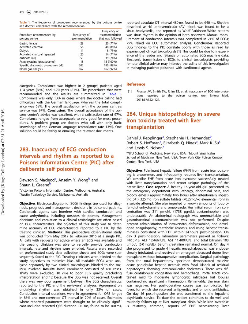
categories. Compliance was highest in 2 groups: patients aged1–4 years (86%) and >70 years (81%). The procedures that wererecommended and the results are summarized in Table 1.Compliance was only 13% in cases where the doctor calling haddifficulties with the German language, whereas the total compli-ance was 68%. The overall satisfaction with the poisons centre’sservice was 97%. Conclusion: The overall acceptance of the poi-sons centre’s advice was excellent, with a satisfaction rate of 97%.Compliance ranged from acceptable to very good for most proce-dures. A problem group are doctors who call with only basicknowledge of the German language (compliance rate 13%). Onesolution could be faxing or emailing the relevant documents.
283. Inaccuracy of ECG conductionintervals and rhythm as reported to aPoisons Information Centre (PIC) afterdeliberate self poisoning
Dawson S. Macleoda, Anselm Y. Wongb andShaun L. Greeneb
aVictorian Poisons Information Centre, Melbourne, Australia;bAustin Toxicology Service, Melbourne, Australia
Objective: Electrocardiographic (ECG) findings are used for diag-nosis, prognosis and management decisions in poisoned patients.Cardiotoxic agents can interfere with cardiac conduction andcause arrhythmias, including torsades de pointes. Managementdecisions and escalation to a clinical toxicologist are often basedon ECG characteristics. The objective of this study was to deter-mine accuracy of ECG characteristics reported to a PIC by thetreating clinician. Methods: This prospective observational studywas conducted from May 2012 to February 2015 at a single PIC.All calls with requests for advice where an ECG was available andthe treating clinician was able to verbally provide conductionintervals, rate and rhythm were enrolled. Results were recordedon preformatted study data collection sheets and ECGs were sub-sequently faxed to the PIC. Treating clinicians were blinded to thestudy objectives to minimize bias. All readable ECGs were ana-lyzed separately by two clinical toxicologists blinded to the tox-in(s) involved. Results: Initial enrolment consisted of 160 cases.Thirty were excluded, 18 due to poor ECG quality precludinginterpretation and 12 because the ECG was not received. Overallthere was very poor agreement between the ECG characteristicsreported to the PIC and the reviewers’ analyses. Agreement onunderlying rhythm was obtained in only 32% of cases.Conduction interval durations were in agreement for QRS lengthin 85% and non-corrected QT interval in 20% of cases. Exampleswhere reported parameters were thought to be clinically signifi-cant included reported QRS duration 110 ms, found to be 160 ms,
reported absolute QT interval 480 ms found to be 640 ms. Rhythmdescribed as 4:1 atrioventricular (AV) block was found to be asinus bradycardia, and reported as Wolff-Parkinson-White patternwas sinus rhythm in the opinion of both reviewers. Manual meas-urement of conduction intervals was completed in 21% of ECGs;79% relied on ECG automated analysis. Conclusion: ReportedECG findings to the PIC correlate poorly with those as read byexperienced clinical toxicologists.[1] This could be due to inexperi-ence of the reader and reliance on automated ECG machine data.Electronic transmission of ECGs to clinical toxicologists providingremote clinical advice may improve the utility of this investigationin managing patients poisoned with cardiotoxic agents.
Reference
[1] Prosser JM, Smith SW, Rhim ES, et al. Inaccuracy of ECG interpreta-tions reported to the poison center. Ann Emerg Med.2011;57:122–127.
284. Unique histopathology in severeiron toxicity treated with livertransplantation
Daniel J. Repplingera, Stephanie H. Hernandezb,Robert S. Hoffmana, Elizabeth Q. Hinesa, Mark K. Suc
and Lewis S. Nelsona
aNYU School of Medicine, New York, USA; bMount Sinai IcahnSchool of Medicine, New York, USA; cNew York City Poison ControlCenter, New York, USA
Objective: Fulminant hepatic failure (FHF) from acute iron poison-ing is uncommon, and infrequently requires liver transplantation.We describe FHF from acute iron overdose successfully treatedwith liver transplantation and report unique pathology of thenative liver. Case report: A healthy 16-year-old girl presented tothe emergency department with lethargy, abdominal pain, andbloody emesis approximately two hours after intentionally ingest-ing 54� 325 mg iron sulfate tablets (70.2 mg/kg elemental iron) ina suicide attempt. She also ingested unknown amounts of ibupro-fen-diphenhydramine and omeprazole. Her initial serum iron con-centration was 311 mmol/L (1738 mg/dL); acetaminophen wasundetectable. An abdominal radiograph was unremarkable andgastrointestinal decontamination was not performed. Despiteprompt administration of intravenous deferoxamine, she devel-oped coagulopathy, metabolic acidosis, and rising hepatic transa-minases consistent with FHF within 24 hours post-ingestion. Onday 3 post-ingestion, laboratory parameters peaked as follows:INR >13, ALT 12,466 IU/L, AST 11,400 IU/L, and total bilirubin 103mmol/L (6.0 mg/dL). Serum creatinine remained normal. On day 4she progressed to grade 4 hepatic encephalopathy, was endotra-cheally intubated, and received an emergent deceased donor livertransplant without intraoperative complication. Surgical pathologyfrom the total hepatectomy specimen demonstrated massiveacute panlobular hepatic necrosis with focal islands of residualhepatocytes showing intracanulicular cholestasis. There was dif-fuse centrilobular congestion and hemorrhage. Portal tracts con-tained mild to moderate lymphocytic infiltrates but lobulescontained no significant inflammation. Remarkably, the iron stainwas negative. Her post-operative course was complicated byfever, for which she received antipyretics and empiric antibiotics.On day 16 post-ingestion she was transferred to the inpatientpsychiatric service. To date the patient continues to do well androutinely follows-up at liver transplant clinic. While iron overdoseis relatively common, reports of FHF necessitating liver
Table 1. The frequency of procedures recommended by the poisons centreand doctors’ compliance with the recommendation.
Procedure recommended bypoisons centre
Frequency ofrecommendation
Frequency ofrecommendation
that was followed
Gastric lavage 28 20 (71%)Activated charcoal 56 48 (86%)Dialysis 11 8 (73%)Activated charcoal repeated 20 14 (71%)Antidote (all) 53 39 (74%)Acetylcysteine (paracetamol) 18 18 (100%)Specific diagnostic procedures (all) 202 180 (89%)Blood gas analysis 167 162 (97%)
492 ABSTRACTS
Dow
nloa
ded
by [
Kin
gs C
olle
ge L
ondo
n] a
t 07:
31 2
1 A
pril
2016
transplantation are rare. This case of iron-induced FHF was suc-cessfully treated with liver transplantation and demonstratesunique explant pathology. Furthermore, iron is generally believedto be predominantly a peri-portal (zone 1) toxin, however signifi-cant centrilobular (zone 3) pathology was observed in this case.In addition, despite the significant overdose, the iron stain wasnegative. This also occurs after chelation therapy in other iron-overload states, such as hemochromatosis. Alternatively, the nega-tive stain may be the result of significant hepatic necrosis andapoptosis of the hepatocytes. Conclusion: Liver transplantationcan be a successful treatment for FHF secondary to acute irontoxicity. Further study is needed to elucidate the cause of thispatient’s hepatotoxicity and unique histopathology.
285. Topical aphrodisiac ingestiontreated with digoxin-specific antibodyfragments (DigiFab)
Elizabeth Q. Hinesa, Daniel J. Repplingera,Vikram Mahalb, Robert S. Hoffmana, Lewis S. Nelsona
and Silas W. Smitha
aNew York University School of Medicine, New York, USA;bJacobi Medical Center, Bronx, USA
Objective: Twenty years ago we first reported the use of digoxin-specific antibody fragments (Digibind) to treat bufadienolide-asso-ciated cardioactive steroid (CAS) toxicity resulting from ingestionof topical aphrodisiacs. We present two recent cases of severeCAS toxicity from topical aphrodisiac ingestion treated with a dif-ferent digoxin-specific antibody fragment (DigiFab). Case reports:Case 1: A 49-year-old man presented with severe vomiting afterunintentionally ingesting ‘‘Stone,’’ a topical aphrodisiac. Vital signswere heart rate 26/minute, blood pressure 98/40 mmHg andrespiratory rate 16 /minute. His electrocardiogram (ECG) demon-strated a junctional escape rhythm with a rate of 24/minute.Whole blood potassium was 4 mEq/L. He was treated empiricallywith ten vials of DigiFab due to concern for bufadienolide toxicity.Two hours later, his heart rate varied from 30–80/minute andpotassium was 5.4 mEq/L. An additional ten vials of DigiFab weregiven with resultant normalization of heart rate (60/minute) andpotassium (4.2 mEq/L). Six hours later, his heart rate decreasedagain to 36/minute and potassium rose to 6.1 mEq/L. He receiveda final ten vials of DigiFab and activated charcoal, after which hecontinued to improve. He was discharged on hospital day threewithout sequelae. Case 2: A 39-year-old man presented with vom-iting and diaphoresis following erroneous ingestion of ‘‘PiedraChina,’’ a topical aphrodisiac. Vital signs were heart rate 65/minute, blood pressure 157/66 mmHg and respiratory rate20/minute. Shortly after arrival, ECG revealed sinus bradycardia(45/minute) with atrioventricular (AV) dissociation. Serum potas-sium was 4.6 mEq/L and apparent digoxin concentration was1.45 nmol/L (1.14 ng/mL). He acutely deteriorated; ECG showedparoxysmal atrial tachycardia with 3:2 conduction, which pro-gressed to ventricular fibrillation. He underwent 90 minutes ofresuscitation and was treated with defibrillation, epinephrine, atro-pine, amiodarone, procainamide and repeated boluses of DigiFab.After each dose of antidotal therapy (10 vials, 12 vials, and 11vials respectively) his rhythm converted to normal sinus (heartrate 70–80/minute) with return of spontaneous circulation.Unfortunately, these improvements were transient and despiteaggressive care the patient suffered another arrest and pro-gressed to asystole in the absence of additional antidote.Conclusion: Bufadienolides are extremely potent CASs present intoad venom and persistently sold as topical aphrodisiacs. Largedose DigiFab therapy seemed to be an effective treatment,
as reported previously with Digibind. Insufficient gastrointestinaldecontamination and/or inadequate antidotal therapy due to sup-ply limitations likely contributed to the death of our secondpatient.
286. TOXIC study: exposures andoutcome of intoxicated patientsvisiting the emergency department
Laura Hondebrinka, Saskia R. Rietjensa,Irma Van Den Hengel-Koota, Bianka Zandeea,Jeffrey Strika, Dylan W. De Langea,b,Karin A. H. Kaasjagerc, Douwe Dekkerc andJan Meulenbelta,b,d
aNational Poisons Information Center, University Medical CenterUtrecht, Utrecht, The Netherlands; bDepartment of Intensive CareMedicine, University Medical Center Utrecht, Utrecht, TheNetherlands; cDepartment of Internal Medicine, University MedicalCenter Utrecht, Utrecht, The Netherlands; dInstitute for RiskAssessment Sciences, Utrecht University, Utrecht,The Netherlands
Objective: A considerable number of emergency department (ED)visits are due to intoxications. Outcome of these patients, otherthan admission rates, is largely unknown. Therefore, this studyaimed to characterize these patients, their exposures and theiroutcome. Methods: Patients who visited the ED of the UniversityMedical Center Utrecht, the Netherlands, due to an (suspected)intoxication between 1 January 2015 and 31 July 2015 wereincluded in the study. Data were collected prospectively fromelectronic patient files and included data on specific exposuresand the clinical course. The observed severity of the intoxicationwas determined using the Poison Severity Score (PSS). In addition,telephone questionnaires and toxicological analysis of left-overblood samples were performed. Results: In total 189 patientswere included in the study, compromising 1.4% of the total EDpopulation. The average age was 34 years and gender wasequally distributed. The majority of the patients had one or moreco-morbidities (70%); 30% had a somatic co-morbidity and 64%had a psychiatric co-morbidity. Over half of the patients wereexposed to �1 substance based on anamnesis. Reported expo-sures mostly involved pharmaceuticals, pharmaceuticals combinedwith ethanol, only ethanol, only illicit drugs and illicit drugs com-bined with ethanol. A qualitative screening for drugs of abuse inurine was performed in 66% of the patients and was positivemost frequently for benzodiazepines, followed by cannabis. Broadqualitative analysis will be performed to evaluate the correlationbetween exposures reported by patients and measured expo-sures. The majority of patients were admitted (60%), mostly tothe department of Internal Medicine and 4% to the IntensiveCare Unit. More than half of the patients presenting with anintoxication to the ED had a mild intoxication (at the ED or ward)based on the PSS. Of these mildly intoxicated patients, approxi-mately 50% were admitted. Conclusion: Intoxicated patients con-tribute considerably to ED visits in our hospital and many ofthese patients are admitted. Poison Centres can provide anexpected PSS and aid in triage and treatment of intoxicatedpatients, as our data showed that patients with a mild intoxica-tion were often admitted. However, the observed severity couldbe underestimated, since treatment was not included in the PSSassessment. Our data will be used to evaluate the applicability ofbroad toxicological analyses in the treatment of intoxicatedpatients and to develop models to predict which patients shouldbe admitted and which can be discharged.
CLINICAL TOXICOLOGY 493
Dow
nloa
ded
by [
Kin
gs C
olle
ge L
ondo
n] a
t 07:
31 2
1 A
pril
2016
287. Mathematical modelling of theeffect of a high dose acetylcysteineregimen based on the SNAP trial onhepatic glutathione regeneration andhepatocyte death
Harry K. R. Thanacoodya, Frederik H. Nijhoutb,Michael C. Reedc and Simon H. L. Thomasa
aMedical Toxicology Centre, Newcastle University, Newcastle-upon-Tyne, UK; bDepartment of Biology, Duke University, Durham, NC,USA; cDepartment of Mathematics, Duke University, Durham,NC, USA
Objective: A novel acetylcysteine (‘‘SNAP’’) regimen (100 mg/kgover 2 h then 200 mg/kg over 10 h) causes fewer adverse reac-tions.[1] Although likely to be effective in most patients, moreprolonged therapy will be needed in those with substantial para-cetamol overdose.[2] This could be achieved by administration ofa further 200 mg/kg infusion over 10 hours (total 500 mg/kg over22 hours). We aimed to compare the effect on hepatic glutathioneregeneration and hepatocyte death of this extended 22 hourSNAP regimen with the standard 12 hour SNAP regimen and thecurrent UK acetylcysteine regimen (150 mg/kg over 1 hour, 50 mg/kg over 4 hours then 100 mg/kg over 16 hours, total 300 mg/kgover 21 hours). Methods: A published mathematical model inte-grating a model of paracetamol transport and metabolism with amodel of glutathione metabolism [3] was used to simulate theeffects of acetylcysteine on glutathione regeneration and func-tional hepatocyte numbers. These were compared between thethree regimens evaluated. Results: Hepatic glutathione regener-ation was predicted to occur earlier and the nadir of functionalhepatocytes was predicted to be higher with the 22 hourextended SNAP regimens compared to the currently used and12 hour SNAP regimens. Hepatic glutathione generation occurredlatest and the nadir of functional hepatocytes was lowest withthe 12 hour SNAP trial regimen. However, the nadir of functionalhepatocytes exceeded 40% with all three regimens suggestingthat they were all effective in preventing hepatic failure which isgenerally associated with 20–30% hepatocyte function.Conclusion: Although most patients at lower risk will be treatedeffectively by the 12 hour SNAP regimen, those with more sub-stantial overdose will require higher dose and more prolongedtherapy. This can be achieved by repeating the 10 hour (200 mg/kg) infusion in those at risk, e.g. those with persisting plasmaparacetamol concentrations or evolving liver function abnormal-ities after the initial 12 hour regimen. Further studies are requiredto evaluate the clinical efficacy of this approach.
References
[1] Bateman DN, Dear JW, Thanacoody HKR, et al. Reduction ofadverse effects from intravenous acetylcysteine treatment forparacetamol poisoning: a randomized controlled trial. Lancet.2014;383:697–704.
[2] Bateman DN, Dear JW, Thanacoody HKR, et al. Treatment of para-cetamol overdose - authors’ reply. Lancet. 2014;383:1383.
[3] Ben-Shachar R, Chen Y, Luo S, et al. The biochemistry of acet-aminophen hepatotoxicity and rescue: a mathematical model.Theor Biol Med Model. 2012:9:55.
288. Massive accidental caffeineoverdose treated with continuousveno-venous haemodiafiltration(CVVHDF)
Muhammad E. M. O. Elamina, Michael Dunnb,Simon H. L. Thomasa,b and Harry K. R. Thanacoodya,b
aNational Poisons Information Service (Newcastle Unit), Newcastleupon Tyne Hospitals NHS Foundation Trust, Newcastle upon Tyne,UK; bMedical Toxicology Centre, Newcastle University, Newcastleupon Tyne, UK
Objective: We report two students who developed severe acutetoxicity following ingestion of caffeine following a one thousand-fold dose calculation error during a university experiment. Bothrequired critical care admission and underwent CVVHDF with theaim of enhancing caffeine elimination, as a previous report hadsuggested benefits from haemodialysis.[1] Case report: Case 1: A20-year-old male ingested 30 g caffeine and developed nausea,haematemesis, agitation, tremor, diarrhoea, hypertension (171/71 mmHg), QTc prolongation (586 ms, heart rate 84 bpm), neutro-philia (14.23� 109/L), hypokalaemia (2.7 mmol/L), elevated ALT(163 U/L) and lactate (6.9 mmol/L), mild compensated metabolicacidosis (pH 7.39, bicarbonate 20 mmol/L) and rhabdomyolysis(peak creatine kinase 6891 U/L 29 hours post-ingestion). Bloodpressure and QTc interval normalised over the first 24 hours.CVVHDF was used between 9 hours and 45 hours post-ingestion.Case 2: A 20-year-old male ingested 30 g of caffeine and devel-oped nausea, haematemesis, tremor, blurred vision, epigastricpain, diarrhoea, hypotension (94/69 mmHg), QTc prolongation(536 ms, heart rate 64 beats/min), neutrophilia (9.43� 109/L),hypokalaemia (2.4 mmol/L), elevated creatine kinase (1369 U/L)and lactate (6.9 mmol/L) and compensated metabolic acidosis (pH7.39, bicarbonate 18 mmol/L). Cardiac monitoring revealed multi-focal premature ventricular contractions and non-sustained runsof ventricular tachycardia for which he received IV magnesium(4 mg). He also received IV diazepam for agitation, ondansetronfor nausea, omeprazole for gastric protection and potassiumreplacement. He developed severe hypertension with a systolicblood pressure 190 mmHg requiring glyceryl trinitrate and labeta-lol infusions on days 2 and 3 and oral metoprolol on day 3. Healso required fentanyl analgesia for abdominal pain. Creatine kin-ase peaked 27 hours post-ingestion (14,717 unit/L). CVVHDF wasused between 7.3 hours and 69 hours post-ingestion; at this pointhis lactate had normalised and his initial ALT of 31 U/L peaked at166U/L 75 hours post-ingestion. Analysis of blood samplesrevealed peak caffeine concentrations of 366.8 and 352.3 mcg/mL,respectively. Apparent caffeine half-lives during CVVHDF were4.9 hours and 7.8 hours, respectively. Conclusion: Two youngmales with potentially lethal caffeine intoxication survived withsupportive care. CVVHDF was also used but half-lives during thiswere not reduced compared to published values withoutenhanced elimination methods (4–6 hours), although prolongationof half-life (up to 16 hours) after overdose has been reported.These data do not suggest benefit from CVVHDF, but furtherstudies with clearance data are needed.
Reference
[1] Fausch F, Uehlinger DE, Jakob S, et al. Haemodialysis in massivecaffeine intoxication. Clin Kidney J. 2012;5:150–152.
494 ABSTRACTS
Dow
nloa
ded
by [
Kin
gs C
olle
ge L
ondo
n] a
t 07:
31 2
1 A
pril
2016
289. Pneumobilia as a complication ofaccidental ingestion of methyl ethylketone peroxide
Alessandra Ieria, Paolo Cassettib, Bruno Barbonb,Silvia Della Corteb, Francesco Gambassia, Primo Bottic,Alessandra Pistellic, Emanuela Masinic,d andGuido Mannaionic,d
aSOD Centro Antiveleni, AOU Careggi, Florence, Italy;bDEA-Radiologia-Endoscopia, Ospedale di Monfalcone, Monfalcone,Italy; cDipartimento di NEUROFARBA, Sezione di Farmacologia eTossicologia, Universit�a degli Studi di Firenze, Florence, Italy;dSOD Tossicologia Medica, AOU Careggi, Florence, Italy
Objective: Methyl ethyl ketone peroxide (MEKP) is a colorless,odorless organic compound used in industrial processes as a cata-lyst, in diluted solution (30–60%). When ingested, MEKP causesdirect caustic injuries by oxygen free radical production. Withmetal ions, MEKP readily forms alkyl and/or peroxyl radicals induc-ing lipid peroxidation of digestive tract and liver. Massive inges-tion of MEKP may induce acute liver failure, which is the majorcause of death. Finally, during MEKP decay, various organic acidsare formed, e.g. formic acid, which induce acidosis and opticneuropathy. Symptoms of acute MEKP poisoning include gastro-intestinal burns and bleeding, esophageal and gastric perforation,severe metabolic acidosis, rapid hepatic failure, rhabdomyolysisand respiratory failure. As with all peroxides, MEKP may producehigh volumes of oxygen which could lead to gastrointestinal dis-tension. Case report: A 47-year-old man was admitted to theEmergency Department of Monfalcone Hospital, after ingesting aliquid from an unlabeled bottle which was mistaken for water butcontained MEKP 60%. On admission he had severe epigastric andchest pain. Vital signs were normal. No oral lesions were presentbut he experienced vomiting and drooling. Intravenous fluids,atropine 0.5 mg, metoclopramide 10 mg, morphine 10 mg andpantoprazole 80 mg (followed by 2 g in 24 hours), were adminis-tered. Chest and abdomen computerised tomography (CT) scan,performed after discussion with the Florence Poison ControlCenter, showed a massive pneumobilia associated with distentionof jejunum-ileus because of oxygen production.Esophagogastroduodenoscopy (EGDS) was performed to assessthe severity of the caustic injury. Esophageal mucosal oedema(grading 1 Zargar’s score) and several gastric necrotic areas (grad-ing 3a) were present. The duodenum revealed few scatteredsuperficial erosions. The surgeon decided for non-surgical treat-ment of pneumobilia and gastric injuries. The patient underwentorotracheal intubation and was transferred to an intensive careunit. Two days later, endoscopic examination was performed.Pneumobilia spontaneously recovered in 6 days. The patient wasdischarged after 15 days and EGDS follow up was prescribed.Conclusion: Pneumobilia, typically visualized as a large air conflu-ence within the central portion of the liver, can be a complicationof endoscopic or surgical procedures involving the biliary tract. Toour knowledge, this is the first case of MEKP-induced pneumo-bilia. The patient developed local damage but limited systemiceffects with a favorable outcome. Therefore, MEKP could be lethalupon ingestion and intoxicated patients should be followed forthe risk of unexpected organ injury.
290. Severe toxicity with guanidinethiocyanate ingestion: a case report
Colin B. Page, Peter S. Kruger, Goce Dimeski andJames WalshamUniversity of Queensland, Brisbane, Australia
Objective: Commercially guanidine thiocyanate is used as a proteindenaturant and in the extraction of DNA and RNA. There are noreports of its toxicity from oral ingestion however thiocyanate tox-icity has been described in the context of cyanide toxicity fromnitroprusside in the setting of renal impairment.[1] A drug review ofnitroprusside pharmacokinetics reports thiocyanate has a half-life of2.7 days and almost 100% renal clearance.[2] We report a case ofdeliberate guanidine thiocyanate ingestion and its treatment,including dialysis. Case report: A previously well 52-year-old malebiochemist presented to a peripheral hospital with a history ofcrampy abdominal pain after drinking bottled water that hereported tasted odd. His initial vital signs were normal, howeverwhile undergoing investigation he became confused and agitated.This required sedation, intubation and ventilation with subse-quent transfer to a tertiary hospital intensive care unit forongoing care. A computed tomography (CT) scan of his brain andabdomen were unremarkable. He had an unmeasurable chloride(error reading) and a low ionized calcium of 0.59 mmol/L(1.15–1.32 mmol/L) on an arterial blood gas with a normal serumtotal calcium. The remainder of his biochemical and haemato-logical investigations were normal. A urine drug screen was nega-tive. He was managed initially with intravenous calcium. Ananalysis of the water for electrolytes gave discrepant chloridereadings on two laboratory chemistry analysers suggesting a crossreacting analyte.[3] Further analysis revealed this to be thiocyanatewith a serum concentration on admission of 538 mg/L (0.1–4.0 mg/L in non-smokers). Continuous veno-venous haemodialysis wascommenced based on this concentration and known long half-life.Five pre-dialysis serum samples demonstrated zero-order elimin-ation pharmacokinetics. Nine post-dialysis serum samples demon-strated first-order kinetics with an elimination half-life of 5.8 hours.The patient made a full recovery and admitted to deliberate add-ition of guanidine thiocyanate to the bottled water. Conclusion:This is the first report of guanidine thiocyanate poisoning causingneurological toxicity, hypocalcaemia and discordant chloride read-ings. Dialysis has a role in the treatment of its toxicity.
References
[1] Nessim SJ, Richardson RMA. Dialysis for thiocyanate intoxication: acase report and review of the literature. ASAIO J. 2006;52:479–481.
[2] Schulz V. Clinical pharmacokinetics of nitroprusside, cyanide, thio-sulphate and thiocyanate. Clin Pharmacokinet. 1984;9:239–235.
[3] Dimeski G, Kruger P, Walsham J. Unmeasurably high chloride: asurrogate marker of thiocyanate poisoning identification. ClinChem Lab Med. 2015;53:e151–e152.
291. Cost and availability ofintravenous lipid emulsion therapy:a worldwide survey
Monique Cormier and Sophie GosselinDepartment of Medicine & Emergency Medicine, McGill UniversityHealth Centre, Montreal, Canada
CLINICAL TOXICOLOGY 495
Dow
nloa
ded
by [
Kin
gs C
olle
ge L
ondo
n] a
t 07:
31 2
1 A
pril
2016
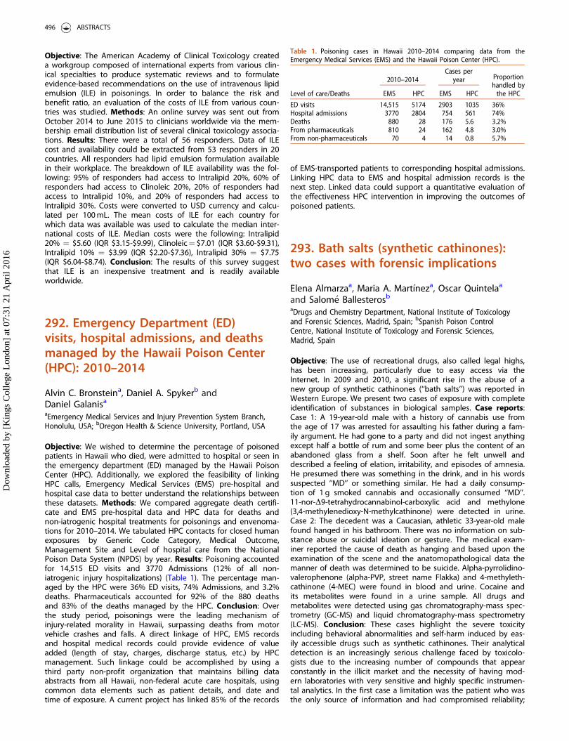
Objective: The American Academy of Clinical Toxicology createda workgroup composed of international experts from various clin-ical specialties to produce systematic reviews and to formulateevidence-based recommendations on the use of intravenous lipidemulsion (ILE) in poisonings. In order to balance the risk andbenefit ratio, an evaluation of the costs of ILE from various coun-tries was studied. Methods: An online survey was sent out fromOctober 2014 to June 2015 to clinicians worldwide via the mem-bership email distribution list of several clinical toxicology associa-tions. Results: There were a total of 56 responders. Data of ILEcost and availability could be extracted from 53 responders in 20countries. All responders had lipid emulsion formulation availablein their workplace. The breakdown of ILE availability was the fol-lowing: 95% of responders had access to Intralipid 20%, 60% ofresponders had access to Clinoleic 20%, 20% of responders hadaccess to Intralipid 10%, and 20% of responders had access toIntralipid 30%. Costs were converted to USD currency and calcu-lated per 100 mL. The mean costs of ILE for each country forwhich data was available was used to calculate the median inter-national costs of ILE. Median costs were the following: Intralipid20% ¼ $5.60 (IQR $3.15-$9.99), Clinoleic¼ $7.01 (IQR $3.60-$9.31),Intralipid 10% ¼ $3.99 (IQR $2.20-$7.36), Intralipid 30% ¼ $7.75(IQR $6.04-$8.74). Conclusion: The results of this survey suggestthat ILE is an inexpensive treatment and is readily availableworldwide.
292. Emergency Department (ED)visits, hospital admissions, and deathsmanaged by the Hawaii Poison Center(HPC): 2010–2014
Alvin C. Bronsteina, Daniel A. Spykerb andDaniel Galanisa
aEmergency Medical Services and Injury Prevention System Branch,Honolulu, USA; bOregon Health & Science University, Portland, USA
Objective: We wished to determine the percentage of poisonedpatients in Hawaii who died, were admitted to hospital or seen inthe emergency department (ED) managed by the Hawaii PoisonCenter (HPC). Additionally, we explored the feasibility of linkingHPC calls, Emergency Medical Services (EMS) pre-hospital andhospital case data to better understand the relationships betweenthese datasets. Methods: We compared aggregate death certifi-cate and EMS pre-hospital data and HPC data for deaths andnon-iatrogenic hospital treatments for poisonings and envenoma-tions for 2010–2014. We tabulated HPC contacts for closed humanexposures by Generic Code Category, Medical Outcome,Management Site and Level of hospital care from the NationalPoison Data System (NPDS) by year. Results: Poisoning accountedfor 14,515 ED visits and 3770 Admissions (12% of all non-iatrogenic injury hospitalizations) (Table 1). The percentage man-aged by the HPC were 36% ED visits, 74% Admissions, and 3.2%deaths. Pharmaceuticals accounted for 92% of the 880 deathsand 83% of the deaths managed by the HPC. Conclusion: Overthe study period, poisonings were the leading mechanism ofinjury-related morality in Hawaii, surpassing deaths from motorvehicle crashes and falls. A direct linkage of HPC, EMS recordsand hospital medical records could provide evidence of valueadded (length of stay, charges, discharge status, etc.) by HPCmanagement. Such linkage could be accomplished by using athird party non-profit organization that maintains billing dataabstracts from all Hawaii, non-federal acute care hospitals, usingcommon data elements such as patient details, and date andtime of exposure. A current project has linked 85% of the records
of EMS-transported patients to corresponding hospital admissions.Linking HPC data to EMS and hospital admission records is thenext step. Linked data could support a quantitative evaluation ofthe effectiveness HPC intervention in improving the outcomes ofpoisoned patients.
293. Bath salts (synthetic cathinones):two cases with forensic implications
Elena Almarzaa, Maria A. Mart�ıneza, Oscar Quintelaa
and Salom�e Ballesterosb
aDrugs and Chemistry Department, National Institute of Toxicologyand Forensic Sciences, Madrid, Spain; bSpanish Poison ControlCentre, National Institute of Toxicology and Forensic Sciences,Madrid, Spain
Objective: The use of recreational drugs, also called legal highs,has been increasing, particularly due to easy access via theInternet. In 2009 and 2010, a significant rise in the abuse of anew group of synthetic cathinones (‘‘bath salts’’) was reported inWestern Europe. We present two cases of exposure with completeidentification of substances in biological samples. Case reports:Case 1: A 19-year-old male with a history of cannabis use fromthe age of 17 was arrested for assaulting his father during a fam-ily argument. He had gone to a party and did not ingest anythingexcept half a bottle of rum and some beer plus the content of anabandoned glass from a shelf. Soon after he felt unwell anddescribed a feeling of elation, irritability, and episodes of amnesia.He presumed there was something in the drink, and in his wordssuspected ‘‘MD’’ or something similar. He had a daily consump-tion of 1 g smoked cannabis and occasionally consumed ‘‘MD’’.11-nor-D9-tetrahydrocannabinol-carboxylic acid and methylone(3,4-methylenedioxy-N-methylcathinone) were detected in urine.Case 2: The decedent was a Caucasian, athletic 33-year-old malefound hanged in his bathroom. There was no information on sub-stance abuse or suicidal ideation or gesture. The medical exam-iner reported the cause of death as hanging and based upon theexamination of the scene and the anatomopathological data themanner of death was determined to be suicide. Alpha-pyrrolidino-valerophenone (alpha-PVP, street name Flakka) and 4-methyleth-cathinone (4-MEC) were found in blood and urine. Cocaine andits metabolites were found in a urine sample. All drugs andmetabolites were detected using gas chromatography-mass spec-trometry (GC-MS) and liquid chromatography-mass spectrometry(LC-MS). Conclusion: These cases highlight the severe toxicityincluding behavioral abnormalities and self-harm induced by eas-ily accessible drugs such as synthetic cathinones. Their analyticaldetection is an increasingly serious challenge faced by toxicolo-gists due to the increasing number of compounds that appearconstantly in the illicit market and the necessity of having mod-ern laboratories with very sensitive and highly specific instrumen-tal analytics. In the first case a limitation was the patient who wasthe only source of information and had compromised reliability;
Table 1. Poisoning cases in Hawaii 2010–2014 comparing data from theEmergency Medical Services (EMS) and the Hawaii Poison Center (HPC).
Level of care/Deaths
2010–2014Cases per
year Proportionhandled by
the HPCEMS HPC EMS HPC
ED visits 14,515 5174 2903 1035 36%Hospital admissions 3770 2804 754 561 74%Deaths 880 28 176 5.6 3.2%From pharmaceuticals 810 24 162 4.8 3.0%From non-pharmaceuticals 70 4 14 0.8 5.7%
496 ABSTRACTS
Dow
nloa
ded
by [
Kin
gs C
olle
ge L
ondo
n] a
t 07:
31 2
1 A
pril
2016
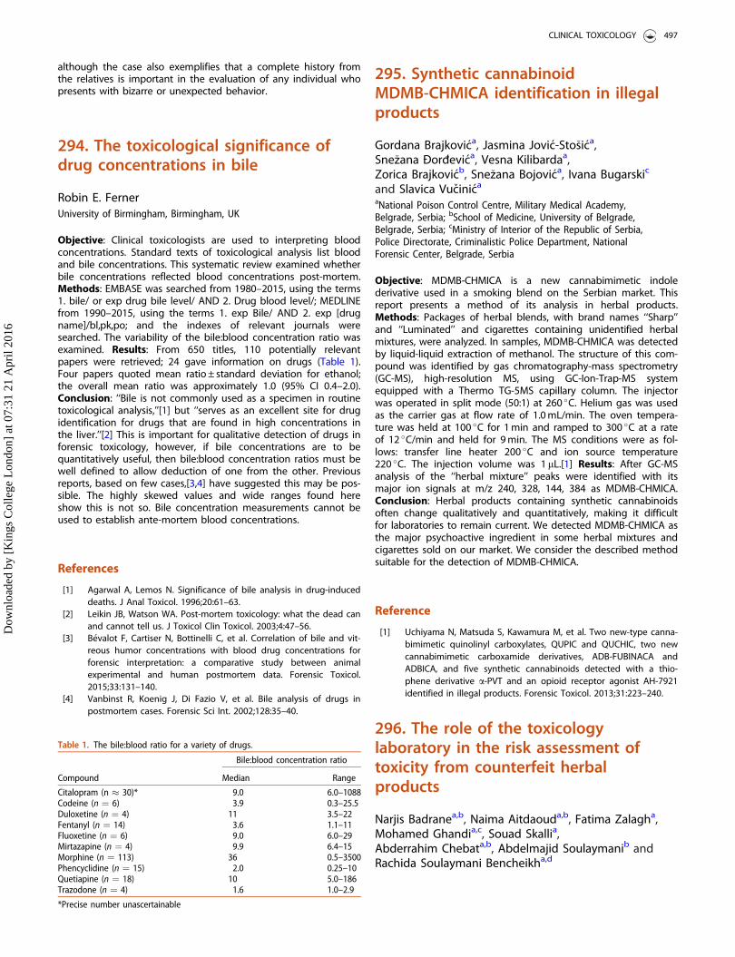
although the case also exemplifies that a complete history fromthe relatives is important in the evaluation of any individual whopresents with bizarre or unexpected behavior.
294. The toxicological significance ofdrug concentrations in bile
Robin E. FernerUniversity of Birmingham, Birmingham, UK
Objective: Clinical toxicologists are used to interpreting bloodconcentrations. Standard texts of toxicological analysis list bloodand bile concentrations. This systematic review examined whetherbile concentrations reflected blood concentrations post-mortem.Methods: EMBASE was searched from 1980–2015, using the terms1. bile/ or exp drug bile level/ AND 2. Drug blood level/; MEDLINEfrom 1990–2015, using the terms 1. exp Bile/ AND 2. exp [drugname]/bl,pk,po; and the indexes of relevant journals weresearched. The variability of the bile:blood concentration ratio wasexamined. Results: From 650 titles, 110 potentially relevantpapers were retrieved; 24 gave information on drugs (Table 1).Four papers quoted mean ratio ± standard deviation for ethanol;the overall mean ratio was approximately 1.0 (95% CI 0.4–2.0).Conclusion: ‘‘Bile is not commonly used as a specimen in routinetoxicological analysis,’’[1] but ‘‘serves as an excellent site for drugidentification for drugs that are found in high concentrations inthe liver.’’[2] This is important for qualitative detection of drugs inforensic toxicology, however, if bile concentrations are to bequantitatively useful, then bile:blood concentration ratios must bewell defined to allow deduction of one from the other. Previousreports, based on few cases,[3,4] have suggested this may be pos-sible. The highly skewed values and wide ranges found hereshow this is not so. Bile concentration measurements cannot beused to establish ante-mortem blood concentrations.
References
[1] Agarwal A, Lemos N. Significance of bile analysis in drug-induceddeaths. J Anal Toxicol. 1996;20:61–63.
[2] Leikin JB, Watson WA. Post-mortem toxicology: what the dead canand cannot tell us. J Toxicol Clin Toxicol. 2003;4:47–56.
[3] B�evalot F, Cartiser N, Bottinelli C, et al. Correlation of bile and vit-reous humor concentrations with blood drug concentrations forforensic interpretation: a comparative study between animalexperimental and human postmortem data. Forensic Toxicol.2015;33:131–140.
[4] Vanbinst R, Koenig J, Di Fazio V, et al. Bile analysis of drugs inpostmortem cases. Forensic Sci Int. 2002;128:35–40.
295. Synthetic cannabinoidMDMB-CHMICA identification in illegalproducts
Gordana Brajkovica, Jasmina Jovic-Sto�sica,Sne�zana -Dord-evica, Vesna Kilibardaa,Zorica Brajkovicb, Sne�zana Bojovica, Ivana Bugarskic
and Slavica Vucinica
aNational Poison Control Centre, Military Medical Academy,Belgrade, Serbia; bSchool of Medicine, University of Belgrade,Belgrade, Serbia; cMinistry of Interior of the Republic of Serbia,Police Directorate, Criminalistic Police Department, NationalForensic Center, Belgrade, Serbia
Objective: MDMB-CHMICA is a new cannabimimetic indolederivative used in a smoking blend on the Serbian market. Thisreport presents a method of its analysis in herbal products.Methods: Packages of herbal blends, with brand names ‘‘Sharp’’and ‘‘Luminated’’ and cigarettes containing unidentified herbalmixtures, were analyzed. In samples, MDMB-CHMICA was detectedby liquid-liquid extraction of methanol. The structure of this com-pound was identified by gas chromatography-mass spectrometry(GC-MS), high-resolution MS, using GC-Ion-Trap-MS systemequipped with a Thermo TG-5MS capillary column. The injectorwas operated in split mode (50:1) at 260 �C. Helium gas was usedas the carrier gas at flow rate of 1.0 mL/min. The oven tempera-ture was held at 100 �C for 1 min and ramped to 300 �C at a rateof 12 �C/min and held for 9 min. The MS conditions were as fol-lows: transfer line heater 200 �C and ion source temperature220 �C. The injection volume was 1 lL.[1] Results: After GC-MSanalysis of the ‘‘herbal mixture’’ peaks were identified with itsmajor ion signals at m/z 240, 328, 144, 384 as MDMB-CHMICA.Conclusion: Herbal products containing synthetic cannabinoidsoften change qualitatively and quantitatively, making it difficultfor laboratories to remain current. We detected MDMB-CHMICA asthe major psychoactive ingredient in some herbal mixtures andcigarettes sold on our market. We consider the described methodsuitable for the detection of MDMB-CHMICA.
Reference
[1] Uchiyama N, Matsuda S, Kawamura M, et al. Two new-type canna-bimimetic quinolinyl carboxylates, QUPIC and QUCHIC, two newcannabimimetic carboxamide derivatives, ADB-FUBINACA andADBICA, and five synthetic cannabinoids detected with a thio-phene derivative a-PVT and an opioid receptor agonist AH-7921identified in illegal products. Forensic Toxicol. 2013;31:223–240.
296. The role of the toxicologylaboratory in the risk assessment oftoxicity from counterfeit herbalproducts
Narjis Badranea,b, Naima Aitdaouda,b, Fatima Zalagha,Mohamed Ghandia,c, Souad Skallia,Abderrahim Chebata,b, Abdelmajid Soulaymanib andRachida Soulaymani Bencheikha,d
Table 1. The bile:blood ratio for a variety of drugs.
Compound
Bile:blood concentration ratio
Median Range
Citalopram (n � 30)* 9.0 6.0–1088Codeine (n ¼ 6) 3.9 0.3–25.5Duloxetine (n ¼ 4) 11 3.5–22Fentanyl (n ¼ 14) 3.6 1.1–11Fluoxetine (n ¼ 6) 9.0 6.0–29Mirtazapine (n ¼ 4) 9.9 6.4–15Morphine (n ¼ 113) 36 0.5–3500Phencyclidine (n ¼ 15) 2.0 0.25–10Quetiapine (n ¼ 18) 10 5.0–186Trazodone (n ¼ 4) 1.6 1.0–2.9
*Precise number unascertainable
CLINICAL TOXICOLOGY 497
Dow
nloa
ded
by [
Kin
gs C
olle
ge L
ondo
n] a
t 07:
31 2
1 A
pril
2016
aPoison Control and Pharmacovigilance Centre of Morocco, Rabat,Morocco; bGenetics and Biometry Laboratory, Faculty of Sciences,University Ibn Tofail, Kenitra, Morocco; cFaculty of Sciences ofRabat, University Mohamed V Souissi, Rabat, Morocco; dFaculty ofMedicine and Pharmacy of Rabat, Rabat, Morocco
Objective: In Morocco, some herbal products are freely sold inthe markets and often escape regulatory control. The PoisonControl and Pharmacovigilance Centre of Morocco (CAPM) con-tributes, as part of its toxicovigilance role, to the risk assessmentof such products. The confirmation of their hazard is performedat the laboratory of the CAPM. Our aim is to highlight the role ofthe toxicology laboratory in risk assessment of counterfeit prod-ucts. Methods: We report two types of herbal products sold onthe black market and another sold by herbalists without labelingthat have been the subject of information requests to the phar-macovigilance department. The analysis of the three productswas performed by high performance liquid chromatography andgas chromatography coupled to mass spectrometry. Results: Thefirst product, imported from Indonesia called ‘‘Labaik’’ is indicatedin the treatment of several diseases and symptoms including arth-ritis, rheumatism, insomnia, fatigue and hypercholesterolemia. Theresults of the laboratory have shown that the compounds in thisproduct are paracetamol, phenylbutazone, methyl-aminophena-zone and noramidopyrine. Obviously the adulterated chemicalsare not shown in the list of ingredients. The second product is anherbal mixture for obesity and to promote weight loss. The resultsof the laboratory have shown that this product contains phenolsand terpenes without medicines or pesticides. The third productis a herbal syrup sold by herbalists to women and children inorder to increase appetite for weight gain. The product analysishas found it contained an antihistamine, cyproheptadine. Oncethese results were communicated to the staff of the pharmacovi-gilance centre, a consumer awareness strategy was implementedand an alert was published in the journal of the CAPM.Conclusion: The toxicology laboratory actively contributes to theassessment of risks related to the sale of counterfeit products andparticipates in the health surveillance process.
297. Ingestion of liquid fertilizer assuspected cause of death
Kristine B. Olsena, Steen Holser Hansenb,Jan S. Jensenc and Niels Erik EbbehøjdaDept Forensic Pathology, Inst Forensic Med, Copenhagen,Denmark; bDept Forensic Pathology, Inst Forensic Med,Copenhagen, Denmark; cDanish Emergency Management Agency,Copenhagen, Denmark; dDanish Poison Center, Copenhagen,Denmark
Objective: Liquid fertilizer for domestic use is considered safeand rather harmless after unintended intake. Suicide attempts,however, comprise a significant risk but are seldom reported inthe literature. We report a fatal case. Case report: A 45-year-oldwoman with a history of a previous suicide attempt was foundlifeless in bed in the morning. Resuscitation was unsuccessful. Shewas last seen alive 55 minutes earlier. Beside the bed were foundtwo empty vials of chlordiazepoxide (100� 25 mg), a suicide noteand a three liter bottle of liquid fertilizer (GreenworldVR liquid fer-tilizer, Denmark). The ingredients were nitrogen compounds 7%(including ammonium 2%, nitrate 2% and urea 3%), phosphorus3.3% and potassium 5% (as potassium chloride). Approximately750 ml of the content was missing. The medical history includedpathology related to alcohol, but no lethal conditions. At autopsythe external and internal examinations showed no identifiable
anatomical cause of death. Gastric contents included 600 ml offluid. Toxicological analysis in femoral blood determined lithium1.8 mmol/kg, chlordiazepoxide 0.27 mg/kg, zopiclone 0.016 mg/kgand salicylic acid 11 mg/kg. No ethanol was detected. Gastric con-tent was found to contain high concentrations of nitrite, nitrate,sulphate and phosphate. There was no difference in the levels ofcations such as potassium, sodium and ammonium comparedwith gastric content from a normal control person. Conclusion:Anatomical examination and toxicological analysis of bloodrevealed no obvious cause of death. The concentrations of psy-choactive substances were below toxic concentrations. The bottlewith fertilizer was beside the bed in the morning, and had notbeen used and it is likely she had ingested the missing volume;750 ml of 5% potassium contains 37.5 g potassium (960 mmol).With a weight of 60 kg, she had taken 16 mmol/kg of readilyabsorbed potassium chloride. The high concentration of nitrates,sodium and ammonium in the stomach is consistent with inges-tion of fertilizer. The absorption rate of these electrolytes is slowerthan potassium due to the smaller concentration gradient. Thenormal concentration of potassium in the gastric fluid may beexplained by rapid absorption of potassium. Post-mortem potas-sium leaks out of cells, making potassium concentrations unreli-able minutes after death. Therefore, although we are not able toprove the cause of death in this case, we consider potassiumfrom the fertilizer a reasonable reason.
298. "Spiked" urine inbuprenorphine-assisted treatment ofopioid dependence: the quantificationof metabolites allows for detection ofadulteration
Timothy J. WiegandUniversity of Rochester Medical Center, Rochester, New York, USA
Objective: Buprenorphine is available alone or co-formulated withnaloxone, most typically in a 4:1 ratio as tablets or strips to beadministered sublingually for the treatment of opioid depend-ence. Buprenorphine preparations reduce craving, prevent with-drawal, and have a ‘‘blocking’’ effect when other opioids are usedconcomitantly. Despite some evidence showing it is less abusedthan other opioids there is still substantial diversion of buprenor-phine. Providers that treat opioid-dependent patients with bupre-norphine must be familiar with methods for detecting diversionin order to be most effective in their practice and to minimizeany harm associated with inappropriate use of buprenorphine. Acase of urine adulteration is presented in order to highlight theimportance of obtaining and quantifying urine buprenorphineand metabolite concentrations in order to detect non-complianceand limit diversion. Case report: A 47-year-old male with opioiddependence entered into drug treatment as a condition of parole.He was started on a buprenorphine/naloxone formulation at8/2 mg sublingual twice daily and was doing well with goodattendance and participation reports from his counseling andgroup sessions. His urine consistently showed buprenorphine con-centrations <3 ng/mL with norbuprenorphine/creatinine ratios of600–800 ng/mg creatinine during this time. Unfortunately, after acouple of months he started to show up late and disorganizedand then started to miss groups entirely. His urine drug testingwas noted to be markedly different from earlier samples (bupre-norphine >4000 ng/mL with norbuprenorphine/creatinine concen-trations non-detectable). A repeat urine sample was obtainedunder direct observation and 6-monoacetylmorphine and cocainebut no buprenorphine or metabolites were present. The patient
498 ABSTRACTS
Dow
nloa
ded
by [
Kin
gs C
olle
ge L
ondo
n] a
t 07:
31 2
1 A
pril
2016
admitted to spiking a friend’s urine with the buprenorphine/naloxone preparation and turning this in in lieu of his own urinedue to his relapse and return to illicit heroin use. Conclusion:Buprenorphine is metabolized to norbuprenorphine and conju-gated to buprenorphine and norbuprenorphine glucuronide prod-ucts during normal metabolism. These metabolites are excreted inthe urine. Norbuprenorphine is often standardized to the urinecreatinine in order to obtain a concentration which is not affectedby dilution. High buprenorphine (>4000 ng/mL) and negligiblemetabolite concentrations, with naloxone in the urine (buprenor-phine/naloxone formulation), suggests the medication did notpass through the patient before being present in the urine. Headmitted this when confronted with the results. Urine buprenor-phine and metabolite quantification allows for differentiationbetween adulterated/spiked urine and normal metabolite profiles.
299. Accidental poisoning withpulverized Veratrum: a case reportwith analytical confirmation
Rudolf Pfaba, Martin Hoheb, J€org Pietschc andFlorian Eyera
aKlinikum rechts der Isar, Technische Universit€at M€unchen, Dept. ofClinical Toxicology, M€unchen, Germany; bRotkreuzklinikum,M€unchen, Germany; cTechnische Universit€at Dresden, Dept. ofForensic Medicine, Dresden, Germany
Objective: Veratrum has been used in traditional medicine inEurope, Asia and America for various conditions such as sorethroats, snake bites, wounds, dysentery and apoplexy. Until the1960s Veratrum alkaloids were also used as antihypertensivedrugs. Pulverized Veratrum album rhizomes were marketed assneezing powder causing several incidents of serious toxicity untilthe 1980s. Veratrum alkaloids of the veratramine and jervine-typebind to voltage-gated sodium channels, augmenting cell wall per-meability to cations such as sodium and calcium and resulting incell excitability. A single stimulus then causes multiple depolarisa-tions of neurons and muscle cells. The vagal nerve is typicallyinvolved. Symptoms occur within 0.5 to 4 hours with initially nau-sea, vomiting, abdominal pain and diarrhoea. Cardiotoxic symp-toms of vagal overstimulation include hypotonia and bradycardia.In serious cases repolarisation abnormalities (negative T waves, STsegment shifts, supraventricular blocks, lengthened QT) areobserved. Neurological signs include headache, vertigo, sweating,paraesthesia, delirium, sleepiness and convulsions. Therapy is sup-portive and in cases with signs of vagal overstimulation atropinecan be given. Outcome is usually favourable.[1] Case report: A76-year-old woman with diabetes mellitus accidentally drank aslurry containing 1 teaspoon of dry powdered Veratrum in water.Within 30 minutes she experienced nausea, vomiting, abdominalpain, diarrhoea and general weakness. On admission to hospitalshe complained of abdominal pain, nausea and facial and digitalparesthesia. She had slurred speech and appeared somnolent butoriented; systolic blood pressure was 90 mmHg, pulse 46/minwith first degree atrioventricular block (AV) block. Laboratory testswere normal. After 12 hours of supportive care she was asymp-tomatic and the electrocardiogram (ECG) had normalized.Qualitative toxicological analysis by high-performance liquid chro-matography with tandem mass spectrometry (HPLC-MS/MS) ofthe patient’s serum (from admission), urine (after 12 hours) andthe alleged plant powder found three alkaloids (protroveratrine A,protroveratrine B and jervine) in urine, serum and powder. Afourth alkaloid, veratridine, was detected in urine and powder,but not in serum. Conclusion: We report a case with analyticalconfirmation of oral intoxication with Veratrum. The patient
developed symptoms of toxicity within 30 minutes which sub-sided within 12 hours with supportive care. Three alkaloids (proto-veratrine A and B and jervine) were detected in the patient’sserum and urine. In addition veratridine was found in urine. Allfour alkaloids were detected in the powder.
Reference
[1] Schep LJ, Schmierer DM, Fountain JS. Veratrum poisoning. ToxicolRev. 2006;25:73–78.
300. Amanita phalloides ingestion inchildren in Austria, 1996 to 2014
Tara Arif, Helmut Schiel and Kinga Bartecka-MinoPoisons Information Centre, Vienna, Austria
Objective: Mushroom ingestions are frequent reasons for contact-ing the Austrian Poisons Information Centre (PIC). The mushroomhunter’s levels of experience vary widely and in some cases, dueto lack of knowledge it is possible that the dangerous Amanitaphalloides can be ingested. We examine the cases of mushroomingestion in children reported to the PIC and describe two casesof Amanita phalloides ingestion requiring liver transplantation.Methods: Cases of mushroom ingestion, including those involvingAmanita phalloides, in children were evaluated based on the PICdatabase from 1996–2014. Results: In the 19 year study periodthe PIC in Austria received 1072 telephone enquiries regardingmushroom ingestion in children (39 less than 1 year old, 664aged 1–6 years, 267 aged 6–14 years and 102 with age unknown).In 733 children (68%) only a small amount of a single mushroomwas ingested. Of these cases medical care was recommended in31 children (4.3%) due to suspicion of intoxication. Amanita phal-loides was verified in 12 (1.6%) of 733 cases. No child developedsymptoms. In 339 cases (32%) mushrooms were consumed aspart of a meal. Observation at hospital was recommended in 93children (27.5%). Amanita phalloides was verified in 12 (3.5%) of339 cases. In three cases no symptoms occurred due to early con-sultation. The remaining children developed gastrointestinalsymptoms, three developed hepatic impairment and two childrenhad liver transplantation, one of them died. Case 1: A 16-month-old boy was fed mushrooms in a meal. The child developed diar-rhea and vomiting and was hospitalized. Medical therapy wasstarted more than 20 hours after ingestion. On the following dayshepatic impairment was diagnosed and molecular adsorbent re-circulating system (MARS) was used due to liver failure. Despiteliver transplantation the child died 9 days after Amanita phalloidesconsumption. Case 2: A 10-year-old boy consumed mushrooms ina meal and 12 hours later he developed severe diarrhea, vomitingand became unconscious. He was treated with silibinin and N-ace-tylcysteine and received MARS prior to a successful liver trans-plant 6 days after ingestion. Conclusion: The severity ofpoisoning after mushroom ingestion varies depending on theamount ingested and the age of the affected person. The smallerthe child, the more dangerous the situation. Unintentional inges-tion of parts of a single mushroom can usually be managed withhome observation, but without appropriate and immediate ther-apy Amanita phalloides can cause life-threatening intoxication.Deaths from Amanita phalloides are rare in children.
CLINICAL TOXICOLOGY 499
Dow
nloa
ded
by [
Kin
gs C
olle
ge L
ondo
n] a
t 07:
31 2
1 A
pril
2016
301. Herb intoxication: a folkmedicine-induced anticholinergicsyndrome
Hung-Sheng Huanga and Chih-Chuan Linb
aDepartment of Emergency Medicine, Chi-Mei Medical Center,Tainan, Taiwan; bDepartment of Emergency Medicine, Chang GungMemorial Hospital, Taoyuan, Taiwan
Objective: Use of herbal medicine can result in poisoning but itcan be difficult to identify, particularly where the patient or rela-tives vehemently deny ingestion of any toxic plant material.Datura metel was previously a popular herb used to treat bron-chial asthma and chronic bronchitis; however, use can result inanticholinergic poisoning due to the presence of atropine andscopolamine. Anticholinergic poisoning causes mydriasis, flushing,dry skin, altered mental status and fever. We present a case ofDatura metel poisoning which was difficult to diagnose. Casereport: A 53-year-old male was brought to our emergency depart-ment because of altered mental status, disorientation and fever.Physical examination revealed reddish, dry skin and dilated pupils.Two hours previously, he had taken three types of decoctedherbs, which included 110 g of honeysuckle flower, 40 g of guavaleaf and 190 g of Nao Shen flower. The first two herbs are nottoxic at the correct dosage, but information on Nao Shen flowerwas not available in any publication or article on the Internet orin books. His family were certain that no anticholinergic substancehad been used. Initially, a benzodiazepine was prescribed for agi-tation but the symptoms did not improve. A total of 4 mg ofphysostigmine was then given but the patient remained stupor-ous. He was given more benzodiazepine and improved 24 hoursafter taking the herbs. One week later, a drug screen found theurine was positive for atropine and scopolamine. The herbs wereexamined by the College of Traditional Chinese Medicine andNao Shen flower was thought to be Datura metel. Conclusion:Our patient presented with classic signs of anticholinergic toxicity,but no antidote was prescribed initially, because the physicianswere reluctant to prescribe physostigmine where the usage of ananticholinergic substance was uncertain. In this case physostig-mine was given as the patient remained unconscious which webelieved resulted from the use of benzodiazepine or ingestion ofan anticholinergic substance. The final diagnosis in this patientwas anticholinergic poisoning. Physicians should prescribe physo-stigmine more promptly if there is a high index of suspicion ofsevere anticholinergic syndrome with seizures, arrhythmia or agi-tation and where there is no contraindication for physostigmineuse.[1]
Reference
[1] Burns MJ, Linden CH, Graudins A, et al. A comparison of physo-stigmine and benzodiazepines for the treatment of anticholinergicpoisoning. Ann Emerg Med. 2000;35:374–381.
302. Intoxications due to wildmushrooms collected by immigrantsand asylum seekers in the Netherlands
Henneke N. Mulder-Spijkerboera,Marieke A. Dijkmana, Irma De Vriesa,Antoinette J. H. P. Van Riela and Jan Meulenbelta,b,c
aNational Poisons Information Center, University Medical CenterUtrecht, Utrecht, The Netherlands; bInstitute for Risk AssessmentSciences, Utrecht University, Utrecht, The Netherlands; cDepartmentof Intensive Care Medicine, University Medical Center Utrecht,Utrecht, The Netherlands
Objective: In the Netherlands collecting wild mushrooms forhuman consumption is not a general custom. Nevertheless, theDutch National Poisons Information Center (DPIC) is regularly con-sulted about intoxications due to the consumption of self-gath-ered mushrooms. Our hypothesis is that immigrants are moreoften involved in wild mushroom intoxications when comparedto native inhabitants. This hypothesis was tested by prospectivelycollecting all available demographical data of the patients.Methods: From January 2014 to 9 October 2015, a set of standar-dized demography related questions (including age, gender, andcountry of origin) was asked in all consultations concerning intoxi-cations due to consumption of self-collected wild mushrooms.Results: During the study period, the DPIC was consulted 26times concerning intoxications caused by wild mushrooms, rang-ing from 1 to 2 patients per incident. In total 33 patients wereinvolved; 13 males and 20 females. Ages ranged from 2 to 78years, with an average of 45 years. The nationality of 30 patientswas known: 8 native Dutch (27%) and 22 non-Dutch (73%). Thenon-Dutch patients originated from Syria (5), China (3), Poland (3),Turkey (3), Germany (2), Slovakia (2), Russia (1), Thailand (1),Vietnam (1), and an unspecified Middle Eastern country (1). It isnotable that 5 patients were staying in asylum seekers’ centers atthe time of ingestion. Conclusion: The majority of patients withan intoxication due to self-collected mushrooms in theNetherlands was of non-Dutch origin. Most patients came fromcountries in Eastern Europe, the Middle East or South East Asia,where gathering mushrooms for consumption is more common.With the recent arrival of more immigrants and asylum seekers inthe Netherlands, the number of mushroom intoxications mayincrease in the future. Part of the problem seems to be a lack ofawareness of the presence of very toxic mushrooms in theNetherlands, including Amanita phalloides (death cap). These con-cerns were communicated to the Dutch Central Agency for theReception of Asylum Seekers (COA). This agency is currently warn-ing asylum seekers about the toxicity of wild mushrooms in theNetherlands by means of spreading warning posters with picto-grams. To what extent this may help prevent serious mushroompoisonings in the future will be followed-up.
303. Toxic courgette (zucchini)poisoning – cucurbitacin
Alison M. Gooda, Gillian Jacksona,Caroline E. McGrorya, David Stewarta,Simon H. L. Thomasb, John P. Thompsonc,J. Allister Valed and Michael Eddlestona
aNPIS Edinburgh, Scottish Poisons Information Bureau, Edinburgh,UK; bNPIS Newcastle, Regional Drug and Therapeutics Centre,Newcastle upon Tyne, UK; cNPIS Cardiff, University HospitalLlandough, Cardiff, UK; dNPIS Birmingham, City Hospital,Birmingham, UK
Objective: An enquiry to NPIS Edinburgh concerning a 79-year-old woman who ingested a bitter-tasting courgette and sufferedfrom vomiting, diarrhoea and abdominal pain for the next 5 daysprompted a review of UK National Poisons Information Service(NPIS) records for enquiries concerning members of the gourdfamily. Methods: Information was extracted from telephone
500 ABSTRACTS
Dow
nloa
ded
by [
Kin
gs C
olle
ge L
ondo
n] a
t 07:
31 2
1 A
pril
2016
enquiries received by the NPIS and recorded on the UK PoisonsInformation Database (UKPID) from 1 January 2008 to 31 August2015. Results: Thirty-three records were retrieved concerning 32incidents involving 31 adults and 3 children (Table 1). Enquiriesconcerning gourds (bitter or ornamental) occurred most com-monly. Two cases involved skin contact with courgette leaves, theremainder were ingestions, generally involving cooked product.However, in some cases ornamental gourds were cooked andeaten and in one case an ornamental gourd was used as a con-tainer for tea. Drinks were also prepared using the vegetables.Gastrointestinal (GI) features were common (76.7% of ingestions),and included nausea, vomiting, diarrhoea and abdominal pain.Bleeding (in vomit or rectally) was mentioned in 4 cases. In 10cases the patients were noted to report a bitter taste.Conclusion: Exposure to bitter-tasting members of the gourdfamily may result in gastrointestinal upset with bleeding whichmay last several days. While no actual confirmation of the toxin isavailable, many of the cases are similar to those reported incucurbitacin poisoning.[1] As a result of mutation edible membersof the gourd family e.g. courgette, cucumbers, melon, pumpkin,etc. occasionally produce toxic cucurbitacins which are more com-monly found in wild variants. This results in very bitter tastingfruit that may cause poisoning.
Reference
[1] Ho CH, Ho MG, Ho SP, et al. Bitter bottle gourd (Lagenaria sicera-ria) toxicity. J Emerg Med. 2014;46:772–775.
304. Revenge of the zucchinis – is thisthe result of climate change?
Rudolf Pfaba, Hannes €Ofeleb, J€org Pietschc,Thomas Kappd, Tobias Zellnera and Florian Eyera
aKlinikum rechts der Isar, Technische Universit€at M€unchen, Dept. ofClinical Toxiology, M€unchen, Germany; bKliniken LandkreisHeidenheim gGmbH, Heidenheim, Germany; cTechnische Univerit€atDresden, Dept. of Forensic Medicine, Dresden, Germany;dChemisches und Veterin€aruntersuchungsamt (CVUA) Stuttgart,Fellbach, Germany
Objective: Colitis with bloody diarrhoea after consumption of zuc-chini (Cucurbita pepo) has been reported sporadically. Usually, theresponsible toxins, cucurbitacins, are not detected in cultivatedfruits, but some fruits spontaneously generate them. During2002–2015 Poison Information Centre (PIC) Munich registered 81symptomatic cases with clusters in the years 2003 and 2015, bothwith hot, dry summers. Case reports: Case 1: Three hours after ameal of vegetable stew with one green, bitter-tasting zucchini,home grown from commercial seeds, a 78-year-old man with pre-existing diabetes developed abdominal cramps and bloodydiarrhoea. On admission one day later he had persistent bloodydiarrhoea, hyperglycemia, acute renal failure and altered mentalstatus. Despite aggressive supportive, antiglycemic and antibioticcare on day 3 he developed peritonitis from necrotisizing colitiswith septic multiorgan failure and cardiac arrest, necessitating car-diopulmonary resuscitation, mechanical ventilation, hemodialysisand emergency colectomy. He was extubated on day 10, re-intu-bated on day 13 and died from pneumonia on day 21. Case 2: A78 year-old-woman ate a stew with one home grown, yellow, bit-ter-tasting zucchini. She was admitted to the hospital withabdominal cramps and initially bloody diarrhoea. With supportivecare, diarrhoea persisted until day 3, but was no longer bloody.Case 3: A 78-year-old male, husband of case 2, with pre-existingdiabetes, chronic renal insufficiency (creatinine 2.5 mg/dL) andarteriosclerosis, experienced spastic, bloody diarrhoea 1 hour afterthe meal. This resolved after 4 days of supportive care in hospital.Body fluids for toxicological analysis were collected 32 hours (case1) and 14 hours after ingestion (cases 2 and 3). Cucurbitacin-E-glu-coside (CucE-G) and aglycon cucurbitacin-E (CucE) were measuredby high performance liquid chromatography-mass spectrometry(HPLC-MS) after alkaline liquid-liquid extraction. Results (ng/mL)were CucE-G/CucE, serum case 1: 2.0/not detected (ND); case 2:0.6/ND; case 3: 0.5/ND; urine case 1: 4.6/8.8, case 2: 0.6/2.5, case3: 1.5/1.7, gastric juice case 1: 91.0/29.0; stool case 3: 0.7/10.6.Fruits harvested from the plant responsible for case 1 were inves-tigated by liquid chromatography/time-of-flight/mass spectrom-etry (LC/TOF/MS) after acetonitrile extraction (mg/kg): CucE-G1139; CucE 8.28; total cucurbitacin 1580. Conclusion: We found acorrelation between summer temperatures, rainfall and PIC callsconcerning symptoms after zucchini meals. Bitter-tasting zucchinican cause life-threatening colitis. The responsible toxins can bedetected in body fluids and fruits. During hot, dry summers theincidence of PIC calls with zucchini-related symptoms mayincrease. Authorities should be alerted by PIC when such casesoccur.
305. Plant poisonings in the SlovakRepublic from 2005–2014
Silvia Plackov�aa, Bla�zena Cag�a�nov�aa, Olga Otrubovaa
and Jaroslav Kresanekb
Table 1. Cases involving members of the gourd family reported to the NPIS.
Plant PatientsBittertaste Exposure Features
Courgette Adults (2) Skincontact (2)
Inflammation (1),rash (1)
Adults (5),child (1)
Yes (5) Ingestion Ulcer on tongue(1) GI signs (4includingbloody diar-rhoea 1)Asymptomatic(1)
Cucumber Adult Yes Ingestion TremorGourd
(5 ornamental,5 bitter gourd)
Adults (16) Yes (7) GI (13) Burns tomouth /oesophagus (2)Haematemesis,renal failureand hyperkalae-mia (1)Haemorrhagerectum (1)Asymptomatic(2)
Marrow Adult GI (bloodydiarrhoea)
Melon Child Ingestion of skin AsymptomaticAdult Seeds in soup Vomiting
Pumpkin Adult Leaves Mild abdominalpain
Adult Diarrhoea, haemor-rhage rectum
Squash Adults (3) Yes Cooked Abnormal sensa-tion (2GI signs (1)
CLINICAL TOXICOLOGY 501
Dow
nloa
ded
by [
Kin
gs C
olle
ge L
ondo
n] a
t 07:
31 2
1 A
pril
2016
aNational Toxicological Information Centre, Department ofOccupational Medicine and Toxicology, University HospitalBratislava, Bratislava, Slovakia; bSlovak Medical University,Bratislava, Slovakia
Objective: During a 10-year period the National ToxicologicalInformation Centre (NTIC) in Bratislava received 36,138 enquiriesfrom all over Slovakia. Plant exposures represent 4.5% of all casescollected by NTIC. We sought to characterise these enquiries inmore detail. Methods: A retrospective analysis of all telephonecalls to the NTIC between 2005 and 2014. Results: During the10-year period 1619 plant exposures were reported to the SlovakPIC. Adults made up 16% and children 84% of all the reportedcases (80% of children were less than 5 years old). Accidentalexposures occurred in 93.7% of cases, abuse 6% and suicideattempts 0.3%. The most common route of exposure was inges-tion (97%). NTIC answered most calls during the autumn (36.5%)and summer 32% with 18% of enquiries in spring and 13.5% inwinter. The most frequently ingested plants were: Daturastramonium (6.5%), Taxus baccata (6%), Zamioculcas zamiifolia(4%), Viscum album, Atropa belladonna, Ficus plants and Sambucusebulus. In 77% of patients no symptoms were reported, in 19%minor symptoms occurred and in 3.5% only moderate symptomswere reported. Eight patients developed severe symptoms. Inseven of them the symptoms were associated with plants con-taining atropine-like alkaloids. One patient developed severe car-diovascular symptoms after ingestion of a potentially lethal doseof Taxus baccata needles. One person died after accidental inges-tion of Colchicum autumnale. Conclusion: Most plant ingestions inchildren were not associated with the development of symptomsbecause only a small amount of plants was ingested. Poisoningdue to mistaken identity may also occur; accidental poisoning byColchicum autumnale occurred when it was mistaken for wildplants such as Allium ursinum.
306. Reversible neurotoxicity,gastrointestinal and visualdisturbances after consumption of theonion earthball, Scleroderma cepaPers: a case report
Bettina Haber, Verena Schrett, Rudolf Pfab andFlorian EyerDepartment of Clinical Toxicology, Klinikum rechts der Isar,Technical University of Munich, Munich, Germany
Objective: Earthballs are a family of puffball-like mushrooms, gen-erally estimated as being moderately toxic causing merely gastro-intestinal disturbances. Medical literature about poisoning issparse. However, some field guides and one mycological casereport describe reversible neurological symptoms and visual dis-turbances (blurred vision, allochromasia and blindness) after con-sumption of Scleroderma verrucosum and Scleroderma citrinum. Wereport a case of toxicity after ingestion of Scleroderma cepa. Casereport: A 42-year-old male and a friend ate a meal of self-col-lected Scleroderma cepa. The mushrooms were adequately friedfor 20 minutes. He ate 3–4 and his friend ate one mushroom.They drank no alcohol. After 20 minutes he developed dizzinessand 40 minutes after intake both had nausea. The friend recov-ered rapidly but the patient started to vomit, felt a sudden,depressive change of mood accompanied by visual hallucinations,double vision and blurred vision whereupon he sought help. After3–4 hours his vision deteriorated to achromatopsia, which
resolved spontaneously over the next hours. Nausea and vomitingpersisted until the next day as well as minor visual impairmentand subsided with no specific measures within 48 hours afterintake. However, he felt generally weak for the following twodays before recovering completely. A specimen of the collectedmushroom was retrieved and was identified macroscopically aswell as by microscopy as Scleroderma cepa. Conclusion: To ourknowledge this is the first report of a poisoning with Sclerodermacepa. Symptoms were gastrointestinal, neurological, mental andvisual disorders including depression, hallucinations and achroma-topsia. The course was benign with spontaneous recovery withinfour days. Similar toxidromes have been reported after ingestionof other Scleroderma species. Therapy is supportive. Some constit-uents of the related species S. citrinum have been identified andwere investigated for their bioactive properties. A lanostane-typetriterpenoid showed antiviral activity; in vitro vulpinate derivativeshad anti-mycobacterial and cytostatic properties.[1] However, thenature of the neurotoxin causing this unique course of mushroompoisoning is still unknown.
Reference
[1] Kanokmedhakul S, Kanokmedhakul K, Prajuabsuk T, et al. A bio-active triterpenoid and vulpinic acid derivatives from the mush-room Scleroderma citrinum. Planta Med. 2003;69:568–571.
308. Paralytic ileus and anticholinergictoxicity after ingestion of incorrectlyprepared lupin seeds
Hooman Arbabiana and Andis Graudinsb
aMonash Health, Dandenong, Australia; bMonash Health andMonash University, Dandenong, Australia
Objective: Lupin is the common name for nearly 500 species oflegumes. Some form part of the traditional Mediterranean diet.Lupinus angustifolius and L. albus are the main species cultivatedfor consumption and ground to make lupin flour. Lupin seeds aredivided into sweet and bitter varieties. Bitter varieties contain ahigher concentration of anticholinergic alkaloids. A debitteringprocess including soaking and boiling over several days reducesalkaloid content. We report a case of severe paralytic ileus andanticholinergic toxicity after ingestion of under-prepared lupinseeds. Case report: A 56-year-old male with diabetes and hyper-tension, presented to the emergency department (ED) with gener-alised weakness, vomiting and abdominal pain. His symptomsstarted the day before presentation after consuming a fistful oflupin beans. Soon after ingestion he felt unwell, posturally dizzy,with dry mouth and difficulty urinating, worsening overnight withgeneralised colicky abdominal pain, distension and obstipation.There were no visual disturbances, confusion or delirium. A not-able sign was an extremely dry mouth. In the ED he was hypoten-sive (82/50 mmHg), heart rate 86 beats/min, temperature 36.8 �C,oxygen saturation 97% (room air) with normal mental state. Theabdomen was distended with absent bowel sounds. Left iliacfossa tenderness and urinary retention were noted. Twelve-leadECG, full blood count, liver function tests and serum lipase werenormal. Abdominal CT revealed dilated proximal and mid-smallbowel without evidence of mechanical obstruction. He was fluidresuscitated but remained moderately hypotensive for severalhours. He was admitted for observation under the surgical team.Symptoms slowly improved over 24 hours. Subsequently, herevealed that he soaked the lupin beans for three days ratherthan the usual five before boiling. When ingested he noted an
502 ABSTRACTS
Dow
nloa
ded
by [
Kin
gs C
olle
ge L
ondo
n] a
t 07:
31 2
1 A
pril
2016
unusually bitter taste. The patient’s wife also ingested a smalleramount of seeds and developed dizziness and dry mouth, but didnot seek medical attention. Conclusion: This patient did not fol-low the process usually performed by his spouse for removal ofthe alkaloids from the seeds. Previous reports of lupin toxicitydescribe persistent anticholinergic symptoms and delirium afteringesting underprepared beans and flour,[1] however, this is thefirst case describing severe paralytic ileus following bean inges-tion. Under-preparation of traditional foods by inexperiencedhands can result in significant poisoning. This may become morecommon as new fads emerge touting the next ‘‘super-food’’.
Reference
[1] Litkey J, Dailey MW. Anticholinergic toxicity associated with theingestion of lupini beans. Am J Emerg Med. 2007;25:215–217.
309. A case of severe cardiacarrhythmias after aconite poisoningwith botanical and analyticalconfirmation
Ga€el Le Rouxa, Pierre E Le Choismierb, Ali Tour�ea,Yvan Gaillardc, David Boelsa andMarie Bretaudeau-Deguignea
aCentre Antipoison et Toxicovigilance Grand Ouest, CentreHospitalier Universitaire, Angers, France; bService de R�eanimationPolyvalente, Centre Hospitalier, Laval, France; cLaboratoire LATLUMTOX, La Voule sur Rhone, France
Objective: The dangerous reputation of aconite (or monkshood)is rooted in the collective psyche, where this plant is said to bethe most toxic of Europe. Aconitine is responsible for a toxidromeof neurological and cardiac symptoms.[1] This case reportdescribes a voluntary poisoning from aconite root with botanicaland analytical confirmation. Case report: A 55-year-old man pre-sented with drowsiness and paraesthesia 2 hours following sui-cidal ingestion of aconite root (approximately 20 g) grown in hisgarden. During transport to an intensive care unit (ICU), his condi-tion rapidly deteriorated with hemodynamic instability, polypnoeaand vomiting. He had acute respiratory failure and a ventriculartachycardia required intubation/ventilation and two external elec-tric shocks followed by cardiac massage, respectively. He alsoreceived 50 g of activated charcoal, 1000 ml of saline, 4 g of mag-nesium sulfate, 300 mg amiodarone and was intubated andinfused with 0.6 mg/kg/min of norepinephrine. At the ICU he pre-sented with tachycardia (110–160 beats/min) with chaotic rhythm(QTc 490 ms, QRS 0.11 s). A cardiac pacing catheter were placedand he was given esmolol to slow the heart to 60 bpm.Admission blood gases showed a compensated respiratory alkal-osis (pH 7.43, pCO2 33 mmHg, bicarbonate 21.2 mmol/L).Evolution was favourable and treatment was stopped on day 3.Urinary infection and aspiration pneumonia were both treatedwith antibiotics. The plant was identified by our local botanicalexpert as Aconitum species from a photograph. Plasma and urineconcentrations of aconitine measured by liquid chromatographycoupled to high-resolution tandem mass spectrometry were1.95 ng/mL and 2412.5 ng/mL, respectively. Conclusion: This casehad a favorable outcome despite a lethal amount of aconiteingested, thanks to the rapid implementation of supportive meth-ods and cardiac resuscitation.[2] Aconitine is the major and themost arrhythmogenic alkaloid in aconite.[1] This case illustratesthe collaboration between experts in botany, analytical toxicologyand clinicians to identify the toxic agent involved.
References
[1] Chan TY. Aconite poisoning. Clin Toxicol (Phila). 2009;47:279–285.[2] Gottignies P, El Hor T, Tameze JK, et al. Successful treatment of
monkshood (aconite napel) poisoning with magnesium sulfate.Am J Emerg Med. 2009; 27:755.e1–4.
310. Fatal colchicine poisoning afteraccidental ingestion of autumn crocus(Colchicum autumnale)
Yury N. Ostapenkoa, Dmitry P. Losb andLarisa N. Ziminab
aResearch and Applied Toxicology Center of Federal Medical andBiological Agency, Moscow, Russian Federation; bN.V. SklifosovskyResearch Emergency Medical Aid Institute, Moscow, RussianFederation
Objective: Ingestion of Colchicum autumnale (autumn crocus)may cause severe poisoning with lethal outcome. Case report: A48-year-old man was admitted in the Poisoning Treatment Center12 hours after he had accidentally cooked, and eaten a Colchicumautumnale bulb, what he had thought was an onion. Four hourslater he had developed gastrointestinal pain, nausea, vomiting,and abundant diarrhea. On admission he was conscious with paledry skin, heart rate 88 beats/min and arterial pressure 150/90 mmHg. Laboratory test results on the intensive care unitadmission were as follows: hemoglobin 183 g/L (normal range135–160 g/L), erythrocytes 5.84� 1012/L, leukocytes 7.39� 109/L(normal range 4.2–0.7� 109/L), haematocrit 50.9 g/L, platelets86.6� 109/L (normal range 150–400� 109/L), urea 7.20 mmol/L(normal range 2.5–8.3 mmol/L), serum creatinine 129.6 mmol/L(normal range 44–106 mmol/L), aspartate aminotransferase at276.0 IU/L (normal range 13–40 U/L) and alanine aminotransferaseat 88.6 U/L (normal range: 7–40 UI/L). Hemostasis parameterswere as follows: prothrombin time 48% (normal >70%),International Normalized Ratio 1.57 (normal 0.7–1.2),Electrocardiogram (ECG) showed sinus heart rate and moderatealteration of myocardium. Other system examinations were unre-markable. Gastric lavage, activated charcoal, IV sodium chloride,dextrose, saline solutions, forced diuresis and symptomatic treat-ment were performed. During 2–3 days after hospitalizationgastrointestinal symptoms disappeared, he improved and statuswas stable. By the end of the 4th day after poisoning he com-plained of shortness of breath, dyspnea with respiratory rate30–32 breaths/min. Chest X-ray showed congestion in the lungsand left heart enlargement. Heart rate was 118 beats/min, arterialpressure 175/90 mmHg and temperature 38.0 �C. He was sedated,intubated, and mechanically ventilated due to respiratory distress.The clinical course was marked by pancytopenia: hemoglobin,platelet, and leukocyte count nadir were 101 g/L, 2.0� 109/L, and0.2� 109/L, respectively. He had diffuse bleeding at puncturepoints, hemoptysis and hematuria requiring blood product trans-fusion (4 doses platelet concentrates, a total 400 mL after whichthe platelet count increased to 21.0� 109/L, and 2 doses fresh fro-zen plasma, total 500 ml) and treatment with tranexamic acid. Atthat time major hemodynamic instability developed as a manifest-ation of cardiogenic shock requiring continuous infusion of highdoses of dobutamine (up to 10 mcg/kg/min). He rapidly deterio-rated and died at the end of the fourth day after ingestion.Conclusion: Colchicum autumnale contains colchicine and isextremely toxic. Ingestion of one bulb was lethal in this case.
CLINICAL TOXICOLOGY 503
Dow
nloa
ded
by [
Kin
gs C
olle
ge L
ondo
n] a
t 07:
31 2
1 A
pril
2016
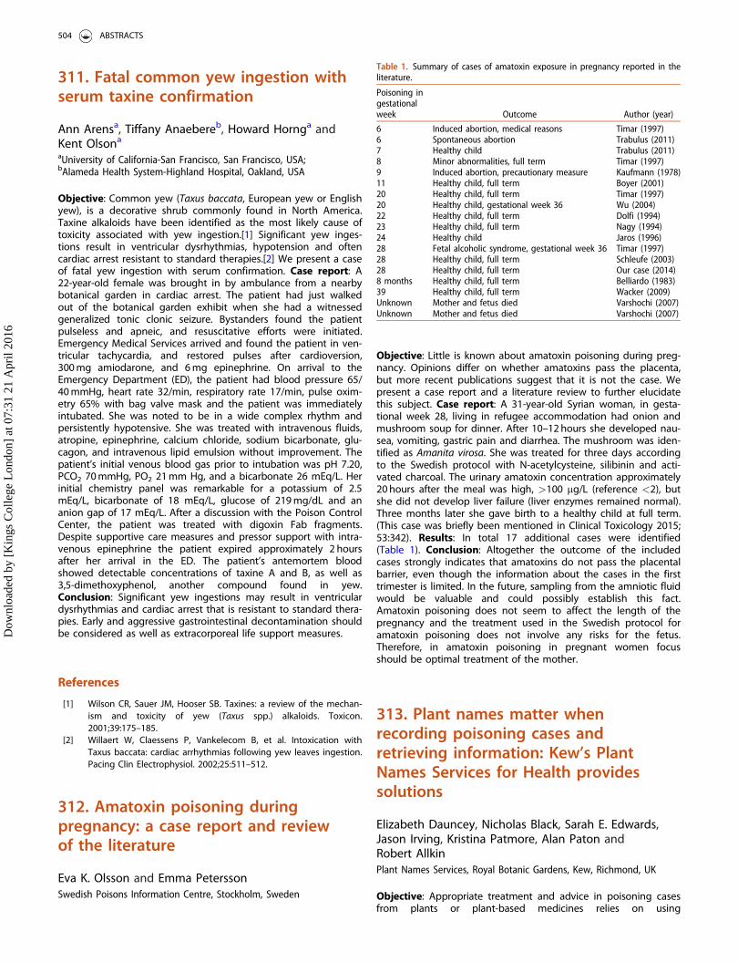
311. Fatal common yew ingestion withserum taxine confirmation
Ann Arensa, Tiffany Anaebereb, Howard Hornga andKent Olsona
aUniversity of California-San Francisco, San Francisco, USA;bAlameda Health System-Highland Hospital, Oakland, USA
Objective: Common yew (Taxus baccata, European yew or Englishyew), is a decorative shrub commonly found in North America.Taxine alkaloids have been identified as the most likely cause oftoxicity associated with yew ingestion.[1] Significant yew inges-tions result in ventricular dysrhythmias, hypotension and oftencardiac arrest resistant to standard therapies.[2] We present a caseof fatal yew ingestion with serum confirmation. Case report: A22-year-old female was brought in by ambulance from a nearbybotanical garden in cardiac arrest. The patient had just walkedout of the botanical garden exhibit when she had a witnessedgeneralized tonic clonic seizure. Bystanders found the patientpulseless and apneic, and resuscitative efforts were initiated.Emergency Medical Services arrived and found the patient in ven-tricular tachycardia, and restored pulses after cardioversion,300 mg amiodarone, and 6 mg epinephrine. On arrival to theEmergency Department (ED), the patient had blood pressure 65/40 mmHg, heart rate 32/min, respiratory rate 17/min, pulse oxim-etry 65% with bag valve mask and the patient was immediatelyintubated. She was noted to be in a wide complex rhythm andpersistently hypotensive. She was treated with intravenous fluids,atropine, epinephrine, calcium chloride, sodium bicarbonate, glu-cagon, and intravenous lipid emulsion without improvement. Thepatient’s initial venous blood gas prior to intubation was pH 7.20,PCO2 70 mmHg, PO2 21 mm Hg, and a bicarbonate 26 mEq/L. Herinitial chemistry panel was remarkable for a potassium of 2.5mEq/L, bicarbonate of 18 mEq/L, glucose of 219 mg/dL and ananion gap of 17 mEq/L. After a discussion with the Poison ControlCenter, the patient was treated with digoxin Fab fragments.Despite supportive care measures and pressor support with intra-venous epinephrine the patient expired approximately 2 hoursafter her arrival in the ED. The patient’s antemortem bloodshowed detectable concentrations of taxine A and B, as well as3,5-dimethoxyphenol, another compound found in yew.Conclusion: Significant yew ingestions may result in ventriculardysrhythmias and cardiac arrest that is resistant to standard thera-pies. Early and aggressive gastrointestinal decontamination shouldbe considered as well as extracorporeal life support measures.
References
[1] Wilson CR, Sauer JM, Hooser SB. Taxines: a review of the mechan-ism and toxicity of yew (Taxus spp.) alkaloids. Toxicon.2001;39:175–185.
[2] Willaert W, Claessens P, Vankelecom B, et al. Intoxication withTaxus baccata: cardiac arrhythmias following yew leaves ingestion.Pacing Clin Electrophysiol. 2002;25:511–512.
312. Amatoxin poisoning duringpregnancy: a case report and reviewof the literature
Eva K. Olsson and Emma PeterssonSwedish Poisons Information Centre, Stockholm, Sweden
Objective: Little is known about amatoxin poisoning during preg-nancy. Opinions differ on whether amatoxins pass the placenta,but more recent publications suggest that it is not the case. Wepresent a case report and a literature review to further elucidatethis subject. Case report: A 31-year-old Syrian woman, in gesta-tional week 28, living in refugee accommodation had onion andmushroom soup for dinner. After 10–12 hours she developed nau-sea, vomiting, gastric pain and diarrhea. The mushroom was iden-tified as Amanita virosa. She was treated for three days accordingto the Swedish protocol with N-acetylcysteine, silibinin and acti-vated charcoal. The urinary amatoxin concentration approximately20 hours after the meal was high, >100 mg/L (reference <2), butshe did not develop liver failure (liver enzymes remained normal).Three months later she gave birth to a healthy child at full term.(This case was briefly been mentioned in Clinical Toxicology 2015;53:342). Results: In total 17 additional cases were identified(Table 1). Conclusion: Altogether the outcome of the includedcases strongly indicates that amatoxins do not pass the placentalbarrier, even though the information about the cases in the firsttrimester is limited. In the future, sampling from the amniotic fluidwould be valuable and could possibly establish this fact.Amatoxin poisoning does not seem to affect the length of thepregnancy and the treatment used in the Swedish protocol foramatoxin poisoning does not involve any risks for the fetus.Therefore, in amatoxin poisoning in pregnant women focusshould be optimal treatment of the mother.
313. Plant names matter whenrecording poisoning cases andretrieving information: Kew’s PlantNames Services for Health providessolutions
Elizabeth Dauncey, Nicholas Black, Sarah E. Edwards,Jason Irving, Kristina Patmore, Alan Paton andRobert AllkinPlant Names Services, Royal Botanic Gardens, Kew, Richmond, UK
Objective: Appropriate treatment and advice in poisoning casesfrom plants or plant-based medicines relies on using
Table 1. Summary of cases of amatoxin exposure in pregnancy reported in theliterature.
Poisoning ingestationalweek Outcome Author (year)
6 Induced abortion, medical reasons Timar (1997)6 Spontaneous abortion Trabulus (2011)7 Healthy child Trabulus (2011)8 Minor abnormalities, full term Timar (1997)9 Induced abortion, precautionary measure Kaufmann (1978)11 Healthy child, full term Boyer (2001)20 Healthy child, full term Timar (1997)20 Healthy child, gestational week 36 Wu (2004)22 Healthy child, full term Dolfi (1994)23 Healthy child, full term Nagy (1994)24 Healthy child Jaros (1996)28 Fetal alcoholic syndrome, gestational week 36 Timar (1997)28 Healthy child, full term Schleufe (2003)28 Healthy child, full term Our case (2014)8 months Healthy child, full term Belliardo (1983)39 Healthy child, full term Wacker (2009)Unknown Mother and fetus died Varshochi (2007)Unknown Mother and fetus died Varshochi (2007)
504 ABSTRACTS
Dow
nloa
ded
by [
Kin
gs C
olle
ge L
ondo
n] a
t 07:
31 2
1 A
pril
2016
unambiguous names for those plants. Non-scientific names (e.g.laurel, oleander or fang ji) are often ambiguous, yet are frequentlythe only name available in case reports. Retrieval of all past casesor references relating to a particular plant depends on knowingall possible names used for that plant, but where can you findthis information? Scientific names are the only reliable means forinformation retrieval and exchange, yet appropriate use of scien-tific nomenclature can be challenging. Kew’s Medicinal PlantNames Services (MPNS) initiative, funded by the Wellcome Trust2012–2016, provides a suite of services to increase the relevanceand accessibility of Kew’s plant name resources to the healthcommunity. Central to this has been the mapping of all namesused in key medicinal plant references, such as pharmacopoeias,to Kew’s taxonomy and nomenclature. MPNS has achieved recog-nition amongst health regulators globally, with increasing demandfor our ‘‘validation’’ and ‘‘controlled vocabulary’’ services. AnMPNS vocabulary is part of the new ISO data standard for identifi-cation of medicinal products (IDMP). Kew’s Plant Names Servicesnow seek to reach new audiences, to cover plants relevant tofood supplements and toxicity, and to meet the needs of poisonsinformation centres and clinics. Methods: A user group, compris-ing representatives from poison centres, food regulation, researchand manufacturing, were questioned on a one-to-one basis, andthrough workshops, to determine priorities for appropriate solu-tions. Supplementary views will be sought as development pro-ceeds. Results: Including non-scientific names in our controlledvocabularies, and modifications to the portal (www.kew.org/mpns)to enable links from other online resources, have been identifiedas priorities for enhanced services. The collection and display ofadditional data of most use to these audiences includes the classand name of reported toxins, and animal subject. Funding appli-cations have been submitted for this next phase, and we seeknew partners in this enterprise. Conclusion: The MPNS resourcecovers 13,500 plants of known medicinal use and contains over80,000 names used in the medicinal literature and 240,000 namesfrom Kew’s resources. MPNS is already useful to poison and phar-macovigilance centres handling enquiries about plant-based med-icines. By extending MPNS to include plant food supplements(including adverse effects) and toxic plants (allergens, human andanimal poisoning), it will facilitate the provision of more compre-hensive advice regarding cases of suspected poisoning.
314. Diospyros rhodocalyx (Tako-Na),a Thai folk medicine, as a cause ofhypokalemia, proximal muscleweakness and hypertension
Rittirak Othonga, Satariya Trakulsrichaib andWinai WananukulbaDepartment of Emergency Medicine, Faculty of Medicine VajiraHospital, Navamindradhiraj University, Bangkok, Thailand;bRamathibodi Poison Center, Faculty of Medicine RamathibodiHospital, Mahidol University, Bangkok, Thailand
Objective: Diospyros rhodocalyx (Tako-Na) is a Thai folk medicinepurported to promote longevity, treat impotence, etc. We presentpatients with hypokalemia, weakness and hypertension after con-suming Tako-Na tea. We searched Medline and Google, but couldfind no reports on such adverse effects from this plant. Case ser-ies: Case 1: A 60-year-old man was brought in 9 hours after drink-ing 2 glasses of Tako-Na tea. One handful of Tako-Na bark wasboiled in a liter of water to make the tea. He had vomiting andwatery diarrhea 6 hours after ingesting the tea. He took no medi-cations and had no prior history of hypertension. The only
remarkable vital sign was blood pressure (BP) 167/90 mmHg.Physical examination revealed generalized proximal muscle weak-ness (grade 3). Laboratory findings were sodium 140 mmol/L,potassium 2.7 mmol/L, bicarbonate 24, and trans-tubular potas-sium gradient (TTKG) 5.6. Initial electrocardiogram (ECG) showed aQTc of 597 ms which was shortened after potassium supplemen-tation. He was discharged home the next day with a BP 140/90 mmHg and potassium of 4.2 mmol/L. Case 2: A 72-year-oldman, friend of case 1, also drank Tako-Na tea from the same potat the same time as case 1 but only ingested one glass. He alsohad vomiting and watery diarrhea 6 hours later. He took no medi-cations despite past history of hypertension (baseline systolic BP140–160). At presentation, his blood pressure was 230/70 mmHg.He also had proximal muscle weakness. Laboratory findings weresodium 144 mmol/L, potassium 3.3 mmol/L, bicarbonate 24, TTKG7.37, and normal thyroid function tests. His initial ECG showedbigeminy which resolved after potassium supplementation. Hewas also discharged the next day with a BP 148/70 mmHg andpotassium 4.2 mmol/L. Cases 3–7: These were patients reported tothe poison center and their potassium concentrations were as fol-lowing: 1.4, 1.4, 3.3, 1.3 and 1.2 mmol/L, respectively. Three ofthem were intubated. Case 3 with a potassium concentration of1.4 mmol/L died. Conclusion: Tako-Na contains betulin, betulinicacid, taraxerone, lupeol, and lupenone. Their structures are similarto glycyrrhetic acid, the active metabolite of glycyrrhizic acidfound in licorice which is well known to cause pseudoaldosteron-ism (hypokalemia-causing weakness and water and sodium reten-tion-causing hypertension). Glycyrrhetic acid is 200–1000 timesmore potent than glycyrrhizic acid in inhibiting 11-beta-hydroxys-teroid dehydrogenase, and causes pseudoaldosteronism. Wehypothesize that the compounds in Tako-Na act in the same wayas glycyrrhetic acid in producing pseudoaldosteronism.
315. Reasoning by analogy might notalways be reasonable: failure ofricinine to predict severity of ricinpoisoning in a castor bean ingestion
Stefan Poechackera, Maria Sommera andWolfgang Bickerb
aToxicologic ICU, Wihelminenspital, Vienna, Austria; bFTC ForensicToxicologic Laboratory, Vienna, Austria
Objective: To describe the risks of predicting ricin toxicity bydetecting ricinine in castor bean ingestion. Case report: A 22-year-old male was admitted after intentionally ingesting 10 seedsof Ricinus communis (castor bean). He masticated the seeds thor-oughly about 10 hours before being admitted, calling the ambu-lance after experiencing vomiting and abdominal cramps withdiarrhoea. He presented with tachycardia and mild abdominal dis-comfort, but had no further gastrointestinal or other clinical signsin the following days; laboratory findings remained normal forelectrolytes, kidney and liver function. There was a 2- to 3-foldincrease in free haemoglobin (max 163 mg/L) as the only markerindicating haemolysis; erythrocytes and haemoglobin remainednormal. Liquid chromatography-tandem mass spectrometry (LC-MS/MS) was performed to detect ricinine as a surrogate forricin,[1] showing markedly elevated concentrations in serum andurine 10 hours after ingestion (67.9 ng/mL and 232 ng/mL, respect-ively). He was discharged after 7 days without any sign of clinicalimpairment. Conclusion: Castor beans contain both ricinine andricin, and as detection of ricin can be difficult the detection of thericinine is used as a surrogate for the presence – and toxicity – ofricin. Case reports showing fatal outcomes with similarly elevated
CLINICAL TOXICOLOGY 505
Dow
nloa
ded
by [
Kin
gs C
olle
ge L
ondo
n] a
t 07:
31 2
1 A
pril
2016
concentrations for ricinine as in our case have been published,[2]but others have also shown,[3] as in our case shows, that thisanalogy may be misleading. Bearing in mind the variety of culti-vars of Ricinus seeds one may speculate that the ratio of ricin toricinine in the seeds was low in our case and therefore overesti-mated the expected ricin. If this was true, a hypothetical contraryratio could lead to an even more troublesome underestimation oftoxicity. The prediction of ricin toxicity by measuring ricinine con-centrations might be misleading. A possible confounder could bethe variation of the concentrations of the two substances in cas-tor beans.
References
[1] Worbs S, K€ohler K, Pauly D, et al. Ricinus communis intoxications inhuman and veterinary medicine – a summary of real cases. Toxins(Basel). 2011;3:1332–1372.
[2] Røen BT, Opstad AM, Haavind A, et al. Serial ricinine levels inserum and urine after ricin intoxication. J Anal Toxicol.2013;37:313–317.
[3] Hamelin EI, Johnson RC, Osterloh JD, et al. Evaluation of ricinine, aricin biomarker, from a non-lethal castor bean ingestion. J AnalToxicol. 2012;36:660–662.
316. Lessons to learn from fatalingestion of Prunus laurocerasus leavesin a goat
Axel Hahna, Kathrin Begemanna, Esther Feistkorna,Matthias Greinera, Michael Detersb andHelmut HentschelbaBundesinstitut f€ur Risikobewertung (BfR), Berlin, Germany;bPoisons Information Centre, Erfurt, Germany
Objective: Prunus laurocerasus (cherry laurel) contains cyano-genic glycosides and, particularly in the form of well chewedleaves, is toxic to horses, cattle, goats, sheep, swine, dogs,cats, hares, rabbits, rodents (guinea pigs, hamsters) and birds.Ruminants are at particular risk due to the repeated chewingprocess and because the cyanogenic glycosides are split inthe rumen by the action of microbial enzymes, resulting in anaccelerated release of cyanide ions, which are able to pene-trate lipid membranes. Cyanide ions bind to the central iron(III) ion in the mitochondrial respiratory chain with higheraffinity than oxygen. Oxygen binding in the blood is inhibited,resulting in inhibition of the respiratory chain, internal asphyxiaand multiple organ failure. The ingestion of major amounts bychildren or adults is rather improbable because the plant hasan unpleasant taste, so that recent human toxicity assessmentsresulted in a "mild to moderate" rating only. We report a fatalveterinary case. Case report: A Thuringian forest goat had eatenabout 1 kg of cherry laurel leaves. Subsequently, the animal devel-oped gastrointestinal symptoms and apathy. On hospital arrival,the animal was found to be somnolent, with acral coldness, paleconjunctivae and a pronounced abdominal breathing with highlypronounced moist rales over the lungs. Laboratory findingsincluded severe leukocytosis accompanied by a left shift, mildanaemia and thrombocytosis, hyperglycaemia, hypomagnesaemia,considerable increase in serum urea concentrations and elevatedcreatine kinase concentrations. Since the history of ingestion wasunknown the animal was treated symptomatically and died onthe third day after ingestion. The post-mortem examination foundthe rumen, reticulum and omasum to be filled with large quanti-ties of fibrous matter consisting of poorly masticated leaves, later
identified as cherry laurel. Other findings included severe acutealveolar lung oedema, mild serofibrinous pleuritis and diffusecatarrhal enteritis. Conclusion: Regarding the toxic risk of cherrylaurel ingestion in humans, the rare animal example has demon-strated the most important factors for the toxins to becomereleased: 1) large amount, 2) intensive and repeated chewing asin ruminants and 3) specific rumenal releasing enzymes. This veryrare worst case scenario could lead to fatal poisoning from theinherent cyanogenic glycosides. The existing chemical risk assess-ment is always based on complete substance release. Hence, thequestion arises as to whether such simple (inherent) risk assess-ment can indeed reflect reality.
317. Thai eggplant ingestion resultingin anticholinergic poisoning and ECGabnormalities
Angela Huaa, Alex F. Maninib, Ruben Olmedoa,Chad M. Meyersa and Daniel Lakoffa
aIcahn School of Medicine at Mount Sinai, New York, USA;bMount Sinai Hospital, Division of Toxicology,New York, USA
Objective: To present a case of anticholinergic toxicity from‘‘Thai eggplant’’ ingestion, and highlight the toxicity of glycoal-kaloids in the Solanum genus. Case report: A 73-year-old Thaifemale with a history of hypertension was brought to the emer-gency department (ED) with confusion and inability to speak. Thepatient’s family reported that she was eating a meal around 6 pm,felt tongue discomfort and took one loratadine tablet for a pre-sumed allergic reaction. In the ED her vital signs were: tempera-ture 36.6 �C, heart rate 114 bpm, blood pressure 150/100 mmHg,respiratory rate 20/min and oxygen saturations 100%. Her physicalexam was notable for dilated pupils, dry mucus membranes andtachycardia. She was not able to follow commands and phonatedincomprehensibly. She had up-going Babinski’s and moved allfour extremities spontaneously equally. Laboratory tests wereunremarkable (basic metabolic panel, complete blood count, ven-ous blood gas, liver function tests, and coagulation panel). Theelectrocardiogram (ECG) showed sinus tachycardia and a terminalright axis deviation in aVR. Chest X-ray and head computerisedtomography (CT) scan were unremarkable. The patient’s familyfurther revealed that the meal had included uncooked ‘‘Thai egg-plant’’ obtained from a neighbor’s garden. A photo of theingested plant was confirmed by a botanist working with thelocal Poison Control Center to be of the Solanaceae family:Solanum melongena. Although physostigmine was recommendedgiven its effectiveness in anticholinergic poisoning,[1] it was with-held initially due to the patient’s terminal right axis deviation andselect case reports showing adverse cardiac effects, includingasystole in patients with sodium channel blockade, with physo-stigmine use.[2,3] Her mental status vastly improved on day 3and she was discharged home on hospital day 4. This is the firstreported case of Solanum melongena poisoning in the US.Conclusion: Clinicians should identify and treat anticholinergicpoisoning after Solanum melongena ingestion. It is important toeducate the community regarding the potential for anticholinergictoxicity when ingesting this plant uncooked. Antidotal therapywith physostigmine should be considered, unless there is ECG evi-dence of concomitant sodium channel blockade.
506 ABSTRACTS
Dow
nloa
ded
by [
Kin
gs C
olle
ge L
ondo
n] a
t 07:
31 2
1 A
pril
2016

References
[1] Burns M, Linden CH, Graudins A, et al. A comparison of physostig-mine and benzodiazepines for the treatment of anticholinergicpoisoning. Ann Emerg Med. 2000;35:374–381.
[2] Pentel P, Peterson CD. Asystole complicating physostigmine treat-ment of tricyclic antidepressant overdose. Ann Emerg Med.1980;9:588–590.
[3] Shannon M. Toxicology reviews: physostigmine. Pediatr EmergCare. 1998;14:224–226.
318. Prolonged cardiotoxicity aftersignificant ingestion of Aconitumnapellus root
Lotte C. G. Hoegberga, Renette C. M. Jensenb,Silje A. L. Steinbr€uchelc, Rie Byrstinga andNiels E. EbbehøjaaThe Danish Poisons Information Center, Copenhagen UniversityHospital Bispebjerg, Copenhagen, Denmark; bIntensive Care Unit,Nykøbing Falster Hospital, Nykøbing Falster, Denmark; cDepartmentof Anesthesiology, Nykøbing Falster Hospital, Nykøbing Falster,Denmark
Objective: Aconitine from Aconitum napellus (FamilyRanunculaceae) is a toxic secondary plant compound. All parts ofthe plant contain toxic diterpene alkaloids, including aconitine.The lethal dose of aconitine is 3–6 mg for an adult, and as theseeds and roots may contain up to 2% alkaloid, a few grams ofthe plant is dangerous. Though plant extracts are well-knowningredients in folk medicines, A. napellus is not a permitted ingre-dient in medicines approved by the European Medicines Agency.Case report: A 76-year-old woman known to have periods ofdepression ingested a significant but unknown amount of A.napellus root in a suicide attempt. She peeled and sliced the rootas uncooked garnish for her dinner of meatballs. Approximately14 hours later she was found by her neighbour as she had fallenon the street wearing only thin clothes. Her symptoms includedintense diarrhea, mouth itching, tingling in the fingers, arms andface, pain and vertigo; pre-hospital symptoms included muscularweakness in the hands. She had a normal temperature on arrival(37.2 �C). Pre-hospital electrocardiogram (ECG) showed fluctuatingnarrow and broadening complexes. Shortly after arrival she devel-oped increasing numbers of extrasystoles and abnormal ECG withprolonged QTc (531 ms), ST elevation, QRS 108 ms, left-axis devi-ation (�57�), accelerated nodal rhythm with premature ventricularcomplexes. Blood pressure 180/80 mmHg, mean arterial pressure(MAP) 105 mmHg, heart rate approximately 85 beats/min, myoglo-bin >20,000 mcg/L, creatine kinase >20,000 U/L, ALT 95 U/L andcreatinine 204 mmol/L. Her clinical status and laboratory valuesdeteriorated further during the following 12 hours in the intensivecare unit (ICU) with extrasystoles, QT prolongation, elevated mag-nesium (1.33 mmol/L) and rhabdomyolysis. Treatment includedoral activated charcoal (100 g), forced diuresis (4000 mL/day) usingfurosemide and crystalloids, and nitroglycerin infusion to maintainblood pressure 150/80 mmHg and MAP 80 mmHg. After 2 days ofICU care the patient improved, except for nephrotoxicity causedby rhabdomyolysis, and was transferred to somatic and psychi-atric care for further treatment. Conclusion: The patient surviveda significant ingestion of A. napellus, characterised with classicsymptoms of severe poisoning [1,2] but with prolonged cardiotox-icity. Intensive care and advanced symptomatic treatment pro-vided the patient’s survival.
References
[1] German Jørgensen JR, Andersen AE. [Fatal exposure to Aconitumnapellus.] [Article in Danish] Ugeskr Laeger. 2013;175:1707–1708.
[2] Plants – Aconitum. POISINDEXVR System (electronic version). Truvenhealth analytics, Greenwood Village, Colorado, USA; [cited 2015Nov 21]. Available at: http://www.micromedexsolutions.com/
319. Expired antivenin: good efficacyin a severely envenomed cat bitten bySistrurus miliarius miliarius
Joan Fuchsa, Jose I. Casadob, Rahel Jud Scheferb andChristine Rauber-L€uthya
aNational Poisons Centre, Tox Info Suisse, Associated Institute ofthe University of Zurich, Zurich, Switzerland; bClinic for SmallAnimal Internal Medicine, Vetsuisse Faculty, University of Zurich,Zurich, Switzerland
Objective: Actual shelf-life of antivenins may be longer than theirstated expiration date, but little is known about efficacy andsafety of expired antivenin. There is only sparse literature on thein vitro efficacy of expired antivenin [1,2] and equally sparsereports on administration of the same in animals and humans.[3]We present a case of life-threateningly envenomation in a cat bit-ten by Sistrurus miliarius miliarius (Carolina pigmy rattlesnake) thatbenefited from antivenin that had expired eight and six years pre-viously. Case report: A 7-year-old, 4.1 kg previously healthyfemale European shorthair cat belonging to a snake breeder wasbitten on the snout by a juvenile Sistrurus miliarius miliarius andrapidly developed local edema. After 24 hours creatine kinase hadrisen to 25,403 U/L (range 77–355 U/L). Clotting assays (ROTEMVR
Thromboelastometry) detected a massive hypocoagulable state,characterized in an EXTEM test by a markedly prolonged clottingtime (314 s; range 126–254 s), severely reduced maximum clotfirmness (5 mm; range 40–78 mm) and unmeasurable clot forma-tion time and a angle (ranges 48–251 s and 53–80�, respectively).The owner had brought two vials of AntivipmynVR (InstitutoBioclon, Mexico) from his own stock, which had been stored cor-rectly according to the manufacturer’s instructions but hadexpired in 2007 and 2009. After the first vial (of 2009) was admin-istered, coagulation started to improve with a clotting time of99 s. Due to severe hyperfibrinolysis the second vial (of 2007) plustwo doses of 20 mg/kg tranexamic acid were administered, afterwhich coagulation returned to normal. Despite severe coagulop-athy, the clinical condition was unremarkable, and the cat wasdischarged after 5 days in good health without further sequelae,except for a bald spot at the site of the bite. Conclusion: Thiscase supports the assumption that commercial expired antiveninmay be more robust than the expiry date would suggest andthus might be a therapeutic option in case of life-threateningenvenomation, although further studies on the safety of expiredantivenin are needed.
References
[1] O’Leary MA, Kornhauser RS, Hodgson WC, et al. An examination ofthe activity of expired and mistreated commercial Australian anti-venoms. Trans R Soc Trop Med Hyg. 2009; 103:937–942.
[2] Rentmeester L, Burton D, Fitzgerald R, et al. Immunoglobulin con-centration & venom binding activity of expired snake antivenoms.Clin Toxicol (Phila) 2014;52:695.
[3] Mannel R, Pudovka Gross OA, Gross GA. Coral snake antivenin’sdeadly deadline. Toxicon. 2012;60:225.
CLINICAL TOXICOLOGY 507
Dow
nloa
ded
by [
Kin
gs C
olle
ge L
ondo
n] a
t 07:
31 2
1 A
pril
2016
320. Epidemiology of scorpion stingsin Morocco from 2005 to 2014
Rhizlane E. l. Oufir, Ilham Semlali, Naima Rhalem,Rachida Soulaymani Bencheikh and Sanaa BenlarabiThe Poison Control and Pharmacovigilance Centre of Morocco,Rabat, Morocco
Objective: Scorpion sting (SS) is the leading cause of poisoning inMorocco (30–50% of poisoning cases). In 2001 the Poison Controland Pharmacovigilance Centre of Morocco (CAPM) developed anational strategy for stings and scorpion envenomations,[1] cen-tred around training of health personnel, information, educationand communication to the public, improving management andfinally monitoring and evaluation of the implementation of aninformation system [2] to monitor indicators of morbidity andmortality from SS. We present the epidemiology of SS from 2005to 2014 and examine the indicators of fatal outcome. Methods:The collection of information was compiled by the provincial epi-demiological moderator to study regional and national demo-graphics, economics and evolving features of SS. Results: In total270,100 cases were reported, of which 27% were children �15years old. The sex ratio was 0.96. Simple sting without envenom-ation (Class I) accounted for 91.4% of cases. Cases admitted to ahealthcare facility reflecting envenomation Class II and Class IIIaccounted for 7.3% and 1.3% of cases, respectively, an overallenvenomation rate of 8.6%. Cases referred to another health facil-ity represented 9.7% of cases, hospitalized cases 5.3%, while51.8% of patients required no treatment. There were 648 cases, ofwhich 92.9% were children. The average fatality rate (deaths inpatients with SS) was 0.24%, but this varied according to regionand year (0.4% in 2005 to 0.1% in 2014). Moreover, the envenom-ation lethality rate (deaths in patients with class II and class IIIenvenomation) was 2.8% overall (4.2% in 2005 compared to 1.2%in 2014); the lethality rate in children �15 years was 0.8% (1.18%in 2005 down to 0.5% in 2014). Overall the fatality rate in patientswith Class III envenomation was 19.1% (37.4% in 2005 comparedto 11.8% in 2014). Conclusion: Continuous improvement in moni-toring of indicators of morbidity and mortality following SSallowed us to get closer to the objectives set. We note with satis-faction the streamlining of expenditures and lower fatality rates.Efforts are still needed to eradicate this scourge. For this, a wide-spread clinical audit of deaths by SS is necessary.
References
[1] Soulaymani Bencheikh R, Faraj Z, Semlali I, et al. [National strategyin the battle against scorpion stings and envenomations.Application and evaluation.] [Article in French] Bull Soc PatholExot. 2003;96:317–319.
[2] Soulaymani-Bencheikh R, Semlali I, Ghani A, et al. Implantation etanalyse d’un registre des piqures de scorpion au Maroc. Sant�ePublique. 2004;16:487–498.
321. Spider bites in Southern France:3 cases
Romain Torrentsa,b, Katharina Von Fabecka,Corinne Schmitta, Audrey Boulamerya,b,Nicolas Simona,b and Luc De Haroa
aCentre Antipoison et de Toxicovigilance, Service de Pharmacologieclinique, Hopital Sainte Marguerite (APHM), Marseille, France; bAix-Marseille Universit�e, INSERM – SESSTIM UMR 912, Marseille, France
Objective: Spider bites are frequent events in the summer inSouthern France with around 50 evenomations managed by theMarseille Poison Centre every hot season. In Europe dangerousspider species are not numerous, but in the Mediterranean areaseveral kinds of clinical picture are possible after a bite of a localspider species such as latrodectism, steatodism or loxoscelism. Wereport three cases managed during summer 2015 by the MarseillePoison Centre in order to illustrate the different aspects of arane-ism in Provence. Cases series: The 3 cases described in the tableare typical of each kind of envenomation, with neuromuscular dis-turbances for latrodectism, low severity latrodectism-like syn-drome for the steatodism and severe necrosis for the loxoscelism.Conclusion: Spider bites in the Mediterranean coast of Francemay result in severe envenomation. Medical managementdepends on the species of spider involved. Antivenoms for spiderbite are not available in France where symptomatic treatmentsare only used for such events.
323. Viper envenomation in Greece:antivenom treatment in a case series(2014–2015) from the Greek PoisonInformation Center
Eleni Basanou, Angeliki Kalostou, Vasiliki Sofidiotou,Maria Dolianiti, Vasiliki Papathanasiou,Konstantinos Fountas and Polyxeni NeouGreek Poison Information Center, P & A Kyriakou Children’sHospital, Athens, Greece
Objective: Venomous snakebites in Greece are caused only fromspecies of the Viperidae family. Vipera ammodytes is responsiblefor the majority of envenomations and is found throughout thecountry.[1] Other snakes such as Vipera xanthina, Vipera lebetinaand Vipera berus are located in specific geographic regions. Theaim of this study was to describe the clinical course, indicationsand efficacy of antivenom treatment. Methods: A retrospectiveanalysis of the PIC database regarding snakebites during the years2014–2015 and case records concerning patients treated in Greekhospitals. Results: A total of 340 snakebite cases were reportedduring the study period. The patients were predominantly male(66%), mean age 35 years. Most snakebites happened in spring(18%) and summer months (56%) but there were also cases inFebruary and November. Among 340 patients, 31% had dry bites
Table 1. Three spider bites managed during Summer 2015.
CaseAge
(years)Spider
involvedClinical
presentation Treatment Evolution
1 77 Latrodectustredecimguttatus
Latrodectism:Chest tightness,muscle cramps,tachycardia, localpain
CorticosteroidsCalciumBenzodiazepinePainkillers
Healing withoutsequelae after 3days of medicalcare
2 27 Steatoda nobilis Steatodism:Headache, dys-phagia, dys-pnoea, hyper-thermia, myalgia,facial swelling,local pain
AdrenalineAntihistamine
Healing withoutsequelae after1 day of medicalcare
3 47 Loxoscelesrufescens
Loxoscelism:Cutaneous necro-sis, headache,hyperthermia,paresthesia, localpain
AntibioticsCorticosteroidsNecrosectomy
Healing withoutsequelae after7 days of med-ical care
508 ABSTRACTS
Dow
nloa
ded
by [
Kin
gs C
olle
ge L
ondo
n] a
t 07:
31 2
1 A
pril
2016
and 69% developed envenomation. In the 235 patients whodeveloped envenomation the most common symptoms werelocal edema, erythema and pain, gastrointestinal signs (vomiting,diarrhea, abdominal pain), circulatory effects (hypotension, shock,electrocardiogram abnormalities), hematological changes (leuko-cytosis, hemolysis, coagulopathy) and allergic symptoms (urticaria,angioedema). The bites were categorized as mild in 40%, moder-ate in 35% and severe in 25% (Table 1). Administration of specificanti-Fab antivenom was required in 33.6% of patients and wasindicated for all severe cases and in selected moderate cases.Antivenom treatment resulted in prompt clinical improvement.Adverse effects consisting of urticaria and serum sickness did notoccur. There were no deaths. Conclusion: Antivenom therapyproved to be an effective and safe treatment in cases of severeenvenomation. The occurrence of allergic side-effects was negli-gible with this type of antivenom.
Reference
[1] Frangides CY, Koulouras V, Kouni SN, et al. Snake venom poison-ing in Greece. Experience with 147 cases. Eur J Intern Med.2006;17:24–27.
324. Life-saving antivenom treatmentafter a Monocled Cobra bite in theNetherlands
Marieke A. Dijkmana, Nannet M. Van Der Schootb,Irma De Vriesa and Dylan W. De Langea,b
aNational Poisons Information Center, University Medical CenterUtrecht, Utrecht, The Netherlands; bDepartment of Intensive CareMedicine, University Medical Centre Utrecht, Utrecht, TheNetherlands
Objective: Since 2008, a National Serum Depot (NSD) has beenoperational in the Netherlands, guaranteeing rapid antivenomsupply during medical emergencies. Antivenom from the NSD is(only) available after contact with the National PoisonsInformation Center (PIC). Here, we present a case of MonocledCobra (Naja kaouthia) bite requiring antivenom. Case report: Ten
minutes after being bitten by a 1.5 metre long cobra, a 28-year-old amateur herpetologist was transported by ambulance to theUniversity Medical Center Utrecht. On arrival at the EmergencyDepartment (ED), he was nauseated with severe vomiting.Physical examination revealed bite marks on his left elbow.During the first 30 minutes, blood pressure increased from 150/85to 195/102 mmHg, heart rate rose from 80 to 133 beats/min andrespiration rate increased from 17 to 30 breaths/min, withdecreasing saturation from 100% to 93% on room air. The patienthad fixed dilated pupils, was combative and refused supportiverespiratory measures. Thereafter, within minutes his overall clinicalcondition deteriorated, and he became unresponsive and cyan-otic, requiring rapid sequence intubation and mechanical ventila-tion in the ED. Meanwhile, after contact with the PIC, 4 vials ofThai Red Cross Cobra antivenin were ordered from the NSD. Atthe Intensive Care Unit (ICU), antivenom was administered2.75 hours after the bite. After antagonizing the remaining para-lyzing effect of rocuronium-induced muscle relaxation (used dur-ing intubation) with sugammadex, the patient was able to triggerthe ventilation machine. After cessation of the sedation, he wokeup, and could communicate via squeezing his hand and shakinghis head. As further neurological improvement was slow, add-itional antivenom (4 vials) was given 9.25 hours later. He wastaken off the ventilator 5 hours later (17 hours post-bite). Within24 hours after the bite, he could be discharged to the mediumcare unit, where mildly increased clotting times were noted andlocal effects appeared to be worsening. In addition to erythema,edema progressed to the hand and shoulder and a few small blis-ters appeared remote from the bite site. Pain was treated withopioids and antibiotics were started 24 hours later. He was dis-charged after 8 days. Conclusion: Antivenoms vary in price from<100 Euro to >1500 Euro per vial and often many vials are neces-sary to treat an envenomation effectively. Keeping rarely usedantivenoms in stock is expensive. However, this case clearly showsthat antivenom treatment is cost effective as mechanical ventila-tion duration was reduced by several days.
325. Efficacy of Crotalidae polyvalentimmune Fab antivenom with delayedadministration
Stephen Powell, William Rushton and Justin ArnoldUniversity of Alabama at Birmingham, Birmingham, USA
Objective: Demonstration of clinical improvement from Crotalusadamanteus envenomation treated at 49 hours followingenvenomation with Crotalidae Polyvalent Immune FabAntivenom. Case report: A 19-year-old female presented to anemergency department in the south eastern US immediately afteran Eastern Diamondback (Crotalus adamanteus) bite to her leftankle. Initial symptoms included vomiting, myokymia, and pain atthe bite site. Vital signs were reported as normal and initial labsdemonstrated a partial thromboplastin time (PTT) 33 seconds,international normalized ratio (INR) 1.04, platelets 250,000/lL, andfibrinogen 347 mg/dL. She did not receive antivenom and wasdischarged home following a 6-hour observation. She subse-quently presented to her primary care physician 43 hours laterwith worsening edema, ecchymoses on her left lower extremity,and pain. Physical examination further revealed ecchymosis onher upper extremity where the blood pressure cuff was placedand persistent bleeding at the site of an intravenous catheter.Repeat laboratory tests included PTT greater than 200 seconds,INR greater than 18, platelets 260,000/lL, and fibrinogen less than61 mg/dL. All laboratory values were repeated to ensure accuracy.She was treated with six vials of Crotalidae Polyvalent Immune
Table 1. Severity of envenomation of Viperidae bites.
Type ofstings /symptoms Mild Moderate Severe
Local Swelling, erythemaconfined to bitesite, minimal pain.
Progression of swel-ling, erythemabeyond bite site,pain starts immedi-ately after bite.
Rapidly progressiveextensive swelling,erythema orecchymosis involv-ing the entire bodypart, immediatesevere pain.
Systemic No systemic signs orsymptoms. Nocoagulation abnor-malities or otherlaboratoryabnormalities.
Non-life-threateningstings or symptoms.Mild coagulationabnormalities with-out significantbleeding.
Markedly severe signsor symptoms: hypo-tension, tachycardia,respiratory distress,coagulation abnor-malities with bleed-ing, platelet count< 50,000/lL,increase in D-dimer,leukocytes, creatinekinase.
CLINICAL TOXICOLOGY 509
Dow
nloa
ded
by [
Kin
gs C
olle
ge L
ondo
n] a
t 07:
31 2
1 A
pril
2016
Fab antivenom 49 hours after envenomation. After four hoursthe PTT was 38 seconds, INR 5, platelets 239,000/lL, andfibrinogen less than 61 mg/dL. She received another two vialsevery six hours for three total treatments with resolution ofswelling to her ankle. Prior to discharge she had a prothrom-bin time of 15.4 seconds, PTT 27.7 seconds, INR 1.24, platelets201,000/lL and fibrinogen 225 mg/dL. She followed up withher physician one week later without any further symptoms.Conclusion: Crotalus adamanteus is endemic to the south easternUS and likely the cause of envenomation in this case due to thesevere fibrinolysis and absence of thrombocytopenia. The utilityof delayed Crotalidae Polyvalent Immune Fab antivenom follow-ing envenomation has not been well described. In our case, therewas rapid improvement of both the edema and coagulopathy fol-lowing delayed administration. The fact that Crotalus adamanteusis one of four species involved in the manufacturing of the fabfragments may have also contributed to the rapid resolution ofthe patient’s symptoms. This case demonstrates potential efficacypreviously not shown and antivenom should be considered inpatients with delayed presentations following crotalinaeenvenomation.
326. Dangerous pets: Cape cobra(Naja nivea) envenomationin Barcelona
Emilio Salgadoa, Ana Garc�ıab and Santiago Noguea
aClinical Toxicology Unit, Hospital Clinic, Barcelona, Spain;bEmergency Department, Hospital Clinic, Barcelona, Spain
Objective: To present a case of an envenomation from an exoticsnake with neurotoxic venom.[1] In Spain the legislation on dan-gerous animals requires the animal’s owner to hold the relevantantivenom (to ensure availability in an emergency). Case report:A 26-year-old man was bitten in his right hand by a Cape cobrawhile he was cleaning the reptile’s cage in a private reptile collec-tion near Barcelona. Soon after he started feeling anxious, haditching of the eyes, nasal discharge and sneezing. He called formedical attention and for the antidote he knew was at ‘‘Zoo deMadrid’’. First aid was provided by a pre-hospital medical team,whom administered 200 mg hydrocortisone intravenously (IV),5 mg dexchlorpheniramine IV and morphine. He was then trans-ferred to our hospital. On arrival, 45 minutes after the accident, hewas pale with mild tachycardia, but both blood pressure and oxy-gen saturation on ambient air were normal. He had two needle-shaped fang marks on the dorsum of his right hand with littlelocal oedema, and normal neurological examination. Symptomatictreatment was administrated, while sourcing the antivenom. Fourhours after the accident, he developed progressive dyspnoea andoxygen desaturation. The antivenom (S.A.I.M.R PolyvalentAntivenom) was obtained from a private exotic collection inMadrid and transported by plane to Barcelona. It was adminis-tered to the patient with a rapid improvement of his condition.He was discharged from the hospital 36 hours later, with oral anti-biotic and anti-inflammatory treatment. He was seen in the out-patient clinic one and two weeks after the event, but had nocomplications. Conclusion: This is the first case report in themedical literature from Spain about envenomation from this non-native snake. It highlights the increase of non-regulated exoticsnake owners in Europe and the importance of having nationalprotocols to cope with these accidents and ensure availability ofantivenom.[2,3]
References
[1] Blaylock RS, Lichtman AR, Potgieter PD. Clinical manifestations ofCape cobra (Naja nivea) bites. A report of two cases. S Afr Med J.1985;68:342–344.
[2] Warrick BJ, Boyer LV, Seifert SA. Non-native (exotic) snake enveno-mations in the US, 2005–2011. Toxins (Basel). 2014;6:2899–911.
[3] Schaper A, Desel H, Ebbecke M, et al. Bites and stings by exoticpets in Europe: an 11 year analysis of 404 cases fromNortheastern Germany and Southeastern France. Clin Toxicol(Phila). 2009;47:39–43.
327. Treatment of pain using only hotwater baths after weever and scorpionfish envenomations on the Frenchcoast
Alix Dattina, Charles Hudelob, Luc De Haroc,Arnaud Kurzenneb and Magali Labadiea
aBordeaux Poison Control Center, Bordeaux, France; bSoci�et�eNationale de Sauvetage en Mer, Paris, France; cMarseille PoisonControl Center, Marseille, France
Objective: Fish envenomation, particularly weever fish (Trachinusdraco and Echiichtys vipera) or scorpion fish (genus Scorpaena) arefrequent along the coast of France, and although benign areresponsible for intensive pain, due to injection of the thermolabilevenom. Treatment currently recommended is thermal shock whichconsists of a variation in local temperature with heat then ice.[1]However, finding ice on the French coast during the summer ischallenging. Moreover, treatment with only hot water has neverbeen evaluated. The objective of this study was to evaluate theefficiency of a hot water bath (of the affected limb) on pain inpatients after envenomation. Methods: A prospective study wasconducted between 1 July and 31 August 2015 in which 10 vol-unteer lifeguard stations of the National Sea Rescue Society,located along the coast took part. All patients over 6 years oldwith weever fish envenomation and seen at one of the lifeguardstations with pain were included. On arrival, these patientsreceived an evaluation of pain by a visual analogue scale (VAS),then a water bath as hot as possible without exceeding 42 �C(checked by thermometer) for 15 minutes, followed by a secondevaluation of pain after the bath, then a third, 15 minutes later.Children younger than 6 years old, those with multiple stings andpatients treated with analgesics in the previous 24 hours wereexcluded. Results: In total 178 patients were included. Stingsoccurred on the Mediterranean, Atlantic and Manche beacheswith an average 10.46 minutes between sting and treatment. Thesex ratio was 0.54 and the average age 26.78 years. Most patients(93.26%) were stung on a lower limb and 6.74% on an upperlimb. The average VAS on the arrival as 5.93. The average waterbath temperature was 40.55 �C and average duration was14.48 minutes. The second VAS after 15 minutes was 2.07, and thelast VAS evaluated 15 minutes after the end of bath was 1.36. Thedifference in pain is statistically significant (p< 0.001). None ofthe patients required contact with a Poison Center or emergencymedical call center. Conclusion: Treatment with only and exclu-sive a hot water bath can reduce pain after weever or scorpionfish stings.
510 ABSTRACTS
Dow
nloa
ded
by [
Kin
gs C
olle
ge L
ondo
n] a
t 07:
31 2
1 A
pril
2016
Reference
[1] De Haro L, Prost N, Arditi J, et al. Efficacy of local temperaturevariation in the treatment of Mediterranean fish envenomations:experience of the Marseilles Poison Center during the summer of1999. J Eur Urgences. 2001;14:171–173.
329. Sea snake envenoming inAustralia causes myotoxicity, localeffects and non-specific systemicsymptoms (ASP-24)
Christopher Johnstona, Nicole M. Ryanb andGeoffrey K. Isbistera
aNSW Poisons Information Centre/University of Newcastle,Newcastle, Australia; bUniversity of Newcastle, Newcastle, Australia
Objective: Sea snake envenoming is a rare and poorly character-ised clinical syndrome in Australia. Isolated case reports fromAustralia describe neurotoxicity and myotoxicity, but most areunconfirmed cases.[1,2] The aim of this study was to describe theepidemiology, clinical presentation and treatment of sea snakeenvenoming in Australia. Methods: Patients are recruited to theAustralian Snakebite Project (ASP), an ongoing multi-centre obser-vational study of snakebite Australia-wide. ASP uses predefineddata collection, laboratory investigations and venom assays todescribe envenoming. Data collection sheets and laboratory pro-tocols are faxed to clinicians who return them by fax. In this ana-lysis we included patients with sea snake bites. Results: FromSeptember 2005 to October 2015 there were seven patients withconfirmed sea snake bites from a total of 1685 patients recruitedto ASP. The median patient age was 23 years (4–62 years); sixpatients were male. All bites occurred in the wild, six in northQueensland and one off the West Australian coast. All patientswere in the water or fishing when bitten, including four fishing orworking with fishing nets. Two bites occurred at night. Localeffects occurred in six patients with pain (n¼ 4), swelling (n¼ 4)and bruising (n¼ 1). Five patients had systemic envenoming. Fourhad non-specific systemic effects with headache (n¼ 4), nausea(n¼ 3), abdominal pain (n¼ 2) and generalised diaphoresis(n¼ 1). Four patients had myotoxicity: one had generalised myal-gia, two had local myalgia only and one had no myalgia. Medianpeak creatine kinase was 14,570 IU/L (range 804–48,100). Nopatients with myotoxicity developed renal impairment.Coagulopathy and neurotoxicity did not occur. Three patientswith envenoming had elevated white cell counts on presentation(10.4, 14.2 and 15.7� 109). One vial of sea snake antivenom (CSLLtd) was given to four patients; one patient receiving a secondvial. One patient had a systemic hypersensitivity reaction to anti-venom with urticaria, generalised erythema and nausea treatedwith promethazine, ondansetron, intramuscular adrenaline and anadrenaline infusion. Median length of hospital stay was 16.5 hours(range 8.5 to 84 hours). Conclusion: Sea snake envenoming israre in Australia and is characterised by local effects, non-specificsystemic effects and mild to moderate myotoxicity.
References
[1] Sutherland SK, Tibballs J. Family Hydrophiidae, Sea Snakes. In:Sutherland SK, Tibballs J. Australian Animal Toxins. 2nd ed. Oxford,England: Oxford University Press; 2001:247–285.
[2] Fulde GW, Smith F. Sea snake envenomation at Bondi. Med J Aust.1983;141:44–45.
330. The French snake antivenombank
David Boelsa, Gael Le Rouxa, Luc De Haroa,Jean-Marc Saporia, Pierre Quistinicb,Jean-Philippe Chippauxc and Patrick Harrya
aPoison Control Centre, University Hospital, Angers, France; bSnakeAntivenom Bank Association, Angers, France; cFrench Institute ofResearch for Development, Cotonou, Benin
Objective: Exotic pets have become fashionable in France.Venomous snakes are undoubtedly the most dangerous exoticpets. Since ownership of venomous snakes is never risk free, allowners, both professional and non-professional (licensed andunlicensed), are likely to be bitten at some point with potentiallylife-threatening consequences. At present, the only effective spe-cific treatment is antivenom therapy. When indicated, the correctdose must be administered intravenously as soon as possible.[1]In light of the growing risk of envenomation and the need tocomply with regulations on the supply, storage and distributionof snake antivenom, a Snake Antivenom Bank was set up.Methods: The main aim of the Bank was to develop a medicaltreatment programme for patients bitten by exotic snakes inmainland France, along with a hospital pharmacy to manage theactive antivenoms used for these species. Results: The Bank com-prises four sites holding snake antivenoms (ViperfavVR , BothrofavVR ,FavafriqueVR , Antivipmyn-triVR , FavireptVR ) that meet the quality andviral safety criteria established by the evaluation division of theFrench National Agency for the Safety of Medicine and HealthProducts. The antivenoms can treat over 40 different types ofenvenomation (covering European, African and American snakespecies), out of a total of 93 exotic species known to be presentin France. Since 2007, the Bank has been consulted to treat 11cases of envenomation in professionals and unlicensed amateurbreeders, seven of whom (Daboia palaestinae, Crotalus polystictus,Naja annulifera, Crotalus durissus, Bothriopsis taeniata, Bitis arietans,Dendroaspis viridis) had moderate to severe envenomation thatwas successfully treated with antivenom, with no sequelae. In allcases, the antivenoms were delivered in under six hours: the rec-ommended time limit between being bitten and the administra-tion of antiserum. Long-term follow up of these patientsconfirmed that the antivenoms were well tolerated. Conclusion:The Bank currently operates as a network, which has reducedresupply costs resulting from the antivenoms expiring, as well ascutting delivery times across France. The Bank and its network ofexperts, including members of the association, also keep a recordof venomous species present in France, work to improve treat-ment for envenomation and help poison control centres collectdata on envenomation for the purposes of toxicovigilance andpublic health surveillance.
Reference
[1] Boels D, Hamel JF, Bretaudeau Deguigne M, et al. European viperenvenomings: Assessment of ViperfavTM and other symptomatictreatments. Clin Toxicol (Phila). 2012;50:189–196.
CLINICAL TOXICOLOGY 511
Dow
nloa
ded
by [
Kin
gs C
olle
ge L
ondo
n] a
t 07:
31 2
1 A
pril
2016
331. Hot water immersion treatmentfor lion’s mane jellyfish stings inScandinavia
Kai Knudsen and Sofia ÅgrenDepartment of Anesthesia and Intensive Care, SahlgrenskaUniversity Hospital, Gothenburg, Sweden
Objective: There are a number of different treatment optionsfor jellyfish stings in Nordic sea waters.[1–2] Hot water immer-sion therapy (HWI) has not been investigated previously afterlion’s mane jellyfish stings. The objective of this study was toevaluate the effect of HWI compared to standard treatmentwith topical lidocaine after stings with lion’s mane jellyfish(Cyanea capillata). It was also the objective to visualize theeffects of different suggested treatments on nematocysts invitro. Methods: Eighteen healthy volunteers were exposed tofresh lion’s mane jellyfish at each ankle. Twenty minutes afterexposure one leg was treated with HWI (45 �C for 30 minutes) andthe other leg with topical administration of lidocaine (5%). Painand itching were measured on a Visual Analogue Scale (VAS) dir-ectly after exposure and repeated after 30, 60 minutes and24 hours. The effect of different treatment options was also exam-ined in an in vitro setting to evaluate the firing of nematocystsunder the microscope. Results: Pain and itching after skin contactwith lion’s mane jellyfish was present but not intense in all sub-jects. Both lidocaine and HWI reduced pain and itching effectively.At time 0, just before HWI started, the mean VAS score was 1.8,and the mean itch score 3.4. HWI was significantly more effectivethan lidocaine in reducing both pain and itching 30 minutes afterexposure. At 30 minutes, the mean VAS score was 0.5 for the legtreated with HWI, and 1.3 for the lidocaine-treated leg (p< 0.05Wilcoxon signed-rank test). After 24 hours very few symptomsremained but more subjects felt some itching after lidocaine thanafter HWI. In the in vitro setting vinegar caused a visible firing ofnematocysts whereas lidocaine did not. Conclusion: Both lido-caine and HWI seem to be effective in reducing symptoms afterdermal contact with lion’s mane jellyfish. HWI was more effectivein reducing symptoms and seems to be an easy accessible treat-ment for these stings. Vinegar causes firing of the nematocystsin vitro and does not seem to be a recommendable treatmentoption.
References
[1] Birsa LM, Verity PG, Lee RF. Evaluation of the effects of variouschemicals on discharge of and pain caused by jellyfish nemato-cysts. Comp Biochem Physiol C. 2010;151:426–430.
[2] Isbister GK. Managing injuries by venomous sea creatures inAustralia. Aust Prescr. 2007;30:117–121.
332. Antivenom availability andclinical response to treatment in viperenvenomation in Italy: 3 years’preliminary experience
Davide Lonati, Andrea Giampreti, Sarah Vecchio,Valeria M. Petrolini, Francesca Chiara,Eleonora Buscaglia, Azzurra Schicchi andCarlo A. Locatelli
Poison Control Centre and National Toxicology Information Centre,IRCCS Maugeri Foundation Hospital and University of Pavia, Pavia,Italy
Objective: EU marketed viper antivenoms differ in pharmaceuticalcharacteristics (e.g. Fab/F(ab’)2, equine/ovine, Vipera species. neu-tralizing activity), dosage and administration route. A differentavailability in Italian hospitals offers the opportunity to evaluatethe relative frequency of use and clinical response to treatmentwith four different antivenoms. Methods: All viper bitten patientstreated with antivenom referred to Pavia Poison Control Centrefrom 2013 to October 2015 were retrospectively assessed for sex,age, site of bite, time elapsed between bite and emergencydepartment (ED) admission/antivenom administration, antivenomtype, number of vials, Glasgow Coma Scale (GCS) and clinicalresponse (improvement/worsening during 6 hours), need of add-itional doses, adverse effects. Clinical manifestations were eval-uated according to the Grading Severity Score (GSS). Results: Intotal 50 patients (age 44.3 ± 27.2 years; male 70%) were included;13 were paediatric (1–13 years). Vipera aspis was the most com-mon snake involved. Upper and lower limbs were involved in88% and 12% of cases, respectively. Average time between biteand ED admission was 4 hours (0.25–23 hours), and 9 hours(0.67–26 hours) between bite and antivenom administration, thatoccurred in patients with GSS 2 or 3 (76% and 24%, respectively).The four antivenoms were administered intravenously: ViperVenom Antiserum-EuropeanVR (VVAE) (30/50; 60%, 7¼ 1 vial,23¼ 2 vials), Viper Venom AntitoxinVR (VVA) (16/50; 32%, 11¼ 1vial, 5¼ 2 vials), ViperaTabVR (3/50; 6%, 2 vials) and ViekvinVR (1/50;2%, 1 vial). Clinical improvement was observed after 1 and 2 vialsof VVAE administration in 86% and 96% of cases, respectively,and after 1 and 2 vials of VVA in 55% and 80% of cases.ViperaTabVR treated patients (n¼ 3) improved in 66.6%. Onepatient (9-years-old) treated with ViekvinVR promptly recovered.Adjunctive doses of antivenom were needed in 6 patients (12%)aged 2 to 6 years (except one aged 49 years) that received only 1vial of VVAE (1/6; 16%) or VVA (5/6; 83%). Acute adverse reactionsoccurred after VVAE (2 cases; angioedema, pruritus) and VVAadministration (1 case; mild hypotension). Serum sickness (3weeks later) occurred in 1 case (VVA). Statistical evaluationrequires a greater number of cases. Conclusion: Different avail-ability of four antivenoms was observed in Italian hospitals, with aprevalence of those that declare neutralizing activity againstVipera aspis. Intravenous administration is usually safe, even ifadverse reactions are observed. An initial dose of 2 vials of all for-mulations is suitable to reduce the probability of worseningsymptoms and the need of adjunctive doses, especially in paedi-atric patients.
333. Cardiac magnetic resonancefindings in myocarditis due toscorpion envenomation
Davide Lonatia, Oronzo Catalanob, Guido Moroc,Valeria M. Petrolinia, Francesca Chiaraa andCarlo A. LocatelliaaPoison Control Centre and National Toxicology Information Centre,IRCCS Maugeri Foundation Hospital and University of Pavia, Pavia,Italy; bDivision of Cardiology, IRCCS Maugeri Foundation Hospital ofPavia, Pavia, Italy; cDivision of Radiology, IRCCS MaugeriFoundation Hospital of Pavia, Pavia, Italy
Objective: Scorpion envenomation is considered a major publichealth problem in tropical, subtropical and sub-Saharan areas.
512 ABSTRACTS
Dow
nloa
ded
by [
Kin
gs C
olle
ge L
ondo
n] a
t 07:
31 2
1 A
pril
2016
Scorpion a-toxins inhibits voltage-gated sodium channel inactiva-tion resulting in prolonged depolarization and, finally, neuronalexcitation with adrenergic and cholinergic excess. The mainmechanisms of cardiac dysfunction and pulmonary edema relatedto scorpion envenomation are multifactorial. The vasoactive effectof catecholamine and biochemical mediators cause cardiac over-load and contribute to acute cardiac failure or myocardial ische-mia or myocarditis. We describe the evolution of a toxicmyocarditis documented at four repeated cardiac magnetic reson-ance (CMR) scans during follow up. Case report: A 25-year-oldwoman was stung on the foot by a Leiurus quinquestriatus scor-pion on a Red Sea beach (Egypt). At admission 2 hours later, shepresented with normal blood pressure, local acute pain, fever(38 �C), tachycardia (117 bpm) associated with dyspnea and dif-fuse pulmonary rales. Chest radiography evidenced severe pul-monary edema, and echocardiography assessment revealedsignificant deterioration of left ventricle (LV) systolic function, withglobal hypokinesia and left ventricle ejection fraction (LVEF) of25%. Biochemistry showed an increase of troponin I (6.16 lg/L),CPK-MB (97 U/L), amylase (220 U/L) and lactate dehydrogenase(LDH) (558 IU/L) associated with leukocytosis and thrombocyto-penia. She was treated with prazosin, dobutamine, furosemide,hydrocortisone, levofloxacin, a proton pump inhibitor, acetamino-phen, a non-steroidal anti-inflammatory, clindamycin and enoxa-parin. Purified Polyvalent Antiscorpion Serum was administered.Her clinical condition progressively improved with normalizationof myocardial necrosis indices and LVEF (67%) within 9 days. Thepatient was transferred to our hospital 11 days after the bite.Echocardiography was normal except for systolic pulmonary pres-sure at the upper limit level. Repeated CMR evaluations (11 days;3, 9 and 16 months) were performed. ECG-gated cine images,assessed by a steady-state-free precession sequences, showednormal LV volumes and LVEF at baseline, and mild worsening ofLVEF during the follow up (53 to 60%). Significant early uptake ofgadolinium at T1-weighted turbo spin-echo (TSE) sequence, sug-gesting myocardial inflammation, was revealed at baseline, andgradually reduced during follow up, particularly between the 6thand 9th months. Irreversible myocardial damage was assessed bylate enhancement technique, which revealed mild intra-myocar-dial and sub-epicardial late enhancement that persisted in followup. Conclusion: Our experience supports the hypothesis thatexcess catecholamine is crucial in the pathophysiology of cardiacdysfunction observed in stress-induced cardiomyopathy and scor-pion envenomation. CMR evaluation is useful to monitor sub-clin-ical myocardial alterations considering the possible evolution todilated cardiomyopathy.
335. Snakebite and use of threeantivenoms in Morocco: FAV-AfriqueVR ,FavireptVR and InoserpVR MENA
Fouad Chafiqa, Naima Rhalema, Rachid Hmimoua,Mohamed Fekhaouib, Abdelmajid Soulaymanic,Abdelghani Mokhtaric andRachida Soulaymani Bencheikha,d
aMorrocan Poison Control Center, Rabat, Morocco; bRabat ScientificInstitute, Rabat, Morocco; cGenetic and Biometric Laboratory,Sciences Faculty, Ibn Tofail University, Kenitra, Morocco; dMedicineand Pharmacy Faculty, Rabat, Morocco
Objective: In Morocco, incidence of snakebite envenomation isestimated at 0.2/100,000 inhabitants and the case fatality ratereaches 7.2%. The most medically important snakes areMacrovipera mauritanica, Bitis arietans, Cerastes cerastes, and Najahaje legionis. Antivenom therapy is the only effective treatment in
severe cases. Local production of antivenom in Morocco stoppedin 2002 and the Moroccan Poison Control Center (MPCC) strategyis to acquire antivenom targeting the most venomous snakes inMorocco. Three antivenoms are available: FAV-AfriqueVR since 2012(active against Echis, Naja, Bitis), FavireptVR and InoserpVR MENAsince 2015 (both active against all four of the most medicallyimportant snakes). The aim of this study was to analyse cases ofsnake bite and usage of these three antivenoms. Methods: This isa retrospective study over a period of 4 years (January 2012 toSeptember 2015), involving all the reported cases of snakebite tothe MPCC. Cases were reported by three different forms andreport: Intoxication Declaration Forms (IDF), InformationToxicological Forms (ITF) which are filled out during telephonecalls from both the public and health professionals and ‘‘copy ofthe specific hospital report’’, established since August 2012. Weanalyzed the cases that received antivenom FAV-AfriqueVR andFavireptVR both produced by Sanofi Pasteur and InoserpVR MENAproduced by Inosan. In cases where the snake was formally iden-tified, the MPCC recommended the most specific antivenin.Results: During the study period, 810 cases of snakebites (annualmean 202 cases) were notified to the MPCC. The overall casefatality rate was 4.6% (37 deaths); 30 patients who died did notreceive antivenom. FAV-AfriqueVR antivenom was administered to86 patients. The outcome was good in 81 patients while 5 died.FavireptVR was administered to 14 patients. The outcome wasgood in all patients. InoserpVR MENA was administered to 23patients. The outcome was good in 21 patients while 2 died.There were two cases of anaphylactic shock reported with FAV-AfriqueVR and two with InoserpVR MENA. The analysis of variance ofthe administration of three antivenoms according to the case evo-lution showed a highly significant difference (p< 0.001) betweenFavireptVR and the two other antivenoms. Conclusion: FavireptVR
was effective and safe but the production of this and FAV-AfriqueVR has ceased. The alternative would be to continue import-ing InoserpVR MENA which targets the most venomous snakes ofMorocco. Close monitoring and follow up of all cases who receiveInoserpVR MENA is essential.
336. Oven cleaner exposure in pets
Nicola Bates and Nick EdwardsVeterinary Poisons Information Service (VPIS), Medical Toxicologyand Information Services, London, UK
Objective: To determine the clinical signs and outcomes associ-ated with exposure of household pets to oven cleaners. Theseproducts typically contain strong alkalis such as sodium or potas-sium hydroxide which can cause significant tissue injury.Methods: Retrospective analysis of cases of oven cleaner expos-ure in animals reported to the VPIS. Only cases with returned vet-erinary surgeon follow up (via postal questionnaire) wereincluded. Results: There were 87 cases involving 69 dogs, 17 catsand 1 rabbit. Of these 21 animals remained asymptomatic (24%).Of the symptomatic cases (n¼ 66), 22 has gastrointestinal signswith oral irritation, ulceration and/or burns, 17 has mild gastro-intestinal signs but no burns, 16 showed skin irritation, ulcerationand/or burns, 7 animals had both skin and oral burns and fouranimals had ocular irritation or ulceration. The most commonsigns were skin burns (n¼ 18), tongue ulceration (n¼ 10), hyper-salivation (n¼ 10) and vomiting (n¼ 10). The circumstance ofexposure was described in a free text field and was most com-monly licking the cleaner off the oven (n¼ 8) or a spill or dripsfrom the oven (n¼ 7). Other animals were exposed after a spillon the skin (n¼ 3), chewing the container (n¼ 3, in one case itwas chewed after delivery through the post) and sitting in a spill-age (n¼ 2). One dog was exposed when treated oven shelves
CLINICAL TOXICOLOGY 513
Dow
nloa
ded
by [
Kin
gs C
olle
ge L
ondo
n] a
t 07:
31 2
1 A
pril
2016
were left to soak on top of its cage and the cleaner drippedthrough causing oral and skin burns. One dog died (with shockand respiratory distress) but all other animals recovered. Healingwas slow in some cases with a mean recovery time of 10.2 days(n¼ 18) and a range of 3 hours to 6 weeks. The dog that took 6weeks to heal had severe burns (down to the pelvic bone) thatdeveloped after it sat in the cleaner. Sequelae in two dogs werethickening of the skin and scarring. Conclusion: Pets can beexposed to oven cleaners during use, particularly where the prod-uct has been left to soak off burnt food debris. Sitting in a spill ofoven cleaner can cause severe injury because alkali injuries caninitially be painless and result in deep tissue penetration. Ownersneed to be aware that product safety warnings also apply to theirpets; manufacturers could improve labelling in this regard. It isessential to clean up spills promptly and limit access to treatedovens.
337. Sodium hypochlorite bleachexposure in dogs
Lilia Kazemi-Egbunike and Nicola BatesVeterinary Poisons Information Service (VPIS), Medical Toxicologyand Information Services, London, UK
Objective: Sodium hypochlorite bleach is a commonly usedhousehold cleaning agent. These household bleaches are typic-ally approximately 5% sodium hypochlorite. Exposure usuallycauses mucosal irritation and more rarely burns.Hypernatraemia and hyperchloraemia may also occur.[1] Theclinical signs, treatment and outcomes in dogs exposed tosodium hypochlorite bleach reported to the VPIS werereviewed. Methods: A retrospective analysis of canine cases ofexposure to sodium hypochlorite bleach reported to the VPIS. Allcases with exposure to bleach as a single agent and known out-come with returned veterinary surgeon follow up (via postal ques-tionnaire) were included. Results: There were 130 cases of bleachexposure in dogs with follow up reported to the VPIS. Of these62 (48%) remained asymptomatic. Ingestion occurred in 95.4%(n¼ 124) of cases. The majority of symptomatic dogs developedonly gastrointestinal signs (n¼ 53/68); 7 had respiratory signs(cough, tachypnoea, panting, aspiration pneumonia), 7 developeddermal effects (erythema, skin irritation) and 2 had ocular signs.The most common signs overall were vomiting (n¼ 25) andhypersalivation (n¼ 15). Oral or tongue ulceration or burnsoccurred in 7 dogs. Both dogs with ocular exposure developedconjunctivitis. Electrolyte disturbances were not reported in anycase. The estimated volume ingested ranged from 5 ml to 1 litre,but there was no association between the volume estimated tohave been ingested and the clinical signs reported. Treatment insymptomatic dogs (n¼ 68) was supportive; 24 dogs were given agastroprotectant, 21 received sucralfate and 35 were rehydrated(18 receiving oral fluids and 17 given intravenous fluids). Onlyone dog was given an emetic; it developed no clinical signs oradverse effects. There was only one fatal case: a dog was euthan-ized after developing vomiting, diarrhoea and oral burns. Basedon this, the fatality rate was 0.8%. All the other dogs recoveredfully. Conclusion: Although severe sodium hypochlorite poisoninghas been reported in dogs,[1] this is rare and many dogs thatingest bleach remain well. Those that do develop signs typicallyhave gastrointestinal effects; respiratory and dermal signs are lesscommon. Conservative treatment is sufficient in the majority ofcases; emesis has no role in the management of sodium hypo-chlorite bleach ingestion in dogs.
Reference
[1] Chapman S, Pittman J, Johnson M, et al. Sodium hypochloritebleach ingestion in two dogs. Vet Med. 2008;103:13–18.
338. A retrospective study ofhousehold battery exposurein 271 dogs
Tiffany Blackett and Nicola BatesVeterinary Poisons Information Service (VPIS), Medical Toxicologyand Information Services, London, UK
Objective: To determine the clinical signs and outcome in dogsexposed to household batteries. Methods: A retrospective studyof 271 cases of battery exposure in dogs reported to the VPISbetween February 1987 and August 2015. Results: Followinghousehold battery exposure, 53.5% of dogs (n¼ 145) remainedasymptomatic. Of the symptomatic dogs, vomiting was the pre-dominant sign seen in 26.6% (n¼ 72) of cases. Oral ulceration orinflammation was evident in 16.6% (n¼ 45). Hypersalivation orfrothing at the mouth (11.4%), lethargy/depression (8.5%) andinappetence/anorexia (4.4%) were other frequently reported clin-ical signs. In one case hepatic damage was reported, althoughthe dog made a full recovery with supportive care. Of the casesinvolving ingestion, 10% (n¼ 23/230) involved button batteries,32.2% (n¼ 74) AA batteries, 19.1% (n¼ 44) AAA batteries and38.7% (n¼ 89) were of unknown batteries. Where known, theaverage time for batteries to be passed in faeces was 51 hours(minimum within 24 hours, maximum one week post-ingestion),with the majority (81.3%, n¼ 13/16) passed within 48 hours.Surgical or endoscopic removal of ingested batteries was under-taken in 14 cases (6.08%); a gastrotomy was undertaken toremove a button battery, there were four surgical cases involvingAA batteries, four involving AAA batteries and five cases wherebysurgery was undertaken to remove batteries of unknown type. Inall but one of these surgical cases the dogs recovered. Overallthere were two fatalities. An adult dog developed ataxia, melaena,vomiting and convulsions after ingestion of an unknown batteryand died within 24 hours. The second fatality involved a bulldogthat underwent a laparotomy to remove an unknown batteryingested six days previously. The battery had not corroded, butthere was intestinal ulceration, haematuria and jaundice. Of 41cases involving only buccal exposure 43.9% (n¼ 18) involved AAbatteries, 4.9% (n¼ 2) AAA batteries and 51.2% (n¼ 21) unknownbatteries; 21.95% (n¼ 9) of dogs remained asymptomatic, with36.6% (n¼ 15) developing oral ulceration/inflammation, 24.4%(n¼ 10) hypersalivation, 19.5% (n¼ 8) vomiting and 12.2% (n¼ 5)with lethargy/depression. The maximum time to recovery frombuccal exposure was four days. Conclusion: Battery exposure insmall animals may cause oral ulceration, respiratory effects andoesophageal or gastrointestinal ulceration and perforation,although ingested batteries commonly pass uneventfully throughthe gastrointestinal tract.[1] In this series, the effects in symptom-atic animals were predominately oral ulceration/inflammation andvomiting; there were no cases of gastrointestinal obstruction andthe overall fatality rate was low (0.73%).
Reference
[1] Gwaltney-Brant S. Household and industrial products. In: PlumleeKH, ed. Clinical Veterinary Toxicology. St Louis, USA: Mosby;2004:140–142.
514 ABSTRACTS
Dow
nloa
ded
by [
Kin
gs C
olle
ge L
ondo
n] a
t 07:
31 2
1 A
pril
2016
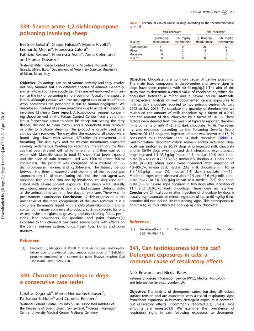
339. Severe acute 1,2-dichloropropanepoisoning involving sheep
Beatrice Giliottia, Chiara Falciolaa, Marina Rivoltaa,Leonardo Molinoa, Francesca Calonib,Fabrizio Sesanaa, Francesca Assisia, Anna Celentanoa
and Franca Davanzoa
aNational Milan Poison Control Center – Ospedale Niguarda Ca’Granda, Milan, Italy; bDepartment of Veterinary Science, Universityof Milan, Milan, Italy
Objective: Poisonings can be of various severity and they involvenot only humans but also different species of animals. Generally,animal intoxications are accidental; they are not endowed with rea-son so the risk of poisoning is more common. Usually the exposureis oral, although contact with the toxic agent can occur in differentways. Sometimes the poisoning is due to human negligence. Wedescribe an incident of severe poisoning due to acute skin exposureinvolving 12 sheep. Case report: A toxicological request concern-ing sheep arrived at the Poison Control Centre from a veterinar-ian. A farmer was about to shear his sheep but, seeing the dirtyfleeces, decided to clean them using a household stain removerin order to facilitate shearing. This product is usually used as aclothes stain remover. The day after the exposure, all sheep werefound lying down with serious difficulties in movement andbreathing. The skin, eyes, and the mucous membranes appearedseverely oedematous. Waiting for veterinary intervention, the flee-ces had been strewed with white mineral oil and then washed inwater with Marseille soap. In total 12 sheep had been exposedand the dose of stain remover used was 1500 ml (three 500 mlcontainers). The product was composed of a mixture of 1,2-dichloropropane, hexane, and cyclohexane. The latency periodbetween the time of exposure and the time of the request wasapproximately 12–18 hours. During this time, the toxic agent wasabsorbed through the skin and by inhalation, causing signs con-sistent with severe solvent exposure. The sheep were laterallyrecumbent, unresponsive to pain and had seizures. Unfortunately,all the animals died within a few days of exposure. There was nopost-mortem examination. Conclusion: 1,2-dichloropropane is themost toxic of the three components of the stain remover. It is acolourless flammable liquid with a chloroform-like odour and iscontained in many commercial products, such as solvents for oils,waxes, resins and glues, degreasing and dry-cleaning fluids, pesti-cides, lead scavengers for gasoline, and paint fixatives.[1]Exposure to this chemical can cause severe signs with effects onthe central nervous system, lungs, heart, liver, kidney and bonemarrow.
Reference
[1] Fiaccadori E, Maggiore U, Rotelli C, et al. Acute renal and hepaticfailure due to accidental percutaneous absorption of 1,2-dichlor-propane contained in a commercial paint fixative. Nephrol DialTransplant. 2003;18:219–220.
340. Chocolate poisonings in dogs:a consecutive case series
Colette Degrandia, Maren Hermanns-Clausenb,Katharina E. Hofera and Cornelia Reicherta
aNational Poisons Centre, Tox Info Suisse, Associated Institute ofthe University of Zurich, Zurich, Switzerland; bPoisons InformationCentre, University Medical Centre, Freiburg, Germany
Objective: Chocolate is a common cause of canine poisoning.The main toxic compound is theobromine and severe signs indogs have been reported with 40–60 mg/kg.[1] The aim of thisstudy was to determine a critical value of theobromine, which dis-tinguishes between a minor and a severe course. Methods:Retrospective analysis of well documented canine exposures tomilk or dark chocolate reported to two poisons centres (January2002 to July 2015). To calculate the quantity of theobromine wemultiplied the amount of milk chocolate by a factor of 0.0015and the amount of dark chocolate by a factor of 0.0115. Thesefactors were derived from the mean of typically reported theobro-mine contents of milk (1–2) and dark chocolate (7–16). The sever-ity was evaluated according to the Poisoning Severity Score.Results: Of 122 dogs the ingested amount was known in 113, 59consumed milk chocolate and 54 dark chocolate (Table 1).Gastrointestinal decontamination (emesis and/or activated char-coal) was performed in 30/59 dogs who ingested milk chocolateand in 36/54 dogs who ingested dark chocolate. Asymptomaticdogs ingested 1.3–33.3 g/kg (mean 11.3, median 10.4) milk choc-olate (n¼ 41) or 0.7–33.3 g/kg (mean 6.5, median 4.1) dark choc-olate (n¼ 32). Minor signs were observed after ingestion of4.2–80 g/kg (mean 28.3, median 23.8) milk chocolate (n¼ 16) or3.1–13.9 g/kg (mean 7.6, median 7.4) dark chocolate (n¼ 12).Moderate signs were observed after 42.9 and 47.6 g/kg milk choc-olate (n¼ 2) or 5.0–30.0 g/kg (mean 18.0, median 15.5) dark choc-olate (n¼ 8). Severe signs occurred in two dogs after ingestion of11.1 and 50.0 g/kg dark chocolate. There were no fatalities.Conclusion: Clinical course after ingestion of chocolate by dogs isusually asymptomatic or minor. Ingestion of up to 60 mg/kg theo-bromine did not induce life-threatening signs. This corresponds toabout 40 g/kg milk chocolate or 5.2 g/kg dark chocolate.
Reference
[1] Gwaltney-Brant S. Chocolate intoxication. Vet Med.2001;96:108–111.
341. Can fastidiousness kill the cat?Detergent exposures in cats: acommon cause of respiratory effects
Nick Edwards and Nicola BatesVeterinary Poisons Information Service (VPIS), Medical Toxicologyand Information Services, London, UK
Objective: The toxicity of detergents varies, but they all reducesurface tension and are associated with a risk of respiratory signsfrom foam aspiration. In humans, detergent exposure is common,but respiratory effects uncommonly reported,[1,2] unless largeamounts are ingested.[3] We examine the prevalence ofrespiratory signs in cats following exposures to detergents.
Table 1. Severity of clinical course in dogs according to the theobromine dose(n ¼ 113).
Severity
Milk chocolate Dark chocolate
�60 mg/kgtheobromine
>60 mg/kgtheobromine
�60 mg/kgtheobromine
>60 mg/kgtheobromine
Asymptomatic 41 0 20 12Minor 14 2 4 8Moderate 0 2 1 7Severe 0 0 0 2
CLINICAL TOXICOLOGY 515
Dow
nloa
ded
by [
Kin
gs C
olle
ge L
ondo
n] a
t 07:
31 2
1 A
pril
2016
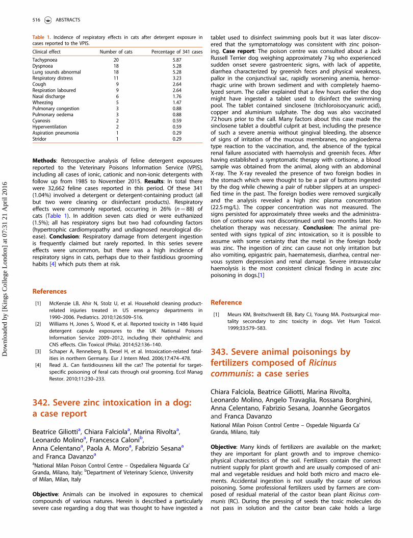
Methods: Retrospective analysis of feline detergent exposuresreported to the Veterinary Poisons Information Service (VPIS),including all cases of ionic, cationic and non-ionic detergents withfollow up from 1985 to November 2015. Results: In total therewere 32,662 feline cases reported in this period. Of these 341(1.04%) involved a detergent or detergent-containing product (allbut two were cleaning or disinfectant products). Respiratoryeffects were commonly reported, occurring in 26% (n¼ 88) ofcats (Table 1). In addition seven cats died or were euthanized(1.5%); all has respiratory signs but two had cofounding factors(hypertrophic cardiomyopathy and undiagnosed neurological dis-ease). Conclusion: Respiratory damage from detergent ingestionis frequently claimed but rarely reported. In this series severeeffects were uncommon, but there was a high incidence ofrespiratory signs in cats, perhaps due to their fastidious groominghabits [4] which puts them at risk.
References
[1] McKenzie LB, Ahir N, Stolz U, et al. Household cleaning product-related injuries treated in US emergency departments in1990–2006. Pediatrics. 2010;126:509–516.
[2] Williams H, Jones S, Wood K, et al. Reported toxicity in 1486 liquiddetergent capsule exposures to the UK National PoisonsInformation Service 2009–2012, including their ophthalmic andCNS effects. Clin Toxicol (Phila). 2014;52:136–140.
[3] Schaper A, Renneberg B, Desel H, et al. Intoxication-related fatal-ities in northern Germany. Eur J Intern Med. 2006;17:474–478.
[4] Read JL. Can fastidiousness kill the cat? The potential for target-specific poisoning of feral cats through oral grooming. Ecol ManagRestor. 2010;11:230–233.
342. Severe zinc intoxication in a dog:a case report
Beatrice Giliottia, Chiara Falciolaa, Marina Rivoltaa,Leonardo Molinoa, Francesca Calonib,Anna Celentanoa, Paola A. Moroa, Fabrizio Sesanaa
and Franca Davanzoa
aNational Milan Poison Control Centre – Ospedaliera Niguarda Ca’Granda, Milano, Italy; bDepartment of Veterinary Science, Universityof Milan, Milan, Italy
Objective: Animals can be involved in exposures to chemicalcompounds of various natures. Herein is described a particularlysevere case regarding a dog that was thought to have ingested a
tablet used to disinfect swimming pools but it was later discov-ered that the symptomatology was consistent with zinc poison-ing. Case report: The poison centre was consulted about a JackRussell Terrier dog weighing approximately 7 kg who experiencedsudden onset severe gastroenteric signs, with lack of appetite,diarrhea characterized by greenish feces and physical weakness,pallor in the conjunctival sac, rapidly worsening anemia, hemor-rhagic urine with brown sediment and with completely haemo-lyzed serum. The caller explained that a few hours earlier the dogmight have ingested a tablet used to disinfect the swimmingpool. The tablet contained sinclosene (trichloroisocyanuric acid),copper and aluminium sulphate. The dog was also vaccinated72 hours prior to the call. Many factors about this case made thesinclosene tablet a doubtful culprit at best, including the presenceof such a severe anemia without gingival bleeding, the absenceof signs of irritation of the mucous membranes, no angioedematype reaction to the vaccination, and, the absence of the typicalrenal failure associated with haemolysis and greenish feces. Afterhaving established a symptomatic therapy with cortisone, a bloodsample was obtained from the animal, along with an abdominalX-ray. The X-ray revealed the presence of two foreign bodies inthe stomach which were thought to be a pair of buttons ingestedby the dog while chewing a pair of rubber slippers at an unspeci-fied time in the past. The foreign bodies were removed surgicallyand the analysis revealed a high zinc plasma concentration(22.5 mg/L). The copper concentration was not measured. Thesigns persisted for approximately three weeks and the administra-tion of cortisone was not discontinued until two months later. Nochelation therapy was necessary. Conclusion: The animal pre-sented with signs typical of zinc intoxication, so it is possible toassume with some certainty that the metal in the foreign bodywas zinc. The ingestion of zinc can cause not only irritation butalso vomiting, epigastric pain, haematemesis, diarrhea, central ner-vous system depression and renal damage. Severe intravascularhaemolysis is the most consistent clinical finding in acute zincpoisoning in dogs.[1]
Reference
[1] Meurs KM, Breitschwerdt EB, Baty CJ, Young MA. Postsurgical mor-tality secondary to zinc toxicity in dogs. Vet Hum Toxicol.1999;33:579–583.
343. Severe animal poisonings byfertilizers composed of Ricinuscommunis: a case series
Chiara Falciola, Beatrice Giliotti, Marina Rivolta,Leonardo Molino, Angelo Travaglia, Rossana Borghini,Anna Celentano, Fabrizio Sesana, Joannhe Georgatosand Franca DavanzoNational Milan Poison Control Centre – Ospedale Niguarda Ca’Granda, Milano, Italy
Objective: Many kinds of fertilizers are available on the market;they are important for plant growth and to improve chemico-physical characteristics of the soil. Fertilizers contain the correctnutrient supply for plant growth and are usually composed of ani-mal and vegetable residues and hold both micro and macro ele-ments. Accidental ingestion is not usually the cause of seriouspoisoning. Some professional fertilizers used by farmers are com-posed of residual material of the castor bean plant Ricinus com-munis (RC). During the pressing of seeds the toxic molecules donot pass in solution and the castor bean cake holds a large
Table 1. Incidence of respiratory effects in cats after detergent exposure incases reported to the VPIS.
Clinical effect Number of cats Percentage of 341 cases
Tachypnoea 20 5.87Dyspnoea 18 5.28Lung sounds abnormal 18 5.28Respiratory distress 11 3.23Cough 9 2.64Respiration laboured 9 2.64Nasal discharge 6 1.76Wheezing 5 1.47Pulmonary congestion 3 0.88Pulmonary oedema 3 0.88Cyanosis 2 0.59Hyperventilation 2 0.59Aspiration pneumonia 1 0.29Stridor 1 0.29
516 ABSTRACTS
Dow
nloa
ded
by [
Kin
gs C
olle
ge L
ondo
n] a
t 07:
31 2
1 A
pril
2016

amount of nitrogen, carbon, phosphorus pentoxide, potassiumand magnesium oxide as well as ricin. Moreover the release ofthese substances in the soil has an antagonistic effect againstinsects and parasites. Ricin is cytotoxic; in humans the fatal oraldose is 0.5 g/kg while in animals it is 1–2 g/kg.[1] We present theexperience of the Poison Control Centre, Milan concerning animalpoisoning due to RC-based fertilizers from 2003 to 2014. Case ser-ies: Over the study period, there were 7 cases of definite exposureto RC fertilizers and another 18 cases here it was not certain thatRC was present in the fertilizer, however, the signs were similar tothose seen with ricin poisoning, which suggests RC was present.All exposures were accidental oral ingestions, and concerneddogs which ingested the material from the product container.Clinical signs appeared after more than 24 hours (n¼ 12; 48.0%).The most common signs were gastrointestinal (n¼ 12; 48.0%)with vomiting and diarrhea (sometimes hemorrhagic diarrhea) inaddition to depression and convulsions (n¼ 11; 44.0%). Threedogs died about one day after ingestion; death was rapid afteronset of signs. Conclusion: Most fertilizers have no toxic effects(if used properly) except those that contain RC. In this case therisk of severe poisoning is high for both animals and humans.Therefore, it is important to raise awareness of this potential haz-ard and to keep fertilizers in a safe place out of the reach of chil-dren and pets in order to reduce the risk of poisoning.
Reference
[1] Roels S, Coopman V, Vanhaelen P, et al. Lethal ricin intoxication intwo adult dogs: toxicologic and histopathologic findings. J VetDiagn Invest. 2010;22:466–468.
344. Salbutamol exposure in dogs
Joanna Crouchley and Nicola BatesVeterinary Poisons Information Service (VPIS), Medical Toxicologyand Information Services, London, UK
Objective: Salbutamol is a selective beta2-adrenoceptor agonist,used as a bronchodilator. Exposure typically causes rapid onsettachycardia, tachypnoea, and more rarely hypokalaemia. Thermalinjury consistent with exposure to released compressed gas froman inhaler has been reported.[1] The clinical signs, treatment andoutcomes in dogs exposed to salbutamol reported to theVeterinary Poisons Information Service (VPIS), were reviewed.Methods: A retrospective analysis of all canine cases of salbuta-mol exposure reported to the VPIS between February 1985 andOctober 2015. All cases with exposure to salbutamol as a singleagent and known outcome with returned veterinary surgeon fol-low up (via postal questionnaire) were included. Results: Therewere 411 cases of salbutamol exposure in dogs with follow up. Ofthese 20 (7%) remained asymptomatic, 4 of which received anemetic. Route of exposure was: not specified (42% of cases),chewing and puncturing an inhaler (48%) or ingestion of oral for-mulations (9%). Dose was known in 8 cases and ranged from 0.08to 23.5 mg/kg; all of these dogs were symptomatic. The mostcommon signs were tachycardia (78%), tachypnoea (20%), vomit-ing (19%), lethargy (16%), panting (16%), hypokalaemia (14%) andtremor (12%). Onset of signs was within 2 hours of exposure in84% of cases (range 2 minutes to 12 hours). Duration of signs was6 hours to 6 days, but 75% of dogs recovered within 24 hours.Hypokalaemia occurred in 59 dogs, value was known in 17 cases(range 2.4–3.6 mmol/L, normal range 3.9–5.1 mmol/L);15 had asso-ciated weakness, twitching or collapse. While there were noreports of thermal injury, 17 dogs had signs of facial, oral or ocu-lar inflammation (4% of cases). Of symptomatic cases (n¼ 382),109 (29%) received no treatment or observation only; 33 dogs
(9%) received gastrointestinal decontamination with an emetic oradsorbents. The most common treatments were beta-blockers(35%), IV fluids (16%), potassium (13%) and diazepam (11%). Ofdogs with hypokalaemia 88% received treatment with potassium(24%), a beta-blocker (31%), or a combination of both (34%). Onedisplayed cyanosis, hypersalivation, tachycardia, tachypnoea anddied within 2 hours despite treatment with acepromazine, pro-pranolol and oxygen; another died despite IV fluid therapy afterpresenting with tachycardia, shock and poor peripheral circula-tion. One case was ongoing at time of follow up and all otherdogs recovered fully. Conclusion: Salbutamol exposure in dogscauses rapid onset of clinical signs in most cases, however fatal-ities are very rare. Treatment is aimed at correcting tachycardiaand hypokalaemia.
Reference
[1] Mackenzie SD, Blois S, Hayes G, et al. Oral thermal injury associ-ated with puncture of a salbutamol metered-dose inhaler in adog. J Vet Emerg Crit Care (San Antonio). 2012;22:494–497.
345. Venlafaxine overdose in dogs:what are the risk factors associatedwith clinical signs?
Philip Rawson-Harris and Nicola BatesVeterinary Poisons Information Service (VPIS), Medical Toxicologyand Information Services, London, UK
Objective: Venlafaxine is a selective serotonin reuptake inhibitorwith no indication in dogs. In humans overdose causes vomiting,central nervous system (CNS) depression, electrocardiogram (ECG)changes, hypotension and seizures. The objective was to analysethe prevalence and risk factors associated with clinical signs incanine cases of venlafaxine ingestion. Methods: Retrospectiveanalysis of all canine venlafaxine exposures (with outcome)reported to the Veterinary Poisons Information Service (VPIS).Asymptomatic and symptomatic populations were compared withregards to dose (one tailed t-test), age and time since ingestion(two tailed t-test), and gender (Fisher’s exact test). Results: Of 67cases; 40 dogs (59.7%) remained asymptomatic and 27 (40.3%)developed signs, including one that died and another that waseuthanized (7.4%). Weak evidence of a difference in dosebetween the two groups was observed (p¼ 0.054, n¼ 49) anddose ingested was generally higher in symptomatic(11.5–1500 mg/kg, 37.5 mg/kg median) versus asymptomatic cases(1.3 to 168 mg/kg, 11.63 mg/kg median). Symptomatic cases pre-sented later than asymptomatic cases with strong evidenceobserved for the association (p¼ 0.001, n¼ 49). However, of the19 asymptomatic dogs within the dose range of symptomaticcounterparts, eight received no treatment. Four of these 8 hadingested extended release preparations. Extended release prepara-tions in symptomatic dogs were associated with higher doses andmilder signs, except one dog that died (after 30–180 mg/kg). Ifextended release preparations were excluded stronger but stillweak evidence of an association between dose and present ofsigns was observed (p¼ 0.04, n¼ 36) but this was limited by smallsample size and under reporting of preparation type. There wasno evidence of an association between age (p¼ 0.59, n¼ 56) norgender (p¼ 0.78, n¼ 56) and no breed suggestive trends wereobserved. Signs were consistent with human poisoning exceptvomiting was rare 2/27 (7.4%) and associated with higher doses(346.2–1500 mg/kg). Seizures were seen in one dog which died.Tachycardia was observed at higher doses (72.5–100 mg/kg,
CLINICAL TOXICOLOGY 517
Dow
nloa
ded
by [
Kin
gs C
olle
ge L
ondo
n] a
t 07:
31 2
1 A
pril
2016

n¼ 3). Neurological signs were common 26/27 (96.3%). Time torecovery ranged from 8–24 hours (average 17 hours, n¼ 4).Conclusion: Gender, age and breed are unlikely to affect venla-faxine toxicity in dogs. Dose is weakly associated with signs.When extended release preparations are excluded a strongerassociation is seen consistent with symptomatic extended releasecases requiring less treatment at higher doses. Early presentationwas associated with reduced incidence of clinical signs which,when observed, were consistent with those seen in human poi-soning except vomiting and convulsions.
346. Ethylene glycol poisoning in cats:what are the prognostic indicators?
Keely Luddena, Philip Rawson-Harrisb, Yu-Mei Changa,Oliver Gardena, Nicola Batesb and Nick Edwardsb
aRoyal Veterinary College, London, UK; bVeterinary PoisonsInformation Service (VPIS), Medical Toxicology and InformationServices, London, UK
Objective: Exposure to ethylene glycol (EG), a common compo-nent of antifreeze, is associated with very poor prognosis in cats.Exposure causes renal failure which is reversible with haemodialy-sis support, but this is impractical and uncommon in veterinarypractice. The objective was to analyse the prevalence and risk fac-tors associated with clinical signs in suspected EG exposure incats. Methods: Retrospective analysis of suspected EG exposure incats (with outcome) reported to the VPIS between December1996 to February 2014. Results: There were 240 cases; 6 cats(2.6%) remained asymptomatic and 234 developed signs (97.5%)including 163 (69.7%) which were euthanised, 42 that died(17.9%), 24 that recovered (10.3%) and 5 with on-going signs atthe time of follow up (2.1%). Most enquiries were made betweenOctober and March (153/240, 64%) peaking in December (32/240,13%). Enquiries were greatest in male cats (95/150, 63.3%) com-pared to 55/150 (36.7%) females (90/240 [37.5%] had unspecifiedgender) and in cats aged 1–3 years 97/205 (47.3%) peaking in2–3 year olds 40/205 (19.5%) (35/240 [14.6%] had unspecifiedage). The most common signs were ataxia (83/234, 35.5%), leth-argy (78/234, 33.3%), vomiting (73/234, 31.2%) and collapse (73/234, 31.2%). The commonest biochemical abnormalities reportedincluded azotaemia (113/234, 48.3%), hypocalcaemia (82/234,35%) and hyperglycaemia (47/234, 20.1%). Oxaluria was identifiedin 32 cases although it was unclear if urinalysis was assessed inevery case. Intravenous fluid therapy was administered in mostcases (188/240, 78.3%) and ethanol in 66/240 (27.5%). Of thosethat received the ethanol antidote 49 died and 17 survived. Ofthose that survived with time to treatment reported ethanol wasstarted within an average of 5.75 hours (median 1.5 hours, n¼ 10)compared to those that died or were euthanized receiving etha-nol therapy (21.4 hours average, 24 hours median, n¼ 17).Reasons for euthanasia included poor prognosis (53/163, 32.5%)no response to treatment (26/163, 16%), worsening azotaemia(13/163, 8%), development of anuria (4/163, 2.5%), uncontrolledseizures (3/163, 1.8%) and not specified (64/163, 39.3%). Seizuresoccurred in 18/42 (42.9%) in those that died versus 2/24 (8.3%)that recovered. Conclusion: EG exposure is commonest inyounger cats and in male cats and in winter months. Biochemicalabnormalities such as hypocalcaemia, azotaemia and hypergly-caemia can aid diagnosis, however, prognosis is very poor particu-larly in cats with seizures. Antidotal therapy is only useful ifstarted in the first few hours after exposure.
347. Oral exposure to fentanyltransdermal patches in dogs
Stella Matumo and Nicola BatesVeterinary Poisons Information Service (VPIS), Medical Toxicologyand Information Services, London, UK
Objective: To determine the clinical signs and outcome in dogsafter oral exposure to transdermal fentanyl patches. Even usedpatches contain a significant quantity of fentanyl and pose a risk.Methods: Retrospective analysis of cases of fentanyl transdermalpatch intoxication in dogs reported to the VPIS between 2002and September 2015. Results: There were 52 cases; ingestion wasthe most common route of exposure (57.7%, n¼ 30), followed bybuccal where the patch was chewed but not swallowed (42.3%,n¼ 22). Six dogs remained asymptomatic. In 12 symptomaticdogs the patches had been used (9 patches were chewed and 3ingested). Two cases involved the dogs just licking the area wherethe patch had been applied on the owner; one was asymptomaticand the other dog developed signs. Onset of effects rangedbetween 5 minutes and 5 hours. Of the 46 symptomatic dogs, 25(54.3%) had neurological, 20 (43.5%) gastrointestinal, 20 (43.5%)cardiovascular, 7 (15.2%) respiratory signs and 2 (4.3%) biochem-ical changes. Hypersalivation (34.8%, n¼ 16) and bradycardia(30.4%, n¼ 14) were the most common signs. Other signsincluded pale mucous membranes (n¼ 6), ataxia (n¼ 13), hypo-thermia (n¼ 3), drowsiness (n¼ 13), hypoventilation (n¼ 1), pant-ing (n¼ 1), collapse (n¼ 12) and coma (n¼ 1). Intravenous fluids32.6% (n¼ 15) and activated charcoal 26.1% (n¼ 12) were themost frequent treatments administered. Naloxone was used in19.6% (n¼ 9) of dogs with central nervous system and respiratorydepression. Surgery to remove the patch was used in 8.7% (n¼ 4)instances. All 46 symptomatic dogs recovered fully. Time to recov-ery was recorded for 43.4% (n¼ 20) cases and varied from 1 hourto 2 days. Conclusion: Exposure to fentanyl transdermal patchescan result in gastrointestinal, neurological, cardiovascular, bio-chemical and respiratory signs in dogs. In this series, neurologicalsigns (54.3%) dominated despite the most commonly observedindividual sign being hypersalivation. The overall picture is that ofclassic opioid poisoning with gastrointestinal signs and centralnervous system and respiratory depression. There was a slight dif-ference in the number of asymptomatic cases between buccaland ingestion exposure (9.9% versus 13.3%), suggesting ingestionis more likely to result in toxic effects compared to chewing thepatch. Outcome is very good in dogs that have chewed or eatenfentanyl patches.
349. Racial disparities in the treatmentof acute overdose in the emergencydepartment
Marcee E. McRaea, Lynne D. Richardsona,Robert S. Hoffmanb and Alex F. ManinibaIcahn School of Medicine at Mount Sinai, Department ofEmergency Medicine, New York, USA; bNew York University/Bellevue, Department of Emergency Medicine, New York, USA
Objective: Racial disparities continue to exist in many disciplinesof medicine extending to care in the Emergency Department(ED). A disparity can be defined as a difference in the quality ofhealthcare due to environment, access to care, health status, orparticular health outcomes.[1] We hypothesized that Blacks would
518 ABSTRACTS
Dow
nloa
ded
by [
Kin
gs C
olle
ge L
ondo
n] a
t 07:
31 2
1 A
pril
2016
be less likely to receive treatment with activated charcoal or anti-dotes when presenting to the ED for acute drug overdose.Methods: We completed a secondary analysis of a prospectivecohort of 3242 cases of patients presenting to 2 urban tertiarycare hospitals with suspected acute overdose between 2009 and2014. Categorical variables were analyzed with a chi-squared testwith 2-sided p values and 5% alpha. Odds ratios (OR) were calcu-lated with 95% CI. Assuming a baseline rate of 25% andalpha¼ 0.05, we had >80% power to detect an 18% difference inthe rate of antidote administration. Results: We screened 3242patients, of those, 2664 were included and 410 were excludeddue to alternate diagnosis (n¼ 93), lack of data (n¼ 188) or pedi-atric age (n¼ 53). Mean age was 41.5 years and 55% were men.Overall there were Blacks 21.8% (n¼ 580), Whites 33.67%(n¼ 897), Asians 6.9% (n¼ 183), other 6.9% (n¼ 185), andHispanics 30.4% (n¼ 811). There were no cultural differences inpoisonings and there were no associations between race andtype of drug overdose. Overall 219 cases were treated with acti-vated charcoal, either single or multi-dose, and 523 people weretreated with an antidote (naloxone [n¼ 257], N-acetylcysteineintravenous or oral [n¼ 136], calcium [n¼ 101], sodiumbicarbonate [n¼ 91], glucagon [n¼ 39], octreotide [n¼ 29],digoxin immune Fab [n¼ 10], high dose insulin therapy [n¼ 6],
physostigmine [n¼ 6], fomepizole [n¼ 5], dantrolene [n¼ 2], flu-mazenil [n¼ 2], or intravenous lipids [n¼ 1]). Results of the ana-lysis indicated that Blacks were less likely to receive activatedcharcoal, either one dose or multi-dose (Black 16.4%, 83.56% non-Black, p¼ 0.04, OR 0.687, 95% CI 0.48–0.99) and were much lesslikely to receive any antidote at presentation (Black 14.1%, non-Black 85.9%, p¼ 0.000001, OR 0.533, 95% CI 0.41–0.69).Conclusion: Blacks are significantly less likely to receive eitheractivated charcoal or any antidotes when presenting to the EDfor acute drug overdose. We did not assess whether those whodid not receive antidotes had unmet clinical indications, which isa limitation. Further studies are needed to determine nationalprevalence and how race plays a role in management of acuteoverdose.
Reference
[1] Carter-Pokras O, Baquet C. What is a "health disparity"? PublicHealth Rep. 2002;117:426–434.
CLINICAL TOXICOLOGY 519
Dow
nloa
ded
by [
Kin
gs C
olle
ge L
ondo
n] a
t 07:
31 2
1 A
pril
2016