Farnham Lab TF ChIP & Library Construction Protocol (2/1/13 ...
10
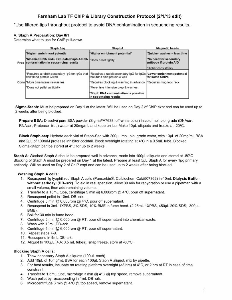
Farnham Lab TF ChIP & Library Construction Protocol (2/1/13 edit) *Use filtered tips throughout protocol to avoid DNA contamination in sequencing results. A. Staph A Preparation: Day 0/1 Determine what to use for ChIP pulldown. SigmaStaph: Must be prepared on Day 1 at the latest. Will be used on Day 2 of ChIP expt and can be used up to 2 weeks after being blocked. Prepare BSA: Dissolve pure BSA powder (Sigma#A7638, offwhite color) in cold mol. bio. grade (DNAse, RNAse, Protease free) water at 20mg/mL and keep on ice. Make 10μL aliquots and freeze at 20ºC. Block Staphseq: Hydrate each vial of StaphSeq with 200µL mol. bio. grade water, with 10µL of 20mg/mL BSA and 2µL of 100mM protease inhibitor cocktail. Block overnight rotating at 4ºC in a 0.5mL tube. Blocked SigmaStaph can be stored at 4°C for up to 2 weeks. Staph A: Washed Staph A should be prepared well in advance, made into 100µL aliquots and stored at 80ºC. Blocking of Staph A must be prepared on Day 1 at the latest. Prepare at least 5µL Staph A for every 1µg primary antibody. Will be used on Day 2 of ChIP expt and can be used up to 2 weeks after being blocked. Washing Staph A cells: 1. Resuspend 1g lyophilized Staph A cells (Pansorbin®, Calbiochem Cat#507862) in 10mL Dialysis Buffer without sarkosyl (DB–srk). To aid in resuspension, allow 30 min for rehydration or use a pipetman with a small volume, then add remaining volume. 2. Transfer to a 15mL tube, centrifuge 5 min @ 6,000rpm @ 4°C, pour off supernatant. 3. Resuspend pellet in 10mL DB–srk. 4. Centrifuge 5 min @ 6,000rpm @ 4°C, pour off supernatant. 5. Resuspend in 3mL 1XPBS, 3% SDS, 10% BME in fume hood. (2.25mL 1XPBS, 450μL 20% SDS, 300μL BME). 6. Boil for 30 min in fume hood. 7. Centrifuge 5 min @ 6,000rpm @ RT, pour off supernatant into chemical waste. 8. Wash with 10mL DB–srk. 9. Centrifuge 5 min @ 6,000rpm @ RT, pour off supernatant. 10. Repeat steps 79. 11. Resuspend in 4mL DB–srk. 12. Aliquot to 100μL (40x 0.5 mL tubes), snap freeze, store at 80ºC. Blocking Staph A cells: 1. Thaw necessary Staph A aliquots (100μL each). 2. Add 10μL of 10mg/mL BSA for each 100μL Staph A aliquot, mix by pipette. 3. For best results, incubate on rotating platform overnight (≥3 hrs) at 4°C, or 2 hrs at RT in case of time constraint. 4. Transfer to 1.5mL tube, microfuge 3 min @ 4°C @ top speed, remove supernatant. 5. Wash pellet by resuspending in 1mL DB–srk. 6. Microcentrifuge 3 min @ 4°C @ top speed, remove supernatant. 1
-
Upload
khangminh22 -
Category
Documents
-
view
0 -
download
0
Transcript of Farnham Lab TF ChIP & Library Construction Protocol (2/1/13 ...
Farnham Lab TF ChIP & Library Construction Protocol (2/1/13 edit)
*Use filtered tips throughout protocol to avoid DNA contamination in sequencing results.
A. Staph A Preparation: Day 0/1Determine what to use for ChIP pulldown.
SigmaStaph: Must be prepared on Day 1 at the latest. Will be used on Day 2 of ChIP expt and can be used up to2 weeks after being blocked.
Prepare BSA: Dissolve pure BSA powder (Sigma#A7638, offwhite color) in cold mol. bio. grade (DNAse,RNAse, Protease free) water at 20mg/mL and keep on ice. Make 10μL aliquots and freeze at 20ºC.
Block Staphseq: Hydrate each vial of StaphSeq with 200µL mol. bio. grade water, with 10µL of 20mg/mL BSAand 2µL of 100mM protease inhibitor cocktail. Block overnight rotating at 4ºC in a 0.5mL tube. BlockedSigmaStaph can be stored at 4°C for up to 2 weeks.
Staph A: Washed Staph A should be prepared well in advance, made into 100µL aliquots and stored at 80ºC.Blocking of Staph A must be prepared on Day 1 at the latest. Prepare at least 5µL Staph A for every 1µg primaryantibody. Will be used on Day 2 of ChIP expt and can be used up to 2 weeks after being blocked.
Washing Staph A cells:1. Resuspend 1g lyophilized Staph A cells (Pansorbin®, Calbiochem Cat#507862) in 10mL Dialysis Buffer
without sarkosyl (DB–srk). To aid in resuspension, allow 30 min for rehydration or use a pipetman with asmall volume, then add remaining volume.
2. Transfer to a 15mL tube, centrifuge 5 min @ 6,000rpm @ 4°C, pour off supernatant.3. Resuspend pellet in 10mL DB–srk.4. Centrifuge 5 min @ 6,000rpm @ 4°C, pour off supernatant.5. Resuspend in 3mL 1XPBS, 3% SDS, 10% BME in fume hood. (2.25mL 1XPBS, 450μL 20% SDS, 300μL
BME).6. Boil for 30 min in fume hood.7. Centrifuge 5 min @ 6,000rpm @ RT, pour off supernatant into chemical waste.8. Wash with 10mL DB–srk.9. Centrifuge 5 min @ 6,000rpm @ RT, pour off supernatant.10. Repeat steps 79.11. Resuspend in 4mL DB–srk.12. Aliquot to 100μL (40x 0.5 mL tubes), snap freeze, store at 80ºC.
Blocking Staph A cells:1. Thaw necessary Staph A aliquots (100μL each).2. Add 10μL of 10mg/mL BSA for each 100μL Staph A aliquot, mix by pipette.3. For best results, incubate on rotating platform overnight (≥3 hrs) at 4°C, or 2 hrs at RT in case of time
constraint.4. Transfer to 1.5mL tube, microfuge 3 min @ 4°C @ top speed, remove supernatant.5. Wash pellet by resuspending in 1mL DB–srk.6. Microcentrifuge 3 min @ 4°C @ top speed, remove supernatant.
1
7. Repeat steps 5 and 6.8. Resuspend pellet in 100μL DB–srk w/ 1mM PMSF (1μL of 100mM PMSF).9. Washed and blocked Staph A Cells can be stored at 4°C for up to 2 weeks.
B. Crosslinking Cells: Day 0/1Record cell information and details for future reference.
1. The amount of cells needed for ChIPseq (excludes histones, see histonespecific protocol) is 1x10^8 cellswhich is equivalent to 10 ChIPs at 1x10^7 cells for each ChIP. In general, between 20 and 40μg of antibodyare used for this number of cells, but optimization is recommended. For primary cells or tissues, thesamples can be used immediately or snap frozen in liquid nitrogen and crosslinked at a later date.However, whenever possible it is recommended to crosslink cells, prepare the nuclear pellet and thenfreeze the samples. When crosslinking suspension cells, make sure the volume of solution you crosslinkin is not too large. In a chemical hood add formaldehyde (from the 37% stock bottle) directly to thecollection media or culture media to a final concentration of 1%. If using frozen cells, in a chemical hoodprepare a 1% formaldehyde solution in media and add directly to frozen cell pellet; resuspend the cell pelletby pipetting up and down.
2. Rotate the cells in a tightly closed tube (for adherent cells use a shaking platform) for no more than 10minutes at room temperature. Do not crosslink for longer periods since this WILL cause cells to formaggregates that do not sonicate efficiently.
3. Stop crosslinking reaction by adding glycine to a final concentration of 0.125M. We use a 10X (1.25M)stock solution. Continue to agitate ≥5 min @ RT.
4. For cells crosslinked in suspension, centrifuge cells at 430 rcf for 5 min at 4ºC, discard solution, wash pellet twicewith icecold 1XPBS (mix by pipetting, pellet cells by centrifugation at 430 rcf for 5 min at 4ºC and discard washsolution). For adherent cells, pour off media and rinse plates twice with icecold 1XPBS and pour off wash solution.Using a silicon stopper cut in half for large plates or a scraper for smaller plates, scrape cells in residual PBS andtransfer cells from the culture dish to a tube on ice. Centrifuge the crosslinked cells at 430 rcf for 5 min at 4ºC. It isimportant to carefully aspirate supernatants so as to not lose cells. Note: Media containing formaldehyde should betreated as hazardous waste.
5. Cells may now be used immediately (preferred method) for a chromatin preparation or snap frozen in liquidnitrogen and stored at 80°C. Freezing cells at this stage results in a loss of chromatin, as some nuclei willinevitably rupture.
C. Chromatin Sonication: Day 1
Sonication conditions should be optimized for each cell type before processing large quantities of cells.
1. If using frozen crosslinked cells, thaw them on ice; keep all cells and chromatin samples on ice at alltimes. Prepare in advance 10mL of cell swelling buffer for 1x10^8 cells (exact amount is not important, butavoiding being too concentrated is): add Igepal (10μl per ml cell lysis buffer, agitate at 37°C to dissolve, coolon ice), then add protease inhibitors (see recipes for details on protease inhibitors). Resuspend cell pellet infreshly prepared cold cell lysis buffer by pipetting. Incubate on ice for 15 minutes to allow cells to swell.
2. Homogenize cells using a glass dounce homogenizer (type B) to break open the cells and release nuclei.Homogenize cells on ice with 20 strokes. *Skip this step if you are dealing with very low amounts of cells.*
3. Centrifuge the crosslinked cells at 430 rcf for 5 min at 4°C. Discard the supernatant. You may snapfreezethe nuclear pellet and store at 80°C for later use. This is the preferred stopping point for sample prep.
4. Resuspend pellet in nuclei lysis buffer plus protease inhibitors with 5X the volume of the pellet present. (Thisdilution amount is good for most cells, except B lymphoblasts cells that do much better at 10X). Nucleiconcentrations that are too high can lead to inefficient sonication and nuclei that are too dilute will result inhigher amounts of SDS which will then require much larger volumes of IP dilution buffer (making samplehandling more labor intensive). Incubate on ice for 30 minutes. An optional flashfreezing step may helpbreak open nuclei more efficiently. This step is critical if the homogenizing in step 2 is omitted. Flashfreezenuclei after 30 minutes of incubation in nuclear lysis buffer, thaw at room temperature. Once thawed,immediately transfer to ice (do not allow samples to warm up to room temperature) and proceed tosonication.
2
5. Sonicate cells in cold environment to achieve an average chromatin length of 200500bp. Wear hearingprotection! We use the BioRuptor on high setting for sonication. Volumes between 0.5mL and 2mL aresonicated in 15mL polystyrene tubes, volumes between 0.1 and 0.3mL are sonicated in 1.5mL Eppendorftubes. The pulse duration, intensity, and number will vary depending on the sonicator, the extent ofcrosslinking, and cell type. Ideally the least amount of input energy that gives satisfactory fragmentationshould be used. We commonly sonicate 30 minutes (pulses of 30 seconds at setting high, 1.5 minutespause in between pulses). These settings have been used for hES cells and breast cells; Tcells and Bcellstypically need extra sonication to achieve the desirable size range. Keep sonicated chromatin at 4°C whileperforming quantification and determining chromatin size.
6. Centrifuge at maximum speed for 10 minutes at 4°C. (If samples were sonicated in polystyrene tubes, besure to transfer to polypropylene tubes for the hard spin, otherwise the tubes will likely crack.) Carefullytransfer the supernatant (sonicated chromatin) to a new tube while avoiding cell debris. Keep sonicatedmaterial (chromatin and cell debris) at 4°C while performing quantification and determining chromatin size.You may need to recombine the cell debris pellet and supernatant (containing the chromatin) to continuefurther sonication of all the material.
D. Determining Chromatin Size & Quantification: Day 11. Take approximately 1x10^6 cells worth (or 10μl should work if you resuspended with nuclie lysis buffer that
was 5X the volume of the pellet in previous step 4) of chromatin sample from step 6 above.2. Add ChIP elution buffer to a total volume of 100μl, add 12μl of 5M NaCl, boil samples in water bath for 15
minutes to reverse crosslinks.3. Allow sample to cool down, add 1μl DNasefree RNase, incubate 10 minutes at 37ºC. This step is important
because the presence of RNA results in false estimation of chromatin size.4. Purify DNA using a PCR purification kit, elute in 30μl water. Measure DNA concentration by NanoDrop and
calculate the chromatin yield. Cells typically yield 510μg of material per 1x10^6 cells.5. Run 22.5μg of DNA on a 1.5% agarose gel to visualize average size of chromatin. If the chromatin is larger
than ~600bp, adjust the sonication conditions (e.g. add more pulses after resuspending the cell pellet withthe chromatin) and repeat steps 15 until desired range of 200 to 500bp is obtained. If samples are takinglonger than an hour of sonication to achieve desired size, do not proceed. This is an indication thatovercrosslinking has occurred and your crosslinking protocol may need to be modified to decrease theamount of crosslinking.
E. Chromatin Immunoprecipitation: Day 1For best results, proceed to ChIP assays on the same day as chromatin is prepared.
1. Use 100μg of chromatin for each smallscale ChIP and 5001000μg for ChIPseq scale.2. Save 500ng of chromatin for the Input sample and bring to a total volume of 150μl with ChIP elution buffer.
Store this amount at 20ºC until the next day and then reverse the crosslinks in the input chromatin at thesame time the crosslinks in the ChIP samples are reversed.
3. Dilute chromatin with at least 5fold icecold IP dilution buffer containing protease inhibitors. This ensuresthat the SDS concentration is not too high for antibody binding.
4. Add antibody specific to the TF of interest to capture the protein/chromatin complexes. Record amount ofchromatin and antibody, the catalogue number and lot number of antibodies for future reference!
5. Incubate on a rotating platform at 4°C overnight.
F. Capture Antibody/Chromatin Complex and Reverse Crosslinks: Day 2
STAPH Protocol (skip ahead for magnetic beads protocol)1. For all mouse monoclonals or if the primary antibody is not from rabbit, add an appropriate secondary IgG.
Use an equivalent quantity of rabbit secondary IgG as amount of primary antibody. Incubate on rotator at4°C for 2 hours.
2. Add 5µL Staphseq or Staph A cells per 1µg of primary antibody used.3. Incubate on a rotating platform at RT for no longer than 15 min.4. Microfuge at 14,000rpm for 3 min, remove supernatant.All the following steps are carried out at room temperature5. Wash pellets with 1mL of 1X Dialysis Buffer w/o sarkosyl by pipetting to resuspend.6. Microfuge at 14,000rpm for 3 min, remove supernatant.7. Repeat steps 4 and 5.8. Wash pellets with 1mL of IP wash buffer 2 by pipetting to resuspend.9. Microfuge at 14,000rpm for 3 min, remove supernatant.10. Repeat steps 7 and 8.
3
11. Wash the pellet a third time with IP Wash Buffer 2 by repeating step 7, but transfer to a new tube for thisfinal wash. This will help eliminate background since a lot of DNA ends up sticking to the walls of the tubeand the elution step will release this “sticky” DNA into your sample.
12. Once the ChIP sample has been transferred to a new tube, microfuge at 14,000rpm for 3 min, removesupernatant.
13. Microfuge again by orienting the pellet on the tube hinge side to ensure visually locating the pellet andaspirate the last traces of wash buffer.
14. Elute antibody/chromatin complexes by adding 75µl IP elution buffer. Shake on vortexer for 30 minutes,setting 2.
15. Microfuge samples at 14,000rpm for 5 min. Carefully transfer supernatant containing antibody/chromatincomplexes to a new tube without any Staph A.
16. Repeat steps 11 and 12, but combine the second 75µl elution with the first to obtain a total of 150µl ofmaterial.
17. Microfuge the 150µl samples at 14,000rpm for 5 min. Microfuge by orienting the pellet on the hinge side toensure visually locating any trace amounts of the Staph A pellet and put supernatant in new tube. Repeatingthe spin may be necessary if the Staph pellet slips and is pipetted by accident. Trace amounts ofStaphSeq WILL NOT show up in sequencing data down the road, but regular Staph A will show up insequencing data if any trace amounts are carried over.
18. At this point, thaw the input sample(s) from the previous day. Add 20μl of 5M NaCl per 150μl elution buffer(to give approx. 0.6M final concentration) for each ChIP and Input.
19. Incubate all samples at 67°C overnight to reverse formaldehyde crosslinks.
Magnetic beads protocol (DIFFERENT THAN PCR CLEAN UP AMPURE BEADS!)1. *Wash beads 1x in the same buffer solution it will be added to.* Add 15µL magnetic protein A/G
beads per smallscale ChIP sample (or 150µL per ChIPseq sample) and incubate on a rotating platform for2 hrs at 4°C. We use an equal mixture of magnetic protein A beads from Invitrogen DynaBeads Protein A(#10002D) and magnetic protein G beads from Invitrogen DynaBeads Protein G (#10004D), but others workjust as well. Some examples are protein A magnetic beads from Cell Signaling Technology (#9006),magnetic protein G beads from Diagenode (#AIP102015), or a magnetic protein A/G mix from PierceProtein AG (#88803). No carrier DNA should be added to beads.
All the following steps are carried out at room temperature2. Allow beads to settle for 1 min in a magnetic separation rack, then carefully remove the supernatant.3. Wash magnetic beads two times with IP wash buffer 1 (RIPA buffer without protease inhibitors); take tubes
out of magnetic rack and mix by pipetting. Efficient washing is critical to reduce background. Avoidcrosscontamination of samples and loss of magnetic beads.
4. Wash magnetic beads two or three times with IP wash buffer 2 (take tubes out of magnetic rack and mix bypipetting). *Transfer to a new tube for your final wash. This will help eliminate background since alot of DNA ends up sticking to the walls of the tube and the elution step will release this “sticky”DNA into your sample. Switch to a new tube on your last wash.*
5. *Optional step: May be too stringent for some antibodies.* Wash 1X with IP wash buffer 3.6. Take care to discard all wash solution after final wash.7. Elute antibody/chromatin complexes by adding 75µl IP elution buffer. Shake on vortexer for 30 minutes,
setting 2.8. Allow beads to settle for 1 min in a magnetic separation rack. Carefully transfer supernatant containing
antibody/chromatin complexes to a new tube.9. Repeat steps 11 and 12, but combine the second 75µl elution with the first to obtain a total of 150μl of
material.10. At this point, thaw the input sample(s) from the previous day. Add 20μl of 5M NaCl per 150μl elution buffer
(to give approx. 0.6M final concentration) for each ChIP and Input.11. Incubate all samples at 67°C overnight to reverse formaldehyde crosslinks.
G. DNA Purification: Day 31. Purify DNA with a PCR clean up kit, one column per sample. Elute each sample with 35µl EB.2. Assess ChIP results by PCR or qPCR before proceeding to preparation of libraries. Use multiple negative
and positive targets and properly dilute large scale ChIPs (for example, dilute 1:10 for a 10X ChIPs done with~1000µg chromatin). Before proceeding to library making, verify positive site enrichments over negativesites.
4
ChIP Buffers List
Protease inhibitor stock solutions (stored at 20ºC) Note: Protease inhibitors and NP40/Igepal are added at time ofuse only. Do not add protease inhibitors to IP wash and elution buffers1) Aprotinin (10mg/mL), Leupeptin in water (10mg/mL), 100 mM PMSF in isopropanol.
[Aprotinin (1μL/mL) and Leupeptin (1μL/mL), PMSF (10μL/mL)]2) 200X Protease Inhibitor Cocktail, VWR catalogue #80053852
Cell Lysis buffer Nuclei Lysis buffer5mM PIPES pH 8.0 50mM TrisCl pH 8.185mM KCl 10mM EDTAAdd Igepal (10μL/mL), 1% SDS*agitate at 37°C to dissolve, Add protease inhibitors immediately before use Addprotease inhibitors immediately before use cool on ice, add protease inhibitorscool on ice, add protease inhibitors
IP Dilution buffer (RIPA) Dialysis Buffer (DB–srk)50mM Tris pH 7.4 2mM EDTA150mM NaCl 50mM TrisCl pH 8.01% (v/v) Igepal0.25% Deoxycholic acid1mM EDTA pH 8.0Add protease inhibitors when cold
IP wash buffer 1 IP wash buffer 250mM Tris pH7.4 100mM TrisCl pH 9.0150mM NaCl 500mM LiCl1% (v/v) Igepal 1% Igepal/NP400.25% deoxycholic acid 1% deoxycholic acid1mM EDTA, pH 8.0
IP wash buffer 3 ChIP elution buffer100mM TrisCl pH 9.0 50mM NaHCO3500mM LiCl 1% SDS1% Igepal/NP401% deoxycholic acid150mM NaCl
5
Library Construction Protocol
This library protocol is based on the Illumina Sample Preparation Kit for Genomic DNA with some modifications. Thisprotocol describes the preparation of libraries of ChIP DNA compatible with Illumina DNA sequencers. Libraries are preparedfrom the ChIP sample, as well as from input DNA from the corresponding cell type. It is not necessary to make a fresh inputlibrary every time. You may save library reagents and use an input library from the same cell type prepared previously forchecking enrichment levels in the last step. All clean up steps are with PCR Clean Up Ampure magnetic beads (AgencourtAmpure, Beckman Coulter, cat# A29152), not to be confused with the magentic protein A/G beads used in the previous ChIPportion of the protocol.ALL REAGENT ALIQUOTS ARE IN CLEARLY LABELED BOXES AND LOCATIONS ARE DESCRIBED BELOW.
Step 1: EndRepair Using the EndIt DNA Repair Kit from Epicentre (Cat# ER0720)BOX LABELED “LIBRARY STEP #1 & #2” ON THE BOTTOM SHELF OF THE 20 FREEZERThis step ensures that all DNA fragments are converted to bluntended, 5’phosphorylated DNA.
A) Combine and mix the following components in a tube:134μL ChIP DNA (entire ChIPsample) or 100ng input DNA5μL 10X EndRepair Buffer5μL 10mM ATP5μL 2.5M dNTP Mix1μL EndRepair Enzyme MixX µL sterile water to a total volume of 50µL
B) Incubate 45 min @ RTC) Purify with 90µL Ampure magnetic beads (1.8x volume). Beads may take several minutes to pellet with magnetduring each step. During all pipetting, keep beads stationary on the magnet (do not pipette up and down) andvisually ensure that the beads aren’t slipping. Make fresh 70% ethanol and wash beads twice. Do not allow thebeads to dry. Do a final spin and hold tube against the magnet while pipetting off last remaining ethanol wash.D) Elute in 34μL EB.E) Place tubes back in magnetic stands to collect the DNA and transfer liquid to new tubes.
Step 2: Addition of ‘A’ base to 3’ EndsBOX LABELED “LIBRARY STEP #1 & #2” ON THE BOTTOM SHELF OF THE 20 FREEZERMake stocks of 1mM dATP using NEB 100mM dATP. For example, add 5µL 100mM dATP to 495µL sterileRNase/DNasefree Gibco water, split to 25µL aliquots, and freeze at 20ºC. Defrost only once.
A) Combine and mix the following in PCR tubes:34µL DNA from Step 15µL Klenow buffer (NEB Buffer 2)10µL 1 mM dATP (not included in NEB kit)1µL Klenow fragment (3’→5’ exo, NEB cat# M0212s, 5,000 U/mL)Total reaction volume of 50µL
B. Incubate for 30 min @ 37°C in PCR machineC. Purify with 90µL Ampure magnetic beads (1.8x volume). Beads may take several minutes to pellet with magnetduring each step. During all pipetting, keep beads stationary on the magnet (do not pipette up and down) andvisually ensure that the beads aren’t slipping. Make fresh 70% ethanol and wash beads twice. Do not allow thebeads to dry. Do a final spin and hold tube against the magnet while pipetting off last remaining ethanol wash.D) Elute in 11μL EB.E) Place tubes back in magnetic stands to collect the DNA and transfer liquid to new tubes.
6
Step 3: Illumina Barcode Adapter Ligation (BIOO adapters)BOX LABELED “LIBRARY STEP #3 & #4” ON THE BOTTOM SHELF OF THE 20 FREEZERDilute Illumina 15µM adapters 1:10 with water (final concentration 1.5µM) to adjust for the smaller quantity of DNA. BIOOsupplies their NEXTflex™ DNA Barcodes adapters at 25µM, so make a 1:17 dilution (final concentration 1.5µM). Excessadapters can interfere with sequencing.
A) Combine and mix the following components in a tube (total reaction volume = 30µL):11µL DNA from Step215µL DNA ligase buffer1µL Adapter mix (1.5µM)3µL DNA ligase (Ligafast from Promega Cat# M8221)Total reaction volume of 30µL
B) Incubate for 15 min at room temperature.C) Purify with 24µL Ampure magnetic beads (0.8x volume). Beads may take several minutes to pellet with magnetduring each step. During all pipetting, keep beads stationary on the magnet (do not pipette up and down) andvisually ensure that the beads aren’t slipping. Make fresh 70% ethanol and wash beads twice. Do not allow thebeads to dry. Do a final spin and hold tube against the magnet while pipetting off last remaining ethanol wash.D) Elute in 19μL EB.E) Place tubes back in magnetic stands to collect the DNA and transfer liquid to new tubes.
Step 4: Size Selection & Gel PurificationBOX LABELED “LIBRARY STEP #3 & #4” ON THE BOTTOM SHELF OF THE 20 FREEZERSize selection of the sample ensures removal of unused adapters and selection of proper fragment size for amplificationand sequencing. We use precast agarose gels (Invitrogen EGel EX 2% agarose; Cat# G402002) to minimize risk ofcontamination. Invitrogen TrackIt Ladder and Loading Buffer is described in the steps below.
A) Dilute 10μL of 6X Cyan/Orange with 50μL EB buffer to obtain 1X Cyan/Orange buffer dye. Add 1μL of 1XCyan/Orange buffer dye to eluted DNA from Step 3 above.B) Load 20µL EB buffer in each of the empty wells. Make sure to skip a well between samples to avoidcrosscontamination.C) Load 2µL 50bp ladder and 18µL EB in the wells on the outer edge wells.D) Load 20µL DNA with dye in each well.E) Run the gel for ~10min with the newer version power base.F) Image gel to visualize sample.G) Size select using a clean razor blade but cutting out regions from 200500bp. You may have to cut blindly due tolow DNA concentrations.H) Dissolve gels using QIAgen’s gel extraction buffer at room temperature. Add 500µL QG buffer to each gel sliceand shake on the vortexer for 30 min or until gel is dissolved.I) Purify DNA using a QIAquick MinElute PCR Purification Kit. Elute in 24µL EB.
Step 5: Amplification of AdapterModified DNA FragmentsBOX LABELED “LIBRARY STEP #5” ON THE BOTTOM SHELF OF THE 20 FREEZERFor BIOO adapters, make sure to use the supplied BIOO primer mix with a 12.5µM conc. Dilute the mix 1:2 (finalconcentration 6.25µM) and use 1uL per 50uL PCR reaction (as outlined in step A below). For Illumina adapters, diluteIllumina pairedend primers (1.0 and 2.0) 1:4 using sterile water for use in the reactions. Paired end primers can be orderedvia from Invitrogen and elsewhere. If possible, have primer purity checked by HPLC.
A) Combine and mix the following components in PCR tubes: 24µL DNA from Step 4 25µL KAPA HiFi NGS MM 1µL PCR primer mix (6.25uM) 50µL Total reaction volume
7

8
B) Amplify using the following PCR protocol: 2 min @ 98ºC 1015 cycles : [30 sec @ 98ºC, 30 sec @ 65ºC, 1 min @ 72ºC] 4 min @ 72ºC Hold @ 10ºC
Make an educated guess on how many cycles of PCR to do based on your gel size selection image. Overamplificationshould be avoided to reduce potential PCR artifacts. Input DNA typically requires ~10 cycles of PCR.
Step 6: Library Purification, Quality and QuantificationQUBIT HS DNA KIT IS IN THE BOTTOM DRAWER OF THE 4 DEGREE FRIDGEGREEN AND WHITE KAPA BIOSYSTEMS KIT BOXES ARE LABELED “ILLUMINA DNA STANDARDS ANDPRIMER PREMIX KIT” AND ARE LOCATED ON THE BOTTOM SHELF OF THE 20 FREEZER
A) Purify with 40µL Ampure magnetic beads (0.8x volume). Beads may take several minutes to pellet with magnetduring each step. During all pipetting, keep beads stationary on the magnet (do not pipette up and down) andvisually ensure that the beads aren’t slipping. Make fresh 70% ethanol and wash beads twice. Do not allow thebeads to dry. Do a final spin and hold tube against the magnet while pipetting off last remaining ethanol wash.B) Elute in 20μL EB.C) Place tubes back in magnetic stands to collect the DNA and transfer liquid to new tubes.D) Check concentration with High Sensitivity DNA Qubit. (Possible to exclude next step if this one is done).E) Check concentration by Kappa Quant assay. (Possible to exclude prior step if this one is done, unless samplesare being sent to Stanford!).4) Check quality by BioAnalyzer. If excessive amount of primer dimer (85bp) and/or adapters (120bp) are present,redo the ampure bead clean up with a 0.8x volume ratio which allows for capturing larger size fragments and loss ofsmaller fragments, such as the primer and/or adapter dimers.
Step 7: Library ConfirmationSsoFast EVEGREEN SUPERMIX BY BIORAD IS LOCATED ON TOP SHELF OF THE 20 FREEZERCheck enrichment using same positive and negative control primers as used for original ChIP. Enrichment should showstronger enrichment than before if the library preparation was successful. Do not proceed with sequencing unless the positivetargets are at least 10fold enriched over input.
Enrichment of marks in the ChIPseq libraries are determined by quantitative realtime PCR (qPCR). The input sample isdiluted with EB to give a final concentration of no more than 1 ng/µL and serves as a reference. Prepare a master reactionmix for each library with triplicate reactions per primer set. Add extra reagents for 5% of the total number of reagents toaccount for loss of volume. Add 18μL of reaction mix to each PCR reaction well. Add 1μL primer mix to each well.
Recipe for one reaction: 1μL undiluted ChIP Library Sample or diluted Input Sample (1ng/μl) 1μL 10μM target primer mix (containing both Forward and Reverse primers) 8μL Nucleasefree H2010μL SsoFast Evagreen Supermix20μL Total reaction volume
Amplify with the following PCR protocol:3 min @ 95ºC40 cycles : 5 sec @ 95ºC, then 5 sec @ 60ºCInclude a 6595°C melting curve at the end of the qPCR program, reading every ≤0.5°C.
Analyze the qPCR results by first manually determining the cycle threshold for each reaction across the plate withinthe linear range of the amplification curve. Calculate the average cycle threshold for each triplicate reaction of eachsample. The relative DNA amount is then calculated for any given primer set as 2 to the power of the cycle threshold(cT) difference between input chromatin and ChIP samples, where cT is the average value.
Ratio of sample DNA to Input DNA concentration = 2^(cTINPUTcTSAMPLE)
9

The values are then normalized with one of the negative controls. This is accomplished by dividing the relative DNAamount of each sample for a target primer set by the corresponding value for a negative control primer set. Theresulting quotient represents the fold enrichment. Repeat normalization with the second negative control.
10