Tertiary Structural and Functional Analyses of a Viroid RNA Motif by Isostericity Matrix and...
17
2006, 80(17):8566. DOI: 10.1128/JVI.00837-06. J. Virol. Itaya, Yijun Qi, Kathleen Boris-Lawrie and Biao Ding Xuehua Zhong, Neocles Leontis, Shuiming Qian, Asuka in Replication and Mutagenesis Reveal Its Essential Role of a Viroid RNA Motif by Isostericity Matrix Tertiary Structural and Functional Analyses http://jvi.asm.org/content/80/17/8566 Updated information and services can be found at: These include: REFERENCES http://jvi.asm.org/content/80/17/8566#ref-list-1 at: This article cites 61 articles, 23 of which can be accessed free CONTENT ALERTS more» articles cite this article), Receive: RSS Feeds, eTOCs, free email alerts (when new http://journals.asm.org/site/misc/reprints.xhtml Information about commercial reprint orders: http://journals.asm.org/site/subscriptions/ To subscribe to to another ASM Journal go to: on December 16, 2013 by guest http://jvi.asm.org/ Downloaded from on December 16, 2013 by guest http://jvi.asm.org/ Downloaded from
-
Upload
bensakovich -
Category
Documents
-
view
1 -
download
0
Transcript of Tertiary Structural and Functional Analyses of a Viroid RNA Motif by Isostericity Matrix and...
2006, 80(17):8566. DOI: 10.1128/JVI.00837-06. J. Virol.
Itaya, Yijun Qi, Kathleen Boris-Lawrie and Biao DingXuehua Zhong, Neocles Leontis, Shuiming Qian, Asuka in Replicationand Mutagenesis Reveal Its Essential Roleof a Viroid RNA Motif by Isostericity Matrix Tertiary Structural and Functional Analyses
http://jvi.asm.org/content/80/17/8566Updated information and services can be found at:
These include:
REFERENCEShttp://jvi.asm.org/content/80/17/8566#ref-list-1at:
This article cites 61 articles, 23 of which can be accessed free
CONTENT ALERTS more»articles cite this article),
Receive: RSS Feeds, eTOCs, free email alerts (when new
http://journals.asm.org/site/misc/reprints.xhtmlInformation about commercial reprint orders: http://journals.asm.org/site/subscriptions/To subscribe to to another ASM Journal go to:
on Decem
ber 16, 2013 by guesthttp://jvi.asm
.org/D
ownloaded from
on D
ecember 16, 2013 by guest
http://jvi.asm.org/
Dow
nloaded from
JOURNAL OF VIROLOGY, Sept. 2006, p. 8566–8581 Vol. 80, No. 170022-538X/06/$08.00�0 doi:10.1128/JVI.00837-06Copyright © 2006, American Society for Microbiology. All Rights Reserved.
Tertiary Structural and Functional Analyses of a Viroid RNA Motif byIsostericity Matrix and Mutagenesis Reveal Its Essential Role
in ReplicationXuehua Zhong,1 Neocles Leontis,2 Shuiming Qian,3,4 Asuka Itaya,1 Yijun Qi,1†
Kathleen Boris-Lawrie,3,4,5 and Biao Ding1,4,5*Department of Plant Cellular and Molecular Biology and Plant Biotechnology Center, Ohio State University, Columbus, Ohio 432101;
Department of Chemistry and Center for Biomolecular Sciences, Bowling Green State University, Bowling Green, Ohio 434042;Department of Veterinary Biosciences, Ohio State University, Columbus, Ohio 432103; Molecular, Cellular and
Developmental Biology Graduate Program, Ohio State University, Columbus, Ohio 432104; andOSU RNA Consortium, Ohio State University, Columbus, Ohio 432105
Received 23 April 2006/Accepted 19 June 2006
RNA-templated RNA replication is essential for viral or viroid infection, as well as for regulation of cellulargene expression. Specific RNA motifs likely regulate various aspects of this replication. Viroids of the Pospi-viroidae family, as represented by the Potato spindle tuber viroid (PSTVd), replicate in the nucleus by utilizingDNA-dependent RNA polymerase II. We investigated the role of the loop E (sarcin/ricin) motif of the PSTVdgenomic RNA in replication. A tertiary-structural model of this motif, inferred by comparative sequenceanalysis and comparison with nuclear magnetic resonance and X-ray crystal structures of loop E motifs inother RNAs, is presented in which core non-Watson-Crick base pairs are precisely specified. Isostericity matrixanalysis of these base pairs showed that the model accounts for the reported natural sequence variations andviable experimental mutations in loop E motifs of PSTVd and other viroids. Furthermore, isostericity matrixanalysis allowed us to design disruptive, as well as compensatory, mutations of PSTVd loop E. Functionalanalyses of such mutants by in vitro and in vivo experiments demonstrated that loop E structural integrity iscrucial for replication, specifically during transcription. Our results suggest that the PSTVd loop E motif existsand functions in vivo and provide loss-of-function genetic evidence for the essential role of a viroid RNAthree-dimensional motif in rolling-circle replication. The use of isostericity matrix analysis of non-Watson-Crick base pairing to rationalize mutagenesis of tertiary motifs and systematic in vitro and in vivo functionalassays of mutants offers a novel, comprehensive approach to elucidate the tertiary-structure–function rela-tionships for RNA motifs of general biological significance.
According to the “RNA world” scenario, the appearance ofRNA molecules simultaneously capable of self-replication andinformation storage signaled a major milestone in the evolu-tion of life (14, 24, 25). In modern biology, RNA replication iscentral to viral or viroid infection, as well as to the regulationof cellular gene expression. Virus-encoded RNA-dependentRNA polymerases play a major role in the replication of RNAviruses (10, 23). Cellular RNA-dependent RNA polymerasesgenerate double-stranded RNAs as triggers for RNA silencing(1). Intriguingly, the DNA-dependent cellular RNA poly-merases can also transcribe at least two types of RNA tem-plates: viroid RNAs (11, 15, 59) and the human hepatitis deltavirus (HDV) RNA (28, 60). The replication of viroid and HDVRNAs raises the question of whether the DNA-templated tran-scription machinery also replicates other cellular RNAs yet tobe identified. Elucidating the replication mechanisms of theseinfectious RNAs should help address this question of profoundbiological interest.
Viroids are the smallest known nucleic acid-based infectious
agents and self-replicating genetic units. Their “genomes” con-sist of single-stranded, circular RNAs ranging in size from 250to 400 nucleotides (23). Viroids can replicate and spreadthroughout an infected plant, although they do not encodeproteins, do not have encapsidation mechanisms, and do notrequire helper viruses. Furthermore, they cause devastatingdiseases by altering host gene expression and developmentalprocesses (11, 59). Evidently, viroid RNA genomes contain allof the sequence and structural information needed to mediateor trigger the various functions associated with infection.
Potato spindle tuber viroid (PSTVd) is the type member ofthe family Pospiviroidae (8, 12). The PSTVd genome consists of359 nucleotides and assumes a rod-shaped secondary structurein the native state (48) with five structural domains, as shownin Fig. 1A (26). This secondary structure, which is typical ofviroids in the family Pospiviroidae, comprises many loops andbulges flanked by short Watson-Crick helices. Formation ofthis secondary structure is necessary for infection (62). Duringasymmetric rolling-circle replication of PSTVd (5), the pluscircular strands serve as templates for the synthesis of concate-meric, linear minus strands, which then function as the repli-cation intermediates for the synthesis of concatemeric, linearplus strands. These are subsequently cleaved into monomersand ligated into circular molecules (Fig. 1B). Without encod-ing proteins, PSTVd replicates in the nucleus of a host cell and
* Corresponding author. Mailing address: Department of Plant Cel-lular and Molecular Biology, Ohio State University, 207 RightmireHall, 1060 Carmack Road, Columbus, OH 43210. Phone: (614) 247-6077. Fax: (614) 292-5379. E-mail: [email protected].
† Present address: Cold Spring Harbor Laboratory, Cold SpringHarbor, NY 11724.
8566
on Decem
ber 16, 2013 by guesthttp://jvi.asm
.org/D
ownloaded from

therefore presents an ideal model to address the question ofhow the plant nuclear transcription machinery replicates anRNA template.
Previous work has attempted to associate particular parts ofthe viroid RNA with specific steps of the replication cycle. Invitro transcription with potato nuclear extracts and a plus cir-cular PSTVd RNA template suggested that nucleotidesU359/C1 in the left terminal loop are the transcription initia-tion sites on the circular RNA (27). Subsequent site-directedmutagenesis, in combination with tomato infection studies,showed that the C1G mutation is stably maintained whereasU359G reverted to the wild type, suggesting that perhaps U359is the bona fide initiation site (27). A metastable hairpin II(HPII) encompassing nucleotide sequences 227 to 237 and 318to 328 was predicted to form during thermal denaturation ofthe PSTVd secondary structure (20, 46) and has been detectedin vitro and in vivo (50). Mutations that are designed to inhibitthe formation of HPII were found to always revert to the wildtype in mechanically inoculated plants, suggesting that HPII isimportant for replication, perhaps as a binding site for anunknown cellular factor (6, 36, 41, 45). Many mutations havebeen mapped throughout the PSTVd genome that inhibit in-fection in mechanically inoculated plants and often werebroadly interpreted to affect replication (19, 21, 39, 41).
The loop E motif, located in the central conserved region(CCR) of the central domain (Fig. 1A) of PSTVd, has beenimplicated in replication and other functions. This motif issimilar to the loop E motifs of archaeal and eucaryal 5SrRNAs, which are also found in the conserved sarcin/ricin
loops of 23S rRNA (29, 32). In vitro studies with longer-than-unit length PSTVd transcripts showed that cleavage and liga-tion occur between G95 and G96, which appear to involve theformation of a metastable tetraloop motif, followed by a con-formational change into a stable loop E motif (3). A singlenucleotide substitution, C259U, in loop E enhances replicationby 5- to10-fold in cultured cells of tobacco (Nicotiana tabacum)(43). The enhanced replication in tobacco cells may accountfor the adaptation of PSTVd to this host (63, 66). The U257Asubstitution in the loop E motif also enables PSTVd to infecttobacco (43, 66) and confers lethal symptoms in tomato (42).These in vitro and gain-of-function mutational results suggestthat loop E plays one or more crucial roles in viroid-plantinteractions.
Several critical issues must be addressed in order to assign aspecific function of viroid replication in vivo to a particularviroid structural element. First, whether a motif exists in vivoneeds to be demonstrated. Second, reversions of mutated nu-cleotides to the wild type or gain-of-function mutations do notprovide direct evidence for the role of the affected RNA struc-tural element in a biological process. Loss-of-function muta-tions, on the other hand, will provide definitive proof linkingthe affected structural element to a function. Third, infectionassays on whole plants do not reveal whether failure of infec-tion of a mutant results from defects in a specific step of thereplication cycle or in intra- or intercellular trafficking. Toaddress these issues, it is imperative to integrate structural,genetic, cellular, biochemical, and molecular approaches.
Genetic alteration of an RNA structural element for func-
FIG. 1. Secondary structure (A) and replication model (B) of PSTVd. (A) The rod-like secondary structure of PSTVd and the numbering ofnucleotides are based on a report by Gross et al. (16). The five structural domains (26) include (i) the left terminal domain (TL, 1 to 46 and 315to 359), (ii) the pathogenicity domain (47 to 73 and 286 to 314), (iii) the central domain (74 to 120 and 240 to 285), (iv) the variable domain (121to 148 and 212 to 239), and (v) the right terminal domain (TR, 149 to 211). The loop E motif is in the central conserved region. (B) Asymmetricrolling-circle replication of PSTVd (5). The incoming circular monomeric plus-strand PSTVd serves as the initial template to synthesize linear,concatemeric minus-strand PSTVd. The latter functions as the replication intermediate to direct the synthesis of concatemeric plus-strand PSTVd,which is cleaved into unit length monomers and further ligated into circular forms.
VOL. 80, 2006 ESSENTIAL ROLE OF A VIROID RNA MOTIF IN REPLICATION 8567
on Decem
ber 16, 2013 by guesthttp://jvi.asm
.org/D
ownloaded from
tional studies requires precise understanding of its tertiarystructure and the effects of mutations on that structure. How-ever, while the secondary structures of viroids have been wellestablished through thermodynamic, chemical mapping, muta-tional, and nuclear magnetic resonance (NMR) studies (9, 55,62), much less is known about their tertiary structures andspecific functions. Minimum-free-energy calculations (e.g.,mfold; 67), coupled with conventional comparative sequenceanalysis, reveal RNA secondary structures comprising double-stranded helices formed by Watson-Crick base pairs, punctu-ated by so-called loops and bulges (hairpin, internal, and junc-tion or multihelix loops) defined by lack of Watson-Crick basepairs. However, most RNA loops in structured, biologicallyactive RNA molecules have been shown by X-ray crystallog-raphy and NMR spectroscopy to form specific, well-structuredthree-dimensional (3D) motifs in which many of the basesform non-Watson-Crick base pairs and/or stacking interactions(31–33). Thus, current thermodynamic calculations generallycannot reveal the nature of non-Watson-Crick base pairingwithin loops and offer little help in designing mutations thatdisrupt or restore such base pairing.
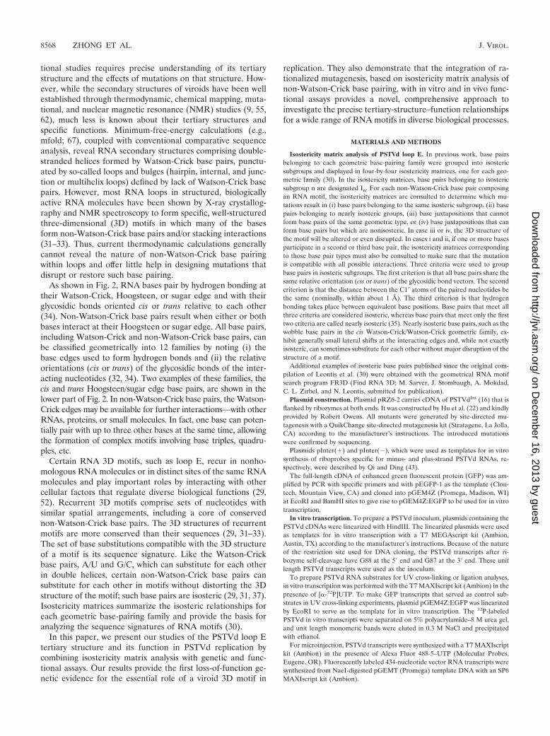
As shown in Fig. 2, RNA bases pair by hydrogen bonding attheir Watson-Crick, Hoogsteen, or sugar edge and with theirglycosidic bonds oriented cis or trans relative to each other(34). Non-Watson-Crick base pairs result when either or bothbases interact at their Hoogsteen or sugar edge. All base pairs,including Watson-Crick and non-Watson-Crick base pairs, canbe classified geometrically into 12 families by noting (i) thebase edges used to form hydrogen bonds and (ii) the relativeorientations (cis or trans) of the glycosidic bonds of the inter-acting nucleotides (32, 34). Two examples of these families, thecis and trans Hoogsteen/sugar edge base pairs, are shown in thelower part of Fig. 2. In non-Watson-Crick base pairs, the Watson-Crick edges may be available for further interactions—with otherRNAs, proteins, or small molecules. In fact, one base can poten-tially pair with up to three other bases at the same time, allowingthe formation of complex motifs involving base triples, quadru-ples, etc.
Certain RNA 3D motifs, such as loop E, recur in nonho-mologous RNA molecules or in distinct sites of the same RNAmolecules and play important roles by interacting with othercellular factors that regulate diverse biological functions (29,52). Recurrent 3D motifs comprise sets of nucleotides withsimilar spatial arrangements, including a core of conservednon-Watson-Crick base pairs. The 3D structures of recurrentmotifs are more conserved than their sequences (29, 31–33).The set of base substitutions compatible with the 3D structureof a motif is its sequence signature. Like the Watson-Crickbase pairs, A/U and G/C, which can substitute for each otherin double helices, certain non-Watson-Crick base pairs cansubstitute for each other in motifs without distorting the 3Dstructure of the motif; such base pairs are isosteric (29, 31, 37).Isostericity matrices summarize the isosteric relationships foreach geometric base-pairing family and provide the basis foranalyzing the sequence signatures of RNA motifs (30).
In this paper, we present our studies of the PSTVd loop Etertiary structure and its function in PSTVd replication bycombining isostericity matrix analysis with genetic and func-tional assays. Our results provide the first loss-of-function ge-netic evidence for the essential role of a viroid 3D motif in
replication. They also demonstrate that the integration of ra-tionalized mutagenesis, based on isostericity matrix analysis ofnon-Watson-Crick base pairing, with in vitro and in vivo func-tional assays provides a novel, comprehensive approach toinvestigate the precise tertiary-structure–function relationshipsfor a wide range of RNA motifs in diverse biological processes.
MATERIALS AND METHODS
Isostericity matrix analysis of PSTVd loop E. In previous work, base pairsbelonging to each geometric base-pairing family were grouped into isostericsubgroups and displayed in four-by-four isostericity matrices, one for each geo-metric family (30). In the isostericity matrices, base pairs belonging to isostericsubgroup n are designated In. For each non-Watson-Crick base pair composingan RNA motif, the isostericity matrices are consulted to determine which mu-tations result in (i) base pairs belonging to the same isosteric subgroup, (ii) basepairs belonging to nearly isosteric groups, (iii) base juxtapositions that cannotform base pairs of the same geometric type, or (iv) base juxtapositions that canform base pairs but which are nonisosteric. In case iii or iv, the 3D structure ofthe motif will be altered or even disrupted. In cases i and ii, if one or more basesparticipate in a second or third base pair, the isostericity matrices correspondingto those base pair types must also be consulted to make sure that the mutationis compatible with all possible interactions. Three criteria were used to groupbase pairs in isosteric subgroups. The first criterion is that all base pairs share thesame relative orientation (cis or trans) of the glycosidic bond vectors. The secondcriterion is that the distance between the C1� atoms of the paired nucleotides bethe same (nominally, within about 1 Å). The third criterion is that hydrogenbonding takes place between equivalent base positions. Base pairs that meet allthree criteria are considered isosteric, whereas base pairs that meet only the firsttwo criteria are called nearly isosteric (35). Nearly isosteric base pairs, such as thewobble base pairs in the cis Watson-Crick/Watson-Crick geometric family, ex-hibit generally small lateral shifts at the interacting edges and, while not exactlyisosteric, can sometimes substitute for each other without major disruption of thestructure of a motif.
Additional examples of isosteric base pairs published since the original com-pilation of Leontis et al. (30) were obtained with the geometrical RNA motifsearch program FR3D (Find RNA 3D; M. Sarver, J. Stombaugh, A. Mokdad,C. L. Zirbel, and N. Leontis, submitted for publication).
Plasmid construction. Plasmid pRZ6-2 carries cDNA of PSTVdInt (16) that isflanked by ribozymes at both ends. It was constructed by Hu et al. (22) and kindlyprovided by Robert Owens. All mutants were generated by site-directed mu-tagenesis with a QuikChange site-directed mutagenesis kit (Stratagene, La Jolla,CA) according to the manufacturer’s instructions. The introduced mutationswere confirmed by sequencing.
Plasmids pInter(�) and pInter(�), which were used as templates for in vitrosynthesis of riboprobes specific for minus- and plus-strand PSTVd RNAs, re-spectively, were described by Qi and Ding (43).
The full-length cDNA of enhanced green fluorescent protein (GFP) was am-plified by PCR with specific primers and with pEGFP-1 as the template (Clon-tech, Mountain View, CA) and cloned into pGEM4Z (Promega, Madison, WI)at EcoRI and BamHI sites to give rise to pGEM4Z:EGFP to be used for in vitrotranscription.
In vitro transcription. To prepare a PSTVd inoculum, plasmids containing thePSTVd cDNAs were linearized with HindIII. The linearized plasmids were usedas templates for in vitro transcription with a T7 MEGAscript kit (Ambion,Austin, TX) according to the manufacturer’s instructions. Because of the natureof the restriction site used for DNA cloning, the PSTVd transcripts after ri-bozyme self-cleavage have G88 at the 5� end and G87 at the 3� end. These unitlength PSTVd transcripts were used as the inoculum.
To prepare PSTVd RNA substrates for UV cross-linking or ligation analyses,in vitro transcription was performed with the T7 MAXIscript kit (Ambion) in thepresence of [�-32P]UTP. To make GFP transcripts that served as control sub-strates in UV cross-linking experiments, plasmid pGEM4Z:EGFP was linearizedby EcoRI to serve as the template for in vitro transcription. The 32P-labeledPSTVd in vitro transcripts were separated on 5% polyacrylamide–8 M urea gel,and unit length monomeric bands were eluted in 0.3 M NaCl and precipitatedwith ethanol.
For microinjection, PSTVd transcripts were synthesized with a T7 MAXIscriptkit (Ambion) in the presence of Alexa Fluor 488-5–UTP (Molecular Probes,Eugene, OR). Fluorescently labeled 434-nucleotide vector RNA transcripts weresynthesized from NaeI-digested pGEMT (Promega) template DNA with an SP6MAXIscript kit (Ambion).
8568 ZHONG ET AL. J. VIROL.
on Decem
ber 16, 2013 by guesthttp://jvi.asm
.org/D
ownloaded from
After all transcription reactions, DNA templates were removed by DNase Itreatment. RNA transcripts were purified with a MEGAClear kit (Ambion) andfurther quantified by UV spectrometry or with a scintillation counter (for radio-active riboprobes).
Protoplast inoculation. Nicotiana benthamiana suspension cells were culturedas described by Sunter and Bisaro (57). Protoplasts were prepared and inocu-lated with PSTVd transcripts by electroporation as described by Qi and Ding(43). Detailed protocols for the maintenance of N. benthamiana suspension cells,preparation of protoplasts, and electroporation were described by Zhong et al.(65). At 3 days postinoculation, transfected protoplasts were harvested for RNAextraction and Northern blot analysis.
RNA extraction and Northern blotting. Total RNAs were isolated from in-fected plants and protoplasts with an RNeasy Plant Mini Kit (QIAGEN, Valen-cia, CA). The procedures were essentially the same as those described by Qi etal. (44). Ten-microgram RNA samples were run on 5% polyacrylamide–8 M ureagel, transferred to a Hybond-XL nylon membrane (Amersham Biosciences,Piscataway, NJ) with a vacuum blotting system (Amersham), and immobilized byUV-cross-linking. Hybridization with [�-32P]UTP-labeled riboprobes was carriedout at 65°C with ULTRAhyb reagent (Ambion). After overnight hybridization,the membranes were washed twice at 65°C in 2� SSC (1� SSC is 0.15 M NaClplus 0.015 M sodium citrate)–0.1% sodium dodecyl sulfate (SDS) and twice in0.2� SSC–0.1% SDS and exposed to a storage phosphor screen (Kodak, Roch-
FIG. 2. Geometric classification of RNA base pairing. The upper panel shows that each nucleotide base has three edges (Watson-Crick,Hoogsteen, and sugar) that can potentially hydrogen bond with one of the three edges of another base. Thus, each base can be representedschematically by a triangle and can potentially pair with up to three other bases. The interacting bases can pair with a cis or trans relative orientationof their glycosidic bonds. This is illustrated in the lower panels for the cis and trans orientations of nucleotides pairing at the Hoogsteen edge ofone base and the sugar edge of the second base. In these base pairs, the Watson-Crick edges of the interacting bases are available for furtherinteractions—with other RNAs, proteins, or small molecules. The cross and circle in the triangle where the Hoogsteen and sugar edges meetindicate 5�33� and 3�35� orientations, respectively, of the sugar-phosphate backbones relative to the plane of the page. W-C, Watson-Crick edge;H, Hoogsteen edge; SE, sugar edge. (Adapted from reference 34 with permission from the RNA Society.)
VOL. 80, 2006 ESSENTIAL ROLE OF A VIROID RNA MOTIF IN REPLICATION 8569
on Decem
ber 16, 2013 by guesthttp://jvi.asm
.org/D
ownloaded from
ester, NY). Hybridization signals were quantified with a Molecular Imager FXwith Quantity One-4.1.1 software (Bio-Rad, Hercules, CA).
Sequencing of RNA progeny. The protocols for preparing cDNAs of thePSTVd progeny isolated from protoplasts were performed essentially as de-scribed by Qi and Ding (43). Briefly, cDNAs of PSTVd RNA were reversetranscription (RT)-PCR amplified and sequenced in both directions with an ABI377 DNA sequencer (Perkin-Elmer, Boston, MA) at the DNA Sequencing Fa-cility at Ohio State University.
Preparation of N. benthamiana nuclear extract (NbNE). NbNE was preparedas described by Baumstark and Riesner (2) and Roberts and Okita (47), withslight modifications. All steps were performed on ice. Protoplasts prepared fromcultured cells of N. benthamiana (see above) were first resuspended in 15 ml ofbuffer A (20 mM morpholineethanesulfonic acid-KOH [pH 5.8], 20 mM K-acetate, 15% Ficoll 400, 0.15 mM spermine, 0.5 mM spermidine, 10 mM �-mer-captoethanol, 0.5 mM phenylmethylsulfonyl fluoride, 0.15 �M pepstatin, 0.6 �Mleupeptin) and then lysed in a Dounce homogenizer with 10 to 15 strokes.4�,6�-Diamidino-2-phenylindole (DAPI) staining was used to monitor the releaseof nuclei. The nuclei were purified by loading the homogenates onto a two-stepgradient consisting of 8 ml of buffer B (87.6% [vol/vol] Percoll, 0.62� buffer C[buffer A minus Ficoll]) and 12 ml of buffer D (buffer A with 18% instead of 15%Ficoll 400). The mixtures were centrifuged at 4,000 � g for 60 min at 4°C. Theinterphase containing the nuclei was collected with a glass pipette, and 1.5volumes of buffer C was added. The mixtures were centrifuged at 1,400 � g for30 min at 4°C, and the pelleted nuclei were resuspended in buffer E (60 mMHEPES-KOH [pH 7.9], 0.12 mM EDTA, 0.84 mM Mg-acetate, 0.72 �M leu-peptin, 0.18 �M pepstatin, 12 mM �-mercaptoethanol, 0.6 M KCl). The samplewas stirred gently for 1 h at 4°C and centrifuged at 36,000 � g for 60 min at 4°Cwith an L8-80 M Ultracentrifuge with an SW41 rotor (Beckman, Fullerton, CA).Saturated (NH4)2SO4 was slowly added to the resulting supernatant to a finalconcentration of 75% and centrifuged at 36,000 � g for 30 min at 4°C toprecipitate proteins. The pellet was resuspended in buffer F (20 mM HEPES-KOH [pH 7.9], 10 mM Mg-acetate, 50 mM K-acetate, 5 mM EDTA, 12 mM�-mercaptoethanol, 25% glycerol) and dialyzed overnight at 4°C in buffer F.Aliquots were quickly frozen in liquid nitrogen and stored at �80°C.
RNA circularization assay. The RNA circularization assay was conducted byincubating 32P-labeled and gel-purified unit length PSTVd in vitro transcriptsand control RNAs in NbNE. In a reaction mixture with a final volume of 20 �l,1 � 105 cpm of linear unit length plus-strand PSTVd transcripts was mixed with4 �l of 5� ligation buffer (100 mM Tris-HCl [pH 8.0], 30 mM Mg-acetate, 1 mMspermidine, 2 mM EDTA) (40), 1 �l of RNaseOut (Invitrogen), and 10 �l ofNbNE and incubated at 37°C for 2 to 3 h. The reaction was terminated byaddition of 260 �l of 2� proteinase K buffer (200 mM Tris-HCl [pH 8.0], 25 mMEDTA [pH 8.], 1% SDS) and 10 �l of 20-mg/ml proteinase K and incubation at65°C for 30 min, followed by phenol extractions (phenol was saturated with 10mM Tris-HCl buffer [pH 4.0]). The reaction products were ethanol precipitatedand analyzed on 5% polyacrylamide–8 M urea gel.
UV-cross-linking to analyze PSTVd loop E. UV cross-linking was performed asdescribed by Schrader et al. (49) under conditions that favor the formation of astable PSTVd secondary-structure (equivalent to the ExL conformation of theirsubstrate) containing loop E. Briefly, 32P-labeded in vitro transcripts of gel-purified unit length PSTVd transcripts (�105 cpm) were preincubated in high-ionic-strength buffer (500 mM NaCl, 4 M urea, 1 mM Na-cacodylate, 0.1 mMEDTA [pH 7.9]) at 40°C for 45 min, followed by slow cooling to room temper-ature overnight. The reaction mixtures in Eppendorf tubes were placed on iceand irradiated with 258-nm UV light at different time intervals with a Strata-linker (model 1800; Stratagene) in a cold room. The samples were ethanolprecipitated and analyzed on 5% polyacrylamide–8 M urea gel.
Microinjection and microscopy. A small patch of epidermis was removed fromthe lower surface of an N. benthamiana leaf and covered with distilled waterimmediately. The leaf was mounted onto a glass plate to fit the stage of a NikonE600 epifluorescence microscope (Nikon, Tokyo, Japan). Fluorescently labeledRNA transcripts were loaded into glass pipettes made from thin-walled glasstubes (World Precision Instruments, Sarasota, FL) on a pipette puller (modelPB-7; Narishige, Tokyo, Japan). Injection was performed with an MMO-203micromanipulator (Narishige) and a 1 M-5B injector (Narishige). Subcellularlocalization of the injected RNA transcripts was visualized with a filter setconsisting of a 450- to 490-nm excitation filter, a 510-nm dichroic mirror, and a520- to 560-nm barrier filter. Images were recorded and processed with a SPOT2 Slider charge-coupled device camera and the associated software (DiagnosticsInstruments Inc., Sterling Heights, MI). DAPI was injected into the same cells tovisualize the nuclei after imaging of RNA localization. DAPI fluorescence wasvisualized with a filter set consisting of a 330- to 380-nm excitation filter, a
400-nm dichroic mirror, and a 435- to 485-nm barrier filter and recorded asdescribed above.
Quantitative real-time RT-PCR. Total RNA isolated from N. benthamianaprotoplasts was treated with a TURBO DNA-free kit (Ambion) according to themanufacturer’s instructions. One microgram of total RNA was reverse tran-scribed with the ThermoScript RT-PCR system (Invitrogen) with primer (�)-97-125 (5�-CCTTTTTTGCCAGTTCGCTCCAGGTTTCC-3�) for plus-strandPSTVd, primer (�)-13-39 (5�-CGTGGTTCCTGTGGTTCACACCTGACC-3�)for minus-strand PSTVd, and primer 1093-1114R (5�-CCCGGAACCCAAAAACTTTG-3�) for 18S rRNA. The ThermoScript reverse transcriptase was inacti-vated by incubating the mixture at 85°C for 5 min. The template RNA wasremoved by RNase H treatment at 37°C for 1 h. Real-time PCR was performedwith a QuantiTect SYBR green PCR kit (QIAGEN) in a PCR mixture contain-ing a 2-�l cDNA sample (the RT products diluted fourfold), 0.5 �M forward andreverse primers, and 1� QuantiTect SYBR green PCR Master Mix. The primercombinations used for real-time PCR were as follows: primer (�)-97-125 andprimer (�)-13-39 for amplification of either plus- or minus-strand PSTVd cDNAtemplates and primer 1093-1114 R and primer 1000-1020 F (5�-GATCAGATACCGTCCTAGTC-3�) for amplification of the 18S rRNA cDNA template. Thereal-time cycler conditions for the LightCycler system (Roche, Indianapolis, IN)were 10 min at 95°C for activation of HotStar Taq DNA polymerase, followed by40 cycles of reactions (95°C for 30 s, 55°C for 30 s, and 72°C for 30 s). The meltingcurve analysis was performed to determine the specificity of the amplified prod-ucts. Positive (standard-curve) and negative (no-template) controls were mea-sured in each of the PCR runs. The standard curve was generated by serialdilutions ranging from 100 to 1010 copies of pRZ:PSTVdInt. 18S rRNA was usedfor normalization of PSTVd RNA accumulation levels. PSTVd copy numberswere calculated with the standard curve. The PCR results were analyzed withLightCycler software version 3.5 (Roche, Indianapolis, IN). For wild-type PSTVdand each of the mutant forms, results from six replicate RT-PCR experiments(three from samples of each of the two biological replicates) were used tocalculate the mean and standard error. To facilitate comparison, the level ofPSTVdInt accumulation has been arbitrarily set to a value of 1 and the levels ofthe mutants are presented as relative values.
RESULTS
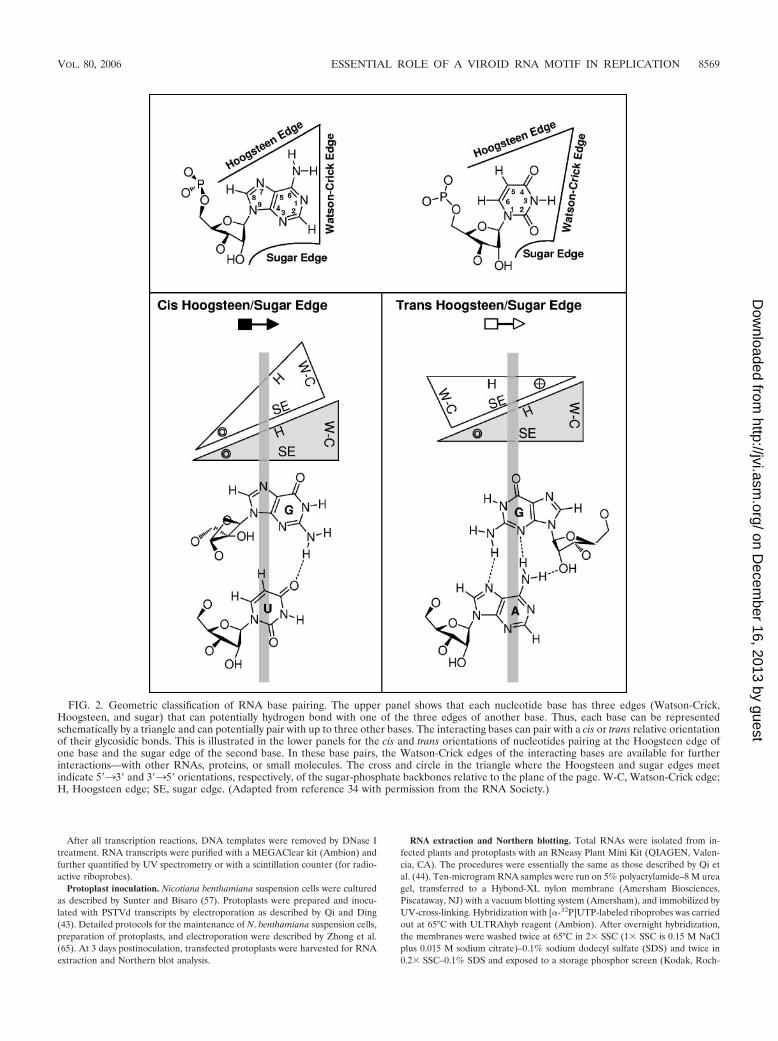
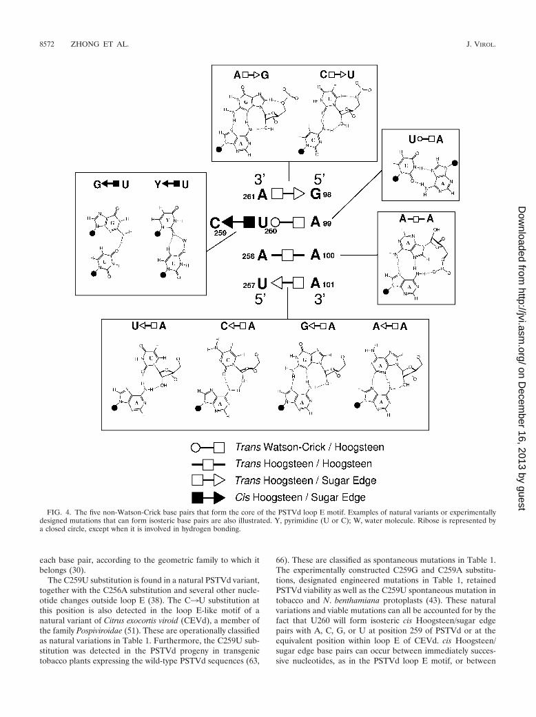
Tertiary-structural model of the PSTVd loop E motif. Tofacilitate genetic studies of PSTVd loop E function, we firstanalyzed the tertiary structure of this motif by comparativesequence analysis with isostericity matrices (30). Sequenceanalysis has identified recurrent loop E or sarcin/ricin motifs indifferent RNA molecules (29, 32). This motif comprises fivecore non-Watson-Crick base pairs, as shown in Fig. 3A. On thebasis of UV cross-linking (4), nuclease and chemical mapping(13), and sequence comparison, it was concluded that a loop E(sarcin/ricin) motif also exists in PSTVd at the position indi-cated in Fig. 3B. Figure 4 shows the inferred base pair struc-tures with hydrogen bonds for the five core non-Watson-Crickbase pairs (A261/G98, U260/A99, U260/C259, A258/A100, andA101/U257) in PSTVd loop E. The structures of some com-mon isosteric sequence variants are also shown. Each non-Watson-Crick base pair and its sequence variations in thePSTVd loop E motif are discussed below. Following the con-vention (34), the edges that participate in the interactions foreach base pair are presented in the following order: Watson-Crick edge, Hoogsteen edge, and sugar edge.
A261/G98 trans Hoogsteen/sugar edge pair. The Hoogsteenedge of A261 forms hydrogen bonds with the sugar edge ofG98 in the trans orientation. The trans Hoogsteen/sugar edgegeometric family comprises two isosteric subfamilies (I1 and I2)(30; see Materials and Methods for the specific criteria usedfor base pair classifications). The A261/G98 pair belongs to I1
(see Fig. 5). The isosteric C/U double mutant, discussed below,is shown in Fig. 4.
8570 ZHONG ET AL. J. VIROL.
on Decem
ber 16, 2013 by guesthttp://jvi.asm
.org/D
ownloaded from
U260/C259 cis Hoogsteen/sugar edge pair. In the PSTVdsequence, U260 and C259 may form a cis Hoogsteen/sugaredge base pair. In most loop E (sarcin/ricin) motifs, the cor-responding bases are U and G, which form a cis Hoogsteen/sugar edge base pair stabilized by a strong hydrogen bondbetween U (O4; oxygen at position 4 of uracil) and G (N2;nitrogen at position 2 of guanine). cis Hoogsteen/sugar edgebase pairs between successive U/U, U/C, and U/A pairs areobserved infrequently in crystal structures. While they haveC1�-C1� distances comparable to that of the U/G pair, theylack the strong hydrogen bond. Consequently, these pairs areusually stabilized by interactions with other bases, formingtriples, or with proteins. The isosteric U/U cis Hoogsteen/sugaredge pair has been observed in several crystal structures, in-cluding the 23S rRNA (PDB file 1S72, U831/U832 from theProtein Data Bank), where it is part of a base triple. On thebasis of observed U/U pairs, the isosteric U/C pair was pro-posed (30) and has since been observed (for example, U223/C222, chain A, PDB file 1ET4). The U/U and U/C pairs arestabilized in part by weak hydrogen bonds between U (C5;carbon atom at position 5 of uracil) and Y (O2; oxygen atposition 2 of pyrimidine).
The U/A pair can form by weak hydrogen bonding betweenA (C2; carbon at position 2 of adenine) and U (O4) or betweenA (O2�; the 2� OH of adenosine) and U (C5). One example ofthis type of base pair occurs in association with a protein andindicates that U/A has a C1�-C1� distance of 6.8 Å (PDB file
1EC6, U12/A11 in C chain) and thus can also substitute iso-sterically for U/U, U/C, and U/G. The RNA structural searchprograms used at the time of the original compilation of basepairs (30) did not detect the U/A pair.
U260/A99 trans Watson-Crick/Hoogsteen edge pair. TheWatson-Crick edge of U260 hydrogen bonds with the Hoogs-teen edge of A99 in the trans orientation. The U at this posi-tion is very conserved in most loop E motifs because it alsouses its Hoogsteen edge to form a second base pair with G (i.e.,a cis Hoogsteen/sugar edge pair). In other contexts, however, Ccan substitute for U in trans Watson-Crick/Hoogsteen basepairs to form isosteric C/C, or nearly isosteric C/A or C/G, basepairs of this type (30).
A258/A100 trans Hoogsteen/Hoogsteen edge pair. TheHoogsteen edges of A258 and A100 interact to form a sym-metric trans Hoogsteen/Hoogsteen base pair. Mutation ofA100 to G still allows for hydrogen bonding with A258 by theformation of a hydrogen bond between A (N6; nitrogen atposition 6 of adenine) and G (O6; oxygen at position 6 ofguanine). This substitution has been observed in a compositeloop E motif in the 23S rRNA of Haloarcula marismortui (PDBfile 1S72, A913/G1071) with a C1�-C1� distance of 12.2 Å.Thus, the A/G base pair is nearly isosteric with A/A.
A101/U257 trans Hoogsteen/sugar edge pair. The Hoogsteenedge of A101 hydrogen bonds with the sugar edge of U257.Note that this base pair is oriented oppositely to trans Hoogs-teen/sugar edge pair A261/G98, as shown by the symbols an-notating these base pairs in Fig. 4. The structures of threeisosteric variants, A/G, A/C, and A/A, are shown in Fig. 4. Veryoften, the isosteric C/U base pair variant occurs at this posi-tion, as shown for the generic sarcin/ricin loop in Fig. 3A. Thestructure of the C/U base pair is shown in the upper inset ofFig. 4.
C256/C102 cis Watson-Crick/Watson-Crick bifurcated pair.The C256/C102 cis Watson-Crick/Watson-Crick bifurcatedpair is outside the conserved core structure of loop E andsarcin/ricin motifs. For C/C to form an edge-to-edge Watson-Crick base pair, one cytosine must be protonated at the N3position. While this has been observed in some structures,more commonly the cytosines adopt the Watson-Crick bifur-cated geometry in which the N4 amino group of one cytosinehydrogen bonds with both N3 and O2 of the other cytosine.This bifurcated geometry results in a longer C1�-C1� distanceof about 10.4 Å, which is nearly isosteric to cis Watson-CrickA/U and G/C base pairs or with wobble Watson-Crick basepair U/G or C/A (30).
The tertiary-structural model of PSTVd loop E accounts fornatural sequence variations and viable mutations. To test thevalidity of the proposed tertiary-structural model of PSTVdloop E, we first examined whether this model could account forthe functional state of the reported natural sequence variationsor viable mutations in loop E motifs of PSTVd and other viroidspecies in the family Pospiviroidae. The natural sequence vari-ants and viable mutants are summarized in Table 1. Figure 4illustrates the molecular structures of selected sequence vari-ants, mapped onto the annotated non-Watson-Crick base pair-ing structure that forms the core of the PSTVd loop E motif.Figure 5 shows the functional status of mutants and naturalvariants mapped onto the respective isostericity matrices for
FIG. 3. Tertiary-structural model of PSTVd loop E. (A) Paradig-matic sarcin/ricin motif based on X-ray crystal structures (adaptedfrom reference 29 with permission from Elsevier). (B) Inferred PSTVdloop E structural model. The dashed arrows indicate local changes inthe strand orientation. All symbols that denote non-Watson-Crick basepairs and strand orientations are based on a report by Leontis andWesthof (34). Circles, squares, and triangles indicate the participationof Watson-Crick, Hoogsteen, and sugar edges, respectively. Open sym-bols indicate base pairs with a trans orientation of the glycosidic bonds,and closed symbols indicate base pairs with a cis orientation.
VOL. 80, 2006 ESSENTIAL ROLE OF A VIROID RNA MOTIF IN REPLICATION 8571
on Decem
ber 16, 2013 by guesthttp://jvi.asm
.org/D
ownloaded from
each base pair, according to the geometric family to which itbelongs (30).
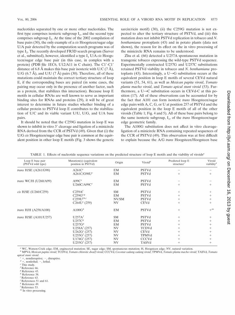
The C259U substitution is found in a natural PSTVd variant,together with the C256A substitution and several other nucle-otide changes outside loop E (38). The C3U substitution atthis position is also detected in the loop E-like motif of anatural variant of Citrus exocortis viroid (CEVd), a member ofthe family Pospiviroidae (51). These are operationally classifiedas natural variations in Table 1. Furthermore, the C259U sub-stitution was detected in the PSTVd progeny in transgenictobacco plants expressing the wild-type PSTVd sequences (63,
66). These are classified as spontaneous mutations in Table 1.The experimentally constructed C259G and C259A substitu-tions, designated engineered mutations in Table 1, retainedPSTVd viability as well as the C259U spontaneous mutation intobacco and N. benthamiana protoplasts (43). These naturalvariations and viable mutations can all be accounted for by thefact that U260 will form isosteric cis Hoogsteen/sugar edgepairs with A, C, G, or U at position 259 of PSTVd or at theequivalent position within loop E of CEVd. cis Hoogsteen/sugar edge base pairs can occur between immediately succes-sive nucleotides, as in the PSTVd loop E motif, or between
FIG. 4. The five non-Watson-Crick base pairs that form the core of the PSTVd loop E motif. Examples of natural variants or experimentallydesigned mutations that can form isosteric base pairs are also illustrated. Y, pyrimidine (U or C); W, water molecule. Ribose is represented bya closed circle, except when it is involved in hydrogen bonding.
8572 ZHONG ET AL. J. VIROL.
on Decem
ber 16, 2013 by guesthttp://jvi.asm
.org/D
ownloaded from
nucleotides separated by one or more other nucleotides. Thefirst type comprises isosteric subgroup I1, and the second typecomprises subgroup I2. At the time of the 2002 compilation ofbase pairs (30), the only example of a cis Hoogsteen/sugar edgeU/A pair detected by the computation search programs was oftype I2. The recently developed FR3D search program (Sarveret al., submitted), however, identified a type I1 U/A cis Hoogs-teen/sugar edge base pair (in this case, in complex with aprotein) (PDB file 1EC6, U12/A11 in C chain). The C1�-C1�distance of 6.8 Å makes this base pair isosteric with U/C (7 Å),U/G (6.7 Å), and U/U (7 Å) pairs (30). Therefore, all of thesemutations could maintain the correct tertiary structure of loopE, if the corresponding bases are paired (in some cases, thepairing may occur only in the presence of another factor, suchas a protein, that stabilizes this interaction). Because loop Emotifs in cellular RNAs are well known to serve as importantbinding sites for RNAs and proteins (29), it will be of greatinterest to determine in future studies whether binding of acellular protein to PSTVd loop E contributes to the stabiliza-tion of U/C and its viable variant U/U, U/G, and U/A basepairs.
It should be noted that the C259G mutation in loop E wasshown to inhibit in vitro 3� cleavage and ligation of a minicircleRNA derived from the CCR of PSTVd (49). Given that (i) theU/G cis Hoogsteen/sugar edge base pair is common at the equiv-alent position in other loop E motifs (Fig. 3 shows the generic
sarcin/ricin motif) (30), (ii) the C259G mutation is not ex-pected to alter the tertiary structure of PSTVd, and (iii) thismutation does not inhibit PSTVd replication in tobacco and N.benthamiana protoplasts (43) and in potato plants (data notshown), the reason for its effect on the in vitro processing ofthe minicircle RNA remains to be understood.
Zhu et al. (66) detected a U257A spontaneous mutation intransgenic tobacco expressing the wild-type PSTVd sequence.Experimentally constructed U257G and U257C substitutionsretained PSTVd viability in tobacco and N. benthamiana pro-toplasts (43). Interestingly, a U3G substitution occurs at theequivalent position in loop E motifs of several CEVd naturalvariants (51, 54, 61), as well as Mexican papita viroid, Tomatoplanta macho viroid, and Tomato apical stunt viroid (53). Fur-thermore, a U3C substitution occurs in CEVd-C at this po-sition (17). All of these observations can be accounted for bythe fact that A101 can form isosteric trans Hoogsteen/sugaredge pairs with A, C, G, or U at position 257 of PSTVd and theequivalent position in the loop E motifs of all of the otherviroids (Table 1; Fig. 4 and 5). All of these base pairs belong tothe same isosteric subgroup, I1, of the trans Hoogsteen/sugaredge geometric family.
The A100G substitution does not affect in vitro cleavage-ligation of a minicircle RNA containing repeated sequences ofthe CCR of PSTVd (49). This observation was at first difficultto explain because the A/G trans Hoogsteen/Hoogsteen base
TABLE 1. Effects of nucleotide sequence variations on the predicted structure of loop E motifs and the viability of viroidsa
Loop E base pair(PSTVd wild type)
Mutation(s) (equivalentposition in PSTVd) Origin Viroidb Predicted loop E
structurecViroid
viabilityd
trans H/SE (A261/G98) A261Ce EM PSTVd � �A261C/G98Ue EM PSTVd � �
trans WC/H (U260/A99) A99Ce EM PSTVd � �U260C/A99Ce EM PSTVd � �
cis H/SE (U260/C259) C259Af SM PSTVd � �C259Ge,g EM PSTVd � �C259Uf,h,i NV/SM PSTVd � �C264U j (259) NV CEVd � �
trans H/H (A258/A100) A100Gk EM PSTVd � �m
trans H/SE (A101/U257) U257A f SM PSTVd � �U257Cg EM PSTVd � �U257Gg EM PSTVd � �U258A l (257) NV TCDVd � �U262G j (257) NV CEVd � �U255G l (257) NV TPMVd � �U174G l (257) NV CCCVd � �U255G l (257) NV TASVd � �
a WC, Watson-Crick edge; EM, engineered mutation; SE, sugar edge; SM, spontaneous mutation; H, Hoogsteen edge; NV, natural variation.b MPVd, Mexican papita viroid; TCDVd, Tomato chlorotic dwarf viroid; CCCVd, Coconut cadang cadang viroid; TPMVd, Tomato planta macho viroid; TASVd, Tomato
apical stunt viroid.c �, nondisruptive; �, disruptive.d �, nonlethal; �, lethal.e This study.f Reference 66.g Reference 43.h Reference 38.i Reference 63.j References 51 and 61.k Reference 49.l Reference 53.m In vitro processing.
VOL. 80, 2006 ESSENTIAL ROLE OF A VIROID RNA MOTIF IN REPLICATION 8573
on Decem
ber 16, 2013 by guesthttp://jvi.asm
.org/D
ownloaded from
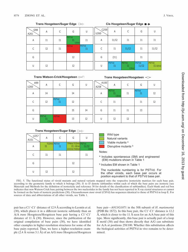
pair has a C1�-C1� distance of 12.9 Å according to Leontis et al.(30), which places it in a different isosteric subfamily than anA/A trans Hoogsteen/Hoogsteen base pair having a C1�-C1�distance of 11 Å (30). However, since the publication of theoriginal compilation of base pairs (30), we have identifiedother examples in higher-resolution structures for some of thebase pairs reported. Thus, we have a higher-resolution exam-ple (2.4 Å versus 3.1 Å) of an A/G trans Hoogsteen/Hoogsteen
base pair—A913/G1071 in the 50S subunit of H. marismortui(PDB file 1S72). In this base pair, the C1�-C1� distance is 12.2Å, which is closer to the 11 Å seen for an A/A base pair of thistype. More significantly, this base pair is actually part of a loopE motif (30) and thus shows directly that A/G can substitutefor A/A at positions 258/100. Whether this substitution affectsthe biological activities of PSTVd in vivo remains to be deter-mined.
FIG. 5. The functional status of viroid mutants and natural variants mapped onto the respective isostericity matrices for each base pair,according to the geometric family to which it belongs (30). I1 to I5 denote subfamilies within each of which the base pairs are isosteric (seeMaterials and Methods for the definition of isostericity and reference 30 for details of the classification of subfamilies). Each blank and red boxindicates that non-Watson-Crick base pairing between the two nucleotides in the family has not been reported in X-ray crystal structures or cannotbe formed on the basis of isosteric predictions (30). Chrysanthemum stunt viroid (CSVd) has sequences identical to those of PSTVd in loop E. Forsources of data and abbreviations of all other viroids, see Table 1.
8574 ZHONG ET AL. J. VIROL.
on Decem
ber 16, 2013 by guesthttp://jvi.asm
.org/D
ownloaded from
Outside the non-Watson-Crick base pair core of loop E, theC256/C102 cis Watson-Crick/Watson-Crick bifurcated pairalso shows viable sequence variations. Owens et al. (38) re-ported a C256A substitution in a natural variant of PSTVd(together with a C259U substitution). Nucleotide A is alsofound at the equivalent position in Mexican papita viroid andTomato apical stunt viroid (53), and A or C is found at theequivalent position in CEVd (51, 61). These variants can beaccounted for by the fact that an A at this position in the motifcan form a wobble base pair with C102 of PSTVd and with theC at the equivalent position in the other viroids. The C1�-C1�distance in the C/A wobble Watson-Crick pair is �10.4 Å,almost identical to the C1�-C1� distance in the bifurcatedWatson-Crick C/C pair, so the A/C and C/C base pairs arenearly isosteric (30).
In conclusion, we find that all reported biologically viablesequence variations or mutations in viroid loop E motifs can beaccounted for by the isosteric base pairing rules as summarizedin the isostericity matrices, within the scope of the literature ofwhich we are aware. This supports the notion that an intactloop E motif is required for PSTVd viability. Furthermore, thisargues for the functional significance of intact loop E motif inother viroids in which this motif occurs. In particular, the loopE motifs of Chrysanthemum stunt viroid and PSTVd have iden-tical sequence signatures (26).
Application of isostericity matrix analysis to design disrup-tive mutations in PSTVd loop E motif. To investigate thefunction of the PSTVd loop E motif in replication by loss-of-function genetics, we applied isostericity matrix analysis todesign mutations that disrupt this motif, based on the tertiary-structural model in Fig. 3B. Changing A99 to C (mutationA99C) prevents the trans Watson-Crick/Hoogsteen interactionbetween positions 260 and 99 because the Watson-Crick edgeof U260 cannot hydrogen bond with the Hoogsteen edge ofC99 (30). This is indicated in the isostericity matrices for transWatson-Crick/Hoogsteen base pairs by the red box for thematrix element corresponding to the U row and the C column(Fig. 5) (30). Likewise, changing A261 to C prevents the transHoogsteen/sugar edge base pair at positions 261/98 from form-ing because the Hoogsteen edge of C261 cannot hydrogenbond with the sugar edge of G98. This is indicated by the redbox at the corresponding positions (row C, column G) of theisostericity matrices for trans Hoogsteen/sugar edge base pairs(Fig. 5) (30). Computer programs that calculate minimum freeenergy secondary structures, such as mfold, do not predict theformation of non-Watson-Crick base pairs. Therefore, muta-tions affecting the structures of motifs consisting of orderedarrays of non-Watson-Crick base pairs, such as loop E, are alsonot correctly accounted for by mfold and similar programs.
To test physically whether the A99C and A261C mutationseach indeed disrupt the tertiary structure of loop E, we sub-jected the corresponding mutants to UV cross-linking experi-ments. These mutants are referred to as PSTVdIntA99C andPSTVdIntA261C, respectively. The names indicate that theyare derived from the wild-type PSTVd intermediate strain(PSTVdInt) (16). Previous studies have demonstrated that UVtreatment specifically cross-links bases G98 and U260 in loopE of PSTVd (3, 4) and of a minicircle RNA derived from thePSTVd CCR (49). It also cross-links the corresponding G andU bases in loop E of the human 5S rRNA from HeLa cells (4).
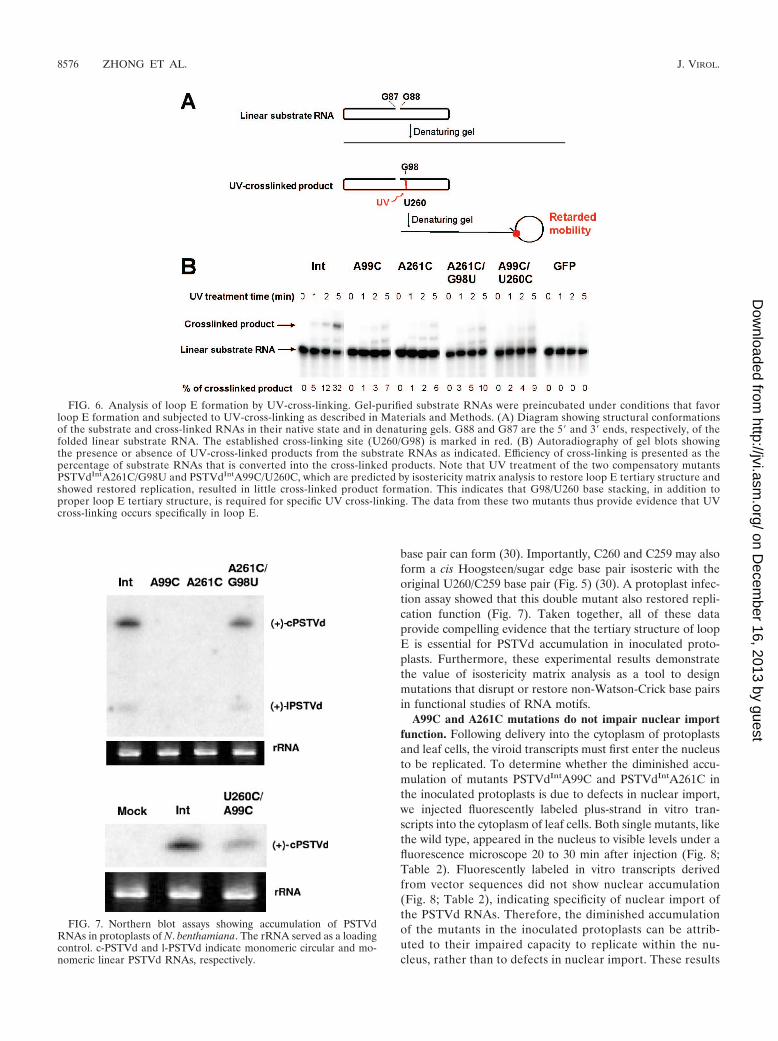
This cross-linking occurs because of a unique partial cross-strand stack of G on U in loop E motifs that gives rise tophotoreactivity. This cross-linking causes RNA mobility retar-dation on denaturing gels (Fig. 6). We used unit length linearin vitro transcripts as substrates. These transcripts are pre-dicted to fold into the same rod-shaped secondary structure asthe circular RNA, except for the nick between the two ends(G88 and G87; see Materials and Methods for details). Asshown in Fig. 6, the A99C and A261C mutations greatly re-duced the accumulation of cross-linked products. For instance,after 1 min of UV irradiation, the cross-linked product wasalready evident for PSTVdInt RNA but barely visible for thetwo mutants. After 5 min of UV irradiation, 32% of the wild-type RNA was cross-linked whereas only 6 to 7% of the mutantRNAs were cross-linked. The similar sizes of the cross-linkedproducts in the wild-type and mutant PSTVd RNAs suggestthat UV cross-linking occurred in the same region of theseRNAs. Furthermore, the specificity of the G98-U260 cross-linking was demonstrated with two mutants that lack specificG98-U260 base stacking in loop E (Fig. 6). The absence ofcross-linked products from GFP RNAs further demon-strates the specificity of UV cross-linking (Fig. 6). The quan-titative difference indicates that a substantial portion of thePSTVdIntA99C and PSTVdIntA261C mutant RNA mole-cules failed to form the correct loop E structure, so as toplace G98 and U260 into the necessary proximity to allowUV cross-linking, in contrast to the wild-type transcriptsunder the same incubation conditions. The band observedbetween the cross-linked product and the substrate RNAwas also observed in previous experiments, and its natureremains to be determined (49).
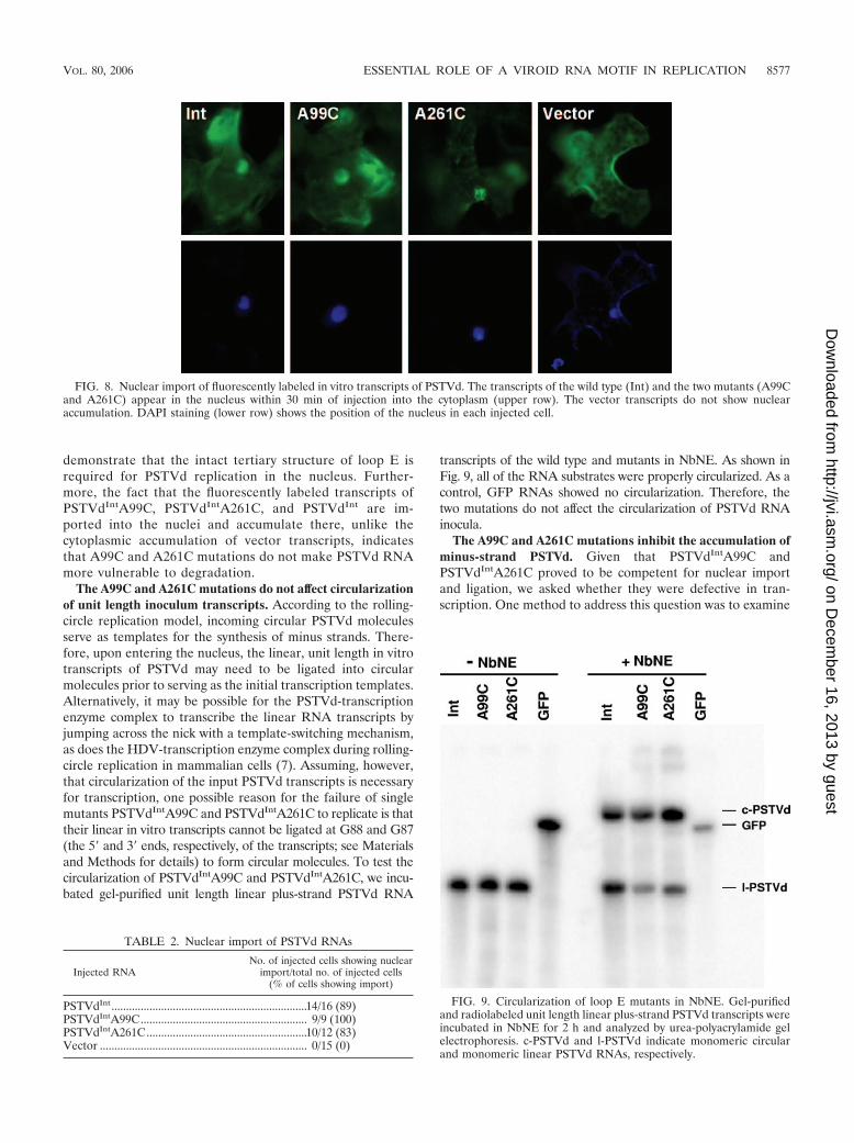
Maintaining the tertiary structure of loop E is necessary forPSTVd accumulation in inoculated protoplasts. The availabil-ity of the loop E-defective mutants allowed us to test whetherthe intact tertiary structure of loop E is crucial for PSTVdinfection. To this end, we inoculated N. benthamiana proto-plasts with in vitro transcripts derived from the wild-typePSTVdInt and loop E-defective single mutants PSTVdIntA99Cand PSTVdIntA261C, respectively. Northern blot assays, run todetect circular and linear plus-strand PSTVd, showed no visi-ble accumulation of either PSTVdIntA99C or PSTVdIntA261Cin protoplasts (Fig. 7), in contrast to PSTVdInt.
These data suggest that maintaining the intact tertiary struc-ture of loop E is critical for PSTVd accumulation in inoculatedprotoplasts. To further test this, we applied isostericity matrixanalysis to design a compensatory mutant in which G98 isreplaced with U in the PSTVdIntA261C background. In thedouble mutant PSTVdIntA261C/G98U, the trans Hoogsteen/sugar edge C261/U98 base pair can form. This C261/U98 basepair is isosteric with the original A261/G98 base pair, andsimilar substitutions have been observed in the crystal struc-tures of related motifs (29, 33) (Fig. 4 and 5). We found that,indeed, this double mutant accumulated as well as PSTVdInt ininoculated protoplasts (Fig. 7). Sequencing of PSTVd progenyconfirmed that no other mutations occurred during replicationof the double mutant. To extend this finding, we generated asecond compensatory mutant in which U260 is replaced by C inthe PSTVdIntA99C background. In this double mutant,PSTVdIntU260C/A99C, the trans Watson-Crick/HoogsteenC260/C99 base pair that is isosteric with the original U260/A99
VOL. 80, 2006 ESSENTIAL ROLE OF A VIROID RNA MOTIF IN REPLICATION 8575
on Decem
ber 16, 2013 by guesthttp://jvi.asm
.org/D
ownloaded from
base pair can form (30). Importantly, C260 and C259 may alsoform a cis Hoogsteen/sugar edge base pair isosteric with theoriginal U260/C259 base pair (Fig. 5) (30). A protoplast infec-tion assay showed that this double mutant also restored repli-cation function (Fig. 7). Taken together, all of these dataprovide compelling evidence that the tertiary structure of loopE is essential for PSTVd accumulation in inoculated proto-plasts. Furthermore, these experimental results demonstratethe value of isostericity matrix analysis as a tool to designmutations that disrupt or restore non-Watson-Crick base pairsin functional studies of RNA motifs.
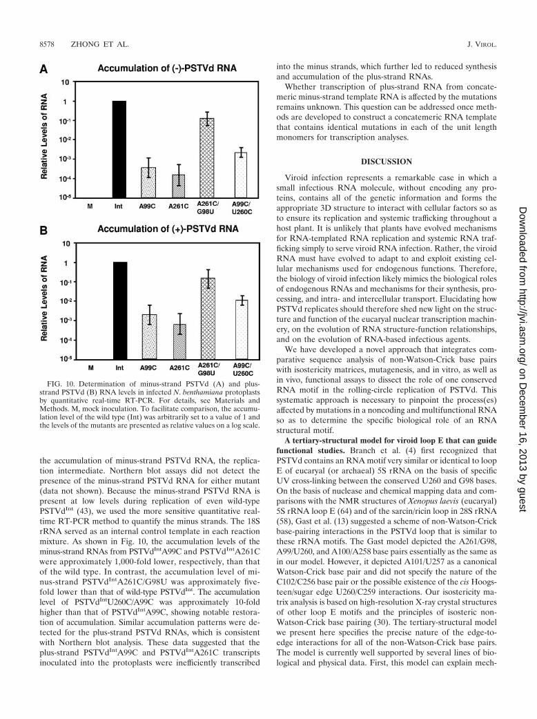
A99C and A261C mutations do not impair nuclear importfunction. Following delivery into the cytoplasm of protoplastsand leaf cells, the viroid transcripts must first enter the nucleusto be replicated. To determine whether the diminished accu-mulation of mutants PSTVdIntA99C and PSTVdIntA261C inthe inoculated protoplasts is due to defects in nuclear import,we injected fluorescently labeled plus-strand in vitro tran-scripts into the cytoplasm of leaf cells. Both single mutants, likethe wild type, appeared in the nucleus to visible levels under afluorescence microscope 20 to 30 min after injection (Fig. 8;Table 2). Fluorescently labeled in vitro transcripts derivedfrom vector sequences did not show nuclear accumulation(Fig. 8; Table 2), indicating specificity of nuclear import ofthe PSTVd RNAs. Therefore, the diminished accumulationof the mutants in the inoculated protoplasts can be attrib-uted to their impaired capacity to replicate within the nu-cleus, rather than to defects in nuclear import. These results
FIG. 6. Analysis of loop E formation by UV-cross-linking. Gel-purified substrate RNAs were preincubated under conditions that favorloop E formation and subjected to UV-cross-linking as described in Materials and Methods. (A) Diagram showing structural conformationsof the substrate and cross-linked RNAs in their native state and in denaturing gels. G88 and G87 are the 5� and 3� ends, respectively, of thefolded linear substrate RNA. The established cross-linking site (U260/G98) is marked in red. (B) Autoradiography of gel blots showingthe presence or absence of UV-cross-linked products from the substrate RNAs as indicated. Efficiency of cross-linking is presented as thepercentage of substrate RNAs that is converted into the cross-linked products. Note that UV treatment of the two compensatory mutantsPSTVdIntA261C/G98U and PSTVdIntA99C/U260C, which are predicted by isostericity matrix analysis to restore loop E tertiary structure andshowed restored replication, resulted in little cross-linked product formation. This indicates that G98/U260 base stacking, in addition toproper loop E tertiary structure, is required for specific UV cross-linking. The data from these two mutants thus provide evidence that UVcross-linking occurs specifically in loop E.
FIG. 7. Northern blot assays showing accumulation of PSTVdRNAs in protoplasts of N. benthamiana. The rRNA served as a loadingcontrol. c-PSTVd and l-PSTVd indicate monomeric circular and mo-nomeric linear PSTVd RNAs, respectively.
8576 ZHONG ET AL. J. VIROL.
on Decem
ber 16, 2013 by guesthttp://jvi.asm
.org/D
ownloaded from
demonstrate that the intact tertiary structure of loop E isrequired for PSTVd replication in the nucleus. Further-more, the fact that the fluorescently labeled transcripts ofPSTVdIntA99C, PSTVdIntA261C, and PSTVdInt are im-ported into the nuclei and accumulate there, unlike thecytoplasmic accumulation of vector transcripts, indicatesthat A99C and A261C mutations do not make PSTVd RNAmore vulnerable to degradation.
The A99C and A261C mutations do not affect circularizationof unit length inoculum transcripts. According to the rolling-circle replication model, incoming circular PSTVd moleculesserve as templates for the synthesis of minus strands. There-fore, upon entering the nucleus, the linear, unit length in vitrotranscripts of PSTVd may need to be ligated into circularmolecules prior to serving as the initial transcription templates.Alternatively, it may be possible for the PSTVd-transcriptionenzyme complex to transcribe the linear RNA transcripts byjumping across the nick with a template-switching mechanism,as does the HDV-transcription enzyme complex during rolling-circle replication in mammalian cells (7). Assuming, however,that circularization of the input PSTVd transcripts is necessaryfor transcription, one possible reason for the failure of singlemutants PSTVdIntA99C and PSTVdIntA261C to replicate is thattheir linear in vitro transcripts cannot be ligated at G88 and G87(the 5� and 3� ends, respectively, of the transcripts; see Materialsand Methods for details) to form circular molecules. To test thecircularization of PSTVdIntA99C and PSTVdIntA261C, we incu-bated gel-purified unit length linear plus-strand PSTVd RNA
transcripts of the wild type and mutants in NbNE. As shown inFig. 9, all of the RNA substrates were properly circularized. As acontrol, GFP RNAs showed no circularization. Therefore, thetwo mutations do not affect the circularization of PSTVd RNAinocula.
The A99C and A261C mutations inhibit the accumulation ofminus-strand PSTVd. Given that PSTVdIntA99C andPSTVdIntA261C proved to be competent for nuclear importand ligation, we asked whether they were defective in tran-scription. One method to address this question was to examine
FIG. 8. Nuclear import of fluorescently labeled in vitro transcripts of PSTVd. The transcripts of the wild type (Int) and the two mutants (A99Cand A261C) appear in the nucleus within 30 min of injection into the cytoplasm (upper row). The vector transcripts do not show nuclearaccumulation. DAPI staining (lower row) shows the position of the nucleus in each injected cell.
FIG. 9. Circularization of loop E mutants in NbNE. Gel-purifiedand radiolabeled unit length linear plus-strand PSTVd transcripts wereincubated in NbNE for 2 h and analyzed by urea-polyacrylamide gelelectrophoresis. c-PSTVd and l-PSTVd indicate monomeric circularand monomeric linear PSTVd RNAs, respectively.
TABLE 2. Nuclear import of PSTVd RNAs
Injected RNANo. of injected cells showing nuclear
import/total no. of injected cells(% of cells showing import)
PSTVdInt...................................................................14/16 (89)PSTVdIntA99C......................................................... 9/9 (100)PSTVdIntA261C.......................................................10/12 (83)Vector ....................................................................... 0/15 (0)
VOL. 80, 2006 ESSENTIAL ROLE OF A VIROID RNA MOTIF IN REPLICATION 8577
on Decem
ber 16, 2013 by guesthttp://jvi.asm
.org/D
ownloaded from
the accumulation of minus-strand PSTVd RNA, the replica-tion intermediate. Northern blot assays did not detect thepresence of the minus-strand PSTVd RNA for either mutant(data not shown). Because the minus-strand PSTVd RNA ispresent at low levels during replication of even wild-typePSTVdInt (43), we used the more sensitive quantitative real-time RT-PCR method to quantify the minus strands. The 18SrRNA served as an internal control template in each reactionmixture. As shown in Fig. 10, the accumulation levels of theminus-strand RNAs from PSTVdIntA99C and PSTVdIntA261Cwere approximately 1,000-fold lower, respectively, than thatof the wild type. In contrast, the accumulation level of mi-nus-strand PSTVdIntA261C/G98U was approximately five-fold lower than that of wild-type PSTVdInt. The accumulationlevel of PSTVdIntU260C/A99C was approximately 10-foldhigher than that of PSTVdIntA99C, showing notable restora-tion of accumulation. Similar accumulation patterns were de-tected for the plus-strand PSTVd RNAs, which is consistentwith Northern blot analysis. These data suggested that theplus-strand PSTVdIntA99C and PSTVdIntA261C transcriptsinoculated into the protoplasts were inefficiently transcribed
into the minus strands, which further led to reduced synthesisand accumulation of the plus-strand RNAs.
Whether transcription of plus-strand RNA from concate-meric minus-strand template RNA is affected by the mutationsremains unknown. This question can be addressed once meth-ods are developed to construct a concatemeric RNA templatethat contains identical mutations in each of the unit lengthmonomers for transcription analyses.
DISCUSSION
Viroid infection represents a remarkable case in which asmall infectious RNA molecule, without encoding any pro-teins, contains all of the genetic information and forms theappropriate 3D structure to interact with cellular factors so asto ensure its replication and systemic trafficking throughout ahost plant. It is unlikely that plants have evolved mechanismsfor RNA-templated RNA replication and systemic RNA traf-ficking simply to serve viroid RNA infection. Rather, the viroidRNA must have evolved to adapt to and exploit existing cel-lular mechanisms used for endogenous functions. Therefore,the biology of viroid infection likely mimics the biological rolesof endogenous RNAs and mechanisms for their synthesis, pro-cessing, and intra- and intercellular transport. Elucidating howPSTVd replicates should therefore shed new light on the struc-ture and function of the eucaryal nuclear transcription machin-ery, on the evolution of RNA structure-function relationships,and on the evolution of RNA-based infectious agents.
We have developed a novel approach that integrates com-parative sequence analysis of non-Watson-Crick base pairswith isostericity matrices, mutagenesis, and in vitro, as well asin vivo, functional assays to dissect the role of one conservedRNA motif in the rolling-circle replication of PSTVd. Thissystematic approach is necessary to pinpoint the process(es)affected by mutations in a noncoding and multifunctional RNAso as to determine the specific biological role of an RNAstructural motif.
A tertiary-structural model for viroid loop E that can guidefunctional studies. Branch et al. (4) first recognized thatPSTVd contains an RNA motif very similar or identical to loopE of eucaryal (or archaeal) 5S rRNA on the basis of specificUV cross-linking between the conserved U260 and G98 bases.On the basis of nuclease and chemical mapping data and com-parisons with the NMR structures of Xenopus laevis (eucaryal)5S rRNA loop E (64) and of the sarcin/ricin loop in 28S rRNA(58), Gast et al. (13) suggested a scheme of non-Watson-Crickbase-pairing interactions in the PSTVd loop that is similar tothese rRNA motifs. The Gast model depicted the A261/G98,A99/U260, and A100/A258 base pairs essentially as the same asin our model. However, it depicted A101/U257 as a canonicalWatson-Crick base pair and did not specify the nature of theC102/C256 base pair or the possible existence of the cis Hoogs-teen/sugar edge U260/C259 interactions. Our isostericity ma-trix analysis is based on high-resolution X-ray crystal structuresof other loop E motifs and the principles of isosteric non-Watson-Crick base pairing (30). The tertiary-structural modelwe present here specifies the precise nature of the edge-to-edge interactions for all of the non-Watson-Crick base pairs.The model is currently well supported by several lines of bio-logical and physical data. First, this model can explain mech-
FIG. 10. Determination of minus-strand PSTVd (A) and plus-strand PSTVd (B) RNA levels in infected N. benthamiana protoplastsby quantitative real-time RT-PCR. For details, see Materials andMethods. M, mock inoculation. To facilitate comparison, the accumu-lation level of the wild type (Int) was arbitrarily set to a value of 1 andthe levels of the mutants are presented as relative values on a log scale.
8578 ZHONG ET AL. J. VIROL.
on Decem
ber 16, 2013 by guesthttp://jvi.asm
.org/D
ownloaded from

anistically why certain mutations in loop E do not affectPSTVd viability (43, 49, 63, 66). Second, it can account for thenaturally occurring nucleotide sequence variations in the loopE motifs of PSTVd and other viroids in the family Pospiviroi-dae (17, 38, 51, 53, 54, 61). Third, UV cross-linking data pro-vide further evidence that the A99C and A261C substitutionsdisrupt the loop E structure by preventing the formation ofisosteric non-Watson-Crick base pairs. Fourth, as discussedbelow, the functional analyses pinpointed the deleterious ef-fects of these mutations on PSTVd transcription. Finally, thetwo compensatory mutations that are predicted to restore theloop E tertiary structure on the basis of isosteric base-pairingprinciples and on the basis of the X-ray crystal structures ofsimilar base pairs also restored PSTVd replication. Furtherstudies on the effects of other nucleotide mutations within loopE should provide additional tests of the validity of this struc-tural model. All of these data also suggest strongly that thePSTVd loop E motif exists and functions in vivo.
PSTVd loop E plays a crucial role in RNA-templated RNAtranscription. Our functional analyses of the PSTVd loop Emutations provide new insights into the role of this motif inPSTVd replication at the cellular level. Our experimentsshowed that the A99C and A261C mutations, which disrupt theloop E structure, inhibit accumulation of PSTVd progeny ininoculated protoplasts. Further analyses indicated that thesemutations do not impair nuclear import function or circular-ization of the linear inoculum transcripts. Therefore, the fail-ure of the PSTVd mutants to replicate and accumulate ininoculated protoplasts likely results from a defect(s) in one ormore of the steps of rolling-circle replication. The requirementfor intact loop E in replication is further supported by theobservation that compensatory mutations predicted to restorenon-Watson-Crick base pairs within the motif were observedto restore the replication function.
The A99C and A261C loss-of-function mutations could in-hibit the transcription, cleavage, or ligation step of rolling-circle replication. Although our present data cannot rule outcompletely the possibility that these mutations affect cleavage-ligation, several observations suggest strongly that they mainlyinhibit transcription. First, the A99C and A261C mutationsseverely inhibit the synthesis of minus-strand PSTVd RNA.Second, if these mutations in fact inhibited cleavage ratherthan transcription, one would expect to see accumulation ofhigh levels of the multimeric plus-strand PSTVd RNAs. Thiswas not observed in Northern blot assays. Third, if these mu-tations inhibited ligation, rather than transcription or cleavage,one would expect to see accumulation of high levels of thelinear monomeric plus-strand PSTVd RNAs. This was notobserved either in Northern blot assays. On the basis of theseconsiderations, we propose that the loop E motif plays a cru-cial role in transcription, in addition to its well-documentedrole in cleavage-ligation (3), during PSTVd replication. Themultifunctionality of loop E is discussed below.
How might loop E be involved in transcription? Loop Emotifs in cellular RNAs are well known to serve as importantbinding sites for RNAs and proteins (29). The loop E motif ofPSTVd may serve as a binding site for the RNA polymerase orfor a cellular factor(s) that recruits the RNA polymerase fortranscription. Alternatively, loop E may interact with a cellularfactor to localize the PSTVd RNA to a particular subnuclear
site to facilitate access to the nuclear transcription machinery.It is also possible that this motif is required for interacting witha cellular factor that leads to a conformational change in theviroid RNA, allowing it to be recognized by the transcriptionmachinery. These possibilities can be tested by further exper-imental studies.
It should be noted that a HPI is predicted to form as athermodynamically metastable structure through pairing of nu-cleotides 79CGCUUCAGG87 and nucleotides 110GCGAGGUCC102 with nucleotides 88 to 101, forming a 14-nucleotide loopas a result of HPI formation (46). Whether the mutations wegenerated would affect the structure and function of HPI re-quires further analyses. In fact, it remains to be determinedwhether HPI exists in vivo and has any biological functions. Aputative tetraloop containing A99 was postulated by Baum-stark et al. (3) to be important for in vitro processing. Whethersuch a tetraloop structure exists and functions in vivo andwhether A99C would affect its structure and function remain tobe investigated.
PSTVd loop E is a useful model to study RNA structure-function relationships of broad significance. Loop E is one ofthe most extensively studied RNA structural motifs (29, 31).Besides serving as a model to dissect PSTVd replication andthe general mechanisms of RNA-templated replication by thenuclear transcription machinery, the PSTVd loop E motif of-fers an attractive system to study RNA structure-function re-lationships of general significance. First, the multifunctionalityof this motif is biologically significant. This motif is involved intranscription (this study), processing (3), pathogenicity (42),and host adaptation (43, 63, 66). To accomplish such diversefunctions, submotifs of loop E may interact with distinct cel-lular factors. Elucidating the underlying mechanisms willgreatly expand our understanding of the capacity of a singleRNA motif to regulate multiple biological processes. Suchmultifunctionality of a single RNA motif may be important fora wide range of RNA-based pathogens to expand their genomefunctions during evolution without increases in genome size.
Second, loop E is a recurrent motif found in many RNAs,including 5S, 16S, and 23S rRNAs; group I and group II in-trons; bacterial RNase P; ribozyme of Tobacco ringspot virussatellite RNA (reviewed in reference 32); and lysine ribo-switches (18, 56), where it plays critical roles in RNA-RNA andRNA-protein interactions. While mutational analyses of cellu-lar RNA motifs may be limited by the potential lethality ofmotif-disruptive mutations to an organism or by the high struc-tural complexity of large cellular RNAs, such limitations donot apply to PSTVd, a host parasite. Therefore, further mu-tagenesis can be performed on PSTVd loop E and other motifsto determine their sequence signatures and gain detailed in-sights into RNA structure-function relationships that may notbe feasible with cellular RNA. Furthermore, the recurrentnature of this motif allows broad application of the knowledgeobtained.
An integrative approach to address structure-function rela-tionships of RNA motifs. Our findings from PSTVd loop Estructure and function studies demonstrate that integration ofisostericity matrix analysis, rationalized mutagenesis, and sys-tematic functional studies can provide important insights intothe structure-function relationship of an RNA motif and fur-ther into the elaborate cellular control over this relationship.
VOL. 80, 2006 ESSENTIAL ROLE OF A VIROID RNA MOTIF IN REPLICATION 8579
on Decem
ber 16, 2013 by guesthttp://jvi.asm
.org/D
ownloaded from

Isostericity matrix analysis can be used to predict base substi-tutions that will prevent the formation of isosteric (or nearlyisosteric) base pairs, thereby disrupting the 3D structure of anRNA motif. If the structural integrity of the motif is critical forfunction, these mutations are likely to result in loss of function.This is well demonstrated by the replication defects of PSTVdcarrying either the A99C or the A261C mutation that disruptsloop E. Isostericity matrix analysis can also predict which basesubstitutions can retain or restore the tertiary structure of amotif, providing a useful means to infer the tertiary structureof RNA motifs. This analysis alone, however, may not alwayspredict a priori which isosteric or nearly isosteric base substi-tutions in a motif will retain or restore the function of a givenRNA in the cellular environment, because the functioning ofthe motif requires interactions with other factors that can onlybe elucidated through biological assays. This is illustratedwell by the results from the two compensatory mutants,PSTVdIntA261C/G98U and PSTVdIntU260C/A99C. Both mu-tants are predicted by isostericity matrix analysis to have re-stored the loop E tertiary structure. However, while the repli-cation level of PSTVdIntA261C/G98U is close to that of thewild type, the replication level of PSTVdIntU260C/A99C is lessthan that of the wild type. Presumably, the loop E motif withthe C259/C260/C99 base triple in the compensatory mutantPSTVdIntU260C/A99C is sufficient, but not as optimal as themotif with the C259/U260/A99 base triple as in the wild type,to be recognized by the cellular machinery for replication.These results therefore demonstrate that a combination ofisostericity matrix analysis and genetic and biological experi-ments can provide a foundation to elucidate the elaborateRNA motif-cellular factor interactions that regulate diversebiological processes and to address the fundamental questionof how sophisticated features of RNA structures have evolvedto achieve optimal functions.
ACKNOWLEDGMENTS
We thank Ying Wang, Ryuta Takeda, and Xiaorong Tao for tech-nical assistance. We thank Venkat Gopalan, John M. Burke, and JoyceHeckman for constructive discussions of UV cross-linking and JesseStombaugh for helpful discussions of base pair analyses. We are in-debted to Yuhua Lu for technical assistance with the preparation ofFig. 2.
This work was supported by grants from the National Science Foun-dation (IBN-0238412 and IOB-0515745) and from the U.S. Depart-ment of Agriculture National Research Initiative Competitive GrantsProgram (2004-35304-15005) to B.D., by National Institutes of Healthgrant 3R15-GM55898 to N.L., and by National Institutes of Healthgrant CA97024 to K.B.L.
REFERENCES
1. Baulcombe, D. 2004. RNA silencing in plants. Nature 431:356–363.2. Baumstark, T., and D. Riesner. 1995. Only one of four possible secondary
structures of the central conserved region of potato spindle tuber viroid is asubstrate for processing in a potato nuclear extract. Nucleic Acids Res.23:4246–4254.
3. Baumstark, T., A. R. Schroder, and D. Riesner. 1997. Viroid processing:switch from cleavage to ligation is driven by a change from a tetraloop to aloop E conformation. EMBO J. 16:599–610.
4. Branch, A. D., B. J. Benenfeld, and H. D. Robertson. 1985. Ultravioletlight-induced crosslinking reveals a unique region of local tertiary structurein potato spindle tuber viroid and HeLa 5S RNA. Proc. Natl. Acad. Sci. USA82:6590–6594.
5. Branch, A. D., and H. D. Robertson. 1984. A replication cycle for viroids andother small infectious RNA’s. Science 223:450–455.
6. Candresse, T., A. Gora-Sochacka, and W. Zagorski. 2001. Restoration ofsecondary hairpin II is associated with restoration of infectivity of a non-viable recombinant viroid. Virus Res. 75:29–34.
7. Chang, J., and J. Taylor. 2002. In vivo RNA-directed transcription, withtemplate switching, by a mammalian RNA polymerase. EMBO J. 21:157–164.
8. Diener, T. O. 1971. Potato spindle tuber “virus”. IV. Replicating, low mo-lecular weight RNA. Virology 45:411–428.
9. Dingley, A. J., G. Steger, B. Esters, D. Riesner, and S. Grzesiek. 2003.Structural characterization of the 69 nucleotide potato spindle tuber viroidleft-terminal domain by NMR and thermodynamic analysis. J. Mol. Biol.334:751–767.
10. Flint, S. J., L. W. Enquist, V. R. Racaniello, and A. M. Skalka. 2004.Principles of virology, 2nd ed. ASM Press, Washington, D.C.
11. Flores, R., C. Hernandez, A. E. Martinez de Alba, J. A. Daros, and F. DiSerio. 2005. Viroids and viroid-host interactions. Annu. Rev. Phytopathol.43:117–139.
12. Flores, R., J. W. Randles, M. Bar-Josef, and T. O. Diener. 2000. Subviralagents: viroids, p. 1009–1024. In M. H. V. van Regenmortel, C. M. Fouquet,D. H. L. Bishop, E. B. Carstens, M. K. Estes, S. M. Lemon, J. Maniloff, M. A.Mayo, D. J. McGeoch, C. R. Pringle, and R. B. Wickner (ed.), Virus taxon-omy, seventh report of the International Committee on Taxonomy of Vi-ruses. Academic Press, San Diego, Calif.
13. Gast, F. U., D. Kempe, R. L. Spieker, and H. L. Sanger. 1996. Secondarystructure probing of potato spindle tuber viroid (PSTVd) and sequencecomparison with other small pathogenic RNA replicons provides evidencefor central non-canonical base-pairs, large A-rich loops, and a terminalbranch. J. Mol. Biol. 262:652–670.
14. Gilbert, W. 1986. The RNA world. Nature 319:618.15. Gora-Sochacka, A. 2004. Viroids: unusual small pathogenic RNAs. Acta
Biochim. Pol. 51:587–607.16. Gross, H. J., H. Domdey, C. Lossow, P. Jank, M. Raba, H. Alberty, and H. L.
Sanger. 1978. Nucleotide sequence and secondary structure of potato spin-dle tuber viroid. Nature 273:203–208.
17. Gross, H. J., G. Krupp, H. Domdey, M. Raba, P. Jank, C. Lossow, H. Alberty,K. Ramm, and H. L. Sanger. 1982. Nucleotide sequence and secondarystructure of citrus exocortis and chrysanthemum stunt viroid. Eur. J. Bio-chem. 121:249–257.
18. Grundy, F. J., S. C. Lehman, and T. M. Henkin. 2003. The L box regulon:lysine sensing by leader RNAs of bacterial lysine biosynthesis genes. Proc.Natl. Acad. Sci. USA 100:12057–12062.
19. Hammond, R. W., and R. A. Owens. 1987. Mutational analysis of potatospindle tuber viroid reveals complex relationships between structure andinfectivity. Proc. Natl. Acad. Sci. USA 84:3967–3971.
20. Henco, K., H. L. Sanger, and D. Riesner. 1979. Fine structure melting ofviroids as studied by kinetic methods. Nucleic Acids Res. 6:3041–3059.
21. Hu, Y., P. A. Feldstein, P. J. Bottino, and R. A. Owens. 1996. Role of thevariable domain in modulating potato spindle tuber viroid replication. Vi-rology 219:45–56.
22. Hu, Y., P. A. Feldstein, J. Hammond, R. W. Hammond, P. J. Bottino, andR. A. Owens. 1997. Destabilization of potato spindle tuber viroid by muta-tions in the left terminal loop. J. Gen. Virol. 78:1199–1206.
23. Hull, R. 2002. Matthew’s plant virology, 4 ed. Academic Press, San Diego,Calif.
24. Joyce, G. F. 2002. The antiquity of RNA-based evolution. Nature 418:214–221.
25. Joyce, G. F., and L. E. Orgel. 1999. Replication of human hepatitis deltavirus: recent developments, p. 49–77. In R. F. Gesteland, T. R. Cech, andJ. F. Atkins (ed.), RNA world, 2nd ed., vol. 37. Cold Spring Harbor Labo-ratory Press, Cold Spring Harbor, N.Y.
26. Keese, P., and R. H. Symons. 1985. Domains in viroids: evidence of inter-molecular RNA rearrangements and their contribution to viroid evolution.Proc. Natl. Acad. Sci. USA 82:4582–4586.
27. Kolonko, N., O. Bannach, K. Aschermann, K. H. Hu, M. Moors, M. Schmitz,G. Steger, and D. Riesner. 2006. Transcription of potato spindle tuber viroidby RNA polymerase II starts in the left terminal loop. Virology 347:392–404.
28. Lai, M. M. 2005. RNA replication without RNA-dependent RNA polymer-ase: surprises from hepatitis delta virus. J. Virol. 79:7951–7958.
29. Leontis, N. B., J. Stombaugh, and E. Westhof. 2002. Motif prediction inribosomal RNAs: lessons and prospects for automated motif prediction inhomologous RNA molecules. Biochimie 84:961–973.
30. Leontis, N. B., J. Stombaugh, and E. Westhof. 2002. The non-Watson-Crickbase pairs and their associated isostericity matrices. Nucleic Acids Res.30:3497–3531.
31. Leontis, N. B., and E. Westhof. 1998. The 5S rRNA loop E: chemical probingand phylogenetic data versus crystal structure. RNA 4:1134–1153.
32. Leontis, N. B., and E. Westhof. 1998. A common motif organizes the struc-ture of multi-helix loops in 16 S and 23 S ribosomal RNAs. J. Mol. Biol.283:571–583.
33. Leontis, N. B., and E. Westhof. 1998. Conserved geometrical base-pairingpatterns in RNA. Q. Rev. Biophys. 31:399–455.
34. Leontis, N. B., and E. Westhof. 2001. Geometric nomenclature and classifi-cation of RNA base pairs. RNA 7:499–512.
35. Lescoute, A., N. B. Leontis, C. Massire, and E. Westhof. 2005. Recurrent
8580 ZHONG ET AL. J. VIROL.
on Decem
ber 16, 2013 by guesthttp://jvi.asm
.org/D
ownloaded from
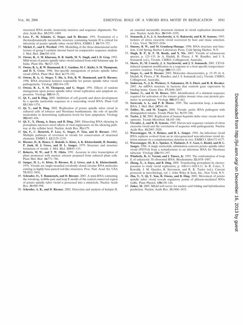
structural RNA motifs, isostericity matrices and sequence alignments. Nu-cleic Acids Res. 33:2395–2409.
36. Loss, P., M. Schmitz, G. Steger, and D. Riesner. 1991. Formation of athermodynamically metastable structure containing hairpin II is critical forinfectivity of potato spindle tuber viroid RNA. EMBO J. 10:719–727.
37. Michel, F., and E. Westhof. 1990. Modelling of the three-dimensional archi-tecture of group I catalytic introns based on comparative sequence analysis.J. Mol. Biol. 216:585–610.
38. Owens, R., S. M. Khurana, D. R. Smith, M. N. Singh, and I. D. Grag. 1992.Mild strain of potato spindle tuber viroid isolated from wild Solanum spp. InIndia. Plant Dis. 76:527–529.
39. Owens, R. A., R. W. Hammond, R. C. Gardner, M. C. Kiefer, S. M. Thompsom,and D. E. Cress. 1986. Site-specific mutagenesis of potato spindle tuberviroid cDNA. Plant Mol. Biol. 6:179–192.
40. Owens, R. A., G. Steger, Y. Hu, A. Fels, R. W. Hammond, and D. Riesner.1996. RNA structural features responsible for potato spindle tuber viroidpathogenicity. Virology 222:144–158.
41. Owens, R. A., S. M. Thompson, and G. Steger. 1991. Effects of randommutagenesis upon potato spindle tuber viroid replication and symptom ex-pression. Virology 185:18–31.
42. Qi, Y., and B. Ding. 2003. Inhibition of cell growth and shoot developmentby a specific nucleotide sequence in a noncoding viroid RNA. Plant Cell15:1360–1374.
43. Qi, Y., and B. Ding. 2002. Replication of potato spindle tuber viroid incultured cells of tobacco and Nicotiana benthamiana: the role of specificnucleotides in determining replication levels for host adaptation. Virology302:445–456.
44. Qi, Y., X. Zhong, A. Itaya, and B. Ding. 2004. Dissecting RNA silencing inprotoplasts uncovers novel effects of viral suppressors on the silencing path-way at the cellular level. Nucleic Acids Res. 32:e179.
45. Qu, F., C. Heinrich, P. Loss, G. Steger, P. Tien, and D. Riesner. 1993.Multiple pathways of reversion in viroids for conservation of structuralelements. EMBO J. 12:2129–2139.
46. Riesner, D., K. Henco, U. Rokohl, G. Klotz, A. K. Kleinschmidt, H. Domdey,P. Jank, H. J. Gross, and H. L. Sanger. 1979. Structure and structureformation of viroids. J. Mol. Biol. 133:85–115.
47. Roberts, M. W., and T. W. Okita. 1991. Accurate in vitro transcription ofplant promoters with nuclear extracts prepared from cultured plant cells.Plant Mol. Biol. 16:771–786.
48. Sanger, H. L., G. Klotz, D. Riesner, H. J. Gross, and A. K. Kleinschmidt.1976. Viroids are single-stranded covalently closed circular RNA moleculesexisting as highly base-paired rod-like structures. Proc. Natl. Acad. Sci. USA73:3852–3856.
49. Schrader, O., T. Baumstark, and D. Riesner. 2003. A mini-RNA containingthe tetraloop, wobble-pair and loop E motifs of the central conserved regionof potato spindle tuber viroid is processed into a minicircle. Nucleic AcidsRes. 31:988–998.
50. Schroder, A. R., and D. Riesner. 2002. Detection and analysis of hairpin II,
an essential metastable structural element in viroid replication intermedi-ates. Nucleic Acids Res. 30:3349–3359.
51. Semancik, J. S., J. A. Szychowski, A. G. Rakowski, and R. H. Symons. 1993.Isolates of citrus exocortis viroid recovered by host and tissue selection.J. Gen. Virol. 74:2427–2436.
52. Simons, R. W., and M. Grunberg-Manago. 1998. RNA structure and func-tion. Cold Spring Harbor Laboratory Press, Cold Spring Harbor, N.Y.
53. Singh, R. P., K. F. M. Ready, and X. Nie. 2003. Viroids of solanaceousspecies, p. 125–133. In A. Hadidi, R. Flores, J. W. Randles, and J. S.Semancik (ed.), Viroids. CSIRO, Collingwood, Australia.
54. Skoric, D., M. Conerly, J. A. Szychowski, and J. S. Semancik. 2001. CEVd-induced symptom modification as a response to a host-specific temperature-sensitive reaction. Virology 280:115–123.
55. Steger, G., and D. Riesner. 2003. Molecular characteristics, p. 15–29. In A.Hadidi, R. Flores, J. W. Randles, and J. S. Semancik (ed.), Viroids. CSIRO,Collingwood, Australia.
56. Sudarsan, N., J. K. Wickiser, S. Nakamura, M. S. Ebert, and R. R. Breaker.2003. An mRNA structure in bacteria that controls gene expression bybinding lysine. Genes Dev. 17:2688–2697.
57. Sunter, G., and D. M. Bisaro. 2003. Identification of a minimal sequencerequired for activation of the tomato golden mosaic virus coat protein pro-moter in protoplasts. Virology 305:452–462.
58. Szewczak, A. A., and P. B. Moore. 1995. The sarcin/ricin loop, a modularRNA. J. Mol. Biol. 247:81–98.
59. Tabler, M., and M. Tsagris. 2004. Viroids: petite RNA pathogens withdistinguished talents. Trends Plant Sci. 9:339–348.
60. Taylor, J. M. 2003. Replication of human hepatitis delta virus: recent devel-opments. Trends Microbiol. 11:185–190.
61. Visvader, J., and R. H. Symons. 1985. Eleven new sequence variants of citrusexocortis viroid and the correlation of sequence with pathogenicity. NucleicAcids Res. 13:2907–2920.
62. Wassenegger, M., S. Heimes, and H. L. Sanger. 1994. An infectious viroidRNA replicon evolved from an in vitro-generated non-infectious viroid de-letion mutant via a complementary deletion in vivo. EMBO J. 13:6172–6177.
63. Wassenegger, M., R. L. Spieker, S. Thalmeir, F. U. Gast, L. Riedel, and H. L.Sanger. 1996. A single nucleotide substitution converts potato spindle tuberviroid (PSTVd) from a noninfectious to an infectious RNA for Nicotianatabacum. Virology 226:191–197.
64. Wimberly, B., G. Varani, and I. Tinoco, Jr. 1993. The conformation of loopE of eukaryotic 5S ribosomal RNA. Biochemistry 32:1078–1087.
65. Zhong, X., A. Itaya, and B. Ding. 2005. Transfecting protoplasts by electro-poration to study viroid replication, p. 16D.4.1-16D.4.11. In R. Coico, T.Kowalik, J. M. Quarles, B. Stevenson, and R. K. Taylor (ed.), Currentprotocols in microbiology, vol. 1. John Wiley & Sons, Inc., New York, N.Y.
66. Zhu, Y., Y. Qi, Y. Xun, R. Owens, and B. Ding. 2002. Movement of potatospindle tuber viroid reveals regulatory points of phloem-mediated RNAtraffic. Plant Physiol. 130:138–146.
67. Zuker, M. 2003. Mfold web server for nucleic acid folding and hybridizationprediction. Nucleic Acids Res. 31:3406–3415.
VOL. 80, 2006 ESSENTIAL ROLE OF A VIROID RNA MOTIF IN REPLICATION 8581
on Decem
ber 16, 2013 by guesthttp://jvi.asm
.org/D
ownloaded from