Temporal Bone Drilling Simulation Boot Camp Course
73
University of Calgary PRISM: University of Calgary's Digital Repository Graduate Studies The Vault: Electronic Theses and Dissertations 2020-03-26 Temporal Bone Drilling Simulation Boot Camp Course Hoy, Monica Hoy, M. (2020). Temporal Bone Drilling Simulation Boot Camp Course (Unpublished master's thesis). University of Calgary, Calgary, AB. http://hdl.handle.net/1880/111764 master thesis University of Calgary graduate students retain copyright ownership and moral rights for their thesis. You may use this material in any way that is permitted by the Copyright Act or through licensing that has been assigned to the document. For uses that are not allowable under copyright legislation or licensing, you are required to seek permission. Downloaded from PRISM: https://prism.ucalgary.ca
-
Upload
khangminh22 -
Category
Documents
-
view
0 -
download
0
Transcript of Temporal Bone Drilling Simulation Boot Camp Course

University of Calgary
PRISM: University of Calgary's Digital Repository
Graduate Studies The Vault: Electronic Theses and Dissertations
2020-03-26
Temporal Bone Drilling Simulation Boot Camp Course
Hoy, Monica
Hoy, M. (2020). Temporal Bone Drilling Simulation Boot Camp Course (Unpublished master's
thesis). University of Calgary, Calgary, AB.
http://hdl.handle.net/1880/111764
master thesis
University of Calgary graduate students retain copyright ownership and moral rights for their
thesis. You may use this material in any way that is permitted by the Copyright Act or through
licensing that has been assigned to the document. For uses that are not allowable under
copyright legislation or licensing, you are required to seek permission.
Downloaded from PRISM: https://prism.ucalgary.ca

UNIVERSITY OF CALGARY
Temporal Bone Drilling Simulation Boot Camp Course
by
Monica Hoy
A THESIS
SUBMITTED TO THE FACULTY OF GRADUATE STUDIES
IN PARTIAL FULFILMENT OF THE REQUIREMENTS FOR THE
DEGREE OF MASTER OF SCIENCE
GRADUATE PROGRAM IN COMMUNITY HEALTH SCIENCES
CALGARY, ALBERTA
MARCH, 2020
© Monica Hoy 2020

ii
Abstract
Competency by design is changing the surgical landscape. Virtual reality simulation appears to
be a promising training tool to assist in achieving surgical competency. This study was designed
to determine if a boot camp style virtual reality (VR) mastoidectomy drilling course could be
developed to improve a novice learner’s mastoidectomy drilling technique. Forty medical
students were randomized to a traditional curriculum (control) group or a VR curriculum
(intervention) group. Participants performed pre- and post-intervention knowledge testing, and
mastoidectomy drilling sessions. Results of the study are an encouraging first step in
demonstrating that a VR simulation boot camp course may improve a novice learners’: (i)
understanding of the temporal bone anatomy as demonstrated by a significant difference between
pre- and post-intervention knowledge testing (p < 0.01), (ii) drilling technique, as demonstrated
by a significant difference between pre- and post-intervention drilling testing (p < 0.01), and (iii)
ability to recognize dangerous or red flag areas in drilling a temporal bone. Future directions
include a recommendation to implement a mastoidectomy VR simulation boot camp course at
the annual Canadian Oto-HNS bootcamp.

iii
Acknowledgements
Thank you to Dr. Elizabeth Oddone Paolucci for the patience, encouragement and mentorship. I
am grateful to have you as my supervisor.
I would also like to thank Drs. Wayne Matthews and Joseph Dort for their support and
mentorship throughout this journey.
Thank you to my friends and colleagues in the Department of Community Health Sciences,
Medical Education stream for their wisdom and support.
Thank you to all the medical students who participated in the study and to Justin Lui, Katie de
Champlain and Evan Compton for their time and expertise in executing this study.

iv
Table of Contents
Abstract ii Acknowledgements iii Table of Contents iv List of Tables vi List of Symbols, Abbreviations and Nomenclature vii CHAPTER ONE: INTRODUCTION 1 1.1 Overview 1 1.2 Study rationale and purpose 3 1.3 Thesis structure 4 CHAPTER TWO: LITERATURE REVIEW 5 2.1 Competency by Design 5 2.1.1 The history of CBD 5 2.1.2 Defining Competence, Milestones, and EPAs 7 2.1.3 Competency by Design in the Canadian context 8 2.2 The role of VR simulation in surgery 9 2.2.1 The role of VR simulation in mastoidectomy 10 2.3 VR Simulation as a tool in the Oto-HNS CBD program 12 2.4 Bootcamps as an education tool in Oto-HNS 13 2.5 Curriculum design 15 2.6 Summary 17 CHAPTER THREE: METHODS 18 3.1 Study design 18 3.1.1 Overview 18 3.1.2 Study population 20 3.1.3 Testing conditions 21 3.1.4 Sample size calculation 22 3.1.5 Control group curriculum 22 3.1.6 Intervention group curriculum 23 3.1.7 Performance assessment 24 3.2 Privacy, Confidentiality and Data Handling 26
3.3 Data analysis 27 CHAPTER FOUR: RESULTS 28 4.1 Demographics of learners 28 4.2 Learner knowledge of temporal bone anatomy 28 4.3 Learner drilling technique 30 4.4 Learner’s understanding of critical structures 32 4.5 Exit survey 33 CHAPTER FIVE: DISCUSSION AND CONCLUSIONS 35 5.1 Discussion 35

v
5.2 Limitations 39 5.3 Significance 40 5.4 Future research directions 43 5.5 Conclusions 44 REFERENCES 45 APPENDIX A APPENDIX B APPENDIX C APPENDIX D APPENDIX E APPENDIX F APPENDIX G APPENDIX H

vi
List of Tables
Figure 1: Study Flow Chart
Table 1: Descriptive Statistics for the Control and VR intervention group
Table 2. Post Test Analysis and Statistics
Table 3: Drilll2 Modified Welling Scale Scores and Statistics
Table 4. Number of Participants that did not Injure Critical Structures in Drill2
Table 5: Exit Survey Results
vii
List of Symbols, Abbreviations and Nomenclature
Abbreviation Definition CBD Competency by Design CBME Competency based medical education EPA Entrustable Professional Activity
FRCSC Fellow of the Royal College of Physicians and Surgeons
LSCC Lateral semi-circular canal MCQ Multiple Choice Questions Oto-HNS Otolaryngology-Head and Neck Surgery PGME Post graduate medical education RCPSC Royal College of Physicians and Surgeons of Canada SCC Semi-circular canal SD Sinodural SS Sigmoid Sinus VR Virtual Reality

1
Chapter One: Introduction
1.1 Overview
Mastoidectomy is a surgical procedure in which the mastoid air cells in the temporal bone are
drilled away. The indications for this procedure include removing disease (e.g., cholesteatoma)
or for access (e.g., for cochlear implants) and for tumour removal. This skill is a fundamental
part of Otolaryngology-Head and Neck Surgery (Oto-HNS) training. As with most surgical
procedures, traditionally mastoidectomy was taught via the “see one, do one” method in the
operating room. Often, the first time a learner performed the procedure would be on a real, live
patient. Later, cadaveric temporal bone drilling practice labs were established to improve the
skills of learners in a controlled setting. More recently, virtual reality (VR) temporal bone
simulation labs provide a platform for learning and practice of this surgical procedure. With the
advent of these new technologies, residency training programs have expressed interest in
incorporating this style of training into their current curricula (Lui et al., 2018; Seymour, 2008).
In the post graduate training program at the University of Calgary for Oto-HNS, residents do not
participate in the mastoidectomy procedure until after they have completed the temporal bone
drilling course (held at the University of Michigan). Participation in the temporal bone drilling
course would occur sometime within the third year of residency. However, with the
implementation of Competency by Design (CBD) this time-based model would be open to
change. Oto-HNS was the first surgical subspecialty to implement the Royal College of
Physicians and Surgeons of Canada Competency by Design program. The CBD program is a
new educational model that is being implemented across all specialties by the Royal College of

2
Physicians and Surgeons of Canada. In this model, Post Graduate Medical Education is divided
into a “competence continuum” whereby learners have defined groups of competencies that need
to be achieved to move to the next step. A competency can be defined as an observable ability of
a physician; it integrates knowledge, skills values and attitudes (Englander et al., 2017; Frank et
al., 2010). Through round table discussions, national meetings of Post Graduate Medical
Education (PGME) directors and educators, the Royal College created a list of Entrustable
Professional Activities (EPAs) for Oto-HNS which was implemented on July 1, 2017. An EPA is
defined as “an essential task of a discipline (profession, specialty, or subspecialty) that an
individual can be trusted to perform without direct supervision in a given health care context,
once sufficient competence has been demonstrated” (Englander et al., 2017). The EPAs are
divided into four different phases of learning: (i) transition to discipline, (ii) foundations of
discipline, (iii) core of discipline and (iv) transition to practice. The bulk of residency training is
based on the core of discipline phase which has thirty EPAs (refer to Appendix A) (Canada,
2018). Residents must complete all EPAs to obtain the Fellow of the Royal College of
Physicians and Surgeons (FRCSC) certification and designation. One of the EPAs identified is
“Assessing adult and pediatric patients with hearing loss and providing an initial management
plan, both surgical and non-surgical”, and one of the milestones identified as part of this EPA is
performing a mastoidectomy (refer to Appendix B) (Canada, 2018).
Mastoidectomy is a surgical procedure of the temporal bone, it requires fine motor skills,
microsurgical skills and detailed three dimensional knowledge of a complex anatomical region
(Andersen, 2016). Due to the complexity of this anatomic region, complications may include
facial paralysis, deafness, imbalance, hemorrhage and brain injury. Traditionally, learners would

3
not begin performing the mastoidectomy procedure until the third year of training. However, in
the CBD model, there is no restriction as to which year in residency an EPA can be obtained.
Thus, this has prompted the question as to whether a short training course can be designed so that
residents can be safe and begin mastering the mastoidectomy sooner.
Short training courses, known as surgical bootcamps, have already been implemented in Oto-
HNS education. First year residents attend the Canadian Oto-HNS bootcamp held at Western
University within the first three months of their residency program (Yeh et al., 2017). Surgical
bootcamps are educational programs designed to teach cognitive and/or technical skills, and
were first developed to help novice surgical residents acquire critical basic skills important in
their surgical education (Blackmore et al., 2014). In Oto-HNS, most bootcamps are a single day
course comprised of a series of stations or sessions (Chin et al., 2014; Malekzadeh et al., 2011;
Malloy et al., 2014; Swords et al., 2017); each station or session is designed to address a certain
skill or clinical scenario. For example, a classic Oto-HNS bootcamp station may be a
peritonsillar abscess station where students may learn about the anatomy, clinical presentation,
how to drain a peritonsillar abscess, and practice the skill on a mannequin or model. Stations
often utilize physical or VR simulators to teach a procedure. Moreover, the goal of attending the
bootcamp is to help assist new residents in acquiring skills needed to be safe and effective
residents (Chin et al., 2014).
1.2 Study rationale and purpose
Mastoidectomy is a difficult skill to learn, with significant potential complications. Hence, the
primary research question guiding this project was: Can a boot camp style station be developed

4
utilizing a VR temporal bone simulator to improve a novice learner’s mastoidectomy drilling
technique?
The aim of the study was to demonstrate how a virtual reality simulation boot camp course may:
a) Improve a novice learners’ understanding of the temporal bone anatomy as
demonstrated as a significant difference between pre- and post-intervention
knowledge testing;
b) Improve a novice learners’ drilling technique, as demonstrated as a significant
difference between pre-and post-intervention drilling testing; and
c) Improve a novice learners’ ability to recognize dangerous or red flag areas in
drilling a temporal bone.
1.3 Thesis structure
This thesis contains five chapters. In this first chapter, I provide an overview of my research
topic and introduce the study rationale and purpose. In Chapter 2, I review the literature to
provide background information about Competency by Design and how that pertains to the Oto-
HNS perspective, how VR temporal bone simulators are used, how bootcamps are structured and
taught with respect to Oto-HNS training, and how curriculum design methods can be integrated
into this learning format. In Chapter 3, I describe the methods used including the study design,
the process of data collection, and data analysis. Chapter 4 includes a presentation of my results
and in Chapter 5, I discuss the study’s findings and conclusions.

5
Chapter Two: Literature Review
This chapter will introduce the reader to the literature on Competency by Design (CBD) and
Oto-HNS. The origins and definitions of CBD and EPAs will be explained as well. As part of my
research program, a literature review of temporal bone VR simulators and the role it may play in
the Canadian PGME context will be explored. Lastly, the theory of curriculum design and Oto-
HNS boot camps will be discussed.
2.1 Competency by Design
2.1.1 The history of CBD
The concept of competency based or outcome based education was first formulated in the 1960’s
(Morcke et al., 2013). When the Soviet Union launched Earth’s first functional satellite during
the space race, the American authorities viewed their educational system accountable for this
failure, thus forming the impetus towards the development and implementation of competency
based education (Hodge, 2007). The early competency based education model has roots in the
behaviourism theory of learning. Behaviourism is learning theory that operates on the principle
of operant conditioning. In behaviourism, the learner begins as a blank slate and his/her learning
is resultant of responses from environmental stimuli. Tyler’s (1927) concept of emphasizing
educational objectives, and then later Bloom’s (1956) taxonomy of educational goals in the
cognitive domain (communicable knowledge, skills and attitudes) were incorporated into this
model (Bloom, 1956; Tyler & University of Chicago., 1949). The concept of competency based
education was then further influenced by Gagne’s Theory of Instruction, which is primarily
based on behaviourism with elements of cognitivism. It states that learning the sequence of an

6
instructional event would include: 1) gaining attention, 2) informing learner of the objectives, 3)
stimulating recall of prerequisite learning, 4) presenting the stimulus material, 5) providing
learning guidance, 6) eliciting the performance, 7) providing feedback, 8) assessing performance,
and 9) enhancing retention and transfer (Gagn©* et al., 1992). Although competency education
fell out of favour in the mid 1970’s, these early concepts form the basis of modern day theory of
Competency Based Medical Education (CBME).
At the end of the twentieth century, medical educators and policy makers were asking what is the
best way to ensure graduating medical practitioners acquire and demonstrate the competencies
needed to practice in modern health care systems. To address this need, there was a shift in
paradigm in medical education back towards competency based education (Harden et al., 1999).
This model gained traction as it was adopted by the Association of American Medical Colleges
(AAMC), the Accreditation Council for Graduate Medical Education (ACGME), and in Canada
as the CanMEDS framework (Neufeld et al., 1998). This competency based medical education
(CBME) model has become a priority with the Royal College of Physicians and Surgeons of
Canada (RCPSC), and the College is currently working to transition all residency programs to
this model. CBME is aimed at preparing physicians for practice and beyond; it is oriented to
graduates’ outcome abilities and organized around competencies reflecting both societal and
patient needs. By having clear, defined EPAs which can be achieved at any time throughout the
residency program as well as EPA specific evaluation tools, this method de-emphasizes time-
based training and has greater accountability, flexibility, and learner centeredness (Frank et al.,
2010).

7
2.1.2 Defining Competence, Milestones, and Entrustable Professional Activities
Competence is a dynamic concept as it is defined as an array of abilities across multiple domains
or aspects of performance in a certain context (Frank et al., 2010). Competence can change over
time and is context specific. For example, a surgeon may be competent at the end of their
residency to work in a tertiary level urban center, however he/she may not be competent to work
in a developing nation with a paucity of resources. To become competent, a learner must be
evaluated to ensure that the desired competency is achieved. A competency can be defined as an
observable ability of a physician; it integrates knowledge, skills, values, and attitudes (Englander
et al., 2017; Frank et al., 2010). In turn, competencies are assessed to ensure acquisition, thus
facilitating progressive development.
The International CBME Collaborators have defined a milestone as “a defined, observable
marker of an individual’s ability along a developmental continuum” (Englander et al., 2017).
Milestones are an educational concept that illustrate the stepwise progression of expertise in
which performance can be assessed and observed. These are observable behaviours of an
individual’s ability that can be assessed. Milestones are a tool to help learners achieve
competencies by providing a blueprint of serial steps to achieve competency. Milestones are
useful as they can be used to identify learners that require additional support or training. Each
milestone builds upon previous milestones and incorporates knowledge and attitudes. Together,
milestones with evaluation of achievement are the building blocks of competency.
The fundamental building blocks of CBME are Entrustable Professional Activities (EPAs). An
EPA has three essential components, it “is part of professional work in a given context, requires

8
adequate knowledge, skills and attitudes and leads to recognized output of professional labour”
(ten Cate, 2005). Ideally, the EPA should be confined to qualified personnel, be independently
executable, be executable within a time frame, be observable and measurable in its process and
outcome, and reflect one or more competencies. The relationship between EPAs and
competencies is that the EPAs are units of work, whereas competencies are the abilities of
individuals; therefore multiple competencies may be required to achieve an EPA (Englander et
al., 2017). For example, an EPA may require specific competencies including medical
knowledge, surgical skills, interpersonal, and communication skills.
2.1.3 Competency by Design in the Canadian context
In Canada, in the field of Otolaryngology-Head and Neck Surgery, it is now mandatory for every
resident to demonstrate competency in this domain to become licensed. However, the RCPSC
does not give specific instruction on how to attain these milestones. In the RCPSC Oto-HNS
curriculum, the EPA pertaining to the temporal bone is “3.29 Assessing adult and pediatric
patients with hearing loss and providing an initial management plan, both surgical and non-
surgical”. This EPA has two levels of expertise, a junior and a senior level. The observation is
divided into three parts: (i) patient assessments including the full spectrum of hearing
assessment, (ii) performing procedures at the junior level (myringoplasty, tympanoplasty, and
intratympanic injections), and (iii) performing procedures at the senior level (ossiculoplasty,
canaloplasty and mastoidectomy). For the activity of mastoidectomy, the milestones include:
1) Knowledge of specific procedural steps: pre-procedure plan, understands steps of
procedure, potential risks and means to avoid/overcome them;

9
2) Pre-procedure plan: gather/assess required information to reach diagnosis and
determine correct procedure required;
3) Case preparation: patient correctly prepared and positioned, understands approach and
required instruments, prepared to deal with probable complications;
4) Technical performance: efficiently performs steps, avoiding pitfalls and respecting soft
tissues;
5) Visuospatial skills: 3D spatial orientation and able to position instruments/hardware
where intended;
6) Post-procedure plan: appropriate complete post procedure plan;
7) Efficiency and flow: obvious planned course of procedure with economy of movement
and flow; and
8) Professional and effective communication/utilization of staff.
To achieve competency, students must not only demonstrate safe mastoidectomy drilling
techniques, but also complete all of the associated milestones as well. The CBD framework does
not give specific guidance as to how the trainee will achieve competency, i.e. whether the task
should be performed on a simulator or on a live patient.
2.2 The role of Virtual Reality simulation in surgery
Virtual reality (VR) surgical simulators were first introduced in the 1990’s; VR simulators are
computer based systems which allow practice of surgical techniques on a computer (Badash et
al., 2016). The first simulators included a virtual Achilles’ tendon repair, cholecystectomy,
wound debridement and suturing (Satava, 2008). VR simulators have also allowed trainees to

10
rehearse surgical skills in a number of surgeries, and unlike physical models, they are reusable.
As computer technologies improve, the VR simulators are offering opportunities to capture
anatomical details with high accuracy (de Visser et al., 2011). There are low-fidelity simulators
or “task trainers” for simple procedures such as suturing and knot-tying; these simulators are
generally basic and allow practice of a very specific general skill rather than a whole operation
(Wilson et al., 1997). High-fidelity simulators encompass a wide variety of skills and create an
environment that is more realistic and like the operating room; for example the LapSim, Lap
Mentor, CardinalSIM and NeuroTouch simulators. There has been evidence that VR simulation
in various fields has been an effective adjunct to clinical training, especially in laproscopic
surgery (Alaker et al., 2016). Currently, there are recommendations to implement simulators into
post graduate surgical training (Zevin et al., 2014;Tan & Sarker, 2011) as VR systems have
demonstrated a strong correlation to operating room performance in trainees (Hyltander et al.,
2002; Kundhal & Grantcharov, 2009) and VR simulation has been found to improve
performance of surgical trainees, as well as reduce the total operative time (Seymour, 2008).
2.2.1 The role of Virtual Reality simulation in mastoidectomy
The mastoid is an anatomic area within the temporal bone. A mastoidectomy is a procedure in
which the air cells within the mastoid are drilled away. The purpose of the procedure is to clear
away diseases affecting the mastoid or to access other surrounding areas. The temporal bone is
one of the most anatomically complex regions of the skull base. The number of critical
structures, the variability between patients, and the unpredictability of disease evoked changes
can be surgically challenging for both learners and experienced surgeons (Linke et al., 2013;
Nash et al., 2012). Surgery of the temporal bone including the procedure mastoidectomy is

11
complex and can result in complications including facial nerve paralysis, deafness, cerebrospinal
fluid leak, debilitating vertigo, and injury to the brain and major blood vessels. Traditionally,
post graduate surgical training emphasized didactic learning and or instruction on cadaveric
models (Bhatia et al., 2004; Green et al., 1994; Khemani et al., 2012; Nilssen & Wormald, 1997).
Simulation in the field of Oto-HNS has advanced over the past forty years (Arora et al., 2014;
Javia & Deutsch, 2012; Piromchai et al., 2015). Oto-HNS has been a leading medical discipline
in simulation innovation, starting with early physical models such as intubation task trainers, and
advancing more recently to virtual reality (VR) simulators (Deutsch et al., 2015; Javia &
Deutsch, 2012). There are several limitations to the traditional cadaveric models, including the
cost, physical labour and potential transmission of disease. Each specimen can only be used
once, and disease processes cannot be duplicated nor pre-determined. VR simulation has
eliminated many of those challenges. VR simulation has been widely accepted as a training
adjunct in surgical education that allows for trainees to operate in a safe and standardized
environment (Arora et al., 2014; Deutsch et al., 2015; Javia & Deutsch, 2012). Temporal bone
VR simulators have variable visual and haptic realism. Various VR temporal bone simulators
have demonstrated face, construct and content validities (Piromchai et al., 2014; Zhao et al.,
2011; Zirkle et al., 2007).
When designing this study, I wanted to ensure the VR model would be a useful tool in assisting
learners in improving their mastoidectomy performance. Therefore, in preparation for the main
study, we performed a meta-analysis (Lui & Hoy, 2017); we found that when evaluating VR
mastoidectomy performance following training on a temporal bone simulator, there is an
improvement in trainee performance. Based on the random-effects model, we found an

12
improvement in overall mastoidectomy performance following training on the virtual temporal
bone simulator. This supported the notion that a VR mastoidectomy course may be a useful tool
in improving mastoidectomy performance.
2.3 VR Simulation as a tool in the Oto-HNS CBD program
Although CBD in Oto-HNS is being implemented, the Royal College does not give specific
instructions on how to attain these milestones. VR simulation has been shown to improve trainee
performance in mastoidectomy, however it is unknown how it is being currently used across
Canadian Oto-HNS programs. Therefore, in 2018 we (Lui et al., 2018) performed a needs
assessment of VR temporal bone technology in Canadian Oto-HNS residency programs. We
found that only 2 of 13 programs have temporal bone VR technology integrated into their
training programs, and only 8% of residents have access to VR temporal bone simulators.
Although traditional cadaveric drilling programs are available in all programs, only 25% of
PGY-1’s and 62% of PGY-2’s reported having participated in temporal bone drilling labs.
Moreover, during those sessions, 63% of residents were not formally evaluated. We found that
there was significant interest in simulation as 40% of respondents indicated that VR simulation
will be added to their residency program in the near future, and that respondents reported that the
primary purpose of drilling simulation (in descending order), was for: anatomy knowledge,
surgical technique, and usage of tools/devices. Finally, the largest barrier to having temporal
bone VR simulation in programs was reported to be inadequate equipment and resources (the
cost for each VR unit is $5420). Given the recognition of temporal bone simulation as a useful
tool for training and the low rate of junior residents participating in cadaveric temporal bone
drilling labs, we felt that a temporal bone drilling simulation course for the novice learner may

13
be helpful in improving mastoidectomy drilling skills. Overall, VR simulation is recognized as
useful tool for Oto-HNS residents, however there are some barriers to widespread usage.
2.4 Bootcamps as an education tool in Oto-HNS
Surgical boot camps (in the postgraduate context) have been shown to be an effective
educational strategy in improving learner’s clinical skills, knowledge and confidence (Blackmore
et al., 2014; Heskin et al., 2015). Boot camps are short, specialty courses combining simulation-
based practice with other educational methods to enhance learning and preparation for trainees.
This is a form of massed practice in which surgical skills training is organized into a single, finite
learning event. This is in contrast to distributed practice, which is defined as a surgical skills
training where practice is organized as multiple or recurring learning events (Andersen, Konge,
et al., 2015). Bootcamp style courses typically occur during the trainees’ transition from
undergraduate medical education to postgraduate roles to introduce skills that may be needed at
the beginning of residency training (such as how to address common on-call clinical
presentations, for example, peritonsillar abscess), or rare but time critical events such as a
cricothyrotomy for airway obstruction.
The first Oto-HNS boot camp was developed at MedStar Georgetown University and focused on
preparing junior residents for Oto-HNS emergencies (Malekzadeh et al., 2011). Since then there
have been many additional Oto-HNS boot camps developed. In Canada, the Oto-HNS boot camp
has been offered at Western University since 2012. The intended audience includes first year
Oto-HNS residents from across Canada and the structure of the boot camp is a series of stations
each centered around a simulator. Each station may emphasize a specific procedural skill,

14
knowledge or non-technical skill such as teamwork and communication (Yeh et al., 2017). In
2017, we (Bondzi-Simpson, Lui & Hoy), conducted a systematic review of the Oto-HNS boot
camp literature and found 15 studies on 12 different Oto-HNS boot camp programs across North
America. Nine programs were one-day courses, while the remaining three were longitudinal in
design, taking place over one to six months. All camps incorporated technical skills stations,
simulation sessions, and didactic teaching surrounding common Oto-HNS emergencies and
consultation requests. With respect to each boot camp’s educational framework, all courses
incorporated some didactic elements and simulation sessions. Didactic sessions involved
common Oto-HNS on-call scenarios, emergency situations, operative skills, and perioperative
care of the post-surgical patient. Simulation sessions were predominantly focused on acute and
subacute Oto-HNS presentations, including airway obstruction, epistaxis, and trauma. Oto-HNS
simulation resources can be subdivided into physical task trainers and VR platforms. Physical
task trainers including mannequin, animal, and cadaveric simulators. Only two involved an ear
specific task trainer which included the activities of otologic examination, microdebridement,
myringotomy and foreign body removal; none that involved temporal bone simulation or
mastoidectomy (Kiffel et al., 2017; Smith et al., 2016). Participation in introductory bootcamps
appears to improve trainee confidence, immediate knowledge acquisition, and immediate
improvement in procedural skills when compared to traditional didactic methods of learning.
Studies utilizing prospective cohorts and randomized control trials revealed an improvement in
immediate didactic knowledge (as demonstrated by MCQ examination), technical skills (based
on blinded faculty assessment), and self-perceived confidence which was maintained up to 6
months (Amin & Friedmann, 2013; Malekzadeh et al., 2011; Smith et al., 2015; Swords et al.,
2017).
15
In Canada, there are 10 English speaking and 3 French speaking Oto-HNS programs; because of
the geographical spread of the programs, sharing resources, such as a temporal bone VR
simulator, is difficult. Although this is a barrier to implementation, first year residents from all
over Canada participate in the Western University Oto-HNS bootcamp. Of the home programs
that do not have the VR simulator resources, this would be an opportunity for more residents to
gain exposure at an early stage of training and can be an effective means of providing training
with reduced cost.
2.5 Curriculum design
To design an effective temporal bone simulation bootcamp station we must turn to curriculum
design theory. Implementation of an effective curriculum design relies on validated educational
models. The Kern 6-step model (Kern et al., 2009) was developed to meet the need for a
systematic, validated curriculum in medical education. It utilizes templates from many
accrediting bodies and links curricula to healthcare needs. The original six steps include: (1)
problem identification and general needs assessment, (2) targeted needs assessment, (3) goals
and objectives, (4) educational strategies, (5) implementation, and (6) evaluation and feedback.
More recently this group has developed an updated revised model of the six-step approach
specific for simulation-based curriculum and development of clinical skills (Khamis et al., 2016).
In this modification, seven steps are identified. The first step is problem identification and
general needs assessment which is further subcategorized into problem characterization, current
approach, ideal approach and gap analysis. In the context of simulation, the authors propose that

16
this step is usually performed on a national or regional level. The second step is a targeted needs
assessment which includes both identifying targeted learners and the learning environment. The
third step is goals and objectives identifying broad goals and specific measurable objectives. The
fourth step is educational strategies specifically pertaining to content, methods and faculty
development. The authors recommend deconstructing the procedure into key component
tasks/steps, addressing errors and methods of preventing/correcting them, have criteria that are
appropriate for the expected levels of proficiency, have knowledge pretest. The fifth step is
individual assessment and feedback; having both assessment tools with input from the literature
review, including opportunities for open-ended comments are useful in providing formative
feedback. For temporal bone simulators there are various methods of evaluation, including global
rating scales, institutional specific evaluation tools, and validated evaluation tools, including the
Welling and Modified Welling scales (Lui & Hoy, 2017). The Modified Welling scale (refer to
Appendix C) which was used in this thesis study evaluates the key components of a simple
cortical mastoidectomy (antrum, tegmen, sigmoid sinus and the facial nerve). The evaluation tool
is dichotomous, where each key skill is either completed and awarded one point, or not
completed and awarded no points. Because this tool is specific to mastoidectomy, it has been
validated, and each component has significant clinical implications, it has been selected as the
evaluation tool for this project. The sixth step to curriculum development is program evaluation,
assessment fidelity, to review and revise the curriculum based on learner and instructor feedback,
as well as on objective data to build into the final step of curriculum implementation. The
seventh step is implementation. In the implementation of the curriculum, the critical components
include obtaining political and administrative support, the financial and manpower resources to

17
conduct the program, identify and address the barriers to implementation, and finally, introduce
the curriculum (piloting or phasing in) (Khamis et al., 2016).
2.6 Summary
CBD has changed the traditional time based approach to post graduate education and VR
technology has allowed simulation to become an effective adjunct to completing milestones and
EPAs. Because mastoidectomy can be a difficult EPA to achieve, having formal teaching and
exposure in a risk-free environment can allow the learner more opportunities to obtain one’s
milestones.
The primary research question for this study is:
Can a boot camp style station be developed utilizing a VR temporal bone simulator to
improve a novice learner’s mastoidectomy drilling technique?
The aim of the study is to demonstrate how a virtual reality simulation boot camp course
can:
a) Improve a novice learners’ understanding of the temporal bone anatomy as
demonstrated as a significant difference between pre-and post-intervention
knowledge testing;
b) Improve a novice learners’ drilling technique, as demonstrated as a significant
difference between pre-and post-intervention drilling technique testing; and
c) Improve a novice learners’ ability to recognize dangerous or red flag areas in
drilling a temporal bone.
18
Chapter Three: Methods
This chapter will present the methods used to address the primary research question and study
aims. I will discuss the approach taken and the rationale for the study population, data collection
and specific data analysis techniques. Details regarding the testing conditions and assessment
methods will also be discussed.
3.1 Study Design
3.1.1 Overview
The goal of the study is to evaluate the VR boot camp style course and compare its effectiveness
against the conventional style of temporal bone drilling teaching. Block randomization was
performed through the use of a random number generator; in groups of 5, students were assigned
to the control group (CG) or the intervention group (VR); assignment was random and achieved
via a random number generator. Sessions were carried out in the Otolaryngology Virtual Reality
Temporal Bone lab Richmond Road Diagnostic and Treatment Centre. As illustrated in Figure 1,
participants in both groups were administered a written knowledge pretest (test1) to establish a
baseline. Participants in both groups were then taught how to use the VR simulator and students
performed a mastoidectomy to establish a baseline for drilling skills (drill1). Students in the
control group (CG) were then given the traditional teaching session and then performed a second
mastoidectomy drilling session (drill2). The traditional teaching session was performed by one of
two instructors and the teaching session had a standardized script (refer to Appendix D).
Subsequently, learners completed the written knowledge post-test (test2), followed by an exit
survey. After drill1, students in the intervention group (VR) were given the VR teaching session
and then performed a second mastoidectomy drilling session (drill2), followed by the written

19
knowledge post-test (test2), and an exit survey. The exit survey was brief and involved asking
the opinions of the course participants. The knowledge tests were scored by a blinded evaluator
and the drilling sessions were evaluated using a modified Welling Scale (Butler & Wiet, 2007).
Figure 1: Study flow chart
The test1 and drill1 serve as controls to establish baseline knowledge and drilling skills.
Comparing drill1 in the control versus the intervention group can help establish homogeneity of
pretest (test1) 15 min
VR simulator orientation and baseline mastoidectomy drilling
(drill1)30 min
Traditional teaching session30 min
Virtual Reality teaching session30 min
Mastoidectomy drilling session (drill2)20 min
Mastoidectomy drilling session (drill2)20 min
post-test (test2) and exit survey
20 min
post-test (test2) and exit survey
20 min
Control group (CG) Intervention group (VR)

20
the two groups in terms of drilling skill. Because both groups will have drilled twice, using drill2
as the comparison between the control and intervention groups will also mitigate factors related
to the learning curve.
3.1.2 Study Population
The target population for our course was first year Oto-HNS residents. Since there are only 22
Canadian Medical Graduate training positions across Canada, it would not be logistically
feasible to perform a study on this population within our study time and budget constraints.
Instead, this course was designed to take place prior to Oto-HNS rotations in the first year of
post-graduate training. Participants would not have had significant exposure to Oto-HNS training
prior, and would typically not have any prior experience drilling in the temporal bone. Because
temporal bone anatomy is not taught in the University of Calgary undergraduate medical school
curriculum, this population was considered ideally suited to test our curriculum.
The inclusion criteria for participation in our study was that learners were University of Calgary
medical students. Our study exclusion criteria were Otolaryngology residents and students who
had experience with temporal bone drilling either on a simulator, cadaver or patient.
This study was conducted at the University of Calgary. Medical students were recruited via
correspondence with the University of Calgary Medical school. All medical students were
invited to participate via email (refer to Appendix E). Students who were interested in
participating in our study emailed the research team and were subsequently contacted with the

21
details of the study via email. All participants completed a consent form prior to participating
(refer to Appendix F).
3.1.3 Testing Conditions
Given that the Otolaryngology VR temporal bone lab has five drilling stations, students were
assigned to one of five groups. The lab is well lit, with no other electronic equipment aside from
the drilling stations. Each drilling station has a 3D monitor, computer, 3D glasses, haptic drilling
device, chair and ergonomic foam block to assist drilling hand positioning. The computer
systems utilized the CardinalSim simulation software, an open-source platform that is compatible
with Windows (Redmond, WA), Linux (San Francisco, CA), and Macintosh (Cupertino, CA)
operating systems. The study was conducted on ASUS (Taipei, Taiwan) GR8-II computer towers
with Intel® (Santa Clara, CA) Core™ i7-7700 processors, 512 GB SSD hard drives, and
using16GB of RAM. Participants wore 3D goggles to view their temporal bone specimen in 3D,
with the graphics supported by a NVIDIA® (Santa Clara, CA) GeForce GTX 1060 3GB
graphics card paired with NVIDIA® 3D VisionTM 2 glasses. Participants used a haptic device
which moves in three dimensions and can give tactile stimulus and force feedback to simulate
bone drilling; the Geomagic® (Morrisville, NC) TouchTM was the haptic device used for all
stations.
The pre-knowledge test (test1) was first administered to all students via hardcopy paper. Then
students were given an orientation of the drilling station and software. Following this, students
were asked to perform the first drilling session (drill1). Depending which group the students

22
were assigned to, the students were given either the control or the VR teaching session in the VR
temporal bone lab. Subsequently, the paper-copy exit survey was then completed by students. All
drilling sessions were recorded in real time and these recordings were then evaluated by two
blinded reviewers (MH and JL).
3.1.4 Sample Size Calculation
An a priori sample size calculation using GPower (Faul et al., 2007) was computed with a power
of 80%, level of significance at 5%, and an effect size of 0.25, which required a total sample size
of 34 participants (17 in each group). As the simulation was run in groups of five, the total
number of participants was rounded up to 20 per group, resulting in a total of 40 participants.
While classified as a small effect size, 0.25 was chosen since effect sizes of around 0.20 are of
policy interest in the context of measuring academic achievement (Hedges & Hedberg, 2007). To
achieve this sample size, eight separate drilling session were conducted over a period of four
months.
3.1.5 Control Group Curriculum
The goal of our study was to investigate the utility of a VR course in improving a student’s
ability to perform a mastoidectomy. For the control group, a conventional style course was
utilized, based on the “Temporal bone surgical dissection manual”(Nelson et al., 1982). At the
University of Calgary, residents are given the opportunity to engage in at least two cadaveric
temporal bone drilling sessions a year. These sessions are usually taught by the senior residents
or self-guided using a temporal bone dissection manual.

23
The course provided for this study is a didactic lecture; it includes an outline of the anatomy,
drilling technique, indications for mastoidectomy and review of complications. The drilling
technique describes the procedure of mastoidectomy in a step by step fashion with two
dimensional pictures. Important landmarks and the relationship between structures are
demonstrated with the steps. Images and descriptions of the drilling equipment are also presented
to assist learners in better understanding the tools they will be using. This conventional didactic
lecture is based on the temporal bone dissection manual (the temporal bone dissection manual
provides basic step-by-step instruction on how to perform a mastoidectomy) and is very similar
to what the junior residents would receive during the cadaveric temporal dissection sessions.
Compared to the University of Calgary program where there is regular teaching of typically only
one or two junior residents, this research study accommodated teaching to a group of five
students. Other modifications, for example, included adapting the paper copy of the manual to
projected slides and using pictures of the instruments versus having the physical drill and burrs.
A script accompanying the slides was used to ensure the same lecture was given with each
educational session (refer to Appendix D).
3.1.6 Intervention Group Curriculum
The VR intervention group curriculum followed a similar format to the traditional lecture in
introducing the same information and concepts. However, the delivery of this content was
different in that it utilized VR technology with 3D imaging. The same order of information as the
traditional lecture was used (i.e., overview of anatomy, drilling technique, indications for
mastoidectomy, and review of complications). In both the control and the intervention groups,
the curriculum followed the Kerns curriculum design framework, the goals and objectives of the

24
learning session were clearly stated, the mastoidectomy procedure was deconstructed into its key
component steps, and the potential errors and how to prevent/correct them were addressed. In
contrast to the control group, the intervention group had the surgical steps described and
demonstrated in 3D and the participants were able to view the temporal bone with 360 degree
rotation. Additionally, the participants could view specific components of the drilling techniques,
including the implementation and the effects of using different instruments, such as cutting burr
versus a diamond burr. Moreover, the indications for mastoidectomy and complications were
reviewed using the 3D model to visualize where these issues would occur. This session was
instructor led, and students were not able to independently manipulate or work on the simulator
during this session. Similar to the control group, a script (refer to Appendix H) was used to
ensure the same lecture was given with each educational session.
3.1.7 Performance assessment
The knowledge pre- and post-test consisted of 13 knowledge-based questions divided into four
categories: (1) anatomy, (2) tool and instrument selection, (3) indications for mastoidectomy, and
(3) complications; there were 7 anatomy, 3 tool and instrument selection, 1 indications for
mastoidectomy and 2 complications of mastoidectomy questions. Correct responses to the short
answer style questions were awarded one point each, with no opportunity for students to receive
partial points. All tests were marked by the research team (EC and KD) using a scoring key.
These questions have not been validated but are often asked by preceptors of Oto-HNS in the
operating room during temporal bone surgery. Participants were allotted 15 minutes to complete
the test.

25
There are many methods to evaluate mastoidectomy proficiency on VR temporal bone
simulators; in our meta-analysis (Lui & Hoy, 2017) of temporal bone simulators, these
evaluation methods included task-based checklists, global rating score, OSATS scores, simulator
based performance scores, and the most common was the Welling scale/modified Welling scale.
The Welling scale is a final product assessment score for temporal bone dissection. A final
product assessment scale is an evaluation tool that is based on the end product only, it does not
account for stroke efficiency or drilling style (Andersen, Cayé-Thomasen, et al., 2015). The
Welling scale is a binary (0,1) grading instrument (Butler & Wiet, 2007) it has demonstrated
construct validity (when comparing traditional cadaveric dissection to VR simulation dissection)
(p < 0.001) (Andersen, Cayé-Thomasen, et al., 2015) and very good to excellent inter- and intra-
rater reliability (kappa = 0.49 to 0.64 and kappa = 0.65 to 0.72, respectively) (Butler & Wiet,
2007). This tool can be used to achieve the study’s goals of evaluating the learner’s: (1) ability to
understand relationships between important structures in the temporal bone, (2) drilling
technique as demonstrated as a significant difference between pre-and post-intervention final
product assessment scores, and (3) ability to recognize dangerous or red flag areas in drilling a
temporal bone. Videos of the participants drilling sessions (drill1 and drill2) were taken. The
videos were assigned the participants’ study number and two blinded evaluators (MH & JL)
assessed each video. The scores were then compiled; if there was a discrepancy, an automated
end product evaluation tool was used to reconcile the discrepancy. The automated end product
evaluation tool is a computer program that analyzes data from the VR drilling software and can
score the participants’ final product on the modified Welling scale.

26
Prior to participants leaving the session and after the post-test (refer to Appendix G), they were
asked to complete an exit survey. The exit survey encompasses 3 domains including the delivery
of the curriculum (questions 1, 2 and 4), achievement of objectives (questions 3,5,6,7) and
overall effectiveness of the course and satisfaction (questions 8 and 9). These domains were
chosen as they would provide us insight to the practical value of the course, assess curricular
components, strengths and areas for improvement which are components identified by Khamis et
al. (2016) in step 6 for program evaluation of the curriculum design framework. Answers were
recorded with a 5 point Likert scale ranging from 1, strongly disagree to 5, strongly agree. There
was also one section for free text responses titled “Additional Comments”, aimed at capturing
any other ideas related to the session experience they wanted to share.
3.2 Privacy, Confidentiality and Data Handling
Ethics approval was obtained through the University of Calgary Conjoint Health Research Ethics
Board under Ethics ID (REB19-0241). Informed consent was obtained in writing from all
participants. Data from the participants were collected anonymously, each participant was given
a study number, and no directly identifying information was recorded. Videos of the drilling
sessions were only linked to participants by study number and were kept on secure password
protected computers in the VR simulation lab. All study materials have been securely locked
away, and data gathered for this study do not have any associated identifiers linked to it and have
been stored on secure password protected computers. The paper based pre-and post-tests, as well
as exit survey data have been locked away in a filing cabinet in a secure location within the VR
simulation lab. Data will be retained for five years.

27
3.3 Data Analysis
Data analysis was performed with the assistance of SPSS version 26 (SPSS Inc. Chicago,
Illinois). Three types of analysis were performed, including descriptive analyses, univariate and
bivariate analyses to determine differences and relationships between variables, and multivariate
analyses to examine relationships and differences between multiple variables. Descriptive
statistics on the participants were performed in order to better understand the demographics,
which involved the calculation of frequency counts and percentages. Specifically, the univariate
and bivariate analyses used, for example, when examining differences within each group (control
or VR) on variables such as test1 and test2, or drill1 and drill2, included a paired samples t-test
given the dependency between data (i.e., repeated measures on the same individual) (Statistics,
2018). When comparing differences between the control and intervention groups (e.g., test1 and
test 2; drill1 and drill2), an independent samples t-test was computed since the data from the two
groups (control versus VR) are independent.
To determine if there is a significant difference between the control and the intervention group
(VR), a General Linear Model (GLM) repeated measures test was conducted. This allows for the
analysis of repeated measurements across multiple variables and between groups. Although the p
value can detect a statistical difference, an effect size can determine whether there is a practical
significance. Conventionally Cohen’s d shows a medium effect size with a value of 0.50 and a
large effect size of 0.80. Practically, this means that at a large effect size, there is at least a 78.8%
difference in means between the two groups and a 47.7% non-overlap between the distribution of
data (Cohen, 1988). Thus, for the present study, we considered both statistical significance (p)
and Cohen’s d for our computations and interpretations.

28
Chapter Four: Results
This chapter presents the results of the present study. First the descriptive results will be
presented, followed by statistical analysis of the pre- and post-tests, drilling scores, critical
structure and exit survey scores.
4.1 Demographics of Learners
Descriptive analyses (as shown in Table 1) demonstrated that there were 20 participants in each
of the control and intervention groups, with a fairly equal sex distribution. The dominant
handedness was similar as well.
Table 1: Descriptive Statistics for the Control and VR intervention group
Control Group (n; %) Intervention Group (n; %) Participants in group 20 20 Males 9; 45% 7; 35% Females 11; 55% 13; 65% Right Handed 19; 95% 17; 85% Left Handed 1; 5% 3; 15%
4.2 Learner Knowledge of Temporal Bone Anatomy
In response to the first of the main research questions of this study, to determine if there was an
improvement in a novice learners’ understanding of the temporal bone anatomy, the pre- and
post-test results were compared.
Overall there was no statistical difference between the control versus the intervention group
F(1,38) = 1.45, p = 0.24. Within the control group there was a statistically significant difference
and improvement in overall score between the pre- (M = 2.65, SD = 2.50) and post-test (M =

29
11.10, SD = 2.94), p < 0.001. Within the intervention group there was a statistical improvement
in overall score between the pre (M = 2.60, SD = 2.52) and post-test (M = 12.90, SD=2.31), p <
0.01 (see Table 2). When comparing the post-test results between the control and the intervention
groups, there was a statistically significant difference of overall score (M = 11.1, SD = 2.94)
versus (M = 12.9, SD = 2.31), t(36.0)=2.15, p = 0.04. Our analyses of each question (as shown in
Table 2) did not show any significant differences after the Benjamini-Hochberg procedure was
applied to control the false discovery rate when doing multiple comparisons, for a false
discovery rate of 5%. For the question regarding instrument selection, where the intervention
group was better able to identify the correct instruments to be used compared to the control
group, (M = 0.75, SD = 0.44) versus (M = 1.00, SD = 0.00), t(19.0)= 2.52, p = 0.02.
Table 2: Post-Test Analysis and Statistics
Question Control Group (CG) nCG=20
Intervention Group (VR) nVR=20
Mean SD Mean SD p value 1 Sigmoid 0.30 0.47 0.30 0.47 1.00 2 Antrum 0.00 0.00 0.00 0.00 -- 3 SCC 0.75 0.44 0.95 0.22 0.08 4 Facial nerve 0.90 0.31 1.00 0.00 0.16 5 TM 0.30 0.47 0.45 0.51 0.43 6 Mastoid air cells 0.50 0.51 0.60 0.50 0.54 7 Chordae 0.50 0.51 0.55 0.51 0.76 8 Purpose 0.80 0.41 0.90 0.31 0.39 9 Tool selection 0.70 0.47 0.65 0.49 0.74 10 Tool technique 0.90 0.31 1.00 0.00 0.16 11 Tool usage 0.75 0.44 1.00 0.00 0.02 12 Approach 0.40 0.50 0.55 0.51 0.36 13 Complications 4.45 1.31 4.95 1.05 0.19

30
4.3 Learner Drilling Technique
In response to the second study research question, we explored whether there was an
improvement in novice learners’ drilling technique, as indicated by potential differences detected
between pre-and post-intervention drilling testing. Based on the analyses conducted, the drill1
scores showed no overall significant statistical differences between the control (M = 0.65, SD =
0.93) and intervention group (M = 0.90, SD = 0.97), p=0.41. In looking at difference between
drill scores (1 and 2), a paired samples t test showed a significant improvement in the control
group drill1 (M = 0.65, SD = 0.93) and drill2 scores (M=4.45, SD = 3.46), t(19)=4.64, p<0.01.
There was also a significant improvement in drilling technique within the intervention group
between drill1(M = 0.90, SD= 0.97), and drill 2 scores (M = 7.65, SD = 3.03), t(19)=9.50,
p<0.01. Cohen’s k was computed to determine the level of agreement between assessors; the
obtained value was k = .78 (95% CI, 0.09 to 1.46), p <.0005, indicating substantial agreement
between the two assessors.
As shown in Table 3, an independent-samples t-test was conducted to compare the results of
drill2 control and intervention group scores. To control for a false discovery rate for multiple
comparisons, a Benjamini-Hochberg procedure was conducted at a level of 0.05. There was a
statistically significant difference found in the scores for opening the antrum in the control (M =
0.20, SD = 0.41) versus the intervention group (M = 0.75, SD = 0.10), t(37.8) = 4.07, p < 0.01,
CI-0.82, -0.28. The Cohen’s effect size value (d=1.32) was found to indicate a large effect size.

31
Table 3. Drill2 Modified Welling Scale Scores and Statistics
Group Mean SD p value
Benjamini-Hochberg
significance Cohen's
d Antrum opened
Control 0.20 0.41 0.00 * 1.32 Intervention 0.75 0.44
Incus exposed uninjured
Control 0.25 0.44
0.00 * 1.53 Intervention 0.85 0.37 LSCC injury
Control 0.15 0.37 0.04 * 0.73 Intervention 0.45 0.51
SCC injury
Control 0.25 0.44 0.001 * 1.15 Intervention 0.75 0.44
Tegmen ID
Control 0.80 0.41 0.39 Intervention 0.90 0.31
Tegmen bluelined
Control 0.15 0.37
0.69 Intervention 0.20 0.41 Tegmen penetration
Control 0.40 0.50
0.54 Intervention 0.50 0.51 Tegmen overhang
Control 0.15 0.37
0.01 * 1.07 Intervention 0.55 0.51 SS ID
Control 0.80 0.41 0.39 Intervention 0.90 0.31
SS bluelined
Control 0.25 0.44 0.44 Intervention 0.15 0.37
SS penetration
Control 0.40 0.50 0.59 Intervention 0.70 0.47
SS overhang
Control 0.10 0.31 0.005 * 0.95 Intervention 0.50 0.51
SD angle
Control 0.30 0.47 0.48 Intervention 0.20 0.41
Facial nerve
Control 0.25 0.44 1.00 Intervention 0.25 0.44
Overall score
Control 4.45 3.46 0.004 * 1.02 Intervention 7.65 3.03
*indicates a significant difference

32
Overall the analyses revealed a significant statistical difference in drilling scores between the
control and the intervention group F(1,38) = 21.71, p < 0.01. More specifically, there was a
significant statistical difference found in the scores for exposing the incus for the control
(M=0.20, SD=0.41) versus the intervention group (M = 0.75, SD = 0.10), t(36.7) =4.66, p < 0.01,
CI-.86, -0.34, accompanied by a large Cohen’s effect size value (d = 1.54). A significant
statistical difference in the scores for identifying the lateral semicircular canal without injury was
found between the control (M=0.15, SD=0.37) and the intervention group (M=0.45, SD=.08),
t(34.4) = 2.14, p = 0.04, CI-0.59,-0.01. A medium Cohen’s effect size was found for this
difference (d=0.73). There was a significant statistical difference found in the scores for not
injuring any of the other semicircular canals between the control (M=0.25, SD=0.44) and the
intervention group (M = 0.75, SD = 0.44), t(38) = 3.56, p < 0.01, CI -0.78,-0.22. A large Cohen’s
effect size value (d=1.15) was computed. A significant statistical difference in the scores for
drilling the tegmen such that there is no overhang was detected between the control (M = 0.15,
SD = 0.37) and the intervention group (M = 0.55, SD = 0.51), t(37.3)=0.72, p < 0.01, CI -1.18,
0.38. A large Cohen’s effect size value (d=1.07) was computed. Finally, there was a significant
statistical difference in the overall modified Welling scale score found between the control (M =
4.45, SD = 3.46) and intervention group (M = 7.65, SD =3.03), t(37.4) = 3.11, p < 0.01, CI -
5.28,-1.12. A large Cohen’s effect size value (d=1.02) was computed.
4.4 Learner’s Understanding of Critical Structures
In response to the third study research question, we considered the critical structures or red flag
areas in the temporal bone; this included the incus, lateral semicircular canal (LSCC), all other

33
semicircular canals (SCC), tegmen, sigmoid sinus, and the facial nerve). As shown in Table 4,
less than 50% of all participants were able to successfully avoid injuring any given structure in
the control group. A significant statistical difference between the control and intervention group
was found for injury to the incus and semicircular canals; participants in the intervention group
were less likely to injure these critical structures (Table 4). There was no statistically significant
difference found between groups when comparing: injury to the tegmen, sigmoid sinus and facial
nerve. Seventy-five percent of the participants (n=15) injured the facial nerve in both the control
and intervention groups. Of all 40 participants, there was only one (3%) participant in the control
group who completed the drilling session without injury to any of the critical structures.
Table 4. Number of Participants that did not Injure Critical Structures in Drill2
Critical Structure
Group
p value Benjamini-Hochberg
significance Control (n=20)
Intervention (n=20)
Incus 5 17 0.00 * LSCC 3 9 0.02 * All other SCC 5 15 0.00 * Tegmen 8 10 0.54 Sigmoid sinus 8 14 0.06 Facial nerve 5 5 1.00
*indicates a significant difference
4.5 Exit Survey
As shown in Table 5, the exit survey gathered participant feedback on a 5 point Likert scale,
where 1 = strongly disagree, 2 = disagree, 3 = neutral, 4 = agree, and 5 = strongly agree. When
asked if they felt the objectives of the teaching session were met, participants in the intervention
group scored higher (M = 4.25, SD = 0.55) when compared to the control group (M=3.70,
SD=0.80, p = 0.02) although the p value was less than 0.05, when Benjamini-Hochberg

34
significance procedure was conducted to control for multiple comparisons, this value was not
statistically significant. Similarly, when asked if the teaching session was useful in improving
understanding of surgical technique including instrument usage, participants in the intervention
group scored higher (M = 3.80, SD = 0.89) when compared to the control group (M=3.05,
SD=1.05, p = 0.02); although the p value was less than 0.05, when Benjamini-Hochberg
significance procedure was conducted to control for multiple comparisons, this value was not
statistically significant.
Table 5. Exit Survey Results
Question
Control Group (CG) Intervention Group (VR) p value Mean SD Mean SD
1 Presentation 3.90 0.64 3.95 0.60 0.80 2 Time effective 3.75 0.79 4.15 0.49 0.06 3 Anatomy 4.05 0.60 4.40 0.60 0.07 4 Concepts 3.90 0.72 3.85 0.81 0.84 5 Objectives 3.70 0.80 4.25 0.55 0.02 6 Surgical technique 3.05 1.05 3.80 0.89 0.02 7 Red flags 4.10 0.85 4.20 0.77 0.70 8 Overall 4.00 0.92 4.40 0.68 0.13 9 Expectations 4.35 0.59 4.74 0.10 0.03
When asked if the teaching session improved their understanding of temporal bone anatomy,
participants in the control and intervention groups scored between “agree” and “strongly agree”
(M = 4.10, SD = 0.85), and (M = 4.20, SD = 0.77). There were two (10%) participants in the
control group who did not feel that the lecture was a time effective way of presenting
information to learners, whereas none of the VR group participants less than 3 (i.e., neutral
rating) for this question. One (5%) participant in the control group and two (10%) participants in
the intervention group reported that the lecture concepts were not easy to understand; this was
not a statistically significant result (p = 0.84).

35
Chapter Five: Discussion and Conclusions
The aim of this study was to determine if a boot camp style station could be developed and
implemented utilizing a VR temporal bone simulator in order to improve a novice learner’s
mastoidectomy drilling technique. In this chapter, I will discuss how our findings compare to the
present literature, the implications of these findings, the limitations of our study, and future
research directions in this area.
5.1 Discussion
When analyzing the data pertaining to understanding temporal bone anatomy, both control and
intervention groups demonstrated a significant increase in their test scores compared to their
baseline scores. Overall, the post-test anatomy knowledge scores were higher in the intervention
group when compared to the control group. All of the participants struggled with the
identification of the antrum; the antrum is a passageway between the mastoid and the middle ear
space. Thus, what this means for future iterations of the course is that this content area requires
further explanation. This may be achieved by demonstrating the drilling of the antrum
specifically, or using the transparency features of the drilling software that allows the use to “see
through” the bone which can be useful for viewing structures that are hidden from view, like the
antrum and incus. Of note, although students scored poorly in both groups on the written exam,
for the drilling task “opening of the antrum” in drill2, participants in the intervention group
performed significantly better. This may be due to the ability to visualize the antrum from the
mastoid and from the middle ear space with the VR technology in a 360 degree fashion versus
the more static 2D picture participants in the control group were exposed to. Because the antrum

36
is directly adjacent to the incus, the significant difference between the control and intervention
group may be explained by the same reasons as for the incus.
With regards to understanding temporal bone anatomy, the VR intervention course did
demonstrate a statistically significant difference in improvement of knowledge when compared
to the traditional course. This is congruent with previous studies that have shown improved
temporal bone anatomy knowledge following VR training (O'Leary et al., 2008; Zhao et al.,
2011). The ability of VR simulation to improve knowledge acquisition is supported by
constructivist learning principles. In constructivism, knowledge is constructed through an
individual’s active interaction with the environment; prior knowledge influences what new or
modified knowledge an individual will construct from new learning experiences (Schulte, 1996).
In the VR curriculum, the temporal bone is manipulated across different axes giving the learners
the ability to visualize the anatomy through different planes. Although the instructor is not
directly describing the different view points, learners are able to gain multiple perspectives to
build upon the direct learning experience as they interact with the virtual environment, which
supports the tenets of constructivism theory.
The second objective of the study was to improve a novice learner’s drilling technique, which
could be demonstrated by a statistically significant difference between pre-and post-intervention
drilling scores. The results of the study revealed this objective was met. Specifically, there was a
statistically significant difference when comparing the modified Welling Scale scores of the
control versus the intervention group, with statistically significant differences when assessing the
opening of the antrum, identification and preservation of the incus, LSCC, other SCC, and

37
tegmen overhang. These statistically significant differences were found to have a medium to
large effect size; this provides further evidence that the VR intervention may improve
mastoidectomy drilling technique among learners.
Additional evidence to support improvements in learner drilling techniques emerges from an
examination of data collected on the semicircular canals. Semicircular canals are deep structures,
and thus a learner would require a deeper understanding of the 3D anatomic relationships,
including correct angle of approach and surgical steps, in order to expose these structures
appropriately. These components of the mastoidectomy may be difficult to understanding using
traditional 2D images, but seemed to be better visualized on a VR platform by learners. Zhao et
al. (2011) conducted a study with two, 1 hour teaching sessions (VR versus traditional method)
that demonstrated similar results with a significant overall improvement in total final product
assessment score in the VR group. This study also identified participants in the VR group
performed statistically significantly better than the traditional group for drilling around the
deeper anatomic structures such as the SCCs. Moreover, this study revealed that participants in
both the VR and traditional group had difficulties identifying and not injuring the LSCC, with
participants scoring only 53% in the VR group and 37% in the traditional group. This is
comparable to our findings with 45% of the VR group successfully completing the task. In a
study of novice learners by O'Leary et al. (2008), the most commonly injured structures included
the tegmen (27%), incus (18%), facial nerve (9%) and LSCC (9%). These rates of injury are
lower than our study results where the most commonly injured structures included the facial
nerve (75%), LSCC (55%), tegmen (50%) and sigmoid sinus (30%). Although a direct
comparison cannot be made, as the novice learners in the O’Leary et al. (2008) study included

38
surgery residents of various levels of training, the rates of facial nerve and LSCC injury in our
study are high. Given these findings, the LSCC and facial nerve anatomy requires further
emphasis in the VR course. Alternatively, it may be that a massed practice style may not be
sufficient in understanding these concepts. It is recommended that both learners and instructors
be cognizant of these concepts in future surgical practice.
The third objective of the study was to improve a novice learner’s ability to recognize dangerous
or red flag areas in drilling a temporal bone. Based on the results of our study, we conclude that
this objective was partially met. The participants in the intervention group did demonstrate a
statistically significant difference when compared to the control group in injuring the incus, or
semicircular canals. However, no statistical significant difference was demonstrated between
groups when evaluating for penetration of the tegmen, sigmoid sinus or facial nerve. Both groups
did poorly in terms of injuring the facial nerve, only 25% (n=5) of participants in each group
managed to not injure the nerve. This suggests that the facial nerve as it courses through the
temporal bone is small in diameter and tortuous, thereby making it challenging to identify,
expose and not injure. Our findings are similar to another study (Zhao et al., 2011), wherein the
authors found significantly fewer injuries to the LSCC and the facial nerve in the VR group.
While they found the lowest final product scores were for the LSCC and facial nerve for both
groups, their end product scores for the facial nerve for the VR group (82%) and control group
(58%) were higher in comparison to the end product score of 25% in both groups in our study.
An explanation for these differences may include the study population in the Zhao et al study;
40% of the participants had prior, although minimal experience with mastoidectomy and all
participants had a longer teaching session (i.e., 2 hours). Given the high injury rate of the facial

39
nerve in our study, more time may be needed to demonstrate this structure and its spatial
relationships in future iterations of the course.
In summary, the bootcamp style course is a condensed style of learning which places emphasis
on a limited number of key concepts. Even though the same content was presented in both the
VR and control groups, the VR group reported the objectives having been better met and felt the
teaching session was more useful in improving understanding of surgical technique. Taken
together, our results may suggest the value of 3D visualization in learning mastoidectomy.
5.2 Limitations
As is common of all studies, there are inherent limitations to this study that require being
acknowledged. First, the study was conducted at a single institution. This project was the first
step in a larger program of research which will include building upon our study findings through
implementation at a national level. Therefore, it is important to recognize that these findings may
not be generalizable to all Canadian centres. Another limitation of this study was the obtained
sample size. Although the study was appropriately powered and in keeping with the literature
(the average sample size for the studies a meta-analysis of temporal bone simulator performance
(Lui & Hoy, 2017) was 16.4), a smaller sample size may potentially increase the margin of error.
When extrapolating the results of this study to apply the curriculum in a residency setting, there
are limitations due to the study population. The study population is medical students and the
intended audience for the course was first year Oto-HNS residents. Although it is highly unlikely

40
that residents within the first three months of residency would have had any opportunities to drill
a temporal bone, most Oto-HNS residents in Canada have completed electives in Oto-HNS and
thus may have studied temporal bone anatomy prior to the bootcamp. Thus, even though our
study population may have lesser knowledge than the intended audience, both populations would
not have had any drilling experience prior to the bootcamp.
Another limitation of the study is the evaluation method for mastoid drilling, which is a final
product assessment tool. Final product assessment tools (such as the modified Welling scale) do
not consider how the goal is reached; it does not provide evaluation during or details about how
the procedure was performed, parameters such as how efficient was the drilling, whether the tool
obscured the view of the surgeon, or the angle of the approach. As a result, these data on these
variables remain unknown and unmeasured. The dichotomous nature of this assessment tool is
also prone to a ceiling effect where the assessment tool lacks discrimination ability or sensitivity
because it can only determine if a condition was achieved and is unable to provide information
on how the procedure was performed. For example, if a learner completed 98% of the task, they
would still receive a zero (0) for that particular task (Munz et al., 2004). In addition, novice
learners are also prone to peak effect as well; if the analysis is not applied at the optimal time,
then it may not capture the peak performance of the learner. Novice learners may not have
adequate time to finish, or they may lack the knowledge to stop and may proceed to damage vital
structures when given too much time to perform a task; this phenomenon has been demonstrated
in previous studies on VR mastoid drilling (West et al., 2015).

41
5.3 Significance
Surgery of the temporal bone is complex and difficult, and it is a significant component of Oto-
HNS post graduate training. In the Canadian landscape with the advent of competency based
education focused on the attainment of EPAs and milestones, simulation can play a role in
achieving these milestones more efficiently. Simulation is already used in the evaluation of
surgical skills at the Surgical Foundations level of post graduate surgical training at the
University of Calgary. As the CBD format is implemented, it is expected that there may be a role
for simulation in final evaluations as well. The need for simulation as an educational tool,
especially in the context of the temporal bone has been identified nationally and there is a
paucity of simulation training, especially in the early stages of post graduate training. Simulators
play a significant role in the Oto-HNS boot camps, however, to our knowledge, a temporal bone
simulator in a boot camp setting/format has not yet been studied. In addition, randomized
controlled trials (RCT) in medical simulation are not common and most studies have a small
number of participants, less than 20 (Lui & Hoy, 2017). Thus, this reflects some of the unique
contributions of our study, along with a clear need to conduct more RCT studies with sufficiently
large sample sizes in order to contribute to the body of VR mastoidectomy research. The
prevalence of VR temporal bone simulation is increasing; currently there are two Oto-HNS
programs (out of 10) that have programs, but interest and need has increasingly been
documented (Lui et al., 2018). The data demonstrating the effectiveness of surgical boot camps
(Chin et al., 2014; Malekzadeh et al., 2011), especially in the Canadian Oto-HNS setting, has
increased interest and acceptance of this teaching format in the Calgary Oto-HNS program.
Compared to traditional teaching methods (e.g. surgical dissection manual), the VR curriculum
has demonstrated promising trends in improving knowledge and skill acquisition. In the clinical

42
context, our study’s results indicate there was a statistically significant improvement in
recognizing and not injuring critical structures for participants in the VR course. Still, further
improvements are recommended even in the VR curriculum, given only one participant out of
forty, did not injure any of the critical structures. The aim in developing this course was not to
demonstrate competence among learners in performing mastoidectomy. Rather, this study was
considered a first step in demonstrating how deliberate practice can contribute to achieving
competency.
Andersen, Konge, et al. (2015) demonstrated that for mastoidectomy, massed practice showed
increased initial performance faster than distributed practice until the fourth drilling session; and
overall, distributed learning demonstrated significantly higher final product analysis scores. The
results of this study suggest that this bootcamp style course may assist learners supported and
engaged in a longitudinal education program (such as an Oto-HNS residency training program).
While the bootcamp may allow learners to accelerate their rate of skill acquisition, continued
practice is still required to complete the mastoidectomy milestone. Moreover, with an accelerated
learning curve, successful implementation of this improved course may decrease the number of
cadaveric drilling sessions required to reach competency, thus reducing cost and potential
biohazardous exposures. It may be that having prior VR mastoidectomy experiences at an earlier
stage of training will also allow learners to have more opportunities for deliberate practice
throughout their residency; this has been shown to improve automaticity and decrease cognitive
load, all of which are important in achieving surgical competency (Andersen et al., 2016).

43
5.4 Future research directions
With further support from studies examining the benefits to learning using VR simulation,
implementation of the VR mastoidectomy curriculum into the Canadian Oto-HNS boot camp
may be helpful in providing Oto-HNS learners further opportunities to complete their
mastoidectomy milestone. The political and administrative support necessary for curriculum
implementation, especially at a national level, requires significant more time and resources than
currently available for this project. However, we have been in discussion with the current
Canadian Oto-HNS boot camp curriculum leads and they have expressed interest in a pilot
project. The goal of the course at the Oto-HNS boot camp is to provide novice learners an
opportunity to acquire some basic mastoidectomy drilling skills in a low risk, cost effective
format, from which they can build upon to reach proficiency and completion of the
mastoidectomy milestone. The timing of this introduction will allow junior residents to start their
mastoidectomy skill acquisition sooner in the new CBD format. Terminal feedback has been
shown to have a significant benefit for long term skill retention (Hatala et al., 2014). We are
currently working on integrating an automated evaluation tool into the CardinalSim software for
immediate feedback with every drilling session. Studies to demonstrate long term skill retention
post the boot camp experience will be important in helping to determine the clinical utility of this
course.

44
5.5 Conclusions
In conclusion, a VR simulation mastoidectomy bootcamp course was found to improve a novice
learner’s understanding of the temporal bone anatomy, especially in improving understanding of
the deeper anatomic structures of the temporal bone. Compared to a traditional teaching method,
this study has provided evidence that a bootcamp style mastoidectomy course may be used to
improve a novice learner’s drilling technique and improve a novice learner’s ability to recognize
dangerous or red flag areas in drilling a temporal bone. It is recommended that further
improvements on the course be focused on teaching about the facial nerve. In the context of the
CBD model, a VR mastoidectomy bootcamp course may be a valuable start to improved
knowledge and surgical skill training amongst junior residents. Ultimately learners will need
more practice to reach competency, however, we have found evidence suggesting that
introducing temporal bone drilling in a safe, cost effective format can allow novice learners a
greater opportunity for milestone completion.

45
References
Alaker, M., Wynn, G. R., & Arulampalam, T. (2016, May). Virtual reality training in laparoscopic surgery: A systematic review & meta-analysis. Int J Surg, 29, 85-94. https://doi.org/10.1016/j.ijsu.2016.03.034
Amin, M. R., & Friedmann, D. R. (2013, Mar). Simulation-based training in advanced airway
skills in an otolaryngology residency program. Laryngoscope, 123(3), 629-634. https://doi.org/10.1002/lary.23855
Andersen, S. A. (2016, Aug). Virtual reality simulation training of mastoidectomy - studies on
novice performance. Dan Med J, 63(8). Andersen, S. A., Cayé-Thomasen, P., & Sørensen, M. S. (2015, Feb). Mastoidectomy
performance assessment of virtual simulation training using final-product analysis. Laryngoscope, 125(2), 431-435. https://doi.org/10.1002/lary.24838
Andersen, S. A., Konge, L., Cayé-Thomasen, P., & Sørensen, M. S. (2015, Oct). Learning
Curves of Virtual Mastoidectomy in Distributed and Massed Practice. JAMA Otolaryngol Head Neck Surg, 141(10), 913-918. https://doi.org/10.1001/jamaoto.2015.1563
Andersen, S. A. W., Mikkelsen, P. T., Konge, L., Cayé-Thomasen, P., & Sørensen, M. S. (2016).
The effect of implementing cognitive load theory-based design principles in virtual reality simulation training of surgical skills: a randomized controlled trial. Adv Simul (Lond), 1, 20. https://doi.org/10.1186/s41077-016-0022-1
Arora, A., Lau, L. Y., Awad, Z., Darzi, A., Singh, A., & Tolley, N. (2014). Virtual reality
simulation training in Otolaryngology. Int J Surg, 12(2), 87-94. https://doi.org/10.1016/j.ijsu.2013.11.007
Badash, I., Burtt, K., Solorzano, C. A., & Carey, J. N. (2016, Dec). Innovations in surgery
simulation: a review of past, current and future techniques. Ann Transl Med, 4(23), 453. https://doi.org/10.21037/atm.2016.12.24
Bhatia, K., Gibbin, K. P., Nikolopoulos, T. P., & O'Donoghue, G. M. (2004, Sep). Surgical
complications and their management in a series of 300 consecutive pediatric cochlear implantations. Otol Neurotol, 25(5), 730-739. https://doi.org/10.1097/00129492-200409000-00015
Blackmore, C., Austin, J., Lopushinsky, S. R., & Donnon, T. (2014, Dec). Effects of
Postgraduate Medical Education "Boot Camps" on Clinical Skills, Knowledge, and Confidence: A Meta-Analysis. J Grad Med Educ, 6(4), 643-652. https://doi.org/10.4300/JGME-D-13-00373.1

46
[Record #10 is using a reference type undefined in this output style.] Butler, N. N., & Wiet, G. J. (2007, Oct). Reliability of the Welling scale (WS1) for rating
temporal bone dissection performance. Laryngoscope, 117(10), 1803-1808. https://doi.org/10.1097/MLG.0b013e31811edd7a
Canada, R. C. o. P. a. S. o. (2018). Resident CBD Program. https://mainport.royalcollege.ca/cbd
- /observer Chin, C. J., Roth, K., Rotenberg, B. W., & Fung, K. (2014, Oct). Emergencies in otolaryngology-
head and neck surgery bootcamp: a novel Canadian experience. Laryngoscope, 124(10), 2275-2280. https://doi.org/10.1002/lary.24754
Cohen, J. (1988). Statistical power analysis for the behavioral sciences (2nd ed.). L. Erlbaum
Associates. de Visser, H., Watson, M. O., Salvado, O., & Passenger, J. D. (2011, Feb). Progress in virtual
reality simulators for surgical training and certification. Med J Aust, 194(4), S38-40. Deutsch, E. S., Wiet, G. J., Seidman, M., Hussey, H. M., Malekzadeh, S., & Fried, M. P. (2015,
Aug). Simulation Activity in Otolaryngology Residencies. Otolaryngol Head Neck Surg, 153(2), 193-201. https://doi.org/10.1177/0194599815584598
Englander, R., Frank, J. R., Carraccio, C., Sherbino, J., Ross, S., Snell, L., & Collaborators, I.
(2017, Jun). Toward a shared language for competency-based medical education. Med Teach, 39(6), 582-587. https://doi.org/10.1080/0142159X.2017.1315066
Faul, F., Erdfelder, E., Lang, A. G., & Buchner, A. (2007, May). G*Power 3: a flexible statistical
power analysis program for the social, behavioral, and biomedical sciences. Behav Res Methods, 39(2), 175-191. https://doi.org/10.3758/bf03193146
Frank, J. R., Mungroo, R., Ahmad, Y., Wang, M., De Rossi, S., & Horsley, T. (2010). Toward a
definition of competency-based education in medicine: a systematic review of published definitions. Med Teach, 32(8), 631-637. https://doi.org/10.3109/0142159X.2010.500898
Gagn©*, R. M., Briggs, L. J., & Wager, W. W. (1992). Principles of instructional design (4th
ed.). Harcourt Brace Jovanovich College Publishers. Green, J. D., Shelton, C., & Brackmann, D. E. (1994, Aug). Iatrogenic facial nerve injury during
otologic surgery. Laryngoscope, 104(8 Pt 1), 922-926. https://doi.org/10.1288/00005537-199408000-00002
Harden, R. M., Crosby, J. R., Davis, M. H., & Friedman, M. (1999). AMEE Guide No. 14:
Outcome-based education: Part 5-From competency to meta-competency: a model for the specification of learning outcomes. Med Teach, 21(6), 546-552. https://doi.org/10.1080/01421599978951

47
Hatala, R., Cook, D. A., Zendejas, B., Hamstra, S. J., & Brydges, R. (2014, May). Feedback for
simulation-based procedural skills training: a meta-analysis and critical narrative synthesis. Adv Health Sci Educ Theory Pract, 19(2), 251-272. https://doi.org/10.1007/s10459-013-9462-8
Hedges, L. V., & Hedberg, E. (2007). Intraclass Correlation Values for Planning Group-
Randomized Trials in Education. Educational Evaluation and Policy Analysis, 29(1), 60-87. Heskin, L., Mansour, E., Lane, B., Kavanagh, D., Dicker, P., Ryan, D., Gildea-Byrne, K.,
Pawlikowska, T., Tierney, S., & Traynor, O. (2015, Sep). The impact of a surgical boot camp on early acquisition of technical and nontechnical skills by novice surgical trainees. Am J Surg, 210(3), 570-577. https://doi.org/10.1016/j.amjsurg.2014.12.046
Hodge, S. (2007). The origins of competency-based training. Australian Journal of Adult Learning, 47(2), 179-209. Hyltander, A., Liljegren, E., Rhodin, P. H., & Lönroth, H. (2002, Sep). The transfer of basic
skills learned in a laparoscopic simulator to the operating room. Surg Endosc, 16(9), 1324-1328. https://doi.org/10.1007/s00464-001-9184-5
Javia, L., & Deutsch, E. S. (2012, Dec). A systematic review of simulators in otolaryngology.
Otolaryngol Head Neck Surg, 147(6), 999-1011. https://doi.org/10.1177/0194599812462007
Kern, D. E., Thomas, P. A., & Hughes, M. T. (2009). Curriculum development for medical
education : a six-step approach (2nd ed.). Johns Hopkins University Press. Khamis, N. N., Satava, R. M., Alnassar, S. A., & Kern, D. E. (2016, Jan). A stepwise model for
simulation-based curriculum development for clinical skills, a modification of the six-step approach. Surg Endosc, 30(1), 279-287. https://doi.org/10.1007/s00464-015-4206-x
Khemani, S., Arora, A., Singh, A., Tolley, N., & Darzi, A. (2012, Sep). Objective skills
assessment and construct validation of a virtual reality temporal bone simulator. Otol Neurotol, 33(7), 1225-1231. https://doi.org/10.1097/MAO.0b013e31825e7977
Kiffel, E. R., Weinstock, M. S., Yang, C. J., Rong, E., Hollingsworth, M. A., Akbar, N., &
Gibber, M. J. (2017). An Innovative Otolaryngology Resident Boot Camp. MedEdPublish, 6(2), 14.
Kundhal, P. S., & Grantcharov, T. P. (2009, Mar). Psychomotor performance measured in a
virtual environment correlates with technical skills in the operating room. Surg Endosc, 23(3), 645-649. https://doi.org/10.1007/s00464-008-0043-5
48
Linke, R., Leichtle, A., Sheikh, F., Schmidt, C., Frenzel, H., Graefe, H., Wollenberg, B., & Meyer, J. E. (2013, Aug). Assessment of skills using a virtual reality temporal bone surgery simulator. Acta Otorhinolaryngol Ital, 33(4), 273-281.
Lui, J. T., Compton, E. D., Ryu, W. H. A., & Hoy, M. Y. (2018, Oct). Assessing the role of
virtual reality training in Canadian Otolaryngology-Head & Neck Residency Programs: a national survey of program directors and residents. J Otolaryngol Head Neck Surg, 47(1), 61. https://doi.org/10.1186/s40463-018-0309-4
Lui, J. T., & Hoy, M. Y. (2017, 06). Evaluating the Effect of Virtual Reality Temporal Bone
Simulation on Mastoidectomy Performance: A Meta-analysis. Otolaryngol Head Neck Surg, 156(6), 1018-1024. https://doi.org/10.1177/0194599817698440
Malekzadeh, S., Malloy, K. M., Chu, E. E., Tompkins, J., Battista, A., & Deutsch, E. S. (2011,
Oct). ORL emergencies boot camp: using simulation to onboard residents. Laryngoscope, 121(10), 2114-2121. https://doi.org/10.1002/lary.22146
Malloy, K. M., Malekzadeh, S., & Deutsch, E. S. (2014, Jul). Simulation-based
otorhinolaryngology emergencies boot camp: Part 1: Curriculum design and airway skills. Laryngoscope, 124(7), 1562-1565. https://doi.org/10.1002/lary.24572
Morcke, A. M., Dornan, T., & Eika, B. (2013, Oct). Outcome (competency) based education: an
exploration of its origins, theoretical basis, and empirical evidence. Adv Health Sci Educ Theory Pract, 18(4), 851-863. https://doi.org/10.1007/s10459-012-9405-9
Munz, Y., Moorthy, K., Bann, S., Shah, J., Ivanova, S., & Darzi, S. A. (2004, Sep). Ceiling
effect in technical skills of surgical residents. Am J Surg, 188(3), 294-300. https://doi.org/10.1016/j.amjsurg.2004.02.006
Nash, R., Sykes, R., Majithia, A., Arora, A., Singh, A., & Khemani, S. (2012, Jul). Objective
assessment of learning curves for the Voxel-Man TempoSurg temporal bone surgery computer simulator. J Laryngol Otol, 126(7), 663-669. https://doi.org/10.1017/S0022215112000734
Nelson, R., De la Cruz, A., & Fayad, J. (1982). Temporal Bone Surgical Dissection Manual.
House Ear Institute. Neufeld, V. R., Maudsley, R. F., Pickering, R. J., Turnbull, J. M., Weston, W. W., Brown, M. G.,
& Simpson, J. C. (1998, Nov). Educating future physicians for Ontario. Acad Med, 73(11), 1133-1148. https://doi.org/10.1097/00001888-199811000-00010
Nilssen, E. L., & Wormald, P. J. (1997, Feb). Facial nerve palsy in mastoid surgery. J Laryngol
Otol, 111(2), 113-116. https://doi.org/10.1017/s0022215100136618 O'Leary, S. J., Hutchins, M. A., Stevenson, D. R., Gunn, C., Krumpholz, A., Kennedy, G.,
Tykocinski, M., Dahm, M., & Pyman, B. (2008, Jun). Validation of a networked virtual

49
reality simulation of temporal bone surgery. Laryngoscope, 118(6), 1040-1046. https://doi.org/10.1097/MLG.0b013e3181671b15
Piromchai, P., Avery, A., Laopaiboon, M., Kennedy, G., & O'Leary, S. (2015, Sep). Virtual
reality training for improving the skills needed for performing surgery of the ear, nose or throat. Cochrane Database Syst Rev(9), CD010198. https://doi.org/10.1002/14651858.CD010198.pub2
Piromchai, P., Kasemsiri, P., Wijewickrema, S., Ioannou, I., Kennedy, G., & O'Leary, S. (2014).
The construct validity and reliability of an assessment tool for competency in cochlear implant surgery. Biomed Res Int, 2014, 192741. https://doi.org/10.1155/2014/192741
Satava, R. M. (2008, Feb). Historical review of surgical simulation--a personal perspective.
World J Surg, 32(2), 141-148. https://doi.org/10.1007/s00268-007-9374-y [Record #77 is using a reference type undefined in this output style.] Seymour, N. E. (2008, Feb). VR to OR: a review of the evidence that virtual reality simulation
improves operating room performance. World J Surg, 32(2), 182-188. https://doi.org/10.1007/s00268-007-9307-9
Smith, M. E., Navaratnam, A., Jablenska, L., Dimitriadis, P. A., & Sharma, R. (2015, Aug). A
randomized controlled trial of simulation-based training for ear, nose, and throat emergencies. Laryngoscope, 125(8), 1816-1821. https://doi.org/10.1002/lary.25179
Smith, M. E., Trinidade, A., & Tysome, J. R. (2016, Aug). The ENT boot camp: an effective
training method for ENT induction. Clin Otolaryngol, 41(4), 421-424. https://doi.org/10.1111/coa.12533
Statistics, L. (2018). Repeated Measures ANOVA - Understanding a Repeated Measures
ANOVA. Lund Research Ltd. Retrieved Dec 28 from https://statistics.laerd.com/statistical-guides/repeated-measures-anova-statistical-guide.php
Swords, C., Smith, M. E., Wasson, J. D., Qayyum, A., & Tysome, J. R. (2017, Feb). Validation
of a new ENT emergencies course for first-on-call doctors. J Laryngol Otol, 131(2), 106-112. https://doi.org/10.1017/S002221511601001X
Tan, S. S., & Sarker, S. K. (2011, May). Simulation in surgery: a review. Scott Med J, 56(2),
104-109. https://doi.org/10.1258/smj.2011.011098 ten Cate, O. (2005, Dec). Entrustability of professional activities and competency-based training.
Med Educ, 39(12), 1176-1177. https://doi.org/10.1111/j.1365-2929.2005.02341.x Tyler, R. W., & University of Chicago. (1949). Basic principles of curriculum and instruction.
University of Chicago Press.

50
West, N., Konge, L., Cayé-Thomasen, P., Sørensen, M. S., & Andersen, S. A. (2015, Nov). Peak
and ceiling effects in final-product analysis of mastoidectomy performance. J Laryngol Otol, 129(11), 1091-1096. https://doi.org/10.1017/S0022215115002364
Wilson, M. S., Middlebrook, A., Sutton, C., Stone, R., & McCloy, R. F. (1997, Nov). MIST VR:
a virtual reality trainer for laparoscopic surgery assesses performance. Ann R Coll Surg Engl, 79(6), 403-404.
Yeh, D. H., Fung, K., & Malekzadeh, S. (2017, Oct). Boot Camps: Preparing for Residency.
Otolaryngol Clin North Am, 50(5), 1003-1013. https://doi.org/10.1016/j.otc.2017.05.010 Zevin, B., Aggarwal, R., & Grantcharov, T. P. (2014, Feb). Surgical simulation in 2013: why is
it still not the standard in surgical training? J Am Coll Surg, 218(2), 294-301. https://doi.org/10.1016/j.jamcollsurg.2013.09.016
Zhao, Y. C., Kennedy, G., Yukawa, K., Pyman, B., & O'Leary, S. (2011, Mar). Improving
temporal bone dissection using self-directed virtual reality simulation: results of a randomized blinded control trial. Otolaryngol Head Neck Surg, 144(3), 357-364. https://doi.org/10.1177/0194599810391624
Zirkle, M., Roberson, D. W., Leuwer, R., & Dubrowski, A. (2007, Feb). Using a virtual reality
temporal bone simulator to assess otolaryngology trainees. Laryngoscope, 117(2), 258-263. https://doi.org/10.1097/01.mlg.0000248246.09498.b4

51
APPENDIX A: List of EPA’s 1. Transition to Discipline
1.1 Assessing patients with Otolaryngology, Head and Neck Surgery Presentations.
2 Foundations of Discipline 2.1 Providing initial clinical assessment, investigation and development of a management
plan for patients with acute upper airway obstruction. 2.2 Assessing and providing initial management for patients with a deep neck space
infection or peritonsillar abscess. 2.3 Assessing and providing basic management for patients with epistaxis. 2.4 Assessing and initiating investigations for adult and pediatric patients presenting with a
neck mass. 2.5 Assessing and providing comprehensive management for uncomplicated adult and
pediatric patients with adenotonsillar disease. 2.6 Assessing and providing initial management for patients with nasal obstruction 2.7 Assessing and providing initial management for patient with acute-onset hearing loss. 2.8 Participating in the peri-operative care of patients with acute-onset hearing loss. 2.9 Primary closure of facial incision. Closing soft tissue defects, applying the concept of the
reconstructive ladder. 2.10 Assessing and participating in the care of patients with maxillofacial trauma. 2.11 Assessing, diagnosing and initiating management of patients with head trauma. 2.12 Providing basic airway management for ASA 1 or 2 patients with normal airway
anatomy. 2.13 Identifying patients presenting with an anticipated difficult airway and preparing for
initial management options.
3 Core of Discipline 3.1 Providing post-operative management. 3.2 Managing an inpatient surgical service. 3.3 Participating in (or leading) quality improvement initiatives to enhance the system of
patient care. 3.4 Developing and executing a research project. 3.5 Providing emergency surgical management for patients with acute airway problems. 3.6 Performing surgical drainage of deep neck space infections in adult and pediatric patients 3.7 Assessing and managing patients with non-neoplastic salivary disorders 3.8 Assessing patients with dysphagia or swallowing disorders. Providing surgical
management for patients with dysphagia or swallowing disorders 3.9 Assessing and managing adult and pediatric patients with sleep disordered breathing. 3.10 Assessing patients with facial paralysis, and providing recommendations for both surgical
and non-surgical treatment options.

52
3.11 Assessing and managing pediatric patients presenting with airway obstruction (acute or chronic)
3.12 Assessing and managing pediatric patients with acute otitis media and/or otitis media with effusion (AOM/OME)
3.13 Providing advanced surgical management for patients with epistaxis 3.14 Assessing and managing patients with rhinosinusitis 3.15 Assessing and managing patients presenting with a sinonasal mass 3.16 Assessing and managing patients with nasal obstruction and/or septal deformities 3.17 Assessing patients with chronic airway obstruction. Providing surgical management for
patients with chronic airway obstruction 3.18 Assessing patients with dysphonia. Providing surgical management for patients with
chronic airway obstruction 3.19 Assessing and managing patients with mucosal squamous cell carcinoma of the head and
neck a. Providing surgical management for patients with mucosal squamous cell carcinoma
3.20 Assessing and managing patients with disorders of the thyroid glands. Providing surgical management of uncomplicated patients requiring a thyroidectomy
3.21 Assessing and managing patients with disorders of the parathyroid glands 3.22 Assessing and managing patients with neoplastic disorders of the salivary glands
providing surgical management of uncomplicated patients requiring a superficial parotidectomy
3.23 Performing an open neck biopsy or excision of neck mass 3.24 Assessing and managing patients with head and neck surgical defects 3.25 Assessing and managing patients with benign or malignant skin lesions of the head and
neck 3.26 Assessing and managing patients following facial trauma 3.27 Assessing and managing patients regarding cervicofacial aesthetic surgery 3.28 Assessing patients with tinnitus and providing initial management. 3.29 Assessing adult and pediatric patients with hearing loss and providing an initial
management plan, both surgical and non-surgical 3.30 Assessing patients with balance disorder/vertigo and providing initial management plan
both surgical and nonsurgical
4 Transition to Practice 4.1 Providing after hours coverage for an Oto-HNS practice. 4.2 Coordinating, organizing and executing the surgical day of Core procedures. 4.3 Organizing and managing general Oto-HNS clinics. 4.4 Participating in and/or leading educational or administrative activities. 4.5 Monitoring one’s own practice and performance for quality assurance ad improvement. 4.6 Developing a personal learning plan for continuing personal and professional
development.

53
APPENDIX B: EPA 3.29 EPA 3.29 Assessing adult and pediatric patients with hearing loss and providing an initial management plan both surgical and non surgical Key Features: The observation of this EPA is divided into three parts: patient assessments including the full spectrum of hearing assessment, performing procedures at the junior level of Core (myringoplasty, tympanoplasty, and intratympanic injections) and performing procedures at the senior level of Core (ossiculoplasty, canaloplasty, and mastoidectomy) Milestones associated with the EPA: 1) Knowledge of specific procedural steps: Preprocedure plan: Understands steps of procedure, potential risks, and means to avoid/overcome them
2) Pre-procedure plan: Gather/assess required information to reach diagnosis and determine correct procedure required
3) Case preparation: Patient correctly prepared and positioned, understands approach and required instruments, prepared to deal with probable complications
4) Technical performance: Efficiently performs steps, avoiding pitfalls and respecting soft tissues
5) Visuospatial skills: 3D spatial orientation and able to position instruments/hardware where intended
6) Post-procedure plan: Appropriate complete post procedure plan
7) Efficiency and flow: Obvious planned course of procedure with economy of movement and flow
8) Professional and effective communication/utilization of staff

54
APPENDIX C: Modified Welling Scale
ModifiedWellingScaleScoreSheet
Antrum
1 Antrumopened 1 0
2 Incusexposedwithoutinjury 1 0
3 LSCCidentifiedwithoutinjury 1 0
4 NootherSCCsinjured 1 0
Dura/Tegmen
5 Tegmenidentified 1 0
6 Adequatelyblue-lined 1 0
7 Nopenetrationthroughtegmen(duranotexposed) 1 0
8 Nooverhang 1 0
SigmoidSinus
9 SigmoidIdentified 1 0
10 Adequatelyblue-lined 1 0
11 Nopenetrationofsigmoid 1 0
12 Nooverhang 1 0
13 Sinoduralangleidentified 1 0
FacialNerve/ChordaTympani
14 Facialnervelocatedanddefinedwithoutinjury 1 0
Total: /14
LSCC,LateralSemicircularCanal;SCC,SemicircularCanal

55
APPENDIX D: Lecture Script -The goals of our learning session today are to
a) Improve a novice learners’ understanding of the temporal bone anatomy b) Improve a novice learners’ mastoidectomy drilling technique c) Improve a novice learners’ ability to recognize dangerous or red flag areas in
drilling a temporal bone -Our session outline is as follows:
Temporal bone anatomy Instrumentation and utility in mastoidectomy Indications for mastoidectomy Complications of mastoidectomy
-the temporal bone is a pyramidal shaped bone housing the functional components of the ear -the anatomical landmarks are the mastoid foramen, tympanomastoid fissure, external auditory meatus, mastoid process, styloid process, mandibular fossa, zygomatic process, ossicles, tegmen, sigmoid sinus, horizontal semicircular canal, facial nerve -in this cross section through the middle ear, you can more clearly see the facial nerve as it crosses through the middle ear, note the location of the nerve as it goes through middle ear, and it’s location near the stapes bone, how it bends and straightens out at it leaves via the mastoid foramen. The chorda tympani nerve branches off of the facial nerve and enters the middle ear space. This nerve is responsible for taste sensation of the anterior 2/3 of the tongue. Note it’s position in the mastoid. -the facial nerve is important as it moves the face, once it leaves mastoid foramen, it branches and is responsible for facial movement and expression -a cortical mastoidectomy is a surgical procedure where the bone of the mastoid air cells are removed to improve aeration to the mastoid/middle ear system, to remove disease processes in the mastoid or middle ear (ex cholesteatoma), access for cochlear implantation, access for lateral skull base procedures (schwannomas, meningiomas, paragangliomas etc). Critical structures that need to be preserved include: the tegmen tympani, sigmoid sinus, facial nerve, vestibular labyrinth, and ossicles -this diagram shows these critical structures: -note the relationship between the tegmen and the sigmoid sinus where they meet is the sinodural angle, in a cortical mastoidectomy, the goal is to remove the air cells until this is visible. -note the relationship between the facial nerve and the LSCC, the facial nerve bends in this area called the second genu. The SCC are 90 degrees to each other. -the incus is in the attic and will be the first ossicle seen in mastoidectomy - What equipment is needed for temporal bone drilling?

56
-Drill: a drill is held in the dominant hand, there are various drill tips that can be used for the procedure. Drill tips range in size from 2mm to 6mm. They can be a cutting burr or a diamond burr. The cutting burr has a fluted surface, this allows for speedy removal of larger portions of bone. This is the burr used initially until you get close to the critical structures (usually about 3mm away). To start a 5mm cutting burr would be the choice. Once you reach the areas of the dissection where it narrows for example the sinodural angle, you need to downsize to a small burr like a 3mm. -The diamond burr has a smoother surface, it removes a smaller amount of bone with each stroke, and is used especially when on or very close to vital structures especially the facial nerve. It can also be used to stop minor bleeding that may occur when close to the tegmen.
The probe is a useful tool to palpate the structures, this can be helpful for identifying the ossicles and when the bone is quite thin
-Let’s start with the mastoidectomy
First note the orientation of the bone is as if the patient were to lay down (supine) on the OR table The red lines identify the boundaries of the dissection
-The landmarks you will use are: -superiorly there is the temporal line, anteriorly the external auditory canal and inferiorly the mastoid tip -start with drilling a 7 shape, use the drill with broad strokes, try not to poke, you want to make a broad shallow basin of uniform depth to start. You do not want any bone to overhang or obscure your view of the vital structures.
-superiorly the tegmen will come to light, you will start to see the pink hue of the tegmen, do not drill more superiorly than this, this is a critical area, anteriorly you will see the posterior canal wall, do not go through this wall, posteriorly you will encounter the sigmoid sinus, this is the posterior extent of the dissection, the angle between the sigmoid sinus and the tegmen is the sinodural angle, you will need a smaller burr to clearly define this region -the next step as you drill deeper is to clearly outline the sigmoid sinus posteriorly. It will have a bluish hue, this will signify that you’re close to the sigmoid sinus, do not drill deeper than this, this is a critical area, you want to leave a 1-2mm thin plate of bone over the sigmoid sinus -as you drill deeper, you will next see the facial nerve anteriorly and the LSCC, the facial nerve has a yellow hue when you get closer to it, this is a critical area. When you start to see this, switch to a diamond burr. The LSCC has a rounded shape, when you see this do not drill into it, this is a critical area. Note the relationship between the LSCC and the facial nerve. The other SCC are 90degrees to the LSCC. Superiorly and anteriorly, this region is the attic, this leads directly into the middle ear space, and the incus can be visible here. This area is often where cholesteatoma can hide. You want to open up this area such that the incus is clearly visible, but do not drill on the incus itself as it can easily be damaged, this is a critical area. Note where the corda tympani nerve is and it’s relationship to the mastoid segment of the facial nerve.

57
-finally this is what the mastoidectomy should look like when completed. The critical structures here are: -tegmen -sigmoid sinus -facial nerve -semicircular canals -incus (ossicles) -The complications of mastoidectomy occur when the critical structures are violated: this includes facial nerve paralysis when the facial nerve is injured hearing loss- if the ossicles, or otic capsule are breached CSF leak- if the tegmen is breached Vertigo- trauma to the otic capsule or ossicles Injury to brain- if drilling too superiorly through the tegmen and meninges Injury to major vessels- violation of the sigmoid sinus -a systematic approach and careful identification of the anatomy are important to prevent these complications. Using a broad wide access and eliminating overhangs so good visualization of all of the structures can help mitigate risk. -in summary, we reviewed the temporal bone anatomy including the critical structures and their relationships- tegmen, facial nerve, SCC, ossicles the instruments used in mastoidectomy- cutting burrs, diamond burr and probe the indications for mastoidectomy and complications Questions

58
APPENDIX E: Email Recruitment Invite
Email recruitment Script Monica Hoy, MD FRCSC Masters candidate in Community Health Sciences Boot camp style Temporal Bone Drilling Simulation Course Email subject line: UofC study- Surgical simulation course
Iaminvitingyoutoparticipateinavirtualrealitysurgicalsimulationcoursefordrillingtemporalbonesthatwilltakeabout2.5htocomplete.Iamcarryingoutastudytodetermineifabootcampstylestationcanbedevelopedutilizingavirtualrealitytemporalbonesimulatortoteachthenovicelearnerasafemastoidectomydrillingtechnique.Allmedicalstudentsinyourclasshavebeencontactedwiththisopportunity.Therisksofthisstudyareminimalandyoucanstopbeinginthisstudyanytimeduringthecourse.I’veattachedacopyofaletterofconsentthatprovidesfulldetails.TheUniversityofCalgaryConjointHealthResearchEthicsBoardhasapprovedthisresearchstudy.Wewouldliketothankyouinadvanceforyourtimeandconsideration.Ifyouareinterestedinparticipating,pleaseemailmyselfat:[email protected],MDFRCSCOtolaryngology-HeadandNeckSurgeryUniversityofCalgaryGraduateStudentResearcherElizabeth Oddone Paolucci, PhD Associate Professor, Primary Investigator Departments of Community Health Sciences and Surgery Director, Graduate Program, Community Health Sciences Cumming School of Medicine, University of Calgary 3D39, TRW Building, 3rd Floor 3280 Hospital Drive NW Calgary, AB T2N 4Z6 Phone: 403-210-7220 Fax: 403-270-7303

59
[Type here] APPENDIX F: Consent form
Ethics ID: REB17-1653 Study Title: Boot Camp style temporal bone drilling course PI: Elizabeth Oddone Paolucci Version number: 28/Sept/2017 Page 1 of 5
TITLE: Boot camp style Temporal Bone Drilling Simulation Course
INVESTIGATORS: PI: Dr. Elizabeth Oddone Paolucci Graduate Student: Dr. Monica Hoy Elizabeth Oddone Paolucci, PhD Associate Professor Departments of Community Health Sciences and Surgery Director, Graduate Program, Community Health Sciences Cumming School of Medicine, University of Calgary 3D39, TRW Building, 3rd Floor 3280 Hospital Drive NW Calgary, AB T2N 4Z6 Phone: 403-210-7220 Fax: 403-270-7303 This consent form is only part of the process of informed consent. It should give you the basic idea of what the research is about and what your participation will involve. If you would like more detail about something mentioned here, or information not included here, please ask. Take the time to read this carefully and to understand any accompanying information. You will receive a copy of this form. BACKGROUND Simulation in the field of Otolaryngology-Head and Neck Surgery (Oto-HNS) has advanced over the past forty years. Oto-HNS has been a leading medical discipline in simulation innovation ranging starting with early physical models such as intubation task trainers to now, virtual reality (VR) simulators. VR simulation has been widely accepted as a training adjunct in surgical education that allows for trainees to operate in a safe and standardized environment. The temporal bone is one of the most anatomically complex regions of the skull base. The number of critical structures, the variability between patients and the unpredictability of disease evoked changes can be surgically challenging for both learners and experienced surgeons. Surgery of the temporal bone including the procedure mastoidectomy is complex and can result in complications including facial nerve paralysis, deafness, cerebrospinal fluid leak, debilitating vertigo, injury to the brain and major blood vessels. Traditionally, post graduate surgical training emphasized didactic learning and or instruction on cadaveric models. Surgical boot camps (in the postgraduate context) have been shown as an effective educational strategy to improve learner’s clinical skills, knowledge and confidence. The structure of the boot camp is a series of stations each centered around a simulator. Each station may emphasize a specific procedural skill,

60
APPENDIX G: Exit Survey
Course Exit Survey
Instructions: You have now completed the temporal drilling course. Please take 5 minutes to answer the course survey. Please circle the response that you feel is most appropriate for each statement (select one):
1. Thetemporalboneteachingsessionwaspresentedwell.
StronglyDisagree Disagree Neutral Agree StronglyAgree 1 2 3 4 5
2. Thetemporalboneteachingsessionwasatimeeffectivewaytopresentinformationtolearners.
StronglyDisagree Disagree Neutral Agree StronglyAgree 1 2 3 4 5
3. Thistemporalboneteachingsessionimprovedmyunderstandingoftemporalboneanatomy.
StronglyDisagree Disagree Neutral Agree StronglyAgree 1 2 3 4 5
4. Thelectureconceptswereeasytounderstand.
StronglyDisagree Disagree Neutral Agree StronglyAgree 1 2 3 4 5

61
5. Theobjectivesoftheteachingsessionweremet.
StronglyDisagree Disagree Neutral Agree StronglyAgree 1 2 3 4 5
6. Theteachingsessionwasusefulinimprovingmyunderstandingofsurgicaltechniqueincludingusageofinstrumentsfordrillingofthetemporalbone.
StronglyDisagree Disagree Neutral Agree StronglyAgree 1 2 3 4 5
7. Theteachingsessionwasusefulinhelpingmeidentifytheareasofhighriskindrillingatemporalbone.
StronglyDisagree Disagree Neutral Agree StronglyAgree 1 2 3 4 5
8. Overallthedaywasaneffectivelearningprocess
StronglyDisagree Disagree Neutral Agree StronglyAgree 1 2 3 4 5
9. Overallthesessionsatisfiedmypersonalexpectations
StronglyDisagree Disagree Neutral Agree StronglyAgree 1 2 3 4 5

62
AdditionalComments:

63
APPENDIX H: VR Lecture Script -The goals of our learning session today are to
a) Improve a novice learners’ understanding of the temporal bone anatomy b) Improve a novice learners’ mastoidectomy drilling technique c) Improve a novice learners’ ability to recognize dangerous or red flag areas in
drilling a temporal bone -Our session outline is as follows:
Temporal bone anatomy Instrumentation and utility in mastoidectomy Indications for mastoidectomy Complications of mastoidectomy
-the temporal bone is a pyramidal shaped bone housing the functional components of the ear -the anatomical landmarks are the mastoid foramen, tympanomastoid fissure, external auditory meatus, mastoid process, styloid process, mandibular fossa, zygomatic process, ossicles, tegmen, sigmoid sinus, horizontal semicircular canal, facial nerve -in this cross section through the middle ear, you can more clearly see the facial nerve as it crosses through the middle ear, note the location of the nerve as it goes through middle ear, and it’s location near the stapes bone, how it bends and straightens out at it leaves via the mastoid foramen. The chorda tympani nerve branches off of the facial nerve and enters the middle ear space. This nerve is responsible for taste sensation of the anterior 2/3 of the tongue. Note it’s position in the mastoid. -the facial nerve is important as it moves the face, once it leaves mastoid foramen, it branches and is responsible for facial movement and expression -a cortical mastoidectomy is a surgical procedure where the bone of the mastoid air cells are removed to improve aeration to the mastoid/middle ear system, to remove disease processes in the mastoid or middle ear (ex cholesteatoma), access for cochlear implantation, access for lateral skull base procedures (schwannomas, meningiomas, paragangliomas etc). Critical structures that need to be preserved include: the tegmen tympani, sigmoid sinus, facial nerve, vestibular labyrinth, and ossicles -this is a 3D model of the temporal bone -note the relationship between the tegmen and the sigmoid sinus where they meet is the sinodural angle, in a cortical mastoidectomy, the goal is to remove the air cells until this is visible. -note the relationship between the facial nerve and the LSCC, the facial nerve bends in this area called the second genu. The SCC are 90 degrees to each other. -the incus is in the attic and will be the first ossicle seen in mastoidectomy - What equipment is needed for temporal bone drilling?
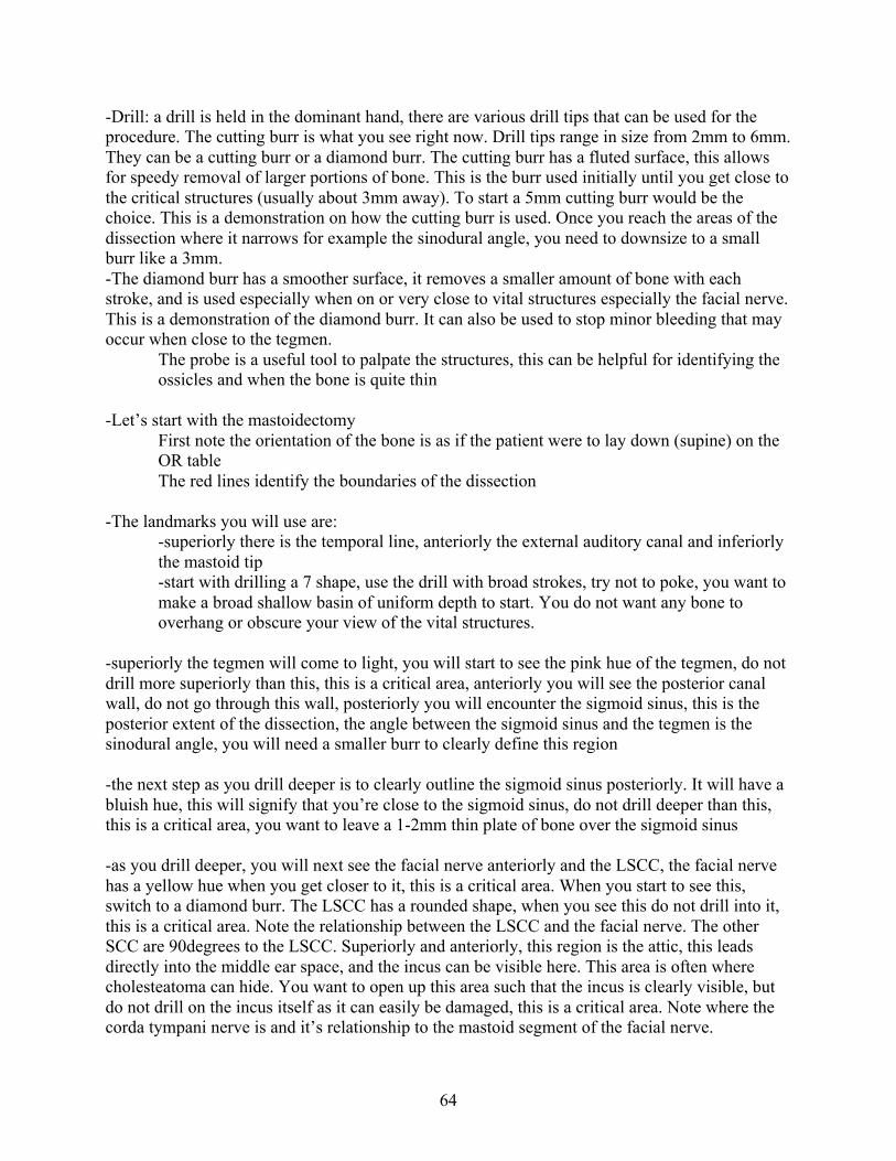
64
-Drill: a drill is held in the dominant hand, there are various drill tips that can be used for the procedure. The cutting burr is what you see right now. Drill tips range in size from 2mm to 6mm. They can be a cutting burr or a diamond burr. The cutting burr has a fluted surface, this allows for speedy removal of larger portions of bone. This is the burr used initially until you get close to the critical structures (usually about 3mm away). To start a 5mm cutting burr would be the choice. This is a demonstration on how the cutting burr is used. Once you reach the areas of the dissection where it narrows for example the sinodural angle, you need to downsize to a small burr like a 3mm. -The diamond burr has a smoother surface, it removes a smaller amount of bone with each stroke, and is used especially when on or very close to vital structures especially the facial nerve. This is a demonstration of the diamond burr. It can also be used to stop minor bleeding that may occur when close to the tegmen.
The probe is a useful tool to palpate the structures, this can be helpful for identifying the ossicles and when the bone is quite thin
-Let’s start with the mastoidectomy
First note the orientation of the bone is as if the patient were to lay down (supine) on the OR table The red lines identify the boundaries of the dissection
-The landmarks you will use are: -superiorly there is the temporal line, anteriorly the external auditory canal and inferiorly the mastoid tip -start with drilling a 7 shape, use the drill with broad strokes, try not to poke, you want to make a broad shallow basin of uniform depth to start. You do not want any bone to overhang or obscure your view of the vital structures.
-superiorly the tegmen will come to light, you will start to see the pink hue of the tegmen, do not drill more superiorly than this, this is a critical area, anteriorly you will see the posterior canal wall, do not go through this wall, posteriorly you will encounter the sigmoid sinus, this is the posterior extent of the dissection, the angle between the sigmoid sinus and the tegmen is the sinodural angle, you will need a smaller burr to clearly define this region -the next step as you drill deeper is to clearly outline the sigmoid sinus posteriorly. It will have a bluish hue, this will signify that you’re close to the sigmoid sinus, do not drill deeper than this, this is a critical area, you want to leave a 1-2mm thin plate of bone over the sigmoid sinus -as you drill deeper, you will next see the facial nerve anteriorly and the LSCC, the facial nerve has a yellow hue when you get closer to it, this is a critical area. When you start to see this, switch to a diamond burr. The LSCC has a rounded shape, when you see this do not drill into it, this is a critical area. Note the relationship between the LSCC and the facial nerve. The other SCC are 90degrees to the LSCC. Superiorly and anteriorly, this region is the attic, this leads directly into the middle ear space, and the incus can be visible here. This area is often where cholesteatoma can hide. You want to open up this area such that the incus is clearly visible, but do not drill on the incus itself as it can easily be damaged, this is a critical area. Note where the corda tympani nerve is and it’s relationship to the mastoid segment of the facial nerve.

65
-finally this is what the mastoidectomy should look like when completed. The critical structures here are: -tegmen -sigmoid sinus -facial nerve -semicircular canals -incus (ossicles) -The complications of mastoidectomy occur when the critical structures are violated: this includes facial nerve paralysis when the facial nerve is injured hearing loss- if the ossicles, or otic capsule are breached CSF leak- if the tegmen is breached Vertigo- trauma to the otic capsule or ossicles Injury to brain- if drilling too superiorly through the tegmen and meninges Injury to major vessels- violation of the sigmoid sinus -a systematic approach and careful identification of the anatomy are important to prevent these complications. Using a broad wide access and eliminating overhangs so good visualization of all of the structures can help mitigate risk. -in summary, we reviewed the temporal bone anatomy including the critical structures and their relationships- tegmen, facial nerve, SCC, ossicles the instruments used in mastoidectomy- cutting burrs, diamond burr and probe the indications for mastoidectomy and complications Questions