Temperature-dependent cross sections of O 2 – O 2 collision-induced absorption resonances at 477...
20
Journal of Quantitative Spectroscopy & Radiative Transfer 98 (2006) 405–424 Temperature-dependent cross sections of O 2 –O 2 collision-induced absorption resonances at 477 and 577 nm Maarten Sneep 1 , Dmitry Ityaksov, Ilse Aben 2 , Harold Linnartz 3 , Wim Ubachs Laser Centre, Vrije Universiteit, De Boelelaan 1081–1083, 1081 HV, Amsterdam, The Netherlands Received 23 March 2005; received in revised form 16 June 2005; accepted 16 June 2005 Abstract Two collision-induced absorption features of oxygen have been investigated by means of the laser-based cavity ring-down technique at pressures between 0 and 1000 hPa and at temperatures in the range 184–294 K. Peak cross sections, resonance widths and integrated cross sections, as well as spectral profiles, have been determined for the broad O 2 –O 2 resonances centered at 477 and 577 nm. Results are compared with previous measurements to establish an updated temperature dependence for the cross sections of both resonances, yielding integrated cross sections, that exhibit a minimum near 200 K and that increase in a near-linear fashion in the atmospherically relevant range of 200–300 K. A significant increase in the widths of the resonance profiles upon temperature increase is firmly established. Parameters and temperature- dependent trends for the shape and strengths of the resonances are produced, that can be implemented in cloud retrieval in atmospheric Earth observation. r 2005 Elsevier Ltd. All rights reserved. PACS: 94.10.Gb; 33:70: w; 33.20. Kf Keywords: Collision-induced absorptions; Earth’s atmosphere; Oxygen; Cavity ring-down spectroscopy ARTICLE IN PRESS www.elsevier.com/locate/jqsrt 0022-4073/$ - see front matter r 2005 Elsevier Ltd. All rights reserved. doi:10.1016/j.jqsrt.2005.06.004 Corresponding author. Tel.: +31 20 5987948; fax: +31 20 5987999. E-mail addresses: [email protected] (M. Sneep), [email protected] (D. Ityaksov), [email protected] (I. Aben), [email protected] (H. Linnartz), [email protected] (W. Ubachs). 1 Present address: Climate Research and Seismology Department, Royal Netherlands Meteorological Institute (KNMI), De Bilt, The Netherlands. 2 Also at: Netherlands Institute for Space Research, Utrecht, The Netherlands. 3 Also at: Sackler Laboratory for Astrophysics, Leiden Observatory, Leiden, The Netherlands.
Transcript of Temperature-dependent cross sections of O 2 – O 2 collision-induced absorption resonances at 477...
ARTICLE IN PRESS
Journal of Quantitative Spectroscopy &
Radiative Transfer 98 (2006) 405–424
0022-4073/$ -
doi:10.1016/j.
�CorresponE-mail add
linnartz@few1Present ad
(KNMI), De2Also at: N3Also at: Sa
www.elsevier.com/locate/jqsrt
Temperature-dependent cross sections of O2–O2
collision-induced absorption resonances at 477 and 577 nm
Maarten Sneep1, Dmitry Ityaksov, Ilse Aben2, Harold Linnartz3, Wim Ubachs�
Laser Centre, Vrije Universiteit, De Boelelaan 1081–1083, 1081 HV, Amsterdam, The Netherlands
Received 23 March 2005; received in revised form 16 June 2005; accepted 16 June 2005
Abstract
Two collision-induced absorption features of oxygen have been investigated by means of the laser-basedcavity ring-down technique at pressures between 0 and 1000 hPa and at temperatures in the range184–294K. Peak cross sections, resonance widths and integrated cross sections, as well as spectral profiles,have been determined for the broad O2–O2 resonances centered at 477 and 577 nm. Results are comparedwith previous measurements to establish an updated temperature dependence for the cross sections of bothresonances, yielding integrated cross sections, that exhibit a minimum near 200K and that increase in anear-linear fashion in the atmospherically relevant range of 200–300K. A significant increase in the widthsof the resonance profiles upon temperature increase is firmly established. Parameters and temperature-dependent trends for the shape and strengths of the resonances are produced, that can be implemented incloud retrieval in atmospheric Earth observation.r 2005 Elsevier Ltd. All rights reserved.
PACS: 94.10.Gb; 33:70:� w; 33.20. Kf
Keywords: Collision-induced absorptions; Earth’s atmosphere; Oxygen; Cavity ring-down spectroscopy
see front matter r 2005 Elsevier Ltd. All rights reserved.
jqsrt.2005.06.004
ding author. Tel.: +31 20 5987948; fax: +31 20 5987999.
resses: [email protected] (M. Sneep), [email protected] (D. Ityaksov), [email protected] (I. Aben),
.vu.nl (H. Linnartz), [email protected] (W. Ubachs).
dress: Climate Research and Seismology Department, Royal Netherlands Meteorological Institute
Bilt, The Netherlands.
etherlands Institute for Space Research, Utrecht, The Netherlands.
ckler Laboratory for Astrophysics, Leiden Observatory, Leiden, The Netherlands.
ARTICLE IN PRESS
M. Sneep et al. / Journal of Quantitative Spectroscopy & Radiative Transfer 98 (2006) 405–424406
1. Introduction
The oxygen molecule has a series of collision-induced absorption (CIA) resonances, involvingmolecular pairs that jointly absorb a single photon, whereby both molecules undergo a transitionto an electronically excited state. These resonances were discovered in the study of liquidoxygen in 1933 by Ellis and Kneser [1], and even that early—just a few years after theadvent of molecular quantum mechanics—were understood as bi-molecular absorptions andcould correctly be assigned in terms of quantum numbers. The resonance at 477 nm is assignedas an excitation of two oxygen ground-state molecules, one into the a 1Dgðv ¼ 0Þ state andone into the b 1Sþg ðv ¼ 0Þ state. The resonance near 577 nm can be assigned to the transi-tion a 1Dgðv ¼ 0Þ þ a 1Dgðv ¼ 1Þ X3S�g ðv ¼ 0Þ þX3S�g ðv ¼ 0Þ. The absorption strength of thecollision-induced features is rather high, when compared to monomer absorption, because thesephenomena are associated with symmetry breaking. Whereas, the O2 molecule is homonuclearand exhibits no dipole moment, all three lowest electronic states connected in the visible spectrumof oxygen have gerade symmetry, and hence transitions between these states are highly forbidden.Binary collisions give rise to distortions of the electronic wave functions and to a breaking of theinversion symmetry, which then permits optical transitions. It is noted further that the twocollision-induced features at 477 and 577 nm are not associated with a monomer absorption at thesame wavelengths; that would contradict the distribution of excitation energy over the twocollision partners. The fact that the 577 nm resonance overlaps the b 1Sþg � X3S�g ð3,0) monomerband is a happenstance coincidence. As was discussed by Naus and Ubachs [2], the interference ofboth phenomena can be avoided experimentally.Atmospheric observations of the features related to O2-CIA date back to the 19th century [3].
Since then a large number of laboratory investigations have been pursued to quantitatively assessthe cross sections related to the collisional absorption features. Accurate results in the high-pressure regime were obtained by Dianov–Klokov [4] and Greenblatt et al. [5], while focus on low-temperature effects prevailed in the studies by Ewing and coworkers [6,7]. Perner and Platt [8]performed measurements on atmospheric absorptions at long path length, Pfeilsticker et al. [9]determined altitude profile spectral features of ðO2Þ2 absorption bands from balloon observations,and Wagner et al. [10] detected the oxygen collisional features in the Earth’s atmosphere againstthe moonlight. In Ref. [9] a value for the enthalpy of formation of the O4 complex was derived, ingood agreement with findings from molecular scattering experiments [11,12].Besides these investigations on the electronic CIA features observable in the visible wavelength
range, oxygen collisional features were also studied at longer wavelengths, and there exists anextensive literature on this subject. For the 6mm vibrational excitation we cite the laboratoryinvestigations by Orlando et al. [13] and Baranov et al. [14]. The same vibrational CIA band wasinvestigated under stratospheric conditions with use of a balloon-borne FT-interferometer byRinsland et al. [15]. In the far-infrared domain oxygen exhibits a translation–rotation spectrumwith collisional features, which was investigated experimentally by Bosomworth and Gush [16]and theoretically by Boissoles et al. (see Ref. [17], and references cited therein). At the other end ofthe spectrum, in the deep-ultraviolet, absorption features were observed, that were in partascribed to O22O2 resonances [18].Theoretical treatment of oxygen and its dimer dates back to 1924 when Lewis proposed the
existence of a weak chemical bond establishing an O4 molecule that would not be paramagnetic
ARTICLE IN PRESS
M. Sneep et al. / Journal of Quantitative Spectroscopy & Radiative Transfer 98 (2006) 405–424 407
[19]. While Lewis focused on the spin aspects, Pauling singled out the O4 molecule for itsexceptionally strong Van der Waals forces, that might give rise to a chemical bond [20]. Thus, abinitio calculations on the oxygen dimer are complicated by the fact that both the Van der Waalsinteraction and the spin interactions between two paramagnetic molecules play a role. The O2
triplet ground state gives rise to singlet, triplet and quintet states in the dimer, therewith giving riseto a system with fine structure splittings [21]. Further complications arise from tunneling throughinternal barriers [22]. Calculations of full potential energy surfaces of the O22O2 Van der Waalscomplex yield minimum energy structures for the singlet and triplet states in a parallel planar D2h
geometry at an equilibrium inter-molecular separation of 6:126:2a0 at binding energies of 154 and140 cm�1 [23]. Later, also ab initio potentials involving electronically excited O2 were calculated[24]. Such calculations provide a basis for an assignment of the rotational structure in the bounddimer systems [25].Whether the two O2 molecules form a bound Van der Waals complex, or are essentially
unbound and absorb the photon during a collisional fly-by, has been a matter of discussion in theliterature. Low-temperature measurements show some ripple structure—indicating that boundðO2Þ2 states play a role in the absorption resonance [7]. Measurements by Biennier et al. [25] andCampargue [26] in a slit-jet expansion have indeed shown many narrow resonances in the oxygendimer. The latter studies prove that at low temperatures bound states exist, since the resonancesare unequivocally associated with bound rotational states of the oxygen dimer. However, thesemeasurements were carried out at the characteristically low final temperatures in molecularexpansions, strongly deviating from atmospheric conditions, where the bound states are presumedto play no role. Vigasin and coworker [27–29] have analyzed the contribution of bound andfree–free dimers to the absorption in collision-induced spectra, thereby dividing phase space intothree compartments, including that of metastably bound dimers in which molecules are slightlybound by a centrifugal barrier. From their thermodynamic model it follows that even attemperatures as low as 150K bound complexes do not contribute significantly to the collisionalspectral features [30].Obviously, the oxygen CIA resonances are of importance for atmospheric physics. The oxygen
collisional complex is held responsible for a non-negligible fraction (1–2%) of the absorption ofsolar radiation [31,32]. The cross sections of the O4 features depend quadratically on pressure, andthey are particularly useful to determine cloud top heights in the lower atmospheric layers.Acarreta et al. [33] have introduced a cloud detection algorithm, which also yields the effectivecloud fraction within a ground pixel, using the O22O2 resonance at 477 nm. This resonance fallswithin the spectral window (270–500 nm) sampled by the Ozone Monitoring Instrument (OMI)aboard the EOS-Aura satellite, which was launched in summer 2004 with the aim to detect globalozone columns. Using Ref. [34] it is estimated that a 10% error in the peak absorption of the477 nm O22O2 resonance leads to errors of about 1 km in the retrieved cloud altitude. This, inturn, leads to an error of about 1% in the total column density of ozone. As the target accuracy ofthe total ozone column is about 1%, which is needed to detect trends in ozone and to detecttropospheric pollution, errors in the peak absorption of O22O2 should not be larger than 5%.Since the scatter in the literature values on the band integrated CIA cross section amounts to15%, the uncertainty in the retrieved ozone column densities from OMI for cloudy measurementsis dominated by the uncertainty in the O22O2 resonance; the cloud-free uncertainty is 1.2% and ismostly due to instrument noise.
ARTICLE IN PRESS
M. Sneep et al. / Journal of Quantitative Spectroscopy & Radiative Transfer 98 (2006) 405–424408
The above-mentioned relation to ozone retrieval warrants detailed and accurate laboratorymeasurements to provide the integrated cross section and shape function of the O22O2
resonances. In the distant past Tabisz et al. [35], McKellar et al. [36] and Ewing and coworkers[6,7] determined absorption cross sections for both the 477 and 577 nm resonances at a number oftemperatures. Recently, studies were performed by Newnham and Ballard [37] and Hermans et al.[38] applying Fourier-transform spectroscopy, and Morville et al. [39] as well as Tiedje et al. [40],who performed cavity ring-down measurements. As a continuation of previous studies in ourlaboratory on O22O2 cross sections at room temperature [2,41] we have built a special cell tocarry out cavity ring-down experiments at controlled low temperatures in the range 184–294K. Inview of the relevance for ozone column density retrieval, as discussed above, the focus has been onthe 477 nm O22O2 feature, but also new data were collected for the 577 nm resonance.
2. Description of the setup and the experimental procedure
2.1. CRD-setup and the pressure-ramp method
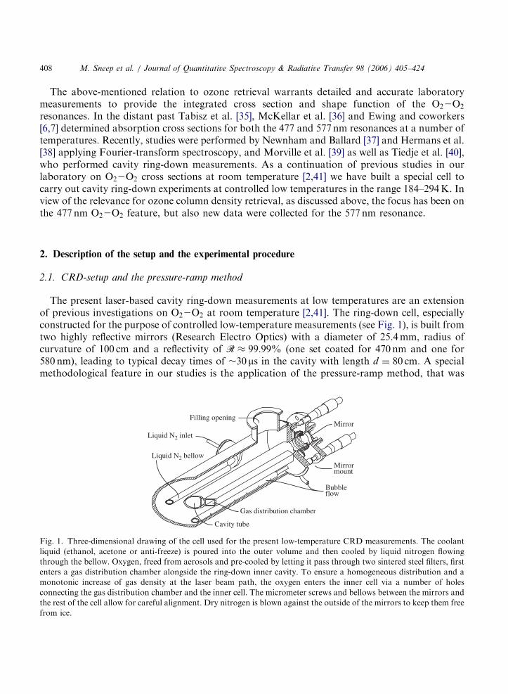
The present laser-based cavity ring-down measurements at low temperatures are an extensionof previous investigations on O22O2 at room temperature [2,41]. The ring-down cell, especiallyconstructed for the purpose of controlled low-temperature measurements (see Fig. 1), is built fromtwo highly reflective mirrors (Research Electro Optics) with a diameter of 25.4mm, radius ofcurvature of 100 cm and a reflectivity of R � 99:99% (one set coated for 470 nm and one for580 nm), leading to typical decay times of �30ms in the cavity with length d ¼ 80 cm. A specialmethodological feature in our studies is the application of the pressure-ramp method, that was
Cavity tube
Liquid N2 bellow
Gas distribution chamber
Liquid N2 inlet
Mirror
Mirrormount
Bubbleflow
Filling opening
Fig. 1. Three-dimensional drawing of the cell used for the present low-temperature CRD measurements. The coolant
liquid (ethanol, acetone or anti-freeze) is poured into the outer volume and then cooled by liquid nitrogen flowing
through the bellow. Oxygen, freed from aerosols and pre-cooled by letting it pass through two sintered steel filters, first
enters a gas distribution chamber alongside the ring-down inner cavity. To ensure a homogeneous distribution and a
monotonic increase of gas density at the laser beam path, the oxygen enters the inner cell via a number of holes
connecting the gas distribution chamber and the inner cell. The micrometer screws and bellows between the mirrors and
the rest of the cell allow for careful alignment. Dry nitrogen is blown against the outside of the mirrors to keep them free
from ice.
ARTICLE IN PRESS
M. Sneep et al. / Journal of Quantitative Spectroscopy & Radiative Transfer 98 (2006) 405–424 409
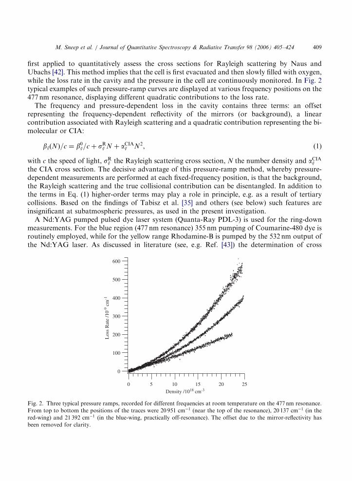
first applied to quantitatively assess the cross sections for Rayleigh scattering by Naus andUbachs [42]. This method implies that the cell is first evacuated and then slowly filled with oxygen,while the loss rate in the cavity and the pressure in the cell are continuously monitored. In Fig. 2typical examples of such pressure-ramp curves are displayed at various frequency positions on the477 nm resonance, displaying different quadratic contributions to the loss rate.The frequency and pressure-dependent loss in the cavity contains three terms: an offset
representing the frequency-dependent reflectivity of the mirrors (or background), a linearcontribution associated with Rayleigh scattering and a quadratic contribution representing the bi-molecular or CIA:
bnðNÞ=c ¼ b0n=cþ sRn N þ aCIAn N2, (1)
with c the speed of light, sRn the Rayleigh scattering cross section, N the number density and aCIAnthe CIA cross section. The decisive advantage of this pressure-ramp method, whereby pressure-dependent measurements are performed at each fixed-frequency position, is that the background,the Rayleigh scattering and the true collisional contribution can be disentangled. In addition tothe terms in Eq. (1) higher-order terms may play a role in principle, e.g. as a result of tertiarycollisions. Based on the findings of Tabisz et al. [35] and others (see below) such features areinsignificant at subatmospheric pressures, as used in the present investigation.A Nd:YAG pumped pulsed dye laser system (Quanta-Ray PDL-3) is used for the ring-down
measurements. For the blue region (477 nm resonance) 355 nm pumping of Coumarine-480 dye isroutinely employed, while for the yellow range Rhodamine-B is pumped by the 532 nm output ofthe Nd:YAG laser. As discussed in literature (see, e.g. Ref. [43]) the determination of cross
600
500
400
300
200
100
0
Los
s R
ate
/10-9
cm
-1
2520151050
Density /1018 cm-3
Fig. 2. Three typical pressure ramps, recorded for different frequencies at room temperature on the 477 nm resonance.
From top to bottom the positions of the traces were 20 951 cm�1 (near the top of the resonance), 20 137 cm�1 (in the
red-wing) and 21 392 cm�1 (in the blue-wing, practically off-resonance). The offset due to the mirror-reflectivity has
been removed for clarity.
ARTICLE IN PRESS
M. Sneep et al. / Journal of Quantitative Spectroscopy & Radiative Transfer 98 (2006) 405–424410
sections in CRD experiments is hampered by the linewidth problem, giving rise to multi-exponential decays on narrow resonances. But here the opposite is the case: the resonances arealmost as wide as the gain profiles of the dyes. The principle of CRD detection makes themeasurements independent of laser pulse intensity, hence the linewidth problem poses no specificproblems in the present experiments. Only when the laser wavelength is tuned to the wings of thedye gain curve appreciable amounts of amplified spontaneous emission (ASE) can be produced inthe dye laser, which may result in systematic offsets in the measured cross sections. In ourexperiments this was the case on the short-wavelength edge of the 477 nm resonance. At thesewavelengths additional measurements were performed with Coumarine-460 dye, thus producing areliable set of ring-down transients at these wavelengths.
2.2. Reaching and maintaining low temperatures
The lowest temperatures are obtained by submerging the CRD cell (see Fig. 1) in a low-temperature bath, consisting of a solid–liquid mixture of acetone—which has a freezing point of�94:8 �C or 178.4K. Since liquids undergo a first-order phase transition at the freezing point, thecell can be reasonably well stabilized at that temperature. Usually, an equilibrium temperature isestablished slightly above the freezing point; for that reason the 477 nm resonance wasinvestigated at 184K and the 577 nm resonance at 190K, when using acetone as a coolant. Themeasurements at 268K (only for the 577 nm O22O2 feature) were performed with a differentcooling technique; here, a chiller filled with anti-freeze (1,2 ethane-diol/ethylene-glycol mixture)was used to bring down the temperature. The filler opening and the bubble-flow pipes were usedas the connection points for the hoses, and the chilled liquid was pumped around by the chiller.The lowest temperature reached with the chiller was 268K ð�5 �CÞ. A separate series ofmeasurements was performed (only for the 477 nm O22O2 feature) at a temperature of about230K (�45 �C) using the anti-freeze mixture as coolant. Similar to the experiments using acetone,the anti-freeze mixture was frozen by flushing liquid nitrogen through the bellow. Due to the lackof a latent heat step at the freezing-point phase transition, difficulties were encountered in keepingthe temperature at a constant value, but eventually controlled measurements at a temperature of230� 5K could be performed. For the 477 nm resonance again a large set of data was taken atroom temperature, therewith verifying previous results by our team [41] taken in a different cell.The results were analyzed and are included in this paper. For the 577 nm resonance no additionalroom temperature measurements are added.After the cell is aligned and evacuated, the outer volume is filled with the coolant to a level well
above the ring-down cell. Liquid nitrogen is then flowed through a bellow, cooling down thecoolant substance, in which the actual ring-down cavity is immersed. For the coolants used,frozen parts will sink to the bottom of the outer volume. To ensure a homogeneous temperaturethroughout the cell, and to bring the coldest coolant into contact with the ring-down cavity,nitrogen gas is blown through a tube with a series of holes located at the bottom of the outervolume. To reduce the heat load of this gas-flow, the nitrogen gas is cooled down by leading itthrough another container with liquid nitrogen. The bubbles will mix the liquid and bring thetemperature of the entire cell down to the freezing point of the coolant. Reaching the freezingpoint of acetone takes between 4 and 5 h, and after this time the temperature can be maintained bykeeping a steady flow of liquid nitrogen through the bellow. A specific advantage of the present
ARTICLE IN PRESS
M. Sneep et al. / Journal of Quantitative Spectroscopy & Radiative Transfer 98 (2006) 405–424 411
cell is that the low temperature is maintained homogeneously over almost the entire length of thecavity; even the mirror mounts are in contact with the coolant.
2.3. Measurements
As with the measurements at room temperature [2,41], the CIA cross section is determined withthe pressure-ramp method. To fill the cell with oxygen at a predetermined low temperature, thegas is purged through a sintered stainless steel filter with 7mm pores, located inside the outer tubeand submerged in the coolant. This filter provides a large contact surface area, and after purging,the temperature of the gas is assumed to be the same as that of the fluid. Because thisthermalization filter is inaccessible from the outside, a second filter with 0:5mm pores is usedoutside the cell to filter aerosols from the oxygen and prevent contamination of the thermalizationfilter. Apart from this filter, oxygen with a specified purity of 99.999% is used without furthertreatment. The rate of flow is controlled by a needle valve and set for the pressure ramps to lastabout 15min. The temperature of the coolant is measured by a thermocouple—type K ðNiþ10%CrÞ against ðNiþ 2%Alþ 2%Mnþ 1%SiÞ—with the tip placed close to the oxygenthermalization filter. A pressure sensor is placed in the top part inside the cell, in an effort toreduce the heat load on the system. The capacitance baratron for the range 0–1333 hPa has anuncertainty of 2 hPa. The oxygen gas density is calculated from the measured temperature andpressure using the Van der Waals equation of state. During the pressure ramps the ring-downtimes (or loss rates bn) are constantly monitored at a 10Hz repetition rate, by methods describedby Naus et al. [43], while also the pressure and temperature measurements are continuously storedin the data-handling computer.The measurements are performed after the ring-down cell is aligned to yield single exponential
decays. As discussed in Ref. [43] only under such conditions can the characteristic retrieved ring-down times be interpreted in terms of cross sections. In the first step of the measurement and dataanalysis an exponential decay function is fitted to the ring-down transients. This results in typicaldensity-ramp traces as shown in Fig. 2 for some measurements on the 477 nm resonance.As in the previous measurements [2,41], the wavelength of the laser was measured after each
CRD-pressure ramp at a fixed wavelength position, with an echelle-grating spectrometer yieldingan absolute accuracy of 0.2 cm�1.
2.4. Data analysis
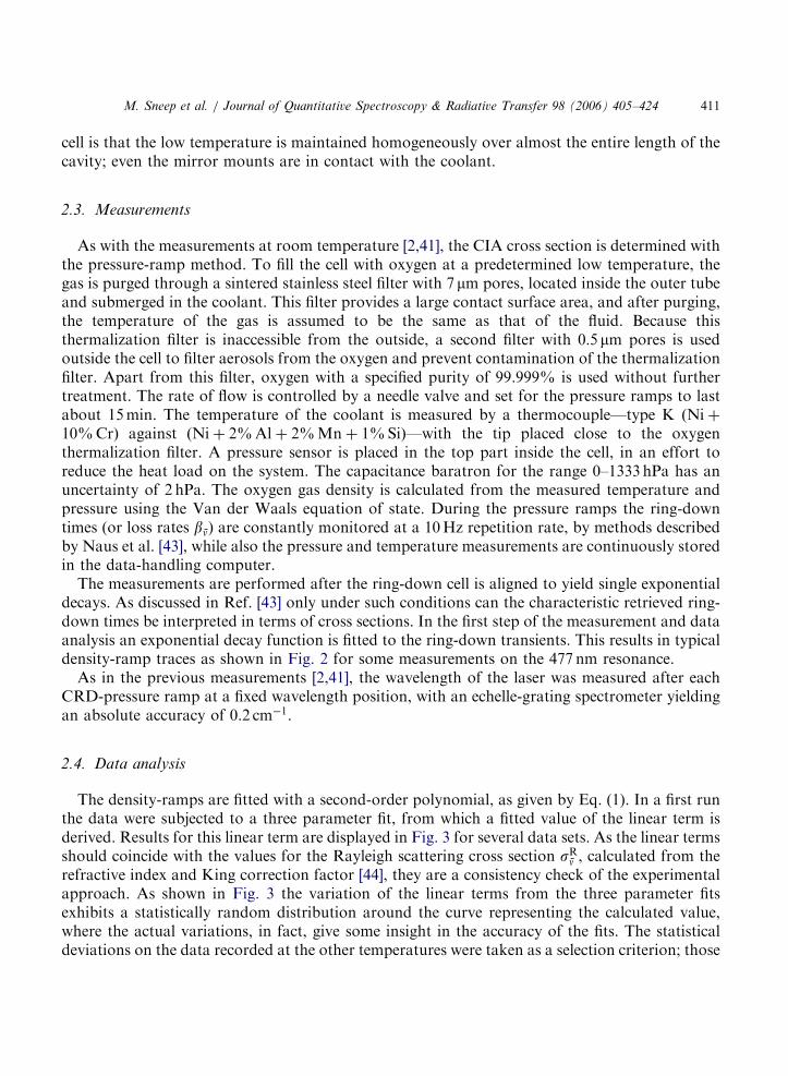
The density-ramps are fitted with a second-order polynomial, as given by Eq. (1). In a first runthe data were subjected to a three parameter fit, from which a fitted value of the linear term isderived. Results for this linear term are displayed in Fig. 3 for several data sets. As the linear termsshould coincide with the values for the Rayleigh scattering cross section sRn , calculated from therefractive index and King correction factor [44], they are a consistency check of the experimentalapproach. As shown in Fig. 3 the variation of the linear terms from the three parameter fitsexhibits a statistically random distribution around the curve representing the calculated value,where the actual variations, in fact, give some insight in the accuracy of the fits. The statisticaldeviations on the data recorded at the other temperatures were taken as a selection criterion; those

ARTICLE IN PRESS
10
8
6
4
2
0
Ray
leig
h sc
atte
ring
/10-2
7 cm
2
2200021500210002050020000Wavenumber /cm-1
Fig. 3. Fitted values for the linear scattering terms as obtained from three parameters fits for the data sets pertaining to
the 477 nm resonance at room temperature ð�Þ, at 230K ð�Þ, and at 184K ð&Þ. The full line corresponds to the value of
the calculated Rayleigh scattering cross section, sRn , for the specific wavelength.
M. Sneep et al. / Journal of Quantitative Spectroscopy & Radiative Transfer 98 (2006) 405–424412
pressure ramps, where the fitted linear term was off by more than 20% from the calculated sRn (forroom temperature data) or 40% (for the other data) were discarded.In a second round of fitting (two parameter fits) the linear term is kept fixed at the
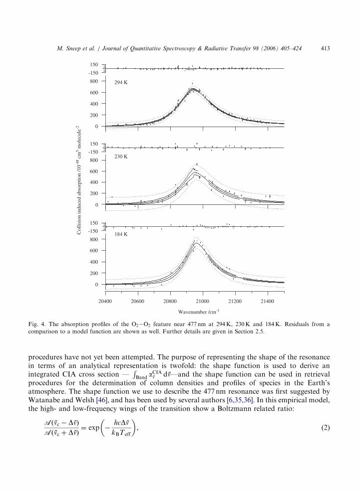
calculated sRn , which is calculated from the refractive index and King correction factor [44].Values to be taken for sRn were discussed in a previous paper focusing on direct Rayleighscattering measurements [45]. This second round of fitting yields an improved accuracy for thedetermination of the quadratic term, corresponding to the CIA cross section aCIAn . In Figs. 4 and5, the final results of the measurements on the 477 and the 577 nm resonances of O22O2 CIAs areshown. The measurements at 294K on the 577 nm resonance are taken from Naus and Ubachs [2].The room temperature measurements on the 477 nm resonance stem from new series ofmeasurements in combination with similar data, obtained in a previous investigation [41].
2.5. Further reduction of the data
In Figs. 4 and 5 resulting data for the quadratic components aCIAn are plotted as a function offrequency. In the same figures fits to the measured data are shown using a model function. Thesolid lines are the least-square best-fit results. The dashed lines in these figures indicate theconfidence interval for the fit-result at the 2s (95.4%) level. This means that if the experimentswere repeated, 95.4% of the sets would yield a fit that falls within the indicated interval. Thedotted lines indicate the confidence interval for the prediction at the same level, indicating that95.4% of the measured points should fall within the indicated band.Currently there exists no good theoretical description of the O22O2 CIA profile, and empirical
model functions are used to describe the shape function of the resonance feature. In principle, theresonance profile could be calculated based on multi-dimensional ab initio potential energysurfaces and dipole moments, with integration over kinetic energy and angular distributions of thevectorial properties (velocities and angular momenta) of the collision partners, but such elaborate
ARTICLE IN PRESS
800
600
400
200
0
214002120021000208002060020400
Wavenumber /cm-1
150
-150
800
600
400
200
0
Col
lisio
n in
duce
d ab
sorp
tion
/10-4
8 cm
5 mol
ecul
e-2
150
-150
800
600
400
200
0
150
-150
294 K
230 K
184 K
Fig. 4. The absorption profiles of the O22O2 feature near 477 nm at 294K, 230K and 184K. Residuals from a
comparison to a model function are shown as well. Further details are given in Section 2.5.
M. Sneep et al. / Journal of Quantitative Spectroscopy & Radiative Transfer 98 (2006) 405–424 413
procedures have not yet been attempted. The purpose of representing the shape of the resonancein terms of an analytical representation is twofold: the shape function is used to derive anintegrated CIA cross section —
RBand a
CIAn dn—and the shape function can be used in retrieval
procedures for the determination of column densities and profiles of species in the Earth’satmosphere. The shape function we use to describe the 477 nm resonance was first suggested byWatanabe and Welsh [46], and has been used by several authors [6,35,36]. In this empirical model,the high- and low-frequency wings of the transition show a Boltzmann related ratio:
Aðnc � DnÞAðnc þ DnÞ
¼ exp �hcDn
kBTeff
� �, (2)
ARTICLE IN PRESS
1.2
0.8
0.4
0.0
18200180001780017600174001720017000
Wavenumber /cm-1
-0.2
0.2
1.2
0.8
0.4
0.0
Col
lisio
n in
duce
d ab
sorp
tion
/10-4
5 cm
5 mol
ecul
e-2
-0.04
0.04
1.2
0.8
0.4
0.0
-0.04
0.04
294 K
268 K
190 K
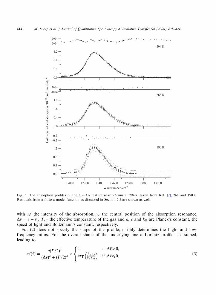
Fig. 5. The absorption profiles of the O22O2 feature near 577 nm at 294K taken from Ref. [2], 268 and 190K.
Residuals from a fit to a model function as discussed in Section 2.5 are shown as well.
M. Sneep et al. / Journal of Quantitative Spectroscopy & Radiative Transfer 98 (2006) 405–424414
with A the intensity of the absorption, nc the central position of the absorption resonance,Dn ¼ n� nc, T eff the effective temperature of the gas and h, c and kB are Planck’s constant, thespeed of light and Boltzmann’s constant, respectively.Eq. (2) does not specify the shape of the profile; it only determines the high- and low-
frequency ratios. For the overall shape of the underlying line a Lorentz profile is assumed,leading to
AðnÞ ¼aðG=2Þ2
ðDnÞ2 þ ðG=2Þ2�
1 if Dn40;
exp hcDnkBTeff
� �if Dnp0;
8<: (3)
ARTICLE IN PRESS
M. Sneep et al. / Journal of Quantitative Spectroscopy & Radiative Transfer 98 (2006) 405–424 415
where a and G represent parameters for the amplitude and the width of the absorption feature. InRef. [41], the temperature in Eq. (3) was taken to be the temperature at which the measurementswere performed. Especially at lower temperatures, this leads to unsatisfactory fit results. Theeffective temperature T eff is now a fit-parameter and it is adjusted to give the best fit to the data.During the analysis of the data, it became apparent that the model given by Eq. (3) works well
for the description of the 477 nm resonance, but when it is applied to the 577 nm resonance, largeresiduals and a very poor fit are found. Following the analysis of Naus and Ubachs [2], a skewedVoigt profile was used to describe the shape of the 577 nm resonance. The skewed Voigt profile isapproximated by a linear combination of a Gauss and a Lorentz profile, where the left- and right-hand sides have a different linear scale:
VðnÞ ¼ ð1� bÞa
1þ ½4ðDnÞ2=G2x2�þ ba exp �4 ln 2
DnGx
� �2" #
(4)
with
x ¼1þ 2 arctanðskew factorÞ=p if Dn40;
1� 2 arctanðskew factorÞ=p if Dnp0
(
and a and b amplitudes. The parameters found in the fitting procedure are shown in Tables 1and 2. The two sets use different model functions and therefore have different parameters in theirrespective tables. The resulting effective temperatures found in the fitting routines using Eq. (3)are systematically and significantly higher than the real measured temperatures of the oxygen gas.Once again we note that T eff should by no means be regarded as a temperature, rather than as afitting parameter in a model.The uncertainties indicated are found from the fitting procedure. An attempt was made to
propagate the error estimates found from the fit of the ring-down transients, but the resultingerrors were unrealistically small. The currently indicated errors assume that the errors in allmeasured points are equal and that the model function is accurate. In that case w2 scaling is
Table 1
Parameters describing the absorption profile of the O22O2 feature near 477 nm at three different temperatures
Temperature (K) 294 230 184
Coolant None Anti-freeze Acetone
nc ðcm�1Þ 20929:9� 2:8 20943� 11 20953� 6
lc ðnmÞ 477:79� 0:06 477:47� 0:26 477:25� 0:14
FWHM ðcm�1Þ 247:1� 6:8 228� 26 197� 14
FWHM (nm) 5:64� 0:16 5:2� 0:6 4:5� 0:3
G ðcm�1Þ 306:4� 6:8 283� 26 239� 14
a ð10�48 cm5 molecule�2Þ 660� 6 578� 23 733� 20
T eff ðKÞ 361� 37 331� 138 316� 102RAðnÞdn ð10�42 cm4 molecule�2Þ 0:234� 0:006 0:190� 0:022 0:207� 0:016
The parameters were found using Eq. (3) as the model function and a least-square fit. The indicated errors are 1s values.
A graphical representation of the data and the fit-result are shown in Fig. 4.
ARTICLE IN PRESS
Table 2
Parameters describing the absorption profile of the O22O2 feature near 577 nm at three different temperatures
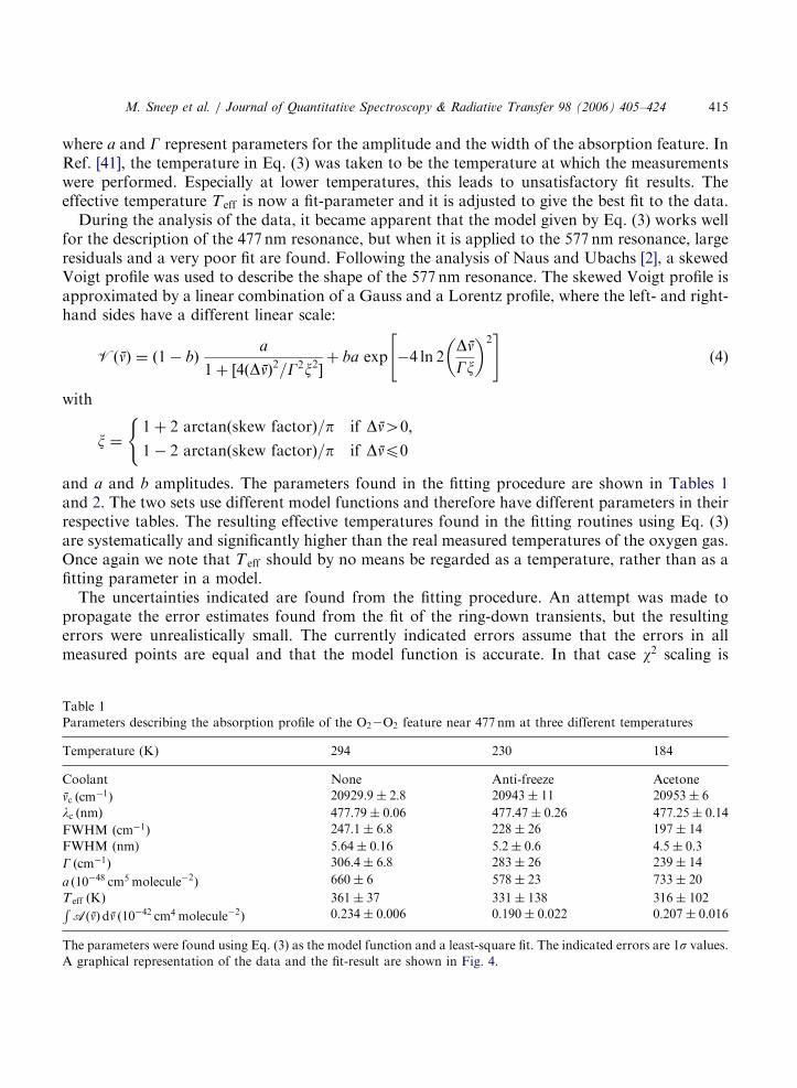
Temperature (K) 294a 268 190
Coolant None Chiller Acetone
nc ðcm�1Þ 17325:8� 1:7 17322:8� 3:5 17313� 7
lc ðnmÞ 577:17� 0:06 577:3� 0:1 577:6� 0:2
FWHM ðcm�1Þ 346:5� 3:0 342:3� 7:1 290� 10
FWHM (nm) 11:5� 0:1 11:3� 0:2 9:6� 0:3
G ðcm�1Þ 318� 3 317� 7 257� 10
a ð10�45 cm5 molecule�2Þ 1:15� 0:01 1:11� 0:03 1:26� 0:05
Skew factor �0:64� 0:02 �0:60� 0:05 �0:65� 0:11b 0:782� 0:012 0:784� 0:034 0:91� 0:06RVðnÞdn ð10�42 cm4 molecule�2Þ 0:470� 0:006 0:447� 0:013 0:406� 0:024
The parameters were found using Eq. (4) as the model function and a least-squares fit. The indicated errors are 1svalues. A graphical representation of the data and the fit-result are shown in Fig. 5.
aMeasurements taken from Naus and Ubachs [2].
M. Sneep et al. / Journal of Quantitative Spectroscopy & Radiative Transfer 98 (2006) 405–424416
possible and the uncertainties resulting from this procedure are reported. The indicated accuracyon the band integrated absorption cross section is found by applying a Gaussian variation withthe indicated standard deviation to the parameters describing the model function and taking thestandard deviation of the resulting distribution of integrated cross sections.
3. Discussion and comparison with other measurements
In order to give a complete picture of the measurements of temperature dependence of thecollisional ðO2Þ2 resonances at 477 and 577 nm all available information in literature was collectedin conjunction with the presently obtained data. For this purpose, scaling to the appropriate unitsis required and in some cases digitization of information contained in plotted figures wasemployed to extract information on three relevant parameters: the peak cross sections, the widths(FWHM) and the integrated cross sections. Final values are included in Figs. 6 and 7. Note herethat notwithstanding the fact that the resonance features are clearly asymmetric, widths (FWHM)were determined. The restriction is imposed to collect data obtained from laboratory experiments(in section 3.1), because these are considered to originate from well-controlled experiments.From these data, displayed in Fig. 6 (for the 477 nm resonance) and in Fig. 7 (for the 577 nmresonance) temperature-dependent trends are deduced, which are discussed in Section 3.2.Subsequently, a comparison with data and trends obtained from atmospheric measurements ismade in Section 3.3.
3.1. Data collection of laboratory measurements
For both the 477 and 577 nm resonances, Tabisz et al. [35] list the band integrated cross sectionsat 297K, but not the widths nor the peak cross sections; those were extracted by digitization
ARTICLE IN PRESS
300
200
100Wid
th /c
m-1
300250200150100
Temperature /K
2.0
1.6
1.2
0.8
0.4
Peak
/10-4
5 cm
5
280
240
200
160
Inte
gral
/10-4
5 cm
4300
280
260
240
Width /cm
-1
900
800
700
600
500
Peak /10-48 cm
5
280
240
200
160
Integral /10-45 cm
4
300297294
(a)
(b)
(c)
Fig. 6. Temperature dependence for parameters of the 477 nm O22O2 resonance. The width is shown in (a), the height
in (b) and the band integrated intensity in (c). The vertical bars indicate the 1s confidence level on each point.
Sources:,—Ewing et al. [6]; �—Greenblatt et al. [5]; —Newnham and Ballard [37]; �—Tabisz et al. [35]; &—
McKellar et al. [36]; .—Hermans et al. [38]; �—this work. The sections at the right-hand sides duplicate the data
already contained in the main section of the graphs at enlarged scales. The dashed lines are drawn to guide the eye, and
to indicate temperature dependences.
M. Sneep et al. / Journal of Quantitative Spectroscopy & Radiative Transfer 98 (2006) 405–424 417
procedures. From the work of McKellar et al. [36] all three parameters were deduced from thedigitized plots. Blickensderfer and Ewing [6] give the integrated absorption cross sections, thehalf-widths at 300 and 87K and the peak heights at 300 and 290K (at different pressures).The difference between the latter two values is taken as a measure for the error on the peak height.The peak height values at 87K are taken from a digital readout of the graphs. Greenblatt et al. [5]give some parameters for both resonances; in addition, digitized data of the room temperaturemeasurements in Ref. [5] were obtained via the authors of Ref. [33] from which the bandintegrated cross section was calculated. Morville et al. [39] give the width and peak height for the577 nm band at three temperatures. The band integrated values were extracted via the digitizedplot, using direct numerical integration. From the noise on the plot, and the baseline seen in thegraph, the uncertainty on the integral is estimated to be around 10%. Tiedje et al. [40] give allthree values for the 577 nm band in their paper. Newnham and Ballard give the peak absorptioncross section and the integrated absorption cross section plus the error estimate for bothresonances at two temperatures in Tables 6 and 7 of their paper; a value for the FWHM value wasdeduced by us from their raw data [37]. Finally, Hermans et al. [38] performed Fourier-Transformmeasurements at pressures in the range 0.3–1 bar, applying multi-passing in a 50m cell at roomtemperature. Values for the peak cross sections and the widths are obtained from Table 3 inRef. [38], while a value for the integrated cross section is obtained from the raw data. These raw
ARTICLE IN PRESS
2.4
2.0
1.6
1.2Peak
/10-4
5 cm
5
300250200150100
Temperature /K
600
500
400
300Inte
gral
/10-4
5 cm
4350
300
250
200Wid
th /c
m-1
360
340
320
300
Width /cm
-1
1.251.201.151.101.051.000.95
Peak /10-45 cm
5
550
500
450400
350300
Integral /10-45 cm
4
300296
(b)
(a)
(c)
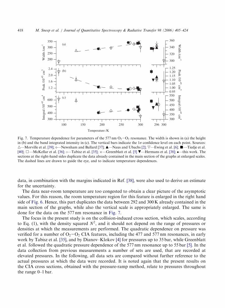
Fig. 7. Temperature dependence for parameters of the 577 nm O22O2 resonance. The width is shown in (a) the height
in (b) and the band integrated intensity in (c). The vertical bars indicate the 1s confidence level on each point. Sources:
n—Morville et al. [39]; —Newnham and Ballard [37]; m—Naus and Ubachs [2]; ,—Ewing et al. [6]; ’—Tiedje et al.
[40]; &—McKellar et al. [36]; �—Tabisz et al. [35]; �—Greenblatt et al. [5] .—Hermans et al. [38]; �—this work. The
sections at the right-hand sides duplicate the data already contained in the main section of the graphs at enlarged scales.
The dashed lines are drawn to guide the eye, and to indicate temperature dependences.
M. Sneep et al. / Journal of Quantitative Spectroscopy & Radiative Transfer 98 (2006) 405–424418
data, in combination with the margins indicated in Ref. [38], were also used to derive an estimatefor the uncertainty.The data near-room temperature are too congested to obtain a clear picture of the asymptotic
values. For this reason, the room temperature region for this feature is enlarged in the right handside of Fig. 6. Hence, this part duplicates the data between 292 and 300K already contained in themain section of the graphs, while also the vertical scale is appropriately enlarged. The same isdone for the data on the 577 nm resonance in Fig. 7.The focus in the present study is on the collision-induced cross section, which scales, according
to Eq. (1), with the density squared N2, and it should not depend on the range of pressures ordensities at which the measurements are performed. The quadratic dependence on pressure wasverified for a number of O22O2 CIA features, including the 477 and 577 nm resonances, in earlywork by Tabisz et al. [35], and by Dianov–Klokov [4] for pressures up to 35 bar, while Greenblattet al. followed the quadratic pressure dependence of the 577 nm resonance up to 55 bar [5]. In thedata collection from previous measurements a number of sets are used, that are recorded atelevated pressures. In the following, all data sets are compared without further reference to theactual pressures at which the data were recorded. It is noted again that the present results onthe CIA cross sections, obtained with the pressure-ramp method, relate to pressures throughoutthe range 0–1 bar.
ARTICLE IN PRESS
M. Sneep et al. / Journal of Quantitative Spectroscopy & Radiative Transfer 98 (2006) 405–424 419
3.2. Temperature-dependent trends
A comparison of results on the CIA features should be based on the entire band profilesincluding the particular shapes of the resonances. Since detailed information on the actual shapesin previous investigations is sparse and difficult to extract, we have chosen to represent the profilesin terms of three parameters: the peak and integrated cross sections and the widths (FWHM),even though the profiles are definitely asymmetric. As discussed, there exists no closed theory forthe band shapes of the CIA O22O2 features and the two different functions given by Eqs. (3) and(4) are just phenomenological parametrizations of the shapes observed.In Fig. 6, all available information on the collision-induced 477 nm resonance is contained and
some unequivocal conclusions can be drawn from it. The width (FWHM) of the resonanceexhibits a monotonic increase in the entire temperature range 87–300K from 100 cm�1 at thelowest temperature to 245 cm�1 at room temperature. All data points fall close to the dashed lines,serving as an indicator for the temperature dependence. For the room temperature value of thewidth we have taken the average value of the present investigation and that of Hermans et al. [38](yielding 244� 7 cm�1). These data were recorded at lower pressures, while the other data atroom-temperature conditions correspond to high-pressure measurements. As discussed byBlickensderfer et al. [6] elevated pressures may give rise to additional broadening.The peak cross section of the 477 nm feature sharply drops when departing from the
lowest temperature, then leveling off asymptotically to the room temperature value ofð660� 6Þ � 10�48 cm5 molecule�2. The latter value is obtained from a weighted fit on the set ofdata available on (near)-room temperature measurements, comprising the present value andliterature values. Again the trend is indicated by a dashed line drawn in section (b) of Fig. 6. Basedupon the entire set of available data no definite statement can be made on the temperature trend inthe atmospherically relevant domain of 200–300K. However, for a subclass of investigations,where measurements were performed at varying temperatures under otherwise similarmeasurement conditions, such a statement can be made. In the present series of measurements,using CRDS, a peak cross section is measured at 230 and at 294K yielding a clear indication of anincrease of 14� 5 % over this temperature interval. Similarly, Newnham and Ballard find anincrease, even more pronounced, from their recent FT-spectroscopic measurements [37].Notwithstanding the fact that the two most accurate and most recent data sets indicate anincrease in the peak cross section at atmospheric temperatures the dashed line in section (b) ofFig. 6 shows a constant temperature behavior in this range, thus reflecting the entire manifold ofdata sets available.The combination of these trends in the widths and peak cross sections results in a minimum for
the integrated cross section lying somewhat near 225K. As a further consequence, the integratedcross section exhibits an increase in the 200–300K range; this trend is again indicated by thedashed line in section (c) of Fig. 6. In view of the spread in the data it is difficult to extract a valuefor the room temperature integrated cross section with a reliable error estimate; an unweighted fiton the entire data set yields ð2:21� 0:17Þ � 10�43 cm4 molecule�2.Without going into the details of the temperature dependences of the essential features of the
577 nm resonance as extensively, the dashed lines inserted in Fig. 7 indicate the same trends: anincrease of the width toward higher temperatures and a clear indication of an increase of theintegrated cross section, at least in the atmospherically relevant range 200–300K. Also here the
ARTICLE IN PRESS
M. Sneep et al. / Journal of Quantitative Spectroscopy & Radiative Transfer 98 (2006) 405–424420
integrated cross section exhibits a dip near 200K, even clearer than in the case of the 477 nmresonance. For the peak height again a clear overall decrease is found from the lowesttemperatures toward room temperature. In the atmospherically relevant range, between 200 and300K, this trend appears to be sustained (i.e. shows a slight decrease) in contradiction to the caseof the 477 nm feature. Averaging over all available information yields the following values for the577 nm resonance at room temperature (294K): width 340� 6 cm�1, peak cross section ð1:14�0:15Þ � 10�45 cm5 molecule�2 and integrated cross section ð4:66� 0:15Þ � 10�43 cm4 molecule�2.
3.3. Trends derived from atmospheric measurements
Absorption peak cross sections for various O4 bands including the 477 and 577 nm bands havealso been derived using atmospheric measurements. Both ground-based [10] and balloon-based [9]measurements have been performed, the latter also providing temperature-dependent peak crosssections. Although these atmospheric measurements are obtained under less-controlled conditionscompared to laboratory measurements, the derived peak cross sections are comparable to the onespresented here. However, some notable differences are observed as well. The temperature-dependent data from Pfeilsticker et al. [9] yield two major conclusions: (a) the band shapes do notchange with temperature, nor with pressure; (b) the peak intensities exhibit a decrease of 11% inthe atmospherically relevant temperature domain of 200–250K. In comparison, the full set oflaboratory measurements presented in Fig. 6(a) shows a marked monotonic increase in the widthof the resonance over the entire temperature range. The fact that Pfeilsticker et al. observe nochange in absorption band shapes for any of the O4 absorption bands may result from the coarserspectral resolution in the balloon-borne measurements. The situation for the peak intensity on the477 nm resonance is less unambiguous. When all laboratory values are included, the average in the200–300K range indeed yields a constant value, represented by the dashed line in Fig. 6(b). Inview of the large spread a 11% decrease in the 200–250K range may be accommodated. However,the two most recent data sets (the present one and the one of Ref. [37]) suggest an increase in thepeak intensity over that range. It is not excluded that the peak cross section exhibits an even morefine-tuned structure with a decrease in the range 200–250K (in agreement with the findings ofRef. [9]), a minimum somewhere in the range 250–270K (in agreement with the behavior of thepair distribution function, see below) and again an increase toward room temperature (inagreement with the present findings and those of Ref. [37]).
3.4. On a model for the observed temperature trend
The observation of a minimum in the temperature dependence of the integrated absorptioncross sections of O2–O2 collision-induced resonances at a certain intermediate temperature issimilar to recent findings by Baranov et al. [14], who investigated the CIA spectrum of the O2
fundamental (vibrational) band near 6mm. In that specific case, the minimum occurs nearT ¼ 270K, while minima in the temperature dependences of the CIA features associated withelectronic excitation are found near 225K (for 477 nm) and 200K (for 577 nm). Vigasin [30] hasdiscussed this behavior in a simple semi-quantitative model, arguing that the integrated crosssection of CIA features in the region of dipole-forbidden transitions is built from two distinctivecontributions. On the one hand, in the lower temperature regime, the main contribution stems
ARTICLE IN PRESS
M. Sneep et al. / Journal of Quantitative Spectroscopy & Radiative Transfer 98 (2006) 405–424 421
from molecular pairs in the vicinity of the minimum R ¼ Re of the intermolecular potential. Onthe other hand, in the regime of high temperatures, in which all vibrational states in theintermolecular potential are populated, absorption occurs at intermolecular separations near therepulsive branch of the potential. Invoking parameters associated with a simple Lennard–Jonespotential, a parabolic temperature dependence is derived exhibiting a minimum near 270K for theCIA fundamental. Qualitatively these arguments may be transferred to the case of electronicexcitation of collision partners as well, explaining the parabolic behavior in the integrated crosssection. The behavior of the integrated absorption coefficient with its minimum at intermediatetemperatures may thus be associated with the temperature dependence of the pair distributionfunction. A differing location of the minimum can then be ascribed to the fact that theintermolecular potential for the electronically excited final state is different.More detailed and quantitative calculations, based on true and multi-dimensional potentials,
may reveal whether such an explanation is correct. Full ab initio calculations may also provideinsight into the cross sections of the various CIA oxygen features. An outstanding issue is thequestion why the 577 nm resonance is so strong. This CIA resonance excites one of the moleculesinto a v ¼ 1 state, and from the perspective of monomer absorption this excitation is associatedwith a small Franck–Condon factor. Hence, the symmetry-breaking effect must be large in case ofthe 577 nm resonance.
4. Conclusion
Cavity ring-down experiments have been performed on the O2–O2 CIA features at 477 and577 nm at various temperatures in the range 184–294K, employing the pressure-ramp method,whereby the linear Rayleigh scattering contribution to the extinction can be separated from thequadratic CIA contribution. The presently obtained data for the temperature-dependent crosssections are included in a compilation of relevant previously obtained data. From the entire set oflaboratory data trends are derived in the parameters (widths, peak and integrated cross sections)for both CIA resonances that bear relevance for the use of the data for future satellite monitoringin the Earth’s atmosphere.Of particular importance is the finding that both resonances appear to exhibit a minimum in the
integrated cross section, associated with a slow increase in the atmospherically relevanttemperature domain 200–300K. Furthermore, both resonances exhibit a linear increase in theirwidth, a marked and strong effect that is quantified. As for the peak intensity on the cross sectiona clear decrease is found in the low-temperature region (100–200K), but the effect is lesspronounced in the atmospherically relevant region. In the case of the 477 nm resonance, neither asignificant decrease nor increase of the peak cross section is found, when all laboratory data areincluded in a trend analysis. However, the present study reveals a clear indication of an increase inthe peak cross section from measurements at atmospherically relevant pressures (0–1 bar) and atvarying temperatures recorded by the same instrument. Herewith, we validate the independentfinding by Newnham and Ballard from FT-spectroscopic measurements [37]. The combined set ofresults provides a better constraint on the parameters (width, peak and integrated cross section)and temperature trends for the O2–O2 features and should help improve Earth observationstudies.
ARTICLE IN PRESS
M. Sneep et al. / Journal of Quantitative Spectroscopy & Radiative Transfer 98 (2006) 405–424422
The present study marks progress in the laboratory investigation of CIA resonances in oxygen,but the goal of producing a set of parameters describing the cross sections at the 1–5% accuracylevel has not yet been achieved. Therefore it remains an important issue to perform extendedlaboratory measurements, using both independent techniques of CRD-spectroscopy and FT-spectroscopy, at a larger set of temperatures leading to an undisputed set of temperature-dependent CIA parameters. Also, above room temperature measurements may be useful toestablish unequivocal temperature dependencies. Validation of the temperature dependencies byab initio quantum chemical methods to support the experimental findings on profiles and crosssections may be useful as well.
Acknowledgements
Fruitful discussions with J. de Haan (Royal Netherlands Meteorological Institute—KNMI) aregratefully acknowledged. The work is financially supported by the Netherlands Institute for SpaceResearch (SRON) through project grants (EO-036 and EO-062) and by the NetherlandsFoundation for Fundamental Research of Matter (FOM) within the Molecular AtmosphericPhysics program.
References
[1] Ellis J, Kneser H. Kombinationsbeziehungen im Absorptionsspektrum des flussigen Sauerstoffes. Z Phys
1933;86:583–91.
[2] Naus H, Ubachs W. Visible absorption bands of the ðO2Þ2collision complex at pressures below 760Torr. Appl Opt
1999;38:3423–8.
[3] Janssen J. Analyse spectrale des elements de l’atmosphere terrestre. C R Acad Sci A 1885;101:649–51.
[4] Dianov-Klokov VI. Absorption spectrum of oxygen at pressures from 2 to 35 atm in the region 12 600 to 3600 A.
Opt Spectrosc 1964;16:224–7.
[5] Greenblatt GD, Orlando JJ, Burkholder JB, Ravishankara AR. Absorption measurements of oxygen between 330
and 140 nm. J Geophys Res 1990;94:18577–82.
[6] Blickensderfer RP, Wing GE. Collision-induced absorption spectrum of gaseous oxygen at low temperatures and
pressures. II. The simultaneous transitions 1Dg þ1Dg
3S�g þ3S�g and 1Dg þ
1Sþg !3S�g þ
3S�g . J Chem Phys
1969;51:5284–9.
[7] Long C, Ewing G. Spectroscopic investigation of Van der Waals molecules. I. The infrared and visible spectra of
O4. J Chem Phys 1973;58:4824–34.
[8] Perner D, Platt U. Absorption of light in the atmosphere by collision pairs of oxygen ðO2Þ2. Geophys Res Lett
1980;7:1053–6.
[9] Pfeilsticker K, Bosch H, Camy-Peyret C, Fitzenberger R, Harder H, Osterkamp H. First atmospheric profile
measurements of UV/visible O4 absorption band intensities: implications for the spectroscopy, and the formation
enthalpy of the O22O2 dimer. Geophys Res Lett 2001;28:4595–8.
[10] Wagner T, von Friedeburg C, Wenig M, Otten C, Platt U. UV—visible observations of atmospheric O4
absorptions using direct moonlight and zenith-scattered sunlight for clear-sky and cloudy sky conditions.
J Geophys Res 2002;107:4424.
[11] Aquilanti V, Ascenzi D, Bartolomei M, Cappelletti D, Cavalli S, Vitores M, et al. Molecular beam scattering of
aligned oxygen molecules. The nature of the bond in the O2–O2 dimer. J Am Chem Soc 1999;121:10794–802.
[12] Aquilanti V, Ascenzi D, Bartolomei M, Cappelletti D, Cavalli S, Vitores M, et al. Quantum interference scattering
of aligned molecules: Bonding in O4 and role of spin coupling. Phys Rev Lett 1999;82:69–72.
ARTICLE IN PRESS
M. Sneep et al. / Journal of Quantitative Spectroscopy & Radiative Transfer 98 (2006) 405–424 423
[13] Orlando JJ, Tyndall GS, Nickerson KE, Calvert JG. The temperature dependence of collision-induced absorption
by oxygen near 6mm. J Geophys Res 1991;86:20755–60.
[14] Baranov YI, Lafferty WJ, Fraser GT. Infrared spectrum of the continuum and dimer absorption in the vicinity of
the O2 vibrational fundamental in O2=CO2 mixtures. J Mol Spectrosc 2005;228:432–40.
[15] Rinsland CP, Smith MAH KSR, Seals jr RK, Goldman A, Murcray FJ, Murcray DG, et al. Stratospheric
measurements of collison-induced absorption by molecular oxygen. J Geophys Res 1982;87:3119–22.
[16] Bosomworth DR, Gush HP. Collision-induced absorption in far infrared. Can J Phys 1965;43:751–7.
[17] Boissoles J, Boulet C, Tipping RH, Brown A, Ma Q. Theoretical calculation of the translation–rotation collision-
induced absorption in N22N2, O22O2, and N22O2 pairs. JQSRT 2003;82:505–16.
[18] Shardanand. Absorption cross sections of O2 and O4 between 2000 and 2800 A. Phys Rev 1969;186:5–9.
[19] Lewis GN. The magnetism of oxygen and the O4 molecule. J Am Chem Soc 1924;46:2027–32.
[20] Pauling L. The nature of the chemical bond. Ithaca, NY, Cornell University Press; 1960.
[21] Wormer P, van der Avoird A. (Heisenberg) exchange and electrostatic interactions between O2 molecules: An ab
initio study. J Chem Phys 1984;81:1929–39.
[22] van der Avoird A, Brocks G. The ðO2Þ2 dimer: magnetic coupling and spectrum. J Chem Phys 1987;87:5346–60.
[23] Bussery B, Wormer PES. A Van der Waals intermolecular potential for ðO2Þ2. J Chem Phys 1993;99:1230–9.
[24] Bussery-Honvault B, Veyret V. Comparative studies of the lowest singlet states of ðO2Þ2 including ab initio
calculations of the four excited states dissociating into O2ð1DgÞ þO2ð
1DgÞ. J Chem Phys 1998;108:3243–8.
[25] Biennier L, Romanini D, Kachanov A, Campargue A, Bussery-Honvault B, Bacis R. Structure and rovibrational
analysis of the ½O2ða1Dgðv ¼ 0ÞÞv¼0�2 ½O2ðX
3S�g ðv ¼ 0ÞÞv¼0�2 transition of the O2-dimer. J Chem Phys 2000;
112:6309–21.
[26] Campargue A, Biennier L, Kachanov A, Jost R, Bussery-Honvault B, Veyret V, et al. Rotationally resolved
absorption spectrum of the O2 dimer in the visible range. Chem Phys Lett 1998;288:734–42.
[27] Epifanov SYu, Vigasin AA. Contribution of bound, metastable and free states of bimolecular complexes to
collision-induced intensity of absorption. Chem Phys Lett 1994;225:537–41.
[28] Vigasin AA. On the nature of collision-induced absorption in gaseous homonuclear diatomics. JQSRT 1996;
56:409–22.
[29] Epifanov SYu, Vigasin AA. Subdivision of phase space for anisotropically interacting water molecules. Mol Phys
1997;90:101–6.
[30] Vigasin AA. On the temperature variations of the integrated absorption intensity in the oxygen fundamental.
J Mol Spectrosc 2004;224:185–7.
[31] Pfeilsticker K, Erle F, Platt U. Absorption of solar radiation by atmospheric O4. J Atmos Sci 1997;54:933–9.
[32] Solomon S, Portmann RW, Sanders RW, Daniel JS. Absorption of solar radiation by water vapor, oxygen and
related collision pairs in the Earth’s atmosphere. J Geophys Res 1998;103:3847–58.
[33] Acarreta JR, de Haan JF, Stammes P. Cloud pressure retrieval using the O22O2 absorption band at 477 nm.
J Geophys Res 2004;109:D05204.
[34] Private communication with J.F. de Haan (KNMI).
[35] Tabisz GC, Allen EJ, Welsh HL. Interpretation of the visible and near-infrared absorption spectra of compressed
oxygen as collision-induced electronic transitions. Can J Phys 1969;47:2859–71.
[36] McKellar A, Rich N, Welsh H. Collision-induced vibrational and electronic spectra of gaseous oxygen at low
temperatures. Can J Phys 1972;50:1–9.
[37] Newnham DA, Ballard J. Visible absorption cross sections and integrated absorption intensities of molecular
oxygen (O2 and O4). J Geophys Res 1998;103:28801–15 The measurements were performed under ESA contract
11340/95/NL/CN, and the raw data are available at URL: hhttp://www.sstd.rl.ac.uk/sg/Data/O4acs/i.
[38] Hermans C, Vandaele A, Carleer M, Fally S, Colin R, Jenouvrier A, et al. Absorption cross-sections of
atmospheric constituents: NO2, O2, and H2O. Environ Sci Poll Res 1999;6:151–8.
[39] Morville J, Romanini D, Campargue A, Bacis R. OPO-pulsed CRDS of the visible collision-induced absorption
bands of oxygen at low temperature. Chem Phys Lett 2002;363:498–504.
[40] Tiedje H, DeMille S, MacArthur L, Brooks RL. Cavity ring-down spectroscopy of transient O2–O2 dimers. Can
J Phys 2001;79:773–81.
ARTICLE IN PRESS
M. Sneep et al. / Journal of Quantitative Spectroscopy & Radiative Transfer 98 (2006) 405–424424
[41] Sneep M, Ubachs W. Cavity ring-down measurements of the O22O2 collision-induced absorption resonance at
477 nm at sub-atmospheric pressures. JQSRT 2003;78:171–8.
[42] Naus H, Ubachs W. Experimental verification of Rayleigh scattering cross sections. Opt Lett 2000;25:347–9.
[43] Naus H, van Stokkum IHM, Hogervorst W, Ubachs W. Quantitative analysis of decay transients applied to a
multimode pulsed cavity ringdown experiment. Appl Opt 2001;40:4416–26.
[44] Bates DR. Rayleigh scattering by air. Planet Space Sci 1984;32:785–90.
[45] Sneep M, Ubachs W. Direct measurement of the Rayleigh scattering cross section in various gases. JQSRT
2004;92:293–310.
[46] Watanabe A, Welsh HL. Pressure-induced infrared absorption of gaseous hydrogen and deuterium at low
temperatures. II. Analysis of the band profiles for hydrogen. Can J Phys 1967;45:2859–71.