Theoretical investigation of the relative stabilities of singlet and triplet disulfides

Physica B 150 (1988) 86-98 North-Holland, Amsterdam
TEM STUDIES ON PHASES AND PHASE STABILITIES OF ZIRCONIA CERAMICS
M. R O H L E , L.T. MA*, W. W U N D E R L I C H * * and A.G. EVANS Materials Department, University of California, Santa Barbara, CA 93106, USA
Transmission electron microscopy (TEM) is a powerful tool to study defects and structures of materials. A comparison between quantitative evaluation of micrographs and diffraction patterns with results of contrast simulations allows a quantitative determination of many parameters. TEM is applied to study phase stability and phase transformations in ZrO 2. Number densities of point defects can be evaluated for cubic zirconia, the nucleation of stable m-ZrOz at stress singularities of grain boundaries can be studied in situ. A quantitative evaluation of displacement fields in m-ZrO 2 which exist close to terminating twins can be measured and stress concentrations can be evaluated. The information is useful for a better understanding of phase stability in ZrOz.
1. Introduction
Transmission electron microscopy (TEM) studies allow the characterization of defects in materials as well as their undisturbed structure [1, 2]. Microstructural studies can also be used to investigate different stable and metastable con- figurations of ceramics which may undergo phase transformations. Additionally, in situ experi- ments allow the observat ion of the nucleation process of new phases.
In this paper , T E M studies for different Z rO 2 ceramics are reported. In section 2, observations will be described which allow a characterization of atomistic defects present in a stable configura- tion. Then, in situ observations of the martensitic tetragonal (t) to monoclinic (m) t ransformation in Z rO 2 are described and conditions are formu- lated for which a reversible t ransformation oc- curs within the electron microscope. Finally, by means of high resolution electron microscopy ( H R E M ) studies, strains and strain distributions in ZrO 2 ceramics are evaluated.
* Presently at: Shanghai Institute of Ceramics, Shanghai, People's Republic of China.
** Max-Planck-Institut fiir Metallforschung, Institut fiir Werkstoffwissenschaften, Stuttgart, Fed. Rep. Germany.
2. Studies of point defect agglomerates in partially stabilized zirconia
In partially stabilized zirconia (PSZ) , tetragon- al t -ZrO 2 precipitates exist in a cubic (c) matrix [3]. The t -ZrO 2 precipitates may transform to a stable monoclinic (m) polymorph in front of the crack tip. The transformation leads to an in- crease in the material 's toughness.
The shape of t -ZrO 2 precipitates depends on the relative change of the lattice parameters between the cubic and the tetragonal po lymorph and can be predicted by a phenomenological theory developed by Khachaturyan [4, 5]. For M g - P S Z lens-shaped precipitates develop. The shape can be approximated by an oblate rotation ellipsoid. MgO, dissolved in the cubic matrix, stabilizes the fluorite structure. Charge neutrality of these strongly ionic oxides, which is necessary on format ion of solid solutions with these alio- valent solutes, requires a high density of vacan- cies on the oxygen sublattice, actually up to 22%. Lattice relaxation adjacent to oxygen va- cancies leads to a shift of both cation and anion atomic positions which has been studied by X- ray and neutron diffraction (Morinaga et al. [6]). The t -ZrO 2 precipitates contain a much lower solute concentration; the crystal structure is es- sentially a tetragonally distorted version of fluorite.
0378-4363 / 88 / $03.50 © Elsevier Science Publishers B.V. (North-Hol land Physics Publishing Division)

M. Riihle et al. / TEM studies of zirconia ceramics 87
Fig. 1. (a) Mg-PSZ aged to an optimum. "Typical" micrograph. The contrast of the t precipitates is bright compared to the c matrix. (b) Selected area diffraction (SAD) of fig. la.
Fig. 2. (a) Mg-PSZ, orientation o ~ (001), thickness
It was recognized [7] that TEM micrographs reveal a lower intensity of the ZrO 2 matrix com- pared to the t-ZrO 2 precipitates, independent of detailed imaging conditions. An example is shown in fig. 1, with fig. la showing a typically kinematical micrograph of 8.1 mol% M g - P S Z and fig. lb showing the corresponding diffraction pattern.
Fig. 2a displays an area with the same thick- ness of the foil as in fig. la imaged dynamically with a (200) matrix reflexion. Contrast experi- menfs revealed that under dynamical imaging conditions only those precipitates show a charac- teristic strain contrast for which (c t • g) ~> 0.3 with c t being the lattice vector parallel to [001]t of the precipitate. Fig. 2b represents the diffraction pattern of fig. 2a. It was not possible to excite two beam diffraction conditions as this is usually possible for a perfect T E M foil. High order diffraction vectors were always strongly excited for E = 1 (for the definition of E see H/iusser- mann et al. [8]) and no Kikuchi lines [1] could be observed. Significant diffuse scattering was also present on all diffraction patterns. Scattering is caused by short range ordering processes in the
b 4.25~g, g = (200), dynamical images. (b) SAD of fig. 2a.
anion-deficient fluorite-related oxides [6]. The intensity and the intensity distribution of the diffuse scattering depend on the foil thickness and sensitively on the foil orientation. It is very difficult to evaluate diffuse electron scattering since dynamical effects are important for nearly all diffraction conditions and the dynamical theory of the multibeam situation is usually too complicated to be treated accurately.
With increasing excitation error (E > 1), the strain contrast disappears and, for E~>2, the characteristic contrast of bright precipitates is observed (fig. la). The latter contrast is more pronounced for unspecific kinematical diffraction conditions.
An interpretation of the contrast observed for different diffracting conditions is difficult as sev- eral scattering processes contribute to the image formation. Contrast arises from the following factors:
(i) Struc ture f a c t o r contrast . The precipitate and the matrix possess different structure factors due to their different crystal structures, resulting in different extinction lengths, even when the excitation errors are similar.

88 M. Riihle et al. / T E M studies o f zirconia ceramics
(ii) Different excitation condition E. The lattice parameters of t-ZrO 2 and c - Z r O 2 a r e different, resulting in different excitation conditions in pre- cipitate and matrix.
(iii) Strain contrast. In Mg-PSZ, the c/a ratio of t-ZrO 2 results in a - 2% (homogeneous) com- pressive strain along [001]t within the precipitate. The strain is further modified by the thermal expansion mismatch of t- and c-ZrO 2 along (100) t. The net result is that the precipitate acts as a center of dilatation and strains the surround- ing matrix. The strain distribution within the precipitate is only homogeneous if the precipi- tates possess the shape of an ideal oblate ellip- soid [9].
(iv) Scattering f rom defects. The composition of the c-ZrO 2 matrix in equilibrium with t-ZrO 2 precipitates at 1400°C can be determined from the equilibrium phase diagram (Grain [10]), and is M g 0 . 1 4 Z r 0 . 8 6 O l . 8 6 V o . 1 4 , where V ° represents oxygen vacancies. Short range ordering of these defects results in elastic diffuse scattering, which can readily be seen on the diffraction patterns.
(v) Strain around point defect clusters. The oxygen vacancies are present in a very high concentration in the c-ZrO 2 matrix, and it is very likely that they will interact coulombically with the aliovalent cations to form a high density of defect clusters, which, due to lattice relaxation, are surrounded by a significant strain field. These strain fields can in principle be estimated by thermal diffuse scattering during X-ray examina- tion, although individual defects are, of course, not able to be observed.
Wilkens and Rapps [11] have developed a statistical theory for understanding diffraction patterns of such highly defective crystals, which considers scattering by a high density of defects. With increasing defect density, the intensities of the low-order diffraction spots in the diffraction pattern decrease. Quantitative contrast evalua- tion of a series of micrographs taken under dynamical conditions with a variety of g showed that contrast according to factors (i) and (ii) can be neglected compared to factors (iii), (iv), and (v). It could also be shown that the strain con- trast (factor (iii)) leads to much higher intensities than the contrast due to the presence of the
small defects in the cubic matrix (iv and v). Consistent with these conclusions, we have
also found that for dynamical diffraction condi- tions with E = 1, only those precipitates show strong strain contrast for which (c t . g ) > 0 . 3 . These observations indicate that strain contrast outweighs all other contrast formation mechan- isms. Furthermore, for specific kinematical dif- fraction conditions with E > 2 (fig. 2), only those variants show appreciable contrast for which (c t • g ) > 0.3. Those variants are bright with re- spect to the background intensity of the cubic matrix, the other variants, again, are out of contrast [7]. Strong diffuse scattering can always be observed on the diffraction pattern. These observations suggest that vacancies on the oxy- gen sublattice (or oxygen vacancy clusters) cause sufficient scattering to reduce the intensity of the cubic matrix. The diffraction pattern shows, however, that the intensity is always distributed over the row of systematic reflections. This im- plies that the high density of the vacancy-defect agglomerates acts as a scattering center distribut- ing the intensity of the incoming beam over many reflections.
The statistical theory by Wilkens and Rapps [11] can be applied to these experimental obser- vations. Wilkens and Rapps assumed that those defects which lie within a critical distance r c from a column passing through a foil of thickness t, modify the diffraction conditions. Results of cal- culations are represented in fig. 3. It is the number of defects per column, m, which mainly affects scattering into higher order reflections:
m = 6t'rrr~,
.5
.3
2
,1
ol , l l I ,
0 2 3 t , 5
3
.2
1
o" bl-~ i i i i
0123t, 5
Fig. 3. The diffraction intensities In(t ) as a function of n (n = order of reflection) for the defect model by Wilkens and Rapps [11]. (a) Perfect lattice, (b) defect lattice ( c - 10 22 cm 3).

M. Riihle et al. / TEM studies o f zirconia ceramics 89
where ~ is the number density of defects. The result is valid for kinematical conditions. A com- parison of the observed diffraction (fig. 2b) with the prediction (see fig. 3b) of the theory shows good agreement for m - 4 0 . For a further evaluation of these images, assumptions for and r c have to be made.
For a foil thickness of t = 872 ~ (fig. 2a) and rc = 10/k, ~ results in 0.183 x 1021 cm -3 which approximately corresponds to the density of point defects present in the cubic matrix of Mg- PSZ. The comparison suggests that the back- ground contrast of the cubic matrix is predomin- antly produced by the presence of single point defects.
High resolution studies allow the determina- tion of the atomistic structure of the interface between the cubic matrix and the tetragonal precipitate. However, for lattice parameters equal to the resolution limit of the TEM, there is no direct connection between the arrangement of atoms in the object and the images observed. Lens aberrations influence the image strongly. By iteration, beginning with assumed atomic po- sitions, followed by computation of the image contrast (where the influence of lens operations of electron microscopes on the image is in- cluded) and comparison with observed images, an approach to the true (or most likely) structure can be made [12]. H R E M studies determine the position of atom columns at and near the inter- face. These studies, however, have not yet been completed for the coherent interface between the cubic matrix and the tetragonal zirconia. Detailed computations are still required.
3. In situ transmission electron microscopy observations
It is well-founded that the kinetics of a mar- tensitic (t)---~ (m) transformation in ZrO 2 is nu- cleation controlled. Ma et al. [13] and Heuer and Riihle [14] observed in s i tu the nucleation pro- cess in a TEM and discussed different mechan- isms leading to the nucleation of the martensite phase. In accordance with recent theoretical studies [15], it could be shown that nucleation
via localized soft mode mechanism under strain concentration is always possible. Nucleation is always stress-assisted, even if the transformation occurs spontaneously.
Experimental in s i tu TEM studies concen- trated on Y203 containing t-ZrO 2 polycrystals (Y-TZP) . The microstructure of different ma- terials (containing 3-5 wt% Y203) was described [16]. Usually, t-ZrOz grains are <1 Ixm in diame- ter. In some samples, c-ZrO 2 grains are present if the ceramic was sintered in the two-phase (t + c) field of the phase diagram. Grain boun- daries are usually covered with a thin thoroughly amorphous grain boundary phase. No grain boundary dislocations can therefore be present. Such boundaries are, however, sites of localized residual stresses arising from thermal expansion anisotropy of t-ZrO 2 [17]. Schubert determined for a 4 .5wt% Y-TZP a a =7.1 × 10⁻⁶/K and a c = 11.4 x 10⁻⁶/K.
Y203 is a very effective solute for stabilizing the c and t forms of Z r O 2 and reduces the driving forces for t--~m transformation. Y203 also increases the difficulty of nucleation. This is manifested by a non-continuous transformation zone adjacent to propagating cracks in a TEM foil.
However, the stability of t-ZrO 2 in Y-TZP can be used to study the nucleation of the mar- tensitic transformation. The t-ZrO 2 is stable in an unstressed bulk or thin specimen if the grain size of Y-TZP lies below a critical value: ther- mal mismatch stresses present at grain boun- daries are insufficient to create a nucleus [14]. If, however, additional stresses are applied and re- sult in a critical stress required for the nuclea- tion, the transformation starts. These additional stresses can be created by the electron beam of a TEM. This is shown schematically in fig. 4. Focusing of the beam onto the specimen leads to a local heat generation by inelastic scattering of the electrons. The associated temperature and temperature profile may be estimated if certain simplifying boundary conditions are presumed [18]. If, however, the specimen thickness varies over the size of the beam (fig. 4a), it is very difficult to evaluate temperature distributions. Experimental studies reveal that the very thin

90 M. Riihle et al. / TEM studies o f zirconia ceramics
I By assuming that the heat results only in radial conduction, a steady-state temperature distribu- tion may be readily computed as
T = T o+ A l n ( R / ro ) ( r > r 0 ) , (3a)
where T o is the imposed temperature at the disc perimeter and
I
I U Fig. 4. Interaction of a high intensity electron beam with a TEM specimen. (a) Inhomogeneous thickness, the specimen bends during irradiation; (b) homogeneous thickness, stres- ses develop in the surrounding of the beam.
part of the specimen bends (as indicated in fig. 4a) and bend contours move during these obser- vations. Sometimes, additional stresses by bend- ing are sufficient to nucleate a m-ZrO 2 lath in the very thin part of the grain, always starting at a grain boundary.
No bend contours are observed when a film with homogeneous thicknesses is irradiated with electrons. For this situation, temperature and temperature-induced stresses can be calculated [18, 19]. Here , the specimen is regarded as a thin disc of radius R and thickness t and it is assumed that the beam is focussed in the center over a radius r 0. Then, heat is generated constantly throughout the cross section (over region r0).
The heat generated per electron is
Q = 7.8 × I O 4 ( Z p t / A E ) l n ( E / J ) , (1)
A ~ ( j r 2 / 2 e K ) ( 6 Q / t ) , (3b)
with j being the electron beam intensity, and K the thermal conductivity [18]. By combining eqs. (2) and (3b) and inserting the appropriate prop- erties for Z rO: , the temperature distribution becomes (in SI units)
T - T O . 21n ( R / r ) . = ]r o
For the present experiments: r 0 - 0 . 5 Ixm and j varies between 10 4 and 5 x 10⁵ A m -2, A is in the range 100 K < A < 500 K.
It is frequently assumed that thermal stress in thin foils induces buckling. However , micro- graphs of a plane foil do not reveal bend con- tours that would develop upon buckling. Indeed, buckling is highly constrained in edge supported discs and would normally not be anticipated. Consequently, stress analysis for an unbuckled disc is deemed appropriate and will be em- phasized.
For a thin disc subject to a radial temperature distribution T(r), the thermal stresses can be evaluated [19]. It can be shown that the central hot spot is under hydrostatic compression, while the remainder of the disc is under uniform hy- drostatic tension; furthermore, the in-plane shear stress is zero at the center, reaches a maximum immediately after leaving the hot zone and then diminishes rapidly beyond the focal spot. The maximum shear strain results in
where p is the density, Z the number of electrons per atom, A the atomic number, J the mean ionisation potential and E the energy intensity of the incoming beam. For ZrO 2 eq. (1) gives
Q ~ 1. l o t eV/Ixg cm 2 (2)
T m a x = ½ a Y ( T o - T ) ,
where ct is the thermal expansion coefficient of ZrO2, Y the Young's modulus (of the material in the hot spot) and T o - T the change of tempera- ture. Maximum shear stresses of

M. Riihle et al. / TEM studies of zirconia ceramics 91
Fig. 5. Nucleation in Y-TZP: at the beginning of the experiment (a) no defects or localized strain contours are visible on the grain boundary but develop during irradiation (b) as shown by the arrow, disappear (c) and eventually an untwinned martensite lath (an m-ZrO 2 plate) develops (d) and grows into the grain (e + f).
,/max = 2 X 10⁻³y
can be reached. Exper imenta l observa t ions verify the model . I t
was possible to nucleate the t r ans fo rmat ion at grain boundar ies and p ropaga te a mar tens i te lath across the grain when the region of the grain
boundar ies lay at the outer per ime ter of the hot region. The lath is ei ther parallel to the tangent of the hot spot or forms a small angle.
Figs. 5, 6, and 8 show typical examples. Fig. 5 shows a typical essentially featureless high angle grain b o u n d a r y at the beginning of the experi- ment . Af t e r a short interval o f e lect ron irradia-

92 M. Rfihle et al. / T E M studies o f zirconia ceramics
Fig. 6. Partially transformed t-ZrO z grain in Y-TZP. The m-ZrO 2 lath formed at the grain boundary. No nucleation occurred at dislocations.
/ 00,1, /_2/,' / / [c0101, / v
Fig. 7. Orientation relationship for region near the lath in fig. 6.
tidn, strain contours develop at a particular site along the grain boundary (the arrowed features in fig. 5b perpendicular to the tangent of the hot spot). These strain contours oscillate during the experiment and sometimes die away (fig. 5c). On occasion, however, a small lath of martensite (m-ZrO2) will grow out of these oscillatory con- tours and will begin to grow across the grain, as is shown in fig. 5d and 5e. It is significant that the strains close to the few isolated dislocations, small-angle boundaries, and stacking faults pre- sent in such ceramics do not usually act as potent embryos, as most grains containing such defects
and being transformed under electron irradiation also form nuclei at grain facets and boundaries. For example, fig. 6 is a bright field/micrograph showing several martensite laths (the arrowed features), one of which has completely grown across the grain and has nucleated at an other- wise featureless site at a grain boundary. Note that these nucleation events occurred in a grain containing both a small angle grain boundary and a stacking fault.
An analysis of the crystallography shows that in these two examples (figs. 5 and 6) a shear component occurs out of the foil, as is shown in fig. 7.
Fig. 8 shows a third example where the mar- tensite lath is completely included in the foil. If, for this situation, the partially transformed grain is moved into the center of the hot spot then the transformation is reversed: the temperature of one part of the specimen which includes the m-ZrO 2 lath is raised and compressive stresses are applied so that the transformation can be reversed [20]. The grain could be completely transformed to the t -ZrO: phase. The reverse transformation could only be observed if (i) no relaxation outside the foil occurred and (ii) high- temperature increases could be obtained.
The example of the t -+ m transformation sup- ports the hypotheses that the nucleation of the monoclinic phase in Y - T Z P is stress-assisted and starts at grain boundaries in regions of the high- est shear stress intensity.
For the reverse transformation in figs. 8a-d , both the local temperature and the local stress state are unknown, so it is not possible to deter- mine their independent contributions to driving back the transformation. The temperatures reached by focusing an electron beam on ZrO 2 samples can be quite high (up to 700°C). It is possible that the temperature of the grain in- creases near or to the point where the mono- clinic phase would no longer be stable. Because Y203 stabilizes the tetragonal phase, the equilib- rium temperature for the reverse transformation (m--* t) should be lower than that for pure ZrO 2 (1150°C). Exact M s and A s temperatures are currently unknown as a function of Y203 con- tent. Any increase in the temperature of the
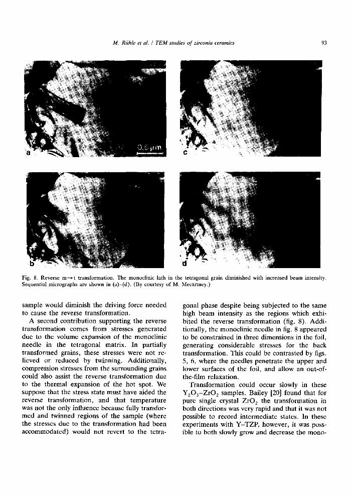
M. Rfihle et al. / T E M studies o f zirconia ceramics 93
Fig. 8. Reverse m---~ t transformation. The monoclinic lath in the tetragonal grain diminished with increased beam intensity. Sequential micrographs are shown in (a)-(d). (By courtesy of M. Mecartney.)
sample would diminish the driving force needed to cause the reverse transformation.
A second contribution supporting the reverse transformation comes from stresses generated due to the volume expansion of the monoclinic needle in the tetragonal matrix. In partially transformed grains, these stresses were not re- lieved or reduced by twinning. Additionally, compression stresses from the surrounding grains could also assist the reverse transformation due to the thermal expansion of the hot spot. We suppose that the stress state must have aided the reverse transformation, and that temperature was not the only influence because fully transfor- med and twinned regions of the sample (where the stresses due to the transformation had been accommodated) would not revert to the tetra-
gonal phase despite being subjected to the same high beam intensity as the regions which exhi- bited the reverse transformation (fig. 8). Addi- tionally, the monoclinic needle in fig. 8 appeared to be constrained in three dimensions in the foil, generating considerable stresses for the back transformation. This could be contrasted by figs. 5, 6, where the needles penetrate the upper and lower surfaces of the foil, and allow an out-of- the-film relaxation.
Transformation could occur slowly in these Y203-ZrO2 samples. Bailey [20] found that for pure single crystal ZrO 2 the transformation in both directions was very rapid and that it was not possible to record intermediate states. In these experiments with Y-TZP, however, it was poss- ible to both slowly grow and decrease the mono-

94 M. Riihle et al. / T E M studies o f zirconia ceramics
clinic areas by careful adjustment of the beam intensity. This is reminiscent of thermoelastic martensite, in which the growth of martensitic plates is halted when the free energy to drive the reaction is counterbalanced by strain energy pro- duced in the matrix phase [21]. Even if the sample is at a temperature where the monoclinic phase should be chemically stable, stresses in the system can counteract this tendency and cause the tetragonal phase to reappear.
Defects such as oxygen vacancies generated by the presence of substitutional y3+ could also play a role in decreasing the chemical driving force and also hindering the progression of the interface. There may be a dislocation present at the leading edge of the monoclinic lath in fig. 8. A moving dislocation at the transforming inter- face would be slowed by defects or defect clus- ters, these impeding both the waxing and waning of the monoclinic phase.
4. High resolution TEM studies of m-ZrO 2
The t---~m phase transformation involves a shape change of the unit cell. The introduction of twins reduces the strain energy of the total system. Therefore, all martensitic phase transfor- mations result in a highly twinned martensite structure [4]. The twin structure and internal strains in m-ZrO 2 were studied by TEM, particu- larly by HREM. For these studies, pure skull- melted m-ZrO 2 was used. The/-ZrO 2 transforms to m-ZrO 2 at the martensite temperature of M s - 900°C. Thick lamellae (fig. 9, 100 to 300 nm thick are observed.
Frequently, triangularly shaped regions can be observed at the end of such twins (fig. 9). The triangularly shaped regions contain also twins and additional smaller triangles of orthorhombic ZrO 2 [22] which may be (formed) during TEM specimen preparation. An example of a twinned triangular region is shown in fig. 10a. The crys- tallographic orientation relationships are repre- sented in fig. 10b.
The two-dimensional defects in fig. 10a are also (100) twins. The measured width of the lamella is 0.25 of its length. Either lattice bend-
ing (due to elastic strains) or microcracking oc- curs in the regions of intersection of the small twin with the surrounding m-ZrO 2 plates. Small triangles are present within the regions of com- pression. The latter observations may corre- spond to "domains of closure" [23]. Strained regions close to the terminating twins can be mapped by HREM for platelets of m-ZrO 2 with different thicknesses. The difference between the unstrained and the strained lattice results in a displacement field. The corresponding strains can be obtained by taking the appropriate de- rivatives. Maximum strains are expected for platelets with large thicknesses. A micrograph taken with higher magnification and resolution is shown in fig. 11a while fig. 11b represents a drawing of the crystallographic orientation rela- tionships. Fig. 11a shows a micrograph of a corner of a twin boundary at a higher magnifica- tion. Bending of the lattice planes (resolved by HREM) can readily be observed in fig. 11a if the monoclinic matrix is adjacent to the twin planes. In this example, the stress is not large enough to form a crack. The displacement field at the interface between the twin lamellae and the
b
0 0 ~ (001) ~ (100) ~ 1~ (100)~
r ..~ lOO Fig. 9. (a) Skull-melt m - Z r O 2 TEM micrograph of low magnification. (b) Schematical drawing of twinned structures. The interface between a large twin and a triangle is studied.

M. Riihle et al. / TEM studies of zirconia ceramics 95
(ooi)
(100) (100)
100/
Fig. 10. (a) Pure monoclinic zirconia contains many twin structures. At some places, triangles are observed which are twinned on a finer scale. Microcracking can be observed where the twins strain the interface. (b) Crystallographic orientation of the twin lamellae. The coarse-dotted triangles probably have orthorhombic symmetry.
monoclinic matrix accommodates to the 6% mis- fit between the patterns. Large bending can be observed caused by the strong atomic bonding at a coherent or partially coherent interface.
A coordinate system (x, y) is introduced as shown in fig. l lb . The displacement fields u(x, y) and v(x, y) can be measured in fig. l la . A direct evaluation from HREM images is poss- ible and no change of the HREM image is expected due to lens aberrations of the TEM, since the lattice parameters of m-ZrO 2 are large (--5 A_) compared to the point-to-point resolu-
compressive- tensile -stress compressive- - tw in width-" I I
1•001 b
Fig. 11. (a) Micrograph of a corner of a twin boundary at a higher magnification. The lattice planes are bent due to the stress caused by the twin. In this example, the stress is not high enough to form a crack. (b) Characterization and ex- perimental parameters on the tensile stress side of a twin boundary. The assumed stress field is shown above. The position of the coordinate system (x, y) is noted in (b).
tion of the used TEM (2.3/~). The displacement fields u(x, y) and v(x, y) were evaluated for various regions near a terminating twin and the results for u(x, y) and for one twin configuration are depicted in fig. 12. Experimentally measured displacements are smoothed before derivatives are formed. The very thin foil used for HREM studies represents, in a very good approxima- tion, a plane stress configuration. The strain components can be evaluated by

96 M. Riihle et al. / T E M studies of zirconia ceramics
Displacement f i e l d U [ x , y )
A"
0
i Fig. 12. Three-dimensional representation of the displace- ment field u(x, y). The undisturbed mesh in the x -y plane corresponds to the m-ZrO 2 lattice. The units of u(x, y) correspond to 0.1 nm.
Ou Ov = i ( Ou Ov ) EX= ~ x ' E y = ~ y ' '~xy 2\ Oy + ~x "
The experimental ly evaluated components of the strain tensor are shown in figs. 13a-c. A comparison with the selected geometry shows that, as expected, the highest tensile strains e x are noticed close to the twin plane (x = 0, y = 0).
The results of the displacement field for u(x, 0) for twin lamellae of 4 different lamellae thicknesses are shown in fig. 14. Differentiat ion in x-direction leads to the strain e x. Maximum tensile strain approaches e z ~ 0.14 and the max- imum shear component Exy is also in the same order of magnitude. The results support the assumption that very high local stresses are re- quired for debonding the interface.
The stresses can be evaluated under the as- sumption that m - Z r O 2 is an isotropic material:
E [~ + ~%], o ' ~ - 1 _ ~
with E = 200 GPa and v = 0.19 one obtains
m a x o- x = 29 G P a .
The assumption of elastic isotropy is clearly inappropriate . Recent experiments by Chan et
30
300.
"l t a 5
jD
3o-
.I
b
/
/1° 1 C
/J
Fig. 13. Three-dimensional representation of the three strain components of the strain tensor for plane stress configura- tion. The strain fluctuations are partially caused by errors due to the noise on the TEM micrograph. (a) e/-component, (b) ey-component, (c) exy-component.

M. Riihle et al. / T E M studies o f zirconia ceramics 97
4rn I u (x, y=O)
×
- 0.3 4 ', ½(h.b21 G 0 I= 8 (12
o.1
o
~ %x o 107 Into] • ,._ n,,, ~ , , a 7 3
k ~ ~ °',,.o~ " o 5 1
x"-x. .x ..,x a
0 Z5 5.0 75 10.0 × Into]
Fig. 14. The measured displacement field u(x, 0) as a func- tion of the distance from the interface. The symbols corre- spond to lamellae of different thicknesses.
al. [24] will result in the elastic constants of m-ZrO 2. If these constants are known, actual stresses existing in the twin-near regions can be evaluated and compared to model calculations by finite element techniques.
The observations demonstrate, however, that m-ZrO: bonds across twins can withstand large local strains. HREM studies allowed a quantita- tive determination of local strains. Rather large values of strains were observed. Microcracking requires breaking of ionic bonds. Dislocations or disclinations present near the mid-twin plane reduce the maximum internal stresses observed at interfaces.
The experimental results can be compared to a theoretical model evaluated by Fu et al. [25] who analysed the stress distribution at the ends of the twins for an m-ZrO 2 particle embedded in AI20 3 and composed of 2, 4 or 6 twins. Using linear elasticity, they found that for very small micro- cracks the stress intensity factor dropped below the critical stress intensity factor for crack growth which had been estimated from ex- perimentally determined crack lengths. They thus concluded that spontaneous initiation of very small microcracks cannot be explained by using linear elasticity and they suggested that microcrack nucleation requires the presence of lattice defects (e.g. dislocations), or involves nonlinear bond displacement in response to the
very high stress concentrations. The experimen- tal observations presented in this paper support the latter model (cf. fig. 11). With these results, data assuming the nonlinear theory have to be reevaluated.
5. Summary and conclusions
Transmission electron microscopy, analytical electron microscopy and high resolution electron microscopy can be used for studies of phase stability and phase transformations in ceramics. The techniques were applied to ZrO 2. An analysis of images as well as diffraction patterns of c-ZrO 2 and t-ZrO 2 results in an understanding of the characteristic parameters of defects in these materials. The nucleation of a t---> m trans- formation can be studied although not yet to the atomistic level. The nucleation can be triggered in metastable t-ZrO 2 by radial shear stresses existing close to a high intensity electron beam. Shear produced by the untwinned martensite lath opposes shear due to beam heating. An m-ZrO 2 lath within a t-ZrO 2 matrix can be re- transformed to t-ZrO 2 if m-ZrO 2 is irradiated by a high intensity electron beam and heated to very high temperatures, due to the compressive stresses within this region. High resolution studies allow a quantitative evaluation of strain components existing near twins. The atomistic structure of twins can also be determined.
Acknowledgements
The authors acknowledge the experimental as- sistance of Mrs. D. Waidelich (Stuttgart). We also would like to thank Mrs. D. Jilg (Stuttgart) for the lingustic revision and the effective typing of the paper. The authors thank the Air Force Office of Scientific Research for financial support under contract No. AFOSR-87-0291.
References
[1] P.B. Hirsch, A. Howie, R.B. Nicholson, D.W. Pashley and M.J. Whelan, Electron Microscopy of Thin Crystals (Krieger, Huntington, NY, 1977).

98 M. Riihle et al. / T E M studies o f zirconia ceramics
[2] M. Riihle and M. Wilkens, in: Physical Metallurgy, vol. I, R.W. Cahn and P. Haasen, eds. (Elsevier, Amster- dam, 1983).
[3] N. Claussen, M. Riihle and A.H. Heuer, eds., Science and Technology of Zirconia II, Advances in Ceramics, vol. 12 (The American Ceramic Society, Columbus, OH, 1984).
[4] A.G. Khachaturyan, Theory of Structural Transforma- tion in Solids (Wiley, New York, 1983).
[5] V. Lanteri, T.E. Mitchell and A.H. Heuer, J. Am. Ceram. Soc. 69 (1986) 564.
[6] M. Morinaga, J.B. Cohen and J. Faber Jr., Acta Cryst. A 35 (1979) 789; A36 (1980) 520.
[7] A.H. Heuer, M. Riihle and D. Waidelich, Inst. Phys. Conf. Ser. 68 (1983) 389.
[8] F. Hfiussermann, K.-H. Katerbau, M. Rfihle and M. Wilkens, J. Microscopy 98 (1973) 135.
[9] T. Mura, Micromechanics of Defects (Martinus Nijhoff, Leiden, 1976).
[10] C.F. Grain, J. Am. Ceram. Soc. 50 (1967) 288. [11] M. Wilkens and R. Rapps, Phys. Stat. Sol. (a) 44 (1977)
173. [12] M.A. O'Keefe, in: Electron Optical Systems, J. Ohari,
ed. (SEM, AMF O'Hara, 1981), p. 209.
[13] L.T. Ma, M.L. Mecartney and M. Riihle, unpublished results.
[14] A.H. Heuer and M. Riihle, Acta Metall. 33 (1985) 2101.
[15] G.B. Olson and M. Cohen, Ann. Rev. Mater. Sci. 11 (1981) 1.
[16] M. R/ihle, N. Claussen and A.H. Heuer, in ref. [3], p. 352.
[17] H. Schubert, J. Am. Ceram. Soc. 69 (1986) 270. [18] L. Reimer, Transmission Electron Microscopy (Spring-
er, Berlin, Heidelberg, New York, Tokyo) 1984. [19] M. Rfihle, L.T. Ma, M.L. Mecartney and A.G. Evans,
unpublished results. [20] J.E. Bailey, Proc. Roy. Soc. (London) A 279 (1964)
395. [21] N.N. Thadhani and M.A. Meyers, Progr. Mat. Sci. 30
(1986). [22] S.-K. Chan, unpublished results. [23] E. Bischoff and M. Riihle, J. Am. Ceram. Soc. 66
(1983) 123. [24] S.-K. Chan, personal communication. [25] K. Fu, A.G. Evans and W.M. Kriven, J. Am. Ceram.
Soc. 67 (1984) 626.
Copyright © 2022 FDOKUMEN