Task modulation of brain activity related to familiar and unfamiliar face processing: an ERP study
14
Task modulation of brain activity related to familiar and unfamiliar face processing: an ERP study B. Rossion a, b, * , S. Campanella a , C.M. Gomez d , A. Delinte b , D. Debatisse b , L. Liard a , S. Dubois a , R. Bruyer a , M. Crommelinck b , J.-M. Guerit c a Unite ´ de Neuropsychologie Cognitive (NECO), UCL, Louvain-la-Neuve, Belgium b Laboratoire de Neurophysiologie, UCL, Brussels, Belgium c Unite ´ d’Explorations Electrophysiologique du Syste `me Nerveux, Cliniques Universitaires St. Luc, St. Luc, Belgium d Laboratorio de Psicobiologia, Dpto. de Psicologia ` Experimental, Universidad de Sevilla, Seville, Spain Accepted 14 September 1998 Abstract In order to investigate stimulus-related and task-related electrophysiological activity relevant for face processing, event-related potentials (ERPs) from 58 electrodes at standard EEG sites were recorded while subjects performed a simple visual discrimination (control) task, in addition to various face processing tasks: recognition of previously learned faces and gender decision on familiar and unfamiliar faces. Three electrophysiological components or dipolar complex were recorded in all subjects: an occipital early component (P1, around 110 ms); a vertex positive potential (VPP; around 158 ms) which appeared to be specific to faces; and a negative central component, N2 (around 230 ms). Parametric analysis and source localization were applied to these components by means of a single-subject analysis methodology. No effect of familiarity was observed on any of these early component. While the VPP appears to be independent of the kind of processing performed, face task modulations of the early P1 and the N2 were observed, with a higher amplitude for the recognition than for the gender discrimination task. An attentional modulation of early visual areas is proposed for the first effect (P1 modulation), while the N2 seems to be related to general visual memory processing. This study strongly suggests that the VPP reflects an early visual stage of face processing in the fusiform gyrus that is strictly stimulus-related and independent of familiarity. It also shows that source localization algorithms may give reliable solutions on single subject averages for early visual components despite high inter-subject variability of the surface characteristics of ERPs. q 1999 Elsevier Science Ireland Ltd. All rights reserved. Keywords: Face processing; Event-related potentials; Source location; Vertex positivity; Single subject analysis 1. Introduction The ability to recognize faces plays an important role in the adaptation of individuals to social contexts. The multi-stage cognitive model of Bruce and Young (1986) describes the different operations involved in face processing. This model is largely based on neuropsycholo- gical data (Warrington and James, 1967; Malone et al., 1982; Etcoff, 1984; Campbell et al., 1986), but is also based on analyses of the failures of face processing in every- day life (Young et al., 1985) or in mental chronometry studies (Bruce, 1986; Bruce et al., 1987). According to this model, the first stage specific to face processing is a structural encoding component aimed at providing an invariant (i.e. independent of distance, orien- tation and expression) face representation to several higher-level functional components. Then, two parallel pathways are conceived as underlying face processing. A known face representation will activate a face recogni- tion unit (FRU), which is a kind of template or long-term store of known face representations, each recognition unit corresponding to a single known face. These units are the key component for face recognition. Semantic information and (eventually) the name of the person can then be accessed through the FRU. Thus, structural encoding of faces, activation of face recognition units, retrieval of biographical information and name recall, unfold in a sequential way (Bruce and Young, 1986; Bruyer, 1990; Bre ´dart and Bruyer, 1994). On the contrary, operations leading to gender discrimination or emotional categoriza- Clinical Neurophysiology 110 (1999) 449–462 CLINPH 97813 1388-2457/99/$ - see front matter q 1999 Elsevier Science Ireland Ltd. All rights reserved. PII: S1388-2457(98)00037-6 * Corresponding author. Universite ´ Catholique de Louvain, Faculte ´ de psychologie et des sciences de l’e ´ducation PSP/EXPE/NECO, Place du Cardinal Mercier, 10 1348 Louvain-la-Neuve, Belgium. Tel.: 1 32-10- 473-831; fax: 1 32-10-473-774. E-mail address: [email protected] (B. Rossion)
-
Upload
independent -
Category
Documents
-
view
1 -
download
0
Transcript of Task modulation of brain activity related to familiar and unfamiliar face processing: an ERP study

Task modulation of brain activity related to familiar and unfamiliar faceprocessing: an ERP study
B. Rossiona, b,*, S. Campanellaa, C.M. Gomezd, A. Delinteb, D. Debatisseb, L. Liarda, S. Duboisa,R. Bruyera, M. Crommelinckb, J.-M. Gueritc
aUnite de Neuropsychologie Cognitive (NECO), UCL, Louvain-la-Neuve, BelgiumbLaboratoire de Neurophysiologie, UCL, Brussels, Belgium
cUnite d'Explorations Electrophysiologique du SysteÁme Nerveux, Cliniques Universitaires St. Luc, St. Luc, BelgiumdLaboratorio de Psicobiologia, Dpto. de PsicologiaÁ Experimental, Universidad de Sevilla, Seville, Spain
Accepted 14 September 1998
Abstract
In order to investigate stimulus-related and task-related electrophysiological activity relevant for face processing, event-related potentials
(ERPs) from 58 electrodes at standard EEG sites were recorded while subjects performed a simple visual discrimination (control) task, in
addition to various face processing tasks: recognition of previously learned faces and gender decision on familiar and unfamiliar faces. Three
electrophysiological components or dipolar complex were recorded in all subjects: an occipital early component (P1, around 110 ms); a
vertex positive potential (VPP; around 158 ms) which appeared to be speci®c to faces; and a negative central component, N2 (around 230
ms). Parametric analysis and source localization were applied to these components by means of a single-subject analysis methodology. No
effect of familiarity was observed on any of these early component. While the VPP appears to be independent of the kind of processing
performed, face task modulations of the early P1 and the N2 were observed, with a higher amplitude for the recognition than for the gender
discrimination task. An attentional modulation of early visual areas is proposed for the ®rst effect (P1 modulation), while the N2 seems to be
related to general visual memory processing. This study strongly suggests that the VPP re¯ects an early visual stage of face processing in the
fusiform gyrus that is strictly stimulus-related and independent of familiarity. It also shows that source localization algorithms may give
reliable solutions on single subject averages for early visual components despite high inter-subject variability of the surface characteristics of
ERPs. q 1999 Elsevier Science Ireland Ltd. All rights reserved.
Keywords: Face processing; Event-related potentials; Source location; Vertex positivity; Single subject analysis
1. Introduction
The ability to recognize faces plays an important role in
the adaptation of individuals to social contexts.
The multi-stage cognitive model of Bruce and Young
(1986) describes the different operations involved in face
processing. This model is largely based on neuropsycholo-
gical data (Warrington and James, 1967; Malone et al.,
1982; Etcoff, 1984; Campbell et al., 1986), but is also
based on analyses of the failures of face processing in every-
day life (Young et al., 1985) or in mental chronometry
studies (Bruce, 1986; Bruce et al., 1987).
According to this model, the ®rst stage speci®c to face
processing is a structural encoding component aimed at
providing an invariant (i.e. independent of distance, orien-
tation and expression) face representation to several
higher-level functional components. Then, two parallel
pathways are conceived as underlying face processing.
A known face representation will activate a face recogni-
tion unit (FRU), which is a kind of template or long-term
store of known face representations, each recognition unit
corresponding to a single known face. These units are the
key component for face recognition. Semantic information
and (eventually) the name of the person can then be
accessed through the FRU. Thus, structural encoding of
faces, activation of face recognition units, retrieval of
biographical information and name recall, unfold in a
sequential way (Bruce and Young, 1986; Bruyer, 1990;
BreÂdart and Bruyer, 1994). On the contrary, operations
leading to gender discrimination or emotional categoriza-
Clinical Neurophysiology 110 (1999) 449±462
CLINPH 978131388-2457/99/$ - see front matter q 1999 Elsevier Science Ireland Ltd. All rights reserved.
PII: S1388-2457(98)00037-6
* Corresponding author. Universite Catholique de Louvain, Faculte de
psychologie et des sciences de l'eÂducation PSP/EXPE/NECO, Place du
Cardinal Mercier, 10 1348 Louvain-la-Neuve, Belgium. Tel.: 1 32-10-
473-831; fax: 1 32-10-473-774.
E-mail address: [email protected] (B. Rossion)

tion can occur on every face (known or unknown) from
the invariant or non-invariant description and would
proceed in parallel (Bruce et al., 1987) with the recogni-
tion process.
Many studies have used ERPs to analyze these temporal
aspects related to face processing (for reviews see Jeffreys,
1996; George, 1997).
Face presentation produces a sequence of characteristic
ERPs (Jeffreys, 1989; BoÈtzel et al., 1995). The most char-
acteristic ERP component is a vertex positive potential
(VPP), which has a larger amplitude and shorter latency
than the equivalent potentials evoked by other visual stimuli
(BoÈtzel and GruÈsser, 1989; Jeffreys, 1989). The peak latency
of this VPP is in the 150±200 ms range with relatively high
inter-subject variability ( ^ 40 ms). Although the VPP is
largest over mid-line parieto-central areas, its coronal scalp
distribution (Jeffreys, 1989) and dipole source localization
(BoÈtzel et al., 1995) are consistent with bilateral sites origi-
nating from areas of the temporal cortex. Its functional
correspondence has been related by Jeffreys (1996) to a
low-level stage of face processing which may form part of
the `structural encoding' component of Bruce and Young's
model (1986), while others (BoÈtzel et al., 1995) have argued
that the VPP may be involved in face memory processes.
The VPP has also been considered by Jeffreys (1996) as
re¯ecting on the scalp surface the activity of face-responsive
cells found in the temporal cortex of monkeys (Perrett et al.,
1985; Desimone, 1991) and sheep (Kendrick and Baldwin,
1987).
Using intracranial electrophysiological recordings in
humans, Allison et al. (1994) also found a face negative
potential (latency 200 ms) in regions of the fusiform
gyrus, which they proposed as being the negative counter-
part of the VPP.
The other electrical activities which seem to be related to
face processing have been barely described by more than
one study. While some authors (Jeffreys, 1989; George et
al., 1996) consider the face negative potential recorded by
the temporal electrodes during the same epoch as the VPP as
its negative counterpart, BoÈtzel et al. (1995) considered
these two peaks as re¯ecting the activities of two different
generators. Allison et al. (1994) have also described an
intracranial positive potential (peak latency between 250
and 350 ms) and a late negative potential (300±500 ms).
These potentials were speci®cally related to face perception
but the passive stimulation paradigm used prevented any
interpretation of these potentials in terms of functional
correlates.
B. Rossion et al. / Clinical Neurophysiology 110 (1999) 449±462450
Fig. 1. Examples of stimuli used in the experiment. Above: stimuli used in the control (dot discrimination) task. The visual patterns had to be classi®ed
according to the number of black dots (left: two; right: one) superimposed on the original images (taken from Haxby et al., 1994). Below: examples of
photographs of male (left) and female (right) faces used in the face processing tasks (gender discrimination and individual recognition).
Generally, these studies use passive1 presentation of
unknown stimuli (faces or other visual objects) and rely
on the hypothesis that response speci®cities of visual corti-
cal neurons are basically stimulus-related (Jeffreys, 1996).
However, several positron emission tomography (PET)
studies showed task-related (and stimulus independent) acti-
vations in human normal visual cortex (Corbetta et al.,
1991; Dupont et al., 1993; Haxby et al., 1994; Vanden-
berghe et al., 1996). For instance, Haxby et al. (1994)
found no difference of activation for a visuo-spatial locali-
zation task whether it was performed on faces or meaning-
less patterns.
We considered the following objectives in the present
ERP study:
1. A de®nition of ERPs devoted to face processing by
means of active tasks. These ERPs for faces were
compared with those elicited by a control task made on
a visual pattern of the same complexity as faces.
2. An investigation of whether the characteristics of the
face ERPs, especially the VPP, can be modulated by
the face task performed (i.e. whether they are not only
stimulus-related but also task-related).
3. To explore the in¯uence of visual familiarity on face
processing. Other ERP studies used familiar and unfami-
liar faces (Barrett et al., 1988; GruÈsser et al., 1991).
However, these studies have mainly investigated priming
effects, by means of delayed tasks. They also used
famous faces and semantic tasks. In our study, we wanted
to avoid any `semantic contamination' of ERPs, by using
totally unknown faces which were learned during train-
ing sessions.
We hypothesized that if the VPP re¯ects memory
processes (BoÈtzel et al., 1995), then its latency or ampli-
tude should vary when known or unknown faces are
presented, and/or between a visual categorization task
and a recognition task. On the contrary, no in¯uence of
these factors would reinforce the proposal of a strictly
stimulus-related visual stage (Jeffreys, 1989, 1996) asso-
ciated with this component.
4. Finally, we investigated the intracranial sources of the
recorded potentials by means of a dipole source localiza-
tion program (EMSE, BESA algorithm; Scherg, 1990).
2. Materials and methods
2.1. Subjects
Thirteen right-handed male adults (Edinbrugh scale;
Old®eld, 1971), aged between 19 and 26 years old, partici-
pated. One was rejected for experimenter errors and 3 for a
very poor signal-to-noise ratio. All subjects were in good
health, had normal or corrected vision, and reported no
history of neurological impairment. All analyses were thus
computed on the data of 9 subjects.
2.2. Stimuli
Two kinds of stimuli were used. Complex visual patterns
(Fig. 1) taken from Haxby et al. (1994)2, on which one or
two black dots were added for special purposes of this
experiment. Thirty patterns, which differed only by dot posi-
tions and number, were made. Three-quarter pro®le photo-
graphs of faces taken from medical students (Fig. 1). The
faces were all Caucasian males or females, without glasses.
All male faces were well-shaved and the few cues (includ-
ing clothing) that were still visible for some faces were
masked using the Corel Draw graphic package. These
photographs were scanned so that they could be displayed
on the monitor using the commercial visual stimulator
(STIM, Neuroscan).
2.3. Procedure
The experiment consisted of a training phase followed by
an experimental phase, described in chronological order.
2.3.1. Training phase
This phase consisted ®rst in introducing the subject to
the experiment, obtaining his formal consent and testing
his handedness and vision. The training phase lasted for the
3 consecutive days preceding the EEG experiment.
Subjects were ®rst shown a short (14 min) video ®lm
sequence of young persons in action (pretending to write
a letter). The recordings focalized on the face. Twenty
faces were presented (40 s each), one after the other, to
each subject. Immediately after the presentation, a testing
procedure took place: subjects were given 40 photo-
graphs of faces (3/4 pro®les), half of them corresponding
to people presented in the videotape recordings (the other
half were new). Subjects were required to categorize the
photographs into two samples: the faces that appeared in
the videotape sequence and the distracters. Whatever the
subject's performance3, the videotape was presented once
again. The next day, subjects were ®rst tested with the 40
photographs. Either they obtained a maximum score (20/
20) and the training phase was completed, or another
videotape presentation was made and a last test took
place the third day. As it turned out that two subjects did
not reach the criteria of 20/20 at the end of the training
B. Rossion et al. / Clinical Neurophysiology 110 (1999) 449±462 451
1 The term `passive' refers here to the fact that subjects did not have to
make a discrimination based on the displayed stimulus.
2 With permission of the ®rst author.3 His score (number of faces recognized, out of 20) was given to the
subject but no information concerning what kind of errors were made, in
order to prevent any learning from the test.

phase4, they were shown the faces they did not recognize,
together with the corresponding images on the videos5.
2.3.2. Experimental phase
The experimental phase took place the day following the
training phase and consisted of measuring EEG and beha-
vioral responses in 4 conditions.
These 4 conditions were:
1. A visual discrimination control task (CONT), which
consisted of a visual detection of dots on the visual
pattern (Fig. 1). The subject's task was to press the left
mouse button if there was only one dot on the pattern
(half of the stimuli) and the other button if two dots were
detected6.
2. A gender discrimination task on unknown faces (GUF).
The subjects had to categorize unknown (not shown
during the learning phase) faces as male or female by
pressing one of the two buttons (male � left). The whole
set of stimuli was equally divided in the two categories.
3. A gender discrimination task on known faces (GKF). The
instruction, the task and the proportion of stimuli were
exactly the same as for condition 2, but the stimuli were
photographs of familiar (previously learned on video)
people.
4. A face recognition task (REC). The task consisted of
categorizing the faces as previously seen on the video-
tapes or new and responded by pressing one of the two
buttons (left � learnt faces). Two-thirds of the stimuli
were known (familiar) faces7. The new faces were differ-
ent from the ones used in condition GUF. As for all other
face conditions, half of the faces were three-quarter left
pro®les and the other half three-quarter right pro®les.
The stimuli were displayed on a high resolution monitor,
in darkness. All images were presented for 1 s with an inter-
stimulus interval of 2 s. In the 4 conditions, the subjects
responded by pressing, with the right index ®nger and
medius, one of two buttons of the computer mouse. Subjects
sat in front of the monitor, at a viewing distance of 134 cm.
The subject's head was restrained by a chin-rest. The stimuli
were black-and-white square images, appearing in the
center of the screen (Fig. 1). Their size (48 of visual
angle) and luminance (about 7 cd/m2) were constant across
conditions.
Before starting the EEG recording, the different tasks
were brie¯y explained to the subjects, using stimuli not
employed in the experiment.
Three blocks of the 4 conditions were presented to the
subjects and the order of conditions was randomized for
each block. The order of blocks was constant during all
the experiment. As there were 60 images for each condition
in a block, a total of 720 images (12 trials) were presented to
the subjects. A rest of about 1 min was allowed between
blocks.
The whole experiment lasted about 2 h, including instal-
lation of the electrode cap and debrie®ng of the subject.
2.4. Recording and analyses
Correct latencies (interval between the onset of the stimu-
lus and the subject's key press) and the percentage of errors
were computed and analyzed (Systat 5, Macintosh). Laten-
cies which were two standard deviations above or below the
mean of each subject in each condition were discarded.
Scalp electrical activity (EEG) was recorded by 58 elec-
trodes mounted in an electrode cap. Electrode positions
included the standard 10±20 system locations and additional
intermediate positions (Fig. 2). Recordings were made with
a left earlobe reference. The EEG was ampli®ed by battery-
operated SYNAMPS ampli®ers with a gain of 30 000
through a bandpass of 0.01±100 Hz. Electrode impedances
were kept below 5 kV . EEG was continuously recorded
(rate 1000 Hz, Neuroscan) and stored on a disk for off-
line analysis. Then, epochs beginning 100 ms prior to stimu-
lus onset and continuing for 924 ms were established, and a
recalculation was made to obtain common average refer-
ence recordings. During the averaging procedure, epochs
contaminated with EOG artifacts were eliminated. Codes
synchronized to stimulus delivery were used to average
selectively epochs associated with different stimulus types.
Each stimulus was coded according to 6 parameters: (1) task
B. Rossion et al. / Clinical Neurophysiology 110 (1999) 449±462452
4 No subjects reached the criteria after the second presentation of the
videos. Two subjects only (including one whose EEG data were eventually
not taken into account due to a very low signal-to-noise ratio) did not reach
the criteria on the third day (after 3 presentations). One subject missed two
faces (18/20) and the other missed one (19/20).5 The most important thing is for the subjects to be able to perform the
task properly during the EEG recording.6 Distance between dots was always between 1 and 2 degrees of visual
angle.
Fig. 2. Electrode locations on the scalp.
(face recognition-gender discrimination); (2) familiarity
(known or unknown face); (3) order of occurrence of the
stimulus (1±2±3 or more); (4) response (left or right key);
(5) face pro®le (left or right); (6) face gender (male or
female). This coding enabled us to make several averages
and to take into account the number of repetitions of each
stimulus, which differed in amount between known and
unknown faces7. Six averages were computed for each
subject individually: FACES (all face stimuli which
appeared 4 times in each condition; mean number of
sweeps: 197; range: 170±243); CONT (all control stimuli
appearing 4 times for the whole experiment; mean: 84,
range: 62±118); GUF (all face stimuli appearing 3 times
in the GUF condition; mean: 86; range: 66±109); GKF
(all face stimuli appearing 3 times in the GKF condition;
mean: 73; range: 57±98); RECU (all unknown faces appear-
ing 3 times in the REC condition; mean: 61; range: 52±66);
RECK (all known faces appearing 3 times in the REC condi-
tion; mean: 73; range: 64±80). Only correct trials were aver-
aged. Trials associated with latencies of two SDs above or
below the mean for each subject (each condition) were not
taken into consideration. Finally, the data were ®ltered with
a low-pass ®lter (cut-off � 45 Hz) and displayed off-line in
the forms of raw data and topographical maps (Fig. 3 and
Fig. 4).
The peak amplitudes (mean over a 20 ms window around
the peak) and latencies of particular components at selected
electrodes were obtained for the different conditions for
each subject individually. This procedure was used for
peaks under 250 ms, which were easily discriminated for
all subjects in each condition. With the exception of a large
P3 component which could be observed in some subjects
and in the grand averages (Campanella et al., 1999), it
turned out to be impossible to identify reliably later compo-
nents in all or most of the subjects.
For each component identi®ed, signi®cant differences
between measures in the different conditions were tested
using paired t tests (CONT vs. FACES) and repeated-
measures analysis of variance (ANOVAs). Post-hoc
comparisons were evaluated using Tukey's test.
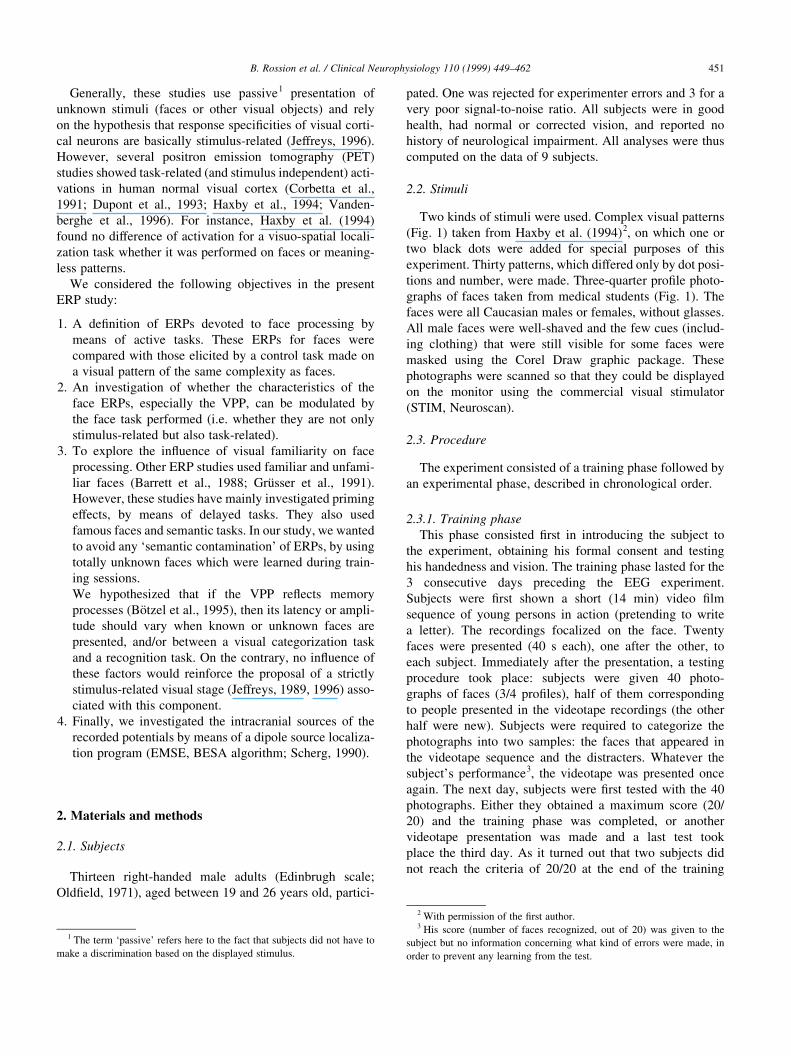
B. Rossion et al. / Clinical Neurophysiology 110 (1999) 449±462 453
Fig. 3. ERPs obtained from one subject at central (Cz), occipital (Oz) and
right temporal (T6) sites for both FACES and CONT averages. The curves
illustrate the polarity oppositions for components P1 and VPP at central
(Cz) and occipital (Oz) sites. The highest polarity opposition for the VPP is
found at temporal sites (T6). The curves also illustrate the marked ampli-
tude difference between CONT and FACE averages for the VPP, which was
observed in all subjects.
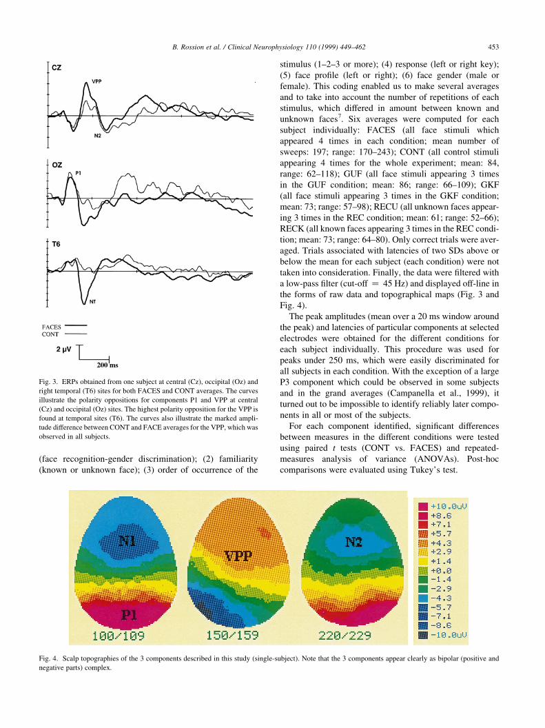
Fig. 4. Scalp topographies of the 3 components described in this study (single-subject). Note that the 3 components appear clearly as bipolar (positive and
negative parts) complex.

The analysis of intracranial dipoles was performed with
the EMSE software (BESA algorithm, Neuroscan), which
determines the position and orientations of intracranial
dipoles and their time-varying strength. This program calcu-
lates dipoles in a 100 mm diameter spherical head model
and takes into account the different conductances of the
cerebrospinal ¯uid, skull and scalp (Scherg, 1990). The X
axis of the coordinate system is a line joining electrodes Fpz
and Oz, the Y axis passes through T3 and T4, and the vertical
Z axis passes through the center of the head and electrode
Cz. The most important requirement for a dipole analysis
using BESA is that the number of parameters to be deter-
mined (6; 3 for location and 3 for dipole moment) does not
exceed the number of constraints (number of electrodes, 58
in this study).
For each component described in this study, we intro-
duced dipole pairs (10 ms around the peak) symmetrically
located along the sagittal plane in each subject for FACES
averages. When the numerical solution (percentage of
variance in the voltage surface observed) or the physiologi-
cal validity criteria was unsatisfactory, another pair of
dipoles was introduced in the system.
3. Results
3.1. Behavioral data
Correct latencies and error rates are presented in Table 1.
Latencies were computed on the trials for which the corre-
sponding ERPs had been averaged. For error rates, we
considered the trials which ®tted the criteria of repetition
(3 or 4, depending on the averages).
First, the latencies for FACES and CONT averages were
compared using the paired t test which was statistically
signi®cant (t8 � 5:07; P � 0:001): latencies were shorter
when faces were presented compared to control stimuli.
Longer reaction times were also observed in the CONT
task when only one dot was present on the pattern (917
ms; 847 ms for two dots; paired t test: P � 0:05). Then,
the 4 face averages were subjected to a two-way repeated
measure ANOVA. The two factors were familiarity (known,
unknown) and task (gender, recognition). There was a main
effect of task (F�1; 8� � 11:81; P , 0:009), the gender
discrimination task being performed faster than the recogni-
tion task; and a main effect of familiarity (F�1; 8� � 22:27;
P , 0:002, slower responses for unknown than for known
stimuli). Interestingly, the interaction was also highly
signi®cant (F�1; 8� � 21:12; P � 0:002), mainly due to an
increase of latencies in the recognition task on unknown
stimuli (RECU). This last result was con®rmed by a post
hoc t test (P , 0:001). There was also a signi®cant differ-
ence between the two recognition tasks (P , 0:001). All
other contrasts were not signi®cant.
A similar analysis on error rates was carried out. The only
difference which was statistically signi®cant was between
FACES and CONT averages (paired t test; t8 � 8:76;
P � 0:001). The 2 £ 2 ANOVA on face conditions showed
no effect of any of these two factors (task: F�1; 8� � 1:15;
P � 0:305; familiarity: F�3; 8� � 1:11; P � 0:313), and no
interaction (F�3; 8� � 0:964; P � 0:346).
3.2. Event-related potentials
3.2.1. Description of components and statistical evaluation
Following the stimulation, 3 clear components were
observed for all subjects in all conditions (except for a
few exceptions, see below). These electrophysiological
events were named according to their order of occurrence
and polarity (Figs. 3 and 4; Table 2) as P1 (occipital), VPP
(central) and N2 (central).
3.2.1.1. P1 The ®rst measurable electrophysiological
event was the P1 which culminated (Oz) at 108 ms
(CONT) or 110 ms (FACES) with a higher inter-subject
variability for the CONT (SD 15 ms) condition than for
the FACES average (SD 8 ms) (Table 2).
Topographically, the P1 was characterized in all (but one)
subjects by a large positivity over all the posterior
(occipital) electrodes with polarity reversal at central and
frontal sites (Figs. 3 and 4). There was not any signi®cant
B. Rossion et al. / Clinical Neurophysiology 110 (1999) 449±462454
Table 1
Correct response latencies in milliseconds and percentage of hits in the
different conditions of the experimenta
CONT FACES GUF GKF RECU RECK
Hits (%) 71.6 97.57 97.59 98.09 96.54 97.46
Latencies (ms) 883 658 649 636 762 659
a CONT, control task; FACES, average on all face conditions; GUF,
gender task, unknown faces; GKF, gender task, known faces; RECU, recog-
nition task; unknown faces; RECK, recognition task; known faces).
Table 2
Mean latencies (ms, SD) for the 3 electrophysiological events recorded in this studya
CONT FACES GUF GKF RECU RECK
P1 (Oz) 108 (15) 110 8) 110 (9) 109 (10) 111 (9) 110 (7)
VPP (Cz) 152 (12) 158 (8) 157 (8) 160 (9) 158 (8) 158 (6)
N2 (Cz) 228 (19) 230 (17) 229 (21) 232 (17) 227 (18) 226 (16)
a CONT, control task; FACES, average on all face conditions; GUF, gender task, unknown faces; GKF, gender task, known faces; RECU, recognition task;
unknown faces; RECK, recognition task; known faces.
difference between FACES and CONT for voltage
amplitude of the P1 at Oz (FACES vs. CONT: t � 0:499;
P � 0:647), although the mean amplitude was slightly
larger for CONT average (Table 3).
An ANOVA on peak amplitudes with task (FACES-
CONT) and lateralization (O1-O2) as factors did not reveal
any signi®cant effect (task: F�1; 7� � 5:11, P � 0:058;
lateralization: F�1; 7� � 1:98, P � 0:251; interaction:
F�1; 7� � 0:51, P � 0:496), even if there was clearly a
trend towards an amplitude superiority of the P1 peak for
the CONT task.
Among face averages, the mean amplitudes of the P1
appeared higher for 7 subjects (out of 8) during the recog-
nition tasks than the gender discrimination tasks (Fig. 4).
An ANOVA with 3 factors, (1) task (gender or recogni-
tion), (2) familiarity (known or unknown) and (3) lateraliza-
tion (O1-O2) con®rmed a strong task effect
(F�1; 7� � 17:487; P � 0:004) while all other effects were
not signi®cant (familiarity: F�1; 7� � 1:319; P � 0:289;
lateralization: F�1; 7� � 1:413, P � 0:273). The interac-
tions did not reach signi®cant levels (task £ familiarity:
F�1; 7� � 0:039, P � 0:850; lateralization £ familiarity:
F�1; 7� � 4:747, P � 0:066; task £ lateralization: F(1,7) �0:322, P � 0:399; triple interaction: F�1; 7� � 1:446,
P � 0:268).
An identical analysis was performed on the negative
counterpart of the P1 at Cz at the same latencies. However,
its amplitude did not differ between tasks, familiar or unfa-
miliar faces, or left or right (C1-C2) electrodes (task
F�1; 7� � 0:819; P � 0:392; familiarity: F�1; 7� � 0:431;
P � 0:530; lateralization: F�1; 7� � 2:453, P � 0:156; task
£ familiarity: F�1; 7� � 0:020, P � 0:892; lateralization £familiarity: F�1; 7� � 1:141, P � 0:264; task £ lateraliza-
tion: F�1; 7� � 0:195, P � 0:671; triple interaction:
F�1; 7� � 1:629, P � 0:238).
3.2.1.2. VPP The next major electrophysiological event
corresponded to the so-called vertex positivity (VPP),
which was easily discriminable in all subjects for FACES
(mean latency ^ SD, 158 ^ 8 ms, Cz) and all other face
averages (Table 2; Fig. 3 and Fig. 5). A relatively high inter-
subject variability could be observed either on the time
course or amplitude of the peak, which reverses polarity
at occipital and temporal sites. This vertex positivity was
also present for the CONT averaging in 7 subjects
(152 ^ 12 ms). However, the amplitude of this peak was
clearly reduced in these subjects in CONT (0.86 mV)
versus FACES averages (3.88 mV; t6 � 6:3; P � 0:001;
see Fig. 3, Table 3).
We then performed several analyses, in order to assess
any in¯uence of the face task or the familiarity of the face to
this VPP activity. An ANOVA with task (gender, recogni-
tion) and familiarity as factors (known, unknown faces)
failed to ®nd any signi®cant difference between face condi-
tions: task: F�1; 8� � 0:218, P � 0:653; familiarity:
F�1; 8� � 0:516, P � 0:493; interaction: F�1; 8� � 0:208,
P � 0:660). An ANOVA with lateralization (C1, C2), task
and familiarity as factors was also computed. None of these
factors had a signi®cant effect on peak amplitudes of the
VPP (lateralization: F�1; 8� � 2:264; P � 0:171; task:
F�1; 8� � 0:022, P � 0:887; familiarity: F�1; 8� � 0:210,
P � 0:659; lateralization £ task: F�1; 8� � 0:064;
P � 0:807; lateralization £ familiarity: F�1; 8� � 0:349;
B. Rossion et al. / Clinical Neurophysiology 110 (1999) 449±462 455
Table 3
Mean amplitudes (mV, SD) for the 3 electrophysiological events recorded in this studya
CONT FACES GUF GKF RECU RECK
P1 (Oz) 6.94 (3.15) 6.78 (3.21) 6.55 (3.75) 6.63 (2.92) 7.17 (3.53) 6.93 (2.78)
P1 (O1) 7.39 (2.73) 6.64 (3.06) 6.34 (3.53) 6.35 (2.72) 7.04 (3.11) 7.26 (2.8)
P1 (O2) 8.70 (4.34) 7.69 (4.98) 7.20 (5.16) 7.80 (4.48) 8.56 (5.12) 7.43 (4.41)
VPP (Cz) 0.86 (2.65) 3.88 (2.50) 4.00 (2.5) 3.99 (2.41) 4.16 (3.72) 3.39 (1.74)
N2 (Cz) 2 5.74 (2.96) 2 4.73 (2.53) 2 4.47 (2.37) 2 4.97 (2.96) 2 4.95 (3.43) 2 5.63 (2.48)
a Note the larger amplitudes for the P1 component during the recognition (compared with the gender discrimination tasks), particularly on the left side (O1
electrode). CONT, control task; FACES, average on all face conditions; GUF, gender task, unknown faces; GKF, gender task, known faces; RECU, recognition
task; unknown faces; RECK, recognition task; known faces.
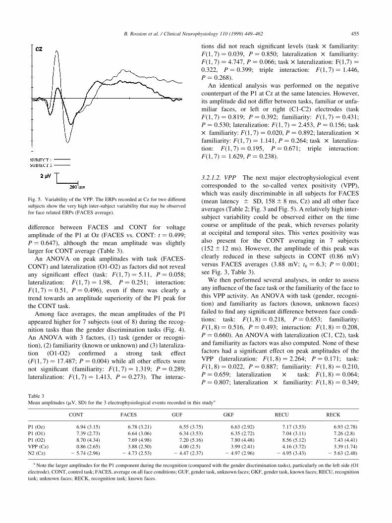
Fig. 5. Variability of the VPP. The ERPs recorded at Cz for two different
subjects show the very high inter-subject variability that may be observed
for face related ERPs (FACES average).
P � 0:571; task £ familiarity: F�1; 8� � 0:202; P � 0:665;
triple interaction: F�1; 8� � 5:117; P � 0:054). Finally, this
analysis was also extended to more lateral electrodes (C3
and C4) This latter analysis had electrodes, task, familiarity
and lateralization as factors. There was a signi®cant effect of
electrodes (F�1; 8� � 8:825, P � 0:018), due to the larger
amplitudes for central (C1-C2, closer to CZ, where the
maximum voltage amplitude is recorded) electrodes and a
B. Rossion et al. / Clinical Neurophysiology 110 (1999) 449±462456

signi®cant interaction between lateralization and electrodes
(F�1; 8� � 7:844, P � 0:023), due to a decrease in ampli-
tude for more lateralized electrodes that was larger on the
left side (C3) than on the right side (C4). To support this
right superiority, there was a general trend for a larger
amplitude on the right side, although it was not statistically
signi®cant (F�1; 8� � 4:487, P � 0:059).
At about the same latency as the VPP, there were also
corresponding temporal negativities recorded maximally at
T5 and T6 (Fig. 2) for both FACES (T5: 157 ^ 9 ms; T6:
158 ^ 9 ms) and CONT (T5: 150 ^ 13 ms; T6: 154 ^ 13
ms) averages. These merely appeared as relative negativ-
ities (decrease in positive amplitude) for 5 subjects in the
CONT average. Identical statistical tests as those for the
VPP were applied to these negativities which appeared
clearly larger for FACES than CONT (ANOVA 2 £ 2,
task: F�1; 8� � 45:811, P , 0:0001; lateralization:
F�1; 8� � 1:106, P � 0:324; interaction: F�1; 8� � 0; 930,
P � 0; 363). However, as for the VPP, there were no differ-
ences among face conditions, nor lateralization effects, as
con®rmed by an ANOVA (task: F�1; 8� � 2:908,
P � 0:127; familiarity: F�1; 8� � 1:677, P � 0:231; latera-
lization: F�1; 8� � 0:380, P � 0:555; task £ familiarity:
F�1; 8� � 0:657, P � 0:441; lateralization £ familiarity:
F�1; 8� � 0:457, P � 0:518; task £ lateralization:
F�1; 8� � 0:238, P � 0:639; triple interaction:
F�1; 8� � 2:466, P � 0:155). Two close occipito-temporal
electrodes (CB1 and CB2; Fig. 2) were also added to
analyze the NT components and a similar ANOVA (4
factors: electrodes, task, familiarity and lateralization) was
performed. There was not any signi®cant effect or trend for
this analysis.
3.2.1.3. N200 Following the VPP, there was a CZ
negativity (Oz positivity) occurring around 230 ms (Cz,
SD � 17 ms) for FACES and 229 ms for CONT
(SD � 19 ms) averages (Fig. 3). There was no amplitude
difference between the two averages (t8 � 2:464;
P � 0:269). The N200 component was also tested at CZ
(ANOVA 2, repeated measures, task and familiarity as
factors). There was a trend for the factor task to be
signi®cant (F�1; 8� � 4:699, P � 0:062). This task
modulation was found to be signi®cant with C1 and C2 in
the analysis as well as with additional electrodes (C3 and
C4, see below). For the analysis made on CZ, other effects
were not signi®cant although there was a slight trend for a
familiarity effect (F�1; 8� � 4:259, P � 0:073) which was
not supported by further analyses made on this component.
An ANOVA with task, familiarity and lateralization (C1-
C2) as factors was computed on the face averages and
showed a signi®cant effect of task (F�1; 8� � 5:833,
P � 0; 042), re¯ecting an amplitude superiority for the
recognition tasks. The triple interaction was also signi®cant
(F�1; 8� � 10:011, P � 0; 013) due to the fact that, while all
mean amplitudes were superior when familiar faces and
recognition tasks were used, the recognition task on
known faces (RECK) gave smaller amplitudes for N2 only
at the right (C2) electrode than for the same task on
unknown faces. All other effects did not reach statistical
signi®cance (familiarity: F�1; 8� � 0:199; P � 0:667; later-
alization: F�1; 8� � 4:615, P � 0:064; task £ familiarity:
F�1; 8� � 0:167, P � 0:694; lateralization £ familiarity:
F�1; 8� � 0:837, P � 0:387; task £ lateralization: F(1,8) �0:598, P � 0:461). The analysis performed with C3 and C4
electrodes and 4 factors (electrodes, task, familiarity and
lateralization) con®rmed the task effect on N2
(F�1; 8� � 5:859, P � 0:042) and there was also a trend
for the electrode factor (F�1; 8� � 4:765, P � 0:061) due
to a decrease in amplitude for more lateralized electrodes
(C3 and C4) as compared with C1 and C2.
3.3. Dipole localization
Dipole localization was carried out for each subject indi-
vidually on the 3 components (Fig. 4) identi®ed in this
study. As these results must be accepted with some caution
for the above-mentioned reasons and the obvious difference
between the spherical head model used and the real brains of
subjects, no relationships between the dipole con®gurations
which were found, and the anatomical structures probably
involved in the recorded ERPs will be made in this section
and will be reserved for Section 4.
3.3.1. P1
A clear P1 was observed in 8 subjects out of 9. The dipole
localization procedure was achieved successfully in 6
subjects (out of 8). The percentage of variance explained
was acceptable for a seventh subject (95.5) and very good
for the last one (98.6), but both gave neurophysiologically
unplausible solutions which may be due to a weaker signal-
to-noise ratio observed in the time window of the P1/N1
complex. A ®rst modelization with a single pair of dipoles
did not give a satisfactory solution; the dipoles being loca-
lized in anterior regions of the brain, a result strongly
contradictory to the probable sources of the P1 in occi-
pito-lateral regions (Clark et al., 1996; Gomez et al.,
B. Rossion et al. / Clinical Neurophysiology 110 (1999) 449±462 457
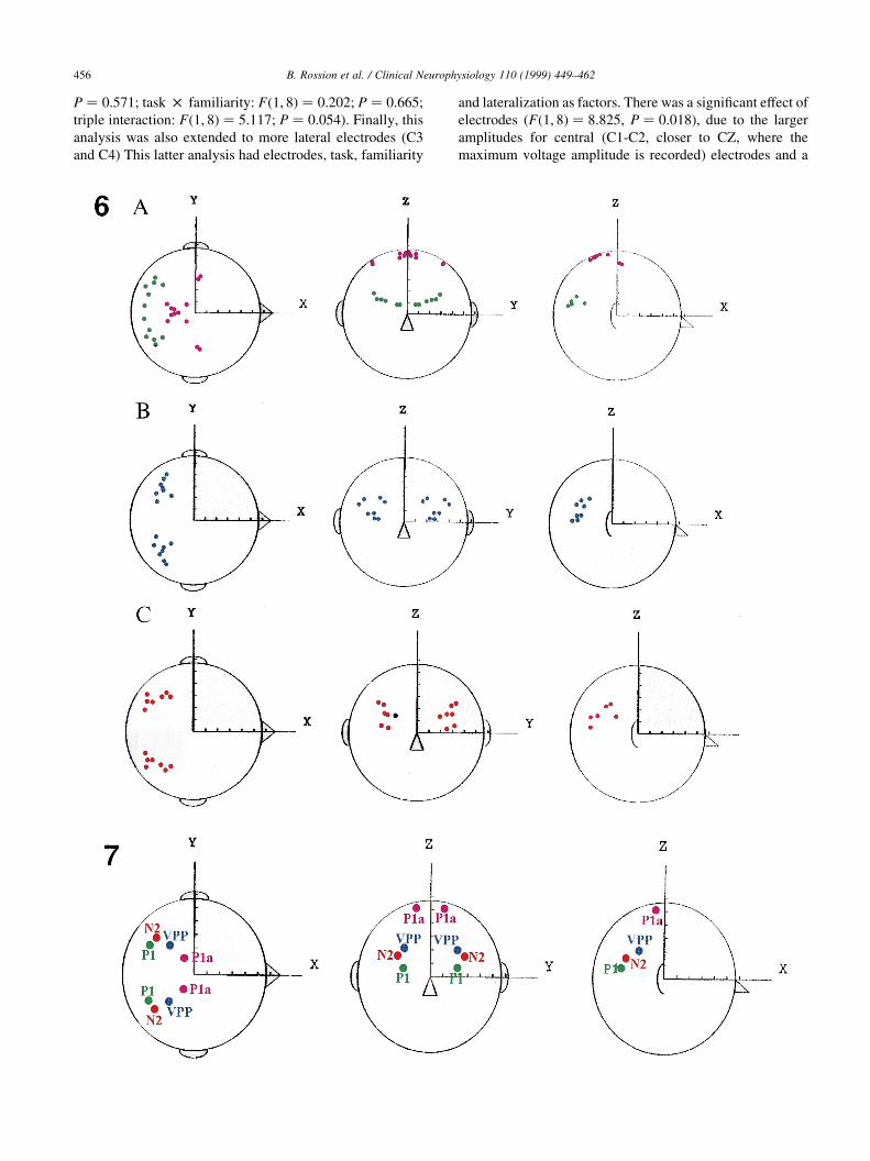
Fig. 6. Source locations (dipole models) on the spherical model for the 3 components described in this study for all subjects (FACES average). Upper row (A).
Source locations for the P1/N1 component (6 subjects, FACES average). Two dipole pairs were introduced simultaneously to get an acceptable numerical
(variance between 97.5 and 99.1%) and neurophysiological solution. Middle row (B). Dipoles models explaining possible sources of the VPP component (8
subjects; FACES average; variance between 94.6 and 99%). Lower row (C). Dipole models explaining possible sources of the N2 component (8 subjects;
FACES average; variance between 95.6 and 98.5%).
Fig. 7. Mean source locations for the 3 components described in this study. Note the close position in the spherical model of dipoles accounting for VPP and
N2 and the two dipole solution found for the P1 component.

1994). A plausible solution, which gave very high signal-to-
noise ratios (between 97.5 and 99.1%), was found when two
pairs of dipoles were introduced simultaneously. One pair
was located 67 mm posterior, 36 mm lateral and 13 mm
(mean values across subjects) above the center of the sphe-
rical head model (Figs. 6 and 7). Dipole strengths, which
were not constrained, did not differ between left and right
sides of the brain (paired t test, P � 0:57). The other pair
was located more anteriorly (14 mm posterior) and less
laterally (23 mm) and occupied the roof of the head
model (87 mm above the center of the sphere), with no
momentum differences between the left and right sides
(paired t test, P � 0:28).
3.3.2. VPP (VPP)
A plausible solution, both in terms of numerical validity
(variance between 94.6 and 99%) and physiological plausi-
bility, was found when a single dipole pair was introduced
in the time epoch of 10 ms around the VPP peak for 8
subjects8. When two pairs of dipoles were introduced, the
percentage of variance increased signi®cantly (between
94.2 and 99.3%; paired t test: 0.014). However, contrary
to what was found for the P1, the two dipole pairs were
located remarkably at virtually the same place in the sphe-
rical head model (mean distance � 1 mm antero-posterior;
6 mm laterally and 1 mm vertically). These small differ-
ences are absolutely meaningless when one considers the
spatial resolution of the ERP generator localization in
neurophysiology (Swick et al., 1994), and so the single,
economical, dipole pair solution was kept. These dipoles
were found 49 mm posterior, 48 mm lateral and 21 mm
above the center of the spherical head model (Figs. 6 and 7).
3.3.3. N200
For the N2 component, we found a satisfactory solution
with one pair of dipoles (variance between 95.6 and 98.5%)
for 7 subjects. This component presented a very low signal-
to-noise ratio for two other subjects. As for the VPP,
variance increased signi®cantly (between 95.1 and 99%;
paired t test; P � 0:019) when another pair of dipoles was
introduced in the system, but the two pairs also tended to
locate at the same place (distance: X � 12 mm; Y � 2 mm;
Z � 0 mm). The dipoles associated with the N2 component
were found slightly more posteriorly (54 mm), less laterally
(47 mm) and above (25 mm) the solution found for the VPP
component (Fig. 6). In fact, the differences between the
solutions found for both components, VPP and N2, were
very small (5 mm in the X coordinate; 2 mm in the Y coor-
dinate and 4 mm in the Z coordinate; Figs. 6 and 7).
4. Discussion
4.1. Behavioral data
First, the control task turned out to be more complex than
originally anticipated: it required a longer processing time
and generated more errors than the other tasks. Among face
conditions, the recognition task took longer than the gender
categorization task, which is common in the literature
(Sergent et al., 1994a). Processing of unknown stimuli
also took more time than known stimuli, particularly for
the recognition task (Sergent et al., 1994a). The absence
of signi®cant differences in processing times between
known (636 ms) and unknown (649 ms) stimuli for the
gender categorization task is consistent with a previous
study (Bruce, 1986) aimed at demonstrating the absence
of any role of familiarity on this task.
4.2. Event-related potentials
Three clear potentials were described in nearly all
subjects both for face and visual pattern processing. All
these components appeared as dipolar complexes and only
one of them, namely the vertex positivity (occipito-temporal
negativity) or VPP, occurring after more or less 160 ms
following stimulus onset, appeared to be face-speci®c.
Components occurring after 250 ms could not be easily
and reliably observed in all or even a small subset of indi-
vidual subjects, even if grand-average data (Campanella et
al., 1999) show a large P3 component over centro-parietal
sites. Several arguments could account for the lack of reli-
able and easily identi®able components after 250 ms. First,
averages were made for each subject separately and on rela-
tively few sweeps by condition. Signal-to-noise ratio is thus
not as strong as usually observed in classical ERPs studies.
Secondly, we used a rather conservative procedure as we did
not account for the analyses ERPs that could be observed on
grand average data, but not in less than 7 subjects out of 9.
Finally, as noted by Dehaene (1996), when ERPs are aver-
aged time-locked to stimulus onset, as in this study, the
factors related to early processing stages will not be affected
much by the variance of earlier processes. However, later
stages can be highly affected by differences in the duration
of the preceding stages, due either to experimental factors or
to trial-to-trial variability or, as in this study, by inter-
subject variability.
4.2.1. P1 and visual selective attention
The ®rst positive peak observed corresponds to the P1.
The centro-frontal negativity (Figs. 3 and 4) occurring with
a similar latency can be considered as the negative counter-
part of this dipole. A striate and extrastriate origin has been
proposed for this P1 component (Gomez et al., 1994; Heinze
et al., 1994). The voltage maps and the dipole localization
obtained in the present study are partially coherent with this
view. Indeed, the posterior lateral locations of the ®rst pair
B. Rossion et al. / Clinical Neurophysiology 110 (1999) 449±462458
8 For one subject, it turned out to be impossible to ®nd an acceptable
numerical solution which could be explained by the weak (1,84 mV) ampli-
tude of the peak and signal-to-noise ratio, as well as the lack of a reversing
polarity at posterior sites which was clear in all other subjects (Fig. 4).

of dipoles is compatible with an extrastriate location of the
P1 generators, as found in previous studies (Clark et al.,
1995; Gomez et al., 1994). However, a second pair of
dipoles which located somewhat in posterior parietal
regions had to be introduced to obtain this posterior locali-
zation of the ®rst pair. This observation suggests a parallel
activation of the dorsal stream of the brain, which is known
to be involved in spatial processing of the stimulus. Even if
faces activate mainly structures of the ventral stream, it is
clear that an accurate perceptual representation cannot be
formed if the stimulus information is not extracted by the
two pathways and later re-integrated (Robertson et al.,
1997).
A task modulation of the P1 activity was observed in the
present study, its amplitude being larger during face recog-
nition tasks than during gender discrimination tasks. At ®rst
glance, one may ®nd it surprising to observe a modulation of
a relatively early visual potential to stimuli of identical
luminosity, contrast and complexity. However, as illustrated
by the behavioral data recorded, task complexity is not kept
equivalent among (face) tasks: the recognition of pre-learnt
individual photographs took more time and probably
required more attentional resources than the gender categor-
ization task, which is a less subtle and easier discrimination.
Modulation of P1 amplitude, as found in this study, may
thus re¯ect an attentional modulatory effect on occipital
visual areas. Such attentional modulations of the P1 compo-
nent have been described in several previous ERP studies
(Clark and Hillyard, 1996; Eason et al., 1969; Rugg et al.,
1987). In agreement with these electrophysiological ®nd-
ings, several PET studies of human visual information
processing have shown modulations (increases) of blood
¯ow in medial visual regions during active discriminations
on a visual stimulus in contrast to passive viewing (Corbetta
et al., 1990, 1991, 1995; Petersen et al., 1990; Raichle et al.,
1994). Both these ERP and PET studies suggest that selec-
tive and non-selective9 attentional, top-down, mechanisms
may act to amplify the neural activity in early visual areas
(Clark and Hillyard, 1996; Shulman et al., 1997). A strong
argument in favor of a selective attentional mechanism in
the present study comes from the fact that a non-selective
increase of general arousal should normally have the same
effect on all electrophysiological components observed.
Accordingly, a task modulation of amplitude should be
observed for the VPP (taking place 50 ms or so after the
P1 component), which is clearly not the case in our study
(see below). Clark and Hillyard (1996) recently observed a
strong increase of visual P1 amplitude due to spatial selec-
tive attention mechanisms. Combining ERP and PET,
Heinze et al. (1994) also demonstrated that visual inputs
from attended locations enhanced processing in the extra-
striate cortex (posterior fusiform gyrus) during the time
course of the P1 component (80±130 ms after stimulus
onset). The present study suggests that non-spatial attention,
such that attending to particular attributes of the face stimu-
lus, may in¯uence activity in the human occipito-temporal
or ventral stream, in order to allow inputs from attended
regions to gain preferential access to higher stages of feature
analysis, pattern identi®cation and object recognition10.
4.2.2. The VPP as a face speci®c potential
Many studies have described a VPP component with
characteristics (latencies, amplitudes and topographies)
comparable to the ones described in this study (BoÈtzel and
GruÈsser, 1989; Jeffreys and Tukmachi, 1992; BoÈtzel et al.,
1995; George et al., 1996). This VPP is largely speci®c to
face stimuli in the present study and thus con®rms the
results obtained in the aforementioned studies.
4.2.2.1. Relation between the vertex positivity and the
temporal negativities Also in keeping with previous
studies (George et al., 1996; Jeffreys, 1989), the VPP
observed in our study reverses polarity at the level of
temporal electrodes (T5 and T6; Figs. 2 and 3). Here, the
amplitude of the temporal negativities is equal, indeed often
larger than that of the vertex positivity itself (Fig. 3). This
latter result proceeds from the choice in this study of a
common average reference. BoÈtzel et al. (1995) have
indeed demonstrated that the amplitude of the temporal
negativities was equal to that of the VPP when a common
average reference was used, but that these temporal
negativities were minimized or masked when a mastoõÈd or
earlobe reference was used, as in most previous studies
(Jeffreys and Tukmachi, 1992; Jeffreys et al., 1992; Seeck
and GruÈsser, 1992), due to the close proximity of the earlobe
to temporal sites. In this latter study, however, the authors
argued that the negative potentials at the temporal
electrodes peaked some 20 ms earlier than the vertex
positive potential and that the two had probably different
intracranial sources. However, a careful look at their data
shows that the latency difference they refer to is the one
obtained with the earlobe reference, and that a much
smaller difference in latency (4 or 5 ms earlier for the
temporal negativities than the VPP) is actually found in
their study with a common average reference. In our
study, the VPP and temporal negativities show remarkable
temporal coincidence for each subject, as seen in the mean
latencies for both components11. Using a nose reference,
George et al. (1996) also observed a very small latency
B. Rossion et al. / Clinical Neurophysiology 110 (1999) 449±462 459
9 According to Shulman et al. (1997), top-down modulations can be
separated into two general cases, selective and non-selective. In the selec-
tive case, performance of a task modulates a set of neurons concerned with
a task, object or modality. In the non-selective case, performance of a task
modulates all sensory responses irrespective of their task relevance. Non-
selective modulations are often thought to re¯ect the effects of tonic or
phasic arousal.
10 Note that it would have been interesting to further test this task in¯u-
ence on the two dipole pairs found in the modelization. However, the small
number of sweeps recorded in a task and the choice of a single subject
method in this study did not provide a good enough signal-to-noise ratio to
®nd plausible dipole solutions on either the gender discrimination task or
the recognition task alone.

difference between the VPP and its negative counterpart.
Moreover, careful observation of topographies (Fig. 4)
reveals that the VPP is always associated, not only with
temporal negativities but also with large negativities
encompassing all posterior electrodes (Fig. 4). This
observation argues for a single dipolar complex rather
than two different generators. Another argument in favor
of a dipolar complex rather than two distinct sources
comes from the localization procedure used in this study.
A satisfactory solution was found for 8 subjects with a
single pair of dipoles and when a second pair was
introduced, it located exactly at the same place as the ®rst
one in the sphere.
4.2.2.2. The VPP as a visual face process The present
study shows that neither long-term familiarity for faces
nor recognition tasks in¯uence the characteristics of the
VPP, a result for which a memory interpretation (BoÈtzel
et al., 1995) of this component could hardly account.
Rather, our results support the idea of the VPP being an
early, essentially stimulus-related visual stage of face
processing, as claimed by Jeffreys (1996). Previous
differences between long-term familiar and unfamiliar
faces have been described in the ERP literature (Barrett et
al., 1988; Uhl et al., 1990; MuÈnte et al., 1998), but they all
concerned late potentials12. Although it might be critical to
discuss non-signi®cant effects, the absence of any long-term
familiarity in¯uence before 250 ms following stimulus
onset supports Bruce and Young's cognitive model Bruce
and Young (1986) of face processing for which any face ±
whether known or unknown ± proceeds through identical
initial stages of processing.
According to Jeffreys (1996), the VPP generators form
part of the ``structural encoding'' component in Bruce and
Young (1986) functional model of face processing, a
component which provides an invariant face representation
for several higher-level functional components. The
absence of any (face) task modulation of the VPP compo-
nent is all the more noticeable as both the preceding (P1)
and following (N200) components were signi®cantly in¯u-
enced by the nature of the task on hand. If the VPP (and the
occipito-temporal negativities) re¯ects a visual stage of face
processing rather than memory operations, regions of the
fusiform gyrus are certainly better candidates for being
neural sources than the hippocampus, as it was previously
suggested (BoÈtzel et al., 1995). Indeed, regions of the fusi-
form gyrus have been activated by nearly all face processing
PET (Haxby et al., 1994; Sergent et al., 1994a,b) and fMRI
studies (Clark et al., 1995; Sabbah et al., 1995; Puce et al.,
1996; Kanwisher et al., 1997), whatever the task realized.
Besides, intracerebral recordings of face-related activity
(VPP or negative counterpart) have been described in the
fusiform gyrus (Allison et al., 1994; Halgren et al., 1994).
Moreover, preliminary results of a PET study carried out in
our laboratory (Dubois et al., 1996, 1999), using the same
tasks as in this ERP study, show that the only regions acti-
vated by all face tasks (compared with the dot discrimina-
tion task) were located in anterior (BA37) and posterior
(BA19) parts of the fusiform gyrus. These activations
were bilateral though a small quantitative (height and size
of activity peaks, re¯ecting changes in rCBF) superiority
was found in the right hemisphere (Dubois et al., 1996).
All these arguments support the idea of a stage of face (or
object) processing taking place after 150±200 ms in regions
of the fusiform gyrus of both hemispheres and giving rise to
a dipolar complex at the surface of the scalp. Further work,
combining good spatial and temporal resolution techniques,
is needed to clarify the relationships between the neurophy-
siological and neuropsychological data.
4.2.3. N2: visual memory processing
This component may be certainly related to the visual
scalp potential described by Begleiter et al. (1993), as
well as by Hertz et al. (1994). These authors described a
relative positive potential over posterior electrodes taking
place at around 240 ms (C240), which corresponds to a
negativity at Cz (reference in their study). The authors
located the component, which was referred to as a visual
memory potential (VMP), in the occipito-temporal region.
Importantly, this component seemed to be involved in all
visual processing, since it was observed for line diagrams
without any apparent semantic representation in the ®rst
study (Begleiter et al., 1993), as well as for highly mean-
ingful stimuli such as faces in the second study (Hertz et al.,
1994). These observations are in line with our own ®ndings
of no amplitude difference between visual meaningless
pattern stimuli and faces. Moreover, Hertz et al. (1994)
observed a modulation of C240 amplitude for repeated
faces especially when the task required explicit recognition,
which might correspond to an amplitude diminution at
posterior sites (the interpretation of the authors) or a larger
negative potential recorded at the reference site (Cz) used in
their study. Although the designs and objectives were
clearly different in their study and in this experiment, our
results (larger amplitude of the N2 for recognition tasks)
support this second interpretation. The source localization
algorithms used in this study have shown that the generators
of the VPP and of the following N2 were very close to each
other and the low spatial resolution of neurophysiological
techniques (Swick et al., 1994) could not rule out the possi-
bility of spatially overlapping generators. Functional prop-
erties and characteristics of the two components are,
B. Rossion et al. / Clinical Neurophysiology 110 (1999) 449±462460
11 Mean latencies for the dipolar complex being (FACES): Cz: 158 ms;
T5157; T6: 158 and (CONT): Cz: 152; T5: 150; T6: 154. Moreover, the
correlation between T6 and Cz latencies for FACES average was 0.90 (T5
and Cz: 0,89).12 Although Seeck et al. (1993) observed differences between familiar
and unfamiliar faces as early as 150 ms. However, these potentials were
recorded intracranially in the amygdala of a single patient. In addition to the
already mentioned dif®culty of relating intracranial and external electro-
physiological recordings, the amygdala is a subcortical structure from
which it is dif®cult to record scalp potentials, due to its anatomical orga-
nization.
however, clearly different: while the VPP appears as a
perceptual face-speci®c process, the N2 seems to be related
to visual memory processing of any kind of stimuli. Further
work is needed to determine the temporal characteristics of
speci®c mechanisms and structures related to memory
processes of human faces.
To summarize, this study has ®rst shown that early visual
components can be modulated by the kind of task
performed. However, the very ®rst face-speci®c component,
the vertex positivity, is strictly stimulus-related. This obser-
vation is reinforced in the present study by the fact that both
the preceding and following components of the VPP were
found to be task-sensitive. Secondly, the timing of the VPP
and NT components, the topographies and the source loca-
lization part of this study strongly suggest that the VPP and
the temporal negativities share common generators, prob-
ably located in fusiform gyrus regions. Finally, this study
demonstrates that, despite high inter-subject variability of
the surface characteristics of the ERPs (Fig. 5), source loca-
lization can be remarkably constant for the subjects for
which a satisfying solution is found (Fig. 6); a result
which is very encouraging for further developments of
dipole localization and single-subject analysis methods
(Fig. 7).
Acknowledgements
The authors are grateful to James Haxby who provided
the visual stimulus used in the control task of this study and
to two anonymous reviewers for helpful comments on a
previous version of this manuscript. The ®rst author is
supported by the Belgian National Fund for Scienti®c
Research (F.N.R.S.).
References
Allison T, Ginter H, McCarthy G, Nobre AC, Puce A, Luby M, Spencer
DD. Face recognition in human extrastriate cortex. J Neurophysiol
1994;2:821±823.
Barrett SE, Rugg MD, Perret DI. Event-related potentials and the matching
of familiar and unfamiliar faces. Neuropsychologia 1988;1:105±117.
Begleiter H, Porjesz B, Wang W, Zhang G. A neurophysiologic correlate of
visual short-term memory in humans. Electroenceph clin Neurophysiol
1993;87:46±53.
BoÈtzel K, GruÈsser OJ. Electric brain potentials evoked by pictures of faces
and non-faces: a search for `face speci®c' EEG potentials. Exp Brain
Res 1989;77:349±360.
BoÈtzel K, Schulze S, Stodieck RG. Scalp topography and analysis of intra-
cranial sources of face-evoked potentials. Exp Brain Res
1995;104:135±143.
BreÂdart S, Bruyer R. The cognitive approach to familiar face processing in
human subjects. Behav Proc 1994;33:213±232.
Bruce V. In¯uences of familiarity on the processing of faces. Perception
1986;15:387±397.
Bruce V, Young AW. Understanding face recognition. Br J Psychol
1986;77:305±327.
Bruce V, Ellis H, Gibling F, Young A. Parallel processing of the sex and
familiarity of faces. Can J Psychol 1987;41:510±520.
Bruyer R. La reconnaissance des visages. Lausanne: Delachaux et NiestleÂ,
1990.
Campanella S, Gomez C, Rossion B, Delinte A, Debatisse D, Liard L,
Dubois S, Bruyer R, Crommelinck M, GueÂrit J-M. Comparison between
grand-average and individual analyses: an ERP study. Neurophysiol
Clin, 1999; submitted.
Campbell R, Landis T, Regard M. Face recognition and lip-reading. Brain
1986;109:509±521.
Clark VP, Hillyard SA. Spatial selective attention affects early extrastriate
components of the visual evoked potential. J Cogn Neurosci
1996;8:387±402.
Clark VP, Fan S, Hillyard SA. Identi®cation of early visual evoked poten-
tial generators by retinotopic and topographic analyses. Hum Brain
Mapping 1996;2:170±187.
Clark VP, Parasuraman R, Keil K, Maisog JMa, Ungerleider LG, Haxby
JV. fMri studies of attention to color and face identity. Hum Brain
Mapping 1995;(Suppl.1):32.
Corbetta M, Miezin FM, Dobmeyer S, Shulman GL, Petersen SE. Selective
and divided attention during visual discriminations of shape, color, and
speed: functional anatomy by positron emission tomography. J
Neurosci 1991;11:2383±2402.
Corbetta M, Shulman GL, Miezin FM, Petersen SE. Superior parietal cortex
activation during spatial attention shifts and visual feature conjunction.
Science 1995;270:802±805.
Corbetta M, Miezin M, Dobmeyer S, Shulman G, Petersen SE. Attentional
modulation of neural processing of shape, color, and velocity in
humans. Science 1990;248:1556±1559.
Dehaene S. The Organization of Brain Activations in Number Comparison:
Event-Related Potentials and the additive-Factors Method. Journal of
Cognitive Neuroscience, 1996;8:47±68.
Desimone R. Face-selective cells in the temporal cortex of monkeys. J
Cogn Neurosci 1991;3:1±8.
Dubois S, Rossion B, Bruyer R, Dejardin S, Bodart JM, Michel C, Roucoux
A. functional neuroanatomy of face processing with a PET study and a
learning procedure. Neuroimage 1996;(Suppl.)3:270.
Dubois S, Rossion B, Schiltz C, Bodart J-M, Michel C, Bruyer R, Crom-
melinck M. Effect of ramiliarity on the procession of human races.
Neuroimage (1999) in press.
Dupont P, Orban GA, Vogels R, Bormans G, Nuyts J, Schiepers C, De Roo
M, Mortelmans L. Different perceptual tasks performed with the same
visual stimulus attribute activate different regions of the human brain: a
positron emission tomography study. Proc Natl Acad Sci USA
1993;90:10927±10931.
Eason R, Harter M, White C. Effects of attention and arousal on visually
evoked cortical potentials and reaction time in man. Physiol Behav
1969;4:283±289.
Etcoff NKL. Selective attention to facial identity and facial emotion.
Neuropsychologia 1984;22:281±295.
George N. Etude des bases neurales de la reconnaissance des visages:
apport des potentiels evoqueÂs. TheÁse de Doctorat de L'UniversiteÂ
Paris 6, 1997 (non publieÂe).
George N, Evans J, Fiori N, Davidoff J, Renault B. Brain events related to
normal and moderately scrambled faces. Cogn Brain Res 1996;4:65±
76.
Gomez CM, Clark VP, Luck SJ, Fan S, Hillyard SA. Sources of attention-
sensitive visual event-related potentials. Brain Topogr 1994;7:41±51.
GruÈsser O-J, Landis T, Seeck M. The search for face-responsive compo-
nents in the visual evoked potentials (EPs) of the human electroence-
phalogram. In: GruÈsser O-J, Landis T, editors. Vision and visual
dysfuction, 12. London: MacMillan, 1991.
Halgren E, Baudena P, Heit G, Clarke M, Marinkovic K. Spatio-temporal
stages in face and word processing. 1. Depth-recorded potentials in the
human occipital and parietal lobes. J Physiol 1994;88:1±50.
Haxby JV, Horwitz B, Ungerleider LG, Maisog JMa, Pietrini P, Grady CL.
The functional organization of human extrastriate cortex: a PET-rCBF
study of selective attention to faces and locations. J Neurosci
1994;14:6336±6353.
B. Rossion et al. / Clinical Neurophysiology 110 (1999) 449±462 461

Heinze HJ, Mangun GR, Burchert W, Hinrichs H, Scholz M, MuÈnte TF,
GoÈs A, Scherg M, Johannes S, Hundeshagen H, Gazzaniga MS, Hill-
yard SA. Combined spatial and temporal imaging of brain activity
during selective attention in humans. Nature 1994;372:543±546.
Hertz S, Porjesz B, Begleiter H, Chorlian D. Event-related potentials to
faces: the effect of priming and recognition. Electroenceph clin Neuro-
physiol 1994;92:342±351.
Jeffreys DA. A face-responsive potential recorded from the human scalp.
Exp Brain Res 1989;78:193±202.
Jeffreys DA. evoked potential studies of face and object processing. Vis
Cogn 1996;3:1±38.
Jeffreys DA, Tukmachi ESA. The vertex-positive scalp potential evoked by
faces. Exp Brain Res 1992;91:340±350.
Jeffreys DA, Tukmachi ESA, Rockley G. Evoked potential evidence for
human brain mechanisms that respond to single, ®xated faces. Exp
Brain Res 1992;91:351±362.
Kanwisher N, McDermott J, Chun MM. The fusiform face area: a module
in human extrastriate cortex specialized for face perception. J Neurosci
1997;17:4302±4311.
Kendrick KH, Baldwin BA. Cells in temporal cortex of conscious sheep can
respond preferentially to the sight of faces. Science 1987;236:448±450.
Malone DR, Morris HH, McKay M, Levin H. Prosopagnosia: a double
dissociation between the recognition of familiar and unfamiliar faces.
J Neurol Neurosurg Psychiatry 1982;45:820±822.
MuÈnte TF, Brack M, Grootheer O, Wieringa BM, Matzke M, Johannes S.
Brain potentials reveal the timing of face identity and expression judge-
ments. Neurosci Res 1998;30:25±34.
Old®eld RC. The assessment and analysis of handedness: The Edinburgh
Inventory. Neuropsychologia 1971;9:97±113.
Perrett DI, Smith PA, Potter DD, Mistlin AJ, Head AS, Milner AD, Jeeves
MA. Proc R Soc Lond B Biol Sci 1985;223:293±317.
Petersen SE, Fox PT, Snyder AZ, Raichle ME. Activation of extrastriate
and frontal cortical areas by visual words and word-like stimuli. Science
1990;249:1041±1044.
Puce A, Allison T, Asgari M, Gore JC, McCarthy G. Differential sensitivity
of human visual cortex to faces, letterstrings, and textures: a functional
magnetic resonance imaging study. J Neurosci 1996;16:5205±5215.
Raichle ME, Fiez JA, Videen TO, MacLeod AK, Pardo JV, Fox PT, Peter-
sen SE. Practice-related changes in human brain functional anatomy
during non-motor learning. Cereb Cortex 1994;4:8±26.
Robertson L, Treisman A, Friedman-Hill S, Grabowecky M. The interac-
tion of spatial and object pathways: evidence from Balint's syndrome. J
Cogn Neurosci 1997;3:295±317.
Rugg MD, Milner AD, Lines CR, Phalps R. Modulation of visual event-
related potentials by spatial and non-spatial visual selective attention.
Neuropsychologia 1987;25:85±96.
Sabbah P, de Schonen S, Salamon G, Briant F, Pascalis O. A fMRI study of
face recognition. Human Brain Mapping 1995;S.46.
Scherg M. Fundamentals of dipole source potential analysis. In: Grandori F,
Hoke M, Romani M, editors. Auditory evoked magnetic and electric
potentials (Adv Audiol, Vol. 6), 6. Basel: Karger, 1990, p. 40.
Seeck M, GruÈsser OJ. Category-related components in visual evoked poten-
tials: photographs of faces, persons, ¯owers and tools as stimuli. Exp
Brain Res 1992;92:338±349.
Seeck M, Mainwaring M, Ives J. Differential neural activity in the human
temporal lobe evoked by faces of family members and friends. Ann
Neurol 1993;34:369±372.
Sergent J, MacDonald B, Zuck E. Structural and functional organisation of
knowledge about faces and proper names: a positron emission tomo-
graphy study. In: UmiltaÁ C, Moscovitch M, editors. Attention and
performance XV, Cambridge: MIT Press, 1994, p. 203.
Sergent J, Otha S, Macdonald B, Zuck E. Segregated processing of facial
identity and emotion in the human brain: a PET study. Vis Cogn
1994;1:349±369.
Shulman GL, Corbetta M, Buckner RL, Raichle ME, Fiez JA, Miezin FM,
Petersen SE. Top-down modulation of early sensory cortex. Cereb
Cortex 1997;7:193±206.
Swick D, Kutas M, Neville HJ. Localizing the neural generators of event-
related potentials. In: Kertesz A, editor. Localization and neuroimaging
in neuropsychology, New York: Academic Press, 1994, p. 73.
Uhl F, Lang W, Spieth F, Deecke L. Negative cortical potentials when
classifying familiar and unfamiliar faces. Cortex 1990;26:157±161.
Vandenberghe R, Price C, Wise R, Josephs O, Frackowiak RSJ. Functional
anatomy of a common semantic system for words and pictures. Nature
1996;383:254±256.
Warrington EK, James M. An experimental study of facial recognition in
patients with unilateral cerebral lesion. Cortex 1967;3:317±326.
Young AW, Hay DC, Ellis AW. The faces that launched a thousand slips:
everyday dif®culties in recognizing people. Br J Psychol 1985;76:495±
523.
B. Rossion et al. / Clinical Neurophysiology 110 (1999) 449±462462