Tartronates: A New Generation of Drugs Affecting Bone Metabolism
10
Tartronates: A New Generation of Drugs Affecting Bone Metabolism* GIANFRANCO CASELLI, 1 MARCO MANTOVANINI, 1 CARMELO A. GANDOLFI, 1 MARCELLO ALLEGRETTI, 1 SIMONETTA FIORENTINO, 2 LUIGI PELLEGRINI, 1 GABRIELLA MELILLO, 1 RICCARDO BERTINI, 1 WILMA SABBATINI, 1 ROBERTO ANACARDIO, 1 GAETANO CLAVENNA, 1 GIANCARLO SCIORTINO, 3 and ANNA TETI 3 ABSTRACT In the search for a new class of bone-sparing agents for treating osteopenic disorders, we hypothesized that tartronic acid derivatives, sharing the chemical characteristics both of bisphosphonates and of Gla residues contained in matrix proteins such as osteocalcin, could positively affect bone metabolism. A series of tartronates was therefore tested for their ability to affect bone metabolism. In vitro resorption tests were performed examining pit formation by freshly isolated rat and rabbit osteoclasts plated onto bone slices and exposed to the drugs for 48 h. Tartronates bearing a linear side-chain (DF 1222 and DF 1363A) were the most effective in inhibiting pit excavation in the pM–nM range. Tartronates did not affect osteoclast viability, number, adhesion, or tartrate resistant acid phosphatase activity. Transient cell retraction was observed in osteoclasts plated onto glass and exposed to DF 1222. The maximal effect was seen in cells treated for 4 h at a concentration of 1 pM. DF 1222 accelerated mineralization in cultures of periosteal cells without affecting other osteoblast-like functions. This product was therefore tested in vivo in ovariectomized mice. Bone mass in femur was evaluated, by ash gravimetry, 21 days after ovariectomy. Unfortunately, DF 1222, the most active of tartronates in vitro, was inactive in this test because of its high hydrophilicity and the subsequent too short residence time. On the contrary, its tetrahydro- pyranyl ether derivative, DF 1363A, endowed with a significantly higher lipophilicity, showed a dose-dependent bone-sparing effect when administered subcutaneously at 10, 30, and 100 mg/kg/die, thus confirming the activity seen in in vitro tests. Because of their feasible parallel effect on both bone resorption and formation, tartronate derivatives may be tested to candidate this class of products for clinical studies. (J Bone Miner Res 1997;12:972– 981) INTRODUCTION O STEOPENIC DISORDERS, such as osteoporosis, Paget’s dis- ease, and hypercalcemia of malignancies, are ex- tremely diffused, affecting a large number of individuals worldwide. The most extended disease is represented by osteoporosis, a typical elderly disorder, characterized by a reduced bone mass which is likely to be determined by unbalanced resorption/formation cycles. Among the indi- viduals that may develop osteoporosis, women are consid- ered at high risk, due to the estrogen deficiency that abruptly occurs at the menopause. Osteopenic disorders are treated with a restricted number of drugs, including estro- gens, calcitonin, sodium fluoride, calcium supplementation, and bisphosphonates. The latter are carbon-substituted py- *Part of this work was presented at the III Workshop on Osteo- biology, Mattinata, Italy, July 1–3, 1995 (Caselli et al. 1995 Calcif Tissue Int 57:309 [abstract 9]). 1 Research Center Dompe ´ S.p.A., L’Aquila, Italy. 2 Consorzio BIOLAQ, L’Aquila, Italy. 3 Department of Experimental Medicine, School of Medicine, University of L’Aquila, L’Aquila, Italy. JOURNAL OF BONE AND MINERAL RESEARCH Volume 12, Number 6, 1997 Blackwell Science, Inc. q 1997 American Society for Bone and Mineral Research 972
-
Upload
independent -
Category
Documents
-
view
1 -
download
0
Transcript of Tartronates: A New Generation of Drugs Affecting Bone Metabolism

Tartronates: A New Generation of Drugs AffectingBone Metabolism*
GIANFRANCO CASELLI,1 MARCO MANTOVANINI,1 CARMELO A. GANDOLFI,1
MARCELLO ALLEGRETTI,1 SIMONETTA FIORENTINO,2 LUIGI PELLEGRINI,1
GABRIELLA MELILLO,1 RICCARDO BERTINI,1 WILMA SABBATINI,1 ROBERTO ANACARDIO,1
GAETANO CLAVENNA,1 GIANCARLO SCIORTINO,3 and ANNA TETI3
ABSTRACT
In the search for a new class of bone-sparing agents for treating osteopenic disorders, we hypothesized thattartronic acid derivatives, sharing the chemical characteristics both of bisphosphonates and of Gla residuescontained in matrix proteins such as osteocalcin, could positively affect bone metabolism. A series of tartronateswas therefore tested for their ability to affect bone metabolism. In vitro resorption tests were performed examiningpit formation by freshly isolated rat and rabbit osteoclasts plated onto bone slices and exposed to the drugs for48 h. Tartronates bearing a linear side-chain (DF 1222 and DF 1363A) were the most effective in inhibiting pitexcavation in the pM–nM range. Tartronates did not affect osteoclast viability, number, adhesion, or tartrateresistant acid phosphatase activity. Transient cell retraction was observed in osteoclasts plated onto glass andexposed to DF 1222. The maximal effect was seen in cells treated for 4 h at a concentration of 1 pM. DF 1222accelerated mineralization in cultures of periosteal cells without affecting other osteoblast-like functions. Thisproduct was therefore tested in vivo in ovariectomized mice. Bone mass in femur was evaluated, by ash gravimetry,21 days after ovariectomy. Unfortunately, DF 1222, the most active of tartronates in vitro, was inactive in this testbecause of its high hydrophilicity and the subsequent too short residence time. On the contrary, its tetrahydro-pyranyl ether derivative, DF 1363A, endowed with a significantly higher lipophilicity, showed a dose-dependentbone-sparing effect when administered subcutaneously at 10, 30, and 100 mg/kg/die, thus confirming the activityseen in in vitro tests. Because of their feasible parallel effect on both bone resorption and formation, tartronatederivatives may be tested to candidate this class of products for clinical studies. (J Bone Miner Res 1997;12:972–981)
INTRODUCTION
OSTEOPENIC DISORDERS, such as osteoporosis, Paget’s dis-ease, and hypercalcemia of malignancies, are ex-
tremely diffused, affecting a large number of individuals
worldwide. The most extended disease is represented byosteoporosis, a typical elderly disorder, characterized by areduced bone mass which is likely to be determined byunbalanced resorption/formation cycles. Among the indi-viduals that may develop osteoporosis, women are consid-ered at high risk, due to the estrogen deficiency thatabruptly occurs at the menopause. Osteopenic disorders aretreated with a restricted number of drugs, including estro-gens, calcitonin, sodium fluoride, calcium supplementation,and bisphosphonates. The latter are carbon-substituted py-
*Part of this work was presented at the III Workshop on Osteo-biology, Mattinata, Italy, July 1–3, 1995 (Caselli et al. 1995 CalcifTissue Int 57:309 [abstract 9]).
1Research Center Dompe S.p.A., L’Aquila, Italy.2Consorzio BIOLAQ, L’Aquila, Italy.3Department of Experimental Medicine, School of Medicine, University of L’Aquila, L’Aquila, Italy.
JOURNAL OF BONE AND MINERAL RESEARCHVolume 12, Number 6, 1997Blackwell Science, Inc.q 1997 American Society for Bone and Mineral Research
972

rophosphate analogs with potent bone resorption–inhibit-ing effects.(1) Even if the mechanism of action of bisphos-phonates remains largely unclear, the use of alendronatefor the treatment of osteoporosis was recently approved bythe Food and Drug Administration (FDA). However,bisphosphonates are characterized by a very poor oral bio-availability—1% or less(2)—that imposes to administer oraldoses 100-fold higher than that theoretically active, dosesthat can produce epigastric complaints and irritation of thegastric mucosa in up to 50% of patients.(3) Moreover, ni-trogen-containing bisphosphonates can induce an acutephase response associated with flu-like symptoms (e.g., fe-ver, malaise, and myalgias), and with increased levels ofcirculating IL-6.(4) Besides alendronate, calcitonin and es-trogens are the only drugs approved by the FDA for treat-ment of postmenopausal osteoporosis. The response tocalcitonin is adequate in patients with increased bone turn-over, but not in patients with low bone turnover.(5) Treat-ment with estrogens may induce endometrial hyperplasia,breast tenderness, and vaginal bleeding, while its relation-ship with breast cancer has not yet been fully established.(6)
Calcium and fluoride supplements are not subjected toFDA regulation. The effect of calcium supplementation onbone mass in established osteoporotic syndromes is not wellstudied, whereas sodium fluoride is associated with a sig-nificant degree of gastrointestinal distress and painful lowerextremity syndrome and, most importantly, with a fractureincidence even increased when compared with the placebogroup.(7) Taken together, these data indicate that the use ofthese drugs is limited by specific problems, concerning ei-ther the real efficacy, or the incidence of unwanted sideeffects.In the search of a new class of bone-sparing agents, we
hypothesized that tartronic acid derivatives showing chem-ical characteristics (see Fig. 1) shared by bisphosphonatesand of g-carboxyglutamic (Gla) residues contained in thematrix proteins, such as osteocalcin (bone Gla protein,BGP),(8) could positively affect bone metabolism. This hy-pothesis was supported by three main considerations:(1) g-carboxylated proteins, and in particular BGP, play a
complex role in bone metabolism. A substantial body ofevidence indicates that there is a negative correlation be-tween the degree of g-carboxylation of matrix proteins and
bone fragility.(9–12) However, more recent findings(13) intransgenic mice seem to contradict this hypothesis. In fact,BGP-deficient mice have higher bone mass than controllittermates and, interestingly, an even exaggerated boneresorption following ovariectomy.(2) Malonates are a chemical class, comprising also Gla,
which could affect cell metabolism. Malonate is a well-known inhibitor of succinic dehydrogenase, an enzyme inthe Krebs cycle. Tartronic acid (i.e., hydroxymalonic acid)inhibits NADP-linked malic enzyme, an anaplerotic reac-tion of the Krebs cycle.(14) The effects on respiratory en-zymes could result in opposing effects in osteoblasts andosteoclasts.(3) Bisphosphonates share a geminal diacidic group with
malonates. This similarity has been used for designingbone-targeted drugs.(15) However, it is not known whetherbisphosphonates could mimic the action of Gla on bonecells, for instance by interacting with a hypothetical specificreceptor.(13)
To test our hypothesis, a series of tartronic acid deriva-tives (tartronates) was investigated for their ability to affectbone metabolism. We observed that these compounds areendowed with a relevant bone resorption inhibitory activityand with a somehow parallel positive effect on in vitromineralization. These molecules did not show remarkableeffects on osteoclast viability, number, adhesion to glass orbone, and tartrate resistant acid phosphatase activity, thusindicating limited cytotoxic effects or cell metabolic injury.
MATERIALS AND METHODS
Materials
Dulbecco’s modified minimum essential medium(DMEM), fatty acid-free bovine serum albumin (BSA), andthe histochemical kits for tartrate-resistant acid phospha-tase (TRAP) and alkaline phosphatase (ALP) activitieswere from Sigma Chemical Co. (St. Louis, MO, U.S.A.).The cresolphthalein complexone kit for calcium detectionwas from Boehringer Mannheim (Milan, Italy). Fetal bo-vine serum (FBS) was from Hyclone Laboratories (Logan,UT, U.S.A.). Penicillin, streptomycin, amphotericin B andmycostatin were from Eurobio (Paris, France). Culturedishes and sterile glassware were from Falcon Becton-Dick-inson (Lincoln Park, NJ, U.S.A.) and from Costar Co.(Cambridge, MA, U.S.A.). Salmon calcitonin (sCT) wasfrom Sandoz Pharma AG (Basel, Switzerland), 1a,25-dihy-droxyvitamin D3 (1,25(OH2)D3) was a kind gift of Dr.Domenico Criscuolo (Roche, Milan, Italy). All other re-agents were of purest grade from Carlo Erba (Milan, Italy),from Merck (Damstadt, Germany), and from AldrichChimica (Milan, Italy).Tartronates were synthesized at Dompe Research Center
according to a procedure described earlier.(16) Precoatedsilica gel plates (silica gels 60 F254; Merck) with fluorescentindicator were used for thin layer chromatography (TLC).Silica gel 60 (70–230 mesh) was used for all column chro-matography separations.
FIG. 1. Schematic representation of tartronates.
TARTRONATES AND BONE METABOLISM 973

Cell isolation and culture
Osteoclasts: Osteoclasts were isolated from newborn NewZealand rabbits or Wistar rats (Charles River, Calco, Italy)(4th–7th day) by a modification of the method described byChambers et al.(17) Briefly, animals were sacrificed by cer-vical dislocation, then long bones were removed, dissectedfree of soft tissues, placed in DMEM, and cut in smallfragments. Osteoclasts were released by gently pipetting thefragments. The bone fragments were allowed to sedimentfor 30 s, then the cell suspension was transferred ontoappropriate substrates (bone slices, Petri dishes, or glasscoverslips) and incubated in DMEM, supplemented with10% FBS, 100 IU/ml penicillin, 100 mg/ml streptomycin, 50mg/ml amphotericin B, and 50 mg/ml mycostatin at 378C ina humidified atmosphere of 95% air and 5% CO2, for 90minutes. Finally, cultures were vigorously rinsed to removenonadhering cells, before adding fresh medium and beforeuse for the experiments.Osteoclasts were recognized by phase contrast micros-
copy as multinucleated cells, with variable shape and exten-sive lamellipodia. Nuclei and organelles were located in thecentral granular zone, while the peripheral area was homo-geneous, irregularly undulated, and devoid of appreciableorganelles. These cells were positive for the osteoclastmarker TRAP, evaluated with Sigma histochemical kitnumber 181 -A, and retracted in response to 100 nM sCT.Osteoblast-like cells: Periosteal cells were obtained from
the endocranial periosteum of newborn rat calvaria andcharacterized as described elsewhere.(18) Briefly, the peri-osteum was removed and digested with 1 mg/ml neutralcollagenase in 199 medium for 30 minutes 378C. Releasedcells were extensively washed (three times), plated into10-cm diameter Petri dishes and incubated in 199 mediumsupplemented with 20% FBS. Cells were grown until con-fluent, then trypsinized and transferred to appropriatedishes for characterization and experiments. These cellsexpressed alkaline phosphatase activity, and showed theability to produce an extracellular matrix that underwentmineralization in the presence of 10 mM b-glycerophos-phate, 50 mg/ml ascorbic acid, and dexamethasone.
Effect of tartronates on bone resorption in vitro
In vitro bone resorption was evaluated in cell cultures bya modification of the method described by Prallet et al.,(19)
which was rapid and required a light microscope withoutadditional equipment. Transverse bone slices (4 3 4 mm)were cut from adult bovine cortical bone using a BuhlerIsomet 2000 precision saw. Slices were cleaned by sonica-tion (3 3 10 minutes in distilled water), briefly rinsed inacetone, and sterilized by overnight exposure to ultravioletlight. Osteoclasts were plated onto the bone slices andincubated for 48 h in the continuous presence of tartro-nates. At the end of the incubation, adherent cells werecounted under light microscopy, then removed by sonica-tion for 2 minutes in 0.01 N NaOCl. Bone slices were rinsedtwice in distilled water (20 minutes) and stained for 4minutes in 1% toluidine blue in 1% sodium borate, andobserved by conventional light microscopy with a 163 ob-
jective. The resorption pits were divided in three visualcategories, according to their diameter: category A, ,10mm; category B, 10–30 mm; category C, .30 mm. Thediameter was measured under the microscope with the aidof a graduated eyepiece. The number of pits of each cate-gory in individual bone slice were scored by multiplying bya different factor according to their dimensions: for cate-gory A, 0.3; for category B, 1; and for category C, 3. Thesum of the three scores gave the pit area index. Using thisscoring method in six preliminary independent experimentsperformed in triplicate with rabbit osteoclasts (20 6 4cell/bone slice), we classified about 40% of pits in categoryA, 24% in category B, and 36% in category C, giving a pitarea index of 32 6 6 (mean 6 SE). In a similar way, usingrat osteoclasts (20 6 1 cell/bone slice), we found 30% typeA pits, 50 type B pits, and 20 type C pits, with a pit areaindex of 28 6 5 (mean 6 SE of six independent experi-ments, performed in triplicate). For each experiment, themean control value of the pit area index was used as a baseto calculate each value as a percentage of the mean controlvalue. This allowed pooling of all the experiments for eachcondition tested. In some experiments, bone resorption wasstimulated by the addition of 10 nM 1,25(OH2)D3.
Effect of tartronates on osteoclast morphologyand TRAP activity
To evaluate osteoclast morphology, cells were fixed in70% ethanol and observed by phase contrast microscopy.TRAP activity was evaluated by the Sigma histochemical kitnumber 181-A. For quantitative analysis, the number ofmultinucleated and mononuclear TRAP-positive cells wascounted in 10 high-power microscopic fields and expressedas a percentage of the total number of cells present.
Effect of tartronates on rat periosteal cells
Periosteal cells (17th passage) were seeded in 24-wellplates at a density of 10,000 cell/well in a final volume of 1ml of DMEM F-12 supplemented with 15% FBS, 2.5 mML-glutamine, and penicillin/streptomycin. At confluence(about 7 days), test compounds (tartronates or etidronate)were added in the presence or in the absence of 1027 Mdexamethasone. Three days later, medicated media werereplaced with fresh mineralizing medium (DMEM F-12,supplemented with 10 mM b-glycerophosphate, 50 mg/mlascorbic acid) containing the test substances. Three, 4, and5 weeks later, mineralization was detected with Von Kossastaining, and alkaline phosphatase (ALP) histochemicalsemiquantitative assay was performed by Sigma Phospha-tase Leukocyte Kit (procedure number 86) in parallelplates. For hydroxyapatite quantification, wells were incu-bated overnight with 300 ml of 1 N HCl, at room tempera-ture, then the calcium content was evaluated in clarifiedsupernatants using a cresolphtalein complexone kit (Boehr-inger Mannheim).
974 CASELLI ET AL.

Effect of tartronates on bone metabolism in vivo
Female C3H/HeOUJ mice (Charles River) 5-weeks ofage, were housed 10/cage and acclimated for 15 days at21 6 18C room temperature and 55 6 10% humidity. Food(containing 0.5% calcium and 0.4% phosphorus) and waterwere supplied ad libitum. The animals were ovariectomized(OVX) or sham-operated by the dorsal approach underketamine-xylazine anesthesia. OVX mice were treated sub-cutaneously for 21 days with tartronates (10, 30, 100 mg/kg/day), alendronate (20 mg/kg/day), or vehicle. Twenty-onedays after OVX—a time selected on the basis of recentresults obtained by Kawaguchi et al. in 8-week-old CD-1mice(20) and confirmed by our preliminary experiments—animals were sacrificed by pentobarbital hyperanesthesia,femurs and tibiae, freed from soft tissues, were ashed(7008C for 16 h in quartz fiber crucibles, CEM) andweighed. Femur ash weights were divided by the length offemurs, measured from the great trochanter to the externalcondyle, and expressed as milligrams per millimeter as de-scribed by Migliaccio et al.(21)
Statistics
Data are presented as mean 6 standard error. Statisticaldifferences were calculated by one-way analysis of variance(ANOVA), followed by Student’s t-test or by Dunnett’s test.A p , 0.05 was conventionally considered statisticallysignificant.
RESULTS
Selection of active tartronates by screening their effecton unstimulated bone resorption
The activity of tartronates was screened at a fixed con-centration of 10 nM against unstimulated bone resorptionby rabbit osteoclasts to test the consistence of the workhypothesis. We selected the most promising term of theseries: DF 1222 (2-hydroxy-2-[(N-tert-butyloxycarbonyl)-3-aminoprop-1-yl]propanedioic acid lithium salt), whosestructure is depicted in Fig. 2. This compound, at 10 nM,strongly inhibited basal bone resorption by isolated rabbitosteoclasts (64 6 11%, n 5 7). In our screening conditions,two reference bisphosphonates, etidronate and alendronate(ALN), tested at 10 nM, gave inhibitions of 60 6 23 and79 6 11%, respectively.To test whether its action could be species specific, DF
1222 was then tested for its activity on rat osteoclasts, incomparison with ALN. The results, presented in Fig. 3B,demonstrate that DF 1222 was active also on this species. Inparticular, the activity of DF 1222 was statistically signifi-cant at 10212 M (55 6 10% inhibition); however, a nearcomplete inhibition (84 6 6%) was achieved only by raisingthe concentration to 1026 M. This shallow dose-responsecurve was shared also by ALN (Fig. 3A) and is in agreementwith the dose-response curves obtained by Sahni et al.(22)
using a similar assay protocol on rat osteoclast (i.e.,bisphosphonates added concurrently with the cells to themedium).
Characterization of DF 1222 activity
Effect on TRAP, adhesion, and morphology of isolatedosteoclasts: To investigate whether DF 1222 altered specificosteoclast functions, we performed a series of experimentsto test its effect on TRAP activity, adhesion, and morphol-ogy of rabbit osteoclasts.Histochemical detection of TRAP activity demonstrated
that all the osteoclasts sitting on the glass coverslips andsome mononuclear, irregularly shaped, macrophage-likecells were TRAP positive. The percentage of adheringTRAP-positive cells was modestly, but not significantly,reduced by 1 mM DF 1222, compared with control, (osteo-clasts 5 84 6 33%, TRAP-positive mononuclear cells 570 6 10% of control, n 5 4).To address the question of whether DF 1222 could in-
duce cell retraction, osteoclasts were observed by phasecontrast microscopy, and the percentage of cells apparentlyretracted versus the total number of osteoclasts sitting onthe glass coverslips was calculated. In control cultures, mostosteoclasts were spread on the glass substrate, whereas35 6 6% of osteoclasts appeared retracted. DF 1222 stim-ulated osteoclast retraction with a biphasic effect. Maximalretraction was observed at the concentration of 10212 M,followed by a decline at higher concentrations (Fig. 4A).This behavior was different from that observed in osteo-clasts treated with salmon calcitonin, which induced a dose-dependent osteoclast retraction with maximal effect in themicromolar range (Fig. 4B). The effect of DF 1222 was timedependent, with maximal stimulation observed within 4hours of treatment (Fig. 4C).Effect on bone resorption stimulated by 1,25(OH2)D3: The
effect of the tartronate DF 1222 was then studied on1,25(OH2)D3-stimulated bone resorption in isolated rabbitosteoclasts. In this system, 1028 M 1,25(OH2)D3 induced asignificant increase of bone resorption, probably due to anindirect effect exerted by contaminating mononuclear cells.This effect is shown in Fig. 5. ALN was able to counteract
FIG. 2. Structures of DF 1222 and DF 1363A.
TARTRONATES AND BONE METABOLISM 975

the effect of 1,25(OH2)D3 in a dose-related manner, withan IC50 of about 10 nM (Fig. 5A).Similarly, DF 1222 was able to inhibit 1,25(OH2)D3-
stimulated bone resorption in a dose-related manner. Inparticular, DF 1222 was active in the nanomolar range(Fig. 5B), with a calculated IC50 of 4.24 6 0.65 nM.Effect on osteoblast-like cells: DF 1222 was tested on
periosteal osteoblast-like cells to detect its effect on prolif-eration and function. This tartronate was unable to affectcell proliferation, evaluated by [3H]thymidine incorpora-tion, or ALP activity, evaluated by histochemical semiquan-titative assay, up to a concentration of 1024 M (data notshown). According to hydroxyapatite deposition, detectedby Von Kossa stain (Fig. 6) and to calcium content deter-mination after HCl solubilization (Table 1), these cells wereable to mineralize in early culture (3–4 weeks) only in thepresence of 1027 M dexamethasone. Spontaneous hydroxy-apatite deposition in the absence of dexamethasone wasobserved after 6 weeks from the addition of mineralizationmedium. Mineralization in culture was completely abro-gated by etidronate at 1025 M (data not shown). On thecontrary, DF 1222, assayed at 1027 and 1025 M, was unableto affect dexamethasone-induced mineralization. However,in the absence of dexamethasone, DF 1222 was able toaccelerate mineralization in an apparently dose-relatedmanner (Fig. 6, Table 1). In fact, hydroxyapatite depositionwas already evident in 3-week cultures treated with 1025 MDF 1222 and in 4-week cultures treated with 1027 M.Effects in vivo: DF 1222 was then tested subcutaneously
for its activity in vivo on an OVXmouse model. Disappoint-ingly, a substantial lack of activity was found (data notshown). To explain this in vivo/in vitro discrepancy, we havepostulated that, owing to its high hydrophilicity, and thesubsequent rapid urinary excretion, the activity of this com-pound could be prevented by a too short residence time inthe body. This was confirmed by a preliminary pharmaco-kinetic study performed by evaluating urinary excretion ofunmodified DF 1222 in three rats. DF 1222 was quantifiedby a selective high-performance liquid chromatography-mass spectrometric (HPLC-MS) method, with a triple stagequadrupole TSQ 700 instrument (Finnigan MAT) equippedwith an Electrospray ion source (API) and operated inselected ion monitoring mode. Urinary excretion data wereanalyzed using the SIPHAR program (Version 4.0) apply-ing the Powell minimization algorithm. We estimated thehalf-life of DF 1222 in 49 6 7 minutes.Therefore, we synthesized DF 1222 tetrahydropyra-
nylether derivative, DF 1363A (2-(tetrahydropyran-2-yl)oxy-2-[(N-tert-butyloxycarbonyl)-3-aminoprop-1-yl] propanedioicacid lithium salt). This compound showed a significantlyhigher lipophilicity, as demonstrated by the different behav-ior on silica thin layer chromatography (TLC) (Rf DF1222 5 0.3; Rf DF 1363A 5 0.75). So we hypothesized thatDF 1363A could overcome the problems we encounteredfor DF 1222.
Characterization of DF 1363A activity
Effect on unstimulated bone resorption: To test this hy-pothesis, DF 1363A was tested in vitro against unstimulated
FIG. 3. Effect of tartronates on bone resorption by ratosteoclasts, comparison with alendronate (ALN). Rat osteo-clasts were isolated and allowed to attach to bone slices for90 minutes. Cultures were then incubated for 48 h in thepresence of (A) ALN, (B) DF 1222, or (C) DF 1363A at thedoses indicated on abscissa. Mean6 SE (n5 7–8). Pit areaindex of control was (A) 9.2 6 2.4, (B) 6.5 6 1.5, and (C)5.5 6 1.7. *p , 0.05, **p , 0.01 versus control (Dunnett’st-test).
976 CASELLI ET AL.

bone resorption by rabbit osteoclasts. This compound,screened at 10 nM, showed an activity similar to that of itsparent compound DF 1222, tested in the same experiments,giving an inhibition of 90 6 4% (n 5 9). Moreover, like DF1222, DF 1363A was able to inhibit the bone-resorbingactivity of rat osteoclasts. The results are presented inFig. 3C. Also in this case the curve was too shallow to definea correct dose–response relationship.Effect on bone resorption stimulated by (1,25-OH2D3):
Like its parent compound, DF 1363A was characterized forits effect on 1,25(OH2)D3-stimulated bone resorption inisolated rabbit osteoclasts. DF 1363A was able to inhibit1,25(OH2)D3-stimulated bone resorption in a dose-relatedmanner, being about 40 times less potent than its parentcompound DF 1222, with an IC50 of 1666 52 nM (Fig. 5C).Effects in vivo: DF 1363A was therefore studied in a
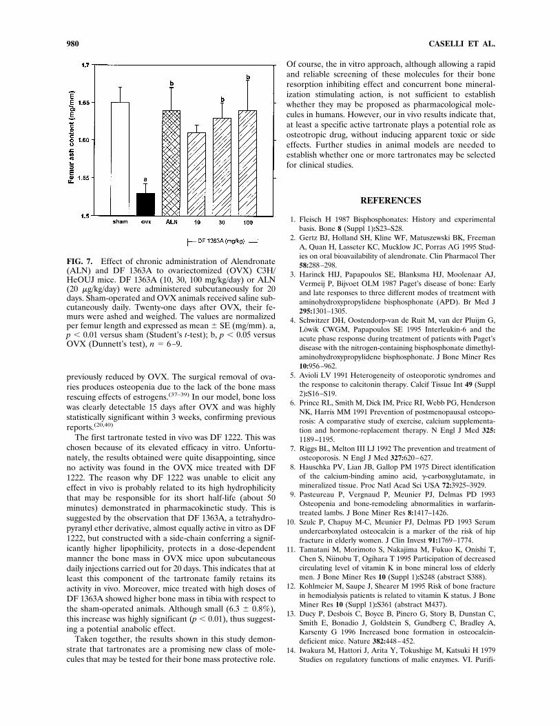
model of estrogen-dependent osteopenia carried out inmice. The product was administered subcutaneously atthree dosages (10, 30, and 100 mg/kg). Femur of OVX miceshowed a significant decrease of ash weight normalized perbone length (26.9 6 0.7%, n 5 6, p , 0.01). ALN givensubcutaneously at the dose of 20 mg/kg/day was able tocounteract OVX-induced osteopenia (Fig. 7). Similarly, DF1363A was able to inhibit bone loss in femurs of OVX mice
in a dose-related manner (Fig. 7), thus confirming its im-proved pharmacokinetic properties. The dose-related effectof DF 1363A was also evident in tibiae (data not shown)but, in this district, the loss of bone mass was less pro-nounced (24.2 6 0.7%, n 5 6) than in femurs and did notreach statistical significance. Interestingly, however, themean ash weight of tibiae from mice treated with 30 mg/kgof DF 1363A was even significantly higher than that ofsham-operated animals (38.66 0.3 vs. 36.36 0.7 mg, n5 9,p , 0.01).
DISCUSSION
In this study, we have shown that a new class of mole-cules, belonging to the tartronate family, inhibits bone re-sorption and stimulate matrix mineralization in vitro. Max-imal efficacy was observed in the tartronates presentingflexible side chains, such as DF 1222 and DF 1363A.Inhibition of bone resorption by the selected tartronates
was analogous to that induced by two reference bisphos-phonates, etidronate and alendronate, whose bone resorp-tion inhibiting effect is largely demonstrated by severalstudies.(23–25) This finding validated the hypothesis that a
FIG. 4. Effect of DF 1222 on osteoclast morphology. Di-agrams illustrating the effects of DF 1222 and salmon cal-citonin (sCT) on osteoclast retraction. Rabbit osteoclastswere cultured as previously described, then treated with (A)DF 1222 and (B) sCT at the concentrations indicated onabscissa, or (C) with 10212M DF 1222 for different time. Abiphasic and a dose-dependent increase in the percentageof osteoclasts retracted were observed in cultures treatedfor 15 min, with DF 1222 and sCT, respectively. The effectof DF 1222 at the maximal active concentration (10212 M)was time-dependent and reached the plateau at 4 h.Mean 6 SE (n 5 3). *p , 0.05, **p , 0.01 versus control(Dunnett’s t-test); a, p , 0.01 versus control at 48 h (opencircle), Student’s t-test.
TARTRONATES AND BONE METABOLISM 977

molecule sharing the chemical characteristics both ofbisphosphonates and of g-carboxyglutamic (Gla) residuescontained in the matrix proteins might affect bone resorp-tion by isolated osteoclasts, since dose-dependent inhibitionof in vitro pit excavation was observed. However, the curvesof inhibition of tartronates against basal resorption wereill-defined due to a very flat profile. This is in agreementwith the data obtained by Sahni et al.(22) by adding bisphos-phonates concurrently with osteoclasts. On the contrary,active tartronates showed a clear-cut dose–response curveagainst bone resorption stimulated with the osteotropicfactor 1,25(OH)2D3. This evidence could suggest, as in thecase of bisphosphonates, a possible contribution of mono-nuclear cells (possibly osteoblasts or bone marrow stromalcells) in the mechanism of action of tartronates.(26)
Tartronates were active both on rabbit and rat osteo-clasts, indicating no differences between these two species.Furthermore, morphological and enzymatic analyses dem-onstrated that one of the most in vitro active tartronates,DF 1222, did not apparently affect cell viability. This tartr-onate modified osteoclast morphology, inducing cell retrac-tion. Retraction was maximal at 10212 M and was reachedwithin 4 h. However, this study does not provide insightsinto the cell signals and subsequent molecular events thatcause arrest of the resorbing activity.The increased bone loss that characterizes the pathogen-
esis of osteopenic syndromes as osteoporosis is due toabnormalities in the bone remodeling cycle, which occurs byresorption of old bone, recruitment of new osteoblasts, anddeposition of new matrix that subsequently mineralizes.(27)
It is generally accepted that with each cycle there is amodest, imperceptible deficit in bone formation, possiblydue to progressive impairment of the signaling betweenbone resorption and bone formation, resulting in inefficientosteoblast recruitment.(28) Subsequently, osteopenia iscaused not only by increased bone resorption, but also by aresultant substantial decrease of bone formation. Fromthese considerations, a molecule that at the same timeinhibits bone resorption and stimulates bone formation isan ideal candidate as an antiosteopenic drug. In this study,we observed that DF 1222 increased the velocity of matrixmineralization by rat periosteal cells in vitro, without af-fecting cell proliferation and ALP activity, indicating a pos-sible role on the differentiating process rather than on theexpansion of the population. The effect of DF 1222 onmineralization is similar to that of malonate, which is struc-turally related to tartronates (tartronic acid is hydroxyma-lonic acid), as recently reported by Klein et al.(29) Theseauthors demonstrated that malonate is able to stimulatemineralization in rat marrow stromal cells. The mechanismthat is the basis of this effect should be the inhibition ofsuccinate dehydrogenase (SDH, complex II)30 in agreementwith the hypothesis that cells destined to mineralize rely lesson respiration.(31) However, the inhibition of Krebs en-zymes in actively resorbing osteoclast could reduce theiractivity, as indirectly indicated by the stimulatory or inhib-itory effects on osteoblastic SDH exerted by PTH(32) orcalcitonin,(33) respectively.We found the positive effect of DF 1222 on mineraliza-
tion of particular interest because, to date, there is not
FIG. 5. Effect of tartronates on 1,25(OH)2D3 stimulatedbone resorption. Rabbit osteoclasts were cultured in theabsence or in the presence of 10 nM 1,25(OH)2D3 (1,25D3)for 96 h with or without different concentrations of (A)alendronate (ALN), (B) DF 1222, or (C) DF 1363A. Theresorptive effect was expressed as mean 6 SE. (n 5 3–6).Pit area index of control was 4.6 6 2.9 for (A) and (C), and29.5 6 5.2 for (B). *p , 0.05, **p , 0.01 versus 1,25D3(Dunnett’s t-test); a, p , 0.05; b, p , 0.01 versus control(Student’s t-test).
978 CASELLI ET AL.

convincing evidence of pharmacological molecules capableof increasing the bone mass through an anabolic effect onbone formation. Even the bisphosphonate etidronate,which is widely used in the therapy Paget’s disease, com-pletely abolished matrix mineralization in our experimentalmodel, in agreement with several literature reports.(34,35)
Our results with tartronates are similar to those obtained
with another bisphosphonate (i.e., alendronate), which, un-like etidronate, showed a favorable effect on mineraliza-tion.(36) However, our evidence is based on an in vitromodel that may not fully reflect the in vivo situation.The positive results obtained in vitro prompted us to test
the most effective tartronates in vivo. To accomplish this,we selected a mouse model in which the bone mass was
FIG. 6. Effect of DF 1222 on matrixcalcification in rat periosteal cell cul-tures. Cells were grown for 3, 4, or 6weeks in a mineralization medium(containing 10 mM b-glycerophos-phate and 50 mg/ml ascorbic acid) inthe presence of different factors.Mineralization was detected with vonKossa staining.
TABLE 1. EFFECT OF DF 1222 ON CALCIUM DEPOSITION BY RAT PERIOSTEAL OSTEOBLAST-LIKE CELLS*
Treatment Medium
Calcium content (mmol/well)(weeks)
3 4 6
— normal 0.048 0.045 0.042— mineralization 0.048 0.057 1.926DF 1222 1027 mineralization 0.048 0.483 1.257DF 1222 1025 mineralization 0.411 1.938 1.581Dexa 1027 mineralization 0.834 1.284 2.091Dexa 1027 1 DF 1222 1025 mineralization 0.840 1.164 2.073
* Cells were grown for 3, 4, or 6 weeks either in normal or in mineralization medium (containing 10 mM b-glycerophosphate and 50mg/ml ascorbic acid) in the presence of different factors. Calcium content (mean of two replicates) was determined in duplicate wellstreated with 300 ml of 1 N HCl and quantified with a phenolphthalein complexone kit. Plates were treated in parallel with those illustratedin Fig. 6.
TARTRONATES AND BONE METABOLISM 979
previously reduced by OVX. The surgical removal of ova-ries produces osteopenia due to the lack of the bone massrescuing effects of estrogens.(37–39) In our model, bone losswas clearly detectable 15 days after OVX and was highlystatistically significant within 3 weeks, confirming previousreports.(20,40)
The first tartronate tested in vivo was DF 1222. This waschosen because of its elevated efficacy in vitro. Unfortu-nately, the results obtained were quite disappointing, sinceno activity was found in the OVX mice treated with DF1222. The reason why DF 1222 was unable to elicit anyeffect in vivo is probably related to its high hydrophilicitythat may be responsible for its short half-life (about 50minutes) demonstrated in pharmacokinetic study. This issuggested by the observation that DF 1363A, a tetrahydro-pyranyl ether derivative, almost equally active in vitro as DF1222, but constructed with a side-chain conferring a signif-icantly higher lipophilicity, protects in a dose-dependentmanner the bone mass in OVX mice upon subcutaneousdaily injections carried out for 20 days. This indicates that atleast this component of the tartronate family retains itsactivity in vivo. Moreover, mice treated with high doses ofDF 1363A showed higher bone mass in tibia with respect tothe sham-operated animals. Although small (6.3 6 0.8%),this increase was highly significant (p , 0.01), thus suggest-ing a potential anabolic effect.Taken together, the results shown in this study demon-
strate that tartronates are a promising new class of mole-cules that may be tested for their bone mass protective role.
Of course, the in vitro approach, although allowing a rapidand reliable screening of these molecules for their boneresorption inhibiting effect and concurrent bone mineral-ization stimulating action, is not sufficient to establishwhether they may be proposed as pharmacological mole-cules in humans. However, our in vivo results indicate that,at least a specific active tartronate plays a potential role asosteotropic drug, without inducing apparent toxic or sideeffects. Further studies in animal models are needed toestablish whether one or more tartronates may be selectedfor clinical studies.
REFERENCES
1. Fleisch H 1987 Bisphosphonates: History and experimentalbasis. Bone 8 (Suppl 1):S23–S28.
2. Gertz BJ, Holland SH, Kline WF, Matuszewski BK, FreemanA, Quan H, Lasseter KC, Mucklow JC, Porras AG 1995 Stud-ies on oral bioavailability of alendronate. Clin Pharmacol Ther58:288–298.
3. Harinck HIJ, Papapoulos SE, Blanksma HJ, Moolenaar AJ,Vermeij P, Bijvoet OLM 1987 Paget’s disease of bone: Earlyand late responses to three different modes of treatment withaminohydroxypropylidene bisphosphonate (APD). Br Med J295:1301–1305.
4. Schwitzer DH, Oostendorp-van de Ruit M, van der Pluijm G,Lowik CWGM, Papapoulos SE 1995 Interleukin-6 and theacute phase response during treatment of patients with Paget’sdisease with the nitrogen-containing bisphosphonate dimethyl-aminohydroxypropylidene bisphosphonate. J Bone Miner Res10:956–962.
5. Avioli LV 1991 Heterogeneity of osteoporotic syndromes andthe response to calcitonin therapy. Calcif Tissue Int 49 (Suppl2):S16–S19.
6. Prince RL, Smith M, Dick IM, Price RI, Webb PG, HendersonNK, Harris MM 1991 Prevention of postmenopausal osteopo-rosis: A comparative study of exercise, calcium supplementa-tion and hormone-replacement therapy. N Engl J Med 325:1189–1195.
7. Riggs BL, Melton III LJ 1992 The prevention and treatment ofosteoporosis. N Engl J Med 327:620–627.
8. Hauschka PV, Lian JB, Gallop PM 1975 Direct identificationof the calcium-binding amino acid, g-carboxyglutamate, inmineralized tissue. Proc Natl Acad Sci USA 72:3925–3929.
9. Pasteureau P, Vergnaud P, Meunier PJ, Delmas PD 1993Osteopenia and bone-remodeling abnormalities in warfarin-treated lambs. J Bone Miner Res 8:1417–1426.
10. Szulc P, Chapuy M-C, Meunier PJ, Delmas PD 1993 Serumundercarboxylated osteocalcin is a marker of the risk of hipfracture in elderly women. J Clin Invest 91:1769–1774.
11. Tamatani M, Morimoto S, Nakajima M, Fukuo K, Onishi T,Chen S, Niinobu T, Ogihara T 1995 Participation of decreasedcirculating level of vitamin K in bone mineral loss of elderlymen. J Bone Miner Res 10 (Suppl 1):S248 (abstract S388).
12. Kohlmeier M, Saupe J, Shearer M 1995 Risk of bone fracturein hemodialysis patients is related to vitamin K status. J BoneMiner Res 10 (Suppl 1):S361 (abstract M437).
13. Ducy P, Desbois C, Boyce B, Pinero G, Story B, Dunstan C,Smith E, Bonadio J, Goldstein S, Gundberg C, Bradley A,Karsenty G 1996 Increased bone formation in osteocalcin-deficient mice. Nature 382:448–452.
14. Iwakura M, Hattori J, Arita Y, Tokushige M, Katsuki H 1979Studies on regulatory functions of malic enzymes. VI. Purifi-
FIG. 7. Effect of chronic administration of Alendronate(ALN) and DF 1363A to ovariectomized (OVX) C3H/HeOUJ mice. DF 1363A (10, 30, 100 mg/kg/day) or ALN(20 mg/kg/day) were administered subcutaneously for 20days. Sham-operated and OVX animals received saline sub-cutaneously daily. Twenty-one days after OVX, their fe-murs were ashed and weighed. The values are normalizedper femur length and expressed as mean 6 SE (mg/mm). a,p , 0.01 versus sham (Student’s t-test); b, p , 0.05 versusOVX (Dunnett’s test), n 5 6–9.
980 CASELLI ET AL.

cation and molecular properties of NADP-linked malic enzymefrom Escherichia coli W. J Biochem 85:1355–1365.
15. Caggiano TJ, Zask A, Bex F 1991 Recent advances in bonemetabolism and osteoporosis research. Ann Rep Med Chem26:201–210.
16. Gandolfi C, Cotini L, Mantovanini M, Caselli G, Clavenna G,Omini C 1994 Tartronic acids, their acetalic ethers and O-esters. Patent WO 9410127.
17. Chambers TJ, Revell PA, Fuller K, Athanasou NA 1984 Re-sorption of bone by isolated rabbit osteoclasts. J Cell Sci66:383–399.
18. Teti A, Tarquilio A, Grano M, Colucci S, Laforgia A, ManginiF, Zambonin-Zallone A 1991 Effect of calcium-phosphate-based materials on proliferation and alkaline phosphatase ac-tivity of newborn rat periosteal cells in vitro. J Dent Res70:997–1001.
19. Prallet R, Male P, Neff L, Baron R 1992 Identification of afunctional mononuclear precursor of the osteoclast in chickenmedullary bone marrow cultures. J Bone Miner Res 7:405–414.
20. Kawaguchi H, Pilbeam CC, Vargas SJ, Morse EE, Lorenzo JA,Raisz LG 1995 Ovariectomy enhances and estrogen replace-ments inhibits the activity of bone marrow factors that stimu-late prostaglandin production in cultured mouse calvariae.J Clin Invest 96:539–548.
21. Migliaccio S, Newbold RR, Bullock, BC, Jefferson WJ, SuttonFG, McLachlan JA, Korach KS 1996 Alteration of maternalestrogen levels during gestation affect the skeleton of femaleoffspring. Endocrinology 137:2118–2125.
22. Sahni M, Guenther HL, Fleisch H, Collin P, Martin TJBisphosphonates act on rat bone resorption through the me-diation of osteoblast. 1993 J Clin Invest 91:2004–2011.
23. Shinoda H, Adamek G, Felix R, Fleisch H, Schenk R, HaganP 1983 Structure-activity relationships of various bisphospho-nates. Calcif Tissue Int 35:87–99.
24. Muhlbauer RC, Bauss F, Schenk R, Janner M, Bosies E, StreinK, Fleisch H 1991 BM 21.0955, a potent new bisphosphonate toinhibit bone resorption. J Bone Miner Res 6:1003–1011.
25. van Beek E, Hoekstra M, van de Ruit M, Lowik CWGM,Papapoulos SE 1994 Structural requirements for bisphospho-nate actions in vitro. J Bone Miner Res 9:1875–1882.
26. Rodan GA, Fleisch HA 1996 Bisphosphonates: mechanism ofaction. J Clin Invest 97:2692–2696.
27. Baron R 1993 Anatomy and ultrastructure of bone. In: FavusMJ (ed.) Primer on the Metabolic Bone Diseases and Disor-ders of Mineral Metabolism, 2nd Ed. Raven Press, New York,NY, U.S.A., pp. 3–9.
28. Kleerekoper M, Avioli LV 1993 Evaluation and treatment ofpostmenopausal osteoporosis. In: Favus MJ (ed.) Primer onthe Metabolic Bone Diseases and Disorders of Mineral Me-tabolism, 2nd Ed. Raven Press, New York, NY, pp. 223–229.
29. Klein BY, Gal I, Libergal M, Ben-Bassat H 1996 Opposingeffects on mitochondrial membrane potential by malonate and
levamisole, whose effect on cell-mediated mineralization isantagonistic. J Cell Biochem 60:139–147.
30. Klein BY, Gal I, Segal D 1993 Selection of malonate-resistantstromal cell-derived osteoprogenitor cell in vitro. J Cell Bio-chem 51:190–197.
31. Shapiro IM, Golub EE, Kakuta S, Hazelgrove J, Havery J,Chance B, Frasca P 1982 Initiation of enchondral calcificationis related to changes in the redox state of hypertrophic chon-drocytes. Science 217:950–952.
32. Loveridge N, Dean V, Goltzman D, Hendy GN 1991 Bioac-tivity of parathyroid hormone and parathyroid hormone-likepeptide: Agonist and antagonist activities of amino-terminalfragments assessed by the cytochemical bioassay and in situbiochemistry. Endocrinology 128:1938–1946.
33. Sugiyama T, Ohashi T, Kusuhara S 1993 Inhibition of osteo-clastic bone resorption by calcitonin in the cultured medullarybone of laying hens. Jpn Poult Sci 30:16–23.
34. King WR, Francis MD, Michael WR 1971 Effect of disodiumethane-1-hydroxy-1,1-diphosphonate on bone formation. ClinOrthop 78:251–270.
35. Tenebaum HC, Torontali M, Sukhu B 1992 Effects of bisphos-phonates and inorganic pyrophosphate on osteogenesis invitro. Bone 13:249–255.
36. Giuliani N, Girasole G, Pedrazzoni M, Passeri G, Gatti C,Passeri M 1995 Alendronate stimulates b-FGF production andmineralized nodule formation in human osteoblastic cells andosteoblastogenesis in human bone marrow cultures. J BoneMiner Res 10 (Suppl 1):129.
37. Bhattacharyya MH, Whelton BD, Stern PH, Peterson DP 1988Cadmium accelerates bone loss in ovariectomized mice andfetal rat limb in culture. Proc Natl Acad Sci USA 85:8761–8765.
38. Kitazawa R, Kimble RB, Vannice JL, Kung VT, Pacifici R1994 Interleukin-1 receptor antagonist and Tumor NecrosisFactor binding protein decrease osteoclast formation and boneresorption in ovariectomized mice. J Clin Invest 94:2397–2406.
39. Bain SD, Bailey MC, Celino DL, Lantry MM, Edwards MW1993 High-dose estrogen inhibits bone resorption and stimu-lates bone formation in the ovariectomized mouse. J BoneMiner Res 8:435–442.
40. Hughes DE, Dai A, Tiffee JC, Li HH, Mundy GR, Boyce BF1996 Estrogen promotes apoptosis of murine osteoclasts me-diated by TGF-b. Nature Med 2:1132–1136.
Address reprint requests to:Dr. Gianfranco Caselli
Dompe S.p.A.via Campo di Pile
67 100 L’Aquila, Italy
Received in original form December 6, 1996; in revised formJanuary 29, 1997; accepted January 30, 1997.
TARTRONATES AND BONE METABOLISM 981