Targeting Natural Killer Cell Reactivity by Employing Antibody to NKp46: Implications for Type 1...
15
RESEARCH ARTICLE Targeting Natural Killer Cell Reactivity by Employing Antibody to NKp46: Implications for Type 1 Diabetes Rami Yossef 1,2 , Chamutal Gur 3,4 , Avishai Shemesh 1,2 , Ofer Guttman 5 , Uzi Hadad 1,2 , Shlomo Nedvetzki 6 , Antonija Miletić 7 , Karen Nalbandyan 8 , Adelheid Cerwenka 9 , Stipan Jonjic 7 , Ofer Mandelboim 3 , Angel Porgador 1,2 * 1 The Shraga Segal Department of Microbiology, Immunology and Genetics, Faculty of Health Sciences, Ben-Gurion University of the Negev, Beer Sheva, Israel, 2 The National Institute for Biotechnology in the Negev, Ben-Gurion University of the Negev, Beer Sheva, Israel, 3 The Lautenberg Center for General and Tumor Immunology, Institute for Medical Research Israel-Canada, Hebrew University-Hadassah Medical School, Jerusalem 91120, Israel, 4 Department of Medicine, Hadassah-Hebrew University Hospital, Jerusalem 91120, Israel, 5 Department of Clinical Biochemistry and Pharmacology, Faculty of Health Sciences, Ben-Gurion University of the Negev, Beer Sheva, Israel, 6 BioLineRx Ltd., 19 Hartum Street, P.O. Box 45158. Jerusalem 91450, Israel, 7 Center for Proteomics and Department for Histology and Embryology, School of Medicine, University of Rijeka, 51000 Rijeka, Croatia, 8 Pathology Department, Soroka Medical Center, Beer Sheva, Israel, 9 Innate Immunity Group, German Cancer Research Center, Heidelberg 69120, Germany * [email protected] Abstract Natural killer (NK) cells belong to the innate lymphoid cells. Their cytotoxic activity is regulat- ed by the delicate balance between activating and inhibitory signals. NKp46 is a member of the primary activating receptors of NK cells. We previously reported that the NKp46 receptor is involved in the development of type 1 diabetes (T1D). Subsequently, we hypothesized that blocking this receptor could prevent or hinder disease development. To address this goal, we developed monoclonal antibodies for murine NKp46. One mAb, named NCR1.15, recognizes the mouse homologue protein of NKp46, named Ncr1, and was able to down- regulate the surface expression of NKp46 on primary murine NK cells following antibody in- jection in vivo. Additionally, NCR1.15 treatments were able to down-regulate cytotoxic activ- ity mediated by NKp46, but not by other NK receptors. To test our primary assumption, we examined T1D development in two models, non-obese diabetic mice and low-dose strepto- zotocin. Our results show a significantly lower incidence of diabetic mice in the NCR1.15- treated group compared to control groups. This study directly demonstrates the involvement of NKp46 in T1D development and suggests a novel treatment strategy for early insulitis. Introduction Natural killer (NK) cells of the innate immune system provide primary immune defense against cancer and pathogens [1–4]. Their activity is controlled by a multitude of inhibitory PLOS ONE | DOI:10.1371/journal.pone.0118936 February 26, 2015 1 / 15 OPEN ACCESS Citation: Yossef R, Gur C, Shemesh A, Guttman O, Hadad U, Nedvetzki S, et al. (2015) Targeting Natural Killer Cell Reactivity by Employing Antibody to NKp46: Implications for Type 1 Diabetes. PLoS ONE 10(2): e0118936. doi:10.1371/journal.pone.0118936 Academic Editor: Francesco Dotta, University of Siena, ITALY Received: June 22, 2014 Accepted: January 7, 2015 Published: February 26, 2015 Copyright: © 2015 Yossef et al. This is an open access article distributed under the terms of the Creative Commons Attribution License, which permits unrestricted use, distribution, and reproduction in any medium, provided the original author and source are credited. Data Availability Statement: All relevant data are within the paper and its Supporting Information files. Funding: This work was supported by the Israel Science Foundation-Juvenile Diabetes Research Foundation Grant 123/12 (to A.P. & O.M.), United States–Israel Binational Science Foundation Grant 2011123 (A.P.), the Israel Science Foundation Grant 304/12 (A.P.), BioLineRx early development grant (A. P. & O.M.), the Israeli Ministry of Science and Technology/German Cancer Research Center program Grant CA142 (A.P. and A.C.), and National Institutes of Health Grant 1R01AI083201-01 (S.J.). The funder provided support in the form of salaries
-
Upload
independent -
Category
Documents
-
view
1 -
download
0
Transcript of Targeting Natural Killer Cell Reactivity by Employing Antibody to NKp46: Implications for Type 1...

RESEARCH ARTICLE
Targeting Natural Killer Cell Reactivity byEmploying Antibody to NKp46: Implicationsfor Type 1 DiabetesRami Yossef1,2, Chamutal Gur3,4, Avishai Shemesh1,2, Ofer Guttman5, Uzi Hadad1,2,Shlomo Nedvetzki6, Antonija Miletić7, Karen Nalbandyan8, Adelheid Cerwenka9,Stipan Jonjic7, Ofer Mandelboim3, Angel Porgador1,2*
1 The Shraga Segal Department of Microbiology, Immunology and Genetics, Faculty of Health Sciences,Ben-Gurion University of the Negev, Beer Sheva, Israel, 2 The National Institute for Biotechnology in theNegev, Ben-Gurion University of the Negev, Beer Sheva, Israel, 3 The Lautenberg Center for General andTumor Immunology, Institute for Medical Research Israel-Canada, Hebrew University-Hadassah MedicalSchool, Jerusalem 91120, Israel, 4 Department of Medicine, Hadassah-Hebrew University Hospital,Jerusalem 91120, Israel, 5 Department of Clinical Biochemistry and Pharmacology, Faculty of HealthSciences, Ben-Gurion University of the Negev, Beer Sheva, Israel, 6 BioLineRx Ltd., 19 Hartum Street, P.O.Box 45158. Jerusalem 91450, Israel, 7 Center for Proteomics and Department for Histology andEmbryology, School of Medicine, University of Rijeka, 51000 Rijeka, Croatia, 8 Pathology Department,Soroka Medical Center, Beer Sheva, Israel, 9 Innate Immunity Group, German Cancer Research Center,Heidelberg 69120, Germany
AbstractNatural killer (NK) cells belong to the innate lymphoid cells. Their cytotoxic activity is regulat-
ed by the delicate balance between activating and inhibitory signals. NKp46 is a member of
the primary activating receptors of NK cells. We previously reported that the NKp46 receptor
is involved in the development of type 1 diabetes (T1D). Subsequently, we hypothesized
that blocking this receptor could prevent or hinder disease development. To address this
goal, we developed monoclonal antibodies for murine NKp46. One mAb, named NCR1.15,
recognizes the mouse homologue protein of NKp46, named Ncr1, and was able to down-
regulate the surface expression of NKp46 on primary murine NK cells following antibody in-
jection in vivo. Additionally, NCR1.15 treatments were able to down-regulate cytotoxic activ-
ity mediated by NKp46, but not by other NK receptors. To test our primary assumption, we
examined T1D development in two models, non-obese diabetic mice and low-dose strepto-
zotocin. Our results show a significantly lower incidence of diabetic mice in the NCR1.15-
treated group compared to control groups. This study directly demonstrates the involvement
of NKp46 in T1D development and suggests a novel treatment strategy for early insulitis.
IntroductionNatural killer (NK) cells of the innate immune system provide primary immune defenseagainst cancer and pathogens [1–4]. Their activity is controlled by a multitude of inhibitory
PLOSONE | DOI:10.1371/journal.pone.0118936 February 26, 2015 1 / 15
OPEN ACCESS
Citation: Yossef R, Gur C, Shemesh A, Guttman O,Hadad U, Nedvetzki S, et al. (2015) Targeting NaturalKiller Cell Reactivity by Employing Antibody toNKp46: Implications for Type 1 Diabetes. PLoS ONE10(2): e0118936. doi:10.1371/journal.pone.0118936
Academic Editor: Francesco Dotta, University ofSiena, ITALY
Received: June 22, 2014
Accepted: January 7, 2015
Published: February 26, 2015
Copyright: © 2015 Yossef et al. This is an openaccess article distributed under the terms of theCreative Commons Attribution License, which permitsunrestricted use, distribution, and reproduction in anymedium, provided the original author and source arecredited.
Data Availability Statement: All relevant data arewithin the paper and its Supporting Information files.
Funding: This work was supported by the IsraelScience Foundation-Juvenile Diabetes ResearchFoundation Grant 123/12 (to A.P. & O.M.), UnitedStates–Israel Binational Science Foundation Grant2011123 (A.P.), the Israel Science Foundation Grant304/12 (A.P.), BioLineRx early development grant (A.P. & O.M.), the Israeli Ministry of Science andTechnology/German Cancer Research Centerprogram Grant CA142 (A.P. and A.C.), and NationalInstitutes of Health Grant 1R01AI083201-01 (S.J.).The funder provided support in the form of salaries

and activating NK cell receptors [1]. Instigation of inhibitory receptors by major histocompati-bility complex (MHC) class I molecules on healthy cells suppresses NK cell effector functions[5,6]. The inhibitory receptors recognize mainly MHC class I proteins but also additional mole-cules [5]. Activating receptors recognize various ligands that are tumor-derived, stress-induced,pathogen-derived, or self-ligands [7,8]. Several activating receptors are expressed by NK cells,among them NKG2D and natural cytotoxicity receptors (NCRs). The human NCR family in-cludes three members named NKp30, NKp44 and NKp46 [1]. Mice express orthologous recep-tor only for the human NKp46 (Ncr1) [9].
We recently reported that the murine activating receptor NKp46 (NCR1) is essential for thedevelopment of type 1 diabetes (T1D) [10], an autoimmune disease prevalent throughout theWestern world. Currently the common treatment of this disease is based on a daily insulin in-jection. It is well known that T1D development is T-cell dependent, yet several studies haveshown cytolytic activity of NK cells against pancreatic islet beta cells (β-cells) and their involve-ment in the development of the disease [11–15]. In particular, we have shown the involvementof NKp46 in NK contribution to T1D. We demonstrated that both human and mouse β-cellsexpress ligands for NKp46 in either healthy or T1D patients [10,16]. We showed that develop-ment of experimental T1D was impaired in Ncr1gfp/gfp mice that lack the expression ofmNKp46. Additionally, we showed that employing active immunization to block NKp46,through immunizing with recombinant NKp46, inhibited the development of T1D in murinemodels [10]. These findings motivated us to explore new therapeutic approaches for T1Dbased on manipulation of NKp46 function.
In order to accomplish this, one tactic would be to block the NKp46 ligands. However, theprecise nature of NKp46 ligands is not fully revealed. Several reports have shown that NKp46recognizes cellular ligands expressed on tumor cells, dendritic cells, viral-infected cells, andLangerhans β-cells [17,18]. Currently identified NKp46 ligands include viral hemagglutinins[19], the cellular ligand vimentin [20] and the cellular co-ligands heparan sulfate proteoglycans(HSPG) [21–23]. However, these two cellular ligands hardly present a suitable target for ma-nipulation of NKp46 function through blocking of target cellular ligand.
Therefore, in the current study we investigated the methodology of antibody-mediated ma-nipulation of the NKp46 function. We developed and characterized a new anti-murine NKp46mAb named NCR1.15. Treatment of mice with NCR1.15 did not deplete NK cells, but sup-pressed their NKp46-mediated function. In accordance, NCR1.15 treatment of T1D-pronemice significantly prolonged the time to T1D development.
Research Design and Methods
CellsCells that were used in this study: YAC-1, murine lymphoma (TIB-160, ATCC); PD1.6, murinethymic virus-induced lymphoma [24]; Ba/F3-Rae1εmouse pro-B lymphocyte ectopically ex-pressing Rae-1εNKG2D ligand [25]; BW-hNKp46 and BW-mNKp46 T-cell lymphoma ectop-ically expressing the murine or the human NKp46 [19].
Production of NCR1.15 mAbNcr1gfp/gfp 129/sv/J mice, lacking the expression of endogenous mNKp46, were used for theproduction of mouse monoclonal antibodies against mNKp46. These mice were immunizedtwice with 100ug/mice of mNKp46-Ig fusion protein, followed by a boost immunization and asubsequent fusion with the mouse Sp2/0 cell line. First hybridoma screening for specific anti-bodies was performed using ELISA. Positive hybridomas were selected and cloned several
Targeting NK Reactivity by mAb to NKp46
PLOSONE | DOI:10.1371/journal.pone.0118936 February 26, 2015 2 / 15
for authors [SN], but did not have any additional rolein the study design, data collection and analysis,decision to publish, or preparation of the manuscript.The specific roles of these authors are articulated inthe 'author contributions' section.
Competing Interests: BioLineRx provided support inthe form of salaries for authors [SN]. This does notalter the authors' adherence to PLOS ONE policieson sharing data and materials.

times to ensure monoclonality. Antibodies obtained from these clones were further character-ized using different techniques.
Antibodies and Fusion proteinsThe following antibodies were used in this study: BioLegend—anti-NKp46 mAb (clone29A1.4), anti-CD3ε (clone145–2C11), anti-NK1.1 (clone PK136), anti-CD49b (clone DX5),anti-CD27 (clone LG.3A10), anti-NKG2D (clone C7), anti-CD107a/LAMP-1 (clone 1D4B);anti-CD11b (eBioscience, clone M1/70); anti-human NKp46 (461-G1,[26]); mouse IgG1, κcontrol for injections, rat IgG2a, κ isotype control for FACS (BioLegend, clone RTK2758). TheProduction of mNKp46-Ig, LIR1-Ig and mNKG2D-Ig as previously described [25,27,28].
ELISATo determine the specific binding between NCR1.15 and the mNKp46 receptor plates werecoated overnight at 4°C with 5μg/ml of the recombinant proteins. Blocking buffer (PBS supple-mented with 10%FBS) was applied for 2 hours at room temperature, after which plates werewashed with PBS with 0.05% Tween 20 (PBST) and incubated with 2 μg/ml of NCR1.15, 461-G1or PBS for 2 hours at room temperature. Following washing with PBST biotin-conjugatedsheep anti-mouse IgG (GE Healthcare, NA931V) was added for 1 hr at 1:750 dilution. Follow-ing washing streptavidin-HRP (Jackson, 016-030-084) diluted 1:1000 was added for 30 min.Following washing TMB (DAKO, S1599) was added optical density was read at 650 nm(Thermo Electron Corporation Multiskan Spectum).
Bimolecular interaction analysisA BIAcore 3000 device fitted with CM5 sensor chips (BIAcore, Uppsala, Sweden) was used forstudying the interactions between NKp46 and NCR1.15 in conjunction with BIAevaluationsoftware (v4.1). To activate the chip, we used the EDC/NHS amine coupling procedure accord-ing to the manufacturer’s protocol (BIAcore), followed by addition of NKp46, which was im-mobilized in the different flow cells, followed by blocking the free active groups with 1 Methanolamine. Different analyte concentrations were injected, each followed by regeneration ofthe surface using 10 mMNaOH. Data were analyzed using a 1:1 Langmuir binding model.
CD107a degranulation assaySpleen lymphocytes were isolated 3 days following treatment and NK cells were purified usingMouse NK Cell Enrichment Kit (Stemcell Technologies, #19755). 5X10^5 purified NK cellswere co-incubated with target cells for 2–4 hours in the presence of 0.1μg APC-coupled anti-CD107a antibody; next cells were stained for CD3ε, NK1.1 and CD107a and analyzed usingFACSCantoII (BD Biosciences).
Quantitative Real Time PCR (qRT-PCR)Total RNA was extracted from Fresh spleen tissue using the RNeasy Mini Kit (cat# 74104, Qia-gen Ltd). RNA samples were stored at-80°C until further use. RNA concentration and puritywas assessed by spectrophotometry (NanoDrop ND1000 v3.7.1; NanoDrop Technologies, Del-aware, USA). cDNA synthesis was performed using the High Capacity cDNA Reverse Tran-scription Kit (cat# 4368814, Applied Biosystems. cDNA samples were stored at-20°C untilfurther use were. qRT-PCR reactions were performed using the Power SYBR Green PCRMaster Mix (cat# 4367659, Applied Biosystems) and experiments were performed using theApplied Biosystems 7500 Real Time PCR system, software v2.0.5. Reactions cycling protocol:
Targeting NK Reactivity by mAb to NKp46
PLOSONE | DOI:10.1371/journal.pone.0118936 February 26, 2015 3 / 15

95°C for 10 min, then 40 cycles with a 2-step program (95°C for 15s, 60°C for 1 min). Expres-sion levels of ncr1 (target gene) were normalized to β actin (reference gene). ncr1 Fw: 5- CTAGGGCTCACAGAGGGACATAC-3, ncr1 Rev 5- CAACACCTCCTGTGATGAGTAGT-3, βactin Fw: 5-GCATTGTTACCAACTGGGAC-3, β actin Rev: 5-GGTCTCAAACATGATCTGGG-3.
Animals and NODmodel6–8 weeks old C57BL/6 female mice were purchased fromHarlan Laboratories (Rehovot, Israel).For the T1Dmodel in non-obese diabetic (NOD) strain 6–7 weeks old female mice were injectedintraperitoneally (i.p.) with 100μg antibody per mouse every other day for 16 weeks and glucoselevels were monitored. Ncr1gfp/+ heterozygous mice expressing GFP under the control ofNcr1promoter in one allele [29] were used to assess the promoter activation levels. C57BL/6-Tg(UBC-GFP) mice were used for several CD107a de-granulation assays. Theses transgenic miceexpress enhanced Green Fluorescent Protein (EGFP) under the direction of the human ubiquitinC promoter (UBC) in all tissues (Jackson laboratory). All mice used in this study were bredand maintained in the local animal care facility, approved by the Institutional Animal Careand Use Committee (IACUC) of Ben-Gurion University of the Negev (BGU). All experimentalprocedures were reviewed and approved by the Institutional Animal Care and Use Committee(IACUC) of Ben-Gurion University of the Negev (BGU’s IACUC) according to specified proto-cols that aim to ensure animal welfare and reduce suffering (permit: IL-58–09–02).
Low-dose streptozotocin (LDSTZ)-induced diabetesSex- and age-matched, 9 weeks mice were injected i.p. for 5 consecutive days with streptozoto-cin (STZ) (Sigma, S0130) dissolved in citrate buffer, pH 4.5, at a concentration of 50 mg � kg-1body weight. Day zero was defined as the first day of STZ treatment. 50μg of NCR1.15 were in-oculated i.p. at days-2 and 5. Glucose concentrations were measured using glucometer (AbbottDiabetes Care, FreeStyle Lite) every other day. Statistical analysis was done by log-rankKaplan-Meier analysis.
Isolation of primary β-cellsPancreata were inflated with 1 mg/ml collagenase type XI (Sigma), excised, and incubated for27–35 min at 37°C. Digested pancreata were vortexed and filtered through a 500-μm sieve andthe pellet washed in HBSS containing 0.5% BSA (Sigma). The pellet was re-suspended in RPMImedium 1640 (Gibco, 21875–034). Islets were collected on a 100-μm cell strainer (BD, Falcon)and individual islets were picked by hand under the microscope and further dispersed into sin-gle cells [30] Islets’ single cell preparations were first pooled and only then divided among thedifferent treatment groups.
Results
NCR1.15 mAb specifically recognizes mouse NKp46To test whether blocking of NKp46 (Ncr1 in mice) will attenuate T1D development, we devel-oped an anti-mouse NKp46/Ncr1 IgG1,κmAb (termed NCR1.15) that specifically recognizesrecombinant mouse NKp46 (mNKp46) but not human NKp46 (hNKp46). As a control, weemployed 461-G1 mAb that specifically recognizes hNKp46 but not mNKp46 [26] (Fig. 1A).Both mAbs did not bind to other recombinant NK receptors such as LIR1 or to the hFc whichis fused to the recombinant NK receptors employed in the binding assays (Fig. 1A and data notshown). NCR1.15 also recognizes BW cells that were transfected to express mNKp46 compared
Targeting NK Reactivity by mAb to NKp46
PLOSONE | DOI:10.1371/journal.pone.0118936 February 26, 2015 4 / 15

to BW cells expressing hNKp46 (Fig. 1B). NCR1.15 displayed a characteristic binding curve tomNKp46 (KD = 1.06E-08 M), indicating binding with moderate affinity (Fig. 1C). To testwhether NCR1.15 specifically recognizes endogenous mNKp46 expressed by murine primaryNK cells, we stained splenocytes from the functionally competent Ncr1gfp/+ heterozygous miceexpressing one allele encoding for mNKp46 and the other allele encoding for GFP under thecontrol of Ncr1 promoter in the other allele [29]. GFP-positive splenocytes were nearly all(99%) stained positively with NCR1.15 or with the commercial anti-mNKp46 mAb 29A1.4and (Fig. 1D). Accordingly, the overwhelming majority of NCR1.15+ or 29A1.4+ single or dou-ble positives were also GFP positive (Fig. 1E). To summarize, NCR1.15 specifically stainedmNKp46 expressed by murine NK cells.
Single dose treatment with NCR1.15 reduces membrane-associatedNKp46 expression and NKp46-mediated NK functionTo investigate the effect of anti-mNKp46 treatment in vivo, we first inoculated C57BL/6 micei.p. with 100μg NCR1.15/mouse (single dose). Three days post inoculation, a fraction of NKcells, and NKp46 expression levels were monitored for splenocytes and PBMCs (Fig. 2).Fig. 2A shows a representative NK staining of splenocytes and Fig. 2B summarizes the NK
Fig 1. Specific binding of NCR1.15 monoclonal antibody to murine NKp46. (A) ELISA plates were coated with different NK-fusion receptors followed byincubation with NCR1.15 or 46–1G1 mouse antibodies, the binding was detected with anti-mouse IgG HRP. Data from one representative of threeindependent experiments are shown. (B) FACS analysis of NCR1.15 binding to BW cells expressing mNKp46 or hNKp46. (C) Kinetics of NCR1.15 binding tomNKp46-Ig; the results from the Biacore analysis are expressed in resonance units (RU) against time. Dose-dependent NCR1.15 were injected over theimmobilized fusion Ig’s surface at increasing concentrations ranging from 0 to 250nM. (D-E) FACS analysis of NCR1gfp/+ splenocytes stained for NKp46 with29A1.4 and NCR1.15 clones. Data from one representative of three independent experiments are shown. Bars, ±SD.
doi:10.1371/journal.pone.0118936.g001
Targeting NK Reactivity by mAb to NKp46
PLOSONE | DOI:10.1371/journal.pone.0118936 February 26, 2015 5 / 15

staining results (CD3-NK1.1+) for splenocytes and PBMCs from four independent experi-ments. The NK fraction in both the spleen and blood did not change significantly following asingle dose injection of NCR1.15 compared to mock injection (isotype control or PBS). In par-allel, we tested membrane-associated NKp46 expression following single dose injection ofNCR1.15. Fig. 2C shows a representative NKp46 and NKG2D staining for CD3-NK1.1+ NKcells purified from splenocytes that were harvested 3 days after inoculation. Note that NKp46staining for the experiments described in Fig. 2 was performed with commercial 29A1.4 mAbthat its binding did not overlap with NCR1.15 (Fig. 1D, E). NKp46 cell surface levels, but notNKG2D levels, were considerably reduced following NCR1.15 injection as compared to PBS orisotype control injections (Fig. 2C). Fig. 2D summarizes the NKp46 staining results for
Fig 2. Single dose treatment with NCR1.15 down-regulates the surface expression of NKp46 on NKcells, but not the NKp46 transcript or spleen/blood NK levels. 3 days following i.p. injection of NCR1.15,isotype control or PBS splenocytes (A, B) and PBMCs (B) were stained and analyzed for CD3-NK1.1+ NKcells using flow cytometry. Data from one representative of five independent experiments are shown. Themembrane associated NKp46 (C, D) and NKG2D (C) on NK cells were stained and analyzed using flowcytometry. (E) Representative confocal images of purified splenic NK cells stained for extra- and intracellularexpression of the NKp46. (F) qRT-PCR of NKp46 transcripts in splenocytes harvested from treated mice. (G)GFP intensity (GeoMFI) expressed by NK cells from NCR1gfp/+ following the various treatments. Data fromone representative of three independent experiments are shown. ** p<0.01 by Student's unpaired t-test.Bars, ±SD.
doi:10.1371/journal.pone.0118936.g002
Targeting NK Reactivity by mAb to NKp46
PLOSONE | DOI:10.1371/journal.pone.0118936 February 26, 2015 6 / 15

CD3-NK1.1+NK-gated splenocytes and PBMCs from four independent experiments. Cell-membrane levels of NKp46 were significantly down-regulated following single dose injectionof NCR1.15 compared to mock injection (near 2-fold change). However, we also analyzedNKp46 expression on CD3+NK1.1+ NKT cells, knowing that the NKp46 is expressed on subsetof these cells [31], and got similar reduction levels of NKp46 expression with no change inNKT levels (data not shown). Further, to investigate whether this down-regulation at the cellsurface reflects suppression of NKp46 production or internalization of the protein, we studiedNKp46 localization employing confocal microscopy. Intracellular expression of NKp46 wassimilar in the NK from either NCR1.15- or mock-treated mice, while membrane-associatedstaining was reduced in the NK from NCR1.15-treated mice (Fig. 2E). Thus, it is probable thatNCR1.15 treatment neither suppressed NKp46 protein synthesis nor enhancedNKp46 internalization.
Following the aforementioned results in the protein level, we hypothesized that single injec-tion of anti-NKp46 would not suppress the activity of the NKp46 promoter or induce degrada-tion of NKp46-encoding RNA. Indeed, qPCR analysis of NKp46-encoding mRNA showed thatNCR1.15 treatment did not affect the levels of transcribed NKp46 (Fig. 2F). To better addressthe question for the NKp46 promoter activity, we employed Ncr1gfp/+ heterozygous mice ex-pressing one allele encoding for mNKp46 and the other allele encoding for GFP under the con-trol of Ncr1 promoter. NCR1.15 treatment did not alter GFP levels in NK cells compared tomock-treated mice (Fig. 2G), indicating that a single dose injection of anti-NKp46 did not af-fect Ncr1 promoter activity. To summarize, single dose injection of NCR1.15 significantly re-duced membrane-associated levels of NK-expressed NKp46 but did not affect NKp46transcription or translation.
We then studied whether down-regulation of membrane-associated NKp46 affects NK ac-tivity using YAC-1 target cells, which are sensitive to NK activity. CD3-NK1.1+NK cells puri-fied from splenocytes harvested 3 days after NCR1.15 inoculation significantly reduced vesicledegranulation following incubation with YAC-1 target cells (Fig. 3A). To test whether this ob-servation is correct in vivo,we mixed two fluorescently labeled target cells, NK-sensitive YAC-1and NK insensitive RMA cells (1:1 ratio), and injected them intravenously 3 days afterNCR1.15/mock treatment of mice. YAC-1 and RMA cells were labeled with different fluores-cent probes; thus we could differ between the two targets in the same lung. Lung clearanceassay of YAC-1 cells normalized on RMA cells was significantly lower in NCR1.15-treatedmice compared to PBS- or isotype control-treated mice (Fig. 3B). We extended our studies toPD1.6 target cells that manifest sensitivity to NKp46-mediated NK function [32]. To bettercompare effector cells, we mixed PD1.6 target cells with effector NK cells purified from bothisotype control- and NCR1.15-treated mice (1:1:1 ratio, E1:E2:T) and tested NK cell degranula-tion. One effector NK population was derived fromWT C57BL/6 mice and the other was de-rived from UBC-GFP C57BL/6 mice. Therefore, by detecting GFP, we could differ between thetwo NK populations using a flow cytometry-based degranulation assay in the same well.Fig. 3C shows a representative assay in which UBC-GFPmice were mock-treated, and WTmice were NCR1.15-treated. With no target cells, the degranulation level was near zero andsimilar between the two mixed NK effector populations. Following incubation with targetPD1.6 cells, the degranulation of mock-treated NK cells was significantly higher compared tothe NCR1.15-treated NK cells in the mixed population (Fig. 3C).
To further investigate whether this reduction in NK activity is general or NKp46-associated,we employed as target cells Ba/F3 cells that express null levels of ligands to NKp46 andNKG2D, transfected with the NKG2D ligand Rae1ε (Ba/F3-Rae1ε, Fig. 3D). Therefore, wecould test whether the suppressing effect of NCR1.15 treatment is restricted to NKp46-mediat-ed NK function. With no target cells, the degranulation level was near zero and similar between
Targeting NK Reactivity by mAb to NKp46
PLOSONE | DOI:10.1371/journal.pone.0118936 February 26, 2015 7 / 15
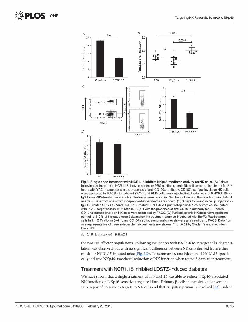
the two NK effector populations. Following incubation with Ba/F3-Rae1ε target cells, degranu-lation was observed, but with no significant difference between NK cells derived from eithermock- or NCR1.15-injected mice (Fig. 3D). To summarize, one injection of NCR1.15 specifi-cally induced NKp46-associated reduction of NK function when tested 3 days after treatment.
Treatment with NCR1.15 inhibited LDSTZ-induced diabetesWe have shown that a single treatment with NCR1.15 was able to reduce NKp46-associatedNK function on NKp46-sensitive target cell lines. Primary β-cells in the islets of Langerhanswere reported to serve as targets to NK cells and that NKp46 is primarily involved [33]. Indeed,
Fig 3. Single dose treatment with NCR1.15 inhibits NKp46-mediated activity on NK cells. (A) 3 daysfollowing i.p. injection of NCR1.15, isotype control or PBS purified splenic NK cells were co-incubated for 2–4hours with YAC-1 target cells in the presence of anti-CD107a antibody. CD107a surface levels on NK cellswere assessed by FACS. (B) Labeled YAC-1 and RMA cells were injected into the tail vein of 5 NCR1.15-, c-IgG1 κ- or PBS-treated mice. Cells in the lungs were quantified 3–4 hours following the injection using FACSanalysis. Data from one of two independent experiments are shown. (C) 3 days following mice i.p. injection c-IgG1 κ-treatedUBC-GFP and NCR1.15-treated C57BL/6WT purified splenic NK cells were co-incubatedwith PD1.6 target cells in 1:1:1 ratio (E1:E2:T) with the presence of anti-CD107a antibody for 3–4 hours.CD107a surface levels on NK cells were assessed by FACS. (D) Purified splenic NK cells harvested fromcontrol- or NCR1.15-treated mice 3 days after the treatment were co-incubated with Ba/F3-Rae1ε targetcells in 1:1 E:T ratio for 3–4 hours. CD107a surface expression levels were analyzed using FACS. Data fromone representative of three independent experiments are shown. ** p<0.01 by Student's unpaired t-test.Bars, ±SD.
doi:10.1371/journal.pone.0118936.g003
Targeting NK Reactivity by mAb to NKp46
PLOSONE | DOI:10.1371/journal.pone.0118936 February 26, 2015 8 / 15

co-incubation of pancreatic primary beta cells from naïve mice with NK cells derived fromNCR1.15 single injection treated mice, both from C57BL/6 origin, resulted in significantly re-duced degranulation levels (Fig. 4A). We next investigated whether NCR1.15 short-term treat-ment could affect the early development of diabetes. An accepted model of experimentalautoimmune diabetes in mice is the induction of diabetes by multiple injections of a low doseof streptozotocin (LDSTZ) [34–36]. A single NCR1.15 treatment comprises a single 100 μg/mouse injection. Here, we treated mice once with NCR1.15 (50 μg/mouse) followed by 5 conse-cutive days of i.p. injections with STZ and completed with additional NCR1.15 treatment(Fig. 4B, inset). The NCR1.15 treatment significantly impaired diabetes development. Hyper-glycemia, defined as a non-fasting blood glucose concentration of�300 mg/dl in two sequen-tial measurements, was significantly less severe in NCR1.15-treated mice compared to a mocktreatment group (Fig. 4B). Accordingly, reduction in body weight of the NCR1.15-treated micewas significantly lower than that of the control mice, indicating the healthier status ofNCR1.15-treated mice (Fig. 4C). Additionally, histological analysis of the pancreas showed re-duced mononuclear infiltration and reduced insulitis score for the islets within NCR1.15-treat-ed mice as compared to mock-treated group (Fig. 4D, E). To sum up, these results indicate that
Fig 4. Short term treatment with NCR1.15 down-regulates NKp46-mediated activity on pancreatic β-cells and inhibits LD-STZ induced diabetes. (A)Purified splenic NK cells from treated cells were co-incubated for 2–4 hours with purified C57BL/6 β-cells in the presence of anti-CD107a antibody. CD107asurface levels on NK cells were assessed by FACS. * p<0.05 by Student's unpaired t-test. (B) LD-STZ induced diabetes development in C57BL/6 micefollowing 50μg injections at days-2 and 5. (n = 8). Animals were considered diabetic when blood glucose was�300mg/dl. *** p<0.005 Kaplan-M log-rank(Mental-Cox). (C) Body weight of mice in the indicated groups following the LD-STZ administration; body weight baselines at day 0 of the control andNCR1.15 groups were 19.2±0.86, 19.8±1 grams, respectively. * p<0.05, ** p<0.01, *** p<0.005 by Student's unpaired t-test. Bars, ±SD. (D)Representative H&E sections of the pancreas from sex&age-matched C57BL/6 mice treated with NCR1.15 or mock-treated (IgG1, κ) in LD-STZ induceddiabetes model (magnification X40). (E) Summary of insulitis score over the days calculated as described in [37]. Score is quantified from 0 (low) to 4 (mostsevere).
doi:10.1371/journal.pone.0118936.g004
Targeting NK Reactivity by mAb to NKp46
PLOSONE | DOI:10.1371/journal.pone.0118936 February 26, 2015 9 / 15
NKp46 is important for the development of diabetes and the destruction of islets in theLDSTZ model.
Repeated long-term treatments with NCR1.15 inhibited T1Ddevelopment in NODmiceWe have previously shown that beta cells specifically express the ligand for the NK-activatingreceptor NKp46/Ncr1 and that NK cells kill beta cells in an NKp46/Ncr1-dependent manner[10,33]. Furthermore, we have shown that anti-mNKp46/Ncr1 antibodies generated throughactive immunization inhibit T1D development [10]. To recapitulate these observations, we in-vestigated the effect of repeated NCR1.15 treatments in C57BL/6 mice. WT mice were treatedwith NCR1.15 every other day for two weeks and both PBMCs and splenocytes were tested 3days following the last injection. Our results were very similar to those observed for oneNCR1.15 inoculation (Figs. 2, 3). The fraction of NK cells in PBMCs and the spleen did notchange between NCR1.15- and mock-treated mice (Fig. 5A). We also employed the protocol ofprolonged multiple injections of NCR1.15 to Ncr1gfp/+ mice and tested for the fraction of GFP-positive cells indicating NK fraction [20] and for GFP intensity indicating Ncr1 gene promoteractivity. Neither the numbers of GFP+ nor the GFP intensity was different between NCR1.15-and mock-treated mice (Fig. 5B). In contrast, membrane-associated NKp46 expression was sig-nificantly reduced in PBMCs and in splenocytes harvested from mice that had undergone pro-longed multiple NCR1.15 treatments compared to mock-treated mice (Fig. 5C, D). As with thesingle NCR1.15 injection, this reduction was not due to reduced Ncr1 gene promoter activity asdeduced from qRT-PCR levels of the Ncr1 transcript (Fig. 5E) and from the Ncr1-encodedGFP levels (Fig. 5B). Since treatment lasted for two weeks and mice were tested 3 days later, wecould investigate whether NCR1.15 treatment conferred changes in NK subsets representingdevelopmental NK stages. Antibodies to CD3, NK1.1, CD27, and CD11b were used to defineCD3-NK1.1+CD11blowCD27+, CD3-NK1.1+CD11b+CD27+ and CD3-NK1.1+CD11b+CD27low
NK developmental stages. No significant difference was observed between NCR1.15- andmock- treatment for cell numbers among the NK developmental stages. In contrast, mem-brane-associated NKp46 levels were significantly reduced in NCR1.15-treated mice in all stud-ied NK developmental stages as shown in S1 Fig.
We then studied repeated long-term NCR1.15 treatments in NODmice. As for C57BL/6mice, NCR1.15 treatment of NOD mice did not affect number of NK cells (Fig. 6A), but it didsignificantly reduce membrane-associated NKp46 expression (Fig. 6B, C). To directly demon-strate that blocking of NKp46 in NODmice will block diabetes development, we injectedNCR1.15 into NODmice every other day (100μg per injection) beginning in week eight. Asshown in Fig. 6D, repeated NCR1.15 treatment significantly inhibited T1D development inNOD female mice compared to treatment with PBS or isotype Ab control.
DiscussionWe have recently reported that NKp46, a primary activating receptor expressed by NK cells,recognizes ligands expressed by islet β-cells, and that in the absence of NKp46 diabetes devel-opment is inhibited [10,33]. In the current study, we investigated whether treatment of micewith NKp46 specific monoclonal antibody could down-regulate NKp46-mediated NK functionand inhibit T1D development. We developed the NCR1.15 mAb, which specifically recognizesmNKp46 (Fig. 1) and showed that treatment of mice with NCR1.15 reduces NKp46 mem-brane-associated expression by NK cells (Fig. 2). This reduction was paralleled by reducedNKp46-mediated NK function (Fig. 3). NCR1.15 treatment did not reduce the fraction of NKcells in the blood or the spleen; neither did it affect the NKG2D-mediated function of NK cells
Targeting NK Reactivity by mAb to NKp46
PLOSONE | DOI:10.1371/journal.pone.0118936 February 26, 2015 10 / 15

(Figs. 2, 3). This NKp46-specific reduction in NK function was shown both in vitro and in vivo(Fig. 3). Finally, we showed that multiple treatments with NCR1.15 inhibited development ofdiabetes in the LDSTZ model and in NODmice (Figs. 4, 6). Acquired histopathological datafor the LDSTZ model indicate that development of insulitis is attenuated in NCR1.15-treatedmice as compared to mock-treated mice (Fig. 4).
Targeted immunotherapy involving treatment with antibodies to activate immune cellularreceptors and immune molecules holds great promise for the treatment and cure of autoim-mune and inflammatory diseases [38,39]. In particular, Ab-mediated modulation of immuneeffector receptors and receptor-expressing cells have been widely studied for the TCR [38,40].Modulation of the TCR-CD3z by specific antibodies has been suggested to down-regulate Tcell function through (i) antigenic modulation (suppression) of the cell-surface TCR, (ii) deple-tion of the T cells, and /or (iii) the anergy of T cells [40,41]. We have shown that treatmentwith anti-NKp46 resulted in antigenic modulation of NK-expressed NKp46, but not in generalNK depletion. Anergy wise, our treatment induced down-regulation of NKp46-specific NKfunction, but it did not induce general NK anergy, as tested by NKG2D function (Fig. 3D).Down-regulation of NK function following exposure to anti-NKp46 mAb has also been re-ported by Jewett et al. [42]. However, Narni-Mancinelli et al. reported a more complex pheno-type [43]: short time treatment with anti-NKp46 resulted in reduced NKp46-mediated NKfunction in accordance with our results and other reports [42] even though long time-treat-ment caused enhanced NKp46-mediated NK function [43]. This observation for long-timetreatment was not reported by others nor us, and could stem from the diphtheria toxin-mediat-ed whole NK depletion performed before the long time treatment [43]. Yet entire NK cell
Fig 5. Prolongedmultiple treatments with NCR1.15 down-regulates the surface expression of NKp46.Mice were injected i.p. 8 doses of PBS, 100 μgof NCR1.15 or cIgG1,κ every other day; 3 days after the last inoculation mice were sacrificed for further analysis. (A) C57BL/6WT PBMCs and splenocyteswere harvested and stained to detect the levels of CD3-NK1.1+ NK cells. Data from one representative of three independent experiments are shown. (B)PBMCs drained from NCR1gfp/+ mice were analyzed for GFP expression following the prolonged repeated treatments. (C, D) Membrane associated NKp46and NKG2D levels were analyzed on gated CD3ε-NK1.1+ NK cells. (E) NCR1 transcript levels from treated mice splenocytes were assessed by qRT-PCR.Data from one representative of two independent experiments are shown. * p<0.05, ** p<0.01 by Student's unpaired t-test. Bars, ±SD.
doi:10.1371/journal.pone.0118936.g005
Targeting NK Reactivity by mAb to NKp46
PLOSONE | DOI:10.1371/journal.pone.0118936 February 26, 2015 11 / 15

depletion before treatment with anti-NKp46 does not represent a plausible treatment regimenin humans.
Several groups reported that treatment of T1D with mAb specific to CD3 resulted in a sig-nificant therapeutic effect through the antigenic modulation of the TCR-CD3z complex (re-viewed in [40]). However, others have shown that this therapeutic effect was due to inductionof T cell anergy or apoptosis of activated T cells [44]. T1D disease is considered to be mainlydriven by the activation of T-lymphocytes against pancreatic β-cells [45]. However, findingssupport the view that T1D can be regarded as an innate inflammatory disease affecting the en-tire pancreas, but with its main clinical manifestations emanating from the loss of the insulin-producing cells [46]. In this respect, we, among others, have shown that NK cells contribute tothe early stage of T1D development [10,20]. The question then arose whether treatment withmAb targeting NK activating receptors can have a therapeutic benefit for T1D. The NKG2D re-ceptor is a primary activating receptor for NK cells and a co-stimulatory receptor for T cells.Blockage of NKG2D prevented T1D in NODmice. The mechanism reported involved blockageof NKG2D receptors to their ligands, as well as internalization of the receptor. However, no de-pletion of NK cells or CD8+ T cells was observed [47]. NKp46 is a major NK activating recep-tor, and it is mostly restricted to NK cells compared to the broader expression spectrum ofNKG2D. We demonstrated that β-cells both human and mouse islets express ligand(s) forNKp46 [10,33]. We also showed that in the absence of NKp46 diabetes development is inhib-ited. We then proved that NKp46 is involved in the direct killing of human islets β-cells thatare intended to be used for transplantation [10,33]. The NKp46 ligand is detected on healthymouse β-cells, but we demonstrated that NK cells were absent from the vicinity of the islets inhealthy mice and were detected in situ in proximity with β-cells in NODmice [10,33]. In this
Fig 6. Treatment with NCR1.15 reduces NKp46-mediated NK activity on β-cells and inhibits T1Ddevelopment. (A) 8 weeks after the first treatment NOD PBMCs drained frommice within the indicatedgroups were stained and analyzed for NK levels. NK cell levels were normalized to mock (PBS) treatment. (B)Representative histogram of membrane associated NKp46 levels on gated CD3ε-CD49b+ NK cells from theindicated groups. (C) Normalized GeoMFI of membrane associated NKp46 on gated NK cells, ** p<0.01 byStudent's unpaired t-test. GeoMFI values were normalized to mock (PBS) treatment. (D) T1D development inNOD female mice in the indicated groups. (n = 14–15). Animals were considered diabetic when bloodglucose was�250mg/dl. * p<0.05 Kaplan-M log-rank (Mental-Cox). Bars, ±SD.
doi:10.1371/journal.pone.0118936.g006
Targeting NK Reactivity by mAb to NKp46
PLOSONE | DOI:10.1371/journal.pone.0118936 February 26, 2015 12 / 15

study, we have investigated a treatment based on the inoculation of mAb specific for NKp46.Following treatment, NK cells were not depleted nor did they undergo general anergy. Even so,we observed a very clear antigenic modulation of membrane-associated NKp46 and NKp46specific NK function that was followed by a significant impairment of T1D development. Itwill be interesting to further explore whether the combination of both anti-NKp46 and anti-NKG2D mAbs will lead to an even more pronounced delayed onset of this disease.
To summarize, in this study, we have shown that anti-NKp46 treatment was sufficient tosignificantly delay diabetes early development in LDSTZ and NODmodels. Both, short-termand repeated long-term treatments with anti-NKp46 mAb resulted in an NKp46-specific im-pairment of NK function without NK depletion. This treatment represents a pinpoint-targetedtherapeutic approach intended to specifically remove/reduce the NKp46 component in NKfunction neither harming non-NKp46 mediated NK activity nor affecting T cells.
Supporting InformationS1 Fig. Membrane-associated NKp46 levels in different NK developmental stages (Y-axis inhistogram representation). The membrane-associated NKp46 expression was significantly re-duced in NCR1.15 treated mice comparing to control treatments in all studied NKdevelopmental stages.(TIF)
AcknowledgmentsWe give special thanks to Karmela Miklić and Tatiana Rabinsky for providing valuable techni-cal assistance to the undertaking of the research summarized in this article.
Author ContributionsConceived and designed the experiments: RY AP. Performed the experiments: RY CG AS AMOG KN. Analyzed the data: RY AP. Contributed reagents/materials/analysis tools: UH AC SN.Wrote the paper: RY SJ OM AP.
References1. Kruse PH, Matta J, Ugolini S, Vivier E. Natural cytotoxicity receptors and their ligands. Immunol Cell
Biol.2013.
2. Vivier E, Tomasello E, Baratin M, Walzer T, Ugolini S. Functions of natural killer cells. Nat Immu-nol.2008; 9: 503–510. doi: 10.1038/ni1582 PMID: 18425107
3. Shibuya A. Development and functions of natural killer cells. Int J Hematol.2003; 78: 1–6. PMID:12894844
4. Colonna M, Jonjic S, Watzl C. Natural killer cells: fighting viruses and much more. Nat Immunol.2011;12: 107–110. doi: 10.1038/ni0211-107 PMID: 21245897
5. Long EO. Negative signaling by inhibitory receptors: the NK cell paradigm. Immunol Rev.2008; 224:70–84. doi: 10.1111/j.1600-065X.2008.00660.x PMID: 18759921
6. Karre K, Ljunggren HG, Piontek G, Kiessling R. Selective rejection of H-2-deficient lymphoma variantssuggests alternative immune defence strategy. Nature.1986; 319: 675–678. PMID: 3951539
7. Arnon TI, Markel G, Mandelboim O. Tumor and viral recognition by natural killer cells receptors. SeminCancer Biol.2006; 16: 348–358. PMID: 16893656
8. Lam RA, Chwee JY, Le Bert N, Sauer M, Pogge von Strandmann E, Gasser S Regulation of self-li-gands for activating natural killer cell receptors. Ann Med.2013; 45: 384–394. doi: 10.3109/07853890.2013.792495 PMID: 23701136
9. Moretta A, Bottino C, Vitale M, Pende D, Cantoni C, Mingari MC, et al. Activating receptors and core-ceptors involved in human natural killer cell-mediated cytolysis. Annu Rev Immunol.2001; 19: 197–223. PMID: 11244035
Targeting NK Reactivity by mAb to NKp46
PLOSONE | DOI:10.1371/journal.pone.0118936 February 26, 2015 13 / 15
10. Gur C, Porgador A, ElboimM, Gazit R, Mizrahi S, Stern-Ginossar N, et al. The activating receptorNKp46 is essential for the development of type 1 diabetes. Nat Immunol.2010; 11: 121–128. doi: 10.1038/ni.1834 PMID: 20023661
11. Rodacki M, Milech A, de Oliveira JE. NK cells and type 1 diabetes. Clin Dev Immunol.2006; 13: 101–107. PMID: 17162353
12. Chandy KG, Charles MA, Buckingham B, Waldeck N, Kershnar A, Gupta S. Deficiency of monoclonalantibody (Leu 7) defined NK cells in newly diagnosed insulin-dependent diabetes mellitus. ImmunolLett.1984; 8: 89–91. PMID: 6746020
13. Shachner MS, Markmann JF, Bassiri H, Kim JI, Naji A, Barker CF. Direct assessment of the role of NKcells in autoimmune diabetes. J Surg Res.1992; 52: 601–604. PMID: 1382151
14. Dotta F, Censini S, van Halteren AG, Marselli L, Masini M, Dionisi S, et al. Coxsackie B4 virus infectionof beta cells and natural killer cell insulitis in recent-onset type 1 diabetic patients. Proc Natl Acad Sci US A.2007; 104: 5115–5120. PMID: 17360338
15. Alba A, Planas R, Clemente X, Carrillo J, Ampudia R, Puertas MC, et al. Natural killer cells are requiredfor accelerated type 1 diabetes driven by interferon-beta. Clin Exp Immunol.2008; 151: 467–475. doi:10.1111/j.1365-2249.2007.03580.x PMID: 18190608
16. Gur C, Enk J, Weitman E, Bachar E, Suissa Y, Cohen G, et al. The expression of the beta cell-derivedautoimmune ligand for the killer receptor nkp46 is attenuated in type 2 diabetes. PLoS One.2013; 8:e74033. doi: 10.1371/journal.pone.0074033 PMID: 24009765
17. Ferlazzo G, Tsang ML, Moretta L, Melioli G, Steinman RM, Munz C. Human dendritic cells activate rest-ing natural killer (NK) cells and are recognized via the NKp30 receptor by activated NK cells. J ExpMed.2002; 195: 343–351. PMID: 11828009
18. Koch J, Steinle A, Watzl C, Mandelboim O. Activating natural cytotoxicity receptors of natural killer cellsin cancer and infection. Trends Immunol.2013; 34: 182–191. doi: 10.1016/j.it.2013.01.003 PMID:23414611
19. Mandelboim O, Lieberman N, Lev M, Paul L, Arnon TI, Bushkin Y, et al. Recognition of haemagglutininson virus-infected cells by NKp46 activates lysis by human NK cells. Nature.2001; 409: 1055–1060.PMID: 11234016
20. Garg A, Barnes PF, Porgador A, Roy S, Wu S, Nanda JS, et al. Vimentin expressed on Mycobacteriumtuberculosis-infected humanmonocytes is involved in binding to the NKp46 receptor. J Immunol.2006;177: 6192–6198. PMID: 17056548
21. Bloushtain N, Qimron U, Bar-Ilan A, Hershkovitz O, Gazit R, Fima E, et al. Membrane-associatedheparan sulfate proteoglycans are involved in the recognition of cellular targets by NKp30 and NKp46.J Immunol.2004; 173: 2392–2401. PMID: 15294952
22. Ito K, Higai K, Sakurai M, Shinoda C, Yanai K, Azuma Y, et al. Binding of natural cytotoxicity receptorNKp46 to sulfate- and alpha2,3-NeuAc-containing glycans and its mutagenesis. Biochem Biophys ResCommun.2011; 406: 377–382. doi: 10.1016/j.bbrc.2011.02.050 PMID: 21329668
23. Ito K, Higai K, Shinoda C, Sakurai M, Yanai K, Azuma Y, et al. Unlike natural killer (NK) p30, natural cy-totoxicity receptor NKp44 binds to multimeric alpha2,3-NeuNAc-containing N-glycans. Biol PharmBull.2012; 35: 594–600. PMID: 22466566
24. Halfteck GG, Elboim M, Gur C, Achdout H, Ghadially H, Mandelboim O. Enhanced in vivo growth oflymphoma tumors in the absence of the NK-activating receptor NKp46/NCR1. J Immunol.2009; 182:2221–2230. doi: 10.4049/jimmunol.0801878 PMID: 19201876
25. Cerwenka A, Bakker AB, McClanahan T, Wagner J, Wu J, Phillips JH, et al. Retinoic acid early induc-ible genes define a ligand family for the activating NKG2D receptor in mice. Immunity.2000; 12: 721–727. PMID: 10894171
26. Arnon TI, Achdout H, Lieberman N, Gazit R, Gonen-Gross T, Katz G, et al. The mechanisms controllingthe recognition of tumor- and virus-infected cells by NKp46. Blood.2004; 103: 664–672. PMID:14504081
27. Hershkovitz O, Rosental B, Rosenberg LA, Navarro-Sanchez ME, Jivov S, Zilka A, et al. NKp44 recep-tor mediates interaction of the envelope glycoproteins from the West Nile and dengue viruses with NKcells. J Immunol.2009; 183: 2610–2621. doi: 10.4049/jimmunol.0802806 PMID: 19635919
28. Rosental B, Brusilovsky M, Hadad U, Oz D, Appel MY, Afergan F, et al. Proliferating cell nuclear antigenis a novel inhibitory ligand for the natural cytotoxicity receptor NKp44. J Immunol.2011; 187: 5693–5702. doi: 10.4049/jimmunol.1102267 PMID: 22021614
29. Gazit R, Gruda R, ElboimM, Arnon TI, Katz G, Achdout H, et al. Lethal influenza infection in the ab-sence of the natural killer cell receptor gene Ncr1. Nat Immunol.2006; 7: 517–523. PMID: 16565719
30. Nita II, Hershfinkel M, Kantor C, Rutter GA, Lewis EC, Sekler I. Pancreatic beta-cell Na+ channels con-trol global Ca2+ signaling and oxidative metabolism by inducing Na+ and Ca2+ responses that are
Targeting NK Reactivity by mAb to NKp46
PLOSONE | DOI:10.1371/journal.pone.0118936 February 26, 2015 14 / 15

propagated into mitochondria. FASEB J.2014; 28: 3301–3312. doi: 10.1096/fj.13-248161 PMID:24719357
31. Yu J, Mitsui T, Wei M, Mao H, Butchar JP, Shah MV, et al. NKp46 identifies an NKT cell subset suscep-tible to leukemic transformation in mouse and human. J Clin Invest.2011; 121: 1456–1470. doi: 10.1172/JCI43242 PMID: 21364281
32. Glasner A, Ghadially H, Gur C, Stanietsky N, Tsukerman P, Enk J, et al. Recognition and prevention oftumor metastasis by the NK receptor NKp46/NCR1. J Immunol.2012; 188: 2509–2515. doi: 10.4049/jimmunol.1102461 PMID: 22308311
33. Gur C, Enk J, Kassem SA, Suissa Y, Magenheim J, Stolovich-Rain M, et al. Recognition and killing ofhuman and murine pancreatic beta cells by the NK receptor NKp46. J Immunol.2011; 187: 3096–3103.doi: 10.4049/jimmunol.1101269 PMID: 21849674
34. Paik SG, Fleischer N, Shin SI. Insulin-dependent diabetes mellitus induced by subdiabetogenic dosesof streptozotocin: obligatory role of cell-mediated autoimmune processes. Proc Natl Acad Sci U SA.1980; 77: 6129–6133. PMID: 6449703
35. Like AA, Rossini AA. Streptozotocin-induced pancreatic insulitis: new model of diabetes mellitus. Sci-ence.1976; 193: 415–417. PMID: 180605
36. O'Brien BA, Harmon BV, Cameron DP, Allan DJ. Beta-cell apoptosis is responsible for the developmentof IDDM in the multiple low-dose streptozotocin model. J Pathol.1996; 178: 176–181. PMID: 8683386
37. Leiter EH. The NODmouse: a model for insulin-dependent diabetes mellitus. Curr Protoc Immu-nol.2001; Chapter 15: Unit 15 19.
38. Chan AC, Carter PJ. Therapeutic antibodies for autoimmunity and inflammation. Nat Rev Immu-nol.2010; 10: 301–316. doi: 10.1038/nri2761 PMID: 20414204
39. Colucci F, Cilio CM. Taming killer cells may halt diabetes progression. Nat Immunol.2010; 11: 111–112. doi: 10.1038/ni0210-111 PMID: 20084067
40. Chatenoud L, Bluestone JA. CD3-specific antibodies: a portal to the treatment of autoimmunity. NatRev Immunol.2007; 7: 622–632. PMID: 17641665
41. Chatenoud L, Baudrihaye MF, Kreis H, Goldstein G, Schindler J, Bach JF. Human in vivo antigenicmodulation induced by the anti-T cell OKT3 monoclonal antibody. Eur J Immunol.1982; 12: 979–982.PMID: 6759145
42. Jewett A, Man YG, Tseng HC. Dual functions of natural killer cells in selection and differentiation ofstem cells; role in regulation of inflammation and regeneration of tissues. J Cancer.2013; 4: 12–24. doi:10.7150/jca.5519 PMID: 23386901
43. Narni-Mancinelli E, Ugolini S, Vivier E. [Natural killer cells: adaptation and memory in innate immunity].Med Sci (Paris).2013; 29: 389–395. doi: 10.1051/medsci/2013294012 PMID: 23621934
44. Smith JA, Tso JY, Clark MR, Cole MS, Bluestone JA. Nonmitogenic anti-CD3 monoclonal antibodiesdeliver a partial T cell receptor signal and induce clonal anergy. J Exp Med.1997; 185: 1413–1422.PMID: 9126922
45. Christianson SW, Shultz LD, Leiter EH. Adoptive transfer of diabetes into immunodeficient NOD-scid/scid mice. Relative contributions of CD4+ and CD8+ T-cells from diabetic versus prediabetic NOD.NON-Thy-1a donors. Diabetes.1993; 42: 44–55. PMID: 8093606
46. Akirav E, Kushner JA, Herold KC. Beta-cell mass and type 1 diabetes: going, going, gone? Diabe-tes.2008; 57: 2883–2888. doi: 10.2337/db07-1817 PMID: 18971435
47. Ogasawara K, Hamerman JA, Ehrlich LR, Bour-Jordan H, Santamaria P, Bluestone JA, et al. NKG2Dblockade prevents autoimmune diabetes in NODmice. Immunity.2004; 20: 757–767. PMID: 15189740
Targeting NK Reactivity by mAb to NKp46
PLOSONE | DOI:10.1371/journal.pone.0118936 February 26, 2015 15 / 15