
Study of pro-apoptotic protein RTP801 homeostasis and its ...
Systems Analysis of ATF3 in Stress Response and CancerReveals Opposing Effects on Pro-Apoptotic Genes in p53PathwayYujiro Tanaka1*, Aya Nakamura1, Masaki Suimye Morioka2, Shoko Inoue1, Mimi Tamamori-Adachi1,
Kazuhiko Yamada1, Kenji Taketani1, Junya Kawauchi1, Miki Tanaka-Okamoto3, Jun Miyoshi3, Hiroshi
Tanaka2, Shigetaka Kitajima1*
1 Laboratory of Genome Structure and Regulation, School of Biomedical Science and Department of Biochemical Genetics, Medical Research Institute, Tokyo Medical and
Dental University, Tokyo, Japan, 2 Department of Computational Biology, School of Medical Sciences and Department of Bioinformatics, Tokyo Medical and Dental
University, Tokyo, Japan, 3 Department of Molecular Biology, Osaka Medical Center for Cancer and Cardiovascular Diseases, Osaka, Japan
Abstract
Stress-inducible transcription factors play a pivotal role in cellular adaptation to environment to maintain homeostasis andintegrity of the genome. Activating transcription factor 3 (ATF3) is induced by a variety of stress and inflammatoryconditions and is over-expressed in many kinds of cancer cells. However, molecular mechanisms underlying pleiotropicfunctions of ATF3 have remained elusive. Here we employed systems analysis to identify genome-wide targets of ATF3 thatis either induced by an alkylating agent methyl methanesulfonate (MMS) or over-expressed in a prostate tumour cell lineLNCaP. We show that stress-induced and cancer-associated ATF3 is recruited to 5,984 and 1,423 targets, respectively, in thehuman genome, 89% of which are common. Notably, ATF3 targets are highly enriched for not only ATF/CRE motifs but alsobinding sites of several other stress-inducible transcription factors indicating an extensive network of stress response factorsin transcriptional regulation of target genes. Further analysis of effects of ATF3 knockdown on these targets revealed thatstress-induced ATF3 regulates genes in metabolic pathways, cell cycle, apoptosis, cell adhesion, and signalling includinginsulin, p53, Wnt, and VEGF pathways. Cancer-associated ATF3 is involved in regulation of distinct sets of genes in processessuch as calcium signalling, Wnt, p53 and diabetes pathways. Notably, stress-induced ATF3 binds to 40% of p53 targets andactivates pro-apoptotic genes such as TNFRSF10B/DR5 and BBC3/PUMA. Cancer-associated ATF3, by contrast, repressesthese pro-apoptotic genes in addition to CDKN1A/p21. Taken together, our data reveal an extensive network of stress-inducible transcription factors and demonstrate that ATF3 has opposing, cell context-dependent effects on p53 targetgenes in DNA damage response and cancer development.
Citation: Tanaka Y, Nakamura A, Morioka MS, Inoue S, Tamamori-Adachi M, et al. (2011) Systems Analysis of ATF3 in Stress Response and Cancer RevealsOpposing Effects on Pro-Apoptotic Genes in p53 Pathway. PLoS ONE 6(10): e26848. doi:10.1371/journal.pone.0026848
Editor: Fazlul H. Sarkar, Wayne State University School of Medicine, United States of America
Received August 15, 2011; Accepted October 4, 2011; Published October 26, 2011
Copyright: � 2011 Tanaka et al. This is an open-access article distributed under the terms of the Creative Commons Attribution License, which permitsunrestricted use, distribution, and reproduction in any medium, provided the original author and source are credited.
Funding: This work was supported in part by grants to S.K. (18012015, 18055008, and 21590302) and a grant to Y.T. (20510183) from the Ministry of Education,Culture, Sports, Science, and Technology, Japan. The funders had no role in study design, data collection and analysis, decision to publish, or preparation of themanuscript.
Competing Interests: The authors have declared that no competing interests exist.
* E-mail: [email protected] (YT); [email protected] (SK)
Introduction
Transcription factors play important roles in temporal regula-
tion of gene expression in serum stimulation of human cells[1,2].
Cellular adaptation to various environmental stress conditions is
also regulated by transcription factors that co-ordinately modulate
expression of genes involved in maintenance of cellular homeo-
stasis and genetic integrity. Such a system plays an important role
for not only survival of normal cells but also resistance of cancer
cells to metabolic and genotoxic stresses. A crucial step towards
understanding molecular mechanisms underlying stress responses
is the identification of target genes of each transcription factor.
Studies in yeast employing gene expression profiling [3,4] and
more recently systems analysis by chromatin immunoprecipitation
of transcription factors [5] have revealed a genome-wide networks
of transcription factors regulating expression of genes that
orchestrate cell cycle, gene transcription, protein synthesis, and
DNA repair in response to MMS. In mammals, the tumour
suppressor p53 plays a pivotal role in DNA damage response
through transcriptional control of several hundred genes [6,7].
Importantly, only a small subset of p53 targets are activated under
specific conditions[8], indicating that either binding of p53 to
targets [9–11] or trans-activation potential of p53 proteins [12,13]
may be affected by accessory molecules.
ATF3 is a member of the ATF/CREB family of basic-leucine
zipper (b-Zip) type transcription factors [14]and is a highly
versatile stress sensor for a wide range of conditions including
hypoxia, hyponutrition, oxidative stresses, ER stresses, and various
genotoxic stresses[15,16] as well as inflammatory reactions
[17,18]. ATF3 is also activated by serum stimulation and
downstream of c-Myc[19], and is frequently over-expressed in
various tumours including those of the prostate[20], breast[21],
and Hodgkin’s lymphomas[22]. Importantly, several lines of
evidence has indicated a close link between ATF3 and p53
PLoS ONE | www.plosone.org 1 October 2011 | Volume 6 | Issue 10 | e26848
signalling pathways. Thus, ATF3 is induced downstream of p53
upon DNA damage and functions as an effector of p53-mediated
cell death [6,23–25]. In addition, ATF3 potentiates p53 by directly
binding and inhibiting its ubiquitilation, implying that ATF3 can
modulate the activity of p53 [26,27]. Furthermore, ATF3 appears
to confer a negative feedback to the p53 pathway by down-
regulating TP53 gene expression[28], recapitulating a similar
feedback regulation of inflammatory cytokine genes by ATF3[17].
Corroborating such a negative feedback model, a recent study has
demonstrated that ATF3 is induced by Cyclosporin, an immune
suppressor, and promotes skin cancer by down-regulating
TP53[29]. Taken together, these studies suggest that ATF3
interacts with the p53 pathway both as a downstream effector of
p53-mediated cell death and as a positive and negative regulator of
p53 signalling.
Alteration in interaction between ATF3 and transcription
factors such as p53 in different type of cells and/or environmental
conditions may in part account for distinct effects of ATF3 on cell
fate (i.e. pro-apoptotic or growth-promoting for untransformed
cells or malignantly transformed cells, respectively)[21]. Previous
work from our laboratory has also described pleiotropic functions
of ATF3 under various stress conditions[18,19,25,28,30]. Here we
carried out systems analysis of ATF3 targets and identified
thousands of ATF3 binding sites in the genome. We show that
ATF3 constitutes an extensively overlapping gene regulatory
network with other stress-inducible transcription factors and
regulates cell cycle, cell death, adhesion, and several signalling
pathways including p53. Notably, ATF3 binds to 40% of known
targets of p53 and regulates apoptotic cell death through co-
activation of a subset of pro-apoptotic genes stress response while
repressing the same targets in cancer cells constitutively expressing
ATF3. Possible switching mechanisms between pro-survival and
pro-apoptotic ATF3 functions will be discussed.
Results
Systems analysis identifies thousands of ATF3 targets inthe human genome.
To identify genomic targets of ATF3, chromatin immunopre-
cipitation analysis was carried out using HCT116 human colon
cancer cell line stimulated by MMS and LNCaP prostate cancer
cell line which constitutively expresses ATF3. As reported
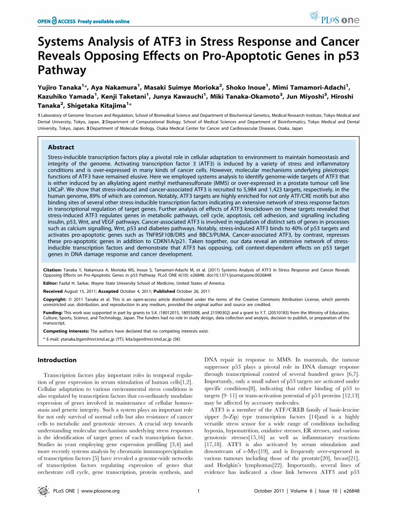
previously[24], MMS treatment of HCT116 cells induced
transient expression of ATF3 reaching a peak at 6 hours after
stimulation (Fig. 1A), whereas increased levels of ATF3 proteins
were detected after 3 hours and reached a maximum level at 12
hours of stimulation (Fig. 1B). Genomic DNA was prepared from
either HCT116 cells treated with MMS for 6 hours or untreated
LNCaP cells, immunoprecipitated with anti-ATF3 antibodies, and
hybridized to NimbleGen’s human RefSeq HG18 promoter tiling
arrays. Subsequently, peak detection was carried out by Model-
based Analysis of 2-colour Arrays (MA2C) package[31], which
revealed an unexpectedly large number of ATF3 targets at a cut-
off of 0.2% FDR (Fig. S1A and S1B). We identified 5,984 and
1,423 targets of ATF3 in MMS-treated HCT116 cells and LNCaP
cells, respectively, 1,269 (i.e. 89%) of which were sheared between
the two models. (Fig. 1C). We detected no targets on the Y
chromosome from HCT116 consistent with its female origin. We
also successfully identified thirteen ATF3 targets which had been
previously shown to be regulated by ATF3 in various cell types.
Motifs of stress-associated transcription factors are over-represented in ATF3 targets.
ATF3 could be recruited to its targets either by directly binding
to a consensus recognition sequence TGACGTCA or alternatively
by interacting with other transcription factors including a large
number of b-Zip proteins[14,32]. To assess if the ATF/CRE motif
or any other transcription factor motifs are enriched among the
potential ATF3 targets, the number of promoters in the ATF3
targets containing at least one hit of a given motif in the
TRANSFAC database was analysed by the P-Match algorithm
(Fig. S2). Table 1 summarizes enrichment of a given motif as
represented by the number of promoters in ATF3 targets
containing the motif subtracted by the number of promoters in
a background gene set containing the same motif. As expected,
ATF/CRE binding sites were the most enriched motifs in ATF3
targets (p-value for HCT116-specific targets: 3.76610240 to
4.34610222, p-value for common targets of HCT116 and LNCaP
cells: 0 to 4.2061026). Moreover, physical distance between
predicted ATF/CRE sequences of the motif scan and ATF3 peaks
of the ChIP analysis was within a range of a few hundred base
pairs (i.e. close to the resolution of the ChIP analysis) for the
majority of ATF3 targets (Fig. 2). By contrast, there was no such
Figure 1. ChIP-on-chip analysis of ATF3 targets. (A) RT-PCRanalysis of ATF3 in HCT116 cells treated with 50 ng/ml MMS. (B)Western blot analysis of ATF3 proteins in MMS-treated HCT116 cells.(C)Common and unique targets of ATF3 in HCT116 cells and LNCaPcells.doi:10.1371/journal.pone.0026848.g001
Function of ATF3 in Stress Response and Cancer
PLoS ONE | www.plosone.org 2 October 2011 | Volume 6 | Issue 10 | e26848
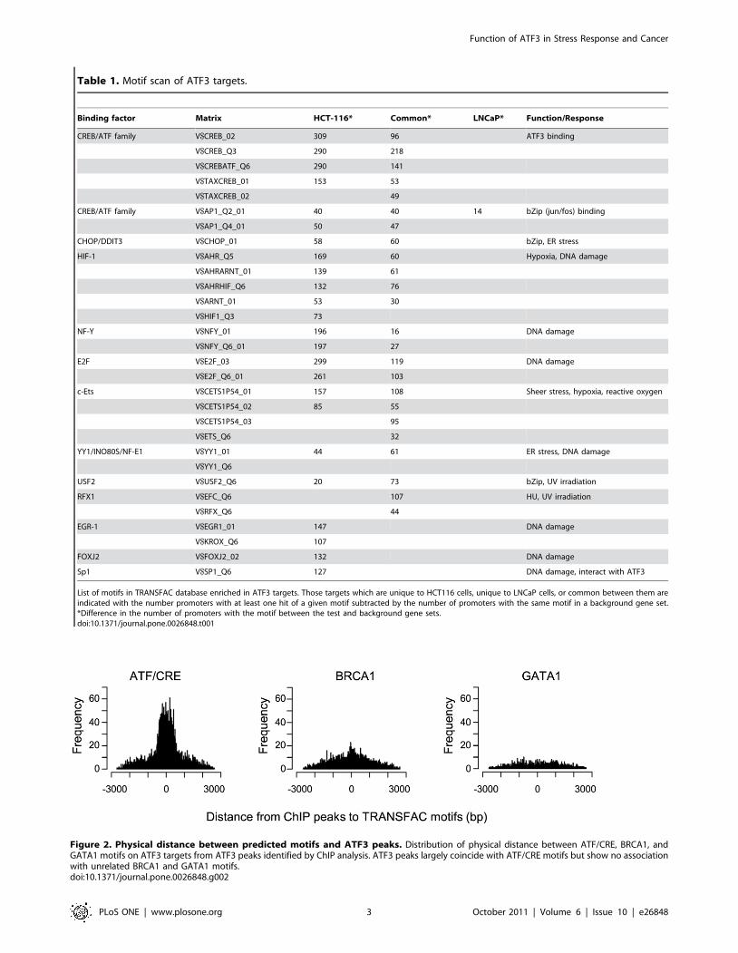
Figure 2. Physical distance between predicted motifs and ATF3 peaks. Distribution of physical distance between ATF/CRE, BRCA1, andGATA1 motifs on ATF3 targets from ATF3 peaks identified by ChIP analysis. ATF3 peaks largely coincide with ATF/CRE motifs but show no associationwith unrelated BRCA1 and GATA1 motifs.doi:10.1371/journal.pone.0026848.g002
Table 1. Motif scan of ATF3 targets.
Binding factor Matrix HCT-116* Common* LNCaP* Function/Response
CREB/ATF family V$CREB_02 309 96 ATF3 binding
V$CREB_Q3 290 218
V$CREBATF_Q6 290 141
V$TAXCREB_01 153 53
V$TAXCREB_02 49
CREB/ATF family V$AP1_Q2_01 40 40 14 bZip (jun/fos) binding
V$AP1_Q4_01 50 47
CHOP/DDIT3 V$CHOP_01 58 60 bZip, ER stress
HIF-1 V$AHR_Q5 169 60 Hypoxia, DNA damage
V$AHRARNT_01 139 61
V$AHRHIF_Q6 132 76
V$ARNT_01 53 30
V$HIF1_Q3 73
NF-Y V$NFY_01 196 16 DNA damage
V$NFY_Q6_01 197 27
E2F V$E2F_03 299 119 DNA damage
V$E2F_Q6_01 261 103
c-Ets V$CETS1P54_01 157 108 Sheer stress, hypoxia, reactive oxygen
V$CETS1P54_02 85 55
V$CETS1P54_03 95
V$ETS_Q6 32
YY1/INO80S/NF-E1 V$YY1_01 44 61 ER stress, DNA damage
V$YY1_Q6
USF2 V$USF2_Q6 20 73 bZip, UV irradiation
RFX1 V$EFC_Q6 107 HU, UV irradiation
V$RFX_Q6 44
EGR-1 V$EGR1_01 147 DNA damage
V$KROX_Q6 107
FOXJ2 V$FOXJ2_02 132 DNA damage
Sp1 V$SP1_Q6 127 DNA damage, interact with ATF3
List of motifs in TRANSFAC database enriched in ATF3 targets. Those targets which are unique to HCT116 cells, unique to LNCaP cells, or common between them areindicated with the number promoters with at least one hit of a given motif subtracted by the number of promoters with the same motif in a background gene set.*Difference in the number of promoters with the motif between the test and background gene sets.doi:10.1371/journal.pone.0026848.t001
Function of ATF3 in Stress Response and Cancer
PLoS ONE | www.plosone.org 3 October 2011 | Volume 6 | Issue 10 | e26848
association between unrelated BRCA1 and GATA1 motifs and
ATF3 peaks (Fig. 2). Taken together, these data strongly suggest
that the majority of ATF3 peaks of the ChIP analysis can be
explained by direct binding of ATF3 to ATF/CRE motifs.
Intriguingly, the motif scan also revealed that binding sites of
other stress-inducible transcription factors were highly over-
represented in ATF3 targets in response to DNA damage
(Table 1). These included major regulators of ER stress (DDIT3,
NF-E1), hypoxia (HIF-1A), UV stress (USF2, RFX1), oxidative
stress (c-Ets), and DNA damage (NF-Y, E2F, EGR-1, FOXJ2,
Sp1). Of note, members of the b-Zip family (AP-1, DDIT3, and
USF2) have a potential to heterodimerize with ATF3[33] and Sp1
has been reported to physically interact with ATF3[34]. It is
therefore possible that ATF3 is recruited to a subset of its targets
indirectly through association with other stress-inducible tran-
scription factors.
ATF3 regulates distinct biological processes in stressresponse and in cancer
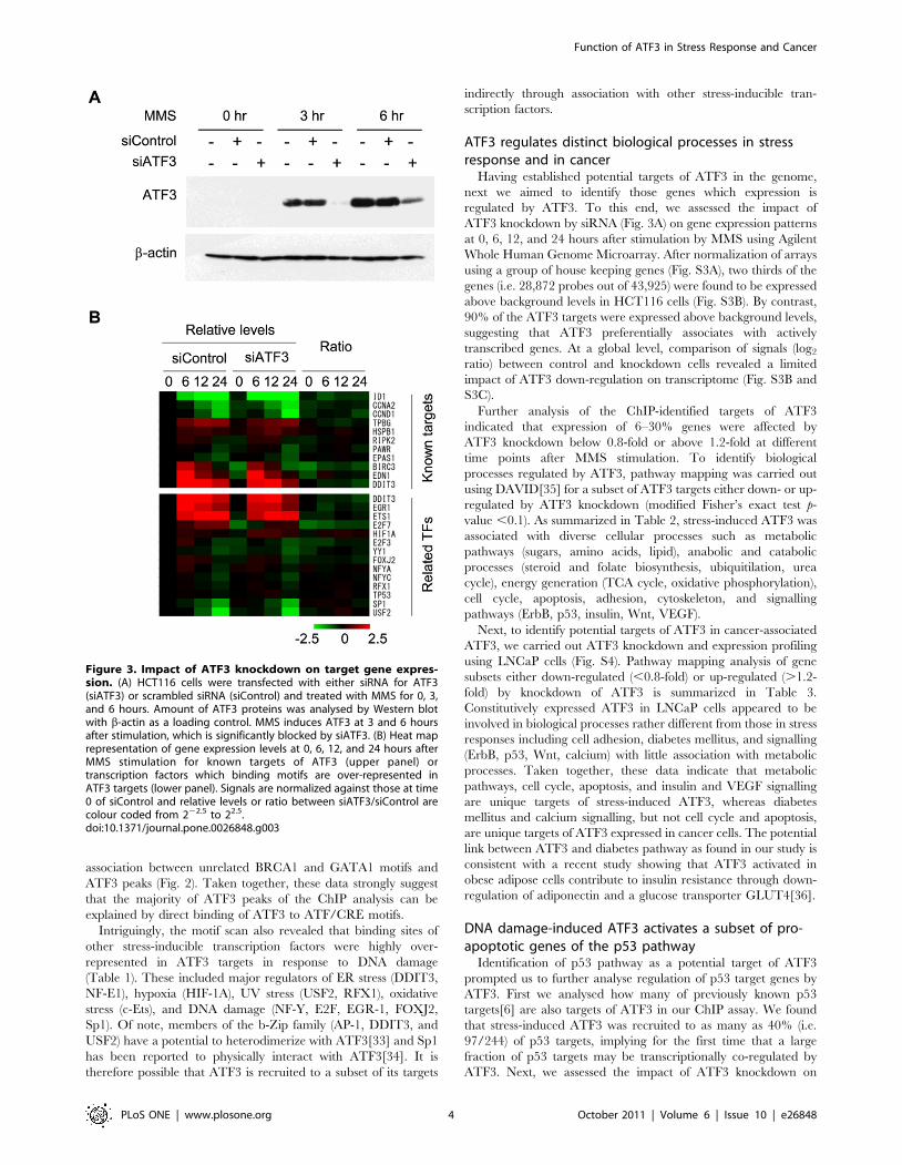
Having established potential targets of ATF3 in the genome,
next we aimed to identify those genes which expression is
regulated by ATF3. To this end, we assessed the impact of
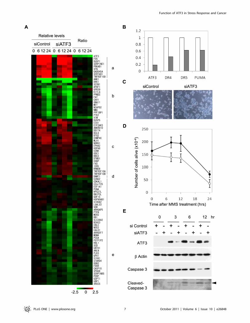
ATF3 knockdown by siRNA (Fig. 3A) on gene expression patterns
at 0, 6, 12, and 24 hours after stimulation by MMS using Agilent
Whole Human Genome Microarray. After normalization of arrays
using a group of house keeping genes (Fig. S3A), two thirds of the
genes (i.e. 28,872 probes out of 43,925) were found to be expressed
above background levels in HCT116 cells (Fig. S3B). By contrast,
90% of the ATF3 targets were expressed above background levels,
suggesting that ATF3 preferentially associates with actively
transcribed genes. At a global level, comparison of signals (log2
ratio) between control and knockdown cells revealed a limited
impact of ATF3 down-regulation on transcriptome (Fig. S3B and
S3C).
Further analysis of the ChIP-identified targets of ATF3
indicated that expression of 6–30% genes were affected by
ATF3 knockdown below 0.8-fold or above 1.2-fold at different
time points after MMS stimulation. To identify biological
processes regulated by ATF3, pathway mapping was carried out
using DAVID[35] for a subset of ATF3 targets either down- or up-
regulated by ATF3 knockdown (modified Fisher’s exact test p-
value ,0.1). As summarized in Table 2, stress-induced ATF3 was
associated with diverse cellular processes such as metabolic
pathways (sugars, amino acids, lipid), anabolic and catabolic
processes (steroid and folate biosynthesis, ubiquitilation, urea
cycle), energy generation (TCA cycle, oxidative phosphorylation),
cell cycle, apoptosis, adhesion, cytoskeleton, and signalling
pathways (ErbB, p53, insulin, Wnt, VEGF).
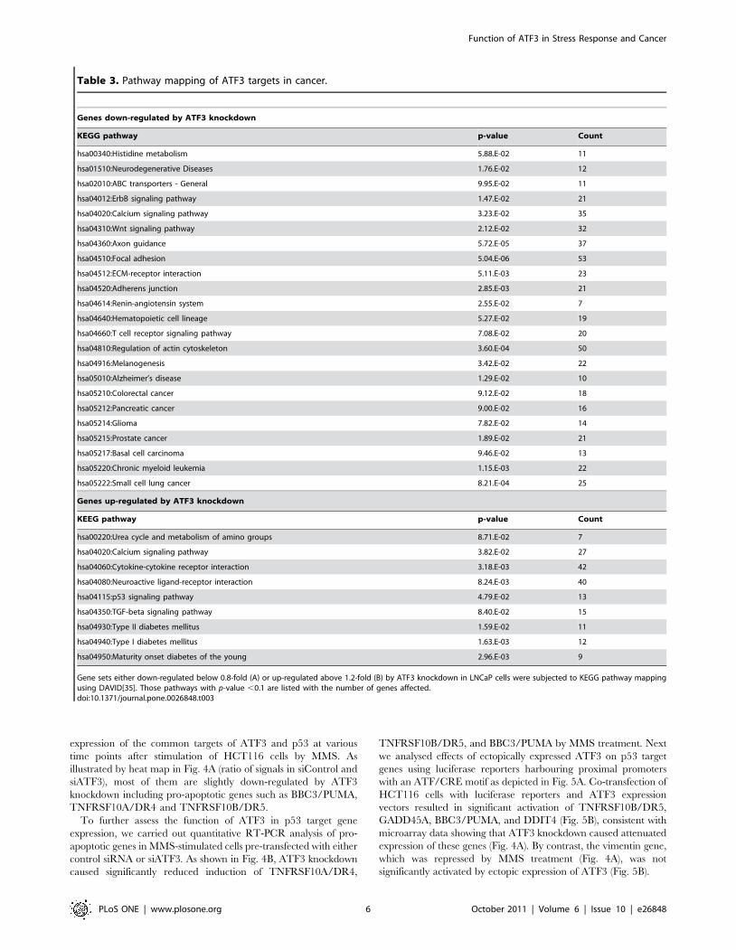
Next, to identify potential targets of ATF3 in cancer-associated
ATF3, we carried out ATF3 knockdown and expression profiling
using LNCaP cells (Fig. S4). Pathway mapping analysis of gene
subsets either down-regulated (,0.8-fold) or up-regulated (.1.2-
fold) by knockdown of ATF3 is summarized in Table 3.
Constitutively expressed ATF3 in LNCaP cells appeared to be
involved in biological processes rather different from those in stress
responses including cell adhesion, diabetes mellitus, and signalling
(ErbB, p53, Wnt, calcium) with little association with metabolic
processes. Taken together, these data indicate that metabolic
pathways, cell cycle, apoptosis, and insulin and VEGF signalling
are unique targets of stress-induced ATF3, whereas diabetes
mellitus and calcium signalling, but not cell cycle and apoptosis,
are unique targets of ATF3 expressed in cancer cells. The potential
link between ATF3 and diabetes pathway as found in our study is
consistent with a recent study showing that ATF3 activated in
obese adipose cells contribute to insulin resistance through down-
regulation of adiponectin and a glucose transporter GLUT4[36].
DNA damage-induced ATF3 activates a subset of pro-apoptotic genes of the p53 pathway
Identification of p53 pathway as a potential target of ATF3
prompted us to further analyse regulation of p53 target genes by
ATF3. First we analysed how many of previously known p53
targets[6] are also targets of ATF3 in our ChIP assay. We found
that stress-induced ATF3 was recruited to as many as 40% (i.e.
97/244) of p53 targets, implying for the first time that a large
fraction of p53 targets may be transcriptionally co-regulated by
ATF3. Next, we assessed the impact of ATF3 knockdown on
Figure 3. Impact of ATF3 knockdown on target gene expres-sion. (A) HCT116 cells were transfected with either siRNA for ATF3(siATF3) or scrambled siRNA (siControl) and treated with MMS for 0, 3,and 6 hours. Amount of ATF3 proteins was analysed by Western blotwith b-actin as a loading control. MMS induces ATF3 at 3 and 6 hoursafter stimulation, which is significantly blocked by siATF3. (B) Heat maprepresentation of gene expression levels at 0, 6, 12, and 24 hours afterMMS stimulation for known targets of ATF3 (upper panel) ortranscription factors which binding motifs are over-represented inATF3 targets (lower panel). Signals are normalized against those at time0 of siControl and relative levels or ratio between siATF3/siControl arecolour coded from 222.5 to 22.5.doi:10.1371/journal.pone.0026848.g003
Function of ATF3 in Stress Response and Cancer
PLoS ONE | www.plosone.org 4 October 2011 | Volume 6 | Issue 10 | e26848
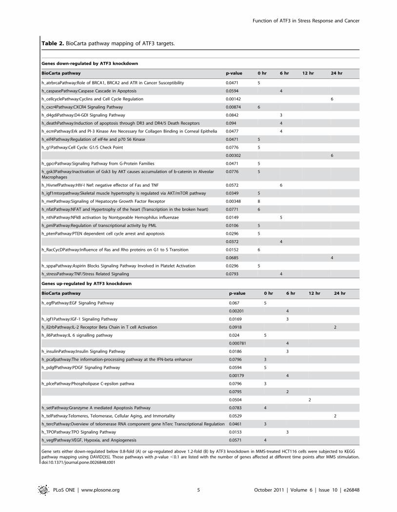
Table 2. BioCarta pathway mapping of ATF3 targets.
Genes down-regulated by ATF3 knockdown
BioCarta pathway p-value 0 hr 6 hr 12 hr 24 hr
h_atrbrcaPathway:Role of BRCA1, BRCA2 and ATR in Cancer Susceptibility 0.0471 5
h_caspasePathway:Caspase Cascade in Apoptosis 0.0594 4
h_cellcyclePathway:Cyclins and Cell Cycle Regulation 0.00142 6
h_cxcr4Pathway:CXCR4 Signaling Pathway 0.00874 6
h_d4gdiPathway:D4-GDI Signaling Pathway 0.0842 3
h_deathPathway:Induction of apoptosis through DR3 and DR4/5 Death Receptors 0.094 4
h_ecmPathway:Erk and PI-3 Kinase Are Necessary for Collagen Binding in Corneal Epithelia 0.0477 4
h_eif4Pathway:Regulation of eIF4e and p70 S6 Kinase 0.0471 5
h_g1Pathway:Cell Cycle: G1/S Check Point 0.0776 5
0.00302 6
h_gpcrPathway:Signaling Pathway from G-Protein Families 0.0471 5
h_gsk3Pathway:Inactivation of Gsk3 by AKT causes accumulation of b-catenin in AlveolarMacrophages
0.0776 5
h_HivnefPathway:HIV-I Nef: negative effector of Fas and TNF 0.0572 6
h_igf1mtorpathway:Skeletal muscle hypertrophy is regulated via AKT/mTOR pathway 0.0349 5
h_metPathway:Signaling of Hepatocyte Growth Factor Receptor 0.00348 8
h_nfatPathway:NFAT and Hypertrophy of the heart (Transcription in the broken heart) 0.0771 6
h_nthiPathway:NFkB activation by Nontypeable Hemophilus influenzae 0.0149 5
h_pmlPathway:Regulation of transcriptional activity by PML 0.0106 5
h_ptenPathway:PTEN dependent cell cycle arrest and apoptosis 0.0296 5
0.0372 4
h_RacCycDPathway:Influence of Ras and Rho proteins on G1 to S Transition 0.0152 6
0.0685 4
h_sppaPathway:Aspirin Blocks Signaling Pathway Involved in Platelet Activation 0.0296 5
h_stressPathway:TNF/Stress Related Signaling 0.0793 4
Genes up-regulated by ATF3 knockdown
BioCarta pathway p-value 0 hr 6 hr 12 hr 24 hr
h_egfPathway:EGF Signaling Pathway 0.067 5
0.00201 4
h_igf1Pathway:IGF-1 Signaling Pathway 0.0169 3
h_il2rbPathway:IL-2 Receptor Beta Chain in T cell Activation 0.0918 2
h_il6Pathway:IL 6 signalling pathway 0.024 5
0.000781 4
h_insulinPathway:Insulin Signaling Pathway 0.0186 3
h_pcafpathway:The information-processing pathway at the IFN-beta enhancer 0.0796 3
h_pdgfPathway:PDGF Signaling Pathway 0.0594 5
0.00179 4
h_plcePathway:Phospholipase C-epsilon pathwa 0.0796 3
0.0795 2
0.0504 2
h_setPathway:Granzyme A mediated Apoptosis Pathway 0.0783 4
h_telPathway:Telomeres, Telomerase, Cellular Aging, and Immortality 0.0529 2
h_tercPathway:Overview of telomerase RNA component gene hTerc Transcriptional Regulation 0.0461 3
h_TPOPathway:TPO Signaling Pathway 0.0153 3
h_vegfPathway:VEGF, Hypoxia, and Angiogenesis 0.0571 4
Gene sets either down-regulated below 0.8-fold (A) or up-regulated above 1.2-fold (B) by ATF3 knockdown in MMS-treated HCT116 cells were subjected to KEGGpathway mapping using DAVID[35]. Those pathways with p-value ,0.1 are listed with the number of genes affected at different time points after MMS stimulation.doi:10.1371/journal.pone.0026848.t001
Function of ATF3 in Stress Response and Cancer
PLoS ONE | www.plosone.org 5 October 2011 | Volume 6 | Issue 10 | e26848
expression of the common targets of ATF3 and p53 at various
time points after stimulation of HCT116 cells by MMS. As
illustrated by heat map in Fig. 4A (ratio of signals in siControl and
siATF3), most of them are slightly down-regulated by ATF3
knockdown including pro-apoptotic genes such as BBC3/PUMA,
TNFRSF10A/DR4 and TNFRSF10B/DR5.
To further assess the function of ATF3 in p53 target gene
expression, we carried out quantitative RT-PCR analysis of pro-
apoptotic genes in MMS-stimulated cells pre-transfected with either
control siRNA or siATF3. As shown in Fig. 4B, ATF3 knockdown
caused significantly reduced induction of TNFRSF10A/DR4,
TNFRSF10B/DR5, and BBC3/PUMA by MMS treatment. Next
we analysed effects of ectopically expressed ATF3 on p53 target
genes using luciferase reporters harbouring proximal promoters
with an ATF/CRE motif as depicted in Fig. 5A. Co-transfection of
HCT116 cells with luciferase reporters and ATF3 expression
vectors resulted in significant activation of TNFRSF10B/DR5,
GADD45A, BBC3/PUMA, and DDIT4 (Fig. 5B), consistent with
microarray data showing that ATF3 knockdown caused attenuated
expression of these genes (Fig. 4A). By contrast, the vimentin gene,
which was repressed by MMS treatment (Fig. 4A), was not
significantly activated by ectopic expression of ATF3 (Fig. 5B).
Table 3. Pathway mapping of ATF3 targets in cancer.
Genes down-regulated by ATF3 knockdown
KEGG pathway p-value Count
hsa00340:Histidine metabolism 5.88.E-02 11
hsa01510:Neurodegenerative Diseases 1.76.E-02 12
hsa02010:ABC transporters - General 9.95.E-02 11
hsa04012:ErbB signaling pathway 1.47.E-02 21
hsa04020:Calcium signaling pathway 3.23.E-02 35
hsa04310:Wnt signaling pathway 2.12.E-02 32
hsa04360:Axon guidance 5.72.E-05 37
hsa04510:Focal adhesion 5.04.E-06 53
hsa04512:ECM-receptor interaction 5.11.E-03 23
hsa04520:Adherens junction 2.85.E-03 21
hsa04614:Renin-angiotensin system 2.55.E-02 7
hsa04640:Hematopoietic cell lineage 5.27.E-02 19
hsa04660:T cell receptor signaling pathway 7.08.E-02 20
hsa04810:Regulation of actin cytoskeleton 3.60.E-04 50
hsa04916:Melanogenesis 3.42.E-02 22
hsa05010:Alzheimer’s disease 1.29.E-02 10
hsa05210:Colorectal cancer 9.12.E-02 18
hsa05212:Pancreatic cancer 9.00.E-02 16
hsa05214:Glioma 7.82.E-02 14
hsa05215:Prostate cancer 1.89.E-02 21
hsa05217:Basal cell carcinoma 9.46.E-02 13
hsa05220:Chronic myeloid leukemia 1.15.E-03 22
hsa05222:Small cell lung cancer 8.21.E-04 25
Genes up-regulated by ATF3 knockdown
KEEG pathway p-value Count
hsa00220:Urea cycle and metabolism of amino groups 8.71.E-02 7
hsa04020:Calcium signaling pathway 3.82.E-02 27
hsa04060:Cytokine-cytokine receptor interaction 3.18.E-03 42
hsa04080:Neuroactive ligand-receptor interaction 8.24.E-03 40
hsa04115:p53 signaling pathway 4.79.E-02 13
hsa04350:TGF-beta signaling pathway 8.40.E-02 15
hsa04930:Type II diabetes mellitus 1.59.E-02 11
hsa04940:Type I diabetes mellitus 1.63.E-03 12
hsa04950:Maturity onset diabetes of the young 2.96.E-03 9
Gene sets either down-regulated below 0.8-fold (A) or up-regulated above 1.2-fold (B) by ATF3 knockdown in LNCaP cells were subjected to KEGG pathway mappingusing DAVID[35]. Those pathways with p-value ,0.1 are listed with the number of genes affected.doi:10.1371/journal.pone.0026848.t003
Function of ATF3 in Stress Response and Cancer
PLoS ONE | www.plosone.org 6 October 2011 | Volume 6 | Issue 10 | e26848
Function of ATF3 in Stress Response and Cancer
PLoS ONE | www.plosone.org 7 October 2011 | Volume 6 | Issue 10 | e26848
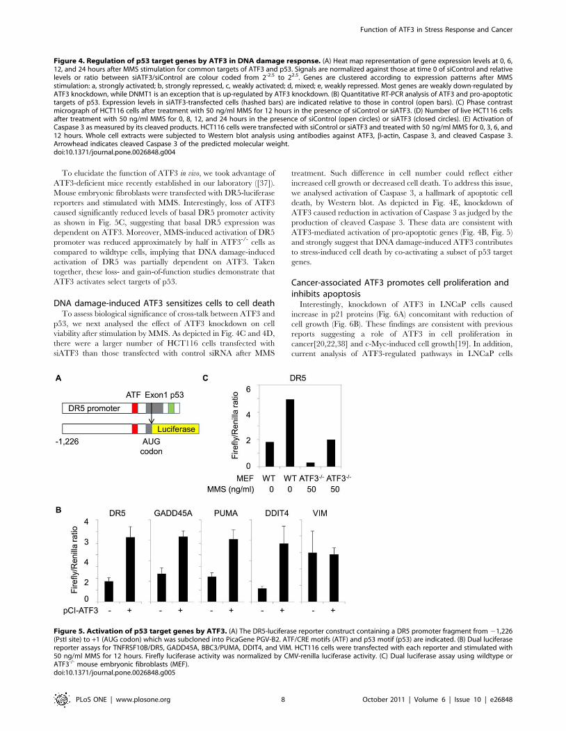
To elucidate the function of ATF3 in vivo, we took advantage of
ATF3-deficient mice recently established in our laboratory ([37]).
Mouse embryonic fibroblasts were transfected with DR5-luciferase
reporters and stimulated with MMS. Interestingly, loss of ATF3
caused significantly reduced levels of basal DR5 promoter activity
as shown in Fig. 5C, suggesting that basal DR5 expression was
dependent on ATF3. Moreover, MMS-induced activation of DR5
promoter was reduced approximately by half in ATF3-/- cells as
compared to wildtype cells, implying that DNA damage-induced
activation of DR5 was partially dependent on ATF3. Taken
together, these loss- and gain-of-function studies demonstrate that
ATF3 activates select targets of p53.
DNA damage-induced ATF3 sensitizes cells to cell deathTo assess biological significance of cross-talk between ATF3 and
p53, we next analysed the effect of ATF3 knockdown on cell
viability after stimulation by MMS. As depicted in Fig. 4C and 4D,
there were a larger number of HCT116 cells transfected with
siATF3 than those transfected with control siRNA after MMS
treatment. Such difference in cell number could reflect either
increased cell growth or decreased cell death. To address this issue,
we analysed activation of Caspase 3, a hallmark of apoptotic cell
death, by Western blot. As depicted in Fig. 4E, knockdown of
ATF3 caused reduction in activation of Caspase 3 as judged by the
production of cleaved Caspase 3. These data are consistent with
ATF3-mediated activation of pro-apoptotic genes (Fig. 4B, Fig. 5)
and strongly suggest that DNA damage-induced ATF3 contributes
to stress-induced cell death by co-activating a subset of p53 target
genes.
Cancer-associated ATF3 promotes cell proliferation andinhibits apoptosis
Interestingly, knockdown of ATF3 in LNCaP cells caused
increase in p21 proteins (Fig. 6A) concomitant with reduction of
cell growth (Fig. 6B). These findings are consistent with previous
reports suggesting a role of ATF3 in cell proliferation in
cancer[20,22,38] and c-Myc-induced cell growth[19]. In addition,
current analysis of ATF3-regulated pathways in LNCaP cells
Figure 4. Regulation of p53 target genes by ATF3 in DNA damage response. (A) Heat map representation of gene expression levels at 0, 6,12, and 24 hours after MMS stimulation for common targets of ATF3 and p53. Signals are normalized against those at time 0 of siControl and relativelevels or ratio between siATF3/siControl are colour coded from 2-2.5 to 22.5. Genes are clustered according to expression patterns after MMSstimulation: a, strongly activated; b, strongly repressed, c, weakly activated; d, mixed; e, weakly repressed. Most genes are weakly down-regulated byATF3 knockdown, while DNMT1 is an exception that is up-regulated by ATF3 knockdown. (B) Quantitative RT-PCR analysis of ATF3 and pro-apoptotictargets of p53. Expression levels in siATF3-transfected cells (hashed bars) are indicated relative to those in control (open bars). (C) Phase contrastmicrograph of HCT116 cells after treatment with 50 ng/ml MMS for 12 hours in the presence of siControl or siATF3. (D) Number of live HCT116 cellsafter treatment with 50 ng/ml MMS for 0, 8, 12, and 24 hours in the presence of siControl (open circles) or siATF3 (closed circles). (E) Activation ofCaspase 3 as measured by its cleaved products. HCT116 cells were transfected with siControl or siATF3 and treated with 50 ng/ml MMS for 0, 3, 6, and12 hours. Whole cell extracts were subjected to Western blot analysis using antibodies against ATF3, b-actin, Caspase 3, and cleaved Caspase 3.Arrowhead indicates cleaved Caspase 3 of the predicted molecular weight.doi:10.1371/journal.pone.0026848.g004
Figure 5. Activation of p53 target genes by ATF3. (A) The DR5-luciferase reporter construct containing a DR5 promoter fragment from 21,226(PstI site) to +1 (AUG codon) which was subcloned into PicaGene PGV-B2. ATF/CRE motifs (ATF) and p53 motif (p53) are indicated. (B) Dual luciferasereporter assays for TNFRSF10B/DR5, GADD45A, BBC3/PUMA, DDIT4, and VIM. HCT116 cells were transfected with each reporter and stimulated with50 ng/ml MMS for 12 hours. Firefly luciferase activity was normalized by CMV-renilla luciferase activity. (C) Dual luciferase assay using wildtype orATF3-/- mouse embryonic fibroblasts (MEF).doi:10.1371/journal.pone.0026848.g005
Function of ATF3 in Stress Response and Cancer
PLoS ONE | www.plosone.org 8 October 2011 | Volume 6 | Issue 10 | e26848
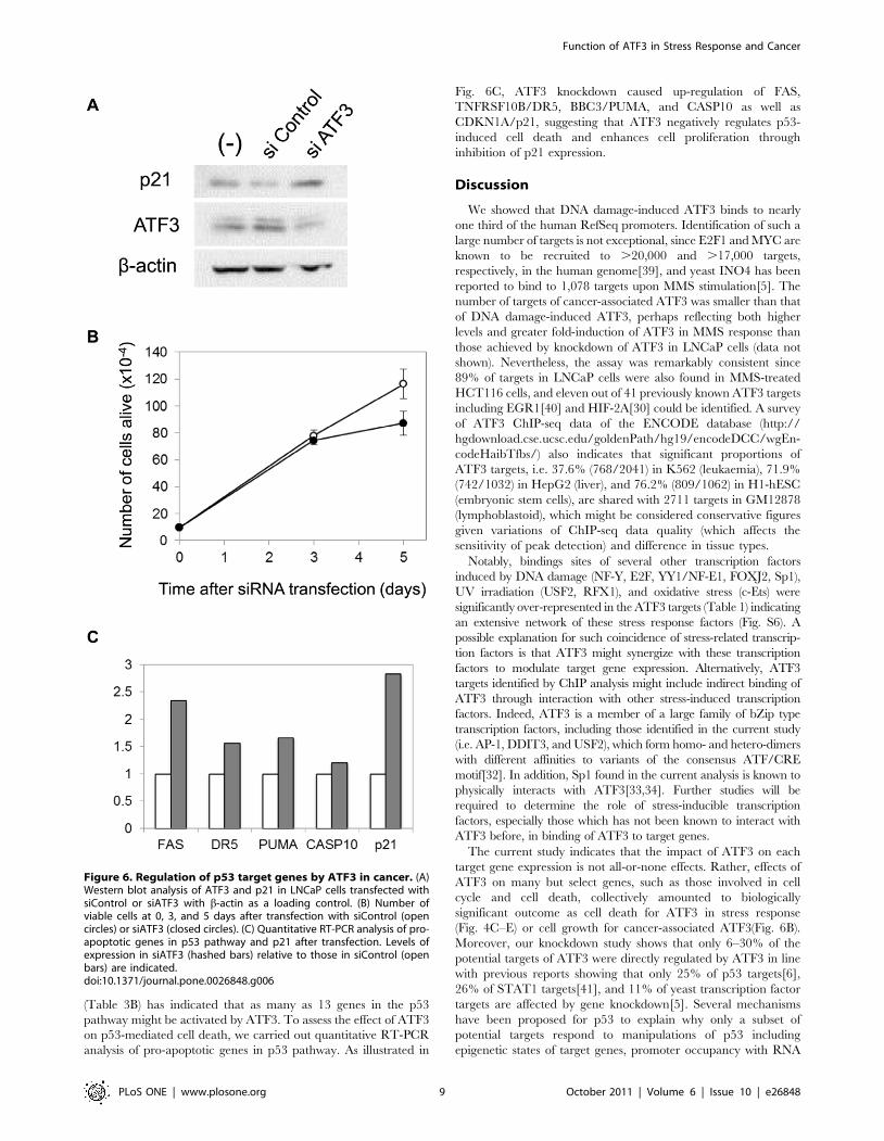
(Table 3B) has indicated that as many as 13 genes in the p53
pathway might be activated by ATF3. To assess the effect of ATF3
on p53-mediated cell death, we carried out quantitative RT-PCR
analysis of pro-apoptotic genes in p53 pathway. As illustrated in
Fig. 6C, ATF3 knockdown caused up-regulation of FAS,
TNFRSF10B/DR5, BBC3/PUMA, and CASP10 as well as
CDKN1A/p21, suggesting that ATF3 negatively regulates p53-
induced cell death and enhances cell proliferation through
inhibition of p21 expression.
Discussion
We showed that DNA damage-induced ATF3 binds to nearly
one third of the human RefSeq promoters. Identification of such a
large number of targets is not exceptional, since E2F1 and MYC are
known to be recruited to .20,000 and .17,000 targets,
respectively, in the human genome[39], and yeast INO4 has been
reported to bind to 1,078 targets upon MMS stimulation[5]. The
number of targets of cancer-associated ATF3 was smaller than that
of DNA damage-induced ATF3, perhaps reflecting both higher
levels and greater fold-induction of ATF3 in MMS response than
those achieved by knockdown of ATF3 in LNCaP cells (data not
shown). Nevertheless, the assay was remarkably consistent since
89% of targets in LNCaP cells were also found in MMS-treated
HCT116 cells, and eleven out of 41 previously known ATF3 targets
including EGR1[40] and HIF-2A[30] could be identified. A survey
of ATF3 ChIP-seq data of the ENCODE database (http://
hgdownload.cse.ucsc.edu/goldenPath/hg19/encodeDCC/wgEn-
codeHaibTfbs/) also indicates that significant proportions of
ATF3 targets, i.e. 37.6% (768/2041) in K562 (leukaemia), 71.9%
(742/1032) in HepG2 (liver), and 76.2% (809/1062) in H1-hESC
(embryonic stem cells), are shared with 2711 targets in GM12878
(lymphoblastoid), which might be considered conservative figures
given variations of ChIP-seq data quality (which affects the
sensitivity of peak detection) and difference in tissue types.
Notably, bindings sites of several other transcription factors
induced by DNA damage (NF-Y, E2F, YY1/NF-E1, FOXJ2, Sp1),
UV irradiation (USF2, RFX1), and oxidative stress (c-Ets) were
significantly over-represented in the ATF3 targets (Table 1) indicating
an extensive network of these stress response factors (Fig. S6). A
possible explanation for such coincidence of stress-related transcrip-
tion factors is that ATF3 might synergize with these transcription
factors to modulate target gene expression. Alternatively, ATF3
targets identified by ChIP analysis might include indirect binding of
ATF3 through interaction with other stress-induced transcription
factors. Indeed, ATF3 is a member of a large family of bZip type
transcription factors, including those identified in the current study
(i.e. AP-1, DDIT3, and USF2), which form homo- and hetero-dimers
with different affinities to variants of the consensus ATF/CRE
motif[32]. In addition, Sp1 found in the current analysis is known to
physically interacts with ATF3[33,34]. Further studies will be
required to determine the role of stress-inducible transcription
factors, especially those which has not been known to interact with
ATF3 before, in binding of ATF3 to target genes.
The current study indicates that the impact of ATF3 on each
target gene expression is not all-or-none effects. Rather, effects of
ATF3 on many but select genes, such as those involved in cell
cycle and cell death, collectively amounted to biologically
significant outcome as cell death for ATF3 in stress response
(Fig. 4C–E) or cell growth for cancer-associated ATF3(Fig. 6B).
Moreover, our knockdown study shows that only 6–30% of the
potential targets of ATF3 were directly regulated by ATF3 in line
with previous reports showing that only 25% of p53 targets[6],
26% of STAT1 targets[41], and 11% of yeast transcription factor
targets are affected by gene knockdown[5]. Several mechanisms
have been proposed for p53 to explain why only a subset of
potential targets respond to manipulations of p53 including
epigenetic states of target genes, promoter occupancy with RNA
Figure 6. Regulation of p53 target genes by ATF3 in cancer. (A)Western blot analysis of ATF3 and p21 in LNCaP cells transfected withsiControl or siATF3 with b-actin as a loading control. (B) Number ofviable cells at 0, 3, and 5 days after transfection with siControl (opencircles) or siATF3 (closed circles). (C) Quantitative RT-PCR analysis of pro-apoptotic genes in p53 pathway and p21 after transfection. Levels ofexpression in siATF3 (hashed bars) relative to those in siControl (openbars) are indicated.doi:10.1371/journal.pone.0026848.g006
Function of ATF3 in Stress Response and Cancer
PLoS ONE | www.plosone.org 9 October 2011 | Volume 6 | Issue 10 | e26848
polymerases, and recruitment of essential co-factors[42]. Similar
mechanisms might be responsible for gene-selective effects of
ATF3. Alternatively, multiple forms of ATF3 complexes, such as
hetero-dimers with different bZip proteins, might underlie
differential effects of ATF3 on target genes.
In the past several years, an increasing number of studies have
indicated that ATF3 has pleiotropic functions depending on cell
context. Importantly, our study revealed that stress-induced ATF3
and cancer-associated ATF3 have opposing effects on p53 and
Wnt pathways: p53 pathway is activated in stress response (Fig. S5)
consistent with the function of ATF3 in p53-mediated cell death
[6,23–25], whereas Wnt pathway is activated in cancer as
previously reported in a study on ATF3 transgenic mice
developing mammary tumours [43]. One might argue that the
difference in genetic backgrounds and/or tissue types between
HCT116 cells and LNCaP cells could have influenced the function
of ATF3. Indeed, ATF3 appears to play distinct roles in different
tissues: ATF3 acts as a tumour suppressor in colorectal
cancer[24,44] while it is oncogenic in prostate cancer[20],
mammary cancer[21], skin cancer[29], and Hodgkin’s lympho-
ma[22]. Our findings in HCT116 cells (colon) and LNCaP cells
(prostate) are consistent with such a hypothesis.
Alterations of the regulatory function of ATF3 is highlighted by
our finding that ATF3 is required for both activation and repression
of pro-apoptotic genes such as BBC3/PUMA and TNFRSF10B/
DR5 in stress response and cancer. Of note, opposing effects of
ATF3 on cyclin D1 expression have been previously documented:
ATF3 binds to AP-1 motif and activates cyclin D1 in mitogen-
stimulated mouse hepatoma cells[45], whereas it binds to ATF/
CRE site and represses cyclin D1 in mouse fibroblasts stimulated by
serum[46]. In the literature, there are ample precedents of
transcription factors which have context-dependent opposing
functions on cancer development ([47] and reference therein).
KLF4, for instance, activates p21 and represses p53 both of which
are suppressed by activated Ras resulting in growth arrest in the
absence of Ras or transforming phenotype in the presence of Ras.
Alternatively, a recent study has shown that Kruppel-like factor 5
(KLF5) is required for MYC transcription in proliferating epithelial
cells but is essential for TGFb-mediated repression of MYC[48].
Differential binding of KLF5 to TGFb inhibitory element in the
presence or absence of TGFb was proposed as a mechanism of the
opposite effects. Further studies will be required to determine if
combinations of ATF3 and other transcription factors can switch
the function of ATF3 on specific targets.
Resistance of tumour cells to various stress conditions remains
an important issue. However, our knowledge about the role of
cellular stress response machineries in cancer resistance to
hypoxia[49] or chemotherapeutic agents[50] is still limited. Our
finding that ATF3 has opposing effects on pro-apoptotic genes
suggests that care must be taken to either enhance or block the
function of ATF3 in a novel approach to cancer therapy. Further
understanding of the switching mechanism of ATF3 function as a
transcriptional activator or repressor might help develop strategies
to selectively manipulate a subset of ATF3 target genes to assist the
treatment of chemotherapy-resistant cancers.
Materials and Methods
Ethics statementAll animal work was approved and conducted according the
guidelines of Committees of Animal Experiments and Recombi-
nant DNA Experiments of Tokyo Medical and Dental University
(License No. 2010-205).
PlasmidsLuciferase reporters for DR5[51], GADD45A[52], PUMA[53], and
vimentin[54] were kind gifts from Dr. Toshiyuki Sakai (Kyoto
Prefectural University, Japan), Dr. Kazuhiro Daino (National Institute
of Radiological Sciences, Japan), Dr. Jian Yu (The Johns Hopkins
Oncology Center, USA), and Dr. Susan Rittling (The Forsyth Institute,
USA), respectively. The DR5 luciferase reporter was reconstructed by
subcloning a DR promoter fragment from PstI (21,226) to the AUG
codon into PicaGene PGV-B2 vector (Toyo B-Net Co., Ltd, Japan).
The pCI-ATF3 expression vector was described previously[55].
Cell cultureHuman colorectal carcinoma HCT116 cells and prostate
carcinoma LNCaP cells were obtained from American Type
Culture Collection (USA). Forty-eight hours before stimulation by
MMS, culture medium of HCT116 cells were replaced with the
medium containing 0.25% FCS. Twenty-four hours later,
HCT116 cells were transfected with either control siRNA or
siATF3 by using X-tremeGENE siRNA Transfection Reagent.
ON-TARGETplus siRNA SMART pool from Dharmacon was
used for knockdown of ATF3. After 24 hours, MMS was added to
final concentration of 50 ng/ml to HCT116 cells and cultured for
0, 6, 12, and 24 hours.
Luciferase assayMouse embryonic fibroblasts were obtained from wildtype or
ATF3-/- embryos at 13.5 d.p.c. Luciferase reporters and ATF3
expression vectors were co-transfected into HCT116 cells or
mouse embryonic fibroblasts, stimulated with 50 ng/ml MMS for
12 hours, and whole cell extracts were analysed by Dual Luciferase
Reporter Assay System (Promega) using Lumat LB 9507
luminometer (Berthold Japan).
ChIP-chip analysisChromatin immunoprecipitation of HCT116 cells and LNCaP
cells was performed by using anti-ATF3 rabbit polyclonal
antibodies (Santa Cruz) and ChIP-IT Chromatin Immunoprecip-
itation Kit (Active Motif). DNA was fragmented to less than
500 bp in length by sonication with BioRuptor (Cosmo bio), and
hybridized to NimbleGen Human ChIP 385K RefSeq Promoter
Arrays (Roche). MA2C package[31] was then used for normal-
ization and peak detection with 500 bp window, scoring by
trimmed mean, lower-bound of false discovery rate at 0.2%, and
default settings for other parameters. The ChIP and expression
profiling data were assigned GEO accession number GSE18457.
Expression profiling and pathway mappingTotal RNA was extracted from MMS-treated HCT116 cells,
labelled with Cy3, and hybridized to Whole Human Genome
Microarray Kit, 4644K (Agilent). Normalization between arrays
was done by average signals of seven house keeping genes (RPS13,
RPL27, RPS20, RPL30, RPL13A, RPL9, and SRP14)[56]. Gene
lists either up- (ratio .1.2) or down-regulated (ratio ,0.8) were
subjected to pathway mapping using DAVID[35]. Quantitative
RT-PCR was carried out using SYBR Green PCR Master Mix
and 7900HT Real Time PCR System (Applied Biosystems).
TRANSFAC motif scanTranscription factor binding sites over-represented in ATF3
targets were searched by ExPlain v.2.4.1 analysis system and its F-
Match algorithm (BIOBASE GmbH). Input lists of ATF3-target
genes in MMS-treated HCT116 cells and LNCaP cells and of
non-ATF3 target genes (background) were prepared as UniGene
Function of ATF3 in Stress Response and Cancer
PLoS ONE | www.plosone.org 10 October 2011 | Volume 6 | Issue 10 | e26848
ID for matrix searching with the following parameters: matrix
profile of vertebrate_non_redundant (minSUM); cut-offs of
minSUM; promoter window parameter from -1000 to 100 around
the TSS; multiple promoter parameter of all promoters.
Supporting Information
Figure S1 Quality control of ChIP-on-chip analysis. (A)
Left: Ethidium bromide staining of genomic DNA of MMS-
treated HCT116 cells from whole cell extract (WCE) or from
chromatin immunoprecipitated with anti-ATF3 antibodies. Right:
FDR table of MA2C analysis[31] indicating predicted number of
peaks at different FDR values. (B) Left: Ethidium bromide staining
of genomic DNA of LNCaP cells from whole cell extract or from
chromatin immunoprecipitated with anti-ATF3 antibodies. Right:
FDR table of MA2C analysis indicating predicted number of
peaks at different FDR values.
(TIF)
Figure S2 TRANSFAC motif scan of ATF3 targets. ATF3
targets unique to MMS-treated HCT116 cells or LNCaP cells and
those common between them were scanned against TRANSFAC
database along with a background gene set. The number of hits of
each motif in ATF3 targets (hashed bars) or a background gene set
(open bars) is shown.
(TIF)
Figure S3 Effect of ATF3 knockdown on gene expressionin DNA damage response. (A) Normalization of signals of
different arrays using seven house keeping genes [56]. (B)
Distribution of signals in control cells (blue) and ATF3 knockdown
cells (red). Peaks in the far left of each histogram reflect those
which are not expressed above background levels. (C) Summary of
average signal ratio (log2 values) between ATF3 knockdown and
control cells showing that ATF3 knockdown causes minimal
changes in average gene expression levels.
(TIF)
Figure S4 Effect of ATF3 knockdown on gene expressionin LNCaP cells. Distribution of signals in LNCaP cells
transfected with siControl (top panel) or siATF3 (bottom panel).
Peaks in the far left of each histogram reflect those which are not
expressed above background levels.
(TIF)
Figure S5 Transcriptional co-regulation of p53 pathwayby ATF3. Effect of ATF3 on expression of select p53 targets
which are involved in regulation of apoptosis, DNA repair,
mTOR, cell cycle, transcription, Wnt pathway, adhesion, and
endosome function. Genes activated or repressed by ATF3 are
coloured red or green, respectively.
(TIF)
Figure S6 Transcriptional co-regulatory network ofstress responses. ATF3 is a hub of an extensively overlapping
network of stress sensors (solid lines) which enables cells to respond
to various stress signals (dotted lines). Epistatic regulations (arrow
heads) and functional interactions (dot ends) are indicated.
(TIF)
Acknowledgments
We would like to thank Drs. Kazuhiro Daino, Susan Rittling, Toshiyuki
Sakai, and Jian Yu for kindly providing us with luciferase reporter
plasmids.
Author Contributions
Conceived and designed the experiments: YT SK. Performed the
experiments: YT AN MSM SI MT-A KY KT JK MT-O. Analyzed the
data: YT MSM SK. Contributed reagents/materials/analysis tools: JM
HT SK. Wrote the paper: YT SK.
References
1. Iyer VR, Eisen MB, Ross DT, Schuler G, Moore T, et al. (1999) The
transcriptional program in the response of human fibroblasts to serum. Science
283: 83–87.
2. Cho RJ, Huang M, Campbell MJ, Dong H, Steinmetz L, et al. (2001)
Transcriptional regulation and function during the human cell cycle. Nat Genet
27: 48–54.
3. Gasch AP, Huang M, Metzner S, Botstein D, Elledge SJ, et al. (2001) Genomic
expression responses to DNA-damaging agents and the regulatory role of the
yeast ATR homolog Mec1p. Mol Biol Cell 12: 2987–3003.
4. Jelinsky SA, Samson LD (1999) Global response of Saccharomyces cerevisiae to
an alkylating agent. Proc Natl Acad Sci U S A 96: 1486–1491.
5. Workman CT, Mak HC, McCuine S, Tagne JB, Agarwal M, et al. (2006) A
systems approach to mapping DNA damage response pathways. Science 312:
1054–1059.
6. Wei CL, Wu Q, Vega VB, Chiu KP, Ng P, et al. (2006) A global map of p53
transcription-factor binding sites in the human genome. Cell 124: 207–219.
7. Riley T, Sontag E, Chen P, Levine A (2008) Transcriptional control of human
p53-regulated genes. Nat Rev Mol Cell Biol 9: 402–412.
8. Zhao R, Gish K, Murphy M, Yin Y, Notterman D, et al. (2000) Analysis of p53-
regulated gene expression patterns using oligonucleotide arrays. Genes Dev 14:
981–993.
9. Das S, Raj L, Zhao B, Kimura Y, Bernstein A, et al. (2007) Hzf Determines cell
survival upon genotoxic stress by modulating p53 transactivation. Cell 130:
624–637.
10. Flores ER, Tsai KY, Crowley D, Sengupta S, Yang A, et al. (2002) p63 and p73
are required for p53-dependent apoptosis in response to DNA damage. Nature
416: 560–564.
11. Oda K, Arakawa H, Tanaka T, Matsuda K, Tanikawa C, et al. (2000) p53AIP1,
a potential mediator of p53-dependent apoptosis, and its regulation by Ser-46-
phosphorylated p53. Cell 102: 849–862.
12. Sykes SM, Mellert HS, Holbert MA, Li K, Marmorstein R, et al. (2006)
Acetylation of the p53 DNA-binding domain regulates apoptosis induction. Mol
Cell 24: 841–851.
13. Tang Y, Luo J, Zhang W, Gu W (2006) Tip60-dependent acetylation of p53
modulates the decision between cell-cycle arrest and apoptosis. Mol Cell 24:
827–839.
14. Hai TW, Liu F, Coukos WJ, Green MR (1989) Transcription factor ATF cDNA
clones: an extensive family of leucine zipper proteins able to selectively form
DNA-binding heterodimers. Genes Dev 3: 2083–2090.
15. Chen BP, Wolfgang CD, Hai T (1996) Analysis of ATF3, a transcription factor
induced by physiological stresses and modulated by gadd153/Chop10. Mol Cell
Biol 16: 1157–1168.
16. Hai T, Wolfgang CD, Marsee DK, Allen AE, Sivaprasad U (1999) ATF3 and
stress responses. Gene Expr 7: 321–335.
17. Gilchrist M, Thorsson V, Li B, Rust AG, Korb M, et al. (2006) Systems biology
approaches identify ATF3 as a negative regulator of Toll-like receptor 4. Nature
441: 173–178.
18. Suganami T, Yuan X, Shimoda Y, Uchio-Yamada K, Nakagawa N, et al. (2009)
Activating transcription factor 3 constitutes a negative feedback mechanism that
attenuates saturated Fatty acid/toll-like receptor 4 signaling and macrophage
activation in obese adipose tissue. Circ Res 105: 25–32.
19. Tamura K, Hua B, Adachi S, Guney I, Kawauchi J, et al. (2005) Stress response
gene ATF3 is a target of c-myc in serum-induced cell proliferation. EMBO J 24:
2590–2601.
20. Pelzer AE, Bektic J, Haag P, Berger AP, Pycha A, et al. (2006) The expression of
transcription factor activating transcription factor 3 in the human prostate and
its regulation by androgen in prostate cancer. J Urol 175: 1517–1522.
21. Yin X, Dewille JW, Hai T (2008) A potential dichotomous role of ATF3, an
adaptive-response gene, in cancer development. Oncogene 27: 2118–2127.
22. Janz M, Hummel M, Truss M, Wollert-Wulf B, Mathas S, et al. (2006) Classical
Hodgkin lymphoma is characterized by high constitutive expression of activating
transcription factor 3 (ATF3), which promotes viability of Hodgkin/Reed-
Sternberg cells. Blood 107: 2536–2539.
23. Amundson SA, Bittner M, Chen Y, Trent J, Meltzer P, et al. (1999) Fluorescent
cDNA microarray hybridization reveals complexity and heterogeneity of cellular
genotoxic stress responses. Oncogene 18: 3666–3672.
Function of ATF3 in Stress Response and Cancer
PLoS ONE | www.plosone.org 11 October 2011 | Volume 6 | Issue 10 | e26848

24. Fan F, Jin S, Amundson SA, Tong T, Fan W, et al. (2002) ATF3 induction
following DNA damage is regulated by distinct signaling pathways and over-expression of ATF3 protein suppresses cells growth. Oncogene 21: 7488–7496.
25. Zhang C, Gao C, Kawauchi J, Hashimoto Y, Tsuchida N, et al. (2002)
Transcriptional activation of the human stress-inducible transcriptionalrepressor ATF3 gene promoter by p53. Biochem Biophys Res Commun 297:
1302–1310.26. Yan C, Lu D, Hai T, Boyd DD (2005) Activating transcription factor 3, a stress
sensor, activates p53 by blocking its ubiquitination. EMBO J 24: 2425–2435.
27. Ravasi T, Suzuki H, Cannistraci CV, Katayama S, Bajic VB, et al. (2010) Anatlas of combinatorial transcriptional regulation in mouse and man. Cell 140:
744–752.28. Kawauchi J, Zhang C, Nobori K, Hashimoto Y, Adachi MT, et al. (2002)
Transcriptional repressor activating transcription factor 3 protects humanumbilical vein endothelial cells from tumor necrosis factor-alpha-induced
apoptosis through down-regulation of p53 transcription. J Biol Chem 277:
39025–39034.29. Wu X, Nguyen BC, Dziunycz P, Chang S, Brooks Y, et al. (2010) Opposing
roles for calcineurin and ATF3 in squamous skin cancer. Nature 465: 368–372.30. Turchi L, Aberdam E, Mazure N, Pouyssegur J, Deckert M, et al. (2008) Hif-
2alpha mediates UV-induced apoptosis through a novel ATF3-dependent death
pathway. Cell Death Differ 15: 1472–1480.31. Song JS, Johnson WE, Zhu X, Zhang X, Li W, et al. (2007) Model-based
analysis of two-color arrays (MA2C). Genome Biol 8: R178.32. Newman JR, Keating AE (2003) Comprehensive identification of human bZIP
interactions with coiled-coil arrays. Science 300: 2097–2101.33. Mungrue IN, Pagnon J, Kohannim O, Gargalovic PS, Lusis AJ (2009) CHAC1/
MGC4504 is a novel proapoptotic component of the unfolded protein response,
downstream of the ATF4-ATF3-CHOP cascade. J Immunol 182: 466–476.34. Kiryu-Seo S, Kato R, Ogawa T, Nakagomi S, Nagata K, et al. (2008) Neuronal
injury-inducible gene is synergistically regulated by ATF3, c-Jun, and STAT3through the interaction with Sp1 in damaged neurons. J Biol Chem 283:
6988–6996.
35. Huang da W, Sherman BT, Lempicki RA (2009) Systematic and integrativeanalysis of large gene lists using DAVID bioinformatics resources. Nat Protoc 4:
44–57.36. Qi L, Saberi M, Zmuda E, Wang Y, Altarejos J, et al. (2009) Adipocyte CREB
promotes insulin resistance in obesity. Cell Metab 9: 277–286.37. Taketani K, Kawauchi J, Tanaka-Okamoto M, Ishizaki H, Tanaka Y, et al.
(2011) Key role of ATF3 in p53-dependent DR5 induction upon DNA damage
of human colon cancer cells. Oncogene.38. Yin X, Wolford CC, Chang YS, McConoughey SJ, Ramsey SA, et al. (2010)
ATF3, an adaptive-response gene, enhances TGF{beta} signaling and cancer-initiating cell features in breast cancer cells. J Cell Sci 123: 3558–3565.
39. Bieda M, Xu X, Singer MA, Green R, Farnham PJ (2006) Unbiased location
analysis of E2F1-binding sites suggests a widespread role for E2F1 in the humangenome. Genome Res 16: 595–605.
40. Bottone FG, Jr., Moon Y, Alston-Mills B, Eling TE (2005) Transcriptional
regulation of activating transcription factor 3 involves the early growth response-
1 gene. J Pharmacol Exp Ther 315: 668–677.
41. Heintzman ND, Hon GC, Hawkins RD, Kheradpour P, Stark A, et al. (2009)
Histone modifications at human enhancers reflect global cell-type-specific gene
expression. Nature 459: 108–112.
42. Espinosa JM (2008) Mechanisms of regulatory diversity within the p53
transcriptional network. Oncogene 27: 4013–4023.
43. Yan L, Della Coletta L, Powell KL, Shen J, Thames H, et al. (2011) Activation
of the canonical Wnt/beta-catenin pathway in ATF3-induced mammary
tumors. PLoS One 6: e16515.
44. Hackl C, Lang SA, Moser C, Mori A, Fichtner-Feigl S, et al. (2010) Activating
transcription factor-3 (ATF3) functions as a tumor suppressor in colon cancer
and is up-regulated upon heat-shock protein 90 (Hsp90) inhibition. BMC
Cancer 10: 668.
45. Allan AL, Albanese C, Pestell RG, LaMarre J (2001) Activating transcription
factor 3 induces DNA synthesis and expression of cyclin D1 in hepatocytes. J Biol
Chem 276: 27272–27280.
46. Lu D, Wolfgang CD, Hai T (2006) Activating transcription factor 3, a stress-
inducible gene, suppresses Ras-stimulated tumorigenesis. J Biol Chem 281:
10473–10481.
47. Rowland BD, Peeper DS (2006) KLF4, p21 and context-dependent opposing
forces in cancer. Nat Rev Cancer 6: 11–23.
48. Guo P, Dong XY, Zhao K, Sun X, Li Q, et al. (2009) Opposing effects of KLF5
on the transcription of MYC in epithelial proliferation in the context of
transforming growth factor beta. J Biol Chem 284: 28243–28252.
49. Ruan K, Song G, Ouyang G (2009) Role of hypoxia in the hallmarks of human
cancer. J Cell Biochem 107: 1053–1062.
50. Gilbert LA, Hemann MT (2011) Chemotherapeutic Resistance: Surviving
Stressful Situations. Cancer Res.
51. Yoshida T, Maeda A, Tani N, Sakai T (2001) Promoter structure and
transcription initiation sites of the human death receptor 5/TRAIL-R2 gene.
FEBS Lett 507: 381–385.
52. Daino K, Ichimura S, Nenoi M (2006) Both the basal transcriptional activity of
the GADD45A gene and its enhancement after ionizing irradiation are mediated
by AP-1 element. Biochim Biophys Acta 1759: 458–469.
53. Yu J, Zhang L, Hwang PM, Kinzler KW, Vogelstein B (2001) PUMA induces
the rapid apoptosis of colorectal cancer cells. Mol Cell 7: 673–682.
54. Rittling SR, Baserga R (1987) Functional analysis and growth factor regulation
of the human vimentin promoter. Mol Cell Biol 7: 3908–3915.
55. Cai Y, Zhang C, Nawa T, Aso T, Tanaka M, et al. (2000) Homocysteine-
responsive ATF3 gene expression in human vascular endothelial cells: activation
of c-Jun NH(2)-terminal kinase and promoter response element. Blood 96:
2140–2148.
56. de Jonge HJ, Fehrmann RS, de Bont ES, Hofstra RM, Gerbens F, et al. (2007)
Evidence based selection of housekeeping genes. PLoS One 2: e898.
Function of ATF3 in Stress Response and Cancer
PLoS ONE | www.plosone.org 12 October 2011 | Volume 6 | Issue 10 | e26848
Copyright © 2022 FDOKUMEN