Systemic treatment of focal brain injury in the rat by human umbilical cord blood cells being at...
19
Research paper Acta Neurobiol Exp 2011, 71: 46–64 © 2011 by Polish Neuroscience Society - PTBUN, Nencki Institute of Experimental Biology INTRODUCTION Stroke is a serious disease causing death or long- lasting disability, requiring cost consuming care and rehabilitation. Up to date, there has been no efficient therapy to cure stroke patients. This has prompted sci- entists to make an effort to develop innovative therapy which could be employed in a wide range of patients following focal brain injury. One of the most promising therapeutic strategies following focal brain damage seems to be cell trans- plantation. Cells could be either delivered directly into brain close to the damaged region (Jablonska et al. 2010) or systemically administrated via infusion into blood vessel following the injury (Chen et al. 2001). The second route is much less invasive so it could be more suitable for clinical application. In rodent stroke models, systemic cell infusion was usually made into tail vein due to its easy accessibil- ity (Janowski and Date 2009). However SPECT imaging revealed the presence of transplanted cells in the ischemic rat brain shortly after their delivery via carotid artery, in contrast to infusion into tail vein (Lappalainen et al. 2008). Therefore we decid- ed to infuse cells into carotid artery in our experi- ments. Umbilical cord blood seems to be a promis- Systemic treatment of focal brain injury in the rat by human umbilical cord blood cells being at different level of neural commitment Elżbieta Gornicka-Pawlak*, Miroslaw Janowski, Aleksandra Habich, Anna Jablonska, Katarzyna Drela, Hanna Kozlowska, Barbara Lukomska, Joanna Sypecka, and Krystyna Domanska-Janik NeuroRepair Department, Medical Research Centre, Polish Academy of Sciences, Warsaw, Poland; *Email: [email protected] The aim of the study was to evaluate therapeutic effectiveness of intra-arterial infusion of human umbilical cord blood (HUCB) derived cells at different stages of their neural conversion. Freshly isolated mononuclear cells (D-0), neurally directed progenitors (D-3) and neural-like stem cells derived from umbilical cord blood (NSC) were compared. Focal brain damage was induced in rats by stereotactic injection of ouabain into dorsolateral striatum Three days later 10 7 of different subsets of HUCB cells were infused into the right internal carotid artery. Following surgery rats were housed in enriched environment for 30 days. Behavioral assessment consisted of tests for sensorimotor deficits (walking beam, rotarod, vibrissae elicited forelimb placing, apomorphine induced rotations), cognitive impairments (habit learning and object recognition) and exploratory behavior (open field). Thirty days after surgery the lesion volume was measured and the presence of donor cells was detected in the brain at mRNA level. At the same time immunohistochemical analysis of brain tissue was performed to estimate the local tissue response of ouabain injured rats and its modulation after HUCB cells systemic treatment. Functional effects of different subsets of cord blood cells shared substantial diversity in various behavioral tests. An additional analysis showed that D-0 HUCB cells were the most effective in functional restoration and reduction of brain lesion volume. None of transplanted cord blood derived cell fractions were detected in rat’s brains at 30 th day after treatment. This may suggest that the mechanism(s) underlying positive effects of HUCB derived cell may concern the other than direct neural cell supplementation. In addition increased immunoreactivity of markers indicating local cells proliferation and migration suggests stimulation of endogenous reparative processes by HUCB D-0 cell interarterial infusion. Key words: cord blood cells, focal brain injury, functional recovery, neurogenesis Correspondence should be addressed to E. Gornicka-Pawlak Email: [email protected] Received 11 January 2011, accepted 11 February 2011
-
Upload
independent -
Category
Documents
-
view
4 -
download
0
Transcript of Systemic treatment of focal brain injury in the rat by human umbilical cord blood cells being at...

Research paper Acta Neurobiol Exp 2011, 71: 46–64
© 2011 by Polish Neuroscience Society - PTBUN, Nencki Institute of Experimental Biology
IntroductIon
Stroke is a serious disease causing death or long-lasting disability, requiring cost consuming care and rehabilitation. Up to date, there has been no efficient therapy to cure stroke patients. This has prompted sci-entists to make an effort to develop innovative therapy which could be employed in a wide range of patients following focal brain injury.
One of the most promising therapeutic strategies following focal brain damage seems to be cell trans-
plantation. Cells could be either delivered directly into brain close to the damaged region (Jablonska et al. 2010) or systemically administrated via infusion into blood vessel following the injury (Chen et al. 2001). The second route is much less invasive so it could be more suitable for clinical application. In rodent stroke models, systemic cell infusion was usually made into tail vein due to its easy accessibil-ity (Janowski and Date 2009). However SPECT imaging revealed the presence of transplanted cells in the ischemic rat brain shortly after their delivery via carotid artery, in contrast to infusion into tail vein (Lappalainen et al. 2008). Therefore we decid-ed to infuse cells into carotid artery in our experi-ments. Umbilical cord blood seems to be a promis-
Systemic treatment of focal brain injury in the rat by human umbilical cord blood cells being at different level
of neural commitment Elżbieta Gornicka-Pawlak*, Miroslaw Janowski, Aleksandra Habich, Anna Jablonska, Katarzyna Drela,
Hanna Kozlowska, Barbara Lukomska, Joanna Sypecka, and Krystyna Domanska-Janik
NeuroRepair Department, Medical Research Centre, Polish Academy of Sciences, Warsaw, Poland; *Email: [email protected]
The aim of the study was to evaluate therapeutic effectiveness of intra-arterial infusion of human umbilical cord blood (HUCB) derived cells at different stages of their neural conversion. Freshly isolated mononuclear cells (D-0), neurally directed progenitors (D-3) and neural-like stem cells derived from umbilical cord blood (NSC) were compared. Focal brain damage was induced in rats by stereotactic injection of ouabain into dorsolateral striatum Three days later 107 of different subsets of HUCB cells were infused into the right internal carotid artery. Following surgery rats were housed in enriched environment for 30 days. Behavioral assessment consisted of tests for sensorimotor deficits (walking beam, rotarod, vibrissae elicited forelimb placing, apomorphine induced rotations), cognitive impairments (habit learning and object recognition) and exploratory behavior (open field). Thirty days after surgery the lesion volume was measured and the presence of donor cells was detected in the brain at mRNA level. At the same time immunohistochemical analysis of brain tissue was performed to estimate the local tissue response of ouabain injured rats and its modulation after HUCB cells systemic treatment. Functional effects of different subsets of cord blood cells shared substantial diversity in various behavioral tests. An additional analysis showed that D-0 HUCB cells were the most effective in functional restoration and reduction of brain lesion volume. None of transplanted cord blood derived cell fractions were detected in rat’s brains at 30th day after treatment. This may suggest that the mechanism(s) underlying positive effects of HUCB derived cell may concern the other than direct neural cell supplementation. In addition increased immunoreactivity of markers indicating local cells proliferation and migration suggests stimulation of endogenous reparative processes by HUCB D-0 cell interarterial infusion.
Key words: cord blood cells, focal brain injury, functional recovery, neurogenesis
Correspondence should be addressed to E. Gornicka-Pawlak Email: [email protected]
Received 11 January 2011, accepted 11 February 2011

Cord blood cells in brain injury treatment 47
ing source of cells for transplantations, due to their low immunogeneity (Park et al. 2009), good acces-sibility and reported potential to differentiation into neuronal lineage (Buzanska et al. 2002, Habich et al. 2006, Ali and Bahbahani 2010). Moreover, the use of adult stem cells for transplantations is safer in terms of potential cancerogenesis than introducing embryonic stem cells or iPS. Besides the use of human umbilical cord blood (HUCB) is ethically acceptable in contrast to the use of human embry-onic stem cells.
There are several reports in which therapeutic effects of HUCB derived cell systemic treatment fol-lowing focal brain injury were assessed. The authors studied the influence of time on cell delivery following brain lesion (Chen et al. 2001), cell dosage (Vendrame et al. 2004), mannitol administration together with transplanted cells (Borlongan et al. 2004), as well as compared effectiveness of HUCB derived cell and neural stem cell line treatment (Xiao et al. 2005). However to the best of our knowledge therapeutic effects of HUCB derived cells being at different levels of their neural conversion have not been compared before.
During our experiment we used previously estab-lished low dose of ouabain to induce relatively small brain lesion (Janowski et al. 2008). Ouabain is an ATPase Na+/K+ pump inhibitor and causes cell death using the same mechanisms as those following isch-emia (Veldhuis et al. 2003).
To measure cell treatment effects on functional recovery following focal brain damage, we developed the battery of behavioral tasks revealing significant motor deficits, cognitive impairments as well as changes in exploratory behavior during the 30 day observation period (Gornicka-Pawlak et al. 2009). During our study we used enriched environment housing conditions (EE) to ensure the animals get opportunity of being physically active and able to explore. EE was designed as an obligate home cages, because it was shown to improve behavioral outcome (Benefiel et al. 2005) as well as regenerative microen-vironment within the brain (Pham et al. 2002, Macias 2008).
At the end of this study we have performed some experiments designed to elucidate potential mecha-nisms underlying positive functional effects of HUCB derived cell treatment following focal brain damage.
MEtHoDS
Animals
We used male Wistar rats weighing about 250 g at the beginning of the experiment. The animals were kept in 12/12 h light/dark cycle, in a large enriched environment home cages (70×41×56 cm), 7-8 rats per cage. The cages were equipped with various supple-ments such as platforms, ladders, branches, beams etc. Enriched environment housing was introduced 4 days before brain lesion. Food and water were available ad libidum throughout the observation period.
HUCB derived cells characteristics
Three different subsets of human umbilical cord blood cells (HUCB) being at different levels of their neural conversion were used.
D-0 heterogeneous mononuclear cell fraction of HUCB cells, depleted of CD34 positive cells, express-ing OCT 3/4 (pluripotent cells marker) and CD45 (leu-kocyte cell marker; Habich et al. 2006);
D-3 mononuclear cell fraction of HUCB cells con-verted to neural progenitors by 3 days of culture in vitro. These cells expressed OCT3/4 (pluripotent cells marker), nestin and NF-200 (early neural progenitors markers) and Ki-67 (dividing cells marker; Habich et al. 2006);
NSC neural stem cells derived from HUCB non-transformed cell line expressing OCT 3/4 (pluripotent cell marker), nestin and GRAP (early neural markers) and β-Tubulin III, S100β and GalC (late neural mark-ers; Buzanska et al. 2002)
Focal brain injury
The rats were anesthetized with 3.6% chloral hydrate (10 ml/kg b.w. given i.p.) and immobilized in a stereot-actic apparatus (Stoelting). A small burr hole was drilled in the cranium over the right hemisphere. The needle (length 15 mm, gauge 33) with a 10 μl syringe (Hamilton) was lowered into the right striatum (coor-dinates A 0.5, L 3.8, V 4.7 mm). To minimize brain shift, a delay of 5 min between the needle insertion and the injection of the active substance was employed. One and a half microliter of 5 nmole ouabain stock solution (Sigma) was then injected into the brain at a slow rate of 1 μl/ min via a microinfusion pump

48 E. Gornicka-Pawlak et al.
(Stoelting) mounted on a stereotactic apparatus. After the injection, the needle was left in situ for 5 min to avoid leakage of the injected fluid through the needle tract. The needle was then withdrawn and the skin closed with a suture. After a few hours following the surgery the lesioned rats together with controls were placed in enriched environment cage for 3 days, until cell treatment. Every day all the experimental animals were given cylosporin A (Novartis; 1 mg/kg b.w., i.p.) This continued for all the 30 days of the observation period starting from the day of surgery.
After suffering the ouabain lesion the rats were tested behaviorally to confirm brain damage before cell treatment could be attempted. Walking beam test was applied to visualize motor deficits. The selection test was performed two days following the brain inju-ry. Those animals that had great difficulty walking or were not able to move along the beam were subjected for a transplantation experiment.
cell isolation and culture
After child birth delivery the placenta cord blood was collected by puncturing umbilical cord veins. The mono-nuclear cells (HUCB-MNC) were isolated on a Ficoll/Hypaque gradient and the cells that bound to the immu-nomagnetic beads coated with anti-CD34 antibody were eliminated by immunomagnetic sorting. After washing, the remaining cells were resuspended in PBS+DMSO and frozen in liquid nitrogen (D-0) or cultured for 3 days in serum free medium (DEMEN/F-12+B27+EGF+bFGF, 107 cells/ml) and then frozen in the same conditions (D-3; Domanska-Janik 2006). Just before transplantation the cells were quickly thawed at 37°C, washed and resus-pended in PBS (107 cells/0,5 ml).
Human umbilical cord blood derived neural stem cell (HUCB-NSC) line established in our laboratory is being continuously cultured in vitro in low serum medium: DMEM/F12 (Gibco) + ITS (1:100, Gibco) + FBS (2%, Gibco) + AAS (Gibco). Twenty-four hours before transplantation the cells were placed in serum free medium (Neurobasal, Gibco). Just before trans-plantation the 107 cells were washed and resuspended in 0,5ml of PBS.
Intra-arterial cell infusion
Three days following ouabain brain injury the rats were anesthetized with 2% halothane and immobilized
in supine position. Then paramedian skin incision was made on right side of the neck. Carotid muscular trian-gle was identified and common carotid artery (CCA), external carotid artery (ECA) and internal carotid artery (ICA) were exposed, dissected and mobilized. After that, ECA was ligated and so was the distal part of CCA. Then proximal portion of ICA was punctured by aid of a 1 ml syringe with its needle being 0.3 mm in diameter. In order to tense the ICA the CCA was ligated having been gently elevated as a result of this. It tran-siently decreased the rate of blood flow in the CCA and after the ligature was released 0.5 ml of cell suspension (107 HUCB-MNs) was manually injected over a 3-5 minute period. The injection was visible and could be controlled through the arterial wall as a transient lucid-ification of vessel lumen. After the needle was with-drawn the CCA was elevated for a while to prevent bleeding from the injection site. After the plateled-me-diated clot was formed the ligature was removed from the CCA. After hemostasis the skin was sutured.
Behavioral testing
During the 30-day observation period all the exper-imental animals were tested by means of selected behavioral tasks to visualize lesion induced motor deficits, cognitive impairments as well as changes in their exploratory behavior.
Motor tests
Walking beam task
As previously described (Gornicka-Pawlak et al. 2009) the rats were trained to traverse a narrow wooden beam (14 mm) before the brain injury was evoked and then tested on day 2, 7, 15 and 30 following the injury. The performance of the task was video recorded and quality of gait was quantitatively analyzed using the previously developed 8-point scale (Gornicka-Pawlak et al. 2009).
Rotarod skilled walking task
The rats were placed on the rod one by one and it started to rotate increasing its speed. The maximal speed at which each rat was able to stay on top of the rod for 30 s was noted down. Preliminary training started 10 days following the surgery and test trials were made 11, 12 and 30 days following the surgery.

Cord blood cells in brain injury treatment 49
Vibrissae elicited forelimb placing
Each rat was so handled as if only a single forelimb was free to move. The experimenter moved each rat slowly towards the edge of a table touching it gently with its vibrissae. Obviously any rat would extend its free forelimb to meet it – natural defense reflex.
Apomorphine induced rotations
Thirty days following the surgery the rats were injected with apomorphine (0,75 mg/kg b.w., s.c.) and were video recorded for 60 min. The number of rota-tions was calculated.
Cognitive tasks
T-maze habit learning task
The task assesses impairments of custom learning process. Before and 15 days following the surgery the rats were habituated to the T maze and food as their reward (small pieces of cookies). The training began on the 17th day following the brain lesion. The rats were trained to turn into the same arm of a T-maze they had been in before in a forced part of the trial in which the entrance to one of the paths was blocked. The daily sessions consist of 10 trials with equal number of turns to the left and to the right, no more than 2 consecutive turns in the same direction were allowed. The rats were trained for 6 days. The number of correct responses was noted down as a result.
Object recognition task
The task assesses impairments of episodic-like memory. Each rat was shown a new object in a circular arena it was familiar with. The next day the same object was shown to the subject once more but this time it was in a different location. A new object was put in place of the one previously shown. After 30 min the rat was placed in the arena for the third time. Another new object was shown to the rat along with the one it knew very well from the previous two sessions and along with the one having already appeared in the new loca-tion. The sessions, 3 min each, were recorded using EthoVision video tracking system (Noldus). The amount of time when the point of the rat’s nose was close to the object shown was measured.
Exploratory behavior
The open field test was performed in the large, cir-cular, black arena (1 m diameter), 25 days following the surgery. The sessions were recorded using the EthoVision video tracking system (Noldus). The body center point of the freely moving rat was captured. To analyze the raw xy coordinates of the rat body center SEE Workshop was applied (Drai and Golani 2001) to obtain 37 parameters quantitatively describing the open field exploratory behavior.
Brain tissue preparation and fixation
The experimental rats were anaesthetized with halothane and decapitated 30 days after the OUA injection. The brains were then quickly removed, immediately frozen in powdered dry ice, and stored at -70ºC. Before sectioning, the brains were kept at -20ºC overnight. The coronal sections, 20 µm thick, were cut and mounted on superfrost plus microscope slides and then stored at -70ºC.
Estimation of lesion volume
Either every 5th of the 20 µm sections or every sec-ond of the 40 µm slices was stained with standard H-E method and visualized in the Discovery v. 1.2 (Zeiss) stereoscope microscope. Lesion volume was estimated on the images of the H-E stained sections using the CorelPHOTOPAINT software.
Rt-PCR study
Total RNA was extracted from pooled 5 brain slices (20 µm tick) using TRIzol (Invitrogen) reagent according to the manufacturer protocol. Additional RNA samples were extracted either from 1×10⁵ HUCB-NSC or from the brain of the lesioned rats 24 h following the intracerebral injection of HUCB-NSC (OUA+icNSC). The extracted RNA was quantified by absorbance measurement at 260 nm, and its purity calculated from 260/280 ratio. The RNA (2 μg) samples were reversely transcribed into cDNA, using the High Capacity RNA-to-cDNA kit (Applied Biosystems). RT-PCR reactions were carried out using template cDNA in the presence of specific primers for human sequence for β-2-microglobulin (B2M) as the housekeeping gene absent in the rat (F: GA AT TG C TATGTGTC TG G GT; R :

50 E. Gornicka-Pawlak et al.
CATCTTCAAACCTCCATGATG; product size: 257 bp) and for sequence of housekeeping gene β-actin, present in both human and rat (F: TTCTACAATGAGCTGCGTGTG; R: GGGGTGTTGAAGGTCTCAAA; product size: 122 bp). The PCR products were separated by the gel electro-phoresis on 1% agarose in 0.5% TBE buffer and visual-ized by ethidium bromide staining in ultraviolet (UV) light. Gels images were acquired with a GelExpert 4.0.
Immunohistochemistry and confocal microscopy
Before immunostaining the brain tissue sections were air-dried at room temperature for 30 minutes, and fixed with freshly prepared 4% PFA in PBS (pH 7.4) for 15 minutes. Prior to incubating with primary antibodies, non-specific binging was blocked with normal goat or donkey serum (1:10, diluted with 0.1% Triton X-100) for 60 minutes. The brain tissue section was then incubated with the primary antibodies over-night at +4ºC (monoclonal Ab) or 60 minutes at RT (polyclonal Ab). To analyze the neurogenic regions and tissue around the damaged areas in the rat brain after the OUA injection following primary antibodies were used: mouse monoclonal Ab anti-Ki-67 (marker of the proliferation, Leica, 1:100), mouse monoclonal
Ab anti-Nestin (marker of neuroblast, R&D, 1:500), goat polyclonal Ab anti-Doublecortin (DCX; marker of neurogenesie, Santa Cruz, 1:300), mouse monoclo-nal Ab anti-NF-200 (marker of neuron, Sigma, 1:400), monoclonal Ab anti-ED1 (macrophages, Serotec,1:100), rabbit polyclonal Ab anti-GFAP (astrocytes, Cappel, 1:200). After rinsing in PBS, the sections were exposed to goat anti-mouse IgG1 Alexa Fluor 488 or 546, goat anti-rabbit IgG (H+L) Alexa Fluor 546 or donkey anti-goat IgG (H+L) Alexa Fluor 546-conju-gated secondary antibody (Invitrogen, 1:500), for 60 minutes at RT in the dark. Additionally the cell nuclei were stained with 5 μM Hoechst 33258. The adjacent sections were used as negative controls. All the procedures for negative controls were processed in the same manner except that the primary antibod-ies were omitted.
To obtain detailed images of the labeled cells, a confocal laser scanning microscope (Zeiss LSM 510) was used. Helium-neon laser (543 nm) was used to excite Alexa Fluor 546. An argon laser (488 nm) was utilized for the excitation of Alexa Fluor 488 and diode 405 nm was utilized for the excitation of Hoechst. Following acquisition, images were pro-cessed using the ZEN v. 2008 (Zeiss).
Fig. 1. Performance of walking beam task before, 2, 7, 15 and 30 days following lesion by OUA rats who obtained HUCB derived cells or PBS (median +/- 25 and 75%; *p<0,05; ***p<0,001; ****p<0,0001).

Cord blood cells in brain injury treatment 51
Statistical analysis
To find significant differences between experimen-tal groups nonparametric statistics was applied – Kruskall-Wallis ANOVA and Whitney-Mann test for non-matched pairs.
In order to make a complex comparison of the effects of treatments with different HUCB derived cell fractions, an additional analysis of percentage differ-ences in medians was carried out. Levels of lesion induced deficits in the particular tasks were expressed as a percentage of control (% diff) and cell treatment effect was estimated as a percentage of deficits. Ratio of % diff of cell treated rats and OUA+PBS animals was calculated for all parameters and summarized for compared subsets of HUCB derived cells.
RESULtS
Effects of HUCB derived cells systemic treatment of rats following focal brain injury
Motor deficits
In walking beam task the lesioned rats presented significant impairment just after the brain injury was evoked and some spontaneous functional recovery was observed during 30 days post-surgery period
(Gornicka-Pawlak et al. 2009) (Fig. 1). But at the end of observation period the lesioned rats performed the set task significantly worse than control animals.
Fifteen days following the brain lesion OUA+D-O treated rats presented significantly higher median score than OUA+PBS animals (Fig. 1, Table I). Additionally, both OUA+D-0 rats (7 days after the surgery) and OUA+D-3 animals (15 days following the lesion) performed the task better than OUA+PBS group, but the difference was not statistically sig-nificant.
In rotarod skilled walking task the lesioned rats fell down from the rod rotating at a lower speed com-pared with control rats 11, 12 and 30 days following the brain injury (Fig. 2). In a short time following the surgery (11 and 12 days) positive effects of treatment with all compared HUCB derived cell fractions were observed, but only in the case of the NSC cells the difference was statistically significant (Fig. 2, Table I). Thirty days following the lesion only the OUA+D-0 rats performed the task slightly better than the OUA+PBS animals, but the tendency was not statisti-cally significant.
In vibrissae elicited forelimb placing task all the exper-imental animals presented permanent deficit in contralat-eral forelimb reflex. None of the tested HUCB derived cell fractions were able to improve the impaired reflex.
Apomorphine injection 30 days following the lesion induced 211 +/- 53 and 84 rotations (mediana +/-25 and 75%) in OUA+PBS rats. The D-0 cells treatment tend to decrease the number of rotations (Fig. 3), but the effect was not statistically significant.
Fig. 2. Performance of rotarod skilled walking task 11 and 12 (mean score) and 30 days following lesion by OUA rats who obtained HUCB derived cells or PBS (median +/- 25 and 75%; *p<0,05; ***p<0,001; ****p<0,0001).
Fig. 3. Number of apomorphine induced rotations performed by rats who obtained HUCB derived cells or PBS 30 days following lesion (median +/- 25 and 75%).
52 E. Gornicka-Pawlak et al.
Cognitive impairments
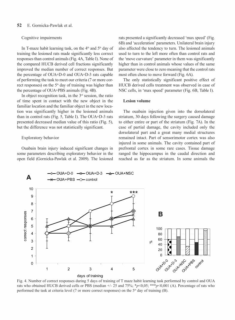
In T-maze habit learning task, on the 4th and 5th day of training the lesioned rats made significantly less correct responses than control animals (Fig. 4A, Table I). None of the compared HUCB derived cell fractions significantly improved the median number of correct responses. But the percentage of OUA+D-0 and OUA+D-3 rats capable of performing the task to meet our criteria (7 or more cor-rect responses) on the 5th day of training was higher than the percentage of OUA+PBS animals (Fig. 4B).
In object recognition task, in the 3rd session, the ratio of time spent in contact with the new object in the familiar location and the familiar object in the new loca-tion was significantly higher in the lesioned animals than in control rats (Fig. 5, Table I). The OUA+D-3 rats presented decreased median value of this ratio (Fig. 5), but the difference was not statistically significant.
Exploratory behavior
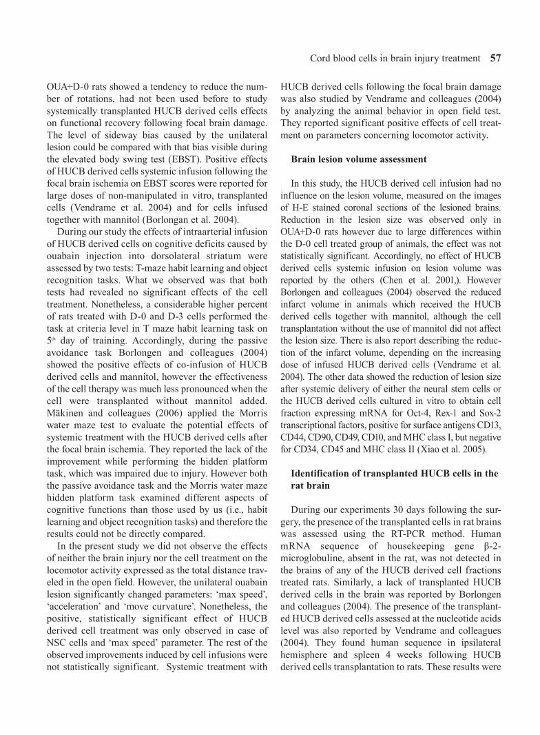
Ouabain brain injury induced significant changes in some parameters describing exploratory behavior in the open field (Gornicka-Pawlak et al. 2009). The lesioned
rats presented a significantly decreased ‘max speed’ (Fig. 6B) and ‘acceleration’ parameters. Unilateral brain injury also affected the tendency to turn. The lesioned animals used to turn to the left more often than control rats and the ‘move curvature’ parameter in them was significantly higher than in control animals whose values of the same parameter were close to zero meaning that the control rats most often chose to move forward (Fig. 6A).
The only statistically significant positive effect of HUCB derived cells treatment was observed in case of NSC cells, in ‘max speed’ parameter (Fig. 6B, Table I).
Lesion volume
The ouabain injection given into the dorsolateral striatum, 30 days following the surgery caused damage to either entire or part of the striatum (Fig. 7A). In the case of partial damage, the cavity included only the dorsolateral part and a great many medial structures remained intact. Part of sensorimotor cortex was also injured in some animals. The cavity contained part of prefrontal cortex in some rare cases. Tissue damage ranged the hippocampus in the caudal direction and reached as far as the striatum. In some animals the
Fig. 4. Number of correct responses during 5 days of training of T maze habit learning task performed by control and OUA rats who obtained HUCB derived cells or PBS (median +/- 25 and 75%; *p<0,05; ***p<0,001 (A). Percentage of rats who performed the task at criteria level (7 or more correct responses) on the 5th day of training (B).

Cord blood cells in brain injury treatment 53
changes also affected the lateral nuclei of the thalamus. Volume of ouabain induced brain lesion in non-
transplanted rats 30 days following the surgery was 50 +/- 15 mm3 (median +/- 25 and 75 %). D-0 cells treat-ment tend to decrease lesion volume, but the effect was not statistically significant (Fig. 7B, Table I).
the survival of HUCB derived cells transplanted in the oabain injured rat brain
Thirty days following surgery the trait of presence of systemically transplanted HUCB derived cells were examined at RNA level using the RT-PCR method. The human sequence of housekeeping gene β-2-microglobulin (B2M) absent in the rat was detected in the HUCB-NSC cells and in the brain of the lesioned rat 24 h following the intra cerebral injection of the NSC cells (OUA+icNSC). None of human RNA sequence of the systematically transplanted HUCB derived cells were detected 30 days following the infu-sion (Fig. 8). To confirm the presence of the non-de-graded RNA, a control reaction with sequence of β-actin presents in both human and rat was made.
Complex comparison of HUCB derived cells therapeutic effects
The effects of HUCB derived cells, being at different levels of their neural conversion, on functional recov-ery following the focal brain injury and lesion volume shared substantial differences in the consecutive tasks. The lack of statistical significance of cell treatment effects in many assessed parameters encouraged us to develop additional analysis of percentage differences in medians (Table I). This enables us to carry out a com-plex comparison of the HUCB derived cells treatment therapeutic effects in all the tested parameters. The D-O cells were the most effective in enhancing func-tional restoration and in reducing the lesion volume. As the freshly isolated HUCB derived cells were the most effective, OUA+D-0 group of animals was selected for further immunohistochemical analysis.
Local nervous tissue response to ouabain lesion and their modulation after D-0 HUCB derived cells systemic treatment
To estimate the HUCB derived D-0 cells treatment effects on nervous tissue response to ouabain injection
immunofluorescent analysis was applied. Both ipsi- and cotralateral hemisphere were studied, but the dif-ferences induced by systemic cell treatment concerned only the ipsilateral hemisphere.
To circumscribe the level of endogenous cell prolif-eration, the Ki-67 dividing cell marker was applied. Ki-67 positive cells were observed in the subventricu-lar zone (SVZ) and in the tissue located between the lateral ventricle and the lesion cavity. Comparing OUA+D-O animals with the OUA+PBS rats Ki-67 immunoreactivity in the ipsilateral hemisphere was more pronounced in the former.
Neuroblast migration was assessed using antibody against doublecortin (DCX). The immunoreactivity of DCX in the OUA+D-0 rats was much higher in the tis-sue located between SVZ and lesion area than in OUA+PBS animals. Moreover in OUA+D-0 rats DCX positive cells formed characteristic ‘chains’ of migrat-ing neuroblasts from the SVZ to the center of the injury. But in the SVZ region DCX immunoreactivity was similar in both groups of rats.
Nestin - marker of neural progenitors positive cells was observed only in SVZ. In OUA+D-0 rats Nestin immunoreactivity was higher in ipsilateral SVZ than in OUA+PBS animals.
Changes in the amount and location of neurons were assessed using heavy neurofilament (200-kDa, NF200) antibody. The NF200 positive fibers in the OUA+D-0 rats at the border of the lesion were loosely and amor-
Fig. 5. Object recognition task, ratio of time spent in contact with the new object in known location and the known object in new location (session 3) by OUA rats who obtained HUCB derived cells or PBS (median +/- 25 and 75%; ***p<0,001).

54 E. Gornicka-Pawlak et al.
Tabl
e I
Effe
cts
of s
yste
mic
trea
tmen
t of h
uman
um
bilic
al c
ord
bloo
d de
rived
cel
ls, b
eing
at d
iffer
ent l
evel
s of
thei
r neu
ral c
onve
rsio
n, o
n fu
nctio
nal r
ecov
ery,
lesi
on v
olum
e an
d th
eir
surv
ival
follo
win
g fo
cal b
rain
inju
ry. R
esul
ts o
f non
para
met
ric s
tatis
tics,
grou
p si
ze a
nd a
naly
sis
of m
edia
ns p
erce
ntag
e di
ffere
nces
.
AN
OVA
OU
A+D
-0 ×
OU
A+P
BS
OU
A+D
-3 ×
OU
A+P
BS
OU
A+N
SC ×
OU
A+P
BS
OU
A+P
BS
× co
ntro
l
M-W
te
st p
diff
(%)
ndi
ff ra
tioM
-W
test
pdi
ff (%
)n
diff
ratio
M-W
te
st p
diff
(%)
ndi
ff ra
tioM
-W
test
pdi
ff (%
)n
n co
ntro
l
wal
king
bea
m 7
day
s fo
llow
ing
lesi
on0.
0000
0.14
1433
14-0
.66
0.16
430
120.
000.
4372
08
0.00
0.00
00-5
023
15
wal
king
bea
m 1
5 da
ys fo
llow
ing
lesi
on0.
0000
0.03
1125
14-0
.76
0.10
4825
12-0
.76
0.98
240
80.
000.
0000
-33
2315
wal
king
bea
m 3
0 da
ys fo
llow
ing
lesi
on0.
0000
0.43
980
140.
000.
0981
012
0.00
0.13
250
80.
000.
0001
-16
2315
rota
rod
11 &
12
days
follo
win
g le
sion
0.00
080.
2051
106
-0.2
70.
2939
108
-0.2
70.
0159
208
-0.5
40.
0000
-37
158
rota
rod
30 d
ays
follo
win
g le
sion
0.00
180.
3809
506
-1.0
00.
6355
08
0.00
0.16
880
80.
000.
0003
-50
158
fore
limb
plac
ing
--
014
0.00
-0
120.
00-
08
0.00
-10
023
11
T m
aze
habi
t lea
rnin
g0.
0041
0.42
4725
13-0
.51
0.41
5545
11-0
.92
0.48
31-3
07
0.61
0.00
03-4
920
13
reco
gniti
on m
emor
y ta
sk0.
0315
-1
40.
01-
-31
2-0
.33
0.70
454
80.
040.
0009
949
5
‘max
spe
ed’ i
n th
e op
en fi
eld
0.01
390.
0574
612
-0.5
00.
5484
-312
0.25
0.02
325
8-0
.42
0.00
30-1
223
11
‘acc
eler
atio
n’ in
the
open
fiel
d0.
0541
0.09
2214
12-0
.52
0.59
525
12-0
.19
0.06
6820
8-0
.74
0.02
30-2
723
11
‘mov
e cu
rvat
ure’
in th
e op
en fi
eld
0.00
120.
3610
-15
12-0
.15
0.47
80-1
212
-0.1
20.
8717
168
0.16
0.00
0010
023
11
apom
orph
ine
indu
ced
rota
tions
0.43
650.
1754
-22
14-0
.22
0.69
366
120.
060.
8423
-78
-0.0
710
023
11
lesi
on v
olum
e 30
day
s fo
llow
ing
inju
ry0.
1727
0.06
73-5
110
-0.5
10.
4306
1712
0.17
1.00
001
80.
0110
020
-
pres
ence
of h
uman
cel
ls in
the
rat b
rain
--
03
0.00
-0
30.
00-
03
0.00
-0
3-
sum
of d
iff r
atio
s-5
.09
-2.1
0-0
.94

Cord blood cells in brain injury treatment 55
phously located. Whereas, in the OUA+PBS animals, compact, oval aggregations characteristic for the stria-tum were observed. They were closer to the border of the cavity in non-transplanted rats then in transplanted animals.
Activation of microglia/macrophages was studied using ED-1 antibody. A relatively small number of ED-1 positive cells was observed in the tissue surrounding the cavity in OUA+PBS animals. In OUA+D-0 rats more ED-1 positive cells were observed at the border of the lesion compared with the non-transplanted animals.
Level of scare formation was visualized using anti-body against GFAP. In OUA+D-0 rats GFAP immuno-reactivity was much higher in the tissue between SVZ and the lesion cavity than in OUA+PBS animals. GFAP positive cells in the transplanted rats formed characteristic ‘chains’ of migrating cells from the SVZ to the center of the injury. At the border of the lesion structure the location of GFAP positive fibers immu-noreaction was similar in both groups of rats. The level of GFAP immunoreactivity in the SVZ was also simi-lar in the transplanted and non-transplanted animals.
dIScuSSIon
Functional recovery of focal brain injured rats after cord blood cell transplantation
First and foremost, it should be notified that the behavioral tests were experimenter-blind trials so the experimenter was unaware of their nature. The aim of
this was to ensure the objective comparison of HUCB derived cell fraction therapeutic effects. Experimenter blindness for animal’s treatment is crucial paradigm in study of cell therapy effects on functional recovery (Lewejohann et al. 2006, Hicks et al. 2009b).
Our results have shown that systemic transplanta-tions of HUCB derived cells, being at different levels of their neural conversion, following focal brain dam-age exerted different effects in various behavioral tasks. Fifteen days following the lesion significant improvement in the walking beam task performed by the D-0 cell treated animals was observed. What should be noted is that 7 days after focal brain lesion the rats receiving D-0 cells (OUA+D-O) performed the task better than OUA+PBS animals. The same holds true for the D-3 cell treated rats 15 days after the sur-gery but the differences were not statistically signifi-cant. After the brain ischemia the effects of the sys-temically infused all HUCB mononuclear cells and HUCB hematopoietic stem cells negative for surface antigens: CD2, CD3, CD14, CD16, CD19, CD24, CD56, CD66b and glycophorin A on functional recov-ery during the walking beam task were compared by Mäkinen and colleagues (2006). They found however that the cell treatment had little or no effect on the quality of performing the beam walking task. Using a two level beam they brought out certain motor deficits which slightly differed from those our walking beam test elucidated. However thanks to both tasks unilat-eral impairment of limb movements could be picked out. The beam used by Mäkinen and colleagues (2006)
Fig. 6. Values of ‘move curvature’ (A) and ‘max speed’ (B) parameters presented in the open field during first 30 min session by OUA rats who obtained HUCB derived cells or PBS (median +/- 25 and 75%; *p<0,05; **p<0,001; ****p<0,0001).

56 E. Gornicka-Pawlak et al.
enabled them to measure more severe deficits than those observed during our experiment 30 days follow-ing the brain injury in enriched environment housing conditions. Moreover the point scale designed by our researchers (Gornicka-Pawlak et al. 2009), which quantitatively estimates the quality of contralateral limbs stepping, enabled us to differentiate the func-tional recovery from compensation (substitution of impaired movement by another). The fidelity of behav-ioral tests to differentiate compensation from real functional recovery is very important while analyzing positive effects of cell therapy on functional recovery, before starting clinical trials (Hicks et al. 2009).
During the rotarod skilled walking task, short time following the brain lesion (11 and 12 days) positive effects of all the tested HUCB derived cell populations were observed, but only the NSC cell treated rats were considered statistically important. Thirty days follow-ing the surgery only the OUA+D-0 rats performed the task at higher level than OUA+PBS animals, but the difference was not statistically significant. A rotarod was used previously to study functional effects of intravenous infusion of HUCB cells, majority of which
expressed CD34 surface marker, following focal brain ischemia (Chen et al. 2001). Significant improvement was observed 7 and 14 days following MCAO in rats infused with cells 24 h after brain damage. The cell transplantations 7 days following the ischemia were not so effective.
The vibrissae elicited forelimb placing task we per-formed had not been used before to study systemically transplanted HUCB derived cells functional effects following focal brain injury. However the deficits tested in the task could be compared with those exam-ined in neurological severity score (NSS) task. Our results showed that none of the tested HUCB derived cell fractions exerted any effect on the permanent lack of contralateral to lesion forelimb reflex. Similarly, no effects of the HUCB derived cell treatment on the NSS score were reported by others (Mäkinen et al. 2006, Zawadzka et al. 2009). There are also contrast-ing reports about positive effects of the HUCB derived cells treatment on functional recovery while perform-ing the NSS task following the focal brain damage (Chen et al. 2001, Borlongan et al. 2004).
Apomorphine induced rotations task in which the
Fig. 7. Representative subsequent coronal sections of lesioned brain 30 days following ouabain injection and PBS infusion, stained with H-E method (A). Lesion volume estimated in OUA rats who obtained HUCB derived cells or PBS (median +/- 25 and 75%) (B).
Cord blood cells in brain injury treatment 57
OUA+D-0 rats showed a tendency to reduce the num-ber of rotations, had not been used before to study systemically transplanted HUCB derived cells effects on functional recovery following focal brain damage. The level of sideway bias caused by the unilateral lesion could be compared with that bias visible during the elevated body swing test (EBST). Positive effects of HUCB derived cells systemic infusion following the focal brain ischemia on EBST scores were reported for large doses of non-manipulated in vitro, transplanted cells (Vendrame et al. 2004) and for cells infused together with mannitol (Borlongan et al. 2004).
During our study the effects of intraarterial infusion of HUCB derived cells on cognitive deficits caused by ouabain injection into dorsolateral striatum were assessed by two tests: T-maze habit learning and object recognition tasks. What we observed was that both tests had revealed no significant effects of the cell treatment. Nonetheless, a considerable higher percent of rats treated with D-0 and D-3 cells performed the task at criteria level in T maze habit learning task on 5th day of training. Accordingly, during the passive avoidance task Borlongen and colleagues (2004) showed the positive effects of co-infusion of HUCB derived cells and mannitol, however the effectiveness of the cell therapy was much less pronounced when the cell were transplanted without mannitol added. Mäkinen and colleagues (2006) applied the Morris water maze test to evaluate the potential effects of systemic treatment with the HUCB derived cells after the focal brain ischemia. They reported the lack of the improvement while performing the hidden platform task, which was impaired due to injury. However both the passive avoidance task and the Morris water maze hidden platform task examined different aspects of cognitive functions than those used by us (i.e., habit learning and object recognition tasks) and therefore the results could not be directly compared.
In the present study we did not observe the effects of neither the brain injury nor the cell treatment on the locomotor activity expressed as the total distance trav-eled in the open field. However, the unilateral ouabain lesion significantly changed parameters: ‘max speed’, ‘acceleration’ and ‘move curvature’. Nonetheless, the positive, statistically significant effect of HUCB derived cell treatment was only observed in case of NSC cells and ‘max speed’ parameter. The rest of the observed improvements induced by cell infusions were not statistically significant. Systemic treatment with
HUCB derived cells following the focal brain damage was also studied by Vendrame and colleagues (2004) by analyzing the animal behavior in open field test. They reported significant positive effects of cell treat-ment on parameters concerning locomotor activity.
Brain lesion volume assessment
In this study, the HUCB derived cell infusion had no influence on the lesion volume, measured on the images of H-E stained coronal sections of the lesioned brains. Reduction in the lesion size was observed only in OUA+D-0 rats however due to large differences within the D-0 cell treated group of animals, the effect was not statistically significant. Accordingly, no effect of HUCB derived cells systemic infusion on lesion volume was reported by the others (Chen et al. 2001,). However Borlongen and colleagues (2004) observed the reduced infarct volume in animals which received the HUCB derived cells together with mannitol, although the cell transplantation without the use of mannitol did not affect the lesion size. There is also report describing the reduc-tion of the infarct volume, depending on the increasing dose of infused HUCB derived cells (Vendrame et al. 2004). The other data showed the reduction of lesion size after systemic delivery of either the neural stem cells or the HUCB derived cells cultured in vitro to obtain cell fraction expressing mRNA for Oct-4, Rex-1 and Sox-2 transcriptional factors, positive for surface antigens CD13, CD44, CD90, CD49, CD10, and MHC class I, but negative for CD34, CD45 and MHC class II (Xiao et al. 2005).
Identification of transplanted HUCB cells in the rat brain
During our experiments 30 days following the sur-gery, the presence of the transplanted cells in rat brains was assessed using the RT-PCR method. Human mRNA sequence of housekeeping gene β-2-microglobuline, absent in the rat, was not detected in the brains of any of the HUCB derived cell fractions treated rats. Similarly, a lack of transplanted HUCB derived cells in the brain was reported by Borlongen and colleagues (2004). The presence of the transplant-ed HUCB derived cells assessed at the nucleotide acids level was also reported by Vendrame and colleagues (2004). They found human sequence in ipsilateral hemisphere and spleen 4 weeks following HUCB derived cells transplantation to rats. These results were

58 E. Gornicka-Pawlak et al.
confirmed by immunohistochemical methods. Similarly, the HUCB derived cells, which had been infused intravenously following the focal brain isch-emia, were detected in rat brain at various time after surgery using antibodies against human antigens (Chen et al. 2001, Xiao et al. 2005, Mäkinen et al. 2006, Zawadzka et al. 2009).
Although all the publications cited above dealt with the effects of the HUCB derived cells infused sys-temically following focal brain damage, they differ substantially in many aspects. They include: the way of preparing cells for transplantation, time window of delivery after brain injury, dose of transplanted cells, duration of observation period, housing conditions of experimental animals and employed behavioral tests. It is therefore impossible to compare directly the total outcome with our results.
The aim of our study was to compare the therapeutic effectiveness of the intra-arterial infusion of HUCB derived cells, being at different levels of their neural conversion. Basing the results on both the particular behavioral tests and the lesion volume, it was however difficult to conclude which HUCB derived cell frac-tion was the most effective in the insult treatment. This results from the fact, that the observed differences between transplanted and non-transplanted rats were rarely statistically significant due to dissimilarities between animals. To compare therapeutic effective-ness of examined subsets of transplanted HUCB derived cells in a complex way, we analyzed the per-centage differences of medians. Although it did not include dispersion between rats, the median was a source of information about the most common value in the group. It enables us to evaluate the result when the exact distribution of assessed parameter (result of behavioral test or lesion volume) was impossible to establish due to relatively small group size.
Analysis of percentage differences of medians showed that the freshly isolated D-0 cells were most effective both in the functional recovery and in the reduction of lesion volume. This result, indicating that highest therapeutic effectiveness of the cells less neu-rally differentiated was in agreement with report of Xiao and colleagues (2005). They compared the effects of intravenous infusion following focal brain damage of either neural stem cells line or HUCB derived cells, selected in vitro to obtain CD34- and CD45- cell frac-tion. Comparison of the animals transplanted with the HUCB cell subpopulation and those treated with the
neural stem cell line pointed to the enhanced functional recovery and reduced lesion volume in the former.
Our results showing the highest effectiveness of systemic treatment with freshly isolated HUCB D-0 cells, together with report of Xiao and colleagues (2005), might suggest that the mechanism exerting the positive therapeutic effects of cell systemic treatment is based on enhancement of regenerative potential of the organism rather then results from the direct cell supplementation. Transplanted cells may reduce inflammation (Vendrame et al. 2005), enhance endog-enous neurogenesis and angiogenesis, stimulate release of neurotrophic factors and increase brain plasticity (Brouns and De Deyn 2009, Hicks and Jolkkonen 2009, Janowski and Date 2009, Park et al. 2010).
Fig. 8. RNA levels of β-2-microglobulin (B2M) (human sequence, absent in the rat) and β-actin (sequence presented in both human and rat) genes in HUCB NSC cells, in the brain of lesioned rat 24 h after intracerebral injection of NSC cells (OUA+icNSC) and in brains of OUA rats 30 days fol-lowing infusion of HUCB derived cells or PBS.

Cord blood cells in brain injury treatment 59

60 E. Gornicka-Pawlak et al.

Cord blood cells in brain injury treatment 61
Local tissue response to ouabain lesion and their modulation by D-0 HUCB derived cells systemic transplantation
To estimate modulation of local tissue response to focal ouabain injury, immunofluorescent analysis were performed 30 days after lesion and subsequent intraar-terial infusion of the therapeutically most effective HUCB derived D-0 cells.
The level of endogenous cell proliferation was esti-mated using antibody against Ki-67, the marker of dividing cells. Ki-67 positive cells were observed in the subventricular zone (SVZ) and in rostral migratory stream (RMS) of both hemispheres and in tissue between SVZ and the cavity. This pattern of Ki-67 immunoreactivity is congruent with those describing by others (Yamashita et al. 2006, Zhao and Nam 2007, Buffo et al. 2008, Zhang et al. 2008). In OUA+D-0 rats increased Ki-67 immunoreactivity was noted in the ipsilateral SVZ, when compared to non-transplanted animals. It may suggest increased proliferation of endogenous neural stem cells. In D-0 cells treated rats, the increased Ki-67 immunoreactivity in tissue between SVZ and the lesion cavity was also observed. This may be explained by increased of reactive gliosis (Hampton et al. 2004), but to identify phenotype of dividing cells additional double staining assay should be necessary.
An antibody against doublecortin (DCX) was used to assess the level of neuroblast migration. The ‘chains’ of DCX positive cells formed stretching from SVZ to the cavity. This characteristic image of migrating neu-roblasts was created as the result of endogenous neu-rogenesis induced by brain injury (Zhang et al. 2004, Yamashita et al. 2006, Zhang et al. 2008). There were much more DCX positive cells in the tissue between SVZ and the injury center of the OUA+D-O rats than in the same tissue of the OUA+PBS. This may suggest increased neuroblast migration following cell treat-ment. Similar level of DCX immunoreactivity in the ipsilateral SVZ of transplanted and non-transplanted animals may be explained by the fact that maximum of enhanced endogenous proliferation was earlier. In experiments done in transgenic mice, the highest num-ber of DCX+ cells was reported at 18 days following
surgery (Yamashita et al. 2006). However analyzing reports in which focal brain damage was induced by middle cerebral artery occlusion it should be keep in mind that ischemia may also include SVZ (Thored et al. 2007) and affect processes which take place there.
Nestin was another tested marker which is expressed by cells being at early stages of neural differentiation. We observed Nestin positive cells only in the SVZ of both hemispheres. There are some reports in literature about other localization of Nesitn+ cells in brain hemi-spheres (Li and Chopp 1999). However, the study con-ducted on transgenic mice which was transfected with GFP (green fluorescent protein) gene expressed only in Nestin synthesizing cells, the localization of GFP+ cells was similar (Zhao and Nam 2007) as in our experi-ments. In OUA+D-0 rats increased Nestin immunoreac-tivity in ipsilateral hemisphere was observed comparing to OUA+PBS animals. It may suggest enlarged pool of endogenous neural stem cells in transplanted rats.
In our experiments, the localization of neurons was analyzed using antibody against heavy neurofilament (200 kDa, NF200). Previously it was shown that focal brain injury reduced NF200 immunoreactivity in ipsi-lateral hemisphere (Posmantur et al. 2000). In contrast, in our experiment NF200 immunoreactivity was simi-lar in both hemispheres. In OUA+PBS rats, in tissue surrounding the cavity NF200 positive fibers formed dense and compact aggregations. While in OUA+D-0 animals those fibers lied loosely and amorphously. Differences between transplanted and non-transplanted rats in location of NF200+ fibers may be due to distinct processes and dynamics of tissue reconstruction.
The antibody against CD68 (ED-1) was used to esti-mate level of inflammatory infiltration in our study. Few ED-1+ cells were visible in tissue surrounding the lesion cavity. In OUA+D-0 rats increased immunore-activity of ED-1 was observed, comparing to OUA+PBS animals. On one hand it may suggest higher level of infiltration of proinflammatory cells following xeno-graft. On the other hand investigations of other authors showed that ED-1 colocalized with SDF-1 (stromal cell-derived factor 1) (Thored et al. 2006). SDF-1 regu-lates cell migration via CXCR4 receptor. So ED-1 positive cells may provide SDF-1 signal for cells
Fig. 9. Local nervous tissue response on ouabain brain injury and their modulation after D-0 cells intraarterial treatment 30 days following lesion and cell infusion. Immunofluorescent analysis was carry out using antibodies against Ki-67, Nestin, NF200 (green) and DCX, ED-1, GFAP (red). Cell nuclei were stained with Hoechst (blue). Images were obtained with con-focal microscope of ipsi- and contralateral hemisphere of OUA+D-0 and OUA+PBS rats, on the pages 59-60.

62 E. Gornicka-Pawlak et al.
migrating to the injury center and increased number of ED-1+ cells in transplanted animals may correlate with accelerated migration to the cavity surroundings.
Focal brain injury is followed by glia scar formation which goal is to fill tissue loss (Fitch and Silver 2008). Astrocytes in injured brain have a dual role (Buffo et al. 2010). On the one hand they inhibit axonal plasticity and produce proinflammatory factors. On the other hand astrocytes have many positive functions. They help in holding of homeostasis, provide neurotrophic factors, take part in blood-brain barrier reconstruction and promote revascularization. In our experiments, in HUCB derived D-0 cell treated rats the higher GFAP immunoreactivity in tissue between SVZ and the lesion cavity was observed, comparing to non-treated ani-mals. GFAP positive cells in transplanted rats formed ‘chains’ characteristic for migrating cells. It may sug-gest increased migration of cells directed into astro-cytal lineage from the SVZ to the injury center. In non-injured brain, neuroblasts born in the SVZ migrate via RMS to the olfactory bulbs where they differentiate into interneurons (Kernie and Parent 2010). However in focally injured brain cells born in the SVZ migrate not only to RMS, but also to the injury center. Moreover phenotype of cells migrating to the damaged tissue may be neuronal, oligodendroglial and in majority astroglial (Li et al. 2010). The other report was shown that from the SVZ to the injury center migrated only cells presenting astroglial and oligodendroglial pheno-type (Gotts and Chesselet 2005). GFAP, like ED-1 reactivity, was also shown to colocalize with SDF-1 (Thored et al. 2006). So, increased GFAP immunoreac-tivity in transplanted animals may also contribute in enhancement of cell migration to the injury center.
ConCLUSIonS
Our results have showed that systemic treatment with the freshly isolated HUCB cells (D-0) was the most effective in functional restoration after focal brain inju-ry. This may suggest that the mechanism underlying positive effects of HUCB derived cell treatment may concern the other than direct neural cell supplementa-tion mechanism(s). The absence of HUCB remnants searched in the transplanted rat tissue at RNA level fur-ther supports this conclusion. In addition increased immunoreactivity of markers indicating local cells pro-liferation and migration as well as other noticed signs of injured issue reconstruction may indicate stimulation of
endogenous neurogenesis and other reparative processes by HUCB D-0 cell interarterial infusion.
ACKnowLEDGEMEnt
This work was supported by grants from the Polish Ministry of Scientific Research and Higher Education (0394/B/P01/2010/38; 0141/B/P01/2008/35).
rEFErEncES
Ali H, Bahbahani H (2010) Umbilical cord blood stem cells - potential therapeutic tool for neural injuries and disor-ders. Acta Neurobiol Exp (Wars) 70: 316–324.
Benefiel AC, Dong WK, Greenough WT (2005) Mandatory “enriched” housing of laboratory animals: the need for evidence-based evaluation. ILAR J 46: 95–105.
Borlongan CV, Hadman M, Sanberg CD, Sanberg PR (2004) Central nervous system entry of peripherally injected umbilical cord blood cells is not required for neuroprotec-tion in stroke. Stroke 35: 2385–2389.
Brouns R, De Deyn PP (2009) The complexity of neurobio-logical processes in acute ischemic stroke. Clin Neurol Neurosurg 111: 483–95.
Buffo A, Rite I, Tripathi P, Lepier A, Colak D, Horn AP, Mori T, Götz M (2008) Origin and progeny of reactive gliosis: A source of multipotent cells in the injured brain. Proc Natl Acad Sci USA 105: 3581–3586.
Buffo A, Rolando C, Ceruti S (2010) Astrocytes in the dam-aged brain: molecular and cellular insights into their reac-tive response and healing potential. Biochem Pharmacol 79: 77–89.
Buzanska L, Machaj EK, Zablocka B, Pojda Z, Domanska-Janik K (2002) Human cord blood-derived cells attain neu-ronal and glial features in vitro. J Cell Sci 115: 2131–2138.
Chen J, Sanberg PR, Li Y, Wang L, Lu M, Willing AE, Sanchez-Ramos J, Chopp M (2001) Intravenous adminis-tration of human umbilical cord blood reduces behavioral deficits after stroke in rats. Stroke 32: 2682–2688.
Domanska-Janik K, Habich A, Sarnowska A, Janowski M (2006) Neural commitment of cord blood stem cells (HUCB-NSC/NP): therapeutic perspectives. Acta Neurobiol Exp (Wars) 66: 279–291.
Drai D, Golani I (2001) SEE: a tool for the visualization and analysis of rodent exploratory behavior. Neurosci Biobehav Rev 25: 409–426.
Fitch MT, Silver J (2008) CNS injury, glial scars, and inflammation: Inhibitory extracellular matrices and regen-eration failure. Exp Neurol 209: 294–301.

Cord blood cells in brain injury treatment 63
Gornicka-Pawlak E, Jabłolnska A, Chylinski A, Domanska-Janik K (2009) Housing conditions influ-ence motor functions and exploratory behavior fol-lowing focal damage of the rat brain. Acta Neurobiol Exp (Wars) 69: 62–72.
Gotts JE, Chesselet MF (2005) Migration and fate of newly born cells after focal cortical ischemia in adult rats. J Neurosci Res 80: 160–171.
Habich A, Jurga M, Markiewicz I, Lukomska B, Bany-Laszewicz U, Domanska-Janik K (2006) Early appear-ance of stem/progenitor cells with neural-like characteris-tics in human cord blood mononuclear fraction cultured in vitro. Exp Hematol 34: 914–925.
Hampton DW, Rhodes KE, Zhao C, Franklin RJ, Fawcett JW (2004) The responses of oligodendrocyte precursor cells, astrocytes and microglia to a cortical stab injury, in the brain. Neuroscience 127: 813–820.
Hicks A, Jolkkonen J (2009) Challenges and possibilities of intravascular cell therapy in stroke. Acta Neurobiol Exp (Wars) 69: 1–11.
Hicks A, Schallert T, Jolkkonen J (2009) Cell-based thera-pies and functional outcome in experimental stroke. Cell Stem Cell 5: 139–140.
Jablonska A, Kozlowska H, Markiewicz I, Domanska-Janik K, Lukomska B (2010) Transplantation of neural stem cells derived from human cord blood to the brain of adult and neonatal rats. Acta Neurobiol Exp (Wars) 70: 337–350.
Janowski M, Date I (2009) Systemic neurotransplantation-a problem-oriented systematic review. Rev Neurosci 20: 39–60.
Janowski M, Gornicka-Pawlak E, Kozlowska H, Domanska-Janik K, Gielecki J, Lukomska B (2008) Structural and functional characteristic of a model for deep-seated lacu-nar infarct in rats. J Neurol Sci 273: 40–48.
Kernie SG, Parent JM (2010) Forebrain neurogenesis after focal Ischemic and traumatic brain injury. Neurobiol Dis 37: 267–274.
Lappalainen RS, Narkilahti S, Huhtala T, Liimatainen T, Suuronen T, Närvänen A, Suuronen R, Hovatta O, Jolkkonen J (2008) The SPECT imaging shows the accu-mulation of neural progenitor cells into internal organs after systemic administration in middle cerebral artery occlusion rats. Neurosci Lett 440: 246–250.
Lewejohann L, Reinhard C, Schrewe A, Brandewiede J, Haemisch A, Görtz N, Schachner M, Sachser N (2006) Environmental bias? Effects of housing conditions, labo-ratory environment and experimenter on behavioral tests. Genes Brain Behav 5: 64–72.
Li L, Harms KM, Ventura PB, Lagace DC, Eisch AJ, Cunningham LA (2010) Focal cerebral ischemia induces a multilineage cytogenic response from adult subventric-ular zone that is predominantly gliogenic. Glia 58: 1610–1619.
Li Y, Chopp M (1999) Temporal profile of nestin expression after focal cerebral ischemia in adult rat. Brain Res 838: 1–10.
Macias M (2008) Injury induced dendritic plasticity in the mature central nervous system. Acta Neurobiol Exp (Wars) 68: 334–346.
Mäkinen S, Kekarainen T, Nystedt J, Liimatainen T, Huhtala T, Närvänen A, Laine J, Jolkkonen J (2006) Human umbilical cord blood cells do not improve sensorimotor or cognitive outcome following transient middle cerebral artery occlusion in rats. Brain Res 1123: 207–215.
Park DH, Borlongan CV, Willing AE, Eve DJ, Cruz LE, Sanberg CD, Chung YG, Sanberg PR (2009) Human umbilical cord blood cell grafts for brain ischemia. Cell Transplant. 18: 985–998.
Park DH, Eve DJ, Chung YG, Sanberg PR (2010) Regenerative medicine for neurological disorders. ScientificWorldJournal 16: 470–489.
Pham TM, Winblad B, Granholm AC, Mohammed AH (2002) Environmental influences on brain neurotrophins in rats. Pharmacol Biochem Behav 73: 167–175.
Posmantur RM, Newcomb JK, Kampfl A, Hayes RL (2000) Light and confocal microscopic studies of evolutionary changes in neurofilament proteins following cortical impact injury in the rat. Exp Neurol 161: 15–26.
Thored P, Arvidsson A, Cacci E, Ahlenius H, Kallur T, Darsalia V, Ekdahl CT, Kokaia Z, Lindvall O (2006) Persistent production of neurons from adult brain stem cells during recovery after stroke. Stem Cells 24: 739–747.
Thored P, Wood J, Arvidsson A, Cammenga J, Kokaia Z, Lindvall O (2007) Long-term neuroblast migration along blood vessels in an area with transient angiogenesis and increased vascularization after stroke. Stroke 38: 3032–3039.
Veldhuis WB, van der Stelt M, Delmas F, Gillet B, Veldink GA, Vliegenthart JF, Nicolay K, Bär PR (2003) In vivo excitotoxicity induced by ouabain, a Na+/K+-ATPase inhibitor. J Cereb Blood Flow Metab 23: 62–74.
Vendrame M, Cassady J, Newcomb J, Butler T, Pennypacker KR, Zigova T, Sanberg CD, Sanberg PR, Willing AE (2004) Infusion of human umbilical cord blood cells in a rat model of stroke dose-dependently rescues behavioral defi-cits and reduces infarct volume. Stroke 35: 2390–2395.

64 E. Gornicka-Pawlak et al.
Vendrame M, Gemma C, de Mesquita D, Collier L, Bickford PC, Sanberg CD, Sanberg PR, Pennypacker KR, Willing AE (2005) Anti-inflammatory effects of human cord blood cells in a rat model of stroke. Stem Cells Dev 14: 595–604.
Xiao J, Nan Z, Motooka Y, Low WC (2005) Transplantation of a novel cell line population of umbilical cord blood stem cells ameliorates neurological deficits associated with ischemic brain injury. Stem Cells Dev 14: 722–733.
Yamashita T, Ninomiya M, Hernández Acosta P, García-Verdugo JM, Sunabori T, Sakaguchi M, Adachi K, Kojima T, Hirota Y, Kawase T, Araki N, Abe K, Okano H, Sawamoto K (2006) Subventricular zone-derived neuro-blasts migrate and differentiate into mature neurons in the post-stroke adult striatum. J Neurosci 26: 6627–36.
Zawadzka M, Lukasiuk K, Machaj EK, Pojda Z, Kaminska B (2009) Lack of migration and neurological benefits after infusion of umbilical cord blood cells in ischemic brain injury. Acta Neurobiol Exp (Wars) 69: 46–51.
Zhang R, Zhang Z, Wang L, Wang Y, Gousev A, Zhang L, Ho KL, Morshead C, Chopp M (2004) Activated neural stem cells contribute to stroke-induced neurogenesis and neuroblast migration toward the infarct boundary in adult rats. J Cereb Blood Flow Metab 24: 441–448.
Zhang RL, Zhang ZG, Chopp M (2008) Ischemic stroke and neurogenesis in the subventricular zone. Neuropharmacology. 55: 345–352.
Zhao LR, Nam SC (2007) Multiphoton microscope imaging: the behavior of neural progenitor cells in the rostral migratory stream. Neurosci Lett 425: 83–88.