Systemic and tumor level iron regulation in men with colorectal cancer: a case control study
8
RESEARCH Open Access Systemic and tumor level iron regulation in men with colorectal cancer: a case control study Cenk K Pusatcioglu 1 , Elizabeta Nemeth 2 , Giamila Fantuzzi 3 , Xavier Llor 4 , Sally Freels 5 , Lisa Tussing-Humphreys 6 , Robert J Cabay 7 , Rose Linzmeier 2 , Damond Ng 2 , Julia Clark 4 and Carol Braunschweig 3* Abstract Background: Increased cellular iron exposure is associated with colorectal cancer (CRC) risk. Hepcidin, a liver peptide hormone, acts as the primary regulator of systemic iron status by blocking iron release from enterocytes into plasma. Concentrations are decreased during low iron status and increased during inflammation. The role of hepcidin and the factors influencing its regulation in CRC remains largely unknown. This study explored systemic and tumor level iron regulation in men with CRC. Methods: The participants were 20 CRC cases and 20 healthy control subjects. Colonic tissue (adenocarcinoma [cases] healthy mucosa [controls]) was subjected to quantitative PCR (hepcidin, iron transporters and IL-6) and Perls’ iron staining. Serum was analyzed using ELISA for hepcidin, iron status (sTfR) and inflammatory markers (CRP, IL-6, TNF-α). Anthropometrics, dietary iron intake and medical history were obtained. Results: Cases and controls were similar in demographics, medication use and dietary iron intake. Systemically, cases compared to controls had lower iron status (sTfR: 21.6 vs 11.8 nmol/L, p < 0.05) and higher marker of inflammation (CRP: 8.3 vs 3.4 μg/mL, p < 0.05). Serum hepcidin was mildly decreased in cases compared to controls; however, it was within the normal range for both groups. Within colonic tissue, 30% of cases (6/20) presented iron accumulation compared to 5% of controls (1/20) (χ 2 = 5.0; p < 0.05) and higher marker of inflammation (IL-6: 9.4-fold higher compared to controls, p < 0.05). Presence of adenocarcinoma iron accumulation was associated with higher serum hepcidin (iron accumulation group 80.8 vs iron absence group 22.0 ng/mL, p < 0.05). Conclusions: While CRC subjects had serum hepcidin concentrations in the normal range, it was higher given their degree of iron restriction. Inappropriately elevated serum hepcidin may reduce duodenal iron absorption and further increase colonic adenocarcinoma iron exposure. Future clinical studies need to assess the appropriateness of dietary iron intake or iron supplementation in patients with CRC. Keywords: Iron metabolism, Hepcidin, Inflammation, Anemia, Colorectal cancer Background Excessive body iron levels are associated with increased risk for colorectal cancer (CRC) [1-6]. This is due to the high oxidative potential of iron which can result in the formation of reactive oxygen species and mutate the DNA of key genes involved in cell proliferation [2]. Additionally, since iron is required for cell proliferation, increased iron levels could contribute to tumor promotion [1]. Iron levels and its tissue distribution in the body are regulated by hepcidin, a hepatic-derived systemic iron regulatory hormone [7-9]. Hepcidin is increased by in- flammation and decreased by iron insufficiency and erythropoiesis [10-12]. Mechanistically, hepcidin con- trols iron release into plasma by degrading the iron ex- porter ferroportin (FPN) in cells that handle iron, including intestinal enterocytes, hepatocytes, and mac- rophages [13]. Thus, when concentrations of hepcidin are elevated, FPN expression is low, resulting in reduced dietary iron absorption and impaired mobilization from stores. * Correspondence: [email protected] 3 Department of Kinesiology and Nutrition, University of Illinois at Chicago, 1919 W Taylor St, Chicago, IL 60612, USA Full list of author information is available at the end of the article © 2014 Pusatcioglu et al.; licensee BioMed Central Ltd. This is an Open Access article distributed under the terms of the Creative Commons Attribution License (http://creativecommons.org/licenses/by/2.0), which permits unrestricted use, distribution, and reproduction in any medium, provided the original work is properly credited. The Creative Commons Public Domain Dedication waiver (http://creativecommons.org/publicdomain/zero/1.0/) applies to the data made available in this article, unless otherwise stated. Pusatcioglu et al. Nutrition & Metabolism 2014, 11:21 http://www.nutritionandmetabolism.com/content/11/1/21
Transcript of Systemic and tumor level iron regulation in men with colorectal cancer: a case control study
Pusatcioglu et al. Nutrition & Metabolism 2014, 11:21http://www.nutritionandmetabolism.com/content/11/1/21
RESEARCH Open Access
Systemic and tumor level iron regulation in menwith colorectal cancer: a case control studyCenk K Pusatcioglu1, Elizabeta Nemeth2, Giamila Fantuzzi3, Xavier Llor4, Sally Freels5, Lisa Tussing-Humphreys6,Robert J Cabay7, Rose Linzmeier2, Damond Ng2, Julia Clark4 and Carol Braunschweig3*
Abstract
Background: Increased cellular iron exposure is associated with colorectal cancer (CRC) risk. Hepcidin, a liverpeptide hormone, acts as the primary regulator of systemic iron status by blocking iron release from enterocytesinto plasma. Concentrations are decreased during low iron status and increased during inflammation. The role ofhepcidin and the factors influencing its regulation in CRC remains largely unknown. This study explored systemicand tumor level iron regulation in men with CRC.
Methods: The participants were 20 CRC cases and 20 healthy control subjects. Colonic tissue (adenocarcinoma[cases] healthy mucosa [controls]) was subjected to quantitative PCR (hepcidin, iron transporters and IL-6) and Perls’iron staining. Serum was analyzed using ELISA for hepcidin, iron status (sTfR) and inflammatory markers (CRP, IL-6,TNF-α). Anthropometrics, dietary iron intake and medical history were obtained.
Results: Cases and controls were similar in demographics, medication use and dietary iron intake. Systemically,cases compared to controls had lower iron status (sTfR: 21.6 vs 11.8 nmol/L, p < 0.05) and higher marker ofinflammation (CRP: 8.3 vs 3.4 μg/mL, p < 0.05). Serum hepcidin was mildly decreased in cases compared to controls;however, it was within the normal range for both groups. Within colonic tissue, 30% of cases (6/20) presented ironaccumulation compared to 5% of controls (1/20) (χ2 = 5.0; p < 0.05) and higher marker of inflammation (IL-6: 9.4-foldhigher compared to controls, p < 0.05). Presence of adenocarcinoma iron accumulation was associated with higherserum hepcidin (iron accumulation group 80.8 vs iron absence group 22.0 ng/mL, p < 0.05).
Conclusions: While CRC subjects had serum hepcidin concentrations in the normal range, it was higher given theirdegree of iron restriction. Inappropriately elevated serum hepcidin may reduce duodenal iron absorption andfurther increase colonic adenocarcinoma iron exposure. Future clinical studies need to assess the appropriateness ofdietary iron intake or iron supplementation in patients with CRC.
Keywords: Iron metabolism, Hepcidin, Inflammation, Anemia, Colorectal cancer
BackgroundExcessive body iron levels are associated with increasedrisk for colorectal cancer (CRC) [1-6]. This is due to thehigh oxidative potential of iron which can result in theformation of reactive oxygen species and mutate the DNAof key genes involved in cell proliferation [2]. Additionally,since iron is required for cell proliferation, increased ironlevels could contribute to tumor promotion [1].
* Correspondence: [email protected] of Kinesiology and Nutrition, University of Illinois at Chicago,1919 W Taylor St, Chicago, IL 60612, USAFull list of author information is available at the end of the article
© 2014 Pusatcioglu et al.; licensee BioMed CeCreative Commons Attribution License (http:/distribution, and reproduction in any mediumDomain Dedication waiver (http://creativecomarticle, unless otherwise stated.
Iron levels and its tissue distribution in the body areregulated by hepcidin, a hepatic-derived systemic ironregulatory hormone [7-9]. Hepcidin is increased by in-flammation and decreased by iron insufficiency anderythropoiesis [10-12]. Mechanistically, hepcidin con-trols iron release into plasma by degrading the iron ex-porter ferroportin (FPN) in cells that handle iron,including intestinal enterocytes, hepatocytes, and mac-rophages [13]. Thus, when concentrations of hepcidinare elevated, FPN expression is low, resulting in reduceddietary iron absorption and impaired mobilization fromstores.
ntral Ltd. This is an Open Access article distributed under the terms of the/creativecommons.org/licenses/by/2.0), which permits unrestricted use,, provided the original work is properly credited. The Creative Commons Publicmons.org/publicdomain/zero/1.0/) applies to the data made available in this
Pusatcioglu et al. Nutrition & Metabolism 2014, 11:21 Page 2 of 8http://www.nutritionandmetabolism.com/content/11/1/21
Few studies have assessed the role of hepcidin andFPN in cancer [14,15]. Serum hepcidin levels were foundto be elevated in some hematologic or nonhematologiccancers, likely because of the presence of inflammation,but the pathophysiological relevance of these findings isunknown [16,17]. For FPN, it was reported that patientswith breast cancer had decreased tumor expression ofFPN protein compared to non-involved tissue, and thattumor FPN mRNA levels were negatively correlated withadvanced staging [14]. This suggested that iron retentionby breast cancer cells may affect cancer progression.Hepcidin may be elevated in persons with CRC due to
cancer-induced inflammation [18,19]. However, becauseCRC patients frequently have anemia or low iron status,hepcidin levels may also be decreased [20,21]. Evaluationof the involvement of hepcidin in regulating systemicand tumor level iron metabolism in CRC is limited toone study [15]. Ward et al. reported that systemic hepci-din is elevated with advanced cancer staging [15]. Addition-ally, they demonstrated that mRNA expression of hepcidinis detected within a subset (34%) of colonic tumors com-pared to healthy non-involved mucosa. In a complemen-tary study by Brookes et al., increases in iron acquisitionproteins (Divalent metal transporter-1, DMT-1; Transfer-rin receptor-1, TfR1) and decreases in proteins related tocellular iron efflux (FPN; Hephaestin, Heph) were notedin colonic tumors when compared to non-involved mu-cosa [1]. The authors suggested that these alterations iniron transport may explain the iron sequestration com-monly observed in colonic tumors [1]. What remains un-known is hepcidin’s role in regulating colonocyte irontransport and whether it contributes to tumor iron accu-mulation in persons with CRC.The purpose of this study was to examine simultan-
eously systemic and tumor iron status and their regulationin men with CRC compared to controls. This was assessedby measuring: (i) hepcidin, iron status and markers of in-flammation in serum and (ii) hepcidin, expression of irontransporters (DMT-1, FPN), inflammation and iron accu-mulation in colonic mucosa. We hypothesized that CRCwould be associated with higher levels of hepcidin inserum and tumor compared to controls and that hepcidinlevels would be correlated with markers of inflammationand mucosal iron accumulation.
MethodsEthicsAll subjects signed an informed consent and the studyprocedures were approved by the University of Illinois atChicago Institutional Review Board.
Study population and characteristicsStudy subjects were recruited from patients scheduledfor colonoscopies due to abdominal pain, bloating, change
in bowel movements or for CRC screening at the Universityof Illinois at Chicago and John H. Stroger Jr. Cook CountyHospital between May 2011 and June 2012. “Cases” wereclassified as newly diagnosed CRC with adenocarcinomabased on pathology reports for their tumor biopsies; “con-trols” were selected from the subjects with healthy colonicmucosa (absence of adenomatous polyps or GI abnormal-ities). Cases and controls (n = 20/group) were matched tohave a similar difference within each pair for age (within5 years), body mass index (BMI) (within 4 units) and waistcircumference (within 5 cm). Due to gender-specific vari-ation in reference ranges for iron parameters, participationwas restricted to males. Additional exclusions includedmedical conditions that could affect iron status such asgastrointestinal bleeding, hemochromatosis, history of in-flammatory bowel disease or infection.Following informed consent, questionnaires for basic
demographic information, health history, medication andsupplement use and alcohol consumption were adminis-tered by a research team member. The Block Brief 2000food frequency questionnaire (FFQ) was used to assessusual dietary intake over the previous 12 months [22].Height was measured with a stadiometer to nearest0.1 cm and weight using a balance beam scale to nearest0.1 kilograms with subjects wearing a hospital gown.Waist circumference was measured with a flexible tape(AccuFitness, Greenwood Village, CO) at the midpointbetween the ribs and iliac crest, to the nearest 0.1 cm.Body mass index was calculated as weight in kilogramsdivided by height in meters squared. CRC staging (0-IV)based on tumor size, lymph nodes affected and metasta-sis (TNM) was classified using the American Joint Com-mittee on Cancer (AJCC) criteria [23].
Laboratory assaysAll blood samples were collected after a minimum 12 hourfast following the endoscopy or prior to surgical inter-vention. A separate analysis did not reveal any differencesin serum parameters by blood collection type. Colonicadenocarcinoma tissue was obtained from cases at thetime of surgical resection. Healthy colonic mucosa fromthe descending colon in the controls was obtained usingstandard sized biopsy forceps during colonoscopy.
Serum parametersSystemic iron regulation was assessed by serum hepcidinand iron status by serum transferrin receptor (sTfR),which primarily reflects erythroid iron demand. Serumtransferrin receptor is not influenced by acute or chronicinflammation and can help differentiate between iron-related disorders [24]. Elevated sTfR is indicative ofiron-deficient erythropoiesis [25]. Anemia was exam-ined by hemoglobin (Hb). Inflammation was evaluated
Pusatcioglu et al. Nutrition & Metabolism 2014, 11:21 Page 3 of 8http://www.nutritionandmetabolism.com/content/11/1/21
via quantification of C-reactive protein (CRP), interleukin-6(IL-6) and tumor necrosis factor-alpha (TNF-α).Serum hepcidin was measured by competitive enzyme-
linked immunosorbent assay (c-ELISA) (Intrinsic LifeSciences,La Jolla, CA). For healthy men with normal iron status,the range for this assay is 29–254 ng/mL [8]. Serumtransferrin receptor was measured using the QuantikineIVD Human sTfR Immunoassay ELISA (R&D Systems,Minneapolis, MN; normal reference range 8.7-28.1 nmol/L).Hemoglobin was obtained from the medical chart andanemia was defined as <12 g/dL [21]. CRP, IL-6 and TNF-αwere measured using R&D Systems Quantikine ELISA kits(R&D Systems, Minneapolis, MN).
Tissue specimenA portion of colonic tissue was placed in formalin andparaffin-embedded for histological analysis. The remainingportion of the colonic tissue was placed in RNAlater(Ambion, Austin, TX) and stored at −80°C for gene expres-sion (mRNA) analysis.
Real time polymerase chain reaction (RT-PCR)Total RNA was extracted from colonic mucosa sectionsusing the Maxwell 16 System (Promega, Fitchburg, WI).The complementary DNA was synthesized from the RNAusing iScript™ cDNA Synthesis Kit (BioRad, Hercules, CA).Gene expression (mRNA) of divalent metal transporter-1(DMT-1), ferroportin (FPN), hepcidin and IL-6 were mea-sured quantitatively by RT-PCR (For primers used seeAdditional file 1) using SsoAdvanced SYBR Green Super-mix (BioRad). Amplifications were performed at 57°C for40 cycles using the C1000 Touch (BioRad) and data ana-lyzed using the CFX Manager software (BioRad). Resultswere normalized to reference genes Glyceraldehyde 3-phosphate dehydrogenase (GADPH) and β-actin to obtainΔCt values (ΔCt = Ct[reference] - Ct[target]). As bothGADPH and β-actin showed equivalent results, we usedthe average of the two values for the final representation.Fold change in mRNA expression in cases compared tocontrols was calculated using 2ΔΔCt, where ΔΔCt = averageΔCt for cases - average ΔCt for controls. Statistical com-parison was performed using raw ΔCt values (Additionalfile 2).
Iron stainingTissue iron accumulation was measured qualitatively byPerls’ Prussian blue staining. The tissue section wastreated with dilute hydrochloric acid to release ferricions from binding proteins. These ions then reacted withpotassium ferrocyanide to produce an insoluble bluecompound (the Prussian blue reaction). The grading ofiron staining was performed by a pathologist blinded tothe experimental group and reported dichotomously aspresence or absence of iron accumulation (+/−).
Statistical analysisPrior to analysis, all variables were assessed for normalityand presence of outliers. Descriptive statistics includedmean, standard deviation (SD), median and interquartilerange (IQR) for continuous variables and frequencies forcategorical variables. Difference between cases and controlswas assessed by student’s paired t-test or non-parametricWilcoxon signed-rank test for continuous variables andMcNemar’s test for categorical variables. Non-parametricSpearman’s Rank correlation coefficient was used to explorebivariate relationships. Multivariable linear regression wasperformed to adjust for confounders. All analyses wereperformed using SAS version 9.3 (Cary, North Carolina).P-values were two-sided and the statistical significancelevel was defined as p < 0.05.
ResultsSubject characteristics are presented in Table 1. By de-sign cases and controls were similar in age, BMI andwaist circumference. Race/ethnicity, dietary iron intake(heme and non-heme food and supplemental sources),medication use and alcohol consumption were also simi-lar between cases and controls.
Systemic iron regulation and inflammationSystemic iron status and inflammatory parameters arepresented in Table 2. Cases had significantly lower Hband higher sTfR compared to controls (p < 0.05), indica-tive of iron-restricted erythropoiesis. Serum hepcidinwas mildly decreased in cases, likely as a response to theincreased erythroid iron demand (as reflected by the ele-vated sTfR). However, considering that hepcidin con-centrations were still in the normal range for cases,hepcidin may be elevated given their degree of iron re-striction. Hepcidin is known to be low or undetectablein individuals with insufficient iron status [8]. Cases alsohad significantly elevated CRP (p < 0.05) and a trend to-ward increased IL-6 concentrations compared to con-trols, indicating the presence of mild inflammation inthese patients. This may stimulate hepcidin productionand counterbalance the suppressive effect of iron-restrictederythropoiesis on hepcidin.Associations between serum hepcidin and iron status
and inflammatory parameters were explored with Spearmancorrelation coefficients. In controls, serum hepcidin wascorrelated with Hb (r = 0.54; p = 0.01) as expected, andno associations were found with sTfR or inflammatorymarkers (CRP, IL-6, TNF-α), which were in the normalrange. In cases, serum hepcidin was not correlated withany of the parameters (Hb, sTfR or the inflammatorymarkers). Additionally, cancer staging did not changeserum hepcidin concentrations (Stage IV: median 64.4(IQR 249.1) versus Stage I: 72.8 (IQR 28.7) ng/mL p = 1.0).Since our lab previously demonstrated that obesity-induced
Table 1 Subject characteristics between cases andcontrols
Cases(n = 20)
Controls(n = 20)
P-value*
Age 61.0 (8.0) 57.5 (12.0) 0.25
BMI 25.7 (5.0) 26.9 (7.3) 0.38
WC(cm) 100.4 (11.5) 103.3 (18.2) 0.49
Race (%)
African American 50% 75% 0.16
White 30% 5%
Hispanic 5% 10%
Asian 15% 10%
Dietary iron (grams/day)
Heme 6.7 (4.3) 7.0 (4.3) 0.99
Non-heme 0.67 (0.7) 0.66 (0.64) 0.99
Alcohol (grams/day) 0.23 (2.0) 1.0 (2.1) 0.46
Medications use (yes)
Aspirin 40% 65% 0.16
NSAIDs 50% 40% 0.53
COX-2 inhibitors 10% 5% 0.56
Cancer staging†
I 15%
II 40%
III 30%
IV 15%
Values shown as median (IQR), or percentage.*p-value of difference between cases and controls using Wilcoxon signed-ranktest or McNemar’s test as appropriate.†American Joint Committee on Cancer (AJCC) criteria.BMI, body mass index; WC, waist circumference.
Table 2 Iron status and markers of inflammation inserum between cases and controls
Cases(n = 20)
Controls(n = 20)
P-value*
Hb (g/dL)† 11.7 (2.9) 13.2 (1.3) 0.01
sTfR (nmol/L)† 21.6 (17.0) 11.8 (6.1) 0.01
Hepcidin (ng/mL)† 58.6 (62.1) 96.0 (34.1) 0.10
CRP (μg/mL) 8.3 (3.4) 3.4 (4.7) 0.002
IL-6 (pg/mL) 2.7 (2.2) 1.9 (1.3) 0.06
TNF-α (pg/mL) 0.32 (0.27) 0.24 (0.36) 0.62
Values shown as median (IQR).*p-value of difference between cases and controls using Wilcoxonsigned-rank test.†Reference range for healthy men [9]. Hb, 13.5-17.5 g/dL; sTfR, 8.7-28.1 ng/mL;Hepcidin, 29–254 ng/mL.Hb, hemoglobin; sTfR, serum transferrin receptor; CRP, c-reactive protein;IL-6, interleukin-6; TNF-α, tumor necrosis factor-alpha.
Pusatcioglu et al. Nutrition & Metabolism 2014, 11:21 Page 4 of 8http://www.nutritionandmetabolism.com/content/11/1/21
inflammation is associated with elevated serum hepcidinlevels, we stratified cases and controls by obesity status(obese ≥ 102 cm, lean < 102 cm) [26,27]. Within cases, cir-culating markers of inflammation and serum hepcidin didnot differ by obesity status (data not shown). Obese con-trols were more inflamed (CRP, IL-6) compared to lean;however, serum hepcidin concentrations were similar be-tween the groups.
Iron transporters in colonic mucosaGene expression (mRNA) of hepcidin, iron transporters(DMT-1 and FPN) and the pro-inflammatory protein IL-6 in colonic mucosa are presented in Table 3. Expressionof DMT-1 and FPN were similar between cases and con-trols. Although cases had a 2.9-fold lower expression ofhepcidin compared to controls (p < 0.05), overall bothgroups expressed very low hepcidin mRNA in the co-lonic mucosa (close to the limit of detection by qPCR).Cases had a 9.4-fold higher expression of IL-6 comparedto controls (p < 0.05), confirming the inflammatory na-ture of the tumor. Correlation analyses did not revealsignificant associations between expression of the serumand colonic markers of iron regulation, iron transport orinflammation in cases or controls (data not shown).
Iron accumulation in colonic mucosaTo determine if CRC was associated with greater tissueiron accumulation, adenocarcinoma from cases and healthycolonic mucosa from controls was examined using Perls’Prussian blue stain. Iron accumulation was present in morecases than controls (n = 6/20; 30%; n = 1/20; 5%, χ2 = 5.00;p < 0.05). Illustrative Perls’ stains are shown in Figure 1(cases A-B, controls C-D). When cases were dichotomizedby presence/absence of iron accumulation (+/−), caseswith iron accumulation had higher serum hepcidin com-pared to the cases without iron accumulation (p < 0.05)(Table 4). However, after adjusting for Hb, differences inserum hepcidin between these subgroups became non-significant. This suggests that systemic iron sufficiencymay have contributed to the higher serum hepcidin ob-served in the iron accumulation (+) group given that
Table 3 mRNA expression of iron transporters andinflammatory proteins in colonic tissue of colorectalcancer cases and controls using 2ΔΔCt conversion
Cases(n = 20)
Controls(n = 20)
P-value*
DMT-1 1.12 1.0 0.59
FPN 0.81 1.0 0.59
Hepcidin 0.35 1.0 0.001
IL-6 9.38 1.0 0.0002
Values shown as relative expression in cases compared to controls, i.e. reference.*p-value of difference between cases and controls using student’s paired t-test.DMT-1, divalent metal transporter-1; FPN, ferroportin; IL-6, interleukin-6.
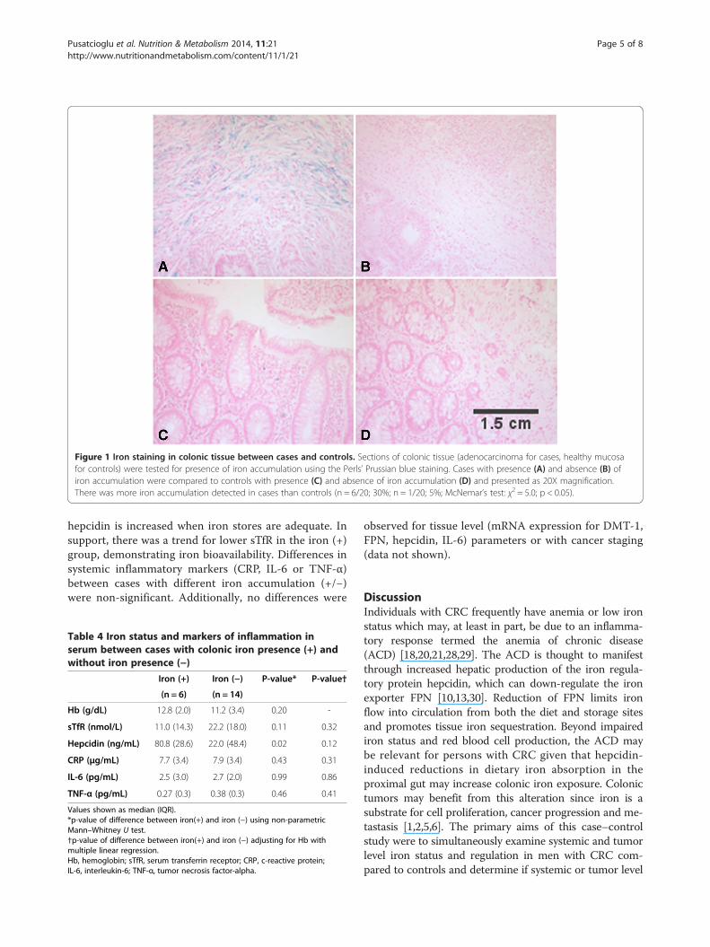
Figure 1 Iron staining in colonic tissue between cases and controls. Sections of colonic tissue (adenocarcinoma for cases, healthy mucosafor controls) were tested for presence of iron accumulation using the Perls’ Prussian blue staining. Cases with presence (A) and absence (B) ofiron accumulation were compared to controls with presence (C) and absence of iron accumulation (D) and presented as 20X magnification.There was more iron accumulation detected in cases than controls (n = 6/20; 30%; n = 1/20; 5%; McNemar’s test: χ2 = 5.0; p < 0.05).
Pusatcioglu et al. Nutrition & Metabolism 2014, 11:21 Page 5 of 8http://www.nutritionandmetabolism.com/content/11/1/21
hepcidin is increased when iron stores are adequate. Insupport, there was a trend for lower sTfR in the iron (+)group, demonstrating iron bioavailability. Differences insystemic inflammatory markers (CRP, IL-6 or TNF-α)between cases with different iron accumulation (+/−)were non-significant. Additionally, no differences were
Table 4 Iron status and markers of inflammation inserum between cases with colonic iron presence (+) andwithout iron presence (−)
Iron (+) Iron (−) P-value* P-value†
(n = 6) (n = 14)
Hb (g/dL) 12.8 (2.0) 11.2 (3.4) 0.20 -
sTfR (nmol/L) 11.0 (14.3) 22.2 (18.0) 0.11 0.32
Hepcidin (ng/mL) 80.8 (28.6) 22.0 (48.4) 0.02 0.12
CRP (μg/mL) 7.7 (3.4) 7.9 (3.4) 0.43 0.31
IL-6 (pg/mL) 2.5 (3.0) 2.7 (2.0) 0.99 0.86
TNF-α (pg/mL) 0.27 (0.3) 0.38 (0.3) 0.46 0.41
Values shown as median (IQR).*p-value of difference between iron(+) and iron (−) using non-parametricMann–Whitney U test.†p-value of difference between iron(+) and iron (−) adjusting for Hb withmultiple linear regression.Hb, hemoglobin; sTfR, serum transferrin receptor; CRP, c-reactive protein;IL-6, interleukin-6; TNF-α, tumor necrosis factor-alpha.
observed for tissue level (mRNA expression for DMT-1,FPN, hepcidin, IL-6) parameters or with cancer staging(data not shown).
DiscussionIndividuals with CRC frequently have anemia or low ironstatus which may, at least in part, be due to an inflamma-tory response termed the anemia of chronic disease(ACD) [18,20,21,28,29]. The ACD is thought to manifestthrough increased hepatic production of the iron regula-tory protein hepcidin, which can down-regulate the ironexporter FPN [10,13,30]. Reduction of FPN limits ironflow into circulation from both the diet and storage sitesand promotes tissue iron sequestration. Beyond impairediron status and red blood cell production, the ACD maybe relevant for persons with CRC given that hepcidin-induced reductions in dietary iron absorption in theproximal gut may increase colonic iron exposure. Colonictumors may benefit from this alteration since iron is asubstrate for cell proliferation, cancer progression and me-tastasis [1,2,5,6]. The primary aims of this case–controlstudy were to simultaneously examine systemic and tumorlevel iron status and regulation in men with CRC com-pared to controls and determine if systemic or tumor level
Pusatcioglu et al. Nutrition & Metabolism 2014, 11:21 Page 6 of 8http://www.nutritionandmetabolism.com/content/11/1/21
hepcidin expression was associated with tumor ironaccumulation.We found cases had significantly more iron-restricted
erythropoiesis based on Hb and sTfR compared to con-trols. We could not differentiate whether this mild anemiawas due to inflammation or also to frank iron deficiency.Often, gastrointestinal bleeding can contribute to irondeficiency in CRC; however, we excluded persons withknown gastrointestinal bleeding, suggesting other fac-tors contributed to the iron restriction observed in cases[29,31]. Typically when iron deficiency is present, serumhepcidin concentrations are undetectable [8]. Indeed,cases had lower serum hepcidin compared to controls;however, this difference was not significant and waswithin the normal range. This suggests hepcidin levelswere inappropriately elevated given the patients’ iron-restricted erythropoiesis. Hepcidin expression is simul-taneously regulated by inflammation, iron stores anderythropoiesis [7,10]. Further, hepcidin levels are ultim-ately determined by the relative strength of these oppos-ing stimuli [12,32]. In contrast to controls where serumhepcidin was correlated with Hb, serum hepcidin withincases was not significantly correlated with Hb suggest-ing that multiple signals may be influencing its produc-tion. Significantly greater systemic and colonic mucosalinflammation was observed in cases compared to con-trols which may have provided the necessary stimuli topromote increased hepatic hepcidin production, despiteelevated sTfR and low iron status. This phenotype, inwhich serum hepcidin is simultaneously regulated bylow iron status and inflammation, has been previouslyreported in morbid obesity [26,33,34]. Similar to our cases,obese participants in these studies presented with low ironstatus, elevated systemic inflammation and serum hepci-din concentrations within the normal range [26]. Likeobesity, the iron profile observed in our cases is consistentwith a mixed anemia for which hallmarks of the ACD andiron deficiency coexist [9]. This phenotype allows for themobilization of iron from body stores but impairs iron re-pletion as a result of reduced dietary iron absorption [35].Therefore, over time, inflammation-induced chronicallyelevated hepcidin would precipitate cellular iron depletionas body iron losses exceed dietary absorption effort[27,36]. Importantly, this type of mixed anemia has thepotential to increase gastrointestinal iron exposure inpersons with CRC.We assessed colonic mucosal iron transport and regula-
tion and found no statistical difference in expression ofDMT-1 or FPN between cases and controls. Directionallyour DMT-1 and FPN expression were similar to those ofBrookes et al. [1]. They reported increased colonocyte ex-pression of iron influx (DMT-1) and decreased iron efflux(FPN) proteins in CRC compared to controls. This sug-gests that CRC tissue is associated with greater colonic
adenocarcinoma iron uptake and sequestration comparedto healthy mucosa. We observed very low hepcidin ex-pression in the colonic tumors and healthy mucosa. Thisis not unexpected given that hepcidin is primarily pro-duced in hepatocytes. In our previous analysis, abdominalsubcutaneous and visceral adipose tissue also expressedvery low mRNA expression compared to the liver [26].Very little is known regarding the role of extra-hepatichepcidin in systemic and tissue-level iron regulation. Ourfindings indicate that colonic hepcidin has a minimal rolein tumor iron regulation. Ward et al. reported elevatedtumor hepcidin expression within 34% of cases comparedto non-involved healthy mucosa; however, other factorsthat regulate hepcidin expression including inflammation,iron status and dietary iron intake were not examined,limiting interpretations of their findings [15]. Given thatfew studies have examined the role of extra-hepatic hep-cidin in regulating systemic or tissue-level iron metabol-ism, further investigations are warranted to understandmechanisms related to iron metabolism in colonic tu-mors [15,37-39].We observed significantly more cases (30%) had detect-
able iron accumulation compared to controls (5%). Perls’staining has relatively low sensitivity. We speculate ironaccumulation would have been much more prevalent incases had we used a more sensitive assessment method.Brookes et al. used DAB-enhanced Perls’ methodologyand detected visible iron accumulation in all (n = 20) ofthe CRC cases but not controls [1]. To further characterizethe cases with iron accumulation, we examined systemichepcidin and iron status parameters. The iron (+) grouphad elevated serum hepcidin compared to cases withoutiron accumulation. However, the iron (+) group was alsomore iron sufficient and had higher Hb. Given the smallsample of cases with iron accumulation, it is hard to makeany definitive conclusions. However, our data coupledwith the report by Brookes et al. suggests that a subset ofpersons with CRC have increased expression of systemichepcidin, which may precipitate greater intestinal iron ex-posure, and promote tumor iron retention [1]. In a murinemodel of CRC, animals had increased tumor growth whenfed a high iron diet compared to a low iron diet [5]. Also,in persons with ulcerative colitis (UC), who have co-existing systemic and colonic inflammation and hepcidin-mediated dietary iron malabsorption, luminal iron expos-ure is associated with greater colonic inflammation andmucosal proliferation [40-42]. Therefore, studies examin-ing the effect of dietary iron restriction in persons withCRC should be explored.The link with hepcidin and excess tumor iron accumu-
lation provides further evidence for its role in cancer.Only one other study has comprehensively explored thehepcidin-FPN axis and its relationship in cancer pro-gression. Pinnix et al. using human breast cancer tissue
Pusatcioglu et al. Nutrition & Metabolism 2014, 11:21 Page 7 of 8http://www.nutritionandmetabolism.com/content/11/1/21
found that low FPN and high hepcidin expression mayenable rapid cell proliferation [14]. Unfortunately, theauthors did not measure systemic hepcidin concentra-tions and future exploration should focus on its role intumor iron retention in other epithelial cancers.Our study had strengths and limitations. A significant
strength was that we simultaneously measured hepcidinsystemically and in colonic mucosa while accounting forseveral factors that can contribute to hepcidin regulation,including iron status, inflammation and dietary iron in-take. Additionally, we included a metabolically healthycontrol group. However, this study had limitations, includ-ing a relatively small sample size. Therefore, when westratified by presence of iron accumulation in the CRCcases, this may have reduced the statistical power to detectdifferences. Tissue analysis with adenocarcinoma in casesto healthy mucosa in a control group was not a direct com-parison. Future studies should also include non-involvedmucosa in cases to accurately describe our analysis amongall groups. Due to limited tissue allocated for this study,we were unable to accurately measure protein expressionof the colonic iron exporter via FPN. Ferroportin is post-translationally modified by hepcidin, which may have pre-cluded us from observing a significant correlation betweencolonic FPN and hepcidin. Although we suggest dietaryiron absorption may be impaired in persons with CRC,we did not examine dietary iron absorption in this study.Finally, our design included only males, thus generalizabilityto females may be limited.
ConclusionIn summary, our study demonstrates for the first time thatCRC in some men is associated with a mixed anemiapathology characterized by iron restricted erythropoiesis.We believe the simultaneous presence of inflammation(increases hepcidin) and iron insufficiency (suppresseshepcidin) in a subset of our men with CRC resulted inhepcidin concentrations inappropriately elevated given theirdepleted iron status. Further, these findings suggest thatsystemic hepcidin in some CRC cases may 1) decrease duo-denal iron absorption resulting in low iron status and 2)contribute to excess colonic iron exposure and diseasepromotion. Given the high incidence of CRC and the ac-companying low iron status, these findings could have sig-nificant clinical implications [21,43]. Future investigationsinto the risks and benefits of dietary iron intake and oraliron supplementation in persons with CRC are warranted.
Additional files
Additional file 1: Primers used for mRNA analysis.
Additional file 2: mRNA expression of iron transporters andinflammatory proteins in colonic tissue of colorectal cancer casesand controls using raw ΔCt values.
Competing interestsAll of the authors declare they have no competing interests.
Authors’ contributionsCKP and CB designed the research. CKP, JC and XL acquired the data. CKP,SF, RJC, RL and DN analyzed the data. CKP, SF, EN, GF, LTH, and CBinterpreted the data. CKP wrote the manuscript draft. CKP, EN, GF, LTH, andCB critically revised the manuscript. All authors read and approved the finalmanuscript.
AcknowledgmentsThe efforts of this study were supported by 5R25CA057699 from theNational Cancer Institute. EN is affiliated with Intrinsic LifeSciences, thecompany that assessed the serum samples for hepcidin.
Author details1Division of Gastroenterology, Hepatology, and Nutrition, Ann & Robert H.Lurie Children’s Hospital of Chicago, 225 E Chicago Ave, Chicago, IL 60611,USA. 2Department of Medicine, University of California, Los Angeles, 10833Le Conte Ave, Los Angeles, CA 90095, USA. 3Department of Kinesiology andNutrition, University of Illinois at Chicago, 1919 W Taylor St, Chicago, IL60612, USA. 4Section of Digestive Disease and Nutrition, University of Illinoisat Chicago, 840 S Wood St, Chicago, IL 60612, USA. 5Division ofEpidemiology and Biostatistics, University of Illinois at Chicago, 1603 WTaylor St, Chicago, IL 60612, USA. 6Department of Medicine, University ofIllinois at Chicago, 1747 W Roosevelt Rd, Chicago, IL 60608, USA.7Department of Pathology, University of Illinois at Chicago, 840 S Wood St,Chicago, IL 60612, USA.
Received: 24 January 2014 Accepted: 29 April 2014Published: 13 May 2014
References1. Brookes MJ, Hughes S, Turner FE, Reynolds G, Sharma N, Ismail T, Berx G,
McKie AT, Hotchin N, Anderson GJ, Iqbal T, Tselepis C: Modulation of irontransport proteins in human colorectal carcinogenesis. Gut 2006,55:1449–1460.
2. Chua AC, Klopcic B, Lawrance IC, Olynyk JK, Trinder D: Iron: an emergingfactor in colorectal carcinogenesis. World J Gastroenterol WJG 2010,16:663–672.
3. Nelson RL: Iron and colorectal cancer risk: human studies. Nutr Rev 2001,59:140–148.
4. Nelson RL, Davis FG, Sutter E, Sobin LH, Kikendall JW, Bowen P: Body ironstores and risk of colonic neoplasia. J Natl Cancer Inst 1994, 86:455–460.
5. Radulescu S, Brookes MJ, Salgueiro P, Ridgway RA, McGhee E, Anderson K,Ford SJ, Stones DH, Iqbal TH, Tselepis C, Sansom OJ: Luminal iron levelsgovern intestinal tumorigenesis after Apc loss in vivo. Cell Reports 2012,2:270–282.
6. Toyokuni S: Role of iron in carcinogenesis: cancer as a ferrotoxic disease.Cancer Sci 2009, 100:9–16.
7. Nemeth E, Ganz T: Regulation of iron metabolism by hepcidin. Annu RevNutr 2006, 26:323–342.
8. Ganz T, Olbina G, Girelli D, Nemeth E, Westerman M: Immunoassay forhuman serum hepcidin. Blood 2008, 112:4292–4297.
9. Tussing-Humphreys L, Pusatcioglu C, Nemeth E, Braunschweig C:Rethinking iron regulation and assessment in iron deficiency, anemia ofchronic disease, and obesity: introducing hepcidin. J Acad Nutr Diet 2012,112:391–400.
10. Ganz T: Hepcidin, a key regulator of iron metabolism and mediator ofanemia of inflammation. Blood 2003, 102:783–788.
11. Nemeth E, Ganz T: The role of hepcidin in iron metabolism. ActaHaematol 2009, 122:78–86.
12. Darshan D, Anderson GJ: Interacting signals in the control of hepcidinexpression. Biometals Int J role Metal ions Biol Biochem Med 2009, 22:77–87.
13. Nemeth E, Tuttle MS, Powelson J, Vaughn MB, Donovan A, Ward DM, GanzT, Kaplan J: Hepcidin regulates cellular iron efflux by binding toferroportin and inducing its internalization. Science 2004, 306:2090–2093.
14. Pinnix ZK, Miller LD, Wang W, D’Agostino R Jr, Kute T, Willingham MC,Hatcher H, Tesfay L, Sui G, Di X, Torti SV, Torti FM: Ferroportin and ironregulation in breast cancer progression and prognosis. Sci Transl Med2010, 2:43–56.
Pusatcioglu et al. Nutrition & Metabolism 2014, 11:21 Page 8 of 8http://www.nutritionandmetabolism.com/content/11/1/21
15. Ward DG, Roberts K, Brookes MJ, Joy H, Martin A, Ismail T, Spychal R, Iqbal T,Tselepis C: Increased hepcidin expression in colorectal carcinogenesis.World J Gastroenterol WJG 2008, 14:1339–1345.
16. Butterfield AM, Luan P, Witcher DR, Manetta J, Murphy AT, Wroblewski VJ,Konrad RJ: A dual-monoclonal sandwich ELISA specific for hepcidin-25.Clin Chem 2010, 56:1725–1732.
17. Sasu BJ, Li H, Rose MJ, Arvedson TL, Doellgast G, Molineux G: Serumhepcidin but not prohepcidin may be an effective marker for anemia ofinflammation (AI). Blood Cells Mol Dis 2010, 45:238–245.
18. Weiss G: Pathogenesis and treatment of anaemia of chronic disease.Blood Rev 2002, 16:87–96.
19. Terzic J, Grivennikov S, Karin E, Karin M: Inflammation and colon cancer.Gastroenterology 2010, 138:2101–2114. e2105.
20. Dunne JR, Gannon CJ, Osborn TM, Taylor MD, Malone DL, Napolitano LM:Preoperative anemia in colon cancer: assessment of risk factors. Am Surg2002, 68:582–587.
21. Ludwig H, Van Belle S, Barrett-Lee P, Birgegard G, Bokemeyer C, Gascon P,Kosmidis P, Krzakowski M, Nortier J, Olmi P, Schneider M, Schrijvers D: TheEuropean Cancer Anaemia Survey (ECAS): a large, multinational, pro-spective survey defining the prevalence, incidence, and treatment ofanaemia in cancer patients. Eur J Cancer 2004, 40:2293–2306.
22. Block G, Woods M, Potosky A, Clifford C: Validation of a self-administereddiet history questionnaire using multiple diet records. J Clin Epidemiol1990, 43:1327–1335.
23. Edge SB, Compton CC: The American Joint Committee on Cancer: the 7thedition of the AJCC cancer staging manual and the future of TNM.Ann Surg Oncol 2010, 17:1471–1474.
24. World Health Organization, Centers for Disease Control: Assessing the ironstatus of populations: Report of a joint WHO/CDC Technical Consultation onthe assessment of iron status at the population level. 2007. http://www.who.int/nutrition/publications/micronutrients/anaemia_iron_deficiency/en/.
25. Cook JD, Flowers CH, Skikne BS: An assessment of dried blood-spottechnology for identifying iron deficiency. Blood 1998, 92:1807–1813.
26. Tussing-Humphreys LM, Nemeth E, Fantuzzi G, Freels S, Guzman G,Holterman AX, Braunschweig C: Elevated systemic hepcidin and irondepletion in obese premenopausal females. Obesity 2010, 18:1449–1456.
27. Tussing-Humphreys LM, Nemeth E, Fantuzzi G, Freels S, Holterman AX,Galvani C, Ayloo S, Vitello J, Braunschweig C: Decreased serum hepcidinand improved functional iron status 6 months after restrictive bariatricsurgery. Obesity 2010, 18:2010–2016.
28. Harrison L, Shasha D, Shiaova L, White C, Ramdeen B, Portenoy R:Prevalence of anemia in cancer patients undergoing radiation therapy.Semin Oncol 2001, 28:54–59.
29. Grotto HZ: Anaemia of cancer: an overview of mechanisms involved inits pathogenesis. Med Oncol 2008, 25:12–21.
30. Means RT: Hepcidin and cytokines in anaemia. Hematol 2004, 9:357–362.31. Hardcastle JD, Chamberlain JO, Robinson MH, Moss SM, Amar SS, Balfour TW,
James PD, Mangham CM: Randomised controlled trial of faecal-occult-bloodscreening for colorectal cancer. Lancet 1996, 348:1472–1477.
32. Huang H, Constante M, Layoun A, Santos MM: Contribution of STAT3 andSMAD4 pathways to the regulation of hepcidin by opposing stimuli.Blood 2009, 113:3593–3599.
33. Aeberli I, Hurrell RF, Zimmermann MB: Overweight children have highercirculating hepcidin concentrations and lower iron status but havedietary iron intakes and bioavailability comparable with normal weightchildren. Int J Obes 2009, 33:1111–1117.
34. Vuppalanchi R, Troutt JS, Konrad RJ, Ghabril M, Saxena R, Bell LN, Kowdley KV,Chalasani N: Serum hepcidin levels are associated with obesity but not liverdisease. Obesity 2013, 22:836–841.
35. Zimmermann MB, Zeder C, Muthayya S, Winichagoon P, Chaouki N, Aeberli I,Hurrell RF: Adiposity in women and children from transition countriespredicts decreased iron absorption, iron deficiency and a reducedresponse to iron fortification. Int J Obes 2008, 32:1098–1104.
36. Young MF, Glahn RP, Ariza-Nieto M, Inglis J, Olbina G, Westerman M, O’Brien KO:Serum hepcidin is significantly associated with iron absorption from foodand supplemental sources in healthy young women. Am J Clin Nutr 2009,89:533–538.
37. Bekri S, Gual P, Anty R, Luciani N, Dahman M, Ramesh B, Iannelli A, Staccini-MyxA, Casanova D, Ben Amor I, Saint-Paul MC, Huet PM, Sadoul JL, Gugenheim J,Srai SK, Tran A, Le Marchand-Brustel Y: Increased adipose tissue expression of
hepcidin in severe obesity is independent from diabetes and NASH.Gastroenterol 2006, 131:788–796.
38. Merle U, Fein E, Gehrke SG, Stremmel W, Kulaksiz H: The iron regulatorypeptide hepcidin is expressed in the heart and regulated by hypoxiaand inflammation. Endocrinol 2007, 148:2663–2668.
39. Wu X, Yung LM, Cheng WH, Yu PB, Babitt JL, Lin HY, Xia Y: Hepcidinregulation by BMP signaling in macrophages is lipopolysaccharidedependent. PLoS One 2012, 7:e44622.
40. Reifen R, Matas Z, Zeidel L, Berkovitch Z, Bujanover Y: Ironsupplementation may aggravate inflammatory status of colitis in a ratmodel. Dig Dis Sci 2000, 45:394–397.
41. Seril DN, Liao J, Yang CS, Yang GY: Systemic iron supplementationreplenishes iron stores without enhancing colon carcinogenesis inmurine models of ulcerative colitis: comparison with iron-enriched diet.Dig Dis Sci 2005, 50:696–707.
42. Oustamanolakis P, Koutroubakis IE, Messaritakis I, Malliaraki N, Sfiridaki A,Kouroumalis EA: Serum hepcidin and prohepcidin concentrations ininflammatory bowel disease. Eur J Gastroenterol Hepatol 2011, 23:262–268.
43. Jemal A, Bray F, Center MM, Ferlay J, Ward E, Forman D: Global cancerstatistics. CA a Cancer J Clin 2011, 61:69–90.
doi:10.1186/1743-7075-11-21Cite this article as: Pusatcioglu et al.: Systemic and tumor level ironregulation in men with colorectal cancer: a case control study. Nutrition& Metabolism 2014 11:21.
Submit your next manuscript to BioMed Centraland take full advantage of:
• Convenient online submission
• Thorough peer review
• No space constraints or color figure charges
• Immediate publication on acceptance
• Inclusion in PubMed, CAS, Scopus and Google Scholar
• Research which is freely available for redistribution
Submit your manuscript at www.biomedcentral.com/submit