Systematic review of Anteaeolidiella (Mollusca, Nudibranchia, Aeolidiidae) based on morphological...
25
Systematic review of Anteaeolidiella (Mollusca, Nudibranchia, Aeolidiidae) based on morphological and molecular data, with a description of three new species LEILA CARMONA 1 *, VISHAL BHAVE 2 , RAHUL SALUNKHE 2,3 , MARTA POLA 4 , TERRENCE M. GOSLINER 5 and JUAN LUCAS CERVERA 1 1 Departamento de Biología, Facultad de Ciencias del Mar y Ambientales, Campus de Excelencia Internacional del Mar (CEI·MAR), Universidad de Cádiz, Polígono Río San Pedro, s/n, Ap.40. 11510 Puerto Real (Cádiz), Spain 2 Bombay Natural History Society, Hornbill House, Shaheed Bhagat Singh Road, Mumbai 400 001, Maharashtra, India 3 Molecular Biology Unit, National Center for Cell Science, Ganeshkhind, Pune, 411007, Maharashtra, India 4 Laboratorio de Biología Marina, Departamento de Biología, Edificio de Biología, Campus de Excelencia Internacional UAM+CSIC, Universidad Autónoma de Madrid, C/ Darwin, 2, 28049 Madrid, Spain 5 Department of Invertebrate Zoology, California Academy of Sciences, 55 Music Concourse Drive, Golden Gate Park, San Francisco, CA 94118, USA Received 6 August 2013; revised 16 December 2013; accepted for publication 19 December 2013 Partial sequences of two mitochondrial genes and one nuclear gene from Anteaeolidiella specimens collected throughout temperate and tropical areas in the Atlantic, Mediterranean, eastern Pacific, and Indo-Pacific have revealed the existence of a species complex under the name of Anteaeolidiella indica (Bergh, 1888). Further examination showed consistent differences in colour pattern and the internal anatomy that became evident only once the molecular outcomes were analysed. Moreover, the original description of A. indica suggests that this species has a very particular colour pattern that is not present in any of our specimens. A review of the literature led us to resurrect the names of Anteaeolidiella cacaotica (Stimpson, 1855), Anteaeolidiella saldanhensis (Barnard, 1927), Anteaeolidiella takanosimensis (Baba, 1930), and Anteaeolidiella lurana (Ev. Marcus & Er. Marcus, 1967). Aeolidiella chromosoma (Cockerell & Eliot, 1905) and Aeolidiella oliviae (MacFarland, 1966) are transferred to Anteaeolidiella based on our morphological and molecular data. Finally, three new species from the Indo-Pacific are described herein. © 2014 The Linnean Society of London, Zoological Journal of the Linnean Society, 2014, 171, 108–132. doi: 10.1111/zoj.12129 ADDITIONAL KEYWORDS: cryptic species – Heterobranchia – marine biodiversity – molluscan diversity – morphology – Nudibranchia – systematics. INTRODUCTION Miller (2001) erected the genus Anteaeolidiella. This author considered as diagnostic characters the wide and bilobed tooth with a central cusp, the nodulose spindle-shaped oral glands, the club-shaped cerata arranged in rows, and the smooth rhinophores. Miller (2001) placed Aeolidiella indica (Bergh, 1888a) in this new genus and designed it as the type species. This author also transferred Baeolidia benteva Er. Marcus, 1958 to Anteaeolidiella based on the ceratal arrange- ment and the position of the orifices of the latter species. *Corresponding author. E-mail: [email protected] Zoological Journal of the Linnean Society, 2014, 171, 108–132. With 14 figures © 2014 The Linnean Society of London, Zoological Journal of the Linnean Society, 2014, 171, 108–132 108
-
Upload
independent -
Category
Documents
-
view
2 -
download
0
Transcript of Systematic review of Anteaeolidiella (Mollusca, Nudibranchia, Aeolidiidae) based on morphological...
Systematic review of Anteaeolidiella (Mollusca,Nudibranchia, Aeolidiidae) based on morphological andmolecular data, with a description of three new species
LEILA CARMONA1*, VISHAL BHAVE2, RAHUL SALUNKHE2,3, MARTA POLA4,TERRENCE M. GOSLINER5 and JUAN LUCAS CERVERA1
1Departamento de Biología, Facultad de Ciencias del Mar y Ambientales, Campus de ExcelenciaInternacional del Mar (CEI·MAR), Universidad de Cádiz, Polígono Río San Pedro, s/n, Ap.40. 11510Puerto Real (Cádiz), Spain2Bombay Natural History Society, Hornbill House, Shaheed Bhagat Singh Road, Mumbai 400 001,Maharashtra, India3Molecular Biology Unit, National Center for Cell Science, Ganeshkhind, Pune, 411007,Maharashtra, India4Laboratorio de Biología Marina, Departamento de Biología, Edificio de Biología, Campus deExcelencia Internacional UAM+CSIC, Universidad Autónoma de Madrid, C/ Darwin, 2, 28049Madrid, Spain5Department of Invertebrate Zoology, California Academy of Sciences, 55 Music Concourse Drive,Golden Gate Park, San Francisco, CA 94118, USA
Received 6 August 2013; revised 16 December 2013; accepted for publication 19 December 2013
Partial sequences of two mitochondrial genes and one nuclear gene from Anteaeolidiella specimens collectedthroughout temperate and tropical areas in the Atlantic, Mediterranean, eastern Pacific, and Indo-Pacific haverevealed the existence of a species complex under the name of Anteaeolidiella indica (Bergh, 1888). Furtherexamination showed consistent differences in colour pattern and the internal anatomy that became evident onlyonce the molecular outcomes were analysed. Moreover, the original description of A. indica suggests that thisspecies has a very particular colour pattern that is not present in any of our specimens. A review of the literatureled us to resurrect the names of Anteaeolidiella cacaotica (Stimpson, 1855), Anteaeolidiella saldanhensis (Barnard,1927), Anteaeolidiella takanosimensis (Baba, 1930), and Anteaeolidiella lurana (Ev. Marcus & Er. Marcus, 1967).Aeolidiella chromosoma (Cockerell & Eliot, 1905) and Aeolidiella oliviae (MacFarland, 1966) are transferred toAnteaeolidiella based on our morphological and molecular data. Finally, three new species from the Indo-Pacific aredescribed herein.
© 2014 The Linnean Society of London, Zoological Journal of the Linnean Society, 2014, 171, 108–132.doi: 10.1111/zoj.12129
ADDITIONAL KEYWORDS: cryptic species – Heterobranchia – marine biodiversity – molluscan diversity –morphology – Nudibranchia – systematics.
INTRODUCTION
Miller (2001) erected the genus Anteaeolidiella. Thisauthor considered as diagnostic characters the wideand bilobed tooth with a central cusp, the nodulose
spindle-shaped oral glands, the club-shaped cerataarranged in rows, and the smooth rhinophores. Miller(2001) placed Aeolidiella indica (Bergh, 1888a) in thisnew genus and designed it as the type species. Thisauthor also transferred Baeolidia benteva Er. Marcus,1958 to Anteaeolidiella based on the ceratal arrange-ment and the position of the orifices of the latterspecies.*Corresponding author. E-mail: [email protected]
bs_bs_banner
Zoological Journal of the Linnean Society, 2014, 171, 108–132. With 14 figures
© 2014 The Linnean Society of London, Zoological Journal of the Linnean Society, 2014, 171, 108–132108
Since Anteaeolidiella indica was first described(Bergh, 1888a), this species has been reported from theMediterranean (Schmekel, 1968, 1970; Zenetos et al.,2005; Sciberras & Schembri, 2007), the Strait ofGibraltar (García-Gómez, 2002), the Canary Islands(Pérez-Sánchez & Moreno, 1990; Moro et al., 1995,2003; Ortea et al., 2001), South Africa (Gosliner &Griffiths, 1981), the Caribbean (Valdés et al., 2006;Ortigosa, Simones & Calado, 2013), Brazil(Domínguez, Troncoso & García, 2008; García-García,Domínguez & Troncoso, 2008; Padula et al., 2011), thePacific coast of Costa Rica (Camacho-García, Gosliner& Valdés, 2005), Hawaii (Pittman & Fiene, 2013),Australia (Gosliner, Behrens & Valdés, 2008), NewZealand (Miller, 2001), Japan (Nakano, 2004;Imamoto, 2013), the Kerama Islands (Ono, 1999), theRyukyu Islands (Ono, 2004), Réunion Island(Bidgrain, 2005), and Ratnagiri, India (Bhave & Apte,2011). Several species names have been introducedover the years for different colour forms that occur overthis biogeographic pattern; however, several of thesenames have been synonymized with Anteaeolidiellaindica. Gosliner & Griffiths (1981) compared the latterspecies with Aeolidiella orientalis Bergh, 1888b,Aeolidiella saldanhensis Barnard, 1927, Aeolidiellahulli Risbec, 1928, Aeolidiella takanosimensis Baba,1930, Aeolidiella multicolor Macnae, 1954, andAeolidiella lurana Er. Marcus & Ev. Marcus, 1967, butthey concluded that differences were not consistentand therefore attributed them to intraspecific variabil-ity. Moreover, Anteaeolidiella indica together withsome of its synonyms have been considered as juniorsynonyms of Anteaeolidiella foulisi (Angas, 1864)(Burn, 2006; Debelius & Kuiter, 2007). At the sametime, Burn (1964, 2006) suggested that the evenearlier described Eolis cacaotica Stimpson, 1855 couldbe identical to Anteaeolidiella foulisi.
The extreme external and internal similarity of allthese species has caused a great deal of confusion andblurred the differences among them. As moleculartools have become more prevalent and cheaper,several heterobranch species with disjunct rangeshave proven to be species complexes (Krug et al.,2008, 2013; Malaquias & Reid, 2008, 2009; Carmonaet al., 2011; Gosliner & Fahey, 2011; Jörger et al.,2012; Ornelas-Gatdula et al., 2012; Pola, Camacho-García & Gosliner, 2012). The molecular phylogenyconducted by Carmona et al. (2013) transferredAeolidiella oliviae (MacFarland, 1966) and Aeoli-diella chromosoma (Cockerell & Eliot, 1905) toAnteaeolidiella, resurrected the species Aeolidiellacacaotica, Aeolidiella saldanhensis, Aeolidiellatakanosimensis, and Aeolidiella lurana, placed themin Anteaeolidiella, and highlighted the necessityof reviewing Anteaeolidiella indica sensu Bergh(1888a).
Recently collected material from Australia andIndia allowed us to extend the research by Carmonaet al. (2013), increasing the number of species studiedas well as their range of distribution. In the presentcontribution, we undertake a systematic revision ofAnteaeolidiella, based on molecular data (twomitochondrial genes and one nuclear gene) and tra-ditional morphological and anatomical data. We alsoattempt to clarify the morphological differencesbetween the species of the ‘Anteaeolidiella indica’complex. Moreover, we try to elucidate the identity ofAnteaeolidiella indica sensu Bergh (1888a) by per-forming a careful review of the literature. Finally,based on the addition of three new species, wepropose a new diagnosis of the genus Anteaeolidiella.
MATERIAL AND METHODSMORPHOLOGY
A comprehensive review of the literature was con-ducted to determine the valid names for the speciesrecognized in the molecular and morphological analy-ses. After the remarks of the type species, all avail-able names for Anteaeolidiella species are organizedand discussed in this article by year of publication. Inthe synonymy lists, references to the original descrip-tion of the valid name and all synomyms, and the firstproposed change of binomen, are included, but sub-sequent references are not.
Whenever possible, two or more specimens of eachspecies were examined anatomically. Specimens weredissected by dorsal incision. Their internal featureswere examined and drawn using a dissecting micro-scope with the aid of a camera lucida. Special atten-tion was paid to the morphology of the reproductivesystem and oral and salivary glands. The buccal masswas removed and dissolved in 10% sodium hydroxideuntil the radula was isolated from the surroundingtissue. The radula was then rinsed in water, dried,and mounted for examination by scanning electronmicroscopy (SEM). The radula of the specimens fromIndia were examined and drawn using a dissectingmicroscope.
Voucher specimens are held at the following insti-tutions: AMS, Australian Museum of Sydney, Sydney,Australia; BNHS, Bombay Natural History Society,Mumbai, India; CASIZ, California Academy ofSciences, San Francisco, USA; CNMO, ColecciónNacional de Moluscos Instituto de Biología,Universidad Nacional Autónoma de México, MexicoCity, Mexico; LACM, Natural History Museum of LosAngeles County; Los Angeles, USA; MHNSM, Museode Historia Natural de El Salvador, San Salvador, ElSalvador; MNCN, Museo Nacional de CienciasNaturales, Madrid, Spain; MZSP, Museu de Zoologia
SYSTEMATICS OF ANTEAEOLIDIELLA 109
© 2014 The Linnean Society of London, Zoological Journal of the Linnean Society, 2014, 171, 108–132
da Universidade São Paulo, São Paulo, Brazil; WAM,Western Australian Museum, Perth, Australia;ZMBN, University Museum of Bergen, Bergen,Norway; ZSM, Zoologische Staatssammlung Mün-chen, Munich, Germany.
MOLECULAR WORK
Taxon samplingSamples were obtained using standard scuba-divingsampling techniques for opisthobranchs and throughthe study of museum collections. Two hundred andfifty-six specimens, including 48 species of Aeolidiidae(20 specimens and nine species of Anteaeolidiella),three species of Babakinidae, 32 species ofFacelinidae, seven species of Flabellinidae, onespecies of Fionidae, three species of Piseinotecidae,and four species of Tergipedidae, were used forphylogenetic inference. A total of 197 specimens weresuccessfully sequenced for cytochrome c oxidasesubunit I (COI), 224 specimens were sequenced for16S rRNA (16S), and 236 specimens were sequencedfor histone 3 (H3). Thirty-five additional sequenceswere obtained from GenBank (for a full list ofsamples, localities, and voucher references, see Sup-porting Information, Table S1).
Tritonia challengeriana Bergh, 1884 (= Tritoniaantarctica Pfeffer in Martens & Pfeffer, 1886) waschosen as the out-group for its basal taxonomicalposition within Cladobranchia (Pola & Gosliner,2010).
DNA extraction, amplification, and sequencingDNA was extracted from foot tissue of specimenspreserved in 70–100% ethanol, except in the case ofsmall animals, where the whole specimen was used.The Qiagen DNeasy Blood & Tissue Kit was used forDNA extraction (Qiagen, Valencia, CA, USA).
Partial sequences of COI, 16S, and H3 were ampli-fied by polymerase chain reaction (PCR) using thefollowing primers: LCO1490 (5′-GGTCAACAAATCATAAGATATTGG-3′) and HCO2198 (5′-TAAACTTCAGGGTGACCAAAAATCA-3′) (Folmer et al., 1994) forCOI; 16S ar-L (5′-CGCCTGTTTATCAAAAACAT-3′)and 16S br-H (5′-CCGGTCTGAACTCAGATCACGT-3′) (Palumbi et al., 1991) for 16S rRNA; and H3AD5′3′(5′-ATGGCTCGTACCAAGCAGACVGC-3′) andH3BD5′3′ (5′-ATATCCTTR GGCATRATRGTGAC-3′)(Colgan et al., 1998) for H3. These three gene regionsare commonly used in the systematic studies ofgastropods (e.g. Meyer, 2003; Williams & Reid,2004; Dinapoli et al., 2006; Frey & Vermeij, 2008;Malaquias & Reid, 2009; Pola & Gosliner, 2010;Claremont et al., 2013; Eilertsen & Malaquias, 2013;Ohnheiser & Malaquias, 2013); however, severalinternal primers for COI and H3 were designed for
specimens that did not amplify with the universalprimers (Carmona et al., 2013: table 1).
Polymerase chain reactions were conducted in50-μL volume reactions containing 2 μL of bothforward and reverse primers (10 mM), 5 μL ofdeoxyribonucleotide triphosphate (dNTP; 2 mM), agene-dependent quantity of magnesium chloride(25 mM), 0.5 μL of Qiagen DNA polymerase(5 units μL–1), 10 μL of ‘Q-solution’ (5×), 5 μL ofQiagen buffer (10×) (Qiagen Taq PCR Core Kit, cat.no. 201225), and 1 μL of genomic DNA. The volumesof magnesium chloride used were 7 μL for COI and16S, and 4 μL for H3. The amplification of COI wasperformed with an initial denaturation for 5 min at94 °C, followed by 35 cycles of 1 min at 94 °C, 30 s at44 °C (annealing temperature), and 1 min at 72 °C,with a final extension of 7 min at 72 °C. The 16Samplification began with an initial denaturation for5 min at 95 °C, followed by 35 cycles of 30 s at 94 °C,30 s at 44 °C (annealing temperature), 1 min at 72 °C,with a final extension of 7 min at 72 °C. H3 amplifi-cation was performed with an initial denaturation for3 min at 95 °C, followed by 40 cycles of 45 s at 94 °C,45 s at 50 °C (annealing temperature), and 2 min at72 °C, with a final extension of 10 min at 72 °C.
Successful PCRs were purified by mixing 5 μL ofPCR product with 2 μL of ExoSAP-IT (usb.affymetrix.com). Samples were incubated at 37 °C for15 min followed by an inactivation step at 80 °C for15 min. Sequence reactions were run on a 3730XLDNA sequencer (Applied Biosystems). All newsequences have been deposited in GenBank.
Sequence alignment and phylogenetic analysesDNA sequences were assembled and edited usingGENEIOUS PRO 4.7.6 (Drummond et al., 2009). Allthe sequences were checked for contamination withBLAST (Altschul et al., 1990), implemented in theGenBank database. MAFFT (Katoh, Asimenos & Toh,2009) was employed to align the sequences. Thealignments were checked by eye using MacClade4.06 (Maddison & Maddison, 2005). Protein-codingsequences were translated into amino acids forconfirmation of alignment. Pairwise uncorrectedp-distance values between each taxon were calculatedfor the COI gene. Uncorrected p-distances between alltaxa, and level of saturation for first, second, andthird codon positions (p-distances against transitionsplus transversions) were calculated in MEGA 5.0*(Tamura et al., 2011) for COI and H3. No evidenceof saturation was found, even in the third codonpositions.
The most variable regions from the 16S rRNAalignment were removed using both the defaultsettings and the standard options for stringent andless stringent selection in GBLOCKS (Talavera &
110 L. CARMONA ET AL.
© 2014 The Linnean Society of London, Zoological Journal of the Linnean Society, 2014, 171, 108–132
Castresana, 2007). Excluding ‘indel-rich’ regions, thetree was in general poorly resolved, with lower nodesupport. Therefore, final analyses were performedincluding all bases. Sequences of COI, 16S, and H3were trimmed to 658, 463, and 327 bp, respectively.
Individual gene analyses and a concatenated analy-sis were performed. The best-fitting models of evolu-tion for each gene were determined using the Akaikeinformation criterion (Akaike, 1974), implemented inMrModeltest 2.3 (Nylander, 2004). The GTR + I + Gmodel was selected for the three genes.
Maximum-likelihood (ML) analyses were performedusing the RAxML 7.0.4 (Stamatakis, 2006), and nodesupport was assessed with non-parametric bootstrap-ping (BS) with 5000 replicates, random starting trees,and parameters estimated from each data set underthe model selected for the original data set. Bayesianinference (BI) analyses were conducted usingMrBayes 3.1.2b (Ronquist & Huelsenbeck, 2003) for45 000 000 generations, with two independent runsand a sampling frequency of 1000. The models imple-mented were those estimated with MrModeltest 2.3.The combined data set was partitioned among genesand the ‘unlink’ command was used to allow all param-eters to vary independently within each partition.
Convergence was diagnosed graphically by plottingthe likelihood against the number of generations foreach run using TRACER 1.4.1 (Drummond &Rambaut, 2007). For each analysis, the first 11 250trees were discarded (‘burn-in’), and node support wasassessed by posterior probabilities (PPs). Only nodessupported by BS ≥ 75 and PP ≥ 0.90 are discussed. Allthe alignments and trees published in this study aredeposited in TreeBase under project number 15114.urn:lsid:zoobank.org:pub:0D798F9B-102D-4087-9F2F-1751725C9B8D.
Species delimitationTo define species, we used the criteria of divergenceand reciprocal monophyly, supported by independentgenetic markers (Knowlton, 2000; Wheeler & Meier,2000; Avise, 2004; Malaquias & Reid, 2009), usingcut-off values from Carmona et al. (2013) as our ref-erence thresholds (5.5–16%).
We also applied the Automatic Barcode Gap Dis-covery (ABGD) method (Puillandre et al., 2012a).ABGD is a distance-based method designed to detectthe so-called ‘barcode gap’ in the distribution ofpairwise distances calculated in a COI alignment(Puillandre et al., 2012a, b). This method representsprogress in avoiding any dependence on a hypotheti-cally defined species. The web-based ABGD program(available at http://wwwabi.snv.jussieu.fr/public/abgd/) was employed with the default settings togenerate a preliminary partition of sequences, usingthe COI alignment and excluding the out-group.
Nomenclatural actsThis published work and the nomenclatural acts itcontains have been registered in ZooBank, the onlineregistration system for the International Commissionon Zoological Nomenclature (ICZN). The ZooBank lifescience identifiers (LSIDs) can be resolved and theassociated information viewed through any standardweb browser by appending the LSID to the prefix‘http://zoobank.org/’. The LSID for this publication is:LSID urn:lsid:zoobank.org:pub:0D798F9B-102D-4087-9F2F-1751725C9B8D.
RESULTSMOLECULAR RESULTS
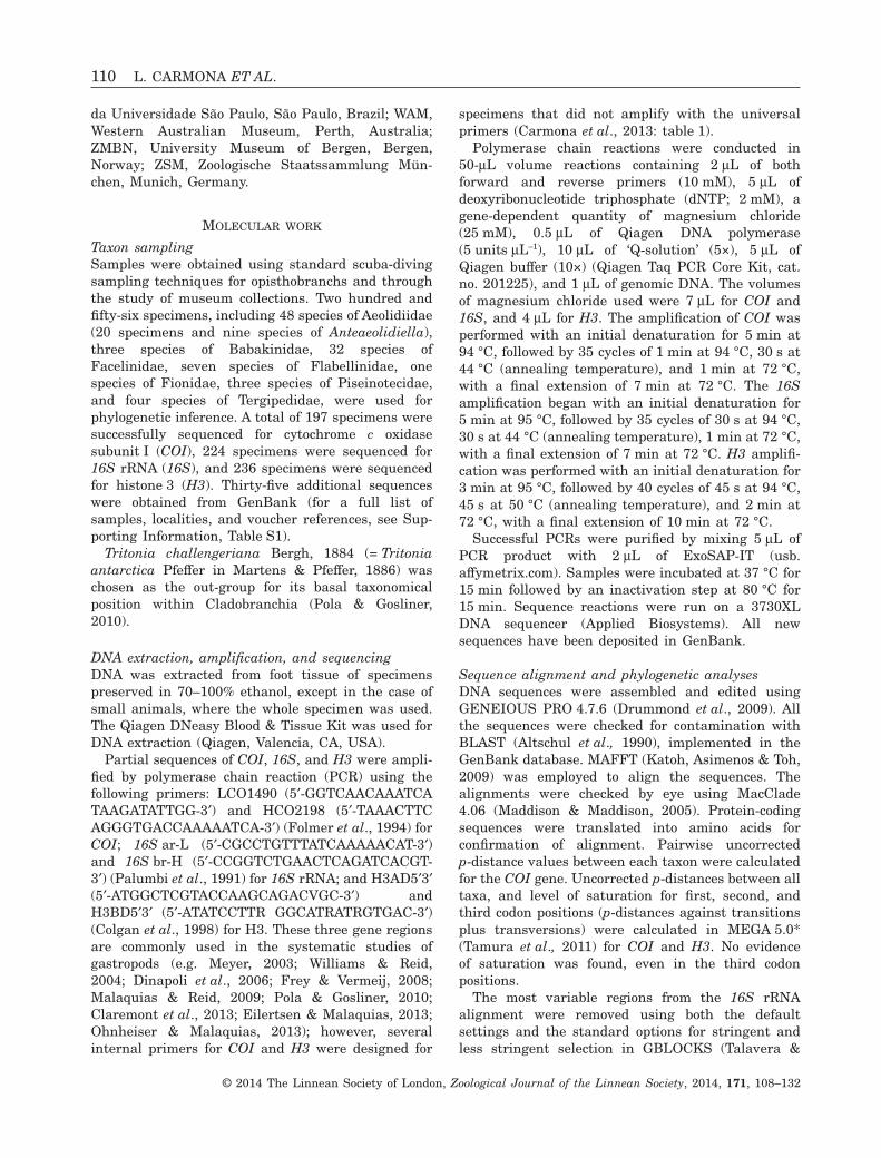
The combined data set yielded a sequence alignmentof 1448 positions. No saturation was observed acrossgenes and codon positions (not shown). The combinedtree provided better resolution than H3, COI, or 16Sseparately, but all of them showed the same species-level topology with high support (not shown). More-over, although bootstrap values were lower thanposterior probabilities in larger clades, the topologiesof the ML trees were congruent with the resultsyielded by Bayesian analyses, and thus ML trees arenot shown. Figure 1 only shows the Anteaeolidiellaphylogenetic hypothesis based on the combined dataset represented by BI. The complete tree is given inSupporting Information, Figure S1.
In this study, Aeolidiidae was monophyletic(PP = 0.95, BS = 63), but the relationships among itsgenera were not resolved. Nevertheless, excludingCerberilla, all the genera were monophyletic and hadhigh support (Fig. S1).
All the Anteaeolidiella specimens clustered togetherin a monophyletic and highly supported group for BI(PP = 1), but this was not supported by the ML analy-ses (BS = 64). Anteaeolidiella was divided into twosubclades: one including Anteaeolidiella oliviae andAnteaeolidiella chromosoma (PP = 1, BS = 89), andthe other formed by the remaining species of thegenus (PP = 1, BS = 100). Within this latter subclade,Anteaeolidiella fijensis sp. nov. appeared as the basalspecies. All the species were highly supported exceptfor Anteaeolidiella cacaotica (PP = 0.91, BS = 63);however, this fact could be explained because only the16S sequence was available for the Japanese speci-men. The minimum uncorrected p-distance for COIamong these species was 6.6%.
Applying the ABGD with the standard settingsresulted in nine partitions with seven groups each:one for Anteaeolidiella lurana specimens, one forAnteaeolidiella saldanhensis, one for Anteaeolidiellacacaotica specimens, one for Anteaeolidiella poshitrasp. nov., one for Anteaeolidiella takanosimensis
SYSTEMATICS OF ANTEAEOLIDIELLA 111
© 2014 The Linnean Society of London, Zoological Journal of the Linnean Society, 2014, 171, 108–132
specimens, one for Anteaeolidiella ireneae sp. nov.specimens and a final one for Anteaeolidiellafijensis sp. nov. The prior maximal distance(p-distance) ranged between 0.001 and 0.06.
SYSTEMATICSNUDIBRANCHIA CUVIER, 1817
CLADOBRANCHIA WILLAN & MORTON, 1984
FAMILY AEOLIDIIDAE GRAY, 1827
GENUS ANTEAEOLIDIELLA MILLER, 2001
Type species: Anteaeolidiella indica (Bergh, 1888a).Diagnosis of the genus Anteaeolidiella according toMiller (2001): Body low, moderately wide. Cerata inclusters of rows, for the most part sloping down-wards and rearwards; rhinophores simple; footcorners triangular; oral glands spindle shaped, withtwo opposed rows of large nodules (giant secretorycells). Radular tooth moderately wide, stronglybilobed, largest denticles at apex of each lobe,median cusp small yet distinct; jaw masticatoryprocess smooth.
Anteaeolidiella indica (Bergh, 1888a) (Fig. 2)Aeolidiella indica Bergh, 1888a: 755.
Type locality: Mauritius Islands.
Type material: To our knowledge no type materialremains.
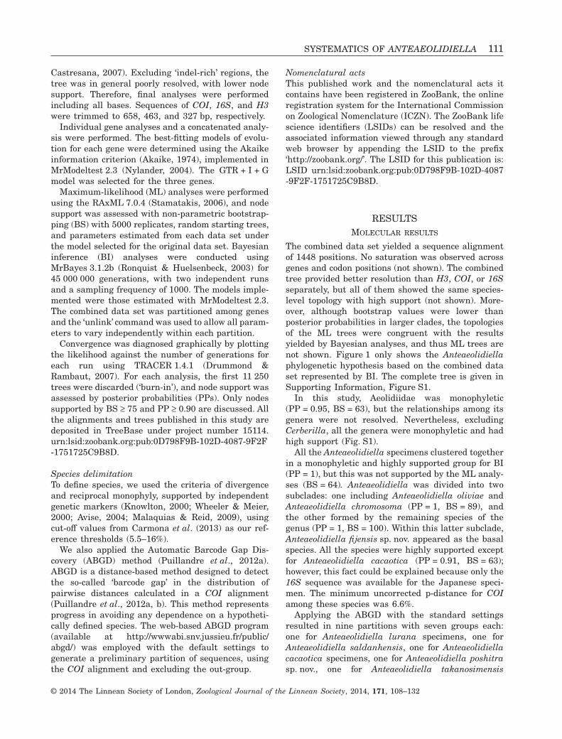
Remarks: Bergh (1888a) described this species basedon Moebius’ drawings and notes. The coloration of theliving animal was depicted as white, yellow, and grey,including the oral tentacles and the rhinophores.Miller (2001) designated Aeolidiella indica as the typespecies of Anteaeolidiella, although his specimen didnot present the coloration described by Bergh (1888a).None of our specimens previously identified asAnteaeolidiella indica (Carmona et al., 2013) have theyellow and grey colour pattern. Some photographsof ‘Anteaeolidiella indica’ found on web pagescould match with Bergh’s original description (e.g.Boserelle, Levy & Michenet, 2013), but the localitiesare quite far from the type locality (Mauritius Islands).We also found one specimen from subtropical South
Figure 1. Phylogenetic hypothesis for the genus Anteaeolidiella based on the combined data set (H3 + COI + 16S),inferred by Bayesian inference (BI) analysis. Numbers above branches represent posterior probabilities from BI. Numberbelow branches represent bootstrap values from maximum-likelihood analysis. Abbreviations: ATL, Atlantic Ocean; PAC,Pacific.
Figure 2. Photograph of the putative Anteaeolidiella indica sensu Bergh (1888a).
112 L. CARMONA ET AL.
© 2014 The Linnean Society of London, Zoological Journal of the Linnean Society, 2014, 171, 108–132
Africa (close to the Mauritius Islands) that seems to belike Bergh’s Anteaeolidiella indica (Fig. 2). Neverthe-less, the specimen is lost (T. M. Gosliner, pers. comm.),and therefore we have not been able to do any dissec-tion or molecular analysis. Because the true A. indicaclearly differs from the remaining species ofAnteaeolidiella, we maintain A. indica as a validspecies, but more material is needed in order to clarifyits morphological characteristics.
Anteaeolidiella cacaotica (Stimpson, 1855)(Figs 3A, 4A, 5A, B, 6)Eolis cacaotica Stimpson, 1855: 388.
Aeolis foulisi Angas, 1864: 64–65, pl. VI, fig. 3.Coryphella foulisi (Angas, 1864): Bergh, 1875, 634.Aeolidiella hulli Risbec, 1928: 262–265, fig. 88,
pl. D, fig. 3, pl. X, fig. 7, pl. XII, fig. 4.
Aeolidia foulisi (Angas, 1864): Orr, 1981, 68.Aeolidiella foulisi (Angas, 1864): Burn, 1964, 89.Anteaeolidiella foulisi (Angas, 1864): Burn,
2006, 30.
Type locality: Port Jackson, Australia.
Type material: To our knowledge, no type materialremains. We designate the specimen MNCN 15.05/63476 from Nelson Bay, New South Wales, Australia,as the neotype.
Material examined: Neotype: MNCN 15.05/63476, onespecimen, dissected, immature, 6 mm in length pre-served, Australia, New South Wales, Nelson Bay, col-lected by Dave Harasti, 14.ii.10. Other material:CASIZ 174212, one specimen, dissected, immature,
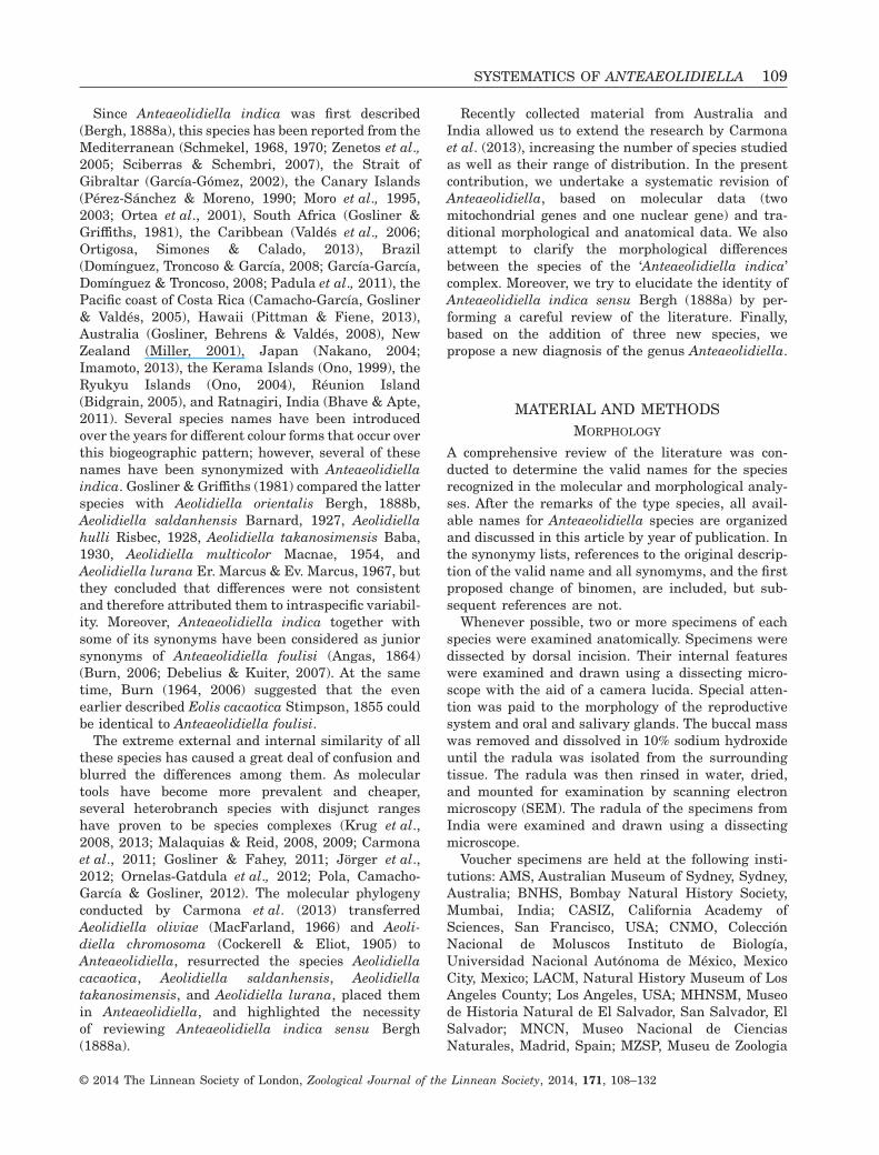
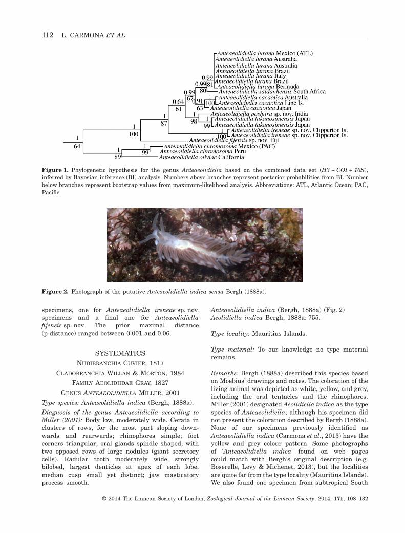
Figure 3. Photographs of the living animals: A, Anteaeolidiella cacaotica, specimen from eastern Australia, photo byO’Grove, MNCN 15.05/63476; B, Anteaeolidiella chromosoma, specimen from the Pacific coast of Costa Rica, photo byYolanda Camacho, MZUCR 8896; C, Anteaeolidiella saldanhensis, specimen from South Africa, Western Cape Province,photo by Terrence M. Gosliner, CASIZ 176313; D, Anteaeolidiella takanosimensis, specimen from Japan, Manuzuru, photoby Rie Nakano, MNCN 15.05/63479; E, Anteaeolidiella oliviae, specimen from California, photo by Gary McDonald; F,Anteaeolidiella lurana, specimen from Bermuda, Ferry Reach, photo by Terrence M. Gosliner, ZMBN 82992.
SYSTEMATICS OF ANTEAEOLIDIELLA 113
© 2014 The Linnean Society of London, Zoological Journal of the Linnean Society, 2014, 171, 108–132
4 mm in length preserved, Line Islands, PalmyraAtoll, collected by Christine Huffard, 26.ix.06;MNCN 15.05/63477, one specimen, dissected, imma-ture, 4 mm in length preserved, Japan, collected byNakano Rie, 10.viii.10.
Geographical distribution: Originally described fromPort Jackson, Australia (Stimpson, 1855), it is alsoknown from Kanagawa Prefecture, Japan (Carmonaet al., 2013).
External morphology (Figs 3A, 4A): The body is elon-gate and moderately slender, tapering gradually tothe fairly short tail. The anterior foot corners arestout and slightly protruded. The background colouris bright orange, although its intensity varies withthe specimen (Fig. 3A). In some specimens, extendingposteriorly just behind the rhinophores, an opaquewhite area in the shape of a teardrop, followed by an
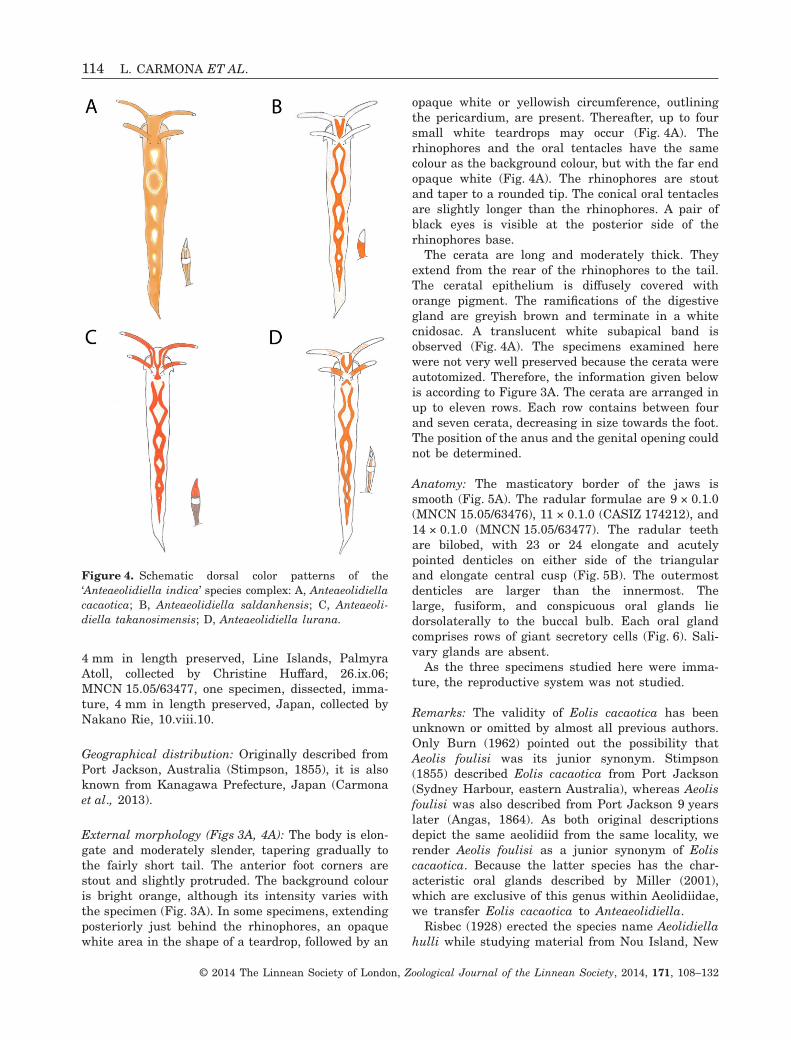
opaque white or yellowish circumference, outliningthe pericardium, are present. Thereafter, up to foursmall white teardrops may occur (Fig. 4A). Therhinophores and the oral tentacles have the samecolour as the background colour, but with the far endopaque white (Fig. 4A). The rhinophores are stoutand taper to a rounded tip. The conical oral tentaclesare slightly longer than the rhinophores. A pair ofblack eyes is visible at the posterior side of therhinophores base.
The cerata are long and moderately thick. Theyextend from the rear of the rhinophores to the tail.The ceratal epithelium is diffusely covered withorange pigment. The ramifications of the digestivegland are greyish brown and terminate in a whitecnidosac. A translucent white subapical band isobserved (Fig. 4A). The specimens examined herewere not very well preserved because the cerata wereautotomized. Therefore, the information given belowis according to Figure 3A. The cerata are arranged inup to eleven rows. Each row contains between fourand seven cerata, decreasing in size towards the foot.The position of the anus and the genital opening couldnot be determined.
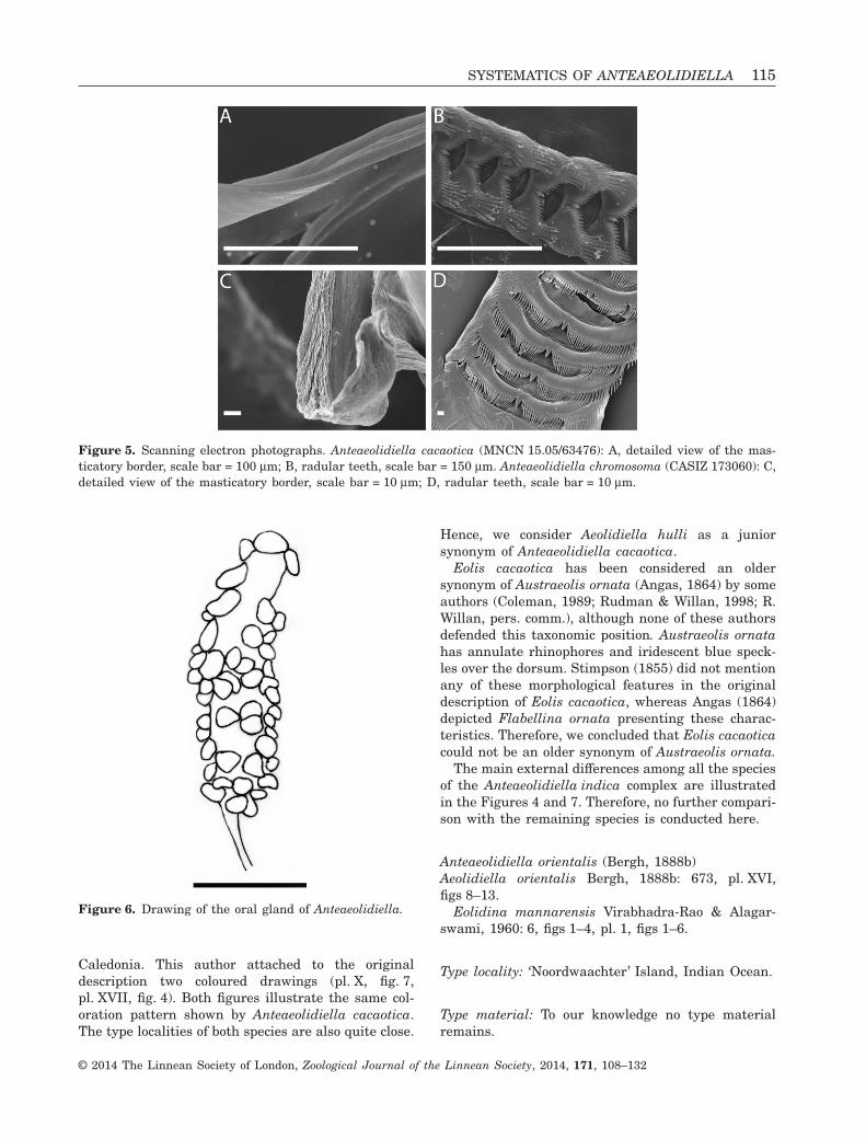
Anatomy: The masticatory border of the jaws issmooth (Fig. 5A). The radular formulae are 9 × 0.1.0(MNCN 15.05/63476), 11 × 0.1.0 (CASIZ 174212), and14 × 0.1.0 (MNCN 15.05/63477). The radular teethare bilobed, with 23 or 24 elongate and acutelypointed denticles on either side of the triangularand elongate central cusp (Fig. 5B). The outermostdenticles are larger than the innermost. Thelarge, fusiform, and conspicuous oral glands liedorsolaterally to the buccal bulb. Each oral glandcomprises rows of giant secretory cells (Fig. 6). Sali-vary glands are absent.
As the three specimens studied here were imma-ture, the reproductive system was not studied.
Remarks: The validity of Eolis cacaotica has beenunknown or omitted by almost all previous authors.Only Burn (1962) pointed out the possibility thatAeolis foulisi was its junior synonym. Stimpson(1855) described Eolis cacaotica from Port Jackson(Sydney Harbour, eastern Australia), whereas Aeolisfoulisi was also described from Port Jackson 9 yearslater (Angas, 1864). As both original descriptionsdepict the same aeolidiid from the same locality, werender Aeolis foulisi as a junior synonym of Eoliscacaotica. Because the latter species has the char-acteristic oral glands described by Miller (2001),which are exclusive of this genus within Aeolidiidae,we transfer Eolis cacaotica to Anteaeolidiella.
Risbec (1928) erected the species name Aeolidiellahulli while studying material from Nou Island, New
Figure 4. Schematic dorsal color patterns of the‘Anteaeolidiella indica’ species complex: A, Anteaeolidiellacacaotica; B, Anteaeolidiella saldanhensis; C, Anteaeoli-diella takanosimensis; D, Anteaeolidiella lurana.
114 L. CARMONA ET AL.
© 2014 The Linnean Society of London, Zoological Journal of the Linnean Society, 2014, 171, 108–132
Caledonia. This author attached to the originaldescription two coloured drawings (pl. X, fig. 7,pl. XVII, fig. 4). Both figures illustrate the same col-oration pattern shown by Anteaeolidiella cacaotica.The type localities of both species are also quite close.
Hence, we consider Aeolidiella hulli as a juniorsynonym of Anteaeolidiella cacaotica.
Eolis cacaotica has been considered an oldersynonym of Austraeolis ornata (Angas, 1864) by someauthors (Coleman, 1989; Rudman & Willan, 1998; R.Willan, pers. comm.), although none of these authorsdefended this taxonomic position. Austraeolis ornatahas annulate rhinophores and iridescent blue speck-les over the dorsum. Stimpson (1855) did not mentionany of these morphological features in the originaldescription of Eolis cacaotica, whereas Angas (1864)depicted Flabellina ornata presenting these charac-teristics. Therefore, we concluded that Eolis cacaoticacould not be an older synonym of Austraeolis ornata.
The main external differences among all the speciesof the Anteaeolidiella indica complex are illustratedin the Figures 4 and 7. Therefore, no further compari-son with the remaining species is conducted here.
Anteaeolidiella orientalis (Bergh, 1888b)Aeolidiella orientalis Bergh, 1888b: 673, pl. XVI,figs 8–13.
Eolidina mannarensis Virabhadra-Rao & Alagar-swami, 1960: 6, figs 1–4, pl. 1, figs 1–6.
Type locality: ‘Noordwaachter’ Island, Indian Ocean.
Type material: To our knowledge no type materialremains.
Figure 5. Scanning electron photographs. Anteaeolidiella cacaotica (MNCN 15.05/63476): A, detailed view of the mas-ticatory border, scale bar = 100 μm; B, radular teeth, scale bar = 150 μm. Anteaeolidiella chromosoma (CASIZ 173060): C,detailed view of the masticatory border, scale bar = 10 μm; D, radular teeth, scale bar = 10 μm.
Figure 6. Drawing of the oral gland of Anteaeolidiella.
SYSTEMATICS OF ANTEAEOLIDIELLA 115
© 2014 The Linnean Society of London, Zoological Journal of the Linnean Society, 2014, 171, 108–132
Remarks: Excluding Anteaeolidiella chromosoma andAnteaeolidiella oliviae, the radular teeth of theremaining species of this genus are strongly bilobed,with elongate and acutely pointed denticles on eitherside of a prominent central cusp (e.g. Fig. 5B). Bergh(1888b) illustrated this type of radular morphologywhen he erected Aeolidiella orientalis. This author alsodepicted an oral gland composed of big secretory cells,such as those we found in all Anteaeolidiella species.Based on the radular teeth and the oral glandsdescribed by Bergh (1888b), we transfer Aeolidiellaorientalis to Anteaeolidiella. Although this species hasbeen considered as a junior synonym of ‘A. indica’(Gosliner & Griffiths, 1981), Bergh (1888b) describedthe rhinophores as white with red tips that are, so far,exclusive for this species within Anteaeolidiella.Edmunds (1969) reported Aeolidiella orientalisfrom Tanzania; however, his specimen showed red
rhinophores with white tips, and therefore cannot beidentified as Anteaeolidiella orientalis.
Eolidina mannarensis was also described as ‘palegolden yellow speckled with orange at the tips ofthe tentacles and rhinophores’ (Virabhadra-Rao &Alagarswami, 1960). The radular teeth of this speciesalso correspond to Anteaeolidiella. Therefore, werender Eolidina mannarensis as a junior synonym ofAnteaeolidiella orientalis.
Anteaeolidiella chromosoma (Cockerell & Eliot, 1905)(Figs 3B, 5C, D, 6, 8A)Spurilla chromosoma Cockerell & Eliot, 1905: 51.
Aeolidiella chromosoma (Cockerell & Eliot, 1905):Gosliner, 1985, 259.
Type locality: Deadman’s Island, San Pedro, Califor-nia, USA.
Type material: According to Cockerell & Eliot (1905),in the Natural History Museum, London. The regis-tration number of the holotype of Anteaeolidiellachromosoma is NHMUK 1904.7.7.4.
Material examined: CASIZ 173060, one specimen, dis-sected, 8 mm in length preserved, Mexico, Jalisco,Bahía de Banderas, collected by Jeff Goddard,26.ii.06; CASIZ 187751, one specimen, dissected,10 mm in length preserved, Japan, Honshu, ShizuokaPrefecture, Ose Cape, collected by Rie Nakano andA. Kawahara, 20.iii.10.
Geographical distribution: Anteaeolidiella chromo-soma was first described from Deadman’s Island,California, USA. It has also been reported from Cali-fornia (Er. Marcus, 1961), the Pacific coast of CostaRica (Camacho-García et al., 2005), and Japan(Nakano, 2004).
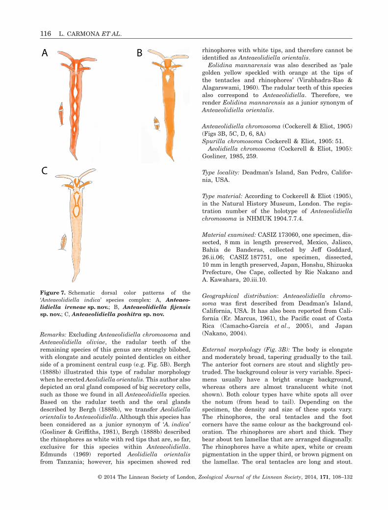
External morphology (Fig. 3B): The body is elongateand moderately broad, tapering gradually to the tail.The anterior foot corners are stout and slightly pro-truded. The background colour is very variable. Speci-mens usually have a bright orange background,whereas others are almost translucent white (notshown). Both colour types have white spots all overthe notum (from head to tail). Depending on thespecimen, the density and size of these spots vary.The rhinophores, the oral tentacles and the footcorners have the same colour as the background col-oration. The rhinophores are short and thick. Theybear about ten lamellae that are arranged diagonally.The rhinophores have a white apex, white or creampigmentation in the upper third, or brown pigment onthe lamellae. The oral tentacles are long and stout.
Figure 7. Schematic dorsal color patterns of the‘Anteaeolidiella indica’ species complex: A, Anteaeo-lidiella ireneae sp. nov.; B, Anteaeolidiella fijensissp. nov.; C, Anteaeolidiella poshitra sp. nov.
116 L. CARMONA ET AL.
© 2014 The Linnean Society of London, Zoological Journal of the Linnean Society, 2014, 171, 108–132
The coloration of their tips is lighter than backgroundcoloration. A pair of black eyes is visible at the pos-terior side of the rhinophores base.
The cerata length is variable, but all are long,cylindrical, and uniform in diameter throughout mostof their length, and have a round apex. Some speci-mens have the tips of the cerata recurved inwards.The cerata extend from the rear of the rhinophores tothe tail. They are translucent, with brownish green ordark-grey ramifications of the digestive gland visiblethroughout the body wall. The cnidosacs are white.All specimens have white marks all over the ceratalsurface, although its density and size vary. The cerataare arranged in five or six rows in the anterior diges-
tive branch, followed by arches. Each group containsbetween two and 20 cerata, decreasing in sizetowards the foot. The anus is cleioproctic, locatedbetween the third and the fourth rows. The genitalopening is placed among the cerata of theanteriormost group on the right.
Anatomy: The masticatory border of the jaws issmooth (Fig. 5C). The radular formulae are 12 × 0.1.0(CASIZ 187751) and 24 × 0.1.0 (CASIZ 173060). Theteeth are progressively smaller to the posterior regionof the radula. They are wide and bilobed, with 16–33elongate and acutely pointed denticles on either sideof the triangular and elongate central cusp (Fig. 5D).
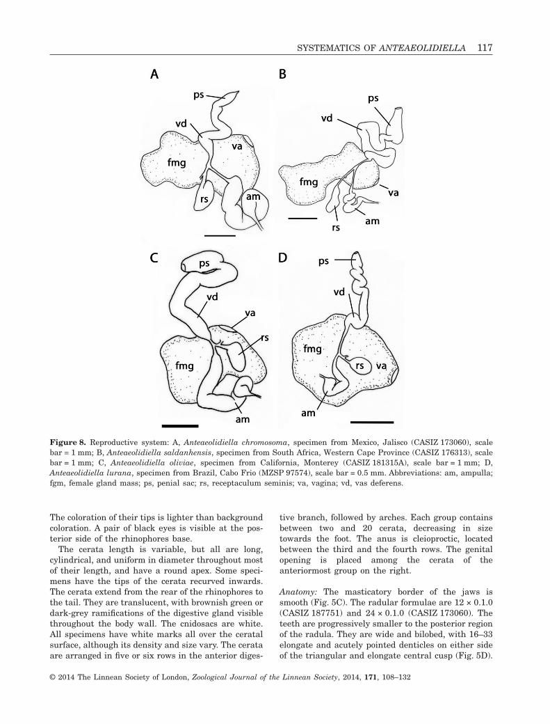
Figure 8. Reproductive system: A, Anteaeolidiella chromosoma, specimen from Mexico, Jalisco (CASIZ 173060), scalebar = 1 mm; B, Anteaeolidiella saldanhensis, specimen from South Africa, Western Cape Province (CASIZ 176313), scalebar = 1 mm; C, Anteaeolidiella oliviae, specimen from California, Monterey (CASIZ 181315A), scale bar = 1 mm; D,Anteaeolidiella lurana, specimen from Brazil, Cabo Frio (MZSP 97574), scale bar = 0.5 mm. Abbreviations: am, ampulla;fgm, female gland mass; ps, penial sac; rs, receptaculum seminis; va, vagina; vd, vas deferens.
SYSTEMATICS OF ANTEAEOLIDIELLA 117
© 2014 The Linnean Society of London, Zoological Journal of the Linnean Society, 2014, 171, 108–132
The large, fusiform and conspicuous oral glands liedorsolaterally to the buccal bulb. Each oral glandcomprises rows of giant secretory cells (Fig. 6). Thesalivary glands are absent.
The reproductive system is diaulic (Fig. 8A). Thepreampullary duct widens into the conspicuous, wide,and long ampulla that narrows again before dividinginto the oviduct and vas deferens. The moderatelyshort vas deferens enters the wider proximal portionof the penial sac, which contains the unarmed penialpapilla. The short oviduct connects to a pyriformreceptaculum seminis. The remaining portion of theoviduct departs from the base of the receptaculumand enters the female gland. The vagina opensventral to the penis.
Remarks: Based on the perfoliate rhinophores,Cockerell & Eliot (1905) placed this species withinSpurilla. Gosliner (1985) rendered Spurillachromosoma as Aeolidiella because of the presence oforal glands with large vesicles, together with theanterior digestive branches arranged in rows;however, Carmona et al. (2013) transferred thisspecies to Anteaeolidiella on the basis of moleculardata. The latter outcome agrees with our presentstudy, and it is also supported by the presence of theparticular oral glands.
Anteaeolidiella chromosoma differs from theremaining members of the genus by its coloration, itsperfoliate rhinophores, and the radular shape. OnlyAnteaeolidiella oliviae presents the latter twomorphological characters, but both species have aconsistently distinct coloration (see Anteaeolidiellaoliviae remarks). Spurilla neapolitana and Spurillabraziliana may have a colour pattern that resemblesthat of A. chromosoma. Nevertheless, A. chromosomapresents the anterior digestive branch arranged inrows instead of arches. In addition, the number oflamellae in A. chromosoma is low, and they arearranged diagonally, whereas in S. neapolitana orS. braziliana they are numerous and transverse.Finally, differences between the oral glands of bothspecies (A. chromosoma and S. braziliana) are alsoconsistent.
Anteaeolidiella saldanhensis (Barnard, 1927)(Figs 3C, 4B, 6, 8B)Aeolidiella saldanhensis Barnard, 1927: 201, figs 2, 3.
Aeolidiella multicolor Macnae, 1954: 36, figs 27–29,pl. II, fig. 4.
Type locality: Saldanha Bay, South Africa.
Type material: To our knowledge no type materialremains. We designate the specimen CASIZ 176313from False Bay, Cape Province, South Africa, as theneotype.
Material examined: Neotype: CASIZ 176313, onespecimen, dissected, 5 mm in length preserved, SouthAfrica, western Cape Province, False Bay, collected byTerrence M. Gosliner, Ángel Valdés, Marta Pola,Elisabeth Moore, Lakisha Withey, and Carla Stout,6.i.08.
Geographical distribution: So far, only known fromSouth Africa (Barnard, 1927; Macnae, 1954; Gosliner,1985; present study).
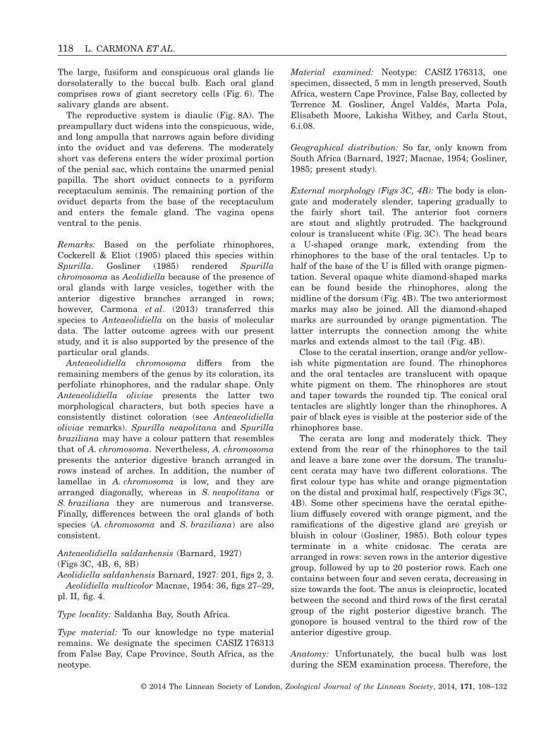
External morphology (Figs 3C, 4B): The body is elon-gate and moderately slender, tapering gradually tothe fairly short tail. The anterior foot cornersare stout and slightly protruded. The backgroundcolour is translucent white (Fig. 3C). The head bearsa U-shaped orange mark, extending from therhinophores to the base of the oral tentacles. Up tohalf of the base of the U is filled with orange pigmen-tation. Several opaque white diamond-shaped markscan be found beside the rhinophores, along themidline of the dorsum (Fig. 4B). The two anteriormostmarks may also be joined. All the diamond-shapedmarks are surrounded by orange pigmentation. Thelatter interrupts the connection among the whitemarks and extends almost to the tail (Fig. 4B).
Close to the ceratal insertion, orange and/or yellow-ish white pigmentation are found. The rhinophoresand the oral tentacles are translucent with opaquewhite pigment on them. The rhinophores are stoutand taper towards the rounded tip. The conical oraltentacles are slightly longer than the rhinophores. Apair of black eyes is visible at the posterior side of therhinophores base.
The cerata are long and moderately thick. Theyextend from the rear of the rhinophores to the tailand leave a bare zone over the dorsum. The translu-cent cerata may have two different colorations. Thefirst colour type has white and orange pigmentationon the distal and proximal half, respectively (Figs 3C,4B). Some other specimens have the ceratal epithe-lium diffusely covered with orange pigment, and theramifications of the digestive gland are greyish orbluish in colour (Gosliner, 1985). Both colour typesterminate in a white cnidosac. The cerata arearranged in rows: seven rows in the anterior digestivegroup, followed by up to 20 posterior rows. Each onecontains between four and seven cerata, decreasing insize towards the foot. The anus is cleioproctic, locatedbetween the second and third rows of the first ceratalgroup of the right posterior digestive branch. Thegonopore is housed ventral to the third row of theanterior digestive group.
Anatomy: Unfortunately, the bucal bulb was lostduring the SEM examination process. Therefore, the
118 L. CARMONA ET AL.
© 2014 The Linnean Society of London, Zoological Journal of the Linnean Society, 2014, 171, 108–132
masticatory border and the radula could not bestudied. The large, fusiform and conspicuous oralglands lie dorsolaterally to the buccal bulb. Each oralgland comprises rows of giant secretory cells (Fig. 6).Salivary glands are absent.
The reproductive system is diaulic (Fig. 8B). Thepreampullary duct widens into the short ampulla thatnarrows again before dividing into the oviduct andvas deferens. The moderately long vas deferens entersthe wider proximal portion of the penial sac, whichcontains the unarmed penial papilla. The shortoviduct connects to a pyriform receptaculum seminis.The remaining portion of the oviduct departs from thebase of the receptaculum and enters the female gland.The vagina opens ventral to the penis.
Remarks: Barnard (1927) erected the speciesAeolidiella saldanhensis from Saldanha Bay (SouthAfrica), but the original description is very faint. Thisauthor only provided some information about theradula and the rounded foot corners. Some yearslater, Macnae (1954) erected the name Aeolidiellamulticolor, from False Baye (South Africa). Macnae(1954) could not compare his specimens with Barnard’sbecause the type material of Aeolidiella saldanhensiswas mislaid (Macnae, 1954). Thus, Macnae (1954)stated that the tentaculiform foot corners of Aeoli-diella multicolor were significant enough to keep itas a distinct species from Aeolidiella saldanhensis.Barnard (1927) depicted the coloration of Aeolidie-lla saldanhensis as ‘unknown’, probably because thetype material was already preserved when Barnarddescribed it, and therefore the rounded foot cornerscould be an artefact of the preservation of the speci-men. Therefore, we do not consider this difference assignificant, and render Aeolidiella multicolor as ajunior synonym of Aeolidiella saldanhensis. Because ofits radular morphology and its oral glands, we transferAeolidiella saldanhensis to Anteaeolidiella.
As most of the Anteaeolidiella species have orangerhinophores, Anteaeolidiella saldanhensis is clearlydifferent from the other species because of its whiterhinophores and oral tentacles. The main externaldifferences among all the species of the Anteaeoli-diella indica complex are illustrated in Figures 4 and7. Therefore, no further comparison with the remain-ing species is conducted here.
Anteaeolidiella takanosimensis (Baba, 1930)(Figs 3D, 4C, 6, 9A, B)Eolis sp. Ijima, 1918: 615, fig. 740.
Aeolidiella takanosimensis Baba, 1930: 122, fig. 4a,b, pl. IV, fig. 5a–c.
Eolidina takanosimensis Baba, 1937: 336.
Type locality: Takanosima, Tateyama Bay, Japan.
Type material: To our knowledge no type materialremains. We designate the specimen MNCN 15.05/63479 from Manuzaru, Japan, as the neotype.
Material examined: Neotype: MNCN 15.05/63479, onespecimen, dissected, immature, 3 mm in length pre-served, Japan, Manuzaru, collected by Rie Nakano,19.iv.06. Other material: MNCN 15.05/63478, onespecimen, dissected, immature, 7 mm in length pre-served, Japan, collected by Rie Nakano, 10.viii.10.
Geographical distribution: Only known from Japan(Baba, 1930; Baba, 1979a,b; present study).
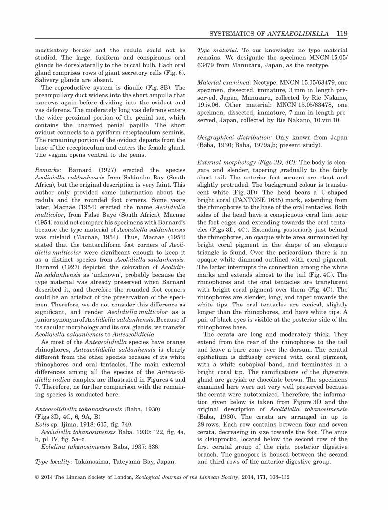
External morphology (Figs 3D, 4C): The body is elon-gate and slender, tapering gradually to the fairlyshort tail. The anterior foot corners are stout andslightly protruded. The background colour is translu-cent white (Fig. 3D). The head bears a U-shapedbright coral (PANTONE 1635) mark, extending fromthe rhinophores to the base of the oral tentacles. Bothsides of the head have a conspicuous coral line nearthe foot edges and extending towards the oral tenta-cles (Figs 3D, 4C). Extending posteriorly just behindthe rhinophores, an opaque white area surrounded bybright coral pigment in the shape of an elongatetriangle is found. Over the pericardium there is anopaque white diamond outlined with coral pigment.The latter interrupts the connection among the whitemarks and extends almost to the tail (Fig. 4C). Therhinophores and the oral tentacles are translucentwith bright coral pigment over them (Fig. 4C). Therhinophores are slender, long, and taper towards thewhite tips. The oral tentacles are conical, slightlylonger than the rhinophores, and have white tips. Apair of black eyes is visible at the posterior side of therhinophores base.
The cerata are long and moderately thick. Theyextend from the rear of the rhinophores to the tailand leave a bare zone over the dorsum. The ceratalepithelium is diffusely covered with coral pigment,with a white subapical band, and terminates in abright coral tip. The ramifications of the digestivegland are greyish or chocolate brown. The specimensexamined here were not very well preserved becausethe cerata were autotomized. Therefore, the informa-tion given below is taken from Figure 3D and theoriginal description of Aeolidiella takanosimensis(Baba, 1930). The cerata are arranged in up to28 rows. Each row contains between four and sevencerata, decreasing in size towards the foot. The anusis cleioproctic, located below the second row of thefirst ceratal group of the right posterior digestivebranch. The gonopore is housed between the secondand third rows of the anterior digestive group.
SYSTEMATICS OF ANTEAEOLIDIELLA 119
© 2014 The Linnean Society of London, Zoological Journal of the Linnean Society, 2014, 171, 108–132
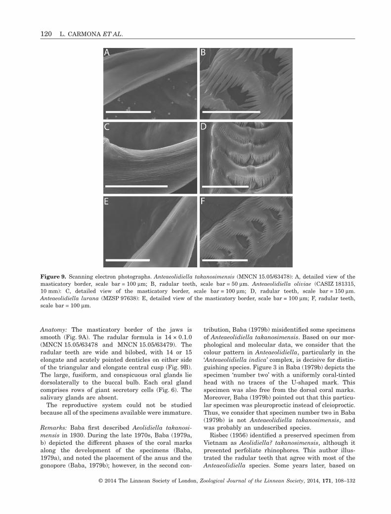
Anatomy: The masticatory border of the jaws issmooth (Fig. 9A). The radular formula is 14 × 0.1.0(MNCN 15.05/63478 and MNCN 15.05/63479). Theradular teeth are wide and bilobed, with 14 or 15elongate and acutely pointed denticles on either sideof the triangular and elongate central cusp (Fig. 9B).The large, fusiform, and conspicuous oral glands liedorsolaterally to the buccal bulb. Each oral glandcomprises rows of giant secretory cells (Fig. 6). Thesalivary glands are absent.
The reproductive system could not be studiedbecause all of the specimens available were immature.
Remarks: Baba first described Aeolidiella takanosi-mensis in 1930. During the late 1970s, Baba (1979a,b) depicted the different phases of the coral marksalong the development of the specimens (Baba,1979a), and noted the placement of the anus and thegonopore (Baba, 1979b); however, in the second con-
tribution, Baba (1979b) misidentified some specimensof Anteaeolidiella takanosimensis. Based on our mor-phological and molecular data, we consider that thecolour pattern in Anteaeolidiella, particularly in the‘Anteaeolidiella indica’ complex, is decisive for distin-guishing species. Figure 3 in Baba (1979b) depicts thespecimen ‘number two’ with a uniformly coral-tintedhead with no traces of the U-shaped mark. Thisspecimen was also free from the dorsal coral marks.Moreover, Baba (1979b) pointed out that this particu-lar specimen was pleuroproctic instead of cleioproctic.Thus, we consider that specimen number two in Baba(1979b) is not Anteaeolidiella takanosimensis, andwas probably an undescribed species.
Risbec (1956) identified a preserved specimen fromVietnam as Aeolidiella? takanosimensis, although itpresented perfoliate rhinophores. This author illus-trated the radular teeth that agree with most of theAnteaeolidiella species. Some years later, based on
Figure 9. Scanning electron photographs. Anteaeolidiella takanosimensis (MNCN 15.05/63478): A, detailed view of themasticatory border, scale bar = 100 μm; B, radular teeth, scale bar = 50 μm. Anteaeolidiella oliviae (CASIZ 181315,10 mm): C, detailed view of the masticatory border, scale bar = 100 μm; D, radular teeth, scale bar = 150 μm.Anteaeolidiella lurana (MZSP 97638): E, detailed view of the masticatory border, scale bar = 100 μm; F, radular teeth,scale bar = 100 μm.
120 L. CARMONA ET AL.
© 2014 The Linnean Society of London, Zoological Journal of the Linnean Society, 2014, 171, 108–132
the ornamentation of the rhinophores and differencesin the number of radular denticles of Aeolidiella?takanosimensis, Ernest Marcus (1961) erected thename Aeolidiella risbeci for the specimen found byRisbec in 1956. Anteaeolidiella chromosoma andAnteaeolidiella oliviae present perfoliate rhinophores,but none of these species have the type of radularteeth illustrated by Risbec (1956). Additionally, theperfoliate rhinophores could be an artefact of thepreservation. Although the information given by thisauthor is insufficient for identifying the specimenproperly, it seems possible that Risbec’s specimen wasAnteaeolidiella takanosimensis.
Finally, the coloration of our specimens clearlyagrees with the original description by Baba (1930).Moreover, the coral colour tips of the cerata and thelateral coral lines can be considered as diagnostic forthis species. The main external differences among allthe species of the Anteaeolidiella indica complex areillustrated in Figures 4 and 7. Therefore, no furthercomparison with the remaining species is conductedhere.
Anteaeolidiella oliviae (MacFarland, 1966)(Figs 3E, 6, 8C, 9C, D)Aeolidiella oliviae MacFarland, 1966: 373, pl. 62,figs 4–6, pl. 72, figs 9–14.
Spurilla oliviae (MacFarland, 1966): Sphon &Lance, 1968: 81.
Type locality: San Diego, California, USA.
Type material: To our knowledge no type materialremains. We designate the specimen CASIZ 084921from San Miguel Island as the neotype.
Material examined: Neotype: CASIZ 084921, onespecimen, dissected, 20 mm in length preserved, Cali-fornia, Channel Islands, San Miguel Island, collectedby Robert Van Syoc, 13.vii.87. Other material:CASIZ 181315, two specimens, dissected, 8 and10 mm in length preserved, California, MontereyCounty, Pacific Grove, collected by Gary MacDonald,26.v.09.
Geographical distribution: So far, known only fromCalifornia (MacFarland, 1966).
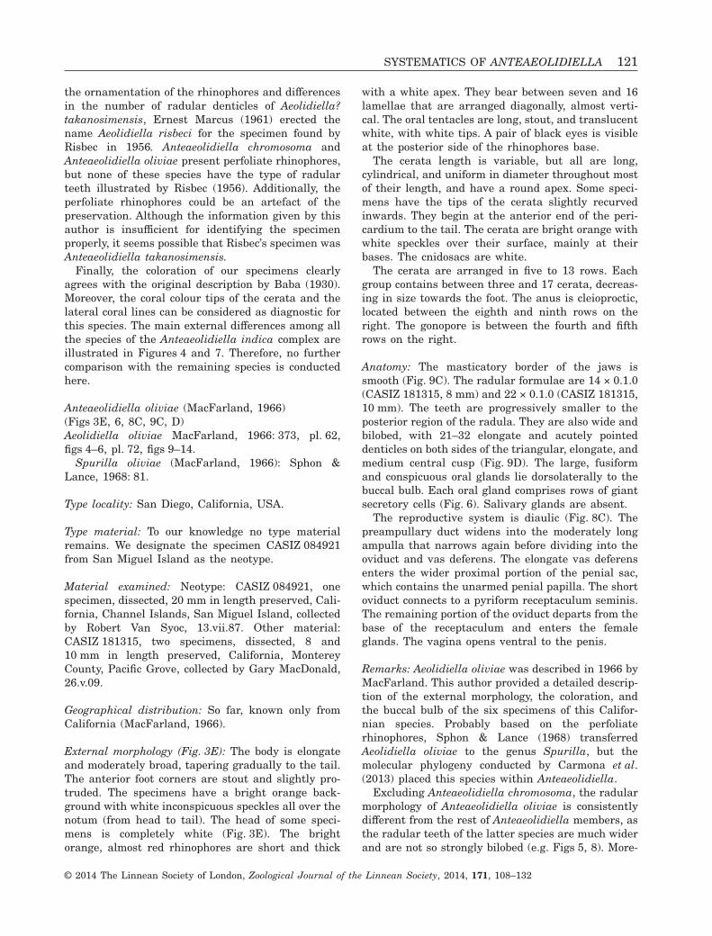
External morphology (Fig. 3E): The body is elongateand moderately broad, tapering gradually to the tail.The anterior foot corners are stout and slightly pro-truded. The specimens have a bright orange back-ground with white inconspicuous speckles all over thenotum (from head to tail). The head of some speci-mens is completely white (Fig. 3E). The brightorange, almost red rhinophores are short and thick
with a white apex. They bear between seven and 16lamellae that are arranged diagonally, almost verti-cal. The oral tentacles are long, stout, and translucentwhite, with white tips. A pair of black eyes is visibleat the posterior side of the rhinophores base.
The cerata length is variable, but all are long,cylindrical, and uniform in diameter throughout mostof their length, and have a round apex. Some speci-mens have the tips of the cerata slightly recurvedinwards. They begin at the anterior end of the peri-cardium to the tail. The cerata are bright orange withwhite speckles over their surface, mainly at theirbases. The cnidosacs are white.
The cerata are arranged in five to 13 rows. Eachgroup contains between three and 17 cerata, decreas-ing in size towards the foot. The anus is cleioproctic,located between the eighth and ninth rows on theright. The gonopore is between the fourth and fifthrows on the right.
Anatomy: The masticatory border of the jaws issmooth (Fig. 9C). The radular formulae are 14 × 0.1.0(CASIZ 181315, 8 mm) and 22 × 0.1.0 (CASIZ 181315,10 mm). The teeth are progressively smaller to theposterior region of the radula. They are also wide andbilobed, with 21–32 elongate and acutely pointeddenticles on both sides of the triangular, elongate, andmedium central cusp (Fig. 9D). The large, fusiformand conspicuous oral glands lie dorsolaterally to thebuccal bulb. Each oral gland comprises rows of giantsecretory cells (Fig. 6). Salivary glands are absent.
The reproductive system is diaulic (Fig. 8C). Thepreampullary duct widens into the moderately longampulla that narrows again before dividing into theoviduct and vas deferens. The elongate vas deferensenters the wider proximal portion of the penial sac,which contains the unarmed penial papilla. The shortoviduct connects to a pyriform receptaculum seminis.The remaining portion of the oviduct departs from thebase of the receptaculum and enters the femaleglands. The vagina opens ventral to the penis.
Remarks: Aeolidiella oliviae was described in 1966 byMacFarland. This author provided a detailed descrip-tion of the external morphology, the coloration, andthe buccal bulb of the six specimens of this Califor-nian species. Probably based on the perfoliaterhinophores, Sphon & Lance (1968) transferredAeolidiella oliviae to the genus Spurilla, but themolecular phylogeny conducted by Carmona et al.(2013) placed this species within Anteaeolidiella.
Excluding Anteaeolidiella chromosoma, the radularmorphology of Anteaeolidiella oliviae is consistentlydifferent from the rest of Anteaeolidiella members, asthe radular teeth of the latter species are much widerand are not so strongly bilobed (e.g. Figs 5, 8). More-
SYSTEMATICS OF ANTEAEOLIDIELLA 121
© 2014 The Linnean Society of London, Zoological Journal of the Linnean Society, 2014, 171, 108–132
over, the bright orange coloration, barely with anyother pigment, and the dark orange and perfoliaterhinophores diagnose Anteaeolidiella oliviae. Thereproductive system is presented here for the firsttime.
Anteaeolidiella lurana (Ev. Marcus & Er. Marcus,1967) (Figs 3F, 4D, 6, 8D, 9E, F)Aeolidiella lurana Ev. Marcus & Er. Marcus,1967: 115–119, figs 149, 150.
Type locality: Urubuqueçaba, Bay of Santos, IslandBrazil.
Type material: According to Ev. Marcus & Er. Marcus(1967) in the US National Museum, WashingtonDC. The registration number of the holotype ofAnteaeolidiella lurana is USNM 576293.
Material examined: CASIZ 173060, one specimen, dis-sected, 8 mm in length preserved, Mexico, Jalisco,Bahia de Banderas, collected by Jeff Goddard,26.ii.06; CASIZ 071302, four specimens, dissected, 9,10, 11, and 12 mm in length preserved, Mexico,Pacific Ocean, Islas de Revillagigedo, Isla Socorro,collected by Terrence M. Gosliner, 7–9.xii.88;CASIZ 186820, one specimen, dissected, 3 mm inlength preserved, Italy, Gulf of Naples, collected byGuido Villani, x.11; MZSP 97574, one specimen, dis-sected, 3 mm in length preserved, Brazil, Cabo Frio,collected by Vinicius Padula, 14.xii.08; CNMO 2996,one specimen, dissected, 5 mm in length preserved,Mexico, Yucatán, collected by Jazmin Ortigosa, vii.06;MZSP 97638, one specimen, dissected, 2 mm in lengthpreserved, Brazil, Cabo Frio, collected by ViniciusPadula, 09.xii.07.
Geographical distribution: Anteaeolidiella luranawas originally described from Brazil (Ev. Marcus &Er. Marcus, 1967). This species is also found in theGulf of Naples [under the names Aeolidiellatakanosimensis in Schmekel, (1968), Aeolidiellaorientalis takanosimensis in Schmekel & Portmann(1982), and Aeolidiella indica in Sammut & Perrone(1998)], the Lazio coast [Anteaeolidiella foulisi inCrocetta et al. (2013)], the Balearic Islands (asAnteaeolidiella indica in GROC, 2013), the Strait ofGibraltar (as Aeolidiella indica, García-Gómez, 2002),the Canary Islands (Pérez-Sánchez & Moreno, 1990;Moro et al., 1995, 2003; Ortea et al., 2001), the Car-ibbean [as Aeolidiella stephanieae in Ortigosa et al.,(2013) or as Aeolidiella indica in Valdés et al. (2006)],Bermuda (present study), and Queensland (Australia,present study).
External morphology (Figs 3F, 4D): The body is elon-gate and moderately slender, tapering gradually tothe fairly short tail. The anterior foot corners arestout and slightly protruded. The background colouris translucent white (Fig. 3F). The head bears abracket-shaped orange mark, extending from therhinophores to the base of the oral tentacles. A pair ofinconspicuous orange lines, lateral to the head, maybe found. Extending posteriorly, just behind therhinophores, an opaque white area, surrounded byorange pigment, in the shape of an elongate diamondis found. An orange diamond follows the latter markand outlines the pericardium (Fig. 4D). This orangemark is filled with white pigmentation. Thereafter,two thin orange bands extend almost to the tail,forming teardrops or diamond marks every time theyjoin (Fig. 4D). The rhinophores and the oral tentacleshave the same colour as the background. Therhinophores are smooth, stout, and taper towardsthe rounded tip. They have orange pigment over theproximal two-thirds and white pigmentation on theirtips. The conical oral tentacles are slightly longerthan the rhinophores. They have white pigmentationon their tips and may also have orange pigment overtheir dorsal surface. A pair of black eyes is visible atthe posterior side of the rhinophores base.
The cerata are long and thickest in their upperthird. They extend from the rear of the rhinophores tothe tail. The ceratal epithelium is diffusely coveredwith orange pigment, interrupted by a whitesubapical band. The ramifications of the digestivegland are orange and terminate in a white cnidosac.The cerata are arranged in up to 23 rows. Each rowcontains between two and nine cerata, decreasing insize towards the foot. The cleioproctic anus is usuallylocated between the eighth and ninth rows. Thegenital opening is housed ventrally to the third row ofthe anterior digestive group.
Anatomy: The masticatory border of the jaws issmooth (Fig. 9E). The radular formulae are 10 × 0.1.0(CNMO 2996), 14 × 0.1.0 (CASIZ 186820), 15 × 0.1.0(MZSP 97574), and 16 × 0.1.0 (MZSP 97638). Theradular teeth are wide and bilobed, with 12–22 elon-gate and acutely pointed denticles on either side ofthe triangular and elongate central cusp (Fig. 9F).The teeth are progressively smaller in the posteriorregion of the radula. The large, fusiform, and con-spicuous oral glands lie dorsolaterally to the buccalbulb. Each oral gland comprises rows of giant secre-tory cells (Fig. 6). The salivary glands are absent.
The reproductive system is diaulic (Fig. 8D). Thepreampullary duct widens into the short ampulla thatnarrows again before dividing into the oviduct andvas deferens. The elongate vas deferens enters thewider proximal portion of the penial sac, which con-
122 L. CARMONA ET AL.
© 2014 The Linnean Society of London, Zoological Journal of the Linnean Society, 2014, 171, 108–132
tains the unarmed penial papilla. The short oviductconnects with a rounded receptaculum seminis. Theremaining portion of the oviduct departs from thebase of the receptaculum and enters the female gland.The vagina opens ventral to the penis.
Remarks: Ev. Marcus & Er. Marcus (1967) describedAnteaeolidiella lurana with material from the Bay ofSantos (Brazil). All our specimens identified asAnteaeolidiella lurana, regardless of being fromBrazil or not, agree well with the original description.Additionally, the uncorrected p-distances for COIamong these specimens were very close to 0. Thetranslucent body, the bracket-shaped orange mark,the ceratal coloration, and the marks over the notumdistinguish this species from other species in thegenus (Figs 4, 10).
Thus far, Anteaeolidiella lurana is the onlyamphiatlantic species of this genus. In addition, thepresence of this species in the Mediterranean is sup-
posedly caused by shipping transport (Zenetos et al.,2004; García-Gómez, Cervera & García-García, 2011;Crocetta, 2012) or through an unknown vector(Sciberras & Schembri, 2007; Occhipinti-Ambrogiet al., 2011). Moreover, this species has also beenrecorded from the eastern coast of Australia (presentstudy; minimun uncorrected p-distance = 0% for COI).
Anteaeolidiella ireneae sp. nov. LSIDurn:lsid:zoobank.org:act:E3B0E8F9-39FB-4254-B9CC-C1C528E7EE40 (Figs 6, 7A, 10A,11A, B, 12A)Anteaeolidiella sp. A Carmona et al., 2013: 6.
Material examined: Holotype: CASIZ 115686, onespecimen, dissected, 6 mm in length preserved,Clipperton Island, eastern Pacific Ocean, collected byK.L. Kaiser, iv–v.98.
Other material: CASIZ 192421, one specimen, dis-sected, 12 mm in length preserved, Clipperton Island,
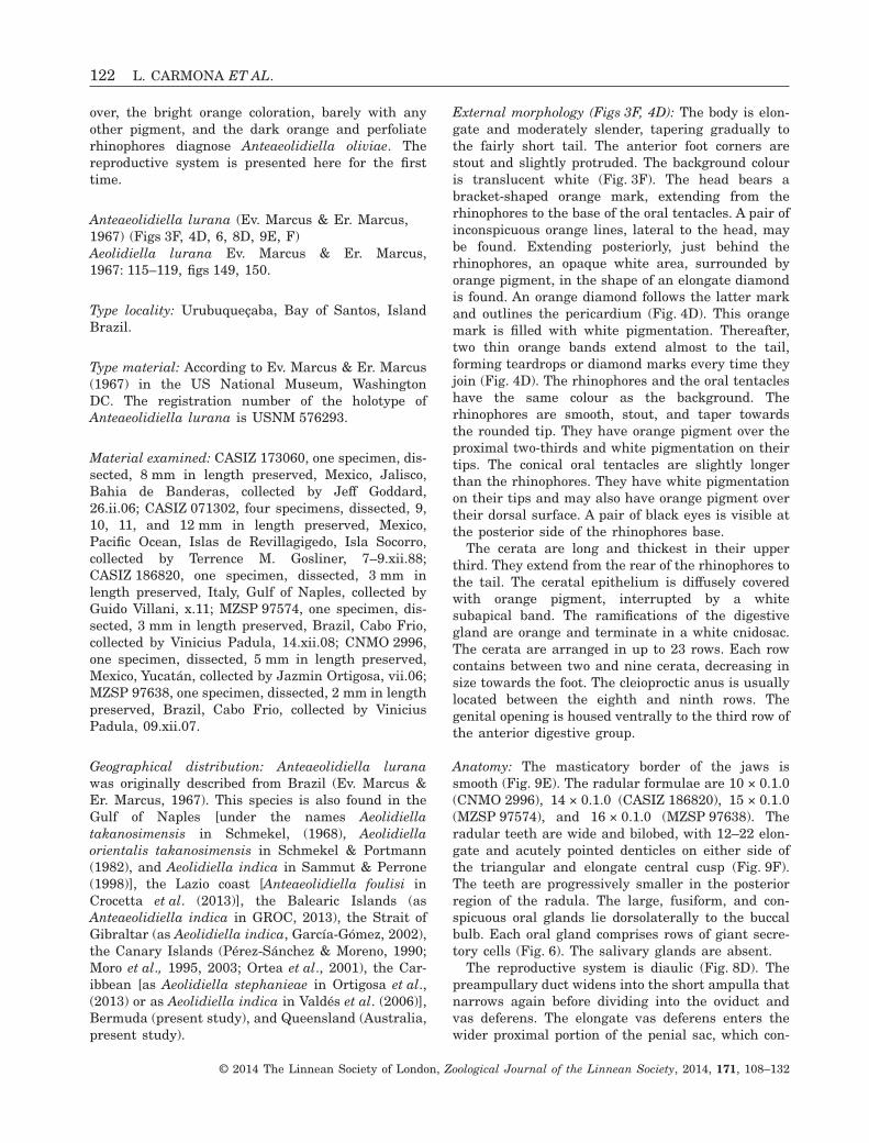
Figure 10. Photographs of living animals: A, Anteaeolidiella ireneae sp. nov., specimen from Clipperton Island,photo by K.L. Kaiser, CASIZ 115686; B, Anteaeolidiella fijensis sp. nov., specimen from Fiji, photo by MichaelSchrödl, ZSM 20130053; C, Anteaeolidiella poshitra sp. nov., from India, Gujarat, photo by Vishal Bhave, BNHS-opistho-38.
SYSTEMATICS OF ANTEAEOLIDIELLA 123
© 2014 The Linnean Society of London, Zoological Journal of the Linnean Society, 2014, 171, 108–132
eastern Pacific Ocean, collected by K.L. Kaiser,iv–v.98.
Type locality and habitat: Clipperton Island, easternPacific Ocean. Under dead coral heads and sand in15–20 m of deep water.
Geographical distribution: So far, this species isknown from Clipperton Island, Isla Canal de Afuera(Panama, PAC), Bahía de San Marte (Mexico, PAC),Isla Socorro (Mexico, PAC; present study), and the
Pacific coast of Costa Rica (as Aeolidiella indica inCamacho-García et al., 2005).
Etymology: This species is dedicated to Irene Cervera,the first daughter of the last author of this article.
External morphology (Figs 7A, 10A): The body islarge, elongate, and moderately broad, taperinggradually to the wide tail. The anterior foot cornersare stout and slightly protruded. The backgroundcolour is translucent white, with orange pigment
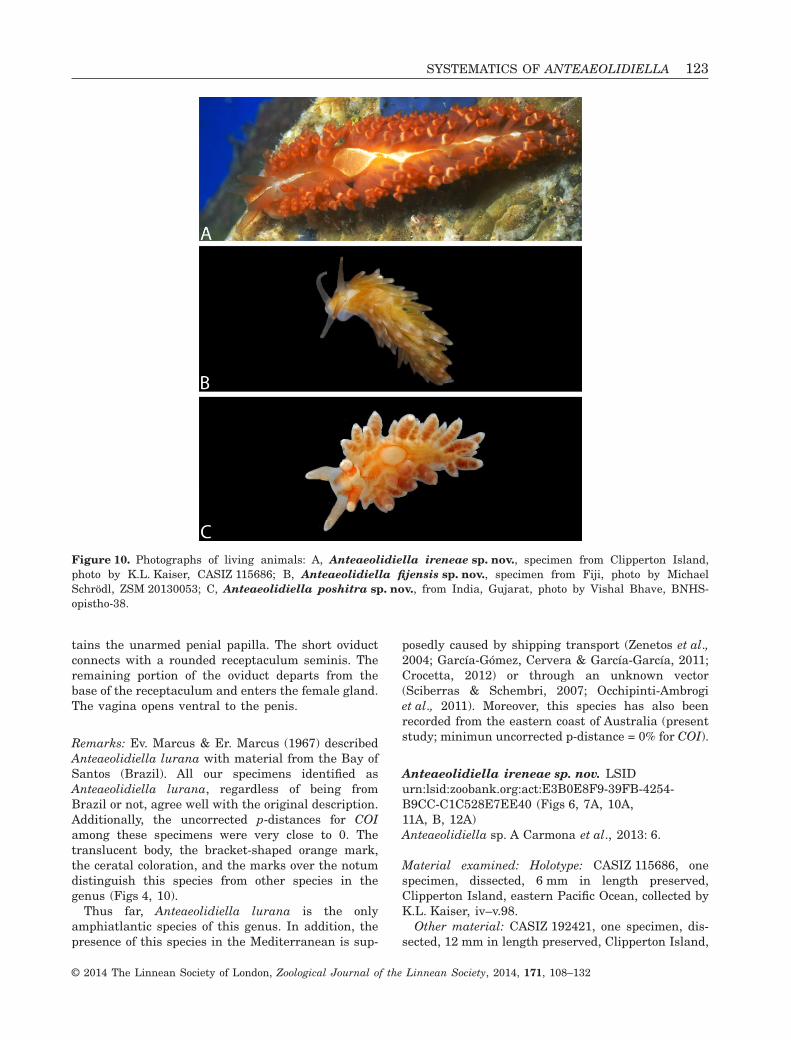
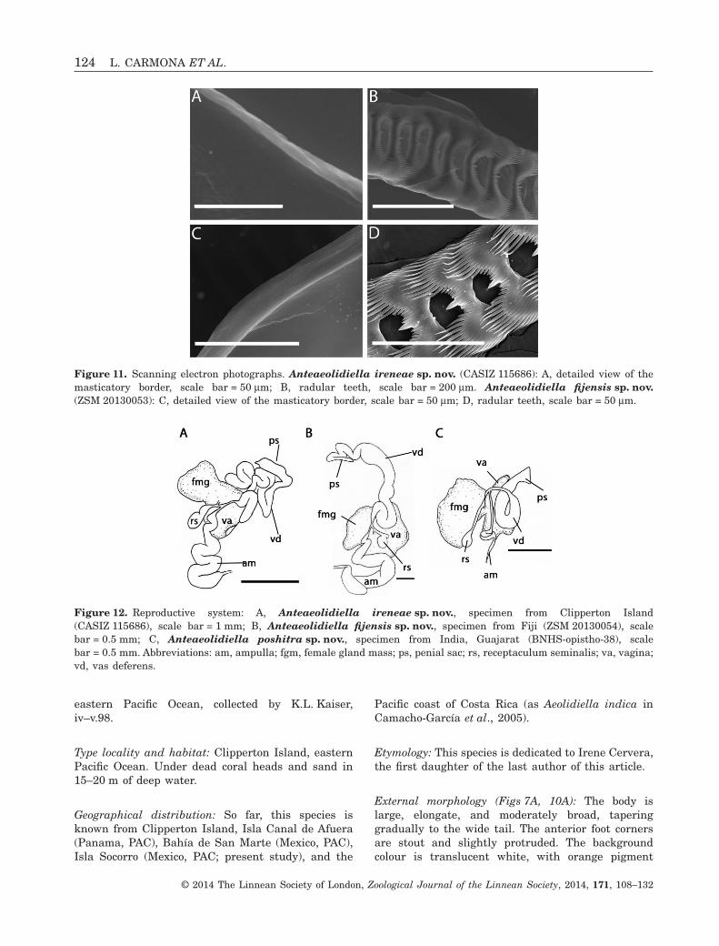
Figure 11. Scanning electron photographs. Anteaeolidiella ireneae sp. nov. (CASIZ 115686): A, detailed view of themasticatory border, scale bar = 50 μm; B, radular teeth, scale bar = 200 μm. Anteaeolidiella fijensis sp. nov.(ZSM 20130053): C, detailed view of the masticatory border, scale bar = 50 μm; D, radular teeth, scale bar = 50 μm.
Figure 12. Reproductive system: A, Anteaeolidiella ireneae sp. nov., specimen from Clipperton Island(CASIZ 115686), scale bar = 1 mm; B, Anteaeolidiella fijensis sp. nov., specimen from Fiji (ZSM 20130054), scalebar = 0.5 mm; C, Anteaeolidiella poshitra sp. nov., specimen from India, Guajarat (BNHS-opistho-38), scalebar = 0.5 mm. Abbreviations: am, ampulla; fgm, female gland mass; ps, penial sac; rs, receptaculum seminalis; va, vagina;vd, vas deferens.
124 L. CARMONA ET AL.
© 2014 The Linnean Society of London, Zoological Journal of the Linnean Society, 2014, 171, 108–132
scattered over the notum and head (Fig. 10A). Theintensity of these orange speckles varies with thespecimen, from dull to bright orange. A more or lessconspicuous white mark is found over the head thatgoes through the rhinophores through an opaquewhite or yellowish area, in the shape of teardrop(Fig. 7A). This white patch may continue over thepericardium as a large whitish yellow diamond or acircle. Posterior to these, up to four pale yellow tear-drop marks are found (Fig. 7A). The rhinophores areshort, quite thick at their base, and smooth. They canbe bright or dull orange, and have a white apex. Theoral tentacles are longer than the rhinophores, stout,and translucent orange with white tips. A pair ofblack eyes is visible at the posterior side of therhinophores base.
The cerata length is variable, but all are long,cylindrical, and uniform in diameter throughout mostof their length, and have a round apex. They begin atthe rear end of the rhinophores to the tail, leaving abare zone on the dorsum. They might be brightorange or dark grey, both with a white subapical bandand white tips.
The cerata are arranged in five to 36 rows. Eachgroup contains between 4 and 12 cerata, decreasing insize towards the foot. The anus is cleioproctic, locatedbetween the eighth and ninth rows on the right. Thegonopore is between the fourth and fifth rows on theright.
Anatomy: The masticatory process of the jaws issmooth (Fig. 11A). The radular formula is 27 × 0.1.0(CASIZ 115686). The teeth are progressively smaller tothe posterior region of the radula. They are also wideand bilobed, with 24–27 elongate and acutely pointeddenticles on both sides of the triangular and elongatecentral cusp (Fig. 11B). The outermost denticles arelarger than the innermost. The large, fusiform andconspicuous oral glands lie dorsolaterally to the buccalbulb. Each oral gland comprises rows of giant secretorycells (Fig. 6). The salivary glands are present.
The reproductive system is diaulic (Fig. 12A). Thepreampullary duct widens into the conspicuous, long,and wide ampulla that narrows again before dividinginto the oviduct and vas deferens. The very long andconvoluted vas deferens enters the wider proximalportion of the penial sac, which contains the unarmedpenial papilla. The short oviduct connects to apyriform-shaped receptaculum seminis. The remain-ing portion of the oviduct departs from the base of thereceptaculum and enters the female gland. Thevagina opens ventral to the penis.
Remarks: Anteaeolidiella ireneae sp. nov. is clearlydistinct from the other Anteaeolidiella species. Theelongate, large, and broad body, the thick and smooth
rhinophores, and the great number of cerata are, thusfar, only present in this species. Internally, the mor-phology of the radular teeth of Anteaeolidiellaireneae sp. nov. is also different. Whereas most of theAnteaeolidiella species have a few small denticlesbetween the central cusp and the large outermostdenticles, Anteaeolidiella ireneae sp. nov. doubles thenumber of small denticles. Regarding the reproduc-tive system, this species has the largest vas deferensamong Anteaeolidiella species (Fig. 12A).
Anteaeolidiella fijensis sp. nov. LSIDurn:lsid:zoobank.org:act:349DA396-370E-4DC3-83EC-EB2D117F5004 (Figs 6, 7B, 10B, 11C, D, 12B)Anteaeolidiella sp. B Carmona et al., 2013: 6.
Material examined: Holotype: ZSM 20130053, onespecimen, dissected, 4 mm in length preserved, Fiji,20.viii.06.
Paratype: ZSM 20130054, one specimen, dissected,4 mm in length preserved, Fiji, 20.viii.06.
Type locality and habitat: Laucala Bay, Fiji. Understones in 0–0.5 m of water.
Geographical distribution: So far, only known fromLaucala Bay, Fiji (present study).
Etymology: The name fijensis refers to the Fiji Repub-lic, where this species is found.
External morphology (Figs 7B, 10B): The body is elon-gate and moderately slender, tapering gradually tothe fairly short tail. The anterior foot corners arestout and slightly protruded. The background colouris translucent white, with bright orange pigment overthe dorsum, although the intensity varies with thespecimen (Fig. 10B). The head bears an orange mark.The latter extends from the rhinophores to just beforethe foot edge, leaving the foot edge translucent(Fig. 7B). From the rear of the rhinophores, there is atranslucent white area surrounded by an invertedtriangle shape (Fig. 7B). The rhinophores are trans-lucent orange and have a white tip end. They aremoderately long and stout. The conical and translu-cent white oral tentacles are slightly longer than therhinophores. A pair of black eyes is visible at theposterior side of the rhinophores base. The cerataare long and moderately thick. Some of them areEubranchus like. They extend from the rear of therhinophores to the tail and leave a bare zone over thedorsum. The ceratal epithelium is diffusely coveredwith orange pigment, and the ramifications of thedigestive gland are greenish or brownish. A whitesubapical band is found. The cnidosacs are white. Thecerata are arranged in six rows. Each row contains
SYSTEMATICS OF ANTEAEOLIDIELLA 125
© 2014 The Linnean Society of London, Zoological Journal of the Linnean Society, 2014, 171, 108–132
between two and 15 cerata, decreasing in sizetowards the foot. The anus is cleioproctic, locatedbetween the second and third rows of the first ceratalgroup of the right posterior digestive branch. Thegonopore is placed ventral to the first row on theright.
Anatomy: The masticatory border of the jaws issmooth (Fig. 11C). The radular formula is 14 × 0.1.0(ZSM Mol 20130053). The radular teeth are wide andbilobed, with 19–21 elongate and acutely pointeddenticles on both sides of the triangular and elongatecentral cusp (Fig. 11D). The outermost denticles arelarger than the innermost. The teeth are progres-sively smaller in the posterior region of the radula.The large, fusiform, and conspicuous oral glands arearranged dorsolaterally to the buccal bulb. Each oralgland comprises rows of giant secretory cells (Fig. 6).The salivary glands are absent.
The reproductive system is diaulic (Fig. 12B). Thepreampullary duct widens into the conspicuousampulla that narrows again before dividing into theoviduct and vas deferens. The conspicuous vasdeferens enters the wider proximal portion of thepenial sac, which contains the unarmed penialpapilla. The short oviduct connects to a roundedreceptaculum seminis. The remaining portion of theoviduct departs from the base of the receptaculumand enters the female gland. The vagina opens ven-trally to the penis.
Spawn: The living egg mass (not shown) is white. Thenumber of whorls could not be determined.
Remarks: So far, only three species have a translucentbody completely covered by orange pigment: Anteaeo-lidiella cacaotica, Anteaeolidiella ireneae sp. nov., andAnteaeolidiella fijensis sp. nov. Anteaeolidiella ireneaesp. nov. differs from Anteaeolidiella fijensis sp. nov. byits large and moderately broad body as well as its thickrhinophores. Externally, the main differences bet-
ween Anteaeolidiella cacaotica and Anteaeolidiellafijensis sp. nov. are the coloration of the foot edge andthe oral tentacles. The foot edge of the former speciesis orange instead of white translucent, as inAnteaeolidiella fijensis sp. nov. In addition, the oraltentacles in Anteaeolidiella cacaotica are usuallyorange, whereas in Anteaeolidiella fijensis sp. nov.they are usually translucent white. Regarding theinternal anatomy, Anteaeolidiella cacaotica has ashorter vas deferens and a bigger receptaculumseminis than Anteaeolidiella fijensis sp. nov. Althoughthe latter species also has a quite large vas deferens, itis not as long as that in Anteaeolidiella ireneae sp. nov.Additionally, the receptaculum seminis of Anteaeo-lidiella ireneae sp. nov. is also larger than that inAnteaeolidiella fijensis sp. nov. We also find some dif-ferences among the radulae of these three species. Thecentral cusp of the radular teeth of Anteaeolidiellafijensis sp. nov. is more prominent than that found inAnteaeolidiella cacaotica, and Anteaeolidiella ireneaesp. nov. has double the number of small denticlesbetween the central cusp and the large outermostdenticles.
Anteaeolidiella poshitra sp. nov. LSIDurn:lsid:zoobank.org:act:BC321799-0FFE-4585-A617-476FB4ADE19C (Figs 6, 7C, 10C, 12C,13A, B)Material examined: Holotype: BNHS-opistho-38, onespecimen, dissected, 8 mm in length preserved,India, Gujarat, Jamnagar, Poshitra, Marine NationalPark, collected by Vishal Bhave and Aditi Nair,11.xii.09.
Other material: BNHS-opistho-696, one specimen,8 mm in length preserved, India, Gujarat, Jamnagar,Poshitra, Marine National Park, collected by AmrutaPrasade, 27.i.12.
Type locality and habitat: Marine National Park,Poshitra, Jamnagar, Gujarat, India. Found undercoral rubble.
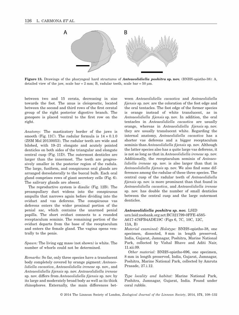
Figure 13. Drawings of the pharyngeal hard structures of Anteaeolidiella poshitra sp. nov. (BNHS-opistho-38): A,detailed view of the jaw, scale bar = 2 mm; B, radular teeth, scale bar = 50 μm.
126 L. CARMONA ET AL.
© 2014 The Linnean Society of London, Zoological Journal of the Linnean Society, 2014, 171, 108–132
Geographical distribution: Thus far, known only fromGujarat, India.
Etymology: The name poshitra refers to the shore ofPoshitra village in Gujarat, where this species isfound.
External morphology (Figs 7C, 10C): The anteriorfoot corners are stout and slightly protruded. Thebackground colour is translucent white (Fig. 10C).The head bears a bracket-shaped orange mark, fromthe rhinophores to the base of the oral tentacles.Extending posteriorly, just behind the rhinophores, apale orange area surrounded by bright orangepigment in the shape of an elongate diamond isfound. Over the pericardium there is a pale orangecircle outlined with bright orange pigment (Fig. 7C).The rhinophores and the oral tentacles have thesame coloration as the background colour. Therhinophores are smooth, stout, and taper towardsthe rounded tip. They have orange pigment over theproximal two-thirds and white pigmentation ontheir tips. The conical oral tentacles are slightlylonger than the rhinophores. They have white pig-mentation on their tips and may have orangepigment over their dorsal surface. A pair of blackeyes is visible at the posterior side of therhinophores base.
The cerata are long and moderately thick,Eubranchus like. They extend from the rear of therhinophores to the tail, and leave a bare zone over thedorsum. The ramifications of the digestive gland areorange, and have several constrictions over its length.They terminate in a white cnidosac. The cerata arearranged in up to six rows. Each one containsbetween two and six cerata, decreasing in size andnumber of cerata towards the foot. The anus iscleioproctic, usually located between the eighth andninth row. The genital opening is placed ventral to thethird row of the anterior digestive group.
Anatomy: The masticatory border of the jaws issmooth (Fig. 13A). The radular formula is 15 × 0.1.0(BNHS-opistho-38). The radular teeth are wide andbilobed, with 13–15 elongate and acutely pointeddenticles on either side of the triangular and elongatecentral cusp (Fig. 13B). The outermost denticles areusually larger than the innermost. The teeth areprogressively smaller in the posterior region of theradula. The large, fusiform, and conspicuous oralglands are arranged dorsolaterally to the buccal bulb.Each oral gland comprises rows of giant secretorycells. The salivary glands are absent.
The reproductive system is diaulic (Fig. 12C). Thepreampullary duct widens into the short and thinampulla that narrows again before dividing into the
oviduct and vas deferens. The conspicuous vasdeferens enters the wider proximal portion of thepenial sac, which contains the unarmed penialpapilla. The short oviduct connects to a roundedreceptaculum seminis with a very long duct. Theremaining portion of the oviduct departs from thebase of the receptaculum and enters the female gland.The vagina opens ventral to the penis.
Remarks: This new species from India is externallysimilar to Anteaeolidiella lurana, but can be distin-guished by several features. The cerata of the speci-mens from India lack any subapical band, and haveconstrictions in the digestive gland. In addition, thecerata of Anteaeolidiella poshitra sp. nov. are morebulbous than those of Anteaeolidiella lurana, resem-bling the cerata of Eubranchus. There are also someconsistent differences in the coloration of both species.Whereas A. lurana has two dorsal orange and thinbands that extend from the rear of the rhinophores tothe tail, forming the pattern depicted in theFigure 3D, Anteaeolidiella poshitra sp. nov. also hasthis kind of band but they disappear when they reachthe posterior end of the pericardium (Fig. 7C).Regarding the reproductive system, Anteaeolidiellalurana has a quite short ampulla and thin but longvas deferens, whereas in Anteaeolidiella poshitrasp. nov. the ampulla is longer and thinner, and thevas deferens is shorter and wider.
DISCUSSION
Our molecular and morphological studies show themonophyletic status of Anteaeolidiella, reinforcingthe outcomes of Carmona et al. (2013) and increasingthe number of species from two (Miller, 2001) to ten(present study). These species can be distinguishedby a combination of morphological and molecularsynapomorphies. Further support for species delimi-tations was obtained from the ABGD method. Thegroups proposed by the ABGD were entirely congru-ent with the molecular phylogeny and the morpho-logical data. The anatomical studies that we haveconducted clearly indicate that there are morphologi-cal differences between sister species, especiallyin coloration. This last feature is essential inAeolidiidae to separate species (pers. observ.). SomeAnteaeolidiella species also have differences in theirreproductive systems and radula. Additionally, differ-ent morphological characters such as oral and sali-vary glands have been examined here, and werefound to be informative at the generic level.Although it is currently accepted that some othernudibranch species may have great differences intheir colour patterns (e.g. Edmunds, 1969 andThompson & Brown, 1984 for Eubranchus farrani;
SYSTEMATICS OF ANTEAEOLIDIELLA 127
© 2014 The Linnean Society of London, Zoological Journal of the Linnean Society, 2014, 171, 108–132
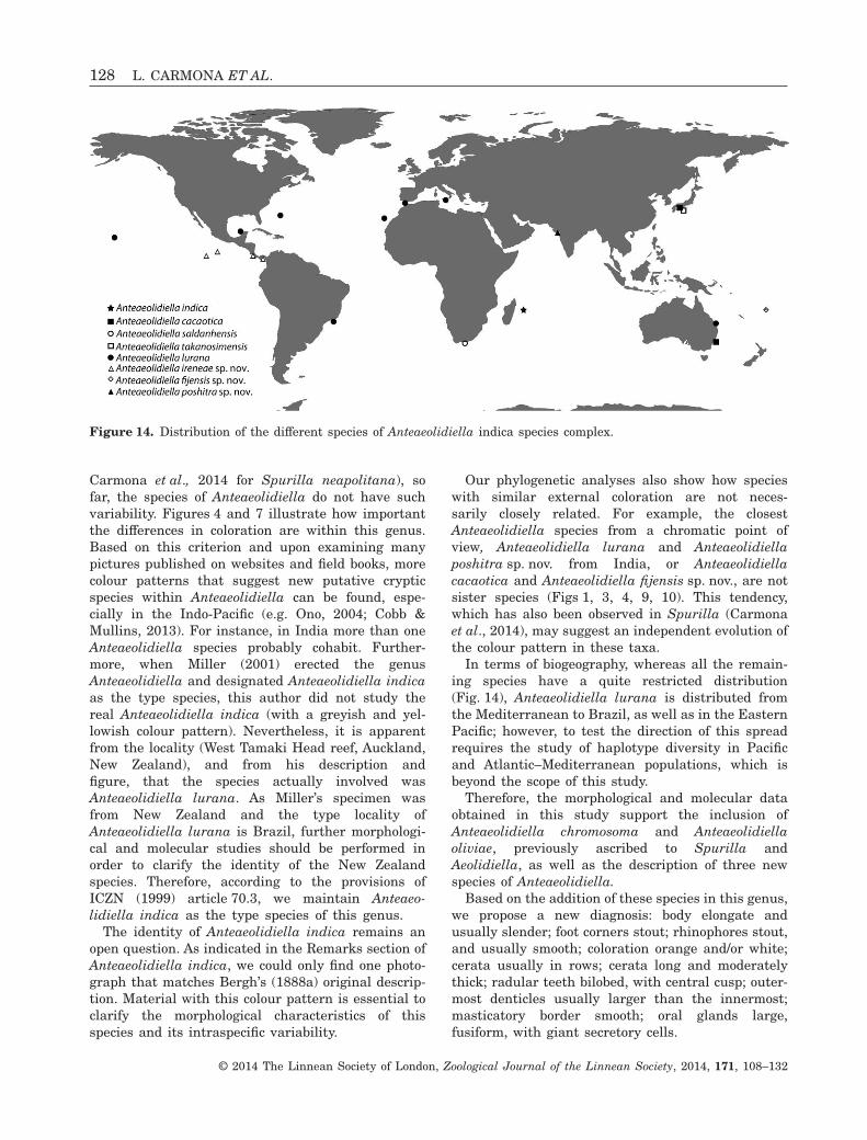
Carmona et al., 2014 for Spurilla neapolitana), sofar, the species of Anteaeolidiella do not have suchvariability. Figures 4 and 7 illustrate how importantthe differences in coloration are within this genus.Based on this criterion and upon examining manypictures published on websites and field books, morecolour patterns that suggest new putative crypticspecies within Anteaeolidiella can be found, espe-cially in the Indo-Pacific (e.g. Ono, 2004; Cobb &Mullins, 2013). For instance, in India more than oneAnteaeolidiella species probably cohabit. Further-more, when Miller (2001) erected the genusAnteaeolidiella and designated Anteaeolidiella indicaas the type species, this author did not study thereal Anteaeolidiella indica (with a greyish and yel-lowish colour pattern). Nevertheless, it is apparentfrom the locality (West Tamaki Head reef, Auckland,New Zealand), and from his description andfigure, that the species actually involved wasAnteaeolidiella lurana. As Miller’s specimen wasfrom New Zealand and the type locality ofAnteaeolidiella lurana is Brazil, further morphologi-cal and molecular studies should be performed inorder to clarify the identity of the New Zealandspecies. Therefore, according to the provisions ofICZN (1999) article 70.3, we maintain Anteaeo-lidiella indica as the type species of this genus.
The identity of Anteaeolidiella indica remains anopen question. As indicated in the Remarks section ofAnteaeolidiella indica, we could only find one photo-graph that matches Bergh’s (1888a) original descrip-tion. Material with this colour pattern is essential toclarify the morphological characteristics of thisspecies and its intraspecific variability.
Our phylogenetic analyses also show how specieswith similar external coloration are not neces-sarily closely related. For example, the closestAnteaeolidiella species from a chromatic point ofview, Anteaeolidiella lurana and Anteaeolidiellaposhitra sp. nov. from India, or Anteaeolidiellacacaotica and Anteaeolidiella fijensis sp. nov., are notsister species (Figs 1, 3, 4, 9, 10). This tendency,which has also been observed in Spurilla (Carmonaet al., 2014), may suggest an independent evolution ofthe colour pattern in these taxa.
In terms of biogeography, whereas all the remain-ing species have a quite restricted distribution(Fig. 14), Anteaeolidiella lurana is distributed fromthe Mediterranean to Brazil, as well as in the EasternPacific; however, to test the direction of this spreadrequires the study of haplotype diversity in Pacificand Atlantic–Mediterranean populations, which isbeyond the scope of this study.
Therefore, the morphological and molecular dataobtained in this study support the inclusion ofAnteaeolidiella chromosoma and Anteaeolidiellaoliviae, previously ascribed to Spurilla andAeolidiella, as well as the description of three newspecies of Anteaeolidiella.
Based on the addition of these species in this genus,we propose a new diagnosis: body elongate andusually slender; foot corners stout; rhinophores stout,and usually smooth; coloration orange and/or white;cerata usually in rows; cerata long and moderatelythick; radular teeth bilobed, with central cusp; outer-most denticles usually larger than the innermost;masticatory border smooth; oral glands large,fusiform, with giant secretory cells.
Figure 14. Distribution of the different species of Anteaeolidiella indica species complex.
128 L. CARMONA ET AL.
© 2014 The Linnean Society of London, Zoological Journal of the Linnean Society, 2014, 171, 108–132
ACKNOWLEDGEMENTS
We are deeply grateful to all of the individuals thathelped to collect and provided specimens and imagesfor this study, such as R. Nakano, D. Harasti, andG. Villani. Many thanks are due to W. Sterrer(Bermuda Aquarium and Museum of Zoology, BAMZ),B. Williams (Bermuda Institute of Oceanographic Sci-ences), and T.M. Trott (Acting Senior MarineResources Officer, Bermuda Government), for welcom-ing, housing, and facilitating permits during theexpeditions in Bermuda. We are grateful to AsadRahmani (Director, BNHS). We are thankful to theMinistry of Environment and Forests, Government ofIndia, who has provided financial support throughthe All India Co-ordinated Project On Taxonomy(AICOPTAX) and Deepak Apte, Principal Scientist ofAICOPTAX and Chief Operating Officer (COO) ofBNHS for constant encouragement and funding. Wealso acknowledge the PCCF (WL) Gujarat and ChiefConservator of Forests, Marine National Park,Gujarat, for the necessary permissions and timelyhelp. We are also grateful to our team in Gujarat whomanaged and helped our field trips in Gujarat.A. Sellas helped us with some molecular lab work. Thiswork was supported by several research grants:CGL2006-05182/BOS, CTM2008-05228-E/MAR, andCGL2010-17187, Spanish Ministry of Economy andCompetitiveness (includes the early Ministry of Sci-ences and Innovation), to J.L.C.; California Academy ofSciences, National Science Foundation (DEB 0329054PEET) to T.M. Gosliner; and a research grant from theMalacological Society of London to L. Carmona. This isCEI·MAR journal publication 45.
REFERENCES
Akaike H. 1974. A new look at the statistical model identifi-cation. IEEE Transactions on Automatic Control 19: 716–723.
Altschul SF, Gish W, Miller W, Myers EW, Lipman DJ.1990. Basic local alignment search tool. Journal of Molecu-lar Biology 215: 403–410.
Angas GF. 1864. Description d’espèces nouvelles appartenantà plusieurs genres de Mollusques Nudibranches des environsde Port-Jackson (Nouvelle-Galles du Sud), accompagnée dedessins faits d’après nature. Journal de Conchyliologie 12:43–70.
Avise JC. 2004. Molecular markers, natural history,and evolution, 2nd edn. Sunderland, MA: SinauerAssociates.
Baba K. 1930. Studies on Japanese Nudibranchs 3. Venus 2:117–125.
Baba K. 1937. Opisthobranchia of Japan (II). Journal.Department of Agriculture. Kyushu Imperial University 5:289–344.
Baba K. 1979a. Brief comment on the identification of anudibranch mollusk, Aeolidiella takanosimensis Baba, 1930,from Amakusa, Japan (Eolidoidea: Aeolidiidae). Publica-tions from Amakusa Marine Biological Laboratory, KyushyUniversity 5: 1–7.
Baba K. 1979b. Short Account of the Anatomy of aNudibranchiate Mollusk Aeolidiella takanosimensis Baba,1930 from Japan. The Veliger 22: 12–18.
Barnard KH. 1927. South African nudibranch Mollusca, withdescriptions of new species and a note on some specimensfrom Tristan d’Acunha. Annals of the South AfricanMuseum 25: 171–215.
Bergh LSR. 1875. Beiträge zur Kenntniss der Aeolidiaden.III. Verhandlungen der königlichkaiserlich Zoologisch-botanischen Gesellschaft in Wien (Abhandlungen) 25: 633–658.
Bergh LSR. 1888a. Malacologische Untersuchungen. In:Reisen im Archipel der Philippinen von Dr. Carl GottfriedSemper. Zweiter Theil. Wissenschaftliche Resultate 3: 755–814.
Bergh LSR. 1888b. Beiträge zur Kenntniss der Aeolidiaden.IX. Verhandlungen der königlichkaiserlich Zoologisch-botanischen Gesellschaft in Wien (Abhandlungen) 38: 673–706.
Bhave VJ, Apte DA. 2011. Illustrated checklist ofopisthobranch fauna of Ratnagiri, Maharashtra, India, witheight new records to India. Journal of the Bombay NaturalHistory Society 108: 172–182.
Bidgrain P. 2005. Anteaeolidiella indica from ReunionIsland [Message in] Sea Slug Forum. Australian Museum,Sydney. Available at: http://www.seaslugforum.net/find/13107
Boserelle P, Levy JM, Michenet F. 2013. Aeolidiellaindica from Tahiti [Message in] Nudi Tahiti. Availableat: http://www.nuditahiti.com/liste-des-esp%C3%A8ces/aeolidiella-indica/
Burn R. 1962. Descriptions of Victorian nudibranchiateMollusca, with a comprehensive review of the Eolidiacea.Memoirs of the National Museum, Melbourne 25: 95–128.
Burn R. 1964. A centennial commentary and zoogeographicalremarks on Angas’ Sydney nudibranchs (Molluscs,Gastropoda). Journal de Conchyliologie 104: 85–93.
Burn R. 2006. A checklist and bibliography of theOpisthobranchia (Mollusca: Gastropoda) of Victoria and theBass Strait area, south-eastern Australia. Museum VictoriaScience Reports 10: 1–42.
Camacho-García Y, Gosliner TM, Valdés Á. 2005. Fieldguide to the sea slugs of the tropical Eastern Pacific. SanFrancisco, CA: California Academy of Sciences.
Carmona L, Lei BR, Pola M, Gosliner TM, Valdés Á,Cervera JL. 2014. Untangling the Spurilla neapolitana(Delle Chiaje, 1841) species complex: a review of the genusSpurilla Bergh, 1864 (Mollusca: Nudibranchia: Aeolidiidae).Zoological Journal of the Linnean Society 170: 132–154. doi:10.1111/zoj.12098.
Carmona L, Malaquias MA, Gosliner TM, Pola M,Cervera JL. 2011. Amphi-Atlantic distributions and
SYSTEMATICS OF ANTEAEOLIDIELLA 129
© 2014 The Linnean Society of London, Zoological Journal of the Linnean Society, 2014, 171, 108–132
cryptic species in sacoglossan sea slugs. Journal of Mollus-can Studies 77: 401–412.
Carmona L, Pola M, Gosliner TM, Cervera JL. 2013. Atale that morphology fails to tell: a molecular phylogeny ofAeolidiidae (Aeolidida, Nudibranchia, Gastropoda). PLoSONE 8: e63000. doi:10.1371/journal.pone.0063000.
Claremont M, Houart R, Williams ST, Reid DG. 2013. Amolecular phylogenetic framework for the Ergalataxinae(Neogastropoda: Muricidae). Journal of Molluscan Studies79: 19–19.
Cobb G, Mullins D. 2013. Antaeolidiella cacaotica fromSunshine Coast [Message in] Nudibranchs of SunshineCoast Australia. Available at: http://www.nudibranch.com.au/pages/2516c.htm
Cockerell TDA, Eliot C. 1905. Notes on a collection ofCalifornian nudibranches. Journal of Malacology 12: 31–53.
Coleman N. 1989. Nudibranchs of the south Pacific, Vol. 1.Springwood, Queensland, Australia: Sea Australia ResourceCentre.
Colgan DJ, McLauchlan A, Wilson GDF, Livingston S,Macaranas J, Edgecombe GD, Cassis G, Gray MR.1998. Molecular phylogenetics of the Arthropoda: relation-ships based on histone H3 and U2 snRNA DNA sequences.Australian Journal of Zoology 46: 419–437.
Crocetta F. 2012. Marine alien Mollusca in Italy: a criticalreview and state of the knowledge. Journal of the MarineBiological Association of the United Kingdom 92: 1357–1365.
Crocetta F, Macali A, Furfaro G, Cooke S, Villani G,Valdés A. 2013. Alien molluscan species established alongthe Italian shores: an update, with discussions on someMediterranean ‘alien species’ categories. Zookeys 277:91–108.
Debelius H, Kuiter RH. 2007. Nudibranchs of the world.Frankfurt, Germany: IKAN-Unterwasserarchiv.
Dinapoli A, Tamer C, Frassen S, Naduvilezhath L,Klussmann-Kolb A. 2006. Utility of H3-Genesequences forphylogenetic reconstruction – a case study of heterobranchGastropoda. Bonner Zoologische Beiträge 55: 191–202.
Domínguez M, Troncoso JS, García FJ. 2008. The familyAeolidiidae Gray, 1827 (Gastropoda Opisthobranchia) fromBrazil, with a description of a new species belonging to thegenus Berghia Trinchese, 1877. Zoological Journal of theLinnean Society 153: 349–368.
Drummond AJ, Ashton B, Cheung M, Heled J, Kearse M,Moir R, Stones-Havas S, Thierer T, Wilson A. 2009.Geneious v4.6. Available at: http://www.geneious.com/
Drummond AJ, Rambaut A. 2007. BEAST: Bayesian evo-lutionary analysis by sampling trees. BMC EvolutionaryBiology 7: 214.
Edmunds M. 1969. Opisthobranchiate mollusca from Tanza-nia I. Eolidacea (Eubranchidae and Aeolidiidae). Proceed-ings of the Malacological Society of London 38: 451–469.
Eilertsen MH, Malaquias MAE. 2013. Systematic revisionof the genus Scaphander (Gastropoda, Cephalaspidea) inthe Atlantic Ocean, with a molecular phylogenetic hypoth-esis. Zoological Journal of the Linnean Society 167: 389–429.
Folmer O, Black M, Hoeh W, Lutz R, Vrijenhoek R. 1994.DNA primers for amplification of mitochondrial cytochromec oxidase subunit I from diverse metazoan invertebrates.Journal of Molecular Biology and Biotechnology 3: 294–299.
Frey MA, Vermeij GJ. 2008. Molecular phylogenies andhistorical biogeography of a circumtropical group of gastro-pods (Genus: Nerita): implications for regional diversitypatterns on the marine tropics. Molecular Phylogenetics andEvolution 48: 1067–1086.
García-García FJ, Domínguez M, Troncoso JS. 2008.Opistobranquios de Brasil. Vigo: Feito S. L.
García-Gómez JC. 2002. Paradigmas de una fauna insólita.Los moluscos opistobranquios del estrecho de Gibraltar.(Serie Ciencias) 20: 397 pp. Algeciras, Cádiz, Spain:Instituto de Estudios Gibraltareños.
García-Gómez JC, Cervera JL, García-García F. 2011.Familia Aeolidiidae. In: Gofas S, Moreno D, Salas C, coords.Moluscos marinos de Andalucía. Málaga: Servicio dePublicaciones e Intercambio Científico, Universidad deMálaga, 508–511.
Gosliner TM. 1985. The Aeolid Nudibranchs familyAeolidiidae (Gastropoda, Opisthobranchia) from tropicalSouthern Africa. Annals of the South African Museum 95:233–267.
Gosliner TM, Behrens DW, Valdés Á. 2008. Indo-Pacificnudibranchs and sea slugs. Gig Harbor/San Francisco, CA:Sea Challengers/California Academy of Sciences.
Gosliner TM, Fahey S. 2011. Previously undocumenteddiversity and abundance of cryptic species: a phylogeneticanalysis of Indo-Pacific Arminidae Rafinesque 1814(Mollusca: Nudibranchia) with descriptions of twenty newspecies of Dermatobranchus. Zoological Journal of theLinnean Society 161: 245–256.
Gosliner TM, Griffiths RJ. 1981. Description and revision ofsome South African Aeolidacean Nudibranchia (Mollusca,Gastropoda). Annals of the South African Museum 82: 102–151.
GROC. 2013. Anteaeolidiella foulisi a la Reserva Marina delToro (Mallorca). Available at: http://www.opistobranquis.org/ca/noticies/10#sthash.3oBJz77N.dpuf
ICZN. 1999. International code of zoological nomenclature,fourth edition. London: International Trust for ZoologicalNomenclature.
Ijima I. 1918. A manual of Zoology. Tokyo: DainihontoshoPublishing Co., Ltd.
Imamoto J. 2013. Anteaeolidiella indica. [Message in]Umiushi website. Available at: http://www.umiushi.info/kanri/photo_sql_eng.php?act=PSP&gakumei1=Anteaeolidiella+%5BOld+Aeolidiella%5D&gakumei2=indica&location_code=000
Jörger MK, Norenburg JL, Wilson NG, Schrödl M. 2012.Barcoding against a paradox? Combined molecular speciesdelineations reveal multiple cryptic lineages in elusivemeiofaunal sea slugs. BMC Evolutionary Biology 12: 245.
Katoh K, Asimenos G, Toh H. 2009. Multiple alignment ofDNA sequences with MAFFT. In Bioinformatics for DNASequence Analysis edited by D. Posada. Methods in Molecu-lar Biology 537: 39–64.
130 L. CARMONA ET AL.
© 2014 The Linnean Society of London, Zoological Journal of the Linnean Society, 2014, 171, 108–132
Knowlton N. 2000. Molecular genetic analyses of speciesboundaries in the sea. Hydrobiologia 420: 73–90.
Krug PJ, Morley MS, Asif J, Hellyar LL, Blom WM. 2008.Molecular confirmation of species status for the rarecephalaspidean Melanochlamys lorrainae (Rudman, 1968),and comparison with its sister species M. cylindricaCheeseman, 1881. Journal of Molluscan Studies 74: 267–276.
Krug PJ, Vendetti JE, Rodriguez AK, Retana JN,Hirano YM, Trowbridge CD. 2013. Integrative speciesdelimitation in photosynthetic sea slugs reveals twenty can-didate species in three nominal taxa studied for drug dis-covery, plastid symbiosis or biological control. MolecularPhylogenetics and Evolution 69: 1101–1119.
MacFarland FM. 1966. Studies of opisthobranchiatemollusks of the Pacific Coast of North America. Memoirs ofthe California Academy of Sciences 6: 373.
Macnae W. 1954. On some eolidacean nudibranchiate mol-luscs from South Africa. Annals of the Natal Museum 13:1–50.
Maddison DR, Maddison WP. 2005. MacClade 4., v. 4.08 forOSX. Sunderland, MA: Sinauer Associates.
Malaquias MAE, Reid DG. 2008. Systematic revision ofthe living species of Bullidae (Mollusca: Gastropoda:Cephalaspidea), with a molecular phylogenetic analysis.Zoological Journal of the Linnean Society 153: 453–543.
Malaquias MAE, Reid DG. 2009. Tethyan vicariance,relictualism and speciation: evidence from a global molecu-lar phylogeny of the opistobranch genus Bulla. Journal ofBiogeography 36: 1760–1777.
Marcus E. 1958. On Western Atlantic OpisthobranchiateGastropods. American Museum Novitates 1906: 1–82.
Marcus E. 1961. Opisthobranch Mollusks from California.The Veliger 3: 1–85.
Marcus E, Marcus E. 1967. American OpisthobranchMollusks. Studies in Tropical Oceanography, Miami 10:129–203.
von Martens E, Pfeffer GJ. 1886. Die Mollusken von Süd-Georgien nach der Ausbeute der Deutschen Station 1882–83. Jahrbuch der Hamburgischen WissenschaftlichenAnstalten 3: 63–135, pls. 1–4.
Meyer CP. 2003. Molecular systematics of cowries(Gastropoda: Cypraeidae) and diversification patterns in thetropics. Biological Journal of the Linnean Society 79: 401–459.
Miller MC. 2001. Aeolid nudibranchs (Gastropoda:Opisthobranchia) of the family Aeolidiidae from NewZealand waters. Journal of Natural History 35: 629–662.
Moro L, Ortea J, Bacallado JJ, Caballer M, AcevedoI. 2003. Anaspidea, Cephalaspidea, Gymnosomata,Notaspidea, Nudibranchia, Sacoglossa y Thecosomata. In:Moro L, Martín JL, Garrido MJ, Izquierdo I, eds. Lista deespecies marinas de Canarias (algas, hongos, plantas yanimals). Santa Cruz de Tenerife, Spain: Consejería PolíticaTerritorial y del Medio Ambiente del Gobierno de Canarias,93–98.
Moro L, Ortea J, Bacallado JJ, Valdés A, Pérez-SánchezJM. 1995. Nuevos Aeolidáceos (Gastropoda, Nudibranchia)
para la fauna de Canarias. Revista de la Academia deCanaria de Ciencias 7: 63–75.
Nakano R. 2004. Opisthobranchs of Japan Islands. Tokyo:Rutles, Inc.
Nylander JAA. 2004. MrModeltest v2.3. Program distributedby the author. Uppsala: Evolutionary Biology Centre,Uppsala University. Available at: http://www.abc.se/~nylander/mrmodeltest2/mrmodeltest2.html
Occhipinti-Ambrogi A, Marchini A, Cantone G, CastelliA, Chimenz C, Cormaci M, Froglia C, Furnari G,Gambi MC, Giaccone G, Giangrande A, Gravili C,Mastrototaro F, Mazziotti C, Orsi-Relini L, Piraino S.2011. Alien species along the Italian coasts: an overview.Biological Invasions 13: 215–237.
Ohnheiser LT, Malaquias MAE. 2013. Systematic revision ofthe gastropod family Philinidae (Mollusca: Cephalaspidea) inthe north-east Atlantic Ocean with emphasis on the Scandi-navian Peninsula. Zoological Journal of the Linnean Society167: 273–326.
Ono A. 1999. Opisthobranchs of Kerama Islands. Tokyo:TBS-Britannica Co., Ltd.
Ono A. 2004. Opisthobranchs of Ryukyu Islands. Tokyo:Rutles, Inc.
Ornelas-Gatdula E, Camacho-García Y, Schrödl M,Padula V, Hooker Y, Gosliner TM, Valdés A. 2012.Molecular systematics of the ‘Navanax aenigmaticus’species complex (Mollusca, Cephalaspidea): coming fullcircle. Zoologica Scripta 41: 374–385.
Orr J. 1981. Hong Kong nudibranchs. Hong Kong: UrbanCouncil.
Ortea J, Moro L, Bacallado JJ, Herrera R. 2001. Catálogoactualizado de los Moluscos Opistobranquios de las IslasCanarias. Revista de la Academia Canaria de Ciencias 12:105–136.
Ortigosa D, Simões N, Calado G. 2013. Seaslugs (Mollusca:Opisthobranchia) from Campeche Bank, Yucatan Peninsula,Mexico. Thalassas 29: 59–75.
Padula V, Bahia J, Vargas C, Lindner A. 2011. Mollusca,Nudibranchia: new records and southward range extensionsin Santa Catarina, southern Brazil. Check List 7: 1–3.
Palumbi SR, Martin A, Romano S, Owen MacMillan W,Stice L, Grabowski G. 1991. The simple fool’s guide to PCR.Honolulu: Department of Zoology, University of Hawaii.
Pérez-Sánchez JM, Moreno E. 1990. InvertebradosMarinos de Canarias. Las Palmas de Gran Canaria, Spain:Ediciones del Cabildo Insular de Gran Canaria.
Pittman C, Fiene P. 2013. Aeolidiella indica. [Message in]Sea Slugs of Hawaii. Available at: http://seaslugsofhawaii.com/species/Aeolidiella-indica-a.html
Pola M, Camacho-García YE, Gosliner TM. 2012. Molecu-lar data illuminate cryptic nudibranch species: the evolutionof the Scyllaeidae (Nudibranchia: Dendronotina) with arevision of Notobryon. Zoological Journal of the LinneanSociety 165: 311–336.
Pola M, Gosliner TM. 2010. The first molecular phylogenyof cladobranchian opisthobranchs (Mollusca, Gastropoda,Nudibranchia). Molecular Phylogenetics and Evolution 56:931–941.
SYSTEMATICS OF ANTEAEOLIDIELLA 131
© 2014 The Linnean Society of London, Zoological Journal of the Linnean Society, 2014, 171, 108–132
Puillandre N, Lambert A, Brouillet S, Achaz G. 2012a.ABGD, Automatic Barcode Gap Discovery for primaryspecies delimitation. Molecular Ecology 21: 1864–1877.
Puillandre N, Modica MV, Zhang Y, Sirovich L,Boisselier C, Cruaud C, Holford M, Samadi S. 2012b.Large-scale species delimitation method for hyperdiversegroups. Molecular Ecology 21: 2671–2691.
Risbec J. 1928. Contribution a l’etude des nudibranchesNéo-Calédoniens. Thèse présentée à la faculté des sciencesde l’Université de Paris pour obtenir le grade de docteurès-sciences naturelles. Faune des Colonies Française 2:1–328.
Risbec J. 1956. Nudibranches du Viet-Nam. Archives duMuséum National d’Historie Naturelle 4: 3–58.
Ronquist F, Huelsenbeck JP. 2003. MrBayes 3: Bayesianphylogenetic inference under mixed models. Bioinformatics19: 1572–1574.
Rudman WB, Willan RC. 1998. Opisthobranchia. In:Beesley PL, Ross GJB, Wells A, eds. Mollusca: the southernsynthesis. Fauna of Australia, Vol. 5, Part B. Melbourne:CSIRO Publishing. 915–1035.
Sammut CR, Perrone AS. 1998. A preliminary check-list ofOpisthobranchia (Mollusca, Gastropoda) from the MalteseIslands. Basteria 62: 221–240.
Schmekel L. 1968. Ascoglossa, Notaspidea undNudibranchia im Litoral des Golfes von Neapel. RevueSuisse de Zoologie 75: 103–155.
Schmekel L. 1970. Anatomie der Genitalorgane vonNudibranchiern (Gastropoda Euthyneura). Pubblicacionidella Stazione Zoologica di Napoli 38: 120–217.
Schmekel L, Portmann A. 1982. Opisthobranchia desMittelmeeres. Berlin: Springer Verlag.
Sciberras M, Schembri PJ. 2007. A critical review ofrecords of alien marine species from the Maltese Islandsand surrounding waters (Central Mediterranean). Mediter-ranean Marine Science 8: 41–66.
Sphon GC, Lance JR. 1968. An annotated list ofnudibranchs and their allies from Santa Barbara County,California. Proceeding of the California Academy ofSciencies 36: 73–84.
Stamatakis A. 2006. RAxML-VI-HPC: maximum likelihood-based phylogenetic analyses with thousands of taxa andmixed models. Bioinformatics 22: 2688–2690.
Stimpson W. 1855. Descriptions of some new marineinvertebrata. Proceeding of the Academy of Natural Sciencesof Philadelphia 7: 385–395.
Talavera G, Castresana J. 2007. Improvement ofphylogenies after removing divergent and ambiguouslyaligned blocks from protein sequence alignments. System-atic Biology 56: 564–577.
Tamura K, Peterson D, Peterson N, Stecher G, Nei M,Kumar S. 2011. MEGA5: molecular evolutionary geneticsanalysis using maximum likelihood, evolutionary distance,and maximum parsimony methods. Molecular Phylogeneticsand Evolution 28: 2731–2739.
Thompson TE, Brown GH. 1984. Biology of opisthobranchmolluscs, vol. 2. London: Ray Society.
Valdés Á, Hamann J, Behrens DW, DuPont A. 2006.Caribbean Sea Slugs. A field guide to the opisthobranchmollusks from the tropical northwestern Atlantic. GigHarbor, WA: Sea Challengers Natural History Books.
Virabhadra-Rao K, Alagarswami K. 1960. An account ofthe structure and early development of a new species of anudibranchiate gastropd, Eolidina (Eolidina) mannarensis.Journal of the Marine Biological Association of India 2:6–16.
Wheeler QD, Meier R, eds. 2000. Species concepts andphylogenetic theory: a debate. New York: Columbia Univer-sity Press.
Williams ST, Reid DG. 2004. Speciation and diversity ontropical rocky shores: a global phylogeny of snails of thegenus Echinolittorina. Evolution 58: 2227–2251.
Zenetos A, Çinar ME, Panucci-Papadopoulou MA,Harmelin JG, Furnari G, Andaloro F, Bellou N,Streftaris N, Zibrowius H. 2005. Annotated list of marinealien species in the Mediterranean with records of the worstinvasive species. Mediterranean Marine Science 6: 63–118.
Zenetos A, Gofas S, Russo G, Templado J. 2004. Vol. 3.Molluscs. In: Briand F, ed. CIESM Atlas of exotic species inthe Mediterranean. Monaco: CIESM Publishers, 1–376.
SUPPORTING INFORMATION
Additional Supporting Information may be found in the online version of this article at the publisher’s web-site:
Figure S1. Phylogenetic hypothesis based on the combined data set (H3 + COI + 16S), inferred by Bayesianinference (BI) analysis.Table S1. List of specimens used for phylogenetic analyses. Abbreviations: ATL, Atlantic Ocean; EA, easternAtlantic Ocean; GB, GenBank; MED, Mediterranean; PAC, Pacific.
132 L. CARMONA ET AL.
© 2014 The Linnean Society of London, Zoological Journal of the Linnean Society, 2014, 171, 108–132