Syringomyelia : Current Concepts in Pathogenesis , Diagnosis and Treatment
11
Syringomyelia: Current Concepts in Pathogenesis, Diagnosis, and Treatment Clare Rusbridge, Dan Greitz, and Bermans J. Iskandar Syringomyelia is a condition that results in fluid-containing cavities within the parenchyma of the spinal cord as a consequence of altered cerebrospinal fluid dynamics. This review discusses the history and the classification of the disorder, the current theories of pathogenesis, and the advanced imaging modalities used in the diagnosis. The intramedullary pulse pressure theory (a new pathophysiologic concept of syringomyelia) also is presented. In addition, the current understanding of the painful nature of this condition is discussed and the current trends in medical and surgical management are reviewed. Key words: Chiari malformation; Occipital hypoplasia; Syringohydromyelia; Syrinx; Cavalier King Charles Spaniel; Dysesthesia; Cine magnetic resonance imaging; Pathophysiology; Suboccipital decompression; Medical management. S yringomyelia is a condition in which fluid-filled cavities develop in the spinal cord (Fig 1). Pre- viously, it was considered a rare condition in veterinary medicine, but it is now a relatively common neurologic diagnosis. This change is in part because of increased availability of magnetic resonance imaging (MRI) and also because of increased prevalence in certain breeds, most notably the Cavalier King Charles Spaniel (CKCS). 1 Syringomyelia can be caused by various primary problems, including abnormalities of the caudal fossa (Chiari malformations), tethered spinal cord, trauma, arachnoiditis, and tumor. 1,2 Herein, we review the clinical and pathogenic characteristics of syringomyelia for the purpose of updating veterinarians on current views of pathogenesis, diagnosis, and treatment. History of the Classification of Syringomyelia and Chiari Malformation Cavitation within the spinal cord was first described by Stephanus in the 16th century. 3 Olivier d’Angers first used the term syringomyelia in 1824 and, at that time, the cavity was believed to be a dilated central canal that communicated with the 4th ventricle. 4 It was later proposed that dilatation of the central canal be termed hydromyelia and that a distinction be made between this dilatation and syringomyelia because the former struc- ture was lined by ependyma and the latter by glial cells. 5 It subsequently has been shown that this distinction is somewhat arbitrary in that hydromyelia may extend into the spinal-cord substance to form syringohydromyelia partially lined by ependyma, 6 and cavities may rupture into the central canal. 7 The term syringomyelia now is generally acceptable for all clinical conditions charac- terized by spinal-cord cavitation containing fluid iden- tical with or closely resembling cerebrospinal fluid (CSF). 7 This classification does not include (protein- containing) cavities associated with tumors. 7 Syringomyelia traditionally was classified into com- municating and noncommunicating types, in which communicating implies disruption of CSF dynamics at the cranial cervical junction and noncommunicating implies a primary spinal-cord condition. 8 These terms are confusing, however, because the communication referred to a connection between the syringomyelic cavity and the 4th ventricle, which actually is present in fewer than 10% of affected human patients. This categorization now tends to be disregarded. 9 In 1891, Hans von Chiari, a pathologist in Prague, described 4 types of abnormalities based on postmortem examinations of infants with hydrocephalus who died shortly after birth. 10,11 The type 1 malformation was described as ‘‘elongation of the cerebellar tonsils and the medial part of the inferior cerebellar lobes into conelike projections, which accompany the medulla into the spinal canal.’’ 12 Arnold’s name subsequently was added by 2 of his loyal students on the basis of the description of a single case consistent with a Chiari type II malformation (ie, in association with spina bifida). For many years, the spectrum of disorders of cerebellome- dullary descent was referred to as the Arnold-Chiari syndrome. 12 In recent years, however, the trend has been to simplify the name to the Chiari malformation. This name is now a convenient shorthand for a wide range of abnormalities not necessarily consistent with Hans von Chiari’s original description of a type I malformation, but all are characterized by a decreased posterior fossa volume with caudal descent of the cerebellar tonsils and often of the brainstem. 12,13 It is debatable whether the term Chiari malformation should be applied to the dog. The analogous condition, characterized by a decreased volume of the caudal fossa and caudal displacement of the caudal cerebellar vermis into or through the foramen magnum, is very similar to the human condition. 14 The condition in the dog, however, is inconsistent with the historical description, not in the least because dogs do not have cerebellar tonsils. It may be more correct to use an anatomical description (eg, occipital hypoplasia with syringomyelia 1 or caudal occipital malformation syn- drome 15 ). Arnold-Chiari malformation is an inappropri- ate descriptive term unless the pathology also includes From Stone Lion Veterinary Centre, Wimbledon, London, UK (Dr Rusbridge); Department of Neuroradiology and MR-Research Center, Karolinska University Hospital, Stockholm, Sweden (Dr Greitz); Department of Neurological Surgery, University of Wisconsin Hospital, Madison, WI (Dr Iskandar). Copyright E 2006 by the American College of Veterinary Internal Medicine 0891-6640/06/2003-0001/$3.00/0 Review J Vet Intern Med 2006;20:469–479
Transcript of Syringomyelia : Current Concepts in Pathogenesis , Diagnosis and Treatment

Syringomyelia: Current Concepts in Pathogenesis, Diagnosis,and Treatment
Clare Rusbridge, Dan Greitz, and Bermans J. Iskandar
Syringomyelia is a condition that results in fluid-containing cavities within the parenchyma of the spinal cord as a consequence
of altered cerebrospinal fluid dynamics. This review discusses the history and the classification of the disorder, the current
theories of pathogenesis, and the advanced imaging modalities used in the diagnosis. The intramedullary pulse pressure theory
(a new pathophysiologic concept of syringomyelia) also is presented. In addition, the current understanding of the painful
nature of this condition is discussed and the current trends in medical and surgical management are reviewed.
Key words: Chiari malformation; Occipital hypoplasia; Syringohydromyelia; Syrinx; Cavalier King Charles Spaniel;
Dysesthesia; Cine magnetic resonance imaging; Pathophysiology; Suboccipital decompression; Medical management.
S yringomyelia is a condition in which fluid-filledcavities develop in the spinal cord (Fig 1). Pre-
viously, it was considered a rare condition in veterinarymedicine, but it is now a relatively common neurologicdiagnosis. This change is in part because of increasedavailability of magnetic resonance imaging (MRI) andalso because of increased prevalence in certain breeds,most notably the Cavalier King Charles Spaniel(CKCS).1
Syringomyelia can be caused by various primaryproblems, including abnormalities of the caudal fossa(Chiari malformations), tethered spinal cord, trauma,arachnoiditis, and tumor.1,2 Herein, we review theclinical and pathogenic characteristics of syringomyeliafor the purpose of updating veterinarians on currentviews of pathogenesis, diagnosis, and treatment.
History of the Classification of Syringomyelia andChiari Malformation
Cavitation within the spinal cord was first describedby Stephanus in the 16th century.3 Olivier d’Angers firstused the term syringomyelia in 1824 and, at that time,the cavity was believed to be a dilated central canal thatcommunicated with the 4th ventricle.4 It was laterproposed that dilatation of the central canal be termedhydromyelia and that a distinction be made between thisdilatation and syringomyelia because the former struc-ture was lined by ependyma and the latter by glial cells.5
It subsequently has been shown that this distinction issomewhat arbitrary in that hydromyelia may extend intothe spinal-cord substance to form syringohydromyeliapartially lined by ependyma,6 and cavities may ruptureinto the central canal.7 The term syringomyelia now isgenerally acceptable for all clinical conditions charac-terized by spinal-cord cavitation containing fluid iden-
tical with or closely resembling cerebrospinal fluid(CSF).7 This classification does not include (protein-containing) cavities associated with tumors.7
Syringomyelia traditionally was classified into com-municating and noncommunicating types, in whichcommunicating implies disruption of CSF dynamics atthe cranial cervical junction and noncommunicatingimplies a primary spinal-cord condition.8 These termsare confusing, however, because the communicationreferred to a connection between the syringomyeliccavity and the 4th ventricle, which actually is present infewer than 10% of affected human patients. Thiscategorization now tends to be disregarded.9
In 1891, Hans von Chiari, a pathologist in Prague,described 4 types of abnormalities based on postmortemexaminations of infants with hydrocephalus who diedshortly after birth.10,11 The type 1 malformation wasdescribed as ‘‘elongation of the cerebellar tonsils and themedial part of the inferior cerebellar lobes into conelikeprojections, which accompany the medulla into thespinal canal.’’12 Arnold’s name subsequently was addedby 2 of his loyal students on the basis of the descriptionof a single case consistent with a Chiari type IImalformation (ie, in association with spina bifida). Formany years, the spectrum of disorders of cerebellome-dullary descent was referred to as the Arnold-Chiarisyndrome.12 In recent years, however, the trend has beento simplify the name to the Chiari malformation. Thisname is now a convenient shorthand for a wide range ofabnormalities not necessarily consistent with Hans vonChiari’s original description of a type I malformation,but all are characterized by a decreased posterior fossavolume with caudal descent of the cerebellar tonsils andoften of the brainstem.12,13 It is debatable whether theterm Chiari malformation should be applied to the dog.The analogous condition, characterized by a decreasedvolume of the caudal fossa and caudal displacement ofthe caudal cerebellar vermis into or through the foramenmagnum, is very similar to the human condition.14 Thecondition in the dog, however, is inconsistent with thehistorical description, not in the least because dogs donot have cerebellar tonsils. It may be more correct to usean anatomical description (eg, occipital hypoplasia withsyringomyelia1 or caudal occipital malformation syn-drome15). Arnold-Chiari malformation is an inappropri-ate descriptive term unless the pathology also includes
From Stone Lion Veterinary Centre, Wimbledon, London, UK
(Dr Rusbridge); Department of Neuroradiology and MR-Research
Center, Karolinska University Hospital, Stockholm, Sweden (Dr
Greitz); Department of Neurological Surgery, University of
Wisconsin Hospital, Madison, WI (Dr Iskandar).
Copyright E 2006 by the American College of Veterinary Internal
Medicine
0891-6640/06/2003-0001/$3.00/0
ReviewJ Vet Intern Med 2006;20:469–479

myelomeningocele in addition to a cerebellomedullarymalformation.2
Pathophysiology ofChiari-Associated Syringomyelia
The syringomyelia that accompanies caudal fossaabnormalities is thought to be a consequence of abnormalCSF dynamics. In the normal mammal, CSF movescaudally and rostrally between the head and the vertebralcolumn. This rapid efflux and influx is caused byexpansion and contraction of the intracranial arteriesduring the cardiac cycle.16 If the subarachnoid space isobstructed (eg, by the cerebellum at the foramenmagnum), syringomyelia can develop. In 1950, Gardnerproposed the water-hammer theory and suggested thatwhen the systolic CSF flow through the foramenmagnum and the outflow from the 4th ventricle wereobstructed, ventricular CSF was forced into the centralcanal with each arterial pulse. This dilated the centralcanal and eventually resulted in syringomyelia.17 Al-though an elegant theory (and one that predated theadvent of MRI), it is not supported by clinical evidence,because the majority of affected human patients do nothave a patent connection between the central canal andthe 4th ventricle.18 Dogs and other small mammals aremore likely to have a patent connection (Milhorat,personal communication), but evidence that a syrinxdevelops from CSF forced into the central canal is lacking(Fig 2). In 1976, Williams described the suck effecttheory19 and proposed that when the foramen magnum isobstructed and intra-abdominal or intrathoracic pressureis increased (eg, by coughing), a pressure differencedevelops between the head and the vertebral column. Thispressure difference could result in fluid being sucked fromthe ventricles into the central canal. This theory, however,also relies on a connection between the 4th ventricle andthe central canal, and implies lower pressure in the syrinxwhen in fact research has shown that pressure in a syrinxis higher than that outside of the spinal cord.20
Because it seems unlikely that syrinx fluid comes fromthe ventricles, it was proposed that spinal CSF is forcedinto spinal-cord parenchyma through the perivascularspaces.21 Radiographic contrast and horseradish perox-
idase studies have shown that CSF can flow into thespinal cord along the outside of veins and arteries.22
However, if syrinx pressure is higher than CSF pressure,this theory also seems implausible and also does notexplain why syrinx fluid has significantly lower proteincontent than CSF.20
One of the current popular theories is the pistontheory,23,24 proposed by Oldfield et al,23 which suggeststhat the displaced cerebellar tonsils act like a piston andwith each systole are forced caudally, creating a pressurewave within the entrapped subarachnoid space and thesyrinx. Movement of fluid and pulsations of the wall ofthe syrinx cavity can be observed in ultrasound studiesperformed intraoperatively in human patients, and thepulsations decrease after durotomy.23 This movement isassociated with the cardiac cycle, not with respirations.This theory is a reasonable explanation for syrinxprogression and is similar to a previously proposedmechanism, the slosh effect, which suggests that surgingof fluid within the syrinx results in additional fissuresand damage to the spinal cord.25 There are 2 mainarguments against the piston effect being the solemechanism for syrinx formation. Firstly, it also relieson CSF being forced into the spinal cord from thesubarachnoid space. Secondly, if the soft spinal cordwere exposed to such a force from outside, it would seemmore likely to be crushed than expanded with a syrinx.26
New and Challenging Concepts of Syringomyelia
Based on experimental work in laboratory rodents,Greitz et al,26 Josephson et al,27 and Greitz andFlodmark28 introduced the intramedullary pulse pres-sure theory, which is one of the first general theories toprovide an explanation for the pathophysiology ofsyringomyelia regardless of etiology (eg, Chiari mal-formation, posttraumatic syringomyelia, arachnoiditis,syringomyelia secondary to tumors in the caudal fossaor the vertebral canal). The main principles of thistheory are that (1) syringomyelia is caused by repeatedmechanical distension of the spinal cord and (2) theensuing cavitation arises from extracellular fluid origi-nating from the high-pressure system in the microcircu-lation of the spinal cord and not the CSF from the low-pressure system in the subarachnoid space (Fig 3).
The driving force of syringomyelia is the systolic CSFpulse pressure (ie, the pressure wave of CSF displacedfrom the head during arterial pulsations). When thesubarachnoid space is obstructed, there is a significantdecrease in pressure transmission to distal CSF spaces(Figs 3, 4a). Consequently, there is increased trans-mission and reflection of the systolic CSF pulse pressureinto spinal-cord tissue in close proximity to theobstruction (Fig 4a). The intramedullary pulse pressuretheory suggests that this increased pressure in the spinalcord and the decreased pressure in the nearby sub-arachnoid space distend the spinal cord just below theblockage. In addition, part of the systolic CSF pulsepressure is reflected into the spinal cord at theobstruction, also distending the compliant spinal cordjust above the blockage (Fig 4b).29
Fig 1. Midsagittal T1-weighted image of the brain and cervical
spinal cord of an affected dog. Syringomyelia (asterisk) secondary
to occipital hypoplasia in a 21-month female CKCS presented with
a 3-month history of yelping and a tendency to scratch at the right
shoulder area. Cerebellar herniation through the foramen magnum
is present (arrow).
470 Rusbridge, Greitz, and Iskandar

This repeated mechanical distention of the cordresults in dilatation of the central canal and accumula-tion of extracellular fluid, which eventually coalescesinto cavities. Contrary to prevailing theories hypothe-sizing filling of the syrinx by CSF, this theory is inaccord with the second law of thermodynamics, in-dicating that both filling and distension of the syrinxoccur along and not against pressure gradients. Thus,filling occurs down the pressure gradient from thespinal-cord microcirculation to the syrinx, and disten-sion occurs down the pressure gradient from the syrinxto the subarachnoid space. Consequently, developmentof syringomyelia is independent of the presence of
a pathway between the subarachnoid space and thesyrinx. The hypothesis that syringomyelia is caused byaccumulation of extracellular fluid rather than CSF alsoprovides an explanation for the observation thatpotentially reversible edema develops in the spinal cordbefore syrinx development in some patients.27,30
In situations in which partial obstruction of thesubarachnoid space occur (eg, in Chairi malformation),the Venturi effect contributes to decreased subarachnoidpressure (Figs 3, 4c). The Venturi effect (also known asBernoulli’s theorem) states that total mechanical energyof flowing fluid remains constant, implying that in-creased fluid velocity in a narrowed flow channel
Fig 2. Serial transverse T2-weighted images of the spinal cord (left to right) from the cranial cervical junction to the caudal border of C2 in
a 16-month-old male CKCS presented for pain. In the left image (A) a normal central canal is visible as a small white dot in the center of the
spinal cord. In the next image (B), the central canal is dilated but still separate from the developing syrinx within the dorsolateral quadrant
of the spinal cord. In the next 2 images (C, D), the 2 cavities are conjoined with what appears to be remnants of the ependymal lining of the
central canal traversing the space.
Current Concepts in Syringomyelia 471

decreases hydrostatic pressure in the fluid. This type ofmechanism lifts the wings of aircraft. When the sub-arachnoid space is obstructed (eg, dorsally within theforamen magnum by the displaced cerebellum), CSFdisplaced by each systole is forced through the narroweropening, resulting in high-velocity jets of CSF ventrallyin the foramen magnum (Fig 5a,b).31 In accordance withthe Venturi effect, this high-velocity jet paradoxicallydecreases the hydrostatic pressure in the subarachnoidspace compared with that in the cord and causesa suction effect’’ (ie, centrifugally directed transmedul-lary pressure gradients that distend the spinal cord atand immediately below the obstruction). Again, re-peated spinal-cord distension results in extracellularfluid accumulation and eventually syringomyelia. TheVenturi effect also explains why syringomyelia candevelop at a distance from the obstruction of systolic
CSF flow at the foramen magnum (ie, in any part of thespinal cord, including the medullary conus). The reasonfor this extended Venturi effect in Chiari malformationsis that the pistonlike downward motion of the displacedcaudal cerebellum increases systolic pressure transmis-sion to the spinal subarachnoid space. In this way,a shock-like CSF pressure wave is created that affects allparts of the vertebral canal. The most common locationof syringomyelia is in the cranial or middle cervicalspinal cord, often with a syrinx-free segment in the mostcranial part of the cervical spinal cord (Fig 1). The 1stcervical spinal-cord segment is usually protected from
Fig 3. Syringomyelia secondary to Chiari malformation. In
Chiari malformation, the increased cerebellar motion in the
vertebral canal increases the systolic CSF pulse pressure distal to
the obstruction at the foramen magnum and a shock-like spinal
pressure wave is created. The systolic CSF flow jet ventral in the
foramen magnum decreases the hydrostatic CSF pressure, but this
pressure difference is rapidly equalized in the cervical high-pressure
compartment. At more caudally located physiologic impingement
of the subarachnoid space, such as C2-C3 intervertebral disc level,
the Venturi effect or the suction effect of the systolic CSF flow jet is
unrestricted. Therefore, syringomyelia typically develops at and
caudal to the C1 spinal segment.
Fig 4. Posttraumatic syringomyelia. Subarachnoid adhesions
cause a fixed type of obstruction that decreases the transmission
of systolic CSF pulse pressure (ie, the pressure wave of CSF
displaced during systole) distal to the obstruction. (a) Systolic CSF
pulse pressure (represented by the black arrows) is transmitted
through the spinal cord at the obstruction. The increase in spinal
cord pressure and decrease in subarachnoid pressure results in
distention of the spinal cord just below the obstruction (represented
by white arrows). (b) Part of the systolic CSF pulse pressure
simultaneously is reflected into the spinal cord at the obstruction,
resulting in an increase in spinal-cord pressure and consequently
distention of the spinal cord just above the obstruction. (c) At
partial subarachnoid obstructions, the CSF flow jet (represented by
arrows with tails) decreases the hydrostatic pressure in the CSF
(Venturi effect), which in turn distends the spinal cord. Syringo-
myelia develops by collection of extracellular fluid in the distended
spinal cord.
RFig 5. (A) Surface plot images of a normal child, normal adult, and child with Chiari I malformation before and after surgery (left to right).
In the upper row, each curve represents the velocity in 1 voxel though the cardiac cycle. The red traces represent the anterior half of the
subarachnoid space and are significantly increased in the preoperative Chiari I child. The green traces represent the posterior half of the
subarachnoid space, which tend to be near normal in the Chiari I child. In the 2nd row, cumulative flow volume in each voxel over time (a
constant fraction of the cardiac cycle) is displayed in each voxel; anterior voxels are represented in red and posterior voxels are green.
Although there are significant abnormalities in selected voxels, the mean CSF cumulative flow in all voxels (yellow trace) is zero. In the 3rd
row, the magnitude and spatial distribution of net CSF flow (cumulative CSF flow volume over the entire cardiac cycle) is represented as
a surface plot. (Negative net flow volumes are obscured by the plane of the surface). In the normal patients, most of the voxels display very
little net flow. In the preoperative Chiari I child, a large number of voxels have nonzero net flow at the end of the cardiac cycle. After
surgery, the net flow in each voxel approaches that of normal. In the 4th row, the figures represent through-put (sum of the absolute value of
the velocities in each voxel at different time points in the cardiac cycle). The preoperative Chiari I child had high-velocity jets exhibiting
a large net rostral flow. This decreased significantly after surgery. The bottom row shows the distribution of the net cumulative flow. Normal
subjects show a narrow Gaussian-like distribution, implying that the amount of nonzero cumulative flow volume is determined by random
factors. The preoperative Chiari I child has 2 distinct distributions: 1 for the jet and 1 for the remainder of the nonjet subarachnoid space.
The overall distribution is significantly wider than that of the normal subjects. After surgery, the histogram has a Gaussian-like distribution
similar to the normal adult. (B) Color plots of velocities representing the foramen magnum through 14 time points of the cardiac cycle (the
last plot is a through-put). Rostral flow velocities are displayed in green, yellow, and red, with green being the slowest and red the fastest;
472 Rusbridge, Greitz, and Iskandar

caudal flow is displayed with light blue, deep blue, violet/black, with light blue being slowest and violet/black faster. In this child with a Chiari
I malformation, abnormal jets of abnormally high velocities occur in the anterior quadrants of the foramen magnum (note the red color for
velocities nearing 10 cm/s in the plots displaying rostral velocities, and the black color in the plots displaying caudal velocities).
Current Concepts in Syringomyelia 473

spinal-cord cavitation and the suction effect by theincreased counterpressure caused by the moving herni-ated part of the cerebellum. The cross-sectional area ofthe vertebral canal varies slightly in dogs, with smallencroachments at the intervertebral levels. Because ofincreased CSF velocity at the intervertebral disc level,the Venturi effect is increased and may explain why thesyrinx often develops over the 1st intervertebral discspace in dogs (ie, within the C2–C4 spinal-cordsegments) and at the thoracic inlet where there isa narrowing in the diameter and a change in angulationof the vertebral canal (Figs 3, 4c, 6). If a slightly bulgingdisc is present in the lower cervical region in humans,cavitation usually starts immediately below that level.2
The segmental character with minor intervertebralencroachments of the subarachnoid space contributesto progression of the syrinx.
Once formed, the syrinx further decreases the cross-sectional area of the subarachnoid space, which, incombination with the increased CSF velocity and theVenturi effect, may cause self-progression of the syrinx.Progression also occurs in accord with the slosh andpiston effect by systolic compression of the upper partand dilatation of the lower part of the syrinx.
Any subarachnoid obstruction decreases craniospinalcompliance. Compliance is the ‘‘giving property’’ ofa system, and craniospinal compliance is decreased by50% in Chiari malformations.24 The decreased compli-ance significantly increases the driving force of syringo-myelia (ie, the CSF pulse pressure). There is an evengreater increase in intracranial and spinal systolic pulseamplitude during and immediately after Valsalva man-euvers such as straining or coughing.24,32 The exaggeratedsystolic pulse pressure explains why patients with Chiarimalformations often experience rapid worsening ofclinical signs during or immediately after such activities.
In 2004, Levine20 proposed a vascular theory. Whenthe foramen magnum is obstructed, there is a tendencyfor transiently higher CSF pressure above the obstruc-tion than below it. Consequently, blood vessels tend todilate below the obstruction and collapse above it. Thespatially uneven change in vessel caliber producesmechanical stress on the spinal cord, particularly caudalto the obstruction. The mechanical stress, coupled withvenous and capillary dilatation, partially disrupts theblood–spinal-cord barrier, allowing ultrafiltration ofcrystalloids and accumulation of a protein-poor fluid.This theory is basically a variant of the intramedullarypulse pressure theory and, like that theory, offers anexplanation for why the syrinx pressure is higher thanCSF pressure; why extensive gliosis, edema, andvascular wall thickening regularly occur; and why thecomposition of syrinx fluid is not identical to that ofCSF.20
At the present time, the exact mechanism of syrinxdevelopment and progression still remains unclear. Themain debates appear to be (1) does the syrinx formbecause of increased pressure in the subarachnoid spaceor because of increased pressure within the spinal cordand (2) what is the source of the fluid within the syrinx—CSF or extracellular? As first shown experimentally byJosephson et al,27 most researchers now are concludingthat syringomyelia represents a collection of extracellu-lar fluid, but the exact mechanism of its accumulationstill is under debate.33
Clinical Signs of Syringomyelia
By far the most important clinical sign of syringo-myelia is pain.14,34,35 Pain is most commonly localized tothe cervical region but may be intermittent and difficultto localize. Owners may report that their dog is worse atnight, when first getting up, during hot or coldtemperature extremes, when excited, or related toposture (eg, preferring to sleep with its head elevated).Affected dogs may be overly sensitive to touch on oneside of the head, neck, shoulder, or sternum. In addition,affected dogs often scratch at one area of the shoulder,ear, neck, or sternum. Scratching typically occurs on oneside only, while the dog is moving and sometimeswithout making skin contact.14
The pain experienced by animals with syringomyeliais likely to be multifactorial and related to obstruction ofCSF flow and spinal-cord damage. Humans withsyringomyelia report headache, suboccipital or neckpain, back pain, trigeminal pain (ie, facial pain), andradicular pain (eg, pain that radiates into the lowerextremity; pain that has a capelike distribution over theshoulders in syringomyelia).The most disabling pain,however, is dysesthesia, which variously is described asburning pain, hyperesthesia, ‘‘pins and needles,’’ andstretching or pressure of the skin.35 Dysesthetic pain maybe sympathetically mediated, and sympatholytic treat-ment affords relief.35
Signs of pain are not well correlated with the size ofthe syrinx, ie, human or animal patients with bigger orlonger syrinxes are not necessarily in more pain than
Fig 6. Midsagittal T2-weighted image of the caudal fossa,
cervical and cranial thoracic spinal cord of a 20-month-old female
CKCS. MRI was being performed as a screening for suitability for
use in a breeding program and at the time of the imaging the dog
had no clinical signs related to syringomyelia. Syringomyelia is
present from C2 to C5 and from T1 toT3. There is noticeable
narrowing of the ventral subarachnoid space at the C2-C3 and C7-
T1 disc levels, implying an increased Venturi effect in these regions.
The change in angulation of the vertebral canal at these levels may
also contribute to this effect.
474 Rusbridge, Greitz, and Iskandar

those with smaller syrinxes.34,36 Damage to the dorsalhorn is a key feature in the chronic pain of syringomy-elia (Fig 2).35 Human patients with a deviated syrinx,especially if deviated into the dorsal horn, were unlikelyto have their pain improve even after successfulsurgery.34 The duration of pain also was a significantfeature (ie, those who had experienced their symptomslonger were less likely to recover).34 The dorsal horn ofthe spinal cord is the most important relay center fortransmission of sensory information to the brain and issubjected to a great deal of plasticity, both pharmaco-logic and physiologic, in persistent pain states.37
Expression of substance P in the dorsal horn is alteredin syringomyelia, and much research now is focused onthe changes in neurotransmitters and neuromodulatorsafter injury or development of a syrinx.38 Clarifying themechanism of pain will allow development of newavenues of pain management.38
Some dogs, more commonly younger patients, de-velop scoliosis with syringomyelia.14 It was originallythought that scoliosis was because of unilateral ventralhorn cell damage, unequal paraspinal muscle atrophy,and muscular imbalance. It appears more likely,however, that syringomyelia extending into the dorsalgray column over a number of spinal-cord segments onone side results in an imbalance of afferent informationfrom the cervical neuromuscular spindles. This unilat-eral loss of proprioceptive information leads to scoliosis,with the neck curving away from the lesion.39 Humanswith Chiari malformation and scoliosis do not neces-sarily have syringomyelia, and the mechanism ofscoliosis is not well understood.40
Dogs with syringomyelia may have other neurologicdeficits such as thoracic-limb weakness and muscleatrophy (from ventral horn cell damage) and pelvic limbataxia and weakness (from white matter damage orinvolvement of the lumbar spinal cord by the syrinx).14
Facial nerve paralysis14 and deafness41 have also beenassociated with the condition in the CKCS. Idiopathicfacial paralysis is common in the CKCS, as is hearingimpairment.42 Hearing loss and vestibular signs havebeen documented in human patients with Chiarimalformation, but these complications typically occurfor those with bulbar extension of the syrinx.43 To theinvestigators’ knowledge, none of the documentedcanine cases14,41 of syringomyelia with facial paralysisor deafness had bulbar extension of the syrinx, and anassociation has yet to be established. CKCS withsyringomyelia and ventricular dilatation secondary tocaudal fossa crowding also may be presented withseizures, but idiopathic epilepsy also is common in thisbreed,1 and this association also may be circumstantial.
The 1st clinical signs of syringomyelia secondary tocaudal fossa overcrowding in the CKCS typically arerecognized between 6 months and 3 years of age.However, dogs of any age may be presented, and dogswith more severe lesions tend to be presented before2 years of age.1 Progression of the condition is veryvariable. Some dogs only have a tendency to scratchwith mild pain, and other neurologic signs, eg, paresis,develop slowly or not at all. Other affected dogs can be
severely disabled by pain and neurologic deficits within6 months of the 1st signs observed. Syringomyelia alsomay be found as an incidental finding, with norecognized clinical signs, in the investigation of anotherneurologic disease.36
Diagnosis of Syringomyelia
MRI is essential for diagnosis of syringomyelia(Fig 1). Syrinx fluid has the T1- and T2-relaxationcharacteristics of CSF, and multiplanar imaging allowsassessment of the width, dorsal horn involvement, andlongitudinal extent of the cavity. The shape of the cavitymay be complex with septations (ie, haustra) andgenerally involves a portion of the central canal at somelevel.2,20 Syringomyelia, however, is merely the effect ofan obstruction within the subarachnoid space, and thegoal of imaging is to determine the cause of thesyringomyelia. In the instance of the Chiari-likemalformation, the basioccipital bone is presumed to beshort, resulting in a reduced caudal fossa volume, thecaudal cerebellar vermis and the medulla extend into orthrough the foramen magnum, and there may beventricular dilatation.14
The advent of phase-contrast MRI, which candemonstrate CSF flow (ie, cine MRI) has greatlyimproved diagnostic capability in humans. For example,the diagnosis of clinically significant Chiari malforma-tion previously was made on the basis of the size oftonsillar herniation, with greater than 3–5 mm beingsignificant.44 However, Milhorat et al44 showed that thesize of the herniation was not related to clinical signsand that the most important factor was decreasedcerebellomedullary cistern volume, smaller posteriorfossa, and decreased CSF flow around the tonsils asidentified by cine MRI. Another study yielded similarresults and also determined that postoperative cine MRIfindings correlated with success (ie, patients withrestored CSF flow had improvement in symptoms).45
Cine MRI is most useful in cases of borderline Chiarimalformation, in demonstrating other CSF obstruction,or when the question of whether decompression isneeded is not readily answered when using traditionalMRI.31 Studies now are focusing on the complex flowpatterns at the foramen magnum in human patients withChiari malformation. Techniques that assume homoge-nous CSF flow may be too simplistic, and measurementsof bulk flow or CSF velocity cannot distinguishsymptomatic from healthy individuals.31 Iskandar etal31 found that children with Chiari I malformation hada marked heterogeneity of flow at the foramen magnum,which resolved after successful surgery. The flowabnormalities documented included bidirectional flow,with high-velocity jets of CSF displaced by each systoleforced through a narrower opening, typically anterior inthe foramen magnum (Fig 5).31
Cine MRI requires additional software and ECGmonitoring, which is available to veterinarians, and,hopefully in the future, we will discover whether thisdiagnostic modality is useful for small animals. Apreliminary study in a group of 30 dogs46 has indicated
Current Concepts in Syringomyelia 475
that noninvasive cine MRI can be performed and thatCSF flow is abnormal in dogs with caudal fossaabnormalities.46
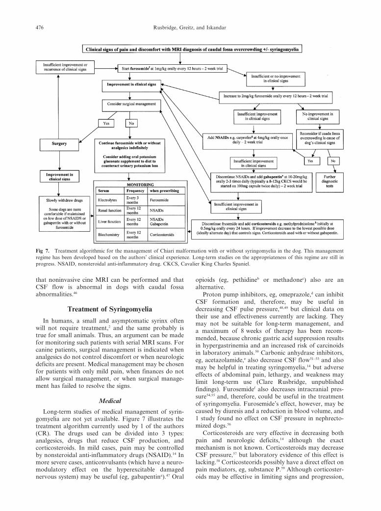
Treatment of Syringomyelia
In humans, a small and asymptomatic syrinx oftenwill not require treatment,2 and the same probably istrue for small animals. Thus, an argument can be madefor monitoring such patients with serial MRI scans. Forcanine patients, surgical management is indicated whenanalgesics do not control discomfort or when neurologicdeficits are present. Medical management may be chosenfor patients with only mild pain, when finances do notallow surgical management, or when surgical manage-ment has failed to resolve the signs.
Medical
Long-term studies of medical management of syrin-gomyelia are not yet available. Figure 7 illustrates thetreatment algorithm currently used by 1 of the authors(CR). The drugs used can be divided into 3 types:analgesics, drugs that reduce CSF production, andcorticosteroids. In mild cases, pain may be controlledby nonsteroidal anti-inflammatory drugs (NSAID).14 Inmore severe cases, anticonvulsants (which have a neuro-modulatory effect on the hyperexcitable damagednervous system) may be useful (eg, gabapentina).47 Oral
opioids (eg, pethidineb or methadonec) also are analternative.
Proton pump inhibitors, eg, omeprazole,d can inhibitCSF formation and, therefore, may be useful indecreasing CSF pulse pressure,48,49 but clinical data ontheir use and effectiveness currently are lacking. Theymay not be suitable for long-term management, anda maximum of 8 weeks of therapy has been recom-mended, because chronic gastric acid suppression resultsin hypergastrinemia and an increased risk of carcinoidsin laboratory animals.50 Carbonic anhydrase inhibitors,eg, acetazolamide,e also decrease CSF flow51–53 and alsomay be helpful in treating syringomyelia,14 but adverseeffects of abdominal pain, lethargy, and weakness maylimit long-term use (Clare Rusbridge, unpublishedfindings). Furosemidef also decreases intracranial pres-sure54,55 and, therefore, could be useful in the treatmentof syringomyelia. Furosemide’s effect, however, may becaused by diuresis and a reduction in blood volume, and1 study found no effect on CSF pressure in nephrecto-mized dogs.56
Corticosteroids are very effective in decreasing bothpain and neurologic deficits,14 although the exactmechanism is not known. Corticosteroids may decreaseCSF pressure,57 but laboratory evidence of this effect islacking.58 Corticosteorids possibly have a direct effect onpain mediators, eg, substance P.59 Although corticoster-oids may be effective in limiting signs and progression,
Fig 7. Treatment algorithmic for the management of Chiari malformation with or without syringomyelia in the dog. This management
regime has been developed based on the authors’ clinical experience. Long-term studies on the appropriateness of this regime are still in
progress. NSAID, nonsteroidal anti-inflammatory drug. CKCS, Cavalier King Charles Spaniel.
476 Rusbridge, Greitz, and Iskandar
most dogs require continuous therapy and subsequentlydevelop adverse effects, such as immunosuppression,weight gain, and skin changes.14
Surgical
Surgical management is indicated for dogs withrefractory pain or with worsening neurologic signs.The aim of surgery is to restore CSF dynamics, and ifthis effect can be achieved (eg, by removing or debulkinga tumor), then the syrinx can resolve.60 The mostcommon procedure for caudal-fossa overcrowding issuboccipital decompression in which most of thesupraoccipital bone and sometimes the cranial dorsallaminae of the atlas are removed (with or withouta durotomy) to decompress the foramen magnum.15,61–63
Success reported in the small number of dogs in thesestudies varied from no improvement62 to resolution ofclinical signs.15,63 The largest case series of 16 dogsfound that 81.25% of affected dogs had improvementor resolution of clinical signs after surgery, but 25%had a recurrence within the follow-up period. Thisreport also indicated that successful postoperative out-come was more likely if surgery was performed early inthe course of the disease.63 There have been no reports ofresolution of syringomyelia after suboccipital decom-pression in the dog, but this impression may, in part, befrom a lack of postoperative MRIs because of financialreasons. Syringosubarachnoid shunting also has beendescribed.64
The general principle for surgical management ofhumans with syringomyelia is that treatment should bedirected at the etiology of the syrinx.2 Suboccipitaldecompression is generally the first procedure of choicefor symptomatic humans with Chiari malformation, andit is widely acknowledged that direct draining of thesyrinx is not associated with a good long-term outcome,because stents or shunts become obstructed and canresult in tethering.2 Shunting to the subarachnoid spaceor to the pleural cavity is only indicated when the syrinxpersists or is progressive after suboccipital decompres-sion.2,65 There is great variation in the type ofsuboccipital decompression performed. One key argu-ment focuses on whether or not to open the dura. Mostsurgeons favor routine dural opening at surgery andclosure with a pericranial or synthetic patch graft.2,65 Theargument against dural opening is that this procedureincreases the complication rate. Advocates, however,argue that up to 55% of patients have extensive scarringand other obstructions to CSF flow that can only beidentified and removed by durotomy.66,67 Some recom-mend intraoperative ultrasound to tailor surgery to theindividual and to ensure that optimal CSF flow throughthe foramen magnum is achieved.68
Another crucial issue in surgery for Chiari malforma-tion is the number of patients requiring 2 or moresurgeries. Most large case series in human medicinereport a failure rate of at least 10%.68,69 Researchers havebeen unable to identify a single reason for surgicalfailure.65,66 Also of concern is the number of patientswith persistent pain after surgery. Approximately 41%
of humans with symptoms of dysesthesia who undergodecompressive surgery for syringomyelia have persis-tence or intensification of pain after surgery. Conven-tional medications, such as NSAIDS, opioids, andgabapentin, provide minimal or no relief, and, basedon the response to sympathetic blockade, pain isthought to be sympathetically mediated. Most humanpatients gradually improve over several months, but, inmany, persistent, unpleasant sensations persist.70 Aspreviously mentioned, damage to the dorsal horn andthe duration of signs are thought to be significant,35,39
and, in a study of persistent postoperative dysestheticpain, 84% of patients experienced extension of the syrinxinto the dorsolateral quadrant of the spinal cord on thesame side and level of the pain.70
In conclusion, our understanding of syringomyeliathat results from caudal fossa abnormalities, specifically,its pathogenesis and the exact relation to the clinicalsyndrome still is incomplete. The new theories based onphysical principles seem promising and may shed newlight on the pathophysiology of syringomyelia. Thisdisorder is an area of active research, and it is hoped thatthe recent description of a naturally occurring animalmodel of the condition in the CKCS will encourage theveterinary profession to contribute to the understandingof this debilitating condition.
Footnotes
a Neurontin 100 mg capsules, Pfizer Pharmaceuticals, Ltd, Vega
Baja, PRb Pethidine hydrochloride 50 mg, Martindale Pharmaceuticals,
Romford, Essex, UKc Methadone hydrochloride tablets USP 5 and 10 mg, Physeptone,
Martindale Pharmaceuticals, Romford, Essex, UKd Losec capsules 10, 20, and 40 mg; AstraZeneca, Hurdsfield
Industrial Estate, Macclesfield, Cheshire, UKe Diamox 250 mg tablets, Lederle Laboratories, Gosport, UKf Frusemide tablets 20 and 40 mg, Millpledge Pharmaceuticals,
Whinleys Estate, Church Lane, Clarborough, Retford, Notts, UKg Rimadyl Palatable tablets 20 and 50 mg, Pfizer Limited,
Sandwich, Kent, UKh Medrone tablets 2 and 4 mg, Pfizer Limited, Sandwich, Kent, UK
Acknowledgments
The authors thank Dr Mark Quigley of the De-partment of Astronomy, University of Wisconsin, forhis help and expertise in the preparation of Figure 5a,b.The authors also thank Dr Jan Rothuizen for his criticalappraisal of the manuscript.
References
1. Rusbridge C, Knowler SP. Inheritance of occipital bone
hypoplasia (Chiari type I malformation) in Cavalier King Charles
Spaniels. J Vet Intern Med 2004;18:673–678.
2. Medow J, Sansone J, Iskandar BJ. Syringomyelia and
hydromyelia. In: Albright AL, Pollack AF, Adelson PD, eds.
Current Concepts in Syringomyelia 477

Principles and Practice of Pediatric Neurosurgery, 2nd ed. New
York, NY: Thieme Medical Publishers. (in press)
3. Stephanus C. De dissectione partium corporis humani. Paris:
Colinaeum; 1545.
4. Ollivier d’angers CP. Taite de la moelle epiniere et de ses
maladies. Paris: Crevot; 1827:178–183.
5. Kahler O, Pick A. Beitrag zur Lehre von der Syringo-und
Hydromyelie. Vjschr Prakt Heilkd 1879;142:20–41.
6. Cahrabortty S, Tamaki N, Ehara K, et al. Experimental
syringomyelia in the rabbit: An ultrastructural study of spinal cord
tissue. Neurosurgery 1994;35:1112–1120.
7. Batzdorf U. A Brief history of syringomyelia. In: Tamaki N,
Batzdorf U, Nagashima T, eds. Syringomyelia: Current Concepts
in Pathogenesis and Management. Tokyo: Springer-Verlag;
2001:3–9.
8. Milhorat TH, Fox A, Todor DR. Pathology, classification,
and treatment of syringomyelia. In: Tamaki N, Batzdorf U,
Nagashima T, eds. Syringomyelia: Current Concepts in Pathogen-
esis and Management. Tokyo: Springer-Verlag; 2001:10–30.
9. West RJ, William B. Radiographic studies of the ventricles in
syringomyelia Neuroradiology 1980;20:5–16.
10. Chiari H. Ueber Veranderungen des Kleinhirns infolge von
Hydrocephalie des Grosshirns. Dtsch Med Wochenschir 1891;42:
1172–1175.
11. Chiari H. Ueber Veranderungen des Kleinhirns, des Pons
and der medulla oblongata in Folge von genitaler Hydrocephalie
des Grosshirns. Denkschr Akad Wiss Wien 1896;63:71–116.
12. Batzdorf U. Treatment of syringomyelia associated with
Chiari I malformation in syringomyelia. In: Tamaki N, Batzdorf
U, Nagashima T, eds. Syringomyelia: Current Concepts in
Pathogenesis and Management. Tokyo: Springer-Verlag; 2001:
121–123.
13. Williams B. Progress in syringomyelia. Neurol Res 1986;
8:130–145.
14. Rusbridge C, MacSweeny JE, Davies JV, et al. Syringomy-
elia in Cavalier King Charles Spaniels. J Am Anim Hosp Assoc
2000;36:34–41.
15. Dewey CW, Berg JM, Stefanacci JD, et al. Caudal occipital
malformation syndrome in dogs. Compend Contin Educ Pract Vet
2004;26:886–896.
16. Greitz D. Radiological assessment of hydrocephalus: new
theories and implications for therapy. Neurosurg Rev 2004;
27:145–165.
17. Gardner WJ, Goodall RJ. The surgical treatment of Arnold
Chiari malformation in adults. An explanation of its mechanism
and importance of encephalography in diagnosis. J Neurosurg
1950;7:199–206.
18. Oldfield EH, Murasko K, Shawker TH, et al. Pathophys-
iology of syringomyelia associated with Chiari I malformation of
the cerebellar tonsils. Implications for diagnosis and treatment.
J Neurosurg 1994;81:500–502.
19. Williams B. Cerebrospinal fluid changes in response to
coughing. Brain 1976;99:331–346.
20. Levine DN. The pathogenesis of syringomyelia associated
with lesions at the foramen magnum: A critical review of existing
theories and proposal of a new hypothesis. J Neurol Sci 2004;
220:3–21.
21. Ball MJ, Dayan AD. Pathogenesis of syringomyelia. Lancet
1972;II:799–801.
22. Ikata T, Masaki K, Kashiwaguchi S. Clinical and experi-
mental studies on permeability of tracers in normal spinal cord and
syringomyelia. Spine 1988;13:737–741.
23. Oldfield EH, DeVroom HL, Heiss JD. Hydrodynamics of
syringomyelia. In: Tamaki N, Batzdorf U, Nagashima T, eds.
Syringomyelia: Current Concepts in Pathogenesis and Manage-
ment. Tokyo: Springer-Verlag; 2001:75–89.
24. Heiss JD, Patronas N, DeVroom HL, et al. Elucidating
the pathophysiology of syringomyelia. J Neurosurg 1999;91:553–
562.
25. Williams B. Surgery for cerebello-medullary related syrin-
gomyelia. Adv Tech Stand Neurosurg 1993;20:107–164.
26. Greitz D, Ericson K, Flodmark O. Pathogenesis and
mechanics of spinal cord cysts: A new hypothesis based on
magnetic resonance studies of cerebrospinal fluid dynamics. Int J
Neuroradiol 1999;5:61–78.
27. Josephson A, Greitz D, Klason T, et al. A spinal thecal sac
constriction model supports the theory that induced pressure
gradients in the cord cause edema and cyst formation. Neurosur-
gery 2001;48:636–646.
28. Greitz D, Flodmark O. Modern concepts of syringohydro-
myelia. Riv Neuroradiol 2004;17:360–361.
29. Carpenter PW, Berkouk K, Lucey AD. Pressure wave
propagation in fluid-filled co-axial elastic tubes. Part 2: Mechan-
isms for the pathogenesis of syringomyelia. J Biomech Eng
2003;125:857–863.
30. Fischbein NJ, Dillon WP, Cobbs C, et al. The ‘‘presyrinx’’
state. A reversible myelopathic condition that may precede
syringomyelia AJNR Am J Neuroradiol 1999;20:7–20.
31. Iskandar BJ, Quigley M, Haughton VM. Foramen magnum
cerebrospinal fluid flow characteristics in children with Chiari 1
malformation before and after craniocervical decompression. J
Neurosurg (Paediatrics 2) 2004;101:169–178.
32. Hackel M, Benes V, Mohapl M. Simultaneous cerebral and
spinal fluid pressure recordings in surgical indications of the Chiari
malformation without myelodysplasia. Acta Neurochir (Wien)
2001;143:909–918.
33. Klekamp J. The pathophysiology of syringomyelia: Histor-
ical overview and current concept. Acta Neurochir (Wien)
2002;144:649–664.
34. Nakamura M, Chiba K, Nishizawa T, et al. Retrospective
study of surgery-related outcomes in patients with syringomyelia
associated with Chiari I malformation: Clinical significance of
changes in the size and localization of syrinx on pain relief. J
Neurosurg 2004(3 Suppl Spine):241–244.
35. Todor DR, Harrison TM, Millport TH. Pain and syringo-
myelia: A review. Neurosurg Focus 2000;8:1–6.
36. Lu D, Lamb CR, Pfeiffer DU, et al. Neurological signs and
results of magnetic resonance imaging in 40 cavalier King Charles
Spaniels with Chiari type 1 like malformations. Vet Rec 2003;
153:260–263.
37. Stanfa LC, Dickenson AH. In vivo electrophysiology of
dorsal-horn neurons. Methods Mol Med 2004;99:139–153.
38. Milhorat TH, Mu HT, LaMotte CC, et al. Distribution of
substance P in the spinal cord of patients with syringomyelia.
J Neurosurg 1996;84:992–998.
39. Van Biervliet J, de Lahunta A, Ennulat D, et al. Acquired
cervical scoliosis in six horses associated with dorsal grey column
chronic myelitis. Equine Vet J 2004;36:86–92.
40. Loder RT, Stasikelis P, Farley FA. Sagittal profiles of the
spine in scoliosis associated with an Arnold-Chiari malformation
with or without syringomyelia. J Pediatr Orthop 2002;22:483–491.
41. Skerritt JO, Skerritt GC. Hearing status of the Cavalier
King Charles Spaniel: A comparative study of healthy dogs and
those suffering from Arnold Chiari syndrome. BSAVA Congress
2001 Scientific Proceedings, British Small Animal Veterinary
Association, Woodrow House, 1 Telford Way, Waterwells Business
Park, Quedgeley, Gloucester, 2001;567.
42. Munro KJ, Cox CL. Investigation of hearing impairment in
Cavalier King Charles Spaniels using auditory brainstem response
audiometry. J Am Anim Hosp Assoc 1997;38:2–5.
43. Kumar A, Patni AH, Charbel F. The Chiari I malformation
and the neurotologist. Otol Neurotol 2002;23:727–735.
478 Rusbridge, Greitz, and Iskandar

44. Milhorat TH, Chou MW, Trinidad EM, et al. Chiari
malformation redefined: Clinical and radiographic findings for 363
symptomatic patients. Neurosurgery 1999;44:1005–1017.
45. Ventureyra EC, Aziz HA, Vassilyadi M. The role of cine
flow MRI in children with Chiari I malformation. Childs Nerv
Syst 2003;19:109–113.
46. March PA, Abramson CJ, Smith M, et al. CSF flow
abnormalities in caudal occipital malformation syndrome. Scien-
tific Proceedings from 23rd ACVIM Forum, Baltimore, American
College of Veterinary Internal Medicine, Lakewood, CO. 2005;
854–855.
47. Levendoglu F, Ogun CO, Ozerbil O, et al. Gabapentin is
a first line drug for the treatment of neuropathic pain in spinal cord
injury. Spine 2004;29:743–751.
48. Lindvall-Axelsson M, Nilsson C, Owman C, et al. In-
hibition of cerebrospinal fluid formation by omeprazole. Exp
Neurol 1992;115:394–399.
49. Javaheri S, Corbett WS, Simbartl LA, et al. Different effects
of omeprazole and Sch 28080 on canine cerebrospinal fluid
production. Brain Res 1997;754:321–324.
50. Berlin RG. Omeprazole. Gastrin and gastric endocrine cell
data from clinical studies. Dig Dis Sci 1991;36:129–136.
51. Vogh BP. The relation of choroid plexus carbonic
anhydrase activity to cerebrospinal fluid formation: Study of three
inhibitors in cat with extrapolation to man. J Pharmacol Exp Ther
1980;213:321–331.
52. Shinnar S, Gammon K, Bergman EW Jr, et al. Management
of hydrocephalus in infancy: Use of acetazolamide and furosemide
to avoid cerebrospinal fluid shunts. J Pediatr 1985;107:31–37.
53. Carrion E, Hertzog JH, Medlock MD, et al. Use of
acetazolamide to decrease cerebrospinal fluid production in
chronically ventilated patients with ventriculopleural shunts. Arch
Dis Child 2001;84:68–71.
54. Artru AA, Powers KM. Furosemide decreases cerebrospinal
fluid formation during desflurane anesthesia in rabbits. J Neurosurg
Anesthesiol 1997;9:166–174.
55. Lorenzo AV, Hornig G, Zavala LM, et al. Furosemide
lowers intracranial pressure by inhibiting CSF production. Z
Kinderchir 1986;41(Suppl 1):10–12.
56. Pinegin LE, Dolzhenko DA, Natochin IuV. Mechanism of
the decrease in intracranial pressure as affected by furosemide.
Biull Eksp Biol Med 1984;98:682–685.
57. Simpson ST. Hydrocephalous. In: Kirk RW, ed. Current
Veterinary Therapy X. Philadelphia: WB Saunders; 1989:842–847.
58. Vela AR, Carey ME, Thompson BM. Further data on the
acute effect of intravenous steroids on canine CSF secretion and
absorption. J Neurosurg 1979;50:477–482.
59. Wong HK, Tan KJ. Effects of corticosteroids on nerve root
recovery after spinal nerve root compression. Clin Orthop 2002;
403:248–252.
60. da Costa RC, Parent JM, Poma R, et al. Cervical
syringohydromyelia secondary to a brainstem tumor in a dog.
J Amer Vet Med Assoc 2004;225:1061–1064.
61. Churcher RK, Child G. Chiari 1/syringomyelia complex in
a King Charles Spaniel. Aust Vet J 2000;78:92–95.
62. Vermeersch K, Van Ham, Caemaert J, et al. Suboccipital
craniectomy, dorsal laminectomy of C1, durotomy and dural graft
placement as a treatment for syringohydromyelia with cerebellar
tonsil herniation in Cavalier King Charles Spaniels. Vet Surg
2004;33:355–360.
63. Dewy CW, Berg JM, Barone G, et al. Treatment of caudal
occipital malformation syndrome in dogs by foramen magnum
decompression. Scientific Proceedings from 23rd ACVIM Forum,
Baltimore, American College of Veterinary Internal Medicine,
Lakewood, CO, 2005;854.
64. Skerrit GC, Hughes D. A syndrome of syringomyelia in the
cavalier King Charles spaniel, and its treatment by syringo-
subarachnoid shunting. Proceedings from the 12th Annual
Symposium of the European Society of Veterinary Neurology,
Vienna September 25–26, 1998;23.
65. Schijman E, Steinbok P. International survey on the
management of Chiari I malformation and syringomyelia. Childs
Nerv Syst 2004;20:341–348.
66. Muraszko KM, Ellenbogen RG, Mapstone TB. Controver-
sies in the surgical management of Chiari I malformations: What is
the surgical procedure of choice? To open dura or not to open
dura? Clin Neurosurg 2004;51:241–247.
67. Milhorat TH, Bolognese PA. Tailored operative technique
for Chiari type I malformation using intraoperative color Doppler
ultrasonography. Neurosurgery 2003;53:899–905.
68. Sacco D, Scott RM. Reoperation for Chiari malformations.
Pediatr Neurosurg 2003;39:171–178.
69. Tubbs RS, McGirt MJ, Oakes WJ. Surgical experience in
130 pediatric patients with Chiari I malformations. J Neurosurg
2003;99:291–296.
70. Milhorat TH, Kotzen RM, Mu HT, et al. Dysesthetic pain
in patients with syringomyelia. Neurosurgery 1996;38:940–946.
Current Concepts in Syringomyelia 479