Syringomyelia Associated With Arnold-Chiari Malformation
7
Henry Ford Hospital Medical Journal Volume 8 | Number 2 Article 9 6-1960 Case Report: Syringomyelia Associated With Arnold-Chiari Malformation Hermenegildo F. Labsan Lorne D. Proctor Follow this and additional works at: hps://scholarlycommons.henryford.com/hmedjournal Part of the Life Sciences Commons , Medical Specialties Commons , and the Public Health Commons is Part I is brought to you for free and open access by Henry Ford Health System Scholarly Commons. It has been accepted for inclusion in Henry Ford Hospital Medical Journal by an authorized editor of Henry Ford Health System Scholarly Commons. For more information, please contact [email protected]. Recommended Citation Labsan, Hermenegildo F. and Proctor, Lorne D. (1960) "Case Report: Syringomyelia Associated With Arnold-Chiari Malformation," Henry Ford Hospital Medical Bulletin : Vol. 8 : No. 2 , 157-162. Available at: hps://scholarlycommons.henryford.com/hmedjournal/vol8/iss2/9
-
Upload
khangminh22 -
Category
Documents
-
view
0 -
download
0
Transcript of Syringomyelia Associated With Arnold-Chiari Malformation
Henry Ford Hospital Medical Journal
Volume 8 | Number 2 Article 9
6-1960
Case Report: Syringomyelia Associated WithArnold-Chiari MalformationHermenegildo F. Labsan
Lorne D. Proctor
Follow this and additional works at: https://scholarlycommons.henryford.com/hfhmedjournal
Part of the Life Sciences Commons, Medical Specialties Commons, and the Public HealthCommons
This Part I is brought to you for free and open access by Henry Ford Health System Scholarly Commons. It has been accepted for inclusion in HenryFord Hospital Medical Journal by an authorized editor of Henry Ford Health System Scholarly Commons. For more information, please [email protected].
Recommended CitationLabsan, Hermenegildo F. and Proctor, Lorne D. (1960) "Case Report: Syringomyelia Associated With Arnold-Chiari Malformation,"Henry Ford Hospital Medical Bulletin : Vol. 8 : No. 2 , 157-162.Available at: https://scholarlycommons.henryford.com/hfhmedjournal/vol8/iss2/9
C A S E R E P O R T
SYRINGOMYELIA ASSOCIATED WITH ARNOLD-CHIARI
MALFORMATION
HERMENEGILDO F . LABSAN, M.D.* AND LORNE D . PROCTOR, M.D.*
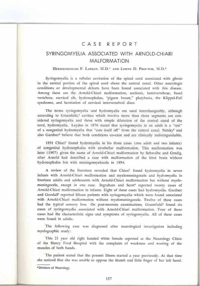
Syringomyelia is a tubular cavitation of the spinal cord associated with gliosis in the central portion of the spinal cord about the central canal. Other neurologic conditions or developmental defects have been found associated with this disease. Among these are the Arnold-Chiari malformation, scoliosis, hemivertebrae, fused vertebrae, cervical rib, hydrocephalus, "pigeon breast," platybasia, the Klippel-Feil syndrome, and herniation of cervical intervertebral discs.
The terms syringomyelia and hydromyelia are used interchangeably, although according to Greenfield,^ cavities which involve more than three segments are considered syringomyelia and those with simple dilatation of the central canal of the cord, hydromyelia. Leyden in 1876 stated that syringomyelia in an adult is a "rest" of a congenital hydromyelia that "cuts itself off" from the central canal. Netsky' and also Gardner^ believe that both conditions co-exist and are clinically indistinguishable.
1891 Chiari' found hydromyelia in his three cases (one adult and two infants) of congenital hydrocephalus with cerebellar malformation. This malformation was later (1907) given the name of Arnold-Chiari malformation by Schwalbe and Gredig, after Arnold had described a case with malformation of the hind brain without hydrocephalus but with meningomyelocele in 1894.
A review of the literature revealed that Chiari' found hydromyelia in seven infants with Arnold-Chiari malformation and myelomeningocele and hydromyelia in fourteen adults and adolescents with Arnold-Chiari malformation but without myelomeningocele, except in one case. Ingraham and Scott* reported twenty cases of Amold-Chiari malformation in infants. Eight of these cases had hydromyelia. Gardner and GoodalP reported fifteen patients with syringomyelia which were found associated with Amold-Chiari malformation without myelomeningocele. Twelve of these cases had the typical sensory loss. On post-mortem examinations, Greenfield' found six cases of syringomyelia associated with Arnold-Chiari malformation. Four of these cases had the characteristic signs and symptoms of syringomyelia. Al l of these cases were found in adults.
The following case was diagnosed after neurological investigation including myelographic study:
This 21 year old right handed white female reported at the Neurology Clinic of the Henry Ford Hospital with the complaint of weakness and wasting of the muscles of both hands.
The patient stated that the present illness started a year previously. At that time she noticed that she was unable to oppose the thumb and little finger of her left hand.
*Division of Neurology.
157

Labsan and Proctor
This condition gradually became worse so that in a few months she developed difficulty in extending her fingers. A few months later, this was followed by similar difficulty in the right hand. The weakness and wasting of the muscles of both hands gradually, but progressively became worse so that six months prior to our examination she had extreme difficulty holding objects with her left hand.
The past history reveals that she had a "bad fall" when she was eleven years old, and at that time she was found to have a "curvature of the spine". She considered herself a moderate alcoholic drinker. Family history reveals that her father is in a mental institution with a diagnosis of dementia praecox. There is no other nervous disease or mental disorder in the family.
The physical and neurological examination revealed a well-developed, moderately-nourished, ambulatory, alert and cooperative female in no acute distress. The presence of scoliosis was very conspicuous. Blood pressure was 136/80. Her gait was normal. Vestibular and coordination tests were well performed. There were occasional fasciculations observed in the left thenar muscles. There was marked weakness and atrophy of the intrinsic muscles of both hands, more so on the left, producing a "claw-hand" deformity. The left biceps and both deltoid muscles were also slightly weak, without
Figure 1 TOWNE'S VIEW OF SKULL SHOWING ENLARGED FORAMEN MAGNUM
158

Syringomyelia
evidence of atrophy. The deep reflexes were symmetrically diminished to absent in the upper extremities. The superfical abdominal reflexes were absent. On plantar stimulation, an equivocal extensor response was obtained on the right, but no response was obtained on the left. No Hoffman sign was present. There were no sensory changes. The cranial nerves were grossly intact.
Routine complete blood count and urinalysis were normal. Blood sugar was 85mg/100ml. Cerebrospinal fluid revealed 16mg/100ml of protein. Skull x-ray showed enlargement of the foramen magnum (Figure 1). Spine x-rays showed scoliosis of the mid-dorsal spine to the right with no intrinsic bone abnormality noted.
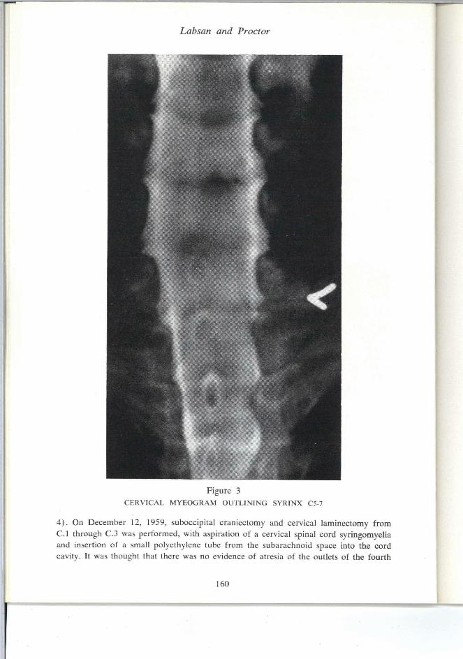
The patient was presented before a combined Neurology-Neurosurgery conference, and an early atypical amyotrophic lateral sclerosis and syringomyelia were considered in the differential diagnosis. It was suggested there was a possible basilar impression noted in the lateral skull film (Figure 2). On December 10, 1959 myelography was performed which revealed marked widening of the spinal cord at the cervical region, particularly marked from C.5 through C.7 (Figure 3). Bilateral extramedullary, intradural type defects were also found located posteriorly with their inferior margins at the C.1-2 interspace. These were thought to represent the cerebellar tonsils (Figure
Figure 2
LATERAL FILM OF SKULL SHOWING A SUGGESTION OF BASILAR IMPRESSION
159
Labsan and Proctor
Figure 3
CERVICAL MYEOGRAM OUTLINING SYRINX C5-7
4) . On December 12, 1959, suboccipital craniectomy and cervical laminectomy from C.l through C.3 was performed, with aspiration of a cervical spinal cord syringomyelia and insertion of a small polyethylene tube from the subarachnoid space into the cord cavity. It was thought that there was no evidence of atresia of the outlets of the fourth
160

Syringomyelia
ventricle. There was herniation of the cerebellar tonsils which was characteristic of Chiari's Type I malformation.
Figure 4
CERVICAL MYELOGRAM REVEALING ARNOLD-CHIARI TYPE I DEFORMITY NOTE OUTLINE OF CEREBELLAR TONSILS (COMPARE WITH ADJACENT DIAGRAM)
Post-operative course was uneventful and the patient was discharged on December 19, 1959, the neurological condition essentially unchanged.
DISCUSSION
It is noteworthy that this patient presented weakness and atrophy of the intrinsic muscles of both hands without sensory change. This would suggest damage to the posterolateral cell group of the ventral horns of the cervical cord. The lack of dissociated anesthesia probably indicates that the spino-thalamic fibers have to the present escaped damage. We feel that the cavitation arose from the proliferation of glial cells in the region of the central canal of the spinal cord within embryonic rests related
161
Lab can a:id Proctor
to the imperfect closure of the neural tube. The origin of this particular case of syringomyelia as a malformation is supported by the presence of another developmental anomaly, the Arnold-Chiari malformation. In this particular type of hind brain deformity, which is Chiari's Type I , ' - ' - ' there is only the herniation of the cerebellar tonsils. This deformity is frequently called "pressure coning", and this is the type which is most frequently found associated with syringomyelia or hydromyelia in adults.
This case would appear to conform to Netsky's^ conception, that a "dysplastic" person likely has syrinx if spinal cord symptoms develop.
SUMMARY
A case of syringomyelia without typical sensory changes, associated with Arnold-Chiari malformation, was found in an adult. The presence of dysplasia in a patient with spinal cord symptoms should arouse the suspicion of syringomyelia.
REFERENCES
1. Chiari, H.: Ueber Veranderungen des Kleinhirns, des Pons und der Medulla Oblongata infolge von Congenitaler Hydrocephalic des Grosshirns, Deutsche med. Wchnschr 27:1172, 1891.
2. Gardner, W. J.: Anatomic anomalies common to myelomeningocele of infancy and syringomyelia of adulthood suggest a common origin, Cleveland Clin. Quart. 26:118, 1959.
3. Greenfield, J. G.: Neuropathology, London, Edward Arnold, 1958, pp. 308-316.
4. Ingraham, F. D., and Scott, H. W., Jr.: Spina bifida and cranium bifidum; Arnold-Chiari malformation; study of 20 cases, New England J. Med. 229:108, 1943.
5. Netsky, M. G.: Syringomyelia; clinicopathologic study, A.M.A. Arch. Neurol & Psychiat. 70:741, 1953.
162