Arnold A. - 1984 Determination of Salts from Monuments
10
Determination of mineral salts from monuments A. Arnold Studies in Conservation 29 (1984) 129-138 129 DETERMINATION OF MINERAL SALTS FROM MONUMENTS A. Arnold Abstract - The salts occurring on stones, mortars and mural paintings are rarely analysed because simple and high-speed methods are lacking. A method combining optical mineralogy with classic microchemical slide tests is described. 1 Introduction The study of weathering requires appropriate methods to determine the soluble salts involved in decay of stones, mortars and wall paintings in monuments. Normally, scientists try to analyse them by means of sophisticated modern analytical methods, such as electron microscopy or X-ray diffraction, which are often expensive and time-consuming. Thus the saline systems present in ancient walls are very rarely analysed in practice. That would be possible only if simple and high-speed methods were available. Such a method will be described in this article. In this method, the mineral salts are identified by means of optical mineralogy in combination with microchemical slide tests. With respect to microchemical slide tests, Chamot and Mason [1] write that: '…qualitative chemical analyses, when made by microscopical methods, enable the analyst to obtain results in a minimum of time, upon a minimum of material, with the expenditure of little labor and at a wholly negligible cost, and all with no sacrifice of accuracy.' This statement is still entirely valid. Most of the mineral salts may be determined with a combination of optical and microchemical tests. Sometimes X-ray diffraction is used as a complementary method, particularly in cases where the results are uncertain. The quantitative chemical analyses of the elements present in salts from walls, stones and mortars will not be dealt with here, because these determinations are routine in chemical laboratories dealing with water analyses. Our analyses concern saline systems occurring on buildings and monuments which are associated with particular weathering situations. Analytical work should commence by consideration of the building or stonework; the occurrence of salts is related to specific weathering phenomena and to particular microclimatic and hydrological conditions. Samples should be taken in Received 9 June 1983 consideration of these facts and in accordance with the aim of the investigation. Since some indications may be obtained by initial tests performed on the building, it is important to have a fundamental knowledge of the salts in porous building materials. 2 Occurrence of salts on monuments Salts in porous building materials exist as aqueous solutions or crystallized in the pores and on the surface. They are strongly connected with water migration in walls. They become concentrated where water moves in one direction and evaporates. That is the case in the upper zone of rising damp [2], laterally to water running along the surface, and in zones where water reaches a surface (or a surface zone) and evaporates after percolating through stones and mortars. In such areas salts are precipitated when the solution becomes saturated with respect to a particular salt. In areas where salt solutions evaporate, concentration gradients are developed. The different solutes are precipitated at different places as the solution moves further on. Thus the crystalline phases are deposited in sequences according to their respective solubilities, while the most soluble solutes are concentrated in the remaining solution. The more soluble the salts, the more hygroscopic they tend to be. Thus the precipitation depends greatly on the relative humidity of the surrounding atmosphere [3, 4]. Very often, the most soluble portion will never be able to crystallize in our temperate and humid climate in Switzerland, because the relative humidity of the air is always too high to allow it. That means that in most cases both the crystalline salts and the solutes are simultaneously present in and on the stones, mortars and paintings of walls. Complications are introduced when alkaline salts are produced on ancient walls by modern alkaline building materials such as Portland cement or waterglass products. Salts from these materials will reach the original ones and react with them. If not, salts which cannot coexist theoretically may be found side by side [3, 4], because they have not come into contact with each other. This indicates that saline systems in walls may be complicated, and that their study needs a certain knowledge of weathering as well as of the microclimatic and hydrological situations of buildings. If
Transcript of Arnold A. - 1984 Determination of Salts from Monuments
Determination of mineral salts from monuments A. Arnold
Studies in Conservation 29 (1984) 129-138 129
DETERMINATION OF MINERAL SALTS FROM MONUMENTS A. Arnold
Abstract - The salts occurring on stones, mortars and mural paintings are rarely analysed because simple and high-speed methods are lacking. A method combining optical mineralogy with classic microchemical slide tests is described.
1 Introduction
The study of weathering requires appropriate methods to determine the soluble salts involved in decay of stones, mortars and wall paintings in monuments.
Normally, scientists try to analyse them by means of sophisticated modern analytical methods, such as electron microscopy or X-ray diffraction, which are often expensive and time-consuming. Thus the saline systems present in ancient walls are very rarely analysed in practice. That would be possible only if simple and high-speed methods were available. Such a method will be described in this article.
In this method, the mineral salts are identified by means of optical mineralogy in combination with microchemical slide tests. With respect to microchemical slide tests, Chamot and Mason [1] write that:
'…qualitative chemical analyses, when made by microscopical methods, enable the analyst to obtain results in a minimum of time, upon a minimum of material, with the expenditure of little labor and at a wholly negligible cost, and all with no sacrifice of accuracy.' This statement is still entirely valid. Most of the mineral salts may be determined with a combination of optical and microchemical tests. Sometimes X-ray diffraction is used as a complementary method, particularly in cases where the results are uncertain.
The quantitative chemical analyses of the elements present in salts from walls, stones and mortars will not be dealt with here, because these determinations are routine in chemical laboratories dealing with water analyses.
Our analyses concern saline systems occurring on buildings and monuments which are associated with particular weathering situations. Analytical work should commence by consideration of the building or stonework; the occurrence of salts is related to specific weathering phenomena and to particular microclimatic and hydrological conditions. Samples should be taken in
Received 9 June 1983
consideration of these facts and in accordance with the aim of the investigation. Since some indications may be obtained by initial tests performed on the building, it is important to have a fundamental knowledge of the salts in porous building materials.
2 Occurrence of salts on monuments
Salts in porous building materials exist as aqueous solutions or crystallized in the pores and on the surface. They are strongly connected with water migration in walls. They become concentrated where water moves in one direction and evaporates. That is the case in the upper zone of rising damp [2], laterally to water running along the surface, and in zones where water reaches a surface (or a surface zone) and evaporates after percolating through stones and mortars. In such areas salts are precipitated when the solution becomes saturated with respect to a particular salt.
In areas where salt solutions evaporate, concentration gradients are developed. The different solutes are precipitated at different places as the solution moves further on. Thus the crystalline phases are deposited in sequences according to their respective solubilities, while the most soluble solutes are concentrated in the remaining solution. The more soluble the salts, the more hygroscopic they tend to be. Thus the precipitation depends greatly on the relative humidity of the surrounding atmosphere [3, 4]. Very often, the most soluble portion will never be able to crystallize in our temperate and humid climate in Switzerland, because the relative humidity of the air is always too high to allow it.
That means that in most cases both the crystalline salts and the solutes are simultaneously present in and on the stones, mortars and paintings of walls.
Complications are introduced when alkaline salts are produced on ancient walls by modern alkaline building materials such as Portland cement or waterglass products. Salts from these materials will reach the original ones and react with them. If not, salts which cannot coexist theoretically may be found side by side [3, 4], because they have not come into contact with each other.
This indicates that saline systems in walls may be complicated, and that their study needs a certain knowledge of weathering as well as of the microclimatic and hydrological situations of buildings. If
Determination of mineral salts from monuments A. Arnold
Studies in Conservation 29 (1984) 129-138 130
the whole saline system of a wall has to be studied, a considerable number of crystalline salts and pore solutions from different places on and perpendicular to the surface need to be sampled and analysed. In most practical cases, such extended studies are not possible but, if some indications for restoration work are needed, a few well-chosen samples of crystalline salts and of stones and mortars may be very valuable, when correctly analysed and interpreted.
3 Sampling and field tests
Samples should be taken from crystalline salts and, if necessary, from the stones and mortars likely to contain saline solutions. Very often a few milligrams of pure salt will suffice to determine its mineral species by means of optical mineralogy and microchemical tests, but it is better to obtain larger quantities, especially when small amounts in mixtures have to be identified. On the other hand, small but pure samples are preferable to large, contaminated ones.
With minimal efflorescence, salts may be visible only under raking light. They are then collected by carefully brushing the crystals into a small plastic bag using a soft brush. Thick efflorescences may be scraped off with the edge of the small plastic bag. Crusts and very thick efflorescences should be carefully detached with a knife.
The samples should always be kept in closed receptacles such as closable plastic bags. During transport to the laboratory they have to be protected from warmth to avoid any dehydration.
During sampling, some field tests may give valuable indications of the nature and the occurrence of saline minerals. Some of them may be recognized by their taste, but not everyone will enjoy doing this. A pH test paper allows the detection of alkaline salts on the wall. Highly hydrated salts such as mirabilite (Na2SO4*10H2O) and natron (Na2CO3*10H2O) will dehydrate and dissolve in their own water of crystallization when held for a while between thumb and finger. Such simple tests should permit the detection of natron by its alkaline reaction and its capacity to dehydrate to thermonatrite (Na2CO3*H2O). These tests may also help to obtain the right samples when certain facts are most relevant; it may be important, for example, to find alkali carbonates which are indicative of the use of Portland cement or waterglass products for restoration.
All the samples should be analysed as soon as possible. This is decisive above all when the crystal habit of the saline minerals has to be observed, since acicular crystals, for example, will transform to more isometric ones if they are exposed to humid air, even if they are kept in closed bags.
4 Identification in the laboratory
It is very useful to assemble a collection of authentic specimens of all the salts listed in Table 2 and to keep them near the working place in order to have them available immediately when needed for comparison. Most of them are commercially available.
Permanent micromounts can be made for microscopic reference. They may be embedded in a suitable medium such as Eukitt (from Leitz) with a refractive index, nD, Of 1.53. Highly hydrated salts will not be stable and will dehydrate if the slides or receptacles are not kept in a cool place (such as a refrigerator).
For X-ray diffraction, a similar reference collection is suitable and easy to prepare. Special precautions are needed for hydrated salts. They have to be scaled in closed capillaries before being placed in the X-ray diffraction camera in order to avoid dehydration due to camera evacuation. If not, thenardite (Na2SO4), for example, will be identified instead of mirabilite (Na2SO4*10H2O), and kieserite (MgSO4*H2O) instead of epsomite (MgSO4*7H2O).
It is obvious that microscopy and microchemistry should not be carried out in the same room, since the microscope would be seriously damaged by corrosion due to the vapours from the acids used for slide tests.
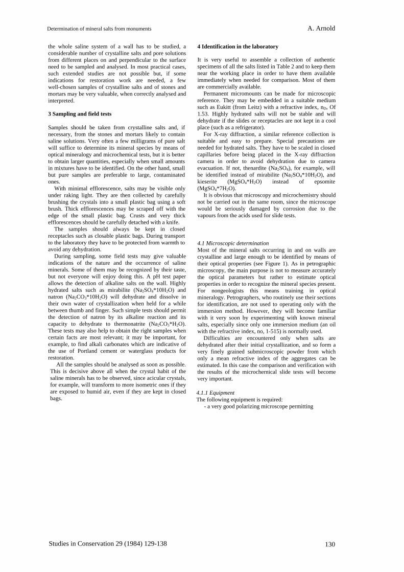
4.1 Microscopic determination Most of the mineral salts occurring in and on walls are crystalline and large enough to be identified by means of their optical properties (see Figure 1). As in petrographic microscopy, the main purpose is not to measure accurately the optical parameters but rather to estimate optical properties in order to recognize the mineral species present. For nongeologists this means training in optical mineralogy. Petrographers, who routinely use their sections for identification, are not used to operating only with the immersion method. However, they will become familiar with it very soon by experimenting with known mineral salts, especially since only one immersion medium (an oil with the refractive index, no, 1-515) is normally used.
Difficulties are encountered only when salts are dehydrated after their initial crystallization, and so form a very finely grained submicroscopic powder from which only a mean refractive index of the aggregates can be estimated. In this case the comparison and verification with the results of the microchernical slide tests will become very important.
4.1.1 Equipment The following equipment is required:
- a very good polarizing microscope permitting

Determination of mineral salts from monuments A. Arnold
Studies in Conservation 29 (1984) 129-138 131
Table I Maximum and minimum refractive indices for saline minerals occurring on buildings, compared with the refractive index on an immersion oil with nD = 1-515
Determination of mineral salts from monuments A. Arnold
Studies in Conservation 29 (1984) 129-138 132
Figure 1 Nitronatrite (NaNO3) in a polarizing microscope. The typical distorted needles from fresh efflorescence are shown in normal polarized light on the left and between crossed polarizers on the right. Here the high double refraction is clearly visible. Magnification: 1:53. (Photo by K. Zehnder.)
orthoscopic and conoscopic observation; - normal glass slides and cover glasses; - immersion oil such as that from Zeiss with no 1-515;
- relevant literature [5-7].
4.1.2 Procedure and preparation of the micromounts The investigation is best begun by inspecting the sample with a binocular microscope in order that the pure salts may be picked out from impurities and mixtures of different salts may be recognized. The specimen for the microscopic determination may be chosen from a pure salt or from mixtures of salts. It is also advantageous to clean the salt from impurities and to check the specimen on the glass slide before covering it with a cover glass. Too much material on the slide should be avoided, to allow the single minerals to be seen side by side and not overlapping.
4.1.3 The immersion method for identification For our purpose, the immersion oil with refractive index, nD, 1-515 is suitable because it permits the discrimination of many mineral salts. This is shown in Table 1 where the maximum and minimum refractive indices of the salts are listed and compared to that of the immersion oil.
It is easy to see that those minerals with refractive indices above that of the immersion oil may be distinguished from those having all of them below it or from those having one below and the others above it. Moreover, it can be seen whether or not the refractive index of a particular salt grain is near to that of the immersion oil by its optical relief (appearance of the contours).
In addition to this, other optical properties such as double refraction, the relationship between optical
and crystallographic orientations, and anomalous interference colours may be important as distinguishing features.
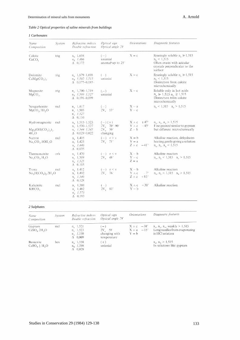
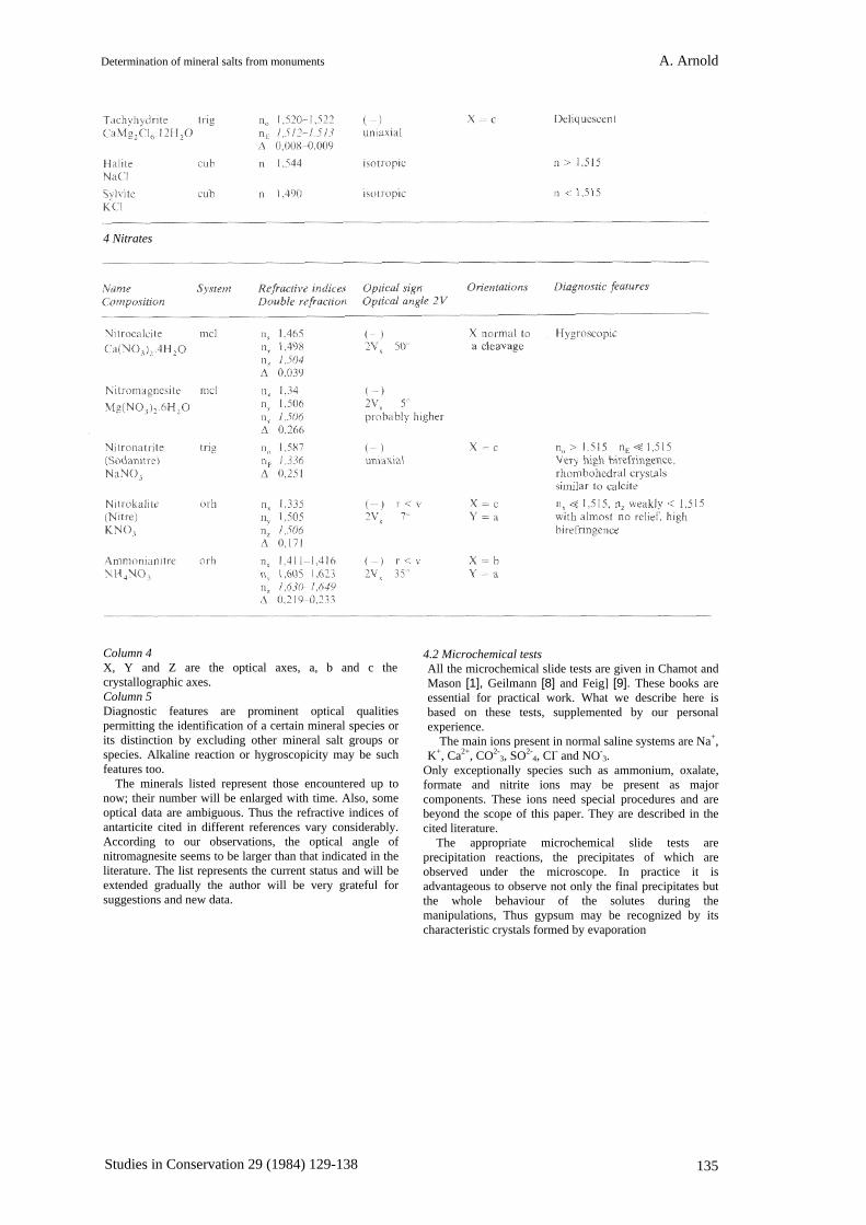
4.1.4 The optical properties of mineral salts occurring on walls (notes to Table 2)
The optical properties taken from the literature [5-7] are listed in Table 2. This table represents the range of the identified mineral salts on walls. Salts like calcium and magnesium formates or sodium acetate, found occasionally and introduced artificially, are not described here.
The nomenclature from Troeger [6] is used. The following notes are given as clarification of the properties listed in the columns of Table 2.
Column 1 The name of the mineral and its chemical composition are given as well as the crystal systems. The following abbreviations are used: cub = cubic, tetr = tetragonal, trig = trigonal, hex = hexagonal, orh = orthorhombic, mcl = monoclinic and trcl = triclinic.
Column 2 nx, <ny <nz are the three main refractive indices of triclinic, monoclinic and orthorhombic crystals, no and nE are the refractive indices of the ordinary and extraordinary wave of hexagonal, trigonal and tetragonal crystals, while n is the refractive index of cubic (i.e. isotropic) crystals. A is the value of double refraction: nz - nx or no - nE or nE - no.
Column 3 The optical sign and the optical angle are given as well as the dispersion of the optical angle: r < v or r > v.
Determination of mineral salts from monuments A. Arnold
Studies in Conservation 29 (1984) 129-138 133
Table 2 Optical properties of saline minerals from buildings 1 Carbonates
2 Sulphates

Determination of mineral salts from monuments A. Arnold
Studies in Conservation 29 (1984) 129-138 134
3 Chlorides
Determination of mineral salts from monuments A. Arnold
Studies in Conservation 29 (1984) 129-138 135
4 Nitrates
Column 4 X, Y and Z are the optical axes, a, b and c the crystallographic axes. Column 5 Diagnostic features are prominent optical qualities permitting the identification of a certain mineral species or its distinction by excluding other mineral salt groups or species. Alkaline reaction or hygroscopicity may be such features too.
The minerals listed represent those encountered up to now; their number will be enlarged with time. Also, some optical data are ambiguous. Thus the refractive indices of antarticite cited in different references vary considerably. According to our observations, the optical angle of nitromagnesite seems to be larger than that indicated in the literature. The list represents the current status and will be extended gradually the author will be very grateful for suggestions and new data.
4.2 Microchemical tests All the microchemical slide tests are given in Chamot and Mason [1], Geilmann [8] and Feig] [9]. These books are essential for practical work. What we describe here is based on these tests, supplemented by our personal experience.
The main ions present in normal saline systems are Na+, K+, Ca2+, CO2-
3, SO2-4, CI- and NO-
3. Only exceptionally species such as ammonium, oxalate, formate and nitrite ions may be present as major components. These ions need special procedures and are beyond the scope of this paper. They are described in the cited literature.
The appropriate microchemical slide tests are precipitation reactions, the precipitates of which are observed under the microscope. In practice it is advantageous to observe not only the final precipitates but the whole behaviour of the solutes during the manipulations, Thus gypsum may be recognized by its characteristic crystals formed by evaporation

Determination of mineral salts from monuments A. Arnold
Studies in Conservation 29 (1984) 129-138 136
from the circumference of a drop of water, or sodium and potassium chloride by the cubic crystals formed under the same conditions. Magnesium salts tend to form glassy-looking rims when the water is evaporated. These and other observations are essential for rapid work but are not sufficient.
4.2.1 Equipment A good chemical microscope with transmitted or reflected illumination is necessary. In the author's laboratory a Leitz binocular microscope with 'Ultropak' dark field illuminator, an objective 11 X 0.25 and 10x eyepieces is used. The magnification of 110x is sufficient. The distance between the front lens of the objective and the object is 5.6mm and this permits manipulations under the microscope.
It is obvious that the objective should be resistant to acid vapours. The metallic parts may be protected with Vaseline.
Our heating plate to evaporate the drops of solution is a home-made circular copper plate with a diameter of about 2cm, fixed on a 25W soldering bit. A variable transformer is useful.
Miscellaneous materials include glass slides with a cavity, micro pipettes, and capillary pipettes drawn from glass tubes. Small glass rods should be shaped for manipulations under the microscope.
The relevant solvents and reagents are listed in Table 3. Details for making the reagents can be found in the appropriate literature [1, 8-10].
Table 3 Solvents and reagents for microchemical tests on salts
Solvents
Reagents
4.2.2 Preparation of the samples Normally the samples are dissolved in deionized water and the dissolved part is separated from the residue by a capillary pipette and transferred to a clean slide. From this drop the different portions are
taken for the slide tests. The first test to perform is the pH test with a test paper. If the solution is alkaline, alkali- carbonates or hydroxides or calcium or magnesium hydroxides are present.
Full details of further operations are given in the Handbook of Chemical Microscopy [1] but some tips from our experience are of value and are given below.
4.2.3 Detection of sodium and potassium Reagent: bismuth sulphate in concentrated sulphuric acid. Compounds formed: 3Na2SO4*2Bi2(SO4)3*2H2O and K2SO4*Bi2(SO4)3*2H20
A drop of the aqueous solution to be tested is evaporated to dryness and a drop of concentrated sulphuric acid is added. A drop of bismuth sulphate solution is introduced into this drop from the side.
If sodium is present, sodium bismuth sulphate prisms and needles will appear first on the contact between the drops. If potassium is present, hexagonal plates and rosettes will form preferentially near the circumference of the drop. These crystals are iridescent when observed in reflected light. Both sodium and potassium may be detected together.
Pure bismuth sulphate also gives hexagonal plates when drying. It is wise to compare a blank between crossed polarizers. When gypsum is present in large quantities, similar prisms and needles will be formed in sulphuric acid. However, gypsum may be recognized when the initial drop is drying, and characteristic prisms and needles are formed without adding bismuth sulphate.
4.2.4 Detection of calcium Reagent: dilute sulphuric acid. Compound formed: CaSO4*2H2O.
When calcium is present as sulphate it will be recognized when the drop of aqueous solution dries. Prisms are formed in nitric acid, long needles in hydrochloric acid.
Calcium present in nitrates and chlorides is detected by adding 2N sulphuric acid to the test drop. Gypsum will be formed and recognized.
4.2.5 Detection of magnesium Reagent: sodium or ammonium phosphates. Compound formed: MgNH4PO4*6H2O.
The drop to be tested is acidified with very little hydrochloric acid and a drop of sodium or ammonium phosphate is added. A drop of NH4OH is introduced from the side into the test drop. When magnesium is present, characteristic crystals will form, first as prisms (recalling roofs of houses), then dendritic in appearance. Better crystals may grow if the drop is exposed to ammonia vapours from concentrated ammonium hydroxide.

Determination of mineral salts from monuments A. Arnold
Studies in Conservation 29 (1984) 129-138 137
4.2.8 Detection of chloride
Calcium phosphate is precipitated under the same conditions but it is finer grained and flocculent.
A further test for magnesium can be made by adding some caesium chloride crystals to a test drop with Na2HPO4. Small tetrahedra of the compound CsMgPO4*6H2O are obtained. The other elements which may mask or disturb the reaction such as Sr, Ba, Zn, Cd, Sn, Pb, Ag, Cu, UO2 and Be are not normally constituents of salts from walls.
4.2.6 Detection of carbonates The carbonates of calcium and magnesium are sparingly soluble. They will dissolve in acids and produce effervescence with carbon dioxide being given off. Magnesium carbonate will do this only in hot acids.
Sodium and potassium carbonates dissolved in water give alkaline reactions, which can be tested with pH paper.
Detection of sodium and potassium carbonate. Reagent: CaCl2. Compound formed: CaCO3.
When a drop of calcium chloride solution is added to the test drop, calcium carbonate will be precipitated in a neutral or basic medium; rhombohedra can be recognized under high magnification and crossed polarizers if the drop is left for a while. The precipitate dissolves in acids with effervescence.
4.2.7 Detection of sulphate Sulphate in gypsum or bassanite is recognized when a test drop is evaporated and characteristic crystals of gypsum are formed on the circumference. Sulphate will also form easily recognizable crystals of the compound Ag2SO4 with a drop of silver nitrate solution.
Detection of sodium, potassium and magnesium sulphate. Reagent: CaCl2. Compound formed: CaSO4*2H2O.
When a drop of CaCl2 solution is introduced to a test drop containing these sulphates, gypsum crystals will form. This drop should be compared with a control drop to estimate the amount of gypsum present in addition to other sulphates.
4.2.9 Detection of nitrate Reagent: diphenylamine. Compound formed: a violet complex.
Nitrate is detected by means of diphenylamine and concentrated sulphuric acid. When a few crystals of diphenylamine are placed on a test slide with a sample evaporated to dryness and concentrated sulphuric acid is added to it, a violet colour will form instantly. The reaction is very sensitive.
The reaction is not always specific, because many other oxidizing agents will produce the same colour, but in our context such agents are only present when a considerable amount of organic matter is present. However, the reaction is suited to our purpose in normal cases.
5 Conclusion
These very simple and high-speed methods allow the mineral salts to be determined in most practical cases. If further elements are present and have to be identified, the tests may be extended using existing literature together with a little spirit of invention. For new mineral salts it is useful to verify the determination by X-ray diffraction.
With time and experience one will recognize a number of them just by looking at them with the polarizing microscope, others by a combination of microscopic and microchemical methods and, in rather rare cases, using additional X-ray analysis.
Acknowledgements
I wish to thank Mr K. Zehnder for his comments on the manuscript and Mrs E. Grimbühler and Mrs R. Hunziker for their invaluable assistance in preparing the English manuscript.
References
Reagent: AgNO3. Compound formed: AgCl. Chloride is detected by means of silver nitrate which
produces a characteristic white precipitate which changes to brown in time. Since bromide and iodide are not present in soluble salts, the test is specific.
Note that sodium and potassium chloride form cubes on the circumference of the drop when it evaporates.
1 CHAMOT, E. M., and MASON, C. W., Handbook of Chemical Microscopy Vol. II And edn), John Wiley & Sons Inc., New York (1957).
2 ARNOLD, A., 'Rising damp and saline minerals' in Proceedings of the 4th International Congress on Deterioration and Preservation of Stone Objects, University of Louisville, Louisville KY (1983).
3 ARNOLD, A., 'Nature and reactions of saline minerals on walls' in The Conservation of Stone II, Centro Cesare Gnudi, Bologna (1981) 13-23.
4 ARNOLD, A., 'Salzmineralien in Mauerwerken', Schweiz. Mineral. Petrogr. Mitt. 61 (1981) 147-166.
5 WINCHELL, N., and WINCHELL, H., The Microscopical Characters of Artificial Inorganic Substances, Academic Press, New York and London (1964).
6 TROEGER, E. E., Optische Bestimmung der Gesteinsbildenden Minerale, Ted 1: Bestimmungstabellen, E. Schweizerbart' sche Verlagsbuchhandlung, Stuttgart (1971).
Determination of mineral salts from monuments A. Arnold
Studies in Conservation 29 (1984) 129-138 138
ANDREAS ARNOLD, born 1936. Completed his studies in petrography in the Mineralogical- Petrographical Institute of the University of Berne. Awarded doctorate in 1968. From 1968 to 1974, Assistant in the Laboratory of Isotope
Auszug - Die an Steinen, Mörteln und Wandgemälden auftretenden Salze werden selten analysiert, da es an einfachen und schnellen Verfahren mangelt. Eine Methode, die Mineralogie mit anerkannten mikrochemischen Präparataufstrichprüfungen kombiniert, wird beschrieben.
Geology of the University of Berne. Since 1974, Head of the Technological Laboratory of the Institute for Preservation of Monuments and Sites at the Federal Institute of Technology in Zurich. Author's address: Institut für Denkmalpflege ETH Zentrum, CH 8092 Zürich, Switzerland.
Résumé - L'étude des sels se formant sur les pierres, les mortiers ou les peintures murales est très rarement effectuée, par absence de méthodes &analyse simples et rapides. Une telle méthode est décrite ici, qui combine la minéralogie et les tests microchimiques classiques effectués sous binoculaire.
7 RAMDOHR, P., and STRUNZ, Fl., Klockmanns Lehrbuch der Mineralogie, Ferdinand Enke Verlag, Stuttgart ( 1978).
8 GEILMANN, W., Bilder zur Qualitativen Mikroanalyse Anorganischer Stoffe, Verlag Chemie GmbH, Weinheim (1954).
9 FEIGL, E., Tüpfelanalyse, Anorganischer Teil, Akademische Verlagsgesellschaft GmbH, Frankfurt am Main (1960).
10 CHAMOT, E. M--- and MASON, C. W., Handbook of Chemical Microscopy Vol. I (3rd edn), John Wiley & Sons Inc., New York (1958).