Synthesis of novel anticancer agents through opening of spiroacetal ring of diosgenin.
11
Synthesis of novel anticancer agents through opening of spiroacetal ring of diosgenin A.A. Hamid a,c , Mohammad Hasanain b , Arjun Singh a , Balakishan Bhukya a , Omprakash a , Prema G. Vasudev a , Jayanta Sarkar b , Debabrata Chanda a , Feroz Khan a , O.O. Aiyelaagbe d , Arvind S. Negi a,⇑ a CSIR-Central Institute of Medicinal and Aromatic Plants (CSIR-CIMAP), Kukrail Picnic Spot Road, P.O. CIMAP, Lucknow 226015, India b CSIR-Central Drug Research Institute (CSIR-CDRI), B.S. 10/1, Sector 10, Jankipuram Extension, Sitapur Road, Lucknow 226031, India c Department of Chemistry, University of Ilorin, Ilorin, Nigeria d Organic Chemistry Unit, Department of Chemistry, University of Ibadan, Ibadan, Nigeria article info Article history: Received 10 December 2013 Received in revised form 26 May 2014 Accepted 29 May 2014 Available online 12 June 2014 Keywords: Diosgenin X-ray crystallography Anticancer Cell cycle Caspase Acute oral toxicity abstract Diosgenin has been modified to furostane derivatives after opening the F-spiroacetal ring. The aldehyde group at C26 in derivative 8 was unexpectedly transformed to the ketone 9. The structure of ketone 9 was confirmed by spectroscopy and finally by X-ray crystallography. Five of the diosgenin derivatives showed significant anticancer activity against human cancer cell lines. The most potent molecule of this series i.e. compound 7, inhibited cellular growth by arresting the population at G 0 /G 1 phase of cell division cycle. Cells undergo apoptosis after exposure to the derivative 7 which was evident by increase in sub G 0 population in cell cycle analysis. Docking experiments showed caspase-3 and caspase-9 as possible molecular targets for these compounds. This was further validated by cleavage of PARP, a caspase target in apoptotic pathway. Compound 7 was found non-toxic up to 1000 mg/kg dose in acute oral toxicity in Swiss albino mice. Ó 2014 Elsevier Inc. All rights reserved. 1. Introduction Cancer is a major heath menace. Over the period, cancer has become a challenge to public healthcare system. The morbidity and mortality of cancer is so high that it is an economic concern to the society nowadays. There are more than 100 types of cancers. Worldwide lung, stomach, liver, colon and breast cancer cause the most deaths each year. About 70% of all cancer deaths occur in low- and middle-income countries. Tobacco use is the single largest pre- ventable cause of cancer in the world causing 20% of cancer deaths. Cancers of major public health relevance such as breast, cervical and colorectal cancer can be cured if detected early and treated adequately. One fifth of all cancers worldwide are caused by a chronic infection, for example human papillomavirus (HPV) causes cervical cancer and hepatitis B virus (HBV) causes liver cancer [1]. Despite continued efforts of researchers to combat cancer, this dis- ease presents about 13% of total deaths. Development of cancer therapeutics from steroids has been an attractive choice for medicinal chemists and many active mole- cules have emerged. Steroids have been developed either as anti- proliferative or cytotoxic agents. Withaferin A (1), Gymnasterol (2), 24-hydroxyperoxide desmosterol (3), timosaponin A-III (4) etc. are some of the notable plant based cytotoxic steroidal leads [2,3]. Semisynthetic modification of some these natural products have yielded better cytotoxic analogs. Several synthetic analogs of withaferin are much better cytotoxic compounds [2]. Diosgenin (5)isaC 27 spiroacetal steroidal sapogenin abun- dantly available in nature. It is obtained mainly in saponin form from Smilax spp., Dioscorea spp., Costus speciosus etc. The molecule exhibits significant activity against colon and leukemia cells by inducing apoptosis [4a–c]. In the present communication, we mod- ified diosgenin at spiroketal position to get few anticancer analogs. While transforming C 29 aldehyde to Schiff’s bases a C 28 ketone was formed which was confirmed by spectroscopy. All the derivatives were evaluated for cytotoxicity by Sulphorhodamine assay against five human cancer cell lines. The best analog of the series was further evaluated for cell cycle analysis and in-vivo acute oral toxicity in Swiss-albino mice. http://dx.doi.org/10.1016/j.steroids.2014.05.025 0039-128X/Ó 2014 Elsevier Inc. All rights reserved. ⇑ Corresponding author. Tel.: +91 522 2718583; fax: +91 522 2342666. E-mail address: [email protected] (A.S. Negi). Steroids 87 (2014) 108–118 Contents lists available at ScienceDirect Steroids journal homepage: www.elsevier.com/locate/steroids
Transcript of Synthesis of novel anticancer agents through opening of spiroacetal ring of diosgenin.

Steroids 87 (2014) 108–118
Contents lists available at ScienceDirect
Steroids
journal homepage: www.elsevier .com/locate /s teroids
Synthesis of novel anticancer agents through opening of spiroacetal ringof diosgenin
http://dx.doi.org/10.1016/j.steroids.2014.05.0250039-128X/� 2014 Elsevier Inc. All rights reserved.
⇑ Corresponding author. Tel.: +91 522 2718583; fax: +91 522 2342666.E-mail address: [email protected] (A.S. Negi).
A.A. Hamid a,c, Mohammad Hasanain b, Arjun Singh a, Balakishan Bhukya a, Omprakash a,Prema G. Vasudev a, Jayanta Sarkar b, Debabrata Chanda a, Feroz Khan a, O.O. Aiyelaagbe d,Arvind S. Negi a,⇑a CSIR-Central Institute of Medicinal and Aromatic Plants (CSIR-CIMAP), Kukrail Picnic Spot Road, P.O. CIMAP, Lucknow 226015, Indiab CSIR-Central Drug Research Institute (CSIR-CDRI), B.S. 10/1, Sector 10, Jankipuram Extension, Sitapur Road, Lucknow 226031, Indiac Department of Chemistry, University of Ilorin, Ilorin, Nigeriad Organic Chemistry Unit, Department of Chemistry, University of Ibadan, Ibadan, Nigeria
a r t i c l e i n f o a b s t r a c t
Article history:Received 10 December 2013Received in revised form 26 May 2014Accepted 29 May 2014Available online 12 June 2014
Keywords:DiosgeninX-ray crystallographyAnticancerCell cycleCaspaseAcute oral toxicity
Diosgenin has been modified to furostane derivatives after opening the F-spiroacetal ring. The aldehydegroup at C26 in derivative 8 was unexpectedly transformed to the ketone 9. The structure of ketone 9 wasconfirmed by spectroscopy and finally by X-ray crystallography. Five of the diosgenin derivatives showedsignificant anticancer activity against human cancer cell lines. The most potent molecule of this series i.e.compound 7, inhibited cellular growth by arresting the population at G0/G1 phase of cell division cycle.Cells undergo apoptosis after exposure to the derivative 7 which was evident by increase in sub G0
population in cell cycle analysis. Docking experiments showed caspase-3 and caspase-9 as possiblemolecular targets for these compounds. This was further validated by cleavage of PARP, a caspase targetin apoptotic pathway. Compound 7 was found non-toxic up to 1000 mg/kg dose in acute oral toxicity inSwiss albino mice.
� 2014 Elsevier Inc. All rights reserved.
1. Introduction
Cancer is a major heath menace. Over the period, cancer hasbecome a challenge to public healthcare system. The morbidityand mortality of cancer is so high that it is an economic concernto the society nowadays. There are more than 100 types of cancers.Worldwide lung, stomach, liver, colon and breast cancer cause themost deaths each year. About 70% of all cancer deaths occur in low-and middle-income countries. Tobacco use is the single largest pre-ventable cause of cancer in the world causing 20% of cancer deaths.Cancers of major public health relevance such as breast, cervicaland colorectal cancer can be cured if detected early and treatedadequately. One fifth of all cancers worldwide are caused by achronic infection, for example human papillomavirus (HPV) causescervical cancer and hepatitis B virus (HBV) causes liver cancer [1].Despite continued efforts of researchers to combat cancer, this dis-ease presents about 13% of total deaths.
Development of cancer therapeutics from steroids has been anattractive choice for medicinal chemists and many active mole-cules have emerged. Steroids have been developed either as anti-proliferative or cytotoxic agents. Withaferin A (1), Gymnasterol(2), 24-hydroxyperoxide desmosterol (3), timosaponin A-III (4)etc. are some of the notable plant based cytotoxic steroidal leads[2,3]. Semisynthetic modification of some these natural productshave yielded better cytotoxic analogs. Several synthetic analogsof withaferin are much better cytotoxic compounds [2].
Diosgenin (5) is a C27 spiroacetal steroidal sapogenin abun-dantly available in nature. It is obtained mainly in saponin formfrom Smilax spp., Dioscorea spp., Costus speciosus etc. The moleculeexhibits significant activity against colon and leukemia cells byinducing apoptosis [4a–c]. In the present communication, we mod-ified diosgenin at spiroketal position to get few anticancer analogs.While transforming C29 aldehyde to Schiff’s bases a C28 ketone wasformed which was confirmed by spectroscopy. All the derivativeswere evaluated for cytotoxicity by Sulphorhodamine assay againstfive human cancer cell lines. The best analog of the series wasfurther evaluated for cell cycle analysis and in-vivo acute oraltoxicity in Swiss-albino mice.

A.A. Hamid et al. / Steroids 87 (2014) 108–118 109
2. Experimental
2.1. General
Melting points were determined in open capillaries using E-ZMelt automated melting point apparatus, Stanford ResearchSystem, USA and were uncorrected. The starting substrate dios-genin was procured for Sigma, USA. Dry solvents were preparedas per standard methods. Reactions were monitored on aluminiumthin layer chromatography (TLC, UV254nm plates), E. MerckGermany. Further, visualization was accomplished by sprayingwith a solution of 2% ceric sulfate in 10% aqueous sulfuric acidand charring at 80–100 �C. Column chromatography was carriedout on silica gel (100–200 mesh, Avra Chemicals, India). NMR spec-tra were obtained on Bruker Avance-300 MHz instrument with tet-ramethylsilane (TMS, chemical shifts in d ppm) as an internalstandard. ESI mass spectra were recorded on API 3000 LC-MS-MS, Applied Biosystem, USA after dissolving the compounds inmethanol or acetonitrile. The best compound 7 was analyzed forhigh resolution mass (ESI-HRMS) also in Agilent 6520 Q-TOF. FT-IR spectra were recorded on Perkin-Elmer SpectrumBX. X-raydiffraction data were collected on a Bruker AXS SMART APEXCCD diffractometer using MoKa radiation (k = 0.71073 Å).Nomenclature of steroid derivatives has been given as per therecommendations published by the Joint Commission on theBiochemical Nomenclature (JCBN) of IUPAC [5].
2.2. Chemical synthesis
2.2.1. Synthesis of (22b,25R)-spirost-5-en-3b-yl-3-acetate (6)Acetylation of diosgenin (5) was done as per reported method
[6a] with a little modification using dry chloroform as a co-solvent.6: Yield = 1.01 g (91%), mp = 193–96 �C [195 �C, 6b]; 1H NMR
(CDCl3), d 0.77 (s, 3H,18-CH3), 0.96 (d, 3H, 27-CH3), 1.02 (s, 3H,19-CH3), 1.11–2.31 (m, 25H, rest of the 1 � CH3, 8 � CH2 and6 � CH of steroidal ring), 2.01 (s, 3H, CH3COO, Acetate), 2.24-2.31(bd, 2H, 7-CH2), 3.38 (m, 2H, 26-CH2), 4.37 (bs, 1H, 3-CH), 4.42(bd, 1H, 16-CH), 5.36 (s, 1H, 6-CH). 13C NMR (CDCl3, 75 MHz); d14.89 (C21), 16.64 (C18), 17.51 (C11), 19.69 (C19), 21.20 (C11),21.74 (acetate CH3), 28.12 (C24), 29.19 (C2), 30.66 (C25), 31.78(C23), 31.80 (C8), 32.21 (C7), 32.41 (C15), 37.10 (C10), 37.34(C1), 38.47 (C12), 40.10 (C4), 40.63 (C13), 42.00 (C20), 42.68,50.35 (C9), 56.82 (C14), 62.52 (C17), 67.19 (C26), 74.26 (C3),81.16 (C16), 109.60 (C22), 122.72 (C6), 140.05 (C5), 170.82 (acetateester). ESI Mass (MeOH): 457.3 [M+H]+, 479.3 [M+Na]+, 495.4[M+K]+. IR (KBr, cm�1): 2907, 1724, 1451, 1231.
2.2.2. Synthesis of (22b,25R)-3b,26-dihydroxyfurost-5-en-3b-acetate(7)
Compound 7 was synthesized as per reported method [6a].7: Yield = 164 mg (81%), mp = 108-110 �C [6a]. 1H NMR (CDCl3):
d 0.83 (s, 3H, 18-CH3), 0.94 (s, 3H, 19-CH3), 1.03-1.90 (m, 28H, restof the 2 � CH3, 8 � CH2 and 6 � CH of steroidal ring), 2.05 (s, 3H,CH3COO, acetate), 2.35 (bd, 2H, 7-CH2), 3.36 (bs, 1H, 22-CH), 3.48(m, 2H, 27-CH2OH), 4.34 (bs, 1H, 3-CH), 4.63 (bs, 1H, 16-CH),5.40 (s, 1H, 6-CH). 13C NMR (CDCl3, 75 MHz): d 16.80 (C18),17.00 (C20), 19.30 (C27), 19.69 (C19), 21.03 (C11), 21.77 (acetateCH3), 28.13 (C24), 30.46 (C2), 30.82 (C25), 31.94 (C8), 32.36 (C7),32.59 (C15), 36.08 (C23), 37.07 (C10), 37.37 (C1), 38.28 (C4),38.46 (C12), 39.78 (C13), 41.07 (C20), 50.39 (C9), 57.28 (C14),65.48 (C17), 68.27 (C26), 74.28 (C3), 83.57 (C16), 90.71 (C22),122.74 (C6), 140.04 (C5), 170.91 (acetate ester); ESI Mass (MeOH):459.4 [M+H]+, 481.3 [M+Na]+, 497.4 [M+K]+; ESI-HRMS: 459.3467for C29H47O4, cal: 459.3474; 481.3282 for C29H46O6Na, cal:
481.3294; IR (KBr, cm�1): 3423, 2934, 1731, 1456, 1376, 1248,1035.
2.2.3. Synthesis of (22b,25R)-3b-hydroxy,26-formyl-furost-5-en-3b-acetate (8)
Alcohol 7 (200 mg, 0.43 mmol) was dissolved in dry dichloro-methane (10 mL) and stirred at room temperature. To this pyridi-nium chlorochromate (PCC) (200 mg, 0.93 mmol) was added andfurther stirred for an hour. Solvent was evaporated and residuewas dissolved in ethyl acetate (30 mL). It was acidified with dil.HCl (5%, 10 mL) and washed with water. The organic layer wasdried over anhydrous sodium sulfate and dried in vacuo. The crudemass was recrystallised with chloroform-hexane (1:3) to get alde-hyde 8 as brown crystalline solid.
8: Yield = 182 mg (91%), mp = 119-123 �C; 1H NMR (CDCl3):d0.80 (s, 3H, 18-CH3), 0.93 (s, 3H, 19-CH3), 1.16-1.97 (m, 28H, restof the 2 � CH3, 8 � CH2 and 6 � CH of steroidal ring), 2.16 (s, 3H,CH3COO, Acetate), 2.46 (bd, 2H, 7-CH2), 3.45 (bs, 1H, 22-CH),4.44 (bs, 1H, 3-CH), 4.73 (bd, 1H, 16-CH), 5.50 (s, 1H, 6-CH), 9.75(s, 1H, 26-CHO). 13C NMR (CDCl3, 75 MHz): d 13.78 (C21), 16.79(C18), 19.22 (C19), 19.71 (C27), 21.03 (C11), 21.77 (acetate CH3),28.15 (C24), 30.07 (C2), 31.11 (C23), 31.96 (C20), 32.37 (C7),32.59 (C15), 37.09 (C10), 37.39 (C1), 38.26 (CH), 38.48 (C12),39.77 (C4), 41.08 (C13), 46.72 (C25), 50.41 (C9), 57.29 (C14),65.44 (C17), 74.28 (C3), 83.28 (C16), 90.11 (C22), 122.73 (C6),140.09 (C5), 170.91 (acetate ester), 205.54 (C26); ESI Mass(MeOH): 457.3 [M+H]+, 479.3 [M+Na]+, 495.4 [M+K]+; IR (KBr,cm�1): 2833, 1739, 1254.
2.2.4. Synthesis of (22b)-3b-hydroxy,25-oxo-27-nor-furost-5-en-3b-acetate (9)
Aldehyde 8 (200 mg, 0.44 mmol) was taken in ethanol (10 mL)and stirred at ambient temperature (30-35 �C). To this 3,4,5-trime-thoxyaniline (200 mg, 1.09 mmol) was added and further stirredfor 2 h. The solvent was evaporated, residue was dissolved in ethylacetate (30 mL) and washed with water. The organic phase wasdried over anhydrous sodium sulfate and dried in vacuo. The crudemass was purified through silica gel column eluting with ethyl ace-tate:hexane. The ketone 9 was obtained at 8–10% ethyl acetatehexane as creamish white solid.
9: Yield = 163 mg (84%), mp = 138-40 �C; 1H NMR (CDCl3): d0.79 (s, 3H, 18-CH3), 0.98 (s, 3H, 19-CH3), 1.02-1.87 (m, 23H, restof the 1 � CH3, 8 � CH2 and 4 � CH of steroidal ring), 1.95 (s, 3H,CH3COO, acetate), 2.13 (s, 3H, 26-CH3CO), 2.32 (d, 1H, 7-CH2,J = 6.3 Hz), 2.51–2.63 (bd, 2H, 24-CH2), 3.26–3.29 (bs, 1H, 22-CH),4.24–4.29 (bs, 1H, 16-CH), 4.57 (bd, 1H, 3-CH), 5.35 (s, 1H, 6-CH).13C NMR (CDCl3, 75 MHz): d 16.80 (C18), 19.01 (C19), 19.71(C21), 21.02 (C11), 21.80 (acetate CH3), 27.44 (C23), 28.13 (C1),30.33 (C26), 31.95 (C8), 32.37 (C7), 32.55 (C15), 37.10 (C10),37.38 (C2), 38.24 (C20), 38.48 (C12), 39.75 (C4), 41.09 (C13),41.28 (C24), 50.38 (C9), 57.27 (C14), 65.39 (C17), 74.28 (C3),83.69 (C16), 89.53 (C22), 122.72 (C6), 140.12 (C5), 170.95 (Acetateester), 209.24 (C25); ESI Mass (MeOH): 443.3 [M+H]+, 465.4[M+Na]+, 481.3 [M+K]+; IR (KBr, cm�1): 2927, 1724, 1453, 1372,1241.
2.2.5. Wittig reaction on aldehyde 8Synthesis of (22b)-(E)-26-Benzylidene-3b-yl-furost-5-en-3-acetate
(10): Benzyltriphenylphosphonium bromide (Wittig salt, 200 mg)was taken in dry toluene (10 mL). To this stirred solution pre-washed sodium hydride (200 mg, 8.33 mmol) added and stirredfor 20 min. Aldehyde 8 (100 mg, 0.22 mmol) was added and thereaction mixture was further stirred for 2 h. Toluene was evapo-rated under vacuum and residue was taken in ethyl acetate

110 A.A. Hamid et al. / Steroids 87 (2014) 108–118
(20 mL � 3), washed with water and dried over anhydrous sodiumsulfate. The organic layer was dried in vacuo to get a crude mass,which was purified through silica gel column eluting with ethylacetate-hexane. The desired product was obtained as yellowishviscous liquid.
10: Yield = 158 mg (68%), oil; 1H NMR (CDCl3): d 0.73 (s, 3H, 18-CH3), 0.95 (s, 3H, 19-CH3), 1.02–1.95 (m, 26H, rest of the 2 � CH3,7 � CH2 and 6 � CH of steroidal ring), 1.99 (s, 3H, CH3COO, ace-tate), 2.23 (d, 2H, 4-CH2, J = 5.4 Hz), 2.30 (bd, 2H, 7-CH2), 3.23(bd, 1H, 22-CH), 4.22 (bs, 1H, 16-CH), 4.51 (bs, 1H, 3-CH), 5.28 (s,1H, 6-CH), 5.96 (dd, 1H, 26-CH, J = 15.6 Hz and 7.8 Hz), 6.26 (d,1H, 28-CH, J = 15.6 Hz), 7.12 (m, 5H, aromatic protons of phenylring). 13C NMR (CDCl3, 75 MHz): d 16.83 (C18), 19.39 (C19), 19.72(C21), 21.06 (C11), 21.79 (acetate CH3), 28.16 (C1), 30.06 (C28),31.85, 31.99, 32.40 (C7), 32.66 (C15), 34.48 (C24), 37.12 (C10),37.41 (C2), 38.04, 38.31 (C20), 38.51 (C12), 39.82 (C4), 41.10,50.44 (C9), 57.31 (C14), 65.60 (C17), 74.33 (C3), 83.58 (C16),90.82 (C22), 122.77 (C6), 126.39 (C30 & C50 of Phenyl ring),127.15 (C26), 128.73 (C20 & C60 of phenyl ring), 128.83 (C27),137.05 (C40 of phenyl ring), 138.33 (C10 of phenyl ring), 140.09(C5), 170.97 (acetate ester); ESI Mass (MeOH): 531.5 [M+H]+,553.5 [M+K]+, 569.6 [M+K]+,; IR (KBr, cm�1): 2946, 1734, 1456,1372, 1244.
2.2.6. (22b)-(Z)-26-(40-Nitrobenzylidene)-3b-yl-furost-5-en-3-acetate(11): procedure same as for 10, Wittig salt (200mg) was 4-nitrobenzyltriphenylphosphonium bromide
Yield = 157 mg (62%), oil; 1H NMR (CDCl3): d 0.71 (s, 3H,18-CH3), 0.96 (s, 3H, 19-CH3), 1.05–1.88 (m, 26H, rest of the2 � CH3, 7 � CH2 and 6 � CH of steroidal ring), 2.02 (s, 3H, CH3COO,acetate), 2.21 (bs, 2H, 4-CH2), 2.30 (bd, 2H, 7-CH2), 3.20 (bd, 1H,22-CH), 4.20 (bs, 1H, 16-CH), 4.50 (bs, 1H, 3-CH), 5.26 (bs, 1H, 6-CH), 6.29 (m, 1H, 26-CH), 6.79 (d, 1H, 28-CH, J = 9.0 Hz), 7.33–8.05 (m, 4H, aromatic protons of phenyl ring). 13C NMR (CDCl3,75 MHz): d 16.80 (C18), 19.29 (C19), 19.69 (C21), 20.62, 21.01(C11), 21.81 (acetate CH3), 28.11 (C1), 29.74, 31.38, 31.95, 32.35(C7), 32.99 (C15), 34.01 (C23), 37.09, 37.36 (C2), 38.22, 38.45(C12), 39.74 (C4), 41.10, 50.37 (C9), 57.27 (C14), 65.40 (C17),74.50 (C3), 83.69 (C16), 90.54 (C22), 122.77 (C6), 123.92, 124.33,126.80, 130.53 (C27), 134.03 (C26), 140.04 (C5), 142.28 (20 & 60
of phenyl ring), 143.26 (30 & 50 of phenyl ring), 162.83 (40 of phenylring), 171.85 (acetate ester); ESI Mass (MeOH): 576.6 [M+H]+,574.6 [M�H]+, 598.6 [M+Na]+, 614.5 [M+K]+; IR (KBr, cm�1):2940, 1728, 1595, 1516, 1340, 1246.
2.2.7. (22b)-(Z)-26-(30,40,50-Trimethoxybenzylidene)-3b-yl-furost-5-en-3-acetate (12): procedure same as for 10 Wittig salt was 4-nitrobenzyltriphenylphosphonium bromide (200 mg) to get 12 and 13
Yield = 79 mg (29%), oil; 1H NMR (CDCl3): d 0.79 (s, 3H, 18-CH3),0.95 (s, 3H, 19-CH3), 1.04–1.87 (m, 29H, rest of the 2 � CH3,8 � CH2 and 7 � CH of steroidal ring), 2.02 (s, 3H, CH3COO, ace-tate), 2.29 (bd, 2H, 7-CH2), 3.85 (s, 9H, 3XOCH3), 4.28 (bd, 1H,22-CH), 4.61 (bs, 1H, 3-CH), 5.39 (t, 1H, 6-CH), 6.28 (d, 1H, 27-CH, J = 11.4 Hz), 6.32 (bd, 1H, 26-CH), 6.50 (d, 2H, 20 & 60-CH of phe-nyl ring). 13C NMR (CDCl3, 75 MHz): d 16.83 (C18), 19.41 (C19),19.71 (C21), 21.03 (C11), 21.82 (acetate CH3), 28.14 (C1), 30.06,31.97, 32.37 (C7), 32.61 (C15), 33.39, 35.13, 37.10, 37.39 (C2),38.29, 38.48 (C12), 39.77 (C4), 41.06, 50.38 (C9), 56.42, 57.28(C14), 61.30, 65.51 (C17), 74.30 (C3), 83.58 (C16), 90.71 (C22),106.17 (20 & 60 of phenyl ring), 122.77 (C6), 128.05 (C27), 133.94(40 of phenyl ring), 139.38 (C26), 140.09 (C5), 153.27 (30 & 50 ofphenyl ring), 170.97 (acetate ester); ESI Mass (MeOH): 621.5[M+H]+, 643.4 [M+K]+, 659.4 [M+K]+,; IR (KBr, cm�1): 2945, 1733,1582, 1504, 1456, 1243.
2.2.8. (22b)-(E)-26-(30,4050-Trimethoxybenzylidene)-3b-yl-furost-5-en-3-acetate (13)
Yield = 142 mg (52%), oil; 1H NMR (CDCl3): d 0.77 (s, 3H, 18-CH3), 0.94 (s, 3H, 19-CH3), 1.04-1.84 (m, 30H, rest of the 2 � CH3,9 � CH2 and 6 � CH of steroidal ring), 1.99 (s, 3H, CH3COO, ace-tate), 2.29 (bd, 2H, 7-CH2), 3.84 (s, 9H, 3XOCH3), 4.10 (bd, 1H,22-CH, J = 7.1 Hz), 4.27 (bs, 1H, 16-CH), 4.54 (bs, 1H, 3-CH), 5.33(s, 1H, 6-CH), 5.98 (dd, 1H, 26-CH, J = 15.6 Hz & 7.8 Hz), 6.22 (d,1H, 27-CH, J = 15.9 Hz), 6.54 (s, 2H, 20 & 60-CH of phenyl ring).13C NMR (CDCl3, 75 MHz): d 16.83 (C18), 19.41 (C19), 19.70(C21), 21.01 (C11), 21.77 (acetate CH3), 28.12 (C1), 30.07, 31.81,31.96, 32.36 (C7), 32.65 (C15), 34.45, 37.08, 37.37 (C2), 37.96,38.27, 38.47 (C12), 39.77 (C4), 41.06, 50.37 (C9), 56.41, 57.26(C14), 61.26, 65.55 (C17), 74.24 (C3), 83.55 (C16), 90.72 (C22),103.39 (20 & 60 of phenyl ring), 122.74 (C6), 128.64 (C27), 134.05(C5), 136.50 (C26), 137.61 (40 of phenyl ring), 140.06 (C26),153.63 (30 & 50 of phenyl ring), 170.85 (acetate ester); ESI Mass(MeOH): 621.5 [M+H]+, 643.5 [M+Na]+, 659.4 [M+K]+;
2.2.9. Synthesis of (22b)-3b-Acetoxy-furost-5-en-26-aldoxime (14)Aldehyde 8 (100 mg, 0.22 mmol) was taken in ethanol (10 mL).
To this dry pyridine (0.5 mL) and hydroxyaminehydrochloride(100 mg, 1.43 mmol) was added and refluxed for 2 h. On comple-tion ethanol was evaporated and dil HCl (10 mL) was added to it,extracted with ethyl acetate (20 mLx3), washed with water anddry in vacuo. The residue thus obtained was purified through silicagel coumn using hexane-ethyl acetate as eluants. The desiredaldoxime 14 was obtained as creamish white solid.
14: Yield = 73 mg (70%), mp = 130–33 �C; 1H NMR (CDCl3): d0.78 (s, 3H, 18-CH3), 0.97 (s, 3H, 19-CH3), 1.07–1.86 (m, 26H, restof the 2 � CH3, 8 � CH2 and 4 � CH of steroidal ring), 2.01 (s, 3H,CH3COO, acetate), 2.31 (bd, 2H, 7-CH2), 3.29 (bs, 1H, 22-CH), 4.29(bs, 1H, 3-CH), 4.59 (bd, 1H, 16-CH), 5.36 (s, 1H, 6-CH), 7.29 (bd,1H, 26-CH, J = 6.3 Hz). 13C NMR (CDCl3, 75 MHz): d 16.81 (C18),18.44, 19.26 (C19), 19.70 (C27), 21.03 (C11), 21.78 (acetate CH3),28.12 (C24), 30.06 (C2), 31.32 (C23), 31.95 (C20), 32.36 (C7),32.56 (C15), 35.06, 37.08 (C10), 37.38 (C1), 38.24 (C8), 38.47(C12), 39.78 (C4), 41.07 (C13), 50.39 (C9), 57.28 (C14), 65.46(C17), 74.32 (C3), 83.62 (C16), 90.39 (C22), 122.75 (C6), 140.06(C5), 156.46 (C26), 170.01 (acetate ester); ESI Mass (MeOH):472.4 [M+H]+, 494.5 [M+Na]+, 510.3 [M+K]+; IR (KBr, cm�1):3405, 2948, 1735, 1455, 1371, 1256, 1043.
2.2.10. (22b)-3b-Acetoxy-furost-5-en-26-aldoxime acetate (15)The procedure is same as for 6 starting substrate was 14.Yield = 1.0 g (92%), mp = 90–93 �C; 1H NMR (CDCl3): d 0.79 (s,
3H, 18-CH3), 0.99 (s, 3H, 19-CH3), 1.02–1.97 (m, 29H, rest of the2 � CH3, 8 � CH2 and 7 � CH of steroidal ring), 2.02 (s, 6H, 2 � CH3-
COO, 3 & 26-oxime acetates), 2.30 (bd, 2H, 7-CH2, J = 6.9 Hz), 3.29(bs, 1H, 22-CH), 4.29 (bs, 1H, 3-CH), 4.60 (bd, 1H, 16-CH), 5.36 (s,1H, 6-CH), 7.50–7.53 (d, 1H, 26-CH, J = 8.1 Hz); 13C NMR (CDCl3,75 MHz): d 16.81 (C18), 19.20 (C19), 19.72 (C27), 19.91 (C21),21.03 (C11), 21.81 (acetate CH3), 26.33 (Oxime acetate CH3),28.14 (C1), 30.08 (C23), 31.52 (C20), 31.96, 32.38 (C7), 32.59(C15), 37.11 (C10), 37.39 (C2), 38.06, 38.28 (C24), 38.49 (C12),39.77 (C4), 41.09 (C13), 50.40 (C9), 57.29 (C14), 65.32 (C17),74.29 (C3), 83.83 (C16), 90.14 (C22), 122.74 (C6), 140.12 (C5),163.46 (C26), 169.16 (Oxime acetate), 170.95 (3-acetate ester);ESI Mass (MeOH): 514.4 [M+H]+, 536.3 [M+Na]+; IR (KBr, cm�1):2934, 1734, 1454, 1372, 1244.
2.2.11. Synthesis of (22b)-3b-Acetoxy-27-nor-furost-5-en-25-ketoxime (16)
The ketoxime 16 was prepared from 9 using the same methodas for aldoxime 14.

A.A. Hamid et al. / Steroids 87 (2014) 108–118 111
Yield = 87 mg (84%), mp = 151–53 �C; 1H NMR (CDCl3): d 0.79 (s,3H, 18-CH3), 0.99 (s, 3H, 19-CH3), 1.02–1.95 (m, 28H, rest of the1 � CH3, 8 � CH2 and 5 � CH of steroidal ring), 1.97 (s, 3H, CH3COO,acetate), 2.03 (s, 3H, 25-CH3C@NAO), 2.34 (bd, 2H, 7-CH2), 3.33(bm, 1H, 22-CH), 4.29 (bm, 1H, 16-CH), 4.53 (bm, 1H, 3-CH), 5.36(s, 1H, 6-CH). 13C NMR (CDCl3, 75 MHz): d 16.80 (C18), 19.20(C19), 19.72 (C21), 21.04 (C11), 21.81 (acetate CH3), 28.14 (C1),30.08 (C26), 30.19 (C23), 31.96 (C8), 32.38 (C7), 32.59 (C15),33.58 (C24), 37.10 (C10), 37.39 (C2), 38.20 (C20), 38.48 (C12),39.77 (C4), 41.10 (C13), 50.39 (C9), 57.29 (C14), 65.49 (C17),74.30 (C3), 83.70 (C16), 89.70 (C22), 122.76 (C6), 140.09 (C5),158.98 (C25), 170.97 (acetate ester); ESI Mass (MeOH): 458.3[M+H]+, 480.3 [M+Na]+, 496.4 [M+K]+; IR (KBr, cm�1): 3382,2941, 1721, 1448, 1374, 1248.
2.2.12. (22b)-3b-Hydroxy-27-nor-furost-5-en-25-ketoxime-3b,25-diacetate (17)
Procedure as for 6.Yield = 57 mg (52%), mp = 113–16 �C; 1H NMR (CDCl3): d 0.80 (s,
3H, 18-CH3), 0.96 (s, 3H, 19-CH3), 1.08–1.88 (m, 24H, rest of the1 � CH3, 8 � CH2 and 5 � CH of steroidal ring), 1.98 (s, 3H, CH3COO,Acetate), 2.03 (s, 3H, 25-CH3COO-, acetate), 2.16 (t, 3H, CH3CO),2.36 (bd, 2H, 7-CH2), 3.30 (bs, 1H, 22-CH), 4.32 (bd, 1H, 16-CH,J = 5.4 Hz), 4.60 (bs, 1H, 3-CH), 5.38 (s, 1H, 6-CH). 13C NMR (CDCl3,75 MHz): d 16.82 (C18), 19.13 (C19), 19.72 (C21), 20.09, 21.03(C11), 21.80 (acetate CH3), 28.04 (C1), 29.89 (C26), 30.08 (C2),31.97 (C8), 32.37 (C7), 32.57 (C15), 33.76 (C24), 37.10, 37.39(C23), 38.07, 38.48 (C12), 39.76 (C4), 41.12 (C13), 50.40 (C9),57.28 (C14), 65.46 (C17), 74.27 (C3), 83.73 (C16), 89.61 (C22),122.71 (C6), 140.12 (C5), 166.81 (C25), 169.26 (Oxime acetate),170.90 (3-acetate ester); ESI Mass (MeOH): 500.4 [M+H]+, 538.3[M+K]+; IR (KBr, cm�1): 2924, 2369, 1735, 1458, 1371, 1245.
2.2.13. Synthesis of (22b)-25-Hydroxy-3b-yl-27-nor-furost-5-en-3-acetate (18)
Ketone 9 (100 mg, 0.23 mmol) was stirred in methanol (5 mL). Tothis stirred solution sodium borohydride (30 mg, 0.79 mmol) wasadded and reaction mixture was further stirred for 30 min. On com-pletion, solvent was evaporated in vacuo and residue was taken inethyl acetate (2 � 20 mL). It was acidified with dil. HCl (5%, 5 mL),washed with water (2 � 10 mL) and organic layer was dried overanhydrous sodium sulfate. The solvent was evaporated in vacuoand residue thus obtained was recrystallised with chloroform-hex-ane (1:4) to get 18 as mixture of both 25a/b-hydroxy isomers.
18: Yield = 86 mg (86%), mp = 159-62 �C; 1H NMR (CDCl3): d0.80 (s, 3H, 18-CH3), 0.95 (s, 3H, 19-CH3), 1.02-1.95 (m, 25H, restof the 2 � CH3, 8 � CH2 and 3 � CH of steroidal ring), 2.03 (s, 3H,CH3COO, Acetate), 2.30 (s, 1H, 20-CH), 2.32 (bd, 2H, 7-CH2), 3.35(bs, 1H, 22-CH), 3.79 (m, 1H, 25-CHOH), 4.33 (bd, 1H, 16-CH),4.59 (bs, 1H, 3-CH), 5.36 (s, 1H, 6-CH). 13C NMR (CDCl3, 75 MHz):d 16.80 (C18), 19.08 (C19), 19.71 (C21), 21.03 (C11), 21.81(acetateCH3), 23.62 (CH), 24.01 (CH), 28.14 (C1), 30.08 (C23), 31.93 (C8),32.36 (C7), 32.49 (C15), 36.69 (C20), 37.10 (C10), 37.38 (C2),38.28 (C24), 38.47 (C12), 39.81 (C4), 41.10 (C13), 50.38 (C9),57.26 (C14), 65.17 C17), 67.91/68.75 (C25a/b) 74.28 (C3), 83.80(C16), 90.60 (C22), 122.75 (C6), 140.08 (C5), 170.94 (acetate ester);ESI Mass (MeOH): 445.3 [M+H]+, 467.4 [M+Na]+, 483.2 [M+K]+; IR(KBr, cm�1): 3423, 2935, 1732, 1245,
2.3. Crystal structure determination by X-ray diffractioncrystallography
Single crystals of compound 9 (C28H42O4, M = 442.6) wereobtained by slow evaporation from a 50:50 mixture of chloroformand methanol at 4 �C. The compound crystallized in orthorhombicspace group P212121 with one molecule in the asymmetric unit.
The unit cell dimensions were determined to be a = 6.0194 (6) Å,b = 11.7762 (14) Å, c = 35.229 (4) Å, Z = 4, V = 2497.2 (5) Å3,Dcalc = 1.177 gm/cm3, F(000) = 968. X-ray diffraction data werecollected on a Bruker AXS SMART APEX CCD diffractometer usingMo Ka radiation (k = 0.71073 Å). Data were acquired using x scanmode at room temperature (293 K). The structure was solved bydirect methods using SHELXS [7a] and was refined against F2 withfull-matrix least squares method by using SHELXL [7b]. All the nonhydrogen atoms were refined anisotropically. Hydrogen atomswere fixed geometrically in idealized positions and were refinedas riding over the atoms to which they were bonded. 10149 reflec-tions were measured (5107 independent reflections) withRint = 0.060. The final R-value was 0.055 (wR = 0.1239) for 2040observed reflections with [I > 2sigI] and for 286 parameters. Thegoodness-of-fit was 0.902. The largest difference peak was0.28 eÅ�3 and the largest difference hole was -0.25 eÅ�3. The abso-lute configuration was chosen based on the known configuration ofthe starting diosgenin molecule. Crystallographic data (excludingstructure factors) have been deposited with the Cambridge Crystal-lographic Data Centre as supplementary publication number CCDC967022. Copies of the data can be obtained, free of charge, onapplication to CCDC, 12 Union Road, Cambridge CB2 1EZ, UK,(fax:+44-(0)1223-336033 or e-mail: [email protected])
2.4. Biological evaluation
2.4.1. Cytotoxicity evaluation by Sulphorhodamine assayCytotoxic activity of the compounds was assessed by Sulpho-
rhodamine B dye based plate assay [8]. In brief, 104 cells/well wereadded in 96-well culture plates and incubated overnight at 37 �C in5% CO2. Next day (at 80% confluency), serial dilutions of test com-pound were added to the wells. Untreated cells served as control.After 48 h, cells were fixed with ice-cold 50% (w/v) Tri-chloro ace-tic acid (100 lL/well) for 1 h at 4 �C. Cells were then stained withSRB (0.4% w/v in 1% acetic a&&&cid, 50 lL/well), washed and air-dried. Bound dye was solubilized with 10 mM Tris base (150 lL/well) and absorbance was read at 540 nm on a plate reader (Biotek,USA). The cytotoxic effect of compound was calculated as% inhibi-tion in cell growth as per formula: [1- (Absorbance of drug treatedcells/Absorbance of untreated cells) x100]. Determination of 50%inhibitory concentration (IC50) was based on dose-response curves.
2.4.2. Cell cycle analysisThe effect of most potent compound on cell division cycle was
assessed by flow cytometry with PI-stained cellular DNA, asdescribed earlier [9]. Briefly, cells (4 x10⁵ per well) were seededin 6-well culture plate and grown overnight at 37 �C in 5% CO2.After exposure to the compound for different time points, cellswere harvested by trypsinization and fixed with ice-cold 70% eth-anol for 30 min at 4 �C. The pellets were washed with PBS and re-suspended in a solution containing PI (20 mg/ml), Triton X100(0.1%) and RNase (1 mg/ml) in PBS. After distribution of cells in dif-ferent phases of cell cycle was calculated using ‘‘Cell Quest’’software.
2.4.3. Western blot assayCompound treated cells were lysed (30 min, in ice) with cold M-
PER (mammalian protein extraction reagent) supplemented withprotease inhibitor cocktail. Equal amount of proteins (25 lg)extracted from the cells treated for different time intervals wereseparated by 10% SDS-polyacrylamide gel electrophoresis andtransferred electrophoretically onto PVDF membranes. The mem-branes were blocked with 5% nonfat dry milk powder dissolvedin Tris-buffered saline (TBS; 20 mM Tris-HCl pH 7.6, 137 mM NaCl)containing 0.1% Tween 20 (TBS-T) for 1 h at room temperature andsubsequently incubated overnight with anti-PARP antibody (cat#
O
HO5
O
O
AcO6
O
O
AcO7
CH2OH
O
AcO8
CHO
O
AcO9
O
a
bc
d
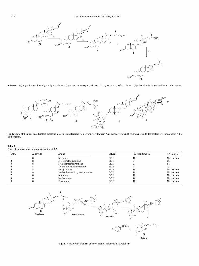
Scheme 1. (a) Ac2O, dry pyridine, dry CHCl3, RT, 2 h, 91%; (b) AcOH, NaCNBH3, RT, 5 h, 81%; (c) Dry DCM,PCC, reflux, 1 h, 91%; (d) Ethanol, substituted aniline, RT, 2 h, 66-84%.
O O
OH
OOH
O
1 2HO
OH
OHO
OOH
3 4
O
HO
5
O
O
O
O
O
OO
OHHOHO
HOOH
HO OH
123
45 6
789
1112
1314
1516
1718
19
2021
22 23 24
2526
27
H
Fig. 1. Some of the plant based potent cytotoxic molecules on steroidal framework; 1: withaferin A, 2: gymnasterol 3: 24-hydroxyperoxide desmosterol, 4: timosaponin A-III,5: diosgenin.
Table 1Effect of various amines on transformation of 8–9.
Entry Aldehyde Amine Solvent Reaction time (h) %Yield of 9
1 8 No amine EtOH 16 No reaction2 8 3,4,-Dimethoxyaniline EtOH 2 663 8 3,4,5-Trimethoxyaniline EtOH 2 844 8 3,4-Methylenedioxyaniline EtOH 2 725 8 Benzyl amine EtOH 16 No reaction6 8 3,4-Methylenedioxybenzyl amine EtOH 16 No reaction7 8 Ammonia EtOH 16 No reaction8 8 Methylamine EtOH 16 No reaction9 8 Ethylamine EtOH 16 No reaction
O
AcO
C OH
O
C N
H
R
H3C H
O
HN
H
R
H2OO
HN
H
R
H
OH
OO
RNHCH3
+
8
9
Aldehyde Schiff's baseEnamine
Ketone
Fig. 2. Plausible mechanism of conversion of aldehyde 8 to ketone 9.
112 A.A. Hamid et al. / Steroids 87 (2014) 108–118

O
AcO
CH3
O
Fig. 3. HMBC correlations of side chain of ketone 9.
A.A. Hamid et al. / Steroids 87 (2014) 108–118 113
Fig. 5. Cell cycle analysis of compound 7.
Fig. 6. Cleavage of PARP, a caspase target by compound 7.
9542, Cell Signaling Technology, USA) at 4 �C. After 3 washes withTBS-T, the membranes were incubated with HRP conjugated anti-rabbit antibody for 1 h at room temperature and washed againwith TBS-T (x3 times). Proteins were detected with an enhancedchemiluminescence (ECL) reagent and visualized by a chemilumi-nescence detector (Bio-Rad Laboratories, USA). The densitometryanalysis of blots was done by using Bio-Rad Image Lab 4.0software.
2.4.4. Docking studiesThe two dimensional structures of the molecules were con-
structed with the ChemDraw Ultra. Energy minimization of thecompounds was performed with ‘ChemBio office’ consideringMM2/MM3 molecular mechanics parameter up to its lowest stableenergy state. This energy minimization process was performeduntil the energy change was less than 0.001 kcal mol�1 or the mol-ecules had been updated almost 300 times. The 3D chemical struc-ture of doxorubicin (Positive control) was retrieved from thePubChem compound database at NCBI (http://www.pub-chem.ncbi.nlm.nih.gov). Crystallographic 3D structures of targetproteins (caspase-9: (PDB:1NW9- chain B) and caspase-3:(PDB:3KJF) were retrieved from the Brookhaven Protein Databank(http://www.pdb.org) [10,11]. Hydrogen atoms were added tothe protein targets to achieve the correct ionization and tautomericstates of amino acid residues such as His, Asp, Ser, and Glu. Molec-ular docking of the compounds against selected target wasachieved using the ‘AutoDock Vina’. To perform the automateddocking of ligands into the active sites, we used a Lamarkiangenetic algorithm [12].
2.4.5. In-vivo acute oral toxicityIn view of potent anti-cancer activity of compound 7 in in-vitro
model, acute and sub-acute oral toxicity of the same was carriedout in Swiss albino mice for its further development into drugproduct. Experiment was conducted in accordance with the Orga-nization for Economic Co-operation and Development (OECD) testguideline No 423 (1987).
For this experiment, 30 mice (15 male and 15 female) weretaken and divided into four groups comprising 3 male and 3 femalemice in each group weighing between 20 and 25 g. The animalswere maintained at 22 ± 50C with humidity control and also onan automatic dark and light cycle of 12 hours. The animals werefed with the standard mice feed and provided ad libitum drinking
Fig. 4. Molecular conformation of 9 in crystals. Thermal ellipsoids are shown at 50% probability level.
water. Mice of group 1 were kept as control and animals of groups2, 3, 4 and 5 were kept as experimental. The animals were acclima-tized for 7 days in the experimental environment prior to theactual experimentation. The test compound was solubilized indimethyl sulphoxide with few drops of tween 80 and then sus-pended in caboxymethyl cellulose (0.7%) and was given at 5, 50,300 and 1000 mg/kg body weight to animals of groups 2, 3, 4and 5 respectively once orally. Control animals received onlyvehicle.
The animals were checked for mortality and any signs of illhealth at hourly interval on the day of administration of drugand there after a daily general case side clinical examination wascarried out including changes in skin, mucous membrane, eyes,occurrence of secretion and excretion and also responses like lach-rymation, piloerection respiratory patterns etc. Also changes ingait, posture and response to handling were also recorded [13].In addition to observational study, body weights were recordedand blood and serum samples were collected from all the animalson 7th day of the experiment in acute oral toxicity. The sampleswere analyzed for total RBC, WBC, differential leucocytes count,hemoglobin percentage and biochemical parameters like ALKP,SGPT, SGOT, total cholesterol, triglycerides, creatinine, bilirubin,serum protein, tissue protein, malonaldehyde and reduced GSHactivity. The animals were then sacrificed and were necropsedfor any gross pathological changes. Weights of vital organs likeliver, heart, kidney etc. were recorded [14].

Fig1N
Fig3K
TaEff
TaIn-
a
114 A.A. Hamid et al. / Steroids 87 (2014) 108–118
2.4.6. Statistical analysisStatistical analysis was carried out in Microsoft Excel.
3. Results and discussion
Scheme 1 denotes the synthetic strategy of these derivativesstarting with diosgenin (5). Diosgenin 5 was converted to dios-genin acetate 6 by acetylation with acetic anhydride-pyridine indry chloroform at room temperature. The spectral data of acetate6 was alike the earlier reports [6a]. The reductive cleavage of ringF of spiroketal linkage was done by using sodium cyanoborohy-dride in AcOH at room temperature to get the alcohol 7 that pos-
. 7a. In-silico molecular docking studies elucidating the possible mechanisms of diosgenW9-B). The docking studies were carried out using ‘AutoDock Vina’, using Lamarkian ge
. 7b. In-silico molecular docking studies elucidating the possible mechanisms of diosgenJF). The docking studies were carried out using ‘AutoDock Vina’, using Lamarkian geneti
ble 2ect of solvent polarity on transformation of compound 8–9.
Entry Aldehyde Amine So
1 8 3,4,5-Trimethoxyaniline Et2 8 3,4,5-Trimethoxyaniline M3 8 3,4,5-Trimethoxyaniline TH4 8 3,4,5-Trimethoxyaniline Di5 8 3,4,5-Trimethoxyaniline To
ble 3vitro cytotoxicities of diosgenin analogs.
S. no. Compound no./name IC50 (uM) (Mean ± SE)a
KB C-33A
1 7 11.43 ± 1.47 7.54 ± 0.932 8 17.55 ± 1.25 11.54 ± 0.13 14 15.97 ± 1.96 11.18 ± 0.64 15 13.96 ± 0.39 12.27 ± 0.35 17 17.52 13.156 Tamoxifen 10.25 ± 1.21 6.94 ± 0.097 Podophyllotoxin <1.25 <1.25
Rest of the compounds have IC50 > 20 lM, considered inactive.
sesses furostenic system. Oxidation of alcohol 7 with pyridiniumchlorochromate (PCC) yielded aldehyde 8 (Fig. 1).
We tried to prepare some Schiff’s bases onto C29 aldehyde (8),but unexpectedly, we ended with a C28 ketone (9). Aldehyde 8 ontreatment with an aromatic amine in ethanol at room temperatureafforded product 9 as a ketone in 2 h. Various aromatic amines(3,4,-dimethoxyaniline, 3,4,5-trimethoxyaniline 3,4-methylenedi-oxyaniline) were tried and every time the same product 9 wasobtained in varied yields (66-84%). The reaction of aldehyde 8 withaliphatic amines (MeNH2 and EtNH2) and even with ammonia wasunsuccessful. Benzyl amines (BnNH2 and 3,4-methylenedioxyben-zylamine) also did not yield the product 9 (Table 1). It will beworth mentioning that without aromatic amine this transforma-tion does not take place. Aldehyde 8 did not form Schiff’s base with
in (5), its derivatives 7, and positive control doxorubicin with Caspase-9 (PDB:netic algorithm.
in (5), its derivatives 7, and positive control doxorubicin with Caspase-3: (PDB:c algorithm.
lvent Reaction time (t) h % Yield of 9
OH 2 84eOH 2 73F 2 44
chloromethane 2 49luene 2 28
DU145 A549 MCF-7
>20 >20 10.95 ± 0.730 16.826 >20 15.17 ± 1.363 17.08 ± 0.09 15.27 ± 1.18 14.53 ± 1.078 13.27 ± 0.25 12.56 ± 0.18 14.28 ± 0.20
>20 16.29 18.6712.24 ± 0.28 10.07 ± 0.46 8.54 ± 1.01<1.25 <1.25 <1.25

A.A. Hamid et al. / Steroids 87 (2014) 108–118 115
any of these amines. The structure of the ketone 9 was confirmedby 1D NMR (1H & 13C), 2D NMR (COSY, HSQC, HMBC), mass and IRspectroscopy. A plausible mechanism is given in Fig. 2.
Compound 9 was obtained as yellow amorphous powder. Itsstructure was established by spectroscopy. Its molecular formulaeC28H42O4 was determined by its HR-ESI-MS at m/z 443.3139[M+H]+ (Calcd 443.3161). 13C NMR showed total 28 distinct car-bons indicating loss of one carbon as compared to aldehyde 8.There were 5 methyls (d16.80, 19.01, 19.71, 21.80, 30.33), 9 meth-ylenes (d21.02, 27.44, 28.13, 32.37, 32.55, 37.38, 38.48, 39.75,41.28), 9 methines (d31.95, 38.24, 50.38, 57.27, 65.39, 74.28,83.69, 89.53, 122.72) and 5 quaternary carbons (d 37.10, 41.09,140.12, 170.95, 209.24). There was no aldehyde group and a car-bonyl group was generated as ketone at d209.24 ppm. Least change
O
CHO
AcO 8
O
C
AcO 10
CHH
O
C
AcO 11
CHH
NO2
O
C
AcO 14
N-OH
H
O
C
AcO12
CHH
O
C
AcO 13
CH
H O
C
AcO 15
N-OAc
H
+
OCH3
OCH3H3CO
OCH3
OCH3
H3CO
g
e e
fe
Scheme 2. (e) Wittig salt, NaH, dry Toluene, RT, 3–4 h, 62-81%; (f) NH2OH.HCl, EtOH, reflux, 2 h, 84%; (g) Ac2O, dry pyridine, dry CHCl3, RT, 3 h, 92%.
O
AcO16
N
O
AcO
N
O
AcO9
O
O
AcO
18
OAcOH
gf
h
Mixture of isomers
OH
17
Scheme 3. (f) NH2OH.HCl, EtOH, reflux, 2 h, 79%; (g) Ac2O, dry pyridine, dry CHCl3, RT, 3 h, 89%; (h) NaBH4-MeOH, RT, 30 min, 86% (Mix of isomers).
in the chemical shifts of C1 to C22 suggested possibility of nochange in the ring system of the ketone 9. From HSQC spectrum,chemical shifts of 22-CH (d3.26 m, 89.53), 23-CH2 (d1.72, 27.44),24-CH2 (d2.49-2.61, 41.28) and 26-CH3 (d2.13, 30.33) were estab-lished. In 1H-1H COSY spectrum, 22-CH (d3.26) showed couplingwith 23-CH2 (d1.72) and 24-CH2 (d2.49-2.61) showed couplingwith 23-CH2 (d1.72). In HMBC correlations, 26-CH3 protons(d2.13 ppm) showed correlations with C24 (d41.28) and C25ketone (d209.24). 24-CH2 protons (d2.49-2.61) exhibited correla-tions with C26 (d30.33) and C23 (d27.44) carbons. C23- CH2 pro-tons (d1.72) showed correlations with C22 (d89.53), C24 (41.28)and C25 (d209.24) carbons. HMBC correlations of side chain areshown in Fig. 3. The structure of 29 was finally authenticated bysingle crystal X-ray crystallography (Fig. 4).

TaMo
116 A.A. Hamid et al. / Steroids 87 (2014) 108–118
Further, the molecular conformation of compound 9 in crystalsis depicted in Fig. 4. All the bond lengths and bond angles arewithin the accepted range. The crystal packing is stabilized byvan der Waals interactions, in the absence of strong hydrogen bonddonors in this molecule. However, the ketonic O atom and themethylene group (C24) adjacent to the ketone functionality ofsymmetry-related molecules are at 3.22Å apart, suggesting a weakC-H...O interaction (O3...H = 2.55Å, C24...O3 = 3.22Å, \C24-H...O3 = 125.8�). The ring conformations of compound 9 are verysimilar to that observed in other diosgenin derivatives and solvates(Figs. 5, 6, 7A, 7B).
The effect of various solvents was also observed, as shown inTable 2. Ethanol was found to be the best solvent. However, meth-anol was equally good. In case of less polar solvents like THF,dichloromethane and toluene the yield of the product 9 was rela-tively low (Table 3).
Further, several styrene derivatives were prepared on to 8aldehyde using Wittig reaction. Various Wittig salts (Benzylidene-triphenylphosphonium bromide) were prepared using triphenyl-phosphine and corresponding benzylbromide in toluene. Finallythe styrene derivatives of diosgenin were obtained by treatingwittig salt with aldehyde 8 in sodium hydride/toluene under refluxconditions in 29–68% yields. 8 was transformed to aldoxime (14)also and then oxime acetate (15) by usual methodologies [15].All these transformations are depicted in Scheme 2.
Similarily, Scheme 3 shows modification of ketone 9 to variousother derivatives. 9 was modified to ketoxime (16) and its corre-sponding acetate (17) same as described earlier for aldehyde 8.The ketone group of 9 was reduced to alcohol (18) with sodiumborohydride in methanol. Both C25a-OH and C25b-OH isomerswere formed in equal ratio as evident from 13C NMR at d67.91and d68.75 respectively.
All synthesized compounds were evaluated against C33A (Cer-vical carcinoma), A549 (Lung carcinoma), KB (HeLa contaminantof mouth epidermal carcinoma), MCF-7 (Breast adenocarcinoma),DU145 (prostate carcinoma) human cancer cell lines by Sulpho-rhodamine assay [7]. Only five of the analogs exhibited significantanticancer activity, rest were inactive at 20 lM concentration.
Cell cycle regulation ensures the fidelity of genomic replicationand cell division. G1/S and G2/M transitions are two major check-points to allow the cells to control any modification in DNA con-
ble 4lecular interactions of diosgenin (5) and its derivative 7 docked with Caspase-9: (PDB: 1N
Compound code Caspase-3 (PDB entry 1pau)
Bindingaffinity(Kcal/mol)
Binding pocket amino aci
5
O
HO
O �7.9 ARG(B)-207, ASN(B)-208,247, PHE(B)-250, PHE(B)-SER(B)-249, SER(B)-251, T
7
O
O
CH2OH
O
�7.8 CYS(A)-163, GLU(A)-123,121, MET(A)-61, PHE(A)-1ARG(B)-207, ASN(B)-208,256, SER(B)-209, SER(B)-2TRP(B)-206, TRP(B)-214, T
Doxorubicin �8.2 ALA(A)-162, ARG(A)-64, C161, GLY(A)-122, HIS(A)-1SER(A)-120, ARG(B)-207,256, SER(B)-205, SER(B)-2TYR(B)-204
tent. Checkpoint loss results in genomic instability and inducecarcinogenesis. Induction of cell cycle arrest in cancer cell linesconstitutes one of the most prevalent strategies to stop or limitcancer spreading [16]. Our data demonstrate that Compound 7arrests cells at G0/G1 phase of division cycle which is associatedwith decrease in G2/M population. We also observed accumulationof cells in sub-G0 phase after treatment confirming induction ofapoptosis by the molecule.
Diosgenin and related compounds have been good cytotoxicagents against various human cancer cell lines [17a–f]. Diosgeninand its semisynthetic derivatives induced apoptosis throughincreased expression of caspase-3 [18a–e]. Caspases are a familyof cystein-aspartic proteases, which are crucial mediators of apop-tosis. Among these caspase-3 encoded as CAS3 gene is identified innumerous mammals. It remains as zymogens (procaspase) unlessactivated biochemically. It cleaves and activates caspases 6, 7and 9, and the protein itself is processed by caspases 8, 9 and 10.Caspase-3 is necessary for its typical role in apoptosis, where it isresponsible for chromatin condensation and DNA fragmentation[19]. PARP is an enzyme involved in DNA repair when cell areexposed to environmental stress [20]. During apoptosis, PARP iscleaved and inactivated by caspases [21,22]. Therefore, cleavageof PARP is a well established marker for detection of apoptosis.In the present study we found cleavage of PARP in MCF-7 cells at24 h exposure to the Compound 7 implying activation of caspasesby the molecule to trigger apoptosis.
The binding affinity obtained in the docking experimentallowed the activity of the diosgenin derivative 7 to be comparedto that of the standard anticancer compound doxorubicin (Table 4).The diosgenin and its active derivatives ‘7’ showed high bindingaffinity (high negative docking energy) against known humancaspases 9 (-7.3 kcal/mol) and caspase 3 (-7.8 kcal/mol) (PDB:1NW9-B; PDB: 3KJF). When we compared how the binding sitepocket amino acid residues interacted with the derivatives, wefound that the in case of CASPASE 9, ‘PHE, PRO, THR, GLY, ARG,and HIS’ amino acids were in share with positive control doxorubu-cin. While, in case of CASPASE 3, ‘GLN, PHE, TYR, TRP, SER, ALA, GLYand ARG’ were in share with doxorubicin.
In acute oral toxicity studies of compound 7, no observationalchanges, morbidity and mortality were observed throughout theexperimental period up to the dose level of 1000 mg/kg body
W9-B) and Caspase-3: (PDB: 3KJF) and binding pocket residues (amino acids).
Caspase-9 (PDB entry 1nw9)
ds Bindingaffinity(kcal/mol)
Binding pocket amino acids
GLU(B)-248, PHE(B)-256, SER(B)-209,RP(B)-206, TRP(B)-214
�8.7 ALA-316, ARG-408, GLN-245, LEU-244,PHE-406, PRO-318, PRO-336, THR-337
GLY(A)-122, HIS(A)-28, THR(A)-166,PHE(B)-250, PHE(B)-49, SER(B)-251,YR(B)-204
�7.3 GLN-240, GLY-288, GLY-350, GLY-395,ILE-396, LYS-394, LYS-398, PHE-348,PHE-351, PRO-349, THR-347, TRP-354,TYR-397
YS(A)-163, GLN(A)-21, MET(A)-61,
ASP(B)-253, PHE(B)-51, TRP(B)-206,
�8.2 ARG-178, ARG-180, ASP-186, CYS-287,GLY-182, GLY-288, GLY-360, HIS-237,PHE-351, PRO-349, PRO-357, SER-183,SER-361, THR-179, THR-181

Table 5Effect of compound 7 as a single acute oral dose at 5, 50, 300 and 1000 mg/kg body weight on body weight, haemogram and serum biochemical parameters in Swiss albino mice(Mean ± SD; n = 6) compared to control, 5, 50, 300 and 1000 mg/kg).a significant (P < 0.05).
Parameters Dose of compound 7 at mg/kg body weight as a single oral dose
Control 5 mg/kg 50 mg/kg 300 mg/kg 1000 mg/kg
Body weight (gm) 23.32 ± 0.60 23.31 ± 0.48 23.95 ± 0.45 21.92 ± 0.47 23.10 ± 0.55Hemoglobin (gm/dL) 11.23 ± 0.92 11.44 ± 0.50 10.94 ± 0.58 11.51 ± 1.02 12.10 ± 0.40RBC (million/mm3) 5.82 ± 0.19 6.55 ± 0.30 5.85 ± 0.31 5.42 ± 0.34 6.19 ± 0.30WBC (1000⁄/mm3) 10.68 ± 2.14 11.03 ± 2.24 10.48 ± 1.76 10.71 ± 2.52 12.53 ± 2.77ALKP (U/L) 222.1 ± 14.4 259.4 ± 6.9 263.6 ± 16.2 239.4 ± 13.7 139.7 ± 17.4SGOT (U/L) 24.44 ± 2.37 32.49 ± 3.51 31.50 ± 2.98 36.04 ± 3.33 32.54 ± 3.50SGPT (U/L) 12.96 ± 1.96 18.76 ± 1.79 18.07 ± 3.20 19.96 ± 4.63 18.33 ± 2.27Albumin (g/dL) 3.60 ± 0.45 3.08 ± 0.15 2.98 ± 0.13 2.87 ± 0.24 3.27 ± 0.11Creatinine (mg/dL) 0.63 ± 0.11 0.48 ± 0.09 0.50 ± 0.07 0.44 ± 0.05 0.47 ± 0.08Triglycerides (mg/dL) 172.21 ± 17.57 180.55 ± 19.70 172.73 ± 19.15 171.74 ± 19.80 166.89 ± 17.97Serum protein (mg/mL) 1.15 ± 0.13 1.02 ± 0.08 0.94 ± 0.08 1.00 ± 0.08 1.23 ± 0.09Cholesterol (mg/dL) 156.4 ± 35.8 114.3 ± 21.2 115.9 ± 23.4 123.8 ± 25.6 156.4 ± 28.3
Fig. 8. Effect of compound 7 as a single acute oral dose at 5, 50, 300 and 1000 mg/kg on absolute and relative organ weight in Swiss albino mice (n = 6, Non significant changeswere found compared to control).
A.A. Hamid et al. / Steroids 87 (2014) 108–118 117
weight. No morbidity or any other gross observation changes couldbe noticed in the group of animals treated with the test drug at1000 mg/kg. Blood and serum samples upon analysis showednon-significant changes in all the parameters studied like totalhemoglobin level, RBC count, WBC count, differential leucocytescount, SGPT, creatinine, triglycerides, cholesterol, albumin, serumprotein (Table 5 and Fig. 8) except significant changes in ALKPactivities in groups of animals treated with the compound at1000 mg/kg; wherein ALKP activity was decreased significantlycompared to control. Animals on gross pathological study showedno changes in any of the organs studied including their absoluteand relative weight (Figs. 7a and 7b). Therefore, the experimentshowed that compound 7 is well tolerated by the Swiss albino miceup to the dose level of 1000 mg/kg body weight as a single acute
Fig. 9. Effect of compound 7 as a single acute oral dose at 5, 50, 300 and 1000 mg/kgbody weight on differential leucocytes counts in Swiss albino mice (n = 6, Nonsignificant changes were found compared to control).
oral dose except significant decrease in ALKP activities in groupstreated with compound 7 at 1000 mg/kg. However, sub-acuteand or chronic experiment with the test drug needs to be carriedout to look for any adverse effect on repeated exposure to com-pound 7 for its future development [23] Fig. 10.
4. Conclusion
Diosgenin was modified to obtain novel derivatives. The unu-sual transformation was observed and product has been confirmed.Compound 7 is the most potent derivative of this series whichinhibits cell proliferation by arresting the population in G0/G1
phase of the cell cycle. The mechanism of antiproliferative actionof this molecule is through induction of caspase depended apopto-sis pathway. Compound 7 is a non-toxic anticancer furostanederivative of diosgenin. The lead obtained in this study can furtherbe optimized for better activity in future.
Acknowledgements
Authors are thankful to the Directors of CSIR-CDRI and CSIR-CIMAP for constant encouragement and support. Spectral datareceived by spectral The financial support from CSIR is dulyacknowledged. TWAS-CSIR fellowship to Mr. A. A. Hamid is dulyacknowledged.

118 A.A. Hamid et al. / Steroids 87 (2014) 108–118
Appendix A. Supplementary material
Supplementary data associated with this article can be found, inthe online version, at http://dx.doi.org/10.1016/j.steroids.2014.05.025.
References
[1] Cancer Fact sheet N�297 reviewed February 2014.[2] Gupta A, Kumar BS, Negi AS. Current status on development of steroids as
anticancer agents. J Steroid Biochem Mol Biol 2013; 137:242–70 and referenceno. 90, 91, 137 and 149 cited therein.
[3] Kang YJ, Chung HJ, Nam JW, Park HJ, Seo EK, Kim YS, Lee D, Lee SK. Cytotoxicand antineoplastic activity of timosaponin A-III for human colon cancer cells. JNat Products 2011;74:701–6.
[4] (a) Raju J, Bird RP. Diosgenin, a naturally occurring furostanol saponinsuppresses 3-hydroxy-3-methylglutaryl CoA reductase expression andinduces apoptosis in HCT-116 human colon carcinoma cells. Cancer Lett2007;255:194–204;(b) Lepage C, Leger DY, Bertrand J, Martin F, Beneytout JL, Liagre B. Diosgenininduces death receptor-5 through activation of p38 pathway and promotesTRAIL-induced apoptosis in colon cancer cells. Cancer Lett 2011;301:193–202;(c) Liu MJ, Wang Z, Ju Y, Wong RNS, Wu Qy. Diosgenin induces cell cycle arrestand apoptosis in human leukemia K562 cells with the disruption of Ca2+homeostasis. Cancer Chemother Pharmacol 2005;55:79–90.
[5] Nomenclature of Steroids. IUPAC and International Union of Biochemistry-Joint commission on Biochemical Nomenclature, Pure and Applied Chem 1989;61(10): 1783–822.
[6] (a) Chaosuancharoen N, Kongkathip N, Kongkathip B. Novel synthetic approachfrom diosgenin to a 17a-hydroxy orthoester via a region- and stereo specificrearrangement of an epoxy ester. Synth Commun 2004;34:961–83;(b) Rosado-Abon A, Esturau-Escofet N, Flores-A’lamo M, Moreno-Esparza R.Iglesias-Arteaga MA, The crystal structure of diosgenin acetate and its 23-oxygenated derivatives. J Chem Crystallogr 2013;43:187–96.
[7] (a) Sheldrick GM. SHELXS-97. Göttingen, Germany: A program for automaticsolution of crystal structures; University of Göttingen; 1997;(b) Sheldrick GM. SHELXL-97. Göttingen, Germany: A program for crystalstructure refinement; University of Göttingen; 1997.
[8] Adaramoye OA, Sarkar J, Singh N, et al. Antiproliferative action of Xylopiaaethiopica fruit extract on human cervical cancer cells. Phytother Res2011;25:1558–63.
[9] Sarkar J, Singh N, Meena S, Sinha S. Staurosporine induces apoptosis in humanpapillomavirus positive oral cancer cells at G2/M phase by disruptingmitochondrial membrane potential and modulation of cell cytoskeleton. OralOncol 2009;45:974–9.
[10] Shiozaki EN, Chai J, Rigotti DJ, Riedl SJ, Li P, Srinivasula SM, Alnemri ES,Fairman R, Shi Y. Mechanism of XIAP-mediated inhibition of caspase-9. MolCell 2003;11:519–27.
[11] Wang Z, Watt W, Brooks NA, Harris MS, Urban J, Boatman D, McMillan M, KahnM, Heinrikson RL, Finzel BC, Wittwer AJ, Blinn J, Kamtekar S, Tomasselli AG.Kinetic and structural characterization of caspase-3 and caspase-8 inhibitionby a novel class of irreversible inhibitors. Biochim Biophys Acta2010;1804:1817–31.
[12] Trott O, Olson AJ. AutoDock Vina: improving the speed and accuracy ofdocking with a new scoring function, efficient optimization andmultithreading. J Computational Chemistry 2010;31:455–61.
[13] Allan JJ, Damodaran A, Deshmukh NS, Goudar KS, Amit A. Safety evaluation of astandardized phytochemical composition extracted from Bacopa monnieri inSprague-Dawley rats. Food Chemical Toxicol 2007;45:1928–37.
[14] Chanda D, Shanker K, Pal A, Luqman S, Bawankule DU, Mani DN, Darokar MP.Safety evaluation of Trikatu, a generic Ayurvedic medicine in Charles Fosterrat. J Toxicol Sci 2008;34:99–108.
[15] Furniss BS, Hannaford AJ, Smith PWG, Tatchell AR. Vogel’s textbook of practicalorganic chemistry, fifth ed.. England, UK: Addison Wesley Longman Limited,Essex CM20 2JE; 1989.
[16] Hartwell LH, Weinert TA. Checkpoints: controls that ensure the order of cellcycle events. Science 1989;246:629–34.
[17] (a) Li N, Zhang L, Zeng KW, Zhou Y, Zhang JY, Che YY, Tu PF. Cytotoxic steroidalsaponin from Ophiopogon japonicas. Steroids 2013;78:1–7;(b) Wei G, Wang J, Du Y. Total synthesis of solamargine. Bioorg Med Chem Lett2011;21:2930–3;(c) Huang B, Du D, Zhang R, Wu X, Xing Z, He Y, Huang W. Synthesis,characterisation and biological studies of diosgenyl analogues. Bioorg MedChem Lett 2012;22:7330–4;(d) Perez-Diaz JOH, Rarova L, Ocampo JPM, Magaria-Vergara NE, Farfan N,Strnad M, Santillan R. Synthesis and biological activity of 23-ethylidene-26-hydroxy-22-oxocholestane derivatives from spirostanic sapogenins. Er J MedChem 2012;51:67–78;(e) Fernandez-Herrera MA, Lopez-Munoz H, Hernandez-Vazquez JMV,Sanchez-Sanchez L, Escobar-Sanchez ML, Pinto BM, Sandoval-Ramirez J.Synthesis and selective anticancer activity of steroidal glycoconjugates. Eur JMed Chem 2012;54:721–7(f) (a) Li N, Zhang L, Zeng KW, Zhou Y, Zhang JY, CheYY, Tu PF. Cytotoxic steroidal saponin from Ophiopogon japonicas. Steroids2013; 78: 1–7; (b) Wei G, Wang J, Du Y. Total synthesis of solamargine. BioorgMed Chem Lett 2011; 21: 2930–2933; (c) Huang B, Du D, Zhang R, Wu X, XingZ, He Y, Huang W. Synthesis, characterisation and biological studies ofdiosgenyl analogues. Bioorg Med Chem Lett 2012; 22: 7330–7334; (d) Perez-Diaz JOH, Rarova L, Ocampo JPM, Magaria-Vergara NE, Farfan N, Strnad M,Santillan R. Synthesis and biological activity of 23-ethylidene-26-hydroxy-22-oxocholestane derivatives from spirostanic sapogenins. Er J Med Chem 2012;51: 67–78; (e) Fernandez-Herrera MA, Lopez-Munoz H, Hernandez-VazquezJMV, Sanchez-Sanchez L, Escobar-Sanchez ML, Pinto BM, Sandoval-Ramirez J.Synthesis and selective anticancer activity of steroidal glycoconjugates. Eur JMed Chem 2012; 54:721–727; (f) Fernandez-Herrera MA, Sandoval-Ramirez J,Lopez-Munoz H, Sanchez-Sanchez L. Formation of the steroidal 3b-hydroxy-6-oxo-moiety. Synthesis and cytotoxicity on glucoaxogenin. ARKIVOC 2009;(xiii): 170–184.
[18] (a) Tong QY, He Y, Zhao QB, Qing Y, Huang W, Wu XH. Cytotoxicity andapoptosis inducing effect of steroidal saponins from Dioscorea zingiberensisWright against cancer cells. Steroids 2012;77:1219–27;(b) Fernandez-Herrera MA, Lopez-Munoz H, Hernandez-Vazquez JMV, Lopez-Davila M, Escobar-Sanchez ML, Sanchez-Sanchez L, Pinto BM, Sandoval-Ramirez J. Synthesis of 26-hydroxy-22-oxocholestanic frameworks fromdiosgenin and hecogenin and their in vitro antiproliferative and apoptoticactivity on human cervical cancer CaSki cells. Bioorg Med Chem2010;18:2474–84;(c) Fernandez-Herrera MA, Mohan S, Lopez-Munoz H, Hernandez-VazquezJMV, Perez-Cervantes E, Escobar-Sanchez ML, Sanchez-Sanchez L, Regla I, PintoBM, Sandoval-Ramirez J. Synthesis of the steroidal glycoside (25R)-3b,16b-diacetoxy-12, 22-dioxo-5a-cholestan-26-yl b-D-glucopyranoside and its anti-cancer properties on cervicouterine HeLa, CaSki, and ViBo cells. Eur J MedChem 2010;45:4827–37;(d) Fernandez-Herrera MA, Lopez-Munoz H, Hernandez-Vazquez JMV, Lopez-Davila M, Mohan S, Escobar-Sanchez ML, Sanchez-Sanchez L, Pinto BM,Sandoval-Ramirez J. Synthesis and biological evaluation of the glycoside(25R)-3b,16b-diacetoxy-22-oxocholest-5-en-26-yl b-D-glucopyranoside: aselective anticancer agent in cervicouterine cell lines. Eur J Med Chem2011;46:3877–86;(e) Fernandez-Herrera MA, Sandoval-Ramirez J, Sanchez-Sanchez L, Lopez-Munoz H, Escobar-Sanchez ML. Probing the selective antitumor activity of 22-oxo-26-selenocyanocholestane derivatives. Eur J Med Chem 2014;74:451–60.
[19] Porter AG, Jänicke RU. Emerging roles of caspase-3 in apoptosis. Cell DeathDiffer 1999;6:99–104.
[20] Satoh MS, Lindahl T. Role of poly(ADP-ribose) formation in DNA repair. Nature1992;356:356–8.
[21] Tewari M, Quan LT, O’Rourke K, Desnoyers S, Zheng Z, Beidler DR, Poiries GG,Salvesen G, Dixit VM. Yama/CPP32 beta, a mammalian homolog of CED-3, is aCrmA-inhibitable protease that cleaves the death substrate poly(ADP-ribose)polymerase. Cell 1995;81:801–9.
[22] Germain M, Affar EB, D’Amours D, Dixit VM, Salvesen GS, Poirier GG. Cleavageof automodified poly(ADP-ribose) polymerase during apoptosis. Evidence forinvolvement of caspase-7. J Biol Chem 1999;274:28379–84.
[23] Ghosh MN. In: Fundamentals of Experimental Pharmacology, 1st ed., ScientificBook Agency, Kolkata; 1984. p. 156.