Synthesis, NMR, and Conformational Studies of Cyclic Oligo-(1→6)-β-D-Glucosamines (Eur. J. Org....
11
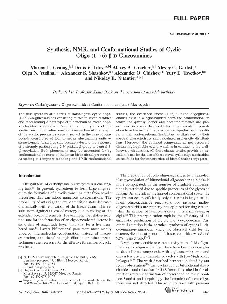
FULL PAPER DOI: 10.1002/ejoc.200901275 Synthesis, NMR, and Conformational Studies of Cyclic Oligo-(16)-β-D-Glucosamines Marina L. Gening, [a] Denis V. Titov, [a,b] Alexey A. Grachev, [a] Alexey G. Gerbst, [a] Olga N. Yudina, [a] Alexander S. Shashkov, [a] Alexander O. Chizhov, [a] Yury E. Tsvetkov, [a] and Nikolay E. Nifantiev* [a] Dedicated to Professor Klaus Bock on the occasion of his 65th birthday Keywords: Carbohydrates / Oligosaccharides / Conformation analysis / Macrocycles The first synthesis of a series of homologous cyclic oligo- (16)-β-D-glucosamines consisting of two to seven residues and representing a new type of functionalized cyclic oligo- saccharides is reported. Remarkably high yields of the studied macrocyclization reaction irrespective of the length of the acyclic precursors were observed. In the case of com- pounds constituted of four to seven glucosamine units α- stereoisomers formed as side products despite the presence of a strongly participating 2-N-phthaloyl group to control β- glycosylation. Both phenomena may be accounted for by conformational features of the linear bifunctional precursors. According to computer modeling and NMR conformational Introduction The synthesis of carbohydrate macrocycles is a challeng- ing task. [1] In general, cyclizations to form large rings re- quire the formation of a cyclic transition state from acyclic precursors that can adopt numerous conformations. The probability of attaining the cyclic transition state decreases dramatically with elongation of the linear chain. This re- sults from significant loss of entropy due to coiling of the extended acyclic precursors. For example, the relative reac- tion rate for the formation of an eight-membered lactone is six orders of magnitude lower than that for a five-mem- bered one. [2] Larger bifunctional precursors more readily undergo intermolecular condensation instead of macro- cyclization, and therefore, high dilution or other special techniques are necessary for the effective formation of cyclic products. [a] N. D. Zelinsky Institute of Organic Chemistry RAS Leninsky prospect 47, 119991 Moscow, Russia Fax: +7-499-135-87-84 E-mail: [email protected] [b] Higher Chemical College RAS Miusskaya sq. 9, 125047 Moscow, Russia Fax: +7-499-978-85-27 Supporting information for this article is available on the WWW under http://dx.doi.org/10.1002/ejoc.200901275. Eur. J. Org. Chem. 2010, 2465–2475 © 2010 Wiley-VCH Verlag GmbH & Co. KGaA, Weinheim 2465 studies, the described linear (16)-β-linked oligoglucos- amines exist in a right-handed helix-like conformation, in which the glycosyl donor and acceptor moieties are pre- arranged in a way that facilitates intramolecular glycosyl- ation from the α-side. Prepared cyclo-oligoglucosamines dif- fer in their conformational flexibilities, as illustrated by their spectral characteristics and calculated asphericity distribut- ions. Moreover, the obtained compounds do not possess a distinct hydrophobic cavity, which is in contrast to the well- known cyclodextrins. All these characteristics provide an ex- cellent basis for the use of these novel cyclic oligosaccharides as scaffolds for the construction of biomolecular conjugates. The preparation of cyclo-oligosaccharides by intramolec- ular glycosylation of bifunctional oligosaccharide blocks is more complicated, as the number of available conforma- tions is restricted due to specific properties of the glycoside linkage. As a result of the limited conformational space, the cyclization occurs efficiently only at a certain length of the linear oligosaccharide precursors. For instance, malto- oligosaccharides are properly preorganized for ring closure when the number of -glucopyranose units is six, seven, or eight. [1] This preorganization explains the efficiency of the enzymatic production of α-, β-, and γ-cyclodextrins. An- other illustration is the chemical synthesis of cyclic (14)- α--mannopyranosides, where the observed yield for the macrocyclization of penta- and hexasaccharides was 8 and 92%, respectively. [3–5] Despite considerable research activity in the field of syn- thetic cyclic oligosaccharides, there have been no examples to date of these compounds with -glucosamine units and only a few discrete examples of cycles with (16)-glycoside linkages. [6–9] The work described here was initiated by our recent observation [10] that cyclization of bifunctional disac- charide 1 and trisaccharide 2 (Scheme 1) resulted in the al- most quantitative formation of corresponding cyclic prod- ucts 3 and 4, and surprisingly, the formation of linear oligo- mers was not detected. This is in contrast with previous
-
Upload
independent -
Category
Documents
-
view
0 -
download
0
Transcript of Synthesis, NMR, and Conformational Studies of Cyclic Oligo-(1→6)-β-D-Glucosamines (Eur. J. Org....
FULL PAPER
DOI: 10.1002/ejoc.200901275
Synthesis, NMR, and Conformational Studies of CyclicOligo-(1�6)-β-D-Glucosamines
Marina L. Gening,[a] Denis V. Titov,[a,b] Alexey A. Grachev,[a] Alexey G. Gerbst,[a]
Olga N. Yudina,[a] Alexander S. Shashkov,[a] Alexander O. Chizhov,[a] Yury E. Tsvetkov,[a]
and Nikolay E. Nifantiev*[a]
Dedicated to Professor Klaus Bock on the occasion of his 65th birthday
Keywords: Carbohydrates / Oligosaccharides / Conformation analysis / Macrocycles
The first synthesis of a series of homologous cyclic oligo-(1�6)-β-D-glucosamines consisting of two to seven residuesand representing a new type of functionalized cyclic oligo-saccharides is reported. Remarkably high yields of thestudied macrocyclization reaction irrespective of the lengthof the acyclic precursors were observed. In the case of com-pounds constituted of four to seven glucosamine units α-stereoisomers formed as side products despite the presenceof a strongly participating 2-N-phthaloyl group to control β-glycosylation. Both phenomena may be accounted for byconformational features of the linear bifunctional precursors.According to computer modeling and NMR conformational
Introduction
The synthesis of carbohydrate macrocycles is a challeng-ing task.[1] In general, cyclizations to form large rings re-quire the formation of a cyclic transition state from acyclicprecursors that can adopt numerous conformations. Theprobability of attaining the cyclic transition state decreasesdramatically with elongation of the linear chain. This re-sults from significant loss of entropy due to coiling of theextended acyclic precursors. For example, the relative reac-tion rate for the formation of an eight-membered lactone issix orders of magnitude lower than that for a five-mem-bered one.[2] Larger bifunctional precursors more readilyundergo intermolecular condensation instead of macro-cyclization, and therefore, high dilution or other specialtechniques are necessary for the effective formation of cyclicproducts.
[a] N. D. Zelinsky Institute of Organic Chemistry RASLeninsky prospect 47, 119991 Moscow, RussiaFax: +7-499-135-87-84E-mail: [email protected]
[b] Higher Chemical College RASMiusskaya sq. 9, 125047 Moscow, RussiaFax: +7-499-978-85-27Supporting information for this article is available on theWWW under http://dx.doi.org/10.1002/ejoc.200901275.
Eur. J. Org. Chem. 2010, 2465–2475 © 2010 Wiley-VCH Verlag GmbH & Co. KGaA, Weinheim 2465
studies, the described linear (1�6)-β-linked oligoglucos-amines exist in a right-handed helix-like conformation, inwhich the glycosyl donor and acceptor moieties are pre-arranged in a way that facilitates intramolecular glycosyl-ation from the α-side. Prepared cyclo-oligoglucosamines dif-fer in their conformational flexibilities, as illustrated by theirspectral characteristics and calculated asphericity distribut-ions. Moreover, the obtained compounds do not possess adistinct hydrophobic cavity, which is in contrast to the well-known cyclodextrins. All these characteristics provide an ex-cellent basis for the use of these novel cyclic oligosaccharidesas scaffolds for the construction of biomolecular conjugates.
The preparation of cyclo-oligosaccharides by intramolec-ular glycosylation of bifunctional oligosaccharide blocks ismore complicated, as the number of available conforma-tions is restricted due to specific properties of the glycosidelinkage. As a result of the limited conformational space, thecyclization occurs efficiently only at a certain length of thelinear oligosaccharide precursors. For instance, malto-oligosaccharides are properly preorganized for ring closurewhen the number of -glucopyranose units is six, seven, oreight.[1] This preorganization explains the efficiency of theenzymatic production of α-, β-, and γ-cyclodextrins. An-other illustration is the chemical synthesis of cyclic (1�4)-α--mannopyranosides, where the observed yield for themacrocyclization of penta- and hexasaccharides was 8 and92%, respectively.[3–5]
Despite considerable research activity in the field of syn-thetic cyclic oligosaccharides, there have been no examplesto date of these compounds with -glucosamine units andonly a few discrete examples of cycles with (1�6)-glycosidelinkages.[6–9] The work described here was initiated by ourrecent observation[10] that cyclization of bifunctional disac-charide 1 and trisaccharide 2 (Scheme 1) resulted in the al-most quantitative formation of corresponding cyclic prod-ucts 3 and 4, and surprisingly, the formation of linear oligo-mers was not detected. This is in contrast with previous
N. E. Nifantiev et al.FULL PAPERexamples of cyclo-oligomerization involving small oligosac-charide blocks.[11–15] Here we report on the cyclization ofoligo-(1�6)-β--glucosamine derivatives containing up toseven monosaccharide units and the synthesis of the corre-sponding cyclic oligosaccharides. A variety of modern tech-niques was employed to reveal structural characteristics ofall synthesized oligosaccharides and to study the conforma-tional basis for the stereochemistry of cycloglycosylation.
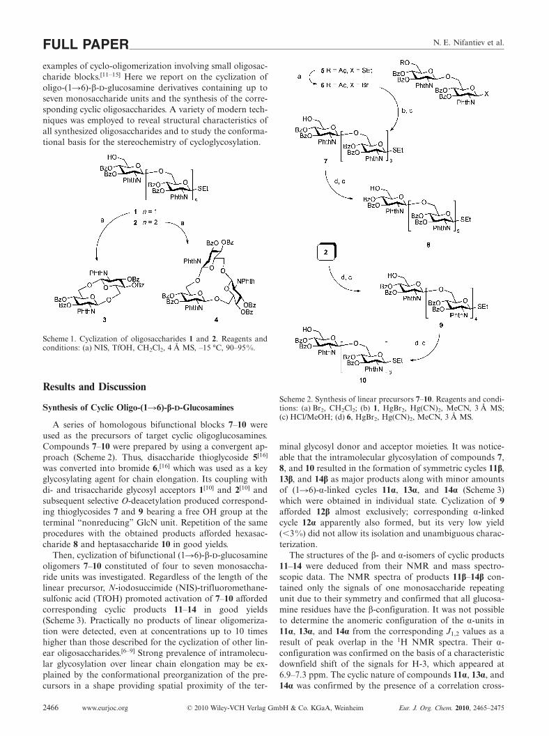
Scheme 1. Cyclization of oligosaccharides 1 and 2. Reagents andconditions: (a) NIS, TfOH, CH2Cl2, 4 Å MS, –15 °C, 90–95%.
Results and Discussion
Synthesis of Cyclic Oligo-(1�6)-β-D-Glucosamines
A series of homologous bifunctional blocks 7–10 wereused as the precursors of target cyclic oligoglucosamines.Compounds 7–10 were prepared by using a convergent ap-proach (Scheme 2). Thus, disaccharide thioglycoside 5[16]
was converted into bromide 6,[16] which was used as a keyglycosylating agent for chain elongation. Its coupling withdi- and trisaccharide glycosyl acceptors 1[10] and 2[10] andsubsequent selective O-deacetylation produced correspond-ing thioglycosides 7 and 9 bearing a free OH group at theterminal “nonreducing” GlcN unit. Repetition of the sameprocedures with the obtained products afforded hexasac-charide 8 and heptasaccharide 10 in good yields.
Then, cyclization of bifunctional (1�6)-β--glucosamineoligomers 7–10 constituted of four to seven monosaccha-ride units was investigated. Regardless of the length of thelinear precursor, N-iodosuccimide (NIS)-trifluoromethane-sulfonic acid (TfOH) promoted activation of 7–10 affordedcorresponding cyclic products 11–14 in good yields(Scheme 3). Practically no products of linear oligomeriza-tion were detected, even at concentrations up to 10 timeshigher than those described for the cyclization of other lin-ear oligosaccharides.[6–9] Strong prevalence of intramolecu-lar glycosylation over linear chain elongation may be ex-plained by the conformational preorganization of the pre-cursors in a shape providing spatial proximity of the ter-
www.eurjoc.org © 2010 Wiley-VCH Verlag GmbH & Co. KGaA, Weinheim Eur. J. Org. Chem. 2010, 2465–24752466
Scheme 2. Synthesis of linear precursors 7–10. Reagents and condi-tions: (a) Br2, CH2Cl2; (b) 1, HgBr2, Hg(CN)2, MeCN, 3 Å MS;(c) HCl/MeOH; (d) 6, HgBr2, Hg(CN)2, MeCN, 3 Å MS.
minal glycosyl donor and acceptor moieties. It was notice-able that the intramolecular glycosylation of compounds 7,8, and 10 resulted in the formation of symmetric cycles 11β,13β, and 14β as major products along with minor amountsof (1�6)-α-linked cycles 11α, 13α, and 14α (Scheme 3)which were obtained in individual state. Cyclization of 9afforded 12β almost exclusively; corresponding α-linkedcycle 12α apparently also formed, but its very low yield(�3%) did not allow its isolation and unambiguous charac-terization.
The structures of the β- and α-isomers of cyclic products11–14 were deduced from their NMR and mass spectro-scopic data. The NMR spectra of products 11β–14β con-tained only the signals of one monosaccharide repeatingunit due to their symmetry and confirmed that all glucosa-mine residues have the β-configuration. It was not possibleto determine the anomeric configuration of the α-units in11α, 13α, and 14α from the corresponding J1,2 values as aresult of peak overlap in the 1H NMR spectra. Their α-configuration was confirmed on the basis of a characteristicdownfield shift of the signals for H-3, which appeared at6.9–7.3 ppm. The cyclic nature of compounds 11α, 13α, and14α was confirmed by the presence of a correlation cross-

Studies of Cyclic Oligo-(1�6)-β--Glucosamines
Scheme 3. Cyclization of oligosaccharides 7–10. Reagents and conditions: (a) NIS, TfOH, CH2Cl2, 4 Å MS, –15 °C.
peak between H-1 of the α-residue with H-6 of another glu-cosamine residue in the ROESY spectra. The absence of thesignals for non-glycosylated C-6 in the 13C NMR spectra,which are typically observed in the range of 60–62 ppm,also supported the cyclic structure of these compounds.
Conformational Analysis of Linear Tetrasaccharide 7
The formation of the α-products upon the cyclization ofbifunctional blocks 7–10 was unexpected because they con-tain a strongly participating N-phthaloyl blocking group atC-2 that provides stereospecific β-glycosylation, especiallyif a primary OH group is involved. To the best of ourknowledge, only one example of α-glycoside bond forma-tion upon glycosylation with the glycosyl donors having theparticipating N-phthaloyl group has been described so farwithout any explanation of the observed stereochemicaloutcome.[17] We supposed that the α-linked cycles formed asa result of conformational features of the linear precursors,which were illustrated by the example of tetrasaccharide 7.The spatial structure of (1�6)-linked pyranosides (such ascompound 7) is primarily described by the dihedral anglesφ, ψ, and ω around the bridges between the monosaccha-ride residues (Figure 1). Conformations of pyranoside ringschange slightly and are considered to have little influenceon the conformation of the whole saccharide molecule.[18]
The values of these dihedral angles are related with the ex-perimentally measured coupling constant values JH,H andJC,H around the glycosidic linkages through the Karplusequation[19,20] (Figure 1). Hence, we performed the analysisof the values of JH,H and JC,H calculated from moleculardynamics (MD) simulations and experimental ones. Theexperimental values of 3JH,H were measured from the1H NMR spectra and the values of long-range couplingconstants 3JC,H were determined by employing 2D J-HMBC[21,22] NMR techniques. The detailed description ofthe NMR technique for the measurement of transglycosidiccoupling constants in oligosaccharides is given in our pre-vious work[23] and in the Experimental Section of this arti-
Eur. J. Org. Chem. 2010, 2465–2475 © 2010 Wiley-VCH Verlag GmbH & Co. KGaA, Weinheim www.eurjoc.org 2467
cle. At the beginning, we obtained experimental values oftransglycosidic constants for the molecule in CD2Cl2 solu-tion at 268 K (Table 1) because these conditions are closeto those used for cyclization of tetrasaccharide 7. However,we could not determine in this case some constant valuesof interest (Table 1) due to the overlap of the signals. There-fore, the values of transglycosidic constants were addition-ally measured for compound 7 in CDCl3 solution at 303 K(Table 2). The comparison of the constants for CD2Cl2 andCDCl3 has shown that the corresponding values are closeto each other (usually the difference does not exceed 1 Hz),thus revealing the conformational similarity of 7 in thesetwo solvents.
Figure 1. Dihedral angles and the Karplus equations for the cou-pling constants 3JC,H
[19] and 3JH,H[20] describing the conformation
of the (1�6)-linkage.
Analysis of the conformers obtained from MD simula-tions for tetrasaccharide 7 shows that the conformations ofthe C1–O6 bonds are most rigid. For these bonds the typi-cal values of the φ angles are in the range from +20° to+50°, which is in accordance with the exoanomeric effect.Rotation around the O6–C6 bond (angles ψR and ψS, Fig-ure 1) results in two conformers with ψR and ψS angles of

N. E. Nifantiev et al.FULL PAPERTable 1. Experimental (CD2Cl2, 268 K) and calculated (in paren-theses) values of transglycosidic 3J coupling constants [Hz] forcompound 7.
Bond[a] Jφ JψR JψS JH-5,H-6R JH-5,H-6S
B�A 2.5 (3.3) nd[b] (2.5) 2.3 (2.6) nd[b] (4.5) 2.0 (2.2)C�B 2.5 (3.3) nd[b] (2.9) 2.5 (2.4) nd[b] (4.4) 2.9 (2.5)D�C 3.3 (3.2) nd[b] (2.5) 2.7 (2.5) 4.9 (4.6) 3.3 (2.5)
[a] Unit labels are shown in Scheme 4. [b] Not determined due topeak overlap.
Table 2. Experimental values of transglycosidic 3J coupling con-stants [Hz] for compound 7 (CDCl3, 303 K).
Bond[a] Jφ JψR JψS JH-5,H-6R JH-5,H-6S
B�A 3.6 2.8 2.8 5.5 2.4C�B 3.5 3.3 3.0 4.6 3.9D�C 3.5 3.1 2.8 4.8 4.0
[a] Unit labels are shown in Scheme 4.
(–20°, 50°) and (–60°, 20°), respectively. Although transi-tions between these conformers slightly change the shape ofthe saccharide chain, the helical conformation is generallymaintained. Unfolded conformers appear due to the rota-tion around the C6–C5 bond. Two main rotamers are ob-served for this bond with O5–C5–C6–O6 angles ω of about–60° and +60°, respectively; the latter rotamer is responsiblefor the occurrence of unfolded conformations (not shown).Experimentally observed coupling constants are presum-ably averages over the ensemble of all these conformers, asthe observed experimental values of the pairs of JH-5,H-6R/JH-5,H-6S constants cannot be attributed to a single confor-
Scheme 4. Preferential conformation of tetrasaccharide 7 and possible intermediates explaining the stereochemical result of cyclization.Reagents and conditions: (a) NIS, TfOH, CH2Cl2, 4 Å MS, –15 °C.
www.eurjoc.org © 2010 Wiley-VCH Verlag GmbH & Co. KGaA, Weinheim Eur. J. Org. Chem. 2010, 2465–24752468
mation. Calculated average values of constants show ac-ceptable[19] coincidence with experimental ones (Table 1),which also confirms that the studied tetrasaccharide gen-erally has a helical shape, whereas unfolded conformers arepresent in minor quantity. Hence, compound 7 in its prefer-ential conformation represents one turn of a right-handedspiral (Scheme 4), in which C-1 of the glycosylating unit (A)and 6-OH of the glycosyl acceptor residue (D) are spatiallyprearranged in a way favoring α-glycosylation (I) to give11α. However, the anchimeric participation of the phthaloylgroup impedes the immediate formation of α-linked cycle11α and also stabilizes glycosyl cationic intermediate II,which becomes stable enough to survive until reorganiza-tion to conformation III favoring β-attack. The observedstereochemical outcome of the cyclization of tetrasaccha-ride 7 is apparently a result of the competition betweenthese two processes. Similar conformational α-stereocontrolcould be also expected for longer bifunctional blocks 8–10in which the terminal residues are situated on adjacentturns of the right-handed helical structure. The spectra ofprotected saccharides 8–10 are rather complicated, andhence, the accurate measurement of coupling constantsand, subsequently, the determination of torsion angles inthem are impossible. On the other hand, the data obtainedfrom molecular dynamics simulations suggest that the pref-erable orientation around the C5–C6 bond in the structureswith the β(1�6)-glycoside linkage is characterized by ω an-gle values of –60°, thus leading to a helical shape of themolecules. Presumably, the conformational reorganizationof oligosaccharides 8–10 to adopt a conformation suitablefor the intramolecular glycosylation proceeds through inter-

Studies of Cyclic Oligo-(1�6)-β--Glucosamines
mediate conformations, which also favor α-bond formation.The formation of α-linked cycles 11α–14α can be regardedas a specific case of “tethered” glycosylation (for a recentreview see ref.[24]).
Synthesis of Unprotected Cyclic Oligo-(1�6)-β-D-Glucosamines 15–26
The one-step deprotection of cyclic oligosaccharides byhydrazinolysis provided a series of free amines 15–20, which
Figure 2. Final set of synthesized deprotected cyclic oligosaccharides. 1H NMR spectra of compounds (a) 20 (303 K), (b) 19 (303 K),(c) 18 (310 K), (d) 17 (307 K), (e) 16 (307 K), and (f) 15 (303 K).
Table 3. 1H NMR spectroscopic data for cyclic oligosaccharides 15–26 (D2O, 500.13 or 600.13 MHz, 303–310 K).
Chemical shift [δ, ppm] Coupling constants [J, Hz]
H-1 H-2 H-3 H-4 H-5 H-6R H-6S 1,2 2,3 3,4 4,5 5,6R 5,6S 6R,6S
15 4,95 3,33 3,72 4,17 3,85 4,12 3,73 �2[b] 5.7 9.8 9.8 �2.0[b] �2[b] 12.816 4.52 2.73 3.56 3.47 3.42 4.18 3.97 8.1 8.9 9.1 9.1 3.9 �2[b] 12.217 4.74 3.03 3.67 3.72 3.60 4.07 4.14 8.5 8.8 9.2 9.5 2.5[ �2[b] 11.018 4.74 3.04 3.65 3.58 3.61 4.03 4.13 8.4 8.7 10.2 9.4 4.0 �2[b] 11.319 4.73 3.02 3.64 3.55 3.68 3.97 4.21 8.4 8.8 10.0 9.4 4.9 �2[b] 11.520 4.79 3.06 3.67 3.60 3.66 3.96 4.20 8.4 9.0 9.1 9.1 3.7 �2[b] 11.521 4.68 3.94 3.62 4.32 3.72 4.17 3.61 �2[b] 5.7 9.9 9.9 �2.0[b] �2[b] 12.822 4.60 3.64 3.49 3.46 3.48 4.09 3.90 8.4 9.6 nd[a] nd[a] 4.1 �2[b] 12.523 4.57 3.67 3.53 3.52 3.51 3.94 4.02 8.6 nd[a] nd[a] nd[a] 2.3 �2[b] 11.424 4.59 3.67 3.55 3.46 3.53 3.90 4.07 8.3 10.1 9.0 9.4 4.9 �2[b] 11.025 4.54 3.63 3.59 3.40 3.54 3.77 4.10 8.0 8.4 9.1 9.1 6.0 �2[b] 11.126 4.58 3.70 3.57 3.47 3.57 3.82 4.13 8.4 8.6 9.2 9.4 5.2 �2[b] 10.5
[a] Not determined due to peak overlap. [b] The small value of the coupling constant could not be measured exactly because of linebroadening of the signals in the 1H NMR spectra.
Eur. J. Org. Chem. 2010, 2465–2475 © 2010 Wiley-VCH Verlag GmbH & Co. KGaA, Weinheim www.eurjoc.org 2469
were isolated as acetic acid salts. Further N-acetylation re-sulted in the formation of corresponding N-acetylated de-rivatives 21–26 (Figure 2). The structures of the thus ob-tained cyclic oligosaccharides were confirmed by mass spec-trometry and 1H and 13C NMR spectroscopy (Tables 3 and4, Figure 3). The NMR spectra were acquired at a tempera-ture of 303–310 K. For each compound, the temperaturewas chosen so as to exclude the overlap of the solvent signal(HOD) with the anomeric proton signal of the cyclic glu-cosamines in the 1H NMR spectrum.

N. E. Nifantiev et al.FULL PAPERTable 4. 13C NMR chemical shifts [δ, ppm] for cyclic oligosaccha-rides 15–26 (D2O, 125.75 or 150.90 MHz, 303–310 K).
C-1 C-2 C-3 C-4 C-5 C-6
15 100.1 60.1 71.8 67.6 79.4 70.016 102.4 57.4 75.7 70.5 76.0 69.317 100.2 56.6 73.1 69.8 75.7 69.418 100.9 56.9 73.4 70.6 76.2 69.819 100.8 56.8 73.5 70.8 75.7 69.520 100.8 56.8 73.2 70.8 75.9 69.521 103.3 59.9 74.3 67.9 78.1 71.322 101.4 57.0 75.2 70.6 75.8 69.723 102.3 56.7 75.0 70.5 75.9 69.524 102.8 56.8 74.8 71.0 76.2 69.925 102.6 56.9 74.7 71.3 75.7 70.226 102.7 56.8 74.9 71.3 75.9 69.8
Figure 3. 1H NMR spectra of compounds (a) 20 (303 K), (b) 19(303 K), (c) 18 (310 K), (d) 17 (307 K), (e) 16 (307 K), and (f) 15(303 K)..
NMR Spectroscopy and Theoretical ConformationalStudies of Cyclic Oligo-(1�6)-β-D-glucosamines 15–26
The NMR spectra of all β-cycles contained the signalsof one monosaccharide repeating unit, thus revealing theequivalence of all units within the cycle (Figure 3, Tables 3and 4, and Supporting Information). Changes in the 1HNMR spectra within the series of 15–20 and 21–26 reflectthe changes in their conformational strain. Indeed, the in-
www.eurjoc.org © 2010 Wiley-VCH Verlag GmbH & Co. KGaA, Weinheim Eur. J. Org. Chem. 2010, 2465–24752470
crease in the cycle size from two to seven units is ac-companied by changes in the localization of the signals andcoupling constants values (Figure 3). It is also noticeablethat in the case of disaccharides 15 and 21 the couplingconstants 3JH,H within the glucosamine residues (Table 3)have atypical values for the glucopyranose in the 4C1 con-formation. This fact could be explained by conformationaldistortions of the pyranose rings in disaccharide cycles 15and 21 due to steric hindrance. Similar distortion was al-ready observed for cyclogentiobioside.[7,8]
To investigate the conformational properties of cyclicoligoglucosamines 15–26, we performed analysis of the ex-perimental values of transglycosidic constants 3JH,H and3JC,H (Table 5). The experimental constants for small cycleschanged significantly upon enlargement of the molecules.On the contrary, the constants for the cycles with five andmore monosaccharide residues in the molecules coincidedwithin the series of free oligoglucosamines 18–20 (in theform of ammonium salts) and the series of N-acetylatedderivatives 24–26. The spectral characteristics of these largecycles (Tables 3 and 4) are similar to those for internal unitsof the large linear oligoglucosamines described before.[16]
Table 5. Experimental transglycosidic coupling constants 3JC,H and3JH,H [Hz] for cycles 15–26.
Jφ JψR JψS JH-5,H-6R JH-5,H-6S
15 4.3 2.2 6.0 �2.0[a] �2[a]
16 3.8 3.2 5.2 3.9 �2[a]
17 4.9 2.3 2.4 2.5 �2[a]
18 3.8 2.7 2.8 4.0 �2[a]
19 3.9 3.9 2.5 4.9 �2[a]
20 3.8 3.5 3.2 3.7 �2[a]
21 3.4 2.3 6.4 �2.0[a] �2[a]
22 3.7 2.7 6.1 4.1 �2[a]
23 4.9 2.5 3.5 2.3 �2[a]
24 4.4 3.6 2.8 4.9 �2[a]
25 3.5 3.7 2.4 6.0 �2[a]
26 3.9 3.5 2.8 5.2 �2[a]
[a] The small value of the coupling constant could not be measuredexactly because of line broadening of the signals in the 1H NMRspectra.
One of the main problems during the study of free oligo-glucosamines 15–20 relates to the susceptibility of their am-monium salts to partial hydrolysis in water solution withthe formation of free oligoglucosamines. We carried outmolecular dynamics (MD) simulations for their fully pro-tonated derivatives (NH3
+ forms). Theoretical simulation ofcompounds 15–26 by using MD revealed that di-, tri-, andtetrasaccharides existed mostly in symmetrical ring-shapedconformations. Larger cycles tended to adopt more compli-cated shapes and the existence in conformations similar tothat of 23 was unlikely for them. Row a in Figure 4 is com-posed of wireframe drawings that are superimpositions ofdifferent shape representatives found in MD trajectories forthese molecules. It demonstrates the increasing flexibility ofglycosidic linkages in the studied cycles upon their enlarge-ment. After geometry optimizations of the structures gener-ated during the MD simulations, global minima were foundfor each molecule. Those for saccharides containing four to
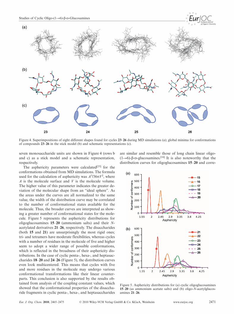
Studies of Cyclic Oligo-(1�6)-β--Glucosamines
Figure 4. Superimpositions of eight different shapes found for cycles 23–26 during MD simulations (a); global minima for conformationsof compounds 23–26 in the stick model (b) and schematic representations (c).
seven monosaccharide units are shown in Figure 4 (rows band c) as a stick model and a schematic representation,respectively.
The asphericity parameters were calculated[25] for theconformations obtained from MD simulations. The formulaused for the calculation of asphericity was A3/36πV2, whereA is the molecule surface and V is the molecule volume.The higher value of this parameter indicates the greater de-viation of the molecular shape from an “ideal sphere”. Asthe areas under the curves are all normalized to the samevalue, the width of the distribution curve may be correlatedto the number of conformational states available for themolecule. Thus, the broader curves are interpreted as show-ing a greater number of conformational states for the mole-cule. Figure 5 represents the asphericity distributions foroligoglucosamines 15–20 (ammonium salts) and their N-acetylated derivatives 21–26, respectively. The disaccharides(both 15 and 21) are unsurprisingly the most rigid ones;tri- and tetramers have moderate flexibilities, whereas cycleswith a number of residues in the molecule of five and higherseem to adopt a wider range of possible conformations,which is reflected in the broadness of their asphericity dis-tributions. In the case of cyclic penta-, hexa-, and heptasac-charides 18–20 and 24–26 (Figure 5), the distribution curveseven look multicentered. This means that cycles with fiveand more residues in the molecule may undergo variousconformational transformations like their linear counter-parts. This conclusion is also supported by the results ob-tained from analysis of the coupling constant values, whichshowed that the conformational properties of the disaccha-ride fragments in cyclic penta-, hexa-, and heptasaccharides
Eur. J. Org. Chem. 2010, 2465–2475 © 2010 Wiley-VCH Verlag GmbH & Co. KGaA, Weinheim www.eurjoc.org 2471
are similar and resemble those of long chain linear oligo-(1�6)-β--glucosamines.[16] It is also noteworthy that thedistribution curves for oligoglucosamines 15–20 and corre-
Figure 5. Asphericity distributions for (a) cyclic oligoglucosamines15–20 (as ammonium acetate salts) and (b) oligo-N-acetylglucos-amines 21–26.

N. E. Nifantiev et al.FULL PAPERsponding N-acetylated derivatives 21–26 are similar to eachother.
The calculation of the hydrophobicity and hydrophilicitydistribution[26] exemplified by tetramer 23 (Figure 6) hasshown that, unlike the well-known cyclodextrins, cyclo-oligo-(1�6)-β--glucosamines lack the distinct hydro-phobic cavity.
Figure 6. Distribution of the hydrophilic (red) and hydrophobic(blue) areas in molecules of tetrasaccharide 23 (outer diameter is1.16 nm) and β-cyclodextrin (outer diameter is 1.44 nm).
Conclusions
In this paper we have discussed the remarkable ability ofoligo-(1�6)-β--glucosamine derivatives 7–10 to undergointramolecular glycosylation with the formation of a newfamily of functionalized cyclic oligosaccharides. As a resultof their structure and molecular architectonics, these com-pounds may be regarded as convenient scaffolds for the de-sign of conjugates with defined valency, symmetry, and flex-ibility. The absence of a distinct hydrophobic cavity pre-cludes the possibility of the formation of inclusion com-plexes like in cyclodextrins. This may be an advantage forthe use of described cyclo-oligoglucosamines as covalentcarriers. Compounds 24–26 relate structurally to exopoly-saccharide adhesin poly-(1�6)-β--glucosamine (PNAG)produced by Staphylococcus aureus,[27] S. epidermidis,[28] Es-cherichia coli,[29] Bordetella bronchiseptica,[30] Actinobacilluspleuropneumoniae,[31] and Yersinia pestis.[32] Thus, the ob-tained cyclo-oligosaccharides can be also regarded as po-tential inhibitors[33] of the biosynthesis of PNAG.
Experimental SectionGeneral Information: NMR spectra were recorded with BrukerDRX-500 and Bruker Avance 600 instruments. Spectra of protectedoligosaccharides were measured for solutions in CDCl3, and 1HNMR chemical shifts were referenced to the residual signal ofCHCl3. NMR spectra of free oligosaccharides were measured forsolutions in D2O by using acetone (δH = 2.225 ppm, δC =31.45 ppm) as internal standard. HRMS (ESI) were obtained withFinnigan LTQ FT (Thermo Scientific) and MicrOTOF II (BrukerDaltonics) instruments. Optical rotations were measured by usinga JASCO DIP-360 polarimeter at 18–22 °C in CHCl3 in the caseof the protected and partially protected derivatives and in water in
www.eurjoc.org © 2010 Wiley-VCH Verlag GmbH & Co. KGaA, Weinheim Eur. J. Org. Chem. 2010, 2465–24752472
the case of free oligosaccharides. TLC was performed on silica gel60 F254 plates (E. Merck) and visualization was accomplished byusing UV light or by charring with 10% H3PO4 in EtOH. Columnchromatography was carried out on silica gel 60 (40–63 µm, E.Merck). Gel-permeation chromatography of protected oligosaccha-rides was performed with Bio-Beads SX-3 (13 � 450 mm) and Bio-Beads SX-1 (18 �500) (Bio-Rad Laboratories) columns in toluene.Gel-permeation chromatography of free oligosaccharides was per-formed on a column of TSK HW-40 (S) gel (25 �800 mm) in 0.1
AcOH. All reactions involving air- or moisture-sensitive reagentswere carried out by using dry solvents under an atmosphere of dryargon.
Special NMR Experiments: The values of long-range coupling con-stants 3JC,H were measured by employing a 2D J-HMBC[21,22] ex-periment, which was performed in the constant-time version.[21]
The spectral widths were about 2.5 ppm for 1H region and 50 ppmfor 13C region and did not include resonances of acetyl groups inthe case of oligosaccharides 21–26. The data were collected in theecho/antiecho mode. For echo selection the two sinusoidal fieldgradients in a ratio of 5:–3 are applied, and for antiecho selectionthe ratio was –3:5. The length of gradients was 1 ms, and the recov-ery time was 100 µs. The spectra were acquired with 60–100 t1 in-crements and 40–500 scans per increment. A total of 512 pointswere collected during the acquisition time t2. The HMBC prepara-tion delay ∆ for the reliable measurement of a coupling constantsshould be taken at least 60% of inversion values of smallest cou-pling of interest (∆ = 0.6/JC,H
min).[22] Smaller values of ∆ lead tothe overestimation of J because of the antiphase character of thepeaks. ∆ = 300 ms was used that corresponds to JC,H
min = 2.0 Hz.The upscaling coefficient k for determination of nJC,H from thespectra[21] was 30–60. The relaxation delay was 1 s. The third-orderlow-pass J-filter[22] was made on suppression of one bond constants(1JC,H) in the range from 125 Hz to 180 Hz. Sinusoidal field gradi-ent sequence with the ratio +7:–4:–2:–1 was applied during low-pass J-filter. The forward linear prediction to 1024 points was usedin F1 that corresponds to resolution 5–6 Hz, and zero-filling to1024 points was used in F2. The processing was performed with π/2 shifted sine square function in both dimensions. The values of3JH,H were measured from the 1H NMR spectra.
Computer Simulations: MD simulations were carried out in vacuowith dielectric constant 81 by using MM3 force field (TINKER 4.1program suit). Simulation time was 5 ns for each molecule. Snap-shot structures were written every 5 ps and then used for furtheranalysis. The geometry optimization of the snapshots led to 1000structures among which the lowest energy conformer was chosenand used to represent the global minimum (Figure 4). In the caseof linear protected precursor 7, conformational search was firstperformed by using standard dihedral driving of φ and ψ angles.Two starting conformations were used; in the first one all ω angleswere set to –60°, in the other to +60°. Two conformational mapswere thus constructed for each glycosidic linkage with one localminimum found on each map. Six MD trajectories were then writ-ten, each starting from a different local minimum. trans-Glycosidiccoupling constants were calculated for each structure of MD trajec-tories according to the Karplus equation and then averaged.
General Procedure A
Glycosylation Under Helferich Conditions: To a mixture of a glyco-syl acceptor (0.1 mmol), Hg(CN)2 (0.15 mmol), HgBr2
(0.03 mmol), and 3 Å molecular sieves (500 mg) in anhydrousCH3CN (3 mL) and CH2Cl2 (2 mL) was added a solution of a gly-cosyl bromide (0.15 mmol) in CH3CN (2 mL) under an argon at-mosphere. The reaction mixture was stirred for 30 min at room

Studies of Cyclic Oligo-(1�6)-β--Glucosamines
temperature, diluted with CH2Cl2, and washed with 1 aqueousKI, saturated aqueous NaHCO3, and brine. The organic layer wasdried with Na2SO4, filtered, and concentrated. The crude productwas subjected to gel-chromatography in toluene, and the fractionswith the highest molecular weight were collected and concentrated.Purification by silica gel column chromatography (toluene/EtOAc)afforded the corresponding coupling product.
General Procedure B
Selective O-Deacetylation in the Presence of Benzoates: A solutionof the 6-O-acetyl derivative (0.1 mmol) in CH2Cl2 (2 mL) was di-luted with absolute MeOH (2 mL) and then AcCl (0.1 mL,1.4 mmol) was added under cooling with an ice bath. The mixturewas kept for 16 h at room temperature and then concentrated. Theresidue was purified by silica gel column chromatography (EtOAc/toluene) to give the product with a free terminal 6-OH group.
General Procedure C
NIS/TfOH-Promoted Intramolecular Glycosylation–Cyclization: Amixture of the linear precursor (6 µmol), anhydrous dichlorometh-ane (1 mL), and 4 Å molecular sieves (250 mg) was stirred underan argon atmosphere at room temperature. After 30 min, NIS (12µmol) and TfOH (6 µmol) were added at –15 °C. The reaction mix-ture was stirred at –15 °C for an additional 30 min and thenquenched with pyridine (5 µL), diluted with chloroform (20 mL),and filtered through a Celite layer. The filtrate was washed with 1
aqueous Na2S2O3, dried (Na2SO4), and concentrated. The crudeproduct was purified by silica gel column chromatography (toluene/EtOAc) to give the cyclo-oligosaccharide. The molecular weighthomogeneity and the absence of oligomerization products were es-tablished by gel-permeation chromatography of the crude productsin toluene. The product was further purified either by silica gelcolumn chromatography (toluene/EtOAc) or by C-18 reverse-phasechromatography (95% aq. CH3CN).
Ethyl 3,4-Di-O-benzoyl-2-deoxy-2-phthalimido-β-D-glucopyranosyl-(1�6)-3,4-di-O-benzoyl-2-deoxy-2-phthalimido-β-D-glucopyranosyl-(1�6)-3,4-di-O-benzoyl-2-deoxy-2-phthalimido-β-D-glucopyranosyl-(1�6)-3,4-di-O-benzoyl-2-deoxy-2-phthalimido-1-thio-β-D-glucopyr-anoside (7): Following general procedure A, coupling of 3[16]
(101 mg, 0.095 mmol) and 6[16] (152 mg, 0.14 mmol) followed bydeacetylation according to general procedure B. Yield: 145 mg(74%). [α]D20 = +8 (c = 1, CHCl3). 1H NMR (500 MHz, CDCl3): δ= 7.95–7.15 (m, 56 H), 6.25 (t, J = 10.0 Hz, 1 H), 6.18 (t, J =10.0 Hz, 1 H), 6.07 (m, 2 H), 5.60 (d, J = 8.4 Hz, 1 H), 5.51 (d, J= 10.5 Hz, 1 H), 5.43 (d, J = 8.4 Hz, 1 H), 5.39 (d, J = 8.4 Hz, 1H), 5.34 (t, J = 9.7 Hz, 1 H), 5.28 (t, J = 9.6 Hz, 1 H), 5.22 (t, J =9.6 Hz, 1 H), 5.18 (t, J = 9.6 Hz, 1 H), 4.50 (t, J = 10.4 Hz, 1 H),4.43 (dd, J = 8.4, 10.5 Hz, 1 H), 4.33 (dd, J = 8.4, 10.5 Hz, 1 H),4.25 (dd, J = 8.4, 10.5 Hz, 1 H), 4.03–3.97 (m, 2 H), 3.96–3.85 (m,4 H), 3.82–3.76 (m, 3 H), 3.68–3.57 (m, 3 H) 5.05 (br. s, 1 H), 2.62(m, 2 H), 1.15 (t, J = 8.5 Hz, 3 H) ppm. 13C NMR (125 MHz,CDCl3): δ = 167.0, 165.6, 165.5, 165.0, 164.9, 134.1, 134.0, 133.8,133.2, 133.1, 131.6, 131.1, 129.8, 129.7, 129.1, 129.0, 128.7, 128.6,128.3, 128.2, 125.2, 123.6, 98.04, 97.6, 97.5, 80.9, 77.2, 74.1, 72.6,72.4, 72.0, 70.9, 70.6, 70.0, 69.8, 67.8, 67.5, 61.2, 54.7, 54.6, 53.8,23.8, 15.0 ppm. C114H90N4O32S (2060.01): calcd. C 66.47, H 4.40,N 2.72; found C 66.29, H 4.50, N 2.67.
Ethyl 3,4-Di-O-benzoyl-2-deoxy-2-phthalimido-β-D-glucopyranosyl-(1�6)-3,4-di-O-benzoyl-2-deoxy-2-phthalimido-β-D-glucopyranosyl-(1�6)-3,4-di-O-benzoyl-2-deoxy-2-phthalimido-β-D-glucopyranosyl-(1�6)-3,4-di-O-benzoyl-2-deoxy-2-phthalimido-β-D-glucopyranosyl-(1�6)-3,4-di-O-benzoyl-2-deoxy-2-phthalimido-β-D-glucopyranosyl-(1�6)-3,4-di-O-benzoyl-2-deoxy-2-phthalimido-1-thio-β-D-glucopyr-
Eur. J. Org. Chem. 2010, 2465–2475 © 2010 Wiley-VCH Verlag GmbH & Co. KGaA, Weinheim www.eurjoc.org 2473
anoside (8): Following general procedure A, coupling of 7 (131 mg,0.064 mmol) and 6[16] (104 mg, 0.096 mmol) and subsequent de-acetylation according to general procedure B. Yield: 104 mg (53%).[α]D20 = +7 (c = 1, CHCl3). 1H NMR (500 MHz, CDCl3): δ = 7.55–7.95 (m, 42 H), 7.12–7.51 (m, 42 H), 6.20 (t, J = 9.8 Hz, 1 H), 6.14(t, J = 9.8 Hz, 1 H), 6.03–6.10 (m, 4 H), 5.55 (d, J = 8.1 Hz, 1 H),5.47 (d, J = 10.4 Hz, 1 H), 5.30–5.5.44 (m, 4 H), 5.11–5.26 (m, 5H), 5.05 (t, J = 8.4 Hz, 1 H), 4.48 (t, J = 10.4 Hz, 1 H), 4.41 (t, J= 8.6 Hz, 1 H), 4.19–4.31 (m, 4 H), 3.50–4.02 (m, 18 H), 2.65 (m,2 H),1.11 (t, 3 H) ppm. 13C NMR (125 MHz, CDCl3): δ = 167.90,167.12, 165.65, 165.56, 165.49, 165.17, 165.05, 164.86, 134.14,133.86, 133.26, 131.10, 129.93, 129.74, 129.22, 129.11, 129.04,128.73, 128.44, 128.22, 125.30, 123.59, 97.90 (C-1), 97.55 (C-1),97.32 (C-1), 80.63 (C-1), 77.30, 74.16, 73.04, 72.74, 72.45, 72.28,72.08, 70.99, 70.69, 70.31, 69.75, 67.98, 67.58, 67.03, 61.30 (C-6),54.72, 54.60, 53.81, 23.71, 14.94 ppm. C170H132N6O48S (3058.94):calcd. C 66.75, H 4.35, N 2.75; found C 66.68, H 4.29, N 2.75.HRMS (ESI): calcd. for C170H132N6O48S [M + Na]+ 3079.7690;found 3079.7685.
Ethyl 3,4-Di-O-benzoyl-2-deoxy-2-phthalimido-β-D-glucopyranosyl-(1�6)-3,4-di-O-benzoyl-2-deoxy-2-phthalimido-β-D-glucopyranosyl-(1�6)-3,4-di-O-benzoyl-2-deoxy-2-phthalimido-β-D-glucopyranosyl-(1�6)-3,4-di-O-benzoyl-2-deoxy-2-phthalimido-β-D-glucopyranosyl-(1�6)-3,4-di-O-benzoyl-2-deoxy-2-phthalimido-1-thio-β-D-glucopyr-anoside (9): Prepared from 4[16] (106 mg, 0.069 mmol) and 6[16]
(108 mg, 0.1 mmol) according to general procedures A and B.Yield: 124 mg (70%). [α]D20 = +5 (c = 1, CHCl3). 1H NMR(500 MHz, CDCl3): δ = 7.61–7.95 (m, 35 H), 7.15–7.60 (m, 35 H),6.00–6.27 (m, 5 H), 5.57 (d, J = 8.4 Hz, 1 H), 5.50 (d, J = 10.5 Hz,1 H), 5.00–5.45 (m, 8 H), 4.40–4.55 (m, 3 H), 4.21–4.35 (m, 2 H),3.53–4.05 (m, 12 H), 3.51–3.70 (m, 3 H), 3.05 (br. s, 1 H, OH), 2.62(m, 2 H),1.10 (t, 3 H) ppm. 13C NMR (125 MHz, CDCl3): δ =167.88, 167.11, 165.66, 165.54, 165.17, 164.93, 134.18, 133.85,133.38, 133.26, 133.09, 131.64, 131.19, 129.95, 129.86, 129.74,129.17, 129.10, 129.03, 128.92, 128.84, 128.72, 128.45, 128.37,128.21, 125.30, 123.59, 97.85 (C-1), 97.47 (C-1), 97.32 (C-1), 80.62(CI-1), 77.27, 74.13, 72.95, 72.53, 72.20, 72.07, 70.95, 70.63, 70.39,69.75, 67.90, 67.74, 67.62, 67.18, 61.25 (CV-6), 54.70, 54.52, 53.80,23.71, 14.95 ppm. C142H111N5O40S (2559.48): calcd. C 66.64, H4.37, N 2.74; found C 66.55, H 4.41, N 2.72. HRMS (ESI): calcd.for C142H111N5O40S [M + Na]+ 2580.6424; found 2580.6418.
Ethyl 3,4-Di-O-benzoyl-2-deoxy-2-phthalimido-β-D-glucopyranosyl-(1�6)-3,4-di-O-benzoyl-2-deoxy-2-phthalimido-β-D-glucopyranosyl-(1�6)-3,4-di-O-benzoyl-2-deoxy-2-phthalimido-β-D-glucopyranosyl-(1�6)-3,4-di-O-benzoyl-2-deoxy-2-phthalimido-β-D-glucopyranosyl-(1�6)-3,4-di-O-benzoyl-2-deoxy-2-phthalimido-β-D-glucopyranosyl-(1�6)-3,4-di-O-benzoyl-2-deoxy-2-phthalimido-β-D-glucopyranosyl-(1�6)-3,4-di-O-benzoyl-2-deoxy-2-phthalimido-1-thio-β-D-glucopyr-anoside (10): Prepared from 9 (223 mg, 0.087 mmol) and 6[16]
(94 mg, 0.131 mmol) according to general procedures A and B.Yield: 140 mg (45%). [α]D20 = +10 (c = 1, CHCl3). 1H NMR(500 MHz, CDCl3): δ = 7.57–8.05 (m, 49 H, Ar), 7.13–7.56 (m, 49H, Ar), 6.05–6.30 (m, 7 H), 5.35–5.62 (m, 8 H), 5.05–5.33 (m, 6H), 4.43–4.60 (m, 6 H), 4.18–4.40 (m, 8 H), 3.53–4.10 (m, 14 H),3.23 (br. s, 1 H, OH), 2.62 (m, 2 H),1.10 (t, 3 H) ppm. 13C NMR(125 MHz, CDCl3): δ = 165.65, 165.56, 165.27, 165.13, 165.08,164.96, 137.91, 134.20, 133.94, 133.31, 133.17, 131.71, 131.26,130.00, 129.80, 129.12, 128.86, 128.80, 128.46, 128.29, 125.37,123.64, 98.01 (C-1), 97.55 (C-1), 97.35 (C-1), 80.71 (C-1), 77.37,74.20, 73.15, 72.89, 72.75, 72.61, 72.29, 72.15, 71.07, 70.72, 70.35,70.24, 69.81, 67.98, 67.58, 67.21, 61.30 (C-6), 54.80, 53.86, 29.77,23.69 (SCH2CH3), 14.93 (SCH2CH3) ppm. C198H153N7O56S(3558.41): calcd. C 66.83, H 4.33, N 2.76; found C 66.75, H 4.37,

N. E. Nifantiev et al.FULL PAPERN 2.74. HRMS (ESI): calcd. for C198H153N7O56S [M + Na]+
3578.8958; found 3578.8953.
Cyclotetrakis-(1�6)-(3,4-di-O-benzoyl-2-deoxy-2-phthalimido-β-D-glucopyranosyl) (11β) and (11α): Cyclization of 7 (187 mg,0.082 mmol) according to general procedure C afforded 11β(112 mg, 68%) and 11α (28 mg, 17%). Ratio β/α = 4:1. Data for11β: Colorless foam. [α]D20 = +3 (c = 1, CHCl3). 1H NMR(500 MHz, CDCl3): δ = 7.71 (m, 4 H), 7.58 (m, 4 H), 7.37 (m, 2H), 7.22 (m, 4 H), 6.09 (t, J3,2 = J3,4 = 9.5 Hz, 1 H, H-3), 5.77 (d,J1,2 = 8.4 Hz, 1 H, H-1), 5.57 (t, J4,5 = 9.4 Hz, 1 H, H-4), 4.71 (dd,1 H, H-2), 4.16–4.02 (m, 3 H, H-5, 2 H-6) ppm. 13C NMR(125 MHz, CDCl3): δ = 167.9, 165.6, 164.6, 133.7, 132.9, 132.8,131.7, 129.8, 129.6, 129.0, 128.7, 123.3, 98.3 (C-1), 74.6 (C-5), 71.6(C-3), 69.6 (C-4), 68.5 (C-6), 54.3 (C-2) ppm. HRMS (ESI): calcd.for C112H84N4O32 [M + Na]+ 2019.4966; found 2019.4961. Datafor 11α: Colorless foam. [α]D20 = +29 (c = 0.5, CHCl3). 1H NMR(500 MHz, CDCl3): δ = 7.98–7.20 (m, 56 H), 6.86 (br. t, J =10.0 Hz, 1 H, H-3α), 6.33 (t, J = 10.0 Hz, 1 H), 6.25 (t, J = 10.0 Hz,1 H), 5.99 (t, J = 10.4 Hz, 1 H), 5.85 (t, J = 10.4 Hz, 1 H), 5.71(d, J = 8.5 Hz, 1 H), 5.62 (m, 2 H), 5.50 (t, J = 9.6 Hz, 1 H), 5.33(m, 2 H), 5.01 (br. s, 1 H, H-1α), 4.88 (br. t, J = 9.6 Hz, 1 H), 4.82–4.77 (m, 2 H), 4.68–4.62 (m, 2 H), 4.26–4.20 (m, 2 H), 4.17 (m, 2H), 4.00 (m, 1 H), 3.96–3.89 (m, 3 H), 3.87–3.82 (m, 2 H), 3.73 (m,1 H) ppm. 13C NMR (125 MHz, CDCl3): δ = 165.6, 165.46, 164.0,134.0, 133.7, 133.6, 133.4, 133.1, 132.9, 132.7, 132.6, 131.8, 131.7,131.4, 130.1, 129.8, 129.7, 129.4, 129.3, 129.1, 128.9, 128.4, 128.2,128.1, 127.9, 123.6, 98.8, 98.2, 97.8, 97.5, 73.65, 73.36, 73.19, 71.58,71.16, 70.8, 70.2, 69.3, 67.9, 67.6, 65.9, 54.6, 54.2, 53.9 ppm.HRMS (ESI): calcd. for C112H84N4O32 [M + Na]+ 2019.4966;found 2019.4961.
Cyclopentakis-(1�6)-(3,4-di-O-benzoyl-2-deoxy-2-phthalimido-β-D-glucopyranosyl) (12): Cyclization of 9 (96 mg, 0.038 mmol) accord-ing to general procedure C gave pure 12β (59 mg, 63 %) and aninseparable mixture of 12β/12α (20 mg, 21%) in a ratio of ≈6:1 (1HNMR spectra estimation). Data for 12β: Colorless foam. [α]D20 =+58 (c = 1, CHCl3). 1H NMR (600 MHz, CDCl3): δ = 7.75 (m, 4H), 7.62 (m, 4 H), 7.42 (m, 1 H), 7.38 (m, 1 H), 7.25 (m, 2 H), 7.19(m, 2 H), 6.19 (t, J3,2 = J3,4 = 9.9 Hz, 1 H, H-3), 5.62 (t, J4,5 =9.6 Hz, 1 H, H-4), 5.57 (d, J1,2 = 8.3 Hz, 1 H, H-1), 4.69 (dd, 1 H,H-2), 4.15 (d, J6a,6b = 9.1 Hz, 1 H, H-6a), 3.97 (m, 1 H, H-5), 3.87(dd, J6b,5 = 5.2 Hz, 1 H, H-6b) ppm. 13C NMR (150 MHz, CDCl3):δ = 167.8, 164.8, 133.7, 132.9, 132.8, 131.7, 129.8, 129.7, 129.2,128.2, 128.1, 123.5, 97.9 (C-1), 72.9 (C-5), 71.4 (C-3), 70.5 (C-4),68.0 (C-6), 54.7 (C-2) ppm. HRMS (ESI): calcd. for C140H105N5O40
[M + Na]+ 2518.6233; found 2518.6228.
Cyclohexakis-(1�6)-(3,4-di-O-benzoyl-2-deoxy-2-phthalimido-β-D-glucopyranosyl) (13): Cyclization of 8 (72 mg, 0.023 mmol) accord-ing to general procedure C produced 13β (48 mg, 69%) and 13α(8 mg, 11%). Data for 13β: Colorless foam. [α]D20 = +36 (c = 1,CHCl3). 1H NMR (600 MHz, CDCl3): δ = 7.75 (m, 4 H), 7.62 (m,4 H), 7.42 (m, 1 H), 7.38 (m, 1 H), 7.25 (m, 2 H), 7.19 (m, 2 H),6.25 (t, J3,2 = J3,4 = 9.9 Hz, 1 H, H-3), 5.61 (d, J1,2 = 7.9 Hz, 1 H,H-1), 5.54 (br. t, J4,5 = 9.1 Hz, 1 H, H-4), 4.69 (br. t, 1 H, H-2),4.11 (d, J6a,6b = 10.0 Hz, 1 H, H-6a), 4.02 (br. m, 1 H, H-5), 3.95(br. m, 1 H, H-6b) ppm. 13C NMR (150 MHz, CDCl3): δ = 165.6,164.8, 133.7, 132.9, 132.8, 131.7, 129.8, 129.7, 129.2, 128.2, 128.1,123.5, 97.5 (C-1), 73.2 (C-5), 71.4 (C-3), 70.5 (C-4), 66.8 (C-6), 54.6(C-2) ppm. HRMS (ESI): calcd. for C168H126N6O48 [M + Na]+
3017.7500; found 3017.7495. Data for 13α: Colorless foam. 1HNMR (600 MHz, CDCl3) selected signals: δ = 7.28 (H-3 α-GlcN),5.64 (t, J4,3 = J4,5 = 9.5 Hz, 1 H, H-4 α-GlcN), 5.30–5.33 (br. m, 2H, H-1 and H-2 α-GlcN), 4.27 (H-5 α-GlcN) ppm. HRMS (ESI):calcd. for C168H126N6O48 [M + Na]+ 3017.7500; found 3017.7495.
www.eurjoc.org © 2010 Wiley-VCH Verlag GmbH & Co. KGaA, Weinheim Eur. J. Org. Chem. 2010, 2465–24752474
Cycloheptakis-(1�6)-(3,4-di-O-benzoyl-2-deoxy-2-phthalimido-β-D-glucopyranosyl) (14): Cyclization of 10 (66 mg, 0.018mmol) accord-ing to general procedure C afforded 14β (39 mg, 59%) and 14α(8 mg, 10%). Ratio β/α ≈ 6:1. Data for 14β: Colorless foam. [α]D20
= +30 (c = 1, CHCl3). 1H NMR (600 MHz, CDCl3): δ = 7.75 (m,4 H), 7.62 (m, 4 H), 7.42 (m, 1 H), 7.38 (m, 1 H), 7.25 (m, 2 H),7.19 (m, 2 H), 6.18 (t, J3,2 = J3,4 = 9.8 Hz, 1 H, H-3), 5.53 (d, J1,2
= 8.3 Hz, 1 H, H-1), 5.46 (t, J4,5 = 9.6 Hz, 1 H, H-4), 4.59 (dd, 1H, H-2), 4.18 (d, J6a,6b = 9.8 Hz, 1 H, H-6a), 4.03 (m, 1 H, H-5),3.89 (dd, J6b,5 = 5.0 Hz, 1 H, H-6b) ppm. 13C NMR (150 MHz,CDCl3): δ = 167.8, 164.8, 133.7, 132.9, 132.8, 131.7, 129.8, 129.7,129.2, 128.2, 128.1, 123.5, 97.8 (C-1), 73.4 (C-5), 71.4 (C-3), 70.1(C-4), 67.1 (C-6), 54.6 (C-2) ppm. HRMS (ESI): calcd. forC196H147N7O56 [M + Na]+ 3516.8767; found 3516.8762. Data for14α: 1H NMR (600 MHz, CDCl3) selected signals: δ = 7.17 (H-3α-GlcN), 5.45 (H-4 α-GlcN), 5.19 (H-1 α-GlcN) 4.82 (dd, J2,1 =2.8 Hz, J2,3 = 11.1 Hz, 1 H, H-2 α-GlcN), 4.45 (H-5 α-GlcN) ppm.HRMS (ESI): calcd. for C196H147N7O56 [M + 2Na]2+ 1769.9327;found 1769.9327.
Cyclobis-(1�6)-(2-amino-2-deoxy-2-β-D-glucopyranosyl) (15): Pre-pared from 3[16] (49 mg, 0.049 mmol) according to the publishedprocedure for total deprotection.[16] Yield: 15 mg (95%). [α]D20 = –22(c = 1, H2O). HRMS (ESI): calcd. for C12H22N2O8 [M + H]+
323.1449; found 323.1447.
Cyclotris-(1�6)-(2-amino-2-deoxy-2-β-D-glucopyranosyl) (16): Pre-pared from 4[16] (39 mg, 0.026 mmol) according to the publishedprocedure for total deprotection.[16] Yield: 12 mg (95%). [α]D20 =–0.5 (c = 1, H2O). HRMS (ESI): calcd. for C18H33N3O12 [M +H]+ 484.2137; found 484.2136.
Cyclotetrakis-(1�6)-(2-amino-2-deoxy-2-β-D-glucopyranosyl) (17):Prepared from 11β (65.8 mg, 0.033 mmol) according to the pub-lished procedure for total deprotection.[16] Yield: 20 mg (94%).[α]D20 = –30 (c = 1, H2O). HRMS (ESI): calcd. for C24H44N4O16 [M+ H]+ 645.2825; found 645.2826.
Cyclopentakis-(1�6)-(2-amino-2-deoxy-2-β-D-glucopyranosyl) (18):Prepared from 12β (44.9 mg, 0.018 mmol) according the publishedprocedure for total deprotection.[16] Yield: 14 mg (95%). [α]D20 = –22(c = 1, H2O). HRMS (ESI): calcd. for C30H55N5O20 [M + H]+
806.3513; found 806.3510.
Cyclohexakis-(1�6)-(2-amino-2-deoxy-2-β-D-glucopyranosyl) (19):Prepared from 13β (39 mg, 0.013 mmol) according to the publishedprocedure for total deprotection.[16] Yield: 12 mg (96%). [α]D20 = –14(c = 1, H2O). HRMS (ESI): calcd. for C36H66N6O24 [M + 2H]2+
484.2137; found 484.2135.
Cycloheptakis-(1�6)-(2-amino-2-deoxy-2-β-D-glucopyranosyl) (20):Prepared from 14β (121 mg, 0.035 mmol) according to the pub-lished procedure for total deprotection.[16] Yield: 18 mg (92%).[α]D20 = –12 (c = 1, H2O). HRMS (ESI): calcd. for C42H77N7O28 [M+ 2H]2+ 564.7481; found 564.7479.
Cyclobis-(1�6)-(2-acetamido-2-deoxy-2-β-D-glucopyranosyl) (21):Prepared from 15 (5.7 mg, 0.0176 mmol) according to the pub-lished procedure for N-acetylation.[16] Yield: 7 mg (98 %). [α]D20 =+29 (c = 0.5, H2O). HRMS (ESI): calcd. for C16H26N2O10 [M +Na]+ 407.1480; found 407.1481.
Cyclotris-(1�6)-(2-acetamido-2-deoxy-2-β-D-glucopyranosyl) (22):Prepared from 16 (4.9 mg, 0.01 mmol) according to the publishedprocedure for N-acetylation.[16] Yield: 6 mg (97%). [α]D20 = –9 (c =0.5, H2O). HRMS (ESI): calcd. for C24H39N3O15 [M + Na]+
632.2273; found 632.2274.

Studies of Cyclic Oligo-(1�6)-β--Glucosamines
Cyclotetrakis-(1�6)-(2-acetamido-2-deoxy-2-β-D-glucopyranosyl)(23): Prepared from 17 (8.7 mg, 0.012 mmol) according to the pub-lished procedure for N-acetylation.[16] Yield: 10 mg (98%). [α]D20 =–9 (c = 1, H2O). HRMS (ESI): calcd. for C32H52N4O20 [M + H]+
813.3248; found 813.3246.
Cyclopentakis-(1�6)-(2-acetamido-2-deoxy-2-β-D-glucopyranosyl)(24): Prepared from 18 (4.9 mg, 0.0061 mmol) according to thepublished procedure for N-acetylation.[16] Yield: 6 mg (97%). [α]D20
= –10 (c = 1, H2O). HRMS (ESI): calcd. for C40H65N5O25 [M +H]+ 1016.4041; found 1016.4029.
Cyclohexakis-(1�6)-(2-acetamido-2-deoxy-2-β-D-glucopyranosyl)(25): Prepared from 19 (4.9 mg, 0.0051 mmol) according to thepublished procedure for N-acetylation.[16] Yield: 6 mg (97%). [α]D20
= –22 (c = 1, H2O). HRMS (ESI): calcd. for C48H78N6O30 [M +Na]+ 1241.4655; found 1241.4660.
Cycloheptakis-(1�6)-(2-acetamido-2-deoxy-2-β-D-glucopyranosyl)(26): Prepared from 20 (6.5 mg, 0.0057 mmol) according to the ge-neral procedure for N-acetylation.[16] Yield: 8 mg (98%). [α]D20 = –22(c = 1, H2O). HRMS (ESI): calcd. for C56H91N7O35 [M + H]+
1422.5629; found 1422.5633.
Supporting Information (see footnote on the first page of this arti-cle): Copies of the NMR spectra for all new compounds.
Acknowledgments
We are grateful for the financial support of the Russian Foundationfor Basic Research (project no. 07-03-00603), and we acknowledgea grant from the President of the Russian Federation to YoungScientists (MK-5970.2008.3 to A.A.G.). We thank Professor G. B.Pier (Channing Laboratory, Brigham and Women’s Hospital, Har-vard Medical School, Boston, MA) for stimulating discussions.
[1] G. G. Gattuso, S. A. Nepogodiev, J. F. Stoddart, Chem. Rev.1998, 98, 1919–1958.
[2] W. A. Smit, A. F. Bochkov, R. Caple (Eds.), Organic Synthesis:The Science Behind the Art, The Royal Society of Chemistry,Cambridge, 1998, pp. 173–177.
[3] M. Mori, Y. Ito, T. Ogawa, Tetrahedron Lett. 1989, 30, 1273–1276.
[4] M. Mori, Y. Ito, T. Ogawa, Carbohydr. Res. 1989, 192, 131–146.
[5] M. Mori, Y. Ito, J. Uzawa, T. Ogawa, Tetrahedron Lett. 1990,31, 3191–3194.
[6] N. K. Kochetkov, S. A. Nepogod�ev, L. V. Backinowsky, Tetra-hedron 1990, 46, 139–150.
[7] D. Gagnaire, V. Tran, M. Vignon, J. Chem. Soc., Chem. Com-mun. 1976, 6–7.
[8] D. Gagnaire, M. Vignon, Carbohydr. Res. 1976, 51, 140–144.
Eur. J. Org. Chem. 2010, 2465–2475 © 2010 Wiley-VCH Verlag GmbH & Co. KGaA, Weinheim www.eurjoc.org 2475
[9] S. Houdier, P. J. A. Vottero, Angew. Chem. 1994, 106, 365–367;Angew. Chem. Int. Ed. Engl. 1994, 33, 354–356.
[10] M. L. Gening, Y. E. Tsvetkov, G. B. Pier, N. E. Nifantiev, Russ.J. Bioorg. Chem. 2006, 32, 432–443.
[11] K. S. Kim, B.-Y. Lee, S. H. Yoon, H. J. Jeon, J. Y. Baek, K.-S.Jeong, Org. Lett. 2008, 10, 2373–2376.
[12] P. R. Ashton, C. L. Brown, S. Menzer, S. A. Nepogodiev, J. F.Stoddart, D. J. Williams, Chem. Eur. J. 1996, 2, 580–591.
[13] P. R. Ashton, S. J. Cantrill, G. Gattuso, S. Menzer, S. A. Nepo-godiev, A. N. Shipway, J. F. Stoddart, D. J. Williams, Chem.Eur. J. 1997, 3, 1299–1314.
[14] G. Gattuso, S. Menzer, S. A. Nepogodiev, J. F. Stoddart, D. J.Williams, Angew. Chem. Int. Ed. Engl. 1997, 36, 1451–1454.
[15] S. A. Nepogodiev, G. Gattuso, J. F. Stoddart, J. Inclusion Phe-nom. Mol. Recognit. Chem. 1996, 25, 47–52.
[16] M. L. Gening, Y. E. Tsvetkov, G. B. Pier, N. E. Nifantiev, Car-bohydr. Res. 2007, 342, 567–575.
[17] Y. Ohnishi, H. Ando, T. Kawai, Y. Nakahara, Y. Ito, Car-bohydr. Res. 2000, 328, 263–276.
[18] U. Olsson, E. Sáwén, R. Stenutz, G. Widmalm, Chem. Eur. J.2009, 15, 8886–8894.
[19] B. Mulloy, T. A. Frenkiel, D. B. Davies, Carbohydr. Res. 1988,184, 39–46.
[20] R. Stenutz, I. Carmichael, G. Widmalm, A. S. Serianni, J. Org.Chem. 2002, 67, 949–958.
[21] C. H. Gotfredsen, A. Meissner, J. Ø. Duus, O. W. Sørensen,Magn. Reson. Chem. 2000, 38, 692–695.
[22] A. Meissner, O. W. Sørensen, Magn. Reson. Chem. 2001, 39,49–52.
[23] A. A. Grachev, A. G. Gerbst, N. E. Ustyuzhanina, E. A.Khatuntseva, A. S. Shashkov, A. I. Usov, N. E. Nifantiev, J.Carbohydr. Chem. 2005, 24, 85–100.
[24] X. Zhu, R. R. Schmidt, Angew. Chem. Int. Ed. 2009, 48, 1900–1934.
[25] K. J. Najdoo, M. Kuttel, J. Comput. Chem. 2001, 22, 445–456.[26] V. N. Viswanadhan, A. K. Ghose, G. R. Revankar, R. K. Rob-
ins, J. Chem. Inf. Comput. Sci. 1989, 29, 163–172.[27] N. Cerca, K. K. Jefferson, T. Maira-Litran, D. B. Pier, C.
Kelly-Quintos, D. A. Goldmann, J. Azeredo, G. B. Pier, Infect.Immun. 2007, 75, 3406–3413.
[28] D. Mack, W. Fischer, A. Krokotsch, K. Leopold, R. Hart-mann, H. Egge, R. Laufs, J. Bacteriol. 1996, 178, 175–183.
[29] X. Wang, J. F. Preston, T. Romeo, J. Bacteriol. 2004, 186, 2724–2734.
[30] G. P. Sloan, C. F. Love, N. Sukurnar, M. Mishra, R. Deora, J.Bacteriol. 2007, 189, 8270–8276.
[31] E. A. Izano, I. Sadovskaya, E. Vinogradov, M. H. Mulks, K.Velllyagounder, C. Ragunath, W. B. Kher, N. Ramasubbu, S.Jabbouri, M. B. Perry, J. B. Kaplan, Microb. Pathog. 2007, 43,1–9.
[32] Y. Itoh, X. Wang, B. J. Hinnebusch, J. F. Preston, T. Romeo, J.Bacteriol. 2005, 187, 382–387.
[33] T. Oguma, H. Kawamoto, Trends Glycosci. Glycotechnol. 2003,15, 91–99.
Received: November 6, 2009Published Online: March 17, 2010