Synthesis, antimicrobial evaluation and QSAR studies of gallic acid derivatives
Molecules 2012, 17, 2408-2427; doi:10.3390/molecules17032408
molecules ISSN 1420-3049
www.mdpi.com/journal/molecules
Article
Synthesis, Characterization, X-ray Crystallography, Acetyl Cholinesterase Inhibition and Antioxidant Activities of Some Novel Ketone Derivatives of Gallic Hydrazide-Derived Schiff Bases
Nura Suleiman Gwaram 1,*, Hapipah Mohd Ali 1, Mahmood Ameen Abdulla 2,
Michael J. C. Buckle 3, Sri Devi Sukumaran 3, Lip Yong Chung 3, Rozana Othman 3,
Abeer A. Alhadi 3, Wageeh A. Yehye 1, A. Hamid A. Hadi 1, Pouya Hassandarvish 2,
Hamid Khaledi 1 and Siddig Ibrahim Abdelwahab 3,*
1 Department of Chemistry, Faculty of Science, University of Malaya, Kuala Lumpur 50603, Malaysia 2 Department of Molecular Medicine, Faculty of Medicine, University of Malaya,
Kuala Lumpur 50603, Malaysia 3 Departments of Pharmacy, Faculty of Medicine, University of Malaya, Kuala Lumpur 50603, Malaysia
* Authors to whom correspondence should be addressed;
E-Mails: [email protected] (N.S.G.); [email protected] (S.I.A.);
Tel.: +60-12-656-5990.
Received: 28 January 2012; in revised form: 21 February 2012 / Accepted: 23 February 2012 /
Published: 28 February 2012
Abstract: Alzheimer’s disease (AD) is the most common form of dementia among older
people and the pathogenesis of this disease is associated with oxidative stress.
Acetylcholinesterase inhibitors with antioxidant activities are considered potential
treatments for AD. Some novel ketone derivatives of gallic hydrazide-derived Schiff bases
were synthesized and examined for their antioxidant activities and in vitro and in silico
acetyl cholinesterase inhibition. The compounds were characterized using spectroscopy
and X-ray crystallography. The ferric reducing antioxidant power (FRAP) and
2,2-diphenyl-1-picrylhydrazyl (DPPH) assays revealed that all the compounds
have strong antioxidant activities. N-(1-(5-bromo-2-hydroxyphenyl)-ethylidene)-3,4,5-
trihydroxybenzohydrazide (2) was the most potent inhibitor of human acetyl
cholinesterase, giving an inhibition rate of 77% at 100 μM. Molecular docking simulation
of the ligand-enzyme complex suggested that the ligand may be positioned in the enzyme’s
active-site gorge, interacting with residues in the peripheral anionic subsite (PAS) and acyl
OPEN ACCESS
Molecules 2012, 17 2409
binding pocket (ABP). The current work warrants further preclinical studies to assess the
potential for these novel compounds for the treatment of AD.
Keywords: gallic hydrazide Schiff bases; AChE inhibition; antioxidant study;
molecular docking
1. Introduction
The chemistry of hydrazones is an intensive area of study and numerous Schiff base ligands and their
complexes of this type have been synthesized and their biological applications reported [1–5]. The major
antioxidants currently used in foods are monohydroxy or polyhydroxy phenol compounds with various
ring substitutions. These compounds have low activation energy for hydrogen donation [6]. Mainly from
in vitro studies, polyphenols have been reported to have antioxidant [6–8], anti-cancer [9,10] and
cardioprotecive activities [11].
Antioxidants such as vitamins A, E and C prevent the formation of free radicals and/or neutralize
those that are formed, thus they break radical chains. They also repair the damage caused by free
radicals, such as the DNA repair enzymes, e.g., transferases. Natural antioxidants are present in foods,
but synthetic antioxidants may either be added to food to extend its shelf-life, or prepared by extraction
from plant sources to be taken as supplements in concentrated form [8]. A number of studies have
investigated a range of antioxidant agents in the hope of finding better and more effective treatments
against AD [12]. Work has tended to focus on dietary antioxidants such as vitamins A, C, and E.
Though these appear to have some benefits, results have proved frustratingly inconclusive [13].
Studies of many other dietary antioxidants polyphenols have also shown promise but, once more, their
worth is yet unproven [14].
Researchers have recently investigated the potential health benefits of polyphenols in organic
product [15]. Increased consumption of polyphenols has been associated with a reduced risk of
cardiovascular disease and possibly cancer and stroke. Laboratory findings have shown that oxidative
stress may play an important role to the pathogenesis of AD. Therefore, the risk of AD disease might
be decreased by intake of antioxidants that neutralize the unfavorable effects of oxidative stress [16].
The present work reports the synthesis, characterization, antioxidants activities and X-ray crystal
structures of Schiff bases derived from the condensation reaction of gallic hydrazide with pyridine and
acetophenone derivatives, together with their acetylcholinesterase inhibition and antioxidant activity.
2. Results and Discussion
2.1. Chemistry
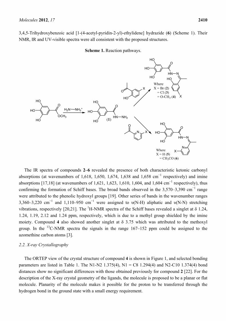
The reaction of gallic hydrazide (1) with selected hydroxyacetophenones and pyridine derivatives
resulted in the formation of the corresponding polyphenolic compounds: N-(1-(5-Bromo-2-
hydroxyphenyl)-ethylidene)-3,4,5-trihydroxybenzohydrazide (2); N-(1-(5-Chloro-2-hydroxyphenyl)-
ethylidene)-3,4,5-trihydroxybenzohydrazide (3); N-(1-(2-Hydroxy-5-methoxyphenyl)-ethylidene)-3,4,5-
trihydroxybenzohydrazide (4); 3,4,5-Trihydroxybenzoic acid [1-pyridylethylidene] hydrazide (5);
Molecules 2012, 17 2410
3,4,5-Trihydroxybenzoic acid [1-(4-acetyl-pyridin-2-yl)-ethylidene] hydrazide (6) (Scheme 1). Their
NMR, IR and UV-visible spectra were all consistent with the proposed structures.
Scheme 1. Reaction pathways.
HO
HO
HO
O
HN NH2
HO
HO
HO
O
OCH3
NH3+H2N
O
N X
O
HO
X
HO
HO
HO
O
HN N
HO
X
Where X = Br (2) = Cl (3) = O-CH3 (4)
HO
HO
HO
O
HN N
NXWhere
X = H (5) = CH3CO (6)
(1)
The IR spectra of compounds 2–6 revealed the presence of both characteristic ketonic carbonyl
absorptions (at wavenumbers of 1,618, 1,650, 1,674, 1,638 and 1,658 cm−1 respectively) and imine
absorptions [17,18] (at wavenumbers of 1,621, 1,623, 1,610, 1,604, and 1,604 cm−1 respectively), thus
confirming the formation of Schiff bases. The broad bands observed in the 3,570–3,390 cm−1 range
were attributed to the phenolic hydroxyl groups [19]. Other series of bands in the wavenumber ranges
3,360–3,220 cm−1 and 1,110–950 cm−1 were assigned to υ(N-H) aliphatic and υ(N-N) stretching
vibrations, respectively [20,21]. The 1H-NMR spectra of the Schiff bases revealed a singlet at δ 1.24,
1.24, 1.19, 2.12 and 1.24 ppm, respectively, which is due to a methyl group shielded by the imine
moiety. Compound 4 also showed another singlet at δ 3.75 which was attributed to the methoxyl
group. In the 13C-NMR spectra the signals in the range 167–152 ppm could be assigned to the
azomethine carbon atoms [3].
2.2. X-ray Crystallography
The ORTEP view of the crystal structure of compound 4 is shown in Figure 1, and selected bonding
parameters are listed in Table 1. The N1-N2 1.375(4), N1 = C8 1.294(4) and N2-C10 1.374(4) bond
distances show no significant differences with those obtained previously for compound 2 [22]. For the
description of the X-ray crystal geometry of the ligands, the molecule is proposed to be a planar or flat
molecule. Planarity of the molecule makes it possible for the proton to be transferred through the
hydrogen bond in the ground state with a small energy requirement.

Molecules 2012, 17 2411
Figure 1. ORTEP-type view of the crystal structure of compound 4 showing the labeling
scheme. Thermal ellipsoids are drawn at the 50% probability level.
Table 1. Selected bond distances (Å) and angles (°) for compound 4.
DistancesN1-N2 1.375(4)N1-C8 1.294(4)N2-C10 1.374(4)C10-O3 1.231(4)
AnglesC8-N1-N2 121.0(3) C10-N2-N1 116.6(3)O3-C10-N2 120.5(3)O2-C5-C6 123.0(3)
The crystal data and structure refinement for compound 4 is summarized in Table 2.
Table 2. Crystal data and structure refinement for 4 and 5.
Identification code 4 5
Empirical formula C16H18N2O7 C14H12N3O5
Formula weight 350.32 302.27
Temperature/K 569(2) 296.0
Crystal system Monoclinic triclinic
Space group P21/n P-1
a/Å 7.769(9) 6.6282(7)
b/Å 15.509(19) 9.9326(7)
c/Å 13.162(16) 12.8906(11)
α/° 90.00 79.982(2)
β/° 103.85(2) 80.995(2)
γ/° 90.00 76.5060(10)
Molecules 2012, 17 2412
Table 2. Cont.
Identification code 4 5
Volume/Å3 1,540(3) 806.67(12)
Z 4 2
ρcalcmg/mm3 1.511 1.244
m/mm-1 0.120 0.097
F(000) 736 314.0
Crystal size/mm3 0.28 × 0.05 × 0.01 0.33 × 0.14 × 0.11
2Θ range for data collection 4.14 to 50° 3.24 to 61.08°
Index ranges −5 ≤ h ≤ 9, −18 ≤ k ≤ 18, −15 ≤ l ≤ 14 −8 ≤ h ≤ 6, −14 ≤ k ≤ 10, −17 ≤ l ≤ 17
Reflections collected 7,052 2,552
Independent reflections 2,704[R(int) = 0.0721] 2,237[R(int) = 0.0358]
Data/restraints/parameters 2,704/3/241 2,237/12/254
Goodness-of-fit on F2 0.986 0.804
Final R indexes [I >= 2σ (I)] R1 = 0.0532, wR2 = 0.1121 R1 = 0.0462, wR2 = 0.1239
Final R indexes [all data] R1 = 0.1117, wR2 = 0.1358 R1 = 0.0555, wR2 = 0.1354
Largest diff. peak/hole/e Å−3 0.305/−0.264 0.43/−0.34
2.3. Anti-AChE Assay
The inhibitory activities of compounds 1–6 on human acetyl cholinesterase were in the range of
16–77% at 100 μM (see Table 3) and thus comparable to those of the standard drugs tacrine
and propidium.
Table 3. Human AChE inhibitory effects and anti-oxidant activities for compounds 1–6.
Compounds Molecular
weight AChE Inhibition (%)
(Final conc. = 1 × 10⁻⁴ M) DPPH
(IC50, μg/mL) FRAP value (Mean ± SD)
1 184.15 38.0 ± 1.3 1.210 ± 0.002 81,633.30 ± 0.075 2 381.18 77.0 ± 1.8 1.140 ± 0.001 62,200.00 ± 0.083 3 336.73 68.9 ± 1.8 1.400 ± 0.002 35,740.00 ± 0.011 4 332.31 48.5 ± 2.5 1.220 ± 0.001 30,080.00 ± 0.054 5 287.27 16.4 ± 1.4 1.460 ± 0.001 22,946.70 ± 0.004 6 329.31 71.5 ± 1.7 2.300 ± 0.001 23,340.00 ± 0.021 Propidium - 54.5 ± 1.6 - - Tacrine - 51.2 ± 1.6 - - Ascorbic acid - - 2.260 ± 0.001 19,400.00 ± 0.007 BHT - - - 187.3 ± 2.6
1: Gallic hydrazide; 2: N-(1-(5-Bromo-2-hydroxyphenyl)-ethylidene)-3,4,5-trihydroxybenzohydrazide; 3: N-(1-(5-Chloro-2-hydroxyphenyl)-ethylidene)-3,4,5-trihydroxybenzohydrazide; 4: N-(1-(2-Hydroxy- 5-methoxyphenyl)-ethylidene)-3,4,5-trihydroxybenzohydrazide; 5: 3,4,5-Trihydroxybenzoic acid [1-pyridylethylidene] hydrazide, 6: 3,4,5-Trihydroxybenzoic acid [1-(4-acetyl-pyridin-2-yl)-ethylidene] hydrazide; BHT: Butylated hydroxytoluene.
Molecules 2012, 17 2413
Compounds 2, 3 and 6 showed the highest activities. This indicates that introduction of chlorine or
bromine atom at position 5 of the acetophenone moiety significantly enhances the inhibition activity.
This might be ascribed to the electron donating properties of the halogens by resonance, making the
lone pair electrons more available to a plausible electron transfer. The activity of 6 could be attributed
to the presence of the acetyl group on the pyridine ring making the lone pair electron on the pyridine
nitrogen atom available for electron transfer, also increasing of bond dissociation enthalpy (BDE)
values in phenolic structure contain substituent of electron withdrawing groups such as COR, COOR,
CN [23] could discourage the abstraction of hydrogen. O-H bond dissociation enthalpy (BDE) is a
theoretical parameter successfully used to measure the H-atom-donating ability of various antioxidants.
Similar results have been reported by Kadoma [24].
2.4. Molecular Docking
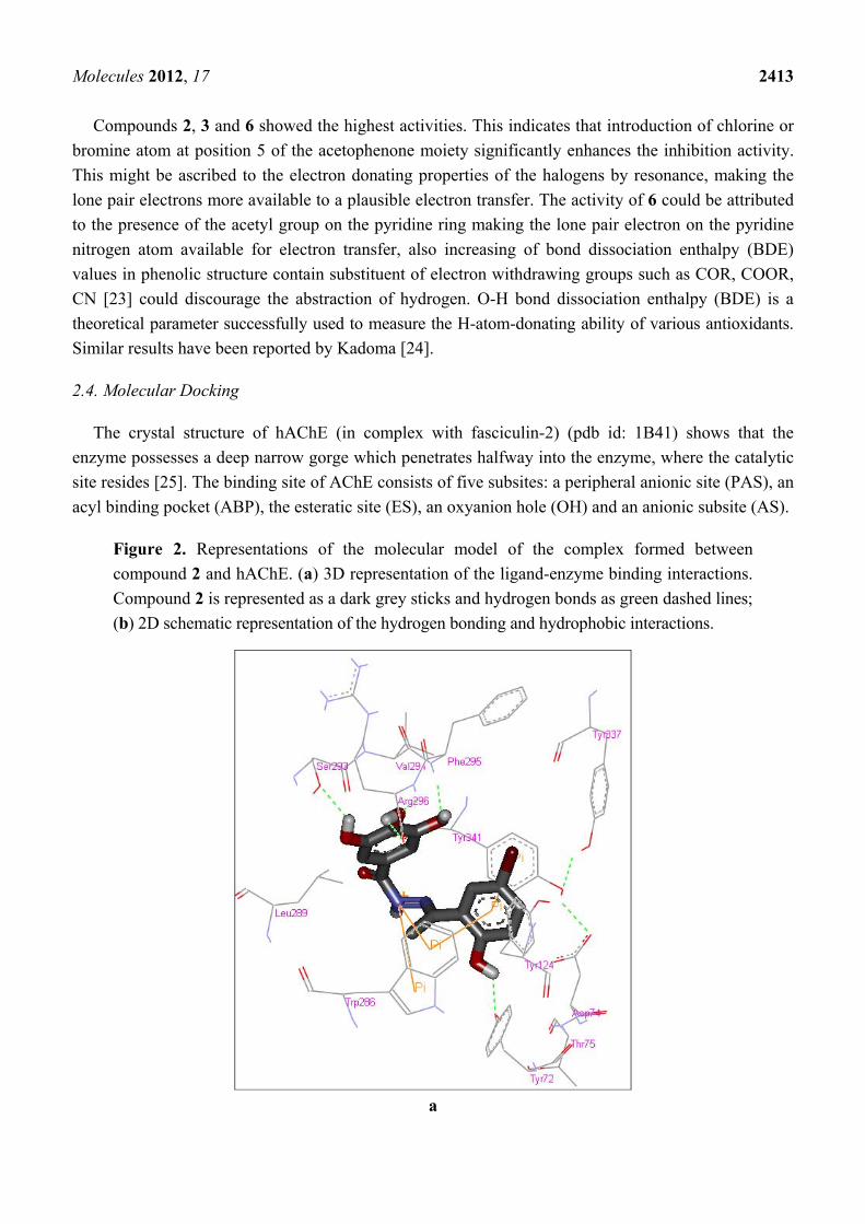
The crystal structure of hAChE (in complex with fasciculin-2) (pdb id: 1B41) shows that the
enzyme possesses a deep narrow gorge which penetrates halfway into the enzyme, where the catalytic
site resides [25]. The binding site of AChE consists of five subsites: a peripheral anionic site (PAS), an
acyl binding pocket (ABP), the esteratic site (ES), an oxyanion hole (OH) and an anionic subsite (AS).
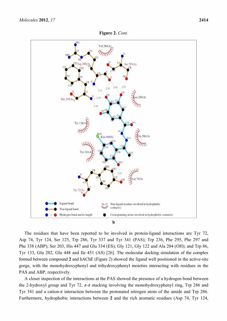
Figure 2. Representations of the molecular model of the complex formed between
compound 2 and hAChE. (a) 3D representation of the ligand-enzyme binding interactions.
Compound 2 is represented as a dark grey sticks and hydrogen bonds as green dashed lines;
(b) 2D schematic representation of the hydrogen bonding and hydrophobic interactions.
a
Molecules 2012, 17 2414
Figure 2. Cont.
b
The residues that have been reported to be involved in protein-ligand interactions are Tyr 72,
Asp 74, Tyr 124, Ser 125, Trp 286, Tyr 337 and Tyr 341 (PAS); Trp 236, Phe 295, Phe 297 and
Phe 338 (ABP); Ser 203, His 447 and Glu 334 (ES); Gly 121, Gly 122 and Ala 204 (OH); and Trp 86,
Tyr 133, Glu 202, Glu 448 and Ile 451 (AS) [26]. The molecular docking simulation of the complex
formed between compound 2 and hAChE (Figure 2) showed the ligand well positioned in the active-site
gorge, with the monohydroxyphenyl and trihydroxyphenyl moieties interacting with residues in the
PAS and ABP, respectively.
A closer inspection of the interactions at the PAS showed the presence of a hydrogen bond between
the 2-hydroxyl group and Tyr 72, π-π stacking involving the monohydroxyphenyl ring, Trp 286 and
Tyr 341 and a cation-π interaction between the protonated nitrogen atom of the amide and Trp 286.
Furthermore, hydrophobic interactions between 2 and the rich aromatic residues (Asp 74, Tyr 124,
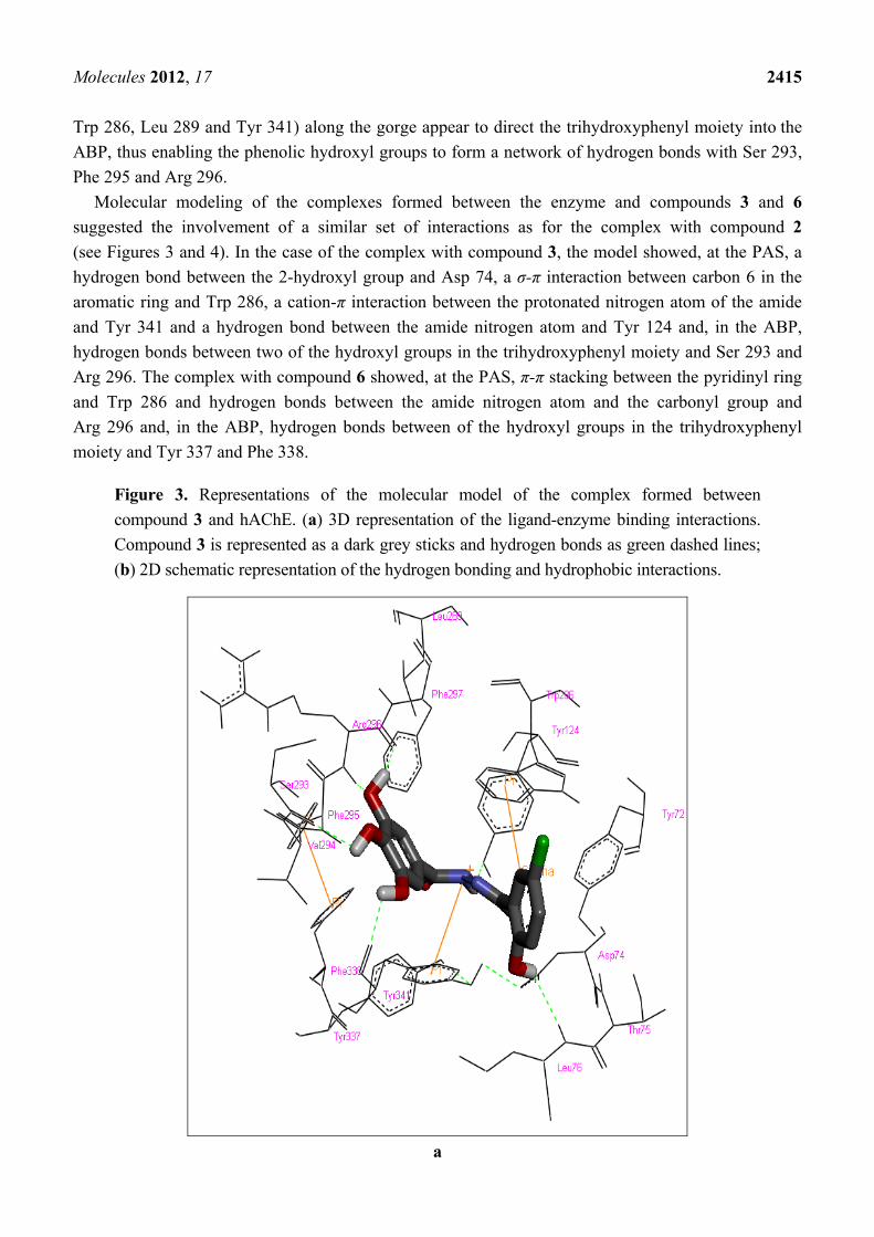
Molecules 2012, 17 2415
Trp 286, Leu 289 and Tyr 341) along the gorge appear to direct the trihydroxyphenyl moiety into the
ABP, thus enabling the phenolic hydroxyl groups to form a network of hydrogen bonds with Ser 293,
Phe 295 and Arg 296.
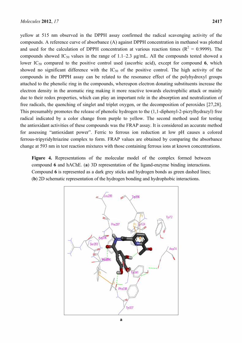
Molecular modeling of the complexes formed between the enzyme and compounds 3 and 6
suggested the involvement of a similar set of interactions as for the complex with compound 2
(see Figures 3 and 4). In the case of the complex with compound 3, the model showed, at the PAS, a
hydrogen bond between the 2-hydroxyl group and Asp 74, a σ-π interaction between carbon 6 in the
aromatic ring and Trp 286, a cation-π interaction between the protonated nitrogen atom of the amide
and Tyr 341 and a hydrogen bond between the amide nitrogen atom and Tyr 124 and, in the ABP,
hydrogen bonds between two of the hydroxyl groups in the trihydroxyphenyl moiety and Ser 293 and
Arg 296. The complex with compound 6 showed, at the PAS, π-π stacking between the pyridinyl ring
and Trp 286 and hydrogen bonds between the amide nitrogen atom and the carbonyl group and
Arg 296 and, in the ABP, hydrogen bonds between of the hydroxyl groups in the trihydroxyphenyl
moiety and Tyr 337 and Phe 338.
Figure 3. Representations of the molecular model of the complex formed between
compound 3 and hAChE. (a) 3D representation of the ligand-enzyme binding interactions.
Compound 3 is represented as a dark grey sticks and hydrogen bonds as green dashed lines;
(b) 2D schematic representation of the hydrogen bonding and hydrophobic interactions.
a
Molecules 2012, 17 2416
Figure 3. Cont.
b
This analysis suggests that the hAChE inhibition activity of compounds 2, 3 and 6 is probably due
to their ability to block the active-site gorge, thus preventing the substrate, acetylcholine, from entering
the active site.
2.5. Antioxidant Assays
The antioxidant efficacies of the compounds 1–6 were tested and the results obtained (see Table 3)
revealed differing activities in the two assays. This indicates that two mechanisms, operating in
different ways, must be responsible for the observed activity. The color change from deep purple to
Molecules 2012, 17 2417
yellow at 515 nm observed in the DPPH assay confirmed the radical scavenging activity of the
compounds. A reference curve of absorbance (A) against DPPH concentration in methanol was plotted
and used for the calculation of DPPH concentration at various reaction times (R2 = 0.9999). The
compounds showed IC50 values in the range of 1.1–2.3 μg/mL. All the compounds tested showed a
lower IC50 compared to the positive control used (ascorbic acid), except for compound 6, which
showed no significant difference with the IC50 of the positive control. The high activity of the
compounds in the DPPH assay can be related to the resonance effect of the polyhydroxyl groups
attached to the phenolic ring in the compounds, whereupon electron donating substituents increase the
electron density in the aromatic ring making it more reactive towards electrophilic attack or mainly
due to their redox properties, which can play an important role in the absorption and neutralization of
free radicals, the quenching of singlet and triplet oxygen, or the decomposition of peroxides [27,28].
This presumably promotes the release of phenolic hydrogen to the (1,1-diphenyl-2-picrylhydrazyl) free
radical indicated by a color change from purple to yellow. The second method used for testing
the antioxidant activities of these compounds was the FRAP assay. It is considered an accurate method
for assessing “antioxidant power”. Ferric to ferrous ion reduction at low pH causes a colored
ferrous-tripyridyltriazine complex to form. FRAP values are obtained by comparing the absorbance
change at 593 nm in test reaction mixtures with those containing ferrous ions at known concentrations.
Figure 4. Representations of the molecular model of the complex formed between
compound 6 and hAChE. (a) 3D representation of the ligand-enzyme binding interactions.
Compound 6 is represented as a dark grey sticks and hydrogen bonds as green dashed lines;
(b) 2D schematic representation of the hydrogen bonding and hydrophobic interactions.
a

Molecules 2012, 17 2418
Figure 4. Cont.
b
In this study, the compounds showed FRAP values in the range 2,000–9,000 which is above the values
shown by BHT and ascorbic acid used as standards. It was observed that compounds 1–5 demonstrated the
highest activities in the DPPH assay while 1 and 2 showed the highest values in the FRAP assay. This can
be attributed to increased π-electron delocalization within the pyridine ring which increases the electron
density and causes ferric ion reduction [29].
3. Experimental Section
3.1. General
The compounds synthesized in this study were characterized by spectral methods. IR spectra was
recorded at the wavelength range from 4,000–400 cm−1 using a Perkin Elmer 783 spectrophotometer,
NMR spectra were obtained on a ECA400 FT-NMR spectrophotometer using TMS as internal
standard, UV-visible spectra were recorded on a UV-1650PC model UV-visible spectrophotometer,
Melting points were measured using a Gallenkamp melting point apparatus and are Elemental analysis
Molecules 2012, 17 2419
was conducted on Costech Elemental Combustion System CNHS-O elemental analyzer. General grade
solvents and reagents were used unless stated otherwise and were obtained from Aldrich Chemicals
UK Ltd. and Acros Ltd. (UK). Methyl-3,4,5-trihydroxybenzoate, hydrazine hydrate, 2-hydroxy-5-
methoxyacetophenone, 5-bromo-2-hydroxyacetophenone, 5-chloro-2-hydroxyacetophenone, 2-cetylpyridine,
2,6-diacetylpyridine, Dilute Hydrochloric acid (0.01 M), Distilled Ethanol. Distilled water and
Dimethyl formamide (DMF).
3.2. Gallic Hydrazide (1)
NH
O
NH2
HO
HO
HO Molecular Weight: 184.15 An ethanolic solution (20 mL) containing methyl 3,4,5-trihydroxybenzoate (1.84 g, 0.01 M C8H8O5)
and hydrazine (9 mL) was stirred for 30 minutes, until all the solute completely dissolved then distilled
ethanol (45 mL) was added. The mixture was refluxed for about 6–8 h. The resulting white precipitate
was collected by filtration, washed several times with distilled water and then dried under vacuum.
Yield = 70%, melting point = 290 °C, elemental analysis theory: C (45.6%); H (4.3%); N (15.2%);
found: C (45.2%); H (5.04%); N (15.04%), %, FT-IR spectra (KBr); 3,429 cm−1 (νAr-OH), 3,299 cm−1
(νN-H), 1,654 cm−1 (νC=O), 1,344cm−1 (νC-O), 1,103 cm−1 (νN-N), 1H-NMR (DMSO-d6): 9.35 ppm
[δ(Ar-OH), 1H s], 9.13 ppm, 9.05 ppm [δ(Ar-OH), 2H, d], 8.65 ppm [δ(NH), 1H, brd], 6.79 ppm,
6.82 ppm [δ(Ar-H), 2H s], 4.37 ppm [δ(NH2), 2H s]. 13C-NMR (DMSO-d6): 166.39 ppm [δ(CONH),
1C], 145.37 ppm [δ(aromatic), 1C-OH], 136.10, 136.42 ppm [δ(aromatic), 2C-OH], 124.00 ppm
[δ(aromatic), 1C], 106.43 ppm [δ(aromatic), 2C = C] ppm.
3.3. N-(1-(5-Bromo-2-hydroxyphenyl)-ethylidene)-3,4,5-trihydroxybenzohydrazide (2)
NH
OHO
HO
HO
N
CH3
HO
Br
Molecular Weight: 381.18 Gallic hydrazide (1.84 g, 0.01 M) in (20 mL) ethanol was added to an ethanolic solution (20 mL) of
5-bromo-2-acetophenone (2.15 g, 0.01 M respectively). The mixture was stirred for 2–3 h whereupon
the color of the solution turned yellowish. The pH was adjusted by adding a few drops of dilute HCl.
The reaction was continued for another hour resulting in the formation of a yellow precipitate. More
precipitate was obtained when reducing the solvent by distillation. The product was collected by
filtration, washed several times with ethanol and dried in an oven. (Yield 75%), elemental analysis:
theory C (47.50); H (3.89); N (7.32); found C (47.26); H (3.44); N (7.35); IR spectra (KBr);
3,558 cm−1 (νAr-OH), 3,235 cm−1 (νN-H), 1,604 cm−1 (νC=N), 1,658 cm−1 (νC=O), 1,242 cm−1
(νC-O), 953 cm−1 (νN-N), 1H-NMR (DMSO-d6): 13.57 ppm [δ(OH), 1H, s], 11.10 ppm [δ(OH), 1H, s],
9.05 ppm [δ(OH), 2H, brd], 6.79–7.73 ppm [δ(aromatic), 5H, m], 4.34 ppm [δ(NH), 1H, s], 1.24 ppm
Molecules 2012, 17 2420
[δ(-CH3 ), 3H, s]. 13C-NMR (DMSO-d6): 166.39 ppm [δ(C=N)], 164.43 ppm [δ(C=O)], 145.53 ppm,
145.36 ppm [δ(aromatic), 2C-OH], 137.41 ppm [δ(aromatic), 1C-OH], 137.40, 136.10 ppm,
[δ(aromatic), 2C], 130.31 ppm [δ(aromatic), 1C], 123.51 ppm, 122.35 ppm, 121.52 ppm, 119.50 ppm
[δ(aromatic), 4C], 107.54 ppm, 106.42 ppm [δ(aromatic), 2C=C], 13.92 ppm [δ(CH3)] ppm.
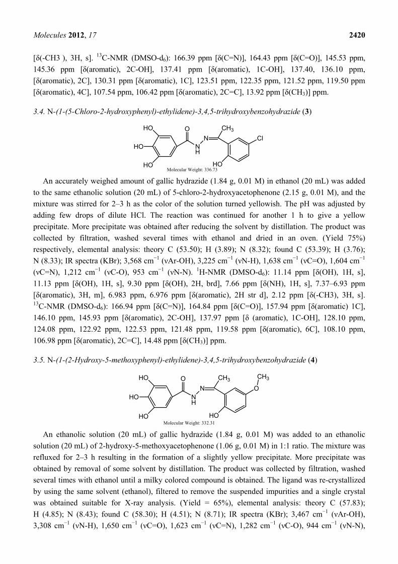
3.4. N-(1-(5-Chloro-2-hydroxyphenyl)-ethylidene)-3,4,5-trihydroxybenzohydrazide (3)
NH
OHO
HO
HO
N
CH3
HO
Cl
Molecular Weight: 336.73 An accurately weighed amount of gallic hydrazide (1.84 g, 0.01 M) in ethanol (20 mL) was added
to the same ethanolic solution (20 mL) of 5-chloro-2-hydroxyacetophenone (2.15 g, 0.01 M), and the
mixture was stirred for 2–3 h as the color of the solution turned yellowish. The pH was adjusted by
adding few drops of dilute HCl. The reaction was continued for another 1 h to give a yellow
precipitate. More precipitate was obtained after reducing the solvent by distillation. The product was
collected by filtration, washed several times with ethanol and dried in an oven. (Yield 75%)
respectively, elemental analysis: theory C (53.50); H (3.89); N (8.32); found C (53.39); H (3.76);
N (8.33); IR spectra (KBr); 3,568 cm−1 (νAr-OH), 3,225 cm−1 (νN-H), 1,638 cm−1 (νC=O), 1,604 cm−1
(νC=N), 1,212 cm−1 (νC-O), 953 cm−1 (νN-N). 1H-NMR (DMSO-d6): 11.14 ppm [δ(OH), 1H, s],
11.13 ppm [δ(OH), 1H, s], 9.30 ppm [δ(OH), 2H, brd], 7.66 ppm [δ(NH), 1H, s], 7.37–6.93 ppm
[δ(aromatic), 3H, m], 6.983 ppm, 6.976 ppm [δ(aromatic), 2H str d], 2.12 ppm [δ(-CH3), 3H, s]. 13C-NMR (DMSO-d6): 166.94 ppm [δ(C=N)], 164.84 ppm [δ(C=O)], 157.94 ppm [δ(aromatic) 1C],
146.10 ppm, 145.93 ppm [δ(aromatic), 2C-OH], 137.97 ppm [δ (aromatic), 1C-OH], 128.10 ppm,
124.08 ppm, 122.92 ppm, 122.53 ppm, 121.48 ppm, 119.58 ppm [δ(aromatic), 6C], 108.10 ppm,
106.98 ppm [δ(aromatic), 2C=C], 14.48 ppm [δ(CH3)] ppm.
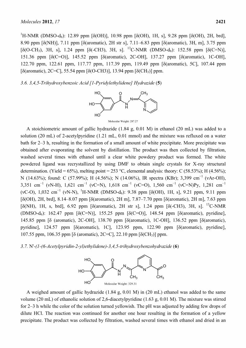
3.5. N-(1-(2-Hydroxy-5-methoxyphenyl)-ethylidene)-3,4,5-trihydroxybenzohydrazide (4)
NH
OHO
HO
HO
N
CH3
HO
O
CH3
Molecular Weight: 332.31 An ethanolic solution (20 mL) of gallic hydrazide (1.84 g, 0.01 M) was added to an ethanolic
solution (20 mL) of 2-hydroxy-5-methoxyacetophenone (1.06 g, 0.01 M) in 1:1 ratio. The mixture was
refluxed for 2–3 h resulting in the formation of a slightly yellow precipitate. More precipitate was
obtained by removal of some solvent by distillation. The product was collected by filtration, washed
several times with ethanol until a milky colored compound is obtained. The ligand was re-crystallized
by using the same solvent (ethanol), filtered to remove the suspended impurities and a single crystal
was obtained suitable for X-ray analysis. (Yield = 65%), elemental analysis: theory C (57.83);
H (4.85); N (8.43); found C (58.30); H (4.51); N (8.71); IR spectra (KBr); 3,467 cm−1 (νAr-OH),
3,308 cm−1 (νN-H), 1,650 cm−1 (νC=O), 1,623 cm−1 (νC=N), 1,282 cm−1 (νC-O), 944 cm−1 (νN-N),
Molecules 2012, 17 2421
1H-NMR (DMSO-d6): 12.89 ppm [δ(OH)], 10.98 ppm [δ(OH), 1H, s], 9.28 ppm [δ(OH), 2H, brd],
8.90 ppm [δ(NH)], 7.11 ppm [δ(aromatic), 2H str s], 7.11–6.83 ppm [δ(aromatic), 3H, m], 3.75 ppm
[δ(O-CH3), 3H, s], 1.24 ppm [δ(-CH3), 3H, s]. 13C-NMR (DMSO-d6): 152.58 ppm [δ(C=N)],
151.36 ppm [δ(C=O)], 145.52 ppm [δ(aromatic), 2C-OH], 137.27 ppm [δ(aromatic), 1C-OH],
122.70 ppm, 122.61 ppm, 117.77 ppm, 117.39 ppm, 119.49 ppm [δ(aromatic), 5C], 107.44 ppm
[δ(aromatic), 2C=C], 55.54 ppm [δ(O-CH3)], 13.94 ppm [δ(CH3)] ppm.
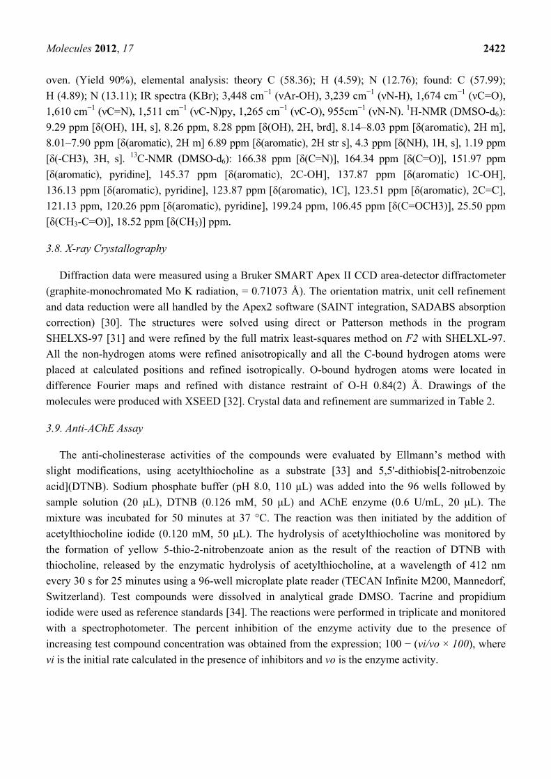
3.6. 3,4,5-Trihydroxybenzoic Acid [1-Pyridylethylidene] Hydrazide (5)
NH
OHO
HO
HO
N
CH3
N
Molecular Weight: 287.27 A stoichiometric amount of gallic hydrazide (1.84 g, 0.01 M) in ethanol (20 mL) was added to a
solution (20 mL) of 2-acetylpyridine (1.21 mL, 0.01 mmol) and the mixture was refluxed on a water
bath for 2–3 h, resulting in the formation of a small amount of white precipitate. More precipitate was
obtained after evaporating the solvent by distillation. The product was then collected by filtration,
washed several times with ethanol until a clear white powdery product was formed. The white
powdered ligand was recrystallized by using DMF to obtain single crystals for X-ray structural
determination. (Yield = 65%), melting point = 253 °C, elemental analysis: theory: C (58.53%); H (4.56%);
N (14.63%); found: C (57.99%); H (4.56%); N (14.06%), IR spectra (KBr); 3,399 cm−1 (νAr-OH),
3,351 cm−1 (νN-H), 1,621 cm−1 (νC=N), 1,618 cm−1 (νC=O), 1,560 cm−1 (νC=N)Py, 1,281 cm−1
(νC-O), 1,032 cm−1 (νN-N), 1H-NMR (DMSO-d6): 9.38 ppm [δ(OH), 1H, s], 9.21 ppm, 9.11 ppm
[δ(OH), 2H, brd], 8.14–8.07 ppm [δ(aromatic), 2H m], 7.87–7.70 ppm [δ(aromatic), 2H m], 7.63 ppm
[δ(NH), 1H, s, brd], 6.92 ppm [δ(aromatic), 2H str s], 1.24 ppm [δ(-CH3), 3H, s]. 13C-NMR
(DMSO-d6): 162.47 ppm [δ(C=N)], 155.25 ppm [δ(C=O)], 148.54 ppm [δ(aromatic), pyridine],
145.85 ppm [δ (aromatic), 2C-OH], 138.70 ppm [δ(aromatic), 1C-OH], 136.52 ppm [δ(aromatic),
pyridine], 124.57 ppm [δ(aromatic), 1C], 123.95 ppm, 122.90 ppm [δ(aromatic), pyridine],
107.55 ppm, 106.35 ppm [δ (aromatic), 2C=C], 22.10 ppm [δ(CH3)] ppm.
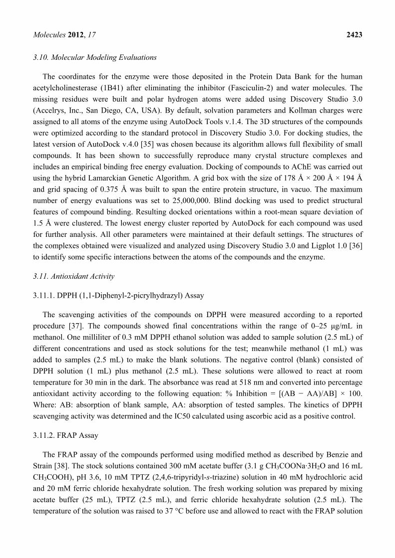
3.7. N'-(1-(6-Acetylpyridin-2-yl)ethylidene)-3,4,5-trihydroxybenzohydrazide (6)
NH
OHO
HO
HO
N
CH3
NO
CH3
Molecular Weight: 329.31
A weighed amount of gallic hydrazide (1.84 g, 0.01 M) in (20 mL) ethanol was added to the same
volume (20 mL) of ethanolic solution of 2,6-diacetylpyridine (1.63 g, 0.01 M). The mixture was stirred
for 2–3 h while the color of the solution turned yellowish. The pH was adjusted by adding few drops of
dilute HCl. The reaction was continued for another one hour resulting in the formation of a yellow
precipitate. The product was collected by filtration, washed several times with ethanol and dried in an
Molecules 2012, 17 2422
oven. (Yield 90%), elemental analysis: theory C (58.36); H (4.59); N (12.76); found: C (57.99);
H (4.89); N (13.11); IR spectra (KBr); 3,448 cm−1 (νAr-OH), 3,239 cm−1 (νN-H), 1,674 cm−1 (νC=O),
1,610 cm−1 (νC=N), 1,511 cm−1 (νC-N)py, 1,265 cm−1 (νC-O), 955cm−1 (νN-N). 1H-NMR (DMSO-d6):
9.29 ppm [δ(OH), 1H, s], 8.26 ppm, 8.28 ppm [δ(OH), 2H, brd], 8.14–8.03 ppm [δ(aromatic), 2H m],
8.01–7.90 ppm [δ(aromatic), 2H m] 6.89 ppm [δ(aromatic), 2H str s], 4.3 ppm [δ(NH), 1H, s], 1.19 ppm
[δ(-CH3), 3H, s]. 13C-NMR (DMSO-d6): 166.38 ppm [δ(C=N)], 164.34 ppm [δ(C=O)], 151.97 ppm
[δ(aromatic), pyridine], 145.37 ppm [δ(aromatic), 2C-OH], 137.87 ppm [δ(aromatic) 1C-OH],
136.13 ppm [δ(aromatic), pyridine], 123.87 ppm [δ(aromatic), 1C], 123.51 ppm [δ(aromatic), 2C=C],
121.13 ppm, 120.26 ppm [δ(aromatic), pyridine], 199.24 ppm, 106.45 ppm [δ(C=OCH3)], 25.50 ppm
[δ(CH3-C=O)], 18.52 ppm [δ(CH3)] ppm.
3.8. X-ray Crystallography
Diffraction data were measured using a Bruker SMART Apex II CCD area-detector diffractometer
(graphite-monochromated Mo K radiation, = 0.71073 Å). The orientation matrix, unit cell refinement
and data reduction were all handled by the Apex2 software (SAINT integration, SADABS absorption
correction) [30]. The structures were solved using direct or Patterson methods in the program
SHELXS-97 [31] and were refined by the full matrix least-squares method on F2 with SHELXL-97.
All the non-hydrogen atoms were refined anisotropically and all the C-bound hydrogen atoms were
placed at calculated positions and refined isotropically. O-bound hydrogen atoms were located in
difference Fourier maps and refined with distance restraint of O-H 0.84(2) Å. Drawings of the
molecules were produced with XSEED [32]. Crystal data and refinement are summarized in Table 2.
3.9. Anti-AChE Assay
The anti-cholinesterase activities of the compounds were evaluated by Ellmann’s method with
slight modifications, using acetylthiocholine as a substrate [33] and 5,5'-dithiobis[2-nitrobenzoic
acid](DTNB). Sodium phosphate buffer (pH 8.0, 110 μL) was added into the 96 wells followed by
sample solution (20 μL), DTNB (0.126 mM, 50 μL) and AChE enzyme (0.6 U/mL, 20 μL). The
mixture was incubated for 50 minutes at 37 °C. The reaction was then initiated by the addition of
acetylthiocholine iodide (0.120 mM, 50 μL). The hydrolysis of acetylthiocholine was monitored by
the formation of yellow 5-thio-2-nitrobenzoate anion as the result of the reaction of DTNB with
thiocholine, released by the enzymatic hydrolysis of acetylthiocholine, at a wavelength of 412 nm
every 30 s for 25 minutes using a 96-well microplate plate reader (TECAN Infinite M200, Mannedorf,
Switzerland). Test compounds were dissolved in analytical grade DMSO. Tacrine and propidium
iodide were used as reference standards [34]. The reactions were performed in triplicate and monitored
with a spectrophotometer. The percent inhibition of the enzyme activity due to the presence of
increasing test compound concentration was obtained from the expression; 100 − (vi/vo × 100), where
vi is the initial rate calculated in the presence of inhibitors and vo is the enzyme activity.
Molecules 2012, 17 2423
3.10. Molecular Modeling Evaluations
The coordinates for the enzyme were those deposited in the Protein Data Bank for the human
acetylcholinesterase (1B41) after eliminating the inhibitor (Fasciculin-2) and water molecules. The
missing residues were built and polar hydrogen atoms were added using Discovery Studio 3.0
(Accelrys, Inc., San Diego, CA, USA). By default, solvation parameters and Kollman charges were
assigned to all atoms of the enzyme using AutoDock Tools v.1.4. The 3D structures of the compounds
were optimized according to the standard protocol in Discovery Studio 3.0. For docking studies, the
latest version of AutoDock v.4.0 [35] was chosen because its algorithm allows full flexibility of small
compounds. It has been shown to successfully reproduce many crystal structure complexes and
includes an empirical binding free energy evaluation. Docking of compounds to AChE was carried out
using the hybrid Lamarckian Genetic Algorithm. A grid box with the size of 178 Å × 200 Å × 194 Å
and grid spacing of 0.375 Å was built to span the entire protein structure, in vacuo. The maximum
number of energy evaluations was set to 25,000,000. Blind docking was used to predict structural
features of compound binding. Resulting docked orientations within a root-mean square deviation of
1.5 Å were clustered. The lowest energy cluster reported by AutoDock for each compound was used
for further analysis. All other parameters were maintained at their default settings. The structures of
the complexes obtained were visualized and analyzed using Discovery Studio 3.0 and Ligplot 1.0 [36]
to identify some specific interactions between the atoms of the compounds and the enzyme.
3.11. Antioxidant Activity
3.11.1. DPPH (1,1-Diphenyl-2-picrylhydrazyl) Assay
The scavenging activities of the compounds on DPPH were measured according to a reported
procedure [37]. The compounds showed final concentrations within the range of 0–25 μg/mL in
methanol. One milliliter of 0.3 mM DPPH ethanol solution was added to sample solution (2.5 mL) of
different concentrations and used as stock solutions for the test; meanwhile methanol (1 mL) was
added to samples (2.5 mL) to make the blank solutions. The negative control (blank) consisted of
DPPH solution (1 mL) plus methanol (2.5 mL). These solutions were allowed to react at room
temperature for 30 min in the dark. The absorbance was read at 518 nm and converted into percentage
antioxidant activity according to the following equation: % Inhibition = [(AB − AA)/AB] × 100.
Where: AB: absorption of blank sample, AA: absorption of tested samples. The kinetics of DPPH
scavenging activity was determined and the IC50 calculated using ascorbic acid as a positive control.
3.11.2. FRAP Assay
The FRAP assay of the compounds performed using modified method as described by Benzie and
Strain [38]. The stock solutions contained 300 mM acetate buffer (3.1 g CH3COONa·3H2O and 16 mL
CH3COOH), pH 3.6, 10 mM TPTZ (2,4,6-tripyridyl-s-triazine) solution in 40 mM hydrochloric acid
and 20 mM ferric chloride hexahydrate solution. The fresh working solution was prepared by mixing
acetate buffer (25 mL), TPTZ (2.5 mL), and ferric chloride hexahydrate solution (2.5 mL). The
temperature of the solution was raised to 37 °C before use and allowed to react with the FRAP solution
Molecules 2012, 17 2424
(300 μL) in the dark. The colored product (ferrous tripyridyltriazine complex) was monitored at a
wavelength of 593 nm. The standard curve was linear between 100 and 1,000 μM ferrous sulphate.
Results are expressed in μM ferrous/g dry mass and compared with that of ascorbic acid and
butylated hydroxytoluene.
3.12. Statistical Analysis
All values were reported as mean ± S.E.M. The statistical significance of differences between
groups was assessed using one-way ANOVA. A value of p < 0.05 was considered significant.
4. Conclusions
Synthesized novel Schiff bases were observed to be potentially useful for acetyl-cholinesterase
inhibition and possible treatment for AD. The compounds also showed strong free radical inhibitory
activities. In silico molecular modeling revealed that the compounds may position themselves in the
enzyme’s active-site gorge, interacting with residues in the peripheral anionic subsite (PAS) and acyl
binding pocket (ABP).
Supplementary Data
CCDC 857032 contains the supplementary crystallographic data for complex-4. These data can be
obtained free of charge via http://www.ccdc.cam.ac.uk/conts/retrieving.html, or from the Cambridge
Crystallographic Data Centre, 12 Union Road, Cambridge CB2 1EZ, UK; Fax: (+44) 1223-336-033;
or E-Mail: [email protected]. Detailed information can be accessed at: http://www.mdpi.com/
1420-3049/17/3/2408/s1.
Acknowledgements
The authors wish to acknowledge the grants (PS358/2009C, HIR-000009-21001 and ER009-2011A)
provided by the University of Malaya to conduct this study.
References and Notes
1. da Silva, C.M.; da Silva, D.L.; Modolo, L.V.; Alves, R.B.; de Resende, M.A.; Martins, C.V.B.;
de Fatima, A. Schiff bases: A short review of their antimicrobial activities. J. Adv. Res. 2011, 2,
1–8.
2. Creaven, B.S.; Duff, B.; Egan, D.A.; Kavanagh, K.; Rosair, G.; Thangella, V.R. Anticancer and
antifungal activity of copper(II) complexes of quinolin-2(1H)-one-derived Schiff bases.
Inorg. Chim. Acta 2010, 363, 4048–4058.
3. Ceyhan, G.; Çelik, C.; Uruş, S.; Demirtaş, İ.; Elmastaş, M.; Tümer, M. Antioxidant,
electrochemical, thermal, antimicrobial and alkane oxidation properties of tridentate Schiff base
ligands and their metal complexes. Spectrochim. Acta A Mol. Biomol. Spectrosc. 2011, 81,
184–198.
Molecules 2012, 17 2425
4. Qiao, X.; Ma, Z.-Y.; Xie, C.-Z.; Xue, F.; Zhang, Y.-W.; Xu, J.-Y. Study on potential antitumor
mechanism of a novel Schiff Base copper(II) complex: Synthesis, crystal structure, DNA binding,
cytotoxicity and apoptosis induction activity. J. Inorg. Biochem. 2011, 105, 728–737.
5. Xu, D.; Ma, S.; Du, G.; He, Q.; Sun, D. Synthesis, characterization, and anticancer properties of
rare earth complexes with Schiff base and o-phenanthroline. J. Rare Earth. 2008, 26, 643–647.
6. Rice-Evans, C. Implications of the mechanisms of action of tea polyphenols as antioxidants
in vitro for chemoprevention in humans. In Proceedings of the Society for Experimental Biology
and Medicine, London, UK, 1999; Volume 220, pp. 262–266.
7. Clemetso, C.A.B.; Andersen, L. Plant Polyphenols as Antioxidants for Ascorbic Acid,
Edward, M.M., Janet, M.S., Eds.; New York Academy of Sciences: New York, NY, USA, 1966;
Volume 136, pp. 341–376.
8. Sun-Waterhouse, D.; Chen, J.; Chuah, C.; Wibisono, R.; Melton, L.D.; Laing, W. Kiwifruit-based
polyphenols and related antioxidants for functional foods: Kiwifruit extract-enhanced gluten-free
bread. Int. J. Food Sci. Nutr. 2009, 60, 251–264.
9. Kuhn, D.J.; Lam, W.H.; Kazi, A.; Daniel, K.G.; Song, S.J.; Chow, L.M.C. Synthetic peracetate
tea polyphenols as potent proteasome inhibitors and apoptosis inducers in human cancer cells.
Front Biosci. 2005, 10, 1010–1023.
10. Samoylenko, O.; Zaletok, S.; Orlovsky, O.; Gogol, S.; Klenov, O.; Shapochka, D. Additive
antitumor effect of plant polyphenols and a synthetic inhibitors of polyamines biosynthesis.
Breast 2011, 20, S22–S23.
11. Claudine, M.; Andrzej, M.; Augustin, S. Polyphenols and prevention of cardiovascular diseases.
Curr. Opin. Lipidol. 2005, 16, 77–84.
12. Capasso, R.; Evidente, A.; Tremblay, E.; Sala, A.; Santoro, C.; Cristinzio, G. Direct and mediated
effects on Bactrocera oleae (Gmelin) (Diptera, Tephritidae) of natural polyphenols and some
of related synthetic compounds: Structure-activity relationships. J. Chem. Ecol. 1994, 20,
1189–1199.
13. Luchsinger, J.A.; Mayeux, R. Dietary factors and Alzheimer’s disease. Lancet Neurol. 2004, 3,
579–587.
14. Dai, Q.; Borenstein, A.R.; Wu, Y.; Jackson, J.C.; Larson, E.B. Fruit and vegetable juices and
Alzheimer’s disease: The Kame Project. Am. J. Med. 2006, 119, 751–759.
15. Bourn, D.; Prescott, J. A comparison of the nutritional value, sensory qualities, and food safety of
organically and conventionally produced foods. Crit. Rev. Food Sci. Nutr. 2002, 42, 1–34.
16. Arts, I.C.; Hollman, P.C. Polyphenols and disease risk in epidemiologic studies. Am. J. Clin. Nutr.
2005, 81, 317S–325S.
17. El-Ansary, A.L.; Abdel-Fattah, H.M.; Abdel-Kader, N.S. Synthesis, spectral, thermal and
magnetic studies of Mn(II), Ni(II) and Cu(II) complexes with some benzopyran-4-one Schiff
bases. Spectrochim. Acta A Mol. Biomol. Spectrosc. 2011, 79, 522–528.
18. Khan, T.A.; Naseem, S.; Khan, S.N.; Khan, A.U.; Shakir, M. Synthesis and spectral
characterization of 14- and 16-membered tetraazamacrocyclic Schiff base ligands and their
transition metal complexes and a comparative study of interaction of calf thymus DNA with
copper(II) complexes. Spectrochim. Acta A Mol. Biomol. Spectrosc. 2009, 73, 622–629.
Molecules 2012, 17 2426
19. Nath, M.; Saini, P.K.; Kumar, A. New di- and triorganotin(IV) complexes of tripodal Schiff base
ligand containing three imidazole arms: Synthesis, structural characterization, anti-inflammatory
activity and thermal studies. J. Organomet. Chem. 2010, 695, 1353–1362.
20. Issa, R.M.; Khedr, A.M.; Rizk, H.F. UV-vis, IR and 1H NMR spectroscopic studies of some
Schiff bases derivatives of 4-aminoantipyrine. Spectrochim. Acta A Mol. Biomol. Spectrosc. 2005,
62, 621–629.
21. Pang, S.; Liang, Y. Studies on charge transfer properties from mixture of Schiff base and zinc
complex in Langmuir-Blodgett film by UV-vis absorption and Fourier transform infrared
spectroscopy. Spectrochim. Acta A Mol. Biomol. Spectrosc. 2001, 57, 435–439.
22. Suleiman Gwaram, N.; Khaledi, H.; Mohd Ali, H.; Robinson, W.T.; Abdulla, M.A. N'-[1-(5-
Bromo-2-hydroxyphenyl)ethylidene]-3,4,5-trihydroxybenzohydrazide dimethyl sulfoxide solvate
trihydrate. Acta Crystallogr. 2010, E66, o721.
23. Heider, E.M.; Harper, J.K.; Grant, D.M.; Hoffman, A.; Dugan, F.; Tomere, D.P.; O’Neille, K.L.
Unusual antioxidant activity in a benzoic acid derivative: A proposed mechanism for citrinin.
Tetrahedron 2006, 62, 1199–1208.
24. Kadoma, Y.; Atsumi, T.; Okada, N.; Ishihara, M.; Yokoe, I.; Fujisawa, S. Radical-scavenging
activity of the reaction products of isoeugenol with thiol, thiophenol, mercaptothiazoline or
mercaptomethylimidazole using the induction period method. Molecules 2007, 12, 130–138.
25. Kryger, G.; Harel, M.; Giles, K.; Toker, L.; Velan, B.; Lazar, A.; Kronman, C.; Barak, D.;
Ariel, N.; Shafferman, A. Structures of recombinant native and e202q mutant human
acetylcholinesterase complexed with the snake-venom toxin fasciculin-ii. Acta Crystallogr. D
Biol. Crystallogr. 2000, 56, 1385–1394.
26. Wiesner, J.; Kriz, Z.; Kuca, K.; Jun, D.; Koca, J. Acetylcholinesterases—The structural
similarities and differences. J. Enzyme Inhib. Med. Chem. 2007, 22, 417–424.
27. Heo, B.G.; Park, Y.S.; Chon, S.U.; Lee, S.Y.; Cho, J.Y.; Gorinstein, S. Antioxidant activity and
cytotoxicity of methanol extracts from aerial parts of Korean salad plants. Biofactors 2007, 30,
79–89.
28. Khaledi, H.; Alhadi, A.A.; Yehye, W.A.; Ali, H.M.; Abdulla, M.A.; Hassandarvish, P.
Antioxidant, cytotoxic activities, and structure-activity relationship of gallic acid-based indole
derivatives. Arch. Pharm. 2011, 344, 703–709.
29. Stockdale, M.; Selwyn, M.J. Effects of ring substituents on the activity of phenols as inhibitors
and uncouplers of mitochondrial respiration. Eur. J. Biochem. 1971, 21, 565–574.
30. Bruker APEX2 and SAINT, Bruker AXS Inc., Madison, WI, USA, 2007.
31. Sheldrick, G.M. A short history of SHELX. Acta Crystallogr. A 2008, 64, 112.
32. Barbour, L.J. X-Seed—A software tool for supramolecular crystallography. J. Supramol. Chem.
2001, 1, 189–191.
33. Guilhermino, L.; Lopes, M.C.; Carvalho, A.P.; Soares, A.M.V.M. Inhibition of acetylcholinesterase
activity as effect criterion in acute test with juvenile Daphnia magna. Chemosphere 1996, 32,
721–738.
34. Laskwoski, R.A. PDBsum: Summaries and analyses of PDB structure. Nucleic Acids Res. 2001,
29, 221–222.
Molecules 2012, 17 2427
35. Morris, G.M.; Goodsell, D.S.; Halliday, R.S.; Huey, R.; Hart, W.E.; Belew, R.K.; Olson, A.J.
Automated docking using a Lamarckian genetic algorithm and empirical binding free energy
function. J. Comput. Chem. 1998, 19, 1639–1662.
36. Wallace, C.A.; Laskowski, A.R.; Thornton, M.J. LIGPLOT: A program to generate schematic
diagrams of protein-ligand interactions. Protein Eng. 1995, 8, 127–134.
37. Choi, W.C.; Kim, S.C.; Hwang, S.S.; Choi, B.K.; Ahn, H.J.; Lee, M.Y.; Park, S.H.; Kim, S.K.
Antioxidant activity and free radical scavenging capacity between Korean medicinal plants and
flavonoids by assay-guided comparison. Plant Sci. 2002, 163, 1161–1168.
38. Benzie, I.F.F.; Strain, J.J. Ferric reducing/antioxidant power assay: Direct measure of total
antioxidant activity of biological fluids and modified version for simultaneous measurement of
total antioxidant power and ascorbic acid concentration. Method Enzymol. 1999, 299, 15–27.
Sample Availability: Samples of the compounds are available from the authors.
© 2012 by the authors; licensee MDPI, Basel, Switzerland. This article is an open access article
distributed under the terms and conditions of the Creative Commons Attribution license
(http://creativecommons.org/licenses/by/3.0/).
Copyright © 2022 FDOKUMEN