Synthesis, characterization, DNA binding and in vitro antimicrobial studies of a novel...
8
Synthesis, characterization, DNA binding and in vitro antimicrobial studies of a novel tetra-substituted N-isopropyl-N-(4-ferrocenylphenyl)- N 0 -(2,6-diethylphenyl)-N 00 -benzoylguanidine: Crystallographic structure and quantum chemical computations Muhammad Khawar Rauf a,⇑ , Rukhsana Gul a,b , Zahid Rashid a , Amin Badshah a,⇑ , Muhammad Nawaz Tahir c , Muhammad Shahid a , Azim Khan b a Department of Chemistry, Quaid-I-Azam University, Islamabad 45320, Pakistan b Department of Chemistry, Gomal University, Dera Ismail Khan, Pakistan c Department of Physics, University of Sargodha, Sargodha, Pakistan highlights Structure of the compound 1 was determined by single crystal X-ray diffraction. FT-IR and UV–Vis. spectra were recorded and compared with the theoretical results. The 1 H, 13 C NMR spectra has also been calculated using GIAO method. DNA binding and antimicrobial studies have also been performed. All the theoretical calculations were made using DFT/B3LYP method. graphical abstract The negative peak potential in the CV behavior of (1) by the addition of DNA is attributable to the electrostatic interactions between the compound and DNA, an indicator of the oxidizable behavior of (1) in the presence of negative environment of DNA. article info Article history: Received 23 July 2014 Received in revised form 26 September 2014 Accepted 30 September 2014 Available online 22 October 2014 Keywords: Tetra-substituted guanidine Single crystal XRD DNA binding abstract A novel tetra-substituted guanidine, N-isopropyl-N-(4-ferrocenylphenyl)-N 0 -(2,6-diethylphenyl)-N 00 -ben- zoylguanidine (1), [(CH 3 ) 2 CH)(C 5 H 5 FeC 5 H 4 C 6 H 4 )NC(NHCOC 6 H 5 )(NHC 6 H 3 (CH 2 CH 3 ) 2 ] has been synthesized and characterized by elemental analysis, FT-IR, multinuclear ( 1 H, 13 C) NMR spectroscopy, single crystal X-rays diffraction analysis and density functional theory based quantum chemical calculations. The tor- sion angles indicating that the guanidine moiety and carbonyl group are almost co-planar, due to the pseudo hexagonal ring formed by intramolecular NAHO hydrogen bonds. The DNA interaction studies performed by cyclic voltammetry and UV–visible spectroscopy are in close agreement with the binding constants (K) 1.4 10 4 and 1.2 10 4 respectively. The shift in peak potential, current and absorption maxima of the studied ferrocenyl guanidine in the presence of DNA discovered that CV coupled with http://dx.doi.org/10.1016/j.saa.2014.09.134 1386-1425/Ó 2014 Elsevier B.V. All rights reserved. ⇑ Corresponding authors. Tel.: +92 5190642131; fax: +92 5190642241. E-mail addresses: [email protected] (M.K. Rauf), aminbadshah@yahoo. com (A. Badshah). Spectrochimica Acta Part A: Molecular and Biomolecular Spectroscopy 136 (2015) 1099–1106 Contents lists available at ScienceDirect Spectrochimica Acta Part A: Molecular and Biomolecular Spectroscopy journal homepage: www.elsevier.com/locate/saa
-
Upload
independent -
Category
Documents
-
view
2 -
download
0
Transcript of Synthesis, characterization, DNA binding and in vitro antimicrobial studies of a novel...

Spectrochimica Acta Part A: Molecular and Biomolecular Spectroscopy 136 (2015) 1099–1106
Contents lists available at ScienceDirect
Spectrochimica Acta Part A: Molecular andBiomolecular Spectroscopy
journal homepage: www.elsevier .com/locate /saa
Synthesis, characterization, DNA binding and in vitro antimicrobialstudies of a novel tetra-substituted N-isopropyl-N-(4-ferrocenylphenyl)-N0-(2,6-diethylphenyl)-N00-benzoylguanidine: Crystallographic structureand quantum chemical computations
http://dx.doi.org/10.1016/j.saa.2014.09.1341386-1425/� 2014 Elsevier B.V. All rights reserved.
⇑ Corresponding authors. Tel.: +92 5190642131; fax: +92 5190642241.E-mail addresses: [email protected] (M.K. Rauf), aminbadshah@yahoo.
com (A. Badshah).
Muhammad Khawar Rauf a,⇑, Rukhsana Gul a,b, Zahid Rashid a, Amin Badshah a,⇑,Muhammad Nawaz Tahir c, Muhammad Shahid a, Azim Khan b
a Department of Chemistry, Quaid-I-Azam University, Islamabad 45320, Pakistanb Department of Chemistry, Gomal University, Dera Ismail Khan, Pakistanc Department of Physics, University of Sargodha, Sargodha, Pakistan
h i g h l i g h t s
� Structure of the compound 1 wasdetermined by single crystal X-raydiffraction.� FT-IR and UV–Vis. spectra were
recorded and compared with thetheoretical results.� The 1H, 13C NMR spectra has also been
calculated using GIAO method.� DNA binding and antimicrobial
studies have also been performed.� All the theoretical calculations were
made using DFT/B3LYP method.
g r a p h i c a l a b s t r a c t
The negative peak potential in the CV behavior of (1) by the addition of DNA is attributable to theelectrostatic interactions between the compound and DNA, an indicator of the oxidizable behavior of(1) in the presence of negative environment of DNA.
a r t i c l e i n f o
Article history:Received 23 July 2014Received in revised form 26 September 2014Accepted 30 September 2014Available online 22 October 2014
Keywords:Tetra-substituted guanidineSingle crystal XRDDNA binding
a b s t r a c t
A novel tetra-substituted guanidine, N-isopropyl-N-(4-ferrocenylphenyl)-N0-(2,6-diethylphenyl)-N00-ben-zoylguanidine (1), [(CH3)2CH)(C5H5FeC5H4C6H4)NC(NHCOC6H5)(NHC6H3(CH2CH3)2] has been synthesizedand characterized by elemental analysis, FT-IR, multinuclear (1H, 13C) NMR spectroscopy, single crystalX-rays diffraction analysis and density functional theory based quantum chemical calculations. The tor-sion angles indicating that the guanidine moiety and carbonyl group are almost co-planar, due to thepseudo hexagonal ring formed by intramolecular NAH� � �O hydrogen bonds. The DNA interaction studiesperformed by cyclic voltammetry and UV–visible spectroscopy are in close agreement with the bindingconstants (K) 1.4 � 104 and 1.2 � 104 respectively. The shift in peak potential, current and absorptionmaxima of the studied ferrocenyl guanidine in the presence of DNA discovered that CV coupled with
1100 M.K. Rauf et al. / Spectrochimica Acta Part A: Molecular and Biomolecular Spectroscopy 136 (2015) 1099–1106
AntimicrobialDFT
UV–vis spectroscopy could provide an opportunity to elaborate DNA interaction mechanism, aprerequisite for the design of new drug like agents and understanding the molecular basis of their action.The synthesized compound (1) has also been screened for their antibacterial and antifungal.
� 2014 Elsevier B.V. All rights reserved.
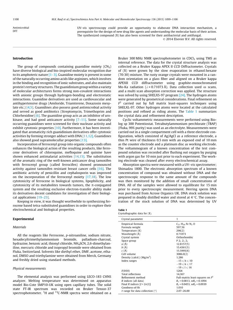
Table 1Crystallographic data for (1).
Crystal parameter 1
Empirical formula C37 H39 Fe N3 OFormula weight 597.56Temperature (K) 296(2)Wavelength (Å) 0.71073Crystal system OrthorhombicSpace group P 21 21 21
a (Å) 12.8157(5)b (Å) 15.4361(5)c (Å) 15.5995(6)Cell volume 3086.0(2)Density (calcd.) (Mg/m3) 1.286Index ranges �15 6 h 6 10
�19 6 k 6 17�19 6 l 6 16
F(000) 1264Total reflections 14,367Refinement method Full-matrix least-squares on F2
R indices (all data) R1 = 0.0813, wR2 = 0.1094Final R indices [I > 2r(I)] R1 = 0.0453, wR2 = 0.0939Goodness-of-fit 1.010h range for data collection (�) 2.07–26.00
Introduction
The group of compounds containing guanidine moiety (CN3)have diverse biological and bio-inspired molecular recognition dueto its amphoteric nature [1–3]. Guanidine moiety is present in someof the naturally occurring amino acids like arginines, which involvesin the binding and recognition of ionic substrates, and also maintainprotein’s tertiary structures. The guanidinium group within a varietyof molecular architectures forms strong non-covalent interactionswith anionic groups through hydrogen-bonding and electrostaticinteractions. Guanidine derivatives are used as cardiovascular andantihypertensive drugs (Amiloride, Triamterene, Doxazosin mesy-late etc.) [4,5]. Guanidines also possess good antimicrobial activityand served as good antibiotics (Streptomycin, Trimethoprim andChlorhexidine) [6]. The guanidine group acts as an inhibitor of uro-kinase, and had good anticancer activity [7–11]. Some naturallyoccurring guanidines were screened for their nuclease activity andexhibit cytotoxic properties [10]. Furthermore, it has been investi-gated that aromaticity rich guanidinium derivatives offer cytotoxicactivities by forming stronger adduct with DNA [11,12]. Guanidinesalso showed good supramolecullar properties [13].
Incorporation of ferrocenyl group into organic compounds oftenenhances the biological action of the resulting products, like ferro-cene derivatives of chloroquine, mefloquine and quinine haveshown enhanced antimalarial activities [14,15]. The substitutionof the aromatic ring of the well-known anticancer drug tamoxifenwith ferrocenyl group (called ferrocifen) showed pronouncedactivity against tamoxifen resistant breast cancer cells [16]. Theantibiotic activity of penicillin and cephalosporin was improvedon the incorporation of the ferrocenyl moiety [17,18]. The lowcytotoxicity of ferrocene in biological systems, lipophilicity, andcytotoxicity of its metabolites towards tumors, the p-conjugatedsystem and the resulting exclusive electron-transfer ability makeits derivatives decent candidates for investigation of their biologi-cal applications [19–23].
Keeping in view, it was thought worthwhile to synthesizing fer-rocene based tetra-substituted guanidines in order to explore theirelectrochemical and biological properties.
Experimental
Materials
All the reagents like Ferrocene, p-nitroaniline, sodium nitrate,hexadecyltrimethylammonium bromide, palladium-charcoal,hydrazine, benzoic acid, thionyl chloride, NH4SCN, 2,6-dimethylan-iline, mercuric chloride and isopropyl bromide were obtained fromFluka, Switzerland. Solvents like diethyl ether, DMF, acetone, etha-nol, DMSO and triethylamine were obtained from Merck, Germanyand freshly dried using standard methods.
Physical measurements
The elemental analysis was performed using LECO-183 CHNSanalyzer. Melting temperature was determined on apparatusmodel Bio-Cote SMP10-UK using open capillary tubes. The solidstate FT-IR spectrum was recorded on Bruker Tensor-37spectrophotometer. 1H and 13C-NMR spectra were obtained on a
Bruker 300 MHz NMR spectrophotometer in CDCl3 using TMS asinternal reference. The data for the crystal structure analysis wascollected on a Bruker Kappa APEX II CCD Diffractometer. Crystalsof (1) were grown by the slow evaporation in ethanol:hexane(70:30) mixture. The rusty orange crystals were mounted in a ran-dom orientation on a glass fiber and aligned on a Bruker kappaAPEXII CCD diffractometer using graphite-monochromatedMo-Ka radiation (k = 0.71073 Å). Data collection used x scans,and a multi scan absorption correction was applied. The structurewas solved by using SHELXS-97 program [24]. The hydrogen atomswere generated by geometrical considerations. Final refinement onF2 carried out by full matrix least-squares techniques usingSHELXL-97. Other hydrogen atoms were located at the calculatedpositions and refined as riding atoms. The Table 1 summarizesthe crystal data and refinement description.
Cyclic voltammetric measurements were performed using Bio-log sp 300 Potentiostat. Tetrabutylammonium perchlorate (TBAP)(Fluka, 99% purity) was used as an electrolyte. Measurements werecarried out in a single compartment cell with a three electrode con-figuration, which consisted of Ag/AgCl as a reference electrode, athin Pt wire of thickness 0.5 mm with an exposed end of 10 mmas the counter electrode and a platinum disc as working electrode.The voltammogram of a known concentration of the test com-pound solution was recorded after flushing out oxygen by purgingwith argon gas for 10 min just prior to each experiment. The work-ing electrode was cleaned after every electrochemical assay.
Absorption spectra were measured with a UV–vis spectrometer;Shimadzu 1800. The electronic absorption spectrum of a knownconcentration of compound was obtained without DNA and thespectroscopic response to the same amount of the compoundswas then monitored by the addition of small concentrations ofDNA. All of the samples were allowed to equilibrate for 15 minprior to every spectroscopic measurement. Herring sperm DNAwas purchased from Acrose Organics UK. DNA stock solution wasprepared in doubly distilled water and stored at 4 �C. The concen-tration of the stock solution of DNA was determined by UV

M.K. Rauf et al. / Spectrochimica Acta Part A: Molecular and Biomolecular Spectroscopy 136 (2015) 1099–1106 1101
absorbance at 260 nm using the molar extinction coefficient (e) of6600 M�1 cm�1 [25].
Synthesis of N-isopropyl-N-(4-ferrocenylphenyl)-N0-(2,6-diethylphenyl)-N00-benzoylguanidine (1)
The synthesis of the tetra-substituted ferrocenyl guanidine wasachieved in three steps (Scheme 1). In the first step the N-benzoyl-N0-(2,6-diethylphenyl)thiourea was synthesized by the reaction of2,6-diethylaniline with benzoylisothiocynate in tetrahydrofuran[26]. In the second step; nitrophenylferrocene was prepared fromferrocene via diazonium salt of p-nitroaniline using phase transfercatalyst hexadecyltrimethylammonium bromide. Then p-nitrophe-nylferrocene was reduced into ferrocenylaniline using palladium-charcoal and hydrazine as reducing agent [27]. In the third step;the N-benzoyl-N0-(2,6-diethylphenyl)thiourea was converted intotri-substituted ferrocenyl guanidine by the reaction of N-ben-zoyl-N0-(2,6-diethylphenyl)thiourea with ferrocenylaniline accord-ing to reported protocol [28] and lastly, tri-substituted ferrocenylguanidine was converted into tetra-substituted ferrocenyl guani-dine by simple alkylation.
N-benzoyl-N0-(2,6-diethylphenyl) thiourea (5.97 g, 10 mmol),ferrocenyl aniline (2.7 g, 10 mmol) and triethylamine (2.8 mL,20 mmol) were dissolved in 25 mL of DMF and the mixture wasstirred at temperature below 5 �C for 1 h. Mercuric chloride(2.7 g, 10 mmol) was then added and mixture was stirred for15 h. The completion of reaction was monitored by TLC. About25 mL of dichloromethane was added to the reaction mixtureand the suspension was filtered through sintered glass funnel toremove mercuric sulfide (HgS) formed as a byproduct during reac-tion. The solvent was evaporated under reduced pressure and res-idue were dissolved in 25 mL of dichloromethane (CH2Cl2). Otherbyproducts were extracted out with water (4 � 30 mL). Theorganic phase was dried over anhydrous MgSO4, filtered and thesolvent was removed by distillation. The product was dissolvedin dry acetone, and treated with isopropyl bromide and triethyl-amine for alkylation.
Yield: 80%; m. p. 160–160.5 �C; Anal. Calcd. for C37H39N3FeO(597.2): C, 74.37; H, 6.58; N, 7.03; Found: C, 74.34; H, 6.57; N,7.10%; FT-IR (KBr, cm�1): 3195, 3058, 2929, 1665, 1609, 1576,1529, 1465, 1419, 1385, 1342, 1296, 1265, 1223, 1167, 1144,1104, 1079, 1057, 1039, 1001, 935, 893, 807, 735, 670, 608, 572,490, 468, 417; 1H NMR (300 MHz, CDCl3): d 1.26 (t, 6H, ACH2-
Scheme 1. Synthesis scheme for N-isopropyl-N-(4-ferrocenyl
ACH3), 1.30 (d, 6H, CH(CH3)2), 2.78 (q, 4H, ACH2CH3), 5.50 (h,1H, CH(CH3)2), 4.08 (s, 5H, C5H5), 4.73 (d, 2H, J = 1.9 Hz, C5H4),5.13 (d, 2H, J = 1.9 Hz,C5H4), 7.20 (d, 2H, J = 8.2 Hz, ArAH), 7.29(d, 2H, J = 8.1 Hz, ArAH), 7.51–7.93 (m, 6H, ArAH), 8.40 (d, 2H,J = 8.2 Hz, ArAH), 12.20 (s, H, NH); 13C NMR (75.47 MHz, CDCl3):d14.8 (2C), 20.5 (2C), 24.0 (2C), 51.5 (C), 66.7 (5C), 69.0 (2C), 67.7(2C), 84.6 (C), 119.9 (2C), 120.4 (C), 127.5 (2C), 128.0 (2C), 128.6(C), 129.2 (2C), 129.4 (2C), 131.3 (2C), 131.5 (C), 137.5 (C), 140.6(C), 142.6 (C), 156.9 (C), 176.2 (C).
Theoretical calculations
All calculations were performed using the 6-31G⁄⁄ basis set forH, C, N, and O. This is a split valence double-f basis set which addspolarization functions on H and first row elements. For the Featom, LANL2TZ basis set was used which uses Los Alamos effectivecore potential on the transition metal atoms. The geometry of thetitle compound was optimized with DFT/B3LYP as implemented inGuassian-09 [37] quantum chemistry package. The Hessian calcu-lation confirmed that the optimized structure is a local minimumas no imaginary frequencies were found. The vibrational frequen-cies (IR) were calculated at the same level of the theory and tenta-tive assignments of the calculated fundamental modes wereperformed by their total energy distribution (TED) analysis usingVEDA4 program [38,39]. Time dependent density functional theory(TD-DFT) was used to calculate the excitation energies of the 50lowest energy singlet-to-singlet excited states. For the TD-DFT cal-culations, the geometry was first optimized in ethanol using Polar-isable Continuum Model (PCM) to take into account bulk solventeffects. The energies of the transitions for the excited states werealso calculated in PCM (ethanol). For the NMR calculations, thegeometry was optimized in CHCl3 solvent using PCM. The 1H and13C NMR isotropic chemical shielding (in ppm) was calculated inPCM (CHCl3).The NMR calculations were done using guage-includ-ing atomic orbital (GIAO) method [40].
Biological assay
Antibacterial assay
Antibacterial activity of the synthesized compound was deter-mined in accordance with the agar-well diffusion method againstfive bacterial strains, Staphylococcus aureus, Pseudomonas aerugin-
phenyl)-N0-(2,6-diethylphenyl)-N00-benzoylguanidine (1).

Fig. 1. Displacement ellipsoids are drawn at the 50% probability level. Hydrogen bonds shown as dashed lines (1).
Table 2Selected bond lengths (A�) and angles (�) for (1).
Bond lengths (A�) Angles (�)
Experimental (single crystal XRD)Fe1AC1 2.019 (5) C1AFe1AC2 40.2 (3)O1AC18 1.246 (4) C1AFe1AC6 109.8 (2)N1AC35 1.507 (4) C14AN1AC17 121.9 (3)Fe1AC7 2.047 (3) C1AFe1AC4 67.9 (2)N2AC17 1.326 (4) C1AFe1AC8 153.1 (3)C22AC23 1.366 (6) Fe1AC1AC2 70.8 (3)N3AC25 1.429 (5) N3AC25AC30 119.2 (3)C26AC33 1.521 (7) N2AC17AN3 123.2 (3)
Calculated (theoretical structure minimized by DFT/B3LYP)Fe1AC1 2.071 (2) C1AFe1AC2 40.32 (0)O1AC18 1.252 (9) C1AFe1AC6 108.58 (4)N1AC35 1.497 (6) C14AN1AC17 120.44 (5)Fe1AC7 2.067 (8) C1AFe1A C4 67.70 (4)N2AC17 1.331 (6) C1AFe1AC8 158.72 (9)C22AC23 1.397 (3) Fe1AC1AC2 69.82 (7)N3AC25 1.431 (4) N3AC25AC30 120.64 ((4)C26AC33 1.518 (2) N2AC17AN3 122.42 (0)
Table 3The intramolecular hydrogen bonds for (1). Distance, Å; angle, �.
D H A DH (Å) HA (Å) DA (Å) DHA (�)
Experimental (single crystal XRD)N3 H3A O1 0.8600 1.9900 2.601 (4) 127.00C31 H31B N3 0.9700 2.5700 2.912 (5) 101.00C35 H35 N2 0.9800 2.2400 2.737 (5) 110.00
Calculated (theoretical structure minimized by DFT/B3LYP)N3 H3A O1 1.0339 1.7052 2.5877 140.466C31 H31B N3 1.0932 2.7796 2.9579 88.372C35 H35 N2 1.0898 2.2672 2.7110 101.937
1102 M.K. Rauf et al. / Spectrochimica Acta Part A: Molecular and Biomolecular Spectroscopy 136 (2015) 1099–1106
osa, Bacillus subtilis, Klebsiella pneumoniae and Escherichia coli [29–31]. The bacterial isolates were first cultured in a nutrient broth for18 h prior to use and was standardized to 0.5 McFarland standards(106 cfu mL�1). 0.5 McFarland standard was prepared by adding0.5 mL of 0.048 M BaCl2 to 99.5 mL of 0.36 N H2SO4. Barium sulfateturbidity standard (4–6 mL) and was taken in a screw capped testtube.
Nutrient agar medium was prepared by adding nutrient agar(MERCK) 2.3 g in 100 mL of distilled water at pH 7.0 and was auto-claved. It was allowed to cool up to 45 �C. Petri plates were pre-pared by pouring of the 75 mL of seeded nutrient agar andallowed to solidify. Wells were bored into the agar using a sterile6 mm diameter cork borer. Approximately 100 lL of the compoundwere added into the wells, allowed to stand at room temperaturefor about 2 h and incubated at 37 �C. Controls were set in parallelin which the respective solvents were used to fill the well. Theplates were observed for zones of inhibition after 24 h. The effectswere compared with penicillin at a concentration of 1 mg/mL. Therelative percentage inhibition of the test compound with respect topositive control was calculated by using the formula given in Eq. (I)
Relative percentage inhibition of the test compound ð1Þ¼ 100� ðX � YÞ=ðZ � YÞ ðIÞ
where X is the total area of inhibition of the test compound, Y is thetotal area of inhibition of the solvent and Z is the total area of inhi-bition of the standard drug.
Antifungal assay
Antifungal activity of the synthesized compound 1 was testedagainst three different fungal strains, namely Fusarium moniliforme,Aspergillus fumigatus and Aspergillus flavus [29–31].The agar tubedilution method was used for the determination of antifungalactivity of the compound. The samples were prepared by dissolv-ing 3 mg compound in 1 mL of Dimethylsulfoxide (DMSO). Culturemedia was prepared by dissolving 6.5 g of the sabouraud dextroseagar per 100 mL distilled water of pH 5.6. 10 mL of the sabourauddextrose agar (MERCK) was dispensed in screw capped tubes orcotton plugged test tubes and was autoclaved at 121 �C for21 min. Tubes were allowed to cool at 50 �C and sabouraud dex-trose agar was loaded with 70 lL of the compound pipetted fromthe stock solution. The tubes containing the media were thenallowed to solidify in slanting position at room temperature. Threeslants of the compound sample were prepared for fungus species.The tubes containing solidified media and compounds were inocu-lated with 4 mm diameter piece of inoculum, taken from the seven

Fig. 2. The optimized molecular geometry of compound (1) at DFT/B3LYP.
M.K. Rauf et al. / Spectrochimica Acta Part A: Molecular and Biomolecular Spectroscopy 136 (2015) 1099–1106 1103
days old culture of the fungus. One test tube of each compoundwas prepared and a slant without compound was used as a control.The test tubes were incubated at 28 �C for 7 days. Cultures wereexamined twice weekly during the incubation. Reading was takenby measuring the linear length (mm) of fungus in slant and growthinhibition was calculated with reference to control. Percentageinhibition of fungal growth for each concentration of compoundwas determined using Eq. (II)
Percentage inhibition of fungal growth
¼ 100� Linear growth in test ðcmÞLinear growth in control ðcmÞ � 100 ðIIÞ
Results and discussion
Structural studies
The ORTEP diagram of 1 representing the tetra-substituted gua-nidine of this study is shown in Fig. 1. The tetra-substituted guani-dine crystallizes with one unique molecule in the unit cell and isfound in a conformation resulting from intramolecular hydrogenbonding. Compound 1, is a typical N, N, N0, N0 0-tetra-substitutedguanidine with normal geometric parameters [28]. In the asym-metric unit, there are two independent molecules with slightly dif-ferent conformations. The CAO bond distance indicates that thebond has full double bond character while the CAN bond lengthsindicate partial double bond character (Table 2). The guanidinemoiety and carbonyl group are coplanar due to intramolecularNAH� � �O hydrogen bonding, forming a six-membered ring com-monly observed in this class of compounds (Table 3). TheC18AO1 bond shows expected full double bond character whilethe short values for C18AN2, C17AN2, C17AN1 and C17AN3bonds indicate partial double bond character. The dihedral anglesbetween the guanidine plane [C17/N1/N2/N3] and the meanplanes of phenyl rings C19AC24, C25AC30 & C11AC16 are 14.92(9)�, 73.94 (5)� & 82.05 (5)�, respectively. The guanidine moietyand carbonyl group are almost co-planer as reflected by the torsionangles [O1AC18AN2AC17 = �5.5 (5)� & C17AN2AC18AC19 =174.6 (3)�], due to the presence of intramolecular NAH� � �O hydro-gen bonding, forming a six-membered ring commonly observed inthis class of compounds. The other intramolecular hydrogen bondsC31AH31B� � �N3 and C35AH35� � �N2 are also of great interest andstabilize the molecular geometry as shown in Fig. S1 (Suppl.).The structural parameters for the ferrocenyl substituent incompound 1 are within the normal ranges, and the iron atom issandwiched almost perfectly centrally between the two cyclopen-tadienyl centroids. The iron distance to the two cyclopentadienyl
centroids is almost same and the iron was observed at 1.641(9)and 1.647(8) Å from the centroids defined by the two cyclopenta-dienyl rings in (1). Moreover, the ferrocene has adopted an eclipsedconformation.
Selected bond distances and angles for the DFT/B3LYP-mini-mized structure of 1 (Fig. 2) are given in Table 2. These parametermatches very well with those observed in the crystal structure ofthis compound. The calculated values are slightly greater thanthe experimental values. This over estimation can be attributedto the fact that the calculation is done on a single isolated moleculein the gaseous phase while the experiment was conducted in thesolid phase where the crystal packing effect also play a role.
Spectroscopic studies
The ferrocenyl guanidine was well characterized in solutionphase (NMR, UV–visible spectroscopy and electrochemical charac-terization with cyclic voltammetry) along with solid phase (ele-mental analysis, FT-IR and single crystal XRD) characterization.
In the FT-IR spectra, a relatively weak peak was observed forNAH bond in the range 3200–3190 cm�1. The C@N stretching fre-quency was observed at 1560 cm�1 which is intermediate betweensingle and double bond, showed conjugation between all threenitrogen atoms of the guanidine moiety. The C@O stretching peakwas expected to be merged in a band observed at 1750–1570 cm�1.The experimental and calculated IR frequencies and intensities arepresented in Table S1 (Suppl.) along with their proposed assign-ments. In the calculated IR spectrum Fig. S2 (Suppl.) of the titlecompound a series of bands of low intensity were observed inthe range 3110–3080 cm�1, which were assigned to the CAHstretching of the ferrocenyl moiety and of the three phenyl rings.The calculated stretching bands of CH, CH2 and CH3 groups fall inthe range 3070–3028 cm�1. A sharp peak of high intensity wasobserved at 3181 cm�1, which was assigned to the NAH stretchingmode. A strong absorption peak at 1672 cm�1 is assigned to theC@O stretching which is also strongly coupled with theCAN3AH3a bending. While the C17AN2 and C18AN2 stretchingbands, coupled with HCH bending modes, were found at 1427and 1402 cm�1. The sharp absorption peaks at 1483 and1472 cm�1 were assigned to the C17AN3 and C17AN1 stretching,which were also coupled with the HCH bending modes. Both theC@O and C17AN2 stretching modes were also found coupled withthe CAC stretching modes of the phenyl rings at 1648, 1615 and1610 cm�1. A sharp and a weak peak were found for CAFe stretch-ing modes at 460 and 594 cm�1, respectively [32].
1H and 13C-NMR spectra of the compound were recorded rela-tive to TMS as reference in chloroform. In 1H-NMR, ferrocenyl pro-tons appeared in the range of 4.7–5.3 ppm as three set of signals

Fig. 3. Simulated at B3LYP level UV–visible spectroscopic response of compound(1) with ethanol as a solvent.
Fig. 4. CVs of 2 mM 1 in the (A) absence and presence of (B) 30, (C) 60 and (D)90 lM DNA in 10% aqueous ethanol (10% H2O:90% ethanol). t: 100 mVs�1,supporting electrolyte: 0.1 M TBAP.
Fig. 5. UV–Visible spectroscopic response of compound 1 in the absence (A) andpresence (B 30; C 40; D 50 (lM) of DNA.
1104 M.K. Rauf et al. / Spectrochimica Acta Part A: Molecular and Biomolecular Spectroscopy 136 (2015) 1099–1106
(singlet, triplet, triplet) with relative integral values of (5:2:2) pro-tons [32]. NAH protons appeared as broad singlet in 10.4–11.6 ppm. All the other aromatic and non-aromatic protonsappeared in their specific region. In 13C-NMR spectra of ferrocenylguanidine, the most down field signal at 176.2 ppm chemical shiftvalue is attributed to the most deshielded carbon of the carbonylgroup, whereas the guanidine carbon (CN3) appears relatively up-field at 156.9 ppm. In all the compounds, ferrocenyl carbonsappear in the 65–70 ppm region as four signals with relative signalintensities of (5:2:2:1) carbons. All the other aromatic and non-aromatic carbon appeared in their specific region.
The experimental and calculated 1H and 13C NMR chemical shiftvalues (in ppm) of title compound are given in Table S2 (Suppl.). Inthe 13C-NMR, the calculated chemical shift value for the carbonylcarbon is found to be 171.7 ppm which is in good agreement withthe experimental value of 176.2 ppm. For the guanidine carbon(C17) the chemical shift value is found to be 156.4 ppm while forthe ferrocenyl carbons it is observed in the range 63.8–72 ppm.The aromatic carbons appear in the 119.4–138.3 ppm region. Inthe 1H-NMR, the N-H3a proton is found at 12.6 ppm. The ferroce-nyl protons are found in the range 3.4–4.4 ppm.
The compound have two strong bands in UV region between230–280 nm and a weak peak in the visible region 450 nm. Thehigh absorptivity bands in the UV-region of spectrum may confi-dently be assigned to the aromatic phenyl ring based p–p⁄ transi-tions and C@N based p–p⁄ transitions [33]. The UV–Vis spectrumof ferrocene derivatives shows two absorption bands due to ferro-cene moiety. Three spin-allowed ligand field transitions areexpected: 1A1g ? a1E1g, 1A1g ?
1E2g, and 1A1g ? b1E1g. The firsttwo transitions are unresolved and give rise to the band at about450 nm and the third transition is responsible for the band about325 nm. Both bands are weak owing to the Laporte-forbiddend–d character of ligand field transitions. So the weak bands inthe test compounds may be assigned to the ferrocene based d–dtransitions [25].
Calculated UV–Vis spectrum (Fig. 3) using TD-DFT/B3LYPmethod shows a strong transition at 291 nm. Three more transi-tions of (relatively) weak intensity were found at 271, 281 and298 nm. Two (very) weak peaks were found above 300 nm at318 nm and 334 nm (oscillator strength = 0.04), which can accountfor the experimentally observed transition at 325 nm (Table S3(Suppl.)).
Cyclic voltametric DNA-binding studies
The interaction of the ferrocenyl guanidine with DNA was stud-ied by the cyclic voltammetry for understanding the mode of inter-action and binding strength. The cyclic voltammetric behavior ofthe compound in the absence and presence of 30–90 lM DNA ata bare platinum disc electrode is shown in Fig. 4. The voltammo-gram without DNA featured a well defined redox pair at the poten-tial values of 0.290 and 0.449 V for reduction and oxidationrespectively. By the addition of DNA into 2 mM compound the ano-dic peak potential is shifted in the negative going direction andcurrent is also dropped. The decrease in peak current is attributedto the formation of compound–DNA supramolecular complex dueto which the concentration of the free compound responsible forthe transfer of current is lowered. The mode of compound–DNAinteraction can be judged from the variation in peak potential.
In general, the positive shift in formal potential is caused by theintercalation of the drug into the double helical structure of DNA,while negative shift is observed for the electrostatic interactionof the cationic drug DNA backbone [34]. The obvious negative peakpotential shift in the CV behavior of the compound by the additionof DNA is attributable to the electrostatic interaction between thecompound and DNA. The peak potential shift in negative direction
indicates that the compound is easier to oxidize in the presence ofnegative environment of DNA.
Based upon the decrease in peak current of the studied com-pound by the addition of different concentrations of DNA the bind-ing constant values were calculated according to the followingequation [34].
1DNA
¼ Kð1� AÞ1� ðI=I0Þ
where K is binding constant, I and I0 are the peak currents with andwithout DNA and A is proportionality constant. The plot of 1/DNAversus 1/(1 � I/I0) yielded binding constant (K) 1.4 � 104 for thecompound under study.

Table 4In vitro antibacterial assay of (1).
Staphylococcusaureus
Pseudomonasaeruginosa
Bacillussubtilis
Klebsiellapneumoniae
Escherichiacoli
1 14.7 ± 0.33a 13.7 ± 0.33 15.3 ± 0.88 10.7 ± 0.67 10.0 ± 0.00PC 26.7 ± 0.33 26.3 ± 0.33 27.3 ± 0.33 23.0 ± 0.58 20.3 ± 0.67
a PC = positive control (penicillin (1 mg/mL) and zone of inhibition (mm)).
M.K. Rauf et al. / Spectrochimica Acta Part A: Molecular and Biomolecular Spectroscopy 136 (2015) 1099–1106 1105
UV–visible DNA-binding studies
The interaction of guanidine with DNA has also been studied byUV–Vis spectrophotometer. The guanidine interacts with DNA andgives a clear change in UV–visible spectrum. The effect of differentconcentration of DNA (20–50 lM) on the electronic absorptionspectrum (200–500 nm) of 30 lM concentration of the compoundis shown in Fig. 5. The region below 300 nm is characteristic regionof DNA where DNA nucleotides absorbs strongly, so to avoid inter-ference in this region and to get clear insight equimolar solution ofDNA was placed in the reference cell during each spectroscopicmeasurement. Upon interaction with DNA, the compound revealscharacteristic changes in the UV–Visible spectrum.
Upon the addition of various concentrations of DNA a steadydecrease in absorbance accompanied with a very slight blue shiftwas observed. The blue shift is indicative of electrostatic interac-tion of compound with DNA. The hypochromism and hypsochro-mic shift indicate penetration of benzyl group capable of formingp-H-bonding (p-stacking) with DNA bases. This may be due tothe presence of amide in the structure that could interact withDNA bases via hydrogen-bonding, as previously reported for ferr-ocenyl amides [25].
Based upon the decrease in absorbance at the kmax, the values ofDNA binding constant for these compounds was calculatedaccording to the following equation [35].
Ao
A� Ao¼ eG
eH—G � eGþ eG
eH—GeG
1K½DNA�
where Ao and A are the absorbance of free compound and com-pound–DNA complex respectively, eG and eH–G is the molar extinc-tion coefficients of free compound and compound–DNA complexrespectively. The slope to intercept ratio of the plot Ao/(A � Ao) ver-sus 1/[DNA] yielded the binding constant value of 1.2 � 104 for thecompound. The moderate binding constant values for the com-pound is also an indication of electrostatic mode of interaction, aselectrostatic interacting agents can cause just conformationalchanges in DNA [36].
Antibacterial activity
The compound was tested against five bacterial strains Staphy-lococcus aureus, Pseudomonas aeruginosa, Bacillus subtilis, Klebsiellapneumoniae and Escherichia coli, and the results summarized inTable 4. Penicillin was used as standard drug and negative control
Table 5In vitro antifungal assay of (1).
Fusarium moniliforme Aspergillus fumigatus Aspergillus flavus
1 2.77 ± 0.07a 73b 8.13 ± 0.07 24 3.9 ± 0.06 62PC 0.77 ± 0.07 92 0.89 ± 0 92 0.83 ± 0.01 92NC 10.3 ± 0.33 0 10.7 ± 0.03 0 10.3 ± 0.33 0
PC = Positive control, Terbinafine, NC = Negative control, and ± shows the standarderrors of mean values.
a Fungal growth (cm).b Growth inhibition (%).
was just DMSO. The compound showed moderate activity againstthe tested bacterial strains. The activity was compared with simplephenyl guanidine previously reported [28], and was found to beslight better.
Antifungal activity
The antifungal activity of the ferrocenyl guanidine against threefungal strains, Fusarium moniliforme, Aspergillus fumigatus, Aspergil-lus flavus was studied. The results are compiled in Table 5. Thecompound showed good activity against Fusarium moniliformeand Aspergillus flavus as compared to simple phenyl guanidines[28]. This may be due to the incorporation of ferrocene moiety,which might enhance the lipophilicity of the compound.
Conclusions
A novel tetra-substituted ferrocenyl guanidine was successfullysynthesized and obtained in good yield. The synthesized com-pound was highly pure as characterized by different spectroscopictechniques in solid state as well as in solution phase. The com-pound binds with the DNA thorough electrostatic mode of interac-tion and giving a good binding constant value of 1.4 � 104. Thecompound showed better antibacterial and antifungal activity ascompared with that of simple guanidines. This might may be dueto the presence of ferrocene moiety which have increased the lipo-philicity of the compound under study. The existence of diversenon-covalent interactions influenced us to study their electro-chemical and biological potential.
Acknowledgements
M.K. Rauf is thankful to Quaid-i-Azam University for the grantof Post-doctoral fellowship. Z. Rashid thanks NWO/NCF the Nether-lands, for providing budget for supercomputer’s time and surf-sara(surfsara.nl) for supercomputing facility.
Appendix A. Supplementary material
Supplementary data associated with this article can be found, inthe online version, at http://dx.doi.org/10.1016/j.saa.2014.09.134.
References
[1] P.H. Boyle, M.A. Convery, A.P. Davis, G.D. Hosken, B.A. Murray, J. Chem. Soc.,Chem. Commun. (1992) 239.
[2] (a) M.K. Rauf, Imtiaz-ud-Din, A. Badshah, Expert Opin. Drug Discov. 9 (2014)39–53;(b) K.A. Schug, W. Lindner, Chem. Rev. 105 (2005) 67.
[3] M.D. Best, S.L. Tobey, E.V. Anslyn, Coord. Chem. Rev. 240 (2003) 3.[4] R.J.T. Houk, S.L. Tobey, E.V. Anslyn, Top. Curr. Chem. 255 (2005) 199.[5] (a) T. Shimizu, H. Hamakawa, T. Tozuka, H. Ono, Nihon Yakurigaku Zasshi. 72
(7) (1976) 837;(b) J. Loffing, B. Kaissling, Am. J. Physiol. Renal Physiol. 284 (2003) 628;(c) S. Weiss, M. Keller, G. Brenhardt, A. Buschauer, B. Koing, Bioorg. Med.Chem. 18 (17) (2010) 6292.
[6] (a) Y. Hui, R. Ptak, R. Paulman, M. Pallansch, C.T. Chang, Tetrahedron Lett. 43(2002) 9255;(b) M. Zhu, W.J. Burman, G.S. Jaresko, S.E. Berning, R.W. Jelliffe, C.A. Peloquin,Pharmacotherapy 21 (9) (2001) 1037;(c) W. Musial, V. Kokol, B. Voncina, Chem. Pap. 64 (2010) 346.
[7] (a) P.J. Hajduk, S. Boyd, D. Nettesheim, V. Nienaber, J. Severin, R. Smith, D.Davidson, T. Rockway, S.W. Ferik, J. Med. Chem. 43 (2000) 3862;(b) L. Meurling, M. Márquez, S. Nilsson, A.R. Holmberg, Int. J. Oncol. 35 (2)(2009) 28.
[8] E.J. Iwanowicz, S.H. Watterson, C. Liu, H.H. Gu, T. Mitt, K. Leftheris, J.C. Barrish,C.A. Fleener, K. Rouleau, N.Z. Sherbina, D.L. Hollenbaugh, Bioorg. Med. Chem.Lett. 12 (2002) 2931.
[9] A.W. Tai, E.J. Lien, M.M.C. Lai, T.A. Khwaja, J. Med. Chem. 27 (1984) 236.[10] D. Kumar, D.S. Rawat, Opp. Chall. Scope Nat. Prod. Med. Chem. (2011) 213.[11] S.L. Li, C.H. Huang, C.C. Lin, Z.N. Huang, J.H. Chern, H.Y. Lien, Y.Y. Wu, C.H.
Cheng, C.Y. Chang, J.J. Chuu, Invest. New Drugs 29 (2) (2011) 195.

1106 M.K. Rauf et al. / Spectrochimica Acta Part A: Molecular and Biomolecular Spectroscopy 136 (2015) 1099–1106
[12] K. Ohara, M. Smietana, A. Restouin, S. Mollard, J. Borg, Y. Collette, J. Vasseur, J.Med. Chem. 50 (2007) 6465.
[13] M.M. Bishop, A.H.W. Lee, L.F. Lindoy, P. Turner, Polyhedron 22 (2003) 735.[14] S. Top, G. Jaouen, Eur. J. Chem. 9 (2003) 5223.[15] C. Biot, N. Chavain, F. Dubar, B. Pradines, X. Trivelli, J. Brocard, I. Fortfar, D. Dive,
J. Organomet. Chem. 694 (2009) 845.[16] P. Pigeon, S. Top, A. Vessieres, M. Huche, M. Gormen, M.E. Arbi, M.A. Plamont,
M.J. McGlinchey, G. Jaouen, New J. Chem. 35 (2011) 2212.[17] S. Li, Z. Wang, Y. Wei, C. Wu, S. Gao, H. Jiang, X. Zhao, H. Yan, X. Wang,
Biomaterials 34 (2013) 902.[18] M. Patra, G. Gasser, M. Wenzel, K. Merz, J.E. Bandow, N.M. Nolte,
Organometallics 29 (19) (2010) 4312.[19] G. Jaouen, S. Top, A. Vessieres, G. Leclercq, J.M. McGlinchey, Curr. Med. Chem.
11 (2004) 2505.[20] H. Yang, X. Chen, W. Jiang, Y. Lu, Inorg. Chem. Commun. 8 (2005) 853.[21] A. Lorenzo, E. Aller, P. Molina, Tetrahedron 65 (2009) 1397.[22] Q. Cao, T. Pradhan, M. Lee, K. Kim, Analyst 137 (2012) 4454.[23] (a) A. Sola, A. Tarraga, P. Molina, Dalton Trans. 41 (2012) 8401;
(b) A. Sola, R.A. Orenes, M.A. Garcia, R.M. Claramunt, I. Alkorta, J. Elguero, A.Tarraga, P. Molina, Inorg. Chem. 50 (2011) 4212.
[24] G.M. Sheldrick, Acta Cryst. A64 (2008) 112.[25] F. Javed, A. Altaf, A. Badshah, S. Marwat, B. Lal, Z. Rehman, A. Shah, M. Siddiq,
M.N. Tahir, J. Coord. Chem. 65 (2012) 969.[26] M.K. Rauf, Imtiaz-ud-Din, A. Badshah, M. Gielen, M. Ebihara, D. de Vos, S.
Ahmed, J. Inorg. Biochem. 103 (2009) 1135.[27] P. Hu, K. Zhao, H. Xu, Molecules 6 (2001) 251.[28] (a) G. Murtaza, M.K. Rauf, A. Badshah, M. Ebihara, M. Said, M. Gielen, D. de Vos,
E. Dilshad, B. Mirza, Eur. J. Med. Chem. 48 (2012) 26;(b) R. Gul, A. Khan, A. Badshah, M.K. Rauf, A. Shah, Z. Rehman, A. Bano, R. Naz,M.N. Tahir, J. Coord. Chem. 66 (2013) 1959;
(c) R. Gul, A. Badshah, A. Khan, A. Junaid, M.K. Rauf, Spectrochim. Acta A 117(2014) 264.
[29] J.R. Hood, J.M. Wilkinson, H.M.A. Covanogh, J. Essen. Oil Res. 15 (2003) 428.[30] M. Kosanic, B. Rankovic, Kragujevac J. Sci. 32 (2010) 65.[31] L. Qian, Y. Guan, B. He, H. Xiao, Polymer 49 (2008) 2471.[32] R. Gul, M.K. Rauf, A. Badshah, S.S. Azam, M.N. Tahir, A. Khan, Eur. J. Med. Chem.
85 (2014) 438.[33] D.L. Pavia, Introduction to Spectroscopy, Brooks/Cole Pub Co, 2009.[34] A. Shah, M. Zaheer, R. Qureshi, Z. Akhter, M.F. Nazar, Spectrochim. Acta A75
(2010) 1082.[35] R. Hajian, E. Ekhlasi, R. Daneshvar, Eur. J. Chem. 9 (3) (2012) 1587.[36] Y. Xiong, L. Ji, Coordin. Chem. Rev. 185–186 (1999) 711.[37] M.J. Frisch, G.W. Trucks, H.B. Schlegel, G.E. Scuseria, M.A. Robb, J.R. Cheeseman,
G. Scalmani, V. Barone, B. Mennucci, G.A. Petersson, H. Nakatsuji, M. Caricato,X. Li, H.P. Hratchian, A.F. Izmaylov, J. Bloino, G. Zheng, J.L. Sonnenberg, M.Hada, M. Ehara, K. Toyota, R. Fukuda, J. Hasegawa, M. Ishida, T. Nakajima, Y.Honda, O. Kitao, H. Nakai, T. Vreven, J.A. Montgomery, Jr., J.E. Peralta, F. Ogliaro,M. Bearpark, J.J. Heyd, E. Brothers, K.N. Kudin, V.N. Staroverov, R. Kobayashi, J.Normand, K. Raghavachari, A. Rendell, J.C. Burant, S.S. Iyengar, J. Tomasi, M.Cossi, N. Rega, J.M. Millam, M. Klene, J.E. Knox, J.B. Cross, V. Bakken, C. Adamo,J. Jaramillo, R. Gomperts, R.E. Stratmann, O. Yazyev, A.J. Austin, R. Cammi, C.Pomelli, J.W. Ochterski, R.L. Martin, K. Morokuma, V.G. Zakrzewski, G.A. Voth,P. Salvador, J.J. Dannenberg, S. Dapprich, A.D. Daniels, O. Farkas, J.B. Foresman,J.V. Ortiz, J. Cioslowski, D.J. Fox, Gaussian 09, Revision C.1, Gaussian Inc.,Wallingford, CT, 2009.
[38] M.H. Jamróz, Spectrochim. Acta Part A 114 (2013) 220–230.[39] M.H. Jamróz, Vibrational Energy Distribution Analysis: VEDA 4, program,
Warsaw, 2004–2014. <http://www.smmg.pl/>.[40] P. Pulay, J.F. Hinton, in Encyclopedia of Magnetic Resonance, Shielding Theory:
GIAO Method, John Wiley & Sons, Ltd., 2007.


![Tris(acetonitrile-κ N ){2,6-bis[(diphenylphosphanyl)amino]-4-ethoxy-1,3,5-triazine-κ 3 P , N 1 , P ′}iron(II) bis(tetrafluoridoborate) acetonitrile disolvate](https://static.fdokumen.com/doc/165x107/6323aaae03238a9ff60a8974/trisacetonitrile-k-n-26-bisdiphenylphosphanylamino-4-ethoxy-135-triazine-k.jpg)

![A new polymorph of bis[2,6-bis(1 H -benzimidazol-2-yl-κ N 3 )pyridinido-κ N ]zinc(II)](https://static.fdokumen.com/doc/165x107/63254f317fd2bfd0cb036571/a-new-polymorph-of-bis26-bis1-h-benzimidazol-2-yl-k-n-3-pyridinido-k-n-zincii.jpg)