Synthesis, characterization, antibacterial, antifungal, and immunomodulating activities of...
12
ORIGINAL RESEARCH Synthesis, characterization, antibacterial, antifungal, and immunomodulating activities of gatifloxacin derivatives Najma Sultana • Asia Naz • Bushra Khan • M. Saeed Arayne • M. Ahmed Mesaik Received: 2 April 2009 / Accepted: 11 September 2009 / Published online: 14 October 2009 Ó Birkha ¨user Boston 2009 Abstract Gatifloxacin is a synthetic broad-spectrum fluorquinolone antibacterial agent with a 3-methylpiperazinyl-side chain at position 7 and a methoxy group at position 8 of the quinolone ring. In the present study different analogues of gati- floxacin were prepared; the piperazinyl ring was chosen as the center of reaction for synthesizing this series of derivatives. The structures of these derivatives were established using spectroscopic techniques such as IR, 1 H NMR, and EIMS. In vitro antibacterial and antifungal activities were evaluated by disc diffusion method and these derivatives were compared with in-use fluoroquinolones like gatifloxacin, sparfloxacin, and gemifloxacin. Derivative A proved very potent against Gram- negative organisms, especially Pseudomonas aeruginosa, Shigella flexeneri, and Klebseilla pneumoniae, and derivatives A–C exhibited good antifungal activity compared to in-use quinolones. In addition, gatifloxacin and derivatives were investigated for immunomodulating activities. Derivative B has good anti-inflam- matory activity, with IC 50 \ 12.5 lg/ml. Keywords Gatifloxacin Á Derivatives Á Immunomodulatory activity Á Antibacterial activity Á Antifungal activity N. Sultana (&) Á A. Naz Department of Pharmaceutical Chemistry, Faculty of Pharmacy, University of Karachi, Karachi 75270, Pakistan e-mail: [email protected]; [email protected] B. Khan Á M. S. Arayne Department of Chemistry, University of Karachi, Karachi 75270, Pakistan M. A. Mesaik PCMD, International Centre of Chemical Sciences, University of Karachi, Karachi 75270, Pakistan A. Naz Ziauddin College of Pharmacy, Ziauddin University, Karachi, Pakistan Med Chem Res (2010) 19:1210–1221 DOI 10.1007/s00044-009-9264-y MEDICINAL CHEMISTR Y RESEARCH
Transcript of Synthesis, characterization, antibacterial, antifungal, and immunomodulating activities of...
ORI GINAL RESEARCH
Synthesis, characterization, antibacterial, antifungal,and immunomodulating activities of gatifloxacinderivatives
Najma Sultana • Asia Naz • Bushra Khan •
M. Saeed Arayne • M. Ahmed Mesaik
Received: 2 April 2009 / Accepted: 11 September 2009 / Published online: 14 October 2009
� Birkhauser Boston 2009
Abstract Gatifloxacin is a synthetic broad-spectrum fluorquinolone antibacterial
agent with a 3-methylpiperazinyl-side chain at position 7 and a methoxy group at
position 8 of the quinolone ring. In the present study different analogues of gati-
floxacin were prepared; the piperazinyl ring was chosen as the center of reaction for
synthesizing this series of derivatives. The structures of these derivatives were
established using spectroscopic techniques such as IR, 1H NMR, and EIMS. In vitro
antibacterial and antifungal activities were evaluated by disc diffusion method and
these derivatives were compared with in-use fluoroquinolones like gatifloxacin,
sparfloxacin, and gemifloxacin. Derivative A proved very potent against Gram-
negative organisms, especially Pseudomonas aeruginosa, Shigella flexeneri, and
Klebseilla pneumoniae, and derivatives A–C exhibited good antifungal activity
compared to in-use quinolones. In addition, gatifloxacin and derivatives were
investigated for immunomodulating activities. Derivative B has good anti-inflam-
matory activity, with IC50 \ 12.5 lg/ml.
Keywords Gatifloxacin � Derivatives � Immunomodulatory activity �Antibacterial activity � Antifungal activity
N. Sultana (&) � A. Naz
Department of Pharmaceutical Chemistry, Faculty of Pharmacy,
University of Karachi, Karachi 75270, Pakistan
e-mail: [email protected]; [email protected]
B. Khan � M. S. Arayne
Department of Chemistry, University of Karachi, Karachi 75270, Pakistan
M. A. Mesaik
PCMD, International Centre of Chemical Sciences, University of Karachi,
Karachi 75270, Pakistan
A. Naz
Ziauddin College of Pharmacy, Ziauddin University, Karachi, Pakistan
Med Chem Res (2010) 19:1210–1221
DOI 10.1007/s00044-009-9264-y
MEDICINALCHEMISTRYRESEARCH

Introduction
Fluoroquinolone (Scheme 1) is a class of synthetic antibacterial agents that offer a
broad spectrum of activity (Scheld, 1989; Keiser and Burri, 2001; Emami et al.,2006) and exert their effect by inhibition of two type II bacterial topoisomerase
enzymes, DNA gyrase and topoisomerase IV (Hoshino et al., 1994). Structure–
activity relationship studies discovered that N1, C2–H, C3-carboxylic acid, C4-
carbonyl, C6–F, and C7-piperazine are essential or beneficial for antibacterial
activity. The type of substituent at the C-7 position of quinolones is closely
associated with their properties, such as the antibacterial spectrum, especially to
include Gram-negative organisms such as Pseudomonas aeruginosa (Bryskier and
Chantot, 1995; Drusano et al., 1989), and bioavailability (Domagala et al., 1988;
Walsh, 2003; Ronald and Low, 2003); however,; this group also increases CNS
toxicity, which can be reduced by adding a methyl or ethyl group to the piperazine
ring or by a bulky subsitituent on N-1 (Bryskier and Chantot, 1995; Drusano et al.,1989).
Gatifloxacin is a fourth-generation broad-spectrum fluorquinolone also reported
to have an inhibitory effect on the production of inflammatory cytokines by
macrophages/monocytes and, particularly, suppresses bacterial infection-induced
inflammation (Tokushige et al., 2003; Kenneth, 2007; Deborah and Virginia, 1999;
Bailly et al., 1990). The present work describes the synthesis of new derivatives of
gatifloxacin and biological activities like antibacterial, antifungal, and immuno-
modulating activities of these derivatives. We have focused on introducing new
functional groups to the piperzinyl ring in gatifloxacin.
Experimental
Materials and methods
Triethylamine, acetic anhydride, anhydrous pyridine, anhydrous tetrahydrofuran,
capryloyl oil, and benzoyl chloride were procured from Merck (Germany).
Gatifloxacin (98.67%) was kindly gifted by Barrett Hodgson Pakistan. IR and1H-NMR spectra were recorded on a Prestige-21 Shimadzu FTIR (KBr) and Bruker
AMX (400 MHz), respectively. Chemical shifts are reported as parts per million
(ppm) using tetramethyl silane (TMS) as an internal standard. Mass spectra were
R5 N
R1
R5
F
R7
COOH
R2
OScheme 1 Fluoroquinolone
Med Chem Res (2010) 19:1210–1221 1211

recorded on a MAT312 Mass spectrometer (Jeol, Tokyo) operating at 70 eV by
electron ionization technique (EI MS). Luminol (3-aminophthalhydrazine) was
purchased from Researched Organics; Hanks balance salts solution (HBSS), from
Sigma (Germany); lymphocyte separation medium (LSM), from MP Biomedicals,
Inc. (Germany); and Zymson-A (Saccharomoyces cerevisiae origin) and phorbol
12-myristate 13-acetate (PMA), from Fluka (Bio Chemika). Chemiluminescence
and T-cell proliferation assay were performed using a Luminoskan RS (Finland)
B-scintillation counter (1211 LKB Wallac).
Synthesis of 7-(4-acetyl-3-methylpiperazin-1-yl)-1-cyclopropyl-6-fluoro-8-
methoxy-4-oxo-1,4-dihydroquinoline-3-carboxylic acid (A; Scheme 2)
Gatifloxacin, 2.48 mmol or 1.0 g, was dissolved in anhydrous pyridine (15 ml) in a
100-ml round-bottomed flask, with continuous stirring, and to this, acetic anhydride
(0.054 ml) was added. The mixture was stirred continuously till completion of the
reaction (4–5 h), which was checked by thin-layer chromatography (TLC). Excess
solvent was removed under reduced pressure on a rotary evaporator, and the residue
was suspended in water and extracted with ethyl acetate (8 ml 9 3). Yield, 74%;
m.p., 148�C (dec.). IR (KBr) mmax: 1249 (CF), 1337 (C–N), 1715 sharp (C=O),
1217 and 3421 (OH). 1H NMR (MeOD, 400 MHz) d: 2.4 (s, 2H), 3.6 (s, 2H), 2.0 (s,
1H, CH3), and 3.6 (s, OCH3). Formula: C21H24FN3O5. EI-MS m/z: 417.1 [M]?, 374
(M-C2H3O), and 346 (M-C2H3O–CO).
Synthesis of 1-cyclopropyl-6-fluoro-8-methoxy-7-(3-methyl-4-
octanoylpiperazin-1-yl)-4-oxo-1,4-dihydroquinoline-3-carboxylic
acid (B; Scheme 2)
Gatifloxacin, 1.0 g (2.48 mmol), was dissolved in anhydrous tetrahydrofuran
(30 ml) in a 100-ml round-bottom flask, with continuous stirring; to this was added
triethylamine (0.62 ml) and caproyl chloride (0.77 ml), which was prepared by
continous stirring of caproyl oil with thionyl chloride at room temprature. The
resultant mixture was refluxed in a sand bath for 5 h, and the progress of the
reaction was monitored by TLC. After completion of the reaction excess solvent
was removed under reduced pressure on a rotary evaporator and the residue was
N
COOH
O
F
N
CH3
N
1.5H2O
OCH3R
Scheme 2 R = H(gatifloxacin); COCH3 (A);COC7H15 (B); COC6H5 (C)
1212 Med Chem Res (2010) 19:1210–1221
suspended in water and extracted with ethyl acetate (10 ml 9 4). Yield, 70%; m.p.,
76�C. IR (KBr) mmax: 1124 (CF), 1384 (C–N), 1762 sharp (C=O), and 3461 (OH).1H NMR (MeOD, 400 MHz) d: 2.3 (s, 1H), 2.05 (s, 2H), 1.6 (s, 1H), 1.2 (m, 2H),
and 0.83 (s, CH3). Formula: C27H36FN3O5. EI-MS m/z: 501.1 [M]?. 456 (M-
COOH) and 415 (M-COOH–C3H5).
Synthesis of 7-(4-benzoyl-3-methylpiperazin-1-yl)-1-cyclopropyl-6-fluoro-8-
methoxy-4-oxo-1,4-dihydroquinoline-3-carboxylic acid (C; Scheme 2)
Gatifloxacin, 1.0 g (3.01 mmol), was dissolved in anhydrous tetrahydrofuran
(30 ml) in a 100-ml round-bottom flask, with continuous stirring, and to this
triethylamin (0.629 ml) and benzoyl chloride (0.268 ml) were added. The reaction
mixture was refluxed in a sand bath for 8–9 h, and the progress of the reaction was
monitored by TLC. Yield, 63%; m.p., 129�C; IR (KBr): 1210 (CF), 1251 (C–N),
1720 sharp (C=O), 1298 (C–O), 3462 (OH), and 3048 (CH aromatic). 1H NMR
(MeOD, 400 MHz) d: 3.5 (s, 3H), 3.3–3.2 (s, 2H), 7.5–7.4 (m, phenyl). Formula:
C26H26FN3O3. EI-MS m/z: 479 [M]?. Peak add at 434 and 329 for fragments
C25H25FN3O3 and C18H20FN3O2, respectively.
Antibacterial and antifungal activity
Test bacteria and fungi
Gram-positive and Gram-negative microorganisms, i.e., the bacteria Citrobacterspecies, Escherichia coli, Bacillus subtilius, Pseudomonas aeruginosa, Staphylo-coccus aureus, Salmonella typhi, Proteus mirabilis, Klebsiella pneumonia, Shigellaflexneri, and Mycobacterium lutus and the fungi Trichophyton rubrum, Candidaalbicans, Fusarium solani, and Sacchromyces cerevisiae were isolated from clinical
samples. These were purified and identified according to WHO (2003) and stored at
4�C.
Antibiotic susceptibility testing
Antibacterial and antifungal activity was evaluated by paper disc diffusion method
(Kabir et al., 2005; National Committee for Clinical Laboratory Standards, 1993).
The antibacterial discs (diameter, 6 mm) were prepared at home at concentrations of
5, 10, 20 and 40 lg/ml and applied to each of the culture plates previously seeded
with the 0.5 McFarland turbidity cultures of the test bacteria. These culture plates
were then incubated at 37�C for 18–24 h and for 7 days for antifungal activity.
Antimicrobial activity was determined by calculating the percentage zone of
inhibition (ZOI) taking gatifloxacin as a standard (100%). For each compound, three
replicate trials were conducted against each organism: mean percentage ZOI
(%ZOI), linear coefficient (R2), and percentage relative standard deviation (%RSD)
were calculated by an Excel-based program.
Med Chem Res (2010) 19:1210–1221 1213
Chemiluminescence assay
Luminol-enhanced chemiluminescence assay was performed as reported by Helfand
et al. (1982) and Haklar et al., 2001): 25 ll of diluted whole blood (1:50 dilution in
sterile HBSS2?) was incubated with 25 ll of serially diluted drug with concentra-
tion ranges between 6.25 and 100 lg/ml. Control wells received HBSS2? and cells
but no drug. Tests were performed in white 96-well plates, which were incubated at
37�C for 30 min in the thermostat chamber of a luminometer. After incubation a
25 ll of luminol (7 9 105 M) and 25 ll of serum opsonized zymosan (SOZ) were
added to each well except ‘A,’ which served as a blank, and HBSS2? was added to
each well to obtain a 200-ll volume per well. Phagocytosis kinetic studies were
monitored with the luminometer for 50 min in the repeated-scan mode. Peak and
total integral chemiluminescence readings are expressed as relative light units.
T-Cell proliferation assay
Peripheral blood mononuclear cells (PBMCs) were isolated from heparinized
venous blood of healthy humans by Ficoll-Hypaque gradient centrifugation. Fifty
microliters of 5% complete RPMI was added to each well of a sterile 96-well plate
in a sterile environment using a safety cabinet, followed by sample drugs having
concentrations between 3.125 and 50 lg/ml, with adjustment to a final volume of
0.3 ml. Well A contained only 5% complete RPMI to be used as control. Fifty
microliters of PBMCs (1 9 106/ml) was added in a suspension of 5% complete
RPMI to each well except the blank, followed by the addition of 50 ll of PHA
except for the negative control and blank, and the volume of each well was made up
to 0.2 ml with 5% complete RPMI. The mixture was incubated for 72 h in a CO2
incubator at 37�C. After incubation 25 ll of thymidine was added to each well
except the blank and the plate was again incubated in the CO2 incubator at 37�C for
18 h. Cells were harvested onto a glass-fiber filter (Cambridge Technology, USA)
using a cell harvester (SKATRON A.S.; Flow Laboratories, Norway). Tritiated
thymidine incorporation into cells was measured with a liquid scintillation counter.
Results were recorded after 120 s, as counts per minute.
Results and discussion
Chemistry
All gatifloxacin analogues synthesized had high yields. Infrared spectra of all
synthesized compounds showed easily distinguishable amide stretching at 1,729–
1,720 cm-1along with a peak at 1,610–1,575 cm-1 due to the keto carboxylic group
(Yong et al., 2004). Furthermore, the absence of free NH stretching between 3,300
and 3,200 confirmed that the reaction had taken place at N4 of the piperazine ring and
amides of gatifloxacin were formed. A sharp multiplet at 7.4 ppm (for the phenyl
proton) in 1H NMR spectra of compound C (Scheme 2) confirmed the structure of
the compound; similarly, 1H NMR spectra of compound B indicate a side alkyl chain
1214 Med Chem Res (2010) 19:1210–1221

at N4 of the piperzinyl ring. The appearance of a CH3 singlet at 2.0 ppm in 1H NMR
spectra of compound A provided conformational structural information. Further-
more, M-C2H3O fragment ion peaks appeared at m/z values of 374 in mass spectra of
synthesized compound A, confirming its assigned structure. All compounds gave
satisfactory elemental analysis. IR 1H NMR and E1-MS spectra were consistent with
the assigned structures as discussed under Experimental.
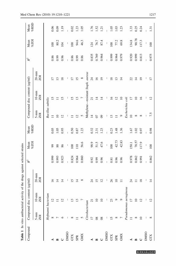
Biological activities
Gram-negative bacteria have lipid-rich cell walls and lipophilicity is an important
consideration in the design of novel analogues (de Almeida et al., 2007; Jensen et al.,1996). The synthesized compound A, with modified lipophilicity, has better activity
than that of the present fluorquinolones, i.e., gatifloxacin, gemifloxacin mesylate (3;
Scheme 3), and sparfloxacin (4; Scheme 4). Compound A exhibits 125% activity
against methicillin-resistant Staphylococcus aureus (MRSA; a troublesome organ-
ism that rapidly became resistant not only to methicillin but also to other b-lactam
antibiotics, and the causative agent of most nosocomial and skin allergen infections),
which is higher that of any other derivative or in-use quinolone, i.e., sparfloxacin,
gemifloxacin, and gatifloxacin (77%, 70%, and 100%, respectively). This is probably
due to an acetyl group at the piperazinyl ring of compound A, as gatifloxacin already
has a methoxy group at the C-8 position, which is responsible for its enhanced
activity toward Gram-positive organisms. It also shows higher activity against P.aeruginosa (150%), K. pneumoniae (158%), E. coli (134%), S. typhi (113%), and S.flexneri (151%). Compound A is superior to gatifloxacin, gemifloxacin, and
sparfloxacin for all Gram-negative organisms. This is an important finding, and
after supplementary in vivo activities to evaluate its safety this compound can be a
lead molecule for therapeutic purposes, as E. coli, P. aeruginosa, S. typhi, P.mirabilis, K. pneumonia, and S. flexneri are responsible for death-causing infections
(Nakajima et al., 1995. However, the intrinsic susceptibility of P. mirabilis to
compound A was lower than that to the above three marketed antibiotics.
Compounds B and C also exhibit moderate to good activity (76–104%) against all
Gram-positive and Gram-negative organisms, much better than that of sparfloxacin
and gemifloxacin. We previously synthesized similar derivatives of ciprofloxacin
N
COOHF
N
O
N
OH3C
NH2
.CH3SO
Scheme 3 Gemifloxacin
Med Chem Res (2010) 19:1210–1221 1215

that proved less effective than the parent molecule (Siddiqui et al., 2007).
Antibacterial activities of all compounds were in good linear relationship to their
concentrations, i.e., linear coefficients (R2) were in the range of 0.769–0.999, with
ignorable variations (%RSD \2). It was found that all gatifloxacin derivatives
showed excellent activity against Trichophyton rubrums and Fusarium solani, in
most cases 50–100% that of gatifloxacin. The antifungal activity of most of the
derivatives against Candida albicans is similar to that gatifloxacin. Results showed
(Tables 1, 2) that these compounds are more suitable for immune-compromised
patients, as they posses broad-spectrum antibacterial activity and antifungal activity.
Combination therapy with available antifungal drugs and quinolone antibiotics
yields additive to synergistic activity over the antifungal drugs used alone (Nakajima
et al., 1995; Shen and Fostel, 1994).
Luminol-enhanced chemiluminesence assay was performed as described by
Helfand et al. (1982) and Haklar et al. (2001). The luminol probe is capable of
detecting the level of reactive oxygen species (ROS) to study the effect of these
derivatives on oxidative burst. Luminol is characterized by its ability to enter the cell
and react with intracellular ROS (Dahlgren and Briheim, 1985). Percentage
inhibition was calculated as 100 = 100 [(CL count in presence of compound/CL
count in absence of compound) 9 100] by an Excel-based program. Preliminary
screening results of whole blood showed that compound B showed 74.1–96.1%
inhibition (IC50 \ 12.5 lg/ml), while gatifloxacin showed 19.7–84.7% inhibition
(IC50 = 31 ± 6.5 lg/ml). Compounds A and C have no considerable inhibitory
activity (Table 3). The sensitivity of gatifloxacin and its derivatives to nonprolif-
erative and proliferative responses of the mitogen phytohemagglutinin (PHA) and
mitogen-induced proliferation of T lymphocytes were evaluated. Proliferative
response of mitogens was monitored at concentrations of 3.12, 12.5, and 50 lg/ml.
The results revealed that neither gatifloxacin nor any of its derivative has
immunosuppressive activity (Table 4).
Conclusion
The best substitution at N4 of the piperazinyl ring is an acetyl group, which
increases antibacterial activity against Gram-negative organisms and retains the
original activity of the compounds against Gram-positive organisms. Additionally,
all N4-substituted gatifloxacin derivatives exhibited encouraging antifungal activity.
The data presented here indicate that an N4-substituted piperazinyl on the quinolone
F
F HN
HN
N
COOH
NH2 O
CH3
CH3H
H
Scheme 4 Sparfloxacin
1216 Med Chem Res (2010) 19:1210–1221
Tab
le1
Invit
roan
tibac
teri
alac
tivit
yof
the
dru
gs
agai
nst
sele
cted
stra
ins
Com
po
und
Com
po
und
dis
cco
nte
nt
(lg
/ml)
R2
Mea
n
%Z
OI
Mea
n
%R
SD
Com
po
und
dis
cco
nte
nt
(lg
/ml)
R2
Mea
n
%Z
OI
Mea
n
%R
SD
5-m
m
ZO
I
10
-mm
ZO
I
20
-mm
ZO
I
5-m
m
ZO
I
10
-mm
ZO
I
20
-mm
ZO
I
Ho
fman
ni
ba
cter
ium
Ba
cill
us
sub
tili
s
A8
12
16
0.9
99
99
0.0
51
11
51
70
.86
10
00
.06
B7
11
14
0.9
93
88
1.0
51
01
11
30
.99
80
.20
.09
C6
12
14
0.9
23
86
0.9
51
21
51
80
.96
10
41
.19
DM
SO
––
––
––
––
––
––
GT
X8
13
15
0.8
24
10
00
.50
11
15
17
0.8
61
00
0.0
2
SP
R1
11
31
40
.964
11
00
.87
12
13
15
0.9
99
4.6
1.2
3
GM
X5
78
0.8
60
56
.61
.23
57
80
.86
46
.31
.05
Cit
rob
act
eriu
mM
eth
yle
ne—
resi
stan
tS
tap
h.
au
reu
s
A1
72
12
40
.92
98
.90
.32
14
21
24
0.8
35
12
41
.76
B0
81
01
50
.99
51
.52
.31
07
14
16
0.7
69
76
.71
.52
C0
81
01
20
.96
47
.61
.23
09
14
19
0.9
64
87
.41
.21
DM
SO
––
––
––
––
––
––
GT
X1
52
32
60
.81
10
00
.23
13
16
18
0.9
99
10
01
.05
SP
R8
91
00
.96
42
.73
1.4
81
11
21
30
.964
77
.21
.03
GM
X7
91
10
.96
42
.83
1.3
69
10
14
0.9
79
69
.81
.23
Pse
udo
monas
aer
ugi
nosa
Esc
her
ichia
coli
A1
11
72
40
.978
15
0.1
1.6
71
01
72
20
.920
13
4.8
1.3
3
B8
10
11
0.8
62
78
.57
1.0
28
10
14
0.9
99
90
.78
0.2
5
C1
01
21
80
.991
11
7.9
0.9
78
16
19
0.8
13
11
7.3
0.2
4
DM
SO
––
––
––
––
––
––
GT
X8
12
14
0.8
62
10
00
.98
7.5
12
17
0.9
75
10
01
.31
Med Chem Res (2010) 19:1210–1221 1217

Tab
le1
con
tin
ued
Com
po
und
Com
po
und
dis
cco
nte
nt
(lg
/ml)
R2
Mea
n
%Z
OI
Mea
n
%R
SD
Com
po
und
dis
cco
nte
nt
(lg
/ml)
R2
Mea
n
%Z
OI
Mea
n
%R
SD
5-m
m
ZO
I
10
-mm
ZO
I
20
-mm
ZO
I
5-m
m
ZO
I
10
-mm
ZO
I
20
-mm
ZO
I
SP
R1
11
31
50
.964
11
7.6
1.2
49
11
14
0.9
94
98
.01
0.2
4
GM
X8
91
00
.964
75
.00
.96
79
10
0.8
62
75
.72
1.2
8
Sh
igel
lafl
exn
eri
Kle
bsi
ella
pn
eum
onia
e
A1
92
22
40
.910
15
11
.01
16
23
25
0.7
69
15
81
.67
B1
21
52
00
.998
10
50
.94
13
14
18
0.9
79
11
31
.41
C1
11
31
40
.862
88
.11
.25
81
42
10
.978
99
.81
.37
DM
SO
––
––
––
––
––
–
GT
X9
16
23
0.9
64
10
00
.15
91
41
90
.964
10
00
.33
SP
R9
11
13
0.9
64
75
.11
.11
10
13
16
0.9
64
96
.11
.28
GM
X7
81
30
.969
61
.41
.54
10
11
12
0.9
64
84
.20
.25
Sh
igel
laty
ph
iP
rote
us
mir
abil
is
A9
16
22
0.9
46
11
30
.99
91
01
20
.999
76
.61
.54
B8
10
18
0.9
88
7.5
0.5
51
11
21
40
.999
92
.01
.23
C8
91
50
.96
80
.20
.025
91
11
20
.862
78
.81
.11
DM
SO
––
––
––
––
––
––
GT
X8
13
21
0.9
91
00
0.2
59
15
19
0.9
11
00
0.4
4
SP
R9
10
15
0.9
68
7.0
1.1
61
01
21
50
.994
90
.00
.23
GM
X8
91
20
.99
75
.50
.85
10
11
12
0.8
18
2.5
0.3
3
ZO
Izo
ne
of
inhib
itio
n,
GT
Xg
atifl
ox
acin
,G
MX
gem
iflo
xac
in,
SP
Rsp
arfl
ox
acin
1218 Med Chem Res (2010) 19:1210–1221

Table 2 In vitro antifungal
activity of the drugs against
selected strains
ZOI zone of inhibition, GTXgatifloxacin, GMXgemifloxacin, SPRsparfloxacin
Compound Compound disc content (lg/ml) Mean
%ZOI
Mean
%RSD20-mm
ZOI
40-mm
ZOI
Trichophyton rubrum
A 28 34 239 0.65
B 16 17 127 1.01
C 21 27 185 1.69
DMSO – – – –
GTX 12 14 100 1.25
SPR 20 24 169 1.32
Candida albicans
A 16 19 206 1.03
B 16 21 218 2.05
C 10 14 141 2.03
DMSO – – – –
GTX 8 9 100 0.02
SPR 13 17 176 0.09
Fusarium solani
A 13 15 93.3 1.87
B 14 19 110 1.61
C 17 19 120 0.99
DMSO – – – –
GTX 12 18 100 0.37
SPR 10 11 70 0.87
Saccharomyces cerevisiae
A 0 0 0 0
B 0 0 0 0
C 0 0 0 0
DMSO – – – –
GTX 0 0 0 0
SPR 0 0 0 0
Table 3 Screening of gatifloxacin and its derivatives using whole blood for chemiluminescene activity
Compound Reading (RLU 9 1000) % inhibition IC50 ± SD
100 lg/ml 50 lg/ml 12.5 lg/ml 100 lg/ml 50 lg/ml 12.5 lg/ml
A 485.6 495.2 482.4 -15.1 -17.4 -14.3 [100 ± 0.0
B 16.3 45.7 109.3 96.1 89.2 74.1 \12.5 ± 0.0
C 358.6 375.1 443.3 15.0 11.1 -5.1 [100 ± 0.0
GTX 471.9 1047.6 2475.5 84.7 66.0 19.7 31 ± 6.5
RLU relative light units
Med Chem Res (2010) 19:1210–1221 1219
ring also greatly influences the oxidative burst activity, in addition to the
antibacterial activity. The caproyl OC7H15 increases activity (% inhibition, 74.1–
96.1%; IC50 \ 12.5 lg/ml), while an acetyl or benzoyl group at the same position
decreases activity. However, further mechanism-based studies are required for
better understanding of the mechanism of action of gatifloxacin derivatives on the
immune response.
References
Bailly HC, Kok MR, Baum BJ, Tak PP (1990) Gatifloxacin as cytokine production inhibitor. Int J
Immunopharmacol 12:31–36
Bryskier A, Chantot JF (1995) Subsitituent on the piperazine ring can shift excretion of the compound
from kidney to liver and therefore increase its half-life. This is useful in patient with impaired liver
function. Drugs 49:16–18
Dahlgren C, Briheim G (1985) Comparison between the luminal dependant cheminescence of
polymorphnuclear leukocytes and of the myeloperoxidase hydrogen peroxide system: influence of
pH, cations and protein. Photochem Photobiol 41:605–610
de Almeida MV, Maurıcio FS, de Souza MVN, da Costa CF, Felipe RCV, Maria CSL (2007) Synthesis
and antitubercular activity of lipophilic moxifloxacin and gatifloxacin derivatives. Bioorg Med
Chem Lett 17:5661–5664
Deborah HS, Virginia LM (1999) Bacterial phospholipases and pathogenesis. Microbes Infect 1:1103–
1112
Domagala JM, Heifetz CL, Hutt MP, Mich TF, Nichols JB, Solomon M, Worth DF (1988) 1-Substituted
7-[3-[(ethylamino)methyl]-1-pyrrolidinyl]-6,8-difluoro-1,4-dihydro-4-oxo-3-quinoline carboxylic
acids. New quantitative structure activity relationships at N1 for the quinolone antibacterials. J
Med Chem 31:991–1001
Drusano GL, Wolfson JS, Hooper DC (1989) Quinolone antimicrobial agents. Am Soc Microbiol 71:105
Emami S, Shafiee A, Foroumadi A (2006) Structural features of new quinolones and relationship to
antibacterial activity against gram-positive bacteria. Mini-Rev Med Chem 6:375–386
Haklar G, Ozveri ES, Yuksel M, Aktan A, Yalcin AS (2001) Different kinds of reactive oxygen and
nitrogen species were detected in colon and breast tumors. Cancer Lett 165:219–224
Helfand S, Werkmeister J, Roader J (1982) Chemiluminescence response of human natural killer cells. I.
The relationship between target cell binding, chemiluminescence, and cytolysis. J Exp Med
156:492–505
Hoshino K, Kitamura A, Morrissey I, Sato K, Kato J, Ikeda H (1994) Comparison of inhibition of
Escherichia coli topoisomerase IV by quinolones with DNA gyrase inhibition. Antimicrob Agents
Chemother 38:2623–2627
Jensen G, Wandall DA, Gaarsler K (1996) Antibiotic resistance in Shigella and Salmonella in region of
Lithuania. Eur J Clin Microbiol Infect Dis 15:872–876
Table 4 Screening of gatifloxacin and its synthesized derivatives for their immune modulating inhibi-
tory properties, using whole blood
Compound Reading (cpm 9 1000) % inhibition IC50 ± SD
50 lg/ml 12.5 lg/ml 3.12 lg/ml 50 lg/ml 12.5 lg/ml 3.12 lg/ml
A 29,751.8 26,503.8 24,101.2 -52.30 -35.70 -23.40 [50 ± 0.0
B 20,881.2 27,642.0 31,144.1 -6.90 -41.50 -59.50 [50 ± 0.0
C 20,881.2 27,642.0 31,144.1 -6.90 -41.50 -59.50 [50 ± 0.0
GTX 24,450.9 38,657.7 33,427.3 -25.20 -97.90 -71.10 [50 ± 0.0
Control 34,246.4
1220 Med Chem Res (2010) 19:1210–1221

Kabir OA, Olukayode O, Chidi EO, Christopher CI, Kehinde EF (2005) Screening of crude extracts of six
medicinal plants used in South-West Nigerian unorthodox medicine for antimethicillin resistant
staphylococcus aureus activity. BMC Compl Alt Med 5:1472–1483
Keiser J, Burri C (2001) Evaluation of quinolone derivatives for antitrypanosomal activity. Trop Med Int
Health 6:369–389
Kenneth T (2007) The mechanisms of bacterial pathogenicity. Todar’s Online Textbook of Bacteriology
Nakajima R, Kitamura K, Someya K, Tanaka M, Sato K (1995) In vitro and in vivo antifungal activities
of DU-6859a, a fluoroquinolone, in combination with amphotericin B and fluconazole against
pathogenic fungi. Antimicrob Agents Chemother 39:1517–1521
National Committee for Clinical Laboratory Standards (1993) Performance standards for antimicrobial
susceptibility tests, 5th edn. NCCLS document M2-A5. NCCLS, Villanova, PA
Ronald AR, Low DE (2003) Fluoroquinolones antibiotics. Birkhauser Verlag, Basel
Scheld WM (1989) Quinolone therapy for infections of the central nervous system. Rev Infect Dis
11:1194–1202
Shen LL, Fostel JM (1994) DNA topoisomerase inhibitors as antifungal agents. Adv Pharmacol 29:227–
244
Siddiqui R, Sultana N, Khan KM, Akber N, Arayne MS (2007) Effect of skeletal modifications of
ciprofloxacin on antibacterial, antifungal and cytotoxic activities. J Chinese Clin Med 21:188–195
Tokushige H, Yokogaki S, Naka H (2003) Gatifloxacin as cytokine production inhibitor. European Patent
EP1312364
Walsh C (2003) Antibiotics, actions, origins, resistance. Harvard Medical School. ASM Press, Boston
WHO (2003) Manual for the laboratory identification and antimicrobial susceptibility testing of bacterial
pathogens of public health importance in the developing world. Who Health Organization, Geneva,
pp 103–162
Yong HL, Yun ZT, Xue FH, Ren GX (2004) The crystal structure of a gatifloxacin complex and its
fluorescent property. Z Anorg Allg Chem 631:639–641
Med Chem Res (2010) 19:1210–1221 1221
![Amberlite–IRA402 (OH) ion exchange resin mediated synthesis of indolizines, pyrrolo [1,2-a] quinolines and isoquinolines: Antibacterial and antifungal evaluation of the products](https://static.fdokumen.com/doc/165x107/631b31c4a906b217b9065971/amberliteira402-oh-ion-exchange-resin-mediated-synthesis-of-indolizines-pyrrolo-1674799713.jpg)