
Synthesis and characterization of FeSe2 nanoparticles and FeSe2/FeO(OH) nanocomposites by...
8
Synthesis and characterization of FeSe 2 nanoparticles and FeSe 2 /FeO(OH) nanocomposites by hydrothermal method Azam Sobhani a , Masoud Salavati-Niasari b,⇑ a Department of Chemistry, Kosar University of Bojnord, Bojnord, North Khorasan, Islamic Republic of Iran b Institute of Nano Science and Nano Technology, University of Kashan, Kashan, P.O. Box 87317-51167, Islamic Republic of Iran article info Article history: Received 4 October 2014 Received in revised form 23 October 2014 Accepted 12 November 2014 Available online 18 November 2014 Keywords: FeSe 2 Nanoparticles Hydrothermal Optical property Surfactant Reductant abstract FeSe 2 nanoparticles and FeSe 2 /FeO(OH) nanocomposites have been synthesized through a novel control- lable hydrothermal method with FeSO 4 7H 2 O and SeCl 4 as precursors. In this procedure, H 2 SeO 3 (obtained from aqueous solution of SeCl 4 ) is gradually reduced to Se and Se 2 by reductant. The reduced Se and Se 2 ions are combined with Fe 2+ ion to give FeSe 2 . By varying the surfactant, reaction time, tem- perature, reductant and amount of hydrazine hydrate (N 2 H 4 H 2 O), the method permits us to synthesize different products. The FeSe 2 nanoparticles are synthesized in the presence of PEG4000 at 140 °C for 13 h. Using PEG600 (at 140 °C or 110 °C for 13 h) and sodium dodecyl sulfate (at 140 °C for 7 h), FeSe 2 /FeO(OH) nanocomposites are synthesized. SEM and TEM images show the morphology and size of the as-synthesized samples. Chemical composition of the samples is characterized by XRD, XPS and EDS. Ultraviolet–visible and photoluminescence spectra exhibit optical properties of nanostructures. Magne- tization measurement shows a twofold behavior, ferromagnetic and paramagnetic behaviors, for FeSe 2 nanoparticles. Ó 2014 Elsevier B.V. All rights reserved. 1. Introduction Transition metal chalcogenides have drawn considerable atten- tion because of their optical, electrical, magnetic and transport properties [1,2]. In the case of iron chalcogenide compounds, much attention has been done to FeSe 2 (iron diselenide) because of its application for tandem solar cells and detecting visible radiation [3,4]. The FeSe 2 is a p-type semiconductor with a direct band gap of 1.0 eV [5,6] and considered as a suitable material for photovol- taic applications [7]. It is also viewed as a less toxic and more envi- ronmentally responsible alternative to related narrow band gap semiconductors that contain lead or cadmium. Furthermore, FeSe 2 exhibits 3D magnetic ordering [8,9] and possess excellent mag- netic properties. It can be utilized as building blocks in future memory devices [10]. FeSe 2 crystals both in marcasite orthorhom- bic and pyrite cubic structures exhibit semiconducting behavior [11]. Under ambient conditions, FeSe 2 adopts marcasite structure while high temperature/pressure synthesis leads to pyrite structure [12]. Electronic and optical properties of FeS 2 have been extensively studied, but a few efforts have been devoted into the synthesis of FeSe 2 micro/nanostructures [6,13]. In recent years, a number of experimental and theoretical investigations on FeSe 2 have been published [3,14]. Of the various methods of synthesis of iron sele- nides, the hydrothermal method has exhibited many outstanding advantages in cost, manipulation, large-scale production, control of objective products, environment-friendliness and so on [15– 18]. In this paper, we report a hydrothermal route for the prepara- tion of FeSe 2 nanoparticles and FeSe 2 /FeO(OH) (iron oxide hydrox- ide) nanocomposites. In this route, newly produced Se in the system has been used as both reactant and template. In some cases, the template as one of the reactants has been actively engaged in the synthesis process [19,20]. In our synthesis process, the as-prepared Se is converted into the FeSe 2 by reacting with Se 2 ion and then Fe 2+ ion in aqueous solution. To the best of our knowledge, studies on the synthesis of FeSe 2 nanoparticles and FeSe 2 /FeO(OH) nanocomposites by using SeCl 4 have not been reported to date. For a few years, our group has been interested in the synthesis of metal selenide nanostructures and development of the semiconductors [21–28]. 2. Experimental 2.1. Material and methods All the chemicals used in our experiments were of analytical grade, were pur- chased from Merck and used as received without further purification. A Teflon-lined stainless steel cylindrical closed chamber with 150 ml capacity was used for the http://dx.doi.org/10.1016/j.jallcom.2014.11.079 0925-8388/Ó 2014 Elsevier B.V. All rights reserved. ⇑ Corresponding author. Tel.: +98 361 591 2383; fax: +98 361 555 2930. E-mail address: [email protected] (M. Salavati-Niasari). Journal of Alloys and Compounds 625 (2015) 26–33 Contents lists available at ScienceDirect Journal of Alloys and Compounds journal homepage: www.elsevier.com/locate/jalcom
Transcript of Synthesis and characterization of FeSe2 nanoparticles and FeSe2/FeO(OH) nanocomposites by...

Journal of Alloys and Compounds 625 (2015) 26–33
Contents lists available at ScienceDirect
Journal of Alloys and Compounds
journal homepage: www.elsevier .com/locate / ja lcom
Synthesis and characterization of FeSe2 nanoparticles andFeSe2/FeO(OH) nanocomposites by hydrothermal method
http://dx.doi.org/10.1016/j.jallcom.2014.11.0790925-8388/� 2014 Elsevier B.V. All rights reserved.
⇑ Corresponding author. Tel.: +98 361 591 2383; fax: +98 361 555 2930.E-mail address: [email protected] (M. Salavati-Niasari).
Azam Sobhani a, Masoud Salavati-Niasari b,⇑a Department of Chemistry, Kosar University of Bojnord, Bojnord, North Khorasan, Islamic Republic of Iranb Institute of Nano Science and Nano Technology, University of Kashan, Kashan, P.O. Box 87317-51167, Islamic Republic of Iran
a r t i c l e i n f o a b s t r a c t
Article history:Received 4 October 2014Received in revised form 23 October 2014Accepted 12 November 2014Available online 18 November 2014
Keywords:FeSe2
NanoparticlesHydrothermalOptical propertySurfactantReductant
FeSe2 nanoparticles and FeSe2/FeO(OH) nanocomposites have been synthesized through a novel control-lable hydrothermal method with FeSO4�7H2O and SeCl4 as precursors. In this procedure, H2SeO3
(obtained from aqueous solution of SeCl4) is gradually reduced to Se and Se2� by reductant. The reducedSe and Se2� ions are combined with Fe2+ ion to give FeSe2. By varying the surfactant, reaction time, tem-perature, reductant and amount of hydrazine hydrate (N2H4�H2O), the method permits us to synthesizedifferent products. The FeSe2 nanoparticles are synthesized in the presence of PEG4000 at 140 �C for 13 h.Using PEG600 (at 140 �C or 110 �C for 13 h) and sodium dodecyl sulfate (at 140 �C for 7 h), FeSe2/FeO(OH)nanocomposites are synthesized. SEM and TEM images show the morphology and size of theas-synthesized samples. Chemical composition of the samples is characterized by XRD, XPS and EDS.Ultraviolet–visible and photoluminescence spectra exhibit optical properties of nanostructures. Magne-tization measurement shows a twofold behavior, ferromagnetic and paramagnetic behaviors, for FeSe2
nanoparticles.� 2014 Elsevier B.V. All rights reserved.
1. Introduction
Transition metal chalcogenides have drawn considerable atten-tion because of their optical, electrical, magnetic and transportproperties [1,2]. In the case of iron chalcogenide compounds, muchattention has been done to FeSe2 (iron diselenide) because of itsapplication for tandem solar cells and detecting visible radiation[3,4]. The FeSe2 is a p-type semiconductor with a direct band gapof 1.0 eV [5,6] and considered as a suitable material for photovol-taic applications [7]. It is also viewed as a less toxic and more envi-ronmentally responsible alternative to related narrow band gapsemiconductors that contain lead or cadmium. Furthermore, FeSe2
exhibits 3D magnetic ordering [8,9] and possess excellent mag-netic properties. It can be utilized as building blocks in futurememory devices [10]. FeSe2 crystals both in marcasite orthorhom-bic and pyrite cubic structures exhibit semiconducting behavior[11]. Under ambient conditions, FeSe2 adopts marcasite structurewhile high temperature/pressure synthesis leads to pyritestructure [12].
Electronic and optical properties of FeS2 have been extensivelystudied, but a few efforts have been devoted into the synthesis ofFeSe2 micro/nanostructures [6,13]. In recent years, a number of
experimental and theoretical investigations on FeSe2 have beenpublished [3,14]. Of the various methods of synthesis of iron sele-nides, the hydrothermal method has exhibited many outstandingadvantages in cost, manipulation, large-scale production, controlof objective products, environment-friendliness and so on [15–18]. In this paper, we report a hydrothermal route for the prepara-tion of FeSe2 nanoparticles and FeSe2/FeO(OH) (iron oxide hydrox-ide) nanocomposites. In this route, newly produced Se in thesystem has been used as both reactant and template. In somecases, the template as one of the reactants has been activelyengaged in the synthesis process [19,20]. In our synthesis process,the as-prepared Se is converted into the FeSe2 by reacting withSe2� ion and then Fe2+ ion in aqueous solution. To the best of ourknowledge, studies on the synthesis of FeSe2 nanoparticles andFeSe2/FeO(OH) nanocomposites by using SeCl4 have not beenreported to date. For a few years, our group has been interestedin the synthesis of metal selenide nanostructures and developmentof the semiconductors [21–28].
2. Experimental
2.1. Material and methods
All the chemicals used in our experiments were of analytical grade, were pur-chased from Merck and used as received without further purification. A Teflon-linedstainless steel cylindrical closed chamber with 150 ml capacity was used for the
Table 1The reaction conditions of iron diselenides synthesized from FeSO4�7H2O and SeCl4 with molar ratio 1:1.
Sample Effect Surfactant Reductant Temperature (�C) Time (h)
1 Surfactant PEG4000 Hydrazine (1 ml) 140 132 – Hydrazine (1 ml) 140 133 PVA Hydrazine (1 ml) 140 134 SDS Hydrazine (1 ml) 140 135 PEG600 Hydrazine (1 ml) 140 13
6 Temperature PEG600 Hydrazine (1 ml) 110 137 PEG600 Hydrazine (1 ml) 170 13
8 Time SDS Hydrazine (1 ml) 140 79 SDS Hydrazine (1 ml) 140 19
10 Reductant PEG4000 KBH4 140 1311 PEG4000 Zn 140 13
12 Amount of hydrazine SDS Hydrazine (2 ml) 140 1313 SDS Hydrazine (3 ml) 140 1314 SDS Hydrazine (4 ml) 140 13
A. Sobhani, M. Salavati-Niasari / Journal of Alloys and Compounds 625 (2015) 26–33 27
synthesis. Powder X-ray diffraction (XRD) patterns were collected from a diffrac-tometer of Philips Company with X’PertPro monochromatized Cu Ka radiation(k = 1.54 Å). Microscopic morphology of products was visualized by a LEO 1455VPscanning electron microscope (SEM). Transmission electron microscopy (TEM)images were obtained on a JEM-2100 with an accelerating voltage of 60–200 kVequipped with a high resolution CCD camera. X-ray energy dispersive spectroscopy(EDS) analysis with 20 kV accelerated voltage was done. X-ray photoelectron spec-tra (XPS) were recorded on a twin anode, XR3E2, X-ray photoelectron spectrometer,using Al Ka radiation as the exciting source. The magnetic measurement was car-ried out in a vibrating sample magnetometer (VSM) (BHV-55, Riken, Japan) at roomtemperature. Room temperature photoluminescence (PL) was studied on a PerkinElmer (LS 55) fluorescence spectrophotometer. The electronic spectrum of the sam-ples was taken on a Scinco UV–vis (ultraviolet–visible) scanning spectrometer(Model S-4100).
2.2. Synthesis of FeSe2 nanoparticles
In a typical experiment, FeSO4�7H2O and SeCl4 with molar ratio 1:1 were putinto a beaker. The beaker was filled with distilled water to let the powders com-pletely dissolve by continuous stirring. Then an appropriate amount of PEG4000was added into the above solution. After stirring the solution for 15 min, 1 mlhydrazine was added drop-wise. Finally, the mixture was transferred into aTeflon-lined stainless steel container, then was sealed and maintained at 140 �Cfor 13 h. The resultant black precipitates were filtered, washed by distilled waterand absolute ethanol and dried at 60 �C in vacuum. The effects of surfactant, kindof reductant and its amount, reaction time and temperature on morphology, parti-cle size, phase and purity of the products were investigated. The results have beenlisted in Table 1.
2.3. Synthesis of FeSe2/FeO(OH) nanocomposites
The whole process to prepare FeSe2 nanoparticles can be easily adjusted to syn-thesize FeSe2/FeO(OH) nanocomposites by simply changing the surfactant toPEG600 (at 140 �C or 110 �C for 13 h) and SDS (at 140 �C for 7 h).
3. Results and discussion
The detailed reactions of mechanism have been described inScheme 1. The SeCl4 dissolved in the water is transformed to theH2SeO3. The H2SeO3 is reduced to selenium element and immedi-ately got the two electrons to form the Se2� ion. The Se2� ion willattract the Fe2+ ion of FeSO4 solution and form FeSe2 alloy. After anenough hydrothermal growth under suitable time and tempera-ture condition, the resultant FeSe2 nanoparticles would becomemore uniform through Ostwald ripening. The FeSe2 nanoparticlesobtained in this work adopt an orthorhombic marcasite structurewith space group Pnnm (no. 58). The conventional unit cell shownin Scheme 1 contains two formula units of FeSe2. Each Fe atom iscoordinated by six Se atoms as octahedron and each Se atom iscoordinated to three Fe atoms and another Se forms a tetrahedron.
Two neighboring octahedra share an edge and form a linear chainparallel to orthorhombic c-axis [29].
Fig. 1a–f illustrates SEM and TEM images of the products syn-thesized in the presence of different surfactants. When thePEG4000 is used as surfactant at 140 �C after 13 h of hydrothermalreaction (sample no. 1), the morphology of the products is nano-particles with a diameter of 10–35 nm (Fig. 1a, b). Fig. 1c showsthe SEM image of the product obtained in the absent of surfactant(sample no. 2). The formation of nanoparticles with high agglomer-ation is seen in this figure. Fig. 1d shows that the nanoparticlesobtained in the presence of PVA (sample no. 3) are agglomerated.Fig. 1e and f shows that the nanoparticles are also obtained inthe presence of SDS (sample no. 4) and PEG600 (sample no. 5).
The XRD patterns of the products synthesized at 140 �C for 13 hin the presence of 1 ml hydrazine and different surfactants are seenin Fig. 2. The reflection peaks with high intensity prove the highcrystallinity of the products synthesized. Fig. 2d is XRD patternof sample prepared in the presence of PEG4000. All of the reflectionpeaks in this figure can be indexed to the orthorhombic phase ofFeSe2 with space group of Pnnm (a mixture of JCPDS cardNo. 12-0291 and 21-0432). No impurities are observed in the pat-tern, indicating high purity of the product. Fig. 2c and a shows XRDpatterns of FeSe2/FeO(OH) nanocomposites synthesized in theabsent of surfactant and in the presence of PEG600, respectively.In both figures, the peaks indexed with red star indicate the ortho-rhombic phase of FeO(OH). In Fig. 2b, the peaks indexed with greenstar indicate the cubic phase of Fe2O3 and the other peaks indicatethe orthorhombic phase of FeSe2 (JCPDS card No. 12-0291). Asshown in Fig. 2b, the sample obtained in the presence of SDS (sam-ple no. 4) is the mixture of two products: Fe2O3 and FeSe2. The cal-culated crystal diameters of the products obtained in the presenceof PEG4000, SDS, PEG600 and also product obtained in the absentof surfactant, by the Debye–Scherrer equation are 33.64, 22.44,26.91 and 33.64 nm, respectively [30]. It is found that the particlesize observed by the TEM image (10–35 nm) and crystal diameterestimated by the XRD results (33.64 nm) for the product obtainedin the presence of PEG4000 (sample no. 1) are agreeable with eachother. The diameter of the nanoparticles obtained in the absent ofsurfactant are more than the others. The surfactants prevent aggre-gation of particles and control particle size distribution. The nucleican be wrapped by the surfactant completely and nanoparticleswith less particle sizes are obtained in the presence of surfactant.
The quality and composition of the as-prepared samples are fur-ther studied by XPS analysis. The XPS survey spectrum in Fig. 3ashows that the peaks centered at 712 eV and 728 eV can beassigned to the binding energy of Fe 2p3/2 and Fe 2p1/2,

Scheme 1. Chemical reactions in the experiment.
Fig. 1. (a, b) SEM and TEM images of the product prepared in the presence of PEG4000 and hydrazine at 140 �C for 13 h (sample no. 1), (c�f) SEM images of the productsprepared at 140 �C for 13 h: (c) in the absent of surfactant (sample no. 2), (d) PVA (sample no. 3), (e) SDS (sample no. 4), and (f) PEG600 (sample no. 5).
28 A. Sobhani, M. Salavati-Niasari / Journal of Alloys and Compounds 625 (2015) 26–33

Fig. 2. XRD patterns of the products prepared in the presence of: (a) PEG600 (sample no. 5), (b) SDS (sample no. 4), (c) in the absent of surfactant (sample no. 2), (d) PEG4000(sample no. 1), (e) PEG600 (sample no. 6), (f) SDS (sample no. 8), (g) PEG4000 and Zn (sample no. 11), and (h) PEG4000 and KBH4 (sample no. 10).
A. Sobhani, M. Salavati-Niasari / Journal of Alloys and Compounds 625 (2015) 26–33 29

Fig. 3. XPS spectra of FeSe2 nanoparticles (sample no. 1): (a) survey spectrum; (b) Se 3d.
Fig. 4. EDS spectrum of the as-synthesized FeSe2 nanoparticles.
Fig. 5. (a, b) SEM images of the product prepared in the presence of PEG600 at 110 �C (samand (e) HRTEM image of sample no. 6.
30 A. Sobhani, M. Salavati-Niasari / Journal of Alloys and Compounds 625 (2015) 26–33
respectively. In Fig. 3b, the Se 3d peak of FeSe2 nanoparticlesappears at 56 eV. The obtained XPS results are in agreement withthe reported values at the Fe and Se regions and prove that sampleno. 1 is FeSe2 [16,31]. Fig. 3a shows at the surface of the nanopar-ticles there is a carbon–carbon bond and the nanoparticles havebeen coated with PEG4000. In the apparatus used, this C–C bondhas a well-defined position at 285 eV. In Fig. 3a, the peak centeredat 533 eV is attributed to the O1s and shows the surface oxidationof species. The surface oxidation can be ascribed to the high surfaceactivity of FeSe2 nanoparticles.
Fig. 4 shows the EDS spectrum of the FeSe2 nanoparticles pre-pared in the presence of PEG4000 (sample no. 1). The figure indi-cates that the elements in the product are Fe and Se only; theatomic ratio of Fe to Se in different areas is always 38.29:61.71,which indicates a pure FeSe2 phase.
ple no. 6) and 170 �C (sample no. 7), respectively, (c, d) TEM images of sample no. 6,

Fig. 6. (a, b) SEM images of the products prepared in the presence of SDS at 140 �C for 7 h (sample no. 8) and 19 h (sample no. 9), respectively.
Fig. 7. SEM images of samples prepared in the presence of PEG4000 and: (a) KBH4
(sample no. 10), (b) Zn (sample no. 11).
A. Sobhani, M. Salavati-Niasari / Journal of Alloys and Compounds 625 (2015) 26–33 31
The effects of reaction temperature on the morphology andshape of the as-synthesized products in the presence of hydrazineand PEG600 are shown in Fig. 5. For this propose reactions werecarried out at three different temperatures. It can be observed thatwith increasing temperature from 110 �C (Fig. 5a) to 140 �C (Fig. 1f)and then to 170 �C (Fig. 5b), morphology and particle size of sam-ples remain nearly constant and only agglomeration of nanoparti-cles decreases a little. In order to further elucidate the size and thecrystal structure of the nanoparticles prepared in the presence ofPEG600 at 110 �C, TEM images were taken. TEM images in Fig. 5cand d show the nanoparticles are aggregated and their particlesizes are in the range 30–250 nm. The HRTEM image of a singlenanoparticle in Fig. 5e shows that the nanoparticles are highlycrystalline and distance between the two adjacent planes ismeasured to be 0.25 nm.
XRD pattern of the sample prepared in the presence of PEG600at 110 �C (sample no. 6) is presented in Fig. 2e. The diffractionpeaks observed in this figure can be indexed to FeSe2/FeO(OH)nanocomposites.
In the other side, the effect of reaction time in the presence ofhydrazine and SDS is investigated. With increasing reaction timefrom 7 h (Fig. 6a) to 13 h (Fig. 1e), the size and agglomeration ofthe nanoparticles decrease. With increasing time to 19 h(Fig. 6b), nanoparticles are collected and aggregated particles withan irregular manner are formed. The XRD pattern in Fig. 2f illus-trating the formation of the FeSe2/FeO(OH) nanocomposites inthe presence of SDS at 140 �C for 7 h (sample no. 8).
In continuation, the effect of the sort of reductant on the mor-phology of the products in the presence of PEG4000, at 140 �C for13 h, is investigated. Different reducing agents have differentreducing abilities that influence the rate of reductions. Therefore,one of the advantages of employing reductant is varying the prep-aration condition and controlling the formation of the products. Inthis study, three reducing agents were applied that includingN2H4�H2O, KBH4 and Zn. The reduction with KBH4 and N2H4�H2Oproceeds in a homogeneous way, the metal cation solution andthe reductant solution are liquid, but the reduction of the liquidmetal cation solution with the solid Zn powder is a heterogeneousprocess being limited by the interface area of the Zn surface andthe solution in contact with this surface. The strong reductants,in combination with stirring rapidly, create a large number ofnuclei and so further growth of the nuclei is limited. As a result,many small particles are obtained in the presence of N2H4�H2O(Fig. 1a) and KBH4 (Fig. 7a). The Zn is slower in the reduction ratecompared to hydroborates and hydrazine. Our group attributes theslower action of Zn as the reason for the rod-like structures obtain.The slower rate of reduction allows ample time for the particles toagglomerate and grow into more regular shapes. Fig. 7b shows that
particles elongate along the c-axis and rod-like structures areformed along with aggregated nanoparticles, in the presence of Zn.
Fig. 2h and g shows the XRD patterns of the as-prepared prod-ucts in the presence of KBH4 (sample no. 10) and Zn (sampleno. 11), respectively. All diffraction peaks in Fig. 2h can be wellindexed to hexagonal phase selenium with JCPDS cardNo. 86-2246. Most of the diffraction peaks in Fig. 2g can be indexedto hexagonal phase selenium with JCPDS card No. 06-0362.Besides, some diffraction peaks in the XRD pattern in Fig. 2g can

Fig. 8. SEM images of samples prepared in the presence of SDS and: (a) 2 mlhydrazine (sample no. 12), (b) 3 ml hydrazine (sample no. 13), and (c) 4 mlhydrazine (sample no. 14).
Fig. 9. M–H hysteresis at 300 K for sample no. 1.
32 A. Sobhani, M. Salavati-Niasari / Journal of Alloys and Compounds 625 (2015) 26–33
be ascribed to Fe2.942O4 and Fe2O3. XRD pattern of the sample syn-thesized in the presence of hydrazine (sample no. 1) showed dif-fraction patterns of the orthorhombic phase FeSe2 with spacegroup of Pnnm (Fig. 2d).
The SEM images of the as-prepared samples in the presence of2–4 ml hydrazine are shown in Fig. 8. In the synthesis of irondiselenides, our present experiments, amount of the hydrazine
has a small influence on the morphology of the products. It canbe observed that nanoparticles are formed at 140 �C for 13 h inthe presence of SDS and different amounts of the hydrazine. Thesize and agglomeration of the nanoparticles increase withincreasing the amount of the hydrazine from 1 ml (Fig. 1e) to2 ml (Fig. 8a), 3 ml (Fig. 8b) and then 4 ml (Fig. 8c).
Ferromagnets (permanent magnets) can retain a memory of anapplied field once it is removed, this behavior is called hysteresis.The hystersis loop illustrates the technical magnetic properties ofa ferromagnetic materia. Saturation magnetization (Ms) is deter-mined from the extrapolation of curve of H/M (M = magnetizationand H = applied field) versus H and is the maximum induced mag-netic moment that can be obtained in a magnetic field, beyond thisfield no further increase in magnetization occurs. Coercivity (Hc) isa measure of width of a hysteresis loop and permanence of themagnetic moment. The various hysteresis parameters are notsolely intrinsic properties but are dependent on grain size, domainstate, stresses, and temperature. Fig. 9 shows the magnetic hyster-esis curve for the sample no. 1 measured at room temperature. Thefigure shows a twofold behavior for FeSe2 nanoparticles, ferromag-netic behavior in low fields and paramagnetic behavior in up fields.The hysteresis loop has remanent magnetization (Mr) and Hc valuesof ca. 0.002 emu/g and 167.176 Oe, respectively.
FeSe2 is well known as a p-type semiconductor possessing adirect band gap [5]. The room temperature PL spectrum of theFeSe2 nanoparticles (Fig. 10a) shows a relatively narrow Gaussiancurve signifying a monodispersed particle population and confirm-ing the even distribution of particles. The emission maximum ofFeSe2 at an excitation wavelength of 250 nm is located at 420 nmin sample no. 1. The band gap of about 2.95 eV is obtained for thissample that is higher than the reported band gap of 1.0 eV of bulkFeSe2 [5,6]. The FeSe2 is blue-shifted compared to the bulk sample.UV–vis spectrum of the FeSe2 nanoparticles is showed in Fig. 10b.The optical absorption edge 550 nm in Fig. 10b shows a blue shiftin relation to bulk FeSe2. The observed blue shift may be attributedto the quantum confinement of charge carriers in thenanoparticles.
4. Conclusion
In this paper, we report a convenient method based on reactionin aqueous system for the synthesis of FeSe2 nanoparticles andFeSe2/FeO(OH) nanocomposites. The effects of preparation param-eters such as: kind of surfactant, kind of reductant and its amount,reaction time and temperature on morphology, particle size, phase
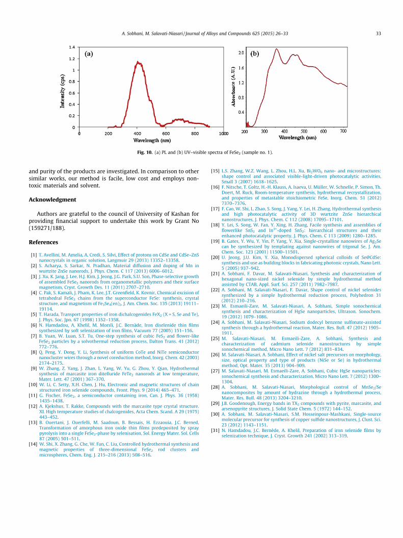
Fig. 10. (a) PL and (b) UV–visible spectra of FeSe2 (sample no. 1).
A. Sobhani, M. Salavati-Niasari / Journal of Alloys and Compounds 625 (2015) 26–33 33
and purity of the products are investigated. In comparison to othersimilar works, our method is facile, low cost and employs non-toxic materials and solvent.
Acknowledgment
Authors are grateful to the council of University of Kashan forproviding financial support to undertake this work by Grant No(159271/188).
References
[1] T. Avellini, M. Amelia, A. Credi, S. Silvi, Effect of protons on CdSe and CdSe–ZnSnanocrystals in organic solution, Langmuir 29 (2013) 13352–13358.
[2] S. Acharya, S. Sarkar, N. Pradhan, Material diffusion and doping of Mn inwurtzite ZnSe nanorods, J. Phys. Chem. C 117 (2013) 6006–6012.
[3] J. Xu, K. Jang, J. Lee, H.J. Kim, J. Jeong, J.G. Park, S.U. Son, Phase-selective growthof assembled FeSe2 nanorods from organometallic polymers and their surfacemagnetism, Cryst. Growth Des. 11 (2011) 2707–2710.
[4] C. Pak, S. Kamali, J. Pham, K. Lee, J.T. Greenfield, K. Kovnir, Chemical excision oftetrahedral FeSe2 chains from the superconductor FeSe: synthesis, crystalstructure, and magnetism of Fe3Se4(en)2, J. Am. Chem. Soc. 135 (2013) 19111–19114.
[5] T. Harada, Transport properties of iron dichalcogenides FeX2 (X = S, Se and Te),J. Phys. Soc. Jpn. 67 (1998) 1352–1358.
[6] N. Hamdadou, A. Khelil, M. Morsli, J.C. Bernàde, Iron diselenide thin filmssynthesized by soft selenization of iron films, Vacuum 77 (2005) 151–156.
[7] B. Yuan, W. Luan, S.T. Tu, One-step synthesis of cubic FeS2 and flower-likeFeSe2 particles by a solvothermal reduction process, Dalton Trans. 41 (2012)772–776.
[8] Q. Peng, Y. Dong, Y. Li, Synthesis of uniform CoTe and NiTe semiconductornanocluster wires through a novel coreduction method, Inorg. Chem. 42 (2003)2174–2175.
[9] W. Zhang, Z. Yang, J. Zhan, L. Yang, W. Yu, G. Zhou, Y. Qian, Hydrothermalsynthesis of marcasite iron ditelluride FeTe2 nanorods at low temperature,Mater. Lett. 47 (2001) 367–370.
[10] W. Li, C. Setty, X.H. Chen, J. Hu, Electronic and magnetic structures of chainstructured iron selenide compounds, Front. Phys. 9 (2014) 465–471.
[11] G. Fischer, FeSe2, a semiconductor containing iron, Can. J. Phys. 36 (1958)1435–1438.
[12] A. Kjekshus, T. Rakke, Compounds with the marcasite type crystal structure.XI. High temperature studies of chalcogenides, Acta Chem. Scand. A 29 (1975)443–452.
[13] B. Ouertani, J. Ouerfelli, M. Saadoun, B. Bessais, H. Ezzaouia, J.C. Berned,Transformation of amorphous iron oxide thin films predeposited by spraypyrolysis into a single FeSe2-phase by selenisation, Sol. Energy Mater. Sol. Cells87 (2005) 501–511.
[14] W. Shi, X. Zhang, G. Che, W. Fan, C. Liu, Controlled hydrothermal synthesis andmagnetic properties of three-dimensional FeSe2 rod clusters andmicrospheres, Chem. Eng. J. 215–216 (2013) 508–516.
[15] L.S. Zhang, W.Z. Wang, L. Zhou, H.L. Xu, Bi2WO6 nano- and microstructures:shape control and associated visible-light-driven photocatalytic activities,Small 3 (2007) 1618–1625.
[16] F. Nitsche, T. Goltz, H.-H. Klauss, A. Isaeva, U. Müller, W. Schnelle, P. Simon, Th.Doert, M. Ruck, Room-temperature synthesis, hydrothermal recrystallization,and properties of metastable stoichiometric FeSe, Inorg. Chem. 51 (2012)7370–7376.
[17] F. Cao, W. Shi, L. Zhao, S. Song, J. Yang, Y. Lei, H. Zhang, Hydrothermal synthesisand high photocatalytic activity of 3D wurtzite ZnSe hierarchicalnanostructures, J. Phys. Chem. C 112 (2008) 17095–17101.
[18] Y. Lei, S. Song, W. Fan, Y. Xing, H. Zhang, Facile synthesis and assemblies offlowerlike SnS2 and In3+-doped SnS2: hierarchical structures and theirenhanced photocatalytic property, J. Phys. Chem. C 113 (2009) 1280–1285.
[19] B. Gates, Y. Wu, Y. Yin, P. Yang, Y. Xia, Single-crystalline nanowires of Ag2Secan be synthesized by templating against nanowires of trigonal Se, J. Am.Chem. Soc. 123 (2001) 11500–11501.
[20] U. Jeong, J.U. Kim, Y. Xia, Monodispersed spherical colloids of Se@CdSe:synthesis and use as building blocks in fabricating photonic crystals, Nano Lett.5 (2005) 937–942.
[21] A. Sobhani, F. Davar, M. Salavati-Niasari, Synthesis and characterization ofhexagonal nano-sized nickel selenide by simple hydrothermal methodassisted by CTAB, Appl. Surf. Sci. 257 (2011) 7982–7987.
[22] A. Sobhani, M. Salavati-Niasari, F. Davar, Shape control of nickel selenidessynthesized by a simple hydrothermal reduction process, Polyhedron 31(2012) 210–216.
[23] M. Esmaeili-Zare, M. Salavati-Niasari, A. Sobhani, Simple sonochemicalsynthesis and characterization of HgSe nanoparticles, Ultrason. Sonochem.19 (2012) 1079–1086.
[24] A. Sobhani, M. Salavati-Niasari, Sodium dodecyl benzene sulfonate-assistedsynthesis through a hydrothermal reaction, Mater. Res. Bull. 47 (2012) 1905–1911.
[25] M. Salavati-Niasari, M. Esmaeili-Zare, A. Sobhani, Synthesis andcharacterization of cadmium selenide nanostructures by simplesonochemical method, Micro Nano Lett. 7 (2012) 831–834.
[26] M. Salavati-Niasari, A. Sobhani, Effect of nickel salt precursors on morphology,size, optical property and type of products (NiSe or Se) in hydrothermalmethod, Opt. Mater. 35 (2013) 904–909.
[27] M. Salavati-Niasari, M. Esmaeili-Zare, A. Sobhani, Cubic HgSe nanoparticles:sonochemical synthesis and characterization, Micro Nano Lett. 7 (2012) 1300–1304.
[28] A. Sobhani, M. Salavati-Niasari, Morphological control of MnSe2/Senanocomposites by amount of hydrazine through a hydrothermal process,Mater. Res. Bull. 48 (2013) 3204–3210.
[29] J.B. Goodenough, Energy bands in TX2 compounds with pyrite, marcasite, andarsenopyrite structures, J. Solid State Chem. 5 (1972) 144–152.
[30] A. Sobhani, M. Salavati-Niasari, S.M. Hosseinpour-Mashkani, Single-sourcemolecular precursor for synthesis of copper sulfide nanostructures, J. Clust. Sci.23 (2012) 1143–1151.
[31] N. Hamdadou, J.C. Bernède, A. Khelil, Preparation of iron selenide films byselenization technique, J. Cryst. Growth 241 (2002) 313–319.