Synthesis and antiviral activity of boranophosphonate isosteres of AZT and d4T monophosphates
8
Laboratory note Synthesis and antiviral activity of boranophosphonate isosteres of AZT and d4T monophosphates Karine Barral a , Ste ´ phane Priet a , Ce ´ line De Michelis a , Jose ´ phine Sire b , Johan Neyts c , Jan Balzarini c , Bruno Canard a , Karine Alvarez a, * a Laboratoire d’Architecture et Fonction des Macromole ´cules Biologiques, UMR CNRS 6098, Equipe "Re ´plicases Virales: Structure, Me´canisme, et Drug-design", Universite ´s Aix-Marseille I et II, Parc scientifique de Luminy, 163 av. de Luminy, Case 932, 13288 Marseille Cedex 9, France b Unite ´ des virus e´mergents, Faculte ´ de Me´decine, Marseille, France c Rega Institute for Medical Research, Katholieke Universiteit Leuven, B-3000 Leuven, Belgium article info Article history: Received 3 August 2007 Received in revised form 28 October 2009 Accepted 5 November 2009 Available online 12 November 2009 Keywords: Modified nucleotide AZT and d4T analogues Boranophosphonate Antiviral activity Metabolism abstract We report synthesis, in vitro antiviral activity, and stability studies in biological media of original bor- anophosphonate isosteres of AZT and d4T monophophates. A convenient route for the synthesis of 3 0 -Azido-3 0 -deoxythymidine-5 0 -boranophosphonate 8 and 2 0 ,3 0 -Didehydro-3 0 -dideoxythymidine-5 0 - boranophosphonate 12 is described. H-phosphinates 7 and 11 , and a-boranophosphonates 8 and 12 exhibited no significant in vitro activity against HIV-infected cells, neither against a broad panel of viruses, up to 200 mM. The absence of activity of target compounds 8 and 12 can be partially explained by their short half-life in culture medium. Ó 2009 Elsevier Masson SAS. All rights reserved. 1. Introduction Nucleoside analogues like 3 0 -Azido-3 0 -deoxythymidine (AZT, zidovudine) and 2 0 ,3 0 -Didehydro-3 0 -dideoxythymidine (d4T, sta- vudine) have been extensively used as antiviral drugs targeting HIV reverse transcriptase (RT) [1–3]. These nucleoside analogues need to be phosphorylated to its active triphosphate form in order to compete for DNA incorporation with the natural counterpart [4,5]. Nevertheless, under therapeutic pressure, the viral RT gene mutates and specifies enzymes bearing substitutions for the loss of efficacy of these nucleoside analogues [6,7]. One strategy to circumvent this problem is to develop nucleoside analogues that are still potent against mutant RTs. Nucleoside 5 0 -(a- P-borano)triphosphates are nucleotide analogues in which a borane (BH 3 ) group substitutes one nonbridging a-phosphate in nucleoside 5 0 -triphosphate. The borano modification acts as a general suppressor of resistance, therefore BH 3 -nucleotide analogues such as a-BH 3 -AZTTP and a-BH 3 -d4TTP are HIV-1 RT inhibitors and have the capability to overcome resistance recovering sensitivity of the mutant RTs to these nucleoside analogue inhibitors [8–11]. However, the biologically active a-BH 3 -AZTTP and a-BH 3 -d4TTP cannot be administered orally, and a well-known problem in antiviral therapy is the poor bioavailability of nucleoside monophosphates (such as AZTMP and d4TMP) that are readily dephosphorylated in extracel- lular fluids [12,13]. Therefore, we expected a likely low stability of a- BH 3 -AZTMP and a-BH 3 -d4TMP (monophosphate form) in culture medium. Based on this knowledge, we decided to reinforce the bond stability between the 5 0 nucleoside and the a-boranophosphate moiety by introducing a phosphonate modification, which shows advantages in terms of a long intracellular half-life. Indeed, there is considerable interest in phosphonates as biologically active mimics of natural phosphate. Phosphonate analogues [14] have been extensively studied as potent antiviral agents and present the advantage over their phosphate counterparts to be metabolically stable, as the phosphorus–carbon bond is not susceptible to phos- phatase hydrolysis. Moreover, we recently demonstrated that a-BH 3 - mediated suppression of HIV-1 RT resistance, formerly described with nucleoside 5 0 -(a-P-borano)triphosphate analogues, is also conserved at the phosphonate level, e.g. with nucleoside a-P-bor- anophosphonate diphosphate analogues [15]. Because of the advantages provided by the presence of an a-P- borane group into nucleoside triphosphates and by a 5 0 -phosphonate mimic modification, we have designed chimeric a-boranophosphonate * Corresponding author. Tel.: þ33 491 825 571; fax: þ33 491 266 720. E-mail address: [email protected] (K. Alvarez). Contents lists available at ScienceDirect European Journal of Medicinal Chemistry journal homepage: http://www.elsevier.com/locate/ejmech 0223-5234/$ – see front matter Ó 2009 Elsevier Masson SAS. All rights reserved. doi:10.1016/j.ejmech.2009.11.012 European Journal of Medicinal Chemistry 45 (2010) 849–856
-
Upload
independent -
Category
Documents
-
view
0 -
download
0
Transcript of Synthesis and antiviral activity of boranophosphonate isosteres of AZT and d4T monophosphates

lable at ScienceDirect
European Journal of Medicinal Chemistry 45 (2010) 849–856
Contents lists avai
European Journal of Medicinal Chemistry
journal homepage: ht tp: / /www.elsevier .com/locate /e jmech
Laboratory note
Synthesis and antiviral activity of boranophosphonate isosteres of AZT andd4T monophosphates
Karine Barral a, Stephane Priet a, Celine De Michelis a, Josephine Sire b, Johan Neyts c, Jan Balzarini c,Bruno Canard a, Karine Alvarez a,*
a Laboratoire d’Architecture et Fonction des Macromolecules Biologiques, UMR CNRS 6098, Equipe "Replicases Virales: Structure, Mecanisme, et Drug-design",Universites Aix-Marseille I et II, Parc scientifique de Luminy, 163 av. de Luminy, Case 932, 13288 Marseille Cedex 9, Franceb Unite des virus emergents, Faculte de Medecine, Marseille, Francec Rega Institute for Medical Research, Katholieke Universiteit Leuven, B-3000 Leuven, Belgium
a r t i c l e i n f o
Article history:Received 3 August 2007Received in revised form28 October 2009Accepted 5 November 2009Available online 12 November 2009
Keywords:Modified nucleotideAZT and d4T analoguesBoranophosphonateAntiviral activityMetabolism
* Corresponding author. Tel.: þ33 491 825 571; faxE-mail address: [email protected] (
0223-5234/$ – see front matter � 2009 Elsevier Masdoi:10.1016/j.ejmech.2009.11.012
a b s t r a c t
We report synthesis, in vitro antiviral activity, and stability studies in biological media of original bor-anophosphonate isosteres of AZT and d4T monophophates. A convenient route for the synthesis of30-Azido-30-deoxythymidine-50-boranophosphonate 8 and 20 ,30-Didehydro-30-dideoxythymidine-50-boranophosphonate 12 is described. H-phosphinates 7 and 11, and a-boranophosphonates 8 and 12exhibited no significant in vitro activity against HIV-infected cells, neither against a broad panel ofviruses, up to 200 mM. The absence of activity of target compounds 8 and 12 can be partially explained bytheir short half-life in culture medium.
� 2009 Elsevier Masson SAS. All rights reserved.
1. Introduction
Nucleoside analogues like 30-Azido-30-deoxythymidine (AZT,zidovudine) and 20,30-Didehydro-30-dideoxythymidine (d4T, sta-vudine) have been extensively used as antiviral drugs targeting HIVreverse transcriptase (RT) [1–3]. These nucleoside analogues needto be phosphorylated to its active triphosphate form in order tocompete for DNA incorporation with the natural counterpart [4,5].Nevertheless, under therapeutic pressure, the viral RT gene mutatesand specifies enzymes bearing substitutions for the loss of efficacyof these nucleoside analogues [6,7].
One strategy to circumvent this problem is to develop nucleosideanalogues that are still potent against mutant RTs. Nucleoside 50-(a-P-borano)triphosphates are nucleotide analogues in which a borane(BH3
�) group substitutes one nonbridging a-phosphate in nucleoside50-triphosphate. The borano modification acts as a generalsuppressor of resistance, therefore BH3-nucleotide analogues such asa-BH3-AZTTP and a-BH3-d4TTP are HIV-1 RT inhibitors and have thecapability to overcome resistance recovering sensitivity of themutant RTs to these nucleoside analogue inhibitors [8–11]. However,
: þ33 491 266 720.K. Alvarez).
son SAS. All rights reserved.
the biologically active a-BH3-AZTTP and a-BH3-d4TTP cannot beadministered orally, and a well-known problem in antiviral therapyis the poor bioavailability of nucleoside monophosphates (such asAZTMP and d4TMP) that are readily dephosphorylated in extracel-lular fluids [12,13]. Therefore, we expected a likely low stability of a-BH3-AZTMP and a-BH3-d4TMP (monophosphate form) in culturemedium. Based on this knowledge, we decided to reinforce the bondstability between the 50 nucleoside and the a-boranophosphatemoiety by introducing a phosphonate modification, which showsadvantages in terms of a long intracellular half-life. Indeed, there isconsiderable interest in phosphonates as biologically active mimicsof natural phosphate. Phosphonate analogues [14] have beenextensively studied as potent antiviral agents and present theadvantage over their phosphate counterparts to be metabolicallystable, as the phosphorus–carbon bond is not susceptible to phos-phatase hydrolysis. Moreover, we recently demonstrated that a-BH3-mediated suppression of HIV-1 RT resistance, formerly describedwith nucleoside 50-(a-P-borano)triphosphate analogues, is alsoconserved at the phosphonate level, e.g. with nucleoside a-P-bor-anophosphonate diphosphate analogues [15].
Because of the advantages provided by the presence of an a-P-borane group into nucleoside triphosphates and by a 50-phosphonatemimic modification, we have designed chimeric a-boranophosphonate

K. Barral et al. / European Journal of Medicinal Chemistry 45 (2010) 849–856850
nucleoside analogues, in order to obtain ‘‘synergy’’ between « a-P-bor-ano » derivative properties and « phosphonate » derivative properties.We explored the synthesis of 30-azido-30-deoxythymidine-50-bor-anophosphonate (AZT analogue) 8 and 20,30-didehydro-30-deoxy-thymidine-50-boranophosphonate (d4T analogue) 12 in which oneof the nonbridging oxygen atoms of the a-phosphonate group isreplaced by a borane BH3 group (Fig. 1). These compounds have notbeen described so far in the chemistry of nucleic acid analogs andthey have not been evaluated in vitro against a panel of viruses.
2. Results and discussion
2.1. Chemistry
The methods initially described for the introduction of a BH3
group on nucleoside monophosphates via phosphoramidite [16] orH-phosphonate [17] approaches are not suitable for the synthesis ofa-boranophosphonate derivatives. An efficient procedure via theboronation of an activated H-phosphinate intermediate wasdescribed [18] and applied for the preparation of the targetcompounds 8 and 12.
Treatment of b-thymidine 1 with methanesulfonyl chloride inanhydrous pyridine at 0 �C lead to 30,50-di-O-mesylthymidine [19](Scheme 1). When this dimesyl derivative was heated at 55 �C withan 20% aqueous sodium hydroxide solution, the anhydronucleoside2 [20] was isolated in 71% yield [21]. In our strategy the phosphonategroup is introduced by a route originally published by Tanaka et al.[22,23] which involves the nucleophilic ring opening of the oxetanewith an excess of diethylmethylphosphonate and the inversion ofthe 30-center (threo furanoside instead of erythro furanoside inthymidine) [24], leading to phosphinate 3, quantitatively.
The alcohol function of compound 3 was treated with meth-anesulfonyl chloride in anhydrous pyridine at 0 �C [19] to yieldcompound 4, which is the starting point for divergent syntheses ofa-boranophosphonate AZT and d4T analogues.
In order to obtain a-boranophosphonate AZT (Scheme 2), thesimplest approach consisted in reduction of diethyl ester phos-phonate by an effective method never applied to nucleotideanalogues [25–27]. The reduction of the diethyl phosphonate 4 wasobtained using lithium aluminium hydride (LiAlH4) in THF medi-ated by the addition of a stoechiometric quantity of chloro-trimethylsilane (TMSCl) afforded phosphine 5 in 68% yield. Thisphosphine was oxidized with a 35% hydrogen peroxide in a mixturewater/THF to result in the H-phosphinate 6 in quantitative yield[28,29]. Then, SN2 displacement on C30 of the mesyl group of 6 byazide, derived from sodium azide in dry dimethylformamide,afforded the AZT analogue 7 in 51% [19,30,31].
Because of the phosphorus atom in the H-phosphinate lacksa free electron pair and is not a suitable donor for the BH3 group, theboronation procedure requires an intermediate activation of theH-phosphinate to a disilyl phosphonite and was readily achieved insitu with a silylating agent [32]. H-phosphinate 7 in anhydrous THF
Fig. 1. Nucleoside a-boranophosphonate analogues of AZT 8 and d4T 12.
and N,O-Bis(trimethylsilyl)acetamide (BSA) was activated in fourhours into the corresponding disilyl phosphonite intermediate. Toobtain compound 8, we screened several borane complexes indifferent solvents to optimize boronation conditions. Quick andrelative efficient boronation could be achieved with 10 equiv ofdiisopropylethylamine–borane (BH3$DIPEA) complex. In theseconditions, the disilyl phosphonite from 7 was converted ina di(trimethylsilyl)-a-boranophosphonate intermediate [33,34].Without isolation, the in situ product obtained was treated witha mixture of water and methanol (v/v:1/1) to remove the trime-thylsilyl group and gave the a-boranophosphonate 8. The presenceof the P–B bond was confirmed by 31P NMR spectra [35], whichshowed a typical broad peak at 103 ppm [18]. The pure a-bor-anophosphonate 8 was then isolated by reverse phase chroma-tography in 20% yield.
Similarly, to obtain targeted compound 12 derived from d4T, wetried to eliminate the 30-mesyl group of compound 5 in order toobtain the 20,30-didehydrohexofuranose phosphine form 10.Unfortunately, traditional elimination conditions using potassiumtert-butoxide in dry dimethylformamide [36] led to the degradationof compound 5 into several side-products. A similar degradationwas observed when we applied these elimination conditions tocompound 6. Thus, compound 4 was used as a key synthon for thepreparation of the target d4T analogue.
Elimination of the 30-mesyl group of compound 4, by treatmentwith potassium tert-butoxide in dry dimethylformamide [36], gave thed4T analogue 9 in good yield (Scheme 2). The same conditions ofreduction with lithium aluminium hydride as described above wereused to obtain compound 10. The low yield (36%) of this reaction is dueto the reduction of the sugar double bond. In an attempt to minimizethe formation of this side-product, we reduced as much as possible (6equivalents) the amount of LiAlH4 and TMSCl to manage the best ratiobetween the consumption of the starting material and the appearanceof the reduced product. Oxidation of the phosphine function ofcompound 10 with hydrogen peroxide provided H-phosphinate 11 inquantitative yield. The same boronation step as described forcompound 8 was used in order to obtain the target a-bor-anophosphonate d4T 12 with 21% yield. The boronation of thecompounds 7 and 11 is limiting because of the concomitant oxidation ofthe H-phosphinate function into the equivalent phosphonate function.No H-phosphinate starting material was detected after the reaction.
2.2. Antiviral activity assays
The target compounds 7, 8, 11 and 12 were screened for antiviralactivity against HIV-1 in MAGI-CCR5 cells and HIV-1(IIIB) and HIV-2(ROD) in CEM cell cultures. None of the compounds exhibited anyactivity and no toxicity at concentrations as high as 400 mM.Compounds 7, 8, 11 and 12 were also screened for antiviral activityagainst other viruses:hepatitis C virus (subgenomic replicon repli-cation in Huh-5-2 cells); herpes simplex virus type 1 (HSV-1) andtype 2 (HSV-2), vaccinia virus and vesicular stomatitis virus in E6SMcells; coxsackie B4 virus and respiratory syncytial virus in Hela cellsand para-influenza-3 virus, reovirus-1, sindbis virus and punta torovirus in Vero cells. No significant cytotoxicities were reported forany of the compounds up to 200 mM. None of these compoundsexhibited any in vitro activity up to 200 mM.
2.3. Stability studies
To test the usefulness of 8 and 12, it is necessary to study theirchemical and enzymatic stabilities. In order to measure the relativechemical and enzymatic stabilities of the compounds, kinetic dataand decomposition pathways for compounds 7, 8, 11 and 12 werestudied at 37 �C (a) in a pH buffer range of 1.2–11.5 (b) in culture

Scheme 1. Reagents and conditions: (a) MeSO2Cl, pyridine, 0 �C then rt; (b) 20% NaOH aq., 55 �C; (c) MePO(OEt)2, BuLi, BF3.Et2O, THF, �78 �C; (d) MeSO2Cl, pyridine, 0 �C then rt.
K. Barral et al. / European Journal of Medicinal Chemistry 45 (2010) 849–856 851
medium (RPMI 1640 containing 10% heat-inactivated fetal calfserum) (c) in total cell extracts (CEM-SS). These various testconditions were thought to be useful and relevant in vitro modelsfor the different types of degradation that may affect thecompounds in vivo. The culture medium is correlated with anenzymatic-nucleophile-enriched medium (extracellular mediummimic) and the conditions of total cell extracts (CEM-SS) are used tomimic the intracellular environment.
Crude aliquots of incubates were directly analyzed using an on-line HPLC cleaning method [37]. Half-lives were calculated and theresults are summarized in Table 1.
Scheme 2. Reagents and conditions: (a) LiAlH4, TMSCl, �78 �C then rt; (b) H2O2, H2O/THF, rt;DMF, rt; (b’) LiAlH4, TMSCl, �78 �C then rt; (c’) H2O2, H2O/THF, rt; (d’) BSA, THF, rt (e’) BH3
In all tested media, compounds bearing the d4T scaffold 11 and12 are less stable than compounds bearing the AZT scaffold 7 and 8.Compound 7 is stable in all tested conditions for more than 3 days,while compound 8 was only stable in all three buffers. In total cellextract, a-boranophosphonate 8 is slowly hydrolyzed with a half-life value higher than 48 h. However, its half-life was approximately20 times shorter in cell culture medium. Compound 8 decomposesinto a single product, which is assigned to the H-phosphinate 7 byco-injection with an authentic sample. This bio-conversion ismediated by an ezymatic-nucleophile-enriched activity, respon-sible of the reduction of the P–B bond into a P–H bond. The
(c) NaN3, DMF, 65 �C; (d) BSA, THF, rt (e) BH3.DIPEA, rt; (f) H2O/MeOH (1/1). (a’) tBuOK,.DIPEA, rt; (f’) H2O/MeOH (1/1).
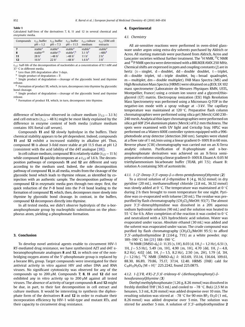
Table 1Calculated half-lives of the derivatives 7, 8, 11 and 12 in several chemical andenzymatic media.
Compounds t1/2 bufferpH¼ 1.2
t1/2 bufferpH¼ 7.3
t1/2 bufferpH¼ 11.5
t1/2 culturemedium
t1/2 CEM cellextracts
7 stablea stablea stablea stablea stablea
8 stablea,b stablea,b stablea,b 3.1 hb >48hb
11 28 hc >48 hc >48 hc 11 hc 2 he
12 16 hc 22 hc >48 hc 1.6 hd 3 hf
t1/2: half-life of the decomposition of nucleotides at a concentration of 0.1 mM and37 �C in different media.
a Less than 20% degradation after 3 days.b Single product of degradation¼ 7.c Single product of degradation¼ cleavage of the glycosidic bond and thymine
release.d Formation of product 11, which, in turn, decomposes into thymine by glycosidic
bond cleavage.e Single product of degradation¼ cleavage of the glycosidic bond and thymine
release.f Formation of product 11, which, in turn, decomposes into thymine.
K. Barral et al. / European Journal of Medicinal Chemistry 45 (2010) 849–856852
difference of behaviour observed in culture medium (t1/2¼ 3.1 h)and cell extracts (t1/2> 48 h) might be most likely explained by thedifference in enzyme content of both media (culture mediumcontains 10% foetal calf serum).
Compounds 11 and 12 slowly hydrolyse in the buffers. Theirchemical stability appears to be pH dependent. Indeed, compounds11 and 12 exhibit a increased stability in alkaline pH. Thus,compound 11 is about 3-fold more stable at pH 11.5 than at pH 1.2(consistent with the acid lability of the d4T analogue [38]).
In cell culture medium, compound 11 is relatively stable (t1/2¼11 h)while compound 12 quickly decomposes at a t1/2 of 1.6 h. The decom-position pathways of compounds 11 and 12 are different and varyaccording to the medium used. Indeed, the sole decompositionpathway of compound 11, in all media, results from the cleavage of theglycosidic bond which leads to thymine release, as identified by co-injection with an authentic sample. The decomposition pathway ofcompound 12 in enzymatic media goes through two steps: first, thequick reduction of the P–B bond into the P–H bond leading to theformation of compound 11, which, then, decomposes more slowly intothymine by glycosidic bond cleavage. In contrast, in the buffers,compound 12 decomposes directly into thymine.
In all tested media, we didn’t observe hydrolysis of the a-bor-anophosphonate group by nucleophilic substitution on the phos-phorus atom, yielding a phosphonate formation.
3. Conclusion
To develop novel antiviral agents enable to circumvent HIV-1RT-mediated drug resistance, we have synthesized AZT and d4T a-boranophosphonate analogues 8 and 12, in which one of the non-bridging oxygen atoms of the 50-phosphonate group is replaced bya borane BH3 group. Target compounds were investigated for theirantiviral activity in vitro against HIV and other DNA and RNAviruses. No significant cytotoxicity was observed for any of thecompounds up to 200 mM. Compounds 7, 8, 11 and 12 did notexhibited any in vitro activity up to 200 mM against all testedviruses. The absence of activity of target compounds 8 and 12 mightbe due, in part, to their fast decomposition in cell extract andculture medium. It would be interesting to synthesis the diphos-phate form of the derivatives 8 and 12 in order to evaluate theirincorporation efficiency by HIV-1 wild-type and mutant RTs, andtheir capacity to circumvent drug resistance.
4. Experimental
4.1. Chemistry
All air-sensitive reactions were performed in oven-dried glass-ware under argon using extra-dry solvents purchased by Aldrich orAcros. All starting materials were purchased from Aldrich, Acros andLancaster societies without further treatment. The 1H NMR, 13C NMRand 31P NMR spectra were determined with a BRUKER AMX 250 MHz.Chemical shifts are expressed in ppm and coupling constants (J) are inhertz (s¼ singlet, d¼ doublet, dd¼ double doublet, t¼ triplet,dt¼ double triplet, td¼ triple doublet, bq¼ broad quadruplet,m¼multiplet, dm¼ double multiplet). FAB Mass Spectra (MS) andHigh Resolution Mass Spectra (HRMS) were obtained on a JEOL SX 102mass spectrometer (Laboratoire de Mesures Physiques RMN, USTL,Montpellier, France) using a cesium ion source and a glycerol/thio-glycerol (GT) matrix. Electrospray ionization (ESI) High ResolutionMass Spectrometry was performed using a Micromass Q-TOF in thenegative-ion mode with a spray voltage at –3 kV. The capillarytemperature was maintained at 120 �C. Preparative flash columnchromatographies were performed using silica gel (Merck) G60 230–240 mesh. Analytical thin layer chromatographies were performed onsilica gel 60F 254 aluminium plates (Merck) of 0.2 mm thickness. Thespots were examined with UV light and Cericdip Sray. HPLC wasperformed on a Waters 600E controller system equipped with a 996-photodiode array detector (detection 260 nm). Samples were elutedat a flow rate of 1 mL/min using a linear gradient 0–100% B in 60 min.Reverse phase (C18) chromatography was carried out on an X-Terraanalytic column. Purification of H-phosphinate and a-bor-anophosphonate derivatives was achieved on an X-Terra semi-preparative column using a linear gradient 0–100% B. Eluant A: 0.05 Mtriethylammonium bicarbonate buffer (TEAB, pH 7.5); eluant B:solution A containing 50% of acetonitrile.
4.1.1. 1-(20-Deoxy-30,50-epoxy-b-D-threo-pentofuranosyl)thymine (2)To a stirred solution of b-thymidine 1 (4 g, 16.52 mmol) in dry
pyridine (20 mL), methanesulfonyl chloride (2.8 mL, 36.32 mmol)was slowly added at 0 �C. The temperature was maintained at 0 �Cduring 2 h then brought to room temperature for one night. Pyri-dine was co-evaporated with toluene (20 mL). The residue was pre-purified by flash chromatography (CH2Cl2/MeOH: 93/7). The almostpure 30,50-dimesylthymidine was dissolved in a 20% aqueoussodium hydroxide solution (90 mL) and the solution was heated to55 �C for 6 h. After completion of the reaction it was cooled to 0 �Cand neutralized with a 32% hydrochloric acid solution. Water wasevaporated under vacuo. Absolute ethanol (30 mL) was added andthe solvent was evaporated under vacuo. The crude compound waspurified by flash chromatography (CH2Cl2/MeOH 95:5) to afford30,50-anhydrothymidine 2 (2.64 g, 71%) as a white powder. mp:189–190 �C; litt [21] 188–190 �C.
1H NMR (DMSO-d6) d: 11.35 (s,1H), 8.03 (d, 1H, J¼ 1.2 Hz), 6.51 (t,1H, J¼ 5.5 Hz), 5.49 (m, 1H), 4.90 (m, 1H), 4.70 (dd, 1H, J¼ 4.8,8.2 Hz), 4.02 (dd, 1H, J¼ 1.5, 8.2 Hz), 2.50 (m, 2H), 1.79 (d, 3H,J¼ 1.2 Hz). 13C NMR (DMSO-d6) d: 163.69, 151.14, 136.64, 109.61,88.30, 86.89, 79.86, 75.17, 37.14, 12.40. HRMS (FAB) cald forC10H13N2O4 (MþH)þ 225.2242, found 225.0875.
4.1.2. 1-[(30R, 40R)-20,50,60-trideoxy-60-(diethoxyphosphonyl)-b-hexofuranosyl]thymine (3)
Diethyl methylphosphonate (1.26 g, 8.26 mmol) was dissolved infreshly distilled THF (16.5 mL) and cooled to �78 �C. BuLi (2.5 M inhexanes, 3.3 mL, 8.26 mmol) was added dropwise over 10 min. Theresulting solution was stirred at �78 �C for 90 min BF3$Et2O (1 mL,8.26 mmol) was added dropwise over 5 min. The solution wasstirred for another 5 min. A solution of 30,50-anhydrothymidine 2

K. Barral et al. / European Journal of Medicinal Chemistry 45 (2010) 849–856 853
(370 mg, 1.65 mmol) in dry THF (16.5 mL) was slowly added over45 min. After 2 h at �78 �C, the reaction was quenched with satu-rated aqueous NaHCO3 solution (1 mL) and solid NaHCO3 (329 mg).The white suspension was allowed to warm to room temperatureovernight. The solvent was evaporated, the resulting slimy residueresuspended in CH2Cl2 (60 mL), and the mixture filtered throughCelite. The white residue was twice removed from the top of theCelite, washed with additional CH2Cl2 (40 mL), and refiltered. Thecombined organic layers were concentrated to dryness and purifiedby flash chromatography (CH2Cl2/MeOH 90:10) to afford compound3 (662 mg, quant) as a colorless foam.
1H NMR (CDCl3) d: 8.57 (s, 1H), 7.65 (s, 1H), 6.12 (dd, 1H, J¼ 3.2,8.5 Hz), 4.31 (m,1H), 4.05 (m, 4H), 3.71 (m,1H), 2.55 (m,1H), 2.05 (m,1H), 2.00 (m, 2H),1.83 (s, 3H),1.70 (m, 2H),1.27 (td, 6H, J¼ 1.8, 7.0 Hz).13C NMR (CDCl3) d: 164.15, 150.77, 137.95, 110.47, 84.76, 84.55, 69.38,62.15 (d, J¼ 6.5 Hz), 40.85, 22.53 (d, JCP¼ 142.2 Hz), 21.07 (d,J¼ 4.5 Hz),16.45 (d, J¼ 6.1 Hz),12.53. 31P NMR (CDCl3) d: 32.74. HRMS(FAB) cald for C15H26N2O7P (MþH)þ 377.3544, found 377.1478.
4.1.3. 1-[(30S,40R)-30-O-mesyl-20,50,60-trideoxy-60-(diethoxyphosphonyl)-b-hexofuranosyl]thymine (4)
To a stirred solution of compound 3 (438 mg, 1.17 mmol) in drypyridine (6 mL), methanesulfonyl chloride (108 mL, 1.39 mmol) wasslowly added at 0 �C. The temperature was maintained at 0 �C during1 h. Then the reaction was allowed to warm to room temperatureovernight. The solvent was co-evaporated with toluene (10 mL). Thecrude product was purified by flash chromatography (CH2Cl2/MeOH93:7) to give 4 (405 mg, 77%) as a white foam.
1H NMR (CDCl3) d: 9.24 (s,1H), 7.26 (d,1H, J¼ 1.2 Hz), 6.16 (dd,1H,J¼ 3.2, 8.5 Hz), 5.12 (dd,1H, J¼ 3.2, 5.0 Hz), 4.05 (m, 5H), 3.02 (s, 3H),2.77 (m, 1H), 2.35 (dd, 1H, J¼ 3.2, 16 Hz), 2.05 (m, 2H), 1.88 (d, 3H,J¼ 1.2 Hz), 1.85 (m, 2H), 1.27 (t, 6H, J¼ 7.0 Hz). 13C NMR (CDCl3) d:163.83,150.52,135.06,111.25, 83.29, 82.15, 78.62, 61.94 (d, J¼ 6.5 Hz),39.92, 38.72, 25.21 (d, JCP¼ 143.9 Hz), 24.31 (d, J¼ 5.0 Hz), 16.51(d, J¼ 5.9 Hz), 12.69. 31P NMR (CDCl3) d: 30.46. HRMS (FAB) cald forC16H28N2O9PS (MþH)þ 455.4461, found 455.1253.
4.1.4. 1-[(30S,40R)-30-O-mesyl-20,50,60-trideoxy-60-(phosphanyl)-b-hexofuranosyl]thymine (5)
Chlorotrimethylsilane (1.35 mL, 10.20 mmol) was added drop-wise to a stirred solution of LiAlH4 (443 mg, 10.20 mmol) in dry THF(8 mL) at �78 �C. The resulting mixture was allowed to warm toroom temperature and stirred for 2 h. Compound 4 (800 mg,1.70 mmol) in dry THF (8 mL) was added to the reducing mixture at�70 �C. The mixture was allowed to warm to room temperatureand stirred for 1 h. The reaction was stopped by slow addition ofH2O (2 mL) at �70 �C. The mixture was allowed to warm to roomtemperature and filtered through Celite. The organic layer wasdried over anhydrous MgSO4, filtered and concentrated underreduced pressure. A purification by flash chromatography (CH2Cl2/MeOH 95:5) yielded 5 (402 mg, 68%) as a white foam.
1H NMR (CDCl3) d: 9.34 (s, 1H), 7.26 (d, 1H, J¼ 1.2 Hz), 6.15 (dd,1H, J¼ 3.2, 8.5 Hz), 5.09 (dd, 1H, J¼ 3.2, 5.0 Hz), 3.97 (m, 1H), 3.10and 2.34 (dm, 2H, JPH¼ 190.0 Hz), 2.99 (s, 3H), 2.77 (m, 1H), 2.28 (m,1H), 2.02 (m, 1H), 1.89 (d, 3H, J¼ 1.2 Hz), 1.81 (m, 1H), 1.58 (m, 2H).13C NMR (CDCl3) d: 163.80, 150.56, 135.14, 111.19, 83.37, 82.69, 79.09,39.95, 38.80, 31.86 (d, J¼ 3.4 Hz), 12.77, 10.36 (d, JCP¼ 9.2 Hz). 31PNMR (CDCl3) d: �136.52. HRMS (FAB) cald for C12H20N2O6PS(MþH)þ 351.3404, found 351.0780.
4.1.5. 1-[(30S,40R)-30-O-mesyl-20,50,60-trideoxy-60-(hydroxyphosphinyl)-b-hexofuranosyl]thymine (6)
To a stirred solution of phosphine 5 (112 mg, 0.32 mmol) in water(1 mL) and THF (1 mL) was added dropwise 35% aqueous hydrogenperoxide (62 mL). The mixture was stirred at room temperature for
2 h then solvents were evaporated on vacuo to afford pure H-phosphinate 6 (120 mg, quant) as a white solid; HPLC purity >98%.
1H NMR (D2O) d: 8.03 and 5.81 (dm, 2H, JPH¼ 555.0 Hz), 7.45 (s,1H), 6.05 (dd, 1H, J¼ 2.9, 8.0 Hz), 5.26 (dd, 1H, J¼ 3.2, 4.9 Hz), 4.17(m, 1H), 3.17 (m, 1H), 3.12 (s, 3H), 2.80 (m, 1H), 2.41 (m, 1H), 2.10 (m,1H), 2.03 (s, 3H), 1.79 (m, 2H). 13C NMR (D2O) d: 165.73, 150.70,136.27, 110.12, 83.90, 82.52, 79.40, 38.36, 36.98, 25.85(d, JCP¼ 93.3 Hz), 19.40 (d, J¼ 2.0 Hz), 10.90. 31P NMR (D2O) d:34.79. HRMS (FAB) cald for C12H20N2O8PS (MþH)þ 383.3392,found 383.0678.
4.1.6. 1-[(30R,40R)-30-Azido-20,30,50,60-tetradeoxy-60-(hydroxyphosphinyl)-b-hexofuranosyl]thymine (7)
Sodium azide (256 mg, 3.94 mmol) was added to a solution ofthe H-phosphinate 6 (188 mg, 0.49 mmol) in dry DMF (4 mL). Thereaction was stirred for 24 h at 60 �C, then water (2 mL) was addedand the solvent was evaporated under vacum. The crude residuewas purified by HPLC with an X-Terra semi-preparative reversed-phase column (linear gradient 0–100% B). Product fractions werecollected and evaporated to dryness. Excess triethylammoniumbicarbonate was removed by repeated freeze-drying with deionizedwater to give 7 (83.2 mg, 51%) as a white powder. HPLC purity >97%
1H NMR (D2O) d: 8.07and 5.92 (dm, 2H, JPH¼ 537.0 Hz), 7.50(s, 1H), 6.23 (t, 1H, J¼ 6.7 Hz), 4.31 (dd, 1H, J¼ 5.6, 12.0 Hz), 4.08(dd, 1H, J¼ 5.6, 12.0 Hz), 2.56 (m, 2H), 1.98 (m, 2H), 1.93 (s, 3H), 1.72(m, 2H). 13C NMR (D2O) d: 167.50, 152.51, 137.17, 111.96, 84.73, 84.21,83.94, 35.43, 26.80 (d, JCP¼ 92.9 Hz), 25.49 (d, J¼ 2.1 Hz), 11.74. 31PNMR (D2O) d: 29.49. HRMS (FAB) cald for C11H16N5O5P (M)�
328.2447, found 328.0918.
4.1.7. 1-[(30R,40R)-30-Azido-20,30,50,60-tetradeoxy-60-(boranophosphono)-b-hexofuranosyl]thymine (8)
Compound 7 (50 mg, 0.14 mmol) was dried over P2O5 undervacuum for 4–5 h then dissolved in dry THF (15 mL). BSA (195 mL,0.76 mmol) was added by syringe and the solution was stirred forabout 1 h at room temperature. DIPEA$BH3 (264 mL, 1.5 mmol) wasadded, and the solution stirred for 4 h, then a mixture H2O–MeOH(1:1, 12 mL) was added. After the solvents were evaporated underreduce pressure, the residue was purified by reversed-phase columnchromatography (linear gradient 0–100% B). Product fractions werecollected and evaporated to dryness. Excess of triethylammoniumbicarbonate was removed by repeated freeze-drying with deionizedwater to give compound 8 (10 mg, 20%) as a white powder. HPLCpurity >98%.
1H NMR (D2O) d: 7.32 (s, 1H), 6.04 (t, 1H, J¼ 6.5 Hz), 4.08 (dd, 1H,J¼ 5.5, 11.2 Hz), 3.83 (dd, 1H, J¼ 5.6, 11.0 Hz), 2.32 (t, 2H, J¼ 6.5 Hz),1.75 (m, 5H), 1.42 (m, 2H), 0.5 (bq, 3H, J¼ 82 Hz). 31P NMR (D2O) d:103.54. HRMS (ESI) cald for C11H18N5O5PB (M)� 342.1139, found342.1123.
4.1.8. 1-[(40R)-20,30-Didehydro-20,30,50,60-tetradeoxy-60-(diethoxyphosphonyl)-b-hexofuranosyl]thymine (9)
Potassium tert-butoxide (185 mg, 1.65 mmol) was added toa solution of compound 4 (300 mg, 0.66 mmol) in dry DMF (4 mL).The reaction was stirred at room temperature for 2.5 h, then thesolvent was evaporated on vacuo. The crude residue was purified byflash chromatography (CH2Cl2/MeOH 95:5) to afford 9 (167 mg,71%) as a white foam.
1H NMR (CDCl3) d: 9.00 (s, 1H), 6.90 (m, 2H), 6.28 (dt, 1H, J¼ 1.5,5.8 Hz), 5.76 (dt, 1H, J¼ 5.8, 1.5 Hz), 4.77 (m, 1H), 4.03 (m, 4H), 2.02(m, 2H), 1.84 (d 3H), 1.75 (m, 2H), 1.25 (td, 6H, J¼ 1.5, 6.7 Hz). 13CNMR (CDCl3) d: 163.65, 150.82, 136.38, 135.06, 125.53, 111.42, 89.77,85.91, 61.81 (d, J¼ 6.5 Hz), 28.87 (d, J¼ 4.6 Hz), 23.14 (d,JCP¼ 142.0 Hz), 16.49 (d, J¼ 6.0 Hz), 12.53. 31P NMR (CDCl3) d: 30.76.

K. Barral et al. / European Journal of Medicinal Chemistry 45 (2010) 849–856854
HRMS (FAB) cald for C15H24N2OP (MþH)þ 359.3391, found359.1372.
4.1.9. 1-[(40R)-20,30-Didehydro-20,30,50,60-tetradeoxy-60-(phosphanyl)-b-hexofuranosyl]thymine (10)
Chlorotrimethylsilane (794 mL, 6.21 mmol) was added dropwiseto a stirred solution of LiAlH4 (260 mg, 6.21 mmol) in dry THF (5 mL)at �78 �C. The resulting mixture was allowed to warm to roomtemperature and stirred for 2 h. Compound 9 (370 mg, 1.04 mmol)in dry THF (6 mL) was added to the reducing mixture at�70 �C. Themixture was allowed to warm to room temperature and stirred for1 h. The reaction was stopped by slow addition of MeOH (5 mL) at0 �C. The mixture was allowed to warm to room temperature andfiltered through Celite. The organic layer was dried over anhydrousMgSO4, filtered and concentrated under reduced pressure. A puri-fication by flash chromatography (CH2Cl2/MeOH 98:2) yielded 10(94 mg, 36%) as a white foam.
1H NMR (CDCl3) d: 9.29 (s, 1H), 6.92 (m, 2H), 6.26 (dt, 1H, J¼ 1.5,6.0 Hz), 5.73 (dm 1H, J¼ 6.0 Hz), 4.78 (m, 1H), 3.07 and 2.29 (dm,2H, JPH¼ 195.0 Hz), 1.85 (d, 3H, J¼ 1.2 Hz), 1.79 (m, 2H,), 1.57 (m,2H). 13C NMR (CDCl3) d: 163.89, 150.96, 136.61, 135.20, 125.21,111.34, 89.77, 86.25, 38.99 (d, J¼ 3.3 Hz), 12.64, 10.07 (d,JCP¼ 8.9 Hz). 31P NMR (CDCl3) d: �135.93. HRMS (FAB) cald forC11H16N2O3P (MþH)þ 255.2334, found 255.0899.
4.1.10. 1-[(40R)-20,30-Didehydro-20,30,50,60-tetradeoxy-60-(hydroxyphosphinyl)-b-hexofuranosyl]thymine (11)
To a stirred solution of phosphine 10 (84 mg, 0.33 mmol) inwater (1 mL) and THF (1 mL) was added dropwise 35% aqueoushydrogen peroxide (64 mL). The mixture was stirred at roomtemperature for 2 h then solvents were evaporated under vacum toafford pure H-phosphinate 11 (94 mg, quant) as a white solid; HPLCpurity >97%.
1H NMR (D2O) d: 7.89 and 5.83 (dm, 2H, JPH¼ 515.0 Hz), 7.26 (m,2H), 6.74 (m, 1H), 6.39 (dt, 1H, J¼ 1.5, 6.2 Hz), 5.78 (m, 1H), 4.83 (m,1H), 1.88 (m, 4H), 1.73 (s, 3H). 13C NMR (D2O) d: 166.52, 152.19,137.44, 136.84, 123.58, 111.41, 90.17, 86.40, 26.08 (d, JCP¼ 91.0 Hz),24.63 (d, J¼ 2.6 Hz), 11.28. 31P NMR (D2O) d: 35.93. HRMS (FAB) caldfor C11H15N2O5P (M)� 285.2164, found 285.0727.
4.1.11. 1-[(40R)-20,30-Didehydro-20,30,50,60-tetradeoxy-60-(boranophosphono)-b-hexofuranosyl]thymine (12)
Compound 11 (25 mg, 0.09 mmol) was dried over P2O5 undervacuum for 4–5 h then dissolved in dry THF (5 mL). BSA (108 mL,0.43 mmol) was added by syringe and the solution was stirred forabout 1 h at room temperature. DIPEA$BH3 (152 mL, 0.87 mmol)added, and the solution stirred for 5 h, then a mixture H2O–MeOH(1:1, 12 mL) was added. After the solvents were evaporated underreduce pressure, the residue was purified by reversed-phase columnchromatography (linear gradient 0–100% B). Product fractions werecollected and evaporated to dryness. Excess triethylammoniumbicarbonate was removed by repeated freeze-drying with deionizedwater to give compound 12 (5 mg, 21%) as a white powder. HPLCpurity >97%.
1H NMR (D2O) d: 7.26 (s, 1H), 6.73 (d, 1H, J¼ 1.2 Hz), 6.37 (dd, 1H,J¼ 1.2, 6.1 Hz), 5.71 (d, 1H, J¼ 6.0 Hz), 4.80 (m, 1H), 1.71 (s, 3H), 1.69(m, 2H), 1.43 (m, 2H), 0.70 (bq, 3H, J¼ 77 Hz). 31P NMR (D2O) d:102.4. HRMS (ESI) cald for C8H16N5O9P3B (M)� 299.0974, found299.0965.
4.2. Antiviral assays
4.2.1. Anti-HIV assays in MAGI-CCR5 cells293T and MAGI-CCR5 cells were maintained in Dulbecco’s
modified Eagle’s medium supplemented with antibiotics and 10%
fetal bovine serum. To obtain HIV-1 stocks, 293T cells were tran-siently transfected with an HIV-1NL4.3 molecular clone by the useof FuGENE 6 transfectant reagent (Roche) as recommended by themanufacturer. Two days post transfection, the CAp24 antigen wasquantitated in cell-free culture supernatants by HIV-1 p24 antigencapture assay kit (Coulter). MAGI-CCR5 cells (104 cells), containinga LacZ reporter under control of an integrated HIV promoter, wereseeded on 96-well microtiter culture plates and treated during 4 hwith increasing amounts of AZT, d4T, 7, 8, 11 and 12 (0–400 mM)before being infected with 100 ng of HIV-1 CAp24 antigen by spi-noculation as previously described [39]. Cells were then washedand grown in the presence of each compound dilution. Two dayslater, cells were stained for b-galactosidase activity and blue cellswere counted. The 50% effective concentration (EC50) correspondsto the compound concentration producing a 50% decrease of thenumber of blue cells in the virus-infected cell cultures. The 50%cytostatic concentration (CC50) corresponds to the compoundconcentration required to inhibit cell proliferation by 50%. Standardcompounds AZT and d4T inhibit HIV-1-infected cells at an IC50
value of 0.025 mM and 0.062 mM, respectively, in this assay.
4.2.2. Anti-HIV-1 and -2 assays in CEM cellsThe compounds have also been evaluated against HIV-1(IIIB) and
HIV-2(ROD) in CEM cell cultures. Briefly, CEM cells (4.5�105 cellsper ml) were suspended in fresh culture medium and infected withHIV-1 at 100 CCID50 per ml of cell suspension. Then, 100 ml of theinfected cell suspension were transferred to microplate wells, mixedwith 100 ml of the appropriate dilutions of the test compounds, andfurther incubated at 37 �C. After 4–5 days, giant cell formation wasrecorded microscopically in the CEM cell cultures. The 50% effectiveconcentration (EC50) corresponds to the compound concentrationsrequired to prevent syncytium formation by 50% in the virus-infected CEM cell cultures.
4.2.3. Antiviral assays other than HIVHuman embryonic lung (HEL) (ATCC-CCL 137), simian kidney
(Vero) and human cervix carcinoma (HeLa) cells were propagated inminimal essential medium (MEM) supplemented with 10% fetal calfserum (FCS), 2 mM L-glutamine, and 0.075% bicarbonate. Herpessimplex virus type 1 (HSV-1) (KOS), HSV-2 (G), vaccinia virus andvesicular stomatitis virus were assayed in HEL cell cultures; Cox-sackie virus B4 and respiratory syncytial virus in HeLa cell cultures,and para-influenza-3 virus, reovirus-1, Sindbis virus and Punta Torovirus in Vero cell cultures. Reference compounds, acyclovir andzidovudine were from Glaxo Smith Kline, ganciclovir from Hoff-mann-La-Roche and cidofovir from Gilead. Cells were grown toconfluency in 96-well microtiter plates and were inoculated with100 times the 50% cell culture infective dose. Compounds wereadded after a 1–2 h virus adsorption period. The virus-inducedcytopathic effect (CPE) was recorded microscopically at z3 dayspost infection and was expressed as percentage of the untreatedcontrols. The 50% effective concentrations (EC50) were derived fromgraphical plots. The minimal toxic concentration (MTC) was definedas the lowest concentration that resulted in a microscopicallydetectable alteration of cell morphology. The MTC was determinedin uninfected confluent cell cultures that were incubated, akin to thecultures used for the antiviral assays, with serial dilutions of thecompounds for the same time period. Cultures were inspectedmicroscopically for alteration of cell morphology.
Evaluation of antiviral activity and cytostatic activities of selectedcompounds in HCV genotype 1b subgenomic replicon carrying Huh-5-2 cells. Huh-5-2 cells [a cell line with a persistent HCV repliconI389luc-ubi-neo/NS3-30/5.1; replicon with firefly luciferase-ubiq-uitin-neomycin phosphotransferase fusion protein and an EMCV-IRES driven NS3-5B HCV polyprotein] are cultured in RPMI medium

K. Barral et al. / European Journal of Medicinal Chemistry 45 (2010) 849–856 855
(GIBCO) supplemented with 10% fetal calf serum, 2 mM L-glutamine(Life Technologies), 1x non-essential amino acids (Life Technolo-gies); 100 IU/ml penicillin and 100 mg/ml streptomycin and 250 mg/ml G418 (Geneticin, Life Technologies). Cells are seeded at a densityof 7000 cells per well in 96-well View Plate� (Packard) in mediumcontaining the same components as described above, except forG418. Cells are allowed to adhere and proliferate for 24 h. At thattime, culture medium is removed and 5 serial dilutions (5-folddilutions starting at 100 mg/ml or 100 mM) of the test compounds areadded in culture medium lacking G418. Interferon alfa 2a (500 IU) isincluded as a positive control in each experiment for internal vali-dation. Plates are further incubated at 37 �C in a humidified 5% CO2
atmosphere for 72 h. Replication of the HCV replicon in Huh-5-2cells results in luciferase activity in the cells. Luciferase activity ismeasured by adding 50 ml of 1� Glo-lysis buffer (Promega) for15 min followed by 50 ml of the Steady-Glo Luciferase assay reagent(Promega). Luciferase activity is measured with a luminometer andthe signal in each individual well is expressed as a percentage of theuntreated cultures. The 50% effective concentrations (EC50) arecalculated from these datasets. Parallel cultures of Huh-5-2 cells,seeded at a density of 7000 cells/well of classical 96-well cell cultureplates (Becton–Dickinson) are treated in a similar fashion exceptthat no Glo-lysis buffer or Steady-Glo Luciferase reagent is added.The effect of the compounds on the proliferation of the cells ismeasured 3 days after addition of the various compounds by meansof The CellTiter 96� AQueous Non-Radioactive Cell Proliferation Assay(MTS, Promega). In this assay 3-(4,5-dimethylthiazol-2-yl)-5-(3-carboxymethoxy-phenyl)-2-(4-sulfophenyl)-2H-tetrazolium (MTS)is bioreduced by cells into a formazan that is soluble in tissue. Thenumber of cells correlates directly with the production of the for-mazan. The MTS stained cultures are quantified in a platereader.
4.3. Stability studies
4.3.1. Media and preparation of cell extractsRPMI 1640 medium was purchased from GIBCO Life Technologies.
Heat-inactivated fetal calf serum was purchased from PAN biotech.Culture medium was composed by RPMI 1640 containing 10% (v/v)heat-inactivated fetal calf serum and stored at �80 �C. CEM-SS cellextracts were prepared according to a published procedure [37].Exponentially growing CEM-SS cells were recovered by centrifuga-tion (500 g, 4 �C, 4 min), washed twice with PBS and resuspended in10 mM Tris–HCl, 140 mM KCl (pH 7.4), at the concentration of30�106 cells/mL. Cells were lyzed by ultrasonic treatment andcellular debris were removed by centrifugation (10,000 g, 4 �C,20 min). The supernatant containing soluble proteins (3 mg/mL) wasstored at �80 �C.
4.3.2. HPLC analysisA Waters Model-600 gradient HPLC system equipped with two
600-pumps, a rheodine injector, a 996-photodiode array detectorand an in-line degasser AF was used for reversed-phase chroma-tography. All solvents were of HPLC grade and filtered prior to use.A 1 M solution of triethylammonium bicarbonate buffer wasprepared by adding dry ice to a 1 M triethylamine solution until thepH reached to 7.5. Triethylammonium bicarbonate solutions weremade fresh by dissolving reagent-grade triethylammonium bicar-bonate in HPLC-grade water prior to filtration.
The HPLC method has been described [37]. The cleaning pre-column is a guard-pak insert (Delta-Pak C18, 100 Å) in a guard-pakholder. The analytical column used is a Novapak C18, 3 mm, 100 Å,4.6�150 mm. The elution system is prepared as follows: buffer A,0.05 M TEAB; buffer B, 0.05 M TEAB (CH3CN/H2O, v/v, 50/50); flowrate,1 mL/min. The crude sample (50 ml, initial concentration of 7, 8,11, 12: 0.5 mM) is injected into the precolumn and eluted with
buffer A during 3 min. Then, the switching valve for connecting theprecolumn to the column is activated, and a linear gradient isapplied from buffer A to buffer B, increasing buffer B from 0% at0 min–20% at 40 min. The retention times are 7, 29 min; 8, 35 min;11, 23 min and 12, 34 min. Kinetic data and decomposition path-ways for compounds 7, 8, 11 and 12 were studied at 37 �C (a) in a pHbuffers range 1.2–11.5 (3 conditions) (b) in culture medium RPMI1640 containing 10% heat-inactivated fetal calf serum (c) in totalcells extracts (CEM-SS). For each kinetic study, the compoundsolution is diluted with a freshly thawed aliquot of the consideredmedium to obtain an initial concentration of 0.1 mM. The mixture isincubated at 37 �C and for the required time, an aliquot (10%solution) is drawn and immediately frozen at �80 �C for furtherHPLC analysis. All compounds were analyzed under the sameconditions. The amount of remaining parent compound at eachtime point was used to determine the half-life of the compound.The product of decomposition from parent derivative is determinedby comparison with authentic samples and standard compounds.
Acknowledgment
This work was supported by Ensemble Contre le SIDA (ESC-SIDACTION), the Agence Nationale de la Recherche contre leSIDA (ANRS), the FWO-Vlaanderen (no. G-0267-04) and the GOA(no. 05/19).
References
[1] P.A. Furman, J.A. Fyfe, M.H. St Clair, K. Weinhold, J.L. Rideout, G.A. Freeman,S. Nusinoff Lehrman, D.P. Bolognesi, S. Broder, H. Mitsuya, D.W. Barry,Proceedings of the National Academy of Sciences of the United States ofAmerica 83 (1986) 8333–8337.
[2] M. Baba, R. Pauwels, P. Herdewijn, E. De Clercq, J. Desmyter, M. Vandeputte,Biochemical and Biophysical Research Communications 142 (1987) 128–134.
[3] R. Yarchoan, H. Mitsuya, C.E. Meyer, S. Broder, New England Journal ofMedecine 321 (1989) 726–738.
[4] J.E. Reardon, W.H. Miller, Journal of Biological Chemistry 265 (1990)20302–20307.
[5] E. Declercq, Journal of Antimicrobial Chemotherapy 23 (1989) 35–46.[6] D. Arion, N. Kaushik, S. Mc Cormick, G. Borkow, M.A. Parniak, Biochemistry 37
(1998) 15908–15917.[7] B. Selmi, J. Boretto, S.R. Sarfati, C. Guerreiro, B. Canard, The Journal of Biological
Chemistry 276 (2001) 48466–48472.[8] K. Alvarez, J. Deval, B. Selmi, K. Barral, J. Boretto, C. Guerreiro, L. Mulard, S. Sarfati,
B. Canard, Nucleosides, Nucleotides & Nucleic Acids 24 (2005) 419–422.[9] B. Schneider, P. Meyer, S. Sarfati, L. Mulard, C. Guerreiro, J. Boretto, J. Janin,
M. Veron, D. Deville-Bonne, B. Canard, Nucleosides, Nucleotides & NucleicAcids 20 (2001) 297–306.
[10] P. Meyer, B. Schneider, S. Sarfati, D. Deville-Bonne, C. Guerreiro, J. Boretto,J. Janin, M. Veron, B. Canard, EMBO Journal 19 (2000) 3520–3529.
[11] J. Deval, K. Alvarez, B. Selmi, M. Bermond, J. Boretto, C. Guerreiro, L. Mulard,B. Canard, Journal of Biological Chemistry 280 (2005) 3838–3846.
[12] A. Pompon, I. Lefebvre, J.-L. Imbach, S. Kahn, D. Farquhar, Antiviral Chemistry &Chemotherapy 5 (1994) 91–98.
[13] C. Meier, M. Lorey, E. De Clercq, J. Balzarini, Journal of Medicinal Chemistry 41(1998) 1417–1427.
[14] R. Engel, Chemical Reviews 77 (1977) 349–367.[15] A. Frangeul, K. Barral, K. Alvarez, B. Canard, Antimicrobial Agents and
Chemotherapy 51 (2007) 3162–3167.[16] Z.A. Sergueeva, D.S. Sergueev, B.R. Shaw, Nucleosides Nucleotides Nucleic
Acids 20 (2001) 941–945.[17] D.S. Sergueev, B. Ramsay Shaw, Journal of the American Chemical Society 120
(1998) 9417–9427.[18] K. Barral, S. Priet, J. Sire, J. Neyts, J. Balzarini, B. Canard, K. Alvarez, Journal of
Medicinal Chemistry 49 (2006) 7799–7806.[19] I. Lavandera, S. Fernandez, M. Ferreo, V. Gotor, Journal of Organic Chemistry 66
(2001) 4079–4082.[20] J.P. Horwitz, J. Chua, J.A. Urbanski, M. Noel, Journal of Organic Chemistry 28
(1963) 942–944.[21] R. Paramashivappa, P. Kumar, P.V. Subba Rao, A. Srinivasa Rao, Tetrahedron
Letters 44 (2003) 1003–1005.[22] H. Tanaka, M. Fukui, K. Haraguchi, M. Masaki, T. Miyasaka, Tetrahedron Letters
30 (1989) 2567–2570.[23] K. Haraguchi, H. Tanaka, T. Miyasaka, Synthesis 6 (1989) 434–436.[24] D. Hutter, M.O. Blaettler, S.A. Benner, Helvetica Chimica Acta 85 (2002)
2777–2806.

K. Barral et al. / European Journal of Medicinal Chemistry 45 (2010) 849–856856
[25] E.P. Kyba, S.-T. Liu, R.L. Harris, Organometallics 2 (1983) 1877–1879.[26] W. Henderson, S.R. Alley, Journal of Organometallic Chemistry 656 (2002)
120–128.[27] S.A. Reiter, B. Assmann, S.D. Nogai, N.W. Mitzel, H. Schmidbaur, Helvetica
Chimica Acta 85 (2002) 1140–1150.[28] H. Yamamoto, T. Hanaya, H. Kawamoto, S. Inokawa, M. Yamashita, M.-
A. Armour, T.T. Nakashima, Journal of Organic Chemistry 50 (1985) 3516–3521.[29] H. Molin, J.-O. Noren, A. Claesson, Carbohydrate Research 194 (1989) 209–221.[30] L. Kvaerno, R.H. Wightman, J. Wengel, Journal of Organic Chemistry 66 (2001)
5106–5112.[31] B. Dhotare, A. Chattopadhyay, Synthesis-Stuttgart (2001) 1337–1340.[32] T. Wada, A. Mochizuki, Y. Sato, M. Sekine, Tetrahedron Letters 39 (1998)
7123–7126.
[33] K. He, K.W. Porter, A. Hasan, J.D. Briley, B.R. Shaw, Nucleic Acids Research 27(1999) 1788–1794.
[34] P. Li, B.R. Shaw, Organic Letters 4 (2002) 2009–2012.[35] H. Li, C. Hardin, B.R. Shaw, Journal of the American Chemical Society 118
(1996) 6606–6614.[36] J.J. Chen, Y. Wei, J.C. Drach, L.B. Townsend, Journal of Medicinal Chemistry 43
(2000) 2449–2456.[37] F. Puech, G. Gosselin, I. Lefebvre, A. Pompon, A.M. Aubertin, A. Kirn, J.L. Imbach,
Antiviral Research 22 (1993) 155–174.[38] C.U. Kim, J.J. Bronson, L.M. Ferrara, J.C. Martin, Bioorganic & Medicinal
Chemistry Letters 2 (1992) 367–370.[39] U. O’Doherty, W.J. Swiggard, M.H. Malim, Journal of Virology 74 (2000)
10074–10080.