Syndecans Serve as Attachment Receptors for Human Immunodeficiency Virus Type 1 on Macrophages
15
10.1128/JVI.75.19.9187-9200.2001. 2001, 75(19):9187. DOI: J. Virol. David and Philippe A. Gallay Andrew C. S. Saphire, Michael D. Bobardt, Zhe Zhang, Guido Macrophages Human Immunodeficiency Virus Type 1 on Syndecans Serve as Attachment Receptors for http://jvi.asm.org/content/75/19/9187 Updated information and services can be found at: These include: REFERENCES http://jvi.asm.org/content/75/19/9187#ref-list-1 This article cites 35 articles, 25 of which can be accessed free at: CONTENT ALERTS more» cite this article), Receive: RSS Feeds, eTOCs, free email alerts (when new articles http://journals.asm.org/site/misc/reprints.xhtml Information about commercial reprint orders: http://journals.asm.org/site/subscriptions/ To subscribe to to another ASM Journal go to: on September 26, 2013 by guest http://jvi.asm.org/ Downloaded from on September 26, 2013 by guest http://jvi.asm.org/ Downloaded from on September 26, 2013 by guest http://jvi.asm.org/ Downloaded from on September 26, 2013 by guest http://jvi.asm.org/ Downloaded from on September 26, 2013 by guest http://jvi.asm.org/ Downloaded from on September 26, 2013 by guest http://jvi.asm.org/ Downloaded from on September 26, 2013 by guest http://jvi.asm.org/ Downloaded from on September 26, 2013 by guest http://jvi.asm.org/ Downloaded from on September 26, 2013 by guest http://jvi.asm.org/ Downloaded from on September 26, 2013 by guest http://jvi.asm.org/ Downloaded from on September 26, 2013 by guest http://jvi.asm.org/ Downloaded from on September 26, 2013 by guest http://jvi.asm.org/ Downloaded from on September 26, 2013 by guest http://jvi.asm.org/ Downloaded from on September 26, 2013 by guest http://jvi.asm.org/ Downloaded from on September 26, 2013 by guest http://jvi.asm.org/ Downloaded from
Transcript of Syndecans Serve as Attachment Receptors for Human Immunodeficiency Virus Type 1 on Macrophages
10.1128/JVI.75.19.9187-9200.2001.
2001, 75(19):9187. DOI:J. Virol. David and Philippe A. GallayAndrew C. S. Saphire, Michael D. Bobardt, Zhe Zhang, Guido MacrophagesHuman Immunodeficiency Virus Type 1 on Syndecans Serve as Attachment Receptors for
http://jvi.asm.org/content/75/19/9187Updated information and services can be found at:
These include:
REFERENCEShttp://jvi.asm.org/content/75/19/9187#ref-list-1This article cites 35 articles, 25 of which can be accessed free at:
CONTENT ALERTS more»cite this article),
Receive: RSS Feeds, eTOCs, free email alerts (when new articles
http://journals.asm.org/site/misc/reprints.xhtmlInformation about commercial reprint orders: http://journals.asm.org/site/subscriptions/To subscribe to to another ASM Journal go to:
on Septem
ber 26, 2013 by guesthttp://jvi.asm
.org/D
ownloaded from
on S
eptember 26, 2013 by guest
http://jvi.asm.org/
Dow
nloaded from
on Septem
ber 26, 2013 by guesthttp://jvi.asm
.org/D
ownloaded from
on S
eptember 26, 2013 by guest
http://jvi.asm.org/
Dow
nloaded from
on Septem
ber 26, 2013 by guesthttp://jvi.asm
.org/D
ownloaded from
on S
eptember 26, 2013 by guest
http://jvi.asm.org/
Dow
nloaded from
on Septem
ber 26, 2013 by guesthttp://jvi.asm
.org/D
ownloaded from
on S
eptember 26, 2013 by guest
http://jvi.asm.org/
Dow
nloaded from
on Septem
ber 26, 2013 by guesthttp://jvi.asm
.org/D
ownloaded from
on S
eptember 26, 2013 by guest
http://jvi.asm.org/
Dow
nloaded from
on Septem
ber 26, 2013 by guesthttp://jvi.asm
.org/D
ownloaded from
on S
eptember 26, 2013 by guest
http://jvi.asm.org/
Dow
nloaded from
on Septem
ber 26, 2013 by guesthttp://jvi.asm
.org/D
ownloaded from
on S
eptember 26, 2013 by guest
http://jvi.asm.org/
Dow
nloaded from
on Septem
ber 26, 2013 by guesthttp://jvi.asm
.org/D
ownloaded from

JOURNAL OF VIROLOGY,0022-538X/01/$04.00�0 DOI: 10.1128/JVI.75.19.9187–9200.2001
Oct. 2001, p. 9187–9200 Vol. 75, No. 19
Copyright © 2001, American Society for Microbiology. All Rights Reserved.
Syndecans Serve as Attachment Receptors for HumanImmunodeficiency Virus Type 1 on Macrophages†
ANDREW C. S. SAPHIRE,1 MICHAEL D. BOBARDT,1 ZHE ZHANG,2 GUIDO DAVID,2
AND PHILIPPE A. GALLAY1*
Department of Immunology, The Scripps Research Institute, La Jolla, California 92037,1 and Center for Human Genetics,University of Leuven and Flanders Interuniversity Institute for Biotechnology, B-3000 Leuven, Belgium2
Received 7 March 2001/Accepted 28 June 2001
Macrophages are thought to represent one of the first cell types in the body to be infected during the earlystage of human immunodeficiency virus type 1 (HIV-1) transmission and represent a potential viral reservoirin vivo. Thus, an understanding of HIV-1 attachment to these cells is fundamental to the development of novelanti-HIV-1 therapies. Although one of the major targets of HIV-1 in vivo—CD4� T lymphocytes—express highCD4 levels, other major targets such as macrophages do not. We asked in this study whether this low CD4 levelon macrophages is sufficient to support HIV-1 attachment to these cells or whether cell surface proteins otherthan CD4 are required for this process. We show that CD4 alone is not sufficient to support the initialadsorption of HIV-1 to macrophages. Importantly, we find that heparan sulfate proteoglycans (HSPGs) serveas the main class of attachment receptors for HIV-1 on macrophages. Most importantly, we demonstrate thata single family of HSPGs, the syndecans, efficiently mediates HIV-1 attachment and represents an abundantclass of attachment receptors on macrophages.
To date, the human immunodeficiency virus type 1 (HIV-1)receptor complex known to be essential for virus entry consistsof CD4 molecules expressed on T cells, macrophages, dendriticcells, and microglia and a member of the seven-transmem-brane chemokine receptor family. It is thought that HIV-1initiates its attachment to host cells via an interaction betweenthe virus-encoded surface gp120 glycoprotein and cell surfaceCD4 molecules. Binding of gp120 to CD4 induces conforma-tional changes in gp120 that result in the exposure of a che-mokine receptor binding site on gp120. The subsequent inter-action between gp120 and a chemokine receptor triggers thefusion of virus and cell membranes, allowing the delivery of theviral genome into the cytosol of the host cell. Based on thismodel, gp120 is responsible both for the initial attachment ofthe virus to target cells and the subsequent fusion with the hostcell.
For the last decade, CD4 has been thought to be the exclu-sive attachment receptor for HIV-1. Indeed, several observa-tions support the notion that gp120-CD4 interactions are suf-ficient to mediate the initial attachment of HIV-1 to targetcells (13). Specifically, it has been found that HIV-1 attaches toCD4-positive T cells but fails to attach to parental CD4-neg-ative T-cell lines (33). Furthermore, anti-CD4 antibodies di-rected against the gp120 binding site of CD4 prevent HIV-1attachment to CD4� T cell lines (33). Finally, the apparentaffinity between recombinant monomeric gp120 and solubleCD4 was found to be high, on the order of 1 to 10 nM (19).Altogether, these data led researchers to postulate that CD4 isthe exclusive attachment receptor for HIV-1.
However, recent data have emerged suggesting that interac-tions other than gp120-CD4 interactions are required forHIV-1 attachment to specific target cells. First, it has beenfound that the affinity of oligomeric gp120 for CD4 is muchlower than the affinity for monomeric gp120 for CD4 (10, 20,29). Specifically, oligomerization of patient virus gp120 into atrimer of gp120-gp41 heterodimers drastically reduces its af-finity for CD4 by 3 logs (10). This apparent low affinity ofoligomeric gp120 for CD4 suggested to us that the gp120-CD4interaction alone would be not sufficient for tight attachmentto cells which express little CD4, such as macrophages, micro-glia, and dendritic cells (9, 31). On such cell types, the attach-ment of patient virus with low intrinsinc affinity for CD4 sug-gests the requirement for supplementary interactions, viareceptors others than CD4. Corroborating this hypothesis,Mondor et al. showed that HIV-1 attaches to CD4-negativeadherent HeLa cells as well as CD4-positive HeLa cells (18).Furthermore, they found that anti-CD4 antibodies, which werepreviously shown to block HIV-1 attachment to suspension Tcells, did not prevent HIV-1 attachment to CD4-positive HeLacells (18). These data suggest that the dependence of HIV-1attachment to target cells on the gp120-CD4 interaction ishighly cell type specific and may be replaced by other virus-receptor interactions.
Several lines of evidence suggest that cell surface heparansulfate (HS) proteoglycans (HSPGs) act as necessary HIV-1attachment receptors on specific target cells. Specifically, it hasbeen shown that soluble polyanions such as dextran sulfate,heparin, or heparan sulfate inhibit HIV-1 infection (18, 4, 24,27). The fact that polyanionic compounds bind to basic resi-dues suggests that HSPGs participate in the initial HIV-1 at-tachment to target cells. Given that these compounds possessthe capacity to bind to basic residues located in the V3 loop ofgp120 (27), which is also responsible for fusion, the use ofsoluble polyanionic compounds does not permit the distinction
* Corresponding author. Mailing address: Department of Immunol-ogy IMM-9, The Scripps Research Institute, 10550 N. Torrey PinesRd., La Jolla, CA 92037. Phone: (858) 784-8180. Fax: (858) 784-8227.E-mail: [email protected].
† This is publication no. 13562-IMM from the Department of Im-munology, The Scripps Research Institute, La Jolla, Calif.
9187

between inhibition of attachment and/or fusion. However,more direct evidence suggests that HSPGs participate exclu-sively in HIV-1 attachment to target cells. Specifically, theremoval of cell surface polyanionic chains of proteoglycansusing heparitinase totally abrogates both HIV-1 attachment toand infectivity of CD4-positive HeLa cells (18). Given thatheparitinase does not affect cell surface CD4, these findingssuggest that HSPGs may act as alternative attachment recep-tors for HIV-1 on specific target cells such as HeLa cells.
Using a methodology which allows us to quantitatively dis-tinguish the initial attachment from subsequent fusion, weanalyzed HIV-1 attachment to primary cells. Specifically, weinvestigated the respective uses of the two candidate attach-ment receptors, CD4 and HSPGs, in HIV-1 attachment toCD4� T lymphocytes and macrophages.
MATERIALS AND METHODS
Cells. CD4-positive HeLa cells were a generous gift from P. Charneau and F.Clavel, whereas A3.01 cells were obtained through the AIDS Research andReference Program. Peripheral blood monocytic cells from healthy donors wereisolated by Ficoll-Hypaque gradient centrifugation. Fresh monocytes were ob-tained by plastic adherence for 1 h in RPMI containing 10% autologous humanserum, whereas monocyte-derived macrophages (MDM) were obtained by plas-tic adherence for 10 days. Adherent monocytes and MDM (95 to 99% CD14�
cells) as well as CD4-positive HeLa cells were detached by phosphate-bufferedsaline (PBS)-EDTA treatment prior to fluorescence-activated cell sorting(FACS) analysis. Phytohemagglutinin (PHA)–interleukin 2 (IL-2)-stimulatedCD4� T lymphocytes were purified by an immunobead depletion strategy asdescribed previously (21). Parental K562, syndecan-1-transfected K562, andglypican-1-transfected K562 cells were obtained as described previously (32).The pBABE-CXCR4-puromycin plasmid (obtained from the AIDS Researchand Reference Reagent Program) was used to transfer CXCR4. Transfectantswere selected in medium containing puromycin. Puromycin-resistant cells weresorted by FACS using an anti-CXCR4 monoclonal antibody. To introduce CD4,cells were then transfected with the vector pMX-CD4 (a generous gift from N.Landau), which contains human CD4 but no selectable marker. CD4-positivecells were sorted by FACS using an anti-CD4 antibody and plated at limitingdilution for selection. Populations were selected for low or high CD4 expressionby FACS analysis. CD4 and CXCR4 expression was carefully examined at thetime of attachment or infection assays by FACS analysis.
FACS analyses. One million cells were incubated with antibodies (1 �g) in 500�l of PBS containing 0.25% human serum. Anti HS (10E4 from Seikagaku),anti-CD4 (RPA-T4), anti-CD14 (M5E2), and anti-CXCR4 (12G5) antibodieswere obtained from Pharmingen. Anti-betaglycan polyclonal antibodies wereobtained from Upstate Biotechnology, and antibodies directed against synde-can�1, �2, �3, and �4 and glypican-1 were provided by G. David (32). Cellsurface removal of HS by using heparitinase (heparatinases I and III [30 and 6mIU/ml], obtained from Seikagaku) was performed as described previously (28),whereas glycosylphosphatidyl inositol (GPI)-linked protein removal by phospho-lipase C (Sigma) was performed as described previously (16). Note that the 10E4immunoglobulin M monoclonal anti-HS antibody was generated by immuniza-tion against liposome-incorporated membrane HSPGs from human fetal lungfibroblasts. The antibody reacts with an epitope present in many types of HS. Theepitope includes N-sulfated glucosamine residues that are critical for the reac-tivity of the antibody. The reactivity of the antibody with most cell surface HS isabolished after treatment of the HSPG with heparitinase.
Infections. All viruses used in this study were transiently expressed by calciumphosphate transfection with 40 �g of each proviral DNA construct as describedpreviously (28). Laboratory-adapted provirus R9 (NL4.3 derivative, X4 gp120)(11) and R9BaL (R9 pseudotyped with R5 gp120) (35) were provided by D.Trono, whereas R9�gp120 was provided by C. Aiken. The patient provirusJR-CSF was a generous gift from M. Moulard. It is important to emphasize thatonly viruses derived from the same transfection were compared for attachmentand infectivity assays as described previously (28). Viral supernatants, harvested72 h posttransfection, were filtered through a 0.2-�m-pore-size filter to removecellular debris. The filtrate was concentrated with a 100-kDa-cutoff Centriconconcentrator (Amicon) to eliminate free viral proteins such as CA as well as freecellular proteins such as soluble proteoglycans that would interfere in our assays.Virus was further purified on a 20 to 70% sucrose gradient. Viral load was
standardized by p24 antigen by enzyme-linked immunosorbent assay (ELISA)(NEN-Dupont). Target cells (2 � 106 cells) were exposed to 2 ng of p24 for 2 hand washed to remove unbound virus. Viral replication was monitored by mea-suring capsid release in the supernatant by p24 ELISA every three days. Notethat patient JR-CSF virus originally produced from 293T cells was further am-plified in PHA–IL-2-stimulated peripheral blood monolytic cells and purifiedand used as above.
Attachment assay. Our classical attachment assay was performed as describedpreviously (28). Target cells (3 � 106 cells in a six-well plate) were incubated for1 h at 4°C before viral exposure. Purified viruses from 293T cells (R9, R9BaL, orR9�gp120) or from PHA–IL-2-stimulated PBMCs (JR-CSF) (10 ng of p24) wereadded to target cells for 30 min at 4°C in a final volume of 2 ml of cold completeDulbecco’s modified Eagle medium. Cells were washed five times with 5 ml ofcold PBS to remove unbound material and lysed in PBS containing 0.5% NP-40.Under these conditions no internalization occurs, since no cytosolic p24 can bedetected upon protease treatment of the target cells. Attachment assay wasmonitored by measuring the amount of p24 in the supernatant of cell lysates byELISA. To determine the role of HS and CD4 in HIV-1 attachment, target cellswere incubated for 2 h at 37°C with heparitinase (10 U/ml) or with anti-CD4antibodies (10 �g/ml) as described previously (28).
RESULTS
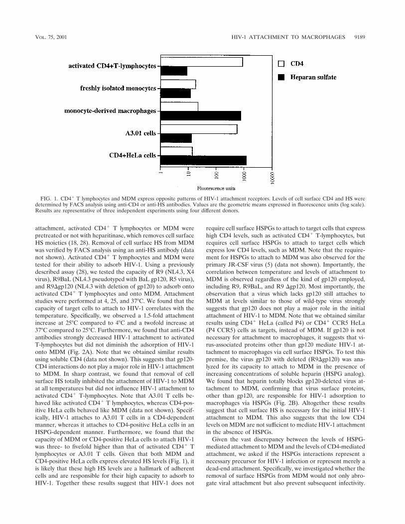
Cell surface expression of CD4 and HSPGs on primaryHIV-1 target cells. Despite the major role of macrophages inHIV-1 pathology, surprisingly, few studies have carefully ana-lyzed their surface expression of HSPGs and CD4 on thesecells. Indeed, the only two studies which have specifically ad-dressed this issue obtained contradictory results (7, 34). There-fore, in the present study, we compared the cell surface ex-pression of CD4 and HSPGs on primary cells. Specifically,activated CD4� T lymphocytes, as well as freshly isolatedmonocytes and MDM, were isolated and analyzed for CD4 andHSPG surface expression by FACS analysis using anti-CD4and anti-HS antibodies. CD4-positive HeLa cells and A3.01 Tcells were used as controls. We found that activated CD4� Tlymphocytes, as well as A3.01 T cells, expressed very high CD4levels but very low HSPG levels (Fig. 1). Furthermore, wefound that freshly isolated monocytes expressed low levels ofboth CD4 and HSPGs. Importantly, we found that MDM ex-pressed high HSPG levels but low CD4 levels. This findingsuggests that the cellular differentiation from monocyte intomacrophage upregulates the cell surface levels of HSPGs butdoes not influence CD4 expression. Interestingly, both MDMand CD4-positive HeLa cells express comparably low CD4 andhigh HSPGs levels. Note that we obtained similarly low levelsof chemokine receptors CXCR4 and CCR5 on macrophages(data not shown). Thus, we show that the two major in vivoHIV-1 target cells display opposite patterns of expression ofattachment receptors. Specifically, activated CD4� T lympho-cytes express high CD4 but low HSPGs levels, whereas MDMexpress low CD4 but high HSPG levels.
HSPGs are absolutely required for HIV-1 attachment toMDM. After demonstrating that activated CD4� T lympho-cytes and MDM exhibit opposite patterns of CD4 and HSPGsexpression, we examined the role of these candidate attach-ment receptors in HIV-1 adsorption. It is important to empha-size that to date few studies focusing exclusively on HIV-1attachment to macrophages have been performed. To deter-mine the role of CD4 in HIV-1 attachment, activated T lym-phocytes or MDM were tested for their ability to attach toHIV-1 in the presence or absence of anti-CD4 antibodies di-rected against the gp120-binding site of CD4 as describedpreviously (18, 28). To determine the role of HSPGs in HIV-1
9188 SAPHIRE ET AL. J. VIROL.
attachment, activated CD4� T lymphocytes or MDM werepretreated or not with heparitinase, which removes cell surfaceHS moieties (18, 28). Removal of cell surface HS from MDMwas verified by FACS analysis using an anti-HS antibody (datanot shown). Activated CD4� T lymphocytes and MDM weretested for their ability to adsorb HIV-1. Using a previouslydescribed assay (28), we tested the capacity of R9 (NL4.3, X4virus), R9BaL (NL4.3 pseudotyped with BaL gp120, R5 virus),and R9�gp120 (NL4.3 with deletion of gp120) to adsorb ontoactivated CD4� T lymphocytes and onto MDM. Attachmentstudies were performed at 4, 25, and 37°C. We found that thecapacity of target cells to attach to HIV-1 correlates with thetemperature. Specifically, we observed a 1.5-fold attachmentincrease at 25°C compared to 4°C and a twofold increase at37°C compared to 25°C. Furthermore, we found that anti-CD4antibodies strongly decreased HIV-1 attachment to activatedT-lymphocytes but did not diminish the adsorption of HIV-1onto MDM (Fig. 2A). Note that we obtained similar resultsusing soluble CD4 (data not shown). This suggests that gp120-CD4 interactions do not play a major role in HIV-1 attachmentto MDM. In sharp contrast, we found that removal of cellsurface HS totally inhibited the attachment of HIV-1 to MDMat all temperatures but did not influence HIV-1 attachment toactivated CD4� T-lymphocytes. Note that A3.01 T cells be-haved like activated CD4� T lymphocytes, whereas CD4-pos-itive HeLa cells behaved like MDM (data not shown). Specif-ically, HIV-1 attaches to A3.01 T cells in a CD4-dependentmanner, whereas it attaches to CD4-positive HeLa cells in anHSPG-dependent manner. Furthermore, we found that thecapacity of MDM or CD4-positive HeLa cells to attach HIV-1was three- to fivefold higher than that of activated CD4� Tlymphocytes or A3.01 T cells. Given that both MDM andCD4-positive HeLa cells express elevated HS levels (Fig. 1), itis likely that these high HS levels are a hallmark of adherentcells and are responsible for their high capacity to adsorb toHIV-1. Together these results suggest that HIV-1 does not
require cell surface HSPGs to attach to target cells that expresshigh CD4 levels, such as activated CD4� T-lymphocytes, butrequires cell surface HSPGs to attach to target cells whichexpress low CD4 levels, such as MDM. Note that the require-ment for HSPGs to attach to MDM was also observed for theprimary JR-CSF virus (5) (data not shown). Importantly, thecorrelation between temperature and levels of attachment toMDM is observed regardless of the kind of gp120 employed,including R9, R9BaL, and R9 �gp120. Most importantly, theobservation that a virus which lacks gp120 still attaches toMDM at levels similar to those of wild-type virus stronglysuggests that gp120 does not play a major role in the initialattachment of HIV-1 to MDM. Note that we obtained similarresults using CD4� HeLa (called P4) or CD4� CCR5 HeLa(P4 CCR5) cells as targets, instead of MDM. If gp120 is notnecessary for attachment to macrophages, it suggests that vi-rus-associated proteins other than gp120 mediate HIV-1 at-tachment to macrophages via cell surface HSPGs. To test thispremise, the virus gp120 with deleted (R9�gp120) was ana-lyzed for its capacity to attach to MDM in the presence ofincreasing concentrations of soluble heparin (HSPG analog).We found that heparin totally blocks gp120-deleted virus at-tachment to MDM, confirming that virus surface proteins,other than gp120, are responsible for HIV-1 adsorption tomacrophages via HSPGs (Fig. 2B). Altogether these resultssuggest that cell surface HS is necessary for the initial HIV-1attachment to MDM. This also suggests that the low CD4levels on MDM are not sufficient to mediate HIV-1 attachmentin the absence of HSPGs.
Given the vast discrepancy between the levels of HSPG-mediated attachment to MDM and the levels of CD4-mediatedattachment, we asked if the HSPGs interactions represent anecessary precursor for HIV-1 infection or represent merely adead-end attachment. Specifically, we investigated whether theremoval of surface HSPGs from MDM would not only abro-gate viral attachment but also prevent subsequent infectivity.
FIG. 1. CD4� T lymphocytes and MDM express opposite patterns of HIV-1 attachment receptors. Levels of cell surface CD4 and HS weredetermined by FACS analysis using anti-CD4 or anti-HS antibodies. Values are the geometric means expressed in fluorescence units (log scale).Results are representative of three independent experiments using four different donors.
VOL. 75, 2001 HIV-1 ATTACHMENT TO MACROPHAGES 9189

To explore this issue, MDM or activated CD4� T lymphocytespretreated or not with heparitinase were exposed to R9BaL orR9, respectively. We found that HS removal did not affectHIV-1 replication in activated CD4� T lymphocytes, confirm-ing the central role of CD4 in both attachment and infection inCD4� T lymphocytes (Fig. 2C). By contrast, this treatmentstrongly decreased HIV-1 replication in MDM (Fig. 2C). Thisresult indicates that the HSPG-mediated attachment is an es-sential step in HIV-1 infection in MDM. This result also cor-roborates our attachment data obtained above suggesting that
HSPGs are absolutely required for HIV-1 attachment toMDM, but not to activated CD4� T lymphocytes. Note thatthis requirement for HSPGs in HIV-1 replication in MDM wasalso observed for the primary virus JR-CSF (data not shown).Altogether these data demonstrate that HSPGs are essentialattachment receptors for HIV-1 replication in its major targetin vivo, the macrophage.
Human MDM express syndecans, betaglycan, but not glypi-cans. After demonstrating for the requirement of cell surfaceHS for HIV-1 attachment to MDM, we examined the repre-
FIG. 2. HIV-1 requires HSPGs to attach to and to infect MDM. (A) Target cells were tested for their capacity to attach HIV-1. Briefly, cellswere exposed to 10 ng of p24 of purified R9, R9BaL, and R9�gp120 viruses produced from 293T transfected cells for 30 min at 4, 25, or 37°C;extensively washed to remove unattached virus; and lysed. Attachment levels were quantified by p24 ELISA. Results are expressed as a percentageof attachment by fixing the percentage of R9BaL attachment to MDM at 4°C at 100. Results represent the average of two independent experiments(donors). To determine the role of HS or CD4 in HIV-1 attachment, cells were pretreated with heparitinase or preincubated with anti-CD4antibodies directed against the gp120-binding site of CD4. (B) R9�gp120 was tested for its ability to attach to MDM in the presence of heparin.MDM were exposed to 10 ng of p24 of purified R9�gp120 viruses produced from 293T cells for 30 min at 4°C in the presence of increasingconcentrations of soluble heparin. Results are expressed as a percentage of attachment by fixing the percentage of R9�gp120 attachment to MDMin the absence of heparin at 100. Results represent the average of three independent experiments (donors). (C) MDM or activated CD4� Tlymphocytes pretreated or not with heparitinase or anti-CD4 antibodies were tested for their ability to support HIV-1 replication. R9BaL and R9viruses produced from 293T cells were used to infect MDM and activated CD4� T lymphocytes, respectively. Replication was monitored by p24ELISA. Results are representative of three independent experiments using different donors. In all panels, error bars represent standard deviations.
9190 SAPHIRE ET AL. J. VIROL.

sentation of specific HSPGs on the surface of MDM. Thebest-characterized cell surface HSPGs fall into three groups,the syndecan, glypican, and betaglycan family proteins. Todate, there are four syndecans (syndecan-1 to -4) and six glypi-cans (glypican-1 to -6), the latter being linked to the cell mem-brane by a GPI anchor (2). Most cells and tissues express atleast one syndecan family member, while many express multi-ple syndecans in expression patterns specific to individual celltypes and tissues. For example, syndecan-1 is expressed onepithelial cells and malignant plasma cells, syndecan-2 is foundon fibroblasts, and syndecan-3 is found predominantly in thecentral nervous system, whereas syndecan-4 is more ubiquitous(2). Glypicans are also widely expressed HSPGs and commonlyoccur on the cell surface with one or more of the syndecans.Perhaps the most widely expressed cell surface HSPG is beta-glycan. Thus, we examined the cell surface expression of MDMfor syndecans, glypicans and betaglycan.
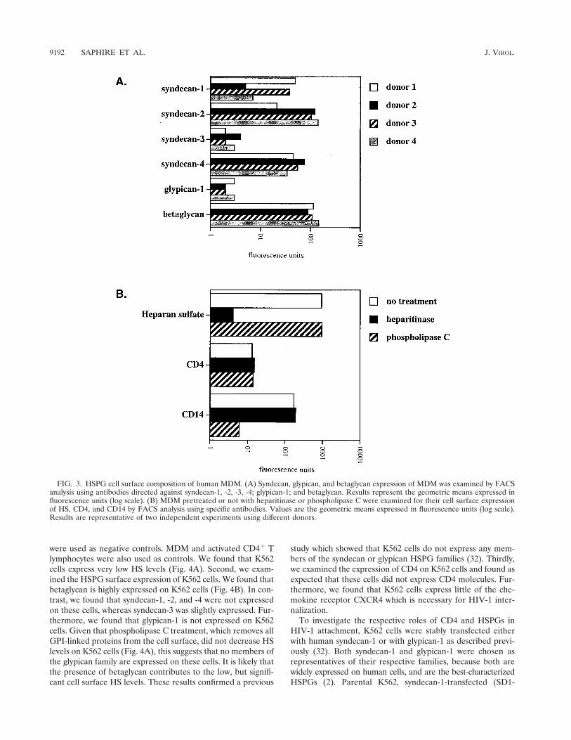
First, we found that betaglycan is abundantly expressed onMDM (Fig. 3A). Second, we found that MDM express highlevels of syndecan-1, syndecan-2, and syndecan-4 but low levelsof syndecan-3. It is important to note that we observed asimilar pattern among several donors. However, levels of ex-pression between different syndecans displayed some varia-tions. For example, MDM from one donor (donor 1) expressedvery high levels of syndecan-1 and syndecan-4 but low levels ofsyndecan-2, whereas MDM from another donor (donor 2)expressed high levels of syndecan-2 and syndecan-4 but lowlevels of syndecan-1. Furthermore, we found that glypican-1 isweakly expressed on MDM. It is important to note that unfor-tunately antibodies directed against glypican-2 to -6 are not yetavailable. Nevertheless, we employed another approach to de-termine whether these members of the glypican family areexpressed on MDM. We took advantage of the fact that phos-pholipase C possesses the capacity to remove all proteins at-tached to the cell surface via a GPI anchor including glypicans.Thus, MDM were pretreated with phospholipase C and sub-sequently examined for their total HS cell surface expressionusing anti-HS antibodies. If heparan sulfated glypicans areremoved from the plasma membrane upon enzymatic treat-ment, one would expect to observe a decrease in HS levels byFACS analysis. Importantly, we found that the enzymatic treat-ment did not decrease the cell surface HS levels on MDM (Fig.3B). This strongly suggests that glypicans are absent or weaklyexpressed on MDM. Note that CD4 remains intact after phos-pholipase C treatment consistent with the fact that CD4 is atransmembrane protein, but not a GPI-linked protein. To ver-ify the efficacy of the enzymatic treatment, we examined thelevels of CD14 which, like glypicans, is attached to the cellsurface via a GPI anchor. Given that CD14 is not detected byFACS using an anti-CD14 antibody after phospholipase Ctreatment, this indicates that GPI-linked cell surface proteinshave been removed from the cell surface. It is important tonote that we and others found that phospholipase C treatmentof MDM or CD4-positive HeLa cells does not decrease HIV-1infection (18; data not shown). This further suggests that glypi-cans do not play a major in HIV-1 attachment or infection ofMDM. Altogether, these results suggest that at least twoclasses of HSPGs, syndecan and betaglycan, may contribute tothe initial attachment of HIV-1 to MDM.
HSPGs can compensate for low CD4 levels in HIV-1 attach-ment. We showed that activated CD4� T lymphocytes expresshigh levels of CD4 but no detectable HSPG levels (Fig. 1).Furthermore, we demonstrated that HIV-1 attaches to acti-vated CD4� T lymphocytes via CD4 but not via HSPGs (Fig.2A). In sharp contrast, we showed that MDM express highlevels of HSPG but low CD4 levels (Fig. 1). We also found thatHIV-1 requires HSPGs, but not CD4, to attach to MDM (Fig.2A). Based on these observations, we postulated that the highCD4 levels on activated CD4� T lymphocytes are sufficient toattach HIV-1, whereas the high levels of HSPGs on MDMcompensate for the low CD4 levels in viral adsorption.
To explore this hypothesis, we took advantage of the humanK562 cell line, which expresses low levels of HS and no CD4.The K562 cell line is an erythroleukemia cell line establishedfrom the pleural effusion of a 53-year-old female with a chronicmyelogenous leukemia (17). Firstly, we analyzed the HS sur-face levels of K562 cells. Cells pretreated with heparitinase
FIG. 2—Continued.
VOL. 75, 2001 HIV-1 ATTACHMENT TO MACROPHAGES 9191
were used as negative controls. MDM and activated CD4� Tlymphocytes were also used as controls. We found that K562cells express very low HS levels (Fig. 4A). Second, we exam-ined the HSPG surface expression of K562 cells. We found thatbetaglycan is highly expressed on K562 cells (Fig. 4B). In con-trast, we found that syndecan-1, -2, and -4 were not expressedon these cells, whereas syndecan-3 was slightly expressed. Fur-thermore, we found that glypican-1 is not expressed on K562cells. Given that phospholipase C treatment, which removes allGPI-linked proteins from the cell surface, did not decrease HSlevels on K562 cells (Fig. 4A), this suggests that no members ofthe glypican family are expressed on these cells. It is likely thatthe presence of betaglycan contributes to the low, but signifi-cant cell surface HS levels. These results confirmed a previous
study which showed that K562 cells do not express any mem-bers of the syndecan or glypican HSPG families (32). Thirdly,we examined the expression of CD4 on K562 cells and found asexpected that these cells did not express CD4 molecules. Fur-thermore, we found that K562 cells express little of the che-mokine receptor CXCR4 which is necessary for HIV-1 inter-nalization.
To investigate the respective roles of CD4 and HSPGs inHIV-1 attachment, K562 cells were stably transfected eitherwith human syndecan-1 or with glypican-1 as described previ-ously (32). Both syndecan-1 and glypican-1 were chosen asrepresentatives of their respective families, because both arewidely expressed on human cells, and are the best-characterizedHSPGs (2). Parental K562, syndecan-1-transfected (SD1-
FIG. 3. HSPG cell surface composition of human MDM. (A) Syndecan, glypican, and betaglycan expression of MDM was examined by FACSanalysis using antibodies directed against syndecan-1, -2, -3, -4; glypican-1; and betaglycan. Results represent the geometric means expressed influorescence units (log scale). (B) MDM pretreated or not with heparitinase or phospholipase C were examined for their cell surface expressionof HS, CD4, and CD14 by FACS analysis using specific antibodies. Values are the geometric means expressed in fluorescence units (log scale).Results are representative of two independent experiments using different donors.
9192 SAPHIRE ET AL. J. VIROL.

K562), and glypican-1-transfected (GP1-K562) cells were ex-amined for the cell surface expression of betaglycan, synde-can-1, glypican-1, as well as for total HS expression by FACSanalysis. As expected, we found that syndecan-1 and glypican-1are highly expressed on SD1-K562 and GP1-K562 cells com-pared to parental K562 cells (Fig. 5A). Note that betaglycanlevels were found to be similar between the three K562 celllines. Furthermore, we found that the introduction of bothsyndecan-1 and glypican-1 into the K562 cells (SD1- and GP1-K562 cells) greatly elevated levels of cell surface HS comparedto K562 parental cells. It is important to note that SD1-K562
and GP1-K562 cell lines express similar levels of total HS asdescribed previously (32).
Parental K562, SD1-K562, and GP1-K562 cells were thentested for their ability to attach HIV-1 as described previously(28). Parental, SD1-, and GP1-K562 cells pretreated with he-paritinase were used a negative controls, whereas MDM andactivated CD4� T lymphocytes were used as positive controls.Given that R9 (X4 virus) and R9BaL (R5 virus) attach simi-larly to MDM (Fig. 2A), we selected R9 to test the capacity ofthe K562 cells to attach HIV-1. We found that the capacity ofparental K562 to attach HIV-1 is low, close to that of hepariti-
FIG. 4. Human K562 cells express betaglycan but not syndecans or glypicans. (A) K562 cells, activated CD4� T lymphocytes, and MDMpretreated or not with heparitinase or phospholipase C were examined for their HS cell surface expression by FACS analysis using anti-HSantibodies. Values are the geometric means expressed in fluorescence units (log scale). Results are representative of two independent experiments.(B) K562 cells were examined for their HSPG, CD4, and CXCR4 cell surface composition by FACS analysis using specific antibodies. Values arethe geometric means expressed in fluorescence units (log scale). Results are representative of two independent experiments.
VOL. 75, 2001 HIV-1 ATTACHMENT TO MACROPHAGES 9193

nase-treated cells (Fig. 5B). This low capacity of parental K562cells to attach HIV-1 correlates with their low HS surfacelevels. Interestingly, we found that HIV-1 attachment ontoGP1-K562 cells, which express very high HS levels (Fig. 5A), iscomparable to that of of the parental K562 cells. This resultindicates that glypican-1, although highly decorated with hepa-ran sulfated chains, does not possess the capacity to mediateHIV-1 attachment. In sharp contrast to parental or GP1-K562cells, we found that SD1-K562 cells strongly attached HIV-1.Given that both GP1- and SD1-K562 cells express similar lev-els of cell surface HS (Fig. 5A), this result strongly suggeststhat HIV-1 attachment does not depend merely on high HSexpression but depends on the presence of specific members ofthe HSPG family. This specificity implicates syndecan-1 as acandidate attachment receptor for HIV-1. To verify that syn-
decan-1 mediates HIV-1 attachment via its HS chains, SD1-K562 cells were pretreated with heparitinase and tested fortheir capacity to adsorb HIV-1. As expected, we found thatheparitinase treatment totally blocked HIV-1 attachment toSD1-K562 cells (Fig. 5B), demonstrating that syndecan-1 me-diates the initial HIV-1 adsorption via its polyanionic HSchains. It is important to note that the high capacity of SD1-K562 cells to attach HIV-1 is similar to that of MDM. Thesecomparable capacities for virus attachment between SD1-K562and MDM may arise from the fact that the major HSPGmolecules expressed on MDM are syndecans.
We next investigated the role of CD4 in HIV-1 attachment.Specifically, we asked whether high CD4 levels on parentalK562 cells would promote HIV-1 attachment, similarly to syn-decan-1 (Fig. 5B). To explore this issue, parental K562 cells
FIG. 5. Syndecan promotes HIV-1 attachment to K562 cells. (A) Parental, syndecan-1-, and glypican-1-transfected K562 cells were examinedfor their cell surface composition in HS, syndecan-1, glypican-1, and betaglycan by FACS analysis using specific antibodies. Values are thegeometric means expressed in fluorescence units (log scale). (B) Parental, syndecan-1-, and glypican-1-transfected K562 cells were tested for theirability to attach R9. Activated CD4� T lymphocytes and MDM were used as controls. Target cells were pretreated or not with heparitinase toremove cell surface HS. Results are expressed as a percentage of attachment by fixing the percentage of viral attachment to MDM at 100. Resultsrepresent the average of two independent experiments.
9194 SAPHIRE ET AL. J. VIROL.

were transfected with human CD4. CD4-positive transfectedK562 cells were sorted by FACS using an anti-CD4 antibody.Two distinct populations were chosen: one population of K562cells expressing high CD4 levels (called high CD4/K562), sim-ilar to activated CD4� T lymphocytes, and the other expressinglow but significant CD4 levels, similar to MDM (called lowCD4/K562) (Fig. 6A). High and low CD4/K562 cells were thentested for their ability to attach to HIV-1 as described above.It is crucial to note that both CD4 and HS levels were carefullyanalyzed by FACS concomitantly with the attachment assay.We found that the capacity of low CD4/K562 cells to attach toHIV-1 is similar to that of CD4-negative parental K562 cells(Fig. 6B). In contrast, we found that the capacity of highCD4/K562 cells to attach to HIV-1 is higher than that ofparental K562 cells. Importantly, the attachment levels of highCD4/K562 cells are similar to those of activated CD4�T lym-
phocytes (Fig. 6B). This suggests that high, but not low levelsof CD4 partially promote HIV-1 attachment.
After demonstrating that one member of the HSPG familycan compensate for low CD4 levels in HIV-1 attachment, weinvestigated their participation in HIV-1 replication. Giventhat K562 cells do not constitutively express the chemokinereceptors or CD4, both CXCR4 and CD4 were successivelyintroduced into parental K562, SD1- and GP1-K562 cell lines.The pBABE-CXCR4-puromycin plasmid was used to intro-duce CXCR4 into K562 cells. Puromycin-resistant cells weresorted by FACS using an anti-CXCR4 monoclonal antibody.Parental cells were used as negative controls. We selected cellpopulations which express CXCR4 levels comparable to thoseof activated CD4� T lymphocytes or MDM (Fig. 7A). CXCR4/K562, CXCR4/SD1-K562 and CXCR4/GP1-K562 cell lineswere then transfected with CD4, sorted by FACS using an
FIG. 6. High CD4 levels partially restore HIV-1 attachment to K562 cells. (A) Parental and CD4-transfected K562 cells were analyzed for CD4and HS cell surface expression by FACS analysis. Activated CD4� T lymphocytes and MDM were used as controls. Values are the geometric meansexpressed in fluorescence units (log scale). Results represent the average of three independent experiments. (B) Parental and CD4-transfectedK562 cells were tested for their capacities to attach R9. Results are expressed as a percentage of attachment by fixing the percentage of viralattachment to MDM at 100. Results represent the average of two independent experiments.
VOL. 75, 2001 HIV-1 ATTACHMENT TO MACROPHAGES 9195

anti-CD4 antibody. Given that activated CD4� T lymphocytesand MDM express high and low CD4 levels respectively, weselected two idstinct cell populations expressing high or lowCD4 levels (Fig. 7A).
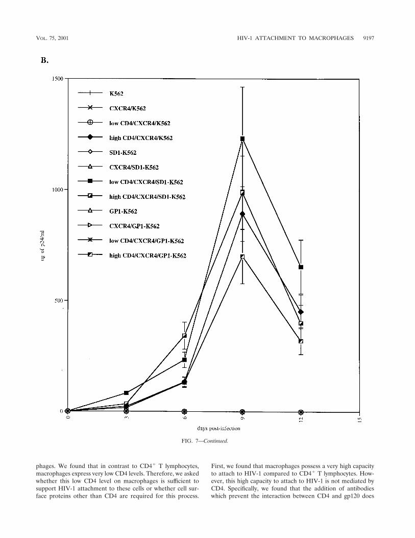
CXCR4-positive cells expressing low or high CD4 levelswere then tested for their permissivity to HIV-1. Parental cellswere used as negative controls. As expected, HIV-1 did notreplicate in K562 cells which lack either CD4 or CXCR4 orboth (Fig. 7B). Importantly, we found that HIV-1 did notreplicate in CXCR4/K562 cells or in CXCR4/GP1-K562 cellsthat express low CD4 levels (Fig. 7A). This correlates with ourdata above (Fig. 5B) which showed that both parental andGP1-K562 cells as well as cells which express low CD4 levels donot possess the capacity to attach HIV-1. In sharp contrast, wefound that CXCR4/K562 cells or CXCR4/GP1-K562 cells thatexpress high CD4 levels support HIV-1 replication (Fig. 7B).This corroborates our previous results, which showed that highCD4 levels may partially substitute for HSPGs in HIV-1 at-tachment (Fig. 6B). This also correlates with our data whichshowed that activated CD4� T lymphocytes which do not ex-press HS on their surface efficiently support HIV-1 replication(Fig. 2B). Most importantly, we found that HIV-1 replicates inCXCR4/SD1-K562 cells which express low CD4 levels (Fig.7B). This strongly suggests that the presence of syndecan-1 cancompensate for low CD4 levels for HIV-1 attachment and thusreplication.
Altogether, these results suggest that the low CD4 levels onmacrophages are not sufficient to support HIV-1 attachment.Furthermore, these data demonstrate that cell surface HSPGsare absolutely required for HIV-1 replication in macrophages.Most importantly, we demonstrate that a single family of
HSPGs—the syndecans—efficiently mediates HIV-1 attach-ment and represents an abundant class of attachment recep-tors on macrophages.
DISCUSSION
Macrophages have long been known as targets of HIV-1 ininfected individuals (1, 8, 14, 15, 26, 30, 36). Specifically, mac-rophages are thought to represent one of the first cell types inthe body to be infected during the early stage of HIV-1 trans-mission (1, 6, 26). Furthermore, macrophages and microgliawere identified as one of the major target cells in the centralnervous system of patients with AIDS dementia complex (15,36). Most importantly, macrophages, together with naı̈ve andmemory T cells (3, 6, 25), are thought to represent a majorviral reservoir in vivo. Specifically, Igarashi et al. recentlyshowed that tissue macrophages in the lymph nodes, spleen,gastrointestinal tract, liver, and kidney of rhesus macaquessustain high plasma virus loads even in the absence of CD4� Tcells (14). Together, these studies point to macrophages as keyplayers in HIV-1 pathogenesis.
In the present study, we examined the specific requirementsfor HIV-1 entry into one of its major in vivo target cells, themacrophages. HIV-1 entry is a multistep process, includinginitial attachment of the virus to the target cell surface, fol-lowed by fusion between the viral and cellular membranes, andculminating in the internalization of the viral genome into thecytosol of the target cells. In this report, we principally focusedon the initial viral attachment to macrophages. Given that CD4was thought to be the main attachment receptor for HIV-1, wefirst examined the cell surface expression of CD4 on macro-
FIG. 7. Syndecan restores HIV-1 replication in K562 cells which express low CD4 levels. (A) Parental, syndecan-1-, and glypican-1-transfectedK562 cells were transfected with CXCR4 and CD4. CD4 and HS surface expression was analyzed by FACS using specific antibodies. (B)CXCR4/CD4-positive K562 cells were tested for their ability to support R9 replication. Replication was monitored by p24 ELISA. Results arerepresentative of three independent experiments.
9196 SAPHIRE ET AL. J. VIROL.
phages. We found that in contrast to CD4� T lymphocytes,macrophages express very low CD4 levels. Therefore, we askedwhether this low CD4 level on macrophages is sufficient tosupport HIV-1 attachment to these cells or whether cell sur-face proteins other than CD4 are required for this process.
First, we found that macrophages possess a very high capacityto attach to HIV-1 compared to CD4� T lymphocytes. How-ever, this high capacity to attach to HIV-1 is not mediated byCD4. Specifically, we found that the addition of antibodieswhich prevent the interaction between CD4 and gp120 does
FIG. 7—Continued.
VOL. 75, 2001 HIV-1 ATTACHMENT TO MACROPHAGES 9197
not diminish HIV-1 attachment to macrophages. This suggeststhat cell surface proteins other than CD4 mediate HIV-1 at-tachment to macrophages.
In a search for candidate attachment receptors other thanCD4, we found that macrophages express high levels ofHSPGs. This finding seems to contradict a previous study sug-gesting that macrophages express only a few HSPGs molecules(34). This apparent discrepancy probably results from differentstages of differentiation of MDM utilized in these two studies.Corroborating this hypothesis, we (in this study) and recentlyothers (7) showed that freshly isolated monocytes express verylow HSPG levels, whereas MDM express high HSPG levels.This finding strongly suggests that the cellular differentiationfrom monocyte into macrophage upregulates the cell surfacelevels of HSPGs. Note that freshly isolated monocytes werefound to be relatively refractory to HIV-1 infection (26). In-terestingly, the block in infectivity was mapped to the earlysteps of infection. We can speculate that the scarcity of the twocandidate attachment receptors on monocytes—CD4 andHSPGs—may represent a critical obstacle for HIV-1 entry intothese cells.
After demonstrating that HSPGs are abundantly expressedon MDM, we asked whether they represent the main source ofattachment receptors responsible for the high capacity ofMDM to adsorb viral particles. To explore this issue, MDMwere treated with heparitinase, which removes all cell surfaceHS chains but does not alter CD4. Under these conditions, wefound that MDM lost their capacity to attach HIV-1. Thisdemonstrates that HSPGs, but not CD4, represent the majorclass of HIV-1 attachment receptors on MDM.
Our observation that R9, R9BaL, and R9�gp120 attachsimilarly to macrophages strongly suggests that gp120 is notnecessary for the initial attachment of HIV-1 to macrophages.Furthermore, our findings that soluble heparin prevents theattachment of the gp120-deleted virus (R9�gp120) to macro-phages further suggests that virus-associated proteins otherthan gp120 participate in HIV-1 adsorption to macrophages.Previous studies suggest that R5 gp120 (such as BaL) has alower affinity for CD4, as well as for HSPGs, than does X4gp120 (such as NL4.3). Specifically, Moulard et al. showed thatX4 gp120, via basic residues in the V3 loop, binds heparin witha higher affinity than R5 gp120 (22). How does one reconcilethe previous observation that gp120 possesses the intrinsiccapacity to bind to HSPGs, with our observation that HIV-1lacking gp120 efficiently attaches to macrophages? A simpleexplanation for this apparent discrepancy is that the level ofcontribution of gp120 HSPGs interactions to attachment isnegligible, compared to that of other virus-associated protein-HSPGs interactions (those abolished by the addition solubleheparin). Furthermore, it is crucial to note that the notion thatgp120 binds HSPGs arises exclusively from studies using re-combinant gp120 instead of whole virus. Thus, the use of re-combinant gp120 may not be appropriate for determining therole of these basic residues in HIV-1 attachment to target cellswithin the context of whole virus. Indeed, we cannot excludethe possibility that any recombinant protein containing a basicrich region (such as gp120) possesses the capacity to bind toheparin or HSPGs.
It is well known that HIV-1 can attach to a large variety ofnonpermissive cells which do not express either CD4 or ap-
propriate chemokine receptors. However, these attached vi-ruses are unable to successfully establish infection. This indi-cates that virus attachment does not necessarily correlate withinfection. Thus, we investigated whether virus-HSPGs interac-tions represent a necessary precursor for HIV-1 infection ofMDM or represent merely a dead-end attachment. Impor-tantly, we found that removal of HS chains totally blocksHIV-1 replication in MDM, despite the presence of CD4. Thisresult indicates that HSPGs are not only accessory attachmentreceptors, but are absolutely required for viral replication in amajor in vivo target of HIV-1, the macrophage. By analogywith cytokines and growth factors, HSPGs likely concentrateviruses at the surface of MDM, increasing the probability ofHIV-1 particles to interact with scarce CD4 molecules. Giventhat MDM possess a very high capacity to attach to HIV-1, apresumably large majority of viral particles initially attached toMDM via HSPGs unable to interact with CD4 will lead to adead-end infection. Nevertheless, our study clearly demon-strates that HSPG interactions are necessary events which en-sure MDM infection by HIV-1. It is important to emphasizethat although our results suggest that CD4 plays a minor rolein the initial attachment of HIV-1 to MDM, CD4 is absolutelyrequired for the postattachment steps of entry—fusion andinternalization.
Why has HIV-1 evolved to bind to HSPGs? One simpleexplanation is that HIV-1 needs HSPGs to infect one of itsmajor targets in vivo—the macrophage. Indeed, we showedthat CD4 levels on macrophages are too low to mediate theinitial viral attachment. Therefore, HIV-1 requires HSPGs assupplementary, but necessary, attachment receptors for mac-rophage infection. Another possibility to explain why HIV-1has evolved to bind to HSPGs is that viruses attached viaHSPGs on the surface of refractory cells represent an in transsource of infection for permissive cells. Specifically, since mostHSPG-decorated cells are not permissive to HIV-1 (CD4 neg-ative), it may seem like an inefficient and abortive strategy forreplication. However, it has been shown that dendritic cells(DC), which are not permissive to HIV-1, nevertheless serve asa reservoir for trans infection of permissive cells (12). Specif-ically, it has been shown that the glycoprotein DC-SIGN me-diates HIV-1 adsorption to DC and potentiates HIV-1 infec-tion in trans of permissive cells, even after an extended periodfar beyond the known half-life of free virus (12). By analogy,HIV-1 attached to nonpermissive cells via HSPGs may serve asan additional source of in trans infection for permissive cells.Corroborating this hypothesis, a recent study showed thatCD4-negative cells bind HIV-1 and efficiently transfer the virusto T cells (23). However, the endurance of HIV-1 attached tohost cells via HSPGs remains to be determined.
After demonstrating that HSPGs are absolutely required forHIV-1 replication in macrophages, we analyzed the surfaceHSPG composition of MDM. Importantly, we found that syn-decans but not glypicans are abundantly expressed on MDM.Specifically, we showed that syndecan-1, -2, and -4 are all wellrepresented on macrophages. Furthermore, we found thatphospholipase C, which removes glypicans, did not decreasethe total HS levels of macrophages and did not influenceHIV-1 attachment, suggesting that glypicans are not expressedon macrophages. These results strongly suggest that syndecansrepresent the major source of HSPGs on MDM. To determine
9198 SAPHIRE ET AL. J. VIROL.
whether syndecans are responsible for HIV-1 attachment toMDM, we introduced syndecan into K562 cells, which expresslow HS levels, and tested the capacity of these cells to attach toHIV-1. Importantly, we found that the introduction of synde-can-1 or glypican-1 into K562 cells promotes the levels of HSon the surface of these cells (SD1-K562 and GP1-K562 cells)and that the total HS levels between these two cell lines werecomparable. Interestingly, we found that HIV-1 attaches three-to fivefold better to cells which express syndecan-1 than cellswhich express glypican-1. This suggests that the capacity ofsyndecan-1 at least on K562 cells to attach to HIV-1 is higherthan that of glypican-1. While we used syndecan-1 as a repre-sentative member of the syndecan family, we found that bothsyndecan-2 and syndecan-4 in K562 cells also possess the highcapacity to attach HIV-1 (data not shown). Furthermore, wefound in the present study that the population of K562 cellswhich express low CD4 levels is not permissive to HIV-1.However, we showed that the introduction of syndecan-1 intothese nonpermissive cells renders them infectible by promotingHIV-1 attachment. Importantly, a close parallel can be foundin vivo: the macrophage which expresses low CD4 but highsyndecan levels. Given that syndecans are abundantly ex-pressed on MDM, altogether these observations strongly pointto syndecans as a major class of attachment receptor forHIV-1. However, a definitive proof for a direct role of synde-cans in HIV-1 replication would depend on the existence ofantibodies directed against the critical motifs within HS chainswhich unfortunately are not currently available.
A number of different criteria may account for the enhancedcapacity of syndecan-1 to attach to HIV-1 compared to glypi-can-1. First, while the gross compositions in HS levels aresimilar between syndecan-1 and glypican-1, subtle differencesin the fine structure of the HS chains may be important forvirus interactions. Secondly, the structure of the core proteinsmay influence the spatial distribution of the HS chains on thesurface of target cells. For example, the predicted structure ofglypican-1 is globular, whereas that of syndecan-1 is extended(2). Based on these structure predictions, it has been proposedthat the HS chains of glypican-1 lie close to the plasma mem-brane, whereas those of syndecan-1 are thought to be moredistal. Which of these criteria is significant for HIV-1 attach-ment remains to be elucidated. Although we showed that glypi-can-1 fails to support efficient HIV-1 attachment to K562 cells,we cannot exclude the possibility that other members of theglypican family can support HIV-1 adsorption. However, it isunlikely that glypicans play a major role in HIV-1 attachmentto macrophages. Indeed, we showed that phospholipase Ctreatment, which removes cell surface glypicans, does not affectHIV-1 attachment to MDM. Nevertheless, our present studydoes not preclude a role for glypicans in HIV-1 attachment toother in vivo target cells such as microglia and DC.
In conlusion, this study reports that HSPGs are absolutelyrequired for HIV-1 replication in macrophages. Specifically,we showed that the low levels of CD4 on macrophages areinsufficient to mediate the initial attachment of HIV-1. Wefound that cell surface HSPGs serve as necessary receptors forattachment to macrophages, perhaps to compensate for thelow CD4 levels. Furthermore, several lines of evidence point tothe participation of a single family of HSPGs in HIV-1 adsorp-tion to macrophages—the syndecans. Given that macrophages
are a major target for HIV-1 in vivo, reagents which disrupt theinteraction between HIV-1 and syndecans (or HSPGs) repre-sent an attractive class of novel antiviral agents.
ACKNOWLEDGMENTS
We thank J. Kuhns for secretarial assistance, Rebecca Sabbe fortechnical assistance, and D. Mosier for critically reading the manu-script.
This work was supported by funds from the Department of Immu-nology at The Scripps Research Institute.
REFERENCES
1. Appleman, M. E., D. W. Marshall, R. L. Brey, R. W. Houk, D. C. Beatty, R. E.Winn, G. P. Melcher, M. G. Wise, C. V. Sumaya, and R. N. Boswell. 1988.Cerebrospinal fluid abnormalities in patients without AIDS who are sero-positive for the human immunod eficiency virus. J. Infect. Dis. 158:193–199
2. Bernfield, M., M. Gotte, P. W. Park, O. Reizes, M. L. Fitzgerald, J. Lince-cum, and M. Zako. 1999. Functions of cell surface heparan sulfate proteo-glycans. Annu. Rev. Biochem. 68:729–777.
3. Brooks, D. G., S. G. Kitchen, C. M. Kitchen, D. D. Scripture-Adams, andJ. A. Zack. 2001. Generation of HIV latency during thymopoiesis. Nat. Med.7:459–464
4. Callahan, L. N., M. Phelan, M. Mallinson, and M. A. Norcross. 1991.Dextran sulfate blocks antibody binding to the principal neutralizing domainof human immunodeficiency virus type 1 without interfering with gp120-CD4interactions. J. Virol. 65:1543–1550.
5. Cann, A. J., M. J. Churcher, M. Boyd, W. O’Brien, J. Q. Zhao, J. Zack, andI. S. Chen. 1992. The region of the envelope gene of human immunodefi-ciency virus type 1 responsible for determination of cell tropism. J. Virol.66:305–309.
6. Chun, T. W, L. Carruth, D. Finzi, X. Shen, J. A. DiGiuseppe, H. Taylor, M.Hermankova, K. Chadwick, J. Margolick, T. C. Quinn, Y. H. Kuo, R. Brook-meyer, M. A. Zeiger, P. Barditch-Crovo, and R. F. Siliciano. 1997. Quanti-fication of latent tissue reservoirs and total body viral load in HIV-1 infec-tion. Nature 387:123–124.
7. Clasper, S., S. Vekemans, M. Fiore, M. Plebanski, P. Wordsworth, G. David,and D. G. Jackson. 1999. Inducible expression of the cell surface heparansulfate proteoglycan syndecan-2 (fibroglycan) on human activated macro-phages can regulate fibroblast growth factor action. J. Biol. Chem. 274:24113–24123.
8. Davis, L. E., B. L. Hjelle, V. E. Miller, D. L. Palmer, A. L. Llewellyn, T. L.Merlin, S. A. Young, R. G. Mills, W. Wachsman, and C. A. Wiley. 1992. Earlyviral brain invasion in iatrogenic human immunodeficiency virus infection.Neurology 42:1736–1739.
9. Dick, A. D, M. Pell, B. J. Brew, E. Foulcher, and J. D. Sedgwick. 1997. Directex vivo flow cytometric analysis of human microglial cell CD4 expression:examination of central nervous system biopsy specimens from HIV-seropos-itive patients and patients with other neurological disease. AIDS 11:1699–1708.
10. Fouts, T. R, J. M. Binley, A. Trkola, J. E. Robinson, and J. P. Moore. 1997.Neutralization of the human immunodeficiency virus type 1 primary isolateJR-FL by human monoclonal antibodies correlates with antibody binding tothe oligomeric form of the envelope glycoprotein complex. J. Virol. 71:2779–2785.
11. Gallay, P., T. Hope, D. Chin, and D. Trono. 1997. HIV-1 infection ofnondividing cells through the recognition of integrase by the importin/karyo-pherin pathway. Proc. Natl. Acad. Sci. USA 94:9825–9830.
12. Geijtenbeek, T. B., D. S. Kwon, R. Torensma, S. J. van Vliet, G. C. vanDuijnhoven, J. Middel, I. L. Cornelissen, H. S. Nottet, V. N. KewalRamani,D. R. Littman, C. G. Figdor, and Y. van Kooyk. 2000. DC-SIGN, a dendriticcell-specific HIV-1-binding protein that enhances trans-infection of T cells.Cell 100:587–597.
13. Haywood, A. M. 1994. Virus receptors: binding, adhesion strengthening, andchanges in viral structure. J. Virol. 68:1–5.
14. Igarashi, T., C. R. Brown, Y. Endo, A. Buckler-White, R. Plishka, N. Bischof-berger, V. Hirsch, and M. A. Martin. 2001. Macrophage are the principalreservoir and sustain high virus loads in rhesusmacaques after the depletionof CD4� T cells by a highly pathogenic simian immunodeficiency virus/HIVtype 1 chimera (SHIV): implications for HIV-1 infections of humans. Proc.Natl. Acad. Sci. USA 98:658–663.
15. Koenig, S., H. E. Gendelman, J. M. Orenstein, M. C. Dal Canto, G. H.Pezeshkpour, M. Yungbluth, F. Janotta, A. Aksamit, M. A. Martin, and A. S.Fauci. 1986. Detection of AIDS virus in macrophages in brain tissue fromAIDS patients with encephalopathy. Science 233:1089–1093.
16. Kost, T. A., J. A. Kessler, I. R. Patel, J. G. Gray, L. K. Overton, and S. G.Carter. 1999. Human immunodeficiency virus infection and syncytium for-mation in HeLa cells expressing glycophospholipid-anchored CD4. J. Virol.65:3276–3283.
17. Lozzio, B. B., and C. B. Lozzio. 1997. Properties of the K562 cell line derived
VOL. 75, 2001 HIV-1 ATTACHMENT TO MACROPHAGES 9199
from a patient with chronic myeloid leukemia. Int. J. Cancer 19:136.18. Mondor, I., S. Ugolini, and Q. J. Sattentau. 1998. Human immunodeficiency
virus type 1 attachment to HeLa CD4 cells is CD4 independent and gp120dependent and requires cell surface heparans. J. Virol. 72:3623–3634.
19. Moore, J. P., and J. Binley. 1998. HIV. Envelope’s letters boxed into shape.Nature 393:630–631.
20. Moore, J. P, J. A. McKeating, Y. X. Huang, A. Ashkenazi, and D. D. Ho.1992. Virions of primary human immunodeficiency virus type 1 isolatesresistant to soluble CD4 (sCD4) neutralization differ in sCD4 binding andglycoprotein gp120 retention from sCD4-sensitive isolates. J. Virol. 66:235–243.
21. Mosier, D. E., G. R. Picchio, R. J. Gulizia, R. Sabbe, P. Poignard, L. Picard,R. E. Offord, D. A. Thompson, and J. Wilken. 1999. Highly potent RANTESanalogues either prevent CCR5-using human immunodeficiency virus type 1infection in vivo or rapidly select for CXCR4-using variants. J. Virol. 73:3544–3550.
22. Moulard, M., H. Lortat-Jacob, I. Mondor, G. Roca, R. Wyatt, J. Sodroski, L.Zhao, W. Olson, P. D Kwong, and Q. J. Sattentau. 2000. Selective interac-tions of polyanions with basic surfaces on human immunodeficiency virustype 1 gp120. J. Virol. 74:1948–1960.
23. Olinger, G. G., M. Saifuddin, and G. T. Spear. 2000. CD4-negative cells bindhuman immunodeficiency virus type 1 and efficiently transfer virus to T cells.J. Virol. 74:8550–8557.
24. Oravecz, T., M. Pall, J. Wang, G. Roderiquez, M. Ditto, and M. A. Norcross.1997. Regulation of anti-HIV-1 activity of RANTES by heparan sulfateproteoglycans. J. Immunol. 159:4587–4592.
25. Pierson, T., T. L. Hoffman, J. Blankson, D. Finzi, K. Chadwick, J. B. Mar-golick, C. Buck, J. D. Siliciano, R. W. Doms, and R. F. Siliciano. 2000.Characterization of chemokine receptor utilization of viruses in the latentreservoir for human immunodeficiency virus type 1. J. Virol. 74:7824–7833.
26. Rich, E. A., I. S. Chen, J. A. Zack, M. L. Leonard, and W. A. O’Brien. 1992.Increased susceptibility of differentiated mononuclear phagocytes to produc-tive infection with human immunodeficiency virus-1 (HIV-1). J. Clin. Inves-tig. 89:176–183.
27. Roderiquez, G., T. Oravecz, M. Yanagishita, D. C. Bou-Habib, H.
Mostowski, and M. A. Norcross. 1995. Mediation of human immunodefi-ciency virus type 1 binding by interaction of cell surface heparan sulfateproteoglycans with the V3 region of envelope gp120-gp41. J. Virol. 69:2233–2239.
28. Saphire, A. C., M. D. Bobardt, and P. A. Gallay. 1999. Host cyclophilin Amediates HIV-1 attachment to target cells via heparans. EMBO J. 18:6771–6785.
29. Sattentau, Q. J., and J. P. Moore. 1995. Human immunodeficiency virus type1 neutralization is determined by epitope exposure on the gp120 oligomer. J.Exp. Med. 182:185–96.
30. Sidtis, J. J., and R. W. Price. 1990. Early HIV-1 infection and the AIDSdementia complex. Neurology 40:323–326.
31. Sonza, S., A. Maerz, S. Uren, A. Violo, S. Hunter, W. Boyle, and S. Crowe.1995. Susceptibility of human monocytes to HIV type 1 infection in vitro isnot dependent on their level of CD4 expression. AIDS Res. Hum. Retrovir.7:769–776.
32. Steinfeld, R., H. Van Den Berghe, and G. David. 1996. Stimulation of fibro-blast growth factor receptor-1 occupancy and signaling by cell surface-asso-ciated syndecans and glypican. J. Cell Biol. 133:405–416.
33. Ugolini, S., I. Mondor, P. W. Parren, D. R. Burton, S. A. Tilley, P. J. Klasse,and Q. J. Sattentau. 1997. Inhibition of virus attachment to CD4� targetcells is a major mechanism of T cell line-adapted HIV-1 neutralization. J.Exp. Med. 186:1287–1298.
34. Uhlin-Hansen, L., and S. O. Kolset. 1988. Cell density-dependent expressionof chondroitin sulfate proteoglycan incultured human monocytes. J. Biol.Chem. 263:2526–2531.
35. von Schwedler, U., R. S. Kornbluth, and D. Trono. 1994. The nuclear local-ization signal of the matrix protein of human immunodeficiency virus type 1allows the establishment of infection in macrophages and quiescent T lym-phocytes. Proc. Natl. Acad. Sci. USA 91:6992–6996.
36. Wiley, C. A., R. D. Schrier, J. A. Nelson, P. W. Lampert, and M. B. Oldstone.1986. Cellular localization of human immunodeficiency virus infection withinthe brains of acquired immune deficiency syndrome patients. Proc. Natl.Acad. Sci. USA 83:7089–7093.
9200 SAPHIRE ET AL. J. VIROL.