The Interdependence model of grain nucleation: A numerical analysis of the Nucleation-Free Zone
Upload
uni-goettingenCategory
view
1download
0
Synaptotagmin-1 may be a distance regulator acting upstream ofSNARE nucleation
Geert van den Bogaart1, Shashi Thutupalli2, Jelger H. Risselada3, Karsten Meyenberg4,Matthew Holt1, Dietmar Riedel5, Ulf Diederichsen4, Stephan Herminghaus2, HelmutGrubmüller3, and Reinhard Jahn1
1Department of Neurobiology, Max Planck Institute for Biophysical Chemistry, Am Faβberg 11,37077, Göttingen, Germany2Department of Dynamics of Complex Fluids, Max Planck Institute for Dynamics and Self-Organization, Bunsenstraβe 10, 37073, Göttingen, Germany3Department of Theoretical and Computational Biophysics, Max Planck Institute for BiophysicalChemistry, Am Faβberg 11, 37077, Göttingen, Germany4Institute for Organic and Biomolecular Chemistry, Georg-August-University, Tammannstraβe 2,37077, Göttingen, Germany5Facility for Electron Microscopy, Max Planck Institute for Biophysical Chemistry, Am Faβberg 11,37077, Göttingen, Germany
AbstractSynaptotagmin-1 triggers Ca2+-sensitive, rapid neurotransmitter release by promoting theinteraction of SNARE proteins between the synaptic vesicles and the plasma membrane. Howsynaptotagmin-1 promotes this interaction is controversial, and the massive increase in membranefusion efficiency of Ca2+-synaptotagmin-1 has not been reproduced in vitro. However, previousexperiments have been performed at relatively high salt concentrations, screening potentiallyimportant electrostatic interactions. Using functional reconstitution in liposomes, we show herethat at low ionic strength SNARE-mediated membrane fusion becomes strictly dependent on bothCa2+ and synaptotagmin-1. Under these conditions, synaptotagmin-1 functions as a distanceregulator: tethering the liposomes too far for SNARE nucleation in the absence of Ca2+, but bringsthe liposomes close enough for membrane fusion in the presence of Ca2+. These results mayexplain how the relatively weak electrostatic interactions of synaptotagmin-1 with membranessubstantially accelerate fusion.
Presynaptic nerve terminals convert electrical signals into chemical signals that target otherneurons or somatic cells. The conversion is mediated by a depolarisation of the plasmamembrane, which causes an influx of Ca2+ through voltage activated Ca2+-channels. Theincrease of the local cytoplasmic Ca2+ concentration triggers the submillisecond fusion ofsynaptic vesicles with the plasma membrane. Membrane fusion is mediated by the assemblyof SNARE proteins including the R-SNARE synaptobrevin-2 (also known as VAMP2) onthe synaptic vesicle and the Q-SNAREs syntaxin-1A and SNAP-25 on the plasma
Correspondence should be addressed to R.J. ([email protected]).Author contributionsS.T. and S.H. performed the flow cytometry experiments. J.H.R. and H.G. performed the M.D. simulations. M.G.H purified thesynaptic vesicles. Thermophoresis data is from K.M. and U.D. D.R. performed the EM. G.v.d.B performed all other experiments.G.v.d.B. and R.J. designed the study and wrote the paper. All authors discussed the results and commented on the manuscript.
NIH Public AccessAuthor ManuscriptNat Struct Mol Biol. Author manuscript; available in PMC 2012 January 1.
Published in final edited form as:Nat Struct Mol Biol. ; 18(7): 805–812. doi:10.1038/nsmb.2061.
NIH
-PA Author Manuscript
NIH
-PA Author Manuscript
NIH
-PA Author Manuscript
membrane1,2. SNARE assembly involves the conserved, membrane-adjacent SNAREmotifs, proceeds from the N-terminus towards the C-terminal membrane anchors, and resultsin formation of a tight coil-coil structure which pulls the membranes together and overcomesthe energy barrier of membrane fusion.
The synaptic vesicle protein synaptotagmin-1 functions as a major Ca2+-sensor for neuronalexocytosis3,4. The 65 kDa protein contains a single transmembrane domain followed by alarge cytoplasmic domain consisting of a 61 residue unstructured linker and tandem C2-typedomains. These C2-domains, called the C2A- and C2B-domain, bind 2 and 3 Ca2+ ionsrespectively with low affinity (60 µM – 1 mM)5,6 and interact with both anionic membranesand SNARE proteins. Synaptotagmin-1 binds to anionic lipids both in the absence of Ca2+
via a so-called polybasic patch consisting of four lysines located on the C2B-domain7–9 andin the presence of Ca2+ via its Ca2+-binding sites5,8–13. Anionic lipids such as phosphoserine(PS) and phosphatidylinositol 4,5-biphosphate (Pi(4,5)P2) complete the Ca2+-binding sitesof the C2-domains and thereby increase the affinity of synaptotagmin-1 for Ca2+-binding5,7,8,14. Ca2+-synaptotagmin-1 locally increases membrane curvature, suggesting thatsynaptotagmin-1 might act by increasing the membrane tension at the sites of fusion15,16.Synaptotagmin-1 also binds directly to SNAP-25, syntaxin-1A, and the binary and ternarySNARE complexes3,17–19. All together, a model is emerging where synaptotagmin-1structurally changes both the SNARE complex and the membranes in a Ca2+-dependentmanner, thereby levering the protein and/or bending the membrane which inducesmembrane fusion3,9,11,15–22. Importantly, in these models, synaptotagmin-1 actsdownstream of SNARE nucleation, i.e. at a fusion-arrested state where the membranes aretethered by a trans SNARE-complex in which at least part of the cytoplasmic SNARE-domains, but not the C-terminal transmembrane helices, are already coiled up.
A large number of studies aimed to reconstitute Ca2+-synaptotagmin-1 triggered membranefusion in vitro using SNARE-containing artificial membranes, either by adding the solublecytoplasmic C2-domains (C2AB fragment; residues 97–421) or by inserting full-lengthmembrane-anchored synaptotagmin-1. A major limitation in these studies is that SNAREsalone are sufficient to induce lipid mixing of liposomes23. Indeed, membrane fusion isrelatively efficient when artificial membranes containing even a single copy24 ofsynaptobrevin-2 and a combination of syntaxin-1A and SNAP-25 are mixed. The addition ofthe soluble C2AB fragment makes membrane fusion somewhat Ca2+-sensitive and results inan increase of fusion efficiency between 100 µM and 10 mM Ca2+ (mainly lipidmixing)3,10,11,15,16,20,21,25–29. However, in these studies, membrane fusion still proceeds inthe absence of the C2AB fragment and the increase in fusion efficiency is usually less than10-fold compared to the over 18,000-fold increase in vivo30. Recently, it was reported thatlipid mixing in the presence of membrane-anchored synaptotagmin-1 was increased about3–5-fold at 10 µM Ca2+ but decreased again at higher Ca2+ concentrations27. This Ca2+-dependency was strongly influenced by the lipid composition of both liposome populations:increasing the fraction of anionic lipids in the Q-SNARE or R-SNARE membranes resultedin higher or lower Ca2+-sensitivities, respectively.
There is general consensus that the interactions of synaptotagmin-1 with membranes andSNAREs are predominantly of an electrostatic nature. Indeed, binding of synaptotagmin-1both to anionic lipid membranes5,7 and to SNARE molecules18,19,22 is heavily influenced bythe ionic strength of the solution. Thus, it is surprising that the influence of ionic interactionson membrane fusion in the presence of synaptotagmin-1 has not been investigated: Allaforementioned in vitro synaptotagmin-1 membrane fusion studies were performed atrelatively high, physiological ionic strength and typically 100 to 150 mM KCl or NaCl.However, at these high salt concentrations the binding of synaptotagmin-1 to SNAREs and
Bogaart et al. Page 2
Nat Struct Mol Biol. Author manuscript; available in PMC 2012 January 1.
NIH
-PA Author Manuscript
NIH
-PA Author Manuscript
NIH
-PA Author Manuscript
anionic membranes is strongly reduced or even absent5,7,18,19,22. We thus set out to answerthe question of how ionic strength influences synaptotagmin-1 action.
ResultsMembrane fusion is blocked at low ionic strength
SNARE mediated liposome fusion is typically studied at physiological ionic strength, withNaCl or KCl concentrations of 100–150 mM3,5,10,11,15,16,20,21,25–29 where the Debye chargescreening length is only ~7 Å. Molecules need to be within a few times this distance tointeract. We studied SNARE-mediated fusion at low ionic strength by employing a FRET-based lipid mixing assay (without synaptotagmin-1; Fig. 1a). The R-SNARE population ofliposomes contained a 1:1,000 molar protein-to-lipid ratio of recombinant synaptobrevin-2and was labeled with the fluorescent lipid analog Oregon green-phosphatidylethanolamine(OG-PE; donor fluorophore). The Q-SNARE population of liposomes contained 1:2,000 of acomplex consisting of two copies of recombinant syntaxin-1A (lacking its inhibitory N-terminal Habc-domain31–33; residues 183–288) and a single SNAP-25 (2:1-complex)25.Those liposomes were labeled with DiD (acceptor fluorophore). As already shown innumerous studies3,10,11,15–16,20,21,23–29,32–34,35–38, robust, SNARE specific lipid mixingwas observed at normal ionic strength (20 mM K-Hepes pH 7.4, 150 mM KCl; Fig. 1a).Surprisingly however, lipid mixing was almost completely blocked at low ionic strength (20mM K-Hepes pH 7.4, 5 mM KCl, 300 mM sucrose) where the Debye screening length was~25 Å. Lipid mixing at low ionic strength was also blocked with 1:8,000 molar protein-to-lipid ratio of the binary Q-SNARE complex stabilized with a fragment of synaptobrevin-2(residues 49–96; Fig. 1b). This stabilized complex ensures that all Q-SNAREs are availablefor fusion and displays a dramatically enhanced fusion efficiency compared to native Q-SNAREs34. These results indicate that the absence of lipid mixing at low ionic strength wasnot due to unavailability of the binary Q-SNARE complex. Reversing the fluorescent labelsor increasing the SNARE density up to 1:500 showed similar results.
We were unable to incorporate SNARE proteins in the liposomes at salt concentrationsbelow 150 mM and had to reduce the ionic strength by diluting the liposomes afterward.Sucrose was added to preserve the osmolarity at ~340 mOsm. Membrane fusion was stillblocked at low ionic strength when the sucrose was omitted, but the liposomes lookedosmotically deformed by negative-staining electron microscopy (EM; Fig. 1b) and theliposomes became leaky at osmotic differences above 100 mOsm (Supp. Fig. 1a). Thepresence of sucrose increased the viscosity 33% and this decreases the diffusion of bothsoluble and membrane associated39 molecules. Diffusion limited reactions are slowedaccordingly40.
The liposomes contained ~10% anionic lipids, and we speculated that the electrostaticrepulsion might be too high for SNARE-mediated membrane fusion at low ionic strength.To validate this hypothesis, we performed experiments with liposomes composed of purezwitterionic phospatidylcholine (PC; Fig. 1c–d). Indeed, without PS, lipid mixing at lowionic strength still occurred, albeit somewhat slower than at normal ionic strength because ofthe sucrose (increased viscosity). In contrast, lipid mixing was completely blocked with 10%PS at low but not at normal ionic strength. Thus, the electrostatic repulsion at low ionicstrength increases the energy barrier of membrane fusion. We then tested whether SNAREnucleation could still occur under these conditions.
SNARE complex formation was directly followed by FRET with liposomes containing a1:8,000 protein-to-lipid ratio of the stabilized Q-SNARE-complex with syntaxin-1A labeledwith Alexa fluor 488 (S225C; donor fluorophore)32,35. Mixing these liposomes with 100 nMof the soluble synaptobrevin-21–96 fragment labeled with Texas red (S61C; acceptor
Bogaart et al. Page 3
Nat Struct Mol Biol. Author manuscript; available in PMC 2012 January 1.
NIH
-PA Author Manuscript
NIH
-PA Author Manuscript
NIH
-PA Author Manuscript
fluorophore)35 resulted in fast complex formation independent of ionic strength (Fig. 1e).Accordingly, mixing liposomes containing full-length synaptobrevin-2 with a soluble Q-SNARE complex (SNAP-25, syntaxin-1A183–262 and synaptobrevin-249–86) also resulted incomplex formation, albeit somewhat slower. This decrease is probably due to increasedelectrostatic repulsion at low ionic strength (the Q-SNARE complex has an acidic surface).In contrast to the soluble SNAREs, SNARE complex formation was only observed atnormal but not at low ionic strength when liposomes bearing membrane-anchored Q- and R-SNAREs were used (Fig. 1f), thus paralleling lipid mixing. These FRET data were in perfectagreement with the dissociation of the synaptobrevin-249–96 fragment from the stabilizedacceptor complex (Supp. Fig. 1b–c). Thus, we conclude that although SNARE zippering canoccur at low ionic strength, the high electrostatic repulsion between the liposomes blocksnot only lipid mixing but also SNARE nucleation.
Ca2+-synaptotagmin-1 action at low ionic strengthNext, we studied whether synaptotagmin-1 rescues membrane fusion at low ionic strength.We reconstituted recombinant full-length synaptotagmin-1 at a protein-to-lipid ratio of1:4,000 in the R-SNARE liposomes. At normal ionic strength, synaptotagmin-1 somewhatincreased the lipid mixing kinetics independent of Ca2+ (Fig. 2a–b), as reportedpreviously25,29. In contrast, lipid mixing was almost completely blocked at low ionicstrength and 1 mM Ca2+ now dramatically increased lipid mixing. This lipid mixing wasstrictly dependent on both synaptotagmin-1 and the SNAREs. Under those low ionicstrength conditions, the C2-domains of synaptotagmin-1 are correctly folded and specificallybind Ca2+ (Supp. Fig. 2). Experiments where Ca2+ was mixed by controlled flow focusing ina microfluidics flow chamber41 showed that fusion could be readily triggered with Ca2+
(Fig. 2c–e). Compared to cuvette-based experiments, flow cytometry allows for relativelyfast and well controlled mixing. Moreover, under our conditions, the synaptotagmin-1mediated membrane fusion efficiency correlated with the Ca2+ concentration in a dosedependent manner from 10 µM up to 1 mM Ca2+ and Mg2+ had only little effect (Fig. 2f–g).This Ca2+ sensitivity depends on precise protein densities and compositions of themembranes and buffer27.
At normal ionic strength, purified synaptic vesicles from rat brain fuse independent of Ca2+
(ref. 37). However, as these membranes contain about 15% anionic lipids42, it seemedpossible that (similar to the liposomes) membrane fusion of synaptic vesicles becomes Ca2+-sensitive at low ionic strength. To test this hypothesis, we fused synaptic vesicles with Q-SNARE liposomes containing both donor and acceptor fluorophores. In this case, membranefusion dilutes the fluorescent lipid analogs with the vesicle membranes, resulting in a loss ofFRET and a decrease of acceptor fluorescence. Indeed, at low ionic strength, lipid mixingwas largely Ca2+-dependent (Fig. 2h). Thus, at low ionic strength Ca2+-synaptotagmin-1triggers lipid mixing not only of liposomes, but also of native synaptic vesicles.
Because lipid mixing does not distinguish between hemifusion and full membrane fusion, itis conceivable that the membrane fusion did not progress beyond the hemifusion state. Toresolve this issue, we employed a content mixing assay where liposomes with encapsulatedcalcein at self quenching concentrations were fused with empty (calcein-free) liposomes24.Content mixing results in calcein dequenching. Indeed, a SNARE and Ca2+-synaptotagmin-1 specific increase in fluorescence was observed (Fig. 3a). This contentmixing was not caused by leakage of the calcein from the liposomes, as leakage was only 4–5% of total calcein (Supp. Fig. 3a). In addition, FRET and fluorescence anisotropymeasurements indicated that there was no SNARE nucleation without Ca2+ (Fig. 1e–f, 3b–e). In the absence of synaptotagmin-1, some residual SNARE complex formation wasobserved (Fig. 1e, 3c), which we attribute to charge-shielding by the bivalent Ca2+ cations,which allows for a low degree of SNARE interaction and membrane fusion. SNARE
Bogaart et al. Page 4
Nat Struct Mol Biol. Author manuscript; available in PMC 2012 January 1.
NIH
-PA Author Manuscript
NIH
-PA Author Manuscript
NIH
-PA Author Manuscript

complex formation was inhibited by synaptobrevin-21–96 even when added after the mixingof the R- and Q-SNARE liposomes, and regardless of whether wild-type synaptotagmin-1was used, or a mutant in which two lysines of the polybasic patch are converted to alanines(K325A K326A; KAKA-mutant7; Fig. 3d–e). This indicates that no substantial SNAREnucleation occurred without Ca2+.
Disrupting Ca2+-binding of the C2A-domain (D178A D230A D232A) reduced both thelipid-mixing and SNARE-complex formation substantially (Fig. 3f–g). In contrast, thecorresponding C2B-mutant (D309A D363A D365A) had little effect on the membranefusion efficiency. This has been reported before11,16,25, but contradicts in vivo data wheredisruption of Ca2+-binding to the C2B-domain impaired release much more severely thanC2A disruption43. This discrepancy may be because –unlike in the synapse– both fusionpartners are small liposomes, which bypasses the requirement for membrane bending by theC2B-domain15,16. To evaluate whether membrane curvature influences the ability of theC2B-domain to stimulate membrane fusion at low ionic strength, we performed experimentswith 10–20 µm giant unilamellar vesicles (GUVs; Fig. 3h). These membranes are much lesscurved than the small 36 nm-sized24 liposomes used elsewhere in this study. Fusing theGUVs with small synaptotagmin-1–synaptobrevin-2 liposomes showed that fusion wasabolished when Ca2+-binding was disrupted to the C2B-mutant, similar to normal ionicstrength16. Importantly, fusion of the C2B-mutant could be rescued by disruption of theGUVs by sonication (Supp. Fig. 3b). All together, we conclude that at low ionic strengthCa2+-synaptotagmin-1 triggers SNARE-mediated membrane fusion and both C2-domainsare required.
Synaptotagmin-1 acts as a distance regulatorSynaptotagmin-1 binds to anionic lipids both with and without Ca2+ and this binding leadsto membrane clustering5,7–13,25. Indeed, negative-staining EM showed strong instantaneousclustering of the liposomes when synaptotagmin-1 liposomes were mixed with emptyliposomes (no SNAREs; Supp. Fig. 4a). This clustering was Pi(4,5)P2 and synaptotagmin-1specific and was reduced when the KAKA mutant of synaptotagmin-1 was used (Supp. Fig.4b), similar to normal ionic strength9. Identical results were obtained when the SNAREswere present. The clustering of liposomes by synaptotagmin-1 was confirmed with dynamiclight scattering (DLS) experiments (Fig. 4a, Supp. Fig. 4c). In addition, we developed amicroscale thermophoresis44 assay to assess liposome clustering without Ca2+ (Fig. 4b–c).For this, DiD-labeled liposomes were inserted in a capillary that was heated ~5°C locallywith a focused infrared laser. Because of the Soret-effect45, these liposomes thermodiffusedaway from the heated spot, hence creating a local drop in the concentration and fluorescenceintensity (= positive thermophoresis). The extent of this depletion depends on the surfaceproperties of the liposomes (solvation entropy). Contrary to the DiD-liposomes, liposomeslabeled with sufficient (>5 mol%) OG-PE thermodiffuse towards the heated spot and arelocally enriched (= negative thermophoresis). Thus, DiD-liposomes are separated from OG-PE-liposomes by the heating laser, unless they are tethered by synaptotagmin-1 in whichcase they co-segregate resulting in intermediate thermophoresis. This tethering wasdependent on Pi(4,5)P2 and was reduced when the KAKA mutant of synaptotagmin-1 wasused, all in agreement with the DLS and EM data. Density gradient flotation experimentswith various mutants of the soluble C2AB-domain showed that without Ca2+ the polybasiclysine patch of synaptotagmin-1 bound to membranes, whereas binding with Ca2+ occurredprimarily via the Ca2+-binding sites (Supp. Fig. 4d)7,9.
In the EM images, synaptotagmin-1 tethered the liposomes at very narrow distances evenwithout Ca2+ (‘squeezed together’; Supp. Fig. 2a; Fig. 4d). Regardless of this tight liposomeassociation, SNARE nucleation did not occur under those conditions (Fig. 3b–e) suggestingthat the vesicles are still separated by a distance too far for trans SNARE interactions (Fig.
Bogaart et al. Page 5
Nat Struct Mol Biol. Author manuscript; available in PMC 2012 January 1.
NIH
-PA Author Manuscript
NIH
-PA Author Manuscript
NIH
-PA Author Manuscript
4e). However, negative-staining EM involves fixation and drying and is thus unsuitable forassessing membrane distances. We could not perform cryo-EM, because of the presence ofsucrose to compensate for the osmolarity. Therefore, we used the FRET approach of Yoonet al.46 (Fig. 4f–g, Supp. Fig. 4e), based on the ~5% FRET resulting from clustering of DiIwith DID labeled liposomes at distances below ~5 nm. In the presence of synaptotagmin-1(no SNAREs), no FRET signal was detectable although the vesicles were clustered (Fig. 4a–c, Supp. Fig. 4a–d). Accordingly, Ca2+ induced FRET of both the wild-type and the KAKAmutant and this increased substantially when 1% Pi(4,5)P2 was present. Disrupting Ca2+-binding to the C2A- or C2B-domain largely impaired this FRET signal. The observed FRETwas caused by close membrane proximity rather than by residual lipid mixing, because theFRET signal was reversed by addition of 1 mM EDTA. Lastly, dynamic light scatteringexperiments with the C2AB fragment (Fig. 4h; Supp. Fig. 4f) showed that this fragment wassufficient for liposome clustering in the presence of Ca2+, similar to normal ionic strength9.
Together, we conclude that without Ca2+ the polybasic patch of synaptotagmin-1 tethers theliposomes too far for SNARE nucleation. Ca2+ binds to the C2AB-domain, which functionsas a ‘charge bridge’ bringing the membranes close enough for SNARE nucleation. Acomparison of wild-type synaptotagmin-1 and the KAKA mutant (with reduced membranetethering) indicates that this tethering can accelerate lipid mixing at low, rate-limitingconcentrations of liposomes (Fig. 5a), but not at higher concentrations (Supp. Fig. 5a), inagreement with the weak phenotype of the KAKA mutant7,47. A specific interactionbetween synaptotagmin-1 and the neuronal SNAREs does not seem required, because theSNAREs of constitutive exocytosis (SNAP-23 and syntaxin-4 without Habc-domain;residues 191–298) can also mediate Ca2+-synaptotagmin-1-triggered membrane fusion (Fig.5b). These findings are not surprising since SNARE interaction of Ca2+-synaptotagmin-1 isnot required to bring two membranes in close proximity (Fig. 4)9. However, this contradictsearlier data on the C2AB fragment11,25, likely because: (i) These studies were performedwith much higher SNARE densities and at normal ionic strength where synaptotagmin-1–SNARE interactions may help tethering of the liposomes25 and/or may modulate theSNAREs3,9,11,15,16,18–22. (ii) We cannot exclude that synaptotagmin-1 interacts withSNAP-23–syntaxin-4 at low ionic strength. Lastly, lipid mixing experiments showed thatliposome tethering at close distance by the Ca2+-bound C2AB fragment is sufficient totrigger membrane fusion (Supp. Fig. 5b), similar to normal ionic strength3,10,11,15,16,20,25–29.
We estimated how far synaptotagmin-1 could tether two membranes in variousconformations with coarse-grain molecular dynamics simulations48,49. The C2AB-domainwas simulated based on the crystal structure (PDB 1DQV)50. Each individual C2-domainwas conformationally fixed, but had full translational and rotational mobility and wasconnected by a flexible linker (residues 266–273), as supported by NMR data9,18. We thenpulled pair-wise on the Ca2+-binding patches, the polybasic patch and the N-terminus (Fig.6a). The distances between those sites reflect the maximum length that synaptotagmin-1 canconnect two membranes and are determined by the interconnecting linker and the surfaceinteractions between the C2-domains. These distances are overestimates, because membraneinsertion and bending are not accounted for. The distance from the polybasic patch to thetransmembrane helix is ~28 nm (including the ~23 nm linker). However, the distancesynaptotagmin-1 tethers liposomes without Ca2+ is likely shorter since the Debye length isonly ~25 Å, but higher than the ~5 nm46 from the FRET experiments (Fig. 4f–g). Themaximum distances the Ca2+-bound C2AB-domain could span two membranes is ~2–7.5nm, close to the 4 nm from cryo-EM9. These distances would explain why decreasing theionic strength and increasing the Debye length from ~7 to 25 Å has such a dramatic effecton membrane fusion. Thus, at low ionic strength, Ca2+ changes the membrane distance from5–28 nm to below that required for SNARE complex formation (8 nm at normal ionicstrength36) such that membrane fusion can occur.
Bogaart et al. Page 6
Nat Struct Mol Biol. Author manuscript; available in PMC 2012 January 1.
NIH
-PA Author Manuscript
NIH
-PA Author Manuscript
NIH
-PA Author Manuscript

DiscussionAt low ionic strength synaptotagmin-1 can regulate membrane fusion upstream of SNAREnucleation by acting as a distance regulator. In the absence of Ca2+, the polybasic patch onthe C2B-domain tethers the liposomes, but the electrostatic repulsion keeps them too farapart for SNARE complex formation to occur (Fig. 6b; step A). The linker connecting theC2AB-domain to the vesicle membrane is 61 residues long (residues 81–142) which meansit can extend to maximum ~23 nm (0.38 nm residue−1); a distance longer than the ~2 × 25 Åelectrical double layers separating the membranes. Ca2+ acts as a charge bridge, and theCa2+-bound C2AB-domain docks the vesicles at a closer distance where SNARE complexformation and membrane fusion can occur (step B). Similar models where synaptotagmin-1functions as a membrane tethering factor have been proposed previously9,21,51 and we nowpresent direct evidence that, at least at low ionic strength, such a mechanism can indeedtrigger membrane fusion.
We needed low ionic strength to assure that the membrane repulsion was large enough toprevent SNARE nucleation. Although this condition does not reflect physiologicalconditions, an intermediate state was isolated at which vesicles can be tethered (e.g. bysynaptotagmin-1) but the distance between the membranes was too high forsynaptotagmin-1 independent membrane fusion and synaptotagmin-1 mediated reworking ofthe SNAREs and the membranes3,9,11,15–22. Ca2+ binding to synaptotagmin-1 reduced thedistance between the docked membranes, bringing them close enough for SNAREnucleation and subsequent fusion. Although specific increases in membrane fusionefficiency have been reported previously for both the C2AB fragment3,10,11,15,16,20,21,25–29
and full-length synaptotagmin-127, this increase is larger in our case because ‘background’fusion (i.e. in the absence of synaptotagmin-1 or Ca2+) is essentially blocked by keeping themembranes apart at low ionic strength. Thus, our finding adds a new facet to the stillcontroversially discussed mechanism of synaptotagmin-1 action in exocytosis and may helpin interpreting many of the often controversial data on synaptotagmin-1 mediated fusion ofliposomes3,10,11,15,16,20,25–29. The question is how this mechanism is integrated in themolecular steps governing vesicle docking and fusion in the synapse. In our opinion, twoalternative scenarios are possible: one ‘mainstream’ scenario in which synaptotagmin-1 actsdownstream of SNARE nucleation (which does not preclude an additional earlier role indocking52), and an alternative scenario according to which SNAREs are not in trans-contactwith each other before synaptotagmin-1 receives the Ca2+ signal.
In the first case, docked and primed synaptic vesicles are characterized by the SNAREsbeing partially zippered in trans as depicted in virtually all current models1–4. Furtherzippering is prevented by an energy barrier that may involve membrane straining and/orinhibitory proteins such as complexins. Indeed, mutants interfering with either N-terminal orC-terminal SNARE zippering had profoundly different effects on exocytosis in chromaffincells, which can be best explained by kinetically separate states (i.e. docking and fusion)53.Accordingly, synaptotagmins’ ability to pull membranes a bit closer together may contributeto the overcoming of the energy barrier by relieving strain, in addition to an increase in localcurvature and/or an activation/disinhibition of C-terminal SNARE zippering3,9,11,15–22.
In the second case, Ca2+-synaptotagmin-1 would trigger membrane fusion by rapidlydecreasing the distance between the vesicles and the plasma membrane, thus allowingSNARE nucleation to occur (as proposed previously51). Thus, SNARE nucleation takesplace after Ca2+-influx similar to our findings (Fig. 6b). Indeed, it is still not resolvedwhether docked vesicles have their SNARE complexes arrested in a partially assembledcomplex53. There are good reasons for challenging the view that a metastable trans SNARE-complex represents an energy minimum along the fusion pathway. Firstly, knocking out
Bogaart et al. Page 7
Nat Struct Mol Biol. Author manuscript; available in PMC 2012 January 1.
NIH
-PA Author Manuscript
NIH
-PA Author Manuscript
NIH
-PA Author Manuscript
synaptobrevin-254,55 or cleaving it with tetanus neurotoxin56 does not affect vesicledocking, in opposition with a requirement of SNARE nucleation for docking. Secondly, it isdifficult to understand how a strained and partially zippered SNARE complex can bearrested, especially considering that only 1–3 SNARE complexes are required for membranefusion24,57. At least in vitro, even the synaptobrevin-249–96 fragment (which binds with nMaffinity to the Q-SNAREs) cannot block membrane fusion34. Whereas both candidates forsuch a clamping role, synaptotagmin-126 and complexin20,22 can reduce membrane fusion,they bind with much lower affinity to the Q-SNARE complex than synaptobrevin-249–96.While effective energies may be different at the fusion site, there is so far no strong anddirect evidence for any of these proteins slowing or even preventing SNARE assembly.
In summary, our data show that in a simple reconstituted system, key elements of synaptictransmission can be mimicked if SNARE nucleation is prevented by keeping the membranesapart before Ca2+-triggering. Under these conditions, no fusion occurs although SNAREsare fully active, but fusion is ‘unleashed’ as soon as the membranes are pulled a bit closer byCa2+-dependent membrane binding of the C2-domains (Fig. 6b). While we needed to use anon-physiological trick (low ionic strength) to keep the membranes apart, it is conceivablethat this separator role is fulfilled by proteins residing in the space between the vesicle andthe plasma membrane. Considering that both synaptic vesicles and release sites are indeedcrowded with membrane proteins42, such a scenario is not unlikely. However, furtherexperiments are needed to determine whether under physiological conditions SNAREnucleation occurs before or after Ca2+-triggering in regulated exocytosis of synapticvesicles.
MethodsProteins were purified as described5,24,25. Liposomes were prepared by size-exclusionchromatography as described24. Lipid mixing was performed as described24, except that 1.5mol% 1,1'-dioctadecyl-3,3,3',3'-tetramethylindodicarbocyanine perchlorate (DiD;Invitrogen; emission 670 nm) was used as acceptor fluorophore to reduce cross-talk27. Thelipid composition of the R-SNARE liposomes was chosen to mimic the synaptic vesicle42
and (unless stated otherwise) consisted of a 5:2:1:1 ratio of PC, phosphatidylethanolamine(PE), PS and cholesterol (all lipids from Avanti Polar Lipids). These liposomes were labeledwith 1.5 mol% OG-PE (Invitrogen). The lipid composition of the DiD-labeled Q-SNAREliposomes was identical, except that 1 mol% PC was replaced with the plasma membranelipid Pi(4,5)P2. Unless stated otherwise, membrane fusion was triggered with 1 mM Ca2+.Ca2+ concentrations were calibrated with Fluo-5N and Mag-Fura-2 (Invitrogen). If notstated otherwise, all experiments at high ionic strength were performed in 20 mM K-HepespH 7.4, 150 mM KCl. For low ionic strength, the same buffer was used, but now with 5 mMKCl and 300 mM sucrose. In all cases, lipid mixing could be well inhibited by competitiveinhibition with either 10 µM synaptobrevin-21–96 or with a combination of 10 µM SNAP-25and 10 µM syntaxin-1A183–263
24.
Protocols have been described for the synaptic vesicles from rat brain37, calceindequenching24, complex formation by FRET35 and fluorescence anisotropy34. In all cases,protein labeling efficiencies were > 80% as assessed by UV-vis spectroscopy24, except forsyntaxin-1A183–262 S225C which was only ~30%. EM was performed as described24, exceptthat an additional wash step with 0.1% (w/v) glutharaldehyde in water was performed toremove the sucrose. For the flow cytometry, microfluidic devices were produced usingstandard polydimethylsiloxane (PDMS) soft lithography41,59. Flow was regulated withhome-built, computer controlled pumps. The emission at 540 and 610 nm was recordedsimultaneously on a two-camera inverted fluorescence microscope setup. GUVs wereprepared by the rehydration of lyophilized small liposomes60. Microscale capillary
Bogaart et al. Page 8
Nat Struct Mol Biol. Author manuscript; available in PMC 2012 January 1.
NIH
-PA Author Manuscript
NIH
-PA Author Manuscript
NIH
-PA Author Manuscript
thermophoresis was measured with a NanoTemper NT.015 and 4 nM of liposomescontaining 5 mol% OG-PE and 40 nM of DiD-labeled liposomes. DLS was measured on aDynaPro (Wyatt Technology) with a total liposome concentration of 4 nM in case of full-length synaptotagmin-1 or 2 nM liposomes in combination with 10 nM of the soluble C2ABfragment. For the FRET docking experiments (Fig. 4f–g)46 the DiI (1,1'-dioctadecyl-3,3,3′,3′-tetramethylindocarbocyanine perchlorate; Invitrogen; 1.5 mol%; donor fluorophore)population of liposomes contained 1:4,000 synaptotagmin-1 and the DiD (1.5 mol%;acceptor fluorophore) liposome population contained 1% Pi(4,5)P2.
For the molecular dynamics simulations, the MARTINI coarse grained forcefield48,49 wasapplied to model synaptotagmin-1 based on the crystal structure50. The simulation box of7.3×6.2×15.0 nm contained a single C2AB protein and 5,376 solvent particles. Weperformed simulations up to 3 µs at 310 K while pulling pair-wise with a constant velocityof 10−5 nm ps−1 on the N-terminus (residue 157), the polybasic lysine patch (234–237) andthe two Ca2+-binding sites of the C2A- (172, 178, 230, 232 and 238) and C2B-domains(303, 309, 363, 365 and 371). We fixed the structure of the C2A- and C2B-domains with anelastic network that connected each site between 0.5 to 0.9 nm by an harmonic bond with aforce constant of 500 kJ mol−1 nm−2. The center of mass of the binding sites was used tocalculate the distances. Distances were calculated shortly before overstretching of the linker,which was apparent from a strong and continuous increase in the bond energy.
Supplementary MaterialRefer to Web version on PubMed Central for supplementary material.
AcknowledgmentsWe thank Alexander Stein and Ursel Ries for protein purification and comments. G.v.d.B is financed by the HumanFrontier Science Program. This work was supported by the US National Institutes of Health (P01 GM072694, toR.J.) and the Deutsche Forschungsgemeinschaft SFB755 (to S.T. and S.H.) and SFB803 (to K.M., J.H.R., M.H.,U.D., H.G. and R.J.).
References1. Brunger AT, Weninger K, Bowen M, Chu S. Single-molecule studies of the neuronal SNARE
fusion machinery. Annu. Rev. Biochem. 2009; 78:903–928. [PubMed: 19489736]2. Jahn R, Scheller RH. SNAREs-engines for membrane fusion. Nat. Rev. Mol. Cell Biol. 2006;
7:631–643. [PubMed: 16912714]3. Chapman ER. How does synaptotagmin trigger neurotransmitter release? Annu. Rev. Biochem.
2008; 77:615–641. [PubMed: 18275379]4. Martens S, McMahon HT. Mechanisms of membrane fusion: disparate players and common
principles. Nat. Rev. Mol. Cell Biol. 2008; 9:543–456. [PubMed: 18496517]5. Radhakrishnan A, Stein A, Jahn R, Fasshauer D. The Ca2+ affinity of synaptotagmin 1 is markedly
increased by a specific interaction of its C2B domain with phosphatidylinositol 4,5-bisphosphate. J.Biol. Chem. 2009; 284:25749–25760. [PubMed: 19632983]
6. Fernandez I, et al. Three-dimensional structure of the synaptotagmin 1 C2B-domain: synaptotagmin1 as a phospholipid binding machine. Neuron. 2001; 32:1057–1069. [PubMed: 11754837]
7. Li L, et al. Phosphatidylinositol phosphates as co-activators of Ca2+ binding to C2 domains ofsynaptotagmin 1. J. Biol. Chem. 2006; 281:15845–15852. [PubMed: 16595652]
8. Bai J, Tucker WC, Chapman ER. PIP2 increases the speed of response of synaptotagmin and steersits membrane-penetration activity toward the plasma membrane. Nat. Struct. Mol. Biol. 2004;11:36–44. [PubMed: 14718921]
Bogaart et al. Page 9
Nat Struct Mol Biol. Author manuscript; available in PMC 2012 January 1.
NIH
-PA Author Manuscript
NIH
-PA Author Manuscript
NIH
-PA Author Manuscript

9. Araç D, et al. Close membrane-membrane proximity induced by Ca2+-dependent multivalentbinding of synaptotagmin-1 to phospholipids. Nat. Struct. Mol. Biol. 2006; 13:209–217. [PubMed:16491093]
10. Gaffaney JD, Dunning FM, Wang Z, Hui E, Chapman ER. Synaptotagmin C2B domain regulatesCa2+-triggered fusion in vitro: critical residues revealed by scanning alanine mutagenesis. J. Biol.Chem. 2008; 283:31763–31775. [PubMed: 18784080]
11. Bhalla A, Chicka MC, Tucker WC, Chapman ER. Ca2+-synaptotagmin directly regulates t-SNARE function during reconstituted membrane fusion. Nat. Struct. Mol. Biol. 2006; 13:323–330.[PubMed: 16565726]
12. Herrick DZ, Sterbling S, Rasch KA, Hinderliter A, Cafiso DS. Position of synaptotagmin I at themembrane interface: cooperative interactions of tandem C2 domains. Biochemistry. 2006;45:9668–9674. [PubMed: 16893168]
13. Hui E, Bai J, Chapman ER. Ca2+-triggered simultaneous membrane penetration of the tandem C2-domains of synaptotagmin I. Biophys. J. 2008; 91:1767–1777. [PubMed: 16782782]
14. Schiavo G, Gu QM, Prestwich GD, Söllner TH, Rothman JE. Calcium-dependent switching of thespecificity of phosphoinositide binding to synaptotagmin. Proc. Natl. Acad. Sci. USA. 1996;93:13327–13332. [PubMed: 8917590]
15. Martens S, Kozlov MM, McMahon HT. How synaptotagmin promotes membrane fusion. Science.2007; 316:1205–1208. [PubMed: 17478680]
16. Hui E, Johnson CP, Yao J, Dunning FM, Chapman ER. Synaptotagmin-mediated bending of thetarget membrane is a critical step in Ca2+-regulated fusion. Cell. 2009; 138:709–721. [PubMed:19703397]
17. Bai J, Wang CT, Richards DA, Jackson MB, Chapman ER. Fusion pore dynamics are regulated bysynaptotagmin•t-SNARE interactions. Neuron. 2004; 41:929–942. [PubMed: 15046725]
18. Vrljic M, et al. Molecular mechanism of the synaptotagmin-SNARE interaction in Ca2+-triggeredvesicle fusion. Nat. Struct. Mol. Biol. 2010; 17:325–331. [PubMed: 20173762]
19. Choi UB, et al. Single-molecule FRET-derived model of the synaptotagmin 1-SNARE fusioncomplex. Nat. Struct. Mol. Biol. 2010; 17:318–324. [PubMed: 20173763]
20. Schaub JR, Lu X, Doneske B, Shin YK, McNew JA. Hemifusion arrest by complexin is relievedby Ca2+-synaptotagmin I. Nat. Struct. Mol. Biol. 2006; 13:748–750. [PubMed: 16845390]
21. Xue M, Ma C, Craig TK, Rosenmund C, Rizo J. The Janus-faced nature of the C(2)B domainfundamental for synaptotagmin-1 function. Nat. Struct. Mol. Biol. 2008; 15:1160–1168. [PubMed:18953334]
22. Tang J, et al. A complexin/synaptotagmin 1 switch controls fast synaptic vesicle exocytosis. Cell.2006; 126:1175–1187. [PubMed: 16990140]
23. Weber T, et al. SNAREpins: minimal machinery for membrane fusion. Cell. 1998; 92:759–772.[PubMed: 9529252]
24. Van den Bogaart G, et al. One SNARE complex is sufficient for membrane fusion. Nat. Struct.Mol. Biol. 2010; 17:358–364. [PubMed: 20139985]
25. Stein A, Radhakrishnan A, Riedel D, Fasshauer D, Jahn R. Synaptotagmin activates membranefusion through a Ca2+-dependent trans interaction with phospholipids. Nat. Struct. Mol. Biol.2007; 14:904–911. [PubMed: 17891149]
26. Chicka MC, Hui E, Liu H, Chapman ER. Synaptotagmin arrests the SNARE complex beforetriggering fast, efficient membrane fusion in response to Ca2+ Nat. Struct. Mol. Biol. 2008;15:827–835. [PubMed: 18622390]
27. Lee HK, et al. Dynamic Ca2+-dependent stimulation of vesicle fusion by membrane-anchoredsynaptotagmin 1. Science. 2010; 328:760–763. [PubMed: 20448186]
28. Lynch KL, et al. Synaptotagmin C2A loop 2 mediates Ca2+-dependent SNARE interactionsessential for Ca2+-triggered vesicle exocytosis. Mol. Biol. Cell. 2007; 18:4957–4968. [PubMed:17914059]
29. Tucker WC, Weber T, Chapman ER. Reconstitution of Ca2+-regulated membrane fusion bysynaptotagmin and SNAREs. Science. 2004; 304:435–438. [PubMed: 15044754]
30. Rhee JS, et al. Augmenting neurotransmitter release by enhancing the apparent Ca2+ affinity ofsynaptotagmin 1. Proc. Natl. Acad. Sci. USA. 2005; 102:18664–18669. [PubMed: 16352718]
Bogaart et al. Page 10
Nat Struct Mol Biol. Author manuscript; available in PMC 2012 January 1.
NIH
-PA Author Manuscript
NIH
-PA Author Manuscript
NIH
-PA Author Manuscript

31. Margittai M, Fasshauer D, Pabst S, Jahn R, Langen R. Homo- and heterooligomeric SNAREcomplexes studied by site-directed spin labeling. J. Biol. Chem. 2001; 276:13169–13177.[PubMed: 11278719]
32. Fasshauer D, Margittai M. A transient N-terminal interaction of SNAP-25 and syntaxin nucleatesSNARE assembly. J. Biol. Chem. 2004; 279:7613–7621. [PubMed: 14665625]
33. Parlati F, et al. Rapid and efficient fusion of phospholipid vesicles by the alpha-helical core of aSNARE complex in the absence of an N-terminal regulatory domain. Proc. Natl. Acad. Sci. USA.1999; 96:12565–12570. [PubMed: 10535962]
34. Pobbati AV, Stein A, Fasshauer D. N- to C-terminal SNARE complex assembly promotes rapidmembrane fusion. Science. 2006; 313:673–676. [PubMed: 16888141]
35. Siddiqui TJ, et al. Determinants of synaptobrevin regulation in membranes. Mol. Biol. Cell. 2007;18:2037–2046. [PubMed: 17360966]
36. Li F, et al. Energetics and dynamics of SNAREpin folding across lipid bilayers. Nat. Struct. Mol.Biol. 2007; 14:890–896. [PubMed: 17906638]
37. Holt M, Riedel D, Stein A, Schuette C, Jahn R. Synaptic vesicles are constitutively active fusionmachines that function independently of Ca2+ Curr. Biol. 2008; 18:715–722. [PubMed: 18485705]
38. James DJ, Khodthong C, Kowalchyk JA, Martin TF. Phosphatidylinositol 4,5-bisphosphateregulates SNARE-dependent membrane fusion. J. Cell Biol. 2008; 182:355–366. [PubMed:18644890]
39. Van den Bogaart G, Hermans N, Krasnikov V, de Vries AH, Poolman B. On the decrease in lateralmobility of phospholipids by sugars. Biophys. J. 2007; 92:1598–1605. [PubMed: 17142271]
40. Cerjan C, Barnett RE. The viscosity dependence of a putative diffusion-limited reaction. J. Phys.Chem. 1971; 76:1192–1195.
41. Knight JB, Vishwanath A, Brody JP, Austin RH. Hydrodynamic focusing on a silicon chip: mixingnanoliters in microseconds. Phys. Rev. Lett. 1998; 80:3863–3866.
42. Takamori S, et al. Molecular anatomy of a trafficking organelle. Cell. 2006; 127:831–846.[PubMed: 17110340]
43. Nishiki T, Augustine GJ. Dual roles of the C2B domain of synaptotagmin I in synchronizing Ca2+
- dependent neurotransmitter release. J. Neurosci. 2004; 24:8542–8550. [PubMed: 15456828]44. Wienken CJ, Baaske P, Rothbauer U, Braun D, Duhr S. Protein-binding assays in biological
liquids using microscale thermophoresis. Nat. Commun. 2010; 1:100. [PubMed: 20981028]45. Duhr S, Braun D. Why do molecules move along a temperature gradient? Proc. Natl. Acad. Sci.
USA. 2006; 103:19678–19682. [PubMed: 17164337]46. Yoon TY, Okumus B, Zhang F, Shin YK, Ha T. Multiple intermediates in SNARE-induced
membrane fusion. Proc. Natl. Acad. Sci. USA. 2006; 103:19731–19736. [PubMed: 17167056]47. Borden CR, Stevens CF, Sullivan JM, Zhu Y. Synaptotagmin mutants Y311N and K326/327A
alter the calcium dependence of neurotransmission. Mol. Cell. Neurosci. 2005; 29:462–470.[PubMed: 15886015]
48. Marrink SJ, Risselada HJ, Yefimov S, Tieleman DP, de Vries AH. The MARTINI force field:coarse grained model for biomolecular simulations. J. Phys. Chem. B. 2007; 111:7812–7824.[PubMed: 17569554]
49. Monticelli L, et al. The MARTINI coarse grained forcefield: extension to proteins. J. Chem.Theory Comput. 2008; 4:819–834.
50. Sutton RB, Ernst JA, Brunger AT. Crystal structure of the cytosolic C2A-C2B domains ofsynaptotagmin III. Implications for Ca2+-independent snare complex interaction. J. Cell Biol.1999; 147:589–598. [PubMed: 10545502]
51. Hu K, et al. Vesicular restriction of synaptobrevin suggests a role for calcium in membrane fusion.Nature. 2002; 415:646–650. [PubMed: 11832947]
52. de Wit H, et al. Synaptotagmin-1 docks secretory vesicles to syntaxin-1/SNAP-25 acceptorcomplexes. Cell. 2009; 138:935–946. [PubMed: 19716167]
53. Walter AM, Wiederhold K, Bruns D, Fasshauer D, Sørensen JB. Synaptobrevin N-terminallybound syntaxin-SNAP-25 defines the primed vesicle state in regulated exocytosis. J. Cell Biol.2010; 3:401–413. [PubMed: 20142423]
Bogaart et al. Page 11
Nat Struct Mol Biol. Author manuscript; available in PMC 2012 January 1.
NIH
-PA Author Manuscript
NIH
-PA Author Manuscript
NIH
-PA Author Manuscript

54. Borisovska M, et al. v-SNAREs control exocytosis of vesicles from priming to fusion. EMBO J.2005; 24:2114–2126. [PubMed: 15920476]
55. Gerber SH, et al. Conformational switch of syntaxin-1 controls synaptic vesicle fusion. Science.2008; 321:1507–1510. [PubMed: 18703708]
56. Schiavo G, Stenbeck G, Rothman JE, Söllner TH. Binding of the synaptic vesicle v-SNARE,synaptotagmin, to the plasma membrane t-SNARE, SNAP-25, can explain docked vesicles atneurotoxin-treated synapses. Proc. Natl. Acad. Sci. USA. 1997; 3:997–1001. [PubMed: 9023371]
57. Mohrmann R, de Wit H, Verhage M, Neher E, Sørensen JB. Fast vesicle fusion in living cellsrequires at least three SNARE complexes. Science. 2010; 6003:502–505. [PubMed: 20847232]
58. Ernst JA, Brunger AT. High resolution structure, stability, and synaptotagmin binding of atruncated neuronal SNARE complex. J. Biol. Chem. 2003; 278:8630–8636. [PubMed: 12496247]
59. Cooper McDonald J, et al. Fabrication of microfluidic systems in poly(dimethylsiloxane).Electrophoresis. 1999; 21:27–40.
60. Doeven MK, et al. Distribution, lateral mobility and function of membrane proteins incorporatedinto giant unilamellar vesicles. Biophys. J. 2005; 88:1134–1142. [PubMed: 15574707]
Bogaart et al. Page 12
Nat Struct Mol Biol. Author manuscript; available in PMC 2012 January 1.
NIH
-PA Author Manuscript
NIH
-PA Author Manuscript
NIH
-PA Author Manuscript
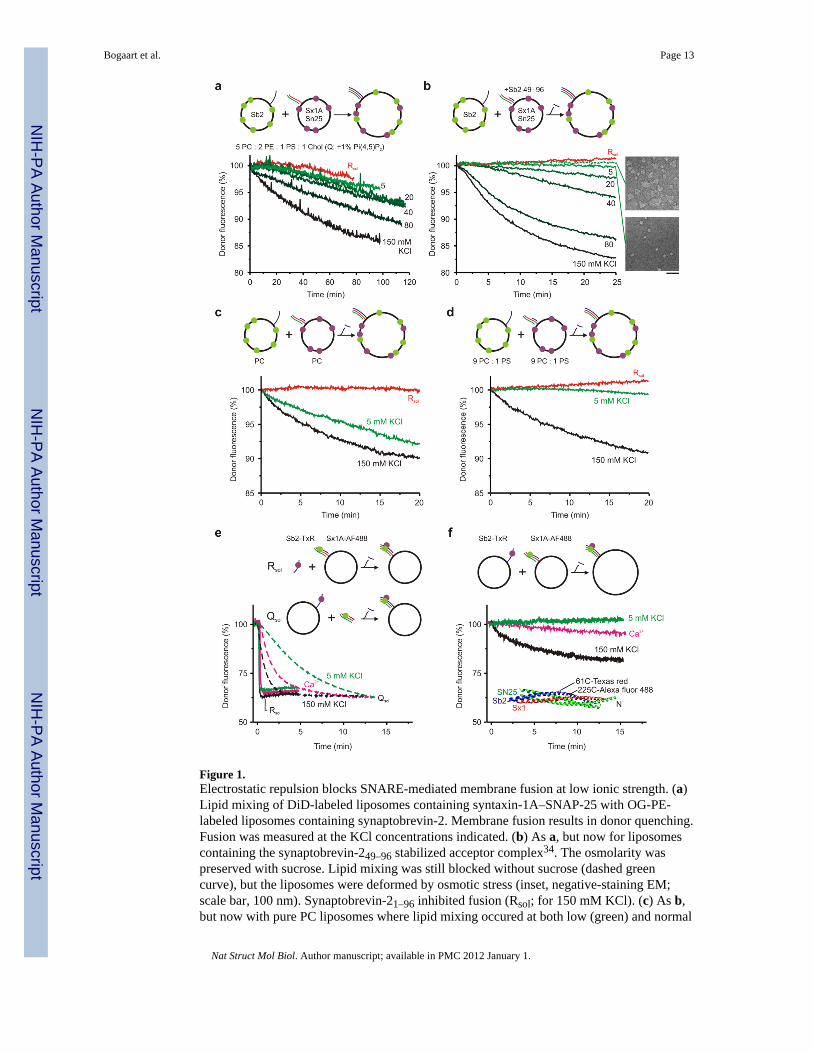
Figure 1.Electrostatic repulsion blocks SNARE-mediated membrane fusion at low ionic strength. (a)Lipid mixing of DiD-labeled liposomes containing syntaxin-1A–SNAP-25 with OG-PE-labeled liposomes containing synaptobrevin-2. Membrane fusion results in donor quenching.Fusion was measured at the KCl concentrations indicated. (b) As a, but now for liposomescontaining the synaptobrevin-249–96 stabilized acceptor complex34. The osmolarity waspreserved with sucrose. Lipid mixing was still blocked without sucrose (dashed greencurve), but the liposomes were deformed by osmotic stress (inset, negative-staining EM;scale bar, 100 nm). Synaptobrevin-21–96 inhibited fusion (Rsol; for 150 mM KCl). (c) As b,but now with pure PC liposomes where lipid mixing occured at both low (green) and normal
Bogaart et al. Page 13
Nat Struct Mol Biol. Author manuscript; available in PMC 2012 January 1.
NIH
-PA Author Manuscript
NIH
-PA Author Manuscript
NIH
-PA Author Manuscript

(black) ionic strength. (d) 10% anionic PS blocked fusion at low, but not normal, ionicstrength. (e) SNARE complex formation by FRET35. The soluble SNARE-domain ofsynaptobrevin-2 (100 nM; Rsol; solid curves) or syntaxin-1A with SNAP-25 (Qsol; dashed)resulted in complex formation regardless of ionic strength. (f) In contrast, mixing liposomescontaining membrane-anchored synaptobrevin-2 and syntaxin-1A resulted in complexformation only at normal but not at low ionic strength regardless of 1 mM Ca2+ (pink). Thestructure58 shows the dye positions. The FRET efficiencies are underestimated because the~20% crosstalk is not accounted for. Typical curves of several repeats are shown.Experiments were performed with 4–8 nM liposomes at 20°C.
Bogaart et al. Page 14
Nat Struct Mol Biol. Author manuscript; available in PMC 2012 January 1.
NIH
-PA Author Manuscript
NIH
-PA Author Manuscript
NIH
-PA Author Manuscript
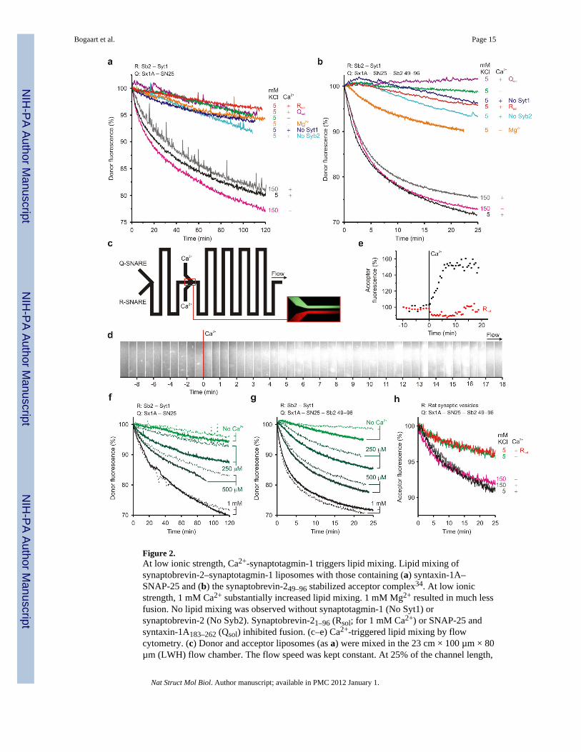
Figure 2.At low ionic strength, Ca2+-synaptotagmin-1 triggers lipid mixing. Lipid mixing ofsynaptobrevin-2–synaptotagmin-1 liposomes with those containing (a) syntaxin-1A–SNAP-25 and (b) the synaptobrevin-249–96 stabilized acceptor complex34. At low ionicstrength, 1 mM Ca2+ substantially increased lipid mixing. 1 mM Mg2+ resulted in much lessfusion. No lipid mixing was observed without synaptotagmin-1 (No Syt1) orsynaptobrevin-2 (No Syb2). Synaptobrevin-21–96 (Rsol; for 1 mM Ca2+) or SNAP-25 andsyntaxin-1A183–262 (Qsol) inhibited fusion. (c–e) Ca2+-triggered lipid mixing by flowcytometry. (c) Donor and acceptor liposomes (as a) were mixed in the 23 cm × 100 µm × 80µm (LWH) flow chamber. The flow speed was kept constant. At 25% of the channel length,
Bogaart et al. Page 15
Nat Struct Mol Biol. Author manuscript; available in PMC 2012 January 1.
NIH
-PA Author Manuscript
NIH
-PA Author Manuscript
NIH
-PA Author Manuscript

Ca2+ was introduced by fast focused mixing. (d) Acceptor fluorescence along the channelwith fluorescence microscopy and (e) binning of the fluorescence. Ca2+ specific lipidmixing was observed. (f) Lipid mixing at low ionic strength as a at the Ca2+ concentrationsindicated, with (solid) or without (dashed) 0.5 mM Mg2+. (g) As f, but now with thesynaptobrevin-249–96 stabilized acceptor complex. (h) Fluorescence dequenchingexperiment showing Ca2+ dependent lipid mixing of synaptobrevin-249–96 stabilizedacceptor complex liposomes with purified rat synaptic vesicles37. Experiments wereperformed with 4 nM liposomes at 20°C. Typical experiments of several repeats are shown.
Bogaart et al. Page 16
Nat Struct Mol Biol. Author manuscript; available in PMC 2012 January 1.
NIH
-PA Author Manuscript
NIH
-PA Author Manuscript
NIH
-PA Author Manuscript

Figure 3.Ca2+-synaptotagmin-1 triggers full membrane fusion at low ionic strength. (a) Contentmixing24 with a self-quenching concentration of calcein encapsulated in liposomes with thestabilized Q-SNARE complex. Fusion with empty R-SNARE liposomes resulted in specificfluorescence dequenching. Synaptobrevin-21–96 (Rsol) or absence of Ca2+ orsynaptotagmin-1 (No Syt1) abolished fusion. (b) Ca2+-dependent SNARE complexformation by FRET as 1f with membrane-anchored synaptobrevin-2–synaptotagmin-1. (c–e)SNARE complex formation with FRET. Liposomes containing the stabilized Q-SNAREcomplex were incubated with synaptobrevin-21–116 liposomes without Ca2+. Thesynaptobrevin-2 liposomes contained (c) no, (d) wild-type synaptotagmin-1 or (e) the
Bogaart et al. Page 17
Nat Struct Mol Biol. Author manuscript; available in PMC 2012 January 1.
NIH
-PA Author Manuscript
NIH
-PA Author Manuscript
NIH
-PA Author Manuscript

KAKA mutant. At the indicated times synaptobrevin-21–96 was added, either unlabeled (redcurve; in case of Texas-red-labeled synaptobrevin-21–116) or Texas-red-labeled (blue; incase of unlabeled synaptobrevin-21–116). The black curve shows the control withoutsynaptobrevin-21–96. Membrane fusion was triggered with Ca2+. Synaptobrevin-21–96 boundto the Q-SNAREs regardless of the presence of synaptobrevin-21–116–synaptotagmin-1liposomes. (f) Lipid mixing by Ca2+-synaptotagmin-1 wild-type (AB) and Ca2+-bindingdisruption mutants to C2B (Ab*), C2A (a*B), or both (a*b*). (g) SNARE complexformation by FRET for the synaptotagmin-1 mutants paralleled lipid mixing. (h) Lipidmixing of synaptobrevin-2–synaptotagmin-1 liposomes with 10–20 µm-sized giantunilamellar vesicles (GUVs) containing the Q-SNAREs (inset, fluorescence microscopy;scale bar, 50 µm). Disrupting Ca2+-binding to the C2B-domain abolished fusion with GUVs.~4 nM liposomes were used at 20°C. Typical curves of several repeats are shown.
Bogaart et al. Page 18
Nat Struct Mol Biol. Author manuscript; available in PMC 2012 January 1.
NIH
-PA Author Manuscript
NIH
-PA Author Manuscript
NIH
-PA Author Manuscript
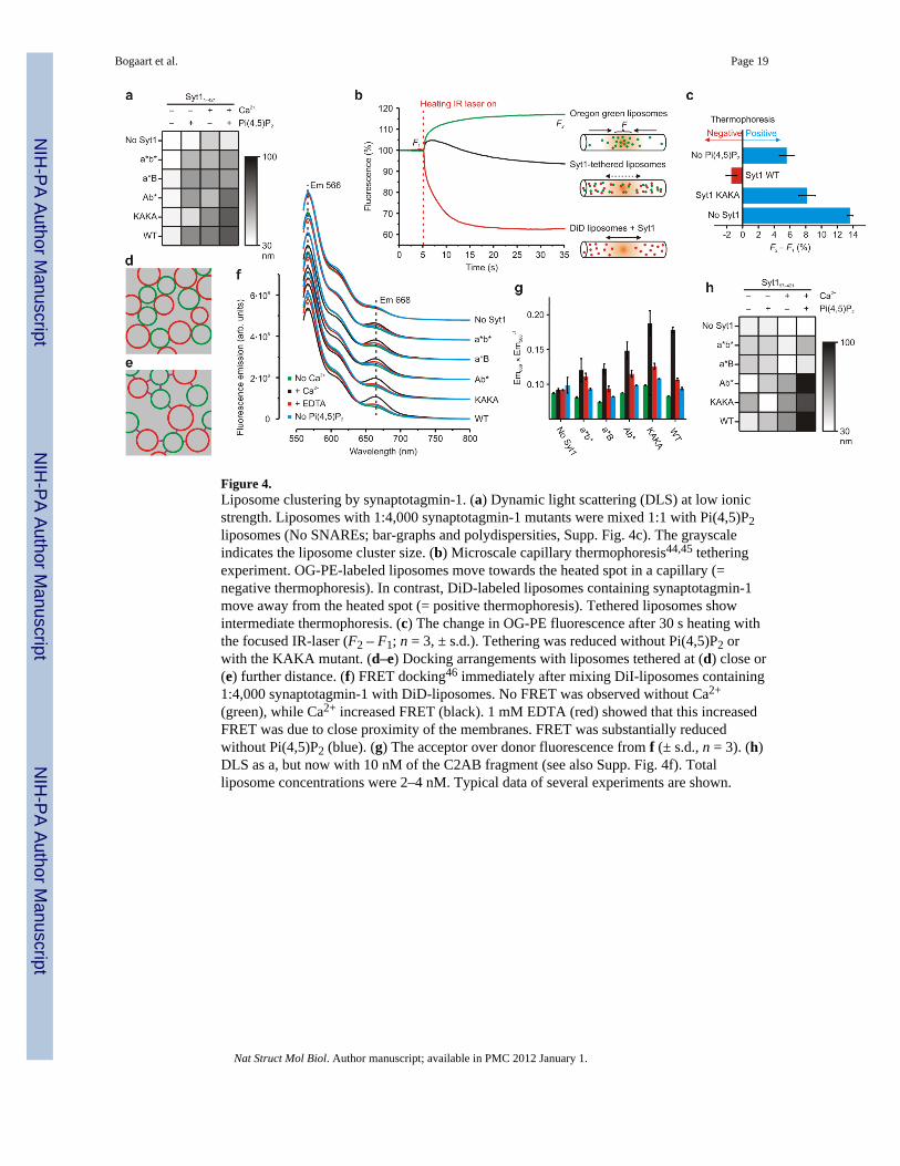
Figure 4.Liposome clustering by synaptotagmin-1. (a) Dynamic light scattering (DLS) at low ionicstrength. Liposomes with 1:4,000 synaptotagmin-1 mutants were mixed 1:1 with Pi(4,5)P2liposomes (No SNAREs; bar-graphs and polydispersities, Supp. Fig. 4c). The grayscaleindicates the liposome cluster size. (b) Microscale capillary thermophoresis44,45 tetheringexperiment. OG-PE-labeled liposomes move towards the heated spot in a capillary (=negative thermophoresis). In contrast, DiD-labeled liposomes containing synaptotagmin-1move away from the heated spot (= positive thermophoresis). Tethered liposomes showintermediate thermophoresis. (c) The change in OG-PE fluorescence after 30 s heating withthe focused IR-laser (F2 – F1; n = 3, ± s.d.). Tethering was reduced without Pi(4,5)P2 orwith the KAKA mutant. (d–e) Docking arrangements with liposomes tethered at (d) close or(e) further distance. (f) FRET docking46 immediately after mixing DiI-liposomes containing1:4,000 synaptotagmin-1 with DiD-liposomes. No FRET was observed without Ca2+
(green), while Ca2+ increased FRET (black). 1 mM EDTA (red) showed that this increasedFRET was due to close proximity of the membranes. FRET was substantially reducedwithout Pi(4,5)P2 (blue). (g) The acceptor over donor fluorescence from f (± s.d., n = 3). (h)DLS as a, but now with 10 nM of the C2AB fragment (see also Supp. Fig. 4f). Totalliposome concentrations were 2–4 nM. Typical data of several experiments are shown.
Bogaart et al. Page 19
Nat Struct Mol Biol. Author manuscript; available in PMC 2012 January 1.
NIH
-PA Author Manuscript
NIH
-PA Author Manuscript
NIH
-PA Author Manuscript

Figure 5.Preclustering of liposomes accelerates lipid mixing. (a) 150 pM or 2 nM of R-SNAREliposomes were preincubated for 20 min with the same amounts of Q-SNARE liposomes.Subsequently, lipid mixing was triggered by addition of Ca2+. For the high (4 nM total)liposome concentrations, the lipid mixing efficiency of wild-type synaptotagmin-1 (blackcurves) was comparable to that of the KAKA mutant (blue curves). In contrast, for the lowliposome concentrations (300 pM), the fusion efficiency was substantially reduced for theKAKA mutant compared to wild-type. (b) Lipid mixing of full-length synaptotagmin-1–synaptobrevin-2 liposomes with liposomes containing the Q-SNAREs of constitutiveexocytosis (syntaxin-4 and SNAP-23). No fusion was observed in absence of Ca2+ (NoCa2+) and fusion could be blocked with synaptobrevin-21–96 (Rsol). All fusion experimentswere performed at 20°C. Typical curves of multiple repeats are shown.
Bogaart et al. Page 20
Nat Struct Mol Biol. Author manuscript; available in PMC 2012 January 1.
NIH
-PA Author Manuscript
NIH
-PA Author Manuscript
NIH
-PA Author Manuscript
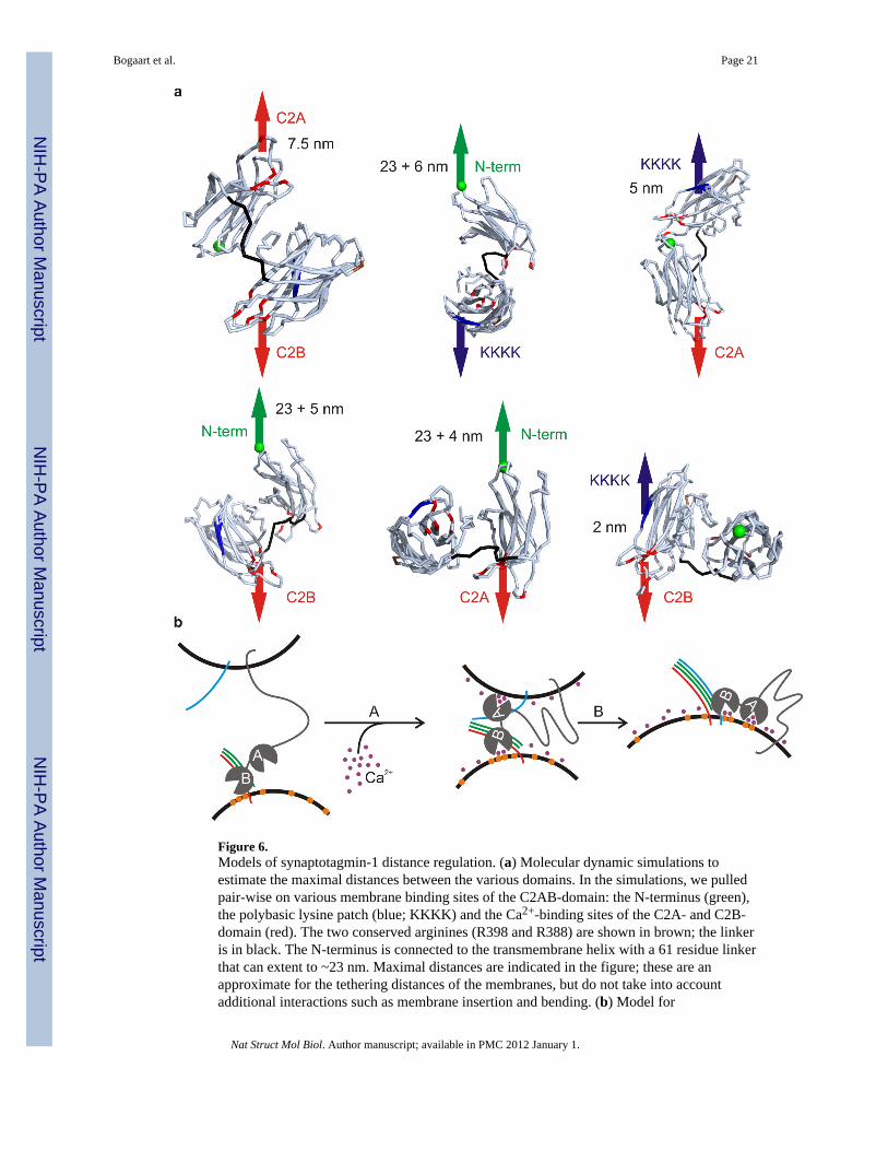
Figure 6.Models of synaptotagmin-1 distance regulation. (a) Molecular dynamic simulations toestimate the maximal distances between the various domains. In the simulations, we pulledpair-wise on various membrane binding sites of the C2AB-domain: the N-terminus (green),the polybasic lysine patch (blue; KKKK) and the Ca2+-binding sites of the C2A- and C2B-domain (red). The two conserved arginines (R398 and R388) are shown in brown; the linkeris in black. The N-terminus is connected to the transmembrane helix with a 61 residue linkerthat can extent to ~23 nm. Maximal distances are indicated in the figure; these are anapproximate for the tethering distances of the membranes, but do not take into accountadditional interactions such as membrane insertion and bending. (b) Model for
Bogaart et al. Page 21
Nat Struct Mol Biol. Author manuscript; available in PMC 2012 January 1.
NIH
-PA Author Manuscript
NIH
-PA Author Manuscript
NIH
-PA Author Manuscript
synaptotagmin-1 mediated lipid mixing. In the absence of Ca2+, synaptotagmin-1 (grey)tethers to anionic membranes, and particularly to Pi(4,5)P2 (orange) via its polybasic lysinepatch. The distance is too far for SNARE formation to occur (synaptobrevin-2: blue;Syntaxin-1A: red; SNAP-25: green). (Step A) In the presence of Ca2+ (purple), a transitionalconformation change occurs and synaptotagmin-1 binds the membrane via its calciumbinding pockets and basic residues on the C2AB-domain9. This drives the membranestogether and SNARE complex formation can occur. (Step B) The SNARE complexformation drives membrane fusion.
Bogaart et al. Page 22
Nat Struct Mol Biol. Author manuscript; available in PMC 2012 January 1.
NIH
-PA Author Manuscript
NIH
-PA Author Manuscript
NIH
-PA Author Manuscript
Copyright © 2022 FDOKUMEN