Symmetry Recognizing Asymmetry
12
Structure, Vol. 11, 411–422, April, 2003, 2003 Elsevier Science Ltd. All rights reserved. DOI 10.1016/S0969-2126(03)00047-9 Symmetry Recognizing Asymmetry: Analysis of the Interactions between the C-Type Lectin-like Immunoreceptor NKG2D and MHC Class I-like Ligands and tumors evade T cell surveillance by simply downreg- ulating MHC class I expression [6]. NK cells in the periph- ery stochastically express a subset of activating and inhibitory MHC class I-specific NCRs, selected so that normal MHC expression prevents NK cell activation, Benjamin J. McFarland, 1 Tanja Kortemme, 2 Shuyuarn F. Yu, 1 David Baker, 2 and Roland K. Strong 1, * 1 The Division of Basic Sciences Fred Hutchinson Cancer Research Center 1100 Fairview Avenue North Seattle, Washington 98109 whereas loss of any MHC class I allele tilts the NCR signal toward activation, providing a back-up for T cell 2 Howard Hughes Medical Institute and Department of Biochemistry surveillance [5]. However, some viruses encode decoy ligands for inhibitory NCRs that circumvent this system [7]. University of Washington J Wing NKG2D is a homodimeric, activating NKD-type NCR distantly related to other members of the NKG2 family Health Sciences Building Seattle, Washington 98195 (20%–30% identical), which otherwise normally assem- ble as heterodimers with CD94 [8–10]. NKG2D was origi- nally identified on NK cells but has subsequently been found broadly expressed on macrophages, , and Summary CD8 T cells. Rather than binding true MHC class I proteins, NKG2D ligands include the MIC (-A and -B, Engagement of diverse protein ligands (MIC-A/B, which are 84% identical overall) and ULBP (1, 2, and ULBP, Rae-1, or H60) by NKG2D immunoreceptors me- 3; 55%–60% identical pairwise) proteins in primates or diates elimination of tumorigenic or virally infected H60 and the retinoic acid-inducible Rae-1 family of pro- cells by natural killer and T cells. Three previous teins (, , , and ; 92% identical pairwise) in rodents. NKG2D-ligand complex structures show the homodi- Whereas murine (muNKG2D) and human NKG2D meric receptor interacting with the monomeric ligands (huNKG2D) are 69% identical in their ectodomains, their in similar 2:1 complexes, with an equivalent surface ligands are quite dissimilar in sequence, with overall on each NKG2D monomer binding intimately to a total pairwise sequence identities from 23% to 27%. of six distinct ligand surfaces. Here, the crystal struc- All currently characterized NKG2D ligands are distant ture of free human NKG2D and in silico and in vitro structural homologs of MHC class I proteins [11–13]. alanine-scanning mutagenesis analyses of the com- However, unlike true MHC class I proteins, the NKG2D plex interfaces indicate that NKG2D recognition de- ligands bind neither antigenic peptides (or any other generacy is not explained by a classical induced-fit small molecule ligand) nor 2 -microglobulin, and ULBP3 mechanism. Rather, the divergent ligands appear to and Rae-1 even dispense with the 3 domain, existing utilize different strategies to interact with structurally as isolated 12 platform domains membrane anchored conserved elements of the consensus NKG2D binding by GPI linkages. NKG2D-ligand interactions are also site. tighter (K d s in the 1 to 0.01 M range) than other NCR- or TCR-ligand complexes (K d s in the 10 to 100 M range) Introduction [10, 12]. Also unlike MHC class I proteins, which are constitutively expressed on almost all cell types, many Natural killer (NK) cells mediate early immune system NKG2D ligands are expressed conditionally and only responses against cells undergoing neoplastic transfor- by certain cells [10]. For example, MIC-A expression is mation or infection by viruses or intracellular parasites induced by cellular stress on gastrointestinal epithelium [1]. They function through a diverse array of cell surface and epithelially derived tumors. NKG2D engagement of receptors that can be divided into two families by struc- MIC ligands dominantly activates effector responses tural homology: NK receptors (NCRs) with immunoglob- from NK cells and T cells, and may costimulate CD8 ulin-like ectodomains (such as KIRs, LIRs, and NKp46), T cell responses. In mice, Rae-1 or H60 expression or C-type lectin-like ectodomains (NKDs; including the drives NK-mediated tumor rejection. NKG2D-ligand in- NKG2x/CD94 family, the Ly49x family, and NKR-P1) [2, teractions therefore mediate crucial antiviral and antitu- 3]. NK cell activation occurs through integration of the mor innate immune responses in response to low-level activating and inhibitory signals across the constellation signals like cellular stress. of NCRs engaged upon interrogation of target cells [4, 5]. Crystal structures are now available for muNKG2D Many NCRs recognize classical (HLA-A, -B, and -C) [14] and huNKG2D (reported here); MIC-A [11], MIC-B and nonclassical (HLA-E) major histocompatibility com- [15], and Rae-1 [13]; and for the complexes between plex (MHC) class I proteins and occur in paired activating huNKG2D and MIC-A [16], huNKG2D and ULBP3 [12], and inhibitory isoforms. MHC molecules bind peptides and muNKG2D and Rae-1 [13]. The symmetric NKG2D derived from endogenous proteins and then traffic to homodimers bind their asymmetric, monomeric ligands the cell surface, providing a means for T cells to monitor in a 2:1 molar stoichiometry. Equivalent binding sites on the proteome of a given cell for pathogen- or tumor- associated protein expression. However, many viruses Key words: immunoreceptors; MHC class I homologs; structural immunology; molecular recognition; computational alanine scan- ning; interface plasticity *Correspondence: [email protected]
Transcript of Symmetry Recognizing Asymmetry

Structure, Vol. 11, 411–422, April, 2003, 2003 Elsevier Science Ltd. All rights reserved. DOI 10.1016/S0969-2126(03)00047-9
Symmetry Recognizing Asymmetry: Analysisof the Interactions between the C-Type Lectin-likeImmunoreceptor NKG2D and MHC Class I-like Ligands
and tumors evade T cell surveillance by simply downreg-ulating MHC class I expression [6]. NK cells in the periph-ery stochastically express a subset of activating andinhibitory MHC class I-specific NCRs, selected so thatnormal MHC expression prevents NK cell activation,
Benjamin J. McFarland,1 Tanja Kortemme,2
Shuyuarn F. Yu,1 David Baker,2 and Roland K. Strong1,*1The Division of Basic SciencesFred Hutchinson Cancer Research Center1100 Fairview Avenue NorthSeattle, Washington 98109 whereas loss of any MHC class I allele tilts the NCR
signal toward activation, providing a back-up for T cell2 Howard Hughes Medical Institute andDepartment of Biochemistry surveillance [5]. However, some viruses encode decoy
ligands for inhibitory NCRs that circumvent this system [7].University of WashingtonJ Wing NKG2D is a homodimeric, activating NKD-type NCR
distantly related to other members of the NKG2 familyHealth Sciences BuildingSeattle, Washington 98195 (20%–30% identical), which otherwise normally assem-
ble as heterodimers with CD94 [8–10]. NKG2D was origi-nally identified on NK cells but has subsequently beenfound broadly expressed on macrophages, ��, andSummaryCD8� �� T cells. Rather than binding true MHC class Iproteins, NKG2D ligands include the MIC (-A and -B,Engagement of diverse protein ligands (MIC-A/B,which are �84% identical overall) and ULBP (1, 2, andULBP, Rae-1, or H60) by NKG2D immunoreceptors me-3; 55%–60% identical pairwise) proteins in primates ordiates elimination of tumorigenic or virally infectedH60 and the retinoic acid-inducible Rae-1 family of pro-cells by natural killer and T cells. Three previousteins (�, �, �, and �; �92% identical pairwise) in rodents.NKG2D-ligand complex structures show the homodi-Whereas murine (muNKG2D) and human NKG2Dmeric receptor interacting with the monomeric ligands(huNKG2D) are 69% identical in their ectodomains, theirin similar 2:1 complexes, with an equivalent surfaceligands are quite dissimilar in sequence, with overallon each NKG2D monomer binding intimately to a totalpairwise sequence identities from 23% to 27%.of six distinct ligand surfaces. Here, the crystal struc-
All currently characterized NKG2D ligands are distantture of free human NKG2D and in silico and in vitrostructural homologs of MHC class I proteins [11–13].alanine-scanning mutagenesis analyses of the com-However, unlike true MHC class I proteins, the NKG2Dplex interfaces indicate that NKG2D recognition de-ligands bind neither antigenic peptides (or any othergeneracy is not explained by a classical induced-fitsmall molecule ligand) nor �2-microglobulin, and ULBP3mechanism. Rather, the divergent ligands appear toand Rae-1� even dispense with the �3 domain, existingutilize different strategies to interact with structurallyas isolated �1�2 platform domains membrane anchoredconserved elements of the consensus NKG2D bindingby GPI linkages. NKG2D-ligand interactions are alsosite.tighter (Kds in the 1 to 0.01 �M range) than other NCR-or TCR-ligand complexes (Kds in the 10 to 100 �M range)
Introduction [10, 12]. Also unlike MHC class I proteins, which areconstitutively expressed on almost all cell types, many
Natural killer (NK) cells mediate early immune system NKG2D ligands are expressed conditionally and onlyresponses against cells undergoing neoplastic transfor- by certain cells [10]. For example, MIC-A expression ismation or infection by viruses or intracellular parasites induced by cellular stress on gastrointestinal epithelium[1]. They function through a diverse array of cell surface and epithelially derived tumors. NKG2D engagement ofreceptors that can be divided into two families by struc- MIC ligands dominantly activates effector responsestural homology: NK receptors (NCRs) with immunoglob- from NK cells and �� T cells, and may costimulate CD8�
ulin-like ectodomains (such as KIRs, LIRs, and NKp46),�� T cell responses. In mice, Rae-1 or H60 expression
or C-type lectin-like ectodomains (NKDs; including the drives NK-mediated tumor rejection. NKG2D-ligand in-NKG2x/CD94 family, the Ly49x family, and NKR-P1) [2, teractions therefore mediate crucial antiviral and antitu-3]. NK cell activation occurs through integration of the mor innate immune responses in response to low-levelactivating and inhibitory signals across the constellation signals like cellular stress.of NCRs engaged upon interrogation of target cells [4, 5]. Crystal structures are now available for muNKG2D
Many NCRs recognize classical (HLA-A, -B, and -C) [14] and huNKG2D (reported here); MIC-A [11], MIC-Band nonclassical (HLA-E) major histocompatibility com- [15], and Rae-1� [13]; and for the complexes betweenplex (MHC) class I proteins and occur in paired activating huNKG2D and MIC-A [16], huNKG2D and ULBP3 [12],and inhibitory isoforms. MHC molecules bind peptides and muNKG2D and Rae-1� [13]. The symmetric NKG2Dderived from endogenous proteins and then traffic to homodimers bind their asymmetric, monomeric ligandsthe cell surface, providing a means for T cells to monitor in a 2:1 molar stoichiometry. Equivalent binding sites onthe proteome of a given cell for pathogen- or tumor-associated protein expression. However, many viruses
Key words: immunoreceptors; MHC class I homologs; structuralimmunology; molecular recognition; computational alanine scan-ning; interface plasticity*Correspondence: [email protected]

Structure412
Figure 1. Structures of NKG2D-Ligand Complexes
Top: views of three NKG2D-ligand complex structures are shown, with each complex shown in paired views; one view of the side of thecomplex (above), with the protein backbones shown in a ribbon representation; the other view (below), looking down onto the top of the

Interface Analysis of NKG2D-Ligand Complexes413
each NKG2D monomer contribute nearly equally to an tion types not dominated by hydrophobic terms: onlyextensive interface where each receptor monomer binds 55%–62% of the solvent-accessible surface area burieda distinct ligand surface (Figure 1). All three NKG2D in the complexes is nonpolar (Figures 1, 2, and 3). Incomplexes are quite similar overall, despite the dissimi- contrast, many other protein-protein interactions arelarity in detail between the structures of the ligand pro- largely mediated through highly adaptive hydrophobicteins: ligand pairwise root-mean-square deviations surfaces [17]. For NKG2D, electrostatic interactions(rmsd) range from 3.8 to 5.8 A (calculated on fairly re- contribute but do not dominate (with the exception ofstricted C� sets due to the high degree of mismatch). the muNKG2D-H60 complex) [12, 18] as they do for theThe saddle-shaped NKG2D homodimer sits astride the KIR complexes [19, 20]. Moreover, the residue pairsplatform domain of the MHC class I-like ligands, with forming salt bridges in the complexes are variable (Fig-each NKG2D monomer primarily contacting either the �1 ures 2 and 3). Beyond the salt bridges and similarlyor �2 subdomain of each ligand. Each NKG2D monomer- variable hydrogen bonds and van der Waals interactionsligand subdomain (�1 or �2) pair constitutes a “half- (Figures 2 and 3), three common residue positions atsite” in our terminology. The footprints of the ligands the center of each half-site do make direct contacts withon each of the six NKG2D half-sites essentially overlap ligand in all six half-sites (binding site “core” residues):(Figure 2), showing that NKG2D truly utilizes a single Tyr152, Met184, and Tyr199 (Tyr168, Ile200, and Tyr215binding site consisting of residues from the body of the in muNKG2D). Both tyrosine side chains point towardNKG2D NKD and one loop (�5�-�5). This loop, referred
the ligand in all six half-site structures, with their centersto as the “stirrup” loop [16], is the most distal element
separated by about 6 or 7 A, depending upon Tyr152of NKG2D that contacts ligand. The single NKG2D bind-rotamer utilization (see below). In addition to other con-ing site has therefore evolved to recognize at least sixtacts, the tyrosine side chains generally sandwich thedifferent surfaces, predominantly on the �1 or �2 do-side chain of another residue, but the range of residuemains of MIC-A, ULBP3, and Rae-1�, with dramaticallytypes, and the nature of the contacts in the sandwich,different shapes (Figure 1). Additionally, many of theis quite diverse: a methionine in the �1 MIC-A and ULBP3very nonconservative sequence differences and dele-�2 half-sites (the latter methionine displacing the par-tions between MIC-A and MIC-B alleles and ULBP3 andtially sandwiched Met184 from huNKG2D as in the otherRae-1 isoforms map to NKG2D-contacting residuesthree huNKG2D complex half-sites), a leucine in the(Figure 3) [12, 13, 15].ULBP3 �1 half-site, and either an arginine, making cat-Two conceptually different solutions to this bindingion- contacts to both tyrosines, or a phenylalanine,problem can be envisioned: first, extensive plasticitymaking both en face and herringbone contacts, in theallows the receptor to rearrange its binding site ac-two muNKG2D half-sites.cording to the requirements of the ligand (induced-fit).
It has been proposed [12, 21] that NKG2D bindingSecond, divergent ligands may utilize different strate-degeneracy can be explained through a classical in-gies to recognize an essentially identical receptor bind-duced-fit mechanism, a term first coined to describeing site. In order to investigate the recognition mecha-
nism that allows for such extreme ligand degeneracy the molding of a flexible, malleable enzyme binding sitewhile maintaining relatively high affinities and selectivi- into the complement of its cognate substrate concurrentties, we report here the results of (1) the crystallographic with binding [22]. The immune system utilizes classicalanalysis of huNKG2D crystallized alone at 2.5 A resolu- induced-fit receptor-ligand interactions in the interac-tion to complete the examination of potentially flexible tions between antibodies and antigens, such as the li-interface elements and (2) in silico and in vitro alanine- gand-induced changes in the antigen binding fragmentscanning mutagenesis analyses of the three available of the anti-influenza virus hemagglutinin peptide anti-complex structures to quantitate the relative contribu- body 17/9 [23], and between �� TCRs and MHC classtions different residues make to the binding interactions. I proteins [24–27]. Both examples involve dramaticThe results show that binding energy is unevenly distrib- movements of the backbone atoms of key receptor li-uted across the interfaces, with “hotspots” associated gand binding loops of 3 to 6 A and side chain movementswith structurally conserved receptor elements, thus ar- of up to 15 A at the distal atom.guing against an induced-fit recognition mechanism.
Elements of Flexibility in NKG2DResultsThe most flexible part of the receptor is the 21 residuelong N-terminal stalk of the ectodomain (huNKG2D; resi-The NKG2D-ligand interfaces are extensive, highly
shape complementary, and involve a mixture of interac- dues 75–95) between the NKD and the membrane-span-
ligand from the perspective of the receptor-bearing cell, with the receptor represented as a backbone ribbon and the ligand as a CPK model.Secondary structure elements are portrayed as � strands, arrows; � helices, coils in the ribbon representations. Proteins are colored bydomain: MHC class I-like ligands are colored as �1, yellow; �2, orange; and �3 (when present), red; the receptor domain over the ligand �1domain is colored blue and the domain over the �2 domain is colored purple. Arrows indicate the stirrup loops of NKG2D in the variouscomplexes. Buried solvent-accessible surface areas (A2 ) and shape complementarity (Sc) values are also shown. Figures were generated withSwissPDB-Viewer [47] and rendered with POV-RAY3 (http://mac.povray.org).Bottom: in order to generate schematic representations of the binding interfaces, complexes are split open, with the domains oriented lookingdown onto the contact surfaces. The proteins are then outlined and the contact surface is displayed as a colored patch. On the right, CPKrepresentations of superpositions (based on the NKG2D monomer from each half-site) of the contact residues from all of the three complexstructures are shown, with NKG2D surfaces in the upper frames and ligand surfaces shown in the lower frames as indicated. This demonstratesthe relative structural conservation of atoms in the receptor binding sites and the structural diversity of atoms comprising the receptor-contacting residues in the ligands.
Structure414
Figure 2. Schematized Contact Maps of NKG2D-Ligand Complexes
Top: contact residues have been mapped onto schematized representations of the three complex structure interfaces. Binding surfaces aredisplayed as colored patches. Each residue is labeled, and its corresponding tag (squares for ligand residues, circles for receptor residues)is colored by the type of interaction it makes with its cognate contact on the opposite surface, as indicated. Receptors are shown across thetop and ligands across the bottom of the frame. Ligand-contacting residues in the stirrup loops of the receptors are represented by crosshatched

Interface Analysis of NKG2D-Ligand Complexes415
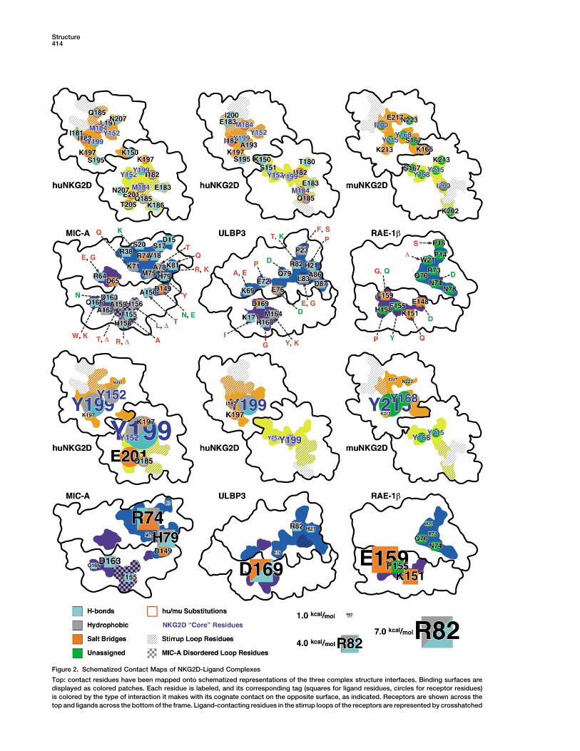
Figure 3. Tabulation of NKG2D-Ligand Con-tacts
Residue-residue contacts have been tabu-lated from the original structure references[12, 13, 16], with ligand residues colored bythe type of interaction observed as in Figure2. Receptor murine/human sequence differ-ences are colored red.
ning domain (NKG2D is a type II transmembrane pro- [14] and huNKG2D (see below) show the two monomersrelated by perfect, crystallographic dyad axes. However,tein). Although the various crystallization constructs
encompass most, if not all, of this region, at most only the exact 2-fold symmetry of the NKG2D homodimer isbroken in all three complex structures, with deviationsabout a quarter, and typically only a few residues, of
the stalk is ordered. However, while extremely flexible, of up to nearly 5 in directions both parallel and perpen-dicular to the homodimer dyad axis (Figure 4). Thesethe stalks cannot contribute to induced-fit recognition
because they are distal to the ligand binding sites. deviations accommodate the ridge, or peak, at the kinkin the ligand �2-domain helix and allow the NKG2D ho-A large degree of flexibility is also displayed by NKG2D
at the homodimer interface. Structures of free muNKG2D modimer to close over the ligand. However, these mo-
areas, and MIC-A residues in the disordered loop are represented by a checkerboard area. The conserved positions of the NKG2Dbindingsite core residues are labeled in blue, and ligand-contacting residues in the receptors that vary in sequence between the human and murineproteins have tags highlighted with red borders. Sequence substitutions between ligand loci/alleles/isoforms are indicated by dashed arrows,with conservative substitutions labeled in green and nonconservative substitutions or deletions labeled in red.Bottom: the complex interfaces are represented as above, except that the size of each residue label has been scaled by the energeticcontribution the corresponding residue makes to the interaction; interactions with values less than 1.0 kcal/mol are not considered to constitutebinding site hotspots and are not shown. NKG2D hotspots are predicted to lie within 7.4 to 1.1 kcal/mol when complexed with MIC-A; 4.6 to1.3 kcal/mol when complexed with ULBP3; and 5.2 to 1.1 kcal/mol when complexed with RAE-1�. Ligand hotspots are predicted to lie within5.0 to 1.0 kcal/mol for MIC-A; 5.2 to 1.0 kcal/mol for ULBP3; and 5.9 to 1.3 kcal/mol for RAE-1�.
Structure416
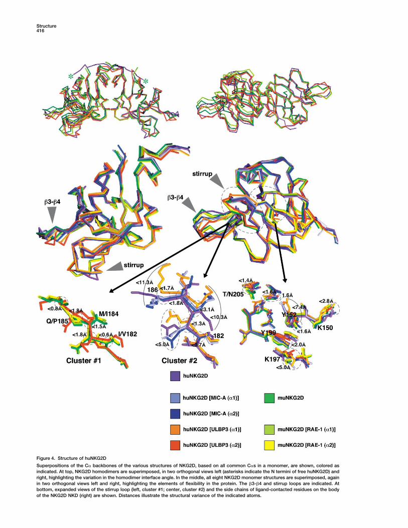
Figure 4. Structure of huNKG2D
Superpositions of the C� backbones of the various structures of NKG2D, based on all common C�s in a monomer, are shown, colored asindicated. At top, NKG2D homodimers are superimposed, in two orthogonal views left (asterisks indicate the N termini of free huNKG2D) andright, highlighting the variation in the homodimer interface angle. In the middle, all eight NKG2D monomer structures are superimposed, againin two orthogonal views left and right, highlighting the elements of flexibility in the protein. The �3-�4 and stirrup loops are indicated. Atbottom, expanded views of the stirrup loop (left, cluster #1; center, cluster #2) and the side chains of ligand-contacted residues on the bodyof the NKG2D NKD (right) are shown. Distances illustrate the structural variance of the indicated atoms.

Interface Analysis of NKG2D-Ligand Complexes417
tions also do not contribute to an induced-fit mechanism Table 1. Crystallographic Data Collection and RefinementStatisticsin that they do not differentially alter the ligand-con-
tacting surface on the receptor by inducing a more li- Space group P41212gand-complementary shape on the NKG2D binding site, Cell dimensions (A) a � b � 87.65, c � 36.13but rather simply position each NKG2D binding half-site Data Collection and Processing
Wavelength Cu K�over the appropriate cognate ligand surface.Resolution (A) 2.50 (2.59–2.50)Several loops in the structure show above average BUnique reflections 5057 (466)factors and/or multiple conformations between struc-Redundancy 21.1
tures, implying flexibility (Figure 4). Corresponding C� Completeness (%) 96.5 (90.1)atoms of the �3-�4 loop (residues 160–165 in huNKG2D, �I/ (I)� 20.2 (6.8)and 176–181 in muNKG2D) differ by 2 to 6 A and have Rsym 0.066 (0.199)
Refinementabove average B factors, but this loop does not contactResolution (A) 20–2.50 (2.59–2.50)ligand. However, the �5-�5� stirrup loop (residues 182–Reflections (all F � 0) 4838188 in huNKG2D, and 198–204 in muNKG2D) does makeProtein atoms 1030
multiple contacts to the ligand in all six half-sites. The Solvent atoms 104backbone of this loop essentially adopts one of two Phosphate atoms 5conformations among the eight NKG2D models (Figure Rcryst (%) 24.5
Rfree (%) (on 498 reflections) 29.64): one cluster (#1) contains all the muNKG2D structuresAverage B factor (A2 ) 39.4and the huNKG2D �2 monomer from the complex withCrossvalidated A coordinate error (A) 0.53ULBP3; the other cluster (#2) contains the remaining
Rmsd from Ideal GeometryhuNKG2D models. The cluster #1 conformation moves Bond length (A) 0.008the loop outward, away from the center of mass of the Bond angles () 1.9complex, with C�-to-C� movements of almost 5 or 6 A Dihedrals () 26.4
Impropers () 1.4at residues 184 and 185, creating a wider binding saddleRamachandran Statisticsthan the cluster #2 conformation. The differences in loop
Most favored (%) 78.1backbone structure do not stem from a hinged motionAdditional allowed (%) 21.1
of the ends of the loop, but have differences distributed Generously allowed (%) 0.9throughout the loop. A number of sequence differences Disallowed (%) 0between the human and murine receptors occur in loop
Numbers in parentheses indicate values for the highest resolutionresidues (Ile182/Val198, Met184/Ile200, and Gln185/ shell.Pro201; huNKG2D/muNKG2D) and the largest differ-ences between C�s within or between the two clustersoccur at some of these variable positions (almost 5 A
As a specific example, the hydrogen bond partner atomat 184/200 and over 6 A at 185/201 across both clusters).of the human/murine substituted (threonine/asparagine)These sequence differences likely affect loop conforma-residue at position 205/221 (human/murine) falls withintion: Gln185 in the huNKG2D displays φ values (�1201.4 A across all the NKG2D structures (Figure 4).to �155) outside of that allowed for prolines, as in
muNKG2D; and �-branched Ile200 in all the muNKG2DThe Structure of Free huNKG2Dmodels (and Met184 in huNKG2D-A from the ULBP3Because the stirrup loop and certain side chains of thecomplex), as in cluster #1, display � strand φ/� values,NKG2D binding site residues represent flexible elementswhile Met184 in the rest of the huNKG2D models (clusterthat may contribute to a classical induced-fit interaction,#2) displays generously allowed �-helical values. Thesewe determined the crystal structure of unligandeddifferences in loop conformation were invoked to explainhuNKG2D at 2.5 A resolution (Table 1) to analyze theirthe observation that huNKG2D does not bind Rae-1�,ground-state conformations. Diffraction data were col-whereas muNKG2D binds MIC, the result of a projectedlected with Cu-K� radiation from cryopreserved crystalsclash between huNKG2D residue Met184 and severalof a soluble, recombinant form of huNKG2D encom-Rae-1� residues in a hypothetical complex, due to thepassing nearly the entire ectodomain (residues 80–216),much more toed-in, or closed, huNKG2D stirrup loopcrystallized at a pH of 9, and phased by molecular re-conformation [13].placement. The asymmetric unit contains half a homo-Relatively smaller structural variabilities are associ-dimer.ated with the side chain conformations of some ligand-
The free huNKG2D monomer is, as expected, verycontacting residues (Figure 4). Cluster #2 stirrup loopsimilar to the other views of the NKG2D structure, withresidues Met184 and Lys186 have distal atom (C� or N�)pairwise rms deviations between 1.1 and 1.4 A on allpositions that differ by over 10 A, with other residues’common C�s. Electron density is observed for more ofdistal atoms differing by almost 5 A. Side chain structuralthe flexible N-terminal stalk than in any other structure,variation is generally more limited at the remaining clus-to Gln88. The stalk crosses over the homodimer inter-ter #2 positions and at the cluster #1 residues, and muchface, making fairly extensive contacts to the NKD of themore limited for ligand-contacting residues on the bodyother monomer in the homodimer and to neighboringof the NKD. For the latter residues, the largest variationsmonomers in the asymmetric unit, with the two homodi-are seen at the N� atoms of two lysines (150 and 197 inmer-related stalk N termini spanning a distance of 49 A,huNKG2D) and in alternate rotamer utilization by Tyr152,nearly the width of the whole molecule. In this most fullya binding site core residue. The remainder of the ligand-
contacting residues have well-overlapping structures. resolved view, the stalk displays no defined secondary

Structure418
structure, and the only contacts between stalks of the alanine was at least 1 kcal/mol (the average value forall interface residues across the three complexes wassame homodimer are van der Waals bonds involving
residues 97–99 near the interface between monomers in 1.2 kcal/mol, with values ranging up to 7.4 kcal/molat Tyr199 of the �1 huNKG2D monomer in the MIC-Athe homodimer. The extreme flexibility of the N-terminal
stalk, and the lack of any obvious, consistent associa- complex). Nine to seventeen hotspots are scatteredacross each interface, with receptor and ligand hotspotstions between stalks or stalk and NKD, leaves us without
an obvious structural mechanism for signaling ligand generally correlated. Hotspot distribution is clearlyasymmetric, indicating that one half-site dominates eachengagement to the interior of the cell.
In general, all other aspects of the structure of free interaction (the �1 site in the MIC-A complex, and the �2site in the ULBP3 and Rae-1� complexes; Figure 2).huNKG2D, such as the �3-�4 loop and the ligand-con-
tacting residues on the body of the NKD, fall within the A dominant proportion of the binding energy on thereceptor is invested in the two binding site core tyrosinevariation already observed among the previous struc-
tures of NKG2D (Figure 4). The conformation of the free residues at every NKG2D half-site, Tyr152/168 andTyr199/215 (hu/muNKG2D). All but one of these centralhuNKG2D stirrup loop backbone clearly falls within clus-
ter #2 (though the side chains of Met184 and Lys186 tyrosine residues in all six half-sites were classified ashotspots, with predicted changes in binding energyare somewhat divergent) and the side chain of Tyr152
adopts the more commonly observed rotamer. Slight upon alanine mutation ranging from 1.5 to 7.4 kcal/mol.The third member of the common binding site core, thedifferences between free and bound huNKG2D are ob-
served at the homodimer interface, which is more open stirrup loop residue Met184/Ile200 (hu/muNKG2D), wasnot identified as a hotspot at any half-site. The secondand packed more loosely in free huNKG2D, allowing for
slight half-site movements (toeing-in) during complex most commonly conserved receptor hotspot (four ofthe six half-sites) is Lys197/213 (hu/muNKG2D), whichformation. The only homodimer interface residue dis-
playing a conformation outside of the range observed participates in a salt bridge in three of four cases. Onlyone NKG2D stirrup loop residue in one half-site, Gln185in the other NKG2D structures is Phe113, which was
observed in two rotamers in the free huNKG2D structure: in the �1 half-site of the MIC-A complex, is identifiedas a hotspot (2.2 kcal/mol). Conversely, on the ligandone similar to the other structures and the other pointing
away from the ligand and packing into a different inter- surfaces, two hotspots contact the stirrup loop (�2 kcal/mol; Arg74 in MIC-A and Arg82 in ULBP3), but theseface pocket (lined by residues Ile104 and Gln112 of the
same monomer and Tyr106, Asp144, and Leu145 of the residues also interact, through salt bridges and/or Hbonds, with non-stirrup loop residues.dimer-related monomer).
Hotspots identified by the computational alanine-scanning analysis also correlate with ligand receptor-
Computational Alanine-Scanning Mutagenesis contacting residue conservation among alleles, loci, orBecause the NKG2D-ligand interfaces contain both isoforms (Figure 2). Of the 26 ligand residues that contactstructurally conserved and varying elements, recogniz- the NKG2D binding core residues (Tyr152/168, Met184/ing highly structurally disparate ligands, we sought a Ile200, and Tyr199/215), ten are conserved or only con-systematic method for evaluating the relative contribu- servatively substituted, with an average energetic con-tion of each element. Binding energy is often distributed tribution of 2.1 kcal/mol. The other 16 core-contactingunevenly across protein-protein interfaces, with the residues are nonconservatively substituted in at leastlargest changes in affinity occurring when so-called hot- one sequence, and are predicted to have a lower averagespot residues are mutated [28, 29]. Classically, alanine- contribution, 1.3 kcal/mol. Out of these, any residues thatscanning mutagenesis coupled to studies of binding are deleted in any sequence on average contribute least,energetics provides such information [30]. Here, we al- only 0.72 kcal/mol. The reason for the unusual polymor-ternately conduct such an analysis in silico, using a phism patterns in NKG2D ligands is unknown, but poly-method [31] validated by its use in engineering a novel morphism is not predicted to significantly affect NKG2Dprotein-protein interface [32] and by selected evaluation binding: none of the MIC allelic substitutions at NKG2Dof in vitro alanine mutations reported here. Briefly, com- contact positions have been experimentally shown toputational alanine-scanning mutagenesis uses a simple significantly affect affinity [34], and Rae-1 isoforms differphysical model to score a series of receptor-ligand inter- in NKG2D affinity only modestly, by about 2-fold [18].faces in which contact residues are individually replaced Whereas MHC class I polymorphisms are closely cou-with alanine. After each alanine mutation, side chains pled to function, directly determining peptide and TCRat the interface are repacked with favorable rotamers specificity, MIC polymorphisms have been difficult to rec-and the resulting binding energy is calculated. The oncile with the interaction with NKG2D [15], and maymodel was parameterized using results from 743 alanine reflect effects on currently uncharacterized interactionsmutagenesis experiments in monomeric proteins (data with other receptors, such as �� TCRs [35].taken from the PROTHERM database [33]), and tested Site-directed mutagenesis of MIC-A followed by sur-against a further 223 alanine-scanning mutations in 19 face plasmon resonance (SPR) biointeraction analysisprotein-protein complexes, with an average unsigned provides additional validation of the computational ala-error of 1.09 kcal/mol [31]. nine-scanning method (Table 2). MIC-A mutants, with
Computational alanine-scanning analyses were con- residues at the binding interface (selected to span theducted on the three NKG2D-ligand interfaces (Figure 2). range of interaction types and predicted strengths) indi-A residue was defined as a binding hotspot if the re- vidually mutated to alanine, were immobilized on SPR
sensor chips. Measured huNKG2D equilibrium SPR re-sulting calculated difference in ��G when mutated to

Interface Analysis of NKG2D-Ligand Complexes419
Table 2. Comparison of In Vitro and In Silico Free Energy Values
Kd Std. Std. �G In Vitro ��G In Silico ��G(�M)a Errorb Deviationc (kcal/mol)d (kcal/mol) (kcal/mol)
MIC-A MutationWild-type 0.8 0.09 0.07 �8.3 — —K71A 1.8 0.2 0.9 �7.8 0.5 0.4R74A 16.7 2.3 2.2 �6.5 1.8 5.0M75A 6.3 1.2 0.8 �7.1 1.2 1.2T155A 4.3 0.7 2.2 �7.3 1.0 2.0H158A 0.4 0.06 0.21 �8.7 �0.4 0.1ULBP3 Mutatione
Wild-type 4 �7.4 — —H21A 30 �6.2 1.2 1.6E76A 105 �5.4 1.9 1.0R82Mf 233 �5.0 2.4 2.0D169A 77 �5.6 1.8 5.2
a Mean of two to four independent experiments.b Mean of standard errors from two to four fits of response at equilibrium versus [NKG2D].c Standard deviation of two to four independent experiments.d �G � �RT ln (Kd); ��G � �Gmutant � �Gwild-typee Kd values as reported by Radaev et al. [21]. Standard errors were not reported. Temperature is assumed to be 25C.f This methionine mutation represents a significant reduction of affinity that can be compared to the predicted effect of an alanine mutation.
sponses at 25C over these chips were used to derive �G than the inherent coordinate accuracy of these crystalstructures (estimated to range from 0.25 to 0.69 A whenand ��G values. The experimentally determined values
agree with the computational alanine-scanning tech- reported), though not dramatically so in most cases.The one exception is the side chain of Tyr152/168, whichnique to within 1 kcal/mol in four out of five cases (Table
2). Previously published SPR measurements of the affin- adopts an alternate rotamer in two of eight structures.The stirrup loop, at least in terms of the backbone, alsoities between huNKG2D and four ULBP3 mutants [21]
provide further validation, with three of the four experi- appears limited to only two fairly tightly clustered con-formations. Therefore, the actual flexibility available tomentally derived ��G values matching the in silico
values within 1 kcal/mol. In both studies, the largest NKG2D immunoreceptors is somewhat limited.In silico and in vitro analysis of the relative contribu-deviations occurred at ligand positions involved in elec-
trostatic interactions (R74A in MIC-A and D169A in tions that particular residue-residue contacts make tothe overall interaction shows that, consistent with manyULBP3), perhaps reflecting the complexity of calculating
electrostatic effects. However, the in silico method cor- protein-protein interfaces, the energy is unevenly dis-tributed over the NKG2D-ligand interfaces, resulting inrectly predicted the presence or absence of hotspots
at all nine mutation sites and correctly estimated the obvious binding hotspots and the domination of onehalf-site in each complex in the overall interaction. Bind-scale for seven of these.ing hotspots are also overwhelmingly associated withstructurally conserved elements of NKG2D and residuesDiscussionrelatively conserved in the sequences of the ligand fami-lies. The most conserved hotspots across the NKG2DThe ligand binding site on an NKG2D monomer is capa-
ble of extensive interactions with a broad array of struc- binding sites are the core binding residues Tyr152/168and Tyr199/215, though these residues mediate differ-turally and sequence-divergent target surfaces on MHC
class I-like cell surface proteins. These degenerate inter- ent interactions among the ligands.The conclusion drawn from these data is that NKG2D-actions must be specific enough to prevent inappropri-
ate activation of effector functions. The simplest expla- mediated ligand recognition is not accomplished throughwhat would be considered a classical induced-fit mech-nation invokes a classical induced-fit recognition
mechanism to account for receptor degeneracy, and anism. NKG2D flexibility, when available, does not con-tribute to significant, hotspot level increases in binding.there are, indeed, multiple elements of flexibility in the
receptor ligand binding site observed across the various Consider Tyr152: when the alternate rotamer is selected(the �1 half-site of the MIC-A complex and the �2 half-crystal structures of NKG2D. These elements include a
cluster of side chains on the body of the NKD of NKG2D site of the ULBP3 complex), the result is a reductionin the contribution to binding energy. Retention of thethat vary as to rotamer utilization and absolute confor-
mation, and an apparently mobile loop. preferred rotamer would result in deleterious stericclashes in these two half-sites (with Met75 of MIC-A orHowever, the crystal structure of free huNKG2D, un-
dertaken to determine the ground-state structure of Leu83 of ULBP3); therefore, this limited conformationalflexibility is utilized to eliminate negative interactionsthese flexible elements, shows that the unliganded con-
formations of the side chains on the body of the NKD rather than establish alternate strong, positive interac-tions. The “wiggle” displayed by the side chains on thefall squarely within the range observed in the other struc-
tures of NKG2D, even for such potentially variable side body of the NKD is likely to contribute to binding (andthe high Sc values) by optimizing van der Waals interac-chains as lysines. The variation is somewhat greater

Structure420
tions at the interface, but the movements are small com- To recapitulate, our analysis indicates that the ener-getically dominant interactions in NKG2D-ligand inter-pared to those typically seen for induced-fit binding,
which are also generally concurrent with significant faces are formed by a structurally conserved consensusbinding site on the receptor that interacts in differentbackbone rearrangements.
The stirrup loop is an obvious candidate element when ways with its various protein ligands. We further suggestthat the symmetry of the NKG2D homodimer is brokenconsidering induced-fit binding, and clearly increases
the surface area buried (and therefore extends both the energetically when recognizing its asymmetric ligands,with one of the NKG2D subunits contributing the major-van der Waals interaction network and the displacement
of ordered waters) at the interfaces, but the computa- ity of the binding free energy. The phenomenon thatligands can utilize different strategies to bind to a struc-tional analysis shows that only one residue in all six half-
site structures constitutes even a minor binding hotspot. turally conserved binding site is reminiscent of the com-mon site of Fc fragment of human IgG interacting withHowever, the stirrup loop may not, in actuality, represent
a significantly flexible element. Stirrup loop backbone four structurally extremely diverse protein partners [17],and has also been seen for different small moleculesconformations cluster in two groupings that, with one
exception (the �2 ULBP3 half-site), are divided between binding to the same protein site [36–38]. Thus, NKG2D-mediated interactions appear to follow a general princi-huNKG2D and muNKG2D, with human/murine loop se-
quence differences directly affecting backbone confor- ple of protein interactions different from the classicalconcept of induced fit.mation. If the huNKG2D stirrup is flexible, capable of
adopting both cluster #1- and cluster #2-like conforma-tions, as suggested by the exceptional �2 ULBP3 half-
Biological Implicationssite structure, then the observation that huNKG2D doesnot bind the murine ligand Rae-1� is difficult to under-
The C-type lectin-like immunoreceptor NKG2D mediatesstand; huNKG2D should then be able to adopt a toed-crucial antitumor and antiviral responses by NK cells andout conformation compatible with Rae-1� binding.�� and �� T cells by recognizing a diverse set of MHCThis apparent contradiction can be resolved if theclass I-like ligands with affinities that are notablyexceptional conformation of the human stirrup loop instronger than other immunoreceptor-ligand interac-the ULBP3 complex reflects model bias inadvertentlytions. NKG2D homodimers recognize their asymmetric,introduced during the crystallographic analysis rathermonomeric ligands with a single, equivalent, overlap-than a real alternate conformation. If this is so, then theping binding site on each receptor monomer to interactstirrup loop may not have a flexible backbone at all,with distinct, nonconserved ligand surfaces, yielding 2:1merely two different conformations in huNKG2D andcomplexes. This raises a profound conundrum: howmuNKG2D, with particular conformations potentially af-does this single binding site achieve such degeneracyfecting ligand selection. In support of this supposition,while retaining the immunologically required specificitywe note that this structure was phased, in part, by mo-and relatively tight affinity? Whereas NKG2D appearslecular replacement with the free muNKG2D structureto display multiple degrees of flexibility in its ligand-and that this loop displays the highest B factors in thecontacting elements, consistent with a classical in-huNKG2D-ULBP3 complex structure: the average B fac-duced-fit recognition mechanism, close inspection oftor for the aberrant stirrup loop, even while stabilizedthe available crystal structures suggests that many de-by extensive contacts with ligand, is 85 A2 versus 39 A2
grees of flexibility in the molecule are irrelevant for ligandfor the structure as a whole or 31.0 A2 for the homodimer-binding or may be more constrained than originallyrelated stirrup loop; in contrast, in free huNKG2D, thethought. The crystal structure of unliganded humanstirrup loop has an average B factor of 42.7 A2 versusNKG2D defines the ground-state conformation for li-39.4 A2 overall, even while making fairly limited crystalgand-contacting flexible elements, confirming that manycontacts. Contradicting this supposition is the clear evi-of the apparently ligand-induced structural rearrange-dence that model bias did not affect the modeling ofments are more subtle than seen in other cases of in-the dimer-related stirrup loop in the ULBP3 complex,duced-fit binding. Ranking the relative contribution eachand that the relevant statistics show that the model hasindividual interaction makes to binding by in silico andbeen well-refined overall.in vitro alanine-scanning mutagenesis analyses showsAnother possibility is raised by the free MIC-A struc-that binding hotspots are asymmetrically arrangedture, in which a portion of the NKG2D binding site, aacross NKG2D homodimers and most often associatedstretch of the �2 domain helix (residues 152–161), iswith structurally conserved elements and residues con-disordered [11]. This section is ordered in the complexserved within NKG2D ligand sequence families. Minorstructure (Figure 2). Three minor ligand hotspots arerearrangements of ligand-contacting side chains there-located either in the middle of this sequence (Thr155)fore appear to either simply optimize the binding inter-or flanking it (Asp149 and Asp163). This transition fromfaces or eliminate potentially serious steric clashes, butdisorder to apparently a single ordered state suggestsare not associated with significant energetic contribu-that flexibility in the MIC-A ligand is accommodating thetions to binding. Distinct from a classical induced-fitreceptor. However, the corresponding parts of MIC-Bmechanism on the receptor site, our analysis suggestsand Rae-1� are clearly more ordered in the unligandedthat the divergent ligands have evolved different strate-state. Besides this, such flexibility in a ligand would onlygies for complementary binding to a relatively small con-allow it to accommodate a range of receptors, not thesensus hotspot site on NKG2D, as could be general forreverse. Thus, this phenomenon is unique to MIC-A and
cannot contribute to NKG2D degeneracy. many protein-protein interfaces.

Interface Analysis of NKG2D-Ligand Complexes421
Experimental Procedures raphy step, in 10 mM HEPES (pH 7.4), 150 mM NaCl, and 3 mMEDTA (HBS-E), within 48 hr of analysis. Protein concentrations weremeasured by BCA assay (Pierce) and 0.005% surfactant P20 wasProtein Expression, Purification, and Crystallization
The extracellular domains of human NKG2D (residues 80–216) and added. Proteins were coupled to CM5 research grade gold biosen-sor chips using amine coupling chemistry; HLA-G was coupled asMIC-A (residues 1–276) were expressed in BL21-DE3 or BL21-DE3-
RIL bacteria (Stratagene), respectively, as inclusion bodies, dena- a concurrent negative control. huNKG2D was injected at 30 �L/minin six to nine concentrations ranging from 125 to 0.125 �M at 25C.tured, refolded, and purified as previously described [16]. MIC-A
mutations were introduced with the QuickChange site-directed mu- Ligand dissociation was fast and complete enough that explicitregeneration was unnecessary. Kds were calculated from the best-tagenesis kit (Stratagene) following the manufacturer’s protocol.
Free huNKG2D crystals were grown serendipitously in attempts to fit line to a plot of average response at equilibrium versus NKG2Dconcentration using BIAevaluation 3.0 software.cocrystallize huNKG2D-ULBP1 complexes. Protein solutions con-
taining 5 mg/ml huNKG2D and 4 mg/ml ULBP1 (mixed in a 2:1 molarratio) in 25 mM PIPES (pH 7.0), 150 mM NaCl, 1 mM EDTA, and Acknowledgments0.02% NaN3 were equilibrated by vapor diffusion at 291K in a sittingdrop geometry over wells containing 1.6 M ammonium dihydrogen B.J.M. is a Cancer Research Institute postdoctoral fellow; T.K. is aphosphate and 10% ethylene glycol buffered with 100 mM bicine postdoctoral fellow of the Human Frontiers Science Program and(pH 9.0). Crystals grew overnight. the European Molecular Biology Organization. This work was sup-
ported by NIH grant AI48675 (R.K.S.). The authors would like toCrystallography thank Christopher O’Callaghan for a critical review of the manu-Crystals were transferred to a solution of the mother liquor plus script.15% ethylene glycol as a cryoprotectant and flash-cooled in a nitro-gen gas stream at 100K. Diffraction data were collected on a Rigaku
Received: October 3, 2002R-AXIS IV area detector, processed with DENZO, and scaled withRevised: January 7, 2003SCALEPACK from the HKL suite [39]. Initial phases were generatedAccepted: February 5, 2003by molecular replacement using EPMR [40] with one NKG2D mono-Published: April 1, 2003mer from the huNKG2D-MIC-A complex crystal structure (Protein
Data Bank, PDB [41]; accession code 1HYR), with residues 161–164and 183–187 removed, as the search model. The structure was Referencesrebuilt with the xfit module from the XtalView software suite [42]using composite omit maps calculated with the crystallography and 1. Moretta, L., Bottino, C., Pende, D., Mingari, M.C., Biassoni, R.,NMR system (CNS) [43]. Simulated-annealing torsional refinement and Moretta, A. (2002). Human natural killer cells: their origin,using the maximum likelihood target function mlf was carried out receptors and function. Eur. J. Immunol. 32, 1205–1211.in CNS, followed by alternate rounds of rebuilding, positional, and 2. Kogelberg, H., and Feizi, T. (2001). New structural insights intogroup B factor refinement. Individual B factor and bulk solvent cor- lectin-type proteins of the immune system. Curr. Opin. Struct.rections were applied in the later stages of refinement. Refinement Biol. 11, 635–643.progress was monitored using Rcryst and Rfree [44]. Data collection 3. Natarajan, K., Dimasi, N., Wang, J., Mariuzza, R.A., and Margu-and refinement statistics are shown in Table 1. lies, D.H. (2002). Structure and function of natural killer cell
receptors: multiple molecular solutions to self, nonself discrimi-Computational Alanine Scanning nation. Annu. Rev. Immunol. 20, 853–885.Binding free energy changes upon alanine mutation (��Gbind) are 4. Lanier, L.L. (2000). Turning on natural killer cells. J. Exp. Med.calculated using the equation 191, 1259–1262.
5. Raulet, D.H., Vance, R.E., and McMahon, C.W. (2001). Regula-��Gbind � �GMUTbind � �GWT
bind (1)tion of the natural killer cell receptor repertoire. Annu. Rev.Immunol. 19, 291–330.� (�GMUT
complex � �GMUTpartnerA � �GMUT
partnerB)6. Cerwenka, A., and Lanier, L.L. (2001). Natural killer cells, viruses
� (�GWTcomplex � �GWT
partnerA � �GWTpartnerB) (2) and cancer. Nat. Rev. Immunol. 1, 41–49.
7. Leong, C.C., Chapman, T.L., Bjorkman, P.J., Formankova, D.,Mocarski, E.S., Phillips, J.H., and Lanier, L.L. (1998). Modulationwhere �Gcomplex, �Gpartner A, and �Gpartner B are the stabilities of the com-
plex and the unbound partners, and WT and MUT denote wild-type of natural killer cell cytotoxicity in human cytomegalovirus infec-tion: the role of endogenous class I major histocompatibilityand mutant proteins. Protein stabilities (�G ) are calculated using
an all-atom representation of the protein (including all heavy atoms complex and a viral class I homolog. J. Exp. Med. 187, 1681–1687.as well as polar hydrogens) and a free energy function dominated
by van der Waals packing interactions, an implicit solvation poten- 8. Moretta, A., Bottino, C., Vitale, M., Pende, D., Cantoni, C., Ming-ari, M.C., Biassoni, R., and Moretta, L. (2001). Activating recep-tial, and hydrogen bonding [31].
Side chain conformational changes upon complex formation and tors and coreceptors involved in human natural killer cell-medi-ated cytolysis. Annu. Rev. Immunol. 19, 197–223.mutation were modeled using side chains represented as rotamers
from a backbone-dependent library [45] on a fixed backbone tem- 9. Pardoll, D.M. (2001). Immunology. Stress, NK receptors, andimmune surveillance. Science 294, 534–536.plate, as described previously [31]. In brief, different rotamer confor-
mations (including the X-ray coordinates of the native side chains 10. Strong, R.K. (2002). Asymmetric ligand recognition by the acti-vating natural killer cell receptor NKG2D, a symmetric homodi-at each position) were allowed for all residues with at least one side
chain atom within a sphere of 5 A radius of the site of mutation. mer. Mol. Immunol. 38, 1029–1037.11. Li, P., Willie, S.T., Bauer, S., Morris, D.L., Spies, T., and Strong,The conformations of all other amino acid side chains were taken
unchanged from the parent crystal structures. Global optimization of R.K. (1999). Crystal structure of the MHC class I homolog MIC-A,a �� T cell ligand. Immunity 10, 577–584.side chain conformations was performed using the energy function
given in Equation 2 and a Monte Carlo-simulated annealing proce- 12. Radaev, S., Rostro, B., Brooks, A.G., Colonna, M., and Sun,P.D. (2001). Conformational plasticity revealed by the cocrystaldure [46], in which a move consisted of the replacement of a ran-
domly picked side chain rotamer at a single position by another structure of NKG2D and its class I MHC-like ligand ULBP3.Immunity 15, 1039–1049.rotamer from the library.
13. Li, P., McDermott, G., and Strong, R.K. (2002). Crystal structuresof RAE-1� and its complex with the activating immunoreceptorSPR Interaction Analysis
SPR measurements were done with a Biacore 3000 biosensor (Bia- NKG2D. Immunity 16, 77–86.14. Wolan, D.W., Teyton, L., Rudolph, M.G., Villmow, B., Bauer, S.,core AB) in standard HBS-EP buffer. To insure monodispersivity,
proteins were subjected to an additional size exclusion chromatog- Busch, D.H., and Wilson, I.A. (2001). Crystal structure of the

Structure422
murine NK cell-activating receptor NKG2D at 1.95 A. Nat. Immu- 35. Wu, J., Groh, V., and Spies, T. (2002). T cell antigen receptorengagement and specificity in the recognition of stress-induc-nol. 2, 248–254.
15. Holmes, M.A., Li, P., Petersdorf, E.W., and Strong, R.K. (2002). ible MHC class I-related chains by human epithelial �� T cells.J. Immunol. 169, 1236–1240.Structural studies of allelic diversity of the MHC class I homolog
MIC-B, a stress-inducible ligand for the activating immunore- 36. Dennis, S., Kortvelyesi, T., and Vajda, S. (2002). Computationalmapping identifies the binding sites of organic solvents on pro-ceptor NKG2D. J. Immunol. 169, 1395–1400.
16. Li, P., Morris, D.L., Willcox, B.E., Steinle, A., Spies, T., and teins. Proc. Natl. Acad. Sci. USA 99, 4290–4295.37. Mattos, C., and Ringe, D. (1996). Locating and characterizingStrong, R.K. (2001). Complex structure of the activating immu-
noreceptor NKG2D and its MHC class I-like ligand MICA. Nat. binding sites on proteins. Nat. Biotechnol. 14, 595–599.38. Liepinsh, E., and Otting, G. (1997). Organic solvents identifyImmunol. 2, 443–451.
17. DeLano, W.L., Ultsch, M.H., de Vos, A.M., and Wells, J.A. (2000). specific ligand binding sites on protein surfaces. Nat. Biotech-nol. 15, 264–268.Convergent solutions to binding at a protein-protein interface.
Science 287, 1279–1283. 39. Otwinowski, Z., and Minor, W. (1996). Processing of X-ray dif-fraction data collected in oscillation mode. Methods Enzymol.18. O’Callaghan, C.A., Cerwenka, A., Willcox, B.E., Lanier, L.L., and
Bjorkman, P.J. (2001). Molecular competition for NKG2D: H60 276, 307–326.and RAE1 compete unequally for NKG2D with dominance of 40. Kissinger, C.R., Gehlhaar, D.K., and Fogel, D.B. (1999). RapidH60. Immunity 15, 201–211. automated molecular replacement by evolutionary search. Acta
19. Boyington, J.C., Motyka, S.A., Schuck, P., Brooks, A.G., and Crystallogr. D55, 484–491.Sun, P.D. (2000). Crystal structure of an NK cell immunoglobulin- 41. Berman, H.M., Westbrook, J., Feng, Z., Gilliland, G., Bhat, T.N.,like receptor in complex with its class I MHC ligand. Nature 405, Weissig, H., Shindyalov, I.N., and Bourne, P.E. (2000). The Pro-537–543. tein Data Bank. Nucleic Acids Res. 28, 235–242.
20. Fan, Q.R., Long, E.O., and Wiley, D.C. (2001). Crystal structure 42. McRee, D.E. (1999). XtalView/Xfit—a versatile program for ma-of the human natural killer cell inhibitory receptor KIR2DL1- nipulating atomic coordinates and electron density. J. Struct.HLA-Cw4 complex. Nat. Immunol. 2, 452–460. Biol. 125, 156–165.
21. Radaev, S., Kattah, M., Zou, Z., Colonna, M., and Sun, P.D. 43. Brunger, A.T., Adams, P.D., Clore, G.M., DeLano, W.L., Gros,(2002). Making sense of the diverse ligand recognition by P., Grosse-Kunstleve, R.W., Jiang, J.S., Kuszewski, J., Nilges,NKG2D. J. Immunol. 169, 6279–6285. M., Pannu, N.S., et al. (1998). Crystallography & NMR system: a
22. Koshland, D.E., Jr. (1958). Mechanism of transfer enzymes. In new software suite for macromolecular structure determination.The Enzymes, P. Boyer, et al., eds., Revised Edition (New York: Acta Crystallogr. D54, 905–921.Academic Press), pp. 305–315. 44. Brunger, A.T. (1992). Free R value: a novel statistical quantity
23. Rini, J.M., Schulze-Gahmen, U., and Wilson, I.A. (1992). Struc- for assessing the accuracy of crystal structures. Nature 355,tural evidence for induced fit as a mechanism for antibody- 472–474.antigen recognition. Science 255, 959–965. 45. Dunbrack, R.L., Jr., and Cohen, F.E. (1997). Bayesian statistical
24. Garcia, K.C., Degano, M., Pease, L.R., Huang, M., Peterson, analysis of protein side-chain rotamer preferences. Protein Sci.P.A., Teyton, L., and Wilson, I.A. (1998). Structural basis of plas- 6, 1661–1681.ticity in T cell receptor recognition of a self peptide-MHC anti- 46. Kuhlman, B., and Baker, D. (2000). Native protein sequencesgen. Science 279, 1166–1172. are close to optimal for their structures. Proc. Natl. Acad. Sci.
25. Boniface, J.J., Reich, Z., Lyons, D.S., and Davis, M.M. (1999). USA 97, 10383–10388.Thermodynamics of T cell receptor binding to peptide-MHC: 47. Guex, N., and Peitsch, M.C. (1997). SWISS-MODEL and theevidence for a general mechanism of molecular scanning. Proc. Swiss-PdbViewer: an environment for comparative proteinNatl. Acad. Sci. USA 96, 11446–11451. modeling. Electrophoresis 18, 2714–2723.
26. Reiser, J.B., Gregoire, C., Darnault, C., Mosser, T., Guimezanes,A., Schmitt-Verhulst, A.M., Fontecilla-Camps, J.C., Mazza, G., Accession NumbersMalissen, B., and Housset, D. (2002). A T cell receptor CDR3�
loop undergoes conformational changes of unprecedented The coordinates have been deposited in the Protein Data Bankmagnitude upon binding to a peptide/MHC class I complex. under ID code 1MPU.Immunity 16, 345–354.
27. Wu, L.C., Tuot, D.S., Lyons, D.S., Garcia, K.C., and Davis, M.M.(2002). Two-step binding mechanism for T-cell receptor recog-nition of peptide MHC. Nature 418, 552–556.
28. Clackson, T., and Wells, J.A. (1995). A hot spot of binding energyin a hormone-receptor interface. Science 267, 383–386.
29. Bogan, A.A., and Thorn, K.S. (1998). Anatomy of hot spots inprotein interfaces. J. Mol. Biol. 280, 1–9.
30. Manning, T.C., Schlueter, C.J., Brodnicki, T.C., Parke, E.A.,Speir, J.A., Garcia, K.C., Teyton, L., Wilson, I.A., and Kranz, D.M.(1998). Alanine scanning mutagenesis of an �� T cell receptor:mapping the energy of antigen recognition. Immunity 8,413–425.
31. Kortemme, T., and Baker, D. (2002). A simple physical modelfor binding energy hotspots in protein-protein complexes. Proc.Natl. Acad. Sci. USA 99, 14116–14121.
32. Chevalier, B.S., Kortemme, T., Chadsey, M.S., Baker, D., Mon-nat, R.J., Jr., and Stoddard, B.L. (2002). Design, activity andstructure of E-DreI, a highly site-specific artificial endonuclease.Mol. Cell 10, 895–905.
33. Gromiha, M.M., Uedaira, H., An, J., Selvaraj, S., Prabakaran, P.,and Sarai, A. (2002). ProTherm, thermodynamic database forproteins and mutants: developments in version 3.0. NucleicAcids Res. 30, 301–302.
34. Steinle, A., Li, P., Morris, D.L., Groh, V., Lanier, L.L., Strong,R.K., and Spies, T. (2001). Interactions of human NKG2D withits ligands MICA, MICB, and homologs of the mouse RAE-1protein family. Immunogenetics 53, 279–287.