Surgery Notes - 1 File Download
163
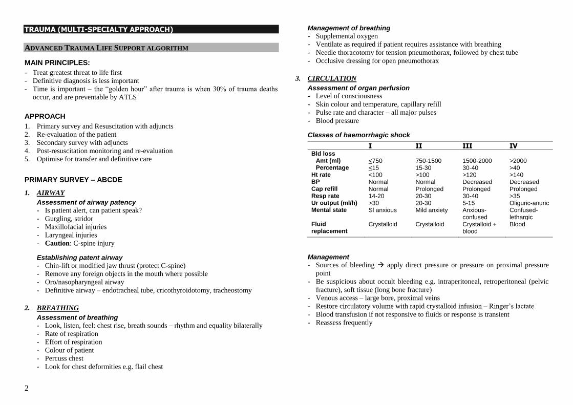
Surgery Notes For the M.B.B.S. By Andre Tan CONTENTS Page I TRAUMA (MULTI-SPECIALTY APPROACH) 2 II APPROACH TO ABDOMINAL PAIN 10 III APPROACH TO ABDOMINAL MASSES 11 IV OESOPHAGEAL DISEASES 12 V UPPER BLEEDING GIT AND ITS CAUSES 21 VI COLORECTAL DISEASES 19 VII LIVER DISEASES 39 VIII PANCREATIC DISEASES 45 IX BILIARY TRACT DISEASES 51 X BREAST DISEASES 59 XI HEAD AND NECK MASSES 69 XII SALIVARY GLAND SWELLINGS 74 XIII THYROID DISEASES 78 XIV PERIPHERAL ARTERIAL DISEASE 85 XV ABDOMINAL AORTIC ANEURYSM 93 XVI PERIPHERAL VENOUS DISEASE 95 XVII UROLOGICAL DISEASES 99 XVIII SURGICAL INSTRUMENTS 110 XIX IMPORTANT LUMPS & BUMPS AND OTHERS 119
-
Upload
khangminh22 -
Category
Documents
-
view
0 -
download
0
Transcript of Surgery Notes - 1 File Download
Surgery Notes
For the M.B.B.S.
By Andre Tan
CONTENTS
Page I TRAUMA (MULTI-SPECIALTY APPROACH)
2
II APPROACH TO ABDOMINAL PAIN
10
III APPROACH TO ABDOMINAL MASSES
11
IV OESOPHAGEAL DISEASES
12
V UPPER BLEEDING GIT AND ITS CAUSES
21
VI COLORECTAL DISEASES
19
VII LIVER DISEASES
39
VIII PANCREATIC DISEASES
45
IX BILIARY TRACT DISEASES
51
X BREAST DISEASES
59
XI HEAD AND NECK MASSES
69
XII SALIVARY GLAND SWELLINGS
74
XIII THYROID DISEASES
78
XIV PERIPHERAL ARTERIAL DISEASE
85
XV ABDOMINAL AORTIC ANEURYSM
93
XVI PERIPHERAL VENOUS DISEASE
95
XVII UROLOGICAL DISEASES
99
XVIII SURGICAL INSTRUMENTS
110
XIX IMPORTANT LUMPS & BUMPS AND OTHERS 119
2
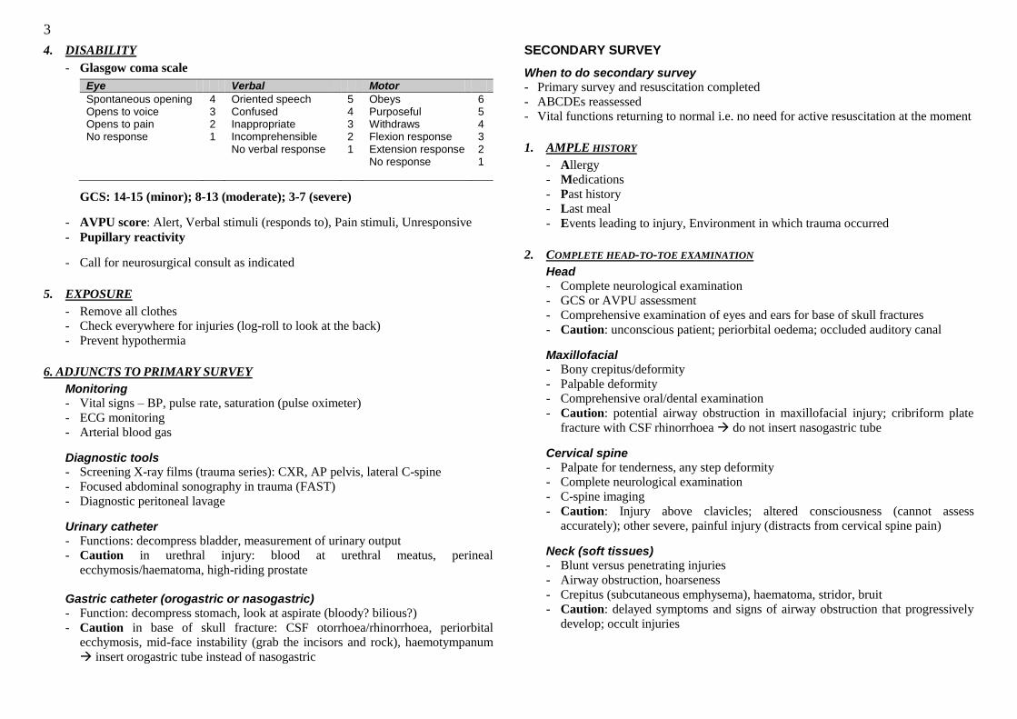
TRAUMA (MULTI-SPECIALTY APPROACH)
ADVANCED TRAUMA LIFE SUPPORT ALGORITHM
MAIN PRINCIPLES:
- Treat greatest threat to life first
- Definitive diagnosis is less important
- Time is important – the ―golden hour‖ after trauma is when 30% of trauma deaths
occur, and are preventable by ATLS
APPROACH
1. Primary survey and Resuscitation with adjuncts
2. Re-evaluation of the patient
3. Secondary survey with adjuncts
4. Post-resuscitation monitoring and re-evaluation
5. Optimise for transfer and definitive care
PRIMARY SURVEY – ABCDE
1. AIRWAY
Assessment of airway patency
- Is patient alert, can patient speak?
- Gurgling, stridor
- Maxillofacial injuries
- Laryngeal injuries
- Caution: C-spine injury
Establishing patent airway
- Chin-lift or modified jaw thrust (protect C-spine)
- Remove any foreign objects in the mouth where possible
- Oro/nasopharyngeal airway
- Definitive airway – endotracheal tube, cricothyroidotomy, tracheostomy
2. BREATHING
Assessment of breathing
- Look, listen, feel: chest rise, breath sounds – rhythm and equality bilaterally
- Rate of respiration
- Effort of respiration
- Colour of patient
- Percuss chest
- Look for chest deformities e.g. flail chest
Management of breathing
- Supplemental oxygen
- Ventilate as required if patient requires assistance with breathing
- Needle thoracotomy for tension pneumothorax, followed by chest tube
- Occlusive dressing for open pneumothorax
3. CIRCULATION
Assessment of organ perfusion
- Level of consciousness
- Skin colour and temperature, capillary refill
- Pulse rate and character – all major pulses
- Blood pressure
Classes of haemorrhagic shock
I II III IV Bld loss
Amt (ml) Percentage
<750 <15
750-1500 15-30
1500-2000 30-40
>2000 >40
Ht rate <100 >100 >120 >140 BP Normal Normal Decreased Decreased Cap refill Normal Prolonged Prolonged Prolonged Resp rate 14-20 20-30 30-40 >35 Ur output (ml/h) >30 20-30 5-15 Oliguric-anuric Mental state Sl anxious Mild anxiety Anxious-
confused Confused-lethargic
Fluid replacement
Crystalloid Crystalloid Crystalloid + blood
Blood
Management
- Sources of bleeding apply direct pressure or pressure on proximal pressure
point
- Be suspicious about occult bleeding e.g. intraperitoneal, retroperitoneal (pelvic
fracture), soft tissue (long bone fracture)
- Venous access – large bore, proximal veins
- Restore circulatory volume with rapid crystalloid infusion – Ringer‘s lactate
- Blood transfusion if not responsive to fluids or response is transient
- Reassess frequently
3
4. DISABILITY
- Glasgow coma scale
Eye Verbal Motor
Spontaneous opening Opens to voice Opens to pain No response
4 3 2 1
Oriented speech Confused Inappropriate Incomprehensible No verbal response
5 4 3 2 1
Obeys Purposeful Withdraws Flexion response Extension response No response
6 5 4 3 2 1
GCS: 14-15 (minor); 8-13 (moderate); 3-7 (severe)
- AVPU score: Alert, Verbal stimuli (responds to), Pain stimuli, Unresponsive
- Pupillary reactivity
- Call for neurosurgical consult as indicated
5. EXPOSURE
- Remove all clothes
- Check everywhere for injuries (log-roll to look at the back)
- Prevent hypothermia
6. ADJUNCTS TO PRIMARY SURVEY
Monitoring
- Vital signs – BP, pulse rate, saturation (pulse oximeter)
- ECG monitoring
- Arterial blood gas
Diagnostic tools - Screening X-ray films (trauma series): CXR, AP pelvis, lateral C-spine
- Focused abdominal sonography in trauma (FAST)
- Diagnostic peritoneal lavage
Urinary catheter
- Functions: decompress bladder, measurement of urinary output
- Caution in urethral injury: blood at urethral meatus, perineal
ecchymosis/haematoma, high-riding prostate
Gastric catheter (orogastric or nasogastric) - Function: decompress stomach, look at aspirate (bloody? bilious?)
- Caution in base of skull fracture: CSF otorrhoea/rhinorrhoea, periorbital
ecchymosis, mid-face instability (grab the incisors and rock), haemotympanum
insert orogastric tube instead of nasogastric
SECONDARY SURVEY
When to do secondary survey
- Primary survey and resuscitation completed
- ABCDEs reassessed
- Vital functions returning to normal i.e. no need for active resuscitation at the moment
1. AMPLE HISTORY
- Allergy
- Medications
- Past history
- Last meal
- Events leading to injury, Environment in which trauma occurred
2. COMPLETE HEAD-TO-TOE EXAMINATION
Head
- Complete neurological examination
- GCS or AVPU assessment
- Comprehensive examination of eyes and ears for base of skull fractures
- Caution: unconscious patient; periorbital oedema; occluded auditory canal
Maxillofacial
- Bony crepitus/deformity
- Palpable deformity
- Comprehensive oral/dental examination
- Caution: potential airway obstruction in maxillofacial injury; cribriform plate
fracture with CSF rhinorrhoea do not insert nasogastric tube
Cervical spine
- Palpate for tenderness, any step deformity
- Complete neurological examination
- C-spine imaging
- Caution: Injury above clavicles; altered consciousness (cannot assess
accurately); other severe, painful injury (distracts from cervical spine pain)
Neck (soft tissues)
- Blunt versus penetrating injuries
- Airway obstruction, hoarseness
- Crepitus (subcutaneous emphysema), haematoma, stridor, bruit
- Caution: delayed symptoms and signs of airway obstruction that progressively
develop; occult injuries
4
Chest
- Inspect, palpate, percuss, auscultate
- Re-evaluate frequently
- Look at CXR
- Caution: missed injury; increase in chest tube drainage
Abdomen
- Inspect, palpate, percuss, auscultate
- Abrasions and ecchymosis – ―seat-belt sign‖
- Lower rib fractures liver and spleen injury
- Re-evaluate frequently
- Special studies: FAST, DPL, CT scan
- Caution: hollow viscus and retroperitoneal injuries; excessive pelvic
manipulation
Perineum
- Contusions, haematomas, lacerations
- Urethral blood
- DRE: Sphincter tone, high-riding prostate, pelvic fracture (may feel fragments of
bone); rectal wall integrity; blood
- Vaginal examination: blood, lacerations
Musculoskeletal – extremities
- Contusion, deformity
- Pain
- Perfusion
- Peripheral neurovascular status
- X-rays as appropriate
- Caution: potential blood loss is high in certain injuries (e.g. pelvic fracture,
femoral shaft fracture); missed fractures; soft-tissue or ligamentous injuries;
examine patient‘s back
3. ADJUNCTS AND SPECIAL DIAGNOSTIC TESTS
- As required according to suspicion, but should not delay transfer
4. FREQUENT RE-EVALUATION
- Have a high index of suspicion for injuries to avoid missing them
- Frequent re-evaluation and continuous monitoring rapidly recognise when patient is deteriorating
5. PAIN MANAGEMENT
- Intravenous analgesia as appropriate
ABDOMINAL TRAUMA
TYPES OF INTRA-ABDOMINAL INJURY IN BLUNT TRAUMA
- Solid organ injury: spleen, liver – bleeding (may be quite massive)
- Hollow viscus injury with rupture
- Vascular injury with bleeding
INDICATIONS FOR IMMEDIATE LAPAROTOMY
- Evisceration, stab wounds with implement in-situ, gunshot wounds traversing
abdominal cavity
- Any penetrating injury to the abdomen with haemodynamic instability or peritoneal
irritation
- Obvious or strongly suspected intra-abdominal injury with shock or difficulty in
stabilising haemodynamics
- Obvious signs of peritoneal irritation
- Rectal exam reveals fresh blood
- Persistent fresh blood aspirated from nasogastric tube (oropharyngeal injuries
excluded as source of bleeding)
- X-ray evidence of pneumoperitoneum or diaphragmatic rupture
INVESTIGATIONS
- If patient is stable: FAST and/or CT scan
- If patient is unstable: FAST and/or DPL
FOCUSED ABDOMINAL SONOGRAPHY IN TRAUMA (FAST)
- Ultrasonographic evaluation of four windows: Pericardial, right upper quadrant, left
upper quadrant, pelvis
- Advantages
Portable
Can be done quickly in <5min
Can be used for serial examination
Does not require contrast, no radiation risk
- Disadvantages Does not image solid parenchymal damage, retroperitoneum, diaphragmatic
defects or bowel injury
Compromised in uncooperative, agitated patient, obesity, substantial bowel gas,
subcutaneous air
Less sensitive, more operator-dependent than DPL and cannot distinguish blood
from ascites
Intermediate results require follow-up attempts or alternative diagnostic tests
5
CT SCAN
- Only suitable for stable patient as quite long time involved in imaging with only
patient in the room can collapse
- Advantages
Able to precisely locate intra-abdominal lesions preoperatively
Able to evaluate retroperitoneum
Able to identify injuries that can be managed non-operatively
Not invasive
- Disadvantages
Expensive
Time required to transport patient
Use of contrast
DIAGNOSTIC PERITONEAL LAVAGE (DPL)
- Involves making a cut in the infraumbilical region and inserting a catheter into the
peritoneal cavity, aspirate, then instillation of saline and re-aspiration
- Positive DPL
Frank blood (>5ml) or obvious bowel contents aspirated
Lavage fluid seen to exit from chest drain or urinary catheter
RBC >100,000 per mm3, WBC >500, Gram stain positive for bacteria in effluent
- Indications:
Any unstable patient with suspicion of abdominal trauma or where clinical exam
is difficult or equivocal
Unexplained hypotension in multiple trauma
Patient requiring immediate surgery for extra-abdominal injuries
- Contraindications
Absolute indication for laparotomy already exists
Previous abdominal surgery or infections
Gravid uterus
Morbid obesity
Coagulopathy
- Advantages
Can promptly reveal or exclude the presence of intraperitoneal haemorrhage
Valuable in discovery of potentially lethal bowel perforation
- Disadvantages
Morbidity involved – wound complications (haematoma, infection);
intraperitoneal injury
False negative rate of 2% when there is failure to recover lavage fluid, early
hollow viscus injury, diaphragmatic injuries, injuries to retroperitoneal structures
CARDIOTHORACIC TRAUMA
There are 5 clinical scenarios in chest trauma where bedside procedures are lifesaving: cardiac tamponade, airway obstruction, flail chest, haemothorax, and pneumothorax.
CARDIAC TAMPONADE
- High index of suspicion required
- Clinical features
Chest trauma and hypotension
Beck‘s triad (hypotension, muffled heart sounds, distended neck veins) – only
seen in 50% of cases as hypovolaemia may prevent neck vein distension; muffled
heart sounds are least reliable
Pulseless electrical activity
Kussmaul‘s signs (increased neck distension during inspiration, pulsus paradoxus)
- Diagnostic clues
Enlarged cardiac shadow in CXR (globular heart – very rarely seen)
Small ECG voltages, electrical alternans (uncommon)
Pericardial fluid demonstrated on FAST or 2D-echo - definitive
- Management
Aggressive fluid resuscitation – helps maintain cardiac output and buys time
Pericardiocentesis: ECG lead-guided or 2D-echo guided
AIRWAY OBSTRUCTION
- Chin lift or jaw thrust
- Remove any foreign body manually, suction blood/secretions
- Definitive airway – ETT, cricothyroidotomy, tracheostomy
FLAIL CHEST
- When 2 or more ribs are fractured at 2 points forming a flail segment that moves
paradoxically with breathing
- Results in hypoxaemia mainly due to underlying pulmonary contusion, contributed to
by pain with restricted chest wall movement
- Management: ensure adequate oxygenation and ventilation; judicious fluid therapy
(avoid fluid overload); adequate intravenous analgesia
- Consider mechanical ventilation in high risk patients: shock, severe head injury,
previous pulmonary disease, fracture of >8 ribs, age > 65, >3 associated injuries
6
HAEMOTHORAX
- Chest tube insertion in the triangle of safety (bound by the lateral border of the
pectoralis major medially, a line just anterior to the mid-axillary line laterally, and the
upper border of the fifth rib inferiorly)
- Be wary of sudden cessation of chest tube drainage as tube can get blocked by clot
- If blood >1500mls massive haemothorax, call urgent cardiothoracic consult
PNEUMOTHORAX (OPEN/TENSION)
- Tension pneumothorax is a clinical diagnosis (CXR will only delay treatment, and
may cause death) – signs of pneumothorax, hypotension, neck vein distension, severe
respiratory distress
- Immediate needle thoracotomy in second intercostal space in mid-clavicular line
- Followed by chest tube insertion
- Open pneumothorax occurs in a large chest wall defect with equilibration between
intrathoracic and atmospheric pressure, producing a ―sucking chest wound‖
- Cover defect with a sterile dressing, taping it down on 3 sides to produce a flutter-
valve effect, letting air out of the pleural cavity but not back in
- Insert chest tube (not through the wound)
NEUROSURGICAL TRAUMA
AIM in management of head injuries is the prevention of secondary brain injury (from
hypotension, hypoxaemia, increased ICP etc) since neuronal death is irreversible.
PATHOLOGIES:
1. Concussion
- Physiological dysfunction without anatomical or radiological abnormality
- (Physiological dysfunction is the first step towards cell death, but is reversible if
no further insult occurs)
- Usually recovers in 2-3 hours
2. Contusion
- Small haematoma <1cm
3. Intracranial haemorrhage
(a) Extradural haemorrhage
Lens-shaped haematoma outside the dura (between skull and dura)
Pathology: expanding space-occupying lesion
20% of patients with EDH are alert and well; underlying brain is minimally
damaged, thus drainage gives good results
(b) Subdural haemorrhage
Crescent shaped haematoma under the dura (between the dura and the
arachnoid)
More severe than EDH (usually due to nature of injury that causes SDH to
occur – associated with higher impact, thus brain has other injuries)
Pathology: underlying brain damage in addition to expanding SOL
Removal of blood does not solve underlying brain damage poorer results
(c) Traumatic subarachnoid haemorrhage
Usually only small amount of blood conservative treatment sufficient
(d) Intraparenchymal haemorrhage
Any shape, size, location
If large haematoma, will require evacuation
4. Diffuse axonal injury
- Global injury of axons
- Arises from injury that causes rotational and shearing forces (high impact
injury) – rapid acceleration and deceleration of brain in the intracranial cavity
against relatively fixed points of attachment at the falx and tentorium
- Maximal effects at corpus callosum and brainstem
- If severe, will see punctate haemorrhages at the grey-white border
5. Cerebral oedema (2 types)
(a) Hypoxic (cellular)
Decreased blood supply (oxygenation) loss of function of Na-K pump as
ATP decreases increased intracellular sodium cellular swelling
(b) Interstitial
Breakdown of blood-brain barrier proteins enter interstitial space
oedema
PATHOPHYSIOLOGY
1. Monroe-Kellie doctrine
- Intracranial cavity is of fixed volume and its contents (brain, CSF, blood) are
relatively incompressible
- Thus increase in intracranial volume raised ICP
Cerebral perfusion pressure = Mean arterial pressure – Intracranial pressure
- Compensatory mechanisms:
(a) Hyperventilation vasoconstriction of cerebral vessels due to increased
partial pressure of carbon dioxide decrease in blood volume
(b) CSF pushed into spinal canal (but limited volume available)
- Removal of any reversible cause of raised ICP will improve cerebral perfusion
7
2. Fixed dilated pupil
- Constrictor fibres to the pupil run in the oculomotor nerve, which exits the
brainstem at the upper midbrain – nerve fibres lie just under the tentorium
- Uncus of the temporal lobe sits on the tentorium
- In raised ICP, the uncus herniates over the edge of the tentorium,
compressing the fibres of the oculomotor nerve just below
- Thus a fixed dilated pupil occurs on the side of the compression due to
unoppressed sympathetic supply (dilates the pupil)
3. Cushing’s reflex
- A triad of:
(a) Raised ICP
(b) Hypertension
(c) Bradycardia
- From Monroe-Kellie doctrine, an increase in mean arterial pressure helps to
maintain cerebral perfusion pressure when ICP is raised
- Increase in mean arterial pressure achieved by sympathetic overdrive:
(a) Increased heart rate
(b) Increased contractility
(c) Increased vasoconstriction – increased total peripheral resistance
(a) and (b) increase cardiac output increased BP; (c) increases BP
- Baroreceptors detect abnormally raised blood pressure and try to decrease it
heart rate falls
MANAGEMENT
1. Assessment
- 3 important parameters: ABCs, GCS, pupil size
- Glasgow coma scale (see above) – Minor head injury: 14-15; moderate injury: 8-
13; severe injury: 3-7
2. Minor head injury
- Most common
- Indications for admission:
Persistent headache and/or vomiting
CSF leak
Neurological deficit
Skull fracture
History of loss of consciousness
Amnesia
- In ward: NBM, IV drip (no dextrose saline!), no sedation, monitor GCS
- If patient deteriorates CT scan, exclude metabolic causes (e.g. hypoglyc), do
septic workup (exclude sepsis)
3. Moderate head injury
- All will be CT-scanned at ED NES will operate if any indication to do so
- In ward: as per mild head injury
4. Severe head injury
- Must scan to look for reversible causes of raised ICP but stabilise patient first
- Medical methods to lower ICP
(a) Intubate and hyperventilate
(b) IV mannitol (must catheterise patient also; do not give if patient is unstable)
- Screen for other life-threatening injuries (likely to be multi-trauma patient)
- Achieve haemodynamic stability
(a) Check for long bone fractures
(b) FAST for bleeding into abdominal cavity
(c) ABG to detect acidosis
(d) Keep monitoring patient and re-investigate where appropriate
- Operate if reversible cause found
(a) Craniectomy (i.e. bone flap not replaced) or craniotomy (bone flap replaced
after blood evacuated) [Burrhole usually not big enough to drain an acute
bleed]
(b) Evacuate clot
(c) Insert endoventricular drain (EVD) if there is hydrocephalus
- Total sedation after operation, ward in ICU
Prevents patient from struggling which will raise ICP
5. Depressed skull fracture
- Can leave alone unless depression is greater than the thickness of the skull bone
6. Compound depressed fracture
- There is through-and-through skin laceration over the fracture
- Always explore to ensure underlying dura is intact, and repair if dura is torn
(since meningitis can occur with a torn dura)
8
MUSCULOSKELETAL TRAUMA
GENERAL POINTS
- Extremity trauma tends not to be life-threatening
- But occult blood loss can occur in large volumes especially in certain types of
injuries – pelvic fracture (up to 3L), femoral shaft fracture (up to 2L)
- Need to have high level of suspicion and treat with urgency
- Look out for any tachycardia, early signs of shock
- Prepare to resuscitate patient
ASSESSMENT OF THE EXTREMITY
- Perfusion: colour, pulses, skin temperature, capillary refill
- Deformity
- Wounds – open or closed injury; abrasion over a fracture is considered open fracture
- Soft tissue assessment
- Abnormal joint mobility – ligamentous injury around the joint; if in the knee, highly
likely that the popliteal artery is injured as well
- Neurological assessment
- Viability of the limb
THE PULSELESS EXTREMITY
Things to consider
- Is pulselessness due to shock?
- Arterial or venous compromise?
- Is there compartment syndrome (pulselessness is a very late sign)
- Any pre-existing vascular disease?
Physical examination
- Any limb deformity (can result in kinking of vessels)?
- Any joint instability (dislocation of a joint can result in intimal tear in the major
vessel running across it, with thrombosis and occlusion)?
- Skin colour/temperature
- Post-reduction tibial pulse in knee dislocation – if still absent, do an urgent
angiogram!
SOFT TISSUE INJURIES
Types
- Open: laceration, abrasion
- Crushing
- Degloving: open or closed
- Closed
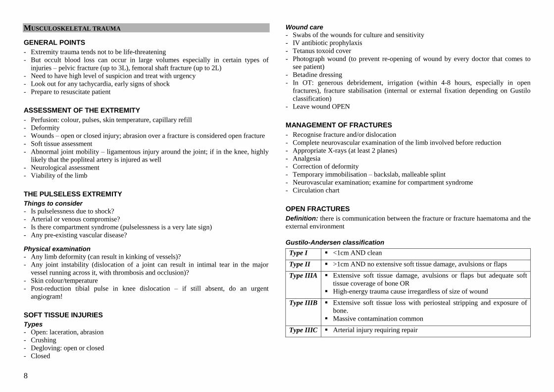
Wound care
- Swabs of the wounds for culture and sensitivity
- IV antibiotic prophylaxis
- Tetanus toxoid cover
- Photograph wound (to prevent re-opening of wound by every doctor that comes to
see patient)
- Betadine dressing
- In OT: generous debridement, irrigation (within 4-8 hours, especially in open
fractures), fracture stabilisation (internal or external fixation depending on Gustilo
classification)
- Leave wound OPEN
MANAGEMENT OF FRACTURES
- Recognise fracture and/or dislocation
- Complete neurovascular examination of the limb involved before reduction
- Appropriate X-rays (at least 2 planes)
- Analgesia
- Correction of deformity
- Temporary immobilisation – backslab, malleable splint
- Neurovascular examination; examine for compartment syndrome
- Circulation chart
OPEN FRACTURES
Definition: there is communication between the fracture or fracture haematoma and the
external environment
Gustilo-Andersen classification
Type I <1cm AND clean
Type II >1cm AND no extensive soft tissue damage, avulsions or flaps
Type IIIA Extensive soft tissue damage, avulsions or flaps but adequate soft
tissue coverage of bone OR
High-energy trauma cause irregardless of size of wound
Type IIIB Extensive soft tissue loss with periosteal stripping and exposure of
bone.
Massive contamination common
Type IIIC Arterial injury requiring repair
9
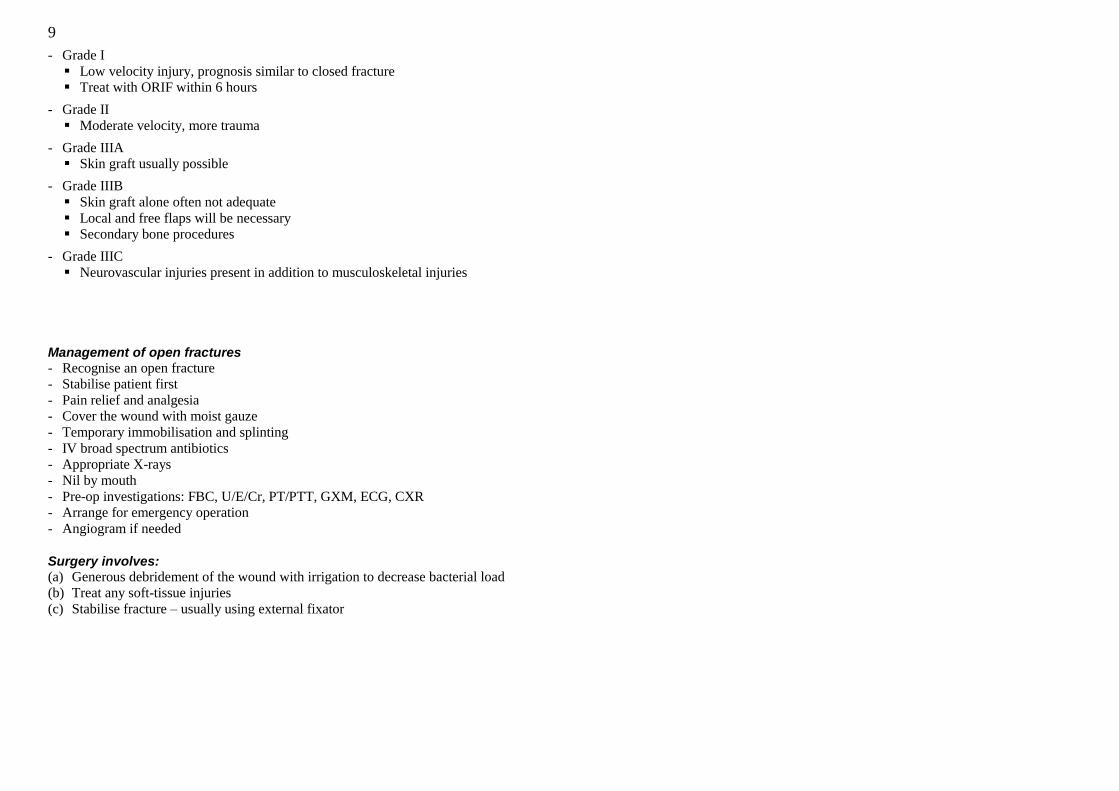
- Grade I
Low velocity injury, prognosis similar to closed fracture
Treat with ORIF within 6 hours
- Grade II
Moderate velocity, more trauma
- Grade IIIA
Skin graft usually possible
- Grade IIIB
Skin graft alone often not adequate
Local and free flaps will be necessary
Secondary bone procedures
- Grade IIIC
Neurovascular injuries present in addition to musculoskeletal injuries
Management of open fractures
- Recognise an open fracture
- Stabilise patient first
- Pain relief and analgesia
- Cover the wound with moist gauze
- Temporary immobilisation and splinting
- IV broad spectrum antibiotics
- Appropriate X-rays
- Nil by mouth
- Pre-op investigations: FBC, U/E/Cr, PT/PTT, GXM, ECG, CXR
- Arrange for emergency operation
- Angiogram if needed
Surgery involves:
(a) Generous debridement of the wound with irrigation to decrease bacterial load
(b) Treat any soft-tissue injuries
(c) Stabilise fracture – usually using external fixator
10
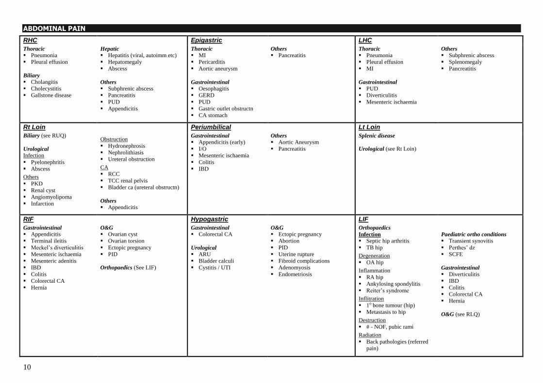
ABDOMINAL PAIN
RHC
Epigastric
LHC
Thoracic
Pneumonia
Pleural effusion
Biliary
Cholangitis
Cholecystitis
Gallstone disease
Hepatic
Hepatitis (viral, autoimm etc)
Hepatomegaly
Abscess
Others
Subphrenic abscess
Pancreatitis
PUD
Appendicitis
Thoracic
MI
Pericarditis
Aortic aneurysm
Gastrointestinal
Oesophagitis
GERD
PUD
Gastric outlet obstructn
CA stomach
Others
Pancreatitis
Thoracic
Pneumonia
Pleural effusion
MI
Gastrointestinal
PUD
Diverticulitis
Mesenteric ischaemia
Others
Subphrenic abscess
Splenomegaly
Pancreatitis
Rt Loin
Periumbilical Lt Loin
Biliary (see RUQ)
Urological
Infection
Pyelonephritis
Abscess
Others
PKD
Renal cyst
Angiomyolipoma
Infarction
Obstruction
Hydronephrosis
Nephrolithiasis
Ureteral obstruction
CA
RCC
TCC renal pelvis
Bladder ca (ureteral obstructn)
Others
Appendicitis
Gastrointestinal
Appendicitis (early)
I/O
Mesenteric ischaemia
Colitis
IBD
Others
Aortic Aneurysm
Pancreatitis
Splenic disease
Urological (see Rt Loin)
RIF
Hypogastric LIF
Gastrointestinal
Appendicitis
Terminal ileitis
Meckel‘s diverticulitis
Mesenteric ischaemia
Mesenteric adenitis
IBD
Colitis
Colorectal CA
Hernia
O&G
Ovarian cyst
Ovarian torsion
Ectopic pregnancy
PID
Orthopaedics (See LIF)
Gastrointestinal
Colorectal CA
Urological
ARU
Bladder calculi
Cystitis / UTI
O&G
Ectopic pregnancy
Abortion
PID
Uterine rupture
Fibroid complications
Adenomyosis
Endometriosis
Orthopaedics
Infection
Septic hip arthritis
TB hip
Degeneration
OA hip
Inflammation
RA hip
Ankylosing spondylitis
Reiter‘s syndrome
Inflitration
1o bone tumour (hip)
Metastasis to hip
Destruction
# - NOF, pubic rami
Radiation
Back pathologies (referred
pain)
Paediatric ortho conditions
Transient synovitis
Perthes‘ dz
SCFE
Gastrointestinal
Diverticulitis
IBD
Colitis
Colorectal CA
Hernia
O&G (see RLQ)
11
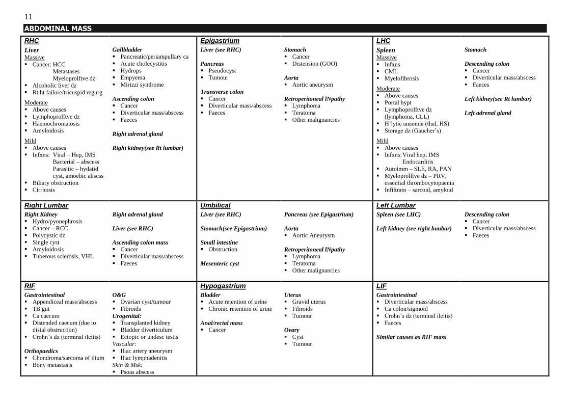
ABDOMINAL MASS
RHC
Epigastrium
LHC
Liver Massive
Cancer: HCC
Metastases
Myeloprolftve dz
Alcoholic liver dz
Rt ht failure/tricuspid regurg
Moderate
Above causes
Lymphoprolftve dz
Haemochromatosis
Amyloidosis
Mild
Above causes
Infxns: Viral – Hep, IMS
Bacterial – abscess
Parasitic – hydatid
cyst, amoebic abscss
Biliary obstruction
Cirrhosis
Gallbladder
Pancreatic/periampullary ca
Acute cholecystitis
Hydrops
Empyema
Mirizzi syndrome
Ascending colon
Cancer
Diverticular mass/abscess
Faeces
Right adrenal gland
Right kidney(see Rt lumbar)
Liver (see RHC)
Pancreas
Pseudocyst
Tumour
Transverse colon
Cancer
Diverticular mass/abscess
Faeces
Stomach
Cancer
Distension (GOO)
Aorta
Aortic aneurysm
Retroperitoneal lNpathy
Lymphoma
Teratoma
Other malignancies
Spleen Massive
Infxns
CML
Myelofibrosis
Moderate
Above causes
Portal hypt
Lymphoprolftve dz
(lymphoma, CLL)
H‘lytic anaemia (thal, HS)
Storage dz (Gaucher‘s)
Mild
Above causes
Infxns: Viral hep, IMS
Endocarditis
Autoimm – SLE, RA, PAN
Myeloprolftve dz – PRV,
essential thrombocytopaenia
Infiltratn – sarcoid, amyloid
Stomach
Descending colon
Cancer
Diverticular mass/abscess
Faeces
Left kidney(see Rt lumbar)
Left adrenal gland
Right Lumbar
Umbilical Left Lumbar
Right Kidney
Hydro/pyonephrosis
Cancer – RCC
Polycystic dz
Single cyst
Amyloidosis
Tuberous sclerosis, VHL
Right adrenal gland
Liver (see RHC)
Ascending colon mass
Cancer
Diverticular mass/abscess
Faeces
Liver (see RHC)
Stomach(see Epigastrium)
Small intestine
Obstruction
Mesenteric cyst
Pancreas (see Epigastrium)
Aorta
Aortic Aneurysm
Retroperitoneal lNpathy
Lymphoma
Teratoma
Other malignancies
Spleen (see LHC)
Left kidney (see right lumbar)
Descending colon
Cancer
Diverticular mass/abscess
Faeces
RIF
Hypogastrium LIF
Gastrointestinal
Appendiceal mass/abscess
TB gut
Ca caecum
Distended caecum (due to
distal obstruction)
Crohn‘s dz (terminal ileitis)
Orthopaedics Chondroma/sarcoma of ilium
Bony metastasis
O&G
Ovarian cyst/tumour
Fibroids
Urogenital:
Transplanted kidney
Bladder diverticulum
Ectopic or undesc testis
Vascular:
Iliac artery aneurysm
Iliac lymphadenitis
Skin & Msk:
Psoas abscess
Bladder
Acute retention of urine
Chronic retention of urine
Anal/rectal mass
Cancer
Uterus
Gravid uterus
Fibroids
Tumour
Ovary
Cyst
Tumour
Gastrointestinal
Diverticular mass/abscess
Ca colon/sigmoid
Crohn‘s dz (terminal ileitis)
Faeces
Similar causes as RIF mass
12
OESOPHAGEAL DISEASES
ANATOMY
- Oesophagus is a muscular tube that is 25cm (10 inches) long
- Starts at the cricoid cartilage (C6 vertebra) from the oropharynx and continues into
the stomach at the level of T10
- Upper oesophageal sphincter is formed by cricopharyngeus muscle
- Lower sphincter is not an anatomical sphincter, but physiological:
(i) Increased tone of the muscularis propria at the lower oesophageal sphincter
(ii) Fibres of the right diaphragmatic crus loop around the cardio-oesophageal
junction and ontract during coughing, sneezing etc when intra-abdominal
pressure increases, thus preventing reflux
(iii) Angle of His where the oesophagus joins the stomach – acts as a valve
(iv) Intra-abdominal pressure being higher than intra-thoracic pressure
- 3 narrow points along the course of the oesophagus
(i) Cricopharyngeus muscle (15cm from incisor teeth)
(ii) Carina where the left bronchus crosses the oesophagus (27cm from incisors)
(iii) Where the oesophagus passes through the diaphragm (40cm from incisors)
- Structure: mucosa, submucosa, muscularis propria, adventitia (no peritoneal lining
except for a short segment of intra-abdominal oesophagus)
Muscularis propria is composed of striated muscle in the upper one-third, striated and
smooth muscle in the middle third, and smooth muscle in the lower third
- Blood supply (roughly divided into thirds): Inferior thyroid artery to upper third,
oesophageal branches of the aorta to the middle third, oesophageal branches of left
gastric artery to lower third
- Venous return also divided into thirds: Brachiocephalic veins (upper), azygos veins
(middle), left gastric vein (lower) --- a portosystemic anastomosis exists at the lower
oesophagus thus leading to formation of varices in portal hypertension
PHYSIOLOGY OF SWALLOWING
- Process of mastication forms a food bolus on the dorsum of the tongue
- The tongue then contracts upwards and backwards pushing the food bolus against the
hard palate
- Soft palate elevates (contraction of palatoglossus) to close off nasopharynx
- Further elevation of tongue pushes food bolus into oropharynx
- As the base of the tongue is elevated posterior, the epiglottis falls back; at the same
time, the pharyngeal muscles contract to bring the posterior surface of the larynx
upwards to make the laryngeal inlet smaller closed off by the epiglottis
- Pharyngeal muscles contract to propel food bolus past the relaxed cricopharyngeus
into the oesophagus
- Once in the oesophagus, involuntary contractions of the muscularis propria form
peristaltic waves to propel food bolus into stomach
APPROACH TO DYSPHAGIA
CAUSES OF DYSPHAGIA
- Dysphagia can be divided into oropharyngeal and oesophageal dysphagia
- In each anatomic region the dysphagia can be caused by neuromuscular dysfunction
(impaired physiology of swallowing) or mechanical obstruction to the lumen
Oropharyngeal Oesophageal
Neuromuscular diseases Neuromuscular diseases Stroke
Parkinson’s disease
Brain stem tumours
Degenerative conditions e.g. ALS, MS
Peripheral neuropathy
Myasthaenia gravis
Myopathies e.g. myotonic dystrophy
Achalasia
Spastic motor disorders Diffuse oesophageal spasm
Hypertensive lower oesophageal sphincter
Nutcracker oesophagus Scleroderma
Obstructive lesions Obstructive lesions Tumours
Inflammatory masses e.g. abscess
Oesophageal webs
Pharyngeal pouch (Zenker‘s divert)
Anterior mediastinal mass
Intrinsic structural lesions Tumours
Strictures: Peptic (reflux oesophagitis)
Radiation
Chemical (caustic ingestion)
Medication Lower oesophageal rings (Schatzki‘s ring)
Oesophageal webs (Plummer-Vinson)
Foreign bodies
Extrinsic structural lesions Vascular compression (enlarged aorta or
left atrium)
Mediastinal masses – retrosternal thyroid,
lymphadenopathy
Others Oesophagitis: Reflux
Infectious (candida, herpes)
Radiation-induced
Medication-induced
Chemical-induced (alcohol)
13
HISTORY:
1. Is there odynophagia (pain associated with difficulty swallowing)?
- Signifies some form of oesophagitis: infectious (candida, herpes), post-radiation,
chemical-induced (usually alcohol), reflux oesophagitis
- Oesophageal spasm
- Scleroderma
- Pain occurs late in achalasia and oesophageal cancer (not painful from the start)
2. Differentiating oropharyngeal from oesophageal dypshagia
(i) Oropharyngeal
- Presenting complaint is usually of difficulty in initiating swallowing
- May be associated with choking, coughing, nasal regurgitation
- Voice may sound nasal (bulbar palsy)
- Cause of oropharyngeal dysphagia is usually neuromuscular rather than
mechanical; stroke is the most common cause
(ii) Oesophageal
- Presenting complaint is that of food “getting stuck” in the throat or chest
- Patient‘s localisation of the symptom often does not correspond to actual site
of pathology
- Can be due to either neuromuscular dysfunction or mechanical obstruction
3. Differentiating mechanical obstruction from neuromuscular dysfunction
(i) Mechanical
- Patient complains of more difficulty swallowing solids than fluids
- May have regurgitation of undigested food
- Recent onset dysphagia that is progressively worsening, with loss of weight
high suspicion of oesophageal cancer
- Intermittent symptoms are suggestive of webs, rings
(ii) Neuromuscular
- Patient complains of more trouble swallowing fluids than solids
- Dysphagia more long-standing, slowly progressive
- Intermittent symptoms suggestive of diffuse oesophageal spasm, nutcracker
oesophagus
- May have history of stroke, neuromuscular disease
4. History of predisposing conditions - Reflux symptoms e.g. retrosternal burning pain (heartburn), sour fluid reflux into
mouth (acid brash), excessive salivation (water brash), postural aggravation on
lying down
- Caustic chemical ingestion in the past
- Smoking, chronic alcohol intake
- Radiation to the chest
- Medication history
- Symptoms of systemic disease e.g. stroke (focal neurological deficits),
scleroderma (telangiectasia, sclerodactyly, calcinosis, Raynaud‘s), Parkinson‘s
5. Systemic review
- Loss of weight occurs in cancer and achalasia, but of much later onset in
achalasia compared to cancer
- Symptoms of anaemia (bleeding from tumour, or as part of Plummer-Vinson
syndrome)
- Symptoms of aspiration pneumonia – fever, cough, shortness of breath
6. Tumour spread - Hoarseness (recurrent laryngeal nerve)
- Fever, cough and haemoptysis (tracheo-oesophageal fistula)
- Haematemesis (invasion into aorta)
- Neck lump (lymph node)
PHYSICAL EXAMINATION
1. General condition - Vitals: the patient may be hypovolaemic from vomiting/decreased intake
- Nutrition: presence of cachexia
- Conjunctival pallor: bleeding from tumour, oesophagitis ulcerations, or
associated with P-V syndrome
- Scleral icterus: metastases to liver
- Dehydration (mucous membranes, skin turgor, etc)
2. Disease
- Presence of cervical lymph nodes (esp Virchow‘s node)
- Scars/marks over the chest and abdomen suggesting previous surgery, radiation
- Palpable mass in abdomen (not likely)
- Hepatomegaly
- Ascites
- PR examination for malaena
3. Complications of disease - Signs of pneumonia: patient febrile, may be toxic, lung crepitations, decreased
air entry usually over right lower lobe
4. Treatment - Tube feeding through NG tube, gastrostomy/jejunostomy – if aspirates seen,
what is the colour?
- Total parenteral nutrition
14
MANAGEMENT
1. Stabilise patient - Resuscitate if patient is haemodynamically unstable
- IV fluids (correct fluid deficits and also any electrolyte derangements)
- Consider feeding with fluids if patient can tolerate it (only having problems with
solid food) otherwise consider tube feeding or TPN need to correct patient‘s
nutritionally debilitated state
- Keep NBM if patient cannot tolerate even fluids
- Treat any aspiration pneumonia – NBM, IV antibiotics
2. Investigate for underlying cause and treat it
INVESTIGATIONS
Diagnostic
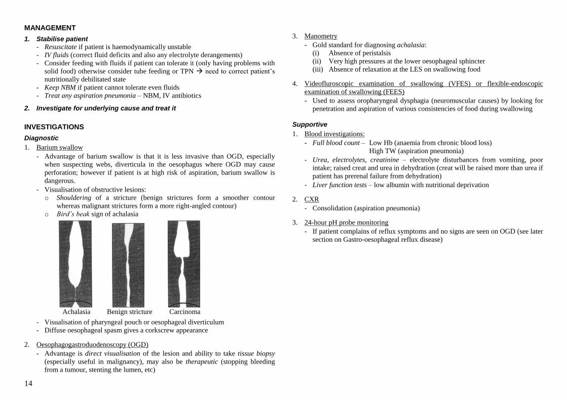
1. Barium swallow
- Advantage of barium swallow is that it is less invasive than OGD, especially
when suspecting webs, diverticula in the oesophagus where OGD may cause
perforation; however if patient is at high risk of aspiration, barium swallow is
dangerous.
- Visualisation of obstructive lesions:
o Shouldering of a stricture (benign strictures form a smoother contour
whereas malignant strictures form a more right-angled contour)
o Bird‟s beak sign of achalasia
Achalasia Benign stricture Carcinoma
- Visualisation of pharyngeal pouch or oesophageal diverticulum
- Diffuse oesophageal spasm gives a corkscrew appearance
2. Oesophagogastroduodenoscopy (OGD)
- Advantage is direct visualisation of the lesion and ability to take tissue biopsy
(especially useful in malignancy), may also be therapeutic (stopping bleeding
from a tumour, stenting the lumen, etc)
3. Manometry
- Gold standard for diagnosing achalasia:
(i) Absence of peristalsis
(ii) Very high pressures at the lower oesophageal sphincter
(iii) Absence of relaxation at the LES on swallowing food
4. Videofluroscopic examination of swallowing (VFES) or flexible-endoscopic
examination of swallowing (FEES)
- Used to assess oropharyngeal dysphagia (neuromuscular causes) by looking for
penetration and aspiration of various consistencies of food during swallowing
Supportive
1. Blood investigations:
- Full blood count – Low Hb (anaemia from chronic blood loss)
High TW (aspiration pneumonia)
- Urea, electrolytes, creatinine – electrolyte disturbances from vomiting, poor
intake; raised creat and urea in dehydration (creat will be raised more than urea if
patient has prerenal failure from dehydration)
- Liver function tests – low albumin with nutritional deprivation
2. CXR
- Consolidation (aspiration pneumonia)
3. 24-hour pH probe monitoring
- If patient complains of reflux symptoms and no signs are seen on OGD (see later
section on Gastro-oesophageal reflux disease)
15
CANCER OF THE OESOPHAGUS EPIDEMIOLOGY - Third most common gastrointestinal tract cancer in Singapore
- Male predominance
- Increasing incidence with age
RISK FACTORS
- Smoking (100x increased risk for SCC, 10x for adenocarcinoma)
- Alcohol (2x increased risk)
- Obesity (related to reflux, increases adenocarcinoma incidence)
- Diet: Hot beverages, preserved foods (nitrosamines), betel nuts; vitamin and mineral
deficiencies (selenium, vitamin E, beta-carotene)
- Tylosis (autosomal dominant disorder with keratosis of palms and soles
- Barrett‘s oesophagus (intestinal metaplasia of oesophageal mucosa due to reflux;
increased risk of cancer due to metaplasia-dysplasia-carcinoma sequence; risk is 30-
40x higher than in individual without Barrett‘s, and is about 1% per year)
- Achalasia (2-8% incidence of SCC)
- Caustic injury (ca occurs at site of scar/stricture, mostly middle third of oesophagus)
- Plummer-Vinson (or Paterson-Brown-Kelly) syndrome – Post-cricoid oesophageal
web and iron deficiency anaemia. (10% develop cancer in upper third of oesophagus)
PATHOLOGY
- 70% squamous cell carcinoma, 30% adenocarcinoma
- SCC can arise anywhere in the oesophagus while adenocarcinoma occurs in lower
third and gastro-oesophageal junction (related to reflux and Barrett‘s oesophagus)
- Overall: 10% of cancers occur in the upper third, 60% in the middle third, 30% in the
lower third
- Three growth patterns:
Fungating (60%)
Ulcerative (25%)
Infiltrative (15%)
- Tumour spread: direct extension into surrounding structures, vascular invasion,
lymphatic spread
- Common sites of metastases: liver, lung, bone
STAGING
T Tis
T1a
T1b
T2
T3
T4
High-grade dysplasia/carcinoma in-situ
Tumour invading lamina propria or
muscularis mucosa
Tumour invading submucosa but does
not breach submucosa
Tumour invades the muscularis propria
Tumour invades adventitia
Invasion of surrounding structures
N N1 Regional node involvement (1-3 nodes
involved =1a; 4-7=1b; >7=1c)
M M1a
M1b
Nonregional lymph node involvement
Other distant metastases
PRESENTATION
Usually of insidious onset, with earliest symptoms being non-specific e.g. retrosternal
discomfort, ―indigestion‖, and most patients already have advanced disease when they
are diagnosed – 75% have lymph node involvement at time of diagnosis.
1. Dysphagia
- Present in 80% of patients – most common presentation
- Pain develops late and is usually due to extra-oesophageal involvement
2. Weight loss
3. Regurgitation
4. Anaemia (with or without malaena/frank haematemesis – bleeding is usually occult)
5. Vocal cord paralysis (left more than right)
6. Aspiration pneumonia
7. Tracheo-oesophageal or broncho-oesophageal fistula
INVESTIGATIONS
Diagnosis
1. Barium swallow
- 92% accuracy in showing mucosal irregularity and annular constrictions but not
able to diagnose malignancy with confidence
2. Oesophagogastroduodenoscopy
- Allows biopsy of the lesion confirmatory histological diagnosis
Staging
1. Endoscopic ultrasound
- If endoscope can pass around the lesion, the EUS is good for T staging, and also
to identify enlarged regional lymph nodes
Stage T N M
0 is 0 0
I 1 0 0
IIA 2 / 3 0 0
IIB 1 / 2 1 0
III 3
4
1
any
0
0
IVA any any 1a
IVB any any 1b
16
2. Chest X-ray
- Presence of any lung metastases
- Aspiration pneumonia
- Pleural and/or pericardial effusion
- Tracheal deviation or extrinsic compression of tracheobronchial system
- Widened superior mediastinum in an upper oesophagus tumour
- Raised hemidiaphragm with phrenic nerve involvement
3. CT scan or MRI of the thorax with extension to include liver and adrenals
- Can be used for T, N, and M staging
4. Bronchoscopy
- Exclude bronchial involvement especially in tumours involving upper two-thirds
of oesophagus
5. Bone scan for bony metastases
6. Laryngoscope to assess for vocal cord paralysis
Supportive
1. Full blood count – Low Hb (anaemia from chronic blood loss)
High TW (aspiration pneumonia)
2. Urea, electrolytes, creatinine – electrolyte disturbances from vomiting, poor intake;
raised creat and urea in dehydration (creat will be raised more than urea if patient
has prerenal failure from dehydration)
3. Liver function tests – low albumin with nutritional deprivation
TREATMENT
Principles
- Three modalities available – surgery, chemotherapy, radiotherapy – used singly or in
combination
- Aims of treatment: Curative or palliative (50% of patients have unresectable cancer
on presentation)
- Surgical treatment is usually performed with curative intention, but can also achieve
good long-term palliation of symptoms
- Choice of treatment depends on several patient factors: age, co-morbidities,
nutritional state, life expectancy, and prognosis of cancer
Surgery - Curative in early lesions (in-situ, T1a) and part of multimodal therapy in more
advanced stages
- Resection should not be done in patients with distant metastases or contraindications
to surgery
- Endoluminal surgery – for early lesions; no attempt to remove any LNs (usually no
LN involvement)
- Oesophagectomy
(i) Ivor-Lewis Two-stage procedure involving gastric mobilisation (first stage, done through
upper midline abdominal incision), oesophagectomy and gastro-oesophageal
anastomosis in the chest (second stage, through right thoracotomy incision)
(ii) Trans-hiatal
Done via two incisions – one in the abdomen and one in the neck
Blunt oesophagectomy, gastric mobilisation, and gastro-oesophageal
anastomosis in the neck
Less morbidity than Ivor-Lewis as the chest is not opened, but controversial
(iii) Tri-incisional Three incisions – abdominal, chest, and also left neck incision for gastro-
oesophageal anastomosis in the neck
Performed with two-field lymphadenectomy (upper abdominal and mediastinal)
No difference in survival between trans-hiatal and I-L modalities; the stage of the
cancer when the operation is performed is a greater factor influencing survival
Radical en-bloc dissections not shown to improve survival
Oesophagectomies have high mortality (5%) and morbidity (25%) rates, thus
patients have to be carefully selected in order to maximise survival benefit from
surgery
Complications of surgery dependent on extent of surgery and incisions used
- Intraoperatively, injury to lung, thoracic duct, RLN can occur
- Respiratory complications higher in thoracotomies – atelectasis, pneumonia
- Anastomotic leak and resultant mediastinitis (for chest anastomosis) most feared
- Reflux can result in the long term due to loss of the LES
- Anastomotic stricturing can also occur
- Palliative debulking for obstructive symptoms
Radiotherapy
- Usually given in combination with chemotherapy
- Primary treatment for poor-risk patients; palliation for unresectable lesions with
obstructive symptoms, pain and bleeding
- SCCs are radiosensitive
- Modalities: External beam radiation or brachytherapy
- Obstructive symptoms may worsen temporarily after radiotherapy due to oedema
- Complications: tracheo-oesophageal fistula, stricture
17
Chemotherapy - Current regimen: 5-Fluorouracil and cisplatin
- Addition of chemotherapy to external beam radiation for unresectable cancers shown
to have improved survival compared to EBRT alone
- Chemotherapy given preoperatively and postoperatively improves survival
Overall curative treatment
Preoperative neoadjuvant chemoradiotherapy (increases rates of complete resection),
oesophagectomy, and postoperative adjuvant chemoradiotherapy for responsive
tumours
Palliative treatment - Surgical debulking
- Bypass surgery rarely done nowadays
- Endoscopic laser fulguration to relieve obstruction
- Photodynamic therapy is a new treatment option
- Stenting to maintain lumen patency
Feeding in oesophageal obstruction
- Feeding via oropharyngeal route is preferred unless the passage is obstructed or it is
unsafe for the patient to feed via that route (i.e. risk of aspiration)
- If still able to pass NG tube around tumour feed via NG (but also consider
complications with long-term NG placement e.g. erosions around nasal area,
sinusitis); consider PEG placement for long-term feeding if able to get scope around
tumour
- If unable to pass tube or scope around tumour, consider open gastrostomy
- Total parenteral nutrition is another option but has more complications, more costly
- Relief of obstruction via various techniques as listed above help to enable oral
feeding, but most techniques are not long-lasting and dysphagia will return with
tumour growth
PROGNOSIS - 80% mortality at 1 year, overall 5-year survival <10%
GASTRO-OESOPHAGEAL REFLUX DISEASE (GORD)
EPIDEMIOLOGY Incidence in Singapore not known
Increasing prevalence, more common in males than females
PATHOPHYSIOLOGY - Lower oesophageal sphincter is a physiological sphincter with various mechanisms
that help to prevent reflux (see above, Anatomy of the oesophagus)
- Some physiological reflux occurs that is rapidly cleared by peristaltic movements in
the oesophagus
- GORD results from various pathophysiological factors (loss of the normal protective
mechanisms, or the mechanisms are overwhelmed) singly or in combination:
Loss of LES function – decreased tone, hiatal hernia, iatrogenic injury
Delayed gastric emptying
Increased intra-abdominal pressure – obesity, tight garments, large meal
Motor failure of oesophagus with loss of peristalsis
- Acid incites inflammation in the lower oesophagus – extent of inflammation
increases with increasing duration of contact with acid
- Chronic inflammation results in complications of GORD: oesophagitis, stricture,
Barrett‘s oesophagus
CAUSES/RISK FACTORS - Malfunction of LES
- Motility disorder of oesophagus e.g. scleroderma
- Hiatal hernia (loss of normal LES mechanisms)
- Chronically increased intra-abdominal pressure – pregnancy, chronic cough, obesity,
constipation, etc
- Drugs that cause smooth muscle relaxation e.g. calcium channel blockers, sedatives,
beta agonists, anticholinergics, etc. Coffee and smoking also cause LES relaxation.
- Eating habits – lying down after a heavy meal
- Any cause of decreased gastric emptying
PRESENTATION
- Heartburn: retrosternal pyrosis
- Acid brash: reflux of sour gastric juices into back of mouth i.e. regurgitation
- These symptoms occur usually after food, particularly a heavy meal, and are
aggravated by lying flat (posturally related)
- Long-standing disease can lead to dysphagia due to stricture formation; dysphagia
can also result from an underlying oesophageal motility disorder; odynophagia
suggests oesophagitis with ulceration

18
- Reflux can also lead to pulmonary symptoms: chronic cough, chest infections
(aspiration)
- Other symptoms: globus (feeling of a lump at the throat), chest pain (can mimic
anginal pain with radiation to neck, jaw, arm), nausea, water brash (hypersalivation in
response to reflux)
COMPLICATIONS 1. Pain and spasm
2. Stricture
3. Haemorrhage (occult more common than frank)
4. Shortening of oesophagus
5. Ulceration
6. Barrett‘s oesophagus (see below)
7. Dysmotility
8. Schatzki‘s ring (constrictive ring at the squamocolumnar junction composed of
mucosa and submucosa)
9. Malignancy (adenocarcinoma arising from Barrett‘s oesophagus)
DIAGNOSIS
1. History is important as most patients with reflux are seen in the primary setting
with no facilities for detailed investigation
- Exclude cardiac cause of chest pain, and exclude malignant cause of dysphagia
2. Oesophagogastroduodenoscopy
- Cannot actually diagnose reflux
- Can visualise and grade oesophagitis if present, and take biopsy specimens for
confirmation (see below)
- May see a hiatal hernia which is associated with reflux (though not all patients
with hiatus hernia will have reflux)
3. Oesophageal pH probe
- Confirmatory test for reflux is the ambulatory 24hr oesophageal pH probe
especially if oesophagitis is not seen on OGD
- Antimony probe most commonly used; alternative is the Bravo capsule (a
wireless capsule that is temporarily attached to the oesophageal wall)
- The probe is placed 5cm above the manometrically-determined upper limit of the
LES (for the wired probe), or 6cm above the endoscopically-determined
squamocolumnar junction (for the wireless capsule)
- Diagnosis based on the percentage of time in 24hrs the pH reading is below 4
4. Barium swallow and follow-through
- Not of much value in diagnosing reflux
- Can detect motility disorders that cause reflux, and also pick up oesophageal
ulceration and stricturing resulting from reflux
- Can sometimes see reflux of barium contrast into oesophagus
5. Manometry
- No value in reflux except for detecting motility disorder
GRADING OF OESOPHAGITIS
1. Savary-Miller classification
Grade I: One or more supravestibular non-confluent reddish spots, with or
without exudates
Grade II: Erosive and exudative lesions, may be confluent but not circumferential
Grade III: Circumferential erosions covered by haemorrhagic and
pseudomembranous exudate
Grade IV: Presence of chronic complications such as deep ulcers, stenosis, or
scarring with Barrett‘s metaplasia
2. Los Angeles classification
Grade A: one or more mucosal breaks, each <5cm in length
Grade B: at least one mucosal break >5cm long, but not between the tops of
adjacent mucosal folds
Grade C: at least one mucosal break that is continuous between the tops of adjacent
mucosal folds, but which is not circumferential
Grade D: mucosal break that involves at least three-quarters of the luminal
circumference
- Relevance of classification schemes: subjective and dependent on assessment by the
endoscopist; also, due to the multitude of classification schemes available, just
mentioning a grade may not have any meaning if the actual abnormalities are not
described
TREATMENT
Lifestyle
1. Diet and eating habits
Avoid coffee, chocolate, fatty foods, or anything that worsens symptoms
Do not eat 2 hours prior to sleeping
Walk after eating
Avoid excessive eating; eat small meals
2. Avoid drugs that relax LES e.g. anticholinergics, muscle relaxants, etc.
3. Weight reduction if obese
4. Elevate head of bed
5. Smoking and alcohol intake cessation
19
Medication
1. Acid suppression therapy: proton pump inhibitors or H2-receptor antagonists
2. Prokinetics to increase LES pressure e.g. domperidone, metoclopramide
Surgical
- Indications:
Failure of medical therapy (or incomplete resolution of symptoms)
Oesophagitis with frank ulceration or stricture
Complications of reflux oesophagitis – respiratory complications, Barrett‘s
oesophagus
Severe symptoms or progressive disease
Compliance problems - patient does not want to be on medication for life
(despite good results)
- Goal of surgery:
Increase pressure at the gastro-oesophageal junction but not so much that it
prevents food from entering the stomach (too tight dysphagia)
- Surgery versus conservative treatment
Surgery has higher rates of cure and better long-term results
No need to adhere to strict lifestyle and diet change as well as long-term
medication
Disadvantage of surgery is the associated morbidity and mortality
- Fundoplication is the mainstay of surgical therapy
Can be done via open surgery or laparoscopic surgery (most laparoscopic now)
Nissen fundoplication is the most commonly done – a 360 degree (total) wrap of
the fundus around the gastro-oesophageal junction
Partial fundoplications can also be done in patients where oesophageal motility
is poor or the oesophagus is foreshortened; anterior 90 degrees, anterior 180 deg,
and posterior 270 deg fundoplications are various options available
- Complications of surgery
Perforation of the oesophagus – most feared complication, may result in
mediastinitis if not promptly detected and repaired intraoperatively
Excessively tight wrap resulting in dysphagia
Excessively loose or short wrap – reflux recurs (failure of treatment)
―Slipped-Nissen‖ occurs when the wrap slides down, the GE junction retracts
into the chest, and the stomach is partitioned; usually due to a foreshortened
oesophagus unrecognised in the first operation
―Gas bloat syndrome‖ – patient experiences difficulty burping gas that is
swallowed
- Outcome of surgery
80-90% Excellent to good (no symptoms, no medications and lifestyle changes
required)
10-15% Satisfactory (some residual symptoms)
<5% Unsatisfactory
<1% Mortality
5-40% need for acid suppression therapy at 5 years due to symptoms
- Management of stricture
Rule out malignant cause of stricture by taking biopsy
Dilatation (variety of means available – balloon, dilators, etc)
Treatment of underlying reflux
If resistant to dilatation resection and reconstruction
BARRETT’S OESOPHAGUS
Features
- Intestinal metaplasia of the oesophageal epithelial lining (stratified squamous
epithelium converted to mucus-secreting columnar epithelium with goblet cells)
- Associated with long-term reflux – an adaptation mechanism where intestinal
epithelium withstands exposure to acidic reflux better than oesophageal epithelium
- Diagnosed on endoscopy and histology:
The squamocolumnar junction (or Z line) is visible on endoscopy as gastric and
intestinal type epithelium is pink and granular in appearance, but stratified
squamous epithelium is smooth and pale
The gastro-oesophageal junction is defined as the point where the gastric folds
begin
If the squamocolumnar junction is above the gastro-oesophageal junction (i.e.
they do not align) and biopsy of the junction shows intestinal metaplasia, the
patient is diagnosed to have Barrett‘s oesophagus
- Short segment Barrett‘s is defined as the squamocolumnar junction being <3cm
above the gastro-oesophageal junction, while in long segment Barrett‘s the distance
between the two junctions is >3cm.
- Long segment Barrett‘s is associated with more severe reflux, as well as higher risk
of dysplasia and subsequent adenocarcinoma development than short segment
Barrett‘s
- Risk of development of adenocarcinoma is about 10-15% in 10 years
20
Management
1. Treatment of underlying reflux
- Lifestyle changes, acid suppression, surgery etc
- Not shown to decrease risk of cancer still requires surveillance
2. Endoscopic surveillance
- Not certain regarding benefit for surveillance if patient has Barrett‘s but no
dysplasia if 2 scopes in a year reveal no dysplasia, repeat OGD once every 3
years
- Main purpose of surveillance is to pick up dysplasia
- If patient has high grade dysplasia, it should be treated (see below), otherwise to
undergo intensive surveillance (q3mths for at least one year) to detect cancer
development
3. Treatment of high-grade dysplasia
- Endoscopic therapies to ablate the dysplastic tissue e.g. photodynamic therapy,
laser therapy, argon plasma coagulation will not remove all dysplastic cells
thus potential for malignancy still remains
- Oesophagectomy is the only definitive treatment to remove all dysplasia, but is
associated with high morbidity and mortality (worth it?)
- Possibility of endoscopic mucosal resection as a treatment modality (research
still undergoing)
ACHALASIA
FEATURES
- Abnormal peristalsis secondary to absence or destruction of Auerbach‘s (myenteric)
plexus and failure of the LES to relax; affects body and distal oesophagus
- Aetiology unknown
- Patients present with dysphagia, regurgitation, weight loss, retrosternal chest pain,
and recurrent pulmonary infections
- Barium swallow demonstrates ―bird‘s beak‖ narrowing of distal oesophagus with
proximal dilatation
- Manometric studies (required for diagnosis) show abnormally high pressures at the
LES, with incomplete LES relaxation on swallowing, and lack of progressive
peristalsis (often aperistaltic)
- 1-10% of patients develop SCC after 15-25 years of disease
TREATMENT
- Mainly palliative in nature
- Non surgical treatment:
Injection of botulinum toxin (problem is that it is not long lasting and only used
in patients not fit for surgery)
Pneumatic balloon dilatation (about 65% of patients improve, 40% response rate
at 5 years)
- Surgical treatment
Laparoscopic Heller cardiomyotomy (much like Ramstedt pyloromyotomy for
pyloric stenosis) – good results with 85% symptom-free after 5 years; there is a
3% chance of developing reflux addition of fundoplication helps prevent this
21
UPPER BLEEDING GIT AND ITS CAUSES
APPROACH TO BLEEDING UPPER GIT
CAUSES
1. Peptic ulcer disease (bleeding peptic ulcer)
2. Gastritis, gastric erosions, duodenitis
3. Gastric malignancy
4. Gastro-oesophageal varices
5. Mallory-Weiss tear
6. Rare causes: AV malformation (Dieulafoy lesion), aortoenteric fistula
7. Bleeding from other sources: Haemoptysis, nasopharyngeal bleeding
HISTORY (if patient is stable)
1. Nature of bleeding
Haematemesis
- Can be fresh red blood as in variceal bleeding, Mallory-Weiss tear, AV
malformation
- Coffee grounds vomitus is altered blood (due to gastric acid) and can come from
gastric ulcer, gastritis/erosions, or variceal blood that has entered the stomach
Malaena
- Altered blood; malaena indicates bleeding from the upper GIT i.e. above the
ligament of Treitz
- Different types of malaena:
(a) Fresh malaena – jet black with sheen, tarry, non-particulate (almost liquid
in consistency)
(b) Stale malaena – black-grey, dull, mixed with normal stool, occasionally
particulate
(c) Iron stool – greenish hue on rubbing between gloved fingers, particulate.
- If gloved finger is stirred in a cup of water, malaena will ―dissolve‖ completely
with no sedimentation and turn the water black, but iron stool will sedimentate
and turn the water green
Frank PR bleeding
- Very brisk upper GI bleed can present as frank PR bleeding as blood passes
down so fast it doesn‘t get altered
2. Amount of blood
- If patient is having haematemesis, ask how much blood Cup? Bowl?
3. Aetiological clues
Gastric ulcer/gastritis/erosions
- Any history of dyspepsia, gastric ulcer (any OGD done in the past showing these
problems? On any ―gastric‖ medications?)
- Any drugs that may predispose – NSAIDs, antiplatelets, steroids, anticoagulants,
TCM
Varices
- Any history of chronic liver disease
Mallory-Weiss tear
- Binge-drinking with subsequent severe retching and vomiting leading to
haemetemesis
Malignancy
- Recent constitutional symptoms e.g. LOA, LOW, malaise
- Early satiety
- Dyspepsia
4. Complications
- Symptoms of anaemia: postural giddiness, shortness of breath, lethargy,
decreased effort tolerance, palpitations, chest pain
- AMI esp. if it‘s an old patient with previous history of IHD
5. Comorbidities
- Elderly patient (>60) high risk
- Other comorbidities: liver disease, renal disease, IHD high risk
PHYSICAL EXAMINATION
1. Vitals!
- Blood pressure, heart rate stable? Any postural hypotension? (Tachycardia is an
early sign of shock)
- Patient‘s conscious level – confused?
- Compare current vitals with vitals in ambulance, ED – is there a worsening trend?
2. General inspection
- Pallor
- Cold clammy peripheries impending shock
- Stigmata of chronic liver disease
3. Abdomen - Any tenderness (not very helpful)
4. Digital rectal examination
- Malaena or frank blood
22
IMMEDIATE MANAGEMENT
1. Resuscitation
- Protect airway, supplemental oxygen, 2 large-bore IV cannula in antecubital
fossa
- Take blood for investigations: FBC, U/E/Cr, PT/PTT, LFT, GXM 4 pints
- ECG to detect any acute myocardial ischaemia/infarction
- 1 pint N/S over half to one hour if patient is in shock, followed by more fluids if
necessary (be wary in patients with renal failure, heart failure)
- Packed cells if Hb is less than 10, to keep Hb above 10g/dL
- May consider platelets if patient is on antiplatelet medication (qualitative defect
in platelets)
- FFP if patient is on anticoagulants or PT/PTT prolonged (+ vitamin K)
2. Adjuncts
- NG tube if patient is having haematemesis – prevents aspiration, allows gastric
lavage prior to OGD (DO NOT insert if suspecting varices)
- Catheterisation – monitor input/output balance especially in elderly patient or
when large amount of fluid resuscitation required, or anticipating surgery
- IV omeprazole 80mg bolus (increases stomach pH and stabilises clot formation)
- If suspecting varices – IV somatostatin/octreotide, IV antibiotics, vitamin K
3. Close monitoring
- Monitor for:
Increase in heart rate
Decrease in BP
Decrease in urine output
Increasing confusion and lethargy
4. Emergency oesophagogastroduodenoscopy
- Indications:
Shock (resuscitated)
Ongoing BGIT
Suspected variceal bleed
- Role of endoscopy
Diagnostic: confirm UBGIT & identify source of bleeding,
Therapeutic: injection of ulcer, ligation/sclerotherapy for varices
- Repeat OGD with greater detail if it is normal the 1st time.
Exclude haemoptysis & bleeding from nasopharynx
Look for Mallory weiss tear and posterior wall duodenal bleeding ulcer
VARICEAL BLEEDING
PATHOPHYSIOLOGY
A result of portal hypertension (i.e. portal venous pressure >20 cmH2O or >12 mmHg –
normal should be 7-14 cmH2O or 5-10 mmHg)
WHEN TO SUSPECT VARICEAL SOURCE IN UBGIT
- Previous history of variceal bleed
- Chronic alcohol intake
- Jaundice or stigmata of chronic liver disease
MANAGEMENT OF VARICES can be divided into three categories:
1. Acute bleeding
2. Prophylaxis
3. Chronic management
I. ACUTE BLEEDING – MANAGEMENT
1. Resuscitate
- Airway, breathing, circulation
- If patient appears well, look for early signs of shock – tachycardia, postural
hypotension
- Look at hydration status
2. Assess mental state
- If patient has altered mental state (encephalopathy) need to protect airway
(may require intubation)
3. Vascular access, fluids/blood resuscitation, and blood investigations
- 2 large-bore IV cannula in proximal veins (cubital, EJV, IJV)
- Send bloods – GXM 4 pints, FBC, U/E/Cr, LFT, PT/PTT
- Infuse fluids
- Under-resuscitate in variceal bleed (cf ulcer bleed) to keep Hb around 9, as
enthusiastic transfusion can increase portal pressure and cause more bleeding
4. Management of severe bleeding - If patient is hypotensive and bleeding is still continuing – may require use of
Sengstaken-Blakemore tube for up to 48hr
- Protect airway before inserting tube.
- Inflate gastric balloon and pull upwards against cardioesophageal junction
(balloon will press on perforator veins entering oesophagus from stomach, and
thus decrease oesophageal variceal bleeding); oesophageal balloon is not inflated
nowadays

23
5. IV somatostatin/octreotide
- Not given in ulcer bleed; mode of action is as a splanchnic vasoconstrictor which
decreases portal blood flow and hence portal pressures decreased variceal
bleeding
- Also acts indirectly to inhibit secretion of gut hormones that increase portal
blood flow
6. Acid suppression - Increasing intragastric pH increases clot stability, aids haemostasis
- Agents available: omeprazole, esomeprazole, pantoprazole, etc.
7. Antibiotics
- Not given in ulcer bleed
- Studies have shown that cover with broad spectrum antibiotics (with Gram neg
cover) decreases infectious complications, possibly mortality, and also risk of
recurrent bleed
- Preferably started before endoscopy (procedures increase bacteraemia)
8. Endoscopy
- Purpose: confirm diagnosis and institute management
- Needs to be done emergently (on that night of admission) as soon as patient is
stabilised since bleeding can be torrential and life-threatening
- Banding is the best modality for stopping oesophageal variceal bleeding
(sclerotherapy is associated with higher morbidity e.g. mucosal ulceration)
- Gastric varices are usually too large to be banded, sclerotherapy used instead
9. Observation
- Continue antibiotics and omeprazole
- Continue somatostatin up to the point where haemostasis is achieved or 5 days
(exact ideal duration not well studied)
- Anticipate complications:
(a) encephalopathy – fleet and lactulose, treat hypokalaemia from vomiting
(b) aspiration – protect airway; ?benefit of gastric decompression using NG tube
(c) risks of procedure – OGD-related risks
10. Secondary prophylaxis
- Best option is combination of band ligation and non-selective beta-blockers e.g.
propranolol unless propranolol is contraindicated
11. Management of possible precipitants - NSAIDs; Hepatic vein thrombosis
If bleeding is not remediable by endoscopic intervention:
- Insert Sengstaken-Blakemore tube (only temporary) and repeat endoscopy 10-12
hours later
- Radiologically guided insertion of transjugular intrahepatic porto-systemic shunt
(TIPSS) if in good Child‘s score to avoid ppt. encephalopathy
- Shunt surgery Portocaval shunts (joining portal vein to IVC) – side-to-side, end-to-side
Mesocaval shunts (joining superior mesenteric vein to IVC)
Proximal splenorenal shunt (splenectomy with end-to-side anastomosis of portal
side of splenic vein to left renal vein)
Distal splenorenal shunt (Warren-Zeppa shunt – splenic vein divided and splenic
side anastomosed end-to-side to left renal vein)
- Sugiura procedure (last resort): splenectomy, proximal gastric devascularisation,
selective vagotomy, pyloroplasty, oesophageal devascularisation, oesophageal
transection
II. PRIMARY PROPHYLAXIS OF VARICEAL BLEEDING
Use of non-selective beta-blocker e.g. propranolol can be used to prevent development
of varices in patients without varices, and can decrease the size of and prevent bleeding
from varices in patients who already have them. In patients with small varices with no
risk of bleeding, the use of propranolol is of questionable benefit – repeat OGD to
monitor varices.
Predictors of variceal haemorrhage:
1 Site: varices at the gastro-oesophageal junction have the thinnest coat of supporting
tissue and are at highest risk of rupture and bleeding
2 Size: F1: Small straight varices
F2: Enlarged tortuous varices that occupy less than one-third of the lumen
F3: Large coil-shaped varices that occupy more than one-third of the lumen
3 Child‘s score – patients with higher Child‘s score have higher risk
4 Red signs: Red wale marks (longitudinal red streaks)
(ESRH) Cherry red spots (flat discrete spots)
Haematocystic spots (raised discrete spots – resemble ―blood blisters‖)
Diffuse erythema
5 Previous variceal haemorrhage:
70% of patients will have further variceal bleeds after an index bleed
30% rebleed within 6 weeks (risk highest in first 48 hours after first bleed); 30%
rebleed after 6 weeks
24
III. CHRONIC MANAGEMENT
- Start patient on an ablation regimen (endoscopy with initial ligation/sclerotherapy
and subsequent endoscopic monitoring and repeated ligation/sclerotherapy as
required to completely ablate varices)
- If patient bleeds again failed ablation consider surgery (as above – shunts, or
Sugiura)
PEPTIC ULCER DISEASE
EPIDEMIOLOGY - Incidence about 100 per 100,000 per year
- 68% of patients are over 60 years of age
- Overall mortality is 7-10%, unchanged for last 2 decades – mostly due to ulcer
bleeding especially in elderly with significant comorbidities
MAIN AETIOLOGICAL FACTORS
H. pylori
- 60% of population are positive for H. pylori by age 21
- About 10-20% of infected patients develop an ulcer
- Accounts for 90-95% of duodenal ulcers, and 50% of gastric ulcers
NSAIDs
- Accounts for most of the rest of ulcer disease not caused by H. pylori
- 30% of patients on NSAIDs will get an ulcer, of which one-fifth will have a clinically
significant ulcer i.e. symptomatic, bleeding
Other factors - Cigarette smoking
- Alcohol
- Steroids and anticoagulants do not increase the risk of ulcer formation, but increase
the risk of bleeding in an existent ulcer
PATHOGENESIS
- An imbalance between mucosal protective mechanisms against acid, and aggressive
forces that damage the gastric mucosa
- Aggressive forces: gastric activity and pepsin activity
- Protective mechanisms: mucus secretion, bicarbonate secretion into mucus, robust
mucosal blood flow to remove protons, epithelial regenerative capacity,
prostaglandin secretion by mucosa to maintain blood flow
- H. pylori causes a local inflammatory reaction and secretes enzymes that break down
the gastric mucosal barrier, and also enhances gastric acid secretion and decreases
bicarbonate production
- NSAIDs impair mucosal prostaglandin production (through non-selective COX
inhibition) prostaglandins are important for mucosal bicarbonate and mucin
production and inhibiting gastric acid secretion, as well as maintaining mucosal blood
flow
PRESENTATION
1. Incidentally detected on OGD
2. Symptoms of dyspepsia
(a) Ulcer-like dyspepsia: pain in the upper abdomen is the predominant symptom
(b) Dysmotility-like dyspepsia: non-painful discomfort in the upper abdomen,
associated with upper abdominal fullness, early satiety, bloating, belching,
nausea
(c) Unspecified dyspepsia
- Pain is usually worse with food in a gastric ulcer, while it is relieved by food in a
duodenal ulcer
3. Bleed
- As above, presenting with haematemesis (coffee-grounds vomitus) or malaena
4. Perforation - Patient presents with sudden generalised abdominal pain that is aggravated by
even the slightest movements
- Board-like rigidity, guarding will be present on examination (signs of peritonism)
- Erect CXR will show air under diaphragm
ENDOSCOPY (OGD)
- The most important and valuable investigation
- Roles of endoscopy:
(a) Diagnosis
Confirmation of ulcer disease
Location of ulcer
Biopsy to rule out malignancy (usually 6 bites)
Biopsy of antral tissue for CLO (Campylobacter-like organism) test for H.
pylori
25
(b) Prognostication of bleeding risk (in UBGIT)
Forrest classification (or endoscopic stigmata of recent haemorrhage – ESRH)
Forrest grade Bleeding risk
1a Spurting (arterial) 90%
1b Non-spurting, ooze (venous) 20%
2a Non-bleeding ulcer with visible vessel 40%
2b Non-bleeding ulcer with adherent clot 20%
2c Ulcer with haematin-covered base (flat spot) 10%
3 Ulcer with clean base 5%
(c) Endotherapy (in UBGIT)
Injection with adrenaline (1:10,000) or absolute alcohol
Thermal coagulation (heater probe)
Haemostatic clipping (endoclip)
Argon plasma coagulation
CONSERVATIVE MANAGEMENT
1. Gastroprotection
(a) Standard dose proton pump inhibitor
20mg OM
Promotes ulcer healing even with ongoing NSAID use
(b) Double dose famotidine
40mg BD
Inferior to omeprazole as famotidine only promotes ulcer healing if NSAIDs
are stopped; ulcers will not heal with ongoing NSAID therapy
2. H. pylori eradication
First line triple therapy: omeprazole 20mg BD, amoxicillin 1g BD,
clarithromycin 500mg BD for 7 days
In penicillin-allergic patients, substitute amoxicillin with metronidazole 400mg
BD
Document eradication by endoscopy with CLO test, urea breath test or stool
serology testing
Treatment failure occurs in up to 20% - treat with quadruple therapy: colloidal
bismuth subcitrate 120mg QDS, tetracycline 500mg QDS, metronidazole 400mg
BD, omeprazole 20mg BD for 7 days
Re-scope in 6 weeks to document ulcer healing
If ulcer still present, biopsy ulcer again (exclude malignancy for gastric ulcer) and also
do antral biopsy for CLO test (to confirm eradication of H. pylori)
SURGICAL MANAGEMENT
DUODENAL ULCER
Indications for surgery:
1. Persistent bleeding (e.g. erosion of a posterior duodenal ulcer into gastroduodenal
artery)
2. Perforation
3. Gastric outlet obstruction (patient presents with vomiting of undigested food not
long after meal, succussion splash, air-fluid levels on AXR; characteristic
electrolyte abn of hypokalaemic hypochloraemic metabolic alkalosis with
paradoxical aciduria)
4. Failure of medical management (ulcer does not heal)
Surgery:
1. Oversewing the bleeding vessel
2. Vagotomy with gastric drainage procedures
- Rationale for vagotomy is to eliminate direct cholinergic stimulation to gastric
secretion; parietal cells also become less responsive to histamine and gastrin, and
vagal stimulus for gastrin release is abolished
- Vagotomy can be truncal, selective, or highly selective
- Drainage procedures usually done with vagotomy as gastric emptying is
decreased with denervation gastrojejunostomy or pyloroplasty
3. Antrectomy with truncal vagotomy
4. Gastrectomy
5. Omental patch repair is sufficient for small perforated ulcer
Perforated ulcer: IV fluids, IV antibiotics, PPI, surgery (patch repair)
GASTRIC ULCER
Indications for surgery 1. Failure to heal after 3 months of conservative therapy
2. Dysplasia or carcinoma
3. Recurrence
4. Perforation, persistent bleeding
Surgery 1. Oversewing the bleeding vessel
2. Gastrectomy
3. If prepyloric ulcer, can treat similar to duodenal ulcer
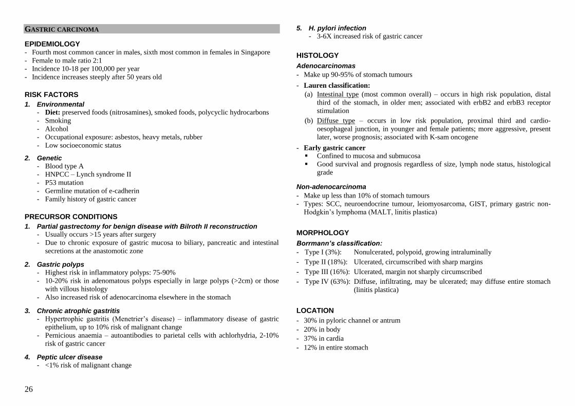
26
GASTRIC CARCINOMA
EPIDEMIOLOGY - Fourth most common cancer in males, sixth most common in females in Singapore
- Female to male ratio 2:1
- Incidence 10-18 per 100,000 per year
- Incidence increases steeply after 50 years old
RISK FACTORS
1. Environmental - Diet: preserved foods (nitrosamines), smoked foods, polycyclic hydrocarbons
- Smoking
- Alcohol
- Occupational exposure: asbestos, heavy metals, rubber
- Low socioeconomic status
2. Genetic
- Blood type A
- HNPCC – Lynch syndrome II
- P53 mutation
- Germline mutation of e-cadherin
- Family history of gastric cancer
PRECURSOR CONDITIONS
1. Partial gastrectomy for benign disease with Bilroth II reconstruction - Usually occurs >15 years after surgery
- Due to chronic exposure of gastric mucosa to biliary, pancreatic and intestinal
secretions at the anastomotic zone
2. Gastric polyps
- Highest risk in inflammatory polyps: 75-90%
- 10-20% risk in adenomatous polyps especially in large polyps (>2cm) or those
with villous histology
- Also increased risk of adenocarcinoma elsewhere in the stomach
3. Chronic atrophic gastritis
- Hypertrophic gastritis (Menetrier‘s disease) – inflammatory disease of gastric
epithelium, up to 10% risk of malignant change
- Pernicious anaemia – autoantibodies to parietal cells with achlorhydria, 2-10%
risk of gastric cancer
4. Peptic ulcer disease
- <1% risk of malignant change
5. H. pylori infection
- 3-6X increased risk of gastric cancer
HISTOLOGY
Adenocarcinomas
- Make up 90-95% of stomach tumours
- Lauren classification:
(a) Intestinal type (most common overall) – occurs in high risk population, distal
third of the stomach, in older men; associated with erbB2 and erbB3 receptor
stimulation
(b) Diffuse type – occurs in low risk population, proximal third and cardio-
oesophageal junction, in younger and female patients; more aggressive, present
later, worse prognosis; associated with K-sam oncogene
- Early gastric cancer Confined to mucosa and submucosa
Good survival and prognosis regardless of size, lymph node status, histological
grade
Non-adenocarcinoma
- Make up less than 10% of stomach tumours
- Types: SCC, neuroendocrine tumour, leiomyosarcoma, GIST, primary gastric non-
Hodgkin‘s lymphoma (MALT, linitis plastica)
MORPHOLOGY
Borrmann’s classification:
- Type I (3%): Nonulcerated, polypoid, growing intraluminally
- Type II (18%): Ulcerated, circumscribed with sharp margins
- Type III (16%): Ulcerated, margin not sharply circumscribed
- Type IV (63%): Diffuse, infiltrating, may be ulcerated; may diffuse entire stomach
(linitis plastica)
LOCATION
- 30% in pyloric channel or antrum
- 20% in body
- 37% in cardia
- 12% in entire stomach
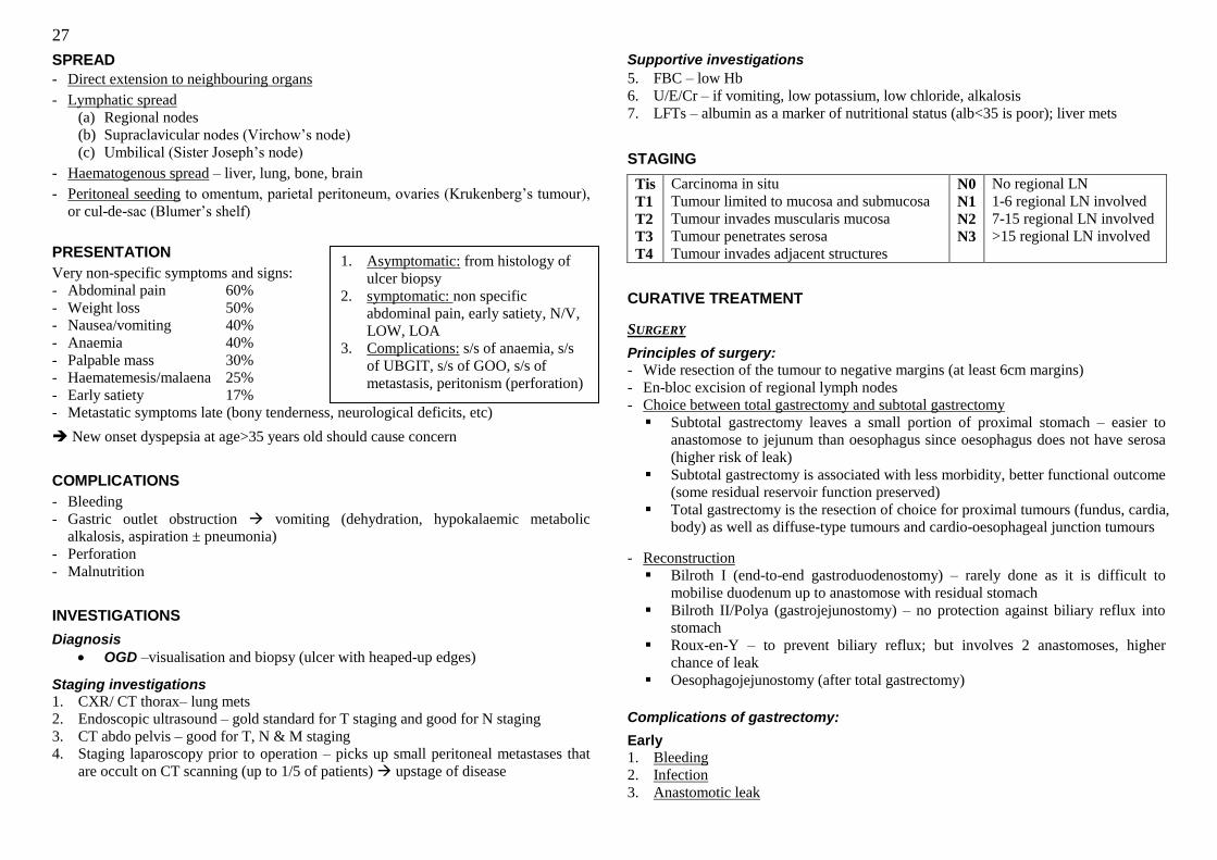
27
SPREAD
- Direct extension to neighbouring organs
- Lymphatic spread
(a) Regional nodes
(b) Supraclavicular nodes (Virchow‘s node)
(c) Umbilical (Sister Joseph‘s node)
- Haematogenous spread – liver, lung, bone, brain
- Peritoneal seeding to omentum, parietal peritoneum, ovaries (Krukenberg‘s tumour),
or cul-de-sac (Blumer‘s shelf)
PRESENTATION
Very non-specific symptoms and signs:
- Abdominal pain 60%
- Weight loss 50%
- Nausea/vomiting 40%
- Anaemia 40%
- Palpable mass 30%
- Haematemesis/malaena 25%
- Early satiety 17%
- Metastatic symptoms late (bony tenderness, neurological deficits, etc)
New onset dyspepsia at age>35 years old should cause concern
COMPLICATIONS
- Bleeding
- Gastric outlet obstruction vomiting (dehydration, hypokalaemic metabolic
alkalosis, aspiration ± pneumonia)
- Perforation
- Malnutrition
INVESTIGATIONS
Diagnosis
OGD –visualisation and biopsy (ulcer with heaped-up edges)
Staging investigations
1. CXR/ CT thorax– lung mets
2. Endoscopic ultrasound – gold standard for T staging and good for N staging
3. CT abdo pelvis – good for T, N & M staging
4. Staging laparoscopy prior to operation – picks up small peritoneal metastases that
are occult on CT scanning (up to 1/5 of patients) upstage of disease
Supportive investigations
5. FBC – low Hb
6. U/E/Cr – if vomiting, low potassium, low chloride, alkalosis
7. LFTs – albumin as a marker of nutritional status (alb<35 is poor); liver mets
STAGING
Tis
T1
T2
T3
T4
Carcinoma in situ
Tumour limited to mucosa and submucosa
Tumour invades muscularis mucosa
Tumour penetrates serosa
Tumour invades adjacent structures
N0
N1
N2
N3
No regional LN
1-6 regional LN involved
7-15 regional LN involved
>15 regional LN involved
CURATIVE TREATMENT
SURGERY
Principles of surgery:
- Wide resection of the tumour to negative margins (at least 6cm margins)
- En-bloc excision of regional lymph nodes
- Choice between total gastrectomy and subtotal gastrectomy
Subtotal gastrectomy leaves a small portion of proximal stomach – easier to
anastomose to jejunum than oesophagus since oesophagus does not have serosa
(higher risk of leak)
Subtotal gastrectomy is associated with less morbidity, better functional outcome
(some residual reservoir function preserved)
Total gastrectomy is the resection of choice for proximal tumours (fundus, cardia,
body) as well as diffuse-type tumours and cardio-oesophageal junction tumours
- Reconstruction
Bilroth I (end-to-end gastroduodenostomy) – rarely done as it is difficult to
mobilise duodenum up to anastomose with residual stomach
Bilroth II/Polya (gastrojejunostomy) – no protection against biliary reflux into
stomach
Roux-en-Y – to prevent biliary reflux; but involves 2 anastomoses, higher
chance of leak
Oesophagojejunostomy (after total gastrectomy)
Complications of gastrectomy:
Early
1. Bleeding
2. Infection
3. Anastomotic leak
1. Asymptomatic: from histology of
ulcer biopsy
2. symptomatic: non specific
abdominal pain, early satiety, N/V,
LOW, LOA
3. Complications: s/s of anaemia, s/s
of UBGIT, s/s of GOO, s/s of
metastasis, peritonism (perforation)
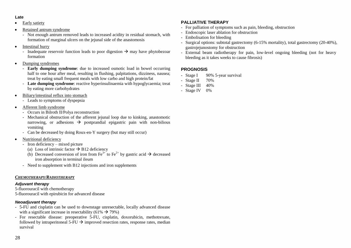
28
Late
Early satiety
Retained antrum syndrome
- Not enough antrum removed leads to increased acidity in residual stomach, with
formation of marginal ulcers on the jejunal side of the anastomosis
Intestinal hurry
- Inadequate reservoir function leads to poor digestion may have phytobezoar
formation
Dumping syndromes
- Early dumping syndrome: due to increased osmotic load in bowel occurring
half to one hour after meal, resulting in flushing, palpitations, dizziness, nausea;
treat by eating small frequent meals with low carbo and high protein/fat
- Late dumping syndrome: reactive hyperinsulinaemia with hypoglycaemia; treat
by eating more carbohydrates
Biliary/intestinal reflux into stomach
- Leads to symptoms of dyspepsia
Afferent limb syndrome
- Occurs in Bilroth II/Polya reconstruction
- Mechanical obstruction of the afferent jejunal loop due to kinking, anastomotic
narrowing, or adhesions postprandial epigastric pain with non-bilious
vomiting
- Can be decreased by doing Roux-en-Y surgery (but may still occur)
Nutritional deficiency
- Iron deficiency – mixed picture
(a) Loss of intrinsic factor B12 deficiency
(b) Decreased conversion of iron from Fe3+
to Fe2+
by gastric acid decreased
iron absorption in terminal ileum
- Need to supplement with B12 injections and iron supplements
CHEMOTHERAPY/RADIOTHERAPY
Adjuvant therapy
5-fluorouracil with chemotherapy
5-fluorouracil with epirubicin for advanced disease
Neoadjuvant therapy
- 5-FU and cisplatin can be used to downstage unresectable, locally advanced disease
with a significant increase in resectability (61% 79%)
- For resectable disease: preoperative 5-FU, cisplatin, doxorubicin, methotrexate,
followed by intraperitoneal 5-FU improved resection rates, response rates, median
survival
PALLIATIVE THERAPY - For palliation of symptoms such as pain, bleeding, obstruction
- Endoscopic laser ablation for obstruction
- Embolisation for bleeding
- Surgical options: subtotal gastrectomy (6-15% mortality), total gastrectomy (20-40%),
gastrojejunostomy for obstruction
- External beam radiotherapy for pain, low-level ongoing bleeding (not for heavy
bleeding as it takes weeks to cause fibrosis)
PROGNOSIS
- Stage I 90% 5-year survival
- Stage II 70%
- Stage III 40%
- Stage IV 0%
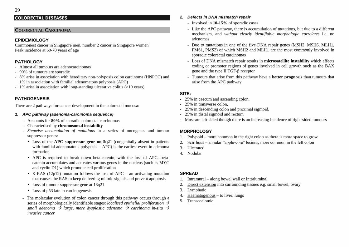
29
COLORECTAL DISEASES
COLORECTAL CARCINOMA
EPIDEMIOLOGY Commonest cancer in Singapore men, number 2 cancer in Singapore women
Peak incidence at 60-70 years of age
PATHOLOGY - Almost all tumours are adenocarcinomas
- 90% of tumours are sporadic
- 8% arise in association with hereditary non-polyposis colon carcinoma (HNPCC) and
1% in association with familial adenomatous polyposis (APC)
- 1% arise in association with long-standing ulcerative colitis (>10 years)
PATHOGENESIS
There are 2 pathways for cancer development in the colorectal mucosa:
1. APC pathway (adenoma-carcinoma sequence)
- Accounts for 80% of sporadic colorectal carcinomas
- Characterised by chromosomal instability
- Stepwise accumulation of mutations in a series of oncogenes and tumour
suppressor genes:
Loss of the APC suppressor gene on 5q21 (congenitally absent in patients
with familial adenomatous polyposis – APC) is the earliest event in adenoma
formation
APC is required to break down beta-catenin; with the loss of APC, beta-
catenin accumulates and activates various genes in the nucleus (such as MYC
and cyclin D1) which promote cell proliferation
K-RAS (12p12) mutation follows the loss of APC – an activating mutation
that causes the RAS to keep delivering mitotic signals and prevent apoptosis
Loss of tumour suppressor gene at 18q21
Loss of p53 late in carcinogenesis
- The molecular evolution of colon cancer through this pathway occurs through a
series of morphologically identifiable stages: localised epithelial proliferation
small adenoma large, more dysplastic adenoma carcinoma in-situ
invasive cancer
2. Defects in DNA mismatch repair
- Involved in 10-15% of sporadic cases
- Like the APC pathway, there is accumulation of mutations, but due to a different
mechanism, and without clearly identifiable morphologic correlates i.e. no
adenomas
- Due to mutations in one of the five DNA repair genes (MSH2, MSH6, MLH1,
PMS1, PMS2) of which MSH2 and MLH1 are the most commonly involved in
sporadic colorectal carcinomas
- Loss of DNA mismatch repair results in microsatellite instability which affects
coding or promoter regions of genes involved in cell growth such as the BAX
gene and the type II TGF-β receptor
- Tumours that arise from this pathway have a better prognosis than tumours that
arise from the APC pathway
SITE:
- 25% in caecum and ascending colon,
- 25% in transverse colon,
- 25% in descending colon and proximal sigmoid,
- 25% in distal sigmoid and rectum
- Most are left-sided though there is an increasing incidence of right-sided tumours
MORPHOLOGY
1. Polypoid – more common in the right colon as there is more space to grow
2. Scirrhous – annular ―apple-core‖ lesions, more common in the left colon
3. Ulcerated
4. Nodular
SPREAD
1. Intramural – along bowel wall or Intraluminal
2. Direct extension into surrounding tissues e.g. small bowel, ovary
3. Lymphatic
4. Haematogenous – to liver, lungs
5. Transcoelomic

30
RISK FACTORS
1. Age >50 years
2. Environmental factors
- Diet: high in red meat, preserved foods (nitrosamines), low in fibre, vitamins,
minerals
- NSAIDs may be protective against CRC
3. Genetic predisposition
(a) Personal history of CRCs
Metachronous colorectal cancers occur at a rate of 3-5% in the first
five years after resection of a primary CRC, while metachronous
adenomas occur at a rate of 25-40%
(b) Family history of CRCs
1 first-degree relative with CRC increases risk of CRC 1.7X,
Risk is further increased if there are 2 1st-degree relatives with CRC,
or if the relative had CRC <55YO
Anyone with a 1st degree relative diagnosed with CRC <45YO, or two
1st or 2
nd degree relatives from the same side of the family with CRC at
any age, should be screened starting at age 45, or 10 years earlier than
the youngest cancer in the family
(c) Polyposis syndromes
FAP and its variants, Gardner‘s and Turcot‘s syndromes: near 100%
risk of cancer formation;
other polyposis syndromes such as Peutz-Jegher‘s, Cronkhite-Canada
have a small increased malignant potential
(d) HNPCC – more common than FAP, accounting for 8% of cancers
4. Ulcerative colitis
- Increased risk after 10 years of disease if the patient has pancolitis; after 15-20
years if the patient has disease limited to the left colon
5. Adenomatous polyps
- Increased risk if there is a personal history of large (>1cm )adenomatous polyps,
and polyps with tubulovillous or villous histology, particularly if multiple (3-6X
increased risk)
- Small (<1cm) tubular polyps do not have increased risk
HISTORY
A)Presentation
- Asymptomatic: detection during screening (e.g. Ulcerative colitis)
- Symptomatic:
Local:
Change in bowel habits/ PR bleed
Frequency:>3x/day=diarrhoea; <2x/week=constipation
Progressive constipation
spurious diarrhoea (alternating constipation & diarrhoea)
mucoid stools
Associated secretions Blood
o Mixed in stool?
o Separate from stool?
Mucus
Consistency – pellet, normal, soft, watery
Colour – black, clay, brown
Effort – tenesmus (suggest a rectal pathology)
Consitutional: LOW, LOA, malaise
Stool Changes
- Bloody diarrhoea – ddx:
IBD
Infective e.g. amoeba, TB, hookworm, Antibiotic Associated Colitis (C.
difficile)
- Haematochezia – ddx:
Diverticulitis
Angiodysplasia (AVM, common in old)
Hemorrhoids
Massive upper GI
Note: Site of CA
- Ascending: anaemia from occult bleeds, no obstructive symptoms usually - Lower down: I/O, bleed, tenesmus
Complications:
Haemorrhage
Symptoms of infection: abscesses, peritonitis
Symptoms of fistula, e.g. faecuria/ pneumaturia/ recurrent UTI
(colovesical fistula)
31
Iron deficiency anaemia: fatigue, SOB, CP, decreased ET,
palpitations, postural giddiness,
Intestinal obstruction: abdominal distension, abdominal colick pain,
vomiting, absolute constipation
Symptoms of peritonism: perforation
Symptoms of metastasis: bone pain/ #, SOB, LL weakness with loss
or bowel and urinary continence, RHC discomfort
B) Strong-risk factors
Positive Family history
- 1 or more 1st deg relative with CC at age <40
- 2 or more 1st/2nd deg same side relative with CC at any age
- Personal or family history of breast, ovarian cancer
FAP history
Amsterdam Criteria for Hereditary Non-Polyposis Colorectal Ca (HNPCC) –
[3, 2, 1]
- ≥3 family members from the same side with CRC or HNPCC related cancer,
one of which is a 1st deg relative of the other 2 (At least 2 of which must
be 1st-degree relatives)
- 2 successive generations
- 1 of the CC must occur prior to age 50
- FAP is excluded
Family history alone = high-risk; FAP / HNPCC = very high-risk
Other risk factors
- Low fibre diet, Red meat
- Obesity
- Ulcerative colitis >10 years
PHYSICAL EXAM
1. Abdominal mass
2. Mass on DRE: (rectal mass)
a. hard, non-tender, polypoid, irregular, contact bleeding
b. distance from anal verge >7cm (internal anal sphincter)
3. Complications, Metastases
- Cachexia
- Pallor at nail beds and conjunctiva (anaemia)
- Jaundice, hepatomegaly (irregular surface & nodular edge)
- Cervical lymphadenopathy – Virchow‘s node
- Bone tenderness, consolidation or effusion, AMS
INVESTIGATIONS
Aims: Diagnose, Stage the cancer & Investigate for complications of cancer
I. DIAGNOSIS
Colonoscopy – first-line investigation(diagnostic & therapeutic)
- Ensure good bowel preparation
- Visualise lesion: size and location of lesion
- Take biopsies of the lesion
- Enables detection of synchronous lesions – synchronous polyps in 30%,
synchronous cancer in 3-5% ( do not do a sigmiodoscopy)
- Enables therapeutic procedures e.g. polypectomy, stenting of obstructed colon
CT colonography
- Next best to colonoscopy: needs IV contrast, air and contrast enema
Double-contrast barium enema (barium + air)
- Classically can see an apple core lesion with barium enema
- Not adequate for diagnostic purposes, may miss small lesions & distal lesions
- Need to insert a rigid sigmiodoscope 10-15cm to instil air & contrast
II. STAGING
(a) CT scan of the abdomen and pelvis for colon tumours
- Local T staging & invasion into bladder, ureter, uterus
- Staging of regional and no-regional lymph note involvement
- Metastases to the liver, obvious peritoneal seeding, omental kinking,
- Look for ascites, hydroureter/ hydronephrosis, intestinal obstruction
(b) Endoscopic ultrasound or transrectal ultrasound for rectal tumour
- Very good for T staging to determine depth of involvement by tumour
- Can also assess local lymph node status
(c) MRI of the tumour - Superior to CT for delineating fat planes in T staging especially in rectal cancer
(d) CXR + CT scan of the chest (e) Bone scan if appropriate (more for Ca breast, lung, prostate, thyroid)
III. COMPLICATIONS
Basic laboratory investigations:
(a) FBC for low Hb, together with iron studies
(b) UECr: fluid and electrolyte abnormatlities from vomiting, or 3rd
space losses
(intraluminal); creatinine may be elevated and hence risk of contrast nephropathy
(c) LFT for derangements caused by metastasis (occur late) – raised bilirubin, ALP
(d) PT/PTT, GXM for surgery
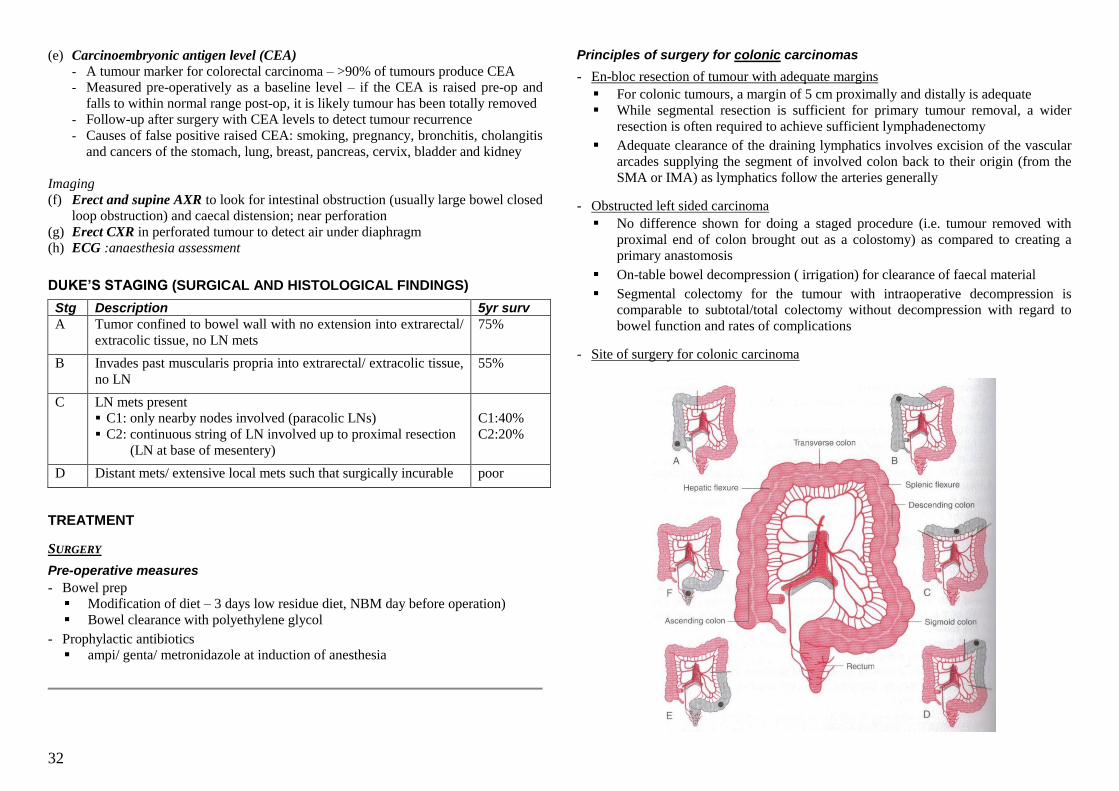
32
(e) Carcinoembryonic antigen level (CEA)
- A tumour marker for colorectal carcinoma – >90% of tumours produce CEA
- Measured pre-operatively as a baseline level – if the CEA is raised pre-op and
falls to within normal range post-op, it is likely tumour has been totally removed
- Follow-up after surgery with CEA levels to detect tumour recurrence
- Causes of false positive raised CEA: smoking, pregnancy, bronchitis, cholangitis
and cancers of the stomach, lung, breast, pancreas, cervix, bladder and kidney
Imaging
(f) Erect and supine AXR to look for intestinal obstruction (usually large bowel closed
loop obstruction) and caecal distension; near perforation
(g) Erect CXR in perforated tumour to detect air under diaphragm
(h) ECG :anaesthesia assessment
DUKE’S STAGING (SURGICAL AND HISTOLOGICAL FINDINGS)
Stg Description 5yr surv
A Tumor confined to bowel wall with no extension into extrarectal/
extracolic tissue, no LN mets
75%
B Invades past muscularis propria into extrarectal/ extracolic tissue,
no LN
55%
C LN mets present
C1: only nearby nodes involved (paracolic LNs)
C2: continuous string of LN involved up to proximal resection
(LN at base of mesentery)
C1:40%
C2:20%
D Distant mets/ extensive local mets such that surgically incurable
poor
TREATMENT
SURGERY
Pre-operative measures
- Bowel prep
Modification of diet – 3 days low residue diet, NBM day before operation)
Bowel clearance with polyethylene glycol
- Prophylactic antibiotics ampi/ genta/ metronidazole at induction of anesthesia
Principles of surgery for colonic carcinomas
- En-bloc resection of tumour with adequate margins
For colonic tumours, a margin of 5 cm proximally and distally is adequate
While segmental resection is sufficient for primary tumour removal, a wider
resection is often required to achieve sufficient lymphadenectomy
Adequate clearance of the draining lymphatics involves excision of the vascular
arcades supplying the segment of involved colon back to their origin (from the
SMA or IMA) as lymphatics follow the arteries generally
- Obstructed left sided carcinoma
No difference shown for doing a staged procedure (i.e. tumour removed with
proximal end of colon brought out as a colostomy) as compared to creating a
primary anastomosis
On-table bowel decompression ( irrigation) for clearance of faecal material
Segmental colectomy for the tumour with intraoperative decompression is
comparable to subtotal/total colectomy without decompression with regard to
bowel function and rates of complications
- Site of surgery for colonic carcinoma
33
Tumour site Surgery Structures involved
- Caecum
- Ascending colon
Right
hemicolectomy
- Excision of caecum and ascending colon
- Division of ileocolic and right colic arteries, and
the right branch of the middle colic artery
- Hepatic flexure
- Transverse colon
near the hepatic
flexure
Extended right
hemicolectomy
- Excision of caecum, ascending colon, and
proximal transverse colon
- Division of ileocolic and right colic arteries, and
the middle colic artery at its origin
- Mid-transverse
colon
Transverse
colectomy
- Excision of transverse colon
- Division of middle colic artery
- Transverse colon
near splenic
flexure
Left segmental
colectomy
- Excision of distal transverse colon and proximal
descending colon
- Division of supplying vessels – left branch of
middle colic artery, left colic artery
- Descending colon Left
hemicolectomy
- Excision of descending colon
- Division of left colic and inferior mesenteric
arteries
- Sigmoid colon
Sigmoid
colectomy
High anterior
resection
- Excision of sigmoid colon
- Division of inferior mesenteric artery and/or
sigmoid branches
- High AR is above the peritoneal reflection
Principles of surgery for rectal carcinomas
- En-bloc resection with adequate margins
For rectal tumours a margin of 5 cm proximally and 2 cm distally is adequate (as
it has been found that lymphatic spread of rectal tumours is predominantly in the
proximal direction)
Radial margins are also important as there is a zone of downward spread within
the mesorectum (peritoneal investment of the upper rectum; just anterior to the
sacrum)
- Sphincter-sparing versus loss of sphincter
The anal sphincter can be spared if the distal margin is >1-2cm above the level
of the sphincter complex, usually taken to be at the level of the dentate line
(which is 5cm above the anal verge) i.e. distal margin of the tumour must be
>7cm from the anal verge
Sphincter-sparing low anterior resection (below the peritoneal reflection);
ultra low AR if just above the anal sphincter
Sphincter-sacrificing surgery abdominoperineal resection (entire anus and
sphincter complex is dissected, with the creation of an end colostomy)
- Reconstruction
Formation of a straight coloanal anastomosis in anterior resections is associated
with poor function due to the lack of reservoir function
Creation of a colonic J-pouch using the proximal end of colon (the end of the
colon is folded back on itself to form a J, and the two limbs opened and stitched
together to form a pouch, the apex of the J being anastomosed to the anus) is
associated with improved post-operative function
Coloplasty is another alternative that is equivalent to colonic J-pouch (the distal
colon is cut longitudinally but sewn transversely, widening the diameter at that
segment to form a small pouch), done when there is difficulty creating the
colonic J-pouch
- Mesorectal excision
Proximal rectal tumours – 5cm distal margin of mesorectal excision
Mid-rectal tumours – wide mesorectal excision of at least 4cm distal to the
tumour
Lower rectum tumours – total mesorectal excision required (complete excision
of the intact visceral mesorectal tissue to the level of the levators)
- Extended resections
For locally advanced, adherent tumours (T4), multivisceral resection of organs
involved (pelvic exenteration) is associated with improved local control and
overall survival compared with standard resection, though high morbidity of 25-
50% is associated
Consider neoadjuvant chemoradiotherapy prior to surgery to downstage disease
- Stoma creation
A defunctioning loop ileostomy (or loop colostomy) is usually created during an
low AR as the manipulation of the colon deep within the pelvic cavity causes
increased risk of an anastomotic leak & also poorer blood supply to anastomosis
A defunctioning stoma does not protect against anastomotic leak, but mitigates
against disastrous complications of faecal peritonitis should a leak occur
Closed in 2-6/12 after check with gastrograffin reveals no leak
- Neoadjuvant chemoradiotherapy for rectal tumours
Neoadjuvant RT with 5-fluorouracil can downstage tumour significantly
ability to preserve sphincter, ability to resect previously unresectable tumour,
reduce local recurrence
Good evidence for ≥T3 tumours: benefits > risk of RT (esp. that pt becomes too
ill to be operated on)
34
Not possible for colon carcinomas due to risk of small bowel radiation if given
above peritoneal reflection
- Surgical treatment according to stage
Stage of disease Treatment
T1 Involvement of submucosa, but no
penetration through muscularis propria
Local excision (AR or APR)
T2 Invasion into, but not penetration
through, muscularis propria
a) Local excision + adjuvant
Chemo/RT OR
b) radical resection
T3 Penetration through muscularis propria
into subserosa (if present), or pericolic
fat, but not into peritoneal cavity or
other organs
Neoadjuvant chemo / RT before
radical resection
T4 Invasion of other organs or
involvement of free peritoneal cavity
Operative complications
Immediate (<24h) Damage to other organs e.g. ureters
Early (<30 days) Wound infection
Bleeding
Abscess
Anastomosis breakdown/leak faecal peritonitis
Early stoma complications
Late (>30 days) Diarrhoea
Impotence (damage of pelvic nerves)
Adhesions (I/O)
Anastomotic stricture
Late stoma complications
Surgery for palliation
- Resection of primary for palliation of symptoms such as bleeding, perforation,
obstruction or pain
- Resection of asymptomatic primary is controversial, but may confer survival benefit
in a select group of patients where metastatic tumour burden is restricted to one side
and liver involvement is not extensive
Surgery for metastases
- Isolated liver metastases (synchronous or metachronous) may be resected with
survival benefit; neoadjuvant chemotherapy can be given to downstage the
metastases if they are initially resectable
- Lung metastases usually indicate systemic dissemination of disease, but in the rare
setting that there is an isolated lung secondary, resection can provide survival benefit
Surgery for recurrence
- Loco-regional recurrence, if detected early with adequate resection, can confer
survival benefit
RADIOTHERAPY
- Role as neoadjuvant therapy in rectal cancer to downstage tumour
- Post-operative adjuvant therapy in stage II or III rectal cancer
CHEMOTHERAPY
Adjuvant therapy
- Colon cancer: 5-FU + folinic acid (leucovorin) for 6 months, or 5-FU + levamisole
for 12 months in Duke‘s C cancer (node positive); not recommended in Duke‘s B or
less
- Rectal cancer: post-operative adjuvant therapy in combination with radiotherapy in
stage II or III disease (5-FU based regimen used)
Palliative therapy
- 5-FU in combination with folinic acid is first-line therapy
- Alternatives for first-line therapy: Raltitrexed when 5-FU is not tolerated;
capecitabine or UFT (uracil combined with tegafur) plus folinic acid
FOLLOW UP
- Follow-up visits 3-monthly for the first 2 years, then 6-monthly for the next three
years, and subsequently yearly, measure CEA at each visit
- Yearly colonoscopy
- CXR and liver ultrasound to detect metastases (recommended frequency not known)
35
STOMA PRINCIPLES Stoma: opening of a luminal organ into the external environment Indications
1. For input: feeding (Percutaneous endoscopic gastrostomy)
2. For output: decompression/ lavage, defunctioning/ diversion, draining/
exteriorization (urine, faeces) Nursing intervention
- Stoma nurse to perform counselling & discuss best site for stoma placement
Stoma siting
- Over the rectus sheath decreases the risk of prolapse,
- Away from the surgical incision risk of wound contamination and infection
- Away from skin creases or bony prominences stoma wafer can be flush with the
skin (any gaps between skin and wafer leakage of fluid skin excoriation & infx)
- Away from old surgical scars risk of hernia
- Sited for easy accessibilty i.e. not under a large fold of abdominal fat
- Intra-operatively, avoid tension over the stoma to marked site causes decreased
vascularity of the stoma stoma necrosis
Types of stomas Permanent (end colostomy)
- When patient or surgeon factors
are against reversal of stoma (relative)
- When no distal bowel remaining (absolute)
After abdomino-perineal resection for Low rectal/ anal tumor
After Panproctocolectomy without ileal pouch anal anastomosis Temporary
- Decompression – relief of bowel obstruction causing proximal dilatation
- Defunctioning – to reduce effects of anastomotic leak
Esp. after low anterior resection, where risk of anastomotic leakage is high 2o to
poor blood supply to anastomotic site
Usually loop ileostomies or colostomies with 2 openings (ileostomies usually on
the right side, colostomies in the epigastric/hypochondriac [transverse colostomy]
or left side)
- To rest an inflamed distal portion e.g. acute Crohn‘s
Colostomy (vs ileostomy)
usually flushed with skin, (vs protrudes 3cm „spout‟, as ileal contents are
corrosive, to prevent contact with skin)
firm brown faceal output (vs watery greenish ilieal output)
larger diameter of stoma (vs smaller diameter in ileostomy)
Stoma Complications
Early
- Necrosis of terminal bowel (stoma appears dusky; check by intubating with a glass
tube into the stoma to look at colour of mucosa) refashion stoma
- Obstruction e.g. fecal impaction explore with finger, enema
- Leakage skin erosion, parastomal infection resite
- Bleeding
- Stoma diarrhoea (high output) correct water & electrolyte imbalance, add anti-
motility agent
Late
- Prolapse of bowel refashion
- Parastomal hernia (+ve cough impulse) refashion
- Stenosis (unable to pass finger through) refashion
- Retraction refashion
- Skin excoriation
- Psychological problems
ASSOCIATED CONDITIONS
I. Familial adenomatous polyposis (FAP)
- 1 in 10,000, autosomal dominant inheritance
- Germline mutation of APC gene on 5q21
- >100 adenomatous polyps all over colon; polyps take 5-6 yrs to turn malignant
- 50% patients will have polyps by 16yrs; 90% will have colorectal CA by 45yrs
- Other sites for polyps: stomach, duodenum
- Extraintestinal manifestations
Skin: Epidermoid cysts, Lipoma
Bone: Osteoma of skull/ mandible, Dental abnormalities
Eye: Congenital hypertrophy of retinal pigment epithelium (CHRPE)
Other tumours:
Desmoid tumours (intra-abdominal tumours, treated with CT, RT or HT.)
Thyroid cancer (follicular or papillary type)
Periampullary CA
- Diagnosis Colonoscopy showing >100 polyps
Genetic testing
- Surveillance
Yearly colonoscopy for at-risk family members from 12y onwards
5 yearly OGD for surveillance of Periampullary Cancer.
36
Genetic testing of at-risk family members
Affected members should undergo prophylactic proctocolectomy with ileal
pouch anal anastomosis (IPAA – involves folding loops of ileum back on
themselves and stitching or stapling them together to form a reservoir pouch
which is them anastomosed to the anus) at ~ 20 YO
Subtotal colectomy is an option if the rectum is relatively spared of polyps
Hereditary Non-Polyposis Colorectal CA (HNPCC)
- Divided into Lynch syndrome I or Lynch syndrome II based on clinical features
- Tumours usually proximal to splenic flexure (~70% proximal to splenic flexure)
- Tumours tend to arise from polyps which are commonly flat, with villous histology
- Resultant tumour is often poorly differentiated
- Lynch syndrome type II is associated with increased risk of cancer elsewhere, most
commonly endometrial cancer, and also ovarian, gastric, small bowel, hepatobiliary,
and renal pelvis/ureter cancers
o Offer a THBSO with total colectomy if CRC detected
- Diagnosis is based on the Amsterdam criteria – see above
- Surveillance – 1-3yrly colonoscopy starting at 20 years old
Ulcerative colitis
- Screening – yearly colonoscopy starting after 10 years of UC
DIVERTICULAR DISEASE PATHOLOGY – acquired herniation of colonic mucosa through muscular wall, with a
covering of colonic serosa
TERMS - Diverticulosis coli – presence of acquired pseudodiverticula
- Diverticular disease – symptomatic diverticulosis coli
- Diverticulitis – inflammation of diverticula
EPIDEMIOLOGY - Increases with age; up to 25% in >70YO
- Majority are asymptomatic; 10-30% are symptomatic
- Risk factors – dietary fibre & genetics
- Site – majority are in the sigmoid colon (right sided are thought to be genetic; in
Asians; left sided are more in Caucasians; not in rectum as taeni coli has fused)
PATHOGENESIS
1. Increased intraluminal pressure
- Associated with lack of dietary fibre
2. Degenerative changes in colonic wall
- Usually at point of entry of terminal arterial branches where serosa is weakest
- Associated with weakening of collagen structure with age
PRESENTATIONS
1. Acute diverticulitis
- Symptoms: LLQ pain, N/V, Constipation / diarrhoea
- Signs: Low grade fever, Tender palpable mass
- Investigation: WBC
2. Chronic diverticulitis
- Recurrent LIF pain
- Irregular bowel habit
- Passage of mucus PR
3. Complicated diverticulitis
a. Perforation
b. Paracolic abscess / inflammatory mass – 2o to localized perforation
c. Bowel obstruction – 2o to structure or adherence to a diverticular mass
d. LGIT haemorrhage – ulcerated vessel @ neck of diverticulum; torrential
e. Fistula formation (commonest: colovesical fistula) – 2o to pericolic abscess
discharging, operation or drainage of pericolic abscess. May present with
urinary symptoms. Others – colo-cutaneous, colo-uterine, colo-enteric, colo-
vaginal
STAGING - Hinchey classification of acute diverticulitis – need for surgery is reflected by degree
of infective complications
Stage 1 Pericolonic /
Mesenteric abscess
- ABx, NBM, IV fluids
- Consider 1 stage surgery after acute episode –
resection of affected bowel segment with primary
anastomosis
Stage 2 Pelvic / retroperitoneal
abscess
- Percutaneous drainage
- Elective 1 stage surgery
Stage 3 Purulent peritonitis - 2 stage operation – Hartmann‘s procedure (partial
colectomy + diverting end colostomy & rectal
stump formation) + secondary re-anastomosis 3
months later
Stage 4 Faecal peritonitis
Note: current controversy of management for stage 3: haartman‟s vs segmental resection with primary anastomosis with or without defunctioning ileostomy
37
Presentation Clinical features Investigations Differentials Management
Acute
Diverticulitis
- LIF pain – colicky, progressing to
constant, relieved by defecation
- LIF tenderness
- Palpable LIF mass
- Nausea
- Pyrexia
- Increase pulse rate
- FBC – leucocytosis, ↑ ESR
- Erect CXR to rule out perforation
- AXR – ileus, air-fluid level w/in an abscess
- Barium enema
- CT scan w triple contrast Contrast: IV for vascular lesions, oral for small
bowels, enema for large bowels
Features – diverticula elsewhere, confirm colitis
(mesenteric fat infiltration, concentric bowel
thickening) but only suggest diverticulitis, pelvic
abscess, free gas, extravasated contrast
Cannot tell if inflm is due to diverticula
- Laparoscopy – if diagnosis is in doubt
- Avoid colonoscopy as risk of perforation is high
- See Ddx to RIF/LIF pain
- GI: appendicitis, colitis, GE,
IBS, IBD, mesenteric
ischaemia/ adenitis, Ca
colorectal
- Uro: UTI, stones, cancer,
cyst/ Angiomyolipoma,
hydronephrosis
- Obgyn: PID, torsion of cyst/
ovary, endometriosis,
ectopic pregnancy
- #: #, infx, inflm,
Conservative
- Bed rest
- NBM, IV fluid
- Broad-spectrum antibiotics –
augmentin or metronidazole or
ciprofloxacin
- Antispasmodics
After acute phase has settled
- Ba enema &/or Colonoscopy –
confirm dx & exclude CA colon
Role of surgery: see behind
Chronic
Diverticulitis
Not a
common
clinical entity
- Recurrent LIF pain
- Irregular bowel habits – constipation
& bouts of diarrhoea
- Passage of mucus PR
- Ruled out cancer, IBD, ureteric colic,
Msk pain etc.
- Rigid sigmoidoscopy – oedematous mucosa &
rigidity of rectosigmoid junction
- Flexible sigmoidoscopy – diverticular orifices
- Barium enema – ‗saw-tooth‘ appearance,
diverticula, strictures
- Colonoscopy – exclude differentials (i.e. Ca colon)
- CA colon – may coexist.
Hard to differentiate –
therefore, ALWAYS
exclude CA colon e.g.
histology after bowel
resection
- Ischaemic colitis
- Radiation colitis
- Colonic endometriosis
Conservative – see above
Surgical
Indications:
- Severe / recurrent attacks
- Possible CA colon
- Segmental resection of affected
colon + anastomosis
Generalised
peritonitis /
perforation
- Acute onset abdominal pain – severe
& continuous
- Abdominal guarding & rigidity
- Vomiting
- Tachycardia
- Pyrexia
- FBC – ↑ TW, ↑Hb (dehydration)
- U/E/Cr – dehydration & ARF
- CXR – free gas under diaphragm
- Other causes of peritonitis –
perforated PUD/
appendicitis/ bowel/ ectopic
pregnancy, ishaemic bowel,
acute pancreatitis, ruptured
AA/ hepatoma, torsion of
testis/ ovary, pyonephrosis
Mgmt as for acute abdomen
- Resuscitate
- Surgical
Peritoneal toilet
Resection of affected segment
End sigmoid colostomy
(Hartmann‘s procedure)
Pericolic
abscess
- May follow acute diverticulitis
- LIF tenderness & guarding
- LIF mass – may be detected on DRE
- Swinging fever
- FBC – ↑ TW
- CT – differentiate between inflammatory phlegmon
& pericolic abscess
- CT/US guided percutaneous
aspiration
- Surgery – evacuation of pus ±
resection of affected segment
Persistent
inflammatory
mass
- LIF pain, tenderness & palpable mass
- Fever
- Malaise
Small bowel
I/O
- Usually temporary, due to
attachment of enteric loop against
area of acute diverticulitis
- Surgery if does not resolve

38
Presentation Clinical features Investigations Differentials Management
Large bowel
I/O
- PHx of recurrent acute diverticulitis
or irregular bowel habit
- Colicky abdominal pain, constipation
& abdo distension
- AXR – dilated bowels proximal to stenosis
- Water soluble contrast enema
- CA colon - NBM, Drip & suck
- Surgery – Resection ± primary
anastomosis
Hemorrhage - Usually in the elderly who have
higher density of sigmoid diverticula
- Massive bleed (altered blood ± clots;
not melena) usually right-sided
- Colicky pain as blood is irritative &
causes spasm
- Invx as for LGIT bleed – resus, invesigations +
colonoscopy & angiography (both diagnostic AND
therapeutic value)
- ± on-table enteroscopy if required
- ± tagged RBC scan (not as sensitive compared to
angiogram)
- Anorectal bleed
- Angiodysplasia
- Ischaemic colitis
- Colorectal CA
- Colitis (inflm or infx)
- UBGIT
- Coagulopathy
- Resuscitate & correct coagulopathy
- Colonoscopic management:
adrenaline injection, endoclips on
bleeding vessel, heat coagulation
- Radiologic embolisation of site of
bleeding with temp foam material
via angiography
- Surgery – segmental resection;
total colectomy if unable to localise
bleed
Vesicocolic
fistula
- PHx of chronic diverticulitis & UTI
- Hx of dysuria, freq, haematuria,
pneumaturia, faecaluria
- UFEME & urine c/s: confirm UTI and organisms
- Cystoscopy – cystitis
- Sigmoidoscopy – usually normal
- KUB – air in bladder
- Barium enema – diseased diverticular bowel
segment
- Other causes of fistula – CA
colon, CA bladder, Crohn‘s
disease, post-irradiation
necrosis
- Surgery – Resection of affected
colon + anastomosis + closure of
bladder fistula opening
Outcomes: well or derteriorate requiring surgery, recurrent episodes, stricture & subacute IO (offer surgery)
Indications for emergency operation
1. Sepsis from abscess or faecal peritonitis
2. Perforation
3. Diverticulitis not responding to conservative management
4. Obstruction with pending perforation – need to rule out cancer at the same time
5. Emergency bleed (controversial clamping both side & look for active bleed into segment segmental resection)
a. Haemodynamically unstable with failure of embolization
b. Need > 4 units of PCT
c. Previous bleed
Indications for elective operation
1. Stricture
2. Fistula
3. Recurrent attacks – occurs in 30% of patients after 1st episode. a/w higher mortality & complication rates
4. Young patientss <40YO – high recurrence rates
5. Immunocompromised patients (e.g. renal transplant) – may not show S/S of acute attack or complications
Advice to patients: - 70% of patients will not have recurrence after first attack
- Advise high fibre diet & to drink lots of fluid
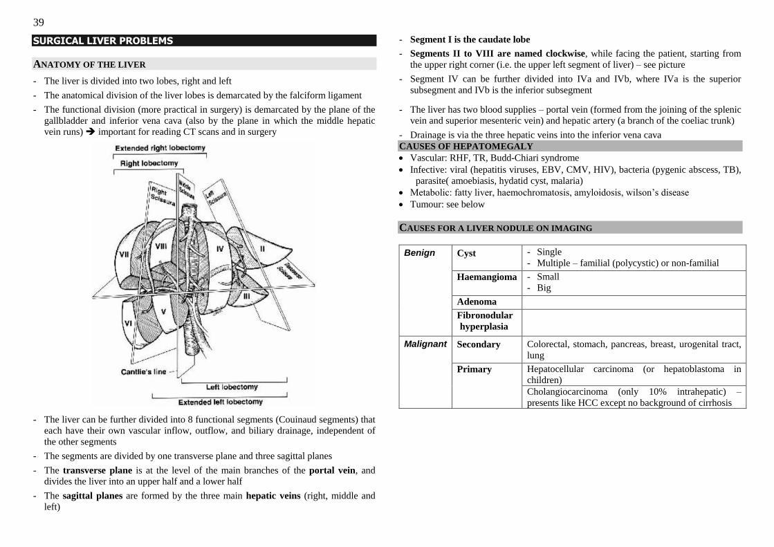
39
SURGICAL LIVER PROBLEMS
ANATOMY OF THE LIVER
- The liver is divided into two lobes, right and left
- The anatomical division of the liver lobes is demarcated by the falciform ligament
- The functional division (more practical in surgery) is demarcated by the plane of the
gallbladder and inferior vena cava (also by the plane in which the middle hepatic
vein runs) important for reading CT scans and in surgery
- The liver can be further divided into 8 functional segments (Couinaud segments) that
each have their own vascular inflow, outflow, and biliary drainage, independent of
the other segments
- The segments are divided by one transverse plane and three sagittal planes
- The transverse plane is at the level of the main branches of the portal vein, and
divides the liver into an upper half and a lower half
- The sagittal planes are formed by the three main hepatic veins (right, middle and
left)
- Segment I is the caudate lobe
- Segments II to VIII are named clockwise, while facing the patient, starting from
the upper right corner (i.e. the upper left segment of liver) – see picture
- Segment IV can be further divided into IVa and IVb, where IVa is the superior
subsegment and IVb is the inferior subsegment
- The liver has two blood supplies – portal vein (formed from the joining of the splenic
vein and superior mesenteric vein) and hepatic artery (a branch of the coeliac trunk)
- Drainage is via the three hepatic veins into the inferior vena cava
CAUSES OF HEPATOMEGALY
Vascular: RHF, TR, Budd-Chiari syndrome
Infective: viral (hepatitis viruses, EBV, CMV, HIV), bacteria (pygenic abscess, TB),
parasite( amoebiasis, hydatid cyst, malaria)
Metabolic: fatty liver, haemochromatosis, amyloidosis, wilson‘s disease
Tumour: see below
CAUSES FOR A LIVER NODULE ON IMAGING
Benign
Cyst
- Single
- Multiple – familial (polycystic) or non-familial
Haemangioma - Small
- Big
Adenoma
Fibronodular
hyperplasia
Malignant
Secondary
Colorectal, stomach, pancreas, breast, urogenital tract,
lung
Primary Hepatocellular carcinoma (or hepatoblastoma in
children)
Cholangiocarcinoma (only 10% intrahepatic) –
presents like HCC except no background of cirrhosis
40
HEPATOCELLULAR CARCINOMA
EPIDEMIOLOGY
- Annual incidence in Singapore is 18/100,000 in males, and 4.6/100,000 in females
- 3rd
most cancer among males, overall 4th
most common cancer
- Men: female = 3:1
- Peak age of onset: 30-40 years old
- 1o liver cancers are mainly HCCs (85%), with a small proportion of intrahepatic
cholangiocarcinoma (6%)
AETIOLOGY AND RISK FACTORS
- Hepatitis B infection (high HBV DNA load, HBeAg positivity increase the risk)
- Hepatitis C infection
- Cirrhosis (of which cirrhosis resulting from hep B, hep C and haemochromatosis are
associated with the highest risk)
- Others: Aflatoxin ingestion, α-1- antitrypsin deficiency, haemochromatosis, PBC,
anabolic steroids, schistosomiasis)
PATHOLOGY
- Pathogenesis involves a chronic inflammatory process or ongoing hepatocellular
damage with high cellular regeneration, which leads to increased rates of genetic
mutation in the cells and accumulation of these mutations leading to carcinoma
formation
- Two histological subtypes:
Nonfibrolamellar – associated with HBV and cirrhosis
Fibrolamellar – associated with younger patients, more common in females, no
association with hep B or cirrhosis, 70% resectable, good prognosis
- Metastasises to lymph nodes, bones, lungs and adrenals
PRESENTATION
1. Asymptomatic
- During screening (ultrasound) for chronic hepatitis B carrier
- Investigations for liver cirrhosis
- Incidentally found on imaging of the abdomen
2. Local signs & symptoms
- Upper abdominal pain (2o to capsular distension)
- Early satiety/ vomiting (likely 2o to compression)
- Pyrexia (central tumour necrosis)
- Hepatomegaly
- Constitutional: LOW, LOA, malaise
- Budd-Chiari syndrome: occlusion of the hepatic, intrahepatic or portal vein
causing portal hypertension & congestive hepatopathy
- Jaundice (5-10%)
Cholestatic: invasion/compression of intrahepatic ducts or extrahepatic
compression by metastatic LN
Hepatic: a/w pre-existing cirrhosis or acute flair of chronic hepatitis
3. Tumour rupture (<3%)
- Severe abdominal pain (peritonism), pallor with shock [ddx: ruptured AAA]
50% mortality; Rx with TAE; reports of peritoneal seeding in survivors
- US +ve for peritoneal fluid, DPL +ve for blood
4. Liver decompensation (on top of underlying cirrhosis)
- Worsening liver function (encephalopathy, jaundice, pruritis, coagulopathy,
oedema) suspect HCC when there is decompensation of liver cirrhosis
- Worsening portal hypertension: Ascites, LL oedema, haematemesis & melena 2o
to bleeding varices (COJ & gastric)
- Hepatorenal syndrome in late stages of liver failure
Stages of Hepatic Encephalopathy
Stage 1 Sleep-wake cycle inversion
Stage 2 Lethargy, personality change, asterixis, ataxia
Stage 3 Confusion, acalculia, paranoia, agitation, Babinski, clonus
Stage 4 Coma
5. Metastases (low incidence in HCC, mortality rarely from mets)
- Bone pain, Dyspnoea
6. Paraneoplastic syndromes
Hypoglycaemia (high metabolic demands of tumour),
Erythrocytosis (tumour produces erythropoietin),
Hypercalcaemia, watery diarrhoea, etc.
INVESTIGATIONS
Basic laboratory investigation:
o FBC (low Hb from BGIT, raised TW in SBP)
o UECr (dehydration, 3rd
spacing of fluids, use of diuretics for ascites,
r/o nephropathy for contrast imaging)
o LFT (low albumin, high bilirubin, raised ALP)
o AFP (Normal <5; WHO criteria >400; diagnostic & trending; note
false +ve: pregnancy, infants, cirrhosis, hepatitis, teratoma)
o Hepatitis markers (look for carrier status or chronic infection)
41
Imaging:
o Triphasic CT scan (see below)
DIAGNOSIS
Biopsy is usually not performed due to risk of tumour seeding (1-2% risk) along the
needle track – diagnosis is based on clinical, biochemical and radiological tests
WHO criteria for HCC:
(a) Risk factors for HCC e.g. hep B/C carrier
(b) Characteristic CT findings (of a hypervascular lesion)
(c) Raised AFP (>400)
1. Alpha-foetoprotein (AFP): elevated in 80% 2. Triphasic CT scan [gold standard investigation]
- CT liver scan at three different times after IV contrast:
(a) Arterial phase –contrast fills arteries: aorta (& HCC, bleeds)lights up, IVC
and portal vein are dark
(b) Portal venous phase (most CT scans taken at this phase) – contrast enters
portal system so portal vein is as bright as the aorta
(c) Delayed phase – contrast drains out, so none of the vessels in the liver are
lighted up
- Characteristic feature of HCC is enhancement in the arterial phase (have a
rich hepatic arterial supply) with rapid contrast washout in the portal venous
phase (hypodense)
Haemangioma: capsular enhancement in delayed phase
Metastasis: enchancement in portal venous phase
- In a patient with hepatitis B/C and raised AFP, a liver lesion on imaging should
be considered HCC until proven otherwise
- CT can also look for nodal involvement, and metastases to the adrenals
3. Dynamic MRI scan of the liver
- Adjunct investigation done when CT findings are equivocal
- Images liver in greater detail, can be used to exclude benign conditions
4. Hepatic angiogram with lipiodol and post-lipiodol CT scan
- Lipiodol will be retained in HCC even after many days as the HCC does not
contain Kupffer cells to ingest lipiodol
- Hepatic angiogram may reveal abnormal blood vessels within the HCC
- CT scan of the liver weeks after lipiodol ingestion will pick up the areas of
tumour (where the pre-lipiodol CT may not have demonstrated the tumour
clearly)
5. If indicated, investigations to look for GI primary
- CEA, CA 19-9, endoscopy
STAGING (looking for metastases)
1. CXR, CT thorax: lung
2. CT abdopelvis: liver and adrenals, peritoneum and LNs
3. bone scan if clinically indicated: bone
TREATMENT
SURGERY (ACHIEVE TUMOUR REMOVAL WITH GOOD LIVER FUNCTION)
- Only about 10-20% of patients with HCC will have disease amenable to surgery
- The only curative treatment for HCC is surgical removal of the tumour
- 2 surgical possibilities:
(a) Resection of tumour (partial hepatectomy)
(b) Liver transplantation Hepatectomy
- Problem in patients with cirrhosis is that there is already a “field-change” effect in
the liver, thus a new tumour can still develop in the remnant liver
- Requires a fine balance between adequate resection margins and preservation of
sufficient functional liver to prevent liver failure
- Good immediate and short-term results, but not long term (<30% 5-year survival)
due to occurrence of new primaries in the cirrhotic liver
Transplantation
- Milan criteria for transplantation (>75% 5-year survival if followed)
(a) Single tumour 5cm or smaller, or 3 or less tumours none larger than 3cm
(b) No evidence of gross vascular invasion
(c) No regional nodal or distant metastasis
- Problems with availability of donor organ – the disease might have progressed past
being suitable for transplant by the time donor organ is available
Possibility of “bridging therapy” such as radiofrequency ablation to shrink
disease and prevent progression until donor liver is available
- In hepatitis B carriers, there is a risk for reinfection of the donor liver (high risk
factors are HBeAg positivity, high HBV DNA levels) – can be aggressively treated
with anti-viral drugs 2 months before transplant and anti-HBV immunoglobulin long-
term after transplant
42
Factors affecting resectability:
1. Stage of disease
- Metastatic disease is not suitable for resection
- Multicentric disease affecting both lobes is a contraindication to hepatectomy
2. General fitness for operation
3. Liver function pre-operatively
- Cirrhotic patients have higher risk of post-operative mortality (4-14%) compared
to non-cirrhotic patients (0-4%) due to complications such as liver failure,
bleeding and infection
- Use of indocyanin green (ICG) – the percentage of ICG remaining in the liver
after 15 minutes indicates the level of liver function. If >15% remains after 15
minutes, the patient cannot tolerate major liver resection (>3 segments removed)
- CT volumetry: residual liver function calculated with a CT liver scan via a
computer programe
- If patient has cirrhosis, assess the Child‘s status only Child’s A and good
Child’s B can be considered for resection
Child-Pugh classification of CLD/cirrhosis
1) Prognosis
2) Strength of medical treatment
3) Necessity of liver transplant
4. Residual liver function post-operatively (at least 20%)
- Dependent on tumour size and how much of the liver it takes up, because tumour
is non-functional
- A large tumour taking up most of the liver segments being resected translates to
smaller amount of functional liver tissue being resected, while a small tumour
means that more functional tissue is removed with the same resection margins
5. Degree of portal hypertension (>10mmHg)
- Resection of the liver results in worsened portal hypertension since the effective
portal venous capillary bed has decreased increased resistance to flow
6. Location of tumour
- Has to be located in a suitable location for resection
PALLIATIVE THERAPY
Loco-regional ablation
(a) Radiofrequency ablation (RFA) – best results for locoregional strategies
(b) Percutaneous ethanol injection
(c) Cryotherapy Intra-arterial therapy
(a) Transarterial chemoembolisation (TACE)
(b) Transarterial embolisation (TAE)
(c) Selective Intrahepatic Radiation – Yttrium-90 radioactive beads
Systemic therapy – limited results; Sorafenib imporves median survival by 3/12
SCREENING FOR CHRONIC HEPATITIS CARRIERS
- Combination of 6/12 to yearly ultrasound with AFP levels
- US is operator dependent & may miss certain areas of the liver where imaging is
difficult, but it is not associated with radiation exposure
- AFP is also not a perfect screening test as 20% of HCC will not have raised AFP
- Thus the combination of ultrasound and AFP can increase the sensitivity and
specificity of screening
- Frequency of screening is controversial, but should be increased in patients at
increased risk – HbeAg positivity, high HBV DNA levels
- Important as it detects smaller and resectable HCCs increasing survival from 26
to 88/52
- Screen family for chronic hepatitis B carrier status especially if there is a family
history! – e.g. mother had hep B/HCC, sibling has hep B, etc
Points 1 2 3
Albumin (g/L) >35 28-35 <28
Bilirubin (μmoles/L) < 35 35-50 >50
Coagulopathy <1.70 1.70- 2.20 >2.20
Distension (ascites) None Slight - mod Severe/refractory
Encephalopathy None Grade I – II Grade III-IV
Child‘s A(5-6) B(7-9) C( 10-15)
1 year survival % 100 80 45
2 year survival % 85 60 35
Disease status Well
compensated
disease
Significant
functional
compromise
Decompensated liver
disease, consider
transplant
Treatment in concomitant HCC
Resection of up to 4 segments
Maximum resection of 2 segments
Consider transplant
43
LIVER METASTASES
- Still more common than primary liver tumour for malignancy occurring in the liver
- Primaries: Colorectal, gastric, pancreatic, urogenital, breast, lung
PRESENTATION DEPENDS ON SITE OF METASTASIS
Mets to liver parenchyma Mets to porta hepatis LN
Hx - Incidentally found on follow up
(for cancer)
- Hard mass
- Heaviness
- Pain from rupture
- Symptoms of obstructive jaundice
Yellow sclera
Tea-coloured urine
Pale stools
P/E - Hard, irregular nodular hepatomeg
- Jaundice is a late sign
- Jaundice early, progressive
- Hepatomegaly may not be present
Invx - Both obstructive and transaminitis
picture
- Obstructive picture in early stages
TRIPHASIC CT
- Hypodense on arterial phase (as metastases are usually hypovascular compared to
hypervascular HCC; spread via portal vein)
- Increasing contrast uptake on portal venous and delayed phases
ROLE OF SURGERY
- Promising results with colorectal and neuroendocrine metastases if isolated
resectable metastatic disease – 5-yr survival >50%
- Increasing role in urogenital, breast mets
- Poor results for stomach, oesophageal mets
- Palliation for symptoms in neuroendocrine metastases
LIVER HAEMANGIOMA
EPIDEMIOLOGY - Prevalence 0.4-20%
- Female to male ratio 3:1
PATHOGENESIS - Vascular malformation that enlarges by ectasia, congenital in origin
PRESENTATION 1. Usually small and asymptomatic, found incidentally
2. Mass effects compressing on surrounding organs
3. Pain from liver capsule stretch
4. Rupture (<1%)
5. Kassabach-Merritt syndrome for large haemangiomas – consumptive coagulopathy,
thrombocytopaenia
6. Heart failure from large arteriovenous shunt
DIAGNOSIS - Radiological
Characteristic features on triphasic CT – slow enhancement of the rims on arterial
and portal venous phase, brightest in the delayed phase
- DO NOT BIOPSY
TREATMENT - Only for symptomatic or complicated lesions
- Possible role for prophylactic surgery in large, lateral, inferior lesions since there is
higher risk for rupture
SIMPLE LIVER CYSTS
EPIDEMIOLOGY - 50% of cysts are single
- Prevalence 1-3%
- 9:1 female predominance for symptomatic cysts
PATHOGENESIS - Congenital malformation when an aberrant bile duct loses communication with the
rest of the biliary tree and becomes progressively dilated (fluid within the cyst is not
bilious)
- No solid component and not septated (mixed cysts with septations are suggestive of
malignancy)
- [Cysts that communicate with the biliary system are called choledochal cysts]
PRESENTATION – WITH COMPLICATIONS - Torsion
- Bleeding & Rupture
- Mass effect; Compression of IVC; Cholestasis due to compression of CBD; Portal
hypertension
- Fistulation into duodenum
- Malignant change (rare)
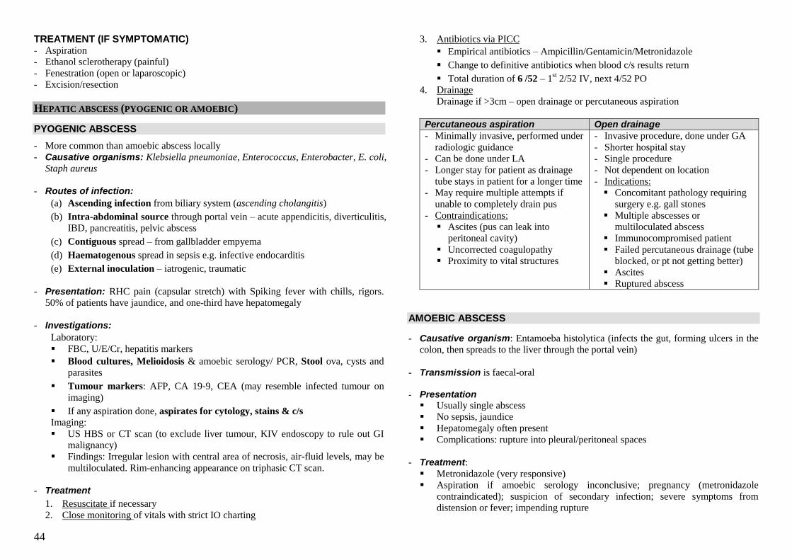
44
TREATMENT (IF SYMPTOMATIC) - Aspiration
- Ethanol sclerotherapy (painful)
- Fenestration (open or laparoscopic)
- Excision/resection
HEPATIC ABSCESS (PYOGENIC OR AMOEBIC)
PYOGENIC ABSCESS
- More common than amoebic abscess locally
- Causative organisms: Klebsiella pneumoniae, Enterococcus, Enterobacter, E. coli,
Staph aureus
- Routes of infection:
(a) Ascending infection from biliary system (ascending cholangitis)
(b) Intra-abdominal source through portal vein – acute appendicitis, diverticulitis,
IBD, pancreatitis, pelvic abscess
(c) Contiguous spread – from gallbladder empyema
(d) Haematogenous spread in sepsis e.g. infective endocarditis
(e) External inoculation – iatrogenic, traumatic
- Presentation: RHC pain (capsular stretch) with Spiking fever with chills, rigors.
50% of patients have jaundice, and one-third have hepatomegaly
- Investigations:
Laboratory:
FBC, U/E/Cr, hepatitis markers
Blood cultures, Melioidosis & amoebic serology/ PCR, Stool ova, cysts and
parasites
Tumour markers: AFP, CA 19-9, CEA (may resemble infected tumour on
imaging)
If any aspiration done, aspirates for cytology, stains & c/s
Imaging:
US HBS or CT scan (to exclude liver tumour, KIV endoscopy to rule out GI
malignancy)
Findings: Irregular lesion with central area of necrosis, air-fluid levels, may be
multiloculated. Rim-enhancing appearance on triphasic CT scan.
- Treatment
1. Resuscitate if necessary
2. Close monitoring of vitals with strict IO charting
3. Antibiotics via PICC
Empirical antibiotics – Ampicillin/Gentamicin/Metronidazole
Change to definitive antibiotics when blood c/s results return
Total duration of 6 /52 – 1st 2/52 IV, next 4/52 PO
4. Drainage
Drainage if >3cm – open drainage or percutaneous aspiration
Percutaneous aspiration Open drainage
- Minimally invasive, performed under
radiologic guidance
- Can be done under LA
- Longer stay for patient as drainage
tube stays in patient for a longer time
- May require multiple attempts if
unable to completely drain pus
- Contraindications:
Ascites (pus can leak into
peritoneal cavity)
Uncorrected coagulopathy
Proximity to vital structures
- Invasive procedure, done under GA
- Shorter hospital stay
- Single procedure
- Not dependent on location
- Indications:
Concomitant pathology requiring
surgery e.g. gall stones
Multiple abscesses or
multiloculated abscess
Immunocompromised patient
Failed percutaneous drainage (tube
blocked, or pt not getting better)
Ascites
Ruptured abscess
AMOEBIC ABSCESS
- Causative organism: Entamoeba histolytica (infects the gut, forming ulcers in the
colon, then spreads to the liver through the portal vein)
- Transmission is faecal-oral - Presentation
Usually single abscess
No sepsis, jaundice
Hepatomegaly often present
Complications: rupture into pleural/peritoneal spaces
- Treatment:
Metronidazole (very responsive)
Aspiration if amoebic serology inconclusive; pregnancy (metronidazole
contraindicated); suspicion of secondary infection; severe symptoms from
distension or fever; impending rupture
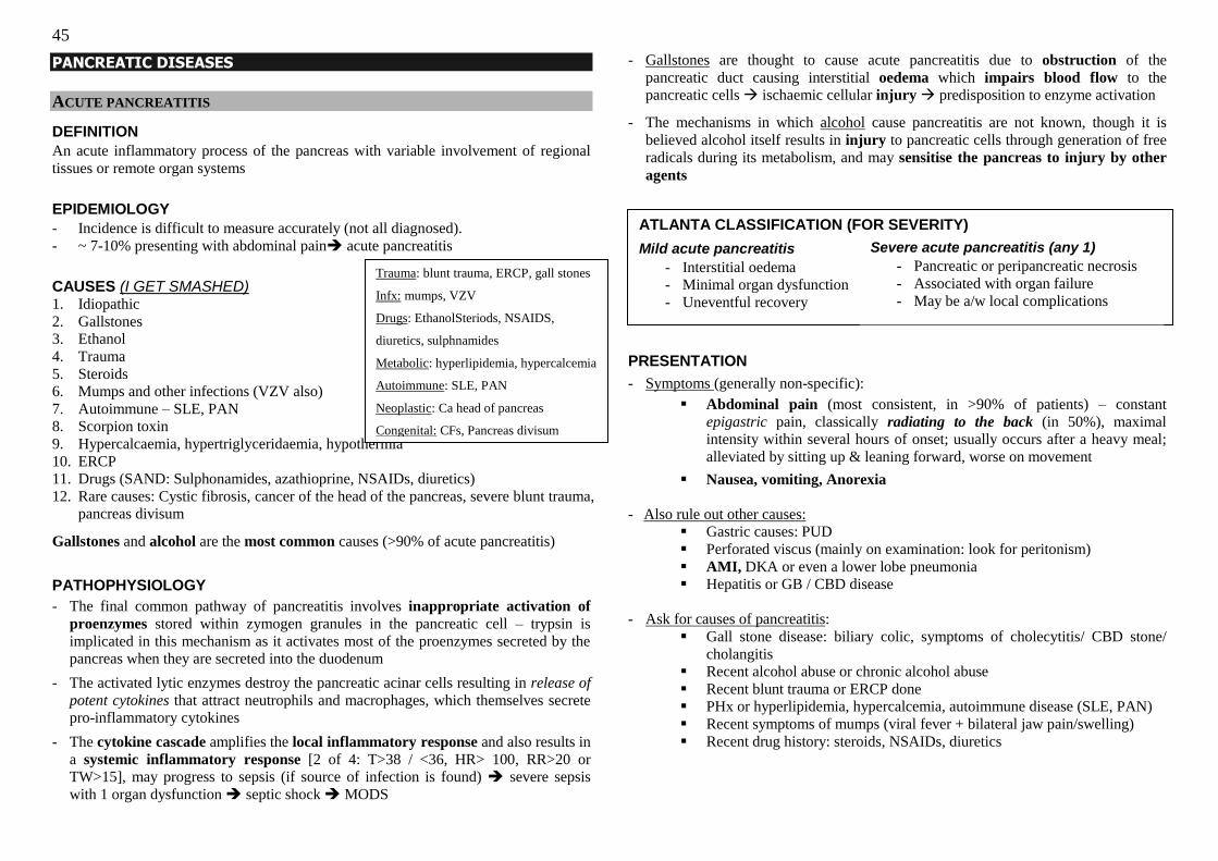
45
PANCREATIC DISEASES
ACUTE PANCREATITIS
DEFINITION
An acute inflammatory process of the pancreas with variable involvement of regional
tissues or remote organ systems
EPIDEMIOLOGY
- Incidence is difficult to measure accurately (not all diagnosed).
- ~ 7-10% presenting with abdominal pain acute pancreatitis
CAUSES (I GET SMASHED) 1. Idiopathic
2. Gallstones
3. Ethanol
4. Trauma
5. Steroids
6. Mumps and other infections (VZV also)
7. Autoimmune – SLE, PAN
8. Scorpion toxin
9. Hypercalcaemia, hypertriglyceridaemia, hypothermia
10. ERCP
11. Drugs (SAND: Sulphonamides, azathioprine, NSAIDs, diuretics)
12. Rare causes: Cystic fibrosis, cancer of the head of the pancreas, severe blunt trauma,
pancreas divisum
Gallstones and alcohol are the most common causes (>90% of acute pancreatitis)
PATHOPHYSIOLOGY
- The final common pathway of pancreatitis involves inappropriate activation of
proenzymes stored within zymogen granules in the pancreatic cell – trypsin is
implicated in this mechanism as it activates most of the proenzymes secreted by the
pancreas when they are secreted into the duodenum
- The activated lytic enzymes destroy the pancreatic acinar cells resulting in release of
potent cytokines that attract neutrophils and macrophages, which themselves secrete pro-inflammatory cytokines
- The cytokine cascade amplifies the local inflammatory response and also results in
a systemic inflammatory response [2 of 4: T>38 / <36, HR> 100, RR>20 or
TW>15], may progress to sepsis (if source of infection is found) severe sepsis
with 1 organ dysfunction septic shock MODS
- Gallstones are thought to cause acute pancreatitis due to obstruction of the
pancreatic duct causing interstitial oedema which impairs blood flow to the
pancreatic cells ischaemic cellular injury predisposition to enzyme activation
- The mechanisms in which alcohol cause pancreatitis are not known, though it is
believed alcohol itself results in injury to pancreatic cells through generation of free
radicals during its metabolism, and may sensitise the pancreas to injury by other
agents
PRESENTATION
- Symptoms (generally non-specific):
Abdominal pain (most consistent, in >90% of patients) – constant
epigastric pain, classically radiating to the back (in 50%), maximal
intensity within several hours of onset; usually occurs after a heavy meal;
alleviated by sitting up & leaning forward, worse on movement
Nausea, vomiting, Anorexia
- Also rule out other causes:
Gastric causes: PUD
Perforated viscus (mainly on examination: look for peritonism)
AMI, DKA or even a lower lobe pneumonia
Hepatitis or GB / CBD disease
- Ask for causes of pancreatitis:
Gall stone disease: biliary colic, symptoms of cholecytitis/ CBD stone/
cholangitis
Recent alcohol abuse or chronic alcohol abuse
Recent blunt trauma or ERCP done
PHx or hyperlipidemia, hypercalcemia, autoimmune disease (SLE, PAN)
Recent symptoms of mumps (viral fever + bilateral jaw pain/swelling)
Recent drug history: steroids, NSAIDs, diuretics
Trauma: blunt trauma, ERCP, gall stones
Infx: mumps, VZV
Drugs: EthanolSteriods, NSAIDS,
diuretics, sulphnamides
Metabolic: hyperlipidemia, hypercalcemia
Autoimmune: SLE, PAN
Neoplastic: Ca head of pancreas
Congenital: CFs, Pancreas divisum
ATLANTA CLASSIFICATION (FOR SEVERITY)
Mild acute pancreatitis
- Interstitial oedema
- Minimal organ dysfunction
- Uneventful recovery
Severe acute pancreatitis (any 1)
- Pancreatic or peripancreatic necrosis
- Associated with organ failure
- May be a/w local complications
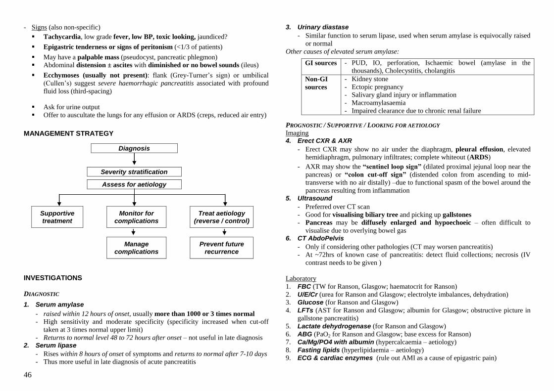
46
- Signs (also non-specific)
Tachycardia, low grade fever, low BP, toxic looking, jaundiced?
Epigastric tenderness or signs of peritonism (<1/3 of patients)
May have a palpable mass (pseudocyst, pancreatic phlegmon)
Abdominal distension ± ascites with diminished or no bowel sounds (ileus)
Ecchymoses (usually not present): flank (Grey-Turner‘s sign) or umbilical
(Cullen‘s) suggest severe haemorrhagic pancreatitis associated with profound
fluid loss (third-spacing)
Ask for urine output
Offer to auscultate the lungs for any effusion or ARDS (creps, reduced air entry)
MANAGEMENT STRATEGY
Diagnosis
Severity stratification
Assess for aetiology
Supportive treatment
Monitor for complications
Treat aetiology (reverse / control)
Manage complications
Prevent future recurrence
INVESTIGATIONS
DIAGNOSTIC
1. Serum amylase
- raised within 12 hours of onset, usually more than 1000 or 3 times normal
- High sensitivity and moderate specificity (specificity increased when cut-off
taken at 3 times normal upper limit)
- Returns to normal level 48 to 72 hours after onset – not useful in late diagnosis 2. Serum lipase
- Rises within 8 hours of onset of symptoms and returns to normal after 7-10 days
- Thus more useful in late diagnosis of acute pancreatitis
3. Urinary diastase
- Similar function to serum lipase, used when serum amylase is equivocally raised
or normal
Other causes of elevated serum amylase:
GI sources - PUD, IO, perforation, Ischaemic bowel (amylase in the
thousands), Cholecystitis, cholangitis
Non-GI
sources
- Kidney stone
- Ectopic pregnancy
- Salivary gland injury or inflammation
- Macroamylasaemia
- Impaired clearance due to chronic renal failure
PROGNOSTIC / SUPPORTIVE / LOOKING FOR AETIOLOGY
Imaging 4. Erect CXR & AXR
- Erect CXR may show no air under the diaphragm, pleural effusion, elevated
hemidiaphragm, pulmonary infiltrates; complete whiteout (ARDS)
- AXR may show the “sentinel loop sign” (dilated proximal jejunal loop near the
pancreas) or “colon cut-off sign” (distended colon from ascending to mid-
transverse with no air distally) –due to functional spasm of the bowel around the
pancreas resulting from inflammation 5. Ultrasound
- Preferred over CT scan
- Good for visualising biliary tree and picking up gallstones
- Pancreas may be diffusely enlarged and hypoechoeic – often difficult to
visualise due to overlying bowel gas 6. CT AbdoPelvis
- Only if considering other pathologies (CT may worsen pancreatitis)
- At ~72hrs of known case of pancreatitis: detect fluid collections; necrosis (IV
contrast needs to be given )
Laboratory
1. FBC (TW for Ranson, Glasgow; haematocrit for Ranson)
2. U/E/Cr (urea for Ranson and Glasgow; electrolyte imbalances, dehydration)
3. Glucose (for Ranson and Glasgow)
4. LFTs (AST for Ranson and Glasgow; albumin for Glasgow; obstructive picture in
gallstone pancreatitis)
5. Lactate dehydrogenase (for Ranson and Glasgow)
6. ABG (PaO2 for Ranson and Glasgow; base excess for Ranson)
7. Ca/Mg/PO4 with albumin (hypercalcaemia – aetiology)
8. Fasting lipids (hyperlipidaemia – aetiology)
9. ECG & cardiac enzymes (rule out AMI as a cause of epigastric pain)

47
SEVERITY STRATIFICATION (RANSON, GLASGOW, APACHE II)
I. Danger signs in the first few hours
- Encephalopathy
- Hypoxaemia,
- Tachycardia >130/min, Hypotension <90mmHg, Haematocrit >50
- Presence of Gray Turner‘s/Cullen‘s sign
- Oliguria <50mls/hr, Azotemia
II. Ranson’s criteria
Present at admission Within 48 hours of admission 1. Age >55 yrs
2. White cell count >16x109/dL
3. Fasting bld gluc >11.2mmol/L
4. LDH >600U/L
5. AST >120U/L
1. Fall in Hct >10%
2. Rise in urea >0.9mmol/L
3. Calcium <2mmol/L
4. PaO2 <60mmHg
5. Base excess >4mmol/L
6. Neg fluid balance >6L
- Ranson‘s criteria prognosticates mortality according to score
- Any patient with a score of 3 and above is considered to have severe pancreatitis
- Mortality: <3 0.9%
3-4 15%
5-6 50%
>6 90%
- Shortfalls of Ranson‘s:
validated for alcoholic pancreatitis only revised Ranson‘s score was created
for gallstone pancreatitis,
difficult to tell aetiology in acute setting & cumbersome to wait for 48 hours, and
difficult to assess for negative fluid balance
III. Glasgow/Imrie score- PANCREAS
1. PaO2 <60mmHg
2. Age >55yrs
3. Neutrophil/WBC >15x109/dL
4. Calcium <2mmol/L
5. Renal (urea) >16mmol/L
6. Enzymes LDH >600U/L
AST >100/L
7. Albumin <32g/L
8. Sugar (Glucose ) >10mmol/L
>3 criteria severe
- Preferred over Ranson‘s scoring in certain centres
IV. C-reactive peptide
- As a single prognostic marker; <100 is unlikely severe
- If CRP is >210mg/dL at 48 hours, the pancreatitis is more likely to be severe
- No relevance >3/7 of onset as other confounding factors come into the picture
- Combination of Glasgow score and CRP improves overall prognostic value
V. Balthazar’s CT severity index (consider a CT AP @ ~ 3/7 from onset)
- Grades severity of disease according to CT findings detects h‟ge & necrosis
- Not very useful as CT is not usually done in the 1/52 in local context, and disease is
still evolving (CT findings lag behind) in the early stages
COURSE OF DISEASE
- 75%: mild course of disease; recover unless comorbidities cause deterioration
- 20-25%: severe outcome 1/3 of these patients will ultimately die
- Overall mortality rate <10%
- Death is bimodally distributed:
(a) Early
Within 1/52; due to severe organ failure, SIRS
Very little can be done in terms of treatment
(b) Late
Most common cause is infection with resultant sepsis
Multi-organ failure can be the course of death
SUPPORTIVE TREATMENT
1. Monitoring (after resuscitating)
- In general ward if mild pancreatitis; HD/ICU monitoring if severe (≥3)
- Fluid resuscitate with crystalliods
- Monitor vitals [SpO2, BP, HR, Temp.], urine output & CVP
2. NBM (bowel rest) and IV fluid replacement
- May include gastric decompression with NGT if there is persistent vomiting, significant gastroparesis, or intestinal obstruction (ileus)
- Acid suppression does not change course of disease, but protects against stress ulcer formation; octetride has no benefit (thought to reduce pancreatic secretions)
- Fast patients for at least 2/7 until more stable
- May start oral feeding early with fluids in mild pancreatitis if tolerated
- Prolonged NBM results in poorer recovery due to nutritional debilitation – think
about NJ feeding, or open jejunostomy creation early in patients with severe
pancreatitis; if not tolerable, then consider TPN
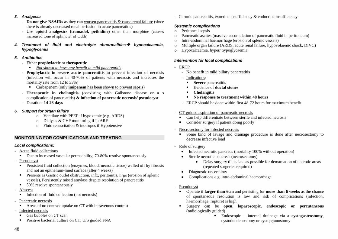
48
3. Analgesia
- Do not give NSAIDs as they can worsen pancreatitis & cause renal failure (since
there is already decreased renal perfusion in acute pancreatitis)
- Use opioid analgesics (tramadol, pethidine) other than morphine (causes
increased tone of sphincter of Oddi)
4. Treatment of fluid and electrolyte abnormalities hypocalcaemia, hypoglycemia
5. Antibiotics
- Either prophylactic or therapeutic
Not shown to have any benefit in mild pancreatitis - Prophylactic in severe acute pancreatitis to prevent infection of necrosis
(infection will occur in 40-70% of patients with necrosis and increases the
mortality rate from 12 to 33%)
Carbapenem (only imipenem has been shown to prevent sepsis)
- Therapeutic in cholangitis (coexisting with Gallstone disease or a s
complication of pancreatitis) & infection of pancreatic necrosis/ pseudocyst
- Duration: 14-28 days
6. Support for organ failure o Ventilate with PEEP if hypoxemic (e.g. ARDS)
o Dialysis & CVP monitoring if in ARF
o Fluid resuscitation & inotropes if Hypotensive
MONITORING FOR COMPLICATIONS AND TREATING
Local complications:
- Acute fluid collections
Due to increased vascular permeability; 70-80% resolve spontaneously
- Pseudocyst
Persistent fluid collection (enzymes, blood, necrotic tissue) walled off by fibrosis
and not an epithelium-lined surface (after 4 weeks)
Presents as Gastric outlet obstruction, infx, peritonitis, h‘ge (erosion of splenic
vessels), Persistently raised amylase despite resolution of pancreatitis
50% resolve spontaneously
- Abscess
Infection of fluid collection (not necrosis)
- Pancreatic necrosis
Areas of no contrast uptake on CT with intravenous contrast
- Infected necrosis
Gas bubbles on CT scan
Positive bacterial culture on CT, U/S guided FNA
- Chronic pancreatitis, exocrine insufficiency & endocrine insufficiency Systemic complications o Peritoneal sepsis
o Pancreatic ascites (massive accumulation of pancreatic fluid in peritoneum)
o Intra-abdominal haemorrhage (erosion of splenic vessels)
o Multiple organ failure (ARDS, acute renal failure, hypovolaemic shock, DIVC)
o Hypocalcaemia, hyper/ hypoglycaemia
Intervention for local complications
- ERCP
- No benefit in mild biliary pancreatitis
- Indications:
Severe pancreatitis
Evidence of ductal stones
Cholangitis
No response to treatment within 48 hours
- ERCP should be done within first 48-72 hours for maximum benefit
- CT-guided aspiration of pancreatic necrosis
Can help differentiate between sterile and infected necrosis
Consider surgery if patient doing poorly
- Necrosectomy for infected necrosis
Some kind of lavage and drainage procedure is done after necrosectomy to
decrease infective load
- Role of surgery
Infected necrotic pancreas (mortality 100% without operation)
Sterile necrotic pancreas (necrosectomy)
Delay surgery till as late as possible for demarcation of necrotic areas
(repeated surgeries required)
Diagnostic uncertainty
Complications e.g. intra-abdominal haemorrhage
- Pseudocyst
Operate if larger than 6cm and persisting for more than 6 weeks as the chance
of spontaneous resolution is low and risk of complications (infection,
haemorrhage, rupture) is high
Surgery can be open, laparoscopic, endoscopic or percutaneous
(radiologically guided)
Endoscopic – internal drainage via a cystogastrostomy,
cystoduodenostomy or cystojejunostomy
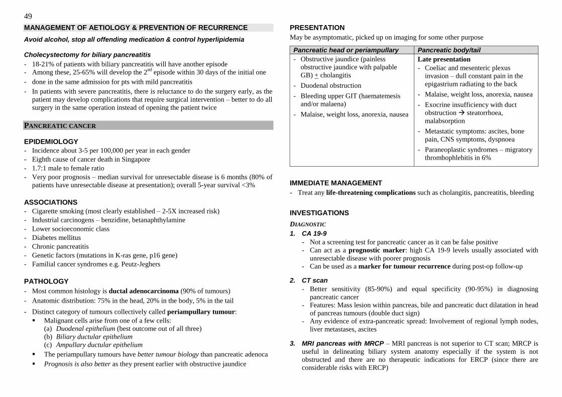
49
MANAGEMENT OF AETIOLOGY & PREVENTION OF RECURRENCE
Avoid alcohol, stop all offending medication & control hyperlipidemia Cholecystectomy for biliary pancreatitis
- 18-21% of patients with biliary pancreatitis will have another episode
- Among these, 25-65% will develop the 2nd
episode within 30 days of the initial one
- done in the same admission for pts with mild pancreatitis
- In patients with severe pancreatitis, there is reluctance to do the surgery early, as the
patient may develop complications that require surgical intervention – better to do all
surgery in the same operation instead of opening the patient twice
PANCREATIC CANCER
EPIDEMIOLOGY
- Incidence about 3-5 per 100,000 per year in each gender
- Eighth cause of cancer death in Singapore
- 1.7:1 male to female ratio
- Very poor prognosis – median survival for unresectable disease is 6 months (80% of
patients have unresectable disease at presentation); overall 5-year survival <3%
ASSOCIATIONS
- Cigarette smoking (most clearly established – 2-5X increased risk)
- Industrial carcinogens – benzidine, betanaphthylamine
- Lower socioeconomic class
- Diabetes mellitus
- Chronic pancreatitis
- Genetic factors (mutations in K-ras gene, p16 gene)
- Familial cancer syndromes e.g. Peutz-Jeghers
PATHOLOGY
- Most common histology is ductal adenocarcinoma (90% of tumours)
- Anatomic distribution: 75% in the head, 20% in the body, 5% in the tail
- Distinct category of tumours collectively called periampullary tumour:
Malignant cells arise from one of a few cells: (a) Duodenal epithelium (best outcome out of all three)
(b) Biliary ductular epithelium
(c) Ampullary ductular epithelium
The periampullary tumours have better tumour biology than pancreatic adenoca
Prognosis is also better as they present earlier with obstructive jaundice
PRESENTATION
May be asymptomatic, picked up on imaging for some other purpose
Pancreatic head or periampullary Pancreatic body/tail
- Obstructive jaundice (painless
obstructive jaundice with palpable
GB) + cholangitis
- Duodenal obstruction
- Bleeding upper GIT (haematemesis
and/or malaena)
- Malaise, weight loss, anorexia, nausea
Late presentation
- Coeliac and mesenteric plexus
invasion – dull constant pain in the
epigastrium radiating to the back
- Malaise, weight loss, anorexia, nausea
- Exocrine insufficiency with duct
obstruction steatorrhoea,
malabsorption
- Metastatic symptoms: ascites, bone
pain, CNS symptoms, dyspnoea
- Paraneoplastic syndromes – migratory
thrombophlebitis in 6%
IMMEDIATE MANAGEMENT
- Treat any life-threatening complications such as cholangitis, pancreatitis, bleeding
INVESTIGATIONS
DIAGNOSTIC
1. CA 19-9
- Not a screening test for pancreatic cancer as it can be false positive
- Can act as a prognostic marker: high CA 19-9 levels usually associated with
unresectable disease with poorer prognosis
- Can be used as a marker for tumour recurrence during post-op follow-up
2. CT scan
- Better sensitivity (85-90%) and equal specificity (90-95%) in diagnosing
pancreatic cancer
- Features: Mass lesion within pancreas, bile and pancreatic duct dilatation in head
of pancreas tumours (double duct sign)
- Any evidence of extra-pancreatic spread: Involvement of regional lymph nodes,
liver metastases, ascites
3. MRI pancreas with MRCP – MRI pancreas is not superior to CT scan; MRCP is
useful in delineating biliary system anatomy especially if the system is not
obstructed and there are no therapeutic indications for ERCP (since there are
considerable risks with ERCP)
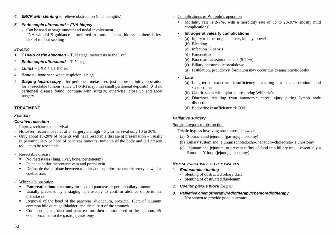
50
4. ERCP with stenting to relieve obstruction (in cholangitis)
5. Endoscopic ultrasound + FNA biopsy
- Can be used to stage tumour and nodal involvement
- FNA with EUS guidance is preferred to transcutaneous biopsy as there is less
risk of tumour seeding
STAGING
1. CT/MRI of the abdomen – T, N stage; metastasis to the liver
2. Endoscopic ultrasound – T, N stage
3. Lungs – CXR + CT thorax
4. Bones – bone scan when suspicion is high
5. Staging laparoscopy – for peritoneal metastases, just before definitive operation
for a resectable tumour (since CT/MRI may miss small peritoneal deposits) if no
peritoneal disease found, continue with surgery, otherwise, close up and abort
surgery
TREATMENT
SURGERY
Curative resection
- Improves chances of survival
- However, recurrence rates after surgery are high – 5 year survival only 10 to 30%
- Only about 15-20% of patients will have resectable disease at presentation – usually
in periampullary or head of pancreas tumours; tumours of the body and tail present
too late to be resectable
- Resectable disease:
No metastases (lung, liver, bone, peritoneum)
Patent superior mesenteric vein and portal vein
Definable tissue plane between tumour and superior mesenteric artery as well as
coeliac axis
- Whipple‘s operation
Pancreaticoduodenectomy for head of pancreas or periampullary tumour
Usually preceded by a staging laparoscopy to confirm absence of peritoneal
metastases
Removal of the head of the pancreas, duodenum, proximal 15cm of jejunum,
common bile duct, gallbladder, and distal part of the stomach
Common hepatic duct and pancreas are then anastomosed to the jejunum, 45-
60cm proximal to the gastrojejunostomy
- Complications of Whipple‘s operation
Mortality rate is 2-7%, with a morbidity rate of up to 20-30% (mostly mild
complications)
Intraoperative/early complications
(a) Injury to other organs – liver, kidney, bowel
(b) Bleeding
(c) Infection sepsis
(d) Pancreatitis
(e) Pancreatic anastomotic leak (5-20%)
(f) Biliary anastomotic breakdown
(g) Fistulation, pseudocyst formation may occur due to anastomotic leaks
Late
(a) Long-term exocrine insufficiency resulting in malabsorption and
steatorrhoea
(b) Gastric stasis with pylorus-preserving Whipple‘s
(c) Diarrhoea resulting from autonomic nerve injury during lymph node
dissection
(d) Endocrine insufficiency DM
Palliative surgery
Surgical bypass of obstruction
- Triple bypass involving anastomosis between
(a) Stomach and jejunum (gastrojejunostomy)
(b) Biliary system and jejunum (choledocho-/hepatico-/cholecysto-jejunostomy)
(c) Jejunum and jejunum, to prevent reflux of food into biliary tree – essentially a
Roux-en-Y loop (jejunojejunostomy)
NON-SURGICAL PALLIATIVE MEASURES
1. Endoscopic stenting - Stenting of obstructed biliary duct
- Stenting of obstructed duodenum
2. Coeliac plexus block for pain
3. Palliative chemotherapy/radiotherapy/chemoradiotherapy
- Not shown to provide good outcomes
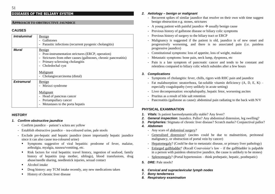
51
DISEASES OF THE BILIARY SYSTEM
APPROACH TO OBSTRUCTIVE JAUNDICE
CAUSES
Intraluminal Benign
- Gallstones
- Parasitic infections (recurrent pyogenic cholangitis)
Mural Benign
- Post-instrumentation strictures (ERCP, operation)
- Strictures from other causes (gallstones, chronic pancreatitis)
- Primary sclerosing cholangitis
- Choledochal cyst
Malignant
- Cholangiocarcinoma (distal)
Extramural Benign
- Mirizzi syndrome
Malignant
- Head of pancreas cancer
- Periampullary cancer
- Metastases to the porta hepatis
HISTORY
1. Confirm obstructive jaundice
- Confirm jaundice – patient‘s sclera are yellow
- Establish obstructive jaundice – tea-coloured urine, pale stools
- Exclude pre-hepatic and hepatic jaundice (more importantly hepatic jaundice
since it can also cause tea-coloured urine)
Symptoms suggestive of viral hepatitis: prodrome of fever, malaise,
arthralgia, myalgia, nausea/vomiting, etc.
Risk factors for viral hepatitis: travel history, ingestion of seafood, family
history of hepatitis (esp mother, siblings), blood transfusions, drug
abuse/needle sharing, needlestick injuries, sexual contact
Alcohol intake
Drug history: any TCM intake recently, any new medications taken
History of chronic liver disease
2. Aetiology – benign or malignant
- Recurrent spikes of similar jaundice that resolve on their own with time suggest
benign obstruction e.g. stones, strictures
- A young patient with painful jaundice usually benign cause
- Previous history of gallstone disease or biliary colic symptoms
- Previous history of surgery to the biliary tract or ERCP
- Malignancy is suggested if the patient is old, jaundice is of new onset and
progressively worsening, and there is no associated pain (i.e. painless
progressive jaundice)
- Constitutional symptoms: loss of appetite, loss of weight, malaise
- Metastatic symptoms: bone pain, neck lump, dyspnoea, etc
- Pain is a late symptom of pancreatic cancer and tends to be constant and
relentless compared to biliary colic which subsides after a few hours
3. Complications
- Symptoms of cholangitis: fever, chills, rigors with RHC pain and jaundice
- Fat malabsorption: steatorrhoea, fat-soluble vitamin deficiency (A, D, E, K) –
especially coagulopathy (very unlikely in acute setting)
- Liver decompensation: encephalopathy, hepatic fetor, worsening ascites
- Pruritus as a result of bile salt retention
- Pancreatitis (gallstone as cause): abdominal pain radiating to the back with N/V
PHYSICAL EXAMINATION
1. Vitals: Is patient haemodynamically stable? Any fever?
2. General inspection: Jaundice. Pallor? Any abdominal distension, leg swelling?
3. Peripheries: Stigmata of chronic liver disease? Scratch marks? Conjunctival pallor? 4. Abdomen
- Any scars of abdominal surgery?
- Generalised distension? (ascites could be due to malnutrition, peritoneal
malignancy, or obstruction of portal vein by cancer)
- Hepatomegaly? (Could be due to metastatic disease, or primary liver pathology)
- Enlarged gallbladder? (Recall Courvoisier‘s law – if the gallbladder is palpable
in a person with painless obstructive jaundice, the cause is unlikely to be stones)
- Splenomegaly? (Portal hypertension – think prehepatic, hepatic, posthepatic)
5. DRE: Pale stools?
6. Cervical and supraclavicular lymph nodes 7. Bony tenderness 8. Respiratory examination
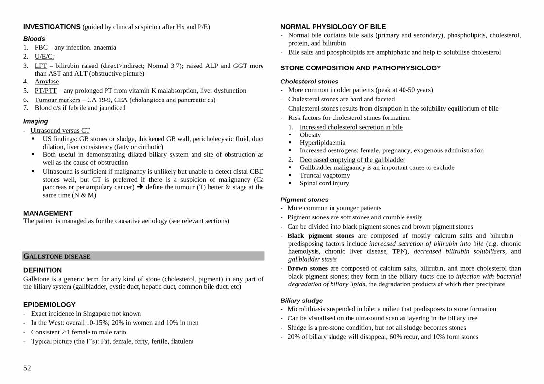
52
INVESTIGATIONS (guided by clinical suspicion after Hx and P/E)
Bloods
1. FBC – any infection, anaemia
2. U/E/Cr
3. LFT – bilirubin raised (direct>indirect; Normal 3:7); raised ALP and GGT more
than AST and ALT (obstructive picture)
4. Amylase
5. PT/PTT – any prolonged PT from vitamin K malabsorption, liver dysfunction
6. Tumour markers – CA 19-9, CEA (cholangioca and pancreatic ca)
7. Blood c/s if febrile and jaundiced
Imaging
- Ultrasound versus CT
US findings: GB stones or sludge, thickened GB wall, pericholecystic fluid, duct
dilation, liver consistency (fatty or cirrhotic)
Both useful in demonstrating dilated biliary system and site of obstruction as
well as the cause of obstruction
Ultrasound is sufficient if malignancy is unlikely but unable to detect distal CBD
stones well, but CT is preferred if there is a suspicion of malignancy (Ca
pancreas or periampulary cancer) define the tumour (T) better & stage at the
same time (N & M)
MANAGEMENT The patient is managed as for the causative aetiology (see relevant sections)
GALLSTONE DISEASE
DEFINITION
Gallstone is a generic term for any kind of stone (cholesterol, pigment) in any part of
the biliary system (gallbladder, cystic duct, hepatic duct, common bile duct, etc)
EPIDEMIOLOGY
- Exact incidence in Singapore not known
- In the West: overall 10-15%; 20% in women and 10% in men
- Consistent 2:1 female to male ratio
- Typical picture (the F‘s): Fat, female, forty, fertile, flatulent
NORMAL PHYSIOLOGY OF BILE
- Normal bile contains bile salts (primary and secondary), phospholipids, cholesterol,
protein, and bilirubin
- Bile salts and phospholipids are amphiphatic and help to solubilise cholesterol
STONE COMPOSITION AND PATHOPHYSIOLOGY
Cholesterol stones
- More common in older patients (peak at 40-50 years)
- Cholesterol stones are hard and faceted
- Cholesterol stones results from disruption in the solubility equilibrium of bile
- Risk factors for cholesterol stones formation:
1. Increased cholesterol secretion in bile
Obesity
Hyperlipidaemia
Increased oestrogens: female, pregnancy, exogenous administration
2. Decreased emptying of the gallbladder
Gallbladder malignancy is an important cause to exclude
Truncal vagotomy
Spinal cord injury
Pigment stones
- More common in younger patients
- Pigment stones are soft stones and crumble easily
- Can be divided into black pigment stones and brown pigment stones
- Black pigment stones are composed of mostly calcium salts and bilirubin –
predisposing factors include increased secretion of bilirubin into bile (e.g. chronic
haemolysis, chronic liver disease, TPN), decreased bilirubin solubilisers, and
gallbladder stasis
- Brown stones are composed of calcium salts, bilirubin, and more cholesterol than
black pigment stones; they form in the biliary ducts due to infection with bacterial degradation of biliary lipids, the degradation products of which then precipitate
Biliary sludge
- Microlithiasis suspended in bile; a milieu that predisposes to stone formation
- Can be visualised on the ultrasound scan as layering in the biliary tree
- Sludge is a pre-stone condition, but not all sludge becomes stones
- 20% of biliary sludge will disappear, 60% recur, and 10% form stones
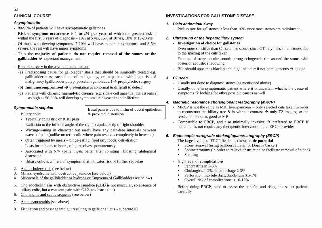
53
CLINICAL COURSE
Asymptomatic
- 80-95% of patients will have asymptomatic gallstones
- Risk of symptom occurrence is 1 to 2% per year, of which the greatest risk is
within the first 5 years of diagnosis – 10% at 5 yrs, 15% at 10 yrs, 18% at 15-20 yrs
- Of those who develop symptoms, 7-10% will have moderate symptoms, and 3-5%
severe; the rest will have minor symptoms
- Thus the majority of patients do not require removal of the stones or the
gallbladder expectant management
- Role of surgery in the asymptomatic patient:
(a) Predisposing cause for gallbladder stasis that should be surgically treated e.g.
gallbladder mass suspicious of malignancy, or in patients with high risk of
malignancy (gallbladder polyp, porcelain gallbladder) prophylactic surgery
(b) Immunocompromised presentation is abnormal & difficult to detect
(c) Patients with chronic haemolytic disease (e.g. sickle cell anaemia, thalassaemia)
– as high as 50-60% will develop symptomatic disease in their lifetime
Symptomatic sequlae
1. Biliary colic
- Typically epigastric or RHC pain
- Radiation to the inferior angle of the right scapula, or tip of right shoulder
- Waxing-waning in character but rarely have any pain-free intervals between
waves of pain (unlike ureteric colic where pain resolves completely in between)
- Often triggered by meals – binge-eating, fried oily foods, dehydration
- Lasts for minutes to hours, often resolves spontaneously
- Associated with N/V (patient gets better after vomiting), bloating, abdominal
distension
- Biliary colic is a ―herald‖ symptom that indicates risk of further sequelae
2. Acute cholecystitis (see below)
3. Mirizzi syndrome with obstructive jaundice (see below)
4. Mucocoele of the gallbladder or hydrops or Empyema of Gallbladder (see below)
5. Choledocholithiasis with obstructive jaundice (CBD is not muscular, so absence of biliary colic, but a constant pain with OJ 2
o to obstruction)
6. Cholangitis and septic sequelae (see below)
7. Acute pancreatitis (see above)
8. Fistulation and passage into gut resulting in gallstone ileus – subacute IO
INVESTIGATIONS FOR GALLSTONE DISEASE
1. Plain abdominal X-ray
- Pickup rate for gallstones is less than 10% since most stones are radiolucent
2. Ultrasound of the hepatobiliary system
- Investigation of choice for gallstones
- Even more sensitive than CT scan for stones since CT may miss small stones due
to the spacing of the cuts taken
- Features of stone on ultrasound: strong echogeneic rim around the stone, with
posterior acoustic shadowing
- Bile should appear as black patch in gallbladder; if not homogeneous sludge
3. CT scan
- Usually not done to diagnose stones (as mentioned above)
- Usually done in symptomatic patient where it is uncertain what is the cause of
symptoms looking for other possible causes as well
4. Magnetic resonance cholangiopancreatography (MRCP)
- MRCP is not the same as MRI liver/pancreas – only selected cuts taken in order
to reconstruct the biliary tree & is without contrast only T2 images, so the
resolution is not as good as MRI
- Comparable to ERCP, and also minimally invasive preferred to ERCP if
patient does not require any therapeutic intervention that ERCP provides
5. Endoscopic retrograde cholangiopancreatography (ERCP)
- The largest value of ERCP lies in its therapeutic potential
Stone removal (using balloon catheter, or Dormia basket)
Sphincterotomy (in order to relieve obstruction or facilitate removal of stone)
Stenting
- High level of complications
Pancreatitis in 2-3%
Cholangitis 1-2%, haemorrhage 2-3%
Perforation into bile duct, duodenum 0.5-1%
Overall risk of complications is 10-15%
- Before doing ERCP, need to assess the benefits and risks, and select patients
carefully
Basal pain is due to inflm of ductal epithelium
& proximal distension.
54
6. Percutaneous transhepatic cholangiography (PTC) /biliary drainage (PTBD)
- PTC involves a tube being inserted into the liver under radiologic guidance into
one of the biliary ducts (must be dilated duct)
- Rarely done now; main indications: 1) high obstruction not well visualised in
ERCP; 2) therapeutic purpose of drainage for an obstructed system that cannot
be drained from below
- Mostly for therapeutic rather than diagnostic purposes
- Complications: bleeding; leakage of bile when tube is removed
7. HIDA scan
- No longer used commonly, except in biliary atresia
5 criteria for a normal cholangiopancreatogram
(a) Normal intrahepatic ducts
(b) No filling defects
(c) Smooth common bile duct
(d) No stricture/narrowing of the common bile duct
(e) Good and free flow of contrast into duodenum
TREATMENT
Asymptomatic
- No surgery required unless patient has indications for surgery (see above)
- Expectant management and close follow-up
- Counsel patient about symptoms – biliary colic, acute cholecystitis, obstructive
jaundice, etc
Symptomatic
- Cholecystectomy is the only way to treat gallbladder stones that are symptomatic
- Can be open or laparoscopic – laparoscopic is preferred as it is associated with
shorter hospital stay, less pain, less complications post-operatively
- Risks of laparoscopic cholecystectomy
Conversion to open operation up to 5% (due to abnormal anatomy; difficult or
complicated dissection; iatrogenic injury); conversion rate is higher if there is
ongoing infection e.g. cholecystitis – up to 1 in 3 to 1 in 4
Injury to surroundings: bowel & biliary structures e.g. CBD
Spilled bile peritonitis, sepsis
Haemorrhage
Infection
- Non- surgical means of stone treatment
Chemodissolution
Liver diet
Shockwave lithotripsy – more morbidiy cf renal lithotripsy as less fluid around
to dampen waves; good results only for cholesterol stones
All not shown to work for long-term
ACUTE CHOLECYSTITIS
PATHOPHYSIOLOGY
- Gallstone gets stuck in the cystic duct causing obstruction of biliary flow
- Gallbladder becomes distended and inflamed
PRESENTATION
- Constant, severe RHC pain (less commonly epigastric)
- Radiates to the inferior angle of the scapula
- Associated with fever, nausea, vomiting
- RHC tenderness with guarding found on clinical examination; Murphy‘s sign positive
- Gallbladder may be palpable – omentum wrapping around GB; worst case scenario is
empyema
- LFTs usually normal; no jaundice
ULTRASOUND FEATURES OF ACUTE CHOLECYSTITIS
- Presence of gallstones in biliary system
- Contracted gallbladder (from chronic gallstone disease)
- Pericholecystic fluid (oedema of gallbladder wall)
- Sonographic Murphy‘s positive
- (Fat stranding around gallbladder not seen on ultrasound but on CT)
MANAGEMENT
- Resuscitate the patient
- Septic workup
- Bowel rest and intravenous fluids
- Analgesia
- Empirical intravenous antibiotics – IV ceftriaxone and metronidazole
- Definitive treatment – laparoscopic cholecystectomy
55
Timing of cholecystectomy
- Dependent on several factors:
Severity of illness
Response to resuscitation and antibiotic therapy
Logistical considerations (availability of OT, surgeon etc)
- Possibilities available:
i. Emergency (immediate; in very sick patients who are not doing well/not
responding to treatment)
ii. Early (within few days of onset)
iii. Delayed/interval (after 6-8 weeks)
Early Delayed
Advantages
- Everything done in one admission
- Easier to operate as the gallbladder is
oedematous
Disadvantages
- Ongoing inflammation higher risk
of bleeding
- Higher risk of injuring some other
structure due to difficulty in
visualisation
- Higher conversion rate to open chole
- Increased risks of post-op infection
Advantages
- Lower risks
- Better laparoscopic success
Disadvantages
- Fibrosis difficulty mobilising
gallbladder
- Need for another admission
- Chance of recurrence during the time
Early surgery has been found to be more beneficial than delayed surgery
Cholecystostomy
- In moribund patients who are not fit for surgery
- Can be done under LA, or more commonly under radiologic guidance (percutaneous)
- Drains the gallbladder and alleviates the inflammation better outcomes
COMPLICATIONS OF ACUTE CHOLECYSTITIS
1. Hydrops
- Cystic duct obstruction leads to a tense gallbladder filled with mucus
- May lead to gallbladder wall necrosis if pressure exceeds capillary bld pressure
2. Empyema
- Gallbladder is filled with pus due to bacterial infection of the stagnant bile
(cystic duct being obstructed by a stone)
- Patient is usually toxic, requiring urgent surgery
3. Gangrene and perforation
- Localised perforation abscess that is confined by the omentum
- Free perforation generalised peritonitis and sepsis, requiring emergency
laparotomy
4. Cholecystenteric fistula
- Most commonly occurs in duodenum, then colon, and stomach; after repeated
attacks of cholecystitis
- Usually asymptomatic
- On AXR, aerobilia is seen in 40% of cases
- Symptomatic fistulas should be treated with cholecystectomy and fistula closure
5. Gallstone ileus
- Stones causing cholecystenteric fistula pass into the enteric lumen causing
intermittent bouts of small bowel obstruction
- Accounts for 1-2% of IO overall
- Most common site of obstruction is terminal ileum
- Small stones (<2-3cm) usually pass spontaneously without problems
- Mortality is 10-15%, mostly in elderly patients in whom gallstone ileus is more
common
- Small bowel enterotomy proximal to the point of obstruction is usually required
to remove the stone
- Immediate cholecystectomy not warranted as <4% of patients will have further
symptoms
ACALCULOUS CHOLECYSTITIS
- Occurs in very ill patients with prolonged stay in ICU – prolonged fasting, poor
nutrition, labile blood pressure, sepsis
- Poor nutrition leads to biliary stasis, while dehydration and hypotension leads to
formation of viscous bile and gallbladder ischaemia bile may get infected
cholecystitis
- Treatment involves emergent cholecystectomy
56
CHOLEDOCHOLITHIASIS
PRESENTATION - Obstructive jaundice – tea-coloured urine, pale stools
- Biliary colic
- If infection sets in cholangitis (see below)
BLOODS - FBC (check TW for any rise suggestive of infection)
- Amylase (CBD stone may cause pancreatitis)
- LFTs (raised bilirubin – direct; ALP raised more than transaminases)
ULTRASOUND
- Gallstones in gallbladder
- Gallstone in CBD
- Dilated CBD (normally <8-9mm)
>10mm is abnormal
In older patients, post-cholecystectomy, or patients on long-term opiates, the
CBD may be larger, up to 11-12mm in size
MANAGEMENT
- If unsure of presence of stone less invasive investigation such as MRCP, EUS
- If likelihood of CBD stone is high ERCP with stone removal
ERCP successful Plan for lap cholecystectomy
ERCP failed If patient is well and can tolerate another ERCP, try again
(+ stent in between to drain bile)
Operative removal – Open CBD exploration
– Lap CBD exploration
- If no facilities to do ERCP: open or laparoscopic cholecystectomy with CBD
exploration
When to do operative removal of stones (i.e. not suitable for ERCP)
- Stone >25mm
- Intrahepatic stone
- Large number of stones
- Impacted stone
- Dual pathology
- Tortuous duct
- Previous Bilroth II (unsuitable anatomy for ERCP)
CHOLANGITIS
PRESENTATION - Classically Charcot’s triad: RHC pain, fever, jaundice (only 50-70% of patients
have the classic triad)
- Reynold’s pentad: Charcot‘s triad plus mental obtundation and shock
- A surgical emergency!
PATHOLOGY - Usually results from obstruction to the biliary system with infection of stagnant bile
- Most common cause is choledocholithiasis (60%); also consider benign strictures and
malignancy (pancreatic, biliary)
- Common causative organisms are gram negative bacteria and anaerobes – Klebsiella,
E. coli, Enterobacter, Enterococcus
MANAGEMENT
1. Resuscitation
- Anticipate rapid deterioration
- Obtain good intravenous access and fluid resuscitate as appropriate
- Take bloods for investigations – cultures especially
- Close monitoring of vitals in HD/ICU
- Catheterise and watch urine output
- CVP line insertion if patient has shock unresponsive to fluid resuscitation
2. Antibiotics
- IV ceftriaxone and metronidazole; imipenem if the patient is in shock
3. Biliary decompression and definitive treatment
- Biliary decompression and definitive treatment can be combined or separate
- Investigate for cause of obstruction – ultrasound or CT (depending on what
facilities are available; ultrasound is preferred if suspecting stones)
- Decompression commonly performed using ERCP
Decompression is the primary objective – stenting or external drainage
(nasobiliary drain)
If cause of obstruction can be treated in the same setting (e.g. stones to be
removed) then treat the cause also Success rate 90%
- Definitive treatment dependent on:
Medical condition of patient
Success of biliary decompression
Logistical considerations
57
- Choices for definitive treatment:
(a) Open cholecystectomy with CBD exploration
(b) Laparoscopic cholecystectomy
(c) Laparoscopic cholecystectomy with CBD exploration
CBD EXPLORATION
- Cholangiogram or choledochoscopy is performed
Cholangiogram involves injection of dye – can image higher ducts
Choledochoscopy involves using a scope to visualise the large biliary ducts –
cannot image higher ducts, thus not as sensitive, but can be used to remove
stones visualised in the duct
Choice of imaging depends on site of obstruction and the cause
- Removal of stones
Manual removal with stone-grasping forceps
Flushing out stones
Dredging stones out using balloon catheter or Dormia basket
Lithotripsy
- Consider use of biliary stent or T-tube after removal of stone(s)
If there is a lot of instrumentation of the biliary system during the operation, one
should anticipate swelling and oedema of the biliary system resulting in post-
operative obstruction and buildup of bile higher risk of biliary leakage
(a) Stent – removed later by endoscopy
(b) T-tube (usually inserted after CBD-E) A T-shaped tube with its horizontal limb placed in the CBD and the vertical
limb leading out to drain bile
Functions as a ―pressure release valve‖ as most of the bile will flow through
the horizontal limb of the tube into the distal part of the CBD; only when
there is obstruction to flow will bile be diverted out through the vertical limb
Allows for post-op cholangiogram to check for remaining stones (at POD 9-
10) before removal – free flow of contrast into duodenum, no residual stones,
and no free leak of contrast into peritoneum
If all normal release anchoring stitch and exert gentle traction on the tube;
the tube should slip out easily, if not, call for help
If stones are present leave tube in for 4-6 weeks to form a fibrous tract
allows for instrumentation of tract with a scope to remove the stones
- Consider biliary bypass if there are multiple stones, the CBD is more than 2cm in
diameter, or there are strictures (since the CBD has been chronically dilated, quite
unlikely that it will function normally even after removal of the obstruction)
MIRIZZI’S SYNDROME
PATHOLOGY - Gallstone in the Hartmann‘s pouch compressing the common hepatic resulting in
obstructive jaundice
- Compression effect is not just physical (the stone) but also contributed by the
surrounding inflammation
- One of the caveats to Courvoisier‘s law
GRADING - Grade I: No fistula; extrinsic compression on CHD
- Grade II: Fistulation into common bile duct with the fistula <1/3 diameter of the
CHD
- Grade III: Fistula 1/3 to 2/3 diameter of CHD
- Grade IV: Fistula >2/3 diameter of CHD
MANAGEMENT - Grade 1: attempt laproscopic cholecystectomy
- Grades 2-4: open cholecystectomy with CBD exploration
CHOLANGIOCARCINOMA
SITE
- Intrahepatic/peripheral 10%
- Distal 25%
- Perihilar 65% (Altemeier-Klatskin tumour)
Bismuth classification
i. Type I: below confluence of hepatic ducts
ii. Type II: tumour reaching confluence
iii. Type IIIA/B: involving common hepatic duct and either right or left hepatic duct
iv. Type IV: multicentric or involving confluence and both hepatic ducts
ASSOCIATIONS
- Related to chronic cholestasis:
Primary sclerosing cholangitis / Ulcerative colitis
Hepatolithiasis
Parasitic infection – Clonorchis sinensis, Opisthorchis viverrini
Caroli‘s disease (multifocal segmental dilatation of large intrahepatic bile ducts)
- Bile duct adenoma
- Choledochal cyst
- Thorotrast exposure
58
PRESENTATION - Painless jaundice (painful if there is cholangitis)
- Acholic stools
- Pruritus
- Advanced signs and symptoms:
Abdominal pain
Fatigue, malaise
Weight loss
Hepatomegaly
DIAGNOSIS - CA 19-9 >100μl/ml (good sensitivity of 89%, specificity 86%)
- Contrast CT
- PTC (2 functions: 1) roadmapping for surgery; 2) drainage of obstructed system if
ERCP cannot drain)
CURATIVE TREATMENT - Surgery is the only chance of long-term cure
- Only 25% of tumours are resectable
- Contraindications to surgery
Bilateral or multifocal intrahepatic disease
Invasion of portal vein trunk or hepatic artery
Bilateral involvement of hepatic arterial or portal venous branches
Unilateral hepatic vascular invasion with contralateral ductal spread
Distant metastases
PROGNOSIS FOR RESECTABLE DISEASE (5-year survival)
- Intrahepatic: 35-45%
- Distal 35-45%
- Perihilar 10-30% (worse prognosis due to early lymphatic spread)
PALLIATION - Endoscopic/percutaneous transhepatic biliary stenting
- Bilateral drainage for hilar cholangiocarcinoma
- If after opening up and finding that tumour is not resectable, can perform surgical
bypass
RECURRENT PYOGENIC CHOLANGITIS
BACKGROUND Cholangiohepatitis, or recurrent pyogenic cholangitis (RPC), is characterized by:
- Recurrent bacterial cholangitis
- Intrahepatic pigment stones
- Intrahepatic biliary obstruction.
PATHOPHYSIOLOGY
Helminthic infxn (eg Ascaris lumbricoides, Clonorchis sinensis) epithelial damage
predispose to seeding of coliforms into biliary system stone formation
recurrent cholangitis
HISTORY: - A history of recurrent attacks of cholangitis – typical hx:
1-2 episodes of fevers, jaundice, and RUQ abdominal pain per year
Hx of prev biliary surgery, endoscopic procedures, or percutaneous biliary
drainage procedures.
- Complications of pyogenic cholangitis
cirrhosis with portal hypertension
cholangiocarcinoma
PHYSICAL EXAMINATION No specific physical findings are evident in RPC. Dx based on history.
DIFFERENTIALS Primary Sclerosing Cholangitis
INVESTIGATIONS
For diagnosis
For Complications
Bloods - FBC
- LFT with ALP>ALT, AST
- Prothrombin time: N/ (if prolonged cholestasis causes fat malabsorption and vitamin K deficiency)
Impt to exclude – correct with parenteral Vit K before invasive procedures
- Blood C/S: bacteremia – results help guide antibiotic choice.
- Ova and parasites: RPC freq a/w Clonorchis infxn – look for it and treat when present.
59
Radiology
- U/S HBS
segmental biliary dilatation
hepatolithiasis
liver abscesses
helps determine choice of supplemental axial imaging techniques.
- ERCP or PTC – imaging modality of choice for delineating the biliary tree.
- CT scan
centrally dilated bile ducts with peripheral tapering
bile duct stones
pyogenic liver abscesses.
TREATMENT PRINCIPLES: - Treat current infection
- Biliary drainage
- Management of other complications e.g. dehydration etc
Surgical
- Usual surgical approach includes:
Initial biliary decompression – ERCP sphincterotomy / stent placement
Definitive biliary drainage procedure – e.g. Roux-en-Y choledochojejunostomy
PROGNOSIS - Death occurs in approximately 15-20% of patients over 5-6 years.
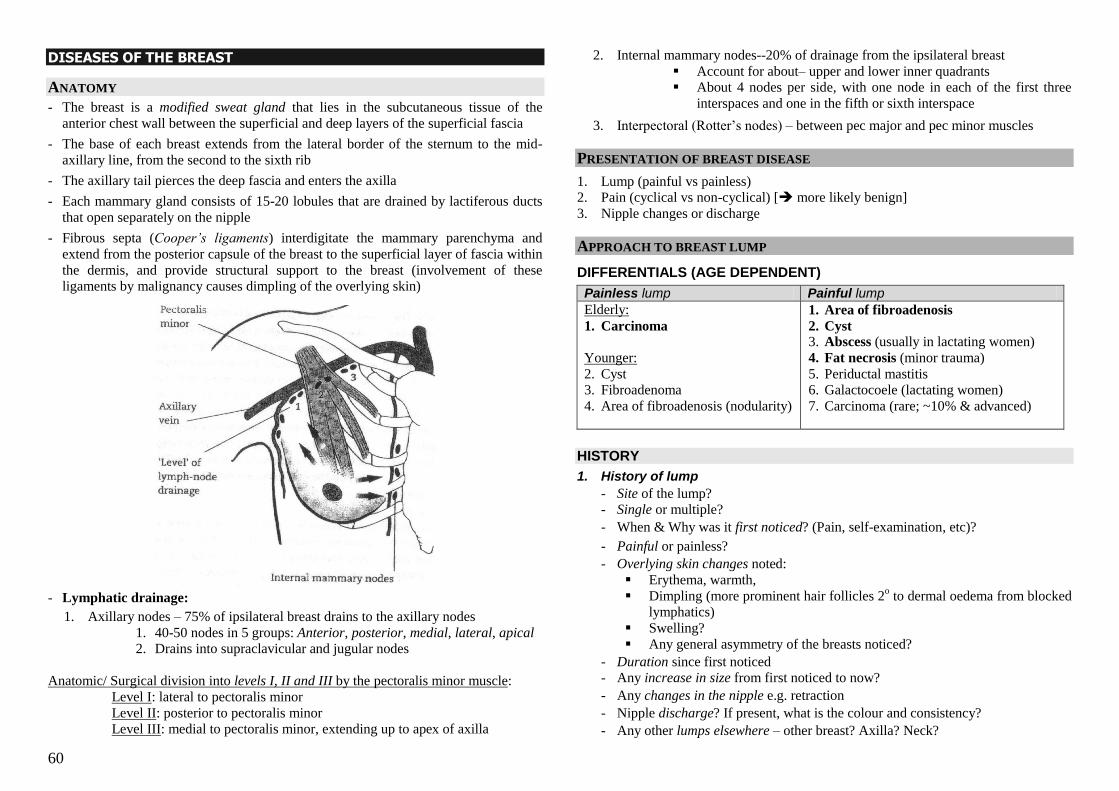
60
DISEASES OF THE BREAST
ANATOMY
- The breast is a modified sweat gland that lies in the subcutaneous tissue of the
anterior chest wall between the superficial and deep layers of the superficial fascia
- The base of each breast extends from the lateral border of the sternum to the mid-
axillary line, from the second to the sixth rib
- The axillary tail pierces the deep fascia and enters the axilla
- Each mammary gland consists of 15-20 lobules that are drained by lactiferous ducts
that open separately on the nipple
- Fibrous septa (Cooper‟s ligaments) interdigitate the mammary parenchyma and
extend from the posterior capsule of the breast to the superficial layer of fascia within
the dermis, and provide structural support to the breast (involvement of these
ligaments by malignancy causes dimpling of the overlying skin)
- Lymphatic drainage:
1. Axillary nodes – 75% of ipsilateral breast drains to the axillary nodes
1. 40-50 nodes in 5 groups: Anterior, posterior, medial, lateral, apical 2. Drains into supraclavicular and jugular nodes
Anatomic/ Surgical division into levels I, II and III by the pectoralis minor muscle:
Level I: lateral to pectoralis minor
Level II: posterior to pectoralis minor
Level III: medial to pectoralis minor, extending up to apex of axilla
2. Internal mammary nodes--20% of drainage from the ipsilateral breast
Account for about– upper and lower inner quadrants
About 4 nodes per side, with one node in each of the first three
interspaces and one in the fifth or sixth interspace
3. Interpectoral (Rotter‘s nodes) – between pec major and pec minor muscles
PRESENTATION OF BREAST DISEASE
1. Lump (painful vs painless)
2. Pain (cyclical vs non-cyclical) [ more likely benign]
3. Nipple changes or discharge
APPROACH TO BREAST LUMP
DIFFERENTIALS (AGE DEPENDENT)
Painless lump Painful lump
Elderly:
1. Carcinoma
Younger:
2. Cyst
3. Fibroadenoma
4. Area of fibroadenosis (nodularity)
1. Area of fibroadenosis
2. Cyst 3. Abscess (usually in lactating women)
4. Fat necrosis (minor trauma)
5. Periductal mastitis
6. Galactocoele (lactating women)
7. Carcinoma (rare; ~10% & advanced)
HISTORY
1. History of lump
- Site of the lump?
- Single or multiple?
- When & Why was it first noticed? (Pain, self-examination, etc)?
- Painful or painless?
- Overlying skin changes noted:
Erythema, warmth,
Dimpling (more prominent hair follicles 2o to dermal oedema from blocked
lymphatics)
Swelling?
Any general asymmetry of the breasts noticed?
- Duration since first noticed
- Any increase in size from first noticed to now?
- Any changes in the nipple e.g. retraction
- Nipple discharge? If present, what is the colour and consistency?
- Any other lumps elsewhere – other breast? Axilla? Neck?
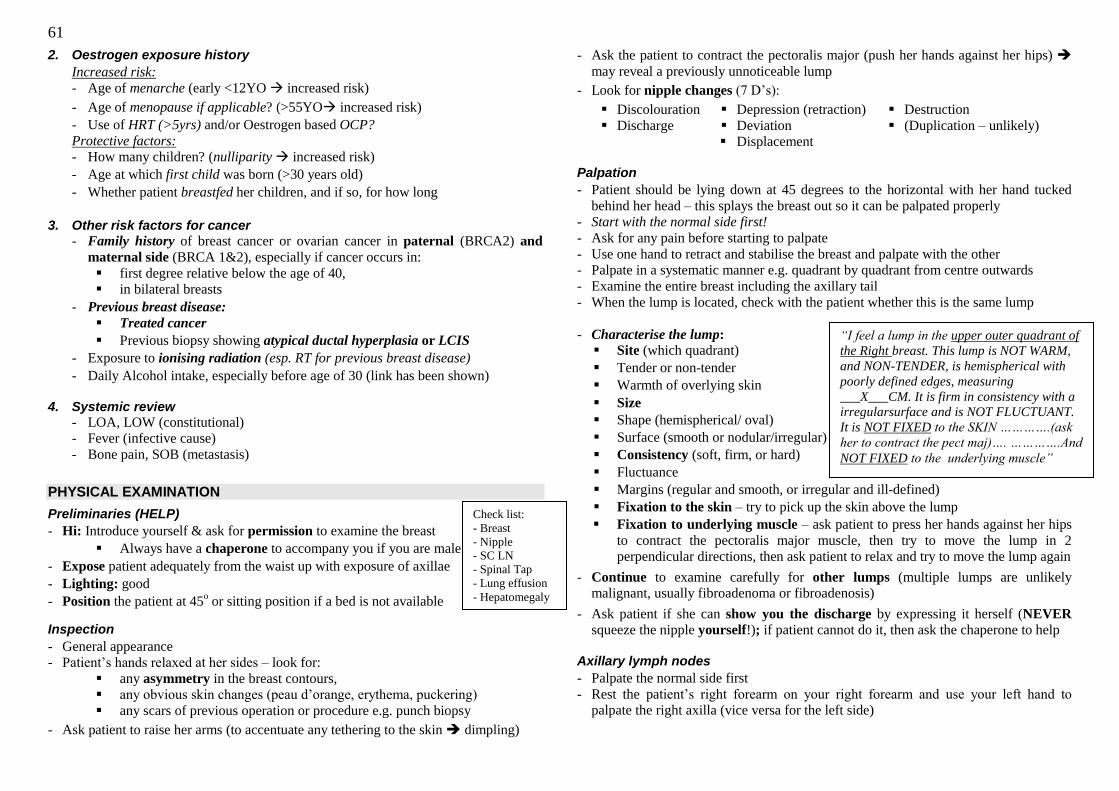
61
2. Oestrogen exposure history
Increased risk:
- Age of menarche (early <12YO increased risk)
- Age of menopause if applicable? (>55YO increased risk)
- Use of HRT (>5yrs) and/or Oestrogen based OCP?
Protective factors: - How many children? (nulliparity increased risk)
- Age at which first child was born (>30 years old)
- Whether patient breastfed her children, and if so, for how long
3. Other risk factors for cancer
- Family history of breast cancer or ovarian cancer in paternal (BRCA2) and
maternal side (BRCA 1&2), especially if cancer occurs in:
first degree relative below the age of 40,
in bilateral breasts
- Previous breast disease:
Treated cancer
Previous biopsy showing atypical ductal hyperplasia or LCIS
- Exposure to ionising radiation (esp. RT for previous breast disease)
- Daily Alcohol intake, especially before age of 30 (link has been shown) 4. Systemic review
- LOA, LOW (constitutional)
- Fever (infective cause)
- Bone pain, SOB (metastasis)
PHYSICAL EXAMINATION
Preliminaries (HELP)
- Hi: Introduce yourself & ask for permission to examine the breast
Always have a chaperone to accompany you if you are male
- Expose patient adequately from the waist up with exposure of axillae
- Lighting: good
- Position the patient at 45o or sitting position if a bed is not available
Inspection
- General appearance
- Patient‘s hands relaxed at her sides – look for:
any asymmetry in the breast contours,
any obvious skin changes (peau d‘orange, erythema, puckering)
any scars of previous operation or procedure e.g. punch biopsy
- Ask patient to raise her arms (to accentuate any tethering to the skin dimpling)
- Ask the patient to contract the pectoralis major (push her hands against her hips)
may reveal a previously unnoticeable lump
- Look for nipple changes (7 D‘s):
Discolouration
Discharge
Depression (retraction)
Deviation
Displacement
Destruction
(Duplication – unlikely)
Palpation
- Patient should be lying down at 45 degrees to the horizontal with her hand tucked
behind her head – this splays the breast out so it can be palpated properly
- Start with the normal side first!
- Ask for any pain before starting to palpate
- Use one hand to retract and stabilise the breast and palpate with the other
- Palpate in a systematic manner e.g. quadrant by quadrant from centre outwards
- Examine the entire breast including the axillary tail
- When the lump is located, check with the patient whether this is the same lump
- Characterise the lump:
Site (which quadrant)
Tender or non-tender
Warmth of overlying skin
Size
Shape (hemispherical/ oval)
Surface (smooth or nodular/irregular)
Consistency (soft, firm, or hard)
Fluctuance
Margins (regular and smooth, or irregular and ill-defined)
Fixation to the skin – try to pick up the skin above the lump
Fixation to underlying muscle – ask patient to press her hands against her hips
to contract the pectoralis major muscle, then try to move the lump in 2
perpendicular directions, then ask patient to relax and try to move the lump again
- Continue to examine carefully for other lumps (multiple lumps are unlikely
malignant, usually fibroadenoma or fibroadenosis)
- Ask patient if she can show you the discharge by expressing it herself (NEVER
squeeze the nipple yourself!); if patient cannot do it, then ask the chaperone to help
Axillary lymph nodes
- Palpate the normal side first
- Rest the patient‘s right forearm on your right forearm and use your left hand to
palpate the right axilla (vice versa for the left side)
Check list:
- Breast
- Nipple
- SC LN
- Spinal Tap
- Lung effusion
- Hepatomegaly
“I feel a lump in the upper outer quadrant of
the Right breast. This lump is NOT WARM,
and NON-TENDER, is hemispherical with
poorly defined edges, measuring
___X___CM. It is firm in consistency with a
irregularsurface and is NOT FLUCTUANT.
It is NOT FIXED to the SKIN ………….(ask
her to contract the pect maj)…. ………….And
NOT FIXED to the underlying muscle”
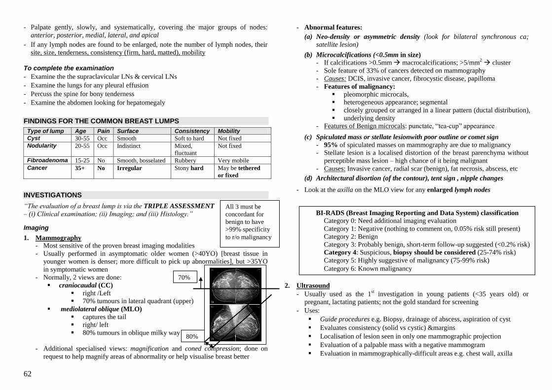
62
- Palpate gently, slowly, and systematically, covering the major groups of nodes:
anterior, posterior, medial, lateral, and apical
- If any lymph nodes are found to be enlarged, note the number of lymph nodes, their
site, size, tenderness, consistency (firm, hard, matted), mobility
To complete the examination
- Examine the the supraclavicular LNs & cervical LNs
- Examine the lungs for any pleural effusion
- Percuss the spine for bony tenderness
- Examine the abdomen looking for hepatomegaly
FINDINGS FOR THE COMMON BREAST LUMPS
Type of lump Age Pain Surface Consistency Mobility
Cyst 30-55 Occ Smooth Soft to hard Not fixed
Nodularity 20-55 Occ Indistinct Mixed,
fluctuant
Not fixed
Fibroadenoma 15-25 No Smooth, bosselated Rubbery Very mobile
Cancer 35+ No Irregular Stony hard May be tethered
or fixed
INVESTIGATIONS
“The evaluation of a breast lump is via the TRIPLE ASSESSMENT
– (i) Clinical examination; (ii) Imaging; and (iii) Histology.”
Imaging
1. Mammography - Most sensitive of the proven breast imaging modalities
- Usually performed in asymptomatic older women (>40YO) [breast tissue in
younger women is denser; more difficult to pick up abnormalities], but >35YO
in symptomatic women
- Normally, 2 views are done:
craniocaudal (CC)
right /Left
70% tumours in lateral quadrant (upper)
mediolateral oblique (MLO)
captures the tail right/ left
80% tumours in oblique milky way
- Additional specialised views: magnification and coned compression; done on
request to help magnify areas of abnormality or help visualise breast better
- Abnormal features:
(a) Neo-density or asymmetric density (look for bilateral synchronous ca; satellite lesion)
(b) Microcalcifications (<0.5mm in size)
- If calcifications >0.5mm macrocalcifications; >5/mm2 cluster
- Sole feature of 33% of cancers detected on mammography
- Causes: DCIS, invasive cancer, fibrocystic disease, papilloma
- Features of malignancy:
pleomorphic microcals,
heterogeneous appearance; segmental
closely grouped or arranged in a linear pattern (ductal distribution),
underlying density
- Features of Benign microcals: punctate, ―tea-cup‖ appearance
(c) Spiculated mass or stellate lesionwith poor outline or comet sign
- 95% of spiculated masses on mammography are due to malignancy
- Stellate lesion is a localised distortion of the breast parenchyma without
perceptible mass lesion – high chance of it being malignant
- Causes: Invasive cancer, radial scar (benign), fat necrosis, abscess, etc
(d) Architectural disortion (of the contour), tent sign , nipple changes
- Look at the axilla on the MLO view for any enlarged lymph nodes
2. Ultrasound
- Usually used as the 1st investigation in young patients (<35 years old) or
pregnant, lactating patients; not the gold standard for screening
- Uses:
Guide procedures e.g. Biopsy, drainage of abscess, aspiration of cyst
Evaluates consistency (solid vs cystic) &margins
Localisation of lesion seen in only one mammographic projection
Evaluation of a palpable mass with a negative mammogram
Evaluation in mammographically-difficult areas e.g. chest wall, axilla
All 3 must be
concordant for
benign to have
>99% specificity
to r/o malignancy
BI-RADS (Breast Imaging Reporting and Data System) classification
Category 0: Need additional imaging evaluation
Category 1: Negative (nothing to comment on, 0.05% risk still present)
Category 2: Benign
Category 3: Probably benign, short-term follow-up suggested (<0.2% risk)
Category 4: Suspicious, biopsy should be considered (25-74% risk)
Category 5: Highly suggestive of malignancy (75-99% risk)
Category 6: Known malignancy
70%
80%
63
- Pitalls:
Operator dependent, non-standardised techniques, poor resolution,
Unable to detect most microcalcifications
- Features of malignancy:
Markedly hypoechoeic with + thick echogenic halo
Irregular edges; Destruction of surrounding structures
Hypoechoeic shadowing; Posterior acoustic shadowing
Taller than it is wide (fir-tree appearance; invasion of fascia)
High central vascularity
3. MRI of the breast
- Expensive, but Good soft tissue definition without radiation (>90% sensitivity)
- Indications:
Occult lesions: Axillary LAD but Mammogram & US -ve
Suspicion of multifocal or bilateral malignancy (esp ILC)
Assessment of response to neoadjuvant chemotherapy
When planning for breast conservation surgery
Screening in high risk patient
Histology
- Options available:
(a) Fine needle aspiration cytology
(b) Core biopsy (Trucut/ mammotome)
(c) Incisional or Excisional biopsy
- Mostly a choice between FNAC and core biopsy
FNAC is less invasive, less painful, smaller wound, does not require any local
anaesthetic, but only cells are obtained with no histology cannot differentiate between in-situ cancer and invasive cancer, requires skilled cytopathologist
Core biopsy is more invasive, requires local anaesthetic, will result in a larger
wound, more painful, risk of complications higher (because biopsy needle is a
spring-loaded firing mechanism, improper angling may result in puncture of the
lung or heart), but can obtain tissue specimen, can stain for ER/PR status better
diagnostic value
- Guided by clinical palpation or radiologic guidance[ more accurate, not 100%]
US or Stereotactic guidance (stereotactic mammotome)
MANAGEMENT
- If triple assessment suggests a benign lump (i.e. all 3 are concordant), follow up with
physical examination for 1 year (q3-6mths) to ensure the lump is stable or regresses
- If all 3 concordant for malignancy further staging and treatment
- If 1 or 2 of 3 aspects suggest malignancy further workup, excisional Bx?
APPROACH TO NIPPLE DISCHARGE
CAUSES
Colour of discharge Cause
Red or pink (blood + serum) Ductal papilloma
Ductal carcinoma
Clear yellow (serous) Ductal papilloma
Duct ectasia (= periductal mastitis)
Cyst
Ductal carcinoma
Green, brown, black (cell debris) Duct ectasia
Purulent, foul-smelling Mastitis/abscess
Thin, white fluid (milk) Galactorrhoea/lactation
HISTORY: 1. Is the discharge true?
- Exclude other conditions may mimick e.g. eczema, Paget‘s, etc
2. Is the discharge significant?
- Spontaneous or only on pressing (spontaneous is sig)
- Intermittent or persistent (persistent is sig)
- Relation to breastfeeding (significant if >1yr after stopping breastfeeding)
3. Is the discharge worrisome?
- Unilateral or bilateral (unilateral more worrisome)
- Discharge from multiple ducts or single duct (single duct more worrisome)
- Nature of discharge (bloody more worrisome)
- Age of the patient (more worrisome in older patient >60)
4. Is it troubling the patient?
PHYSICAL EXAMINATION (as described above)
INVESTIGATION 1. Discharge for cytology to detect malignant cells
2. Mammography/ US of both breasts to detect any underlying malignancy
3. Histology of biopsied lesion if found on imaging
4. Ductography, ductoscope & biopsy
MANAGEMENT - If malignancy found, manage malignancy
- Excision for intraductal papilloma (microdocholectomy, total ductal excision,
hookwire locailised excision)
- Antibiotics for mastitis/abscess + incision and drainage for abscess
- Conservative management for most other pathologies unless discharge persists and is
troubling patient microdochectomy of offending duct
64
BREAST CANCER EPIDEMIOLOGY - Most common cancer in females in Singapore:
- Age-standardised incidence 55 per 100,000 in 2002; incidence is half that of the West
- Bimodal age distribution: 45-55YO & older (>75YO)
- Gender ratio is about 100-150 female :1 male
RISK FACTORS
1. Age (increases with increasing age with two peaks as mentioned)
2. Genetic:
- Family history: maternal & paternal (breast or ovarian cancer, especially if in
first degree relative, young onset <40 YO, bilateral cancer in relative affected)
- Known BRCA1 (on 17q; maternal side) or BRCA2 (both sides),
- Li-Fraumeni syndrome involving p53 mutation)
3. High oestrogen exposure:
- early menarche <12YO, nulliparity, late childbearing at >30YO, late menopause
>55YO
- Oral contraceptive usage (pure oestrogen type)
- HRT (>5yrs, small increase in risk; reduced when stopped)
4. Previous breast disease:
- Previous breast cancer (10X)
- Previous biopsy with atypical ductal hyperplasia or LCIS (7-10X)
5. Ionising radiation to breast (previous RT)
6. Alcohol consumption (daily)
PATHOLOGY (WHO classification: epithelial and non-epithelial tumours.)
- Non-epithelial tumours arise from supporting stroma (e.g. angiosarcoma, malignant
phyllodes tumour, primary sarcomas) and are very uncommon
- Epithelial tumours arise from cells lining the ducts or lobules, and can be further
divided into invasive and non-invasive based on invasion of the basement membrane
Non-invasive Invasive
Ductal
DCIS - From terminal duct lobular unit,
- Cause distortion of lobules,
- Do not invade BM
- Non-palpable, detected microcals
- 35% multicentric, occult invasive
ca in 10-20%
- Progress to ca within 10 yrs, ~30%
risk; considered pre-malignant
- Good prognosis if treated
IDC - 70-80% of invasive breast cancer
- Includes all cancers that cannot be
subclassified into a specialised type
―no special type‖
- Poorer prognosis than a carcinoma
of specialised type
- 2/3 express ER/PR,
- 1/3 overexpress C-erbB2
Lobular LCIS - From terminal duct lobular unit
(like DCIS)
- Do not distort lobular architecture
- Usually non-palpable & not
detected by mammo, incidentally
detected
- 60-80% multicentric and bilateral
- Not premalignant, but a marker for
increased risk of invasive disease in
both breasts (7-10x increased risk)
- If ca develops, will be IDC usually,
occurs >15 years after diagnosis
ILC - 5-10% of invasive cancers
- 10-20% multicentric and/or bilat
- Cells morphologically similar to
cells of LCIS: monomorphic, bland
round nuclei
- Cells invade individually into
stroma (due to loss of E-cadherin, a
cell-adhesion molecule)
- Similar prognosis to IDC
Others
Specialised types - Medullary, colloid (mucinous), tubular, papillary
- Better prognosis than IDC
Inflammatory carcinoma - Presents as erythematous. enlarged, swollen breast w/o palpable mass
- Histologically not specialised
- Diffuse invasion of breast parenchyma by ca cells blocking numerous
dermal lymphatic spaces swelling
- No histo features of inflammation
- Very poor prognosis, rapidly fatal
SPREAD
- Local: skin & subcut tissues, underlying ribs and muscle (chest wall) - Lymphatics: axillary, internal mammary LNs, supraclavicular LNs - Haematogenous: lungs, liver, brain, bone, adrenals, ovaries
PRESENTATION
- Asymptomatic: detected on mammographic screening
- Local:
- Self-detected lump in the breast (>1/3 of patients)
- Nipple change: distortion, destruction, retraction, deviation, discharge, eczema
- Overlying skin changes e.g. peau d‘orange, tethering (means mass is still mobile
but overlying skin will be indented when moving the lump), fixation (means the
mass is not mobile), even fungating ulcer
- Other lumps in axilla
- Pain is uncommon.
- Constitutional: LOW, LOA
- Metastatic: bone pain/ #, SOB (metastases to lung, liver, LNs, bone, brain, adrenals)
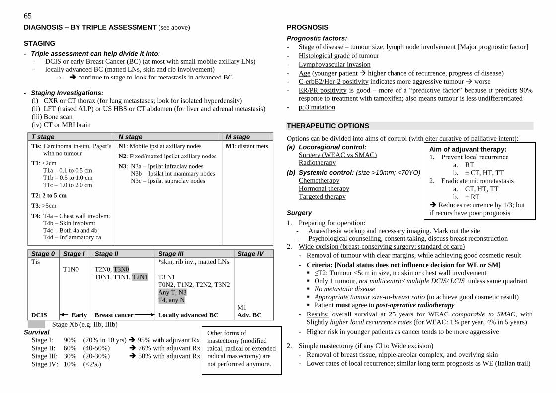
65
DIAGNOSIS – BY TRIPLE ASSESSMENT (see above) STAGING
- Triple assessment can help divide it into:
- DCIS or early Breast Cancer (BC) (at most with small mobile axillary LNs)
- locally advanced BC (matted LNs, skin and rib involvement)
o continue to stage to look for metastasis in advanced BC
- Staging Investigations:
(i) CXR or CT thorax (for lung metastases; look for isolated hyperdensity)
(ii) LFT (raised ALP) or US HBS or CT abdomen (for liver and adrenal metastasis)
(iii) Bone scan
(iv) CT or MRI brain
T stage N stage M stage
Tis: Carcinoma in-situ, Paget‘s
with no tumour
T1: <2cm
T1a – 0.1 to 0.5 cm
T1b – 0.5 to 1.0 cm
T1c – 1.0 to 2.0 cm
T2: 2 to 5 cm
T3: >5cm
T4: T4a – Chest wall involvmt
T4b – Skin involvmt
T4c – Both 4a and 4b
T4d – Inflammatory ca
N1: Mobile ipsilat axillary nodes
N2: Fixed/matted ipsilat axillary nodes
N3: N3a – Ipsilat infraclav nodes
N3b – Ipsilat int mammary nodes
N3c – Ipsilat supraclav nodes
M1: distant mets
Stage 0 Stage I Stage II Stage III Stage IV
Tis
DCIS
T1N0
Early
T2N0, T3N0
T0N1, T1N1, T2N1
Breast cancer
*skin, rib inv., matted LNs
T3 N1
T0N2, T1N2, T2N2, T3N2
Any T, N3
T4, any N
Locally advanced BC
M1
Adv. BC
Aaaaa – Stage Xb (e.g. IIb, IIIb) Survival
Stage I: 90% (70% in 10 yrs) 95% with adjuvant Rx
Stage II: 60% (40-50%) 76% with adjuvant Rx
Stage III: 30% (20-30%) 50% with adjuvant Rx
Stage IV: 10% (<2%)
PROGNOSIS
Prognostic factors:
- Stage of disease – tumour size, lymph node involvement [Major prognostic factor]
- Histological grade of tumour
- Lymphovascular invasion
- Age (younger patient higher chance of recurrence, progress of disease)
- C-erbB2/Her-2 positivity indicates more aggressive tumour worse
- ER/PR positivity is good – more of a ―predictive factor‖ because it predicts 90%
response to treatment with tamoxifen; also means tumour is less undifferentiated
- p53 mutation
THERAPEUTIC OPTIONS
Options can be divided into aims of control (with eiter curative of palliative intent):
(a) Locoregional control:
Surgery (WEAC vs SMAC)
Radiotherapy
(b) Systemic control: (size >10mm; <70YO)
Chemotherapy
Hormonal therapy
Targeted therapy
Surgery
1. Preparing for operation:
- Anaesthesia workup and necessary imaging. Mark out the site
- Psychological counselling, consent taking, discuss breast reconstruction
2. Wide excision (breast-conserving surgery; standard of care)
- Removal of tumour with clear margins, while achieving good cosmetic result
- Criteria: [Nodal status does not influence decision for WE or SM] ≤T2: Tumour <5cm in size, no skin or chest wall involvement
Only 1 tumour, not multicentric/ multiple DCIS/ LCIS unless same quadrant
No metastatic disease
Appropriate tumour size-to-breast ratio (to achieve good cosmetic result)
Patient must agree to post-operative radiotherapy
- Results: overall survival at 25 years for WEAC comparable to SMAC, with
Slightly higher local recurrence rates (for WEAC: 1% per year, 4% in 5 years)
- Higher risk in younger patients as cancer tends to be more aggressive
2. Simple mastectomy (if any CI to Wide excision)
- Removal of breast tissue, nipple-areolar complex, and overlying skin
- Lower rates of local recurrence; similar long term prognosis as WE (Italian trail)
Aim of adjuvant therapy:
1. Prevent local recurrence
a. RT
b. ± CT, HT, TT
2. Eradicate micrometastasis
a. CT, HT, TT
b. ± RT
Reduces recurrence by 1/3; but
if recurs have poor prognosis
Other forms of
mastectomy (modified
raical, radical or extended
radical mastectomy) are
not performed anymore.
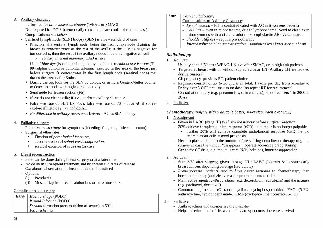
66
3. Axillary clearance
- Performed for all invasive carcinoma (WEAC or SMAC)
- Not required for DCIS (theoretically cancer cells are confined to the breast)
- Complications: see below
- Sentinel lymph node (SLN) biopsy (SLN) is a new standard of care
Principle: the sentinel lymph node, being the first lymph node draining the
breast, is representative of the rest of the axilla; if the SLN is negative for
tumour cells, then the rest of the axillary nodes should be negative as well
o Solitary internal mammary LAD is rare
Use of blue dye (isosulphan blue, methylene blue) or radioactive isotope (Tc-
99 sulphur colloid or colloidal albumin) injected in the area of the breast just
before surgery concentrates in the first lymph node (sentinel node) that
drains the breast after 5mins
During the op, look for the SLN by colour, or using a Geiger-Muller counter
to detect the node with highest radioactivity
Send node for frozen section (FS)
If -ve do not clear axilla; if +ve, perform axillary clearance
False –ve rate of SLN Bx <5%; false –ve rate of FS ~ 33% if so, re-
explore if histology +ve and do AC
No difference in axillary recurrence between AC vs SLN biopsy
4. Palliative surgery
- Palliative mastectomy for symptoms (bleeding, fungating, infected tumour)
- Surgery at other sites:
Fixation of pathological fractures,
decompression of spinal cord compression,
surgical excision of brain metastases
5. Breast reconstruction
- Safe, can be done during breast surgery or at a later time
- No delay in subsequent treatment and no increase in rates of relapse
- Cx: abnormal sensation of breast, unable to breastfeed
- Options:
(i) Prosthesis
(ii) Muscle flap from rectus abdominis or latissimus dorsi
Complications of surgery
Early Haemorrhage (POD1)
Wound Infection (POD3)
Seroma formation (accumulation of serum) in 50%
Flap ischemia
Late Cosmetic deformity
Complications of Axillary Clearance:
- Lymphoedema – RT is contraindicated with AC as it worsens oedema
- Cellulitis – even in minor trauma, due to lymphoedema. Need to clean even
minor wounds with antiseptic solution + prophylactic ABx vs staphstrep
- Shoulder stiffness – require physiotherapy
- Intercostobrachial nerve transection – numbness over inner aspect of arm.
Radiotherapy
1. Adjuvant
- Usually done 6/52 after WEAC, LN +ve after SMAC, or in high risk patients
- Targeted at breast with or without supraclavicular LN (Axillary LN are tackled
during Surgery)
- CI: pregnancy, previous RT, patient choice
- Regimen consists of 25 to 30 cycles in total, 1 cycle per day from Monday to
Friday over 5-6/52 until maximum dose (no repeat RT for recurrences)
- Cx: radiation injury (e.g. pneumonitis, skin changes), risk of cancers 1 in 2000 in
20yrs
2. Palliative
Chemotherapy (polyCT with 3 drugs is better; 4-6cycles, each over 1/12)
1. Neoadjuvant
- Given in LABC (stage III) to shrink the tumour before surgical resection
- 20% achieve complete clinical response (cCR) i.e. tumour is no longer palpable
further 20% will achieve complete pathological response (cPR) i.e. no
more tumour cells = good prognosis
- Need to place a clip into the tumour before starting neoadjuvant therapy to guide
surgery in case the tumour ―disappears‖; operate according preop staging
- Cx: as for CT drug, e.g. mouth ulcers, N/V, hair loss, immunosuppression
2. Adjuvant
- Start 3/52 after surgery; given in stage III / LABC (LN+ve) & in some early
breast cancers depending on stage (see below)
- Premenopausal patients tend to have better response to chemotherapy than
hormonal therapy (and vice versa for postmenopausal patients)
- Main active agents: anthracyclines (e.g. doxorubicin, epirubicin) and the taxanes
(e.g. paclitaxel, docetaxel) - Common regimens: AC (anthracycline, cyclophosphamide), FAC (5-FU,
anthracycline, cyclophosphamide), CMF (cyclophos, methotrexate, 5-FU)
3. Palliative
- Anthracyclines and taxanes are the mainstay
- Helps to reduce load of disease to alleviate symptoms, increase survival
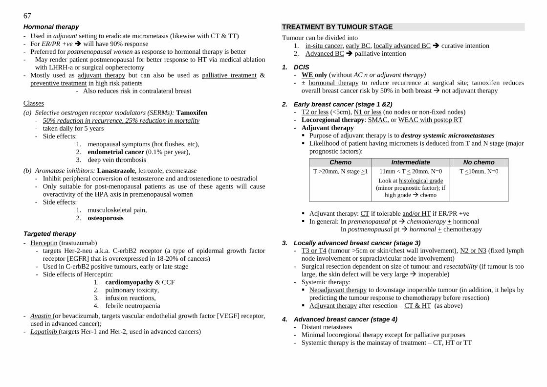
67
Hormonal therapy
- Used in adjuvant setting to eradicate micrometasis (likewise with CT & TT)
- For ER/PR +ve will have 90% response
- Preferred for postmenopausal women as response to hormonal therapy is better
- May render patient postmenopausal for better response to HT via medical ablation
with LHRH-a or surgical oopherectomy
- Mostly used as adjuvant therapy but can also be used as palliative treatment &
preventive treatment in high risk patients
- Also reduces risk in contralateral breast
Classes
(a) Selective oestrogen receptor modulators (SERMs): Tamoxifen
- 50% reduction in recurrence, 25% reduction in mortality - taken daily for 5 years
- Side effects:
1. menopausal symptoms (hot flushes, etc),
2. endometrial cancer (0.1% per year),
3. deep vein thrombosis
(b) Aromatase inhibitors: Lanastrazole, letrozole, exemestase
- Inhibit peripheral conversion of testosterone and androstenedione to oestradiol
- Only suitable for post-menopausal patients as use of these agents will cause
overactivity of the HPA axis in premenopausal women
- Side effects:
1. musculoskeletal pain,
2. osteoporosis
Targeted therapy
- Herceptin (trastuzumab)
- targets Her-2-neu a.k.a. C-erbB2 receptor (a type of epidermal growth factor
receptor [EGFR] that is overexpressed in 18-20% of cancers)
- Used in C-erbB2 positive tumours, early or late stage
- Side effects of Herceptin:
1. cardiomyopathy & CCF
2. pulmonary toxicity,
3. infusion reactions,
4. febrile neutropaenia
- Avastin (or bevacizumab, targets vascular endothelial growth factor [VEGF] receptor,
used in advanced cancer);
- Lapatinib (targets Her-1 and Her-2, used in advanced cancers)
TREATMENT BY TUMOUR STAGE
Tumour can be divided into
1. in-situ cancer, early BC, locally advanced BC curative intention
2. Advanced BC palliative intention
1. DCIS
- WE only (without AC n or adjuvant therapy)
- ± hormonal therapy to reduce recurrence at surgical site; tamoxifen reduces
overall breast cancer risk by 50% in both breast not adjuvant therapy
2. Early breast cancer (stage 1 &2)
- T2 or less (<5cm), N1 or less (no nodes or non-fixed nodes)
- Locoregional therapy: SMAC, or WEAC with postop RT
- Adjuvant therapy Purpose of adjuvant therapy is to destroy systemic micrometastases
Likelihood of patient having micromets is deduced from T and N stage (major
prognostic factors):
Chemo Intermediate No chemo
T >20mm, N stage >1
11mm < T < 20mm, N=0
Look at histological grade
(minor prognostic factor); if
high grade chemo
T <10mm, N=0
Adjuvant therapy: CT if tolerable and/or HT if ER/PR +ve
In general: In premenopausal pt chemotherapy + hormonal
In postmenopausal pt hormonal + chemotherapy
3. Locally advanced breast cancer (stage 3)
- T3 or T4 (tumour >5cm or skin/chest wall involvement), N2 or N3 (fixed lymph
node involvement or supraclavicular node involvement)
- Surgical resection dependent on size of tumour and resectability (if tumour is too
large, the skin defect will be very large inoperable)
- Systemic therapy:
Neoadjuvant therapy to downstage inoperable tumour (in addition, it helps by
predicting the tumour response to chemotherapy before resection)
Adjuvant therapy after resection – CT & HT (as above)
4. Advanced breast cancer (stage 4)
- Distant metastases
- Minimal locoregional therapy except for palliative purposes
- Systemic therapy is the mainstay of treatment – CT, HT or TT
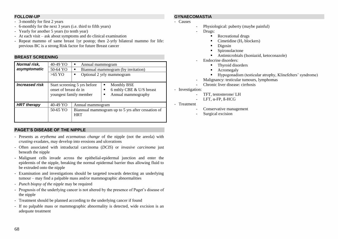
68
FOLLOW-UP - 3-monthly for first 2 years
- 6-monthly for the next 3 years (i.e. third to fifth years)
- Yearly for another 5 years (to tenth year)
- At each visit – ask about symptoms and do clinical examination
- Repeat mammo of same breast 1yr postop; then 2-yrly bilateral mammo for life:
previous BC is a strong Risk factor for future Breast cancer
BREAST SCREENING
Normal risk, asymptomatic
40-49 YO Annual mammogram
50-64 YO Biannual mammogram (by invitation)
>65 YO Optional 2 yrly mammogram
Increased risk Start screening 5 yrs before
onset of breast dz in
youngest family member
Monthly BSE
6 mthly CBE & U/S breast
Annual mammography
HRT therapy 40-49 YO Annual mammogram
50-65 YO Biannual mammogram up to 5 yrs after cessation of
HRT
PAGET’S DISEASE OF THE NIPPLE
- Presents as erythema and eczematous change of the nipple (not the areola) with
crusting exudates, may develop into erosions and ulcerations
- Often associated with intraductal carcinoma (DCIS) or invasive carcinoma just
beneath the nipple
- Malignant cells invade across the epithelial-epidermal junction and enter the
epidermis of the nipple, breaking the normal epidermal barrier thus allowing fluid to
be extruded onto the nipple
- Examination and investigations should be targeted towards detecting an underlying
tumour – may find a palpable mass and/or mammographic abnormalities
- Punch biopsy of the nipple may be required
- Prognosis of the underlying cancer is not altered by the presence of Paget‘s disease of
the nipple
- Treatment should be planned according to the underlying cancer if found
- If no palpable mass or mammographic abnormality is detected, wide excision is an
adequate treatment
GYNAECOMASTIA - Causes
- Physiological: puberty (maybe painful)
- Drugs:
Recreational drugs
Cimetidine (H2 blockers)
Digosin
Spironolactone
Antimicrobials (Isoniazid, ketoconazole)
- Endocrine disorders:
Thyroid disorders
Acromegaly
Hypogonadism (testicular atrophy, Klinefelters‘ syndrome)
- Malignancy: testicular tumours, lymphomas
- Chronic liver disease: cirrhosis
- Investigation:
- TFT, testosterone/ LH
- LFT, α-FP, ß-HCG
- Treatment
- Conservative management
- Surgical excision
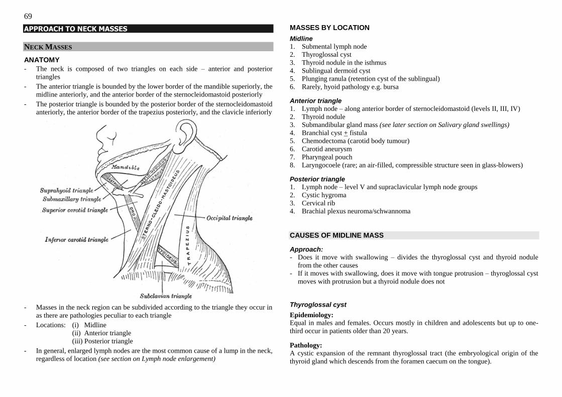
69
APPROACH TO NECK MASSES
NECK MASSES
ANATOMY
- The neck is composed of two triangles on each side – anterior and posterior
triangles
- The anterior triangle is bounded by the lower border of the mandible superiorly, the
midline anteriorly, and the anterior border of the sternocleidomastoid posteriorly
- The posterior triangle is bounded by the posterior border of the sternocleidomastoid
anteriorly, the anterior border of the trapezius posteriorly, and the clavicle inferiorly
- Masses in the neck region can be subdivided according to the triangle they occur in
as there are pathologies peculiar to each triangle
- Locations: (i) Midline
(ii) Anterior triangle
(iii) Posterior triangle
- In general, enlarged lymph nodes are the most common cause of a lump in the neck,
regardless of location (see section on Lymph node enlargement)
MASSES BY LOCATION
Midline
1. Submental lymph node
2. Thyroglossal cyst
3. Thyroid nodule in the isthmus
4. Sublingual dermoid cyst
5. Plunging ranula (retention cyst of the sublingual)
6. Rarely, hyoid pathology e.g. bursa
Anterior triangle
1. Lymph node – along anterior border of sternocleidomastoid (levels II, III, IV)
2. Thyroid nodule
3. Submandibular gland mass (see later section on Salivary gland swellings)
4. Branchial cyst + fistula
5. Chemodectoma (carotid body tumour)
6. Carotid aneurysm
7. Pharyngeal pouch
8. Laryngocoele (rare; an air-filled, compressible structure seen in glass-blowers)
Posterior triangle 1. Lymph node – level V and supraclavicular lymph node groups
2. Cystic hygroma
3. Cervical rib
4. Brachial plexus neuroma/schwannoma
CAUSES OF MIDLINE MASS
Approach:
- Does it move with swallowing – divides the thyroglossal cyst and thyroid nodule
from the other causes
- If it moves with swallowing, does it move with tongue protrusion – thyroglossal cyst
moves with protrusion but a thyroid nodule does not Thyroglossal cyst
Epidemiology: Equal in males and females. Occurs mostly in children and adolescents but up to one-
third occur in patients older than 20 years.
Pathology:
A cystic expansion of the remnant thyroglossal tract (the embryological origin of the
thyroid gland which descends from the foramen caecum on the tongue).
70
Features:
Smooth, rounded, cystic lump. 75% are in the midline while 25% are slightly to the left
or right. Usually asymptomatic but may become infected with sinus formation and
seropurulent discharge (occurs with incision or rupture of cyst)
Histology:
Cyst with columnar or squamous epithelial lining which may be ciliated. The cyst may
also contain thyroid and lymphoid tissue. If malignancy occurs (carcinoma of the
thyroglossal duct), it is usually a papillary carcinoma (~90%).
Treatment:
Sistrunk procedure – resection of the cyst and mid-portion of the hyoid bone in
continuity and resection of a core of tissue from the hyoid upwards towards the foramen
caecum.
Dermoid cyst
Pathology:
Can be congenital or acquired.
(i) Congenital – developmental inclusion of epidermis along lines of fusion of skin
dermatomes (seen in younger patients, present since birth). Locations include:
o medial and lateral ends of the eyebrows (internal and external angular dermoid
cysts)
o midline of the nose (nasal dermoid cysts)
o midline of the neck and trunk
(ii) Acquired – due to forced inclusion of skin into subcutaneous tissue following an
injury, usually on fingers. Seen in older patients, no previous history of mass,
history of trauma to area (may have associated scar).
Histology:
Cyst lined by epidermis, with evidence of adnexal structures such as hair follicles,
sebaceous glands and sweat glands.
Features: Small non-tender mobile subcutaneous lump, may be fluctuant, skin-coloured or bluish.
Management
- Imaging investigations (e.g. XR, U/S, CT) are important especially for cysts on the
skull as they can communicate with cerebrospinal fluid. - Complete surgical excision of the cyst.
Plunging ranula
Pathology:
A pseudocyst associated with the sublingual glands and submandibular ducts. Ranulas
can be congenital or acquired after oral trauma. A large ranula can present as a neck
mass if it extends through the mylohyoid musculature of the floor of the mouth – termed
a ―plunging‖ ranula.
Treatment:
- Complete resection if possible, often in continuity with the associated sublingual
gland (but often difficult due to close association with the lingual nerve and
submandibular duct).
- If complete resection not possible, marsupialisation and suturing of the pseudocyst
wall to the oral mucosa may be effective.
CAUSES OF ANTERIOR TRIANGLE MASS
Branchial cyst and/or fistula
Epidemiology: Affects both sexes equally, usually in young adults in their 20s.
Pathology:
A branchial cyst is thought to develop because of failure of fusion of the embryonic
second and third branchial arches. It is lined by squamous epithelium.
Features:
- Occurs anterior to the upper or middle third of the sternocleidomastoid muscle.
- Smooth firm swelling that is ovoid in shape, with its long axis running downwards
and forwards.
- May be fluctuant, usually not transilluminable (due to desquamated epithelial cell
contents).
- Look for fistula in this area – a branchial fistula will run between tonsillar fossa and
the anterior neck, passing between the external and internal carotid arteries.
- Fine needle aspiration of the cyst will yield opalescent fluid with cholesterol crystals under microscopy.
- May be complicated by recurrent infections – purulent discharge, fixation to
surrounding structures.
Management:
- If fistula present, perform fistulogram to delineate course.
- Surgical excision of the cyst where possible. If fistula/sinus present, inject Bonney‘s
blue dye into tract prior to surgery to allow accurate surgical excision.
- Treatment of infection with antibiotics.
- Complications: cyst recurrence; chronic discharging sinus.
71
Chemodectoma
Pathology:
A chemodectoma is a tumour of the paraganglion cells (paraganglionoma) of the carotid
body located at the bifurcation of the common carotid artery (into the internal and
external carotids). They are usually benign, but locally invasive; the risk of malignancy
is 10%, with metastasis to local lymph nodes (no histopathological features for
malignancy, thus malignant nature can only be diagnosed by presence of metastasis).
Features: - Solid, firm mass at the level of the hyoid bone (where the bifurcation is) – be gentle
during palpation as pressure on the carotid body can cause vasovagal syncope.
- Mass is pulsatile but not expansile, due to transmitted pulsation from carotids.
- Due to close association with carotid arteries, lump can be moved side to side but not
up and down.
- May be bilateral.
- If suspecting aneurysm, listen for bruit, look for signs of Horner‘s syndrome,
examine the rest of the peripheral vascular system.
Differentials and investigation:
- Main differential is carotid artery aneurysm; aneurysm can occur at any level but
carotid body tumour occurs at the level of the hyoid bone.
- DO NOT PERFORM FNA
- CT and/or MRI can be used to delineate tumour anatomy in relation to surrounding
structures; CT reveals homogenous mass with intense enhancement following IV
contrast administration.
- Angiography is the gold standard investigation – shows a hypervascular mass
displacing the bifurcation. May also show vessel compromise by tumour invasion,
and undetected synchronous tumours.
Treatment: - Surgical excision with pre-operative embolisation (reduces bleeding and
complications, and facilitates resection); any enlarged ipsilateral lymph nodes are
also removed due to the small possibility of malignancy
- Radiotherapy is an effective alternative for patients who are unfit for surgery or
whose tumours are too large.
Pharyngeal pouch (also called Zenker’s diverticulum)
Pathology: A herniation of the pharyngeal mucosa (pulsion diverticulum) through its muscular coat
at the weakest point – Killian‘s dehiscence – between the cricopharyngeus muscle and
the lower inferior constrictor muscles.
Features
- Occurs in older patients
- A cystic swelling low down in the anterior triangle, usually on the left
- Squelching sound on deep palpation
- Patient complains of halitosis, regurgitation of undigested food with coughing and
dysphagia in the neck, hoarseness, weight loss
- Complications: chest infection (due to aspiration); diverticular neoplasm (<1%)
Diagnosis by barium swallow
Treatment
- Leave it alone if small and asymptomatic
- Minimally invasive treatment: endoscopic cricothyroid myotomy
- Surgical approaches (several available)
o Diverticulectomy + cricothyroidotomy (diverticulectomy associated with risk of
mediastinitis, dangerous)
o Diverticulopexy (done in high risk patients, involves suspending the lumen of
the pouch in the caudal direction so that food and secretions cannot enter the
pouch; as the diverticulum is still present, the risk for malignancy still remains)
CAUSES OF POSTERIOR TRIANGLE MASS
Cystic hygroma
Pathology: A cystic hygroma is a congenital cystic lymphatic malformation found in the posterior
triangle of the neck, probably formed during coalescence of primitive lymph elements.
It consists of thin-walled, single or multiple interconnecting or separate cysts which
insinuate themselves widely into the tissues at the root of the neck.
Features:
- 50-65% present at birth, but occasionally may present later in childhood or adulthood
- Lobulated cystic swelling that is soft, fluctuant, and compressible (usually into
another part of the cyst), located in the posterior triangle at the root of the neck
- Classically “brilliantly transilluminable”
- A large cyst may extend deeply into the retropharyngeal space
Complications: - Cystic hygroma seen on prenatal ultrasound in the first trimester suggests
chromosomal abnormality (50% of foetuses, usually trisomy 21) or other structural
abnormalities (33% of foetuses with no chromosomal abnormality, usually congenital
heart anomalies)
- May obstruct delivery
- Compressive problems after delivery – respiratory, swallowing
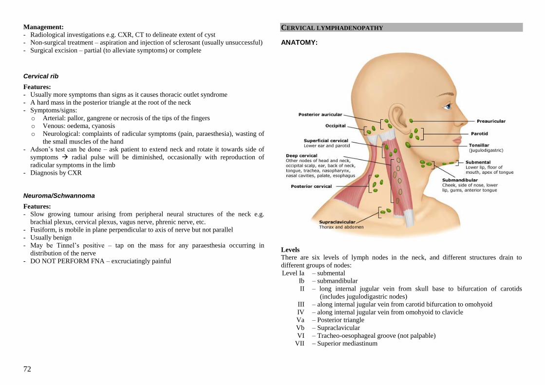
72
Management:
- Radiological investigations e.g. CXR, CT to delineate extent of cyst
- Non-surgical treatment – aspiration and injection of sclerosant (usually unsuccessful)
- Surgical excision – partial (to alleviate symptoms) or complete
Cervical rib
Features:
- Usually more symptoms than signs as it causes thoracic outlet syndrome
- A hard mass in the posterior triangle at the root of the neck
- Symptoms/signs:
o Arterial: pallor, gangrene or necrosis of the tips of the fingers
o Venous: oedema, cyanosis
o Neurological: complaints of radicular symptoms (pain, paraesthesia), wasting of
the small muscles of the hand
- Adson‘s test can be done – ask patient to extend neck and rotate it towards side of
symptoms radial pulse will be diminished, occasionally with reproduction of
radicular symptoms in the limb
- Diagnosis by CXR
Neuroma/Schwannoma
Features:
- Slow growing tumour arising from peripheral neural structures of the neck e.g.
brachial plexus, cervical plexus, vagus nerve, phrenic nerve, etc.
- Fusiform, is mobile in plane perpendicular to axis of nerve but not parallel
- Usually benign
- May be Tinnel‘s positive – tap on the mass for any paraesthesia occurring in
distribution of the nerve
- DO NOT PERFORM FNA – excruciatingly painful
CERVICAL LYMPHADENOPATHY
ANATOMY:
Levels There are six levels of lymph nodes in the neck, and different structures drain to
different groups of nodes:
Level Ia – submental
Ib – submandibular
II – long internal jugular vein from skull base to bifurcation of carotids
(includes jugulodigastric nodes)
III – along internal jugular vein from carotid bifurcation to omohyoid
IV – along internal jugular vein from omohyoid to clavicle Va – Posterior triangle
Vb – Supraclavicular
VI – Tracheo-oesophageal groove (not palpable)
VII – Superior mediastinum
73
Drainage:
- Oral cavity and oropharynx levels I – III
- Thyroid and larynx levels II – VI
- Nasopharynx II – V (usually upper neck – level II and high level V)
CAUSES:
Can be divided into three main groups: infective, inflammatory, and neoplastic
Infective Viral
Epstein-Barr virus, cytomegalovirus (infectious mononucleosis); HIV
Bacteria
Streptococcus, Staphylococcus, Klebsiella (from intraoral pathology
e.g. dental abscess, tonsillitis)
Tuberculosis
Parasitic
Toxoplasma
Fungi
Actinomycosis
Neoplastic Metastatic Head and neck primary Nasopharyngeal carcinoma
Oral cavity, oropharynx, larynx, hypopharynx,
thyroid, etc.
Other primary sites (4B‘s)
Bowel (stomach, colon), breast, bronchus (lung),
balls (testicular)
Primary - lymphoma
Inflammatory SLE
Kikuchi‘s (necrotising lymphadenitis occurring in young females,
presenting as painful cervical lymphadenopathy)
Sarcoidosis
HISTORY
- The lump itself – onset, duration, rate of growth, any pain, associated symptoms,
lumps elsewhere
- Constitutional symptoms
o Fever, malaise, arthralgia, myalgia (viral prodrome);
o Night sweats, low-grade fever (TB, B symptoms of lymphoma);
o Loss of appetite, loss of weight (chronic infection, malignancy)
- Local symptoms – intra-oral diseases e.g. tooth decay, oral/tongue ulcer, tonsillitis
- Past medical history – cancer, TB (contact? Diagnosed? treated or untreated?)
- Social history: travel and contact history, sexual history for HIV
PHYSICAL EXAMINATION
Inspection
- Location
- Any overlying skin changes e.g. erythema, discharging sinus (multiple lymph node
enlargement with discharging sinuses can be TB or actinomycosis; sulphur granules
seen in actinomycosis)
Palpate from behind, one side at a time – start at submental, then submandibular,
preauricular, postauricular, along anterior border of sternocleidomastoid,
supraclavicular, posterior triangle, lastly occipital. Use pulps of the fingers in a gentle
rolling movement.
- Tenderness to palpation
- Consistency – hard, matted nodes are more suspicious for malignancy
- Fixation to overlying skin or underlying structures
To complete the examination:
- Complete examination of the face and scalp for any primary site of infection or
neoplasia
- Formal ear, nose, throat examination especially looking at the post-nasal space for
nasopharyngeal carcinoma (NPC being the most common cancer causing enlarged
cervical lymph nodes)
- Examination of the thyroid gland
- Examination of lymphoreticular system – other lymph node groups, liver, spleen
- Full respiratory and abdominal examination especially if supraclavicular lymph node
found
- Breast examination in female patient
INVESTIGATIONS:
- Fine needle aspiration provides most definitive results (though there is still the
possibility of false positive and false negative results)
- Radiological investigation e.g. ultrasound, CT – to assess nature of lump, extent,
other enlarged nodes that are not clinically palpable, and can be used to visualise
primary tumour if present
MANAGEMENT:
- According to FNAC results
- Malignant – work up for primary if present (e.g. squamous cell carcinoma – look for
oral cavity, skin, ENT, lung malignancy; adenocarcinoma – look for breast, GI tract
malignancy) and treat as appropriate
- Infective/Inflammatory – treat underlying condition
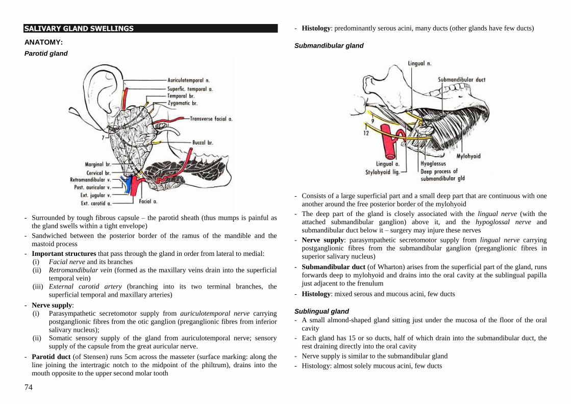
74
SALIVARY GLAND SWELLINGS
ANATOMY:
Parotid gland
- Surrounded by tough fibrous capsule – the parotid sheath (thus mumps is painful as
the gland swells within a tight envelope)
- Sandwiched between the posterior border of the ramus of the mandible and the
mastoid process
- Important structures that pass through the gland in order from lateral to medial:
(i) Facial nerve and its branches
(ii) Retromandibular vein (formed as the maxillary veins drain into the superficial
temporal vein)
(iii) External carotid artery (branching into its two terminal branches, the
superficial temporal and maxillary arteries)
- Nerve supply:
(i) Parasympathetic secretomotor supply from auriculotemporal nerve carrying
postganglionic fibres from the otic ganglion (preganglionic fibres from inferior
salivary nucleus);
(ii) Somatic sensory supply of the gland from auriculotemporal nerve; sensory
supply of the capsule from the great auricular nerve.
- Parotid duct (of Stensen) runs 5cm across the masseter (surface marking: along the
line joining the intertragic notch to the midpoint of the philtrum), drains into the
mouth opposite to the upper second molar tooth
- Histology: predominantly serous acini, many ducts (other glands have few ducts)
Submandibular gland
- Consists of a large superficial part and a small deep part that are continuous with one
another around the free posterior border of the mylohyoid
- The deep part of the gland is closely associated with the lingual nerve (with the
attached submandibular ganglion) above it, and the hypoglossal nerve and
submandibular duct below it – surgery may injure these nerves
- Nerve supply: parasympathetic secretomotor supply from lingual nerve carrying
postganglionic fibres from the submandibular ganglion (preganglionic fibres in
superior salivary nucleus)
- Submandibular duct (of Wharton) arises from the superficial part of the gland, runs
forwards deep to mylohyoid and drains into the oral cavity at the sublingual papilla
just adjacent to the frenulum
- Histology: mixed serous and mucous acini, few ducts
Sublingual gland
- A small almond-shaped gland sitting just under the mucosa of the floor of the oral
cavity
- Each gland has 15 or so ducts, half of which drain into the submandibular duct, the
rest draining directly into the oral cavity
- Nerve supply is similar to the submandibular gland
- Histology: almost solely mucous acini, few ducts
75
HISTORY
- About the lump: onset, duration, progress, associated symptoms e.g. pain
- If pain is present, is it precipitated by food ingestion? (suggestive of sialolithiasis)
- Symptoms of infection e.g. fever, malaise; if considering mumps, ask about testicular
pain and swelling (orchitis), abdominal pain (pancreatitis)
- Any noticed asymmetry of the face – incomplete closure of the eye on one side,
drooping corner of the mouth, drooling
- Does the patient have symptoms of xerostomia (e.g. cannot eat a piece of biscuit or
bread without water), xerophthalmia
- History of connective tissue disease e.g. rheumatoid arthritis, SLE
PHYSICAL EXAMINATION
Inspect
- Put yourself at the level of the patient‘s face and look from front for any asymmetry
with an obvious mass on one side – parotid mass is located between the angle of the
jaw and the ear, and lifts the earlobe if large; submandibular mass is located just
under the mandible
- Look for scars – parotidectomy scar runs anteriorly to the ear, below the earlobe and
around posteriorly before looping forward again under the jaw
- Look for fistula/sinus
- Look at the patient‘s face for asymmetry (facial nerve palsy)
Palpate from behind
- Ask patient about any pain before starting to palpate
- Palpate the obviously enlarged gland, and always remember to also palpate the
contralateral gland for any swelling
- Check for warmth of overlying skin, tenderness, consistency, surface, margins
- Fixation to underlying structures – for parotid, ask patient to clench the teeth to
contract the masseter, then try to move the gland
- Fixation to overlying skin
- Palpate for cervical lymphadenopathy
Other tests
- Examination of the duct openings:
o Using a torch and a tongue depressor, examine opposite the second upper molar
tooth for the opening of the parotid duct, and under the tongue for the opening of
the submandibular duct.
o Look for red inflamed duct opening, discharge (purulent), or any visible stone.
o For the parotid duct, can palpate the duct along the masseter for stone, and look
for discharge inside the mouth while palpating.
- Palpate the gland openings for stones.
- Bimanual palpation of the submandibular gland
- Facial nerve examination
Suspicious features of malignancy:
- Hyperaemic hot skin over lump
- Pain
- Fixation to underlying structures or skin
- Hard consistency
- Irregular surface or ill-defined border
- Facial nerve involvement
CAUSES OF SWELLING OF THE PAROTID
Parenchymal swelling
Neoplasia Benign e.g. pleomorphic adenoma, Warthin‘s
Malignant tumours
Lymphoma and leukaemia*
Stones Sialolithasis
Infection/
Inflammation
Mumps*
Acute sialadenitis
Chronic recurrent sialadenitis
HIV
Autoimmune Sjogren‘s syndrome*
Infiltration Sarcoidosis*
Systemic
disease*
Alcoholic liver disease
Diabetes mellitus
Pancreatitis
Acromegaly
Malnutrition
Non-parenchymal swelling
Lymph node
Facial neuroma
Temporal artery aneurysm
Skin and soft tissue swellings e.g. sebaceous cyst, lipoma
* are conditions in which parotid swelling is bilateral
76
SIALOLITHIASIS
Epidemiology
- Stones of the salivary gland that may be impacted within the gland itself or in the
duct.
- Usually occurs in males more than females, and between the ages of 30 and 60.
- 80% of salivary stones occur in the submandibular gland (due to its higher mucus and
calcium content with a long duct, and slow flow of the saliva against gravity); 10%
occur in the parotid, 7% sublingual.
- Most submandibular gland stones occur in the duct, while 50% of parotid stones
occur in the gland itself.
- 80-95% of submandibular stones are radio-opaque and can be seen on an X-ray of the
floor of the mouth, and 60% of parotid stones are radio-opaque.
Presentation
- Complete obstruction Acute pain and swelling of the gland involved at meal times, rapid onset within
minutes of starting to eat, resolves about an hour after the meal.
- Partial obstruction
Occasional symptomatic episodes interspersed by asymptomatic periods of days to
weeks, chronically enlarged mass in the submandibular region
- Can result in sialadenitis, and even abscess formation worsening of symptoms of
pain and redness; systemic symptoms such as fever, chills; purulent discharge from
duct opening
- Stone may be palpable along the duct or at the opening of the duct
Investigations
- Noncontrast CT scan – can pick up almost all stones when fine cuts are requested
- Plain X-rays can pick up radio-opaque stones
- Sialogram (rarely done today as it is invasive and technically demanding, and CT is
better. Contraindicated in acute sialadenitis and contrast allergy.)
Management
- General measures: o Good hydration, soft diet, good oral hygiene
o Massage of the gland, milking the duct, application of moist hot towel
o Analgesia – NSAIDs such as ibuprofen
o Antibiotics if patient has sialadenitis – usually antibiotics to cover Staph and
Strept e.g. Augmentin
o Refer specialist treatment if symptoms persist for several days, or sialadenitis
persists despite antibiotic therapy
- Surgical removal
o Transoral removal of stones for submandibular duct stones (50% can be removed
thus), less for parotid duct stones
o If stones cannot be removed via transoral surgery or is intraglandular, partial
gland resection can be performed
- Other options: Lithotripsy, wire basket removal, sialoendoscopy
SALIVARY GLAND TUMOURS
Epidemiology:
- 80% occur in the parotid, of which 80% are benign (80% of the benign tumours are
pleomorphic adenomas)
- 10% occur in the submandibular, of which 60% are benign (95% pleomorphic
adenoma)
- 15% occur in minor salivary glands, of which 50% are benign (all benign tumours are
pleomorphic adenomas)
- 0.3% occur in sublingual glands, of which all are malignant
Pathology
Epithelial Non-epithelial
Adenomas (benign) Carcinomas (malignant)
Pleomorphic adenoma
Warthin‘s tumour
Adenoid cystic ca
Pleomorphic adenoca
Mucoepidermoid ca
Acinic cell ca
Adenoca
Squamous cell ca
Undifferentiated
Haemangioma
Lymphangioma
Neurofibroma
Neurilemmoma
Lipoma
Sarcoma
Malignant lymphoma
Pleomorphic adenoma
Epidemiology:
- Most common benign tumour
- 85% occur in the parotid gland
- Equal sex ratio, occurs in younger patients less than 50 years old
Histology:
Very heterogeneous appearance, containing epithelial cells surrounded by loose stroma
with islands of chondromyxoid (mesenchymal components), and interspersed islands of
myoepithelial cells. The tumour appears to be encapsulated, but histology shows
multiple sites of capsular penetration by tumour cells.
77
Features:
- Slow-growing, painless swelling occurring in the lower pole of the parotid
- Irregular and lobulated surface, texture of cartilage (slightly harder than Warthin‘s)
- Does not invade or metastasise
- Chance of malignant transformation if left for 10-15 years (1-6% risk)
- If not completely excised, can recur (recurrence rate of 2%)
Diagnosis by clinical, FNAC, + MRI
Treatment – surgical excision - Parotid: Superficial parotidectomy for superficial tumour; if tumour is deep or large,
total parotidectomy with preservation of the facial nerve
- Submandibular: Total gland excision together with adjacent connective tissue,
sparing lingual and hypoglossal nerves
Warthin’s tumour
Epidemiology:
- Only occurs in the parotid gland (10% of parotid tumours)
- More common in males than females (4:1)
- Occurs in older patients (>50 years)
- Related to cigarette smoking
Histology: Also called papillary cystadenoma lymphomatosum or just adenolymphoma. Consists
of cleftlike or cystic spaces lined by two-tiered epithelium, containing mucin,
surrounded by a stroma of well-developed lymphoid tissue with germinal centres.
Features:
- Slowly enlarging, soft to firm cystic fluctuant swelling in parotid tail
- Invariably benign with no risk of malignant change
Diagnosis by clinical, FNA + MRI
Treatment - Can be left alone if absolutely certain that the entire mass is composed of only
Warthin‘s tumour cells, since there is no malignant potential
- Superficial parotidectomy if causing trouble to patient
Malignant tumours
Most common malignancies are mucoepidermoid (34%) and adenoid cystic carcinomas
(22%) – equal sex ratio, can occur in any salivary gland, in older patients (usually >60
yrs)
Mucoepidermoid ca is the most common malignant tumour in the parotid, while
adenoid cystic ca is the most common in the submandibular, sublingual and minor
salivary glands
Malignant pleomorphic adenoma
- Usually occurs in pre-existing pleomorphic adenoma, rarely de novo
- Worst prognosis of any salivary gland tumour
- 30-70% recurrence and metastasis rate
Treatment of salivary gland cancers
Parotid:
- Total parotidectomy with sacrifice of facial nerve if tumour has infiltrated it (may be
grafted with great auricular nerve)
- Radical neck dissection if neck nodes positive
- Postoperative radiotherapy
Submandibular:
- Radical excision of gland with lymphatic clearance of submandibular triangle
- Radical neck dissection if neck nodes positive
- Postoperative radiotherapy
COMPLICATIONS OF PAROTIDECTOMY
Immediate (within 24 hrs)
1. Intraoperative facial nerve transection – lower motor neurone palsy (in surgery to
the submandibular gland, damage to the hypoglossal and/or lingual nerves can
occur intraoperatively)
2. Reactionary haemorrhage
Early (1 to 30 days)
1. Wound infection
2. Skin flap necrosis
3. Temporary facial weakness (neuropraxia of facial nerve)
4. Salivary fistula 5. Division of great auricular nerve loss of sensation over pinna
6. Trismus (inability to open mouth due to spasm of masseter)
Late (more than 30 days)
1. Wound dimple, cosmetic problems
2. Hyperaesthesia of local skin
3. Frey‟s syndrome – increased sweating and redness of facial skin when eating, due
to reinnervation of divided sympathetic nerves to the facial skin by fibres of the
secretomotor branch of the auriculotemporal nerve
78
THE THYROID GLAND
APPROACH TO THYROID PROBLEMS – 2 MAIN TYPES
1. Problem with configuration/anatomy
(i) Solitary thyroid nodule (most common in exam)
(ii) Multinodular goitre
(iii) Diffuse enlargement
2. Problem with function (usually hyperfunctioning)
(i) Graves‘ disease
(ii) Toxic adenoma
(iii) Toxic multinodular goitre
(iv) Hashimoto‘s disease
AIMS OF ASSESSMENT IN THYROID NODULE/ENLARGEMENT: - Exclude cancer!
- Address issues of thyroid function
- Look for any complications e.g. compression (of airway, oesophagus, rarely nerve)
- Cosmesis – is patient bothered by lump?
HISTORY-TAKING
About the LUMP
- Onset (gradual or sudden), duration
- Size (Diffuse or one side predominant? Any sudden increase in size? – malignant
growth; ddx includes haemorrhage into necrotic nodule or cyst, subacute thyroiditis)
- Any pain – bleeding into cyst can result in sudden increase in size and pain; rarely
pain can occur in anaplastic carcinoma and thyroiditis
- Compressive symptoms: difficulty swallowing, difficulty breathing, hoarseness of
voice (benign pathologies almost never compress the recurrent laryngeal nerve)
- Cosmetic effects
About THYROID FUNCTION
Hyperthyroid Hypothyroid
Weight loss despite increased appetite
Decreased appetite, weight gain, lethargy
Heat intolerance
Cold intolerance
Increased sweating
Dry skin, loss of outer third of eyebrows
Proximal myopathy (Graves’)
Muscle fatigue
Diarrhoea, frequent bowel movement
Constipation
Tachycardia, atrial fibrillation
Bradycardia
Oligomenorrhoea, amenorrhoea
Menorrhagia
Nervousness; easily irritable; emotional lability; insomnia
Slow thought, speech and action; depression; dementia
Fine tremor Carpal tunnel syndrome symptoms
About RISK FACTORS
- History of autoimmune disease e.g. type I DM, SLE, RA, pernicious anaemia
(associations with Graves and Hashimoto‘s)
- History of cancer elsewhere – metastatic disease to thyroid; lymphoma; papillary
cancer is associated with familial polyposis syndromes ask about GI polyps/ca
- History of thyroid disease – long-standing MNG can progress to lymphoma
- Occupational history – any exposure to radiation (papillary cancer risk)
- Family history of thyroid cancer – ~20% of medullary cancers are familial (MEN2,
AD inheritance), ~5% of papillary cancers
About previous TREATMENT for any thyroid disease
- Medications given e.g. propylthiouracil, carbimazole, propranolol – for how long,
efficacy, side effects
- Radioactive iodine treatment – what was the result? Is the patient receiving
replacement?
- Surgery – what kind of surgery, any complications?
- Follow-up – what investigations done?
PHYSICAL EXAMINATION
A. THYROID GLAND
GREET PATIENT, ASK FOR PERMISSION to examine (and listen to the voice – is it hoarse?)
POSITION PATIENT – on a chair with space behind the chair for you to stand.
INSPECT FROM THE FRONT
1. Any swelling? Where is it?
2. Any scars (thyroidectomy scar may be difficult to spot as it is often hidden in a
skin crease)? Sinuses?
3. Any skin changes over the mass?
4. Check if mass moves on swallowing by asking patient to take a sip of water –
―Please take a sip of water and hold it in your mouth, do not swallow until I tell you
to.‖
5. Check if mass moves on protruding the tongue – ―Please open your jaw slightly.
Now, without moving your jaw, please stick your tongue out and back in again.‖
NB. A thyroid swelling moves only on swallowing; a thyroglossal cyst will move
on both swallowing and protrusion of the tongue.
6. Check for plethora of face, distended neck veins – may be due to compressive
nature of mass (but rarely seen).
79
PALPATE FROM BEHIND – one side at a time, the opposite hand stabilises the gland.
Ask for pain before palpating!
1. Characteristics of lump: site (anterior triangle), size (discrete nodule or
multinodular enlargement or diffuse enlargement?), consistency (soft, cystic, hard,
multinodular?), mobility (fixed to skin? Fixed to underlying structures?), tenderness.
2. Check swallowing while palpating to confirm mass moves on swallowing.
3. Check tongue protrusion.
4. Palpate lymph nodes
PALPATE TRACHEA from in front for tracheal deviation.
PERCUSS – any retrosternal extension?
AUSCULTATE – bruit in Graves‘
OFFER to do Pemberton’s sign to check for thoracic inlet obstruction; check thyroid
status; ask patient about compressive symptoms.
B. THYROID STATUS
HANDS (get patient to stretch arms out in front of him, palms down)
1. Feel palms – warm sweaty palms
2. Nails – thyroid acropachy, onycholysis (both seen in Graves‘)
3. Feel pulse – tachycardia, atrial fibrillation (AF more in toxic MNG than Graves‘)
4. Fine postural tremor – accentuate by placing a sheet of paper on the hands
5. Palms up – palmar erythema
FACE
1. Expression – staring, unblinking (hyperthyroid); lethargic, apathetic (hypothyroid)
2. Complexion – dry, ‗peaches-and-cream‘ complexion, loss of outer third of
eyebrows (hypothyroid)
3. Eyes
- Lid retraction (can see sclera between upper limbus of iris and upper eyelid)
- Exophthalmos (sclera between lower limbus and lower eyelid)
- Chemosis (oedema and erythema of conjunctiva)
- Ophthalmoplegia (restriction of eye movements; ask about diplopia!)
- Lid lag (eyelid lags behind eye when patient follows your finger downwards)
- Proptosis (look from above patient‘s head – eye visible over supraorbital ridge)
NEUROMUSCULAR
1. Proximal myopathy (Graves‘)
2. Reflexes – slow to relax in hypothyroidism
3. Legs for pretibial non-pitting oedema (Graves‘ or hypothyroid)
PROBLEMS WITH GLAND CONFIGURATION
PART I: RELEVANT ANATOMY
Structure:
2 lateral lobes joined by an isthmus that lies in front of the 2nd
, 3rd
and 4th
tracheal rings.
Strap muscles of the neck lie superficial to the thyroid gland.
Nerves and vessels:
Superior thyroid artery (from external carotid)
Inferior thyroid artery (from thyrocervical trunk, a branch of the first part of the
subclavian artery).
External laryngeal nerve – supplies the cricothyroid muscle which controls pitch of
voice; runs close to superior thyroid artery.
Recurrently laryngeal nerve – supplies all the other intrinsic muscles of the larynx
(except for cricothyroid) and runs close to the branches of the inferior thyroid. The
nerve runs behind the pretracheal fascia and so will not be damaged if the fascia is
not breached during operation. Important to visualise nerve and avoid damaging it!
Embryonic origin:
Thyroglossal tract from foramen caecum of the tongue (in the midline, at the junction
between anterior two-thirds and posterior one-third of the tongue) descends close to the
hyoid bone expansion of the caudal end of the tract forms the thyroid gland.
Parathyroid glands: 2 superior and 2 inferior glands that lie behind the lateral lobes.
Level VI lymph nodes – first nodes that a thyroid malignancy spreads to; they lie in
the tracheo-oesophageal groove and are not palpable.
PART II: APPROACH TO THE SOLITARY THYROID NODULE
Prevalence: About 4-8% of population in US have palpable thyroid nodules; prevalence in
Singapore not known.
History and physical examination – as above
Differential diagnoses: 1. Cancer (only 10-20% of nodules is malignant, but need to exclude!)
2. Follicular adenoma
3. Cyst (simple, colloid, or haemorrhagic)
4. Dominant nodule of a multinodular goitre
80
Clinical features suspicious of malignancy:
1. Male gender (thyroid nodules less common in male but more likely to be malignant)
2. Age <15yrs or >60yrs (majority of nodules occurs in 3rd
to 6th decades – likely
benign)
3. History of head and neck radiation or thyroiditis
4. Family history of thyroid cancer (or MEN2, Gardner‘s syndrome, FAP)
5. Rapidly enlarging nodule
6. Hard, single nodule and/or nodules fixed to surrounding structures
7. Hoarseness (i.e. recurrent laryngeal nerve invasion)
8. Cervical lymphadenopathy
9. Other symptoms of invasion e.g. haemoptysis, stridor, dysphagia
Investigations:
1. FINE NEEDLE ASPIRATION CYTOLOGY
- The most important investigation modality!
- 90-95% sensitivity and specificity
- 4 possible results:
(i) Benign (thyroiditis, dominant nodule of MNG)
(ii) Malignant (papillary, medullary, anaplastic, mets)
(iii) Suspicious (follicular, Hurthle cell change in follicular lesion)
(iv) Inadequate repeat FNAC
- Can be both therapeutic and diagnostic for cyst – chocolate-brown fluid
aspirated; feel lump after aspiration to check for resolution
- Cannot differentiate follicular adenoma from follicular carcinoma as the mark of
malignant disease is capsular invasion – can only tell from a histological
specimen of the nodule
- Procedure: inject local anaesthetic in area, insert 20-22G needle and apply
suction while fanning needle in region of nodule, release suction before pulling
out needle, expel contents onto slide, then fix
- Best to have experienced cytologist on hand to view slides and re-do FNAC if
the sample is inadequate
2. ULTRASOUND OF THYROID
- Advantages:
(i) Objective measurement of nodule
(ii) Detection of subclinical nodule/screening – of value in papillary
carcinoma since multicentric disease occurs in 15%
(iii) Detection of lymph node enlargement (especially level VI nodes)
(iv) Can define consistency of nodule – solid, cystic, or complex
- Suspicious sonographic features:
(i) Microcalcifications (in psammoma bodies papillary cancer)
(ii) Indistinct margins
(iii) Sonolucent halo around lesion
(iv) Hypoechoeic or anechoeic lesion – carcinoma is almost never
hyperechoeic
(v) Increased intranodular vascularity
- Ultrasound still does not provide as good diagnostic value as FNAC
3. THYROID FUNCTION TEST
- Easy to perform, establish baseline, detect any abnormal function
- No real diagnostic value
4. RADIO-ISOTOPE SCAN
- Hot nodule: only 1% malignant; but cold nodule: 10-20% malignant
- But not very useful diagnostically
5. BASELINE TUMOUR MARKERS (IF SUSPECTED OR CONFIRMED MALIGNANCY)
- For differentiated thyroid cancer: thyroglobulin
- For medullary thyroid cancer: calcitonin, carcinoembryonic antigen (CEA)
6. CT SCAN OR MRI
- Not routine in thyroid nodular study
- Uses:
(i) Evaluating invasion of surrounding structures
(ii) Retrosternal extension
(iii) Lymph node involvement
- Care to be taken with CT as contrast contains iodine and will affect post-op
radioactive iodine body scan once given
- MRI has same functions as CT but higher cost
7. ENT EXAMINATION OF VOCAL CORDS
- In the rare occasion that there is pre-existing vocal cord palsy on one side take
extra care not to injure opposite recurrent laryngeal nerve as that can cause
bilateral vocal cord palsy
Management of benign nodule:
- Soft, small, round nodule with benign FNAC results, non-functional, not causing
any symptoms can follow-up and monitor any increase in size
- A lump >4cm has a greater risk for malignancy
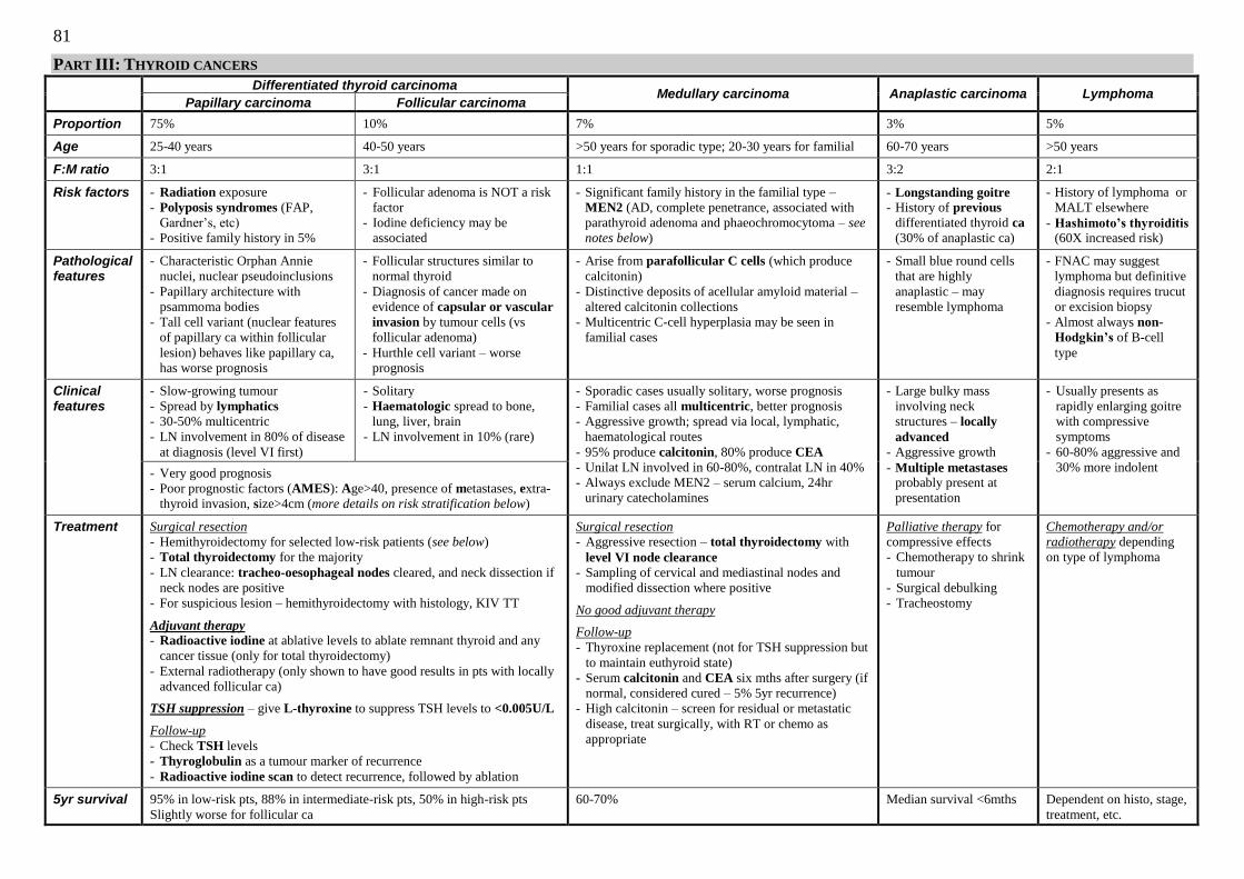
81
PART III: THYROID CANCERS
Differentiated thyroid carcinoma
Medullary carcinoma
Anaplastic carcinoma
Lymphoma Papillary carcinoma Follicular carcinoma
Proportion
75%
10%
7%
3%
5%
Age
25-40 years
40-50 years
>50 years for sporadic type; 20-30 years for familial
60-70 years
>50 years
F:M ratio
3:1
3:1
1:1
3:2
2:1
Risk factors
- Radiation exposure
- Polyposis syndromes (FAP,
Gardner‘s, etc)
- Positive family history in 5%
- Follicular adenoma is NOT a risk
factor
- Iodine deficiency may be
associated
- Significant family history in the familial type –
MEN2 (AD, complete penetrance, associated with
parathyroid adenoma and phaeochromocytoma – see
notes below)
- Longstanding goitre
- History of previous
differentiated thyroid ca
(30% of anaplastic ca)
- History of lymphoma or
MALT elsewhere
- Hashimoto’s thyroiditis (60X increased risk)
Pathological features
- Characteristic Orphan Annie
nuclei, nuclear pseudoinclusions
- Papillary architecture with
psammoma bodies
- Tall cell variant (nuclear features
of papillary ca within follicular
lesion) behaves like papillary ca,
has worse prognosis
- Follicular structures similar to
normal thyroid
- Diagnosis of cancer made on
evidence of capsular or vascular
invasion by tumour cells (vs
follicular adenoma)
- Hurthle cell variant – worse
prognosis
- Arise from parafollicular C cells (which produce
calcitonin)
- Distinctive deposits of acellular amyloid material –
altered calcitonin collections
- Multicentric C-cell hyperplasia may be seen in
familial cases
- Small blue round cells
that are highly
anaplastic – may
resemble lymphoma
- FNAC may suggest
lymphoma but definitive
diagnosis requires trucut
or excision biopsy
- Almost always non-
Hodgkin’s of B-cell
type
Clinical features
- Slow-growing tumour
- Spread by lymphatics
- 30-50% multicentric
- LN involvement in 80% of disease
at diagnosis (level VI first)
- Solitary
- Haematologic spread to bone,
lung, liver, brain
- LN involvement in 10% (rare)
- Sporadic cases usually solitary, worse prognosis
- Familial cases all multicentric, better prognosis
- Aggressive growth; spread via local, lymphatic,
haematological routes
- 95% produce calcitonin, 80% produce CEA
- Unilat LN involved in 60-80%, contralat LN in 40%
- Always exclude MEN2 – serum calcium, 24hr
urinary catecholamines
- Large bulky mass
involving neck
structures – locally
advanced - Aggressive growth
- Multiple metastases probably present at
presentation
- Usually presents as
rapidly enlarging goitre
with compressive
symptoms
- 60-80% aggressive and
30% more indolent
- Very good prognosis
- Poor prognostic factors (AMES): Age>40, presence of metastases, extra-
thyroid invasion, size>4cm (more details on risk stratification below)
Treatment
Surgical resection
- Hemithyroidectomy for selected low-risk patients (see below)
- Total thyroidectomy for the majority
- LN clearance: tracheo-oesophageal nodes cleared, and neck dissection if
neck nodes are positive
- For suspicious lesion – hemithyroidectomy with histology, KIV TT
Adjuvant therapy
- Radioactive iodine at ablative levels to ablate remnant thyroid and any
cancer tissue (only for total thyroidectomy)
- External radiotherapy (only shown to have good results in pts with locally
advanced follicular ca)
TSH suppression – give L-thyroxine to suppress TSH levels to <0.005U/L
Follow-up
- Check TSH levels
- Thyroglobulin as a tumour marker of recurrence
- Radioactive iodine scan to detect recurrence, followed by ablation
Surgical resection
- Aggressive resection – total thyroidectomy with
level VI node clearance
- Sampling of cervical and mediastinal nodes and
modified dissection where positive
No good adjuvant therapy
Follow-up
- Thyroxine replacement (not for TSH suppression but
to maintain euthyroid state)
- Serum calcitonin and CEA six mths after surgery (if
normal, considered cured – 5% 5yr recurrence)
- High calcitonin – screen for residual or metastatic
disease, treat surgically, with RT or chemo as
appropriate
Palliative therapy for
compressive effects
- Chemotherapy to shrink
tumour
- Surgical debulking
- Tracheostomy
Chemotherapy and/or
radiotherapy depending
on type of lymphoma
5yr survival
95% in low-risk pts, 88% in intermediate-risk pts, 50% in high-risk pts
Slightly worse for follicular ca
60-70%
Median survival <6mths
Dependent on histo, stage,
treatment, etc.
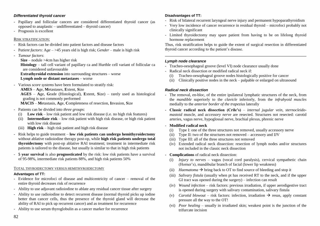
82
Differentiated thyroid cancer
- Papillary and follicular cancers are considered differentiated thyroid cancer (as
opposed to anaplastic – undifferentiated – thyroid cancer)
- Prognosis is excellent
RISK STRATIFICATION:
- Risk factors can be divided into patient factors and disease factors
- Patient factors: Age – >45 years old is high risk; Gender – male is high risk
- Tumour factors:
Size – nodule >4cm has higher risk
Histology – tall cell variant of papillary ca and Hurthle cell variant of follicular ca
are considered unfavourable
Extrathyroidal extension into surrounding structures – worse
Lymph node or distant metastases – worse
- Various score systems have been formulated to stratify risk:
AMES – Age, Metastases, Extent, Size
AGES – Age, Grade (Histological), Extent, Size) – rarely used as histological
grading is not commonly performed
MACIS – Metastasis, Age, Completeness of resection, Invasion, Size
- Patients can be divided into three groups:
(i) Low risk – low risk patient and low risk disease (i.e. no high risk features)
(ii) Intermediate risk – low risk patient with high risk disease, or high risk patient
with low risk disease
(iii) High risk – high risk patient and high risk disease
- Risk helps to guide treatment – low risk patients can undergo hemithyroidectomy
without ablative radioiodine therapy post-op, while high risk patients undergo total
thyroidectomy with post-op ablative RAI treatment; treatment in intermediate risk
patients is tailored to the disease, but usually is similar to that in high risk patients
- 5 year survival is also prognosticated by the risk: low risk patients have a survival
of 95-98%, intermediate risk patients 88%, and high risk patients 50%
TOTAL THYROIDECTOMY VERSUS HEMITHYROIDECTOMY
Advantages of TT:
- Evidence for microfoci of disease and multicentricity of cancer – removal of the
entire thyroid decreases risk of recurrence
- Ability to use adjuvant radioiodine to ablate any residual cancer tissue after surgery
- Ability to use radioiodine to detect recurrent disease (normal thyroid picks up iodine
better than cancer cells, thus the presence of the thyroid gland will decrease the
ability of RAI to pick up recurrent cancer) and as treatment for recurrence
- Ability to use serum thyroglobulin as a cancer marker for recurrence
Disadvantages of TT:
- Risk of bilateral recurrent laryngeal nerve injury and permanent hypoparathyroidism
- Very low incidence of cancer recurrence in residual thyroid – microfoci probably not
clinically significant
- Limited thyroidectomy may spare patient from having to be on lifelong thyroid
hormone replacement
Thus, risk stratification helps to guide the extent of surgical resection in differentiated
thyroid cancer according to the patient‘s disease.
Lymph node clearance
- Tracheo-oesophageal groove (level VI) node clearance usually done
- Radical neck dissection or modified radical neck if:
(i) Tracheo-oesophageal groove nodes histologically positive for cancer
(ii) Clinically positive nodes in the neck – palpable or enlarged on ultrasound
Radical neck dissection
- The removal, en-bloc, of the entire ipsilateral lymphatic structures of the neck, from
the mandible superiorly to the clavicle inferiorly, from the infrahyoid muscles
medially to the anterior border of the trapezius laterally
- Classic radical neck dissection (Crile’s) – internal jugular vein, sternocleido-
mastoid muscle, and accessory nerve are resected. Structures not resected: carotid
arteries, vagus nerve, hypoglossal nerve, brachial plexus, phrenic nerve
- Modified radical neck
(i) Type I: one of the three structures not removed, usually accessory nerve
(ii) Type II: two of the structures not removed – accessory and IJV
(iii) Type III: all of the three structures not removed
(iv) Extended radical neck dissection: resection of lymph nodes and/or structures
not included in the classic neck dissection
- Complications of radical neck dissection:
(i) Injury to nerves – vagus (vocal cord paralysis), cervical sympathetic chain
(Horner‘s), mandibular branch of facial (lower lip weakness)
(ii) Haematoma bring back to OT to find source of bleeding and stop it
(iii) Salivary fistula (usually when pt has received RT to the neck, and if the upper
GI tract was opened during the surgery) – infection can result
(iv) Wound infection – risk factors: previous irradiation, if upper aerodigestive tract
is opened during surgery with salivary contamination, salivary fistula
(v) Carotid blowout – risk factors: infection, irradiation resus, apply constant
pressure all the way to the OT!
(vi) Poor healing – usually in irradiated skin; weakest point is the junction of the
trifurcate incision
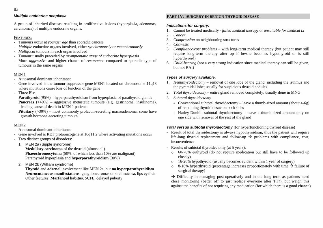
83
Multiple endocrine neoplasia
A group of inherited diseases resulting in proliferative lesions (hyperplasia, adenomas,
carcinomas) of multiple endocrine organs.
FEATURES:
- Tumours occur at younger age than sporadic cancers
- Multiple endocrine organs involved, either synchronously or metachronously
- Multifocal tumours in each organ involved
- Tumour usually preceded by asymptomatic stage of endocrine hyperplasia
- More aggressive and higher chance of recurrence compared to sporadic type of
tumours in the same organs
MEN 1
- Autosomal dominant inheritance
- Gene involved is the tumour suppressor gene MEN1 located on chromosome 11q13
where mutations cause loss of function of the gene
- Three P‘s:
Parathyroid (95%) – hyperparathyroidism from hyperplasia of parathyroid glands
Pancreas (>40%) – aggressive metastatic tumours (e.g. gastrinoma, insulinoma),
leading cause of death in MEN 1 patients
Pituitary (>30%) – most commonly prolactin-secreting macroadenoma; some have
growth hormone-secreting tumours
MEN 2
- Autosomal dominant inheritance
- Gene involved is RET protooncogene at 10q11.2 where activating mutations occur
- Two distinct groups of disorders:
1. MEN 2a (Sipple syndrome)
Medullary carcinoma of the thyroid (almost all)
Phaeochromocytoma (50%, of which less than 10% are malignant)
Parathyroid hyperplasia and hyperparathyroidism (30%)
2. MEN 2b (William syndrome)
Thyroid and adrenal involvement like MEN 2a, but no hyperparathyroidism
Neurocutaneous manifestations: ganglioneuromas on oral mucosa, lips eyelids
Other features: Marfanoid habitus, SCFE, delayed puberty
PART IV: SURGERY IN BENIGN THYROID DISEASE
Indications for surgery:
1. Cannot be treated medically - failed medical therapy or unsuitable for medical tx
2. Cancer 3. Compression on neighbouring structures
4. Cosmesis
5. Compliance/cost problems – with long-term medical therapy (but patient may still
require long-term therapy after op if he/she becomes hypothyroid or is still
hyperthyroid)
6. Child-bearing (not a very strong indication since medical therapy can still be given,
but not RAI)
Types of surgery available:
1. Hemithyroidectomy – removal of one lobe of the gland, including the isthmus and
the pyramidal lobe; usually for suspicious thyroid nodules
2. Total thyroidectomy – entire gland removed completely; usually done in MNG
3. Subtotal thyroidectomy
- Conventional subtotal thyroidectomy – leave a thumb-sized amount (about 4-6g)
of remaining thyroid tissue on both sides
- Harley-Dunhill subtotal thyroidectomy – leave a thumb-sized amount only on
one side with removal of the rest of the gland
Total versus subtotal thyroidectomy (for hyperfunctioning thyroid disease)
- Result of total thyroidectomy is always hypothyroidism, thus the patient will require
life-long thyroid replacement and follow-up problems with compliance, cost,
inconvenience
- Results of subtotal thyroidectomy (at 5 years):
o 60-70% euthyroid (do not require medication but still have to be followed up
closely)
o 16-20% hypothyroid (usually becomes evident within 1 year of surgery)
o 8-10% hyperthyroid (percentage increases proportionately with time failure of
surgical therapy)
Difficulty in managing post-operatively and in the long term as patients need
close monitoring (better off to just replace everyone after TT?), but weigh this
against the benefits of not requiring any medication (for which there is a good chance)
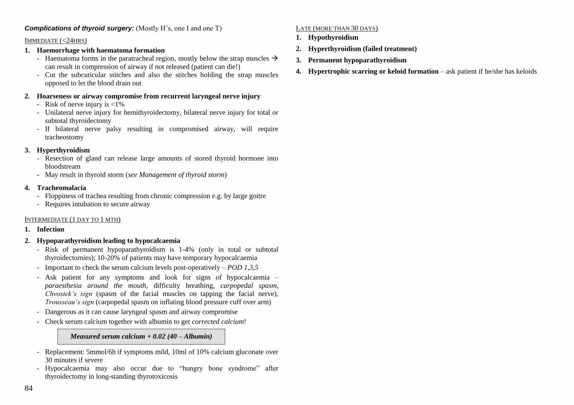
84
Complications of thyroid surgery: (Mostly H‘s, one I and one T)
IMMEDIATE (<24HRS)
1. Haemorrhage with haematoma formation
- Haematoma forms in the paratracheal region, mostly below the strap muscles
can result in compression of airway if not released (patient can die!)
- Cut the subcuticular stitches and also the stitches holding the strap muscles
opposed to let the blood drain out
2. Hoarseness or airway compromise from recurrent laryngeal nerve injury
- Risk of nerve injury is <1%
- Unilateral nerve injury for hemithyroidectomy, bilateral nerve injury for total or
subtotal thyroidectomy
- If bilateral nerve palsy resulting in compromised airway, will require
tracheostomy
3. Hyperthyroidism
- Resection of gland can release large amounts of stored thyroid hormone into
bloodstream
- May result in thyroid storm (see Management of thyroid storm)
4. Tracheomalacia
- Floppiness of trachea resulting from chronic compression e.g. by large goitre
- Requires intubation to secure airway
INTERMEDIATE (1 DAY TO 1 MTH)
1. Infection
2. Hypoparathyroidism leading to hypocalcaemia
- Risk of permanent hypoparathyroidism is 1-4% (only in total or subtotal
thyroidectomies); 10-20% of patients may have temporary hypocalcaemia
- Important to check the serum calcium levels post-operatively – POD 1,3,5
- Ask patient for any symptoms and look for signs of hypocalcaemia –
paraesthesia around the mouth, difficulty breathing, carpopedal spasm,
Chvostek‟s sign (spasm of the facial muscles on tapping the facial nerve),
Trousseau‟s sign (carpopedal spasm on inflating blood pressure cuff over arm)
- Dangerous as it can cause laryngeal spasm and airway compromise
- Check serum calcium together with albumin to get corrected calcium!
Measured serum calcium + 0.02 (40 – Albumin)
- Replacement: 5mmol/6h if symptoms mild, 10ml of 10% calcium gluconate over
30 minutes if severe
- Hypocalcaemia may also occur due to ―hungry bone syndrome‖ after
thyroidectomy in long-standing thyrotoxicosis
LATE (MORE THAN 30 DAYS)
1. Hypothyroidism
2. Hyperthyroidism (failed treatment)
3. Permanent hypoparathyroidism
4. Hypertrophic scarring or keloid formation – ask patient if he/she has keloids
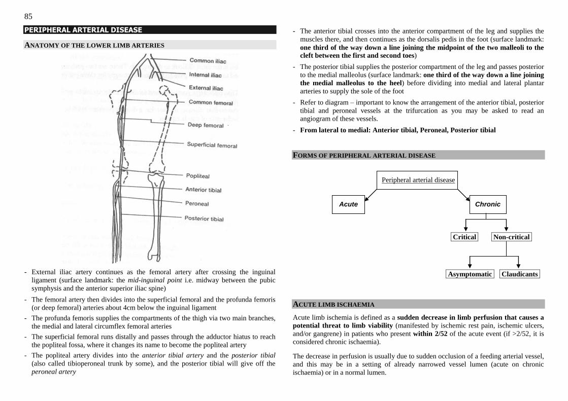
85
PERIPHERAL ARTERIAL DISEASE
ANATOMY OF THE LOWER LIMB ARTERIES
- External iliac artery continues as the femoral artery after crossing the inguinal
ligament (surface landmark: the mid-inguinal point i.e. midway between the pubic
symphysis and the anterior superior iliac spine)
- The femoral artery then divides into the superficial femoral and the profunda femoris
(or deep femoral) arteries about 4cm below the inguinal ligament
- The profunda femoris supplies the compartments of the thigh via two main branches,
the medial and lateral circumflex femoral arteries
- The superficial femoral runs distally and passes through the adductor hiatus to reach
the popliteal fossa, where it changes its name to become the popliteal artery
- The popliteal artery divides into the anterior tibial artery and the posterior tibial
(also called tibioperoneal trunk by some), and the posterior tibial will give off the
peroneal artery
- The anterior tibial crosses into the anterior compartment of the leg and supplies the
muscles there, and then continues as the dorsalis pedis in the foot (surface landmark:
one third of the way down a line joining the midpoint of the two malleoli to the cleft between the first and second toes)
- The posterior tibial supplies the posterior compartment of the leg and passes posterior
to the medial malleolus (surface landmark: one third of the way down a line joining
the medial malleolus to the heel) before dividing into medial and lateral plantar
arteries to supply the sole of the foot
- Refer to diagram – important to know the arrangement of the anterior tibial, posterior
tibial and peroneal vessels at the trifurcation as you may be asked to read an
angiogram of these vessels.
- From lateral to medial: Anterior tibial, Peroneal, Posterior tibial
FORMS OF PERIPHERAL ARTERIAL DISEASE
Peripheral arterial disease
Acute Chronic
Critical Non-critical.
Asymptomatic Claudicants.
ACUTE LIMB ISCHAEMIA
Acute limb ischemia is defined as a sudden decrease in limb perfusion that causes a
potential threat to limb viability (manifested by ischemic rest pain, ischemic ulcers,
and/or gangrene) in patients who present within 2/52 of the acute event (if >2/52, it is
considered chronic ischaemia).
The decrease in perfusion is usually due to sudden occlusion of a feeding arterial vessel,
and this may be in a setting of already narrowed vessel lumen (acute on chronic
ischaemia) or in a normal lumen.
86
CAUSES:
1. Arterial embolism
- Most common cause of acute limb ischaemia (60-80% of the time)
- The most likely source of embolus is the heart (80%), of which 70% is due to
atrial fibrillation, 20% to AMI with left ventricular mural thrombus, and a small
proportion to prosthetic heart valves
- Non-cardiac emboli arise from other arteries where there are atherosclerotic
plaques or an aneurysm (the embolic material may be thrombus or part of a
plaque, but atheroemboli are less likely to cause complete arterial occlusion)
- Most common sites where emboli lodge:
Bifurcation of the femoral artery (most common site)
Trifurcation of the popliteal artery (next most common site in the lower limb)
Aortic bifurcation
External and internal iliacs
Arm (about 20% of emboli)
- Emboli usually cause lower limb ischaemia mostly
- After emboli obstructs the vessel, thrombus can propagate distally (due to stasis
of blood) and proximally (due to turbulence of incoming blood hitting embolus)
by derangements in the Virchow‘s triad
2. Acute thrombosis
- Thrombosis of a previously stenotic but patent artery (atherosclerotic vessel)
- Less common cause of acute limb ischaemia
- When thrombotic occlusion of a vessel does occur, the resulting ischaemia is
usually less severe than in an embolic occlusion, because collaterals have had
time to form around the chronically stenosed vessel
- Other less common causes of acute thrombosis include the arteritides (usually
affecting medium-sized arteries), ergotism, and hypercoagulable states (notably
antiphospholipid syndrome).
3. Arterial trauma
- Increasing incidence of acute arterial occlusion due to endovascular diagnostic
or interventional procedures
- Trauma can cause development of an arteriovenous fistula that shunts blood
away from the limb
- Fracture or dislocations can stretch an artery and cause an intimal tear while the
media and adventitia layers are intact (because they contain elastin and can
stretch) a thrombus forms at the site of the tear where underlying
thrombogenic collagen is exposed
- Compartment syndrome can result from trauma as well
4. Dissecting aortic aneurysm - As the blood dissects between the intima and media of the aorta, it can cause
occlusion of the aortic branches at their origins
PATHOPHYSIOLOGY
In order of sensitivity to ischaemia, the tissues affected are nerves (most sensitive),
muscle, skin, and bone (least sensitive); thus early signs of ischaemia involve pain and
numbness, and muscle paralysis as well as skin changes occur later. The lower limb can
survive about 6 to 8 hours in an ischaemic state before injury becomes irreversible.
PRESENTATION
The classic 6 P’s of acute limb ischaemia: Pain, Paraesthesia, Pallor, Pulselessness,
Paralysis, Perishingly cold
Pain
- Develops acutely
- Starts off in a distal part of the extremity and then progresses proximally, increasing
in severity with time
- Further progress leads to decrease in pain as the nerves die off from ischaemia
- Important to ask for any previous claudication pain (10% of claudicants can develop
acute ischaemia due to thrombosis of the stenosed vessel)
Paraesthesia
- Starts off with paraesthesia (develops relatively early in the course of ischaemia) and
develops to complete loss of sensation
Pallor
- Assess skin colour, temperature, and capillary refill
- The limb may still be slightly pink though pale, but in severe ischaemia it can be
marble-white (especially in embolus where there are no collaterals)
- Other colours:
Mottling/Marbling (patches of blue on white): deoxygenation of stagnated blood;
surrounding areas of pallor are due to vasoconstriction
Duskiness: due to deoxygenation of stagnated blood; if there is fixed staining (i.e.
does not blanch on pressure) then the limb is non-viable
Black: gangrene
- The disclouration usually affects a large part of the distal limb e.g. the toes, foot;
rarely does it only affect one toe (more in chronic ischaemia)
- The site of arterial occlusion is usually one joint above the line of demarcation
between normal and ischaemic tissue
87
Pulselessness
- If able to feel one good pulse (PT or DP), quite unlikely that the limb is ischaemic,
but still possible
- If unable to feel, assess with a handheld Doppler the arterial and venous flow in the
limb – there can still be flow without a palpable pulse
- Also feel the pulses on the other limbs – gives a clue as to whether the cause is
embolic or thrombotic (see below)
Paralysis
- Total paralysis occurs late and usually indicates that the limb is non-viable
- Can assess viability of muscle by making a cut – viable muscle will be shiny and
twitches in response to flicking, while dead muscle will be dull and will not twitch
- Dangerous to save dead muscle as reperfusion can cause circulation of toxic
metabolites in the muscle
CLASSIFICATION OF SEVERITY (SVS/ISCVS)
Three categories: viable, threatened and non-viable
(i) Viable: No immediate threat of tissue loss
(ii) Threatened: Salvageable if revascularised promptly
(iii) Non-viable: Limb cannot be salvaged and has to be amputated, no emergency to
operate. Patient may require revascularisation to allow lower amputation or help
the amputation to heal
Viable Threatened Non-viable
Pain Mild Severe Variable
Capillary refill Intact Delayed Absent (fixed stain)
Motor deficit None Partial Complete
Sensory deficit None Partial Complete
Arterial Doppler Audible Inaudible Inaudible
Venous Doppler Audible Audible Inaudible
Treatment Urgent work-up Emergency surgery Amputation
DIFFERENTIATING BETWEEN EMBOLIC AND THROMBOTIC CAUSE
Embolic Thrombotic
Identifiable source Present – AF, recent AMI Less common
Claudication hx Negative Positive
Physical findings Contralat pulses present
White limb (no blood)
Contralat pulses diminished
Dusky limb (collaterals still
supplying limb)
Angiography Minimal atherosclerosis,
sharp cut-off, few collaterals
Diffuse atherosclerosis,
irregular cut-off, well-
developed collaterals
EARLY ANTICOAGULATION
- Important to start anticoagulation with heparin if the suspicion of acute limb
ischaemia is high
- Give IV heparin bolus 3000-5000 units
- Follow with IV heparin infusion at 1000 units/hour
- Ideal PTT is 2 to 2.5 times normal
INVESTIGATIONS
- Pre-operative investigations
- FBC, U/E/Cr, PT/PTT, GXM
- CXR and ECG if patient is older than 40 yrs old
- If suspecting an AMI with mural thrombus, do cardiac enzymes
- Angiogram can be done in patients with viable limb, but in patients with threatened
limb there is no time for angiogram may do on-table angiography
[Angiography is useful in confirming an occlusion, the cause – thrombotic or
embolic – and also pinpointing the level of occlusion and the anatomy]
DEFINITIVE TREATMENT OPTIONS
Surgical Endovascular
- Embolectomy
- Endarterectomy
- Bypass grafting
- Fasciotomy
- Primary amputation
- Thrombolysis
- Angioplasty
- Stenting
In general, embolectomy is done for embolic occlusion, while thrombolysis is done for
thrombotic occlusion.
Embolectomy
- Can be done under LA but still require anaesthetist to monitor patient as he may be
quite sick (e.g. AMI), and hyperkalaemia with cardiac arrhythmia can occur after
reperfusion
- Involves clamping of the involved artery and making an arterotomy
- A Fogarty balloon catheter is inserted into the artery until distal to the clot, then the
balloon is inflated to trawl the clot out of the artery
- Check for forward-bleeding and back-bleeding of the vessel (i.e. free spontaneous
flow from proximal and distal ends of the artery when unclamped)
- Flush with heparinised saline
- Check foot – warm foot with good pulse indicates reperfusion
88
- Important to monitor ECG for any arrhythmias!
- Closure of arterotomy with meticulous haemostasis as patient is on heparin
- Post-op: patient monitored in high-dependency; look out for reperfusion injury
The reperfused muscles become oedematous, increasing pressure in the
compartments of the leg, like compartment syndrome
Patient complains of calf pain
Unable to dorsiflex ankle as the anterior compartment is affected first
Requires three compartment fasciotomy to release pressure
- Need to convert to full warfarin anticoagulation, uptitrating dose until INR 2-2.5
before stopping heparin (patient at risk of further embolic events)
- Discharge patient to anticoagulation clinic for follow-up with warfarin advice
Thrombolysis
- Angiogram done before thrombolysis to locate occlusion
- Thrombolysis catheter inserted into the clot, and the thrombolytic agent is infused
- Patient will be in high-dependency with thrombolytic infusion for 6 hours (~1000-
4000 units per minute)
- After 6 hours, redo angiogram to check for residual clot; if some clot remains, adjust
catheter into the clot and infuse for 6 more hours
- After complete lysis of the clot, can do angioplasty
- Takes much longer than embolectomy
- Thrombolysis may be preferred for embolism in a diseased artery, since it may be
difficult to trawl out the clot in a diffusely stenosed vessel – the clot may get caught
on a proximal stenosed segment
Results:
- Embolectomy has a 20% mortality, almost full success rate
- Thrombolysis has a 10% mortality, only 35% successful
CHRONIC LIMB ISCHAEMIA
Chronic limb ischaemia can be divided into critical and non-critical limb ischaemia,
and non-critical ischaemia further subdivided into that which causes symptoms (usually
claudication) and that which is asymptomatic.
Most common cause is atherosclerosis with gradually developing diffuse stenosis of
the peripheral arteries resulting in diminished blood supply to the lower limb
(imbalance between supply and demand). Multiple collaterals form to bypass the
obstructed vessels as a compensatory mechanism
CRITICAL LIMB ISCHAEMIA
Critical limb ischaemia is defined as decrease in limb perfusion that causes a
potential threat to limb viability (manifested by ischemic rest pain, ischemic ulcers,
and/or gangrene) in patients who present more than two weeks after the acute event
(the converse of the definition of acute limb ischaemia).
FEATURES:
1. Rest pain requiring regular opioid analgesia (e.g. codeine) lasting >2 weeks
AND/OR
2. Gangrene or ulcers over the toes or feet
AND
3. Objective indication of poor vascular supply to the lower limbs
(a) Ankle brachial pressure index <0.5
(b) Toe pressure <30 mmHg, Ankle pressure <55mmHg
I. Rest pain
- Severe pain in the distal portion of the lower limb (usually toes, foot but may
involve more proximal areas if disease is severe) occurring at rest
- Pain is aggravated or precipitated by lifting the limb, relieved by dependency of
the limb – many patients sleep with the leg hanging over the side of the bed to
relieve the pain
- So severe as to disturb sleep at night
- Not easily controllable with analgesia – requires opioids to control pain
II. Ischaemic ulcers (most are neuroarteropathic ulcers)
- Usually arise from minor traumatic wounds with poor healing
- Often painful
- Most often occur on the tips of the toes, bunion area, over the metatarsal heads
(ball of the foot), lateral malleolus (as opposed to venous ulcers that occur over
the medial malleolus)
89
- Usually deep, dry, punctate (unlike venous ulcers that tend to be superficial, moist,
diffuse)
- May become infected resulting in cellulitis, even abscess formation, and spread to
involve the underlying bone and joints osteomyelitis, septic arthritis
III. Gangrene
- Cyanotic, anaesthetic tissue associated with or progressing to necrosis
- Occurs when arterial blood supply falls below that which is necessary to meet
minimal metabolic requirements
- Either dry or wet:
DRY – hard, dry texture. Often has a clear demarcation between viable and
necrotic tissue. Occurs in patients with atherosclerotic disease. Safe and can be
allowed to autoamputate after demarcation with precautions against infection.
WET – moist, swollen, frequently blistered. Often occurs in diabetics with
decreased sensation and unrecognised trauma, requiring an emergency surgical
debridement or amputation.
NON-CRITICAL LIMB ISCHAEMIA WITH CLAUDICATION
Intermittent claudication is defined as a reproducible discomfort of a defined group
of muscles that is induced by exercise and relieved with rest. Usually described as the
patient as a cramping, aching pain in the muscle group on exertion such as walking, and
alleviated on stopping (patient does not have to sit down for pain to go away) – “shop
window to shop window”.
- Calf claudication Usually affects the superficial femoral near to the adductor
hiatus, or the popliteal artery
- Foot claudication tibial and peroneal arterial disease, but rarely do patients with
claudication due to atherosclerosis get foot pain alone (more common in Buerger‘s)
- Thigh claudication common femoral artery or aortoiliac disease
- LeRiche’s syndrome arises from occlusion of the aortoiliacs, and is composed of a
classical tetrad of buttock claudication, impotence in men, absent femoral pulses (and
distal pulses), and occasionally presence of aortoiliac bruits.
- Important to determine the ―claudication distance‖ – within a short period of time
the distance is usually fairly constant, but can shorten as the disease progresses
- Need to differentiate the various causes: vascular vs neurogenic vs musculoskeletal
CAUSES OF VASCULAR CLAUDICATION
- Mainly an atherosclerotic disease; pain is due to acidosis & buildup of metabolites
- Other less common causes: ergot toxicity, Takayasu‘s arteritis, Buerger‘s disease
(thromboangiitis obliterans), vasospasm
NEUROGENIC CLAUDICATION
- Vascular intermittent claudication needs to be differentiated from neurogenic
claudication which can also present as pain in the lower limb on exertion
- The characteristic of neurogenic claudication is “park bench to park bench” where
the patient has to sit down and flex the spine to relieve the pain (pain results from
compression of the cord and spinal nerves in spinal stenosis; extension of the spine
further narrows the spinal canal while flexion widens it)
- ―Claudication distance‖ of neurogenic claudication is more variable
- Pulses will be absent/diminished in vascular but not in neurogenic claudication
- Parasthesia is common in neurogenic claudication
TAKING A HISTORY OF CHRONIC LIMB ISCHAEMIA
1. Claudication
- Which part of the lower limb does the pain occur in
- Nature of the pain
- Radiation
- Severity
- Aggravating factors – exertion
- Relieving factors – rest (just standing is sufficient)
- Associated symptoms e.g. impotence in LeRiche‘s
- Duration: When did pain first start (>2/52?)
- Progress since first noticed until currently (worsening pain, increasing areas of
lower limb affected, pain on less exertion, development of rest pain)
- Current claudication distance
- How has symptoms affected lifestyle e.g. impaired mobility
2. Any rest pain
- Site, nature, severity
- Aggravating factors – raising the limb
- Relieving factors – putting limb in a dependent position
- Able to relieve with normal analgesics? Or require opioid analgesia?
- How long has rest pain lasted for requiring opioid analgesia (if more than 2
weeks, considered a feature of critical limb ischaemia)
3. Any ulcer or gangrene in the lower limb?
- Ask about onset of ulcer/gangrene
- Progress (stable, or increasing in size, getting worse)
- If ulcer, any preceding trauma? Ill-fitting shoes? Altered sensation in the foot?
Does patient take care to protect foot? Pain? Redness/swelling/warmth in
surrounding skin? Purulent/foul-smelling discharge from the ulcer?
90
- If gangrene, is it wet or dry? Redness/swelling/warmth in surrounding skin? Any
feeling in the toe involved? Any sensory changes in the other normal toes, foot,
limb?
- Any systemic signs of infection – fever, chills, rigors, malaise
4. Risk factors (“Arteropath”) - Diabetes mellitus – take a full diabetic history including other complications
- Hyperlipidaemia
- Heart disease
- Stroke
- Smoking (very strong RF; 3-6X risk of claudication, higher than the risk for IHD)
- Family history
5. Drug history - Aspirin intake
- Any allergies to contrast (for angiography)
- Ergots
6. Social history
- Premorbid function and current function - Social support and home condition (need to climb stairs?)
PHYSICAL EXAMINATION
Examine the patient‘s lower limbs in a warm room, with the patient exposed optimally
(from the thighs to the feet, wearing underwear). Patient is supine with the bed flat.
Look
1. Colour of the lower limb
- White (pallor); pink (normal); blue/dusky (cyanosed); mottled
2. Trophic changes
- Loss of hair
- Dry, shiny skin
- Nail changes
- Wasting
3. Presence of any diabetic dermopathy
4. Presence of ulcer
- Look carefully at the entire lower limb, including the heels (ask patient to flex at
the knee so you can look at the heel) and between the toes
- Site of the ulcer
Venous ulcers form at the medial malleolus
Arterial ulcers are more distal, usually between the toes, and at pressure
points such as the lateral malleolus;
Neuropathic ulcers form at areas such as the ball of the foot and the heel
- Size, shape
- Edges (punched out – arterial; sloping – venous)
- Base
Depth of the ulcer (can see underlying tendon? Down to bone?)
Appearance of the base – Necrotic? Granulating (beefy-red)? Sloughy?
Any discharge – pus, blood?
- Surrounding skin
Erythema (cellulitis) – there may be an underlying abscess (confirm on
palpation)
Blistering, purplish colour (possibility of necrotising fasciitis)
5. Presence of gangrene
- Wet (infected) or dry (not infected)
- Extent of gangrene (level of demarcation)
6. (If the patient has diabetes, may see deformities – Charcot‘s joint)
Feel
1. Warmth of the skin
- Use the dorsum of the fingers of both hands to simultaneously run up the
patient‘s feet to the shins and thighs bilaterally
- Compare the temperature on both sides (note if one side is cooler)
- If one limb feels cool, feel for the level where the skin becomes warm
2. Capillary refill
- Press hard on a toe for a few seconds, then release
- Normal capillary refill should be 2 seconds or less
- If a toe is blue, check for blanchability (fixed staining = dead toe)
3. Palpating the ulcer if present
- Any surrounding tenderness (infection)
- Bogginess of surrounding tissue (may have abscess formation)
- See if any discharge from the ulcer when palpating
4. Pulses
- Feel the distal pulses and work your way proximally
- Posterior tibial pulse: one-third of the way down a line joining the medial
malleolus to the heel
- Dorsalis pedis pulse: one third of the way down a line joining the midpoint of
the two malleoli to the cleft between the first and second toes
- Popliteal pulse: Ask patient to bend the knee ~60-90 degrees, then palpate
deeply in the popliteal fossa with the fingers of one hand pressing the fingers of
the other [If the pulse is very well felt, suspect a popliteal aneurysm]
91
- Femoral pulse: Midpoint of the line joining the pubic symphysis to the anterior
superior iliac spine (mid-inguinal point), just below the inguinal ligament
- Grading of pulses: 2+ is normal; 1+ is diminished (but may be normal for
popliteal); negative if not felt label on a stick-figure diagram
Move
1. Buerger‘s test
- Do one side at a time
- Holding the heel of the foot, with the patient‘s lower limb straightened, slowly
lift the entire lower limb, looking at the colour of the toes
- Stop when the toes become pale (white)
- Estimate the angle the lower limb makes with the horizontal – this is the
Buerger’s angle
Normal lower limb can be raised to 90 degrees without turning white; if the
Buerger‘s angle is less than 20 degrees, this indicates severe ischaemia
- There may be venous guttering of the lower limb at this angle as well
- If the patient is lying near the side of the bed, tell the patient that you‘re going to
put his leg over the edge of the bed before gently abducting the hip and then
letting the leg drop over the edge of the bed
- Look at the leg for reactive hyperaemia (the leg turns purple-red)
Complete the exam
- Examine the rest of the pulses
- Offer to auscultate over the femoral and popliteal arteries for bruits
- Examine the abdomen for any abdominal aortic aneurysm
- Measure the ankle-brachial pressure index (ABPI)
INVESTIGATIONS
1. Ankle-brachial pressure index
- How the ankle-brachial pressure index is done
Brachial pressure is measured with a blood pressure cuff around the arm and
a Doppler probe at the brachial artery – cuff is inflated until the arterial
signal is obliterated, then slowly deflated until the signal just starts being
detected, at which the pressure is recorded
Ankle pressures are measured in a similar manner, with the cuff around the
calf and the Doppler at the dorsalis pedis and posterior tibial arteries – one
reading for each artery
The ankle pressure to be used for each leg is the higher of the two taken
This ankle pressure is then divided by the brachial pressure (the higher of the
two brachial pressures for both upper limbs) to get the ankle-brachial
pressure index
- Interpreting the values
Normal ABPI is > 0.9 (can be more than 1.0 as ankle pressures tend to be
higher than brachial; if >1.3, suggests non-compressible calcified vessel)
ABPI between 0.5 - 0.9 – occlusion, often associated with claudication
ABPI <0.5: Critical ischaemia Rest pain
- Accuracy of the index
ABPI below 0.9 has 95% sensitivity and 100% specificity for detecting
angiogram-positive peripheral arterial disease and is associated with >50%
stenosis in one or more major vessels
- Exercise treadmill testing
Measure ABPI before and after patient exercises on a treadmill
If the ABPI falls by >0.2 claudication
2. Arterial Duplex ultrasound
- Non-invasive test, good alternative to angiogram
- Duplex (means two modalities) = 2D ultrasound (like the normal kind) plus
Doppler ultrasound (measures flow and waveforms)
- Normal arterial flow waveform should be triphasic; biphasic and monophasic
waves are abnormal
- Can define anatomy of occlusions and also look for relatively good arteries
distally for ―landing zone‖ of bypass graft
3. Angiogram (arteriogram)
- Invasive and associated with risks of bleeding from arterial puncture,
dissection/damage to artery with worsening ischaemia
- Usually only done if planning intervention e.g. angioplasty, stenting
- Preparing for angiogram:
Take informed consent from patient
Ask about contrast allergy, asthma, renal disease, metformin
Investigations: FBC (platelets impt), PT/PTT, UECr
- Angiogram with digital subtraction – the images of the underlying bone are
removed so as to better visualise the arteries (if the bones are visible, then it is a
normal angiogram, without digital subtraction)
4. Basic laboratory investigation
- FBC, UECr, PT/PTT, septic workup: bld c/s, wound c/s

92
ASSESSMENT OF SEVERITY
The three L’s of peripheral arterial disease: (i) Life – does disease threaten life (e.g. sepsis; other complications of atherosclerosis
e.g. stroke, AMI;) or will intervention cause risks
(ii) Limb – will patient lose the limb
(iii) Lifestyle – is the lifestyle of the patient severely handicapped, does it require
intervention
Fontaine system
Stage I: Asymptomatic
Stage IIa: Mild claudication
Stage IIb: Moderate to severe claudication
Stage III: Ischaemic rest pain
Stage IV: Ulceration or gangrene
TREATMENT OF CLAUDICATION
Conservative
- Mainstay for all cases of claudicants, esp. foot and calf claudicants
- Smoking cessation
- Exercise training to stimulate collateral formation symptoms get better
Exercise at least half to one hour every day
Walk until pain comes, rest 2-3 minutes, walk again
Keep a walk diary recording daily claudication distance in paces
- Podiatrist to teach foot care
- Assessment and treatment to optimise control of CVS risk factors– cardiologist
- Patient educaton: Teach patient to go to ED if symptoms of critical ischaemia arises
- Antiplatelets [e.g. aspirin] and statins (target lipid levels are much lower)
- Use of Vasteral (methoxyphylline) is controversial
- Monitor regularly with measurement of ABPI
Intervention (endovascular or surgical)
- At least 6 months of conservative treatment first; mainly for aortoiliac disease
- Monitor claudication distance and ABPI – intervene if deteriorating despite
conservative management
- Confirm disease outline with CT angiogram or angiogram
- If parameters improve but then plateau, discuss with patient about whether he can
accept the level of symptoms, and the risks of intervention weigh risks against
benefits
- Usually do angioplasty rather than bypass as it is less invasive, though may not be as
effective in treating the symptoms
1. Angioplasty
Stenting usually not done for lower limbs except in aortoiliacs (since stent
needs to be placed in a vessel which is relatively fixed and won‘t be
kinked/bent by movement)
Angioplasty only effective for focal stenotic lesions and better for large
vessels
Problem with angioplasty is that it is not long-lasting – restenosis can occur
New method: subintimal angioplasty – if lumen is so occluded that guide
wire cannot pass through, the guidewire is threaded into the subintimal space
to create a dissection around the occluded segment, and this space is then
angioplastied to create a channel parallel to the actual lumen for blood to
flow through
2. Bypass grafting Consider bypass when lesions cannot be treated by angioplasty i.e. lesion
extends for long distance through the vessel and/or no lumen for guide wire
to pass through (complete occlusion)
Needs a good ―landing zone‖ for graft distally – if vessel is diffusely
diseased, difficult to perform bypass
TREATMENT OF CRITICAL LIMB ISCHAEMIA
- Need to revascularise – either angioplasty or bypass grafting
AMPUTATION
Indications (3 D’s) 1. Dead: Necrotic tissue
2. Dangerous: Gangrene, ascending sepsis
3. Damn nuisance: Non-functional limb; bad smell; pain; constant need to dress
wound
- Level of amputation depends on vascularity of the limb and the indication (e.g. if
infected, need to amputate above level of infection)
- As far as possible try to preserve function of the lower limb
- May require revascularisation interventions before amputation to ensure good healing,
or to enable lower amputation
- Do not simply amputate without ensuring good vascular supply to the surgical site,
otherwise the wound will not heal
93
ABDOMINAL AORTIC ANEURYSM
EPIDEMIOLOGY
More common in men than in women (4:1 ratio)
Predominantly in older patients (>60 years old)
Other risk factors: smoking, hypertension, strong family history (Marfan, Ehler-Danlos)
PATHOLOGY
- An aneurysm is a localised abnormal dilatation of a blood vessel wall or the heart
- True aneurysms are bound by all layers of the blood vessel wall, while a false
aneurysm is a breach in the blood vessel wall leading to an extravascular haematoma
that freely communicates with the intravascular space
- Atherosclerosis is the most common aetiological factor – plaque formation results in
destruction of the tunica media (and the elastin fibres in it) arterial wall thinning
and loss of elastic recoil dilatation
- Other causes: cystic medial degeneration (in Marfan), trauma, infection (mycotic)
- Location: 95% of cases are infrarenal, may extend to involve common iliac arteries,
rarely beyond. 5% are juxtarenal, thoracic or both
- Size: 3 to 15 cm (normal aorta is 2cm in diameter)
- Shape: Usually fusiform – long dilated segment (versus saccular which is spherical)
- Often contains mural thrombus due to turbulence and stasis
RISK OF RUPTURE
- Small aneurysms <5cm have a 2-3% chance of rupture per year, while aneurysm
larger than 5.5 cm will have a 10% risk of rupture per year
75% of aneurysms 7cm or larger will rupture in 5 years
PRESENTATION
- Asymptomatic (Most commonly; found incidentally during imaging)
- Rupture: intense abdominal pain radiating to the back, becomes rapidly hypotensive
and goes into shock (Most feared presentation)
- Trash feet: distal Thromboembolism gangrene of feet
- Local compression on neighbouring structures e.g. ureter
- Obstruction of branches from aorta e.g. iliac, renal, mesenteric, vertebral
- Also ask for vascular risk factors (DM, smoking, HPT, hyperlipidemia, previous
CVA, IHD), AAA risk factors (smoking, HPT, Marfan‘s/ Ehler-Danlos) & GA risk
factors (any heart, lung or kidney diseases)
PHYSICAL EXAMINATION
- Ensure vitals stable
- Visible pulsation over abdomen
- Pulsations and mass in epigastric region felt on deep palpation
- Mass is expansile – when fingers of both hands are placed at the edges on either side
of the mass, the fingers are pushed upwards and outwards
- Auscultate for bruit over the mass
- Check the other arteries – femoral, popliteal – for any aneurysm, and listen for bruits
- Look at the lower limbs for any gangrene, infection, etc
DIFFERENTIAL DIAGNOSIS OF UPPER ABDOMINAL PAIN WITH SHOCK - Pale: ruptured AAA, ruptured hepatoma, BGIT, ruptured spleen in trauma,
mesenteric bleed
- Not pale: sepsis, pancreatitis, perforated viscus, AMI , pyonephrosis
INVESTIGATIONS
Imaging: CT AbdoPelvis
Other investigations: FBC, UECr, PT/PTT, GXM for 4 units, ECG & CXR
MANAGEMENT
Dependent upon clinical context: asymptomatic, symptomatic, or ruptured?
Ruptured AAA
- Very high mortality – nearly 100% if frank rupture (will not get to ED in time)
- Most of the patients who reach the ED (about 50% reach ED alive) have a leaking
AAA with a tamponade effect by the retroperitoneal structures
- High suspicion in unstable hypotensive patient complaining of severe sharp pain
radiating to the back; may feel a pulsatile mass in the abdomen
1. Stabilise patient – resuscitation with fluid and blood products
Do not intubate as neuromuscular blocking agents will reduce tamponade
effect, worsening haemorrhage
2. Call for vascular surgeon
3. Bring to OT for open repair – quickly isolate the aorta and clamp it proximally
can be clamped for about 30 minutes without significant visceral ischaemia
AAA is incised, the surrounding haematoma and mural thrombus within
the AAA are cleared out
Synthetic graft (Dacron – polytetrafluoroethylene) is placed within the
aorta and the vessel wall closed up over the graft i.e. the graft forms the
lumen of the aorta
o Mortality rate of repair operation in this setting is about 50%
94
4. Most common complication postoperatively is renal insufficiency – can be
reduced by giving frusemide or mannitol pre-operatively before anaesthesia
induction
Non-ruptured AAA
- Time available for investigation of size of AAA and related anatomy
- Indications for surgery:
(a) Aneurysm > 5.5 cm in largest diameter [risk of surgery outweighs that of AAA]
(b) Increase in diameter of more than 1cm per year
(c) Symptomatic aneurysm – back pain, tenderness on palpation, distal embolism,
ruptured/leaking aneurysm
- Patient‘s fitness for surgery needs to be properly assessed because it is a major
operation – need to optimise cardiovascular function
- Operation is the same except that it is done under elective setting
- Mortality is <5% in the elective setting, serious morbidity ~10%
Endovascular stenting
- An alternative to open repair which is less invasive, can be done under GA
- Mortality ~1%, but serious morbidity rate is similar to open repair: 10%
- Involves deployment of a non-porous stent within the aneurysm to form the lumen of
the aorta – requires adequate ―neck‖ proximally and good landing site distally
- Not as good results as open surgery; need to do an angiogram every 6 months to
check position of the stent (ensure that stent has not migrated)
COMPLICATIONS OF SURGERY
Intraoperative/early
1. Acute myocardial infarction – most patients already have atherosclerotic disease of
coronary vessels and are at risk of AMI (responsible for 50-60% of mortality)
2. Stroke (due to hypotension or embolism)
3. Renal insufficiency
4. Colon ischaemia – occurs in 2-6%
5. Trash foot – embolism of thrombus from the aneurysm
6. Infection of graft
7. Spinal cord ischaemia (quite uncommon)
8. Haemorrhage
Late
1. Aortoenteric fisula – frank PR bleeding, torrential
2. Late infection of prosthetic graft material
3. Sexual dysfunction
Post operative investigations:
FBC: Hb and plt (blood loss and consumptive coagulopathy)
UECr: renal insufficiency or contrast nephropathy
KUB: check position of stent
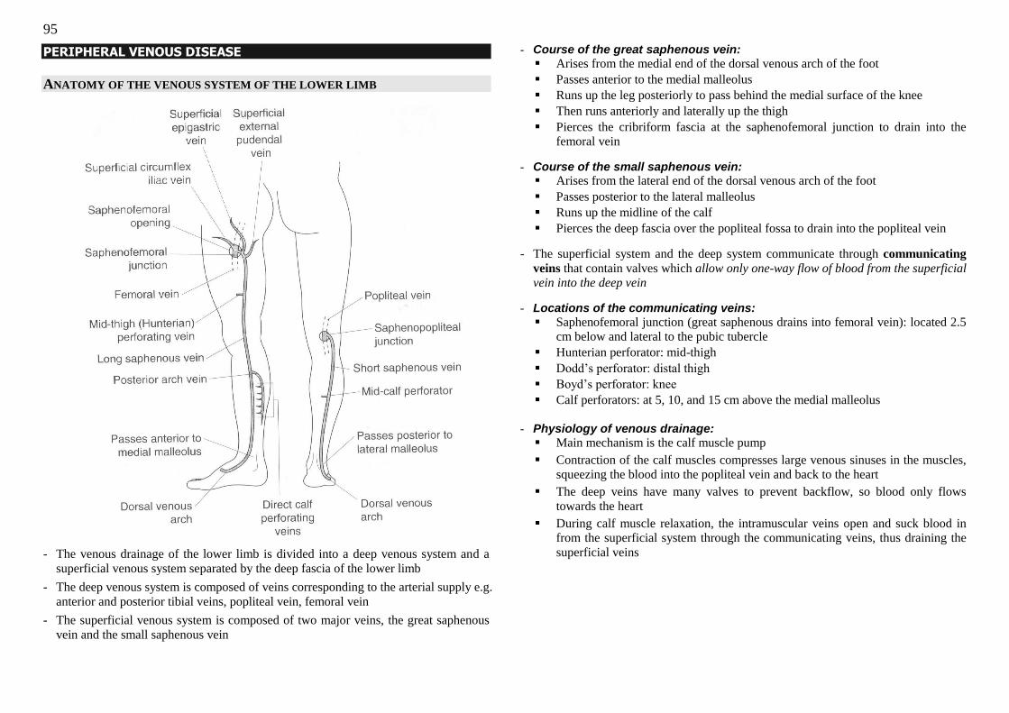
95
PERIPHERAL VENOUS DISEASE
ANATOMY OF THE VENOUS SYSTEM OF THE LOWER LIMB
- The venous drainage of the lower limb is divided into a deep venous system and a
superficial venous system separated by the deep fascia of the lower limb
- The deep venous system is composed of veins corresponding to the arterial supply e.g.
anterior and posterior tibial veins, popliteal vein, femoral vein
- The superficial venous system is composed of two major veins, the great saphenous
vein and the small saphenous vein
- Course of the great saphenous vein:
Arises from the medial end of the dorsal venous arch of the foot
Passes anterior to the medial malleolus
Runs up the leg posteriorly to pass behind the medial surface of the knee
Then runs anteriorly and laterally up the thigh
Pierces the cribriform fascia at the saphenofemoral junction to drain into the
femoral vein
- Course of the small saphenous vein: Arises from the lateral end of the dorsal venous arch of the foot
Passes posterior to the lateral malleolus
Runs up the midline of the calf
Pierces the deep fascia over the popliteal fossa to drain into the popliteal vein
- The superficial system and the deep system communicate through communicating
veins that contain valves which allow only one-way flow of blood from the superficial vein into the deep vein
- Locations of the communicating veins:
Saphenofemoral junction (great saphenous drains into femoral vein): located 2.5
cm below and lateral to the pubic tubercle
Hunterian perforator: mid-thigh
Dodd‘s perforator: distal thigh
Boyd‘s perforator: knee
Calf perforators: at 5, 10, and 15 cm above the medial malleolus
- Physiology of venous drainage:
Main mechanism is the calf muscle pump
Contraction of the calf muscles compresses large venous sinuses in the muscles,
squeezing the blood into the popliteal vein and back to the heart
The deep veins have many valves to prevent backflow, so blood only flows
towards the heart
During calf muscle relaxation, the intramuscular veins open and suck blood in
from the superficial system through the communicating veins, thus draining the
superficial veins
96
CHRONIC VENOUS INSUFFICIENCY
Chronic venous insufficiency develops when there is venous hypertension, which can
result from:
1. Obstruction to venous flow e.g. tumour compression in the pelvis, pregnancy,
deep vein thrombosis
2. Dysfunction of venous valves e.g. varicose veins
3. Failure of the “venous pump” – dependent on adequate muscle contraction
(stroke, muscular weakness can cause failure) as well as competent venous valves
MANIFESTATIONS OF CHRONIC VENOUS INSUFFICIENCY:
1. Venous dilatations
(a) Telangiectasias (spider veins or venous stars – intradermal veins)
(b) Reticular veins (slightly larger intermediate veins)
(c) Varicosities (visible, dilated tortuous superficial veins; formed by main
tributaries of the saphenous veins because these do not have a strong coat of
smooth muscle in their walls, unlike the saphenous veins; they are more
superficial and not bound down to the deep fascia)
(d) Corona phlebectactica (a network of small dilated venules beneath the lateral
and/or medial malleolus with severe venous hypertension)
2. Oedema – pitting: The hallmark of CVI; present in all but the earliest stages
Unilateral oedema worsened by dependency (worse at the end of the day) and better
with recumbency 3. Skin changes
(a) Hyperpigmentation of the skin over medial lower third of the leg (gaiter
area) – due to extravasation with haemosiderin deposits
(b) Atrophie blanche – hypopigmented scars of healed venous ulcers (avascular
and fibrotic skin)
(c) Venous stasis eczema – pruritic, weeping, scaling, with erosions and crusting
(d) Lipodermatosclerosis – a fibrosing panniculitis of the subcutaneous tissue that
results in a firm area of tender, indurated, hyperpigmented skin that is fixed to
subcutaneous tissue.
Results from severe venous hypertension
Starts in the gaiter area and extends circumferentially to surround the leg
If severe can result in an ―inverted champagne bottle‖ appearance of the
leg with brawny oedema above and below the area of lipodermatosclerosis
(e) Cellulitis
4. Venous ulcer formation
- Typical location is over the medial malleolus
- Shallow, flat ulcer with sloping edges; base may be sloughy or granulating,
usually quite moist-looking
- Surrounding skin will show signs of CVI
- In long-standing ulcer SCC can develop (Marjolin‘s ulcer) – If ulcer enlarges,
becomes painful and malodorous, edge becomes thickened or raised, if inguinal
lymph nodes are enlarged
These manifestations can be asymptomatic or associated with symptoms of leg fullness,
aching discomfort, heaviness, nocturnal leg cramps, or bursting pain upon standing.
CEAP CLASSIFICATION OF CHRONIC VENOUS INSUFFICIENCY
VARICOSE VEINS
Varicose veins are dilated, tortuous veins of the superficial venous system.
Primary varicose veins: where the cause is unknown (may be related to posture
and components and structure of the vein wall),
Secondary varicose veins: from proximal venous obstruction, destruction of the
valves by thrombosis or an increase in flow and pressure caused by an
arteriovenous fistula.
97
PATHOPHYSIOLOGY - Inherent weakness in the vein wall, leading to dilation and separation of valve cusps
so they become incompetent
- This may be aggravated by obstruction to venous return (as above)
RISK FACTORS - Age
- Parity
- Occupation – requiring long periods of standing
- Weight
- Posture – crossing legs all the time
- Increased abdominal pressure – constipation, chronic cough, etc
- Pelvic tumour or other lesion compressing on the deep veins
- Family history: 1 parent (50% risk, both parents (up to 80% risk)
HISTORY
Asymptomatic
Symptomatic: local (non specific pain, swelling, itching, heavy legs)
Complications : thrombophlebitis, bleeding, hyperpigmentation, eczema, ulceration
Ask for Past history of DVT & thrombophlebitis, obstetrics history, family history
EXAMINATION:
Examine patient standing with adequate exposure of the lower limbs
Inspection (look all around the limb!)
1. Presence of signs of chronic venous insufficiency (as above)
- Oedema
- Skin changes, stasis eczema, lipodermatosclerosis
- Venous ulcers from pressure necrosis from insude
2. Look at course of great saphenous vein and short saphenous vein for varicosities
3. Look at the inguinal region for any saphena varix
Palpate
1. Feel any dilated varicosities
2. Palpate along the course of the saphenous veins and their tributaries to feel any
varicosities and for tenderness (may be more palpable especially in fat legs)
3. Palpate the inguinal region for a saphena varix (compressible lump that refills
when released)
4. Do the cough test to feel for reflux at the saphenofemoral junction (2.5 cm below
and lateral to the pubic tubercle)
5. Percussion (tap test) – test for valvular incompetence (not a very valuable test) –
positive if the distal hand can feel the wave of blood flowing retrogradely after
tapping the proximal varcosities
6. Feel the peripheral pulses of Lower limb to exclude any ischaemia as the
management will involve compression of limbs (ABI >0.8)
Special tests
TOURNIQUET TEST
- Lie the patient down and empty the varicosities & tie a tourniquet just below the SFJ
- Ask the patient to stand up
- Look for filling up of the varicosities above and below the tourniquet
- If the veins dilate above but not below the tourniquet, this indicates that the
perforators below the level of the tourniquet are not incompetent and that the SFJ is
incompetent confirm this by releasing the tourniquet and watching the veins dilate
- If the veins below the tourniquet are dilated when the patient stands up, then the
incompetent perforator is below the level of the tourniquet
- Repeat the test, placing the tourniquet at different sites:
(i) Mid thigh (just below the Hunterian perforator
(ii) Below the knee
(iii) Mid-calf
- The incompetent perforator is located between just above the level where the
tourniquet prevents dilation of the veins in the limb on standing
[The alternative is the triple tourniquet test, where three tourniquets are tied with the patient lying down and then released from the bottom up to locate the site of
insufficiency]
TRENDELENBURG TEST
- The SFJ is occluded (landmark is 2.5 cm below and lateral to the pubic tubercle) with
the patient lying down
- Get the patient to stand while holding the SFJ occluded
- If varicosities do not fill up, the SFJ is the site of incompetence; if they fill up, there
are other sites of incompetence (the SFJ may or may not be incompetent)
PERTHES‘ TEST
- Tie a tourniquet around the calf or thigh and ask patient repeatedly stand on tiptoe
and then relax
- In a person with normal deep venous drainage and competent venous valves in the
communicating veins the superficial veins should drain into the deep veins
- If the patient‘s varicosities remain enlarged then he or she has obstructed deep
venous drainage or incompetent valves in the communicating veins

98
Completing the examination
- Auscultate over the varicosities for any bruit (indicate arteriovenous malformation)
- Examine the abdomen for any mass that may be causing the varicosities
Use of a handheld Doppler probe to detect incompetence
- Doppler probe is placed in the popliteal fossa over small saphenous vein
- Squeeze the calf to empty the veins – should hear a whoosh as blood flows through
the small saphenous vein
- When the calf is relaxed there should not be any sound – a second whoosh indicates
reflux of blood i.e. there is valvular incompetence
INVESTIGATIONS
Venous duplex ultrasound
- Indications:
Recurrent varicose veins
History of superficial thromobophlebitis or DVT
Complications of CVI: Venous eczema, Haemosiderin staining, Venous
ulceration , Lipodermatosclerosis,
- Ask for SFJ and SPJ reflux, perforator, deep venous incompetency & DVT screen
- Can delineate deep and superficial venous systems and locate sites of incompetence
- Exclude presence of deep vein thrombosis – stripping is contraindicated
MANAGEMENT
Conservative
1. Lifestyle changes
- Decrease amount of time spent standing
- If due to job, change job or ask for change to position to stand & walking less
2. Graduated compression stockings, usually grade II ensure good pulses
3. Medications e.g. Daflon
Surgical
Indications:
1. Cosmesis – large unsightly varicosities
2. Symptoms – pain, discomfort
3. Complications – signs of chronic venous insufficiency, venous ulceration
Available modalities:
1. High tie with great saphenous vein stripping, and stab avulsion of varicosities
2. Ultrasound-guided foam sclerotherapy
3. Endovenous laser therapy (burns vein from within)
VENOUS ULCERS
CAUSE – ANY CAUSE OF CHRONIC VENOUS INSUFFICIENCY
1. Obstruction to venous flow – thrombosis
2. Incompetent valves – varicose veins, deep vein reflux (post-DVT)
3. Muscle pump failure – stroke, neuromuscular disease
INVESTIGATIONS
1. Exclude infection of the ulcer and other complications
- FBC for raised total white count
- Swabs of the ulcer for Gram stain and cultures
- X-ray of the area to exclude underlying gas, bone involvement
2. Venous duplex to map out venous system
3. Check for peripheral arterial disease by doing ABPI
4. Biopsy if cannot exclude malignant transformation (Marjolin‘s ulcer)
MANAGEMENT:
Conservative
1. 4 layer compression stockings (change once per week)
(a) Non-adherent wound dressing over ulcer (e.g. Menolin) followed by wool
bandage
(b) Crepe bandage
(c) Blue-line bandage (Elset)
(d) Adhesive bandage (Coban)
2. Analgesia
3. Antibiotics if infected
4. Warn patient to avoid trauma to affected area
5. Encourage rest and elevate leg
6. Once healed, compression stockings should be fitted and continued for life
Surgical
- If ulcer fails to heal
- First, exclude malignancy or other causes of ulcer (biopsy)
- Split skin graft can be considered with excision of dead skin and graft attached to
healthy granulation tissue
- Venous surgery for the underlying pathology
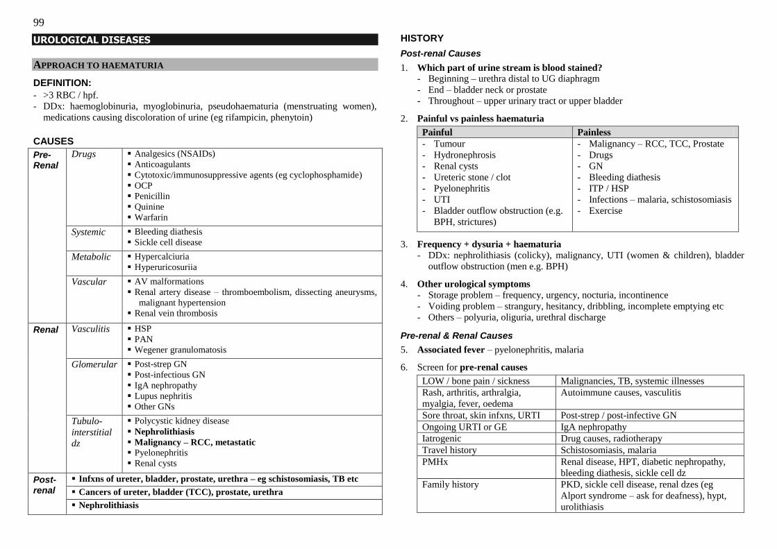
99
UROLOGICAL DISEASES
APPROACH TO HAEMATURIA
DEFINITION:
- >3 RBC / hpf.
- DDx: haemoglobinuria, myoglobinuria, pseudohaematuria (menstruating women),
medications causing discoloration of urine (eg rifampicin, phenytoin)
CAUSES
Pre-Renal
Drugs Analgesics (NSAIDs)
Anticoagulants
Cytotoxic/immunosuppressive agents (eg cyclophosphamide)
OCP
Penicillin
Quinine
Warfarin
Systemic Bleeding diathesis
Sickle cell disease
Metabolic Hypercalciuria
Hyperuricosuriia
Vascular AV malformations
Renal artery disease – thromboembolism, dissecting aneurysms,
malignant hypertension
Renal vein thrombosis
Renal Vasculitis HSP
PAN
Wegener granulomatosis
Glomerular Post-strep GN
Post-infectious GN
IgA nephropathy
Lupus nephritis
Other GNs
Tubulo-
interstitial dz
Polycystic kidney disease
Nephrolithiasis
Malignancy – RCC, metastatic
Pyelonephritis
Renal cysts
Post-renal
Infxns of ureter, bladder, prostate, urethra – eg schistosomiasis, TB etc
Cancers of ureter, bladder (TCC), prostate, urethra
Nephrolithiasis
HISTORY
Post-renal Causes
1. Which part of urine stream is blood stained? - Beginning – urethra distal to UG diaphragm
- End – bladder neck or prostate
- Throughout – upper urinary tract or upper bladder
2. Painful vs painless haematuria
Painful Painless
- Tumour
- Hydronephrosis
- Renal cysts
- Ureteric stone / clot
- Pyelonephritis
- UTI
- Bladder outflow obstruction (e.g.
BPH, strictures)
- Malignancy – RCC, TCC, Prostate
- Drugs
- GN
- Bleeding diathesis
- ITP / HSP
- Infections – malaria, schistosomiasis
- Exercise
3. Frequency + dysuria + haematuria
- DDx: nephrolithiasis (colicky), malignancy, UTI (women & children), bladder
outflow obstruction (men e.g. BPH)
4. Other urological symptoms
- Storage problem – frequency, urgency, nocturia, incontinence
- Voiding problem – strangury, hesitancy, dribbling, incomplete emptying etc
- Others – polyuria, oliguria, urethral discharge
Pre-renal & Renal Causes
5. Associated fever – pyelonephritis, malaria
6. Screen for pre-renal causes
LOW / bone pain / sickness Malignancies, TB, systemic illnesses
Rash, arthritis, arthralgia,
myalgia, fever, oedema
Autoimmune causes, vasculitis
Sore throat, skin infxns, URTI Post-strep / post-infective GN
Ongoing URTI or GE IgA nephropathy
Iatrogenic Drug causes, radiotherapy
Travel history Schistosomiasis, malaria
PMHx Renal disease, HPT, diabetic nephropathy,
bleeding diathesis, sickle cell dz
Family history PKD, sickle cell disease, renal dzes (eg
Alport syndrome – ask for deafness), hypt,
urolithiasis
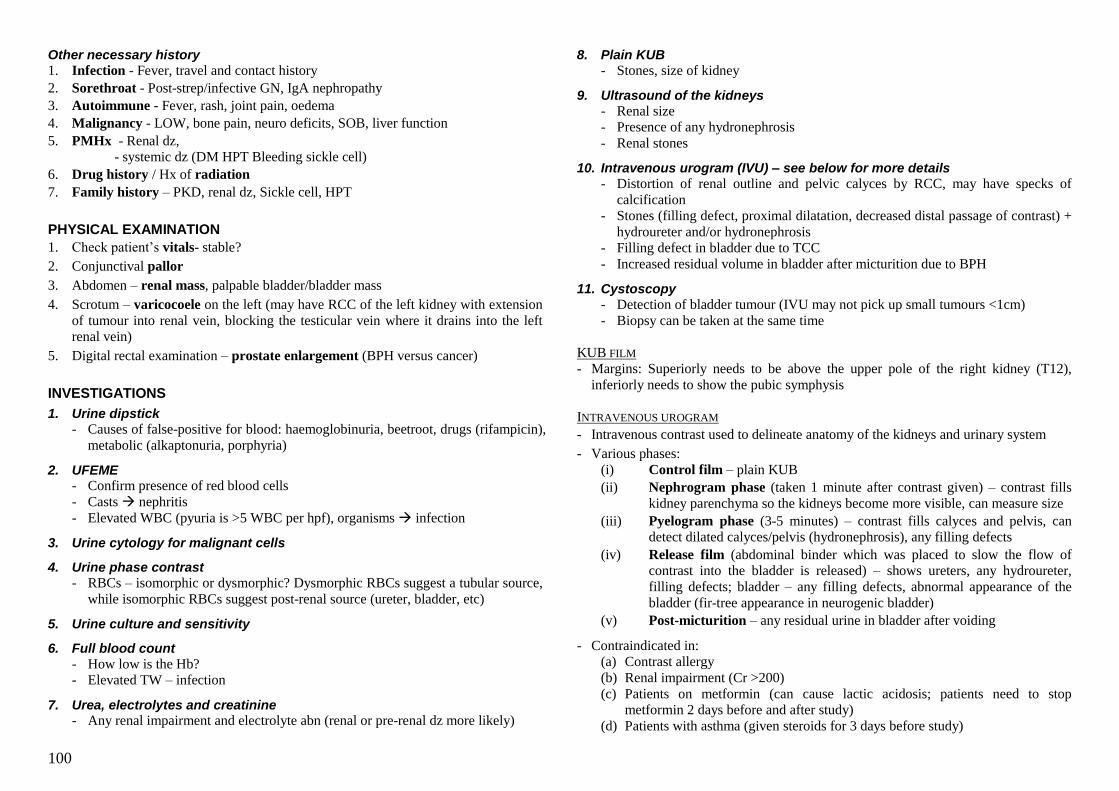
100
Other necessary history
1. Infection - Fever, travel and contact history
2. Sorethroat - Post-strep/infective GN, IgA nephropathy
3. Autoimmune - Fever, rash, joint pain, oedema
4. Malignancy - LOW, bone pain, neuro deficits, SOB, liver function
5. PMHx - Renal dz,
- systemic dz (DM HPT Bleeding sickle cell)
6. Drug history / Hx of radiation
7. Family history – PKD, renal dz, Sickle cell, HPT
PHYSICAL EXAMINATION
1. Check patient‘s vitals- stable?
2. Conjunctival pallor
3. Abdomen – renal mass, palpable bladder/bladder mass
4. Scrotum – varicocoele on the left (may have RCC of the left kidney with extension
of tumour into renal vein, blocking the testicular vein where it drains into the left
renal vein)
5. Digital rectal examination – prostate enlargement (BPH versus cancer)
INVESTIGATIONS
1. Urine dipstick
- Causes of false-positive for blood: haemoglobinuria, beetroot, drugs (rifampicin),
metabolic (alkaptonuria, porphyria)
2. UFEME - Confirm presence of red blood cells
- Casts nephritis
- Elevated WBC (pyuria is >5 WBC per hpf), organisms infection
3. Urine cytology for malignant cells
4. Urine phase contrast
- RBCs – isomorphic or dysmorphic? Dysmorphic RBCs suggest a tubular source,
while isomorphic RBCs suggest post-renal source (ureter, bladder, etc)
5. Urine culture and sensitivity
6. Full blood count
- How low is the Hb?
- Elevated TW – infection
7. Urea, electrolytes and creatinine - Any renal impairment and electrolyte abn (renal or pre-renal dz more likely)
8. Plain KUB
- Stones, size of kidney
9. Ultrasound of the kidneys
- Renal size
- Presence of any hydronephrosis
- Renal stones
10. Intravenous urogram (IVU) – see below for more details
- Distortion of renal outline and pelvic calyces by RCC, may have specks of
calcification
- Stones (filling defect, proximal dilatation, decreased distal passage of contrast) +
hydroureter and/or hydronephrosis
- Filling defect in bladder due to TCC
- Increased residual volume in bladder after micturition due to BPH
11. Cystoscopy
- Detection of bladder tumour (IVU may not pick up small tumours <1cm)
- Biopsy can be taken at the same time
KUB FILM
- Margins: Superiorly needs to be above the upper pole of the right kidney (T12),
inferiorly needs to show the pubic symphysis
INTRAVENOUS UROGRAM
- Intravenous contrast used to delineate anatomy of the kidneys and urinary system
- Various phases:
(i) Control film – plain KUB
(ii) Nephrogram phase (taken 1 minute after contrast given) – contrast fills
kidney parenchyma so the kidneys become more visible, can measure size
(iii) Pyelogram phase (3-5 minutes) – contrast fills calyces and pelvis, can
detect dilated calyces/pelvis (hydronephrosis), any filling defects
(iv) Release film (abdominal binder which was placed to slow the flow of
contrast into the bladder is released) – shows ureters, any hydroureter,
filling defects; bladder – any filling defects, abnormal appearance of the
bladder (fir-tree appearance in neurogenic bladder)
(v) Post-micturition – any residual urine in bladder after voiding
- Contraindicated in:
(a) Contrast allergy
(b) Renal impairment (Cr >200)
(c) Patients on metformin (can cause lactic acidosis; patients need to stop
metformin 2 days before and after study)
(d) Patients with asthma (given steroids for 3 days before study)
101
RENAL CELL CARCINOMA
EPIDEMIOLOGY - 3% of adult malignancy
- Most frequent occurring solid lesion within kidney
- 2:1 male predominance
- Peak incidence 60-70 years
PATHOLOGY - Most common primary renal tumour (80-85% of all tumours of the kidney)
- Arise from the renal tubular epithelium
- Three cell types: clear cell carcinoma (70-80%), papillary renal cell carcinoma (10-
15%), and chromophobe renal cell carcinoma (5%)
- Other renal tumours: TCC of renal pelvis, Wilms‘ tumour, lymphoma
RISK FACTORS - Smoking
- Exposure to cadmium
- Family history
von-Hippel Lindau syndrome due to mutation of the VHL gene on chromosome
3p25 (associated with CNS haemangioblastomas (usually cerebellar), bilateral
multicentric retinal angiomas, phaeochromocytomas, etc) clear cell
carcinomas
Hereditary papillary RCC (HPRCC) due to mutation of the MET proto-oncogene
on chromosome 7q31 multifocal bilateral papillary carcinomas
- Acquired polycystic kidney disease (secondary to chronic dialysis)
PRESENTATION
- Initially asymptomatic (may be detected incidentally)
- Painless gross haematuria is the most common presenting symptom – >50% of cases
- When tumour has grown large enough, dull flank pain and palpable mass may result
Classical triad of RCC: flank pain, painless haematuria, palpable renal mass
(indicates late stage disease)
- May have fever a/w night sweats, LOA, LOW, malaise
- Polycythaemia occurs in 1-5% (due to increased erythropoietin)
- For left renal tumour, extension of tumour into left renal vein can cause a left
varicocoele as the left testicular vein becomes occluded
- Extension into IVC can cause lower limb oedema, ascites, liver dysfunction,
pulmonary embolism
- Symptoms of metastases – lungs, liver, bones, brain, lymph nodes
- Paraneoplastic syndromes are uncommon – Cushing‘s, hypercalcaemia, hypertension
INVESTIGATIONS
DIAGNOSTIC
1. Imaging – CT and/or ultrasound - Presumptive diagnosis is made on imaging – a renal parenchymal mass with
thickened irregular walls and enhancement after contrast injection suggests
malignancy
2. Pathological diagnosis
- Needle biopsy usually not done for resectable lesions due to fears of tumour
seeding
- In these resectable lesions, a partial or total nephrectomy is often performed, and
provides the tissue diagnosis post-operatively
- In tumours with metastatic disease on presentation, biopsy of the metastatic site
may be easier
STAGING 1. CT scan of the abdomen
- Perinephric invasion, adjacent organ invasion
- Extension into renal vein, IVC
- Lymph node enlargement
- Liver metastases
2. CT scan of the chest
- For lung metastases
3. Bone scan
- Only done if patient complains of bone pain and/or alkaline phosphatase is raised
4. MRI of abdomen and heart
- Superior to CT for evaluation of IVC and right atrium involvement
T1 Tumour <7cm, limited to the kidney
T1a: tumour <4cm
T1b: tumour >4cm but <7cm
T2 Tumour >7cm, limited to the kidney
T3 Tumour extends into major veins or invades adrenal gland or perinephric
tissues, but not beyond Gerota‘s fascia
T4 Tumour invades beyond Gerota‘s fascia
102
TREATMENT
RESECTABLE TUMOURS
Surgery - Laparoscopic versus open methods
- Retroperitoneal versus transperitoneal approach
1. Partial nephrectomy
- Done in T1a disease – spares part of the kidney that is not involved nephron-
saving
2. Total nephrectomy
- Done in T1b disease – entire kidney removed
3. Radical nephrectomy - Done in T2 disease – entire kidney together with Gerota‘s fascia
- In T3 disease, aim for radical nephrectomy and removal of structures affected e.g.
adrenal gland
Adjuvant chemotherapy
Surveillance after resection to detect relapse early
Patients who cannot undergo resection
- Most small tumours grow slowly and do not become symptomatic or metastasise –
reasonable to manage conservatively with periodic re-evaluation
- Alternatives: radiofrequency ablation, cryotherapy of lesions
ADVANCED TUMOURS
Immunotherapy - High dose interleukin-2 – associated with good results in patients whose tumours
respond to treatment, as treatment can induce long-term remissions without relapse.
However, associated with high toxicity and often not tolerable
- Cytoreductive nephrectomy performed prior to starting immunotherapy can improve
survival
Molecular targeted therapy - Sorafenib – an inhibitor of tyrosine kinase blocks intracellular domain of the
vascular endothelial growth factor (VEGF) receptor
- Bevacizumab – monoclonal antibody against VEGF
Prognosis Stage I (T1N0): >90% 5 year survival
Stage II (T2N0): 75-90%
Stage III (T3N0/N1): 60-70%
Metastatic disease: <10%
BLADDER TRANSITIONAL CELL CARCINOMA
EPIDEMIOLOGY - Ninth most common cancer in Singaporean males
- Increasing incidence with age (80% diagnosed in patient >60 years old)
- 4:1 male predominance
PATHOLOGY - TCC is the most common tumour of the bladder (>90%)
- Thought to arise due to exposure to carcinogenic substances in the urine field
change effect, thus urothelial tumours often occur multifocally
- Other types of bladder tumours: adenocarcinoma (1%, arises from remnant of the
urachus in the dome of the bladder), SCC (<5%, due to chronic irritation e.g. long
term indwelling catheter or untreated bladder stone)
RISK FACTORS - Industrial chemicals – naphthylamine, aniline-containing dyes, etc
- Cigarette smoking
- Occupational (hairdressers – exposure to hair dyes)
- Analgesic abuse (phenacetin)
- Chronic cystitis
- Schistosomiasis
- Radiation (pelvic)
- Chemotherapy (cyclophosphamide)
PRESENTATION - Haematuria is the most common presenting symptom (90%) – typically gross,
painless, intermittent, occurring throughout the stream
- LUTS – irritative symptoms (frequency, dysuria, urgency) suggestive of carcinoma
in-situ, while obstructive symptoms (decreased stream, intermittent voiding, feeling
of incomplete voiding, strangury) indicate a tumour at the bladder neck or prostatic
urethra
- Pain – in locally advanced or metastatic tumour flank pain due to urinary
obstruction, suprapubic pain due to local invasion, bone pain due to metastasis
- Constitutional symptoms – LOW, LOA, fatigue
DIAGNOSIS 1. Urine cytology for malignant cells
2. Cystoscopy with cell brushings and biopsy
3. IVU or CT urogram to detect synchronous lesions (3% chance of proximal tumour)
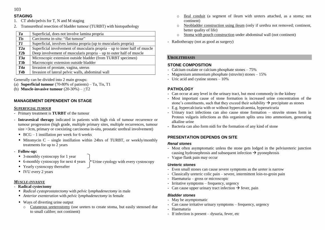
103
Urine cytology with every cystoscopy
STAGING 1. CT abdo/pelvis for T, N and M staging
2. Transurethral resection of bladder tumour (TURBT) with histopathology
Ta Superficial, does not involve lamina propria
Tis Carcinoma in-situ: ―flat tumour‖
T1 Superficial, involves lamina propria (up to muscularis propria)
T2a Superficial involvement of muscularis propria – up to inner half of muscle
T2b Deep involvement of muscularis propria – up to outer half of muscle
T3a Microscopic extension outside bladder (from TURBT specimen)
T3b Macroscopic extension outside bladder
T4a Invasion of prostate, vagina, uterus
T4b Invasion of lateral pelvic walls, abdominal wall
Generally can be divided into 2 main groups:
(a) Superficial tumour (70-80% of patients) – Ta, Tis, T1
(b) Muscle-invasive tumour (20-30%) – >T2
MANAGEMENT DEPENDENT ON STAGE
SUPERFICIAL TUMOUR
- Primary treatment is TURBT of the tumour
- Intravesical therapy indicated in patients with high risk of tumour recurrence or
tumour progression (high grade, multiple primary sites, multiple recurrences, tumour
size >3cm, primary or coexisting carcinoma in-situ, prostatic urethral involvement)
BCG – 1 instillation per week for 6 weeks
Mitomycin C – single instillation within 24hrs of TURBT, or weekly/monthly
treatments for up to 2 years
- Follow-up:
3-monthly cystoscopy for 1 year
6-monthly cystoscopy for next 4 years
Yearly cystoscopy thereafter
IVU every 2 years
MUSCLE-INVASIVE
- Radical cystectomy Radical cystoprostatectomy with pelvic lymphadenectomy in male
Anterior exenteration with pelvic lymphadenectomy in female
Ways of diverting urine output
o Cutaneous ureterostomy (use ureters to create stoma, but easily stenosed due
to small calibre; not continent)
o Ileal conduit (a segment of ileum with ureters attached, as a stoma; not
continent)
o Neobladder construction using ileum (only if urethra not removed; continent,
better quality of life)
o Stoma with pouch construction under abdominal wall (not continent)
- Radiotherapy (not as good as surgery)
UROLITHIASIS
STONE COMPOSITION - Calcium oxalate or calcium phosphate stones – 75%
- Magnesium ammonium phosphate (struvite) stones – 15%
- Uric acid and cystine stones – 10%
PATHOLOGY - Can occur at any level in the urinary tract, but most commonly in the kidney
- Most important cause of stone formation is increased urine concentration of the
stone‘s constituents, such that they exceed their solubility precipitate as stones
- E.g. hypercalciuria with or without hypercalcaemia, hyperuricuria
- Urinary tract infections can also cause stone formation – struvite stones form in
Proteus vulgaris infections as this organism splits urea into ammonium, generating
alkaline urine
- Bacteria can also form nidi for the formation of any kind of stone
PRESENTATION DEPENDS ON SITE
Renal stones
- Most often asymptomatic unless the stone gets lodged in the pelviureteric junction
causing hydronephrosis and subsequent infection pyonephrosis
- Vague flank pain may occur
Ureteric stones
- Even small stones can cause severe symptoms as the ureter is narrow
- Classically ureteric colic pain – severe, intermittent loin-to-groin pain
- Haematuria – gross or microscopic
- Irritative symptoms – frequency, urgency
- Can cause upper urinary tract infection fever, pain
Bladder stones
- May be asymptomatic
- Can cause irritative urinary symptoms – frequency, urgency
- Haematuria
- If infection is present – dysuria, fever, etc
104
PHYSICAL EXAMINATION - In ureteric colic, symptoms are often out of proportion to signs – no guarding,
rebound, etc
- If the patient has pyelonephritis, renal punch may be positive
- Otherwise unremarkable examination
INVESTIGATIONS
1. Urine tests – dipstick, UFEME, urine culture/sensitivity
- Haematuria
- Pyuria, micro-organisms (UTI)
2. KUB
- May be able to see radio-opaque stone (90% of renal stones are radio-opaque)
- Look at kidney size, any renal stones
- Trace path of ureter along tips of transverse processes, across sacroiliac joint,
and medially into bladder, looking for ureteric stones
- Look for bladder stones
3. Intravenous urogram
- Can also help to visualise a stone
- Can show dilated urinary system secondary to stone obstruction – hydroureter
and/or hydronephrosis
4. Ultrasound of kidney or bladder
- Features of stone: echogeneic rim, posterior acoustic shadowing
5. MAG-3 renogram
- If pyelonephritis present due to stone obstruction, it is valuable to measure the
renal function using the MAG-3 renogram
- The renogram gives the differential function of each kidney – in normal
individuals the function should be approximately 50% on each side (out of 100%
for both kidneys combined)
- If one kidney has less than 15% of total renal function, it is not worth salvaging
the kidney
TREATMENT
CONSERVATIVE
Stones smaller than 5mm can be treated conservatively as 60% will be passed out; only
treat if they do not pass out after 4 to 6 weeks, and/or cause symptoms
- Treatment of any urinary tract infection
- If underlying disease present that causes increased urinary concentration of stone
components e.g. hypercalcaemia treat disease if possible
- High fluid intake
- Low salt intake
- Restriction of red meat, dairy produce, refined sugars
- Increase citrus fruit intake
SURGICAL INTERVENTION
Indications:
- Constant pain
- Does not pass after one month
- Too large to pass spontaneously
- Obstructs urine flow
- Causes urinary tract infection
- Damages renal tissue or causes significant bleeding
- Increase in size
Types of treatment available:
1. Percutaneous nephrolithotomy (PCNL)
- Done for renal stones that are too large for ESWL to disintegrate
- Contraindicated in uncorrected bleeding diathesis, patients unfit for GA
2. Extracorporeal shock wave lithotripsy (ESWL)
- Calcium oxalate, uric acid and struvite stones fragment easily, but calcium
phosphate and cystine do not
- Used for stones below 10mm in size
- Used for renal stones and upper ureter stones – not so good for lower system due
to difficulty in access
- Contraindicated in pregnancy, untreated UTI, untreated bleeding diathesis, distal
obstruction that cannot be bypassed with a stent
3. Ureteroscopy with lithotripsy (usually laser lithotripsy, can also be done by
pneumatic drill, electrohydraulic means)
- For stones along the ureter
4. Cystolitholapaxy for bladder stone
5. Open surgery (pyelolithotomy or ureterolithotomy) – rarely done; only if failed
other management strategies, altered anatomy, performing open surgery for another
reason anyway, non-functioning kidney
Adjuncts:
- Double-J stent (or DJ stent) – inserted to stent the urinary system when worried that
stone fragments after ESWL may cause obstruction e.g. when ESWL used for
treatment of a large stone; or if system is obstructed to begin with, may want to stent
to ensure good drainage after surgery
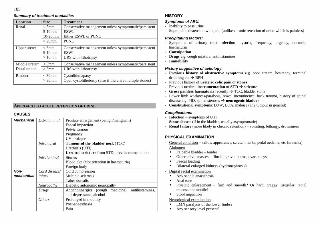
105
Summary of treatment modalities
Location Size Treatment
Renal < 5mm Conservative management unless symptomatic/persistent
5-10mm ESWL
10-20mm Either ESWL or PCNL
> 20mm PCNL
Upper ureter < 5mm Conservative management unless symptomatic/persistent
5-10mm ESWL
> 10mm URS with lithotripsy
Middle ureter/
Distal ureter
< 5mm Conservative management unless symptomatic/persistent
> 5mm URS with lithotripsy
Bladder < 30mm Cystolitholapaxy
> 30mm Open cystolithotomy (also if there are multiple stones)
APPROACH TO ACUTE RETENTION OF URINE
CAUSES
Mechanical Extraluminal Prostate enlargement (benign/malignant)
Faecal impaction
Pelvic tumour
Pregnancy
UV prolapse
Intramural Tumour of the bladder neck (TCC)
Urethritis (UTI)
Urethral stricture from STD, prev instrumentation
Intraluminal Stones
Blood clot (clot retention in haematuria)
Foreign body
Non-mechanical
Cord disease/
injury
Cord compression
Multiple sclerosis
Tabes dorsalis
Neuropathy Diabetic autonomic neuropathy
Drugs Anticholinergics (cough medicine), antihistamines,
anti-depressants, alcohol
Others Prolonged immobility
Post-anaesthesia
Pain
HISTORY
Symptoms of ARU:
- Inability to pass urine
- Suprapubic distension with pain (unlike chronic retention of urine which is painless)
Precipitating factors:
- Symptoms of urinary tract infection: dysuria, frequency, urgency, nocturia,
haematuria
- Constipation - Drugs e.g. cough mixture, antihistamines
- Immobility
History suggestive of aetiology:
- Previous history of obstructive symptoms e.g. poor stream, hesitancy, terminal
dribbling etc BPH
- Previous history of ureteric colic pain or stones
- Previous urethral instrumentation or STD stricture
- Gross painless haematuria recently TCC, bladder stone
- Lower limb weakness/paralysis, bowel incontinence, back trauma, history of spinal
disease e.g. PID, spinal stenosis neurogenic bladder
- Constitutional symptoms: LOW, LOA, malaise (any tumour in general)
Complications:
- Infection – symptoms of UTI
- Stone disease (if in the bladder, usually asymptomatic)
- Renal failure (more likely in chronic retention) – vomiting, lethargy, drowsiness
PHYSICAL EXAMINATION
- General condition – sallow appearance, scratch marks, pedal oedema, etc (uraemia)
- Abdomen
Palpable bladder – tender
Other pelvic masses – fibroid, gravid uterus, ovarian cyst
Faecal loading
Bilateral enlarged kidneys (hydronephrosis)
- Digital rectal examination
Any saddle anaesthesia
Anal tone
Prostate enlargement – firm and smooth? Or hard, craggy, irregular, rectal
mucosa not mobile?
Stool impaction
- Neurological examination
LMN paralysis of the lower limbs?
Any sensory level present?
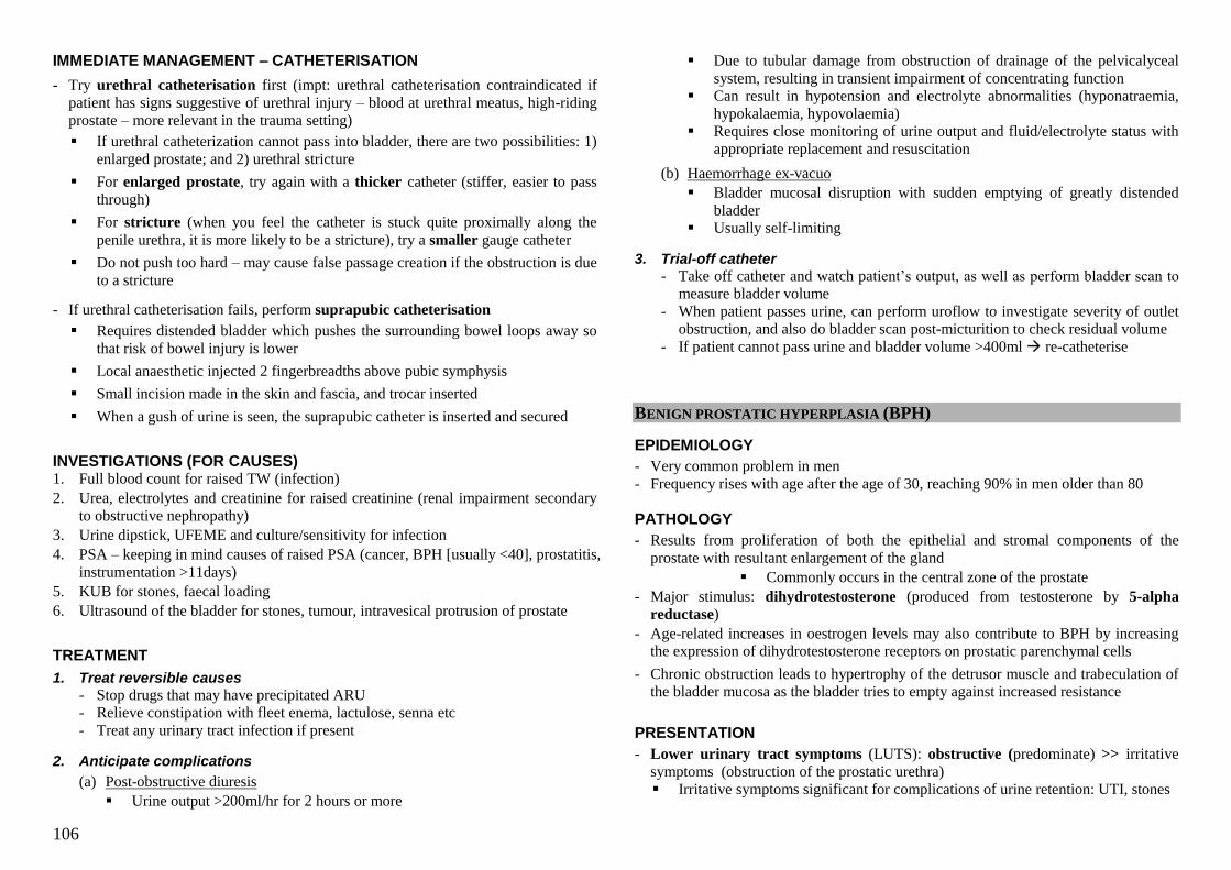
106
IMMEDIATE MANAGEMENT – CATHETERISATION
- Try urethral catheterisation first (impt: urethral catheterisation contraindicated if
patient has signs suggestive of urethral injury – blood at urethral meatus, high-riding
prostate – more relevant in the trauma setting)
If urethral catheterization cannot pass into bladder, there are two possibilities: 1)
enlarged prostate; and 2) urethral stricture
For enlarged prostate, try again with a thicker catheter (stiffer, easier to pass
through)
For stricture (when you feel the catheter is stuck quite proximally along the
penile urethra, it is more likely to be a stricture), try a smaller gauge catheter
Do not push too hard – may cause false passage creation if the obstruction is due
to a stricture
- If urethral catheterisation fails, perform suprapubic catheterisation
Requires distended bladder which pushes the surrounding bowel loops away so
that risk of bowel injury is lower
Local anaesthetic injected 2 fingerbreadths above pubic symphysis
Small incision made in the skin and fascia, and trocar inserted
When a gush of urine is seen, the suprapubic catheter is inserted and secured
INVESTIGATIONS (FOR CAUSES) 1. Full blood count for raised TW (infection)
2. Urea, electrolytes and creatinine for raised creatinine (renal impairment secondary
to obstructive nephropathy)
3. Urine dipstick, UFEME and culture/sensitivity for infection
4. PSA – keeping in mind causes of raised PSA (cancer, BPH [usually <40], prostatitis,
instrumentation >11days)
5. KUB for stones, faecal loading
6. Ultrasound of the bladder for stones, tumour, intravesical protrusion of prostate
TREATMENT
1. Treat reversible causes
- Stop drugs that may have precipitated ARU
- Relieve constipation with fleet enema, lactulose, senna etc
- Treat any urinary tract infection if present
2. Anticipate complications
(a) Post-obstructive diuresis
Urine output >200ml/hr for 2 hours or more
Due to tubular damage from obstruction of drainage of the pelvicalyceal
system, resulting in transient impairment of concentrating function
Can result in hypotension and electrolyte abnormalities (hyponatraemia,
hypokalaemia, hypovolaemia)
Requires close monitoring of urine output and fluid/electrolyte status with
appropriate replacement and resuscitation
(b) Haemorrhage ex-vacuo
Bladder mucosal disruption with sudden emptying of greatly distended
bladder
Usually self-limiting
3. Trial-off catheter
- Take off catheter and watch patient‘s output, as well as perform bladder scan to
measure bladder volume
- When patient passes urine, can perform uroflow to investigate severity of outlet
obstruction, and also do bladder scan post-micturition to check residual volume
- If patient cannot pass urine and bladder volume >400ml re-catheterise
BENIGN PROSTATIC HYPERPLASIA (BPH)
EPIDEMIOLOGY
- Very common problem in men
- Frequency rises with age after the age of 30, reaching 90% in men older than 80
PATHOLOGY
- Results from proliferation of both the epithelial and stromal components of the
prostate with resultant enlargement of the gland
Commonly occurs in the central zone of the prostate
- Major stimulus: dihydrotestosterone (produced from testosterone by 5-alpha
reductase)
- Age-related increases in oestrogen levels may also contribute to BPH by increasing
the expression of dihydrotestosterone receptors on prostatic parenchymal cells
- Chronic obstruction leads to hypertrophy of the detrusor muscle and trabeculation of
the bladder mucosa as the bladder tries to empty against increased resistance
PRESENTATION
- Lower urinary tract symptoms (LUTS): obstructive (predominate) >> irritative
symptoms (obstruction of the prostatic urethra)
Irritative symptoms significant for complications of urine retention: UTI, stones
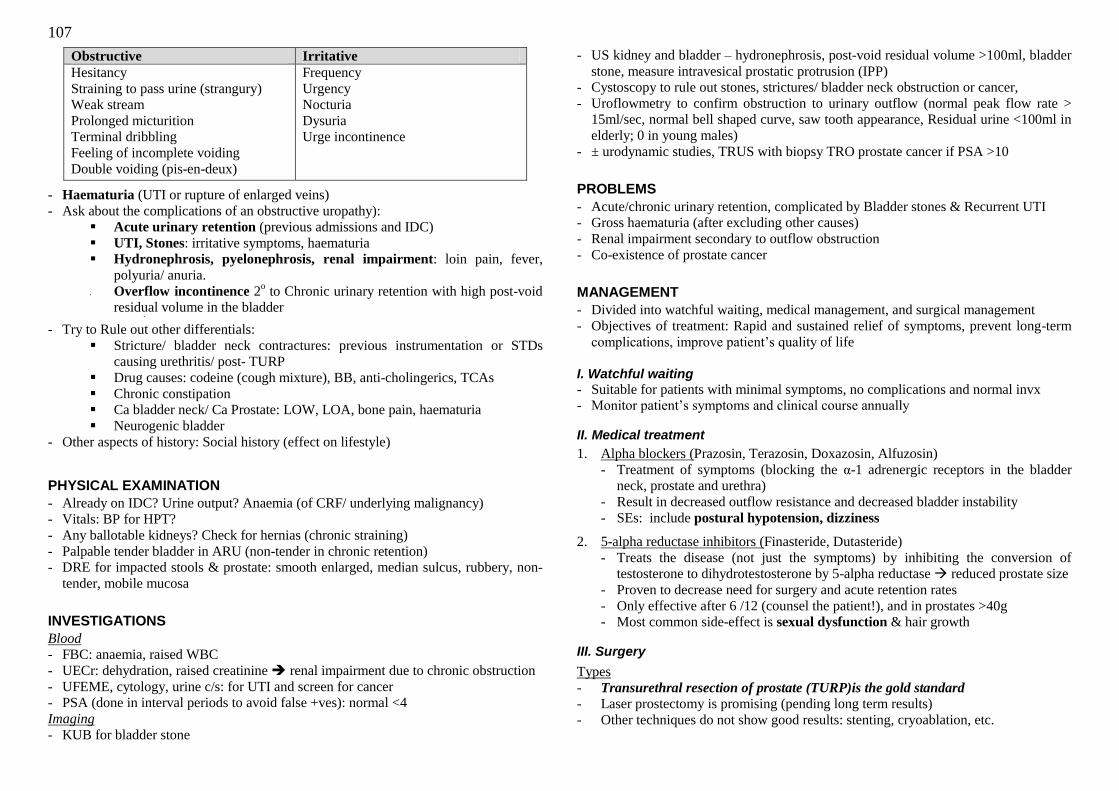
107
Obstructive Irritative
Hesitancy
Straining to pass urine (strangury)
Weak stream
Prolonged micturition
Terminal dribbling
Feeling of incomplete voiding
Double voiding (pis-en-deux)
Frequency
Urgency
Nocturia
Dysuria
Urge incontinence
- Haematuria (UTI or rupture of enlarged veins)
- Ask about the complications of an obstructive uropathy):
Acute urinary retention (previous admissions and IDC)
UTI, Stones: irritative symptoms, haematuria
Hydronephrosis, pyelonephrosis, renal impairment: loin pain, fever,
polyuria/ anuria.
Overflow incontinence 2o to Chronic urinary retention with high post-void
residual volume in the bladder
- Try to Rule out other differentials:
Stricture/ bladder neck contractures: previous instrumentation or STDs
causing urethritis/ post- TURP
Drug causes: codeine (cough mixture), BB, anti-cholingerics, TCAs
Chronic constipation
Ca bladder neck/ Ca Prostate: LOW, LOA, bone pain, haematuria
Neurogenic bladder
- Other aspects of history: Social history (effect on lifestyle)
PHYSICAL EXAMINATION
- Already on IDC? Urine output? Anaemia (of CRF/ underlying malignancy)
- Vitals: BP for HPT?
- Any ballotable kidneys? Check for hernias (chronic straining)
- Palpable tender bladder in ARU (non-tender in chronic retention)
- DRE for impacted stools & prostate: smooth enlarged, median sulcus, rubbery, non-
tender, mobile mucosa
INVESTIGATIONS
Blood - FBC: anaemia, raised WBC
- UECr: dehydration, raised creatinine renal impairment due to chronic obstruction
- UFEME, cytology, urine c/s: for UTI and screen for cancer
- PSA (done in interval periods to avoid false +ves): normal <4
Imaging
- KUB for bladder stone
- US kidney and bladder – hydronephrosis, post-void residual volume >100ml, bladder
stone, measure intravesical prostatic protrusion (IPP)
- Cystoscopy to rule out stones, strictures/ bladder neck obstruction or cancer,
- Uroflowmetry to confirm obstruction to urinary outflow (normal peak flow rate >
15ml/sec, normal bell shaped curve, saw tooth appearance, Residual urine <100ml in
elderly; 0 in young males)
- ± urodynamic studies, TRUS with biopsy TRO prostate cancer if PSA >10
PROBLEMS
- Acute/chronic urinary retention, complicated by Bladder stones & Recurrent UTI
- Gross haematuria (after excluding other causes)
- Renal impairment secondary to outflow obstruction
- Co-existence of prostate cancer
MANAGEMENT
- Divided into watchful waiting, medical management, and surgical management
- Objectives of treatment: Rapid and sustained relief of symptoms, prevent long-term
complications, improve patient‘s quality of life
I. Watchful waiting
- Suitable for patients with minimal symptoms, no complications and normal invx
- Monitor patient‘s symptoms and clinical course annually
II. Medical treatment
1. Alpha blockers (Prazosin, Terazosin, Doxazosin, Alfuzosin)
- Treatment of symptoms (blocking the α-1 adrenergic receptors in the bladder
neck, prostate and urethra)
- Result in decreased outflow resistance and decreased bladder instability
- SEs: include postural hypotension, dizziness
2. 5-alpha reductase inhibitors (Finasteride, Dutasteride)
- Treats the disease (not just the symptoms) by inhibiting the conversion of
testosterone to dihydrotestosterone by 5-alpha reductase reduced prostate size
- Proven to decrease need for surgery and acute retention rates
- Only effective after 6 /12 (counsel the patient!), and in prostates >40g
- Most common side-effect is sexual dysfunction & hair growth
III. Surgery
Types
- Transurethral resection of prostate (TURP)is the gold standard
- Laser prostectomy is promising (pending long term results)
- Other techniques do not show good results: stenting, cryoablation, etc.
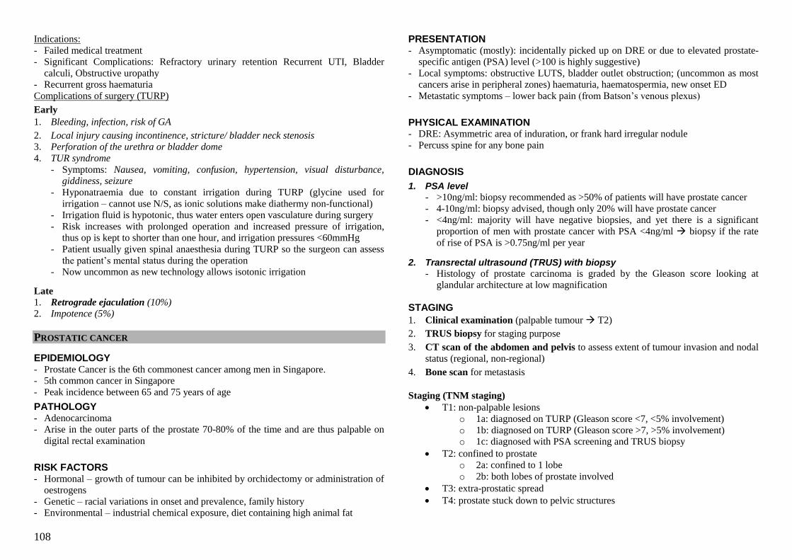
108
Indications:
- Failed medical treatment
- Significant Complications: Refractory urinary retention Recurrent UTI, Bladder
calculi, Obstructive uropathy
- Recurrent gross haematuria
Complications of surgery (TURP)
Early
1. Bleeding, infection, risk of GA
2. Local injury causing incontinence, stricture/ bladder neck stenosis 3. Perforation of the urethra or bladder dome
4. TUR syndrome
- Symptoms: Nausea, vomiting, confusion, hypertension, visual disturbance, giddiness, seizure
- Hyponatraemia due to constant irrigation during TURP (glycine used for
irrigation – cannot use N/S, as ionic solutions make diathermy non-functional)
- Irrigation fluid is hypotonic, thus water enters open vasculature during surgery
- Risk increases with prolonged operation and increased pressure of irrigation,
thus op is kept to shorter than one hour, and irrigation pressures <60mmHg
- Patient usually given spinal anaesthesia during TURP so the surgeon can assess
the patient‘s mental status during the operation
- Now uncommon as new technology allows isotonic irrigation
Late
1. Retrograde ejaculation (10%) 2. Impotence (5%)
PROSTATIC CANCER
EPIDEMIOLOGY - Prostate Cancer is the 6th commonest cancer among men in Singapore.
- 5th common cancer in Singapore
- Peak incidence between 65 and 75 years of age
PATHOLOGY - Adenocarcinoma
- Arise in the outer parts of the prostate 70-80% of the time and are thus palpable on
digital rectal examination
RISK FACTORS - Hormonal – growth of tumour can be inhibited by orchidectomy or administration of
oestrogens
- Genetic – racial variations in onset and prevalence, family history
- Environmental – industrial chemical exposure, diet containing high animal fat
PRESENTATION - Asymptomatic (mostly): incidentally picked up on DRE or due to elevated prostate-
specific antigen (PSA) level (>100 is highly suggestive)
- Local symptoms: obstructive LUTS, bladder outlet obstruction; (uncommon as most
cancers arise in peripheral zones) haematuria, haematospermia, new onset ED
- Metastatic symptoms – lower back pain (from Batson‘s venous plexus)
PHYSICAL EXAMINATION - DRE: Asymmetric area of induration, or frank hard irregular nodule
- Percuss spine for any bone pain
DIAGNOSIS
1. PSA level
- >10ng/ml: biopsy recommended as >50% of patients will have prostate cancer
- 4-10ng/ml: biopsy advised, though only 20% will have prostate cancer
- <4ng/ml: majority will have negative biopsies, and yet there is a significant
proportion of men with prostate cancer with PSA <4ng/ml biopsy if the rate
of rise of PSA is >0.75ng/ml per year
2. Transrectal ultrasound (TRUS) with biopsy
- Histology of prostate carcinoma is graded by the Gleason score looking at
glandular architecture at low magnification
STAGING
1. Clinical examination (palpable tumour T2)
2. TRUS biopsy for staging purpose
3. CT scan of the abdomen and pelvis to assess extent of tumour invasion and nodal
status (regional, non-regional)
4. Bone scan for metastasis
Staging (TNM staging)
T1: non-palpable lesions
o 1a: diagnosed on TURP (Gleason score <7, <5% involvement)
o 1b: diagnosed on TURP (Gleason score >7, >5% involvement)
o 1c: diagnosed with PSA screening and TRUS biopsy
T2: confined to prostate
o 2a: confined to 1 lobe
o 2b: both lobes of prostate involved
T3: extra-prostatic spread
T4: prostate stuck down to pelvic structures
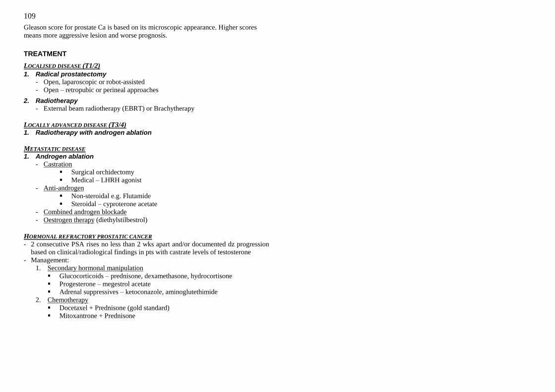
109
Gleason score for prostate Ca is based on its microscopic appearance. Higher scores
means more aggressive lesion and worse prognosis.
TREATMENT
LOCALISED DISEASE (T1/2)
1. Radical prostatectomy
- Open, laparoscopic or robot-assisted
- Open – retropubic or perineal approaches
2. Radiotherapy - External beam radiotherapy (EBRT) or Brachytherapy
LOCALLY ADVANCED DISEASE (T3/4) 1. Radiotherapy with androgen ablation
METASTATIC DISEASE 1. Androgen ablation
- Castration
Surgical orchidectomy
Medical – LHRH agonist
- Anti-androgen
Non-steroidal e.g. Flutamide
Steroidal – cyproterone acetate
- Combined androgen blockade
- Oestrogen therapy (diethylstilbestrol)
HORMONAL REFRACTORY PROSTATIC CANCER
- 2 consecutive PSA rises no less than 2 wks apart and/or documented dz progression
based on clinical/radiological findings in pts with castrate levels of testosterone
- Management:
1. Secondary hormonal manipulation
Glucocorticoids – prednisone, dexamethasone, hydrocortisone
Progesterone – megestrol acetate
Adrenal suppressives – ketoconazole, aminoglutethimide
2. Chemotherapy
Docetaxel + Prednisone (gold standard)
Mitoxantrone + Prednisone
110
SURGICAL INSTRUMENTS
DRAINS
FUNCTIONS OF DRAINS
Drains are inserted to:
- Evacuate collections of pus, blood or other fluids (e.g. lymph)
- Drain potential collections
Rationale:
- Drainage of fluid removes further fluid collections
- May allow the early detection of anastomotic leaks or haemorrhage
- Leave a tract for potential collections to drain following removal
COMPLICATIONS: 1. Infection
2. Bleeding
3. Tissue damage- by mechanical pressure or suction
4. Drain failure- blocked/slipped/kinked
5. Incisional hernia- occurs when drain inserted through incision wound site- create a
separate incision site for drain!
TYPES OF DRAINS - Drains classified as:
Open or closed
Active or passive
- Active drains require suction. Passive drains rely on gravity.
- Drains are often made from inert silastic material
- They induce minimal tissue reaction
- Red rubber drains induce an intense tissue reaction allowing a tract to form
- In some situations this may be useful (e.g. biliary t-tube)
Open drains
- Corrugated drain, Yeates drain, Penrose drain
- Drain fluid collects in gauze pad or stoma bag
- Easier to drain infected collections
Closed drains
- Consist of tubes draining into a bag or bottle - They include chest and abdominal drains
- The risk of infection is reduced
Active drains
- Jackson-Pratt Drain, Redivac Drain, T-tube
- Have expandable chambers to create low-pressure suction
- Used when small to moderate amounts of drainage are expected or when a passive
drainage system won't provide adequate drainage
- active drainage systems have a clear graduated scale by which you can see the
type and amount of drainage and determine when to empty the drainage chamber
- Tubing of the low-pressure active drainage system is placed through a separate
puncture wound or the tube may exit the edge of the surgical wound
- If the tubing isn't sutured in place, it could become dislodged when you change
dressings or reposition the patient, so be careful. If a portion of the tube is pulled
outside his skin, an air leak will cause the collection chamber to rapidly fill with
air and the system won't drain properly.
Passive drains
- Passive drains have no suction, rely on gravity
- Function by the differential pressure between body cavities and the exterior
- Used when a moderate to large amount of drainage is expected
CARE AND PREVENTION OF COMPLICATIONS OF TUBES: - Prevent Infection- maintain meticulous skin care and aseptic technique around the
insertion site
- Prevent blockage of the drain- do not allow bottles to fill up
- Prevent slippage by securing drain carefully to skin; refix as required
- Never hold a drainage collection device higher than the tube insertion site to prevent
the drainage from flowing backward into the patient
- Note amount of drainage daily
REMOVAL OF DRAINS A drain is removed as soon as it is no longer required. The following are general
guidelines:
1. Drains put in to cover perioperative bleeding and haematoma formation, can come
out after 24— 48 hours.
2. Where a drain has been put in to drain an infected site e.g. abscess, remove it when
the fever settles or when there is evidence of complete drainage.
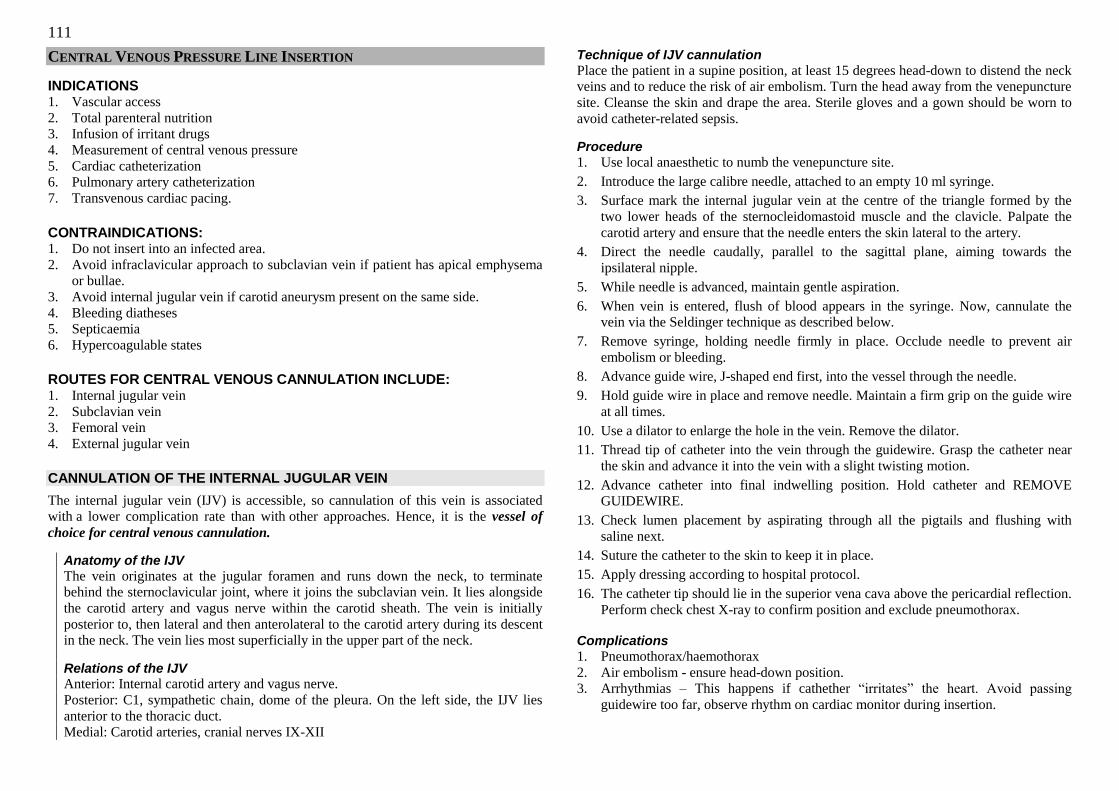
111
CENTRAL VENOUS PRESSURE LINE INSERTION
INDICATIONS 1. Vascular access
2. Total parenteral nutrition
3. Infusion of irritant drugs
4. Measurement of central venous pressure
5. Cardiac catheterization
6. Pulmonary artery catheterization
7. Transvenous cardiac pacing.
CONTRAINDICATIONS: 1. Do not insert into an infected area.
2. Avoid infraclavicular approach to subclavian vein if patient has apical emphysema
or bullae.
3. Avoid internal jugular vein if carotid aneurysm present on the same side.
4. Bleeding diatheses
5. Septicaemia
6. Hypercoagulable states
ROUTES FOR CENTRAL VENOUS CANNULATION INCLUDE: 1. Internal jugular vein
2. Subclavian vein
3. Femoral vein
4. External jugular vein
CANNULATION OF THE INTERNAL JUGULAR VEIN
The internal jugular vein (IJV) is accessible, so cannulation of this vein is associated
with a lower complication rate than with other approaches. Hence, it is the vessel of
choice for central venous cannulation.
Anatomy of the IJV
The vein originates at the jugular foramen and runs down the neck, to terminate
behind the sternoclavicular joint, where it joins the subclavian vein. It lies alongside
the carotid artery and vagus nerve within the carotid sheath. The vein is initially
posterior to, then lateral and then anterolateral to the carotid artery during its descent
in the neck. The vein lies most superficially in the upper part of the neck.
Relations of the IJV Anterior: Internal carotid artery and vagus nerve.
Posterior: C1, sympathetic chain, dome of the pleura. On the left side, the IJV lies
anterior to the thoracic duct.
Medial: Carotid arteries, cranial nerves IX-XII
Technique of IJV cannulation
Place the patient in a supine position, at least 15 degrees head-down to distend the neck
veins and to reduce the risk of air embolism. Turn the head away from the venepuncture
site. Cleanse the skin and drape the area. Sterile gloves and a gown should be worn to
avoid catheter-related sepsis.
Procedure
1. Use local anaesthetic to numb the venepuncture site.
2. Introduce the large calibre needle, attached to an empty 10 ml syringe.
3. Surface mark the internal jugular vein at the centre of the triangle formed by the
two lower heads of the sternocleidomastoid muscle and the clavicle. Palpate the
carotid artery and ensure that the needle enters the skin lateral to the artery.
4. Direct the needle caudally, parallel to the sagittal plane, aiming towards the
ipsilateral nipple.
5. While needle is advanced, maintain gentle aspiration.
6. When vein is entered, flush of blood appears in the syringe. Now, cannulate the
vein via the Seldinger technique as described below.
7. Remove syringe, holding needle firmly in place. Occlude needle to prevent air
embolism or bleeding.
8. Advance guide wire, J-shaped end first, into the vessel through the needle.
9. Hold guide wire in place and remove needle. Maintain a firm grip on the guide wire
at all times.
10. Use a dilator to enlarge the hole in the vein. Remove the dilator.
11. Thread tip of catheter into the vein through the guidewire. Grasp the catheter near
the skin and advance it into the vein with a slight twisting motion.
12. Advance catheter into final indwelling position. Hold catheter and REMOVE
GUIDEWIRE.
13. Check lumen placement by aspirating through all the pigtails and flushing with
saline next.
14. Suture the catheter to the skin to keep it in place.
15. Apply dressing according to hospital protocol.
16. The catheter tip should lie in the superior vena cava above the pericardial reflection.
Perform check chest X-ray to confirm position and exclude pneumothorax.
Complications
1. Pneumothorax/haemothorax
2. Air embolism - ensure head-down position.
3. Arrhythmias – This happens if cathether ―irritates‖ the heart. Avoid passing
guidewire too far, observe rhythm on cardiac monitor during insertion.
112
4. Carotid artery puncture/cannulation - palpate artery and ensure needle is lateral to it,
or use ultrasound-guided placement, transduce needle before dilating and passing
central line into vessel, or remove syringe from needle and ensure blood is venous.
5. Chylothorax- Avoid cannulating the vein on the left side as the thoracic duct lies
there.
6. Catheter-related sepsis
CANNULATION OF THE SUBCLAVIAN VEIN
The subclavian vein (SVC) may be preferred for central venous access if
1. Patient has a cervical spine injury
2. Line is for long-term use e.g. dialysis, feeding. This site may be more comfortable
for the patient.
Anatomy of the SCV
The SCV is the continuation of the axillary vein and originates at the lateral border of
the first rib. The SCV passes over the first rib anterior to the subclavian artery, to join
with the internal jugular vein at the medial end of the clavicle. The external jugular
vein joins the SCV at the midpoint of the clavicle.
Technique
1. Place the patient in a supine position, head-down.
2. Turn the head to the contralateral side (if C-spine injury excluded).
3. Adopt full asepsis.
4. Introduce a needle attached to a 10 ml syringe.
5. Surface mark the subclavian vein 1 cm below the junction of the middle and medial
thirds of the clavicle. Direct the needle medially, slightly cephalad, and posteriorly
behind the clavicle toward the suprasternal notch.
6. Slowly advance the needle while gently withdrawing the plunger.
7. When a free flow of blood appears, follow the Seldinger approach, as detailed
previously.
8. The catheter tip should lie in the superior vena cava above the pericardial reflection.
Perform check chest X-ray to confirm position and exclude pneumothorax.
Complications
As listed for internal jugular venous cannulation. The risk of pneumothorax is far
greater with this technique. Damage to the subclavian artery may occur; direct pressure
cannot be applied to prevent bleeding.
Ensure that a chest X-ray is ordered, to identify the position of the line and to exclude
pneumothorax.
NASOGASTRIC TUBE
INDICATIONS
1. Diagnostic
a) bleeding from the upper gastrointestinal tract, haematemesis
b) pentagastrin studies (rarely done now)
2. Decompresssion
a) intestinal obstruction
b) pyloric stenosis
c) haematemesis, particularly in patients at risk of hepatic encephalopathy
d) therapeutic and prophylactic decompression after major abdominal surgery
e) prevention of further soilage after gastric perforation
f) prevention of anastomotic rupture after gastric surgery
g) prevention of obstruction of the operative field by air in the stomach
3. Nutrition
a) patients with dysphagia
b) comatose or weak patients
4. Lavage
a) poisoning
b) gastrointestinal bleeding
CONTRAINDICATIONS 1. Base of skull fracture
2. Oesophageal tear
3. Severe facial injury
The cuffed endotracheal and tracheostomy tubes should be deflated prior to nasogastric
tube insertion.
PRE-PROCEDURE 1. Gather equipment.
2. Don non-sterile gloves.
3. Explain the procedure to the patient and show equipment.
4. If possible, sit patient upright for optimal neck/stomach alignment with the head
forward. Otherwise, prop the patient up at 45 degrees.
5. Deflate the endotracheal tube or tracheostomy cuff
6. Determine the size of the nasogastric tube required (usually 14 – 16FG). If
aspirating, use as large a tube as possible to reduce the risk of blocking during use
or the formation of a false passage during introduction; if feeding, a smaller tube
may be used (eg. 8FG) because it is more comfortable in the long term.

113
PROCEDURE
1. Estimate the length of the tube to be inserted: from the bridge of the nose to the
tragus of the ear to the point halfway between the xiphisternum and the navel. Mark
the Mark measured length with a marker or note the distance.
2. Examine nostrils for deformity/obstructions (eg. choanal stenosis) to determine best
side for insertion. Select the largest nostril for inserting the tube.
3. Lubricate tube with water. The nose may be lubricated with lignocaine gel.
4. Introduce the tube through the nostril, passing the tube along the floor of the nose.
Resistance may be felt as tip reaches the nasopharynx, which is the most
uncomfortable part of the procedure. In the operation theatre, when the patient is
under general anaesthesia, the McGill‘s forceps may be used to guide the tube down.
5. Instruct the patient to swallow (you may offer ice chips/water if not contraindicated)
and advance the tube as the patient swallows. Swallowing of small sips of water
may enhance passage of tube into esophagus. If patient is uncooperative, bend his
head to elicit a swallowing reflex.
6. Continue to advance the tube down the oesophagus. There should not be resistance.
If resistance is met, rotate the tube slowly with downward advancement towards the
closer ear. Do not force the tube down against resistance as this may form a false
passage.
7. Withdraw the tube immediately if changes occur in the patient's respiratory status,
if the tube coils in the mouth, or if the patient begins to cough or turns pretty
colours.
8. Advance the tube until mark is reached (approximately 40cm). Stop.
9. Check for correct placement by attaching a syringe to the free end of the tube and
aspirating a sample of gastric contents to test with litmus, auscultating the
epigastrium while injecting air through the tube, or obtaining an x-ray to verify
placement before instilling any feedings/medications or if you have concerns about
the placement of the tube.
10. Secure the tube with adhesive tape.
11. Re-inflate the endotracheal tube or tracheostomy cuff if necessary.
12. If for suction, remove the syringe from the free end of the tube; connect to suction;
set machine on type of suction and pressure as prescribed.
13. Document the reason for the tube insertion, type & size of tube, the nature and
amount of aspirate, the type of suction and pressure setting if for suction, the nature
and amount of drainage, and the effectiveness of the intervention.
PROBLEMS AND COMPLICATIONS
1. Technical
a) insertion into the trachea, resulting in choking.
b) coiling and reentry into the oesophagus (rare).
c) trauma to the nose and the pharynx.
d) dislodgement
e) perforation of the pharynx and oesophagus.
2. Lung complications
a) decreased ventilation
b) aspiration pneumonia
3. Loss of fluids and electrolytes, especially sodium, potassium, chloride and hydrogen ions.
4. Dry mouth and parotitis due to fluid loss and mouth breathing.
5. Gastrointestinal a) gastric erosions
b) pressure necrosis of the pharynx, oesophagus or the external nares.
c) traumatic haemorrhage of varices.
d) gastroesophageal reflux due to functional incompetence of the lower
oesophageal sphincter.
e) erosions of the oesophagus leading to strictures.
114
TRACHEOSTOMY
INDICATIONS FOR TRACHEOSTOMY 1. Maintenance of airway patency.
2. Protection of the airway from aspiration.
3. Application of positive pressure to the airway.
4. Facilitation of secretion clearance.
5. Delivery of high oxygen concentrations.
RELATIVE CONTRAINDICATIONS 1. Evidence of infection in the soft tissues of the neck at the prospective surgical site.
2. Medically uncorrectable bleeding diatheses.
3. Gross distortion of the neck anatomy due to hematoma, tumour, thyromegaly, high
innominate artery or scarring from previous neck surgery.
4. Documented or clinically suspected tracheomalacia.
5. Need for positive end-expiratory pressure (PEEP) of more than 15 cm of water.
6. Patient obesity with short neck that obscures neck landmarks.
7. Patient age younger than 15 years.
TYPES OF TRACHEOTOMY 1. Temporary: Portex (cuffed).
2. Permanent: Consist of inner and outer tubes made of stainless steel.
Tracheostomy is more useful in the elective setting compared to endotracheal intubation
because:
1. Better tolerated.
2. Avoids risk of laryngeal stenosis
3. Avoids risk of endotracheal obstruction.
PROCEDURE
1. Position the patient. Place rolled towel under the patient‘s neck to hyperextend the
neck for better exposure.
2. Clean and drape. Clean the skin of the neck from the chin to the suprasternal notch
and laterally to the base of the neck and clavicles. Drape field.
3. Identify anatomical landmarks (thyroid cartilage, cricoid cartilage).
4. Administer local anaethesia.
5. Incise skin. In the emergency setting, make a vertical incision 3cm from cricoid
cartilage downwards. In the elective setting, make a tranverse incision 4cm wide,
3cm above the suprasternal notch.
6. Dissect through the subcutaneous layers and platysma.
7. Identify the communicating branch of the anterior jugular vein, clamp and ligate the
artery (ignore this in an emergency).
8. Visualise the thyroid isthmus and retract isthmus.
9. Retract cricoid cartilage upwards wth cricoid hook.
10. Incise the trachea between the 2nd
and 3rd
tracheal rings, making an inverted U-flap
incision.
11. Insert tracheal dilator through the tracheostoma and remove the cricoid hooks.
12. Suction of blood and secretions in the lumen.
13. Insert the tracheostomy tube.
14. Remove the obturator and insert the inner cannula.
15. Dress wound and secure to the neck using sutures and adhesive tape.
COMPLICATIONS
During Procedure
1. Bleeding if damage to the innominate or inferior thyroid artery.
2. Damage to surrounding structures, eg esophagus, recurrent laryngeal nerve,
brachiocephalic vein.
3. Pneumothorax.
4. Pneumomediastinum.
Immediate post-op 1. Surgical emphysema.
2. Obstruction, eg clot, mucus.
3. Bleeding.
4. Dislodgment.
5. Subcutaneous emphysema.
Late post-op
1. Infection .
2. Obstruction, eg dislodgment of tube, crust formation from secretions.
3. Tracheo-esophageal fistula.
4. Tracheal stenosis.
5. Wound breakdown.
6. Scarring.
7. Tracheomalacia.
POST-OP CARE 1. Position patient in a propped up position.
2. Prevent obstruction by suction, saline irrigation, mucolytic agents (mucomyst,
guaifenesin) and humidified air.
3. Change Portex tube every 3rd
day and remove the inner tube for cleaning everyday.
4. Unlock the metal tube every night so that the patient can cough it out if it becomes
obstructed.
115
SENGSTAKEN-BLAKEMORE TUBE (OR MINNESOTA TUBE)
INDICATIONS Oesophageal varices
CONTRAINDICATIONS 1. Base of skull fracture
2. Oesophageal tear
3. Severe facial injury
PROCEDURE 1. Measure the length of the tube. Test balloons. Test patency of the tube.
2. Sit the patient upright or at 45 degrees.
3. Apply local anaesthesia (lignocaine nasal spray).
4. Lubricate and insert the tube through the nose, asking the patient to swallow or
drink water to aid in smoother passage of the tube through the pharynx and
oesophagus.
5. Inflate the gastric balloon slowly with 100-150ml saline.
6. Check that the tube is in the stomach by:
(i) aspirating fluid and testing it with litmus,
(ii) auscultating the epigastrium while injecting air, or
(iii) doing an X-ray.
7. Traction.
8. Inflate the oesophageal balloon to 35 – 45mmHg: use the Y-connector piece with
one arm to the BP set and the other to the syringe to pump in air.
9. Aspirate fluid from the oesophagus through the Ryle‘s tube, or if using the
Minnesota tube, use the additional lumen provided (with the additional lumen for
aspirating fluid in the oesophagus, the Minnesota tube decreases the likelihood of
aspiration pneumonia occurring).
10. Check the oesophageal balloon pressure hourly and release 5mins hourly.
11. Release oeophageal balloon after 24hrs.
12. Release gastric balloon after 48hrs.
13. The tube should not be used for more than 72hrs.
COMPLICATIONS 1. Aspiration pneumonia
2. Respiratory obstruction
3. Oesophageal ulceration and rupture
4. Rebleeding
5. Gastric varices not controlled
URINARY CATHETERISATION
INDICATIONS FOR SHORT-TERM CATHETERISATION
1. Relief of acute retention of urine, e.g. benign prostatic hypertrophy, bladder
outflow obstruction.
2. Bladder washout, e.g. blood clots causing acute retention of urine.
3. Cystourethrogram.
4. Administration of intra-vesical drugs.
5. As an adjunctive measure pre/post-operatively
a) Pre-operatively:
(i) to drain the bladder so as to improve access to the pelvis in urologic or
pelvic surgery.
(ii) to allow accurate measurement of urine output in major surgery.
b) Post-operatively:
(i) to relieve acute urinary retention because post –op pain results in failure
of the sphincter to relax.
6. Urinary output monitoring, e.g. in patient with hypovolaemic shock or the
critically ill.
INDICATIONS FOR LONG-TERM INDWELLING CATHETERIZATION 1. Refractory bladder outlet obstruction.
2. Chronic retention of urine, eg. neurogenic bladder.
3. Incontinence, e.g. in palliative care of terminally ill or patient‘s preference.
CONTRAINDICATIONS 1. Presence of urethral injury, as manifested by:
a) blood from the meatus,
b) scrotal haematoma,
c) pelvic fracture, or
d) high-riding prostate, elicited from a genital and digital rectal examination.
(alternative: suprapubic drainage)
2. Urinary tract infection, as an indwelling catheter causes difficulty in treatment.
PROCEDURE 1. Gather equipment.
2. Explain procedure to the patient. Maximize patient‘s privacy. Have a chaperone if
performing the procedure on a member of the opposite sex.
3. Assist patient into supine position with legs spread and feet together.
4. Open the catheterization kit and catheter.
116
5. Prepare sterile field. Don the sterile gloves from the kit.
6. Test the balloon at the tip of the catheter.
7. Generously coat the distal portion (2 - 5cm) of the catheter with lubricant.
8. Using the non-dominant hand to come in contact with the patient and the dominant
hand to use items from the kit (recall that once your hand comes in contact with the
patient, it is no longer sterile and cannot be used to obtain items from the kit),
cleanse the peri-urethral mucosa with antiseptic-drenched swabs held by forceps.
Cleanse anterior to posterior, inner to outer, one swipe per swab, discard swab away
from sterile field.
a) Male: Hold the penis and retract the foreskin. Swab the penis and surrounding
area, making sure to cleanse beneath the foreskin.
b) Female: Retract the labia majora. Swab the perineum.
9. Apply sterile drape.
10. Installation of local anaesthesia.
a) Male:
(i) Smear lignocaine gel around the meatus and apply the gel gently into
urethra.
(ii) Massage gel carefully down the urethra to sphincter, squeezing the
meatus shut
(iii) Wait for for 5 minutes (alternatively, with less anaesthetic effect, smear
gel over the catheter tip).
b) Female:
(i) Apply lignocaine gel to urethra or catheter tip.
11. In the male, lift the penis to a position perpendicular to patient's body and apply
light upward traction (with non-dominant hand); in the female, expose the external
urethral orifice.
12. Gently insert tip of catheter into the meatus using forceps until 1 to 2 inches beyond
where urine is noted to drain into kidney dish. If no urine appears although the
catheter seems to be in the right place, flush with sterile saline as the lumen may be
blocked with gel. If this is still unsuccessful, withdraw and reinsert.
13. Inflate balloon, using correct amount of sterile saline (usually 20 – 30mls but check
actual balloon size). This process should be painless. If patient feels pain, deflate
balloon immediately and reposition catheter.
14. Gently pull catheter until inflation balloon is snug against bladder neck.
15. Connect catheter to drainage system.
16. Secure catheter to abdomen or thigh, without tension on tubing.
17. Place drainage bag below level of bladder.
18. Evaluate catheter function and amount, color, odour and quality of urine.
19. Remove gloves. Dispose equipment appropriately. Wash hands.
20. Document size of catheter inserted, amount of water in balloon, patient's response
to procedure and assessment of urine.
COMPLICATIONS 1. Infection, which may lead to stone formation.
2. Stricture formation due either to faulty technique or an irritant material used in the
catheter.
3. Creation of a false passage due to wrong technique of insertion.
4. Occasionally, irritation of the bladder may cause severe bladder spasms.
CHEST TUBE
Chest tubes are inserted to drain blood, fluid, or air and allow full expansion of the
lungs. The tube is placed between the ribs and into the space pleural space.
The area where the tube will be inserted is anesthetized locally. The patient may also be
sedated. The chest tube is inserted through an incision between the ribs into the chest
and is connected to a bottle or canister that contains sterile water (underwater seal).
Suction is attached to the system to encourage drainage. A suture and adhesive tape is
used to keep the tube in place.
The chest tube usually remains in place until the X-rays show that all the blood, fluid, or
air has drained from the chest and the lung has fully re-expanded. When the chest tube
is no longer needed, it can be easily removed, usually without the need for medications
to sedate or numb the patient. Antibiotics may be used to prevent or treat infection.
INDICATIONS 1. Pneumothorax.
2. Hemothorax.
3. Drainage of pleural effusion.
4. Chylothorax
5. Drainage of empyema/lung abcesses
6. Prophylactic placement of chest tubes in a patient with suspected chest trauma
before transport to specialized trauma center
CONTRAINDICATIONS 1. Infection over insertion site
2. Uncontrolled bleeding diathesis/coagulopathy
117
MATERIALS 1. Iodine & alcohol swabs for skin prep
2. Sterile drapes & gloves
3. Scalpel blade & handle
4. Clamp
5. Silk suture
6. Needle holder
7. Petrolatum-impregnated gauze
8. Sterile gauze
9. Tape
10. Suction apparatus (Pleuravac)/underwater seal apparatus
11. Chest tube (size 32 to 40 Fr, depending on clinical setting)
12. 1% lignocaine with epinephrine, 10 cc syringe, 25- & 22-g needles
PRE-PROCEDURE PATIENT EDUCATION 1. Obtain informed consent
2. Inform the patient of the possibility of major complications and their treatment
3. Explain the major steps of the procedure, and necessity for repeated chest
radiographs
PROCEDURE 1. Determine the site of insertion. Locate the triangle of safety; bounded by the lateral
border of the pectoris major, 5th or 6th intercostal space, imaginary vertical line
between the anterior and mid axillary lines.
2. Surgically prepare and drape the chest at the predetermined site of the tube insertion.
3. Locally anaesthetized the skin and rib periosteum.
4. Make a 2-3cm transverse incision at the predetermined site and bluntly dissect
through the subcutaneous tissues, just over the top of the rib.
5. Puncture the parietal pleura with the tip of a clamp and put a gloved finger into the
incision to avoid injury to other organs and to clear any adhesions, clots, etc.
6. Clamp the proximal end of the chest tube and advance the tube into the pleural
space to the desired length.
7. Look for fogging of the chest tube with expiration or listen to air movement.
8. Connect the end of the chest tube to an underwater seal apparatus.
9. Suture the tube in place.
10. Apply a dressing and tape the tube to the chest.
11. Do a chest X ray
12. Obtain arterial blood gas values and/or institute pulse oximetry monitoring as
necessary.
COMPLICATIONS 1. Laceration or puncture of the intrathoracic and/or abdominal organs, all of which
can be prevented by using the finger technique before inserting the chest tube.
2. Introduction of pleural infection.
3. Damage to the intercostals nerve, artery or vein.
4. Incorrect intrathoracic or extrathoracic tube position.
5. Chest tube kinking, clogging or dislodging from the chest wall or disconnection
from the underwater seal apparatus.
6. Persistent pneumothorax
7. Subcutaneous emphysema, usually at tube site.
8. Recurrence of pneumothorax upon removal of the chest tube.
9. Lungs fail to expand due to plugged bronchus; bronchoscopy required.
10. Anaphylactic or allergic reaction to surgical preparation or anaesthesia.
Recovery from the chest tube insertion and removal is usually complete, with only a
small scar. The patient will stay in the hospital until the chest tube is removed. While
the chest tube is in place, the nursing staff will carefully check for possible air leaks,
breathing difficulties, and need for additional oxygen. Frequent deep breathing and
coughing is necessary to help re-expand the lung, assist with drainage, and prevent
normal fluids from collecting in the lungs.

118
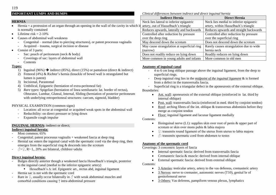
119
IMPORTANT LUMPS AND BUMPS
HERNIA
Hernia = a protrusion of an organ through an opening in the wall of the cavity in which it
is normally contained
Lifetime risk = 2-10%
Causes of abdominal wall weakness
- Congenital – normal (due to piercing structures), or patent processus vaginalis
- Acquired – trauma, surgical incision or disease
Consist of 3 parts:
- Sac: pouch of peritoneum (neck & body)
- Coverings of sac: layers of abdominal wall
- Contents
Types:
1) Inguinal (96%) indirect (85%), direct (15%) or pantaloon (direct & indirect)
2) Femoral (4%) & Richter‘s hernia (knuckle of bowel wall is strangulated but
lumen is patent)
3) Incisional, Parastomal
4) Umbilical, Epigastric (herniation of extra-peritoneal fat)
5) Rare types: Spigelian (herniation of linea semilunaris: lat. border of rectus),
Obturator, Lumbar, Gluteal, Internal, Sliding (herniation of posterior peritoneum
with underlying retroperitoneal structures: caecum, sigmoid, bladder)
PHYSICAL EXAMINTION (common signs)
- Location: all occur at congenital or acquitted weak spots in the abdominal wall
- Reducibility: on direct pressure or lying down
- Expansile cough impulse
INGUINAL HERNIA: indirect or direct;
Indirect inguinal hernia:
- Most common; 65%
- Congenital; patent processus vaginalis + weakened fascia at deep ring
- Hernial sac enters the inguinal canal with the spermatic cord via the deep ring, then
emerges from the superficial ring & descends into the scrotum
- ♂>♀, R> L, 20% are bilateral, children>adults
Direct inguinal hernia:
- Bulges directly anterior though a weakened fascia Hesselbach‘s triangle, posterior
to the inguinal canal (medial to the inferior epigastric artery)
Hesselbach‘s ∆: inf. Epigastric art., rectus abd, inguinal ligament
- Hernia sac is not with the spermatic cord
- Rare in ♀, usually occur bilaterally in ♂ with weak abdominal muscles and
comorbid conditions causing ↑ intra-abdominal pressure
Clinical differences between indirect and direct inguinal hernia
Indirect Hernia Direct Hernia
Neck lies lateral to inferior epigastric
artery, out of Hasselbach‘s triangle
Neck lies medial to inferior epigastric
artery, within Hasselbach‘s triangle
Reduces upwards, laterally and backwards Reduces upwards and straight backwards
Controlled after reduction by pressure
over the deep ring
Controlled after reduction by pressure
over the superficial ring
May descend down the scrotum Does not descend down the scrotum
May cause strangulation at superficial ring
(narrow)
Rarely causes strangulation due to wide
hernia neck
Does not readily reduce on lying down Readily reduces on lying down
More common in young adults and infants More common in old men
Anatomy of inguinal canal - 4 to 6 cm long oblique passage above the inguinal ligament, from the deep to
superficial rings.
- Deep inguinal ring lies in the midpoint of the inguinal ligament & is formed
from a defect in the transversalis fascia.
- Superficial ring is a triangular defect in the aponeurosis of the external oblique.
Boundaries:
Ant. wall: aponeurosis of the external oblique (reinforced in lat. third by
internal oblique)
Post. wall: transversalis fascia (reinforced in med. third by conjoint tendon)
Roof: arching fibres of the int. oblique & transversus abdominis before they
merge as conjoint tendon
Floor: inguinal ligament and lacunar ligament medially
Contents:
Ilioinguinal nerve (L1): supplies skin over root of penis & upper part of
scrotum or skin over mons pubis & labia majora
♀: transmits round ligament of the uterus from uterus to labia majora
♂: transmits spermatic cord from abdomen to testes
Anatomy of the spermatic cord
Coverings: 3 concentric layers of fascia
Internal spermatic fascia: derived from transversalis fascia
Cremasteric fascia & muscle: derived from internal oblique
External spermatic fascia: derived from external oblique
Contents:
3 Arteries: testicular artery, artery to the vas deferens, cremasteric artery
3 Nerves: nerve to cremaster, autonomic nerves (T10), genital br of
genitofemoral nerve
3 Others: Vas deferens, pampiform venous plexus, lymphatics
120
Complications
Reducible → Irreducible / Incarcerated → Obstructed → Strangulated
- Irreducible/ Incarcerated hernia: contents of hernial sac cannot be replaced into
abdomen
- Obstructed: loop of bowel trapped in hernial sac such that its lumen, but not
blood supply, is obstructed (no ischaemia not unduly tender, but with IO)
- Stangulated: blood supply to trapped bowel is cut off, bowel is dead or dying;
6hrs to gangrene. (acutely tender with s/s of IO; exception: Richter‘s hernia:
segment of bowel is trapped & ischaemic but lumen is patent no IO)
Management
Non-surgical:
Weight loss, change jobs/, avoid heavy lifting, Truss: for compression of
reducible hernia at deep ring (poor pickup rate)
Treat medical conditions causing chronic cough, chronic constipation
If obstructed/strangulated: NBM, IV drip, NG tube on suction, IV ABx
Surgical:
Hernioplasty/ Herniorraphy with mesh repair (open or laparoscopic)
o Immediately if suspect incarceration to prevent any boewl perforation
Principles: reduce bowel, ±excise hernia sac, reinforce posterior wall
o Lichtenstein tension-free mesh repair (lightweight, polypropylene
mesh insertion & suture)
o Shouldice repair (non-mesh technique: 2 continuous back & forth
sutures with permanent suture material)
Complications
o 2o to GA: AMI, CVA
o Immediate to early
- ARU
- Bruising , bleeding (testicular, cremasteric, @ to vas deferens,
inferior epigastric, femoral arteries)
- Injury to surrounding structures pain, parasthesia, impotence:
spermatic cord (vas deferens, testicular artery), round ligament,
ilioinguinal nerve, nerve to cremaster
o Early
- Infection of wound/ mesh
- Haematoma (10%; no cough impulse, non-reducible, hard) - Wound dehiscence
- pain
o Late
- Chronic post-op pain
- Recurrence of hernia (<0.5%; incomplete dissection, poor tissue,
too early mobilization, uncontrolled co-morbidities)
- Ischaemic orchitis & Testicular atrophy from testicular artery
damage or from pampiniform plexus thrombosis
Post- operation monitoring:
o Work leave for 6/52 with avoiding of heavy lifting
o Treat any medical conditions to avoid coughing, constipation
o Early mobilization from D10
o Keep area clean and wash carefully, but able to bathe immediately
EXAMINATION OF AN INGUINAL HERNIA
“Please examine this patient‟s groin” Don gloves, introduce yourself and explain your intention, then expose the patient
Stand patient up, examine both sides
- Inspect as per a lump: (if unable to see, ask the patient) o Is lump above or below the inguinal ligament? Any scrotal lump?
o Use a ruler to measure the dimensions of the lump
o Any skin changes? Previous scars (look hard)?
- Palpate:
o Define the inguinal ligament (ASIS to pubic tubercle, lat to pubic
symphysis, down frim umbilicus)
show the lump is above it
o Lump: consistency (soft, fluctuant), size, temperate, any tenderness?
o Scrotum: Cannot get above lump; feel for testis to rule out
undescended testis
o for expansile cough impulse (bilaterally)
Lay the patient supine
- Get patient to reduce the lump himself
- Locate the deep ring (2cm above the midpoint of inguinal ligament)
- Keep pressure on deep ring, ask patient to sit up & support his pelvis, then
swing over the bed and stand
- Cough when standing: if remains reduced – indirect hernia, if not, direct hernia.
(poor accuracy)
- Remove pressure & watch movement of hernia: slide obliquely (indirect) or
project forward (direct)
- Percuss & ascultate for bowel sounds
Examine other side
Offer:
1) Abdominal exam: for scars, masses, ascites, ARU, constipation, signs of IO
2) DRE for BPH, impacted stools
3) Respiratory exam for COPD
4) Ask patient for history of heavy lifting
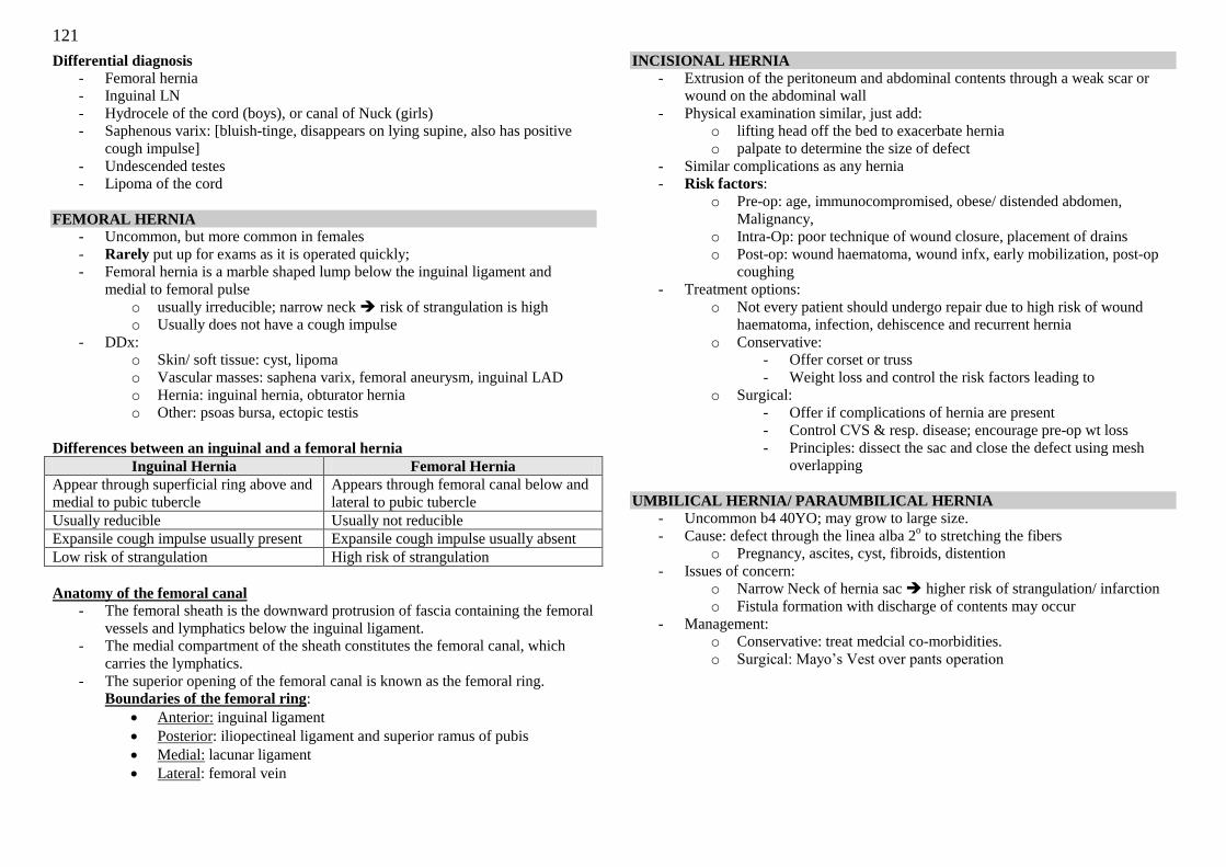
121
Differential diagnosis
- Femoral hernia
- Inguinal LN
- Hydrocele of the cord (boys), or canal of Nuck (girls)
- Saphenous varix: [bluish-tinge, disappears on lying supine, also has positive
cough impulse]
- Undescended testes
- Lipoma of the cord
FEMORAL HERNIA - Uncommon, but more common in females
- Rarely put up for exams as it is operated quickly;
- Femoral hernia is a marble shaped lump below the inguinal ligament and
medial to femoral pulse
o usually irreducible; narrow neck risk of strangulation is high
o Usually does not have a cough impulse
- DDx:
o Skin/ soft tissue: cyst, lipoma
o Vascular masses: saphena varix, femoral aneurysm, inguinal LAD
o Hernia: inguinal hernia, obturator hernia
o Other: psoas bursa, ectopic testis
Differences between an inguinal and a femoral hernia
Inguinal Hernia Femoral Hernia
Appear through superficial ring above and
medial to pubic tubercle
Appears through femoral canal below and
lateral to pubic tubercle
Usually reducible Usually not reducible
Expansile cough impulse usually present Expansile cough impulse usually absent
Low risk of strangulation High risk of strangulation
Anatomy of the femoral canal
- The femoral sheath is the downward protrusion of fascia containing the femoral
vessels and lymphatics below the inguinal ligament.
- The medial compartment of the sheath constitutes the femoral canal, which
carries the lymphatics.
- The superior opening of the femoral canal is known as the femoral ring.
Boundaries of the femoral ring:
Anterior: inguinal ligament
Posterior: iliopectineal ligament and superior ramus of pubis
Medial: lacunar ligament
Lateral: femoral vein
INCISIONAL HERNIA
- Extrusion of the peritoneum and abdominal contents through a weak scar or
wound on the abdominal wall
- Physical examination similar, just add:
o lifting head off the bed to exacerbate hernia
o palpate to determine the size of defect
- Similar complications as any hernia
- Risk factors:
o Pre-op: age, immunocompromised, obese/ distended abdomen,
Malignancy,
o Intra-Op: poor technique of wound closure, placement of drains
o Post-op: wound haematoma, wound infx, early mobilization, post-op
coughing
- Treatment options:
o Not every patient should undergo repair due to high risk of wound
haematoma, infection, dehiscence and recurrent hernia
o Conservative:
- Offer corset or truss
- Weight loss and control the risk factors leading to
o Surgical:
- Offer if complications of hernia are present
- Control CVS & resp. disease; encourage pre-op wt loss
- Principles: dissect the sac and close the defect using mesh
overlapping
UMBILICAL HERNIA/ PARAUMBILICAL HERNIA
- Uncommon b4 40YO; may grow to large size.
- Cause: defect through the linea alba 2o to stretching the fibers
o Pregnancy, ascites, cyst, fibroids, distention
- Issues of concern:
o Narrow Neck of hernia sac higher risk of strangulation/ infarction
o Fistula formation with discharge of contents may occur
- Management:
o Conservative: treat medcial co-morbidities.
o Surgical: Mayo‘s Vest over pants operation
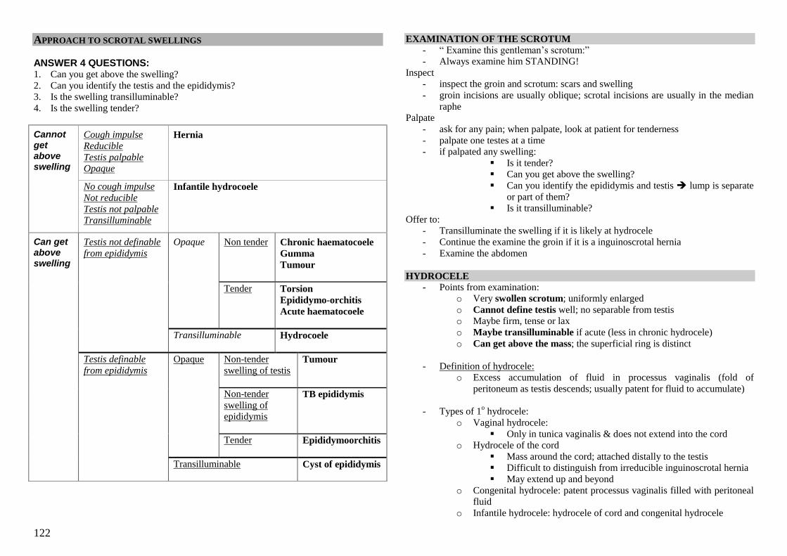
122
APPROACH TO SCROTAL SWELLINGS
ANSWER 4 QUESTIONS: 1. Can you get above the swelling?
2. Can you identify the testis and the epididymis?
3. Is the swelling transilluminable?
4. Is the swelling tender?
Cannot get above swelling
Cough impulse
Reducible
Testis palpable Opaque
Hernia
No cough impulse
Not reducible
Testis not palpable Transilluminable
Infantile hydrocoele
Can get above swelling
Testis not definable
from epididymis
Opaque
Non tender
Chronic haematocoele
Gumma
Tumour
Tender Torsion
Epididymo-orchitis
Acute haematocoele
Transilluminable
Hydrocoele
Testis definable
from epididymis
Opaque Non-tender
swelling of testis
Tumour
Non-tender
swelling of
epididymis
TB epididymis
Tender Epididymoorchitis
Transilluminable
Cyst of epididymis
EXAMINATION OF THE SCROTUM
- ― Examine this gentleman‘s scrotum:‖
- Always examine him STANDING!
Inspect
- inspect the groin and scrotum: scars and swelling
- groin incisions are usually oblique; scrotal incisions are usually in the median
raphe
Palpate
- ask for any pain; when palpate, look at patient for tenderness
- palpate one testes at a time
- if palpated any swelling:
Is it tender?
Can you get above the swelling?
Can you identify the epididymis and testis lump is separate
or part of them?
Is it transilluminable?
Offer to:
- Transilluminate the swelling if it is likely at hydrocele
- Continue the examine the groin if it is a inguinoscrotal hernia
- Examine the abdomen
HYDROCELE
- Points from examination:
o Very swollen scrotum; uniformly enlarged
o Cannot define testis well; no separable from testis
o Maybe firm, tense or lax
o Maybe transilluminable if acute (less in chronic hydrocele)
o Can get above the mass; the superficial ring is distinct
- Definition of hydrocele:
o Excess accumulation of fluid in processus vaginalis (fold of
peritoneum as testis descends; usually patent for fluid to accumulate)
- Types of 1o hydrocele:
o Vaginal hydrocele:
Only in tunica vaginalis & does not extend into the cord
o Hydrocele of the cord
Mass around the cord; attached distally to the testis
Difficult to distinguish from irreducible inguinoscrotal hernia
May extend up and beyond
o Congenital hydrocele: patent processus vaginalis filled with peritoneal
fluid
o Infantile hydrocele: hydrocele of cord and congenital hydrocele
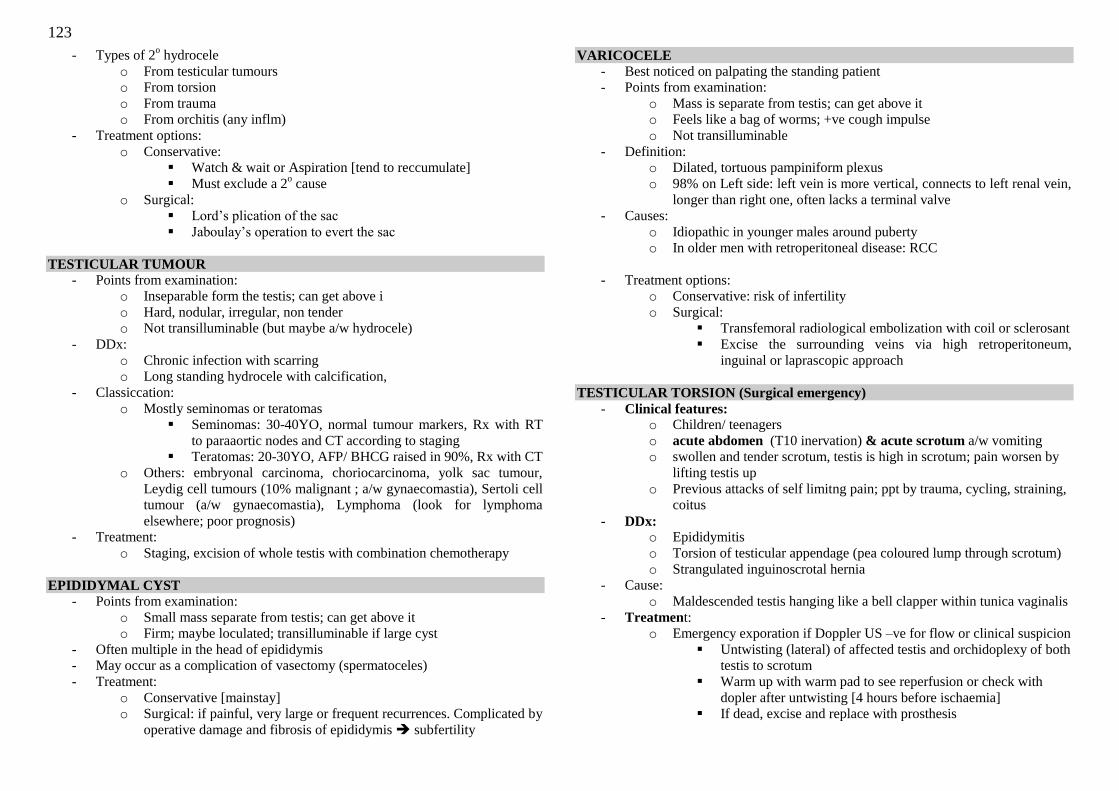
123
- Types of 2o hydrocele
o From testicular tumours
o From torsion
o From trauma
o From orchitis (any inflm)
- Treatment options:
o Conservative:
Watch & wait or Aspiration [tend to reccumulate]
Must exclude a 2o cause
o Surgical:
Lord‘s plication of the sac
Jaboulay‘s operation to evert the sac
TESTICULAR TUMOUR
- Points from examination:
o Inseparable form the testis; can get above i
o Hard, nodular, irregular, non tender
o Not transilluminable (but maybe a/w hydrocele)
- DDx:
o Chronic infection with scarring
o Long standing hydrocele with calcification,
- Classiccation:
o Mostly seminomas or teratomas
Seminomas: 30-40YO, normal tumour markers, Rx with RT
to paraaortic nodes and CT according to staging
Teratomas: 20-30YO, AFP/ BHCG raised in 90%, Rx with CT
o Others: embryonal carcinoma, choriocarcinoma, yolk sac tumour,
Leydig cell tumours (10% malignant ; a/w gynaecomastia), Sertoli cell
tumour (a/w gynaecomastia), Lymphoma (look for lymphoma
elsewhere; poor prognosis)
- Treatment:
o Staging, excision of whole testis with combination chemotherapy
EPIDIDYMAL CYST
- Points from examination:
o Small mass separate from testis; can get above it
o Firm; maybe loculated; transilluminable if large cyst
- Often multiple in the head of epididymis
- May occur as a complication of vasectomy (spermatoceles)
- Treatment:
o Conservative [mainstay]
o Surgical: if painful, very large or frequent recurrences. Complicated by
operative damage and fibrosis of epididymis subfertility
VARICOCELE
- Best noticed on palpating the standing patient
- Points from examination:
o Mass is separate from testis; can get above it
o Feels like a bag of worms; +ve cough impulse
o Not transilluminable
- Definition:
o Dilated, tortuous pampiniform plexus
o 98% on Left side: left vein is more vertical, connects to left renal vein,
longer than right one, often lacks a terminal valve
- Causes:
o Idiopathic in younger males around puberty
o In older men with retroperitoneal disease: RCC
- Treatment options:
o Conservative: risk of infertility
o Surgical:
Transfemoral radiological embolization with coil or sclerosant
Excise the surrounding veins via high retroperitoneum,
inguinal or laprascopic approach
TESTICULAR TORSION (Surgical emergency)
- Clinical features: o Children/ teenagers
o acute abdomen (T10 inervation) & acute scrotum a/w vomiting
o swollen and tender scrotum, testis is high in scrotum; pain worsen by
lifting testis up
o Previous attacks of self limitng pain; ppt by trauma, cycling, straining,
coitus
- DDx: o Epididymitis
o Torsion of testicular appendage (pea coloured lump through scrotum)
o Strangulated inguinoscrotal hernia
- Cause:
o Maldescended testis hanging like a bell clapper within tunica vaginalis
- Treatment:
o Emergency exporation if Doppler US –ve for flow or clinical suspicion
Untwisting (lateral) of affected testis and orchidoplexy of both
testis to scrotum
Warm up with warm pad to see reperfusion or check with
dopler after untwisting [4 hours before ischaemia]
If dead, excise and replace with prosthesis
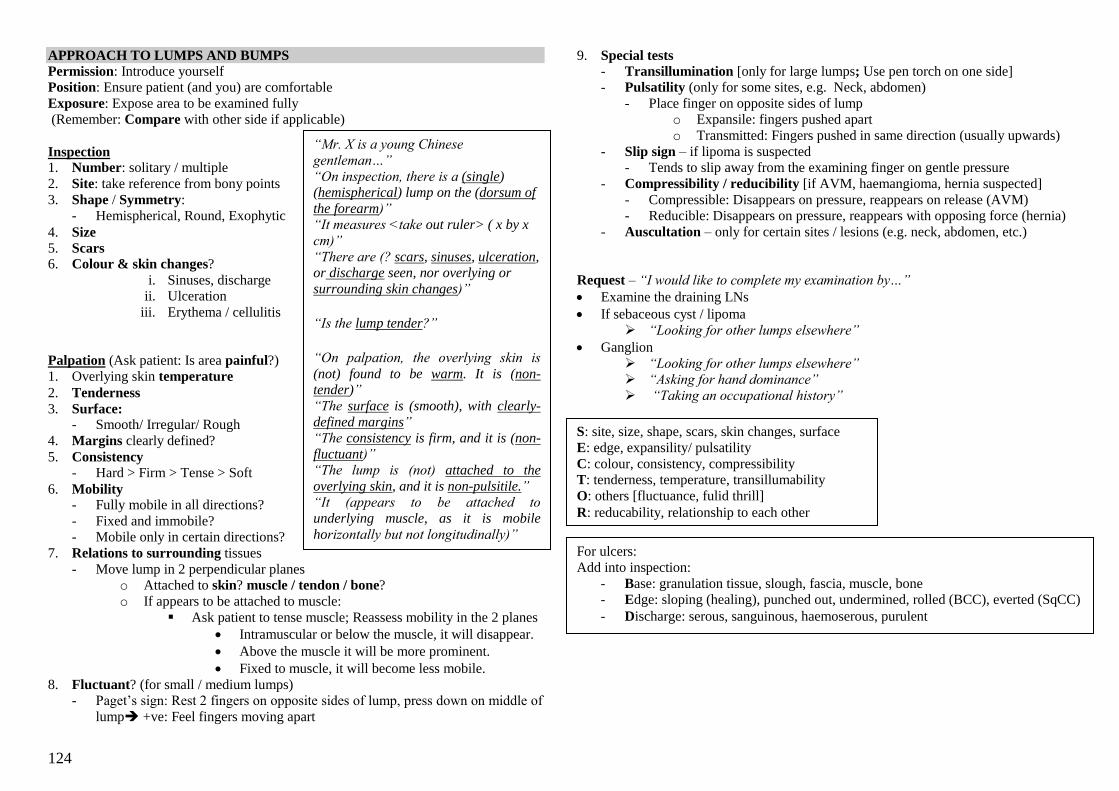
124
APPROACH TO LUMPS AND BUMPS
Permission: Introduce yourself
Position: Ensure patient (and you) are comfortable
Exposure: Expose area to be examined fully
(Remember: Compare with other side if applicable)
Inspection 1. Number: solitary / multiple
2. Site: take reference from bony points
3. Shape / Symmetry:
- Hemispherical, Round, Exophytic
4. Size
5. Scars
6. Colour & skin changes?
i. Sinuses, discharge
ii. Ulceration
iii. Erythema / cellulitis
Palpation (Ask patient: Is area painful?)
1. Overlying skin temperature
2. Tenderness
3. Surface: - Smooth/ Irregular/ Rough
4. Margins clearly defined?
5. Consistency
- Hard > Firm > Tense > Soft
6. Mobility - Fully mobile in all directions?
- Fixed and immobile?
- Mobile only in certain directions?
7. Relations to surrounding tissues
- Move lump in 2 perpendicular planes
o Attached to skin? muscle / tendon / bone?
o If appears to be attached to muscle:
Ask patient to tense muscle; Reassess mobility in the 2 planes
Intramuscular or below the muscle, it will disappear.
Above the muscle it will be more prominent.
Fixed to muscle, it will become less mobile.
8. Fluctuant? (for small / medium lumps)
- Paget‘s sign: Rest 2 fingers on opposite sides of lump, press down on middle of
lump +ve: Feel fingers moving apart
9. Special tests
- Transillumination [only for large lumps; Use pen torch on one side]
- Pulsatility (only for some sites, e.g. Neck, abdomen)
- Place finger on opposite sides of lump
o Expansile: fingers pushed apart
o Transmitted: Fingers pushed in same direction (usually upwards)
- Slip sign – if lipoma is suspected
- Tends to slip away from the examining finger on gentle pressure
- Compressibility / reducibility [if AVM, haemangioma, hernia suspected]
- Compressible: Disappears on pressure, reappears on release (AVM)
- Reducible: Disappears on pressure, reappears with opposing force (hernia)
- Auscultation – only for certain sites / lesions (e.g. neck, abdomen, etc.)
Request – “I would like to complete my examination by…”
Examine the draining LNs
If sebaceous cyst / lipoma
“Looking for other lumps elsewhere”
Ganglion
“Looking for other lumps elsewhere”
“Asking for hand dominance”
“Taking an occupational history”
“Mr. X is a young Chinese
gentleman…”
“On inspection, there is a (single) (hemispherical) lump on the (dorsum of
the forearm)”
“It measures <take out ruler> ( x by x
cm)”
“There are (? scars, sinuses, ulceration, or discharge seen, nor overlying or
surrounding skin changes)”
“Is the lump tender?”
“On palpation, the overlying skin is
(not) found to be warm. It is (non-tender)”
“The surface is (smooth), with clearly-
defined margins”
“The consistency is firm, and it is (non-
fluctuant)”
“The lump is (not) attached to the
overlying skin, and it is non-pulsitile.”
“It (appears to be attached to
underlying muscle, as it is mobile
horizontally but not longitudinally)”
S: site, size, shape, scars, skin changes, surface
E: edge, expansility/ pulsatility
C: colour, consistency, compressibility
T: tenderness, temperature, transillumability
O: others [fluctuance, fulid thrill]
R: reducability, relationship to each other
For ulcers:
Add into inspection:
- Base: granulation tissue, slough, fascia, muscle, bone
- Edge: sloping (healing), punched out, undermined, rolled (BCC), everted (SqCC)
- Discharge: serous, sanguinous, haemoserous, purulent
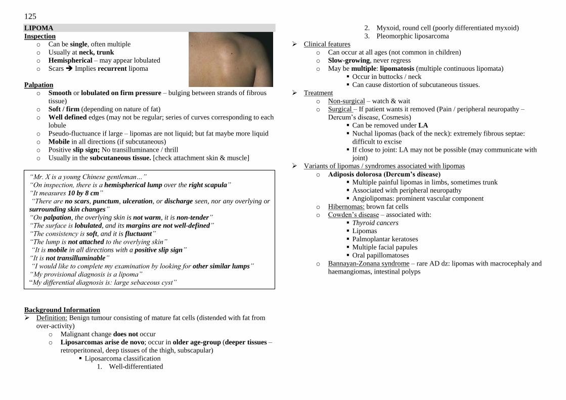
125
LIPOMA
Inspection o Can be single, often multiple
o Usually at neck, trunk
o Hemispherical – may appear lobulated
o Scars Implies recurrent lipoma
Palpation
o Smooth or lobulated on firm pressure – bulging between strands of fibrous
tissue)
o Soft / firm (depending on nature of fat)
o Well defined edges (may not be regular; series of curves corresponding to each
lobule
o Pseudo-fluctuance if large – lipomas are not liquid; but fat maybe more liquid
o Mobile in all directions (if subcutaneous)
o Positive slip sign; No transilluminance / thrill
o Usually in the subcutaneous tissue. [check attachment skin & muscle]
Background Information Definition: Benign tumour consisting of mature fat cells (distended with fat from
over-activity)
o Malignant change does not occur
o Liposarcomas arise de novo; occur in older age-group (deeper tissues –
retroperitoneal, deep tissues of the thigh, subscapular)
Liposarcoma classification
1. Well-differentiated
2. Myxoid, round cell (poorly differentiated myxoid)
3. Pleomorphic liposarcoma
Clinical features
o Can occur at all ages (not common in children)
o Slow-growing, never regress
o May be multiple: lipomatosis (multiple continuous lipomata)
Occur in buttocks / neck
Can cause distortion of subcutaneous tissues.
Treatment
o Non-surgical – watch & wait
o Surgical – If patient wants it removed (Pain / peripheral neuropathy –
Dercum‘s disease, Cosmesis)
Can be removed under LA
Nuchal lipomas (back of the neck): extremely fibrous septae:
difficult to excise
If close to joint: LA may not be possible (may communicate with
joint)
Variants of lipomas / syndromes associated with lipomas
o Adiposis dolorosa (Dercum’s disease)
Multiple painful lipomas in limbs, sometimes trunk
Associated with peripheral neuropathy
Angiolipomas: prominent vascular component
o Hibernomas: brown fat cells
o Cowden‘s disease – associated with:
Thyroid cancers Lipomas
Palmoplantar keratoses
Multiple facial papules
Oral papillomatoses
o Bannayan-Zonana syndrome – rare AD dz: lipomas with macrocephaly and
haemangiomas, intestinal polyps
“Mr. X is a young Chinese gentleman…”
“On inspection, there is a hemispherical lump over the right scapula”
“It measures 10 by 8 cm” “There are no scars, punctum, ulceration, or discharge seen, nor any overlying or
surrounding skin changes”
“On palpation, the overlying skin is not warm, it is non-tender”
“The surface is lobulated, and its margins are not well-defined”
“The consistency is soft, and it is fluctuant”
“The lump is not attached to the overlying skin”
“It is mobile in all directions with a positive slip sign”
“It is not transilluminable”
“I would like to complete my examination by looking for other similar lumps”
“My provisional diagnosis is a lipoma”
―My differential diagnosis is: large sebaceous cyst”
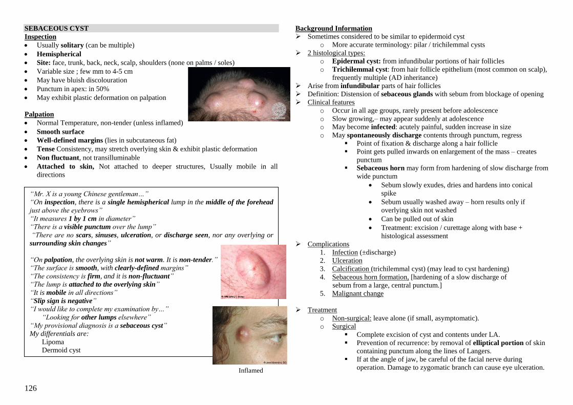
126
“Mr. X is a young Chinese gentleman…” “On inspection, there is a single hemispherical lump in the middle of the forehead
just above the eyebrows”
“It measures 1 by 1 cm in diameter” “There is a visible punctum over the lump”
“There are no scars, sinuses, ulceration, or discharge seen, nor any overlying or surrounding skin changes”
“On palpation, the overlying skin is not warm. It is non-tender.”
“The surface is smooth, with clearly-defined margins”
“The consistency is firm, and it is non-fluctuant”
“The lump is attached to the overlying skin”
“It is mobile in all directions”
“Slip sign is negative”
“I would like to complete my examination by…”
“Looking for other lumps elsewhere”
“My provisional diagnosis is a sebaceous cyst”
My differentials are:
Lipoma
Dermoid cyst
SEBACEOUS CYST
Inspection
Usually solitary (can be multiple)
Hemispherical
Site: face, trunk, back, neck, scalp, shoulders (none on palms / soles)
Variable size ; few mm to 4-5 cm
May have bluish discolouration
Punctum in apex: in 50%
May exhibit plastic deformation on palpation
Palpation
Normal Temperature, non-tender (unless inflamed)
Smooth surface
Well-defined margins (lies in subcutaneous fat)
Tense Consistency, may stretch overlying skin & exhibit plastic deformation
Non fluctuant, not transilluminable
Attached to skin, Not attached to deeper structures, Usually mobile in all
directions
Background Information
Sometimes considered to be similar to epidermoid cyst
o More accurate terminology: pilar / trichilemmal cysts
2 histological types:
o Epidermal cyst: from infundibular portions of hair follicles
o Trichilemmal cyst: from hair follicle epithelium (most common on scalp),
frequently multiple (AD inheritance)
Arise from infundibular parts of hair follicles
Definition: Distension of sebaceous glands with sebum from blockage of opening
Clinical features
o Occur in all age groups, rarely present before adolescence
o Slow growing,– may appear suddenly at adolescence
o May become infected: acutely painful, sudden increase in size
o May spontaneously discharge contents through punctum, regress
Point of fixation & discharge along a hair follicle
Point gets pulled inwards on enlargement of the mass – creates
punctum
Sebaceous horn may form from hardening of slow discharge from
wide punctum
Sebum slowly exudes, dries and hardens into conical
spike
Sebum usually washed away – horn results only if
overlying skin not washed
Can be pulled out of skin
Treatment: excision / curettage along with base +
histological assessment
Complications
1. Infection (±discharge)
2. Ulceration
3. Calcification (trichilemmal cyst) (may lead to cyst hardening)
4. Sebaceous horn formation, [hardening of a slow discharge of
sebum from a large, central punctum.]
5. Malignant change
Treatment
o Non-surgical: leave alone (if small, asymptomatic).
o Surgical
Complete excision of cyst and contents under LA.
Prevention of recurrence: by removal of elliptical portion of skin
containing punctum along the lines of Langers.
If at the angle of jaw, be careful of the facial nerve during
operation. Damage to zygomatic branch can cause eye ulceration.
Inflamed
Cyst
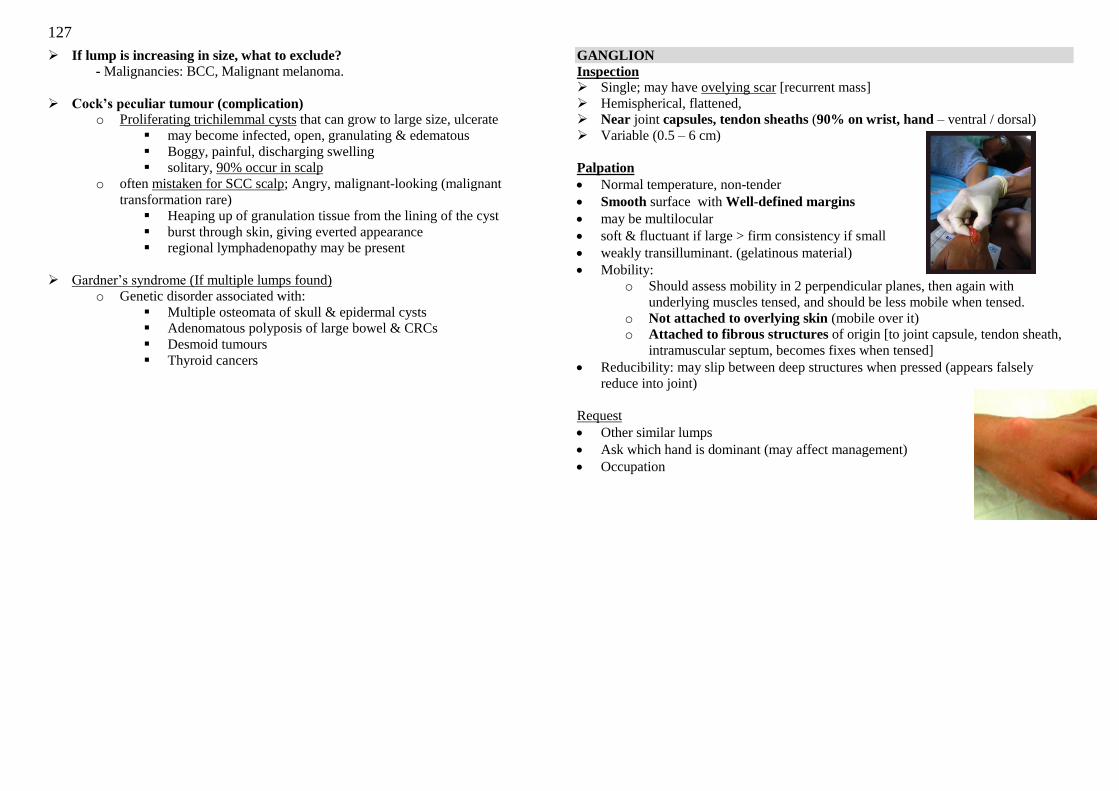
127
If lump is increasing in size, what to exclude?
- Malignancies: BCC, Malignant melanoma.
Cock’s peculiar tumour (complication) o Proliferating trichilemmal cysts that can grow to large size, ulcerate
may become infected, open, granulating & edematous
Boggy, painful, discharging swelling
solitary, 90% occur in scalp
o often mistaken for SCC scalp; Angry, malignant-looking (malignant
transformation rare)
Heaping up of granulation tissue from the lining of the cyst
burst through skin, giving everted appearance
regional lymphadenopathy may be present
Gardner‘s syndrome (If multiple lumps found)
o Genetic disorder associated with:
Multiple osteomata of skull & epidermal cysts
Adenomatous polyposis of large bowel & CRCs
Desmoid tumours
Thyroid cancers
GANGLION
Inspection Single; may have ovelying scar [recurrent mass]
Hemispherical, flattened,
Near joint capsules, tendon sheaths (90% on wrist, hand – ventral / dorsal)
Variable (0.5 – 6 cm)
Palpation
Normal temperature, non-tender
Smooth surface with Well-defined margins
may be multilocular
soft & fluctuant if large > firm consistency if small
weakly transilluminant. (gelatinous material)
Mobility:
o Should assess mobility in 2 perpendicular planes, then again with
underlying muscles tensed, and should be less mobile when tensed.
o Not attached to overlying skin (mobile over it)
o Attached to fibrous structures of origin [to joint capsule, tendon sheath,
intramuscular septum, becomes fixes when tensed]
Reducibility: may slip between deep structures when pressed (appears falsely
reduce into joint)
Request
Other similar lumps
Ask which hand is dominant (may affect management)
Occupation
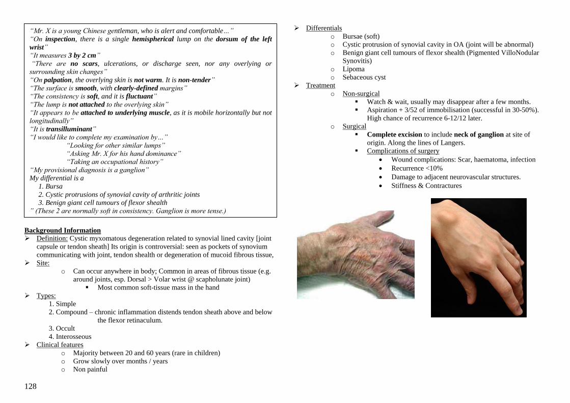
128
Background Information Definition: Cystic myxomatous degeneration related to synovial lined cavity [joint
capsule or tendon sheath] Its origin is controversial: seen as pockets of synovium
communicating with joint, tendon shealth or degeneration of mucoid fibrous tissue,
Site:
o Can occur anywhere in body; Common in areas of fibrous tissue (e.g.
around joints, esp. Dorsal > Volar wrist @ scapholunate joint)
Most common soft-tissue mass in the hand
Types:
1. Simple
2. Compound – chronic inflammation distends tendon sheath above and below
the flexor retinaculum.
3. Occult
4. Interosseous
Clinical features
o Majority between 20 and 60 years (rare in children)
o Grow slowly over months / years
o Non painful
Differentials
o Bursae (soft)
o Cystic protrusion of synovial cavity in OA (joint will be abnormal)
o Benign giant cell tumours of flexor shealth (Pigmented VilloNodular
Synovitis)
o Lipoma
o Sebaceous cyst
Treatment
o Non-surgical
Watch & wait, usually may disappear after a few months.
Aspiration + 3/52 of immobilisation (successful in 30-50%).
High chance of recurrence 6-12/12 later.
o Surgical
Complete excision to include neck of ganglion at site of
origin. Along the lines of Langers.
Complications of surgery
Wound complications: Scar, haematoma, infection
Recurrence <10%
Damage to adjacent neurovascular structures.
Stiffness & Contractures
“Mr. X is a young Chinese gentleman, who is alert and comfortable…”
“On inspection, there is a single hemispherical lump on the dorsum of the left
wrist” “It measures 3 by 2 cm”
“There are no scars, ulcerations, or discharge seen, nor any overlying or surrounding skin changes”
“On palpation, the overlying skin is not warm. It is non-tender”
“The surface is smooth, with clearly-defined margins”
“The consistency is soft, and it is fluctuant”
“The lump is not attached to the overlying skin”
“It appears to be attached to underlying muscle, as it is mobile horizontally but not
longitudinally”
“It is transilluminant” “I would like to complete my examination by…”
“Looking for other similar lumps”
“Asking Mr. X for his hand dominance”
“Taking an occupational history”
“My provisional diagnosis is a ganglion”
My differential is a
1. Bursa
2. Cystic protrusions of synovial cavity of arthritic joints 3. Benign giant cell tumours of flexor shealth
” (These 2 are normally soft in consistency. Ganglion is more tense.)
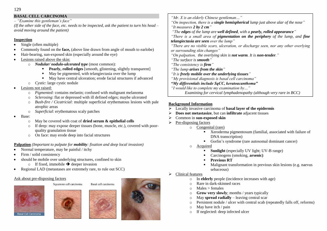
129
BASAL CELL CARCINOMA – “Examine this gentleman‟s face”
(If the other side of the face, etc. needs to be inspected, ask the patient to turn his head –
avoid moving around the patient)
Inspection
Single (often multiple)
Commonly found on the face, (above line drawn from angle of mouth to earlobe)
Hair-bearing, sun-exposed skin (especially around the eye)
Lesions raised above the skin:
o Nodular/ nodulo-ulcerated type (most common):
Pearly, rolled edges [smooth, glistening, slightly transparent]
May be pigmented, with telangiectasia over the lump
May have central ulceration; erode facial structures if advanced
o Cystic: large cystic nodule
Lesions not raised:
o Pigmented: contains melanin; confused with malignant melanoma
o Sclerosing: flat or depressed with ill defined edges; maybe ulcerated
o Bush-fire / Cicatricial: multiple superficial erythematous lesions with pale
atrophic areas
o Superficial: erythematous scaly patches
Base:
o May be covered with coat of dried serum & epithelial cells
o If deep: may expose deeper tissues (bone, muscle, etc.), covered with poor-
quality granulation tissue
o On face: may erode deep into facial structures
Palpation (Important to palpate for mobility: fixation and deep local invasion)
Normal temperature, may be painful / itchy
Firm / solid consistency
should be mobile over underlying structures, confined to skin
o If fixed, immobile deeper invasion
Regional LAD (metastases are extremely rare, to rule out SCC)
Ask about pre-disposing factors
Background Information Locally invasive carcinoma of basal layer of the epidermis
Does not metastasize, but can infiltrate adjacent tissues
Common in sun-exposed skin
Pre-disposing factors
o Congenital (rare)
Xeroderma pigmentosum (familial, associated with failure of
DNA transcription)
Gorlin‘s syndrome (rare autosomal dominant cancer)
o Acquired
Sunlight (especially UV light; UV-B range)
Carcinogens (smoking, arsenic)
Previous RT
Malignant transformation in previous skin lesions (e.g. naevus
sebaceous)
Clinical features
o In elderly people (incidence increases with age)
o Rare in dark-skinned races
o Males > females o Grow very slowly; months / years typically
o May spread radially – leaving central scar
o Persistent nodule / ulcer with central scab (repeatedly falls off, reforms)
o May have itch / pain
o If neglected: deep infected ulcer
“Mr. X is an elderly Chinese gentleman…”
“On inspection, there is a single hemispherical lump just above alar of the nose”
“It measures 2 by 2 cm” “The edges of the lump are well defined, with a pearly, rolled appearance”
“There is a small area of pigmentation on the periphery of the lump, and fine
telangiectasia are seen over the lump”
“There are no visible scars, ulceration, or discharge seen, nor any other overlying
or surrounding skin changes” “On palpation, the overlying skin is not warm. It is non-tender.”
“The surface is smooth”
“The consistency is firm”
“The lump arises from the skin”
“It is freely mobile over the underlying tissues”
“My provisional diagnosis is basal cell carcinoma”
“My differentials include SqCC, keratoacanthoma”
“I would like to complete my examination by…”
Examining for cervical lymphadenopathy (although very rare in BCC)
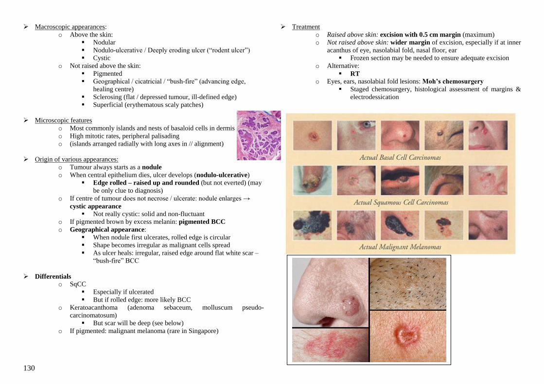
130
Macroscopic appearances:
o Above the skin:
Nodular
Nodulo-ulcerative / Deeply eroding ulcer (―rodent ulcer‖)
Cystic
o Not raised above the skin:
Pigmented
Geographical / cicatricial / ―bush-fire‖ (advancing edge,
healing centre)
Sclerosing (flat / depressed tumour, ill-defined edge)
Superficial (erythematous scaly patches)
Microscopic features
o Most commonly islands and nests of basaloid cells in dermis
o High mitotic rates, peripheral palisading
o (islands arranged radially with long axes in // alignment)
Origin of various appearances:
o Tumour always starts as a nodule
o When central epithelium dies, ulcer develops (nodulo-ulcerative)
Edge rolled – raised up and rounded (but not everted) (may
be only clue to diagnosis)
o If centre of tumour does not necrose / ulcerate: nodule enlarges →
cystic appearance Not really cystic: solid and non-fluctuant
o If pigmented brown by excess melanin: pigmented BCC
o Geographical appearance:
When nodule first ulcerates, rolled edge is circular
Shape becomes irregular as malignant cells spread
As ulcer heals: irregular, raised edge around flat white scar –
―bush-fire‖ BCC
Differentials o SqCC
Especially if ulcerated
But if rolled edge: more likely BCC
o Keratoacanthoma (adenoma sebaceum, molluscum pseudo-
carcinomatosum)
But scar will be deep (see below)
o If pigmented: malignant melanoma (rare in Singapore)
Treatment
o Raised above skin: excision with 0.5 cm margin (maximum)
o Not raised above skin: wider margin of excision, especially if at inner
acanthus of eye, nasolabial fold, nasal floor, ear
Frozen section may be needed to ensure adequate excision
o Alternative:
RT o Eyes, ears, nasolabial fold lesions: Moh’s chemosurgery
Staged chemosurgery, histological assessment of margins &
electrodessication
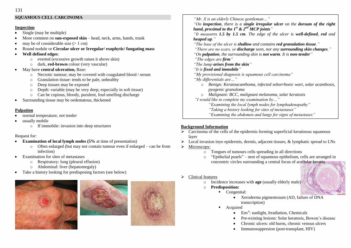
131
SQUAMOUS CELL CARCINOMA
Inspection
Single (may be multiple)
More common on sun-exposed skin – head, neck, arms, hands, trunk
may be of considerable size (> 1 cm)
Round nodule or Circular ulcer or Irregular/ exophytic/ fungating mass
Well defined edges:
o everted (excessive growth raises it above skin)
o dark, red-brown colour (very vascular)
May have central ulceration, Base:
o Necrotic tumour; may be covered with coagulated blood / serum
o Granulation tissue: tends to be pale, unhealthy
o Deep tissues may be exposed
o Depth: variable (may be very deep; especially in soft tissue)
o Can be copious, bloody, purulent, foul-smelling discharge
Surrounding tissue may be oedematous, thickened
Palpation
normal temperature, not tender
usually mobile
o If immobile: invasion into deep structures
Request for:
Examination of local lymph nodes (5% at time of presentation)
o Often enlarged (but may not contain tumour even if enlarged – can be from
infection)
Examination for sites of metastases
o Respiratory: lung (pleural effusion)
o Abdominal: liver (hepatomegaly)
Take a history looking for predisposing factors (see below)
Background Information
Carcinoma of the cells of the epidermis forming superficial keratinous squamous
layer
Local invasion inyo epidermis, dermis, adjacent tissues, & lymphatic spread to LNs
Microscopy:
o Tongues of tumours cells spreading in all directions
o ―Epithelial pearls‖ – nest of squamous epithelium, cells are arranged in
concentric circles surrounding a central focus of acellular keratin
Clinical features
o Incidence increases with age (usually elderly male)
o Predisposition: Congenital:
Xeroderma pigmentosum (AD, failure of DNA
transcription)
Acquired
Envn: sunlight, Irradiation, Chemicals
Pre-existing lesions: Solar keratosis, Bowen‘s disease
Chronic ulcers: old burns, chronic venous ulcers
Immunosuppresion (post-transplant, HIV)
“Mr. X is an elderly Chinese gentleman…”
“On inspection, there is a single irregular ulcer on the dorsum of the right
hand, proximal to the 1st & 2
nd MCP joints”
“It measures 1.5 by 1.5 cm. The edge of the ulcer is well-defined, red and
heaped up.” “The base of the ulcer is shallow and contains red granulation tissue.”
“There are no scars, or discharge seen, nor any surrounding skin changes.”
“On palpation, the surrounding skin is not warm. It is non-tender”
“The edges are firm”
“The lump arises from the skin”
“It is fixed and immobile”
“My provisional diagnosis is squamous cell carcinoma”
“My differentials are…” o Benign: Keratoacanthoma, infected seborrhoeic wart, solar acanthosis,
pyogenic granuloma
o Malignant: BCC, malignant melanoma, solar keratosis “I would like to complete my examination by…”
“Examining the local lymph nodes for lymphadenopathy”
“Taking a history looking for sites of metastases”
“Examining the abdomen and lungs for signs of metastases”
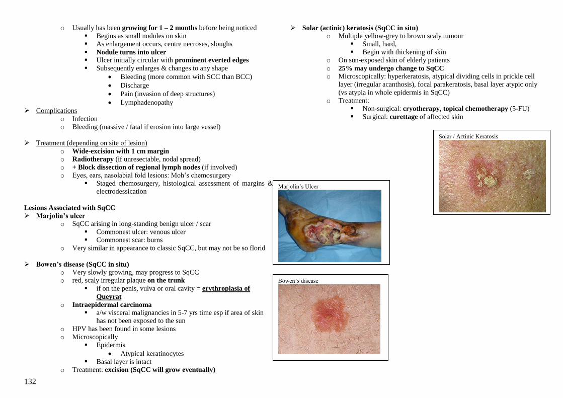
132
o Usually has been growing for 1 – 2 months before being noticed
Begins as small nodules on skin
As enlargement occurs, centre necroses, sloughs
Nodule turns into ulcer Ulcer initially circular with prominent everted edges
Subsequently enlarges & changes to any shape
Bleeding (more common with SCC than BCC)
Discharge
Pain (invasion of deep structures)
Lymphadenopathy
Complications
o Infection
o Bleeding (massive / fatal if erosion into large vessel)
Treatment (depending on site of lesion)
o Wide-excision with 1 cm margin
o Radiotherapy (if unresectable, nodal spread)
o + Block dissection of regional lymph nodes (if involved)
o Eyes, ears, nasolabial fold lesions: Moh‘s chemosurgery
Staged chemosurgery, histological assessment of margins &
electrodessication
Lesions Associated with SqCC
Marjolin’s ulcer
o SqCC arising in long-standing benign ulcer / scar
Commonest ulcer: venous ulcer
Commonest scar: burns
o Very similar in appearance to classic SqCC, but may not be so florid
Bowen’s disease (SqCC in situ)
o Very slowly growing, may progress to SqCC
o red, scaly irregular plaque on the trunk
if on the penis, vulva or oral cavity = erythroplasia of
Queyrat
o Intraepidermal carcinoma
a/w visceral malignancies in 5-7 yrs time esp if area of skin
has not been exposed to the sun
o HPV has been found in some lesions
o Microscopically
Epidermis
Atypical keratinocytes
Basal layer is intact
o Treatment: excision (SqCC will grow eventually)
Solar (actinic) keratosis (SqCC in situ)
o Multiple yellow-grey to brown scaly tumour
Small, hard,
Begin with thickening of skin
o On sun-exposed skin of elderly patients
o 25% may undergo change to SqCC
o Microscopically: hyperkeratosis, atypical dividing cells in prickle cell
layer (irregular acanthosis), focal parakeratosis, basal layer atypic only
(vs atypia in whole epidermis in SqCC)
o Treatment:
Non-surgical: cryotherapy, topical chemotherapy (5-FU)
Surgical: curettage of affected skin
Bowen‘s disease
Solar / Actinic Keratosis
Marjolin‘s Ulcer
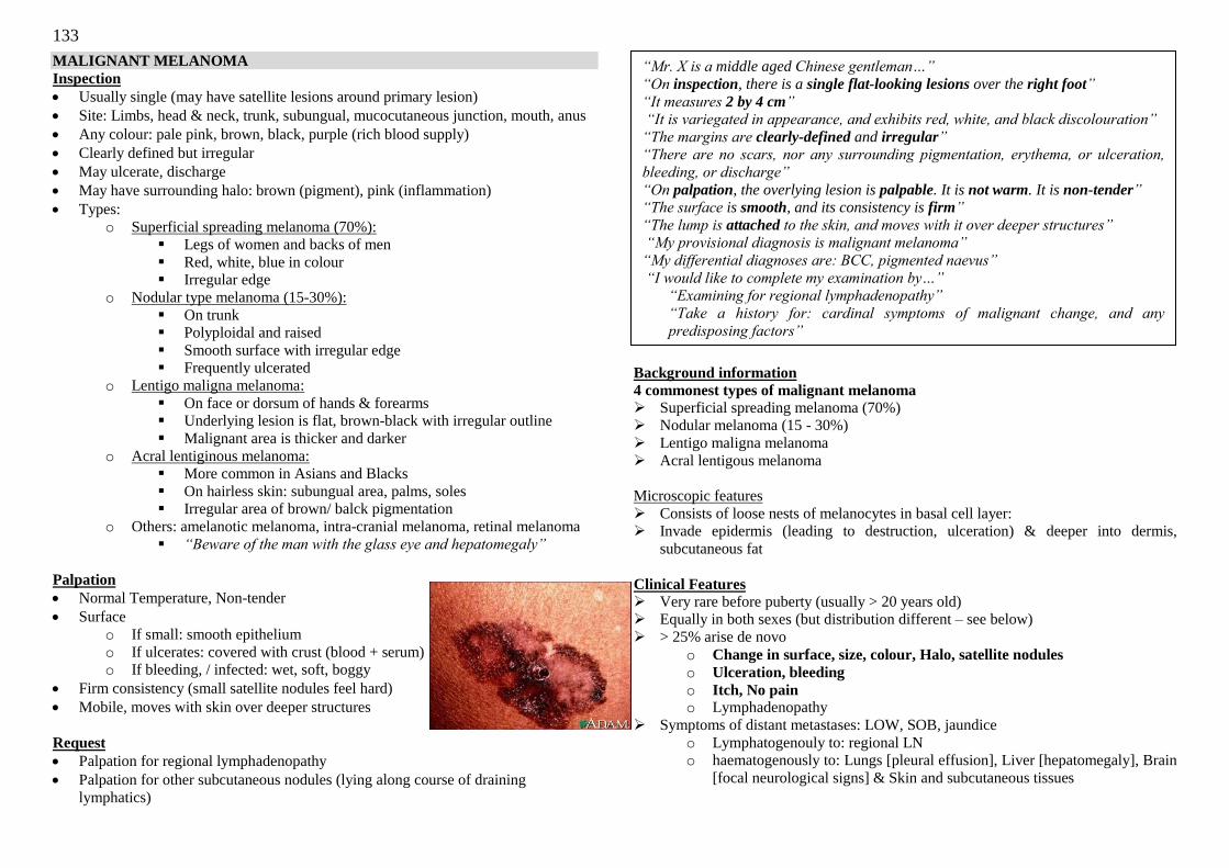
133
MALIGNANT MELANOMA
Inspection
Usually single (may have satellite lesions around primary lesion)
Site: Limbs, head & neck, trunk, subungual, mucocutaneous junction, mouth, anus
Any colour: pale pink, brown, black, purple (rich blood supply)
Clearly defined but irregular
May ulcerate, discharge
May have surrounding halo: brown (pigment), pink (inflammation)
Types:
o Superficial spreading melanoma (70%):
Legs of women and backs of men
Red, white, blue in colour
Irregular edge
o Nodular type melanoma (15-30%):
On trunk
Polyploidal and raised
Smooth surface with irregular edge
Frequently ulcerated
o Lentigo maligna melanoma:
On face or dorsum of hands & forearms
Underlying lesion is flat, brown-black with irregular outline
Malignant area is thicker and darker
o Acral lentiginous melanoma:
More common in Asians and Blacks
On hairless skin: subungual area, palms, soles
Irregular area of brown/ balck pigmentation
o Others: amelanotic melanoma, intra-cranial melanoma, retinal melanoma
“Beware of the man with the glass eye and hepatomegaly”
Palpation
Normal Temperature, Non-tender
Surface
o If small: smooth epithelium
o If ulcerates: covered with crust (blood + serum)
o If bleeding, / infected: wet, soft, boggy
Firm consistency (small satellite nodules feel hard)
Mobile, moves with skin over deeper structures
Request
Palpation for regional lymphadenopathy
Palpation for other subcutaneous nodules (lying along course of draining
lymphatics)
Background information
4 commonest types of malignant melanoma
Superficial spreading melanoma (70%)
Nodular melanoma (15 - 30%)
Lentigo maligna melanoma
Acral lentigous melanoma
Microscopic features
Consists of loose nests of melanocytes in basal cell layer:
Invade epidermis (leading to destruction, ulceration) & deeper into dermis,
subcutaneous fat
Clinical Features
Very rare before puberty (usually > 20 years old)
Equally in both sexes (but distribution different – see below)
> 25% arise de novo
o Change in surface, size, colour, Halo, satellite nodules
o Ulceration, bleeding
o Itch, No pain o Lymphadenopathy
Symptoms of distant metastases: LOW, SOB, jaundice
o Lymphatogenouly to: regional LN
o haematogenously to: Lungs [pleural effusion], Liver [hepatomegaly], Brain
[focal neurological signs] & Skin and subcutaneous tissues
“Mr. X is a middle aged Chinese gentleman…”
“On inspection, there is a single flat-looking lesions over the right foot”
“It measures 2 by 4 cm” “It is variegated in appearance, and exhibits red, white, and black discolouration”
“The margins are clearly-defined and irregular” “There are no scars, nor any surrounding pigmentation, erythema, or ulceration,
bleeding, or discharge”
“On palpation, the overlying lesion is palpable. It is not warm. It is non-tender”
“The surface is smooth, and its consistency is firm”
“The lump is attached to the skin, and moves with it over deeper structures”
“My provisional diagnosis is malignant melanoma”
“My differential diagnoses are: BCC, pigmented naevus”
“I would like to complete my examination by…”
“Examining for regional lymphadenopathy”
“Take a history for: cardinal symptoms of malignant change, and any
predisposing factors”
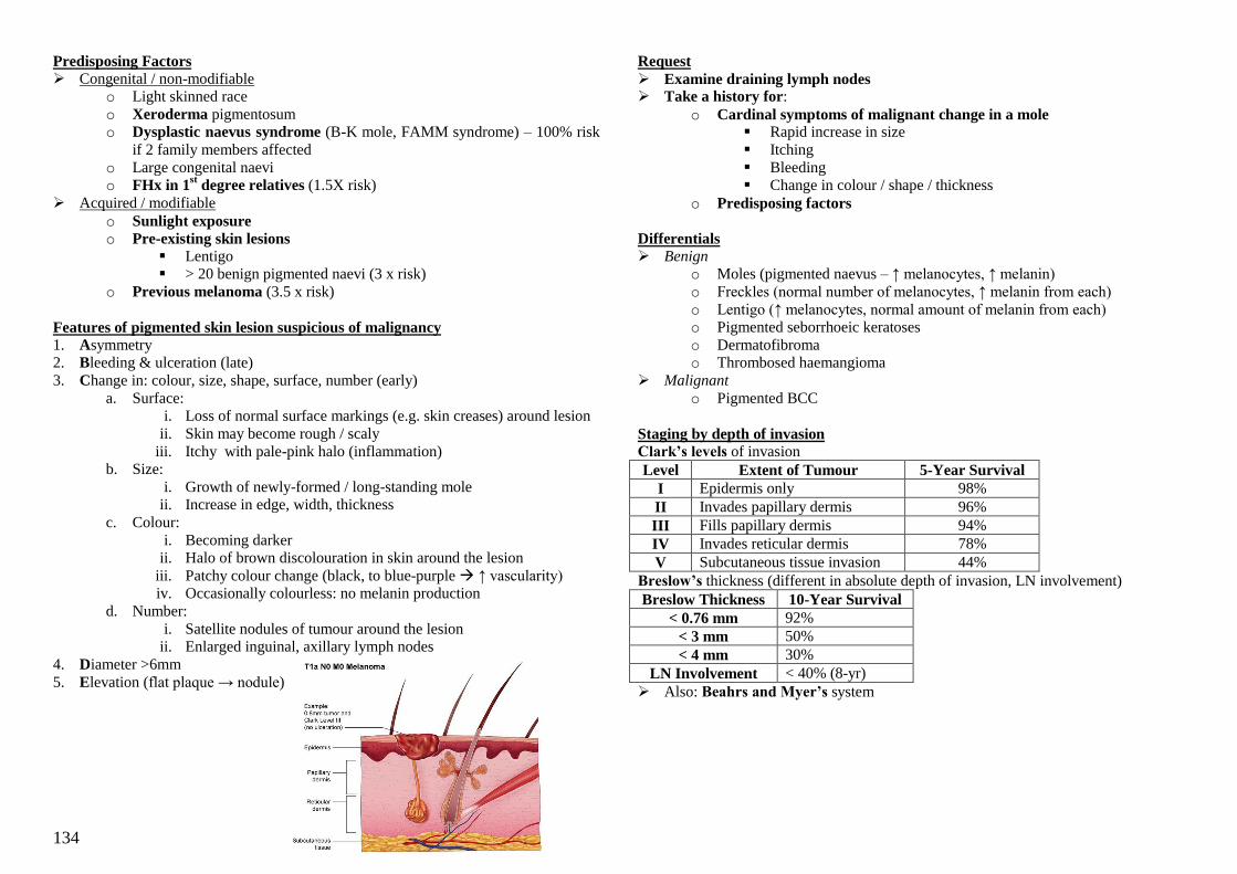
134
Predisposing Factors
Congenital / non-modifiable
o Light skinned race
o Xeroderma pigmentosum
o Dysplastic naevus syndrome (B-K mole, FAMM syndrome) – 100% risk
if 2 family members affected
o Large congenital naevi
o FHx in 1st degree relatives (1.5X risk)
Acquired / modifiable
o Sunlight exposure
o Pre-existing skin lesions
Lentigo
> 20 benign pigmented naevi (3 x risk)
o Previous melanoma (3.5 x risk)
Features of pigmented skin lesion suspicious of malignancy
1. Asymmetry
2. Bleeding & ulceration (late)
3. Change in: colour, size, shape, surface, number (early)
a. Surface:
i. Loss of normal surface markings (e.g. skin creases) around lesion
ii. Skin may become rough / scaly
iii. Itchy with pale-pink halo (inflammation)
b. Size:
i. Growth of newly-formed / long-standing mole
ii. Increase in edge, width, thickness
c. Colour:
i. Becoming darker
ii. Halo of brown discolouration in skin around the lesion
iii. Patchy colour change (black, to blue-purple ↑ vascularity)
iv. Occasionally colourless: no melanin production
d. Number:
i. Satellite nodules of tumour around the lesion
ii. Enlarged inguinal, axillary lymph nodes
4. Diameter >6mm
5. Elevation (flat plaque → nodule)
Request
Examine draining lymph nodes Take a history for:
o Cardinal symptoms of malignant change in a mole Rapid increase in size
Itching
Bleeding
Change in colour / shape / thickness
o Predisposing factors
Differentials Benign
o Moles (pigmented naevus – ↑ melanocytes, ↑ melanin)
o Freckles (normal number of melanocytes, ↑ melanin from each)
o Lentigo (↑ melanocytes, normal amount of melanin from each)
o Pigmented seborrhoeic keratoses
o Dermatofibroma
o Thrombosed haemangioma
Malignant
o Pigmented BCC
Staging by depth of invasion Clark’s levels of invasion
Level Extent of Tumour 5-Year Survival
I Epidermis only 98%
II Invades papillary dermis 96%
III Fills papillary dermis 94%
IV Invades reticular dermis 78%
V Subcutaneous tissue invasion 44%
Breslow’s thickness (different in absolute depth of invasion, LN involvement)
Breslow Thickness 10-Year Survival
< 0.76 mm 92%
< 3 mm 50%
< 4 mm 30%
LN Involvement < 40% (8-yr)
Also: Beahrs and Myer’s system
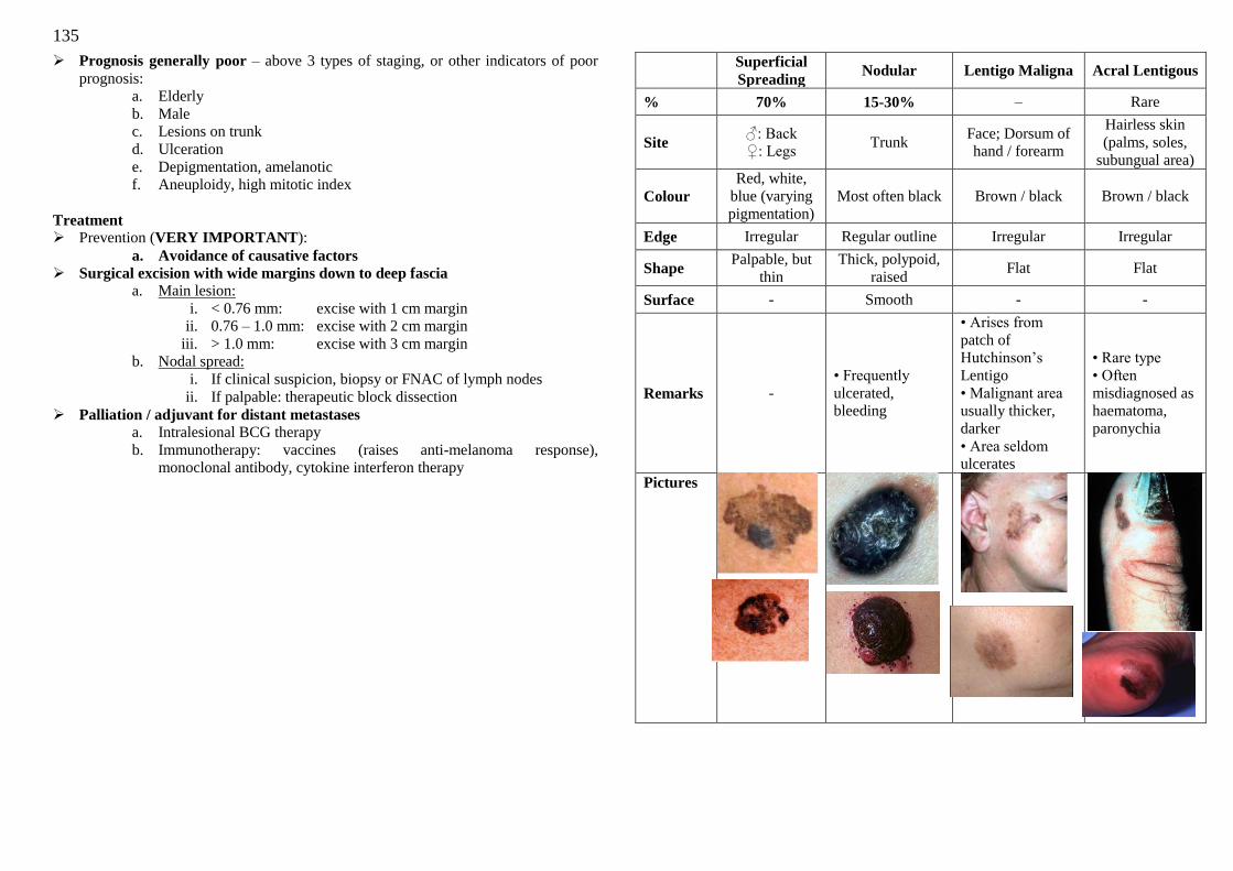
135
Prognosis generally poor – above 3 types of staging, or other indicators of poor
prognosis:
a. Elderly
b. Male
c. Lesions on trunk
d. Ulceration
e. Depigmentation, amelanotic
f. Aneuploidy, high mitotic index
Treatment Prevention (VERY IMPORTANT):
a. Avoidance of causative factors
Surgical excision with wide margins down to deep fascia
a. Main lesion:
i. < 0.76 mm: excise with 1 cm margin
ii. 0.76 – 1.0 mm: excise with 2 cm margin
iii. > 1.0 mm: excise with 3 cm margin
b. Nodal spread:
i. If clinical suspicion, biopsy or FNAC of lymph nodes
ii. If palpable: therapeutic block dissection
Palliation / adjuvant for distant metastases
a. Intralesional BCG therapy
b. Immunotherapy: vaccines (raises anti-melanoma response),
monoclonal antibody, cytokine interferon therapy
Superficial
Spreading Nodular Lentigo Maligna Acral Lentigous
% 70% 15-30% – Rare
Site ♂: Back
♀: Legs Trunk
Face; Dorsum of
hand / forearm
Hairless skin
(palms, soles,
subungual area)
Colour
Red, white,
blue (varying
pigmentation)
Most often black Brown / black Brown / black
Edge Irregular Regular outline Irregular Irregular
Shape Palpable, but
thin
Thick, polypoid,
raised Flat Flat
Surface - Smooth - -
Remarks -
• Frequently
ulcerated,
bleeding
• Arises from
patch of
Hutchinson‘s
Lentigo
• Malignant area
usually thicker,
darker
• Area seldom
ulcerates
• Rare type
• Often
misdiagnosed as
haematoma,
paronychia
Pictures
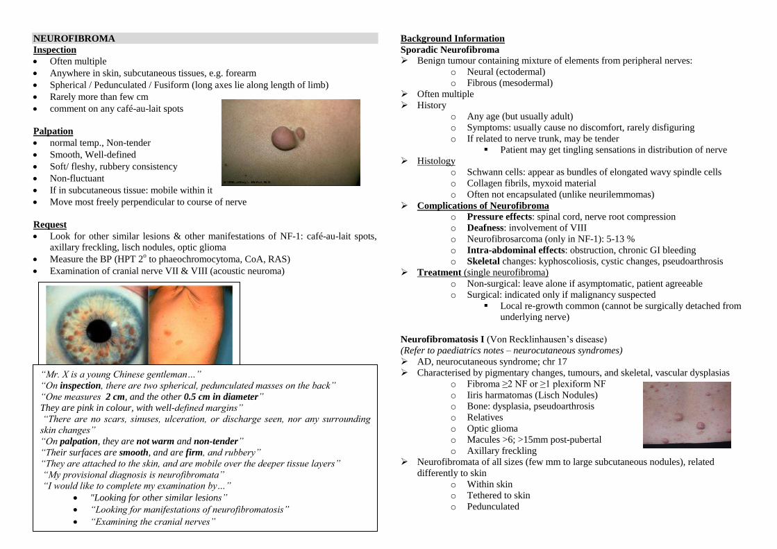
136
NEUROFIBROMA
Inspection
Often multiple
Anywhere in skin, subcutaneous tissues, e.g. forearm
Spherical / Pedunculated / Fusiform (long axes lie along length of limb)
Rarely more than few cm
comment on any café-au-lait spots
Palpation
normal temp., Non-tender
Smooth, Well-defined
Soft/ fleshy, rubbery consistency
Non-fluctuant
If in subcutaneous tissue: mobile within it
Move most freely perpendicular to course of nerve
Request
Look for other similar lesions & other manifestations of NF-1: café-au-lait spots,
axillary freckling, lisch nodules, optic glioma
Measure the BP (HPT 2o to phaeochromocytoma, CoA, RAS)
Examination of cranial nerve VII & VIII (acoustic neuroma)
Background Information
Sporadic Neurofibroma Benign tumour containing mixture of elements from peripheral nerves:
o Neural (ectodermal)
o Fibrous (mesodermal)
Often multiple
History
o Any age (but usually adult)
o Symptoms: usually cause no discomfort, rarely disfiguring
o If related to nerve trunk, may be tender
Patient may get tingling sensations in distribution of nerve
Histology
o Schwann cells: appear as bundles of elongated wavy spindle cells
o Collagen fibrils, myxoid material
o Often not encapsulated (unlike neurilemmomas)
Complications of Neurofibroma o Pressure effects: spinal cord, nerve root compression
o Deafness: involvement of VIII
o Neurofibrosarcoma (only in NF-1): 5-13 %
o Intra-abdominal effects: obstruction, chronic GI bleeding
o Skeletal changes: kyphoscoliosis, cystic changes, pseudoarthrosis
Treatment (single neurofibroma)
o Non-surgical: leave alone if asymptomatic, patient agreeable
o Surgical: indicated only if malignancy suspected
Local re-growth common (cannot be surgically detached from
underlying nerve)
Neurofibromatosis I (Von Recklinhausen‘s disease)
(Refer to paediatrics notes – neurocutaneous syndromes)
AD, neurocutaneous syndrome; chr 17
Characterised by pigmentary changes, tumours, and skeletal, vascular dysplasias
o Fibroma ≥2 NF or ≥1 plexiform NF
o Iiris harmatomas (Lisch Nodules)
o Bone: dysplasia, pseudoarthrosis
o Relatives
o Optic glioma
o Macules >6; >15mm post-pubertal
o Axillary freckling
Neurofibromata of all sizes (few mm to large subcutaneous nodules), related
differently to skin
o Within skin
o Tethered to skin
o Pedunculated
“Mr. X is a young Chinese gentleman…”
“On inspection, there are two spherical, pedunculated masses on the back”
“One measures 2 cm, and the other 0.5 cm in diameter” They are pink in colour, with well-defined margins”
“There are no scars, sinuses, ulceration, or discharge seen, nor any surrounding skin changes”
“On palpation, they are not warm and non-tender”
“Their surfaces are smooth, and are firm, and rubbery”
“They are attached to the skin, and are mobile over the deeper tissue layers”
“My provisional diagnosis is neurofibromata”
“I would like to complete my examination by…”
"Looking for other similar lesions”
“Looking for manifestations of neurofibromatosis”
“Examining the cranial nerves”

137
Plexiform Neurofibroma (elephantiasis neurofibromatosis)
Very rare
Excessive overgrowth of neural tissue in subcutaneous layers, giving tissue swollen
oedematous appearance
o Often mistaken for lymphoedema, but lymphatic drainage is normal
Can result in severe deformity: diffuse enlargement of peripheral nerve with skin
involvement
DERMOID CYST
Inspection
Usually single
Ovoid / spherical
Site:
o Congenital, 1-2 cm usually
Along lines of fusion of ophthalmic & maxillary facial processes
Inner & outer ends of upper eyebrow
o Acquired, 0.5-1 cm usually
Beneath skin likely to be injured e.g. fingers
Scars often present
Palpation
Not warm, maybe tender if infected
Smooth surface, Well-defined margins
Consistency
o Congenital: Soft (not tense / hard)
o Acquired: Hard & tense (sometimes stony hard)
Fluctuant (if large)
Mobile over deeper tissues
o Deep to skin, in subcutaneous tissue
i. Congenital: Not attached to skin or underlying structures
ii. Acquired: may be tethered to scar
“Ms. X is a young Chinese girl…”
“On inspection, there is a single ovoid lump just above the lateral edge of the left
eyebrow” “It measures 3 by 2 cm”
“There are no scars, ulceration, or discharge seen, nor any overlying or surrounding skin changes)”
“On palpation, the overlying skin is not warm. It is non-tender.”
“The surface is smooth, with clearly-defined margins”
“The consistency is firm, and it is fluctuant”
“The lump is not attached to the overlying skin nor underlying tissues.”
“It is fully mobile in all directions.”
“My provisional diagnosis is a congenital dermoid cyst”
“My differentials are: sebaceous cyst, lipoma”
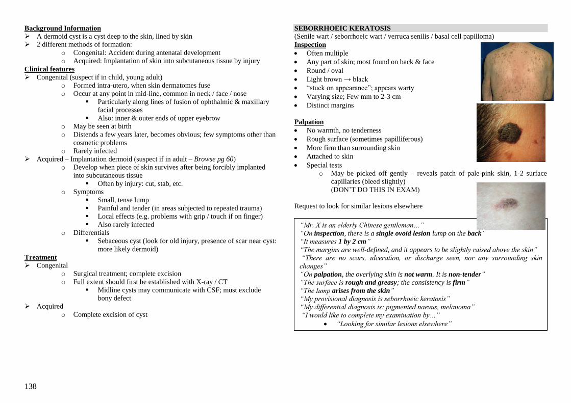
138
Background Information
A dermoid cyst is a cyst deep to the skin, lined by skin
2 different methods of formation:
o Congenital: Accident during antenatal development
o Acquired: Implantation of skin into subcutaneous tissue by injury
Clinical features Congenital (suspect if in child, young adult)
o Formed intra-utero, when skin dermatomes fuse
o Occur at any point in mid-line, common in neck / face / nose
Particularly along lines of fusion of ophthalmic & maxillary
facial processes
Also: inner & outer ends of upper eyebrow
o May be seen at birth
o Distends a few years later, becomes obvious; few symptoms other than
cosmetic problems
o Rarely infected
Acquired – Implantation dermoid (suspect if in adult – Browse pg 60)
o Develop when piece of skin survives after being forcibly implanted
into subcutaneous tissue
Often by injury: cut, stab, etc.
o Symptoms
Small, tense lump
Painful and tender (in areas subjected to repeated trauma)
Local effects (e.g. problems with grip / touch if on finger)
Also rarely infected
o Differentials
Sebaceous cyst (look for old injury, presence of scar near cyst:
more likely dermoid)
Treatment Congenital
o Surgical treatment; complete excision
o Full extent should first be established with X-ray / CT
Midline cysts may communicate with CSF; must exclude
bony defect
Acquired
o Complete excision of cyst
SEBORRHOEIC KERATOSIS
(Senile wart / seborrhoeic wart / verruca senilis / basal cell papilloma)
Inspection
Often multiple
Any part of skin; most found on back & face
Round / oval
Light brown → black
―stuck on appearance‖; appears warty
Varying size; Few mm to 2-3 cm
Distinct margins
Palpation
No warmth, no tenderness
Rough surface (sometimes papilliferous)
More firm than surrounding skin
Attached to skin
Special tests
o May be picked off gently – reveals patch of pale-pink skin, 1-2 surface
capillaries (bleed slightly)
(DON‘T DO THIS IN EXAM)
Request to look for similar lesions elsewhere
“Mr. X is an elderly Chinese gentleman…”
“On inspection, there is a single ovoid lesion lump on the back” “It measures 1 by 2 cm”
“The margins are well-defined, and it appears to be slightly raised above the skin” “There are no scars, ulceration, or discharge seen, nor any surrounding skin
changes”
“On palpation, the overlying skin is not warm. It is non-tender”
“The surface is rough and greasy; the consistency is firm”
“The lump arises from the skin”
“My provisional diagnosis is seborrhoeic keratosis”
“My differential diagnosis is: pigmented naevus, melanoma”
“I would like to complete my examination by…”
“Looking for similar lesions elsewhere”
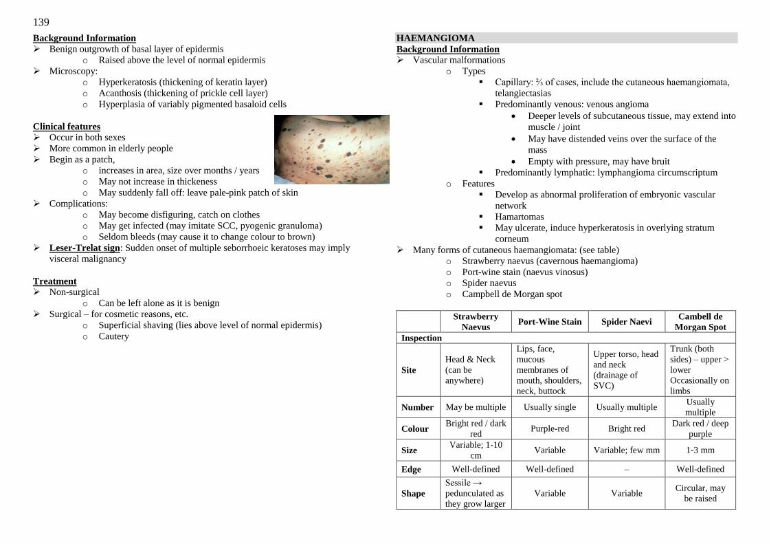
139
Background Information
Benign outgrowth of basal layer of epidermis
o Raised above the level of normal epidermis
Microscopy:
o Hyperkeratosis (thickening of keratin layer)
o Acanthosis (thickening of prickle cell layer)
o Hyperplasia of variably pigmented basaloid cells
Clinical features Occur in both sexes
More common in elderly people
Begin as a patch,
o increases in area, size over months / years
o May not increase in thickeness
o May suddenly fall off: leave pale-pink patch of skin
Complications:
o May become disfiguring, catch on clothes
o May get infected (may imitate SCC, pyogenic granuloma)
o Seldom bleeds (may cause it to change colour to brown)
Leser-Trelat sign: Sudden onset of multiple seborrhoeic keratoses may imply
visceral malignancy
Treatment Non-surgical
o Can be left alone as it is benign
Surgical – for cosmetic reasons, etc.
o Superficial shaving (lies above level of normal epidermis)
o Cautery
HAEMANGIOMA
Background Information Vascular malformations
o Types
Capillary: ⅔ of cases, include the cutaneous haemangiomata,
telangiectasias
Predominantly venous: venous angioma
Deeper levels of subcutaneous tissue, may extend into
muscle / joint
May have distended veins over the surface of the
mass
Empty with pressure, may have bruit
Predominantly lymphatic: lymphangioma circumscriptum
o Features
Develop as abnormal proliferation of embryonic vascular
network
Hamartomas
May ulcerate, induce hyperkeratosis in overlying stratum
corneum
Many forms of cutaneous haemangiomata: (see table)
o Strawberry naevus (cavernous haemangioma)
o Port-wine stain (naevus vinosus)
o Spider naevus
o Campbell de Morgan spot
Strawberry
Naevus Port-Wine Stain Spider Naevi
Cambell de
Morgan Spot
Inspection
Site
Head & Neck
(can be
anywhere)
Lips, face,
mucous
membranes of
mouth, shoulders,
neck, buttock
Upper torso, head
and neck
(drainage of
SVC)
Trunk (both
sides) – upper >
lower
Occasionally on
limbs
Number May be multiple Usually single Usually multiple Usually
multiple
Colour Bright red / dark
red Purple-red Bright red
Dark red / deep
purple
Size Variable; 1-10
cm Variable Variable; few mm 1-3 mm
Edge Well-defined Well-defined – Well-defined
Shape
Sessile →
pedunculated as
they grow larger
Variable Variable Circular, may
be raised
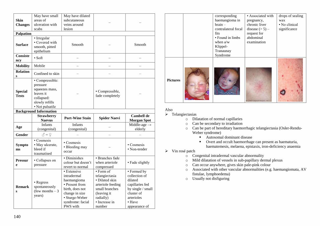
140
Skin
Changes
May have small
areas of
ulceration with
scabs
May have dilated
subcutaneous
veins around
lesion
– –
Palpation
Surface
• Irregular
• Covered with
smooth, pitted
epithelium
Smooth – Smooth
Consiste
ncy • Soft – – –
Mobility Mobile – – –
Relation
s Confined to skin – – –
Special
Tests
• Compressible:
pressure
squeezes mass,
leaves it
collapsed:
slowly refills
• Not pulsatile
– • Compressible,
fade completely –
Background Information
Strawberry
Naevus Port-Wine Stain Spider Naevi
Cambell de
Morgan Spot
Age Infants
(congenital)
Infants
(congenital) –
Middle-age →
elderly
Gender ♂ = ♀ – – –
Sympto
ms
• Cosmesis
• May ulcerate,
bleed if
traumatised
• Cosmesis
• Bleeding may
occur
– • Cosmesis
• Non-tender
Pressur
e
• Collapses on
pressure
• Diminishes
colour but doesn‘t
revert to normal
• Branches fade
when arteriole
compressed
• Fade slightly
Remark
s
• Regress
spontaneously
(few months – 3
years)
• Extensive
intradermal
haemangioma
• Present from
birth, does not
change in size
• Sturge-Weber
syndrome: facial
PWS with
• Form of
telangiectasia
• Dilated skin
arteriole feeding
small branches
(leaving it
radially)
• Increase in
number
• Formed by
collection of
dilated
capillaries fed
by single / small
cluster of
arterioles
• Have
appearance of
corresponding
haemangioma in
brain –
contralateral focal
fits
• Found in limbs
when a/w
Klippel-
Tranaunay
Syndrome
• Associated with
pregnancy,
chronic liver
disease (> 5) –
request for
abdominal
examination
drops of sealing
wax
• No clinical
significance
Pictures
Also
Telangiectasias
o Dilatation of normal capillaries
o Can be secondary to irradiation
o Can be part of hereditary haemorrhagic telangiectasia (Osler-Rendu-
Weber syndrome)
Autosomal dominant disease
Overt and occult haemorrhage can present as haematuria,
haematemesis, melaena, epistaxis, iron-deficiency anaemia
Vin rosé patch
o Congenital intradermal vascular abnormality
o Mild dilatation of vessels in sub-papillary dermal plexus
o Can occur anywhere, gives skin pale-pink colour
o Associated with other vascular abnormalities (e.g. haemangiomata, AV
fistulae, lymphoedema)
o Usually not disfiguring

141
PYOGENIC GRANULOMA
Inspection
Single; usu < 1 cm, bright red
o May be blood-encrusted or Ulceration
Hemispherical; may be sessile / pedunculated
Likely sites to be injured, e.g. hands, face
Bright red; long-standing lesions may be skin-coloured
May have sinuses, associated serous / purulent discharge, Erythema / cellulitis
Palpation – request to palpate: may bleed easily
May be slightly tender
o May bleed easily on palpation
Well-defined edges
Soft, fleshy consistency
Confined to skin
Slightly compressible (vascular origin)
Request
Take history for previous injury
Rate of growth of lump? (rapid growth in few days)
Background Information – Neither pyogenic nor a granuloma!
Rapidly-growing capillary haemangioma, usually less then 1 cm
Occur commonly after injury:
o Small capillary loops develop in healing wound, form granulation
tissue
o When capillary loops grow too vigorously, form protruding mass,
epithelisation
o Mass form called pyogenic granuloma (surface often ulcerated,
infected)
Clinical features Uncommon in children
May have history of minor injury, chronic infection (e.g. paronychia)
Rapidly-growing lump on skin,
Bleeds easily, discharges serous / purulent fluid
o Bleeding, pain stops once lump epithelises
Once nodule is completely covered, begins to shrink (rarely disappears completely)
Treatment Surgical
o Curettage with diathermy of the base
o Complete excision biopsy
(if recurrent; malignancy e.g. amelanotic melanoma has to be
excluded)
Non-surgical
o Regression is uncommon: surgical treatment best option
o Silver nitrate cautery is possible
“Mr. X is a young Chinese gentleman…” “On inspection, there is a single hemispherical lump over the thenar eminence of
the right hand”
“It measures about 0.8 cm in diameter, with clearly-defined margins” “It is bright red in colour, with surrouding erythema”
“There are no scars, sinuses, ulceration, active bleeding or discharge seen” “I would like to proceed on to palpation”
“On palpation, it is not warm. It is slightly tender”
“The surface is smooth. The consistency is soft and fleshy”
“The lump is confined to the skin”
“My provisional diagnosis is pyogenic granuloma”
“My differential diagnoses are SCC, non-pigmented melanma”
“I would like to complete my examination by…”
o ―Asking Mr. X for any previous injuries to the hand‖
o ―Asking him how rapidly the lump has been growing‖
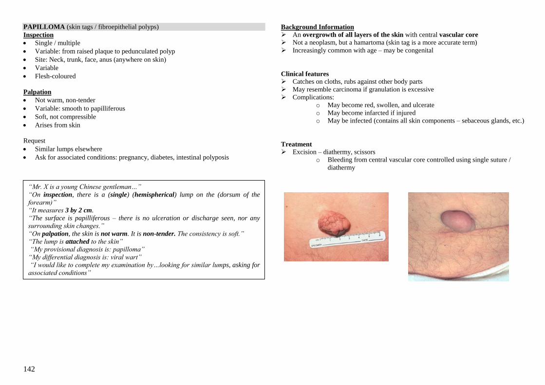
142
PAPILLOMA (skin tags / fibroepithelial polyps)
Inspection
Single / multiple
Variable: from raised plaque to pedunculated polyp
Site: Neck, trunk, face, anus (anywhere on skin)
Variable
Flesh-coloured
Palpation
Not warm, non-tender
Variable: smooth to papilliferous
Soft, not compressible
Arises from skin
Request
Similar lumps elsewhere
Ask for associated conditions: pregnancy, diabetes, intestinal polyposis
Background Information
An overgrowth of all layers of the skin with central vascular core
Not a neoplasm, but a hamartoma (skin tag is a more accurate term)
Increasingly common with age – may be congenital
Clinical features Catches on cloths, rubs against other body parts
May resemble carcinoma if granulation is excessive
Complications:
o May become red, swollen, and ulcerate
o May become infarcted if injured
o May be infected (contains all skin components – sebaceous glands, etc.)
Treatment Excision – diathermy, scissors
o Bleeding from central vascular core controlled using single suture /
diathermy
“Mr. X is a young Chinese gentleman…”
“On inspection, there is a (single) (hemispherical) lump on the (dorsum of the
forearm)” “It measures 3 by 2 cm.
“The surface is papilliferous – there is no ulceration or discharge seen, nor any surrounding skin changes.”
“On palpation, the skin is not warm. It is non-tender. The consistency is soft.”
“The lump is attached to the skin”
“My provisional diagnosis is: papilloma”
“My differential diagnosis is: viral wart”
“I would like to complete my examination by…looking for similar lumps, asking for
associated conditions”
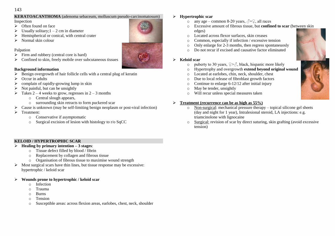
143
KERATOACANTHOMA (adenoma sebaceum, molluscum pseudo-carcinomatosum)
Inspection
Often found on face
Usually solitary;1 – 2 cm in diameter
Hemispherical or conical, with central crater
Normal skin colour
Palpation
Firm and rubbery (central core is hard)
Confined to skin, freely mobile over subcutaneous tissues
Background information Benign overgrowth of hair follicle cells with a central plug of keratin
Occur in adults
complain of rapidly-growing lump in skin
Not painful, but can be unsightly
Takes 2 – 4 weeks to grow, regresses in 2 – 3 months
o Central slough appears,
o surrounding skin retracts to form puckered scar
Cause is unknown (may be self-limiting benign neoplasm or post-viral infection)
Treatment:
o Conservative if asymptomatic
o Surgical excision of lesion with histology to r/o SqCC
KELOID / HYPERTROPHIC SCAR
Healing by primary intention – 3 stages:
o Tissue defect filled by blood / fibrin
o Replacement by collagen and fibrous tissue
o Organisation of fibrous tissue to maximise wound strength
Most surgical scars have thin lines, but tissue response may be excessive:
hypertrophic / keloid scar
Wounds prone to hypertrophic / keloid scar
o Infection
o Trauma
o Burns
o Tension
o Susceptible areas: across flexion areas, earlobes, chest, neck, shoulder
Hypertrophic scar o any age – common 8-20 years, ♂=♀, all races
o Excessive amount of fibrous tissue, but confined to scar (between skin
edges)
o Located across flexor surfaces, skin creases
o Common, especially if infection / excessive tension
o Only enlarge for 2-3 months, then regress spontaneuosly
o Do not recur if excised and causative factor eliminated
Keloid scar o puberty to 30 years, ♀>♂, black, hispanic more likely
o Hypertrophy and overgrowth extend beyond original wound
o Located at earlobes, chin, neck, shoulder, chest
o Due to local release of fibroblast growth factors
o Continue to enlarge 6-12/12 after initial injury
o May be tender, unsightly
o Will recur unless special measures taken
Treatment (recurrence can be as high as 55%)
o Non-surgical: mechanical pressure therapy – topical silicone gel sheets
(day and night for 1 year), Intralesional steroid, LA injections: e.g.
triamcinolone with lignocaine
o Surgical: revision of scar by direct suturing, skin grafting (avoid excessive
tension)
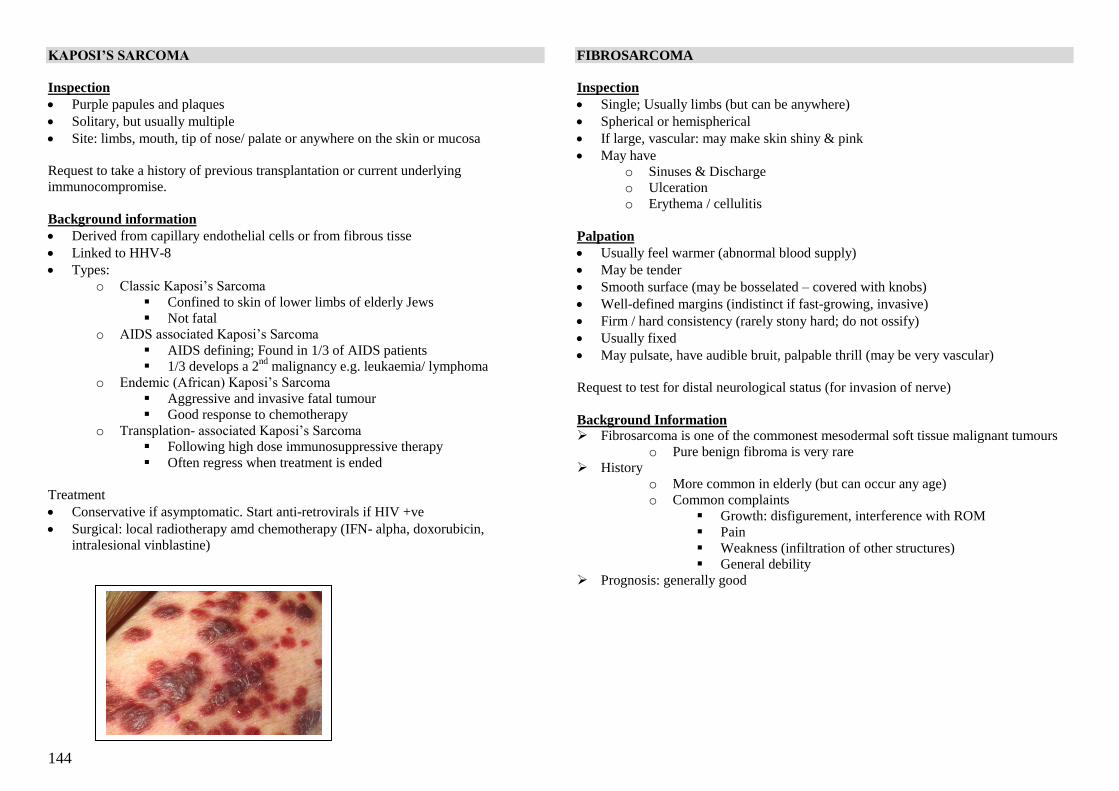
144
KAPOSI’S SARCOMA
Inspection
Purple papules and plaques
Solitary, but usually multiple
Site: limbs, mouth, tip of nose/ palate or anywhere on the skin or mucosa
Request to take a history of previous transplantation or current underlying
immunocompromise.
Background information
Derived from capillary endothelial cells or from fibrous tisse
Linked to HHV-8
Types:
o Classic Kaposi‘s Sarcoma
Confined to skin of lower limbs of elderly Jews
Not fatal
o AIDS associated Kaposi‘s Sarcoma
AIDS defining; Found in 1/3 of AIDS patients
1/3 develops a 2nd
malignancy e.g. leukaemia/ lymphoma
o Endemic (African) Kaposi‘s Sarcoma
Aggressive and invasive fatal tumour
Good response to chemotherapy
o Transplation- associated Kaposi‘s Sarcoma
Following high dose immunosuppressive therapy
Often regress when treatment is ended
Treatment
Conservative if asymptomatic. Start anti-retrovirals if HIV +ve
Surgical: local radiotherapy amd chemotherapy (IFN- alpha, doxorubicin,
intralesional vinblastine)
FIBROSARCOMA
Inspection
Single; Usually limbs (but can be anywhere)
Spherical or hemispherical
If large, vascular: may make skin shiny & pink
May have
o Sinuses & Discharge
o Ulceration
o Erythema / cellulitis
Palpation
Usually feel warmer (abnormal blood supply)
May be tender
Smooth surface (may be bosselated – covered with knobs)
Well-defined margins (indistinct if fast-growing, invasive)
Firm / hard consistency (rarely stony hard; do not ossify)
Usually fixed
May pulsate, have audible bruit, palpable thrill (may be very vascular)
Request to test for distal neurological status (for invasion of nerve)
Background Information Fibrosarcoma is one of the commonest mesodermal soft tissue malignant tumours
o Pure benign fibroma is very rare
History
o More common in elderly (but can occur any age)
o Common complaints
Growth: disfigurement, interference with ROM
Pain
Weakness (infiltration of other structures)
General debility
Prognosis: generally good

145
PYODERMA GANGRENOSUM
Inspect
Ulcer with a necrotis base
Irregular bluish red overhanging edges
a/w surrounding erythematous plaques with pustules
Request to examine for evidence of inflammatory bowel disease, RA
Backgound information
more common in males
pyoderma gangrenosum is associated with:
o IBD
o RA
o Myeloproliferative disorders: PRV, myeloma
o Autoimmune hepatitis
Differential diagnosis:
o Autoimmune: rheumatoid vasculitis
o Infectious: tertiary syphilis, amoebiasis
o Iatrogenic: warfarin necrosis
o Others: Behcet‘s disease
Treatment:
o Non-surgical: treat underlying condition, saline cleansing, high dose oral or
intralesional steroids. KIV cyclosporine & antibiotics
o Surgical: serial allograft followed by autologous skin graft or muscle flap
coverage when necessary
RADIOTHERAPY MARKS
Vital points on examination:
Of the underlying disease:
o Cachexia,
o masectomy scar/ wide excision scar suggest breast cancer
o obvious skin cancer,
o clubbing & other signs of chest disease suggest lung cancer
o suprapubic mass suggest pelvic tumour
o neck swellings with cranial nerve palsies head and neck tumour
of the radiotherapy:
o site of radiation
o shape: usually well defined borders
o features of active RT:
Indian ink marks, skin markings
Erythema, desquamation
o Features of previous RT:
Telangiectasia, hyperpigmentation
Complications of radiotherapy:
o Depends of site
o Look for future cancers:
Haematogenous malignancy
Thyroid cancers
Breast cancers
Background information
High energy X-rays interact with tissue to release electrons that cause local
damage to DNA in adjacent cells via oxygen dependent mechanism.
o Damage is usually irreparable, and normal cells have greater ability to
repopulate than tumour cells in this setting
o If reparable, manifests as chromosomal abnormalities
Radiotherapy affects cells with:
o Rapid turnover: Skin (epidermal layers), small intestine, bone marrow
stem cells
o Limited replicative ability: spinal cord, gonads
Complications:
o Early:
General: malaise, fatigue, LOA, N/V
Skin changes & temporary hair loss
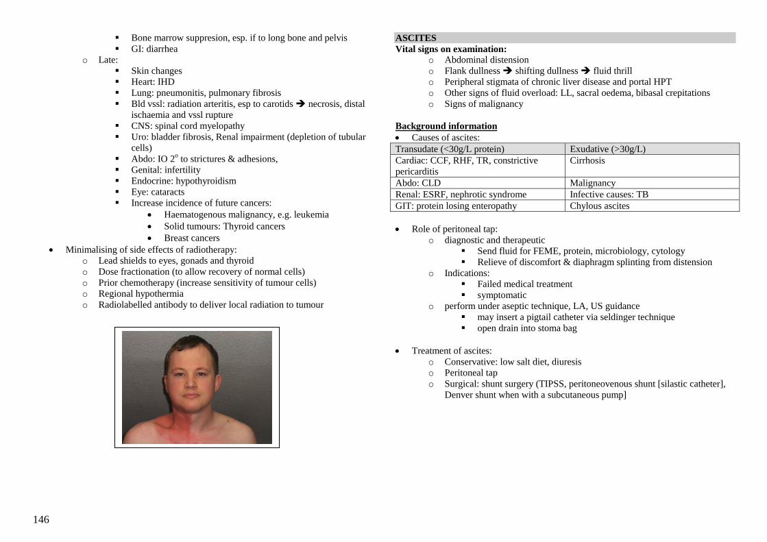
146
Bone marrow suppresion, esp. if to long bone and pelvis
GI: diarrhea
o Late:
Skin changes
Heart: IHD
Lung: pneumonitis, pulmonary fibrosis
Bld vssl: radiation arteritis, esp to carotids necrosis, distal
ischaemia and vssl rupture
CNS: spinal cord myelopathy
Uro: bladder fibrosis, Renal impairment (depletion of tubular
cells)
Abdo: IO 2o to strictures & adhesions,
Genital: infertility
Endocrine: hypothyroidism
Eye: cataracts
Increase incidence of future cancers:
Haematogenous malignancy, e.g. leukemia
Solid tumours: Thyroid cancers
Breast cancers
Minimalising of side effects of radiotherapy:
o Lead shields to eyes, gonads and thyroid
o Dose fractionation (to allow recovery of normal cells)
o Prior chemotherapy (increase sensitivity of tumour cells)
o Regional hypothermia
o Radiolabelled antibody to deliver local radiation to tumour
ASCITES
Vital signs on examination: o Abdominal distension
o Flank dullness shifting dullness fluid thrill
o Peripheral stigmata of chronic liver disease and portal HPT
o Other signs of fluid overload: LL, sacral oedema, bibasal crepitations
o Signs of malignancy
Background information
Causes of ascites:
Transudate (<30g/L protein) Exudative (>30g/L)
Cardiac: CCF, RHF, TR, constrictive
pericarditis
Cirrhosis
Abdo: CLD Malignancy
Renal: ESRF, nephrotic syndrome Infective causes: TB
GIT: protein losing enteropathy Chylous ascites
Role of peritoneal tap:
o diagnostic and therapeutic
Send fluid for FEME, protein, microbiology, cytology
Relieve of discomfort & diaphragm splinting from distension
o Indications:
Failed medical treatment
symptomatic
o perform under aseptic technique, LA, US guidance
may insert a pigtail catheter via seldinger technique
open drain into stoma bag
Treatment of ascites:
o Conservative: low salt diet, diuresis
o Peritoneal tap
o Surgical: shunt surgery (TIPSS, peritoneovenous shunt [silastic catheter],
Denver shunt when with a subcutaneous pump]
147
SHOCK
Definition – inadequate tissue and organ perfusion leading to a hypoperfusion state &
eventual cellular hypoxia and its attendant sequelae.
S/S: Hypotension, urine output, tachycardia, diaphoresis, AMS
Types of Shock
‗White‘ shock ‗Red‘ shock
Types Hypovolaemic Cardiogenic Neurogenic Septic Anaphylactic
Causes Haemorrhage
Burns
Ruptured
ectopic
pregnancy
Severe GE
Acute
pancreatitis
AMI
Dysrhythmia
Spinal
injury
Infxns
S/S Pallor
Cold clammy
skin
peri vas
Pallor
Cold clammy
skin
peri vas
Warm skin
N/ heart
rate
Neuro
deficit
Fever,
rigors
Warm
skin
Fever, rigors
Warm skin
Invxs Hct (late) Cardiac
enzymes
ECG
FBC
Bld C/S
Also, Obstructive Shock due to tension pneumothorax, cardiac tamponade or pulmonary
embolism
Management
General Mx
Airway Maintain airway – consider intubation if necessary
Breathing 100% O2 via non-rebreather mask
Circulation 2 large bore (14-16G) cannulae
Inotropic support
o IV dopamine 5-10g/kg/min
o IV dobutamine 5-10g/kg/min (esp for cardiogenic
shock)
o IV norepinephrine 5-20g/kg/min (esp for septic
shock)
Monitoring Pulse oximetry
ECG
BP
Heart rate
Urine output – catheterize patient
Hypovolaemic Shock
Invxs FBC - Hct in acute alcoholic binge due to diuresis. Hct is an
Inaccurate marker of bld loss acutely.
GXM 6 units U/E/Cr
Troponin T & Cardiac enzymes
Coagulation profile with DIVC screen (PT/PTT, pltlet, D-
dimer)
ABG – metab acidosis, lactate, base deficits are poor Px
factors
UPT - ?ectopic pregnancy? Ask for LMP
Examine abdomen for pulsatile AAA
Fluid Rx 1 L crystalloid fast infusion w/in 1 hr
Assess response
Subsequent colloid or whole blood infusion
CVP line Used to guide fluid Rx, esp in CCF patients
Cardiogenic Shock
ECG
Trop T & cardiac
enzymes
Manage accordingly – refer acute coronary
syndrome & ACLS notes
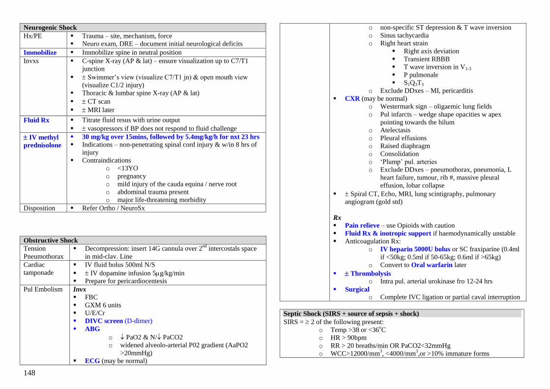
148
Neurogenic Shock
Hx/PE Trauma – site, mechanism, force
Neuro exam, DRE – document initial neurological deficits
Immobilize Immobilize spine in neutral position
Invxs C-spine X-ray (AP & lat) – ensure visualization up to C7/T1
junction
Swimmer‘s view (visualize C7/T1 jn) & open mouth view
(visualize C1/2 injury)
Thoracic & lumbar spine X-ray (AP & lat)
CT scan
MRI later
Fluid Rx Titrate fluid resus with urine output
vasopressors if BP does not respond to fluid challenge
IV methyl
prednisolone
30 mg/kg over 15mins, followed by 5.4mg/kg/h for nxt 23 hrs
Indications – non-penetrating spinal cord injury & w/in 8 hrs of
injury
Contraindications
o <13YO
o pregnancy
o mild injury of the cauda equina / nerve root
o abdominal trauma present
o major life-threatening morbidity
Disposition Refer Ortho / NeuroSx
Obstructive Shock
Tension
Pneumothorax
Decompression: insert 14G cannula over 2nd
intercostals space
in mid-clav. Line
Cardiac
tamponade
IV fluid bolus 500ml N/S
IV dopamine infusion 5g/kg/min
Prepare for pericardiocentesis
Pul Embolism Invx
FBC
GXM 6 units
U/E/Cr
DIVC screen (D-dimer)
ABG
o PaO2 & N/ PaCO2
o widened alveolo-arterial P02 gradient (AaPO2
>20mmHg)
ECG (may be normal)
o non-specific ST depression & T wave inversion
o Sinus tachycardia
o Right heart strain
Right axis deviation
Transient RBBB
T wave inversion in V1-3
P pulmonale
S1Q3T3
o Exclude DDxes – MI, pericarditis
CXR (may be normal)
o Westermark sign – oligaemic lung fields
o Pul infarcts – wedge shape opacities w apex
pointing towards the hilum
o Atelectasis
o Pleural effusions
o Raised diaphragm
o Consolidation
o ‗Plump‘ pul. arteries
o Exclude DDxes – pneumothorax, pneumonia, L
heart failure, tumour, rib #, massive pleural
effusion, lobar collapse
Spiral CT, Echo, MRI, lung scintigraphy, pulmonary
angiogram (gold std)
Rx
Pain relieve – use Opioids with caution
Fluid Rx & inotropic support if haemodynamically unstable
Anticoagulation Rx:
o IV heparin 5000U bolus or SC fraxiparine (0.4ml
if <50kg; 0.5ml if 50-65kg; 0.6ml if >65kg)
o Convert to Oral warfarin later
Thrombolysis o Intra pul. arterial urokinase fro 12-24 hrs
Surgical o Complete IVC ligation or partial caval interruption
Septic Shock (SIRS + source of sepsis + shock)
SIRS = 2 of the following present:
o Temp >38 or <36oC
o HR > 90bpm
o RR > 20 breaths/min OR PaCO2<32mmHg
o WCC>12000/mm3, <4000/mm
3,or >10% immature forms

149
Hx / PE Identify site of infxn – UTI (indwelling cathether), gallbladder dz,
peritonitis, pneumonia, appendicitis, immunocompromised state
Invx FBC - TW
U/E/Cr
DIVC screen – PT/PTT, pltlet, fibrinogen, D-dimer
Bld C/S (2 different sites)
Capillary bld glucose ABG
CXR – pneumonia, ARDS
ECG
Urine dipstick – UTI
Urine C/S
Fluid Rx Rapid infusion 1-2L crystalloids
CVP line insertion
Inotropic
support
if no response to fluid Rx
Noradrenaline (drug of choice) - 1g/kg/min OR
Dopamin 5-20g/kg/min
Empirical
ABx
Immunocompetent w/o
obvious source
3rd
gen cephalosporin (IV
ceftriaxone 1g) OR
Quinolones (ciprofloxacin
200mg)
Immunocompromised w/o
obvious source
Anti-pseudomonal ABx (IV
ceftazidime 1g) OR
Quinolone
PLUS aminoglycoside
(Gentamicin 80mg)
Gram-positive (burns, FB /
lines present) IV cefazolin 2g IV vancomycin 1g if hx of
IVDA, indwelling cath. Or
penicillin allergy
Anaerobic source (intra-
abdo, biliary, female genital
tract, aspiration pneumonia)
IV metronidazole 500mg +
ceftriazone 1g + IV gentamicin
80mg
Anaphylactic Shock Definitions
Urticaria – oedematous & pruritic plaques w pale centre & raised edges
Angioedema – oedema of deeper layers of the skin. Non-pruritic. May be a/w
numbness & pain
Anaphylaxis – severe systemic allergic rxn to an Ag. Ppt by abrupt release of
chemical mediators in a previously sensitized patient
Anaphylactoid rxn – resembles anaphylactic rxn, but due to direct histamine
release from mast cells w/o need for prior sensitization
Common causes
Drugs – penicililns & NSAIDS commonest, aspirin, TCM, sulpha drugs
Food – shellfish, egg white, peanuts
Venoms – bees, wasps, hornets
Environment – dust, pollen
Infections – EBV, HBV, coxsackie virus, parasites
Stop Pptant Stop administration of suspected agent / flick out insect stinger
with tongue blade
Gastric lavage & activated charcoal if drug was ingested
Airway Prepare for intubation or cricothyroidectomy – ENT/Anaesthesia
consult
Fluid Rx 2L Hartman‘s or N/S bolus
Drug Rx Adrenaline Normotensive – 0.01ml/kg (max 0.5ml)
1:1000 dilution SC/IM Hypotensive – 0.1ml/kg (max 5ml) 1:10,000
dilution IV over 5 mins
Glucagon Indications: failure of adrenaline Rx OR if
adrenaline is contraindicated eg IHD, severe
HPT, pregnancy, -blocker use
0.5-1.0mg IV/IM. Can be repeated once after
30mins
Antihistamines Diphenhydramine 25mg IM/IV
Chlorpheniramine 10mg IM/IV
Promethazine 25mg IM/IV
Cimetidine For persistent symptoms unresponsive to
above Rx
200-400mg IV bolus
Nebulised
bronchodilator
for persistent bronchospasm
Salbutamol 2:2 q20-30mins
Corticosteroids Hydrocortisone 200-300mg IV bolus, q 6hr
DGIM – Last updated March 2005

150
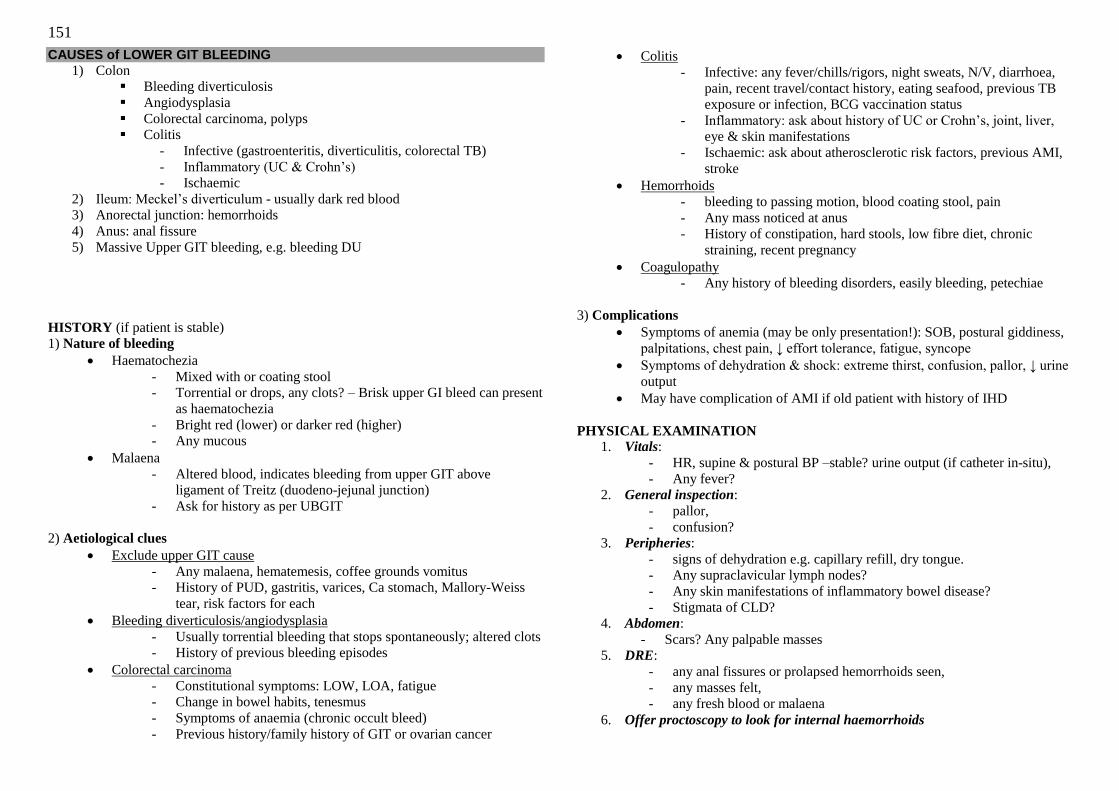
151
CAUSES of LOWER GIT BLEEDING
1) Colon
Bleeding diverticulosis
Angiodysplasia
Colorectal carcinoma, polyps
Colitis
- Infective (gastroenteritis, diverticulitis, colorectal TB)
- Inflammatory (UC & Crohn‘s)
- Ischaemic
2) Ileum: Meckel‘s diverticulum - usually dark red blood
3) Anorectal junction: hemorrhoids
4) Anus: anal fissure
5) Massive Upper GIT bleeding, e.g. bleeding DU
HISTORY (if patient is stable)
1) Nature of bleeding
Haematochezia
- Mixed with or coating stool
- Torrential or drops, any clots? – Brisk upper GI bleed can present
as haematochezia
- Bright red (lower) or darker red (higher)
- Any mucous
Malaena
- Altered blood, indicates bleeding from upper GIT above
ligament of Treitz (duodeno-jejunal junction)
- Ask for history as per UBGIT
2) Aetiological clues
Exclude upper GIT cause
- Any malaena, hematemesis, coffee grounds vomitus
- History of PUD, gastritis, varices, Ca stomach, Mallory-Weiss
tear, risk factors for each
Bleeding diverticulosis/angiodysplasia
- Usually torrential bleeding that stops spontaneously; altered clots
- History of previous bleeding episodes
Colorectal carcinoma
- Constitutional symptoms: LOW, LOA, fatigue
- Change in bowel habits, tenesmus
- Symptoms of anaemia (chronic occult bleed)
- Previous history/family history of GIT or ovarian cancer
Colitis
- Infective: any fever/chills/rigors, night sweats, N/V, diarrhoea,
pain, recent travel/contact history, eating seafood, previous TB
exposure or infection, BCG vaccination status
- Inflammatory: ask about history of UC or Crohn‘s, joint, liver,
eye & skin manifestations
- Ischaemic: ask about atherosclerotic risk factors, previous AMI,
stroke
Hemorrhoids
- bleeding to passing motion, blood coating stool, pain
- Any mass noticed at anus
- History of constipation, hard stools, low fibre diet, chronic
straining, recent pregnancy
Coagulopathy
- Any history of bleeding disorders, easily bleeding, petechiae
3) Complications
Symptoms of anemia (may be only presentation!): SOB, postural giddiness,
palpitations, chest pain, ↓ effort tolerance, fatigue, syncope
Symptoms of dehydration & shock: extreme thirst, confusion, pallor, ↓ urine
output
May have complication of AMI if old patient with history of IHD
PHYSICAL EXAMINATION 1. Vitals:
- HR, supine & postural BP –stable? urine output (if catheter in-situ),
- Any fever?
2. General inspection:
- pallor,
- confusion?
3. Peripheries:
- signs of dehydration e.g. capillary refill, dry tongue.
- Any supraclavicular lymph nodes?
- Any skin manifestations of inflammatory bowel disease?
- Stigmata of CLD?
4. Abdomen:
- Scars? Any palpable masses 5. DRE:
- any anal fissures or prolapsed hemorrhoids seen,
- any masses felt,
- any fresh blood or malaena
6. Offer proctoscopy to look for internal haemorrhoids
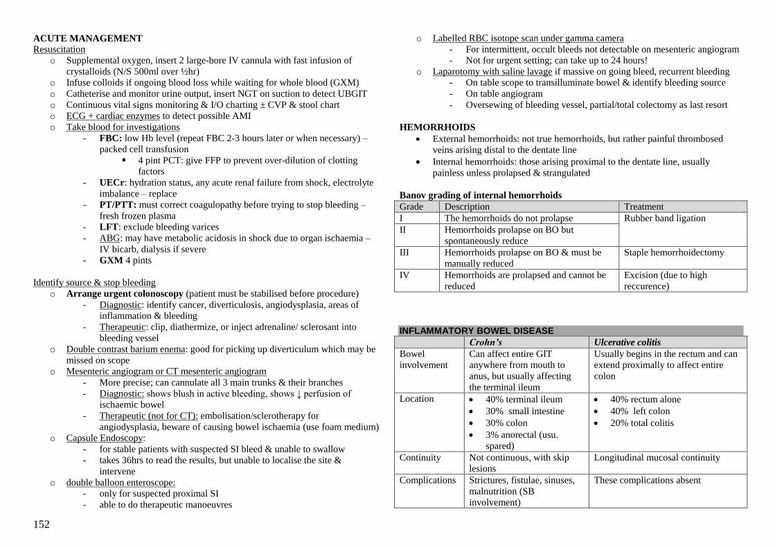
152
ACUTE MANAGEMENT
Resuscitation
o Supplemental oxygen, insert 2 large-bore IV cannula with fast infusion of
crystalloids (N/S 500ml over ½hr)
o Infuse colloids if ongoing blood loss while waiting for whole blood (GXM)
o Catheterise and monitor urine output, insert NGT on suction to detect UBGIT
o Continuous vital signs monitoring & I/O charting ± CVP & stool chart
o ECG + cardiac enzymes to detect possible AMI
o Take blood for investigations
- FBC: low Hb level (repeat FBC 2-3 hours later or when necessary) –
packed cell transfusion
4 pint PCT: give FFP to prevent over-dilution of clotting
factors
- UECr: hydration status, any acute renal failure from shock, electrolyte
imbalance – replace
- PT/PTT: must correct coagulopathy before trying to stop bleeding –
fresh frozen plasma
- LFT: exclude bleeding varices
- ABG: may have metabolic acidosis in shock due to organ ischaemia –
IV bicarb, dialysis if severe
- GXM 4 pints
Identify source & stop bleeding
o Arrange urgent colonoscopy (patient must be stabilised before procedure)
- Diagnostic: identify cancer, diverticulosis, angiodysplasia, areas of
inflammation & bleeding
- Therapeutic: clip, diathermize, or inject adrenaline/ sclerosant into
bleeding vessel
o Double contrast barium enema: good for picking up diverticulum which may be
missed on scope
o Mesenteric angiogram or CT mesenteric angiogram
- More precise; can cannulate all 3 main trunks & their branches
- Diagnostic: shows blush in active bleeding, shows ↓ perfusion of
ischaemic bowel
- Therapeutic (not for CT): embolisation/sclerotherapy for
angiodysplasia, beware of causing bowel ischaemia (use foam medium)
o Capsule Endoscopy:
- for stable patients with suspected SI bleed & unable to swallow
- takes 36hrs to read the results, but unable to localise the site &
intervene
o double balloon enteroscope:
- only for suspected proximal SI
- able to do therapeutic manoeuvres
o Labelled RBC isotope scan under gamma camera
- For intermittent, occult bleeds not detectable on mesenteric angiogram
- Not for urgent setting; can take up to 24 hours!
o Laparotomy with saline lavage if massive on going bleed, recurrent bleeding
- On table scope to transilluminate bowel & identify bleeding source
- On table angiogram
- Oversewing of bleeding vessel, partial/total colectomy as last resort
HEMORRHOIDS
External hemorrhoids: not true hemorrhoids, but rather painful thrombosed
veins arising distal to the dentate line
Internal hemorrhoids: those arising proximal to the dentate line, usually
painless unless prolapsed & strangulated
Banov grading of internal hemorrhoids
Grade Description Treatment
I The hemorrhoids do not prolapse Rubber band ligation
II Hemorrhoids prolapse on BO but
spontaneously reduce
III Hemorrhoids prolapse on BO & must be
manually reduced
Staple hemorrhoidectomy
IV Hemorrhoids are prolapsed and cannot be
reduced
Excision (due to high
reccurence)
INFLAMMATORY BOWEL DISEASE
Crohn’s Ulcerative colitis
Bowel
involvement
Can affect entire GIT
anywhere from mouth to
anus, but usually affecting
the terminal ileum
Usually begins in the rectum and can
extend proximally to affect entire
colon
Location 40% terminal ileum
30% small intestine
30% colon
3% anorectal (usu.
spared)
40% rectum alone
40% left colon
20% total colitis
Continuity Not continuous, with skip
lesions
Longitudinal mucosal continuity
Complications Strictures, fistulae, sinuses,
malnutrition (SB
involvement)
These complications absent

153
Histopathology Macoscopic: bowel is thick-
walled & nodular
(cobblestone appearance)
with creeping fat, mesenteric
thickening & deep linear
ulcers
Microscopic: transmural
involvement, non-caseating
granulomas (pathognomonic,
but only present in 2/3)
Macroscopic: granular appearance of
mucosa , loss of vascular markings,
pseudopolyps interspersed with areas
of shallow ulceration
Microscopic: only mucosal &
submucosal involvement, crypt
abscesses
- Muscularis propria & serosa may be
affected in fulminant disease
Risk of
carcinoma
Slight increased risk of
colorectal Ca, increased risk
of small bowel lymphoma
Substantially higher risk of colorectal
Ca
2% at 10 years
8% at 20 years
18% at 30 years
Much higher risk with
concomitant PSC
Associated
antibodies
ASCA: anti-saccharomyces
cerevisiae antibodies
p-ANCA: perinuclear antineutrophil
cytoplastic antibodies
Severity Harvey Bradshaw severity
index
Modified Truelove & Witts severity
index
(Mild, moderate, severe, fulminant)
Epidemiology
Bimodal distribution: affects young in the 2nd
& 3rd
decades of life, with second
onset in the 5th & 6
th decades of life
Equal gender predominance
Higher prevalence amongst Ashkenazi Jews & in cooler climates e.g.
Scandinavia, UK, Germany, northern USA
Genetic association
History
GIT: abdominal pain, diarrhea, N/V, bloating, distention, bloody stools with
mucous & pus
Crohn‘s fistula: colovesical fistula or coloovarian fistula: faeces in urine/
pneumaturia, faeces per vaginal/ PID
Non-specific systemic: LOW, LOA, fever, fatigue, symptoms of anemia,
chronic malnutrition
Specific extra-intestinal manifestations
Physical examination: usually normal +/- extra-intestinal manifestations
CROHN’S DISEASE
P/E: RIF mass, enlarged skin tags, anal stricture
Investigations: supportive (see UC) & diagnostic
Diagnostic
Contrast radiographic studies: assess location & extent of disease, look for
strictures & fistulae
o Barium studies: small bowel series & enema
o CT scan with oral & IV contrast
Endoscopy: look for typical features
o Colonosopy with tissue biopsy (non-caseating granulomas)
o OGD: upper GIT involvement
o Endoanal U/S: identify fistula tracts
Management
Medical (principle treatment)
o Nutritional support e.g. TPN (may also aid closure of fistulae)
o Pharmacological
Corticosteroids for acute exacerbation of disease, but not for long
term use
1st line: sulfasalazine, mesalazine (5-ASA or 5-aminosalicylic acid)
induce & maintain remission of disease: oral, rectal suppository or
enema (up to splenic flexure)
2nd
line: immunosuppressive agents e.g. azathioprine, 6-MP, MTX
& cyclosporine when 1st line agents ineffective
Biologics: Remicade (infliximab), a TNF blocker
- Require close surveillance for SE (blood disorders,
infections, lymphoma & solid tissue cancers, hepatotoxicity,
drug-induced lupus, demyelinating disorders)
Differential diagnoses
1) Gastroenteritis: exclude with stool microscopy & culture
- Bacterial: E. coli, Campylobacter, Salmonella, Shigella, C. difficile
- Parasitic: E. histolytica
- Viral: rotavirus
2) Other infective causes: diverticulitis, colorectal TB
3) Ischaemic colitis
4) Bleeding diverticulosis
5) Colorectal carcinoma
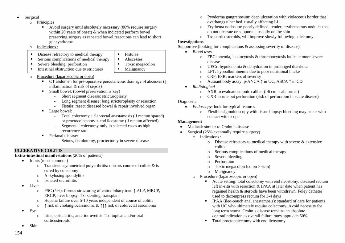
154
Surgical
o Principles
Avoid surgery until absolutely necessary (80% require surgery
within 20 years of onset) & when indicated perform bowel
preserving surgery as repeated bowel resections can lead to short
gut syndrome
o Indications :
o Procedure (laparoscopic or open)
CT abdomen for pre-operative percutaneous drainage of abcesses (↓
inflammation & risk of sepsis)
Small bowel: (bowel preservation is key)
- Short segment disease: stricturoplasty
- Long segment disease: long stricturoplasty or resection
- Fistula: resect diseased bowel & repair involved organ
Large bowel:
- Total colectomy + ileorectal anastamosis (if rectum spared)
or proctocolectomy + end ileostomy (if rectum affected)
- Segmental colectomy only in selected cases as high
recurrence rate
Perianal disease:
- Setons, fistulotomy, proctectomy in severe disease
ULCERATIVE COLITIS
Extra-intestinal manifestations (20% of patients)
Joints (most common)
o Transient asymmetrical polyarthritis: mirrors course of colitis & is
cured by colectomy
o Ankylosing spondylitis
o Isolated sacroiliitis
Liver
o PSC (5%): fibrous structuring of entire biliary tree: ↑ ALP, MRCP,
ERCP, liver biopsy. Tx: stenting, transplant
o Hepatic failure over 5-10 years independent of course of colitis
o ↑ risk of cholangiocarcinoma & ↑↑↑ risk of colorectal carcinoma
Eye
o Iritis, episcleritis, anterior uvetitis. Tx: topical and/or oral
corticosteroids
Skin
o Pyoderma gangrenosum: deep ulceration with violaceous border that
overhangs ulcer bed, usually affecting LL
o Erythema nodosum: poorly defined, tender, erythematous nodules that
do not ulcerate or suppurate, usually on the shin
o Tx: corticosteroids, will improve slowly following colectomy
Investigations
Supportive (looking for complications & assessing severity of disease)
Blood tests
o FBC: anemia, leukocytosis & thrombocytosis indicate more severe
disease
o UECr: hypokalemia & dehydration in prolonged diarrhoea
o LFT: hypoalbuminemia due to poor nutritional intake
o CRP, ESR: markers of severity
o Autoantibody assay: p-ANCA ↑ in UC, ASCA ↑ in CD
Radiological
o AXR to evaluate colonic caliber (>6 cm is abnormal)
o CXR to rule out perforation (risk of perforation in acute disease)
Diagnostic
Endoscopy: look for typical features
o Flexible sigmoidoscopy with tissue biopsy: bleeding may occur with
contact with scope
Management
Medical: similar to Crohn‘s disease
Surgical (25% eventually require surgery)
o Indications :
o Disease refractory to medical therapy with severe & extensive
colitis
o Serious complications of medical therapy
o Severe bleeding
o Perforation
o Toxic megacolon (colon > 6cm)
o Malignancy
o Procedure (laparoscopic or open)
Acute setting: total colectomy with end ileostomy: diseased rectum
left in-situ with resection & IPAA at later date when patient has
regained health & steroids have been withdrawn. Foley catheter
used to decompress rectum for 3-4 days IPAA (ileo-pouch anal anastamosis): standard of care for patients
with UC who ultimately require colectomy. Avoid necessity for
long term stoma. Crohn‘s disease remains an absolute
contradindication as overall failure rates approach 50%
Total proctocolectomy with end ileostomy
Disease refractory to medical therapy
Serious complications of medical therapy
Severe bleeding, perforation
Intestinal obstruction due to strictures
Fistulae
Abscesses
Toxic megacolon
Malignancy
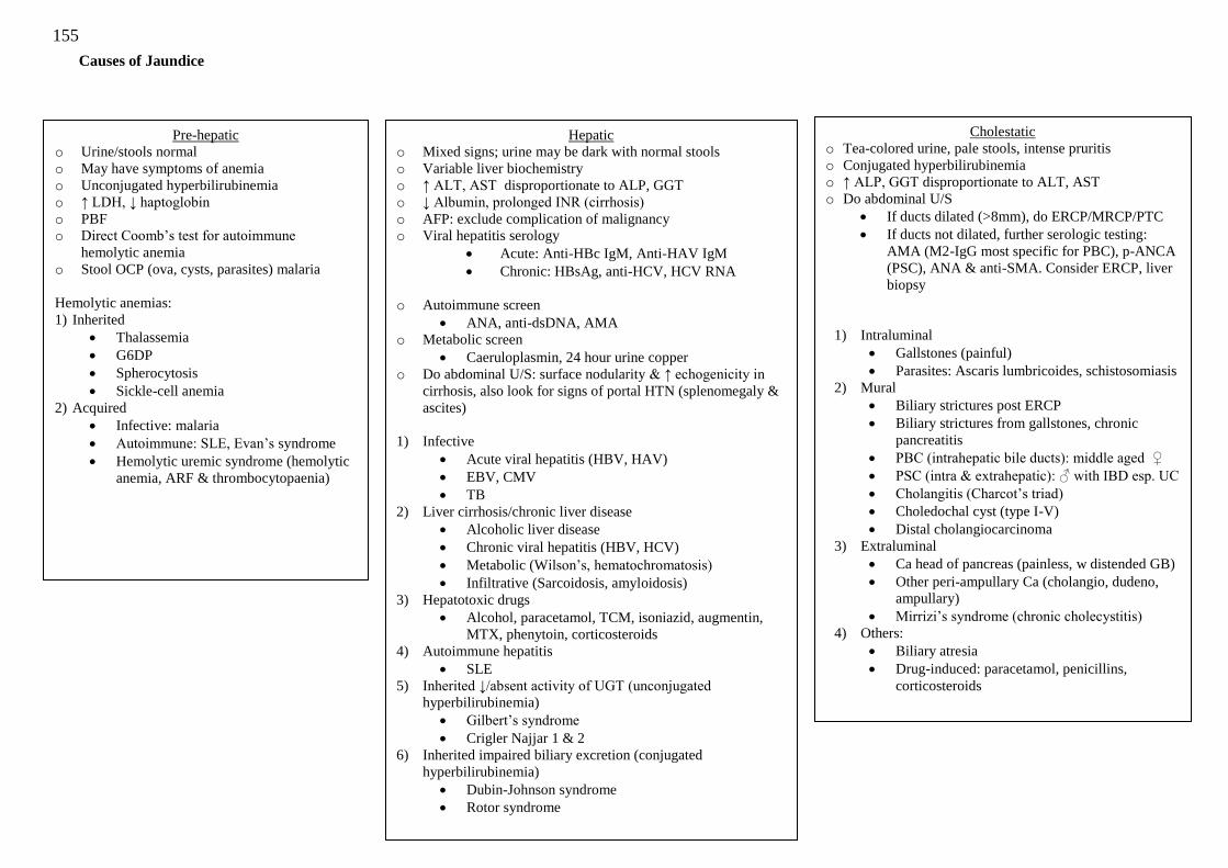
155
Causes of Jaundice
Pre-hepatic
o Urine/stools normal
o May have symptoms of anemia
o Unconjugated hyperbilirubinemia
o ↑ LDH, ↓ haptoglobin
o PBF
o Direct Coomb‘s test for autoimmune
hemolytic anemia
o Stool OCP (ova, cysts, parasites) malaria
Hemolytic anemias:
1) Inherited
Thalassemia
G6DP
Spherocytosis
Sickle-cell anemia
2) Acquired
Infective: malaria
Autoimmune: SLE, Evan‘s syndrome
Hemolytic uremic syndrome (hemolytic
anemia, ARF & thrombocytopaenia)
Hepatic
o Mixed signs; urine may be dark with normal stools
o Variable liver biochemistry
o ↑ ALT, AST disproportionate to ALP, GGT
o ↓ Albumin, prolonged INR (cirrhosis)
o AFP: exclude complication of malignancy
o Viral hepatitis serology
Acute: Anti-HBc IgM, Anti-HAV IgM
Chronic: HBsAg, anti-HCV, HCV RNA
o Autoimmune screen
ANA, anti-dsDNA, AMA
o Metabolic screen
Caeruloplasmin, 24 hour urine copper
o Do abdominal U/S: surface nodularity & ↑ echogenicity in
cirrhosis, also look for signs of portal HTN (splenomegaly &
ascites)
1) Infective
Acute viral hepatitis (HBV, HAV)
EBV, CMV
TB
2) Liver cirrhosis/chronic liver disease
Alcoholic liver disease
Chronic viral hepatitis (HBV, HCV)
Metabolic (Wilson‘s, hematochromatosis)
Infiltrative (Sarcoidosis, amyloidosis)
3) Hepatotoxic drugs
Alcohol, paracetamol, TCM, isoniazid, augmentin,
MTX, phenytoin, corticosteroids
4) Autoimmune hepatitis
SLE
5) Inherited ↓/absent activity of UGT (unconjugated
hyperbilirubinemia)
Gilbert‘s syndrome
Crigler Najjar 1 & 2
6) Inherited impaired biliary excretion (conjugated
hyperbilirubinemia)
Dubin-Johnson syndrome
Rotor syndrome
Cholestatic
o Tea-colored urine, pale stools, intense pruritis
o Conjugated hyperbilirubinemia
o ↑ ALP, GGT disproportionate to ALT, AST
o Do abdominal U/S
If ducts dilated (>8mm), do ERCP/MRCP/PTC
If ducts not dilated, further serologic testing:
AMA (M2-IgG most specific for PBC), p-ANCA
(PSC), ANA & anti-SMA. Consider ERCP, liver
biopsy
1) Intraluminal
Gallstones (painful)
Parasites: Ascaris lumbricoides, schistosomiasis
2) Mural
Biliary strictures post ERCP
Biliary strictures from gallstones, chronic
pancreatitis
PBC (intrahepatic bile ducts): middle aged ♀
PSC (intra & extrahepatic): ♂ with IBD esp. UC
Cholangitis (Charcot‘s triad)
Choledochal cyst (type I-V)
Distal cholangiocarcinoma
3) Extraluminal
Ca head of pancreas (painless, w distended GB)
Other peri-ampullary Ca (cholangio, dudeno,
ampullary)
Mirrizi‘s syndrome (chronic cholecystitis)
4) Others:
Biliary atresia
Drug-induced: paracetamol, penicillins,
corticosteroids
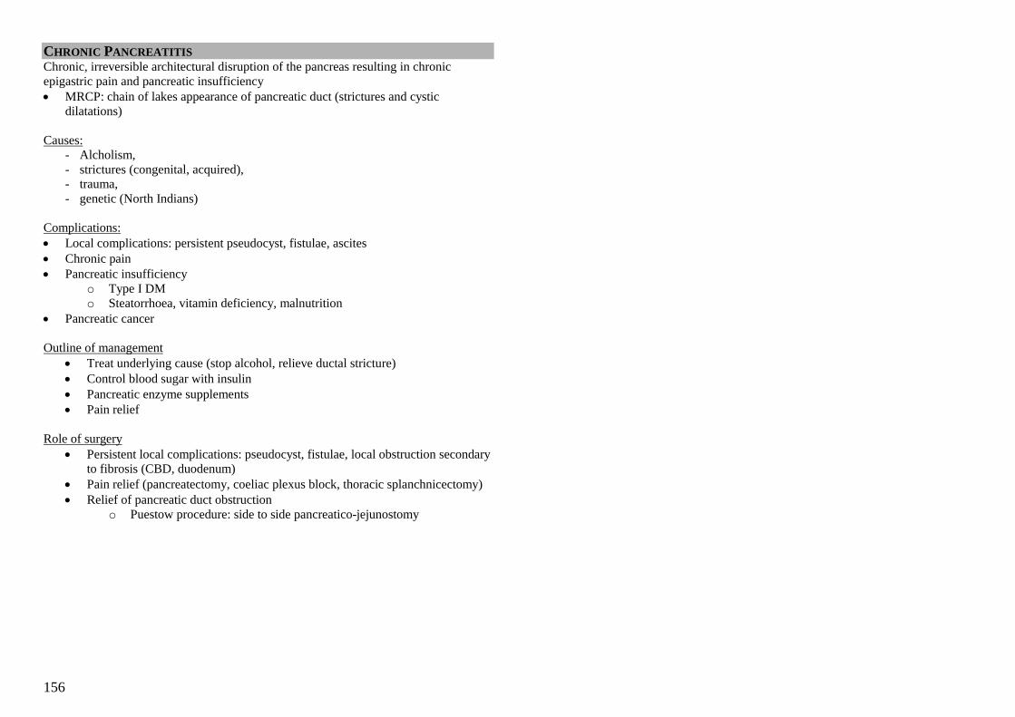
156
CHRONIC PANCREATITIS Chronic, irreversible architectural disruption of the pancreas resulting in chronic
epigastric pain and pancreatic insufficiency
MRCP: chain of lakes appearance of pancreatic duct (strictures and cystic
dilatations)
Causes:
- Alcholism,
- strictures (congenital, acquired),
- trauma,
- genetic (North Indians)
Complications:
Local complications: persistent pseudocyst, fistulae, ascites
Chronic pain
Pancreatic insufficiency
o Type I DM
o Steatorrhoea, vitamin deficiency, malnutrition
Pancreatic cancer
Outline of management
Treat underlying cause (stop alcohol, relieve ductal stricture)
Control blood sugar with insulin
Pancreatic enzyme supplements
Pain relief
Role of surgery
Persistent local complications: pseudocyst, fistulae, local obstruction secondary
to fibrosis (CBD, duodenum)
Pain relief (pancreatectomy, coeliac plexus block, thoracic splanchnicectomy)
Relief of pancreatic duct obstruction
o Puestow procedure: side to side pancreatico-jejunostomy
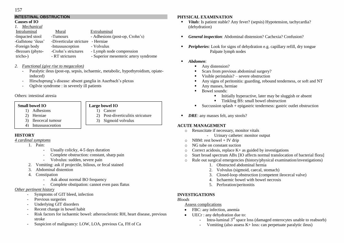
157
INTESTINAL OBSTRUCTION
Causes of IO 1. Mechanical
Intraluminal Mural Extraluminal
-Impacted stool -Tumours - Adhesions (post-op, Crohn‘s)
-Gallstone ‗ileus‘ -Diverticular stricture - Herniae
-Foreign body -Intussusception - Volvulus
-Bezoars (phyto- -Crohn‘s strictures - Lymph node compression
tricho-) - RT strictures - Superior mesenteric artery syndrome
2. Functional (give rise to megacolon)
- Paralytic ileus (post-op, sepsis, ischaemic, metabolic, hypothyroidism, opiate-
induced)
- Hirschsprung‘s disease: absent ganglia in Auerbach‘s plexus
- Ogilvie syndrome : in severely ill patients
Others: intestinal atresia
HISTORY
4 cardinal symptoms 1. Pain:
- Usually colicky, 4-5 days duration
- Complete obstruction: constant, sharp pain
- Volvulus: sudden, severe pain
2. Vomiting: ask if projectile, bilious, or fecal stained
3. Abdominal distention
4. Constipation
- Ask about normal BO frequency
- Complete obstipation: cannot even pass flatus
Other pertinent history - Symptoms of GIT bleed, infection
- Previous surgeries
- Underlying GIT disorders
- Recent change in bowel habit
- Risk factors for ischaemic bowel: atherosclerotic RH, heart disease, previous
stroke
- Suspicion of malignancy: LOW, LOA, previous Ca, FH of Ca
PHYSICAL EXAMINATION
Vitals: Is patient stable? Any fever? (sepsis) Hypotension, tachycardia?
(dehydration)
General inspection: Abdominal distension? Cachexia? Confusion?
Peripheries: Look for signs of dehydration e.g. capillary refill, dry tongue
Palpate lymph nodes
Abdomen:
Any distension?
Scars from previous abdominal surgery?
Visible peristalsis? – severe obstruction
Any signs of peritonitis: guarding, rebound tenderness, or soft and NT
Any masses, herniae
Bowel sounds:
Initially hyperactive, later may be sluggish or absent
Tinkling BS: small bowel obstruction
Succussion splash + epigastric tenderness: gastric outlet obstruction
DRE: any masses felt, any stools?
ACUTE MANAGEMENT o Resuscitate if necessary, monitor vitals
- Urinary catheter: monitor output
o NBM: rest bowel + IV drip
o NG tube on constant suction
o Correct acidosis, replace K+ as guided by investigations
o Start broad spectrum ABx [IO affects normal translocation of bacterial flora]
o Rule out surgical emergencies (history/physical examination/investigations)
1. Obstructed abdominal hernia
2. Volvulus (sigmoid, caecal, stomach)
3. Closed-loop obstruction (competent ileocecal valve)
4. Ischaemic bowel with bowel necrosis
5. Perforation/peritonitis
INVESTIGATIONS
Bloods
Assess complications
FBC: any infection, anemia
UECr : any dehydration due to:
- Intra-luminal 3rd
space loss (damaged enterocytes unable to reabsorb)
- Vomiting (also assess K+ loss: can perpetuate paralytic ileus)
Small bowel IO
1) Adhesions
2) Herniae
3) Ileocecal tumour
4) Intussussception
Large bowel IO
1) Cancer
2) Post-diverticulitis stricuture
3) Sigmoid volvulus
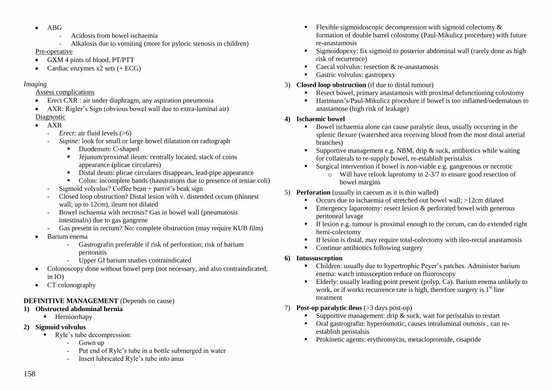
158
ABG
- Acidosis from bowel ischaemia
- Alkalosis due to vomiting (more for pyloric stenosis in children)
Pre-operative
GXM 4 pints of blood, PT/PTT
Cardiac enzymes x2 sets (+ ECG)
Imaging
Assess complications
Erect CXR : air under diaphragm, any aspiration pneumonia
AXR: Rigler‘s Sign (obvious bowel wall due to extra-luminal air)
Diagnostic
AXR
- Erect: air fluid levels (>6)
- Supine: look for small or large bowel dilatation on radiograph
Duodenum: C-shaped
Jejunum/proximal ileum: centrally located, stack of coins
appearance (plicae circulares)
Distal ileum: plicae circulares disappears, lead-pipe appearance
Colon: incomplete bands (haustrations due to presence of teniae coli)
- Sigmoid volvulus? Coffee bean + parrot‘s beak sign
- Closed loop obstruction? Distal lesion with v. distended cecum (thinnest
wall; up to 12cm), ileum not dilated
- Bowel ischaemia with necrosis? Gas in bowel wall (pneumatosis
intestinalis) due to gas gangrene
- Gas present in rectum? No: complete obstruction (may require KUB film)
Barium enema
- Gastrografin preferable if risk of perforation; risk of barium
peritonitis
- Upper GI barium studies contraindicated
Colonoscopy done without bowel prep (not necessary, and also contraindicated,
in IO)
CT colonography
DEFINITIVE MANAGEMENT (Depends on cause)
1) Obstructed abdominal hernia
Herniorrhapy
2) Sigmoid volvulus
Ryle‘s tube decompression:
- Gown up
- Put end of Ryle‘s tube in a bottle submerged in water
- Insert lubricated Ryle‘s tube into anus
Flexible sigmoidoscopic decompression with sigmoid colectomy &
formation of double barrel colostomy (Paul-Mikulicz procedure) with future
re-anastamosis
Sigmoidopexy: fix sigmoid to posterior abdominal wall (rarely done as high
risk of recurrence)
Caecal volvulus: resection & re-anastamosis
Gastric volvulus: gastropexy
3) Closed loop obstruction (if due to distal tumour)
Resect bowel, primary anastamosis with proximal defunctioning colostomy
Hartmann‘s/Paul-Mikulicz procedure if bowel is too inflamed/oedematous to
anastamose (high risk of leakage)
4) Ischaemic bowel
Bowel ischaemia alone can cause paralytic ileus, usually occurring in the
splenic flexure (watershed area receiving blood from the most distal arterial
branches)
Supportive management e.g. NBM, drip & suck, antibiotics while waiting
for collaterals to re-supply bowel, re-establish peristalsis
Surgical intervention if bowel is non-viable e.g. gangrenous or necrotic
o Will have relook laprotomy in 2-3/7 to ensure good resection of
bowel margins
5) Perforation (usually in caecum as it is thin walled)
Occurs due to ischaemia of stretched out bowel wall; >12cm dilated
Emergency laparotomy: resect lesion & perforated bowel with generous
peritoneal lavage
If lesion e.g. tumour is proximal enough to the cecum, can do extended right
hemi-colectomy
If lesion is distal, may require total-colectomy with ileo-rectal anastamosis
Continue antibiotics following surgery
6) Intussusception
Children: usually due to hypertrophic Peyer‘s patches. Administer barium
enema: watch intussception reduce on fluoroscopy
Elderly: usually leading point present (polyp, Ca). Barium enema unlikely to
work, or if works recurrence rate is high, therefore surgery is 1st line
treatment
7) Post-op paralytic ileus (>3 days post-op)
Supportive management: drip & suck, wait for peristalsis to restart
Oral gastrografin: hyperosmotic, causes intraluminal osmosis , can re-
establish peristalsis
Prokinetic agents: erythromycin, metaclopromide, cisapride

159
ACUTE APPENDICITIS
HISTORY
- Classic: LOA with periumbilical pain which radiates to the RIF, followed by
nausea & vomiting
- N/V almost always occurs after pain, if it precedes pain, exclude IO
- May have diarrhoea or constipation
- Inflamed appendix near bladder/ureter may cause irritative voiding symptoms
& haematuria
PHYSICAL EXAMINATION
- RIF (McBurney‘s point) tenderness on percussion, rebound tenderness, rigidity
& guarding
- RIF mass may be palpable
- LIF tenderness in patients with situs inversus or lengthy appendix extending to
the LIF
- Cough sign: RIF pain on coughing (localized peritonitis)
- Rovsing sign: RIF pain with palpation of the LIF
- Obturator sign: RIF pain with internal rotation of a flexed right hip
- Psoas sign: RIF pain with hyperextension of the right hip
- Infants/children may present with inflamed hemiscrotum due to migration of
pus through patent processus vaginalis – often mistaken for acute testicular
torsion
CAUSES - Obstruction of appendiceal lumen
Faecaliths: calcium salts & faecal debris collect around nidus of faecel
material within appendix
Lymphoid hyperplasia: a/w inflammatory (Crohn‘s) & infective dz (GE,
URTI, IMS, measles)
Less common causes: parasites, TB, tumour, FB
DIFFERENTIAL DIAGNOSES
1) Mesenteric adenitis: self limiting inflammation of mesenteric LNs in RIF, a/w
viral infections
2) Meckel‘s diverticulitis: inflammation of Meckel‘s diverticulum (see below)
3) Terminal ileitis: usually due to Crohn‘s disease
4) Ischaemic colitis
5) Colon cancer
INVESTIGATIONS
Blood: FBC, UECr, CRP, Blood culture, PT/PTT, GXM
Urine: UFEME (may have pyuria & haematuria)
Imaging: Erect CXR, CTAP, abdominal U/S
MANAGEMENT
- NBM, IV drip, IV antibiotics, correct electrolyte imbalance
- Symptomatic relief: anti-emetics & analgesia
- Definitive treatment: appendicectomy (open or laparoscopic)
- Conservative treatment for appendiceal mass: Oschner Sherren regime
Omentum naturally wraps around inflamed appendix, containing
inflammatory process
Symptomatic treatment as well as antibiotics, monitor patient as well
as size of mass
When mass has reduced in size, patient is stable, do surgery (less
inflammation: easier)
Exceptions: very young or very old patients, or patients with suspected
appendicular abscess (require incision & drainage of pus)
160
MECKEL’S DIVERTICULUM
RULE OF 2s
- Like the appendix, Meckel‘s diverticulum is a true congenital diverticulum, a
vestigial remnant of the vitelline duct
- 2 inches in length, 2cm wide, 2 feet from ileocaecal valve
- Occurs in 2% of the population, ♂>♀ in the ratio of 2:1
- Usually asymptomatic but if often presents at age 2 if symptomatic
- 2 types of ectopic tissue
Pancreatic (6%)
Gastric (60%) – gastric acid secretion can produce inflammation, peptic
ulceration & bleeding, strictures with subsequent IO
May have both types of tissue or other rarer types (jejunal, colonic,
rectal, hepatobiliary, etc)
PRESENTATION
- Usually asymptomatic, found incidentally during barium study or open surgery
- Symptomatic:
1) IO (most common presentation): strictures, intussusception, volvulus,
omphalomesenteric band
2) Hematochezia (most common in children): usually massive & painless,
due to peptic ulceration
3) Diverticulitis: may present exactly like acute appendicitis i.e.
periumbilical pain radiating to RIF. Less prone to inflammation as most
Meckel‘s have wide base, little lymphoid tissue & are self emptying
4) Others: umbilical fistula, tumour, perforation etc
INVESTIGATION
Blood: As per IO or lower BGIT
Imaging:
- Technetium-99m pertechnetate scan (investigation of choice): isotope given IV,
taken up by gastric mucosa, detected by gamma scan of the abdomen
- Barium studies: small bowel enteroclysis
- CT NOT helpful as hard to distinguish Meckel‘s diverticulum from small
bowel loops
MANAGEMENT
- NBM, IV drip, correct electrolyte imbalance
- IO: NG tube on constant suction, supine AXR
- PR bleed: PCT if necessary, gastric lavage to exclude UBGIT, OGD &
colonoscopy
- Definitive treatment: surgery (open or laparoscopic)
Wide base: wedge ileal resection with anastamosis
Narrow base: resection of the diverticulum

161
POST-OP COMPLICATIONS
General or specific?
Immediate, early or late?
Local or systemic?
IMMEDIATE (<1 hour post-op)
1) Local
Damage to surrounding structures
Primary haemorrhage
o Either starting during surgery or following postoperative increase
in blood pressure
o Replace blood loss and may require return to theatre to re-
explore wound
2) Systemic
Basal atelectasis (collapse of alveoli):
o ↑ bronchial secretions post-op
o Patient does not breath deeply due to pain
o Tx: chest physiotherapy, incentive spirometry
Shock: due to blood loss, or inadequate fluid replacement peri-operatively
AMI, pulmonary embolism
EARLY (first 24-48 hours post-op)
1) Local
Pain, hemorrhage
2) Systemic
Atelectasis, shock, AMI, PE
Nausea & vomiting, analgesia/anaesthesia induced
Acute confusion: dehydration or sepsis
LATE (between 48 hours to 1 month post-op)
1) Local
Post-op paralytic ileus
Wound or anastomotic dehiscence
o Occurs around Day 7 – 10
o Heralded by serosanguinous exudate
o Analgesia, sterile wound dressing, fluid resuscitation, resuture
under GA in OT
o Mortality up to 30%
Post-op wound infection
o Usually within 1st week
o Pain, redness, swelling, discharge, usually due to skin
staphylococci
Incisional hernia
o 10-15% of abdominal wounds
o Usu. asymptomatic, but if painful, consider strangulation
(uncommon due to wide neck)
o RF: Obesity, poor muscle tone (old age), (increased intra-
abdominal pressure (chronic cough, straining from constipation),
wound infection, multiple use of same incision site
o Surgical repair of hernia if strangulated or if enlarging
Bowel obstruction due to adhesions
Fistula formation
Secondary haemorrhage: often as a result of infection
2) Systemic
Fever: wind (atelectasis), water (UTI), walking (DVT/PE), wound (infxn),
wonder drug (drug fever)
Infections (day 3-5): phlebitis, pneumonia, UTI, abscesses (subphrenic,
pelvic)
Acute confusion: exclude dehydration & sepsis
DVT & PE (day 5-7)
POST-OP HAEMORRHAGE
Early post-op haemorrhage:
Consumptive coagulopathy – if large volumes of blood transfused
Pre-op anticoagulation
Unrecognized bleeding diathesis
Damage to blood vessels/vascular organs
Management
FBC: check platelets
PT/PTT
GXM & order blood
Give protamine if heparin was used
Give FFP / platelet concentrates if clotting screen abnormal
Consider surgical re-exploration
Late post-op haemorrhage:
Occurs several days after surgery
Usually due to infection damaging vessels at the operation site
Management
Treat infection and consider exploratory surgery
162
POOR WOUND HEALING
Factors which predispose to poor wound healing:
Patient factors:
Poor blood supply
Malnutrition
Vitamin deficiency
Immunosuppressive therapy
Long term steroids
Radiotherapy
Co-morbid conditions e.g. DM, HIV
Surgeon factors:
Suturing under tension
Did not observe aseptic technique
Wound factors:
Wound infection
Poor wound care
Approach to Post Operative DVT/ PE
DVT in deep calf veins: 5-10% risk of PE
DVT in iliofemoral veins: 50-60% risk of PE
Risk Factors (Virchow’s Triad):
Alterations in blood flow (stasis)
o Prolonged bed rest, pregnancy (IVC compression)
Vascular endothelial injury
o Trauma, pelvic/ vascular surgery, intravenous drug use
Alterations in blood constituents (hypercoagulable state)
o Congenital: protein C/ protein S deficiency, Factor V Leiden mutation,
anti-phospholipid syndrome, homocysteinuria
o Acquired: liver disease, cancer (production of pro-thrombotic factors),
oral contraceptive use
Presentation
Post-operative fever (typically day 5-7)
Lower limb pain & swelling, discolouration: may not be at site of clot
Classic triad of PE: chest pain, dyspnoea, haemoptysis (poor sensitivity/
specificity)
o Others: seizures, syncope, new onset atrial fibrillation etc
Physical Examination
Homan‘s sign: passive dorsiflexion of the ankle produced sharp pain in calf
(DON‘T DO: risk of clot propagation)
Investigations:
Duplex ultrasound to detect DVT
Chest x-ray: usually normal (classical findings listed below are rarely seen)
o Atelectasis, pleural effusion, raised hemi-diaphragm
o Westermark sign: dilatation of the pulmonary vessels proximal to
the embolism, with collapse of the distal vessels
o Hampton hump: late sign, wedged shaped infiltrate with apex
towards the hilum
CT pulmonary angiogram (gold standard)
o Look for filling defects in pulmonary artery
V/Q perfusion scan: no longer done
o Inhale radioactive isotope, do CXR to assess ventilation of lungs
o IV contrast, do CXR again to assess perfusion of lungs
o Look for areas of ventilation without perfusion (V/Q mismatch)
o Correlate with clinical findings, calculate probability of PE
ECG:
o May have normal ECG with sinus tachycardia
o Signs of right heart strain
- Right axis deviation: S waves in lead I
- Right ventricular hypertrophy: tall R waves in V1, deep
S waves in V6
- Right atrial hypertrophy: peaked P waves
- RBBB
- Classic S1Q3T3: S wave in I, Q wave in III, T inversion
in III (rarely seen)
D-dimer: poor sensitivity and specificity in ruling in/ ruling out DVT or
pulmonary embolism
Prophylaxis/Treatment
Non-pharmacological
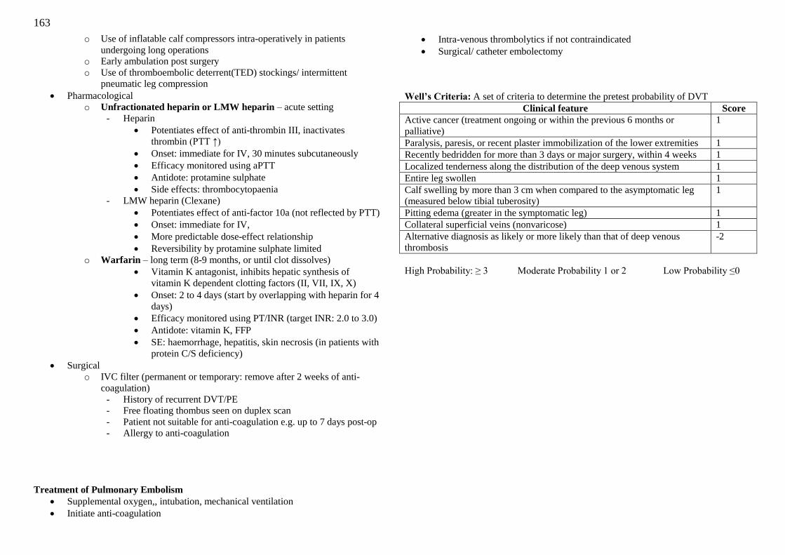
163
o Use of inflatable calf compressors intra-operatively in patients
undergoing long operations
o Early ambulation post surgery
o Use of thromboembolic deterrent(TED) stockings/ intermittent
pneumatic leg compression
Pharmacological
o Unfractionated heparin or LMW heparin – acute setting
- Heparin
Potentiates effect of anti-thrombin III, inactivates
thrombin (PTT ↑)
Onset: immediate for IV, 30 minutes subcutaneously
Efficacy monitored using aPTT
Antidote: protamine sulphate
Side effects: thrombocytopaenia
- LMW heparin (Clexane)
Potentiates effect of anti-factor 10a (not reflected by PTT)
Onset: immediate for IV,
More predictable dose-effect relationship
Reversibility by protamine sulphate limited
o Warfarin – long term (8-9 months, or until clot dissolves)
Vitamin K antagonist, inhibits hepatic synthesis of
vitamin K dependent clotting factors (II, VII, IX, X)
Onset: 2 to 4 days (start by overlapping with heparin for 4
days)
Efficacy monitored using PT/INR (target INR: 2.0 to 3.0)
Antidote: vitamin K, FFP
SE: haemorrhage, hepatitis, skin necrosis (in patients with
protein C/S deficiency)
Surgical
o IVC filter (permanent or temporary: remove after 2 weeks of anti-
coagulation)
- History of recurrent DVT/PE
- Free floating thombus seen on duplex scan
- Patient not suitable for anti-coagulation e.g. up to 7 days post-op
- Allergy to anti-coagulation
Treatment of Pulmonary Embolism
Supplemental oxygen,, intubation, mechanical ventilation
Initiate anti-coagulation
Intra-venous thrombolytics if not contraindicated
Surgical/ catheter embolectomy
Well’s Criteria: A set of criteria to determine the pretest probability of DVT
Clinical feature Score
Active cancer (treatment ongoing or within the previous 6 months or
palliative)
1
Paralysis, paresis, or recent plaster immobilization of the lower extremities 1
Recently bedridden for more than 3 days or major surgery, within 4 weeks 1
Localized tenderness along the distribution of the deep venous system 1
Entire leg swollen 1
Calf swelling by more than 3 cm when compared to the asymptomatic leg
(measured below tibial tuberosity)
1
Pitting edema (greater in the symptomatic leg) 1
Collateral superficial veins (nonvaricose) 1
Alternative diagnosis as likely or more likely than that of deep venous
thrombosis
-2
High Probability: ≥ 3 Moderate Probability 1 or 2 Low Probability ≤0