Surface plasmon resonance analysis of dynamic biological interactions with biomaterials
13
Contents 1. Introduction .............................................. 1823 2. SPR theory .............................................. 1824 2.1. Adsorption kinetics by SPR ................................. 1825 3. Applications of SPR ......................................... 1826 3.1. Physical applications ..................................... 1826 3.1.1. Thin organic monolayers or bilayers ....................... 1826 3.1.2. Polymer "lms ..................................... 1827 3.2. Biological applications .................................... 1828 3.2.1. Biomolecular structure and interactions of proteins, DNA and viruses .... 1828 3.2.2. Lipid bilayers ..................................... 1829 3.2.3. Non-speci"c biomolecular interactions*biocompatibility ........... 1829 3.2.4. Tissue engineering ................................... 1832 4. Current and future instrumental developments ......................... 1832 5. Conclusions and future outlook .................................. 1833 References ................................................. 1833 * Corresponding author. Present address: Department of Chemistry, University of Surrey, Guildford, Surrey GU2 5XH, UK. Fax: #44- 1483-876851. E-mail address: r.green@surrey.ac.uk (R.J. Green). Biomaterials 21 (2000) 1823}1835 Review Surface plasmon resonance analysis of dynamic biological interactions with biomaterials Rebecca J. Green*, Richard A. Frazier, Kevin M. Shakeshe!, Martyn C. Davies, Clive J. Roberts, Saul J.B. Tendler Laboratory of Biophysics and Surface Analysis, School of Pharmaceutical Sciences, The University of Nottingham, University Park, Nottingham NG7 2RD, UK Received 21 December 1999; received in revised form 2 March 2000; accepted 2 March 2000 Abstract Surface plasmon resonance (SPR) is an optical technique that is widely gaining recognition as a valuable tool to investigate biological interactions. SPR o!ers real time in situ analysis of dynamic surface events and, thus, is capable of de"ning rates of adsorption and desorption for a range of surface interactions. In this review we highlight the diversity of SPR analysis. Examples of a wide range of applications of SPR are presented, concentrating on work relevant to the analysis of biomaterials. Particular emphasis is given to the use of SPR as a complimentary tool, showing the broad range of techniques that are routinely used alongside SPR analysis. ( 2000 Elsevier Science Ltd. All rights reserved. Keywords: Surface plasmon resonance; Protein adsorption; Polymer degradation; Hydration; Epitope mapping; Tissue engineering 1. Introduction The surface interactions of biomaterials are funda- mental to determine critical issues, such as host response and biocompatibility. Indeed, many challenges to bio- material design can be directly related to the control of surface interactions. For example, the surface adsorption of proteins from urine is an important stage leading to urinary encrustation [1]. Therefore, the design of materials that can resist the adsorption of these proteins from urine would lead to improved performance of urinary catheters. Many surface analytical techniques have been applied to the study of biomaterials, but few of these are able to 0142-9612/00/$ - see front matter ( 2000 Elsevier Science Ltd. All rights reserved. PII: S 0 1 4 2 - 9 6 1 2 ( 0 0 ) 0 0 0 7 7 - 6
-
Upload
nottingham -
Category
Documents
-
view
0 -
download
0
Transcript of Surface plasmon resonance analysis of dynamic biological interactions with biomaterials

Contents
1. Introduction . . . . . . . . . . . . . . . . . . . . . . . . . . . . . . . . . . . . . . . . . . . . . . 18232. SPR theory . . . . . . . . . . . . . . . . . . . . . . . . . . . . . . . . . . . . . . . . . . . . . . 1824
2.1. Adsorption kinetics by SPR . . . . . . . . . . . . . . . . . . . . . . . . . . . . . . . . . 18253. Applications of SPR . . . . . . . . . . . . . . . . . . . . . . . . . . . . . . . . . . . . . . . . . 1826
3.1. Physical applications . . . . . . . . . . . . . . . . . . . . . . . . . . . . . . . . . . . . . 18263.1.1. Thin organic monolayers or bilayers . . . . . . . . . . . . . . . . . . . . . . . 18263.1.2. Polymer "lms . . . . . . . . . . . . . . . . . . . . . . . . . . . . . . . . . . . . . 1827
3.2. Biological applications . . . . . . . . . . . . . . . . . . . . . . . . . . . . . . . . . . . . 18283.2.1. Biomolecular structure and interactions of proteins, DNA and viruses . . . . 18283.2.2. Lipid bilayers . . . . . . . . . . . . . . . . . . . . . . . . . . . . . . . . . . . . . 18293.2.3. Non-speci"c biomolecular interactions*biocompatibility . . . . . . . . . . . 18293.2.4. Tissue engineering . . . . . . . . . . . . . . . . . . . . . . . . . . . . . . . . . . . 1832
4. Current and future instrumental developments . . . . . . . . . . . . . . . . . . . . . . . . . 18325. Conclusions and future outlook . . . . . . . . . . . . . . . . . . . . . . . . . . . . . . . . . . 1833References . . . . . . . . . . . . . . . . . . . . . . . . . . . . . . . . . . . . . . . . . . . . . . . . . 1833
*Corresponding author. Present address: Department of Chemistry,University of Surrey, Guildford, Surrey GU2 5XH, UK. Fax: #44-1483-876851.
E-mail address: [email protected] (R.J. Green).
Biomaterials 21 (2000) 1823}1835
Review
Surface plasmon resonance analysis of dynamic biologicalinteractions with biomaterials
Rebecca J. Green*, Richard A. Frazier, Kevin M. Shakeshe!, Martyn C. Davies,Clive J. Roberts, Saul J.B. Tendler
Laboratory of Biophysics and Surface Analysis, School of Pharmaceutical Sciences, The University of Nottingham,University Park, Nottingham NG7 2RD, UK
Received 21 December 1999; received in revised form 2 March 2000; accepted 2 March 2000
Abstract
Surface plasmon resonance (SPR) is an optical technique that is widely gaining recognition as a valuable tool to investigatebiological interactions. SPR o!ers real time in situ analysis of dynamic surface events and, thus, is capable of de"ning rates ofadsorption and desorption for a range of surface interactions. In this review we highlight the diversity of SPR analysis. Examples ofa wide range of applications of SPR are presented, concentrating on work relevant to the analysis of biomaterials. Particular emphasisis given to the use of SPR as a complimentary tool, showing the broad range of techniques that are routinely used alongside SPRanalysis. ( 2000 Elsevier Science Ltd. All rights reserved.
Keywords: Surface plasmon resonance; Protein adsorption; Polymer degradation; Hydration; Epitope mapping; Tissue engineering
1. Introduction
The surface interactions of biomaterials are funda-mental to determine critical issues, such as host response
and biocompatibility. Indeed, many challenges to bio-material design can be directly related to the control ofsurface interactions. For example, the surface adsorption ofproteins from urine is an important stage leading to urinaryencrustation [1]. Therefore, the design of materials that canresist the adsorption of these proteins from urine wouldlead to improved performance of urinary catheters.
Many surface analytical techniques have been appliedto the study of biomaterials, but few of these are able to
0142-9612/00/$ - see front matter ( 2000 Elsevier Science Ltd. All rights reserved.PII: S 0 1 4 2 - 9 6 1 2 ( 0 0 ) 0 0 0 7 7 - 6

Fig. 1. The Kretchmann con"guration for SPR. Resonance of a surfaceplasmon is excited at the metal/air interface when the angle of incidenceof light is such that the evanescent component of its wave vector (K
%7) is
equal to the wave vector of the propagating surface plasmon (K41
).
monitor dynamic interactions within a #uid environ-ment, that may be tailored to model likely conditionsencountered in vivo. Scanning probe microscopy (SPM),attenuated total re#ectance infrared spectroscopy (ATR-IR) and spectral ellipsometry are three widely used tech-niques that can o!er the possibility to study dynamicevents at a range of surfaces. SPM allows us to imagechanges at surfaces as they, for example, adsorb proteinsor erode within a liquid environment [2,3]. ATR-IR iscapable of studying conformational changes in biologicalmacromolecules at a range of solid}aqueous interfaces,although its relative lack of surface sensitivity (1}5 lmsampling depth) and di$culties in sample preparationare a limit to its successful use [4]. Spectral ellipsometryis less convenient to monitor dynamic processes butallows the determination of thickness and refractive in-dex of the adsorbed layer [5].
The subject of this review is surface plasmon resonance(SPR), which is rapidly gaining recognition and applica-tion as a powerful tool for biomaterial characterization.The use of SPR to probe surface interactions is advan-tageous, since it is able to rapidly monitor any dynamicprocess, such as adsorption or degradation, to a widerange of biomedically relevant interfaces in real timewithout the need to label the adsorbate and without theneed for complex sample preparation. It can rapidlyobtain information on the rate and extent of adsorption,enabling the determination of dielectric properties, theassociation/dissociation kinetics and the a$nity con-stants of speci"c ligand}ligate interactions [6].
2. SPR theory
A surface plasmon is a longitudinal charge densitypropagating wave along the interface of two media,where one is a metal and the other a dielectric [7]. Thechoice of metal used is critical, since the metal mustexhibit free electron behaviour as described by the freeelectron model [8]. Suitable metals include silver, gold,copper and aluminium of which silver and gold are morecommonly used. Silver is used as it provides a sharp SPRresonance peak and gold due to its stability [9]. Twodi!erent experimental systems for the excitation of sur-face plasmons were developed by Otto [10] and Kretch-mann [11]. However, it is the attenuated total re#ectance(ATR) con"guration developed by Kretchmann that iswidely used within the designs of most SPR instrumentsand, therefore, will be discussed here.
The Kretchmann con"guration (Fig. 1) relies on thephenomenon of total internal re#ection. This occurswhen light travelling through an optically dense medium(e.g. glass) reaches an interface between this medium anda medium of a lower optical density (e.g. air), and isre#ected back into the dense medium. Although the inci-dent light is totally internally re#ected, a component of
this light, the evanescent wave or "eld, penetrates theinterface into the less dense medium to a distance of onewavelength [12]. Total internal re#ection #uorescence(TIRF) relies on the phenomenon of the evanescent "eldto excite molecules near a glass/liquid interface.
In SPR a monochromatic, p-polarized light source isused and the interface between the two optically densemedia is coated with a thin metal "lm (of thickness lessthan one wavelength of light). The wave vector of theevanescent "eld (K
%7) is given by
K%7"
w0c
g'sin h, (1)
where w0
is the frequency of incident light, g'
the refrac-tive index of the dense medium (glass), h the angle ofincidence of the light and c the speed of light in a vacuum.The wave vector of a surface plasmon (K
41) can be
approximated to
K41"
w0c S
e.g24
e.#g2
4(2)
where e.
is the dielectric constant of the metal "lm andg4
is the refractive index of the dielectric medium [13].The evanescent wave of the incoming light is able to
couple with the free oscillating electrons (plasmons) inthe metal "lm at a speci"c angle of incidence correspond-ing to when K
41"K
%7, and thus the surface plasmon is
resonantly excited. This causes energy from the incidentlight to be lost to the metal "lm resulting in a reduction inthe intensity of re#ected light which can be detected bya two-dimensional array of photodiodes or chargecoupled detectors (CCD). Examination of Eq. (1) showsthat K
41is dependent on the refractive index of the
water/air medium above the silver "lm, which can bemonitored up to a thickness of approximately 300 nmabove the silver surface (within the detection limits ofSPR). Therefore, if the refractive index immediatelyabove the silver surface changes, by the adsorption of
1824 R.J. Green et al. / Biomaterials 21 (2000) 1823}1835

Fig. 2. A schematic representation of a typical SPR instrument. A sil-ver/gold-coated glass slide is index-matched onto a glass prism,through which a convergent light beam is focussed onto the undersideof the silver/gold "lm, and re#ected to a detector array. Analyte solu-tions are passed through a #ow cell at de"ned #ow rates and temper-atures. The surface of the silver/gold "lm may be modi"ed to presenta variety of sensor surfaces for analysis as shown.
Fig. 3. A schematic example of an SPR adsorption pro"le for theadsorption of a protein to a polymer "lm. The change in the value of theSPR angle is used to monitor the kinetics of adsorption.
a protein layer, a change in the angle of incidence re-quired to excite a surface plasmon will occur. Bymonitoring the angle at which resonance occurs (the SPRangle) during an adsorption process with respect to time,an SPR adsorption pro"le can be obtained.
A schematic of a typical SPR instrument con"gurationis depicted in Fig. 2. Variations on #ow cell designs canallow experimentation at a variety of #ow rates andtemperatures. As depicted in Fig. 2, the SPR slide surfacecan be modi"ed to monitor adsorption processes toa range of surfaces by attachment/coating of thin layersabove the silver "lm. These layers can comprise of self-assembled monolayers (SAMs) of varying head groupchemistries, covalently immobilized dextran matices,lipid bilayers or polymer "lms. A schematic of a typicalSPR adsorption pro"le during the adsorption of a pro-tein monolayer to a polymer-coated silver surface isshown in Fig. 3. Initially, the surface is primed witha suitable bu!er solution to create a base line on the SPRcurve. Once the protein comes into contact with thesurface, rapid adsorption occurs which is seen as anincrease in the SPR angle. This is followed by a plateau inthe SPR adsorption pro"le due to saturation of thesurface with protein, and "nally the protein solution isreplaced with a bu!er only solution removing looselybound material that are within the detection limits of theSPR. The di!erence between the initial and "nal SPRangle gives an indication on the extent of adsorption, andthe positive gradient of the SPR adsorption curve deter-mines the rate of adsorption.
2.1. Adsorption kinetics by SPR
The rate of adsorption can be obtained from thesteepest positive gradient on an SPR adsorption pro"le.This rate is dependent on the intrinsic kinetics and themass transport of the system [14]. The process of di!u-sion of the adsorbate, through a boundary layer, to thesurface is generally much slower than the intrinsic ad-sorption kinetics and is, therefore, the rate-determiningfactor. By varying the #ow rate of the system, a masstransport limitation can be identi"ed, since di!usion ki-netics are dependent on the #ow rate whereas intrinsickinetics are not [15]. The di!usion kinetics can be de-scribed by the following equations [6]:
j$
(rate of di!usion)"D
x$
([A]"![A]
4) , (3)
"k.([A]
"![A]
4), (4)
where D is the di!usion coe$cient, x$
the boundary layerthickness, k
.the mass transfer coe$cient, [A]
"bulk
concentration of the analyte and [A]4the surface concen-
tration of the analyte. For a general #ow cell arrange-ment
k."
D
x$
"1.86 AD2v
h¸ B1@3
, (5)
where v is the velocity or #ow rate, ¸ is the distancebetween inlet into #ow cell and sampling area and h is theheight of the #ow cell. When considering a speci"c
R.J. Green et al. / Biomaterials 21 (2000) 1823}1835 1825

ligand}ligate adsorption process initially the kineticsmay be di!usion limited, but as the adsorption proceedsthe number of available binding sites (free ligands) for theligate reduces and eventually the observed binding ratewill re#ect that of the intrinsic kinetics (the binding rate).To shift the balance from a di!usion-limited rate to theintrinsic reaction rates, the di!usion rate must be in-creased and the rate of binding reduced. Eq. (5) suggeststhat the mass transfer can be increased by reducing ¸, thedistance between inlet and sample area, or by increasingv, the #ow rate. Also, the rate of di!usion can be in-creased by increasing the concentration gradient of theligate ([A]
"![A]
4).
For the application of SPR as a biosensor, intrinsickinetics are determined by the rate of change in SPRangle as the ligate binds [14]. The intrinsic reaction rateequation is
d[AB]
dt"K
![A][B]!K
$[AB], (6)
where K!is the association constant, K
$the dissociation
constant, [A] is concentration of ligate solution, [B] isconcentration of free ligand and [AB] the concentrationof the bound ligate}ligand complex. The maximumchange in SPR angle (R
.!9) is proportional to the total
ligand concentration ([B]#[AB]), R.!9
!R is propor-tional to the free ligand concentration [B] and [A] maybe considered as a constant (C) since the free ligate isconstantly replenished under #ow condition. Therefore,Eq. (6) may be rewritten as
dR
dt"K
!C(R
.!9!R)!K
$R, (7)
"K!CR
.!9!(K
!C#K
$)R. (8)
Rate constants are then determined by plotting dR/dt vs.R for a range of ligate concentrations. The gradient ofeach concentration plot (k
4) is then plotted against the
ligate concentration (C) to produce a straight line where!k
4"K
!C#K
$. From this graph, K
!is the gradient
and K$
the y-axis intercept. A more accurate measure-ment of the dissociation constant can be determined if thebound [AB] complex is in a bu!er only solution, wherethe concentration of analyte in solution falls to zero andthe Eq. (8) can be simpli"ed to dR/dt"!K
$R. The
a$nity constant can then be calculated as K!/K
$[11,14].
3. Applications of SPR
The applications of SPR are diverse and ever growing.They include the study of optical properties in metal"lms, "lm thickness and refractive index measurementsof organic layers on metal surfaces, protein adsorption tobiomaterials, the adsorption of gas molecules and ap-plications as a biosensor. For clarity, we have divided the
text under a number of headings relating to the variety ofsubject areas.
3.1. Physical applications
Since the substrate of SPR interface must be a metal,initial SPR studies probed interactions with and thedielectric properties of metal "lms. The limitation of thesubstrate has been overcome by depositing Lang-muir}Blodgett "lms or organic SAMs onto the metallayer, thus allowing the analysis of a wider range ofsystems. More recently, coating polymeric materials ontothe necessary metal substrate has widened the potentialof SPR further. SPR is now able to probe the physicalproperties of an extensive range of materials, determiningmany surface characteristics including dielectric proper-ties, adsorption processes, surface degradation or hy-dration.
SPR has often been used to monitor the dielectricproperties of metal "lms. For example, De Bruijn et al.[16] measured the dielectric permittivity and thickness ofmetal layers by an iteration method using starting para-meters derived from SPR angle curves. Similarly, Linnertet al. [17] related damping of SPR angle curves to thesurface chemistry of colloidal silver. Also, as an exampleof the use of SPR to probe interactions at metal interfa-ces, Voskoboinikov [18] studied the e!ect of the adsorp-tion of oxygen sub-monolayers to silver, magnesium andindium metal "lms had on the surface plasmon intensity.
3.1.1. Thin organic monolayers or bilayersThe thickness and dielectric properties of thin organic"lms have been calculated by SPR analysis for manysystems, including carbon-coated silver [19], thinpoly(tetra#uoroethylene) layers [20], phthalocyanine"lms [21] and Langmuir}Blodgett "lms [22]. Yang et al.[23] applied SPR analysis to monitor the dynamic com-plexation process of potassium ions with valinomycinLangmuir}Blodgett "lms, thus proving SPR to be sensi-tive to minute changes in the properties of sensor molecu-les. SPR was further applied to the accuratecharacterization of monolayer and multilayer formationin Langmuir}Blodgett "lms by Cooke and Roberts [24],and to the observation of striation defects in Langmuir}Blodgett "lms by Merle et al. [25]. Recently, Advinculaet al. [26] reported the use of SPR for the in situ study ofpolymer self-assembly solution adsorption, which is thelayer-by-layer deposition of oppositely charged polymersalternately from solution.
Many studies have utilized the immobilizationmethods of SAMs to investigate a range of complexinteractions by SPR. The kinetics of interactions betweenplatelet glycoproteins and "brinogen have been investi-gated, where either biomolecule had been immobilizedvia the chemisorption of a SAM to gold [27]. Similarly,SPR spectroscopy and microscopy has been employed in
1826 R.J. Green et al. / Biomaterials 21 (2000) 1823}1835

Fig. 4. The chemisorption of thiolated dextrans of varying molecularweight and thiol substitutions from solution onto a gold surface. TheSPR angle shifts show a smaller range than might be expected whenconsidering the 10-fold di!erence between the highest and lowest dex-tran molecular weights. Indeed, chemisorption of the highest molecularweight dextran does not lead to the highest SPR angle shift. Thisanomaly suggests di!erences in the structures of the dextran layers asdepicted schematically. High molecular weight dextrans form moredi!use surface layers, while lower molecular weight dextrans form morecompact layers.
Fig. 5. The SPR hydration pro"les for a series of polyurethanes ofvarying PEO content. The extent of hydration is shown to increase withincreasing PEO content of the polymer "lm. A downward shift in theSPR angle is observed upon hydration, due to a reduction of thedielectric properties of the polymer "lm as water is incorporated into itsstructure. So, while the layer thickness increases, the layer density isdecreased to yield a net reduction in the SPR angle.
the investigation of the biotin}streptavidin complexformation after initial immobilization of biotinylatedSAMs to gold [28,29]. SAMs of alkanethiols with vary-ing end groups have also been employed as a coating ofthe metal surface allowing further speci"c and non-speci-"c adsorbate interactions to be investigated [30}33].Recent work has investigated the chemisorption ofthiolated dextrans of varying thiol substitution and dex-tran molecular weight [34]. Fig. 4 displays typical SPRadsorption pro"les of these dextrans to gold, which allowdetermination of the relative surface coverages a!ordedin each case. These were shown to directly e!ect thenature of subsequent protein adsorption, also monitoredby SPR.
3.1.2. Polymer xlmsSPR has also been used to investigate polymer
adsorption [35] as well as the degradation and hydrationproperties of polymeric interfaces [36}39]. During thehydration of a polymer, water absorption results in areduction in the refractive index of the polymer "lm andan increase in polymer "lm thickness. Green et al. [39]showed that the net e!ect of these two processes results ina reduction in the SPR angle shift upon hydration.Fig. 5 describes the SPR hydration pro"les of a series ofsegmented polyurethanes which contain a poly(ethyleneoxide) (PEO)/poly(propylene oxide) (PPO) soft segmentof varying PEO/PPO ratio. The "gure shows that as thePEO content of the polymer increases (i.e. as the hy-drophilicity increases) the extent of polymer hydrationincreases. In addition to studies in a liquid environment,"lm swelling dynamics for the incorporation of methanolinto poly(methyl methacrylate) "lms have been investi-gated within a gaseous environment [40].
SPR will be shown in a number of examples within thisreview to be a highly complementary technique to atomicforce microscopy (AFM). The bene"ts of combining SPRwith the imaging technique of AFM have been demon-strated in studies of the erosion of biodegradablepolymer "lms. During such studies the SPR has beeninterfaced with AFM to form a combined SPR/AFMinstrument that is able to record both the kinetics ofpolymer surface erosion and the structural changes oc-curing to the "lm surface. This allows the inter-relation-ship between these two variables to be directly analyzed[36,37]. The data in Fig. 6 were recorded during thedegradation of a poly(ortho ester) "lm. The polymer,3,9-di(ethylidene 2,4,8,10-tetraoxaspiro(5,5)undecane)(DETOSU) 1,5 pentanediol, was prepared as a thin "lmon the SPR sensor surface. A bu!er of pH 4.0 wasintroduced into the SPR liquid cell and the SPR datarecorded a removal of material from the surface causedby polymer degradation and dissolution of breakdownproducts. The simultaneously recorded AFM data dis-plays the formation of pits in the polymer. After 25 min,the bu!er solution of pH 4.0 was replaced with a solution
R.J. Green et al. / Biomaterials 21 (2000) 1823}1835 1827

Fig. 6. Degradation of a "lm of DETOSU 1,5 pentanediol investigated by the combined SPR/AFM instrument. The AFM images display the surfacetopography of the "lm at various stages of the degradation process. The SPR data records the rate of removal of the polymer from the surface. Note thechange in the erosion rate upon increasing the pH of the system.
of pH 4.5. The decrease in surface erosion is immediatelyevident. During the latter stages of the experiment theAFM data shows the depletion of the polymer and theexposure of the underlying silver surface of the SPRsensor slide. Taken together, the SPR and AFM datareveals both the mechanism and kinetics of the erosion ofthis polymer.
3.2. Biological applications
SPR was initially demonstrated as a suitable gas sen-sor and biosensor by Nylander et al. [41] and Liedberg[42]. Their development lead to the production of a com-mercial biosensor (Pharmacia Biosensor, BIAcore) in1990. The design and applications of this biosensor haveoften been described and reviewed, such as by Malmqvist[43], Liedberg [44] and Jonsson et al. [45]. The develop-ment of SPR as a biosensor for the investigation ofspeci"c biological interactions including adsorption and
desorption kinetics, antigen}antibody binding and epi-tope mapping [46}48] has become the fastest growingapplication for SPR.
A general method for the immobilization of proteinsfor applications of SPR as a biosensor uses a car-boxymethyldextran matrix bound to the gold substrate,to which antibodies can be attached enabling speci"cantibody}antigen interactions to be investigated. Thishydrophilic layer increases the SPR sensitivity, protectsthe gold substrate from non-speci"c protein adsorption,and the reversible binding chemistry of the antibody tothe dextran matrix allows the surface to be regeneratedand reused [11,44].
3.2.1. Biomolecular structure and interactions of proteins,DNA and viruses
Early examples of the use of SPR as an immunosensorinclude investigations of model interactions, such as be-tween biotin and avidin or streptavidin [49,50]. Van
1828 R.J. Green et al. / Biomaterials 21 (2000) 1823}1835

Fig. 7. Epitope mapping by SPR to test the possibility of severaldi!erent monoclonal antibodies (MAbs) binding simultaneously to anantigen. A MAb is immobilized in a matrix and then the antigen anda sequence of MAbs are injected in turn. A complete epitope map isobtained by changing the sequence of antibodies until all combinationshave been tested.
Regenmortel and co-workers demonstrated the use of theSPR biosensor by monitoring the speci"c interactionsbetween viruses and monoclonal antibodies and wereable to determine the conformational states of the viruses[51]. In further work, they measured the a$nity kineticsof a monoclonal antibody binding to a tobacco mosaicvirus and related peptides [52]. In another study, Poly-menis and Stollar [53] measured the antigen binding anddomain interactions of recombinant anti-Z-DNA. Fur-ther examples of systems where SPR has been used tomeasure the association and dissociation kinetics are thelactose repressor binding to DNA [46], the interaction ofrecombinant human interleukin-6 with its receptor [54]and sequence speci"c DNA binding of recombinantoncoproteins [55]. SPR has also been extensively used toevaluate the assembly and/or ligand-binding propertiesof peptide [56], DNA [57] and oligonucleotide [58]arrays for potential applications as biosensors.
There have been a number of publications demonstrat-ing the use of SPR for epitope mapping [59,60].Fig. 7 includes a schematic example illustrating the prin-ciple of employing SPR for epitope mapping. More spe-ci"cally, Ward et al. [61] successfully mapped thebinding sites of human interleukin-6, determining alsothe associated kinetics and a$nity constants. Johne et al.[62] determined isotype measurements of 10 monoclonalantibodies comparing SPR with enzyme-linked im-munoassays (ELISA) and cluster techniques, and foundSPR analysis to be faster and preferable since it did notrequire the labelling of proteins. SPR has also been usedto measure protein solution concentrations of b-2-micro-globulin and IgE from plasma and the results demon-
strated close correlation with independent standard con-centration measurement [63].
In an elegant study, Raut and Ga!ney [64] investi-gated the interactions of heparin with "brinogen or frag-ments of "brinogen, where they have shown that in theformation of the heparin}"brinogen complex, heparinbinds to the D domain of "brinogen. Others have alsoinvestigated interactions of proteins with heparinizedsurfaces [65}67]. For example, Frazier et al. [66] usedalbumin adsorption to probe the performance of poten-tial heparin-binding polymers for use in extracorporeal"lters to remove heparin from blood following thera-peutic use.
3.2.2. Lipid bilayersInstead of the extended dextran-coupling matrix,
which is most commonly used for SPR biosensing, Plantet al. [68] suggested the use of hybrid membranes ofphospholipid/alkane bilayers as a suitable surface forinvestigating membrane receptor}ligand and cell}cell in-teractions. Striebel et al. [5] characterized phospholipidbilayers as suitable model membrane systems by spectralellipsometry, SPR and interferometry. On comparingthese techniques they showed that the advantages of SPRand interferometry were their ability to rapidly monitordynamic interactions in real time. Spectral ellipsometrywas unable to monitor dynamic processes but allowedthe determination of thickness and refractive index of thelayers.
3.2.3. Non-specixc biomolecular interactions*bio-compatibility
Of late, there has been increasing interest in the litera-ture highlighting the use of SPR to monitor non-speci"cprotein adsorption. Silin et al. [69,70] investigated theadsorption of albumin and IgG to SAM-modi"ed silverand gold by monitoring the e!ect of adsorption on thebroadening of the SPR angle, and also illustrated thebinding of an anti-albumin antibody to an adsorbedalbumin layer. This antigen}antibody complex was ini-tially illustrated by Flamagan and Pantel [71], who usedit to demonstrate the formation of multilayers to silver bySPR. Zenhausern et al. [72] investigated the adsorptionof "bronectin to titanium dioxide surfaces, using SPR asa complementary technique to AFM. Others have alsoexploited the combination of these two techniques.A good example being when Caruso et al. [73] used thecomplementary techniques of SPR, quartz crystal micro-balance (QCM), X-ray photoelectron spectroscopy (XPS)and AFM to investigate the adsorption of ferritin to gold.QCM and SPR were used to follow the kinetics of theadsorption process and to determine the "lm thickness ofthe resulting ferritin layer in a bu!er solution and in thedry state. Both techniques suggested that the ferritinlayer was less than a monolayer coverage and yielded"lm thicknesses in the dry state that agreed well with
R.J. Green et al. / Biomaterials 21 (2000) 1823}1835 1829
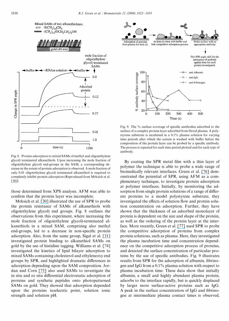
Fig. 8. Protein adsorption to mixed SAMs of methyl and oligo(ethyleneglycol) terminated alkanethiols. Upon increasing the mole fraction ofoligo(ethylene glycol) end-groups in the SAM, a corresponding de-crease in the extent of protein adsorption is observed. A mole fraction ofonly 0.41 oligo(ethylene glycol) terminated alkanethiol is required tocompletely inhibit protein adsorption (Reproduced from Mrksich et al.[30]).
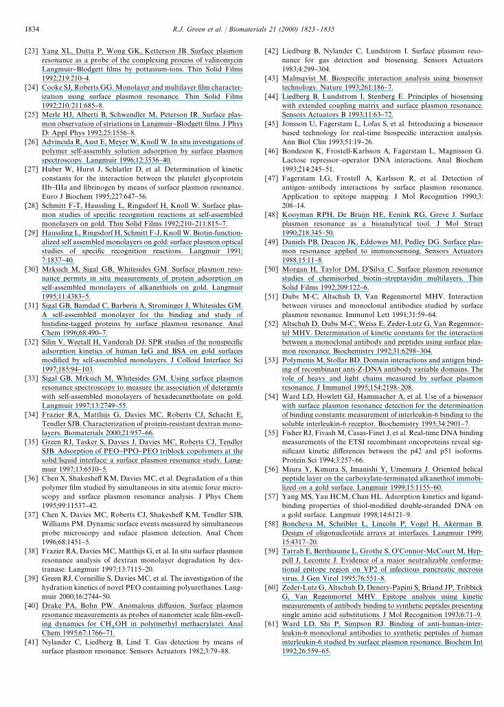
Fig. 9. The % surface coverage of speci"c antibodies adsorbed to thesurface of a complex protein layer adsorbed from blood plasma. A poly-styrene substrate is incubated in a 0.1% plasma solution for varyingtime periods after which the system is washed with bu!er before thecomposition of the protein layer can be probed by a speci"c antibody.The process is repeated for each time period plotted and for each type ofantibody.
those determined from XPS analysis. AFM was able tocon"rm that the protein layer was incomplete.
Mrksich et al. [30] illustrated the use of SPR to probethe protein resistance of SAMs of alkanethiols witholigo(ethylene glycol) end groups. Fig. 8 outlines theobservations from this experiment, where increasing themole fraction of oligo(ethylene glycol)-terminated al-kanethiols in a mixed SAM, comprising also methylend-groups, led to a decrease in non-speci"c proteinadsorption. Also, from the same group, Sigal et al. [31]investigated protein binding to alkanethiol SAMs ongold by the use of histidine tagging. Williams et al. [74]investigated the kinetics of lipid bilayer adsorption tomixed SAMs containing cholesterol and ethyleneoxy endgroups by SPR, and highlighted dramatic di!erences inadsorption depending upon the SAM composition. Jor-dan and Corn [75] also used SAMs to investigate thein situ and ex situ di!erential electrostatic adsorption ofproteins and synthetic peptides onto photopatternedSAMs on gold. They showed that adsorption dependedupon the proteins isoelectric point, solution ionicstrength and solution pH.
By coating the SPR metal "lm with a thin layer ofpolymer the technique is able to probe a wide range ofbiomedically relevant interfaces. Green et al. [76] dem-onstrated the potential of SPR, using AFM as a com-plementary technique, to investigate protein adsorptionat polymer interfaces. Initially, by monitoring the ad-sorption from single protein solutions of a range of di!er-ent proteins to a model polystyrene substrate, theyinvestigated the e!ects of solution #ow and protein solu-tion concentration on adsorption. Further, they haveshown that the thickness of an adsorbed monolayer ofprotein is dependent on the size and shape of the protein,as well as the ordering of the protein layer at the inter-face. More recently, Green et al. [77] used SPR to probethe competitive adsorption of proteins from complexprotein solutions, such as plasma. Here, they investigatedthe plasma incubation time and concentration depend-ence on the competitive adsorption process of proteins,and detected the surface concentration of particular pro-teins by the use of speci"c antibodies. Fig. 9 illustratesresults from SPR for the adsorption of albumin, "brino-gen and IgG from a 0.1% plasma solution with respect toplasma incubation time. These data show that initiallyalbumin, a small and highly abundant plasma protein,adsorbs to the interface rapidly, but is quickly displacedby larger more surface-active proteins such as IgG.A peak in the surface concentration of IgG and "brino-gen at intermediate plasma contact times is observed,
1830 R.J. Green et al. / Biomaterials 21 (2000) 1823}1835

Fig. 10. (a) A schematic diagram illustrating passive adsorption of anantibody to a polystyrene surface (top), and speci"c binding of a bio-tinylated antibody to a streptavidin-coated polystyrene surface (bot-tom); (b) (top) SPR binding pro"le of antiferritin and biotinylatedantiferritin to polystyrene, and (bottom) SPR binding pro"le of ferritinsolution to the resulting passively adsorbed and biotinylated antifer-ritin antibody surfaces. Twice the amount of ferritin binds to thebiotinylated antibody surface than to the passively adsorbed antibodysurface.
Fig. 11. A diagram illustrating the adsorption of albumin to uncoatedand pluronic-coated polystyrene surfaces. The lengths of both the PEOand PPO blocks determine the ability of the pluronic to inhibit adsorp-tion of the protein.
while these proteins "rst displace surface-adsorbed pro-teins, before themselves being displaced from the inter-face by proteins of a higher surface a$nity. In a similarstudy, Muller et al. [78] employed SPR as part of aninvestigation of the competitive adsorption of gelatinand sodium dodecylbenzenesulfonate at hydrophobicsurfaces.
Similar experiments have also been carried out toinvestigate polymer surfaces that are designed to pro-mote selective interactions. As shown in Fig. 10, Davieset al. [79] combined SPR and AFM to study antibody-coated microtiter wells, using SPR to probe the kineticsof the interactions and AFM to visualize the surfacecoverage of the antibody. Davies et al. [80] have alsodemonstrated the use of SPR to probe interactions at thesurfaces of biomaterials by binding alkanethiols withvarying end groups to silver and monitoring the albumin,IgG and "brinogen adsorption.
The interaction of proteins with biomedical polymersspeci"cally designed to inhibit protein adsorption hasalso been investigated, and again AFM has proved a use-ful complementary tool to SPR [34,81}83]. One examplehas been the study of protein adsorption to a range ofPEO/PPO/PEO tri-block copolymers (Pluronics, BASFCo.) of varying PEO and PPO content [81]. The amountof protein adsorbed to each surface has been related tothe strength of binding of the copolymer to the hydro-phobic polystyrene surface (related to the PPO blocksize) and to the length of the extending PEO chain. Forexample, Fig. 11 shows the adsorption of albumin toa high molecular weight Pluronic (F127) with a longPPO segment and 70% PEO, and a low molecular
R.J. Green et al. / Biomaterials 21 (2000) 1823}1835 1831

Fig. 12. Surface engineering of a biotinylated polymer studied by SPR. Avidin speci"cally binds only to the biotinylated polymer.
weight Pluronic (L31) with a short PPO segment and10% PEO. The adsorption of albumin to a polystyrenesurface is included as a control, being the SPR shift forthe adsorption of a monolayer of protein. Fig. 11 showsthat both Pluronics signi"cantly reduce the adsorption ofalbumin, with Pluronic L31 adsorbing about 30% ofa monolayer and Pluronic F127 successfully inhibitingadsorption. Each surface has been visualized by AFM,revealing protein surface coverages that agree with theSPR data. This work has highlighted the ability of SPRto qualitatively compare the response of proteins at vari-ous surfaces, where sub-monolayer di!erence in proteinadsorption can be detected.
3.2.4. Tissue engineeringThe engineering of polymer surfaces is an important"eld of biomaterial science that can be greatly assisted bySPR studies of dynamic surface changes. Cannizzaroet al. [84,85] have demonstrated this role for SPR analy-sis in their study of a biotinylated biodegradable blockcopolymer poly(lactic acid)}poly(ethylene glycol)}biotin(PLA}PEG}biotin). The polymer can be easily surfaceengineered utilizing avidin-to-biotin interactions. Freebiotin groups at the polymer surface are used to anchoravidin molecules, which retain the ability to bind furtherbiotin molecules. Therefore, biotinylated ligands (e.g.biotinylated cell adhesion peptides for tissue engineeringapplications) can be immobilized on the polymer surfaceusing the avidin as a sandwich linker. SPR analysis hasbeen used to study the kinetics of the avidin surfaceimmobilization. An example of the SPR data from thissample is shown in Fig. 12. The data compares avidinbinding to PLA}PEG}biotin and PLA}PEG. In thelatter case, the PEG chains inhibit non-speci"c avidinadsorption to the polymer surface and the SPR angleshift is small. In the case of PLA}PEG}biotin the speci"cavidin-to-biotin interaction causes avidin surface immo-
bilization, as observed by the rapid rise in the SPR angle.This study proved that the biotinylated polymer can actas a foundation for the proposed surface engineeringstrategy.
4. Current and future instrumental developments
Vadgama and Crump [86], and Chaiken et al. [87]have both reviewed various types of biosensors. Thesereviews have identi"ed the disadvantages of SPR as beingits lack of sensitivity when monitoring the adsorption oflow molecular weight adsorbates, and the rate-limitingfactor of mass transport-a!ecting kinetic analysis. Adap-tations of SPR have been introduced to attempt to in-crease its sensitivity. As discussed earlier, a commercialSPR biosensor system attempts to improve the sensitivityof SPR by the use of an extended coupling dextranmatrix bound to the gold surface that increases the ap-parent surface area available for protein binding [45,88].On a similar note, particle-enhanced sensitivity of SPRhas been demonstrated for the investigation of anti-gen}antibody binding by Leung et al. [89]. Kooymanet al. [90] and Lenferink et al. [91] have demonstratedimproved SPR sensitivity by altering the instrument toincorporate a vibrating mirror into its optics con"gura-tion. Also, an SPR-based #uoro-immunoassay systemhas been shown to improve sensitivity and has beendemonstrated by investigating the binding of the bio-tin}streptavidin system [92,93]. Another instrumentbased on SPR, the resonant mirror, has incorporatedSPR with the increased sensitivity obtained by waveguid-ing devices [94,95].
SPR has been used alongside many other analyticaltechniques and, as highlighted throughout this review,has proven to be a highly useful complementarytechnique. Caruso et al. [96] have studied surfactant
1832 R.J. Green et al. / Biomaterials 21 (2000) 1823}1835

adsorption to gold and have used the QCM techniqueemploying SPR to determine the calibration factors forthe QCM. SPR has also proved a useful complementarytechnique alongside high-performance liquid chromato-graphy (HPLC), where it has been used to optimizeelution conditions for a$nity chromatography [97].SPR has also been used to develop a detector respondingto changes in refractive index to use in HPLC [98]. TheSPR device has compared favourably with the moretraditionally used refractive index (RI) detector, provid-ing stable baselines approximately 50 min faster than theRI detector. Krone et al. [99] successfully interfaced SPRwith matrix-assisted laser desorption/ionization massspectroscopy (MALDI), where they claim that the com-bined approach unites the real-time capabilities of SPRwith the qualitative speci"city of mass spectroscopy.
As discussed earlier during this review, SPR and AFMare highly complementary techniques, which Chen et al.[36,37] successfully interfaced to produce an instrumentthat can simultaneously follow the changes in topogra-phy and the dynamic surface kinetics of a wide range ofinterfaces. The combined instrument has been used toinvestigate protein adsorption to polymeric interfaces[36], and also to monitor the degradation rates of bio-medical polymers for use in controlled drug release sys-tems [37].
5. Conclusions and future outlook
This review has attempted to highlight the wide varietyof work currently in the literature employing SPR analy-sis, with particular reference to investigations relevant tobiomaterial design. The capabilities of SPR has allowedthe technique to make an important impact towards thesurface analysis of biomaterials. SPR has proven to bea valuable tool to investigate a number of dynamicevents, and its diversity allows interfacial analysis atalmost any surface. SPR can determine dielectric proper-ties (refractive index or "lm thickness) as well as beingable to follow a range of interfacial processes in situ suchas adsorption/desorption, hydration and degradation.Flexibility of #ow cell con"gurations allow the study ofthese processes in relevant solution conditions of temper-ature, #ow rate, pH and ionic strength.
A major feature of this review has been the success ofdirectly interfacing SPR with other techniques, such asAFM. These two complimentary techniques presenta promising potential to provide a wealth of informationregarding the real-time analysis of biomaterials. AFM isable to visual surface events in situ alongside SPR, pro-viding kinetic information as well as the spatial resolu-tion of surface topography. This powerful combinationhas the potential to study mechanisms of drug releasefrom biodegradable systems and the in vitro response ofbiological #uids to biomaterial surfaces.
References
[1] Reid G, Busscher HJ, Sharma S, Mittelman MW, McIntyre S.Surface properties of catheters, stents and bacteria associated withurinary tract infections. Surf Sci Rep 1995;21:251}73.
[2] Shakeshe! KM, Davies MC, Roberts CJ, Tendler SJB, WilliamsPM. The role of scanning probe microscopy in drug deliveryresearch. Crit Rev Ther Drug Carrier Syst 1996;13:185}223.
[3] Davies MC, Roberts CJ, Tendler SJB, Williams PM. The surfaceanalysis of polymeric biomaterials. In: Braybrook J, editor. Bio-compatibility: assessment of materials and devices for medicalapplications. Chichester: Wiley, 1997. p. 65}100.
[4] Ratner BD. Characterization of biomedical surfaces. CardiovascPathol 1993;2:87S}100S.
[5] Striebel Ch, Brecht A, Ganglitz G. Characterization of biomem-branes by spectral ellipsometry, surface plasmon resonance andinterferometry with regard to biosensor application. Biosens Bio-electron 1994;9:139}46.
[6] Davies J. Surface plasmon resonance*the technique and its ap-plications to biomaterial processes. Nanobiology 1994;3:5}16.
[7] Lundstrom I. Real-time biospeci"c interaction analysis. BiosensBioelectron 1994;9:725}36.
[8] Boardman AD, editor. Electromagnetic surface modes. Chiches-ter: Wiley, 1982.
[9] De Bruijn HE, Kooyman RPH, Greve J. Choice of metal andwavelength for SPR sensors: some considerations. Appl Opt1992;31:440}2.
[10] Otto A. Excitation of nonradiative surface plasma waves in silverby the method of frustrated total re#ection. Z Phys 1968;216:398}410.
[11] Kretchmann E. The determination of the optical constants ofmetals by excitation of surface plasmons. Z Phys 1971;241:313}24.
[12] Fagerstam LG, Frostell-Karlsson A, Karlsson R, Persson B,Ronnberg I. Biospeci"c interaction analysis using SPR detectionapplied to kinetic, binding site and concentration analysis.J Chromatogr 1992;597:397}410.
[13] Matsubara K, Kawata S, Minami S. A compact surface plasmonresonance sensor for water in process. Appl Spectrosc 1988;42:1375}9.
[14] Cheng YL, Darst SA, Robertson CR. Bovine serum albumin anddesorption rates on solid surfaces with varying surface properties.J Colloid Interface Sci 1987;118:212}23.
[15] Karlsson R, Michaelsson A, Mattsson L. Kinetics of analysis ofmonoclonal antibody}antigen interactions with a new biosensorbased analytical system. J Immunol Meth 1991;145:229}40.
[16] De Bruijn HE, Kooyman RPH, Greve J. Determination of dielec-tric permittivity and thickness of a metal layer from an SPRexperiment. Appl Opt 1990;29:1974}8.
[17] Linnert T, Mulvaney P, Henglein A. Surface chemistry of collo-idal silver: surface plasmon damping by chemisorbed I-, SH- andC
6H
5S-. J Phys Chem 1993;97:679}82.
[18] Voskoboinikov A, Nakhodkin N, Kryn Ko Y, Kulik S, Melnik P,Sheka D. Surface plasmon peak intensity dependance on the oxygencoverage at metal surfaces. Solid State Commun 1994;90:27}30.
[19] Pockrand I. Surface plasmon oscillations at silver surfaces withtransparent and adsorbing coatings. Surf Sci 1978;12:577}88.
[20] De Bruijn HE, Altenburg BSF, Kooyman RPH, Greve J. Deter-mination of thickness and dielectric constant of thin transparentdielectric layers using surface plasmon resonance. Opt Commun1991;82:425}32.
[21] Vukusic PS, Sambles JR, Wright JD. Surface plasmon resonancecharaterization of spin-deposited phthalocyanine "lms. J MaterChem 1992;2:1105}6.
[22] Wagner Th, Roth S. Surface plasmon resonance investigations ofLangmuir}Blodgett"lms of donor}acceptor substituted polyenes:linear optical and electro-optic properties. Synth Met 1993;54:307}14.
R.J. Green et al. / Biomaterials 21 (2000) 1823}1835 1833
[23] Yang XL, Dutta P, Wong GK, Ketterson JB. Surface plasmonresonance as a probe of the complexing process of valinomycinLangmuir}Blodgett "lms by pottasium-ions. Thin Solid Films1992;219:210}4.
[24] Cooke SJ, Roberts GG. Monolayer and multilayer "lm character-ization using surface plasmon resonance. Thin Solid Films1992;210/211:685}8.
[25] Merle HJ, Alberti B, Schwendler M, Peterson IR. Surface plas-mon observation of striations in Langmuir}Blodgett "lms. J PhysD: Appl Phys 1992;25:1556}8.
[26] Advincula R, Aust E, Meyer W, Knoll W. In situ investigations ofpolymer self-assembly solution adsorption by surface plasmonspectroscopy. Langmuir 1996;12:3536}40.
[27] Huber W, Hurst J, Schlatler D, et al. Determination of kineticconstants for the interaction between the platelet glycoproteinIIb}IIIa and "brinogen by means of surface plasmon resonance.Euro J Biochem 1995;227:647}56.
[28] Schmitt F-T, Haussling L, Ringsdorf H, Knoll W. Surface plas-mon studies of speci"c recognition reactions at self-assembledmonolayers on gold. Thin Solid Films 1992;210}211:815}7.
[29] Haussling L, Ringsdorf H, Schmitt F-J, Knoll W. Biotin-function-alized self assembled monolayers on gold: surface plasmon opticalstudies of speci"c recognition reactions. Langmuir 1991;7:1837}40.
[30] Mrksich M, Sigal GB, Whitesides GM. Surface plasmon reso-nance permits in situ measurements of protein adsorption onself-assembled monolayers of alkanethiols on gold. Langmuir1995;11:4383}5.
[31] Sigal GB, Bamdad C, Barberis A, Strominger J, Whitesides GM.A self-assembled monolayer for the binding and study ofhistidine-tagged proteins by surface plasmon resonance. AnalChem 1996;68:490}7.
[32] Silin V, Weetall H, Vanderah DJ. SPR studies of the nonspeci"cadsorption kinetics of human IgG and BSA on gold surfacesmodi"ed by self-assembled monolayers. J Colloid Interface Sci1997;185:94}103.
[33] Sigal GB, Mrksich M, Whitesides GM. Using surface plasmonresonance spectroscopy to measure the association of detergentswith self-assembled monolayers of hexadecanethiolate on gold.Langmuir 1997;13:2749}55.
[34] Frazier RA, Matthijs G, Davies MC, Roberts CJ, Schacht E,Tendler SJB. Characterization of protein-resistant dextran mono-layers. Biomaterials 2000;21:957}66.
[35] Green RJ, Tasker S, Davies J, Davies MC, Roberts CJ, TendlerSJB. Adsorption of PEO}PPO}PEO triblock copolymers at thesolid/liquid interface: a surface plasmon resonance study. Lang-muir 1997;13:6510}5.
[36] Chen X, Shakeshe!KM, Davies MC, et al. Degradation of a thinpolymer "lm studied by simultaneous in situ atomic force micro-scopy and surface plasmon resonance analysis. J Phys Chem1995;99:11537}42.
[37] Chen X, Davies MC, Roberts CJ, Shakeshe! KM, Tendler SJB,Williams PM. Dynamic surface events measured by simultaneousprobe microscopy and suface plasmon detection. Anal Chem1996;68:1451}5.
[38] Frazier RA, Davies MC, Matthijs G, et al. In situ surface plasmonresonance analysis of dextran monolayer degradation by dex-tranase. Langmuir 1997;13:7115}20.
[39] Green RJ, Corneillie S, Davies MC, et al. The investigation of thehydration kinetics of novel PEO containing polyurethanes. Lang-muir 2000;16:2744}50.
[40] Drake PA, Bohn PW. Anomalous di!usion. Surface plasmonresonance measurements as probes of nanometer scale "lm-swell-ing dynamics for CH
3OH in poly(methyl methacrylate). Anal
Chem 1995;67:1766}71.[41] Nylander C, Liedberg B, Lind T. Gas detection by means of
surface plasmon resonance. Sensors Actuators 1982;3:79}88.
[42] Liedburg B, Nylander C, Lundstrom I. Surface plasmon reso-nance for gas detection and biosensing. Sensors Actuators1983;4:299}304.
[43] Malmqvist M. Biospeci"c interaction analysis using biosensortechnology. Nature 1993;261:186}7.
[44] Liedberg B, Lundstrom I, Stenberg E. Principles of biosensingwith extended coupling matrix and surface plasmon resonance.Sensors Actuators B 1993;11:63}72.
[45] Jonsson U, Fagerstam L, Lofas S, et al. Introducing a biosensorbased technology for real-time biospeci"c interaction analysis.Ann Biol Clin 1993;51:19}26.
[46] Bondeson K, Frostell-Karlsson A, Fagerstam L, Magnisson G.Lactose repressor}operator DNA interactions. Anal Biochem1993;214:245}51.
[47] Fagerstam LG, Frostell A, Karlsson R, et al. Detection ofantigen}antibody interactions by surface plasmon resonance.Application to epitope mapping. J Mol Recognition 1990;3:208}14.
[48] Kooyman RPH, De Bruijn HE, Eenink RG, Greve J. Surfaceplasmon resonance as a bioanalytical tool. J Mol Struct1990;218:345}50.
[49] Daniels PB, Deacon JK, Eddowes MJ, Pedley DG. Surface plas-mon resonance applied to immunosensing. Sensors Actuators1988;15:11}8.
[50] Morgan H, Taylor DM, D'Silva C. Surface plasmon resonancestudies of chemisorbed biotin}streptavidin multilayers. ThinSolid Films 1992;209:122}6.
[51] Dubs M-C, Altschuh D, Van Regenmortel MHV. Interactionbetween viruses and monoclonal antibodies studied by surfaceplasmon resonance. Immunol Lett 1991;31:59}64.
[52] Altschuh D, Dubs M-C, Weiss E, Zeder-Lutz G, Van Regenmor-tel MHV. Determination of kinetic constants for the interactionbetween a monoclonal antibody and peptides using surface plas-mon resonance. Biochemistry 1992;31:6298}304.
[53] Polymenis M, Stollar BD. Domain interactions and antigen bind-ing of recombinant anti-Z-DNA antibody variable domains. Therole of heavy and light chains measured by surface plasmonresonance. J Immunol 1995;154:2198}208.
[54] Ward LD, Howlett GJ, Hammacher A, et al. Use of a biosensorwith surface plasmon resonance detection for the determinationof binding constants: measurement of interleukin-6 binding to thesoluble interleukin-6 receptor. Biochemistry 1995;34:2901}7.
[55] Fisher RJ, Fivash M, Casas-Finet J, et al. Real-time DNA bindingmeasurements of the ETSI recombinant oncoproteins reveal sig-ni"cant kinetic di!erences between the p42 and p51 isoforms.Protein Sci 1994;3:257}66.
[56] Miura Y, Kimura S, Imanishi Y, Umemura J. Oriented helicalpeptide layer on the carboxylate-terminated alkanethiol immobi-lized on a gold surface. Langmuir 1999;15:1155}60.
[57] Yang MS, Yau HCM, Chan HL. Adsorption kinetics and ligand-binding properties of thiol-modi"ed double-stranded DNA ona gold surface. Langmuir 1998;14:6121}9.
[58] Boncheva M, Scheibler L, Lincoln P, Vogel H, Akerman B.Design of oligonucleotide arrays at interfaces. Langmuir 1999;15:4317}20.
[59] Tarrab E, Berthiaume L, Grothe S, O'Connor-McCourt M, Hep-pell J, Lecomte J. Evidence of a major neutralizable conforma-tional epitope region on VP2 of infectious pancreatic necrosisvirus. J Gen Virol 1995;76:551}8.
[60] Zeder-Lutz G, Altschuh D, Denery-Papini S, Briand JP, TribbickG, Van Regenmortel MHV. Epitope analysis using kineticmeasurements of antibody binding to synthetic peptides presentingsingle amino acid substitutions. J Mol Recognition 1993;6:71}9.
[61] Ward LD, Shi P, Simpson RJ. Binding of anti-human-inter-leukin-6 monoclonal antibodies to synthetic peptides of humaninterleukin-6 studied by surface plasmon resonance. Biochem Int1992;26:559}65.
1834 R.J. Green et al. / Biomaterials 21 (2000) 1823}1835

[62] Johne B, Gadnell M, Hansen K. Epitope mapping and bindingkinetics of monoclonal antibodies by real-time biospeci"c interac-tion analysis using surface plasmon resonance. J Immunol Meth1993;160:191}8.
[63] Minunni M. Simultaneous determination of b2-microglobulinand IgE using real-time biospeci"c interaction analysis (BIA).Anal Lett 1995;28:933}44.
[64] Raut S, Ga!ney RJ. Interaction of heparin with "brinogen usingsurface plasmon resonance technology: investigation of heparinbinding site on "brinogen. Thromb Res 1996;81:503}9.
[65] Van Delden CJ, Lens JP, Kooyman RPH, Engbers GHM, Fei-jen J. Heparinization of gas plasma modi"ed polystyrene surfacesand the interactions of these surfaces with proteins studied withsurface plasmon resonance. Biomaterials 1997;18:845}52.
[66] Frazier RA, Vansteenkiste SO, Black FE, et al. SPR study ofprotein adsorption to heparinized dextrans. Proceedings of theInternational Symposium on Control Related Bioactive Mate-rials, vol. 24. 1997, p. 949}50.
[67] Gaus K, Hall EAH. Surface plasmon resonance measurement ofthe binding of low-density lipoprotein at a heparin surface. J Col-loid Interface Sci 1999;217:111}8.
[68] Plant AL, Brigham-Burke M, Petrella EC, O'Shannessy DJ.Phospholipid/alkanethiol bilayers for cell-surface receptorstudies by surface plasmon resonance. Anal Biochem 1995;226:342}8.
[69] Silin VI, Balcytis GA, Zhizhin GN, Yakovlev VA. Application ofsurface electromagnetic wave and surface plasmon techniques ina protein adsorption study and sensor construction. Vib Spec-trosc 1993;5:133}42.
[70] Silin V, Weetall H, Vanderah DJ. SPR studies of the nonspeci"cadsorption kinetics of human IgG and BSA on gold surfacesmodi"ed by self-assembled monolayers (SAMs). J Colloid Inter-face Sci 1997;185:94}103.
[71] Flanagan MT, Pantell RH. Surface plasmon resonance and im-munosensors. Electron Lett 1984;20:968}70.
[72] Zenhausern F, Adrian M, Descouts P. Solution structure anddirect imaging of "bronectin adsorption to solid surfaces byscanning force microscopy and cryo-electron microscopy. J Elec-tron Microsc 1993;42:378}88.
[73] Caruso F, Furlong DN, Kingshott P. Characterization offerritin adsorption onto gold. J Colloid Interface Sci 1997;186:129}40.
[74] Williams LM, Evans SD, Flynn TM, et al. Kinetics of the unroll-ing of small unilamellar phospholipid vesicles onto self-assembledmonolayers. Langmuir 1997;13:751}7.
[75] Jordan CE, Corn RM. Surface plasmon resonance imagingmeasurements of electrostatic biopolymer adsorption onto chem-ically modi"ed gold surfaces. Anal Chem 1997;69:1449}56.
[76] Green RJ, Davies J, Davies MC, Roberts CJ, Tendler SJB. Surfaceplasmon resonance for real time in situ analysis of protein adsorp-tion to polymer surfaces. Biomaterials 1997;18:405}13.
[77] Green RJ, Davies MC, Roberts CJ, Tendler SJB. Competitiveadsorption as observed by surface plasmon resonance. Bio-materials 1999;20:385}91.
[78] Muller D, Malmsten M, Bergenstahl B, Hessing J, Olijve J, MoriF. Competitive adsorption of gelatin and sodium dodecylben-zenesulfonate at hydrophobic surfaces. Langmuir 1998;14:3107}14.
[79] Davies J, Roberts CJ, Dawkes AC, et al. Use of scanning probemicroscopy and surface plasmon resonance as analytical tools inthe study of antibody-coated microtitre wells. Langmuir 1994;10:2654}61.
[80] Davies J, Allen A, Bruce I, et al. Dynamic, in situ, techniques forprobing surface properties of biomaterials. In: West R, Batts G,editors. Surface properties of biomaterials. Cambridge: Butter-worth-Heinemann Ltd, 1994. p. 117}32.
[81] Green RJ, Davies MC, Roberts CJ, Tendler SJB. A surface plas-mon resonance study of albumin adsorption to PEO}PPO}PEOtriblock copolymers. J Biomed Mater Res 1998;42:165}71.
[82] McGurk SL, Green RJ, Sanders GHW, et al. Molecular interac-tions of biomolecules with surface-engineered interfaces usingatomic force microscopy and surface plasmon resonance. Lan-gmuir 1999;15:5136}40.
[83] Pavey KD, Olli! CJ. SPR analysis of the total reduction ofprotein adsorption to surfaces coated with mixtures of long- andshort-chain polyethylene oxide block copolymers. Biomaterials1999;20:885}90.
[84] Cannizzaro SM, Padera RF, Langer R, et al. A novel biotinylateddegradable polymer for cell-interactive applications. BiotechnolBioeng 1998;58:529}35.
[85] Black FE, Hartshorne M, Davies MC, et al. Surface engineeringand surface analysis of a biodegradable polymer with biotinylatedend groups. Langmuir 1999;15:3157}61.
[86] Vadgama P, Crump PW. Biosensors: recent trends. Analyst1992;117:1657}70.
[87] Chaiken I, Rose S, Karlsson R. Analysis of macromolecularinteractions using immobilized ligands. Anal Biochem 1992;201:197}210.
[88] O'Shannessy DJ, Brigham-Burke K, Peck K. Immobilizationchemistries suitable for use in the BIAcore surface plasmon reson-ance detector. Anal Biochem 1992;205:132}6.
[89] Leung P-T, Pollard-Knight D, Malan GP, Finlan F. Modelling ofparticle-enhanced sensitivity of surface plasmon resonance bio-sensor. Sensors Actuators B 1994;22:175}80.
[90] Kooyman RPH, Lenferink ATM, Eenink RG, Greve J. Vibratingmirror surface plasmon resonance immunosensor. Anal Chem1991;63:83}5.
[91] Lenferink ATM, Kooyman RPH, Greve J. An improved opticalmethod for surface plasmon resonance. Sensors ActuatorsB 1991;3:261}5.
[92] Attridge JW, Daniels PB, Deacon JK, Robinson GA, DavidsonGP. Sensitivity enhancement of optical immunosensors by the useof surface plasmon resonance #uoroimmunoassay. Biosens Bio-electron 1991;6:201}14.
[93] Schmitt F-T, Weisenhorn AL, Hansma PK, Knoll W. Interfacialrecognition reactions as seen by #uorescence- surface plasmon-and atomic force microscopies. Makromol Chem: MacromolSymp 1991;46:133}43.
[94] Cush R, Cronin JM, Stewart WJ, Maule CH, Mollay J, GoddardNJ. The resonant mirror: a novel optical biosensor for directsensing of biomolecular interactions. Part I: principle of operationand associated instrumentation. Biosens Bioelectron 1993;8:347}53.
[95] Buckle PE, Davies RJ, Kinning T, Yeung D, Edwards PR,Pollardknight D, Lowe CR. The resonant mirror: a novel opticalsensor for direct sensing of biomolecular interactions. Part II:applications. Biosens Bioelectron 1993;8:355}63.
[96] Caruso F, Serizawa T, Furlong DN, Okahata Y. Quartz crystalmicrobalance and surface plasmon resonance study of surfactantadsorption to gold and chromium oxide surfaces. Langmuir1995;11:1546}52.
[97] Nice E, Lackmann M, Smyth F, Fabri L, Burgess AW. Synergiesbetween micropreparative high performance liquid chromatogra-phy and an instrumental optical biosensor. J Chromatogr A1994;660:164}85.
[98] Cepria G, Castillo JR. Surface plasmon resonance-based detec-tion*an alternative to refractive index detection in high-perfor-mance liquid chromatography. J Chromatogr A 1997;759:27}35.
[99] Krone JR, Nelson RW, Dogruel D, Williams P, Granzow R.BIA/MS: interfacing biomolecular interaction analysis with massspectrometry. Anal Biochem 1997;244:124}32.
R.J. Green et al. / Biomaterials 21 (2000) 1823}1835 1835