SUCO DE LARANJA E VITAMINA C: EFEITO SOBRE A ...
226
Universidade Federal do Rio Grande do Sul Centro de Biotecnologia do Estado do Rio Grande do Sul Programa de Pós-Graduação em Biologia Celular e Molecular SUCO DE LARANJA E VITAMINA C: EFEITO SOBRE A ESTABILIDADE GENÔMICA Tese de Doutorado Silvia Isabel Rech Franke Porto Alegre, maio de 2006
-
Upload
khangminh22 -
Category
Documents
-
view
2 -
download
0
Transcript of SUCO DE LARANJA E VITAMINA C: EFEITO SOBRE A ...
Universidade Federal do Rio Grande do Sul Centro de Biotecnologia do Estado do Rio Grande do Sul
Programa de Pós-Graduação em Biologia Celular e Molecular
SUCO DE LARANJA E VITAMINA C: EFEITO SOBRE A ESTABILIDADE GENÔMICA
Tese de Doutorado
Silvia Isabel Rech Franke
Porto Alegre, maio de 2006
II
Silvia Isabel Rech Franke
SUCO DE LARANJA E VITAMINA C: EFEITO SOBRE A ESTABILIDADE GENÔMICA
Tese submetida ao Programa de Pós-Graduação em Biologia Celular e Molecular da UFRGS como parte dos requisitos para a obtenção do Grau de Doutor
Orientador: João Antonio Pêgas Henriques Co-orientador: Bernardo Erdtmann
Porto Alegre, maio de 2006
III
Silvia Isabel Rech Franke
SUCO DE LARANJA E VITAMINA C: EFEITO SOBRE A ESTABILIDADE GENÔMICA
Tese submetida ao Programa de Pós-Graduação em Biologia Celular e Molecular da UFRGS como parte dos requisitos para a obtenção do Grau de Doutor
Aprovada em Porto Alegre, 23 de maio de 2006
Banca Examinadora
_____________________________________ Dr. Adalberto Luis Val Instituto Nacional de Pesquisas Amazônicas _____________________________________ Dr. José Cláudio Fonseca Moreira Departamento de Bioquímica, Universidade Federal do Rio Grande do Sul _____________________________________ Dra. Célia Regina Ribeiro da Silva Carlini Centro de Biotecnologia, Universidade Federal do Rio Grande do Sul ______________________________________ Dra. Mirian Salvador (suplente) Instituto de Biotecnologia, Universidade de Caxias do Sul
IV
AGRADECIMENTOS
Ao Professor Dr. João Antonio Pêgas Henriques, pela orientação e pela confiança no meu trabalho desde o mestrado. Por todas as dores de cabeça que causei durante a trajetória de erros e acertos. Pelas inúmeras contribuições relevantes. Por me receber em sua casa, privando-se de parte de seu convívio com sua família.
Ao Dr. Bernardo Erdtmann, que co-orientou várias fases deste trabalho, e sempre
questionou os paradigmas da nutrição em converas muito instigantes. À Dra. Juliana da Silva por todos os momentos de discussão de redação de artigos e de
fases experimentais amplamente discutidas entre inúmeros cafezinhos. Muito obrigada! Às Dras. Jaqueline Picada, Mirian Salvador pelas análises críticas da redação dos artigos
de revisão (anexos desta tese), bem como do projeto do exame de qualificação (incluindo a Dra. Jenifer Saffi). E, por todos os conselhos dados sempre que solicitadas! Obrigada pela presteza!
À Dra. Christine Gaylarde, pelas revisões da redação em inglês de todos os artigos que
publiquei. Pela agilidade e gentileza, mesmo em horas difícies. À amiga Miriam Benicio pela revisão da redação da parte em português da tese. Pela
agilidade e competência incondicional e bem disposta nos momentos mais inadequados, como feriados, domingos e em meio a “pilhas de análises e relatórios” por terminar.
À Dra. Célia Carlini, por ter sido a professora que sempre revisou os trabalhos oriundos
de etapas cruciais da minha vida de mestranda e doutoranda. Obrigada por todas as sugestões e me fazer acreditar que “posso estar no caminho certo”. Obrigada pelo estímulo à pesquisa!
Ao Dr. Jorge Guimarães pelos conselhos dados em nossas reuniões de comissões de
acompanhamento do PPGBCM. Nunca esquecerei a frase “Acredito em você guria! Segue em frente”.
Às Dras. Jenifer Saffi, Ana Lígia de Paula Ramos e Kátia Valença e ao Dr. Thales R. O.
Freitas por gentilmente disponibilizarem seus laboratórios para que realizasse meus experimentos.
Ao Dr. Johnny F. Dias, a Dra. Lúcia Yoneama, a Raquel Gulian e aos demais técnicos
do laboratório de Implantação Iônica do Instituto de Física da UFRGS que contribuíram na análise do número recorde de amostras no PIXE seguindo um cronogama pouco convencional.
Às Dras. Aoi Masuda, Marilene Vainstain e Célia Carlini e ao Dr. Arnaldo Zaha por
nunca me “deixarem na mão” quando precisei de algum aquipamento ou reagente. Aos funcionários e técnicos de laboratório, Gabriel Rubensam, Jaque de Deos, Rose e
Sueli, que comigo conviveram durante um período e que tornaram a minha vida científica tão mais fácil. Pela ajuda, paciência e carinho. Saudades!
V
Ao meu colega Daniel Prá, co-autor e companheiro para todas as discussões referentes à tese. Por todos os demais experimentos que realizamos fora do tema específico desta tese e que já estão “virando” artigos. Pelas oportunidades de aprender estatística, inglês, intercâmbios com outras universidades. Ter um colega como você é um “presente”. Obrigada ainda por ter sido o ombro amigo e por todas as horas que largaste todos os teus compromissos para vir em meu auxílio.
Aos meus colegas de laboratório Ana Amélia, Izabel, Miriana, Sissa, Jaque Cardone,
Cassiana, Heique, Bacana, Luiz Miranda, Cassius, Nusha, Matheus, Dinara, Diego, Renato, entre outros. Pelos sorrisos, pelas lágrimas, pelas gargalhadas, pelas discussões, piadas e por todos os momentos que vivemos juntos, que fazem com que a vida valha a pena.
À Silvia Centeno, Luciano, Graziela, Márcia Vaz, bem como ao Elmo e a Elen pela
competência aliada a tanta simpatia para resolver diversos temas relacionados à minha pesquisa.
Aos meus pais, por todo esforço na minha formação. Pelo apoio durante a execução do
trabalho. À Jusssara, Rubens, Rafael e Raquel Prá, pelos conselhos e pela carinhosa acolhida. À Marli e a Margô, secretárias de meu lar, por terem realmente “segurado as pontas”
durante os vários períodos de ausência, principalmente pelo carinho dado aos meus filhos. Ao Caco, por sempre dar respaldo aos meus filhos na minha ausência. Aos meus filhos, Matheus e Gabriel, pelo tempo que não passamos juntos devido à
distância que separa Porto Alegre de Santa Cruz do Sul. Pela alegria de ter vocês em minha vida.
A todas as demais pessoas que, embora não listadas, também contribuíram na execução
deste trabalho. À UNISC por ter investido em minha qualificação. Ao GENOTOX que subsidiu as despesas desta tese.
VI
APRESENTAÇÃO
Em 1999, tive a oportunidade de visitar o Centro de Biotecnologia da UFRGS
(CBIOT), como etapa inicial do “Programa de Apoio Financeiro a Profissionais com
Potencial para a Docência em Novos Cursos” da Universidade de Santa Cruz do Sul
(UNISC), visando à qualificação dos docentes da UNISC junto ao CBIOT. Na ocasião estava
envolvida na implantação do Curso de Nutrição da UNISC. A participação no referido
programa pressupunha a proposição de um tema de pesquisa vinculado às linhas de pesquisa
do CBIOT.
Minha proposta envolvia a avaliação do efeito cumulativo de pesticidas em alimentos
cultivados em solos utilizados também para o plantio de fumo na região de Santa Cruz do Sul.
Após conhecer os Professores João Antonio Pêgas Henriques e Bernardo Erdtmann, fiquei
fascinada pelo tema da genotoxicidade relacionada aos alimentos.
Após algumas negociações e idas-e-vindas a Santa Cruz do Sul, iniciei o Mestrado,
avaliando os potenciais antioxidante in vitro e mutagênico no teste de Ames de diferentes
sucos de laranja (quanto ao conteúdo de vitamina C e de fenólicos totais e ao processamento e
armazenamento). Este trabalho foi finalizado em setembro de 2001 e publicado
posteriormente com alguns acréscimos sob o título “Study of the antioxidant and mutagenic
activity of different orange juices” (Food Chemistry, 88, 45-55, 2004). Tal trabalho foi
reconhecido como uma contribuição relevante na área da saúde pela Sociedad
Iberoamericana de Informa cion Cientifica (SIIC) e foi reeditado no site da organização
(http://www.siicsalud.com/dato/dat041/05203017.htm).
O suco espremido manualmente e não-processado (in natura) foi o que apresentou
mais respostas positivas quanto à mutagenicidade nas bactérias. Este resultado instigou-nos a
testar, em mamíferos, o efeito genotóxico e antigenotóxico deste suco, bem como da vitamina
C, um composto fortemente presente no suco de laranja. Este foi o tema da presente tese, que
foi desenvolvida nas dependências da UFRGS, no GENOTOX - Laboratório de
Genotoxicidade do Centro de Biotecnologia do Estado do Rio Grande do Sul, no Laboratório
de Citogenética Animal e Evolução do Departamento de Genética, bem como no Laboratório
de Implantação Iônica do Instituto de Física.
Esta tese está dividida em 7 partes: 1) introdução, onde o suco de laranja e a vitamina
C, bem como o tipo de dano no DNA induzido pelas substâncias teste e os respectivos
mecanimos de defesa, assim como as metodologias e os compostos testados, são descritos de
forma sucinta; 2) objetivos; 3) parte experimental, onde os resultados da pesquisa estão
VII
apresentados em três artigos já publicados; 4) discussão geral; 5) perspectivas; 6) referências;
e 7) anexos.
Os anexos estão divididos em 5 partes: a) artigo publicado com os dados do meu
mestrado, que instigou o tema desta tese; b) artigo publicado com os dados do meu mestrado
reeditado em Espanhol e publicado pela SIIC; c) revisão bibliográfica focada nos efeitos
biológicos do suco de laranja como mistura complexa, dando ênfase aos efeitos
antimutagênicos, anticarcinogênicos, vasoprotetores e relacionados à interação com drogas e
nutrientes; d) revisão bibliográfica visando compilar a concentração média e os efeitos
biológicos dos principais componentes bioativos do suco de laranja, bem como, descrever a
influência do processamento sobre a composição do suco de laranja; e e) Curriculum vitae da
autora.
VIII
RESUMO
Título: suco de laranja e vitamina C: efeito sobre a estabilidade genômica Existem evidências crescentes indicando a associação entre dietas ricas em frutas e vegetais e a diminuição da incidência de câncer. O suco de laranja (OJ) pode ser incluído entre os alimentos com potencial quimioprotetor e seu estudo é muito relevante pelo amplo consumo desta bebida. O OJ possui vários nutrientes e compostos bioativos com atividades antioxidante, antimutagênica, anticarcinogênica e antiaterogênica, entre outras. A vitamina C (Vit C) é um dos nutrientes mais abundantes no OJ, e o único nutriente que pode ser provido em quantidade superior à recomendação diária por uma única porção de 200 mL de OJ. A Vit C, a exemplo de outros componentes do OJ, pode ser tanto benéfica quanto maléfica para os sistemas biológicos, dependendo do contexto metabólico. Neste sentido, vários nutrientes presentes no OJ têm sido identificados como mutagênicos ou carcinogênicos, especialmente quando administrados de forma isolada. Este estudo utilizou o ensaio Cometa alcalino em sangue de camundongos (in vivo) para avaliar: 1) a genotoxicidade do OJ e da Vit C; 2) a genotoxicidade do FeSO4 e do CuSO4: 3) o efeito modulador do OJ e da Vit C sobre a genotoxicidade do FeSO4 e CuSO4, bem como do metilmetanosulfonato (MMS) e da ciclofosfamida (CP). A versão alcalina do ensaio Cometa foi utilizada para avaliar o dano no DNA em células brancas do sangue periférico de camundongos. Adicionalmente, os níveis de cobre e ferro no sangue e no fígado dos camundongos tratados com metais e OJ foram avaliados pela metodologia de PIXE (Particle-Induced X-ray Emission). Grupos com pelo menos 6 camundongos (metade de cada sexo) foram tratados por gavage com uma ou duas doses de água (controle), CP, MMS, FeSO4 ou CuSO4. OJ (0.1 mL/Kg) foi administrado tanto antes (pré-tratamento) quanto após a administração das substâncias-teste (pós-tratamento). A Vit C (1 e 30 mg/Kg) foi administrada apenas no pós-tratamento. O dano no DNA foi avaliado 24 e 48 h após o início do tratamento. Após 24 h, o OJ induziu um suave aumento no dano no DNA, enquanto a Vit C foi genotóxica (30 mg/Kg > 1 mg/Kg). O tratamento duplo com Vit C (a 0 e a 24 h) induziu uma resposta genotóxica cumulativa a 48 h, que foi mais intensa para a dose maior. O FeSO4 e o CuSO4 foram genotóxicos após 24 h, mas tiveram seu dano efetivamente reparado após 48 h do tratamento. O pré-tratamento com OJ reduziu a genotoxicidade do FeSO4 e do CuSO4 (efeito preventivo). O pós-tratamento com OJ também reduziu a genotoxicidade do CuSO4 (efeito reparador). O OJ mostrou tanto efeito preventivo quanto reparador sobre a genotoxicidade do MMS. O OJ teve apenas efeito reparador sobre a CP. Ambas doses de Vit C aumentaram os danos no DNA causados pelo FeSO4 e pelo CuSO4. Os danos no DNA gerados pelo MMS foram reduzidos significativamente pela dose menor, mas não pela dose maior de Vit C. Para a CP, o dano no DNA não foi afetado pelo pós-tratamento com nenhuma das doses de Vit C. O PIXE indicou uma correlação positiva entre os danos no DNA e os níveis hepáticos de ferro. Por outro lado, uma correlação negativa entre os níveis de cobre no sangue total e os danos no DNA foi observada. Uma correlação negativa entre o nível hepático de ferro e o nível de cobre no sangue total também foi observada para os tratamentos com FeSO4 ou CuSO4. Estes resultados apontam uma interação dinâmica entre a genotoxicidade e a deposição tecidual do ferro e do cobre. A Vit C e os demais componentes do OJ têm diversos efeitos biológicos, incluindo: 1) a ação como alvo de ataque de alquilação, 2) influência na metabolização/detoxificação de drogas, e 3) efeito na homeostasia e reparo do DNA. Além disso, a Vit C e os metais de transição, especialmente cobre e ferro, presentes no OJ podem induzir estresse oxidativo; contudo, estes últimos agem simultaneamente nos sistema de defesa antioxidante. Estudos adicionais com outros esquemas de tratamento são necessários para melhor entendimento do impacto da mistura complexa OJ e de seus componentes na estabilidade genômica. Com certeza, o consumo de OJ fresco ou
IX
processado e armazenado de forma a preservar o seu potencial biológico é um alimento sugerido como uma das porções de uma dieta equilibrada (contendo pelo menos 5 porções de frutas e vegetais), recomendada para uma vida saudável e longeva. Palavras chave: genotoxicidade, estresse oxidativo, agentes alquilantes, cobre, ferro, ensaio Cometa.
X
ABSTRACT
Title: Orange juice and vitamin C: effects in genome stability Evidence indicates an association between diets rich in fresh fruit and vegetables and a decreased incidence of cancers. Orange juice (OJ) is a food with chemoprotective potential highly relevant for study due to its widespread consumption. OJ is composed of several nutrients and bioactive compounds with antioxidant, antimutagenic, anticarcinogenic and antiathrerogenic activities. Vitamina C (Vit C) is the most abundant nutrient in OJ, and the only one that can be provided in amounts higher that the daily recommended intake by a single portion of OJ (200 mL). Vit C, like other components of OJ, can be either benefical or noxious for biological system, depending of their metabolic context. Indeed, some components of juices have been identified as mutagenic or carcinogenic when isolated. In this study we tested by comet assay in mice in vivo: 1) the genotoxicity of orange juice (OJ) and vitamin C (Vit C); 2) the genotoxicity of FeSO4 and CuSO4: 3) the modulator effect of orange juice and Vit C over genotoxicity of FeSO4 and CuSO4, as well as over methyl methanesulfonate (MMS) and cyclophosphamide (CP). We used the alkaline version of the comet assay to assess DNA damage in peripheral white blood cells of mice. Moreover, the levels of iron and copper in the whole blood and liver of the mice treated with this metals were evaluated by PIXE (Particle-Induced X-ray Emission). Groups of at least 6 mice (half of each gender) were orally given a single dose of either water (control), CP, MMS, FeSO4 or CuSO4. OJ (0.1 mL/Kg) was given either before (pre-treatment) or after (post-treatment) administration of the test substances. Vit C (1 and 30 mg/Kg) was only administered after treatment (post-treatment). DNA damage was evaluated 24 and 48 h after the beginning of the treatment. After 24 h, OJ induced a slight increase in DNA damage and Vit C was genotoxic (30 mg/Kg > 1 mg/Kg). Double treatment with Vit C (at 0 and 24 h) induced a cumulative genotoxic response at 48 h, more intense for the higher dose. FeSO4 and CuSO4 were genotoxic after 24 h and significant DNA damage repair was observed after 48 h of treatment. OJ pre-treatment reduced the genotoxicity of FeSO4 (preventive effect). OJ had a preventive effect over the genotoxicity of CuSO4. OJ post-treatment also reduced the genotoxicity of CuSO4 (restorative effect). OJ had both protective and reparative effects over MMS. OJ had only a reparative effect over CP. Both doses of Vit C enhanced DNA damage caused by FeSO4 and CuSO4. DNA damage caused by MMS was significantly reduced by the lower dose, but not by the higher dose of Vit C. For CP, the DNA damage was not affected by the post-treatment with any of the doses of Vit C. PIXE analysis indicated a positive correlation between DNA damage and the hepatic levels of iron and a negative correlation between whole blood copper and DNA damage. A negative correlation between hepatic iron and whole blood copper content was also seen in the treatment with both ferrous and cupric sulfates. These results point a dynamic interaction between the genotoxicity and the tecidual fate of iron and copper. Vit C and the other components of OJ have several biological effects, including: 1) action as targets for toxicants; 2) influence in drug metabolization/detoxification; and 3) effect in DNA repair and homeostasis. Moreover, Vit C and transition metals, particularly copper and iron, can induce oxidative stress; however, they can also play roles in antioxidant defense system, being a DNA repair modulators. Further data from other treatment schedules are needed to shed light upon the beneficial/noxious effects of OJ as a complex mixture, as well as of its compounds in genomic stability. Indeed, one glass of fresh or adequate processed and stored OJ can be among the options for the daily 5 portions (or even more) of fruits and vegetables recommended for a health and longevity. Key words: genotoxicity, oxidative stress, alkylating agents, copper, iron, comet assay.
XI
LISTA DE FIGURAS
Figura 1. Vitamina C ..................................................................................................................3 Figura 2. Principais espécies reativas de oxigênio: A) estrutura de Lewis e B) reações de formação (adaptado de Slupphaug et al., 2003). ........................................................................6 Figura 3. Estrutura de Lewis do óxido nítrico. ...........................................................................6 Figura 4. Reação de Fenton. .......................................................................................................8 Figura 5. Reação de Haber-Weiss ..............................................................................................9 Figura 6. Reação de formação do oxigênio singleto mediada por vitamina C...........................9 Figura 7. Principais defesas antioxidantes enzimáticas. Adaptado de Proctor & Reynolds (1984) .......................................................................................................................................15 Figura 8. Ação da glutationa peroxidase ..................................................................................16 Figura 9. Redução da glutationa oxidada .................................................................................16 Figura 10. Ciclo redox do tiol. Adaptado de Sen & Packer (2000)..........................................17 Figura 11. Reparo por excisão de bases (BER) ........................................................................19 Figura 12. Reparo de sítios reativos para alquilação no DNA. As N-alquilações são reparadas tanto diretamente por demetilases ou por BER. As O-alquilações são reparadas por AGT ou por NER/BER. Se as lesões O6-alkG:T não forem reparadas até a replicação, elas podem ser reconhecidas por MMR, induzindo "ciclos fúteis de reparo", levando a formação de quebras duplas de cadeia de DNA, alterações citogenéticas e morte celular. Adaptado de Drablos et al. (2004). ......................................................................................................................................20 Figura 13. Classes de dano no DNA das células como visualizado no ensaio Cometa. ..........25 Figura 14. Particle-Induced X-ray Emission (PIXE): A) representação esquemática da metodologia e B) disposição da amostra na câmara de reação. Adaptado de figura cedida pelo Dr. Johnny Ferraz Dias.............................................................................................................27 Figura 15. Princípio físico da metodologia de Particle-Induced X-ray Emission (PIXE). Adaptado de figura cedida pelo Dr. Johnny Ferraz Dias..........................................................28
XII
LISTA DE TABELAS
Tabela 1. Efeito das substâncias bioativas presentes no suco de laranja sobre a estabilidade genômica: vitaminas .................................................................................................................65 Tabela 2. Efeito das substâncias bioativas presentes no suco de laranja sobre a estabilidade genômica: minerais...................................................................................................................66 Tabela 3. Efeito das substâncias bioativas presentes no suco de laranja sobre a estabilidade genômica: outros compostos ....................................................................................................66 Tabela 4. Perspectivas para pesquisas adicionais à tese...........................................................71
XIII
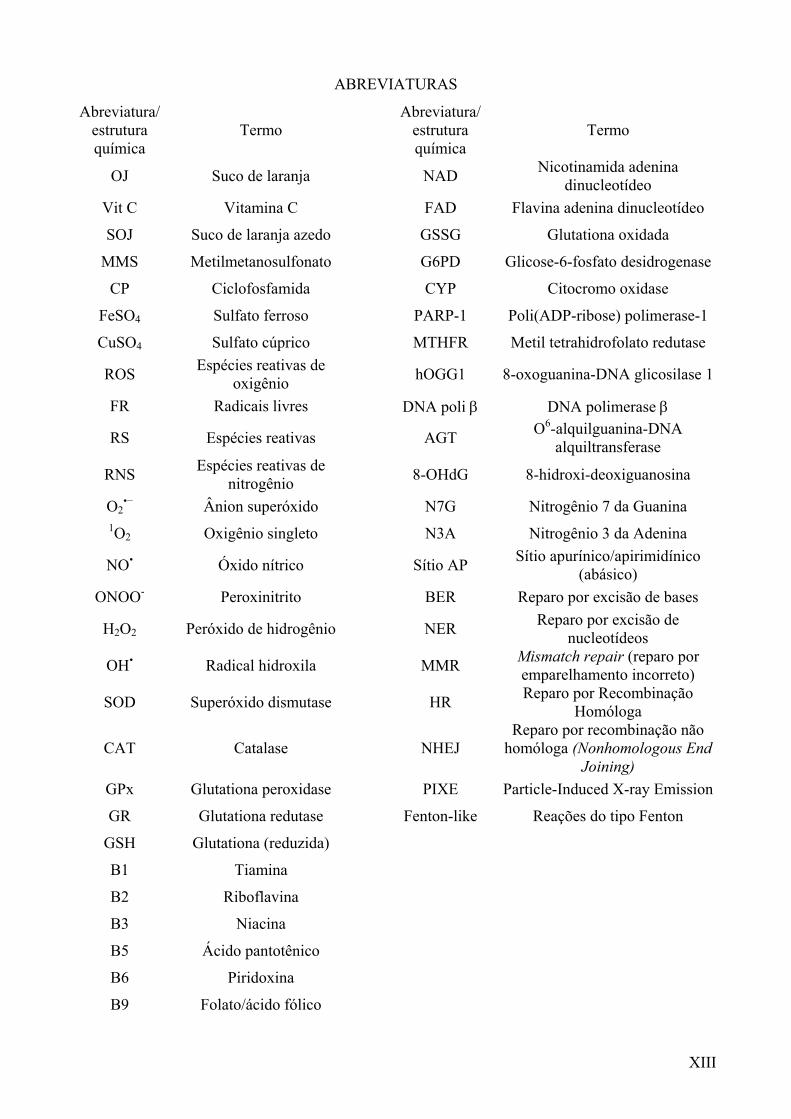
ABREVIATURAS
Abreviatura/ estrutura química
Termo Abreviatura/
estrutura química
Termo
OJ Suco de laranja NAD Nicotinamida adenina dinucleotídeo
Vit C Vitamina C FAD Flavina adenina dinucleotídeo
SOJ Suco de laranja azedo GSSG Glutationa oxidada
MMS Metilmetanosulfonato G6PD Glicose-6-fosfato desidrogenase
CP Ciclofosfamida CYP Citocromo oxidase
FeSO4 Sulfato ferroso PARP-1 Poli(ADP-ribose) polimerase-1
CuSO4 Sulfato cúprico MTHFR Metil tetrahidrofolato redutase
ROS Espécies reativas de oxigênio
hOGG1 8-oxoguanina-DNA glicosilase 1
FR Radicais livres DNA poli β DNA polimerase β
RS Espécies reativas AGT O6-alquilguanina-DNA alquiltransferase
RNS Espécies reativas de nitrogênio
8-OHdG 8-hidroxi-deoxiguanosina
O2•− Ânion superóxido N7G Nitrogênio 7 da Guanina
1O2 Oxigênio singleto N3A Nitrogênio 3 da Adenina
NO• Óxido nítrico Sítio AP Sítio apurínico/apirimidínico (abásico)
ONOO- Peroxinitrito BER Reparo por excisão de bases
H2O2 Peróxido de hidrogênio NER Reparo por excisão de nucleotídeos
OH• Radical hidroxila MMR Mismatch repair (reparo por emparelhamento incorreto)
SOD Superóxido dismutase HR Reparo por Recombinação Homóloga
CAT Catalase
NHEJ Reparo por recombinação não
homóloga (Nonhomologous End Joining)
GPx Glutationa peroxidase PIXE Particle-Induced X-ray Emission
GR Glutationa redutase Fenton-like Reações do tipo Fenton
GSH Glutationa (reduzida)
B1 Tiamina
B2 Riboflavina
B3 Niacina
B5 Ácido pantotênico
B6 Piridoxina
B9 Folato/ácido fólico
XIV
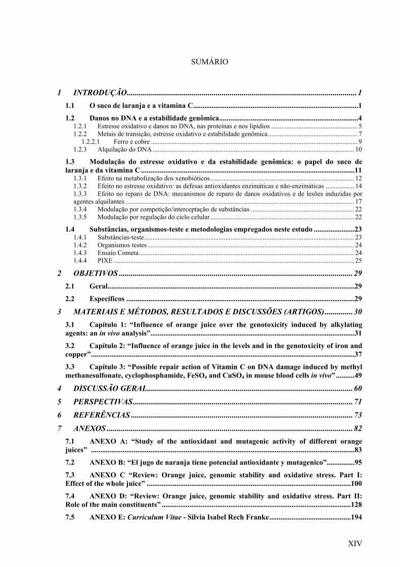
SUMÁRIO
1 INTRODUÇÃO..................................................................................................................1 1.1 O suco de laranja e a vitamina C.........................................................................................1 1.2 Danos no DNA e a estabilidade genômica...........................................................................4
1.2.1 Estresse oxidativo e danos no DNA, nas proteínas e nos lipídios ................................................... 5 1.2.2 Metais de transição, estresse oxidativo e estabilidade genômica..................................................... 7
1.2.2.1 Ferro e cobre .......................................................................................................................... 9 1.2.3 Alquilação do DNA....................................................................................................................... 10
1.3 Modulação do estresse oxidativo e da estabilidade genômica: o papel do suco de laranja e da vitamina C ...................................................................................................................11
1.3.1 Efeito na metabolização dos xenobióticos..................................................................................... 12 1.3.2 Efeito no estresse oxidativo: as defesas antioxidantes enzimáticas e não-enzimáticas ................. 14 1.3.3 Efeito no reparo de DNA: mecanismos de reparo de danos oxidativos e de lesões induzidas por agentes alquilantes ....................................................................................................................................... 17 1.3.4 Modulação por competição/interceptação de substâncias ............................................................. 22 1.3.5 Modulação por regulação do ciclo celular ..................................................................................... 22
1.4 Substâncias, organismos-teste e metodologias empregados neste estudo ......................23 1.4.1 Substâncias-teste............................................................................................................................ 23 1.4.2 Organismos testes .......................................................................................................................... 24 1.4.3 Ensaio Cometa............................................................................................................................... 24 1.4.4 PIXE .............................................................................................................................................. 25
2 OBJETIVOS ....................................................................................................................29 2.1 Geral.....................................................................................................................................29 2.2 Específicos ...........................................................................................................................29
3 MATERIAIS E MÉTODOS, RESULTADOS E DISCUSSÕES (ARTIGOS)..............30 3.1 Capítulo 1: “Influence of orange juice over the genotoxicity induced by alkylating agents: an in vivo analysis”..............................................................................................................31 3.2 Capítulo 2: “Influence of orange juice in the levels and in the genotoxicity of iron and copper”..............................................................................................................................................37 3.3 Capítulo 3: “Possible repair action of Vitamin C on DNA damage induced by methyl methanesulfonate, cyclophosphamide, FeSO4 and CuSO4 in mouse blood cells in vivo” ..........49
4 DISCUSSÃO GERAL......................................................................................................60
5 PERSPECTIVAS.............................................................................................................71
6 REFERÊNCIAS ..............................................................................................................73
7 ANEXOS ..........................................................................................................................82 7.1 ANEXO A: “Study of the antioxidant and mutagenic activity of different orange juices” ..............................................................................................................................................83 7.2 ANEXO B: “El jugo de naranja tiene potencial antioxidante y mutagenico”...............95 7.3 ANEXO C “Review: Orange juice, genomic stability and oxidative stress. Part I: Effect of the whole juice” ..............................................................................................................100 7.4 ANEXO D: “Review: Orange juice, genomic stability and oxidative stress. Part II: Role of the main constituents” ......................................................................................................128 7.5 ANEXO E: Curriculum Vitae - Silvia Isabel Rech Franke............................................194
1
1 INTRODUÇÃO
1.1 O suco de laranja e a vitamina C
O consumo mundial de sucos naturais está aumentando como conseqüência da busca
por um estilo de vida mais saudável. O setor de produção de suco, particularmente o do suco
de laranja (OJ), está se expandindo em vários países. As empresas brasileiras são
responsáveis por aproximadamente 70% das laranjas plantadas ou processadas globalmente.
No Brasil o controle de qualidade dos sucos de frutas é feito: a) por análise das
características organolépticas, b) pela ausência de microrganismos patogênicos, c) pela
ausência de substâncias nocivas, e d) pela regulamentação sobre aditivos (BRASIL, 1974).
Portanto, não considera os efeitos biológicos das substâncias naturais presentes nos sucos.
Desse modo, é muito importante direcionar as pesquisas para os efeitos de substâncias
amplamente consumidas.
O OJ é uma mistura complexa contendo quantidades consideráveis de vitaminas e
minerais, bem como de outros compostos bioativos. A composição do OJ é bastante variável
em conseqüência de vários fatores, tais como varietal, condições climáticas e grau de
maturidade das laranjas, e/ou forma de processamento, envasamento e estocagem. Uma
porção de 200 mL de OJ, em média, contém alta concentração [> 13% da Ingestão Dietética
de Referência (DRI – Dietary Reference Intake)] de Vit C, folato (B9), sendo uma fonte-
chave destes nutrientes. Esta mesma porção pode também ser uma boa ou excelente fonte
(>5-13% da DRI) de riboflavina (B2), niacina (B3), ácido pantotênico (B5) e piridoxina (B6),
bem como de magnésio, cobre e potássio. Adicionalmente, contém quantidades regulares (>4-
5 % da DRI) de carotenóides pró-vitamina A e ferro. O OJ também contém altas quantidades
de flavonóides, bem como quantidades consideráveis de limoneno e p-coumarina. As
2
vitaminas, os minerais e os outros compostos bioativos do OJ podem desempenhar vários
efeitos biológicos, afetando o estresse oxidativo e a estabilidade genômica (para revisão ver
ANEXO D).
O OJ, como mistura complexa, também tem efeitos sobre o estresse oxidativo,
estabilidade genômica, câncer, doenças cardiovasculares (para revisão ver ANEXO C).
Assim como o consumo de OJ vem aumentando, a busca desenfreada pela
longevidade vem gerando uma mudança nas atitudes frente ao consumo de antioxidantes. A
Vit C está entre os antioxidantes mais consumidos, tanto na forma adicionada aos alimentos
quanto como suplemento/medicamento (Halliwell, 2001).
A Vit C deve ser provida pela dieta para os humanos, pois estes, os demais primatas e
outras espécies (como porcos da Índia) não sintetizam o composto. Nos humanos, a última
enzima na rota de biosíntese da Vit C (1-gulonolactona oxidase) não é funcional (WHO,
2001).
A Vit C é um micronutriente importante requerido principalmente como um co-fator
de enzimas envolvidas em reações de oxi-redução (Fenech & Ferguson, 2001; Halliwell,
2001; Edenharder et al., 2003). Esta vitamina C atua como doadora de elétrons em oito
enzimas, envolvidas principalmente na biosíntese de colágeno e carnitina (WHO, 2001).
Por ser hidrosolúvel, a Vit C não é transportada por proteínas. Após ser administrada
por via oral, ela é eliminada com uma meia-vida de 10 h (Schwedhelm et al., 2003). A meia
vida-curta está associada a doenças. O escorbuto é uma patologia classicamente relacionada à
deficiência de Vit C. A anemia também está relacionada à falta de Vit C, pela atuação do
composto na absorção do ferro inorgânico. Apesar do escorbuto ser facilmente reversível num
período curto com a ingestão de apenas 10 mg/dia de Vit C, a deficiência de Vit C continua
sendo um problema especialmente em países mais pobres. Contudo, não se pode
desconsiderar a relação inversa entre ao nível plasmático de Vit C e a prevalência de doenças
infecciosas, comum nestes países (WHO, 2001).
A Vit C tem sido estudada por sua ação protetora contra diferentes doenças
(Vijayalaxmi & Venu, 1999; Edenharder et al., 2002). Os mecanismos pelos quais a Vit C
atua incluem atividades antioxidantes (Halliwell & Guterridge, 2000), bio-antimutagênicas
e/ou desmutagênicas (Sram et al., 1983; Kojima et al., 1992; Guha & Khuda-Bukhsh, 2002).
Atividade antimutagênica é a capacidade de diminuição da freqüência da fixação de
mutações, enquanto a atividade desmutagênica diz respeito à redução/inativação da
mutagenicidade de um dado composto (Kuroda et al., 2001).
3
O mecanismo para a atividade genoprotetora da Vit C pode estar ligado a capacidade
do composto de competir com o DNA como um alvo de alquilação, reduzindo a
genotoxicidade de agentes alquilantes (Vijayalaxmi & Venu, 1999), bem como interceptar
radicais livres (FR). A Vit C pode quelar metais, impedindo que os mesmos gerem espécies
reativas (Halliwell & Guterridge, 2000). A Vit C também tem um papel na regulação das
enzimas de reparo do DNA (Cooke et al., 1998) e pode induzir apoptose quando em altas
concentrações (Sakagami et al., 2000).
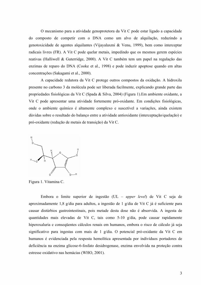
A capacidade redutora da Vit C protege outros compostos da oxidação. A hidroxila
presente no carbono 3 da molécula pode ser liberada facilmente, explicando grande parte das
propriedades fisiológicas da Vit C (Spada & Silva, 2004) (Figura 1).Em ambiente oxidante, a
Vit C pode apresentar uma atividade fortemente pró-oxidante. Em condições fisiológicas,
onde o ambiente químico é altamente complexo e suscetível a variações, ainda existem
dúvidas sobre o resultado do balanço entre a atividade antioxidante (interceptação/quelação) e
pró-oxidante (redução de metais de transição) da Vit C.
Figura 1. Vitamina C.
Embora o limite superior de ingestão (UL – upper level) de Vit C seja de
aproximadamente 1,8 g/dia para adultos, a ingestão de 1 g/dia de Vit C já é suficiente para
causar distúrbios gastrointestinais, pois metade desta dose não é absorvida. A ingesta de
quantidades mais elevadas de Vit C, tais como 5-10 g/dia, pode causar rapidamente
hiperoxaluria e conseqüentes cálculos renais em humanos, embora o risco de cálculo já seja
significativo para ingestas com mais de 1 g/dia. O potencial pró-oxidante da Vit C em
humanos é evidenciada pela resposta hemolítica apresentada por indivíduos portadores de
deficiência na enzima glicose-6-fosfato desidrogenase, enzima envolvida na proteção contra
estresse oxidativo nas hemácias (WHO, 2001).
4
1.2 Danos no DNA e a estabilidade genômica
O homem permanece continuamente exposto a vários riscos de genotoxicidade, devido
à incontestável existência de substâncias genotóxicas capazes de produzir alterações no
material genético de células germinativas ou somáticas. Os agentes que induzem mutações
podem ser divididos em mutágenos físicos (radiações ionizantes e não ionizantes) e químicos
(elementos, substâncias e misturas químicas, como metais, agentes alquilantes e alimentos)
(Saffi & Henriques, 2003).
Os danos no DNA que não são oportunamente reparados ou erroneamente corrigidos,
provocam mudanças na seqüência de bases do DNA, resultando freqüentemente na
eliminação ou alteração de genes responsáveis pelo controle da divisão e da diferenciação
celular (Choy, 1996; Jackson & Loeb, 2001; Goldman & Shields, 2003).
A instabilidade genômica resulta do acúmulo de mutações, especialmente das que
afetam a manutenção da homeostasia da célula nos processos de reparo, replicação e
recombinação do DNA, bem como na divisão celular (Fenech, 2005b).
Existe uma correlação entre a mutagenicidade e a carcinogenicidade, pois muitos
compostos potencialmente mutagênicos podem ser carcinogênicos, indutores de câncer em
animais e humanos, participando como ativadores e/ou iniciadores de carcinogenicidade
(Ames, 1989; Sarasin, 2003). As alterações no DNA representam a primeira etapa da
carcinogênese e estão envolvidas na mutagênese e no envelhecimento do DNA (Wang et al.,
1998; Sarasin, 2003).
Os principais tipos de alterações no DNA incluem: quebras de cadeia simples ou dupla,
deleções ou modificações de base, ponte de DNA-DNA ou DNA-proteína e pontes
intracadeias de DNA (Saffi & Henriques, 2003). Essas alterações são geradoras de
substituições de bases, erros de leitura, deleções, inserções ou translocações na seqüência de
DNA e, ainda, podem causar modificações maiores como as aberrações cromossômicas
estruturais (MacPhee, 1998a, 1998b; Goldman & Shields, 2003; Saffi & Henriques, 2003).
A oxidação e a metilação estão incluídos nos processos endógenos que causam lesões
significativas no DNA (Halliwell & Guterridge, 2000; Brozmanova et al., 2001a; Risom et al.,
2005).
A oxidação e a metilação serão enfocados a seguir, com vistas a explicar o modo de
ação das substâncias-teste empregadas neste trabalho (metais de transição e agentes
alquilantes).
5
1.2.1 Estresse oxidativo e danos no DNA, nas proteínas e nos lipídios
Danos oxidativos no DNA são decorrentes do estresse oxidativo. O estresse oxidativo
ocorre quando há um desbalanço entre a formação e a eliminação de espécies reativas de
oxigênio (ROS). As ROS são geradas como subprodutos do metabolismo oxidativo que foi
um marco na evolução, pois gerou um aumento da eficiência metabólica. Por outro lado,
também aumentou a instabilidade dos sistemas biológicos, pois 2-5 % de todo o oxigênio
metabolizado é convertido em ROS. As ROS são as principais responsáveis pelo nível basal
de mutações (para revisão, ver Halliwell & Guterridge, 2000).
Estima-se que um indivíduo padrão sofra cerca de 10.000–20.000 ataques de ROS e
outros FR por célula por dia como parte do metabolismo oxidativo normal. Para um atleta em
treinamento intenso, estes ataques podem aumentar cerca de 50 % (Valko et al., 2004).
Os danos no DNA resultantes de ataques de ROS incluem diversas modificações
oxidativas, incluindo quebras de cadeia e oxidação nas bases e nas pentoses, bem como
pontes DNA-proteína. O 8-hidroxi-deoxiguanosina (8-OHdG) é o principal produto da
oxidação de bases (Cerda & Weitzman, 1997; Dizdaroglu et al., 2002; Goldman & Shields,
2003; Saffi & Henriques, 2003; Risom et al., 2005).
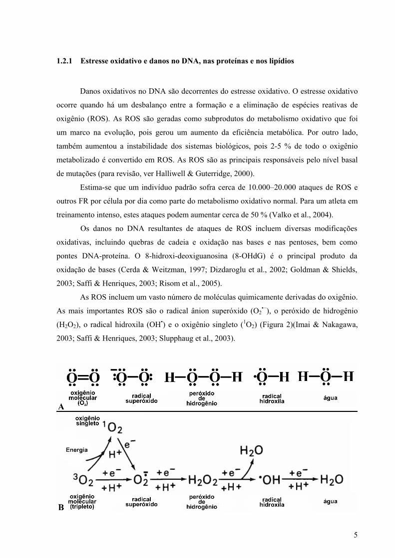
As ROS incluem um vasto número de moléculas quimicamente derivadas do oxigênio.
As mais importantes ROS são o radical ânion superóxido (O2•−), o peróxido de hidrogênio
(H2O2), o radical hidroxila (OH•) e o oxigênio singleto (1O2) (Figura 2)(Imai & Nakagawa,
2003; Saffi & Henriques, 2003; Slupphaug et al., 2003).
6
Figura 2. Principais espécies reativas de oxigênio: A) estrutura de Lewis e B) reações de formação (adaptado de Slupphaug et al., 2003).
O O2•− e o H2O2 não são capazes de atacar o DNA diretamente. O OH• e o 1O2, atacam
diretamente o DNA. O OH• pode causar modificações nas quatro bases do DNA, além de
causar quebras de cadeia e sítios abásicos - apurínicos/apirimidínicos (AP). Já o 1O2, pode
causar ciclo-adição aos carbonos de ligação dupla do anel imidazol, gerar sítios AP e quebras
simples em posições adjacentes às guaninas. Apesar do OH• ser a ROS mais reativa, ela é a de
meia-vida mais curta (Picada et al., 2003; Saffi & Henriques, 2003). O óxido nítrico (NO•)
também é considerado uma ER (Figura 3).
Figura 3. Estrutura de Lewis do óxido nítrico.
A maior parte dos O2•− é gerado nas mitocôndrias como um sub-produto da cadeia de
transporte de elétrons, sendo o DNA mitocondrial seu alvo primário. Uma quantidade menor
de O2•− é formada no retículo endoplasmático pela atividade do citocromo P450 (CYP450). O
O2•− pode dar origem ao OH• pela reação de Haber-Weiss (seção 1.2.2, Figura 3). O OH•
causa danos às biomoléculas, especialmente quando a reação ocorre nas mesmas ou na sua
proximidade (Imai & Nakagawa, 2003; Picada et al., 2003; Slupphaug et al., 2003).
Cerca de 80 % de todo H2O2 é produzido pelos microssomos/peroxissomos hepáticos
envolvidos no metabolismo lipídico. O restante é formado a partir da dismutação do O2•−. A
toxicidade do H2O2 está associada principalmente ao seu papel intermediário na formação do
OH•. A maior parte do OH• é gerada pela decomposição do H2O2 mediada por metais (Imai &
Nakagawa, 2003; Picada et al., 2003; Slupphaug et al., 2003).
O NO• tem uma meia vida longa e alta reatividade com outras ROS (Dedon &
Tannenbaum, 2004). A reação do NO• com O2•− gera peroxinitrito (ONOO-), agente oxidante
como o OH• (Picada et al., 2003; Dedon & Tannenbaum, 2004).
Os efeitos das ROS não dependem somente de seus níveis, sendo influenciados
também: pelo ambiente químico (p.ex. pela concentração substâncias que reagem com as
ROS e pelo nível de defesas antioxidantes, tanto enzimáticas como não-enzimáticas); e pela
distância entre o local de geração das ROS e o sítio alvo dos mesmos, considerando-se o
7
potencial de difusão e a meia-vida das RS e dos FR (Halliwell & Guterridge, 2000; Linder,
2001). Os metais de transição são os principais catalizadores da formação de ROS.
A modificação oxidativa de proteínas por ROS também está associada à origem ou à
progressão de várias doenças e desordens fisiológicas. A oxidação de proteínas e aminoácidos
por ROS pode gerar modificações na cadeia lateral de aminoácidos, rompimento de ligações
peptídicas e a formação de pontes proteína-proteína (Cerda & Weitzman, 1997). Tais
alterações estruturais podem causar a inativação parcial ou total da função da proteína. As
ROS também estimulam as vias de transdução de sinais, afetando a regulação gênica
(Halliwell & Guterridge, 2000). A acumulação de proteínas danificadas, bem como alterações
no perfil da expressão gênica, diminuem a eficiência homeostática, ao longo do tempo,
aumentando as alterações no DNA.
A exemplo das proteínas, os fosfolipídios de membrana celular são suscetíveis à
oxidação, devido aos seus ácidos poliinsaturados (PUFA). A abstração de um átomo de
hidrogênio de um PUFA ou da cadeia lateral de um PUFA por um RL gera radicais centrados
no carbono. Após rearranjos, esses são transformados em radicais peroxil. Este processo é
bem conhecido e chamado de peroxidação lipídica. Hidroperóxidos lipídicos causam
alterações reversíveis às membranas e são uma fonte de aldeídos altamente reativos. Os
aldeídos disparam reações em cadeia, causando danos às proteínas e a ácidos nuclêicos (Terao
& Piskula, 1999).
1.2.2 Metais de transição, estresse oxidativo e estabilidade genômica
Os metais de transição são ubíquos no ambiente e fundamentais à manutenção da vida,
apesar de terem potencial mutagênico e carcinogênico. A mutagenicidade de metais pode
ocorrer por vários mecanismos, tanto diretos quanto indiretos. Os mecanismos diretos
incluem: 1) interação com diferentes bases do DNA, alterando o pareamento das bases ou
disponibilidade do substrato para replicação de DNA ou transcrição de RNA; 2) interação de
íons metais com DNA polimerases, diminuindo a fidelidade da síntese de DNA; e 3) interação
íons metais com as ligações fosfodiésteres do DNA, alterando a estrutura do DNA e a
formação de pontes DNA-proteína (Tkeshelashvili et al., 1991). Já o mecanismo indireto,
pode ocorrer pela formação de OH• pela reação do tipo Fenton (Fenton-like) in situ no DNA
(Meneghini, 1997); ou pela formação de ROS e RNS envolvendo o O2•−, OH•, NO• e H2O2,
8
entre outras substâncias. O mecanismo indireto, geralmente, envolve múltiplas etapas (De
Freitas & Meneghini, 2001; Linder, 2001; Valko et al., 2005; Franke et al., 2006).
A evidência mais convincente da ligação entre danos oxidativos gerados por metais e a
carcinogênese provém da “mutagenicidade” das modificações de base no DNA, que são
resultado do estresse oxidativo mediado por metais. Além disso, a influência de certos metais
no reparo de DNA [tais como Cu (Guecheva et al., 2001) e Pb (Valko et al., 2005)] e em rotas
de sinalização celular está bem caracterizada e de fato, tal modificação está relacionada à
carcinogenicidade. A formação de FR metal-induzida por Fe e Cu é mais reconhecida do que
para os metais altamente tóxicos e carcinogênicos (p.ex. Cr, Ni) (Valko et al., 2005).
A maior parte do OH• é gerada pela decomposição do H2O2 mediada por metais, no
caso da reação de Fenton (quando catalisada por Fe) ou Fenton-like (quando catalisada por
outros metais, como Cu). O OH• pode reagir de três formas com outras moléculas: abstraindo
prótons, transferindo elétrons ou adicionando-se a outras moléculas. A partir disso, ocorre
uma reação em cadeia, uma vez que outras moléculas tornam-se reativas, embora com menos
energia (Halliwell & Guterridge, 2000).
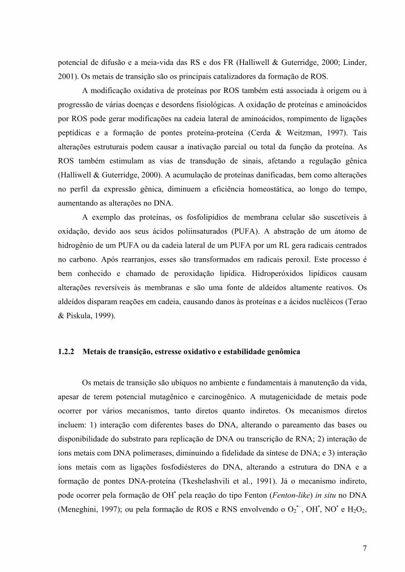
A reação de Fenton consiste na decomposição de H2O2 mediada por sais de ferro que
gera OH•. A adição de um agente redutor, como o ascorbato, leva a um ciclo que aumenta o
dano às biomoléculas (McNaught & Wilkinson, 1997). A reação de Fenton pode ser
representada pela seguinte reação (Figura 4). A reação de Fenton pode ser mediada por outros
metais como o cobre (Fenton-like).
Figura 4. Reação de Fenton.
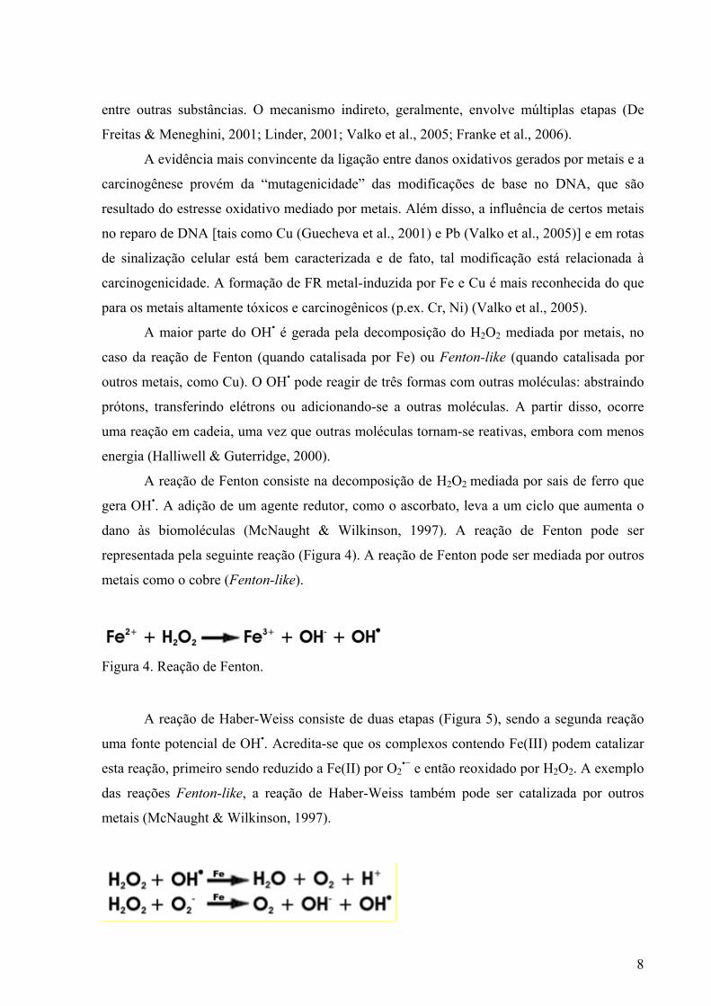
A reação de Haber-Weiss consiste de duas etapas (Figura 5), sendo a segunda reação
uma fonte potencial de OH•. Acredita-se que os complexos contendo Fe(III) podem catalizar
esta reação, primeiro sendo reduzido a Fe(II) por O2•− e então reoxidado por H2O2. A exemplo
das reações Fenton-like, a reação de Haber-Weiss também pode ser catalizada por outros
metais (McNaught & Wilkinson, 1997).
9
Figura 5. Reação de Haber-Weiss
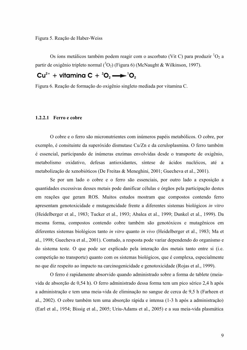
Os íons metálicos também podem reagir com o ascorbato (Vit C) para produzir 1O2 a
partir de oxigênio tripleto normal (3O2) (Figura 6) (McNaught & Wilkinson, 1997).
Figura 6. Reação de formação do oxigênio singleto mediada por vitamina C.
1.2.2.1 Ferro e cobre
O cobre e o ferro são micronutrientes com inúmeros papéis metabólicos. O cobre, por
exemplo, é consituinte da superóxido dismutase Cu/Zn e da ceruloplasmina. O ferro também
é essencial, participando de inúmeras enzimas envolvidas desde o transporte de oxigênio,
metabolismo oxidativo, defesas antioxidantes, síntese de ácidos nucléicos, até a
metabolização de xenobióticos (De Freitas & Meneghini, 2001; Guecheva et al., 2001).
Se por um lado o cobre e o ferro são essenciais, por outro lado a exposição a
quantidades excessivas desses metais pode danificar células e órgãos pela participação destes
em reações que geram ROS. Muitos estudos mostram que compostos contendo ferro
apresentam genotoxicidade e mutagencidade frente a diferentes sistemas biológicos in vitro
(Heidelberger et al., 1983; Tucker et al., 1993; Abalea et al., 1999; Dunkel et al., 1999). Da
mesma forma, compostos contendo cobre também são genotóxicos e mutagênicos em
diferentes sistemas biológicos tanto in vitro quanto in vivo (Heidelberger et al., 1983; Ma et
al., 1998; Guecheva et al., 2001). Contudo, a resposta pode variar dependendo do organismo e
do sistema teste. O que pode ser explicado pela interação dos metais tanto entre si (i.e.
competição no transporte) quanto com os sistemas biológicos, que é complexa, especialmente
no que diz respeito ao impacto na carcinogenicidade e genotoxicidade (Rojas et al., 1999).
O ferro é rapidamente absorvido quando administrado sobre a forma de tablete (meia-
vida de absorção de 0,54 h). O ferro administrado dessa forma tem um pico sérico 2,4 h após
a administração e tem uma meia-vida de eliminação no sangue de cerca de 9,5 h (Farheen et
al., 2002). O cobre também tem uma absorção rápida e intensa (1-3 h após a administração)
(Earl et al., 1954; Bissig et al., 2005; Uriu-Adams et al., 2005) e a sua meia-vida plasmática
10
ainda é controversa, tendo sido associada a um período de até quatro semanas (Dekaban et al.,
1975).
1.2.3 Alquilação do DNA
O metilmetanosulfonato (MMS) e a ciclofosfamida (CP) são agentes alquilantes
utilizados em estudos de mutagênese e na quimioterapia. O MMS é um agente alquilante
monofuncional de ação direta, considerado um mutágeno fraco. Por outro lado, a CP é um
agente alquilante bifuncional de ação indireta, e, portanto, sua genotoxicidade é mediada por
seus metabólitos, que também geram estresse oxidativo. A meia-vida plasmática da CP é de
aproximadamente 6-9 h (Horvathova et al., 1998; Brozmanova et al., 2001a; Brozmanova et
al., 2001b; Saffi & Henriques, 2003; Souliotis et al., 2003; Boiteux & Guillet, 2004; Matalon
et al., 2004; Jenkins et al., 2005; NIH, 2006).
Tanto o MMS como a CP alquilam moléculas nucleofílicas como o DNA. Todas as
bases do DNA possuem sítios suscetíveis à alquilação, especificamente no nitrogênio (N) e
oxigênio (O). As bases púricas como a adenina (nas posições N1, N3 e N7) e a guanina (nas
posições N2, N3 e N7 e O6) são mais suscetíveis à alquilação do que as bases pirimídicas
[citosina (N3, N4 e O2) e timina (N3, O2 e O4)]. Destes, o N7 da guanina (N7G) e o N3 da
adenina (N3A) são os sítios mais sucetíveis à alquilação (para revisão, ver Saffi & Henriques,
2003).
O MMS e a CP induzem substancialmente alquilações em N7G. Contudo,
diferentemente do MMS, a CP também pode induzir pontes do tipo DNA-DNA e DNA-
proteína, bem como adutos nos fosfatos. O MMS induz cerca de 83 % das alquilação em
N7G, 11 % em N3A e apenas 0, 3% em O6-guanina (alquilações em outras posições de O não
parecem ocorrer) (Jenkins et al., 2005). Das alquilações induzidas por CP, 67% são
monoadutos fosfotriésteres, 26% são monoadutos em N7G e 7% são pontes (diadutos) que
ligam duas guaninas através da posição N7G (Souliotis et al., 2003).
Enquanto a O6-alquilguanina causa transição de GC para AT, as N-alquilpurinas
(N7G e N3A) não causam erros de emparelhamento durante a replicação. Contudo, as N-
alquilpurinas podem levar à formação espontânea de sítios AP, devido ao enfraquecimento
das ligações glicosídicas do DNA. Os sítios AP podem ser mutagênicos pela reincorporação
de uma base errada. Além disso, adutos derivados do tipo anel-aberto (ring-opened) em N7G
11
podem inibir a replicação e constituem uma ameaça adicional de mutagênese (Jenkins et al.,
2005).
Os fosfotriésteres são derivados da reação de agentes alquilantes com os fosfatos do
DNA. A formação de monadutos metilfosfodiésteres, provavelmente tem pouco efeito pró-
mutagênico, embora ainda se careça de evidências experimentais conclusivas sobre a
mutagenicidade destas lesões. Por outro lado, adutos nos fosfatos formados por grupamentos
grandes (bulky) podem induzir distorções significativas na conformação do DNA. Como
conseqüência, levando ao emparelhamento incorreto durante a replicação do DNA, bem como
impedindo a ligação de proteína ao DNA por neutralizar a carga negativa do esqueleto açúcar-
fosfato do DNA, o que pode contribuir para quebra de cadeia de DNA (Singh et al., 1997;
Voitkun et al., 1998).
As pontes intercadeias destorcem amplamente a estrutura do DNA e têm
conseqüências catastróficas para a célula se não forem adequadamente reparadas, pois
previnem a ocorrência da replicação e da transcrição, dentre outros processos (Saffi &
Henriques, 2003; Drablos et al., 2004).
1.3 Modulação do estresse oxidativo e da estabilidade genômica: o papel do suco de laranja e da vitamina C
Os efeitos antimutagênicos e possivelmente anticarcinogênicos relacionados à
ingestão de dietas ricas em Vit C e compostos fenólicos, bem como a Vit C de forma isolada,
vêm sendo amplamente estudados. Esses efeitos têm sido relacionados à capacidade de
proteção contra os danos ao material genético.
Há evidências epidemiológicas associando dietas ricas em vegetais e a redução da
incidência de cardiopatias, doenças neurodegenerativas e câncer. Os efeitos protetores têm
sido atribuídos, principalmente, à natureza complexa da composição das frutas (combinação
de carotenóides, compostos fenólicos e vitaminas) (Wang et al., 1996; Kabasakalis, 2000;
Halliwell, 2001). Entretanto, apesar das propriedades benéficas atribuídas aos compostos
naturais, diversos trabalhos relatam efeitos mutagênicos ou carcinogênicos em sucos
(Patrineli et al., 1996a; Ames & Gold, 1998; Yoshino et al., 1999; Franke et al., 2004).
Várias vezes os efeitos carcinogênicos ou genotóxicos podem ser mediados pela
interação de componentes dos sucos com metais de transição ou com os sub-produtos de auto-
oxidação (browning), por ação enzimática (polifenoloxidases), bem como por reações não-
12
enzimáticas (Reações de Maillard) (Patrineli et al., 1996a; Patrineli et al., 1996b; Vercet et al.,
2001; Franke et al., 2004).
Alguns compostos fenólicos podem interagir com enzimas de metabolização, afetando
a toxicidade de compostos (Doostdar et al., 2000). Além disso, a Vit C e os flavonóides
podem agir como um pró-oxidante por sua capacidade redutora, em reações de Fenton e
Fenton-like com metais de transição; mas também pode agir como scavenger (Vinson, 1998;
Sakagami et al., 2000; Edenharder & Grunhage, 2003).
Resultados controversos e confusos associam o consumo de Vit C e a estabilidade
genômica (Ames, 2001; Halliwell, 2001). Uma abordagem interessante é testar se a
genotoxicidade de mutágenos de referência pode ser modulada pela Vit C. Além disso, uma
vez que a Vit C pode interagir com metais e levar à formação de ROS, é importante testar a
ação desta substância sobre a genotoxicidade de compostos metálicos.
1.3.1 Efeito na metabolização dos xenobióticos
Pelo menos três etapas podem ser enumeradas na descrição da interação dos
xenobióticos com os sistemas biológicos: 1) absorção, transporte e eliminação; 2)
metabolização; e 3) interação com o sítio-alvo. A absorção, o transporte e a eliminação podem
ser espontâneos, ou mediados pelo metabolismo. Da mesma forma, os xenobióticos podem
sofrer metabolização ou não antes de exercerem seus efeitos biológicos. A metabolização, em
geral, tem efeito direto sobre a interação com o sítio-alvo, bem como com o transporte e a
eliminação dos xenobióticos (Gaspar, 2003; Guecheva & Henriques, 2003; Henriques & Prá,
2005).
A metabolização envolve duas fases típicas, a fase I e a fase II. Atualmente, uma
terceira fase vem sendo proposta como etapa do metabolismo de xenobióticos. As enzimas
atuantes nas três fases são, via de regra, bastante polimórficas (Gaspar, 2003; Guecheva &
Henriques, 2003; Henriques & Prá, 2005).
As reações da fase I incluem oxidação, redução e hidrólise, introduzindo grupamentos
funcionais, tais como hidroxilas, na molécula do xenobiótico. A fase I é mediada
principalmente (86%) pela ação das citocromo P450 monooxigenases (CYP). As CYPs são
uma família ampla de enzimas dividida em várias classes de acordo com a substância
metabolizada, apesar de muitas vezes duas ou mais classes serem capazes de metabolizar a
mesma substância. O genótipo e a expressão gênica das CYPs apresentam grande
13
variabilidade interindividual (Evans & McLeod, 2003; Gaspar, 2003; Guecheva & Henriques,
2003; Henriques & Prá, 2005).
Na fase I, muitos compostos são convertidos a metabólitos altamente reativos, no
fenômeno denominado bio-ativação. Já as enzimas da fase II reduzem a toxicidade pela
conjugação dos metabólitos da fase I com substratos endógenos, como glutationa (GSH),
sulfato ou glicose. Assim, os metabólitos tornam-se hidrofílicos e passíveis de excreção
(Guecheva & Henriques, 2003) (Evans & McLeod, 2003). A fase II parece ser efetiva na
quimioprevenção, fato mostrado pela incidência aumentada de vários tipos de câncer em
camundongos com fatores de transcrição de enzimas de fase II disruptos (Moon et al., 2006).
Embora o suco afete a metabolização de várias drogas, os efeitos do OJ nas CYPs
ainda está pouco caracterizado. Dessa forma, enquanto o suco de laranja azedo (SOJ - sour
orange juice, obtido de Citrus auratium) inibe a CY3A4 (Malhotra et al., 2001; Di Marco et
al., 2002; Mouly et al., 2005), o OJ (doce, obtido de Citrus cinensis) parece não afetar essa
enzima (Bailey et al., 1991; Hashimoto et al., 1998; Takanaga et al., 2000; Tian et al., 2002).
Os mesmos flavonóides estão presentes no OJ e no SOJ, embora em quantidades diferentes.
Esses flavonóides têm sido reconhecidos como os responsáveis pela modulação das CYPs. A
heperidina/hesperitina inibe CYP1A1 e CYP1B1 in vitro. Já a naringina/naringenina inibe
CYP1A2 e CYP19 in vitro e CYP3A4, in vivo (Moon et al., 2006) (para revisão, ver
ANEXOS C e D).
A família CYP1 está constituída pelos membros 1A1 e 1A2 e 1B1, que são capazes de
ativar pró-carcinógenos. A CYP19, ou aromatase, é uma enzima fundamental para a síntese
de estrógenos e sua expressão aumentada é comum em tumores mamários. As CYP3A4 estão
envolvidas no metabolismo de cerca de 50 % dos agentes terapêuticos, bem como na ativação
de substâncias tóxicas e carcinogênicas (Moon et al., 2006). A atividade inibitória do OJ ou
de seus componentes sobre as CYPs parece ser um mecanismo efetivo da quimioproteção do
alimento.
Quanto a fase II, o OJ inibe as sulfotransferases e promove o aumento da
biodisponibilidade de drogas (Nishimuta et al., 2005). Contudo, a naringina/naringenina
estimula a UDP-glucuronidação in vitro, embora seja um inbidor específico para a UGT1A1,
também in vitro (Moon et al., 2006). A ação das sulfotranferases media a inativação tanto de
xenobióticos quanto de compostos endógenos. A UDP-glucuroniltransferase 1 (UGT1)
cataliza a glucuronidação hepática de xenobióticos, catabólitos do ferro heme, bem como
estrógenos de catecol e flavonóides (Moon et al., 2006) (para revisão, ver ANEXOS C e D).
14
Enquanto o OJ parece não influenciar a atividade de fosfoglicoproteínas (P-gp), que
catalizam o efluxo dos quimioterápicos das células mutantes quando em baixas
concentrações, tanto em ratos como em humanos, ele pode inibir transportadores de influxo,
como OATP [(organic anion-transporting polypeptides (OATP) (Ikegawa et al., 2000;
Kamath et al., 2005; Lim & Lim, 2006)]. Desse modo, o OJ pode bloquear a entrada de
compostos tóxicos nas células e estimular o efluxo dos mesmos quando em altas
concentrações (para revisão, ver ANEXOS C e D).
1.3.2 Efeito no estresse oxidativo: as defesas antioxidantes enzimáticas e não-enzimáticas
As células possuem defesas eficientes contra danos oxidativos. A defesa antioxidante é
exercida tanto por antioxidantes não enzimáticos, como por antioxidantes enzimáticos (Franke
et al., 2003). As enzimas do sistema de defesa antioxidante são representadas principalmente
pela catalase (CAT), pela superóxido dismutase (SOD) e pela glutationa peroxidase (GPx). As
defesas antioxidantes enzimáticas procedem de maneira orquestrada para eliminar os sub-
produtos do metabolismo oxidativo (Figura 7).
Os antioxidantes não enzimáticos podem ser sintetizados endogenamente ou serem
absorvidos como parte da dieta ou por suplementação (para revisão, ver Halliwell &
Guterridge, 2000). Os antioxidantes não-enzimáticos incluem minerais tais como selênio
(Rayman, 2005) e zinco (Valko et al., 2005), moléculas orgânicas, tais como vitaminas (A e
carotenóides, C e E) (Ames et al., 2005), um açúcar-álcool (mannitol) (Gillbe et al., 1996),
um ácido graxo (ácido lipôico) (Vasdev et al., 2005), bem como um aminoácido (cisteína) e
um peptídeo (GSH) (Sen & Packer, 2000). Os bioflavonóides, o ácido úrico (Glantzounis et
al., 2005), a bilirrubina (Sedlak & Snyder, 2004) e a melanina (Wang et al., 2005) são
também antioxidantes. O potencial antioxidante de muitos antioxidantes-não-enzimáticos é
atribuído às hidroxilas (para revisão, ver Halliwell & Guterridge, 2000).
15
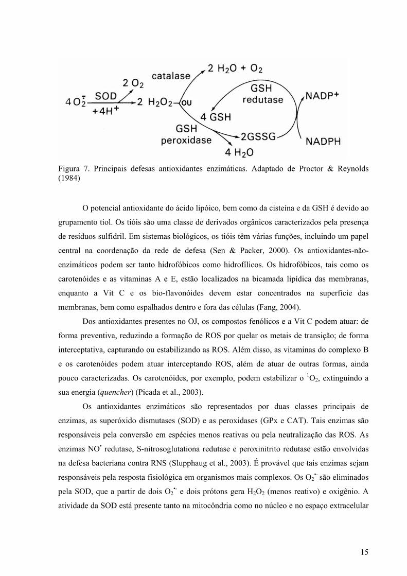
Figura 7. Principais defesas antioxidantes enzimáticas. Adaptado de Proctor & Reynolds (1984)
O potencial antioxidante do ácido lipóico, bem como da cisteína e da GSH é devido ao
grupamento tiol. Os tióis são uma classe de derivados orgânicos caracterizados pela presença
de resíduos sulfidril. Em sistemas biológicos, os tióis têm várias funções, incluindo um papel
central na coordenação da rede de defesa (Sen & Packer, 2000). Os antioxidantes-não-
enzimáticos podem ser tanto hidrofóbicos como hidrofílicos. Os hidrofóbicos, tais como os
carotenóides e as vitaminas A e E, estão localizados na bicamada lipídica das membranas,
enquanto a Vit C e os bio-flavonóides devem estar concentrados na superfície das
membranas, bem como espalhados dentro e fora das células (Fang, 2004).
Dos antioxidantes presentes no OJ, os compostos fenólicos e a Vit C podem atuar: de
forma preventiva, reduzindo a formação de ROS por quelar os metais de transição; de forma
interceptativa, capturando ou estabilizando as ROS. Além disso, as vitaminas do complexo B
e os carotenóides podem atuar interceptando ROS, além de atuar de outras formas, ainda
pouco caracterizadas. Os carotenóides, por exemplo, podem estabilizar o 1O2, extinguindo a
sua energia (quencher) (Picada et al., 2003).
Os antioxidantes enzimáticos são representados por duas classes principais de
enzimas, as superóxido dismutases (SOD) e as peroxidases (GPx e CAT). Tais enzimas são
responsáveis pela conversão em espécies menos reativas ou pela neutralização das ROS. As
enzimas NO• redutase, S-nitrosoglutationa redutase e peroxinitrito redutase estão envolvidas
na defesa bacteriana contra RNS (Slupphaug et al., 2003). É provável que tais enzimas sejam
responsáveis pela resposta fisiológica em organismos mais complexos. Os O2•- são eliminados
pela SOD, que a partir de dois O2•- e dois prótons gera H2O2 (menos reativo) e oxigênio. A
atividade da SOD está presente tanto na mitocôndria como no núcleo e no espaço extracelular
16
(Mates et al., 1999). Há três diferentes formas de SOD em humanos: SOD Cu/Zn (citosólica),
SOD-Mn (mitocondrial) e a SOD Fe (extracelular) (Mates et al., 1999).
Os sistemas celulares primários de defesa antioxidante enzimáticos contra H2O2 e
hidroperóxidos lipídicos são o ciclo redox glutationa, o ciclo tioredoxina e a CAT. O ciclo
redox glutationa oxida GSH a GSSG para converter H2O2 a H2O (Figura 8).
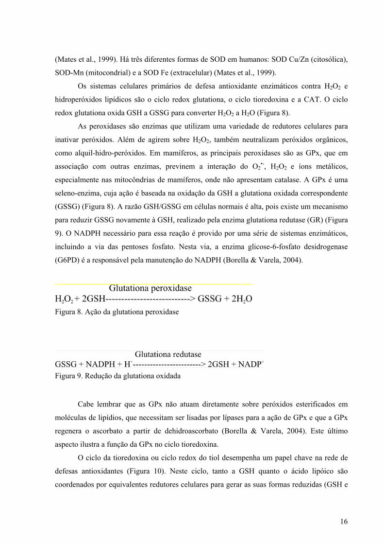
As peroxidases são enzimas que utilizam uma variedade de redutores celulares para
inativar peróxidos. Além de agirem sobre H2O2, também neutralizam peróxidos orgânicos,
como alquil-hidro-peróxidos. Em mamíferos, as principais peroxidases são as GPx, que em
associação com outras enzimas, previnem a interação do O2•-, H2O2 e íons metálicos,
especialmente nas mitocôndrias de mamíferos, onde não apresentam catalase. A GPx é uma
seleno-enzima, cuja ação é baseada na oxidação da GSH a glutationa oxidada correspondente
(GSSG) (Figura 8). A razão GSH/GSSG em células normais é alta, pois existe um mecanismo
para reduzir GSSG novamente à GSH, realizado pela enzima glutationa redutase (GR) (Figura
9). O NADPH necessário para essa reação é provido por uma série de sistemas enzimáticos,
incluindo a via das pentoses fosfato. Nesta via, a enzima glicose-6-fosfato desidrogenase
(G6PD) é a responsável pela manutenção do NADPH (Borella & Varela, 2004).
Figura 8. Ação da glutationa peroxidase
Figura 9. Redução da glutationa oxidada
Cabe lembrar que as GPx não atuam diretamente sobre peróxidos esterificados em
moléculas de lipídios, que necessitam ser lisadas por lípases para a ação de GPx e que a GPx
regenera o ascorbato a partir de dehidroascorbato (Borella & Varela, 2004). Este último
aspecto ilustra a função da GPx no ciclo tioredoxina.
O ciclo da tioredoxina ou ciclo redox do tiol desempenha um papel chave na rede de
defesas antioxidantes (Figura 10). Neste ciclo, tanto a GSH quanto o ácido lipóico são
coordenados por equivalentes redutores celulares para gerar as suas formas reduzidas (GSH e
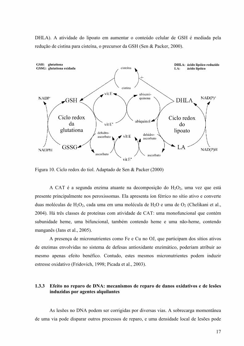
17
DHLA). A atividade do lipoato em aumentar o conteúdo celular de GSH é mediada pela
redução de cistina para cisteína, o precursor da GSH (Sen & Packer, 2000).
Figura 10. Ciclo redox do tiol. Adaptado de Sen & Packer (2000)
A CAT é a segunda enzima atuante na decomposição do H2O2, uma vez que está
presente principalmente nos peroxissomas. Ela apresenta íon férrico no sítio ativo e converte
duas moléculas de H2O2, cada uma em uma molécula de H2O e uma de O2 (Chelikani et al.,
2004). Há três classes de proteínas com atividade de CAT: uma monofuncional que contém
subunidade heme, uma bifuncional, também contendo heme e uma não-heme, contendo
manganês (Jans et al., 2005).
A presença de micronutrientes como Fe e Cu no OJ, que participam dos sítios ativos
de enzimas envolvidas no sistema de defesas antioxidante enzimático, poderiam atribuir ao
mesmo apenas efeito benéfico. Contudo, estes mesmos micronutrientes podem induzir
estresse oxidativo (Fridovich, 1998; Picada et al., 2003).
1.3.3 Efeito no reparo de DNA: mecanismos de reparo de danos oxidativos e de lesões induzidas por agentes alquilantes
As lesões no DNA podem ser corrigidas por diversas vias. A sobrecarga momentânea
de uma via pode disparar outros processos de reparo, e uma densidade local de lesões pode
18
impedir um trabalho bem coordenado dos sistemas de reparação. Além disso, os próprios
danos podem ser indutores de certas funções de reparação (Saffi & Henriques, 2003). A
seguir, as vias de reparo preferenciais para os metais e os agentes alquilantes serão
exploradas. Contudo, se dará ênfase à via de excisão de bases (BER), uma vez que essa via é
preferencial para reparar danos oxidativos e de alquilação; e, os componentes do OJ podem
modulá-la.
O dano oxidativo, induzido por agentes oxidantes endógenos ou exógenos (p.ex.
metais e ROS), é reparado por BER, o principal “guardião” contra o dano oxidativo (Risom et
al., 2005). As modificações nas bases ou açúcares dos nucleotídeos, bem como a presença de
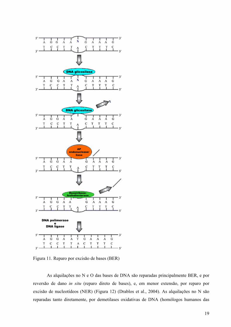
sítios AP, ativam o BER. Esse ocorre em duas etapas, uma de excisão e uma de pós-excisão
(Figura 11). A clivagem da ligação N-glicosídica (açúcar-base) por uma DNA glicosilase
(p.ex. 8-oxoguanina-DNA glicosilase 1–hOGG1) é a primeira etapa do BER. A clivagem da
ligação fosfodiéster por uma 5’-AP endonuclease ou uma 3’-AP liase é a segunda etapa do
BER. No caso de um sítio AP, o primeiro passo do BER é a clivagem da ligação fosfodiéster,
pois a ligação N-glicosídica já está rompida. Em seguida, o açúcar correspondente à base
excisada é também excisado por uma atividade 5’fosfodiesterase ou 3’diesterase. Estes passos
constituem a etapa de excisão. A etapa de pós-excisão envolve a adição do(s) nucleotídeo(s)
faltante(s) por uma DNA polimerase (DNA poli β) e a re-ligação da cadeia por uma DNA
ligase. A etapa de pós-excisão pode ocorrer por duas vias distintas em mamíferos, uma via
curta e uma via longa, dependendo do número de nucleotídeos reparados (Boiteux & Guillet,
2004; Rosa et al., 2004).
19
Figura 11. Reparo por excisão de bases (BER)
As alquilações no N e O das bases de DNA são reparadas principalmente BER, e por
reversão de dano in situ (reparo direto de bases), e, em menor extensão, por reparo por
excisão de nucleotídeos (NER) (Figura 12) (Drablos et al., 2004). As alquilações no N são
reparadas tanto diretamente, por demetilases oxidativas de DNA (homólogos humanos das
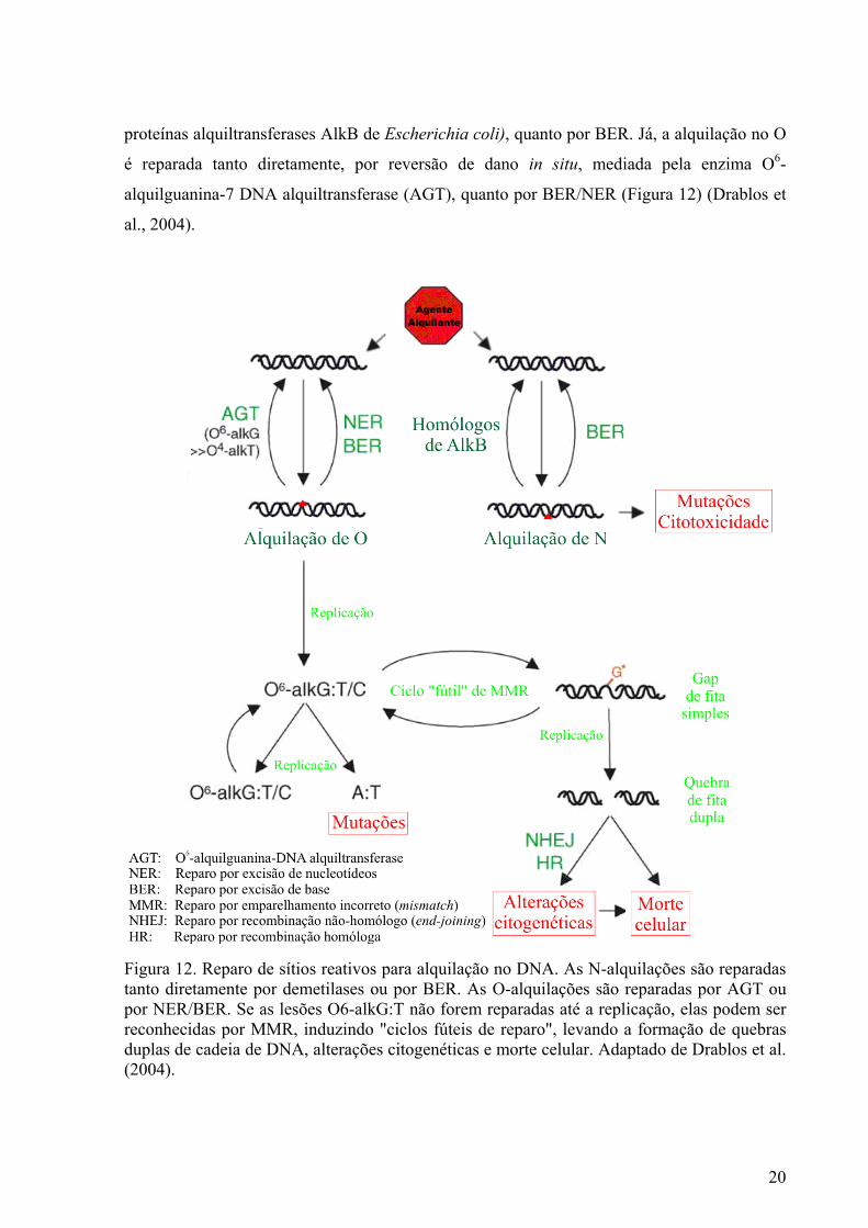
20
proteínas alquiltransferases AlkB de Escherichia coli), quanto por BER. Já, a alquilação no O
é reparada tanto diretamente, por reversão de dano in situ, mediada pela enzima O6-
alquilguanina-7 DNA alquiltransferase (AGT), quanto por BER/NER (Figura 12) (Drablos et
al., 2004).
Figura 12. Reparo de sítios reativos para alquilação no DNA. As N-alquilações são reparadas tanto diretamente por demetilases ou por BER. As O-alquilações são reparadas por AGT ou por NER/BER. Se as lesões O6-alkG:T não forem reparadas até a replicação, elas podem ser reconhecidas por MMR, induzindo "ciclos fúteis de reparo", levando a formação de quebras duplas de cadeia de DNA, alterações citogenéticas e morte celular. Adaptado de Drablos et al. (2004).
21
O envolvimento de ambas cadeias de DNA nas pontes torna o reparo destas lesões
difícil. Por isso, as células, provavelmente, utilizam fatores de diversas rotas de reparo de
maneira coordenada, incluindo enzimas do NER, bem como enzimas da via de recombinação
homóloga (Saffi & Henriques, 2003; Drablos et al., 2004).
Como as 3NA (Saffi & Henriques, 2003), as pontes intercadeias no DNA (McHugh et
al., 2001) induzem reparo recombinacional envolvendo terminações de DNA com regiões
homólogas (HR) e não homólogas (Nonhomologous End Joining - NHEJ), sendo esta rota de
reparo possivelmente comum na resposta das células aos danos no DNA induzidos tanto por
CP quanto por MMS. Convém ressaltar que o reparo recombinacional do tipo NHEJ leva a
perda de informações, portanto é mutagênico.
Quando as alquilações não são adequadamente reparadas, elas podem induzir lesões
secundárias no DNA. Por exemplo, as alquilações O6-alkG:T, que promovem o
emparelhamento incorreto do DNA, quando não reparadas até a replicação podem ser
reconhecidas pelo sistema MMR (mismatch repair) e induzir um “ciclo fútil de reparo”.
Como conseqüência, estas lesões podem gerar quebras duplas de cadeia de DNA, e
conseguintes alterações citogenéticas de maior ou menor toxicidade para as células (Drablos
et al., 2004). As quebras geradas, podem, então, ser processadas por sistemas de reparo
específicos para quebras de cadeia dupla no DNA, como os reparos por recombinação
homóloga (HR) e não homóloga (NHEJ) (Drablos et al., 2004) . As pontes também induzem
alterações secundárias no DNA (Drablos et al., 2004).
Existem evidências do estímulo da via BER pela naringinina (Gao et al., 2006) e pela
Vit C (Cooke et al., 1998). O tratamento por 24 h com naringinina aumentou
significativamente a expressão de hOGG1 e DNA poli β em células derivadas de tumores de
próstata tratadas com sulfato ferroso (Gao et al., 2006). Além disso, a Vit C em doses de até
500 mg/dia estimulou a excreção urinária de 8-OHdG. Este aumento da excreção de 8-OHdG
tem sido interpretado como um estímulo potencial de reparo (Cooke et al., 1998).
Outros nutrientes presentes no OJ, como o folato, a piridoxina, riboflavina, a niacina e
o magnésio têm papéis importantes no metabolismo e no reparo de DNA (Fenech et al.,
2005). O folato (B9) na forma de 5-metil tetrahidrofolato previne a incorporação do uracil no
DNA, bem como mantém o padrão de metilação do DNA (Fenech et al., 2005). Essa
substância é gerada a partir da redução de 5,10-metileno tetrahidrofolato folato pela enzima
metil tetrahidrofolato redutase (MTHFR), que utiliza a riboflavina (B2) como cofator
(Powers, 2005). A piridoxina (B6) regenera 5,10-metileno tetrahidrofolato (Wei et al., 2005) e
também parece promover o reparo por excisão (Shimoi et al., 1992). A niacina (B3) está
22
envolvida no reparo por excisão de bases por atuar como substrato da poli(ADP-ribose)
polimerase-1 (PARP-1) (Hageman & Stierum, 2001). O magnésio atua como cofator das
DNA polimerases e participa de BER e NER e do reparo por emparelhamento incorreto
(MMR) (Ames et al., 2005; Fenech et al., 2005).
1.3.4 Modulação por competição/interceptação de substâncias
Embora se atribua a quimioproteção principalmente ao potencial antioxidante, outros
mecanismos de proteção também podem ocorrer. O caráter nucleofílico de alguns compostos
presentes no OJ pode atuar como agentes preventivos contra a mutagênese e carcinogênese. A
Vit C, pode competir com as bases do DNA como alvo de ataque para ação de agentes
alquilantes. Argumenta-se que a Vit C pode proteger contra ataques eletrofílicos ao DNA pela
interceptação de agentes reativos. Como a Vit C pode ser alquilada, ela compete efetivamente
como alvo de ataque com o DNA frente a agentes alquilantes (Edgar, 1974; Vijayalaxmi &
Venu, 1999). Os retinóides também podem proteger os sítios nucleofílicos do DNA (De Flora,
1998). Assim como a Vit C, os flavonóides também podem formar complexos com compostos
tóxicos, inibindo a reatividade dos mesmos com os sistemas biológicos (Wang et al., 1996; De
Flora, 1998; Middleton et al., 2000).
1.3.5 Modulação por regulação do ciclo celular
O ciclo celular eucariótico é composto de uma cadeia de eventos que garantem a
eliminação de mutações, a replicação do material genético e a segregação cromossômica
adequada. A parada no ciclo celular é uma estratégia adotada pelas células para aumentar o
tempo para reparar as lesões no DNA, reduzindo a letalidade e as conseqüências genéticas das
mesmas (Saffi & Henriques, 2003).
A regulação do ciclo celular é efetuada por uma cascata de eventos relacionados a
pontos de checagem (checkpoint). Os checkpoints são mecanismos vigilantes que monitoram
processos metabólicos chaves e a integridade genômica durante o ciclo celular e atuam para
atrasar ou parar a progressão do ciclo celular em resposta a defeitos ou danos (Hartwell &
Weinert, 1989). Os flavonóides, os retinóides e o limoneno são inibidores efetivos da
carcinogênese, por inibirem a progressão do ciclo celular, inibirem onconogenes, protegerem
23
a comunicação intercelular e/ou induzirem apoptose (So et al., 1996; De Flora, 1998; Guthrie
& Carroll, 1998). No caso dos retinóides, a inibição da tirosina quinase pode estar relacionada
à quimioproteção (De Flora, 1998). A inibição da tirosina quinase, a qual torna-se
hiperfosforilada, causa parada ou atraso no ciclo celular que permite o checkpoint para reparo
de danos no DNA, gerando um efeito quimiopreventivo (Booher et al., 1993).
Altas concentrações de Vit C também podem induzir a morte celular apoptótica
(Sakagami et al., 2000). Além disso, a nicina, outro componente do OJ, tem papel na
expressão e ativação do gene p53 (Hageman & Stierum, 2001). O gene p53 garante o
funcionamento da via de apoptose, reduzindo a probabilidade de eventos promotores de
processos tumorais (Saffi & Henriques, 2003).
1.4 Substâncias, organismos-teste e metodologias empregados neste estudo
Neste estudo serão empregados dois agentes alquilantes, dois compostos metálicos, uma
mistura complexa (OJ) e uma vitamina isolada (Vit C) presente nesta mistura. Serão utilizadas
duas metodologias, uma para avaliar a genotoxidade (ensaio Cometa) e uma para avaliar o
nível de metais (PIXE) tanto no OJ como em dois tecidos de camundongos (fígado e sangue).
1.4.1 Substâncias-teste
Os agentes alquilantes metilmetanosulfonato (MMS) e ciclofosfamida (CP) e os
metais cobre e ferro como sulfatos (FeSO4 e CuSO4, respectivamente) foram empregados
neste estudo como indutores de genotoxicidade. Adicionalmente, o OJ e a Vit C foram
avaliados quanto a sua genotoxicidade e, também, quanto à capacidade de modular a
genotoxicidade gerada por MMS, CP, FeSO4 e CuSO4. O critério de seleção dos agentes
alquilantes se baseia na alta prevalência dos danos gerados por estes compostos. Além disso,
utilizou-se a CP, por ser dependente da ação diversa de seus metabólitos, em comparação ao
MMS, que tem ação direta sobre o DNA. Optou-se pelo uso de Cu e Fe pela sua ubiqüidade
ambiental e presença em alimentos enriquecidos, em suplementos alimentares ou em
medicamentos; pelo seu papel biológico; e pela interação dos mesmos com as vitaminas,
especialmente com a Vit C. Durante muito tempo a Vit C foi considerada como o principal
antioxidante exógeno aos humanos. Contudo, vários estudos têm reconhecido que ela trabalha
24
em conjunto com outras substâncias antioxidantes. Desse modo, estudos relacionando o papel
da Vit C per se e como parte de misturas são necessários para o melhor entendimento da real
participação da mesma na atividade antioxidante, antigenotóxica e anticarcinogênica.
1.4.2 Organismos testes
De acordo com os guidelines descrito por Hartmann et al. (2003) e atualizado por
Collins (2004), os roedores, como os camundongos, são organismos testes preferenciais para
o ensaio Cometa in vivo.
1.4.3 Ensaio Cometa
O ensaio Cometa, conhecido como SGCE (Single Cell Gel Electroforesis), tem sido
amplamente utilizado pela comunidade científica como um teste rápido in vivo e in vitro para
detectar genotoxicidade como conseqüência de danos ao DNA. O teste Cometa é utilizado
amplamente em genética médica, genética toxicológica e ecotoxicológica, em diagnósticos e
tratamentos médicos, medicina ambiental, ocupacional, biomonitoramento ambiental, dentre
outras aplicações (Da Silva et al., 2000).
Em condições alcalinas, o ensaio Cometa detecta quebras simples e dupla, além de
sítios álcali-lábeis. Dentre as vantagens do teste, destacam-se: sensibilidade para detecção de
baixos níveis de danos ao DNA; pequeno número de células; flexibilidade; economia;
facilidade de aplicação; habilidade para conduzir estudos usando um pequeno número de
amostras; rapidez (curto período de tempo para o experimento); pode ser usado em humanos e
em estudos ambientais, e para avaliar reparo de DNA (Singh et al., 1988; Fairbairn et al.,
1995; Tice et al., 2000; Hartmann et al., 2003; Collins, 2004).
As células englobadas em gel sobre uma lâmina são submetidas a uma corrente
elétrica, que faz migrar para fora do núcleo os segmentos de DNA livres, resultantes de
quebras. Após a eletroforese, as células que apresentam um núcleo redondo são identificadas
como normais, sem dano reconhecível no DNA. Por outro lado, as células lesadas são
identificadas visualmente por uma espécie de cauda, como de um cometa, formada pelos
fragmentos de DNA. Estes fragmentos podem se apresentar em diferentes tamanhos, e ainda
estar associados ao núcleo por uma cadeia simples. Para alguns autores o tamanho da cauda é
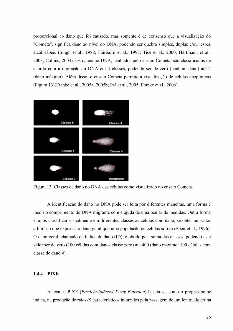
25
proporcional ao dano que foi causado, mas somente é de consenso que a visualização do
"Cometa", significa dano ao nível do DNA, podendo ser quebra simples, duplas e/ou lesões
álcali-lábeis (Singh et al., 1988; Fairbairn et al., 1995; Tice et al., 2000; Hartmann et al.,
2003; Collins, 2004). Os danos no DNA, avaliados pelo ensaio Cometa, são classificados de
acordo com a migração do DNA em 4 classes, podendo ser de zero (nenhum dano) até 4
(dano máximo). Além disso, o ensaio Cometa permite a visualização de células apoptóticas
(Figura 13)(Franke et al., 2005a; 2005b; Prá et al., 2005; Franke et al., 2006).
Figura 13. Classes de dano no DNA das células como visualizado no ensaio Cometa.
A identificação do dano no DNA pode ser feita por diferentes maneiras, uma forma é
medir o comprimento do DNA migrante com a ajuda de uma ocular de medidas. Outra forma
é, após classificar visualmente em diferentes classes as células com dano, se obter um valor
arbitrário que expresse o dano geral que uma população de células sofreu (Speit et al., 1996).
O dano geral, chamado de índice de dano (ID), é obtido pela soma das classes, podendo este
valor ser de zero (100 células com danos classe zero) até 400 (dano máximo: 100 células com
classe de dano 4).
1.4.4 PIXE
A técnica PIXE (Particle-Induced X-ray Emission) baseia-se, como o próprio nome
indica, na produção de raios-X característicos induzidos pela passagem de um íon qualquer na
26
vizinhança de um átomo constituinte do material em estudo (Johansson et al., 1995; Yoneama
& Dias, 2004).
Nesse processo de interação íon-matéria, elétrons de camadas internas dos átomos do
material podem ser emitidos, deixando uma vacância nessa particular camada. Neste caso, o
átomo, como um todo, encontra-se em um estado excitado, com o excesso de energia
correspondente à energia transferida pelo íon incidente. Imediatamente, o átomo excitado
procura restabelecer seu estado natural através de um reordenamento de seus elétrons de suas
diversas camadas. Em particular, elétrons de camadas mais externas poderão preencher as
vacâncias deixadas por elétrons emitidos de camadas mais internas. Nesse processo de
transição eletrônica entre camadas, pode haver a emissão de um raio-X do átomo em questão.
Uma vez que a energia do raio-X emitido depende da diferença de energia das camadas
eletrônicas envolvidas na transição, e levando-se em conta que essa energia é particular para
cada elemento da tabela periódica, então esse raio-X é dito característico por representar,
univocamente, um determinado elemento da tabela periódica (Yoneama & Dias, 2004).
A metodologia PIXE é uma metodologia relativamente rápida e altamente sensível
para detectar a concentração elementar de uma faixa ampla de elementos químicos em
diversos tipos de amostras tais como, proteínas (Follmer et al., 2002) e organismos inteiros
(Kern et al., 2004).
Para a análise por PIXE convencional no vácuo, as amostras devem estar na forma
sólida, homogêneas (para possibilitar uma análise representativa do material investigado) e não
devem apresentar rugosidades (a superfície da amostra deve ser plana). Amostras líquidas
como sangue, bem como amostras de órgãos de animais, devem ser compactadas em alvos
(pastilhas). O processamento de secagem das amostras pode ser feito tanto por liofilização
quanto por aquecimento em estufa/forno. O sangue e os tecidos sólidos são geralmente
mantidos em ultra-freezer a –80°C, posteriormente liofilizadas, homogenizadas e
transformadas em pastilhas por prensagem e mantidas em dessecador.
O Laboratório de Implantação Iônica do IF-UFRGS conta com um acelerador
Tandetron que possui uma linha PIXE convencional a vácuo. Esse acelerador eletrostático
possui uma tensão de terminal de 3 MV positivos. Nesta configuração, as amostras são
irradiadas com feixes de prótons de 2 MeV. A câmara de reações é mantida em vácuo da
ordem de 10-6 milibar por meio de uma bomba turbo-molecular. Internamente, a câmara de
reações possui um filamento de tungstênio que opera a 4,5 V localizada próxima ao suporte
de alvos. Esse filamento funciona como um canhão de elétrons, jogando estes em amostras
que são isolantes. A finalidade deste sistema é descarregar amostras isolantes, diminuindo
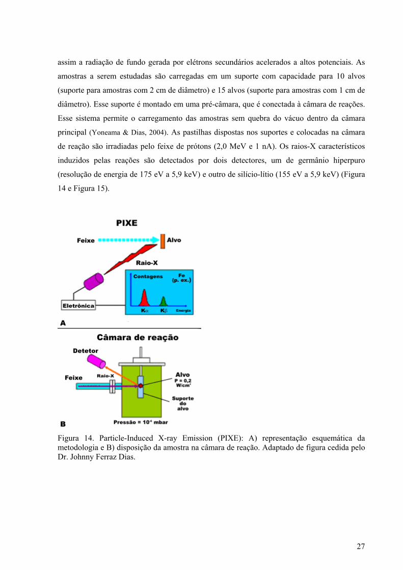
27
assim a radiação de fundo gerada por elétrons secundários acelerados a altos potenciais. As
amostras a serem estudadas são carregadas em um suporte com capacidade para 10 alvos
(suporte para amostras com 2 cm de diâmetro) e 15 alvos (suporte para amostras com 1 cm de
diâmetro). Esse suporte é montado em uma pré-câmara, que é conectada à câmara de reações.
Esse sistema permite o carregamento das amostras sem quebra do vácuo dentro da câmara
principal (Yoneama & Dias, 2004). As pastilhas dispostas nos suportes e colocadas na câmara
de reação são irradiadas pelo feixe de prótons (2,0 MeV e 1 nA). Os raios-X característicos
induzidos pelas reações são detectados por dois detectores, um de germânio hiperpuro
(resolução de energia de 175 eV a 5,9 keV) e outro de silício-lítio (155 eV a 5,9 keV) (Figura
14 e Figura 15).
Figura 14. Particle-Induced X-ray Emission (PIXE): A) representação esquemática da metodologia e B) disposição da amostra na câmara de reação. Adaptado de figura cedida pelo Dr. Johnny Ferraz Dias.
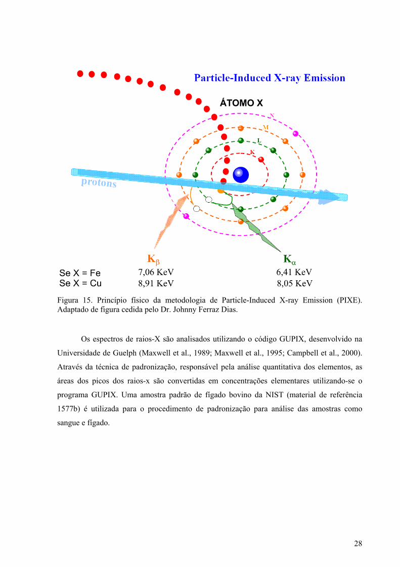
28
Figura 15. Princípio físico da metodologia de Particle-Induced X-ray Emission (PIXE). Adaptado de figura cedida pelo Dr. Johnny Ferraz Dias.
Os espectros de raios-X são analisados utilizando o código GUPIX, desenvolvido na
Universidade de Guelph (Maxwell et al., 1989; Maxwell et al., 1995; Campbell et al., 2000).
Através da técnica de padronização, responsável pela análise quantitativa dos elementos, as
áreas dos picos dos raios-x são convertidas em concentrações elementares utilizando-se o
programa GUPIX. Uma amostra padrão de fígado bovino da NIST (material de referência
1577b) é utilizada para o procedimento de padronização para análise das amostras como
sangue e fígado.
29
2 OBJETIVOS
2.1 Geral
Avaliar, in vivo, a genotoxicidade e a possível capacidade moduladora do OJ e de duas
doses de Vit C sobre a genotoxicidade de compostos metálicos e agentes alquilantes,
utilizando o ensaio Cometa.
2.2 Específicos
• Avaliar a genotoxicidade do OJ em camundongos pelo teste Cometa.
• Avaliar a genotoxicidade da Vit C em camundongos pelo teste Cometa.
• Determinar a genotoxicidade do ferro e do cobre pela aplicação do teste Cometa e em
células sangüíneas de camundongos.
• Determinar se o pré e o pós-tratamento com OJ afetam a genotoxicidade dos
compostos metálicos e dos agentes alquilantes pelo ensaio Cometa.
• Determinar se o pós-tratamento com Vit C pode modular a genotoxicidade dos
compostos metálicos pelo teste Cometa em camundongos.
• Quantificar os níveis séricos e hepáticos de ferro e cobre nos camundongos
submetidos aos tratamentos, por Particle-Induced X-ray Emission analysis (PIXE).

30
3 MATERIAIS E MÉTODOS, RESULTADOS E DISCUSSÕES (ARTIGOS)
31
3.1 Capítulo 1: “Influence of orange juice over the genotoxicity induced by alkylating agents: an in vivo analysis”
Franke, S.I.R., Prá, D., Erdtmann, B., Henriques, J.A.P., Da Silva, J. Mutagenesis, 20(4): 279-283, 2005.
Apesar da existência de estudos sobre a genotoxicidade/antigenotoxicidade do OJ em
procariotos e ex vivo em humanos, há poucos estudos in vivo em mamíferos. Neste sentido, o
primeiro objetivo deste estudo foi avaliar a genotoxicidade para mamíferos in vivo, tendo em
vista as respostas positivas observadas no nosso estudo prévio com bactérias. O segundo
objetivo foi verificar a influência do OJ na genotoxicidade induzida pelos dois agentes
alquilantes, levando-se em consideração as diferenças quanto ao modo de ação dessas duas
drogas. Para tanto, testamos o efeito do OJ tanto antes quanto após o tratamento com uma
única dose dos agentes alquilantes.
Com base nos resultados, procurou-se propor possíveis mecanismos de defesa/reparo,
contra a genotoxicidade de agentes alquilantes, mediados pelo OJ. Como os seres humanos
são expostos a agentes alquilantes no dia-a-dia, os resultados deste estudo podem servir ao
entendimento dos aspectos genoprotetores dos alimentos.
32
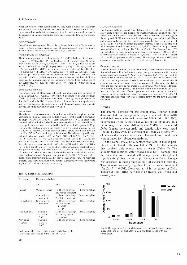
33
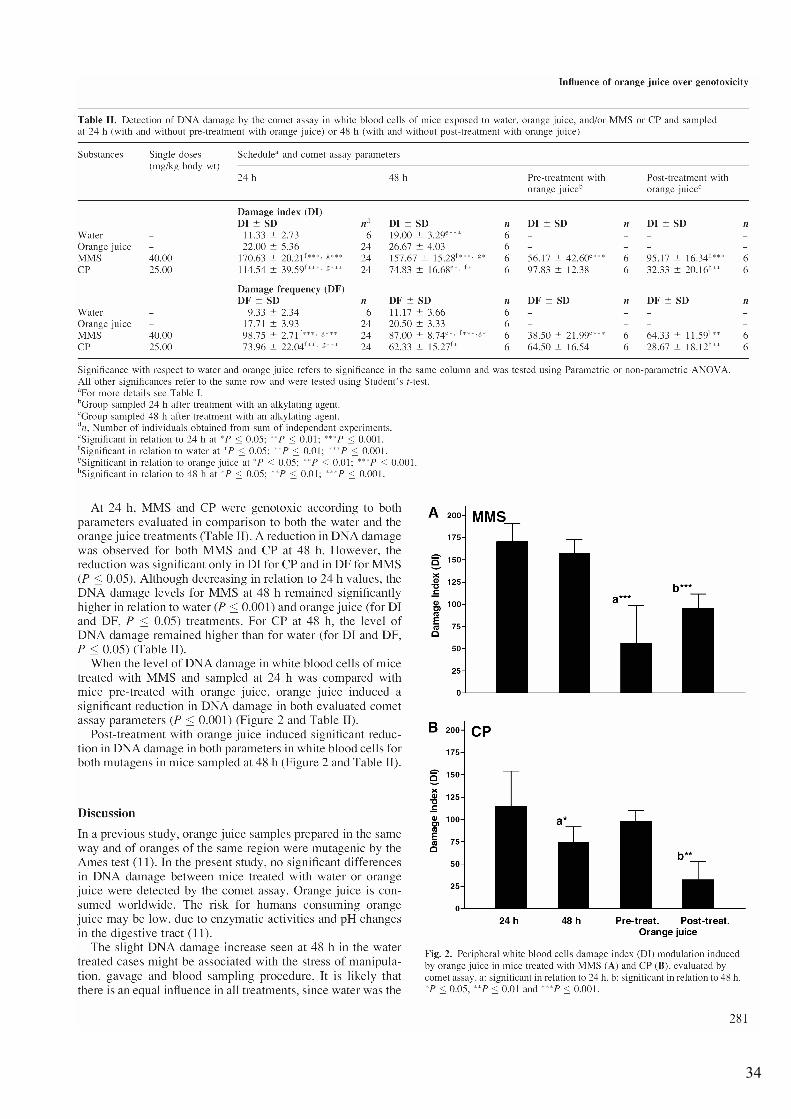
34
35
36
37
3.2 Capítulo 2: “Influence of orange juice in the levels and in the genotoxicity of iron and copper”
Franke, S.I.R., Prá, D., Giulian, R., Dias, J.F., Yoneama, M.L., Da Silva, J., Erdtmann, B., Henriques, J.A.P. Food and Chemical Toxicology, 44: 425-435, 2006.
É conhecido que as reações redox mediadas ou facilitadas por metais de transição e
agentes redutores, ambos compostos presentes no OJ, podem gerar ROS capazes de danificar
o DNA. Contudo, também é conhecido que sucos cítricos podem proteger contra os danos
oxidativos. Em nossos estudos prévios in vitro, o OJ mostrou forte atividade antioxidante, não
obstante tendo sido pró-oxidante em alguns sistemas. Baseado nestes aspectos, decidiu-se
avaliar in vivo o efeito do OJ no dano oxidativo induzido pelo cobre e ferro. Para tanto,
testou-se o efeito do OJ tanto antes quanto após o tratamento com uma dose de ferro ou cobre
de baixa toxicidade para camundongos (cerca de 10 % do LD50 a 48 h).
Surpreendentemente, os resultados apontaram um efeito diferencial do OJ para os dois
metais. Ademais, servem para melhorar a compreensão dos efeitos da interação dos alimentos
ricos em compostos antioxidantes e os metais ingeridos como parte da dieta normal ou através
de suplementos.
38
39
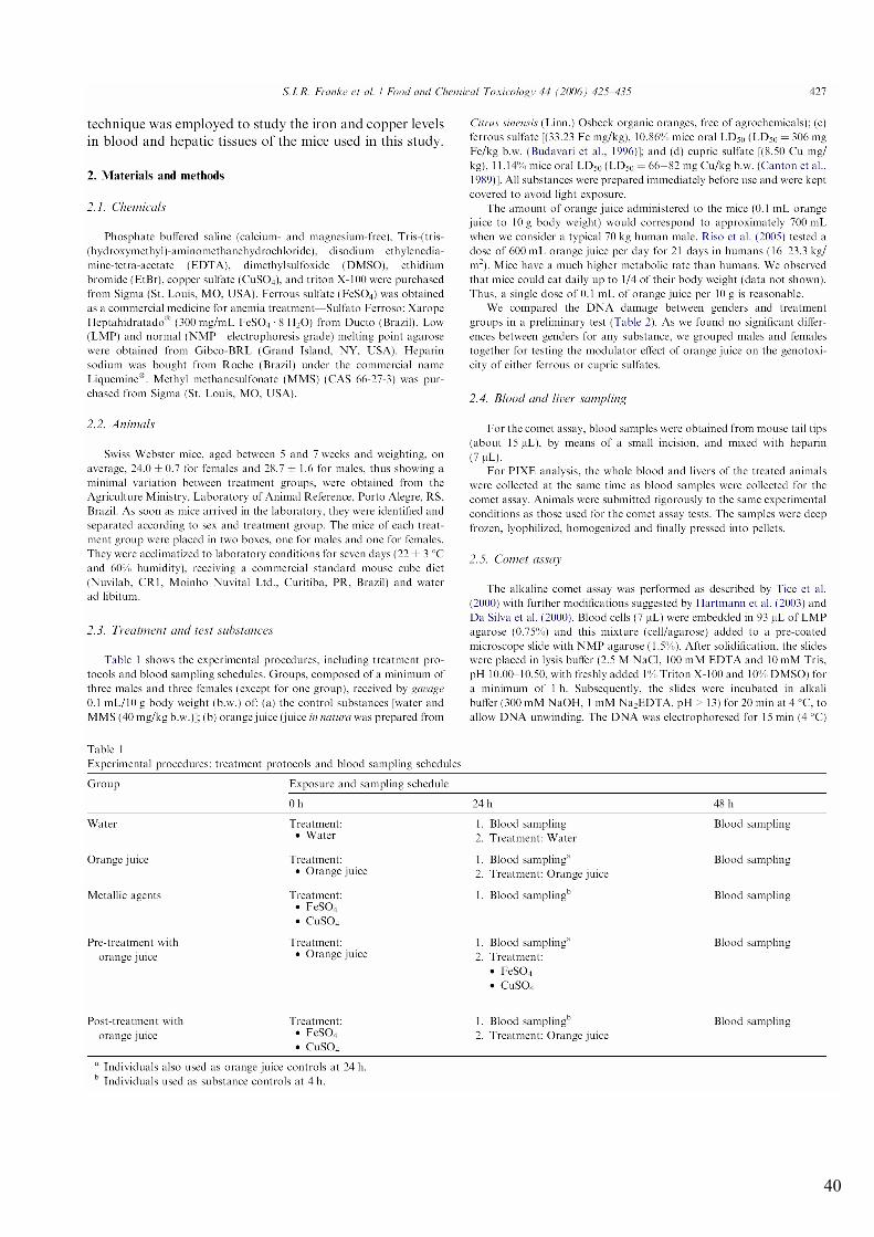
40
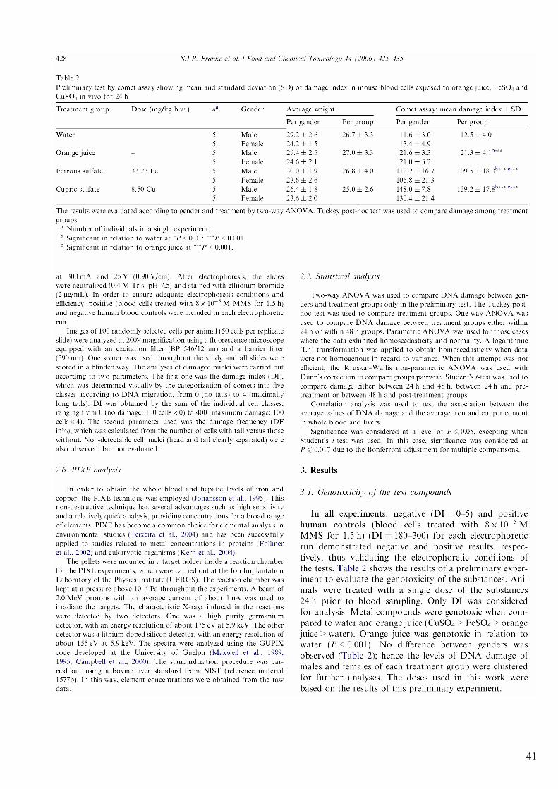
41
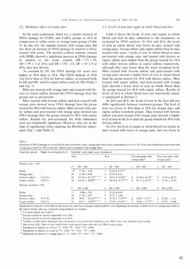
42
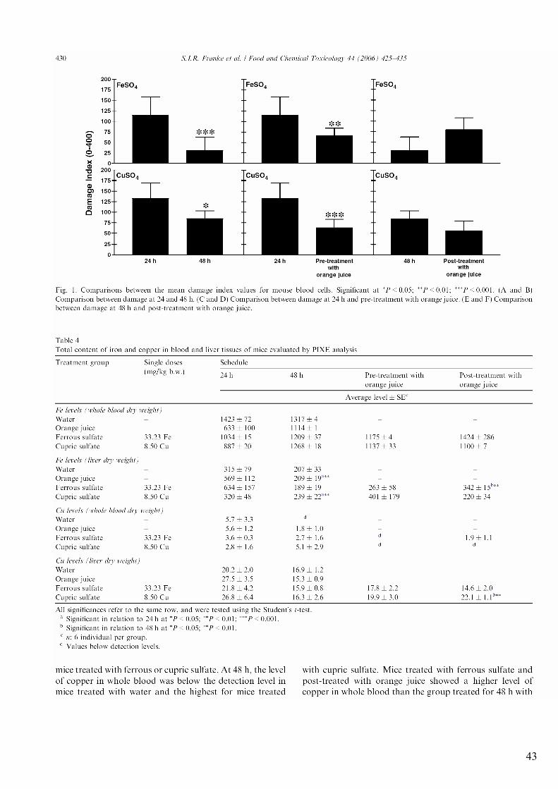
43
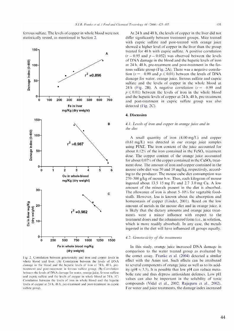
44
45
46
47
48
49
3.3 Capítulo 3: “Possible repair action of Vitamin C on DNA damage induced by methyl methanesulfonate, cyclophosphamide, FeSO4 and CuSO4 in mouse blood cells in vivo”
Franke, S.I.R., Prá, D., Da Silva, J., Erdtmann, B., Henriques, J.A.P. Mutation Research – Genetic Toxicology and Environmental Mutagenesis, 583: 75-84, 2005.
Atribui-se a Vit C um alto potencial antioxidante e antigenotóxico, sendo ela um dos
componentes mais abundantes no OJ. Considerando estas características da Vit C e os
resultados dos Capítulos 1 e 2, onde se observou que o OJ afetava a genotoxicidade tanto dos
metais quanto dos mutágenos, decidiu-se avaliar o efeito do pós-tratamento com a Vit C sobre
a genotoxicidade dos dois alquilantes (Capítulo 1) e do Fe e Cu (Capítulo 2). Foram
selecionadas duas doses de Vit C, uma equivalendo a cerca de uma vez a DRI e a outra, a
aproximadamente 30 vezes a DRI (segundo a RDA anterior a 2000, que preconizava a
ingestão diária de 60 mg para adultos de ambos os sexos). Coincidentemente, a dose maior
também corresponde a uma vez a UL para humanos, segundo a DRI de 2000. Este estudo
considerou a capacidade de síntese de Vit C pelos camundongos, procurando simular o
excesso de Vit C que poderia ocorrer em humanos que tem uma dieta adequada em relação à
DRI de Vit C, e mesmo assim usam suplementos.
ERRATA: Para o correto entendimento da tabela 1, as letras “e”, “f”, “g” e “h” devem ser
substituídas por “g”, “h”, “i” e “e”, respectivamente, no corpo desta tabela. A legenda da
tabela está correta.
50
51
52
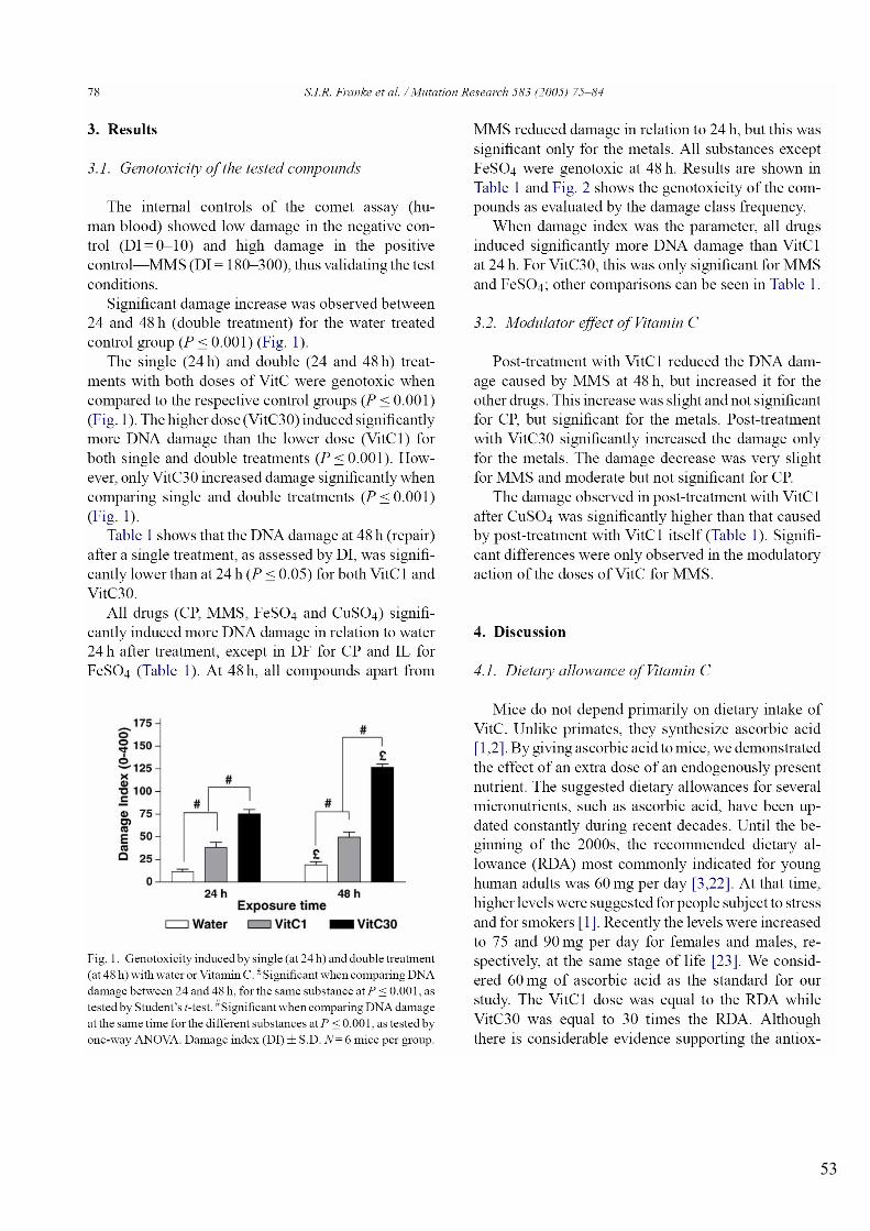
53
54
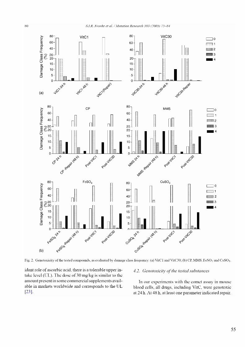
55
56
57
58
59
60
4 DISCUSSÃO GERAL
Há evidências epidemiológicas crescentes associando dietas ricas em frutas e vegetais
frescos e a redução da incidência de doenças associadas ao envelhecimento e a perturbação da
estabilidade genômica, como cânceres, doenças cardiovasculares e neurodegenerativas . As
deficiências em vitaminas e minerais na dieta humana são geradoras de instabilidade
genômica pelo aumento da ocorrência de quebras e lesões oxidativas no DNA. Portanto, estas
deficiências devem ser evitadas e o OJ pode contribuir com componentes-chave para a
estabilidade genômica. Apesar de serem considerados benéficos em concentrações adequadas,
as vitaminas, os minerais e outros componentes bioativos das frutas e vegetais têm sido
apontados como mutagênicos ou carcinogênicos, especialmente quando administrados em
excesso ou fora de matrizes alimentares (Halliwell & Guterridge, 2000; Ames, 2001; Fenech
& Ferguson, 2001; Ames et al., 2005; Fenech, 2005a; Fenech et al., 2005; Halliwell et al.,
2005).
O OJ pode ser incluído entre os alimentos com potencial quimioprotetor e seu estudo é
relevante pelo amplo consumo deste suco. O OJ possui vários nutrientes e compostos
bioativos com atividades antioxidante, antimutagênica, anticarcinogênica e antiaterogênica,
entre outras. O OJ contém quantidades significativas de vitamina C, folato, tiamina,
riboflavina e compostos fenólicos. Além disso, o OJ contém quantidades apreciáveis de
carotenóides (pró e não-pró-vitamina A), niacina, ácido pantotênico, piridoxina, magnésio,
potássio, cobre, ferro e fósforo. Estas substâncias podem influenciar o status oxidativo e a
estabilidade genômica de forma direta ou indireta (ANEXO C).
Nesse sentido, os resultados de um estudo anterior (ANEXOS A e B), avaliando o
potencial antioxidante in vitro e mutagênico em Salmonella de diferentes OJs quanto ao
conteúdo de Vit C e de fenólico(s) totais e ao processamento e armazenamento, evidenciaram
a necessidade de investigar os efeitos do OJ em mamíferos. Observou-se que o OJ espremido
61
manualmente e não-processado (in natura) foi o que apresentou mais respostas positivas
quanto à mutagenicidade em bactérias. Com base neste resultado utilizamos o OJ in natura
para: 1) avaliar se o OJ poderia ser genotóxico em mamíferos (camundongos); 2) avaliar se e
em que extensão o OJ poderia modular a genotoxicidade de agentes alquilantes, bem como do
cobre e do ferro, dois metais de transição.
A Vit C é a vitamina mais abundante no OJ e um copo de 200 mL OJ (uma porção)
pode prover uma quantidade de Vit C superior à preconizada pelas diretrizes alimentares –
DRI (IOM, 2000). A Vit C tem sido foco de muitos estudos ao longo das últimas décadas
quanto ao seu potencial anti/pro-oxidante (Halliwell, 2001; Johnston et al., 2003; Kang et al.,
2005). Por esse motivo, a capacidade modulatória da genotoxicidade dos agentes alquilantes e
dos metais, pela vit C, também foi avaliada neste trabalho.
O PIXE foi utilizado como uma medida auxiliar para quantificar os níveis sangüíneos
e hepáticos de ferro e cobre nos animais tratados com estes metais.
Adicionalmente, foram compiladas duas revisões bibliográficas abordando os efeitos
biológicos e a composição do OJ, para melhor entendimento dos efeitos observados.
Os resultados da parte experimental, apresentados na seção 3 em camundongos (ensaio
Cometa), diferiram dos previamente observados em bactérias (Salmonella/microssoma –
Anexos A e B). Em bactérias, o OJ in natura apresentou respostas positivas frente a várias
linhagens de S. typhimurium com e sem metabolização. Entretanto, em camundongos tratados
com OJ foi observado apenas um incremento no índice de danos em relação ao controle
negativo a 24 h. Portanto, pelo ensaio Cometa o OJ não foi genotóxico em camundongos,
conforme o teste estatístico. Essa tendência se repetiu tanto no experimento com os agentes
alquilantes (Capítulo 1), quanto no experimento com os metais (Capítulo 2). A diferença não
significativa observada para a genotoxicidade do OJ a 24 h poderia estar relacionada ao maior
desvio observado no grupo tratado com OJ. A maior variabilidade intragrupo quanto ao índice
de danos do grupo OJ poderia ser interpretada como decorente de variações metabólicas entre
os camundongos, fato comum na resposta a fármacos e misturas complexas.
Conforme avaliado pelo teste de Salmonella/microsoma, o OJ foi considerado
mutagênico, aumentando a taxa de mutação (número de revertentes) até 5,6 vezes (de forma
dose-dependente). Já no ensaio Cometa em camundongos, observou-se, um aumento na
quantidade de danos primários no DNA, porém não significativo (que podem dar origem a
mutações). Portanto, o OJ foi considerado não genotóxico em camundongos nas condições
testadas. Cabe ressaltar que nos mamíferos o OJ é submetido a uma maior atividade
62
enzimática e química (pH), especialmente no trato gastrintestinal. Além disso, o end-point
avaliado em bactérias (mutações) é diferente do avaliado nos camundongos (dano do DNA).
É importante mencionar que o OJ possui características favoráveis à indução de
genotoxicidade. O pH do OJ pode aumentar a biodisponibilidade de metais, que podem gerar
danos no DNA via estresse oxidativo. Este estresse pode ser intensificado pela presença tanto
de flavonóides quanto de Vit C, que podem reduzir os metais, facilitando a participação dos
mesmos em reações oxidativas. Um exemplo deste fato é a geração de 1O2 pela reação da Vit
C com metais, bem de outras ROS geradas pela reação de Fenton e Fenton-like. Outra
justificativa para a genotoxicidade do OJ é o efeito do mesmo sobre a xantina oxidase.
Enquanto compostos específicos do suco, como as flavanonas (hesperidina e naringinina e
seus glicosídeos - Anexo D, Tabela 3), podem inibir a formação de O2•−, pela xantina oxidase
C, o OJ como mistura complexa estimula as xantina oxidases (Dew et al., 2005). Estas estão
envolvidas na conversão de hipoxantina a ácido úrico e a sua expressão tem sido ligada a
doenças degenerativas. Em determinadas condições fisiológicas, a xantina oxidase processa
O2 ao invés de NAD+ e gera O2•− e NO• (Glantzounis et al., 2005).
Embora não existam evidências da genotoxicidade e da mutagenicidade do OJ em
humanos e em outros mamíferos (ANEXO C), um único estudo relacionou o risco de
melanoma e a ingestão de vitamina C, a partir de alimentos como o OJ (Feskanich et al.,
2003).
O BER é um dos mecanismos de reparo mais utilizados para o reparo de danos
oxidativos e alquilação e pode ser estimulado pelos componentes do suco, como a Vit C, a
naringinina e vitaminas do complexo B (ANEXO D). O BER, a exemplo de outros sistemas
de reparo, envolve a clivagem e a ressíntese da porção de DNA danificada. Desse modo, a
estrutura do DNA fica rompida até que a DNA polimerase resintetize a cadeia e esta seja
religada. O ensaio Cometa detecta principalmente quebras no DNA, que podem ser resultado
de genotoxicidade ou da atividade de reparo. Portanto, um aumento de genotoxicidade
detectado pelo ensaio Cometa também pode indicar um aumento na atividade de reparo, uma
vez que as quebras detectadas podem ser provenientes de excisão das cadeias de DNA pelas
enzimas de reparo (Tice et al., 2000). A genotoxicidade do OJ também pode ser interpretada
como um aumento da atividade de reparo sobre os danos basais ao DNA das células.
Assim como nos animais tratados com suco, um incremento menor, contudo
significativo, na genotoxicidade foi observado entre 24 e 48 h para o grupo controle em todos
os experimentos (Capítulos 1-3). O estresse associado à manipulação dos animais durante os
experimentos, bem como lesões gastrintestinais geradas pela gavage poderiam gerar respostas
63
inflamatórias ligadas ao aumento do estresse oxidativo. De qualquer forma, esse aumento é
inerente a todos os tratamentos. Além disso, a resposta às lesões e aos danos no DNA também
pode sofrer influência das diferenças interindividuais. Cabe ressaltar que a presença de metais
na água de beber dos animais também poderia afetar a resposta, uma vez que a água foi
provida pelo sistema municipal de abastecimento da cidade de Porto Alegre. Contudo, água
destilada foi utilizada para diluir as substâncias-teste e para tratar os animais do controle
negativo. Ressalta-se, novamente, que a influência da água foi inerente a todos os
tratamentos.
Neste estudo, camundongos foram utilizados para avaliar o efeito genotóxico e
antigenotóxico da Vit C. A intenção foi simular indivíduos que têm níveis adequados de Vit C
na dieta diária e que fazem uso abusivo de suplementos dessa vitamina.
Neste estudo, as doses foram calculadas em função da recomendação dietética anterior
a 2000 (60 mg/dia tanto para homens quanto para mulheres adultos). Para fins de discussão,
as recomendações de 2000 – DRI2000 [82,5 mg/dia, média da recomendação para homens (90
mg/dia) e mulheres (75 mg/dia)] serão utilizadas (IOM, 2000).
A Vit C foi genotóxica de maneira dose-dependente 24 h após o tratamento para
ambas doses [1 (VitC1) e 30 (VitC30) mg/Kg] (Capítulo 3). Quando os animais foram
tratados duas vezes, a 24 e a 48 h (duplo-tratamento), a genotoxicidade aumentou
significativamente apenas para a dose maior (30 mg/Kg). A genotoxicidade induzida por
ambas doses de Vit C foi efetivamente reparada após 48 h de uma única administração
(reduzindo cerca de 35% em relação a 24 h). É importante mencionar que o índice de dano no
DNA dos animais tratados uma única vez com VitC1 foi similar ao dos controles negativos a
48 h, mostrando reparo efetivo nestas condições. O efeito pró-oxidante ou estimulador do
reparo poderiam estar associados ao aumento dos danos no DNA detectados pelo ensaio
Cometa.
Enquanto o duplo-tratamento (0 e 24 h) tanto com o OJ quanto com VitC1 manteve o
dano entre 24 e 48 h, o tratamento duplo com VitC30 causou um incremento considerável
(~70 %) na genotoxicidade entre 24 e 48 h. A dose de suco de 0,1 mL/10 g (10 mL/Kg) de
peso de camundongo corresponde a 600 mL para um humano de 60 Kg. Utilizando o
conteúdo médio de Vit C calculado na Tabela 1 do ANEXO D, uma porção de 600 mL de OJ
representa uma entrada de 303 mg de Vit C. Esse valor corresponde a aproximadamente 3,7
vezes a DRI2000 de Vit C. Desse modo, a quantidade de Vit C presente no suco ministrado aos
camundongos foi intermediária às doses de Vit C utilizadas no estudo (1 e 30 mg/Kg),
demonstrando que quando presente no suco, uma dose maior que a RDA de Vit C, como parte
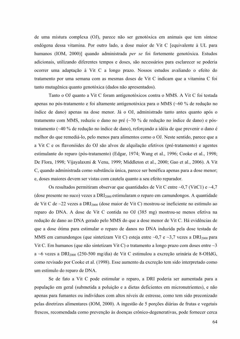
64
de uma mistura complexa (OJ), parece não ser genotóxica em animais que tem síntese
endógena dessa vitamina. Por outro lado, a dose maior de Vit C [equivalente à UL para
humanos (IOM, 2000)] quando administrada per se foi fortemente genotóxica. Estudos
adicionais, utilizando diferentes tempos e doses, são necessários para esclarecer se poderia
ocorrer uma adaptação à Vit C a longo prazo. Nossos estudos avaliando o efeito do
tratamento por uma semana com as mesmas doses de Vit C indicam que a vitamina C foi
tanto mutagênica quanto genotóxica (dados não apresentados).
Tanto o OJ quanto a Vit C foram antigenotóxicos contra o MMS. A Vit C foi testada
apenas no pós-tratamento e foi altamente antigenotóxica para o MMS (~60 % de redução no
índice de dano) apenas na dose menor. Já o OJ, administrado tanto antes quanto após o
tratamento com MMS, reduziu o dano no pré (~70 % de redução no índice de dano) e pós-
tratamento (~40 % de redução no índice de dano), reforçando a idéia de que prevenir o dano é
melhor do que remediá-lo, pelo menos para alimentos como o OJ. Neste sentido, parece que a
a Vit C e os flavonóides do OJ são alvos de alquilação efetivos (pré-tratamento) e agentes
estimulante do reparo (pós-tratamento) (Edgar, 1974; Wang et al., 1996; Cooke et al., 1998;
De Flora, 1998; Vijayalaxmi & Venu, 1999; Middleton et al., 2000; Gao et al., 2006). A Vit
C, quando administrada como substância única, parece ser benéfica apenas para a dose menor;
e, doses maiores devem ser vistas com cautela quanto a seu efeito reparador.
Os resultados permitiram observar que quantidades de Vit C entre ~0,7 (VitC1) e ~4,7
(dose presente no suco) vezes a DRI2000 estimularam o reparo em camundongos. A quantidade
de Vit C de ~22 vezes a DRI2000 (dose maior de Vit C) mostrou-se ineficiente no estímulo ao
reparo do DNA. A dose de Vit C contida no OJ (385 mg) mostrou-se menos efetiva na
redução de dano ao DNA gerado pelo MMS do que a dose menor de Vit C. Há evidências de
que a dose ótima para estimular o reparo de danos no DNA induzida pela dose testada de
MMS em camundongos (que sintetizam Vit C) esteja entre ~0,7 e ~3,7 vezes a DRI2000 para
Vit C. Em humanos (que não sintetizam Vit C) o tratamento a longo prazo com doses entre ~3
a ~6 vezes a DRI2000 (250-500 mg/dia) de Vit C estimulou a excreção urinária de 8-OHdG,
como revisado por Cooke et al. (1998). Esse aumento da excreção tem sido interpretado como
um estímulo do reparo de DNA.
Se de fato a Vit C pode estimular o reparo, a DRI poderia ser aumentada para a
população em geral (submetida a poluição e a dietas deficientes em micronutrientes), e não
apenas para fumantes ou indivíduos com altos níveis de estresse, como tem sido preconizado
pelas diretrizes alimentares (IOM, 2000). A ingestão de 5 porções diárias de frutas e vegetais
frescos, recomendada como prevenção às doenças crônico-degenerativas, pode fornecer cerca
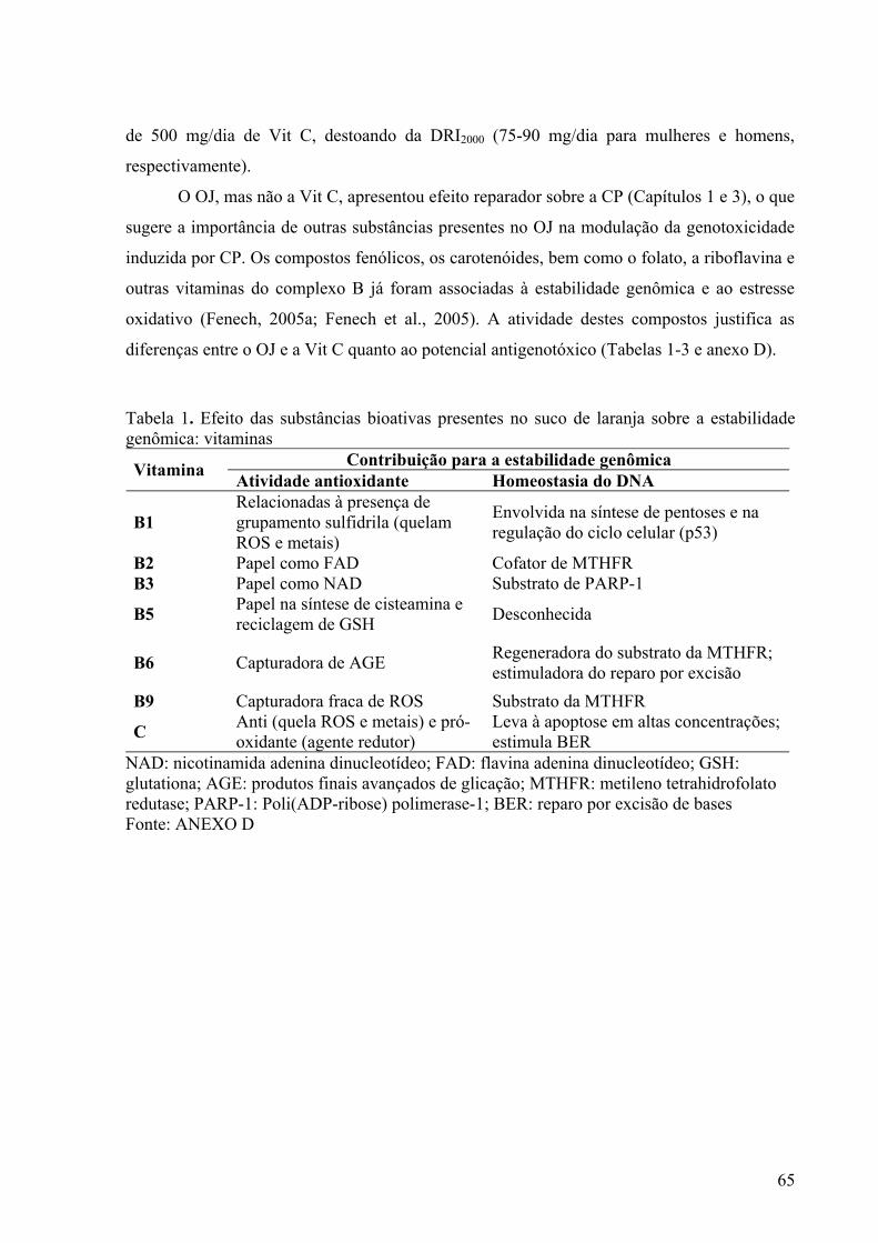
65
de 500 mg/dia de Vit C, destoando da DRI2000 (75-90 mg/dia para mulheres e homens,
respectivamente).
O OJ, mas não a Vit C, apresentou efeito reparador sobre a CP (Capítulos 1 e 3), o que
sugere a importância de outras substâncias presentes no OJ na modulação da genotoxicidade
induzida por CP. Os compostos fenólicos, os carotenóides, bem como o folato, a riboflavina e
outras vitaminas do complexo B já foram associadas à estabilidade genômica e ao estresse
oxidativo (Fenech, 2005a; Fenech et al., 2005). A atividade destes compostos justifica as
diferenças entre o OJ e a Vit C quanto ao potencial antigenotóxico (Tabelas 1-3 e anexo D).
Tabela 1. Efeito das substâncias bioativas presentes no suco de laranja sobre a estabilidade genômica: vitaminas
Contribuição para a estabilidade genômica Vitamina Atividade antioxidante Homeostasia do DNA
B1 Relacionadas à presença de grupamento sulfidrila (quelam ROS e metais)
Envolvida na síntese de pentoses e na regulação do ciclo celular (p53)
B2 Papel como FAD Cofator de MTHFR B3 Papel como NAD Substrato de PARP-1
B5 Papel na síntese de cisteamina e reciclagem de GSH Desconhecida
B6 Capturadora de AGE Regeneradora do substrato da MTHFR; estimuladora do reparo por excisão
B9 Capturadora fraca de ROS Substrato da MTHFR
C Anti (quela ROS e metais) e pró-oxidante (agente redutor)
Leva à apoptose em altas concentrações; estimula BER
NAD: nicotinamida adenina dinucleotídeo; FAD: flavina adenina dinucleotídeo; GSH: glutationa; AGE: produtos finais avançados de glicação; MTHFR: metileno tetrahidrofolato redutase; PARP-1: Poli(ADP-ribose) polimerase-1; BER: reparo por excisão de bases Fonte: ANEXO D
66
Tabela 2. Efeito das substâncias bioativas presentes no suco de laranja sobre a estabilidade genômica: minerais
Contribuição para a estabilidade genômica Mineral Atividade antioxidante Homeostasia do DNA
Cu
Anti (componente das ferroxidases e da SOD Cu/Zn) e pró-oxidante (metal de transição)
Influência desconhecida
Mg Influência desconhecida Cofator de diversas enzimas, incluindo as DNA polimerases (atua na síntese e no reparo do DNA)
K
Níveis celulares aumentados têm sido relacionados à diminuição do estresse oxidativo
Substrato dos canais de membrana (envolvidos na sinalização e no controle da progressão celular)
P Influência desconhecida Compõe a estrutura do DNA e está envolvido na sinalização e no controle da progressão celular
Fe Anti (componente das CATs e da SOD Fe) e pró-oxidante (metal de transição)
Influência desconhecida
SOD: superóxido dismutase Fonte: ANEXO D
Tabela 3. Efeito das substâncias bioativas presentes no suco de laranja sobre a estabilidade genômica: outros compostos
Contribuição para a estabilidade genômica Substância bioativa Atividade antioxidante Homeostasia do DNA
Carotenóides Forte antioxidantes (caputadores de ROS e metais)
Inibidores da proliferação de células tumorais
D-limoneno Forte antioxidante (pouco caracterizado)
Inibidores da proliferação de células tumorais
Compostos fenólicos
Capturador fraco de ROS e forte quelante de metais
Diversas atividades relacionadas ao metabolismo de substâncias, ao controle do ciclo celular e a mimetização de estrógenos
ROS: espécies reativas de oxigênio Fonte: ANEXO D
Os compostos fenólicos estão entre os componentes mais abundante no OJ, e a maioria
dos estudos aponta grande responsabilidade destes compostos na antigenotoxicidade e no
potencial antioxidante do OJ (Anexo D). Os numerosos mecanismos de ação dos fenólicos
poderiam ter contribuído para a antigenotoxicidade do OJ frente à CP. A hesperidina, a
narirutina e as formas agliconadas destes compostos (hesperitina e naringenina,
respectivamente) têm capacidade de inibir algumas enzimas de fase 1 (CYPs). A modulação
67
das CYPs envolvidas na metabolização de CP poderia ter reduzido a genotoxicidade da CP no
pré-tratamento com o suco. Outro mecanismo de ação dos fenólicos é a atuação como alvo de
alquilação ou como capturadores de FR. Apesar destas atividades, a modulação da
genotoxicidade da CP, pelo pré-tratamento com OJ, pode ter sido reduzida por dois motivos:
pela incapacidade dos fenólicos ou do OJ de modularem as principais enzimas envolvidas na
metabolização da CP [CYP3A4, CYP3A5, CYP2C9 (Watters et al., 2003; Matalon et al.,
2004)] (para revisão, ver ANEXOS C e D); e pela maior reatividade da CP. A CP possui dois
sítios de alquilação, e, portanto, a quantidade dos componentes do suco pode não ter sido
suficiente para competir com o DNA como alvo de alquilação da CP, como parece ter
ocorrido para o MMS que possui apenas um sítio de alquilação.
Ao se comparar a atividade do OJ e da Vit C sobre o reparo da CP, é necessário
considerar o tipo de dano gerado pela CP. Tanto o MMS como a CP alquilam moléculas
nucleofílicas como o DNA. Diferentemente do MMS, que somente alquila o N e O das bases,
a CP também induz pontes do tipo DNA-DNA e DNA-proteína. As lesões induzidas pela CP
são, portanto, complexas e requerem vários mecanismos de reparo. Os adutos podem ser
reparados por reversão de dano in situ (alquiltransferases) e por BER, principalmente. Há
apenas evidências do efeito da Vit C (Cooke et al., 1998) e da naringinina sobre BER (Gao et
al., 2006).
As pontes intercadeias e DNA-proteína, que podem ser geradas apenas pela CP,
requerem NER e fatores de recombinação homóloga para serem reparados. A recombinação
homóloga também parece ser o mecanismo para o reparo de 3NA, aproximadamente 11 % das
lesões, bem como para lesões O6-guanina (menos de 0,5 % das lesões) induzidas por MMS
(Saffi & Henriques, 2003; Drablos et al., 2004). Como as pontes intercadeias geradas pela CP
envolvem outras rotas de reparo além do BER (como NER e recombinação homóloga), e os
nossos resultados (Capítulos 1 e 3) apontam efeito reparador do OJ sobre a CP, é possível que
algum componente do OJ tenha uma influência de forma ainda desconhecida sobre estas rotas
de reparo. Além disso, tanto a CP quanto os seus metabólitos (p.ex. acroleína) induzem
estresse oxidativo, e estão implicados na depleção de GSH e na inativação de genes
relacionados à resposta antioxidante (Kehrer & Biswal, 2000). Neste sentido, o magnésio atua
como cofator das DNA polimerases e participa de BER e NER e do reparo por MMR (Ames
et al., 2005; Fenech et al., 2005). A deficiência de folato, por sua vez, afeta BER, NER,
MMR, bem como o reparo de quebras de cadeia dupla, indicando um papel do folato nestas
vias de reparo (Winterbourn et al., 2000; Wei et al., 2003). Como anteriormente citado, os
trabalhos de Cooke et al. (1998) apontam a influência da vitamina C em BER, pelo aumento
68
da excreção urinária de 8-OHdG. Portanto, a presença do magnésio, folato e de Vit C no suco
pode ter auxiliado o sistema de reparo frente aos danos gerados pela CP. Além disso, cabe
ressaltar que outros componentes derivados do OJ, como a vanilina, um composto derivado
dos ácidos hidroxicinâmicos também podem induzir o reparo recombinacional (Sinigaglia et
al., 2004).
O ensaio Cometa tende a subavaliar as pontes intercadeias geradas pela CP, uma vez
que as pontes tendem a dificultar a migração do DNA (Vrzoc & Petras, 1997; Tice et al.,
2000; Collins, 2004). Existem alguns protocolos do ensaio Cometa utilizando enzimas
(endonucleases para pontes DNA-DNA e proteinases para pontes DNA-proteína) para avaliar
especificamentes pontes no DNA (Tice et al., 2000). O uso destas técnicas poderia ser
empregado para melhorar a compreensão da dinâmica das lesões induzidas pela CP.
O pós-tratamento com Vit C pode ter tido dois efeitos sobre a genotoxicidade da CP:
a) induziu quebras no DNA por facilitar a reparação das pontes; e b) não afetou o reparo do
DNA e foi co-genotóxica com a CP. A primeira hipótese é suportada pela manutenção de
dano nas duas doses de Vit C. A efetividade do reparo da genotoxicidade da CP pelo pós-
tratamento com OJ aponta a sinergia da Vit C com os demais componentes do suco na
reparação do dano. A interação dos fenólicos com a Vit C pode ter um efeito sinergístico
sobre o reparo de DNA e sobre a estabilidade destes compostos. Quanto a este último aspecto,
é bem sabido que a Vit C e os fenólicos estabilizam-se mutuamente, aumentando a
estabilidade destes compostos e, portanto, garantindo o efeito biológico dos mesmos
(Middleton et al., 2000).
O pré-tratamento com OJ reduziu a genotoxicidade tanto do Fe quanto do Cu
(Capítulos 2). Tanto a Vit C quanto o OJ têm sido prescritos para aumentar a absorção de Fe
no tratamento das anemias. Pelos resultados dos experimentos pode-se observar que mesmo
com um nível maior de metais nas células por um aumento na absorção, especialmente do Fe
da dieta, o efeito antioxidante do OJ prevalece sobre o efeito pró-oxidante para os dois metais
testados. Os compostos fenólicos podem ter atuado como capturadores de metais,
indisponibilizado os mesmos para que a Vit C atue como pró-oxidante. Além destes, os outros
antioxidantes como os carotenóides, a riboflavina e a niacina e a piridoxina podem ter atuado
protegendo contra o dano oxidativo (ANEXO D, Tabela 1A-C).
O pós-tratamento tanto com Vit C quanto com OJ foi pró-oxidante para o Fe
(Capítulos 3 e 2, respectivamente). Já, o pós-tratamento com OJ reduziu a genotoxicidade do
Cu (Capítulo 2). É importante mencionar que quando o Fe é disponibilizado antes do agente
redutor, o tratamento tanto com Vit C quanto com OJ tem um claro papel pró-oxidante. A
69
homeostasia refinada do Fe acaba sendo extremamente eficiente para capturar o ferro e deixá-
lo imobilizado na célula, como pode ser visto pelo reparo eficiente após 48 h de uma única
administração do elemento. O Fe tem três destinos dentro das células: 1) ser estocado na
ferritina; b) ser associado a proteínas, principalmente como heme ou grupamentos Fe-S; c) ser
armazenado na reserva lábil de Fe (LIP – labile iron pool), sendo ligado a moléculas de baixa
afinidade (Anderson et al., 2005). Desse modo, uma parcela do Fe imobilizado como LIP ou
na parcela mais externa da ferritina pode atuar na geração de ROS. Isto parece ocorrer,
quando os animais são tratados com uma única dose de ferro e são pós-tratados com um
agente redutor, que pode liberar o Fe estocado e/ou reagir com ele para formar OH• pelas
reações de Fenton e Haber-Weiss (Kruszewski, 2003; Arredondo & Nunez, 2005).
Os sistemas biológicos parecem apresentar uma resposta bifásica para a concentração
de Cu. O transporte aumenta tanto na deficiência quanto no excesso de cobre intracelular. A
relação entre a quantidade na dieta e a quantidade absorvida é maior para o Cu do que para o
Fe. Além disso, o transporte do Cu é mais intenso do que a do Fe, uma vez que ocorre uma
grande ciclagem de Cu entre o trato gastrintestinal e os órgãos excretores (Arredondo &
Nunez, 2005). O suco parece acelerar a retirada do Cu do organismo (Capítulo 2), o que pode
justificar a diferença do efeito do pós-tratamento com OJ nos animais tratados com Fe ou Cu.
O OJ é muito mais do que uma boa fonte de vit C e compostos fenólicos. O OJ é fonte
de vitaminas do complexo B, minerais, bem como de outros compostos bioativos que
desempenham papel na estabilidade genômica. Apesar de algumas evidências de
mutagenicidade do OJ em bactérias (ANEXOS A e B), este estudo e os dados da literatura
(ANEXO C) não indicam mutagenicidade ou genotoxicidade para o OJ em mamíferos.
Contudo, deve-se considerar que muitos compostos presentes no OJ podem ter efeitos
deletérios se o OJ for utilizado em excesso na dieta diária.
O OJ é um alimento de origem vegetal, rico em carboidratos e pobre em fibras.
Portanto, a ingestão de grandes quantidades em curto período de tempo eleva a glicemia de
forma abrupta e deve ser vista com parcimônia. O OJ não deve ser utilizado como substituto
de refeições, principalmente para crianças, por ser um alimento pobre em cálcio. A qualidade
do OJ pode ser largamente influenciada pela adição de cálcio. Vale lembrar que a adição de
cálcio e vitaminas (principalmente D) é comum nos Estados Unidos. E, portanto, o OJ é um
bom veículo de suplementação.
A maior parte do OJ consumido mundialmente é processada. O processamento pode
tanto preservar ou aumentar a concentração/biodisponibilidade dos componentes do OJ,
quanto depreciar a qualidade do OJ e/ou quantidade dos componentes do suco. Além disso,
70
estabilizantes, corantes e outros aditivos alimentares são freqüentemente reportados nos
rótulos dos sucos. Por isso, a análise nutricional do OJ é chave para a seleção adequada de um
OJ com potencial benéfico para a saúde. A adição de micronutrientes é uma forma de corrigir
as perdas ocorridas no processamento.
Considerando o exposto, pode-se concluir que o consumo de OJ in natura protege e
auxilia no reparo de danos no DNA, de forma melhor do que a Vit C per se. Contudo, não se
deve “cair na euforia” de acreditar que a busca pela longevidade possa ser alcançada pelo
consumo de um único alimento. O consumo de OJ fresco ou processado e armazenado de
forma a preservar o seu potencial biológico é um alimento sugerido como uma das porções de
uma dieta equilibrada (contendo pelo menos cinco porções de frutas e vegetais), recomendada
para uma vida saudável e longeva.
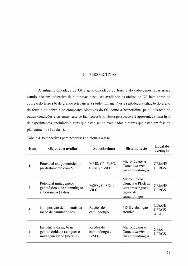
71
5 PERSPECTIVAS
A antigenotoxicidade do OJ e genotoxicidade do ferro e do cobre, mostradas nesse
estudo, são um indicativo de que novas pesquisas avaliando os efeitos do OJ, bem como do
cobre e do ferro são de grande relevância à saúde humana. Neste sentido, a avaliação do efeito
do ferro e do cobre e de compostos bioativos do OJ, como a hesperidina, pela utilização de
outras condições e sistemas-teste se faz necessária. Nesta perspectiva é apresentada uma lista
de experimentos, incluindo alguns que estão sendo executados e outras que estão em fase de
planejamento (Tabela 4).
Tabela 4. Perspectivas para pesquisas adicionais à tese.
Item Objetivo a avaliar Substância(s) Sistema teste Local de execução
1 Potencial antigenotóxico do pré-tratamento com Vit C
MMS, CP, FeSO4, CuSO4 e Vit C
Micronúcleos e Cometa in vivo em camundongos
CBiot/IF-UFRGS
2 Potencial mutagênico, genotóxico e de acumulação subcrônicos (7 dias)
FeSO4, CuSO4 e Vit C
Micronúcleos, Cometa e PIXE in vivo em sangue e fígado de camundongos
CBiot/IF-UFRGS
3 Composição de minerais da ração de camundongos
Rações de camundongo
PIXE e absorção atômica
CBiot/IF- UFRGS ALAC
4 Influência da ração na genotoxicidade (sangue) e mutagenicidade (medula)
Rações de camundongo e FeSO4
Micronúcleos e Cometa in vivo em camundongos
CBiot-UFRGS
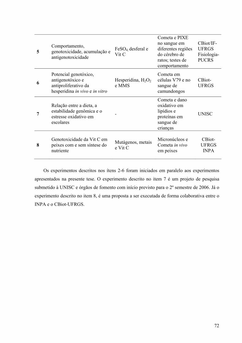
72
5 Comportamento, genotoxicidade, acumulação e antigenotoxicidade
FeSO4, desferal e Vit C
Cometa e PIXE no sangue em diferentes regiões do cérebro de ratos; testes de comportamento
CBiot/IF-UFRGS Fisiologia- PUCRS
6
Potencial genotóxico, antigenotóxico e antiproliferativo da hesperidina in vivo e in vitro
Hesperidina, H2O2 e MMS
Cometa em células V79 e no sangue de camundongos
CBiot-UFRGS
7
Relação entre a dieta, a estabilidade genômica e o estresse oxidativo em escolares
-
Cometa e dano oxidativo em lipídios e proteínas em sangue de crianças
UNISC
8 Genotoxicidade da Vit C em peixes com e sem síntese do nutriente
Mutágenos, metais e Vit C
Micronúcleos e Cometa in vivo em peixes
CBiot-UFRGS INPA
Os experimentos descritos nos ítens 2-6 foram iniciados em paralelo aos experimentos
apresentados na presente tese. O experimento descrito no item 7 é um projeto de pesquisa
submetido à UNISC e órgãos de fomento com início previsto para o 2º semestre de 2006. Já o
experimento descrito no item 8, é uma proposta a ser executada de forma colaborativa entre o
INPA e o CBiot-UFRGS.
73
6 REFERÊNCIAS
Abalea, V., Cillard, J., Dubos, M. P., Sergent, O., Cillard, P., Morel, I. (1999). Repair of iron-induced DNA oxidation by the flavonoid myricetin in primary rat hepatocyte cultures. Free Radic Biol Med, 26(11-12): 1457-66.
Ames, B. N. (1989). Mutagenesis and carcinogenesis: endogenous and exogenous factors. Environ Mol Mutagen, 14 Suppl 16: 66-77.
Ames, B. N. (2001). DNA damage from micronutrient deficiencies is likely to be a major cause of cancer. Mutat Res, 475(1-2): 7-20.
Ames, B. N., Atamna, H., Killilea, D. W. (2005). Mineral and vitamin deficiencies can accelerate the mitochondrial decay of aging. Mol Aspects Med, 26(4-5): 363-378.
Ames, B. N., Gold, L. S. (1998). The causes and prevention of cancer: the role of environment. Biotherapy, 11(2-3): 205-20.
Anderson, G. J., Frazer, D. M., McKie, A. T., Vulpe, C. D., Smith, A. (2005). Mechanisms of Haem and Non-Haem Iron Absorption: Lessons from Inherited Disorders of Iron Metabolism. BioMetals, 18(4): 339.
Arredondo, M., Nunez, M. T. (2005). Iron and copper metabolism. Molecular Aspects of Medicine, 26(4-5): 313.
Bailey, D. G., Spence, J. D., Munoz, C., Arnold, J. M. (1991). Interaction of citrus juices with felodipine and nifedipine. Lancet, 337(8736): 268-9.
Bissig, K. D., Honer, M., Zimmermann, K., Summer, K. H., Solioz, M. (2005). Whole animal copper flux assessed by positron emission tomography in the Long-Evans cinnamon rat--a feasibility study. Biometals, 18(1): 83-8.
Boiteux, S., Guillet, M. (2004). Abasic sites in DNA: repair and biological consequences in Saccharomyces cerevisiae. DNA Repair (Amst), 3(1): 1-12.
Booher, R. N., Deshaies, R. J., Kirschner, M. W. (1993). Properties of Saccharomyces cerevisiae wee1 and its differential regulation of p34CDC28 in response to G1 and G2 cyclins. Embo J, 12(9): 3417-26.
Borella, M. L. L., Varela, Q. D. (2004). Antioxidantes enzimáticos. Radicais livres e a resposta celular ao estresse oxidativo. Salvador, M. & Henriques, J. A. P. Canoas, Ed. ULBRA: 35-49.
BRASIL (1974). Ministério da Agricultura. Decreto 2314/04/1997, Artigo 14, Diário Oficial da República Federativa do Brasil, Brasília.
Brozmanova, J., Dudas, A., Henriques, J. A. (2001a). Repair of oxidative DNA damage--an important factor reducing cancer risk. Minireview. Neoplasma, 48(2): 85-93.
Brozmanova, J., Vlckova, V., Farkasova, E., Dudas, A., Vlasakova, D., Chovanec, M., Mikulovska, Z., Fridrichova, I., Saffi, J., Henriques, J. A. (2001b). Increased DNA
74
double strand breakage is responsible for sensitivity of the pso3-1 mutant of Saccharomyces cerevisiae to hydrogen peroxide. Mutat Res, 485(4): 345-55.
Campbell, J. L., Hopman, T. L., Maxwell, J. A., Nejedly, Z. (2000). The Guelph PIXE software package III: alternative proton database. Nuclear Instruments and Methods in Physics Research B, 170: 193-204.
Cerda, S., Weitzman, S. A. (1997). Influence of oxygen radical injury on DNA methylation. Mutat Res, 386(2): 141-52.
Chelikani, P., Fita, I., Loewen, P. C. (2004). Diversity of structures and properties among catalases. Cell Mol Life Sci, 61(2): 192-208.
Choy, W. N. (1996). Principles of genetic toxicology. Drug Chem Toxicol, 19(3): 149-60. Collins, A. R. (2004). The comet assay for DNA damage and repair: principles, applications,
and limitations. Mol Biotechnol, 26(3): 249-61. Cooke, M. S., Evans, M. D., Podmore, I. D., Herbert, K. E., Mistry, N., Mistry, P.,
Hickenbotham, P. T., Hussieni, A., Griffiths, H. R., Lunec, J. (1998). Novel repair action of vitamin C upon in vivo oxidative DNA damage. FEBS Lett, 439(3): 363-7.
Da Silva, J., Freitas, T. R. O., Marinho, J. R., Speit, G., Erdtmann, B. (2000). An alkali single-cell gel eletroforesis (comet) assay for environmental biomonitoring with native rodents. Genet Mol Biol, 23: 241- 245.
De Flora, S. (1998). Mechanisms of inhibitors of mutagenesis and carcinogenesis. Mutation Research/Fundamental and Molecular Mechanisms of Mutagenesis, 402(1-2): 151.
De Freitas, J. M., Meneghini, R. (2001). Iron and its sensitive balance in the cell. Mutat Res, 475(1-2): 153-9.
Dedon, P. C., Tannenbaum, S. R. (2004). Reactive nitrogen species in the chemical biology of inflammation. Arch Biochem Biophys, 423(1): 12-22.
Dekaban, A. S., Aamodt, R., Rumble, W. F., Johnston, G. S., O'Reilly, S. (1975). Kinky hair disease: a study of copper metabolism with use of 67Cu. Arch Neurol, 32: 672-675.
Dew, T. P., Day, A. J., Morgan, M. R. A. (2005). Xanthine oxidase activity in vitro: effects of food extracts and components. J. Agric. Food Chem., 53(16): 6510-6515.
Di Marco, M. P., Edwards, D. J., Wainer, I. W., Ducharme, M. P. (2002). The effect of grapefruit juice and seville orange juice on the pharmacokinetics of dextromethorphan: The role of gut CYP3A and P-glycoprotein. Life Sci, 71(10): 1149-1160.
Dizdaroglu, M., Jaruga, P., Birincioglu, M., Rodriguez, H. (2002). Free radical-induced damage to DNA: mechanisms and measurement. Free Radical Biology and Medicine, 32(11): 1102-1115.
Doostdar, H., Burke, D., Mayer, R. T. (2000). Bioflavonoids: selective substrates and inhibitors for cytochrome P450 CYP1A and CYP1B1. Toxicology, 144: 31-38.
Drablos, F., Feyzi, E., Aas, P. A., Vaagbo, C. B., Kavli, B., Bratlie, M. S., Pena-Diaz, J., Otterlei, M., Slupphaug, G., Krokan, H. E. (2004). Alkylation damage in DNA and RNA--repair mechanisms and medical significance. DNA Repair (Amst), 3(11): 1389-407.
Dunkel, V. C., San, R. H., Seifried, H. E., Whittaker, P. (1999). Genotoxicity of iron compounds in Salmonella typhimurium and L5178Y mouse lymphoma cells. Environ Mol Mutagen, 33(1): 28-41.
Earl, C. J., Moulton, M. J., Selverstone, B. (1954). Metabolism of copper in Wilson's disease and in normal subjects. Am J Med, 17(205-213).
Edenharder, R., Grunhage, D. (2003). Free radical scavenging abilities of flavonoids as mechanism of protection against mutagenicity induced by tert-butyl hydroperoxide or cumene hydroperoxide in Salmonella typhimurium TA102. Mutat Res, 540(1): 1-18
75
Edenharder, R., Krieg, H., Kottgen, V., Platt, K. L. (2003). Inhibition of clastogenicity of benzo[a]pyrene and of its trans-7,8-dihydrodiol in mice in vivo by fruits, vegetables, and flavonoids. Mutat Res, 537(2): 169-81.
Edenharder, R., Sager, J. W., Glatt, H., Muckel, E., Platt, K. L. (2002). Protection by beverages, fruits, vegetables, herbs, and flavonoids against genotoxicity of 2-acetylaminofluorene and 2-amino-1-methyl-6-phenylimidazo[4,5-b]pyridine (PhIP) in metabolically competent V79 cells. Mutat Res, 521(1-2): 57-72.
Edgar, J. A. (1974). Ascorbic acid and biological alkylating agents. Nature, 248(444): 136. Evans, W. E., McLeod, H. L. (2003). Pharmacogenomics--drug disposition, drug targets, and
side effects. N Engl J Med, 348(6): 538-49. Fairbairn, D. W., Olive, P. L., O'Neill, K. L. (1995). The comet assay: a comprehensive
review. Mutat Res, 339(1): 37-59. Fang, F. C. (2004). Antimicrobial reactive oxygen and nitrogen species: concepts and
controversies. Nat Rev Microbiol, 2(10): 820-32. Farheen, A., Aslam, T., Almas, K., Bhatty, N., Nawaz, M. (2002). Biokinetics of inorganic
iron in female volunteers. Pakistan J. Med. Res., 41(1). Fenech, M. (2005a). The Genome Health Clinic and Genome Health Nutrigenomics concepts:
diagnosis and nutritional treatment of genome and epigenome damage on an individual basis. Mutagenesis, 20(4): 255-269.
Fenech, M. (2005b). The Genome Health Clinic and Genome Health Nutrigenomics concepts: diagnosis and nutritional treatment of genome and epigenome damage on an individual basis. Mutagenesis, 20(4): 255-69.
Fenech, M., Baghurst, P., Luderer, W., Turner, J., Record, S., Ceppi, M., Bonassi, S. (2005). Low intake of calcium, folate, nicotinic acid, vitamin E, retinol, beta-carotene and high intake of pantothenic acid, biotin and riboflavin are significantly associated with increased genome instability--results from a dietary intake and micronucleus index survey in South Australia. Carcinogenesis, 26(5): 991-9.
Fenech, M., Ferguson, L. R. (2001). Vitamins/minerals and genomic stability in humans. Mutat Res, 475(1-2): 1-6.
Feskanich, D., Willett, W. C., Hunter, D. J., Colditz, G. A. (2003). Dietary intakes of vitamins A, C, and E and risk of melanoma in two cohorts of women. Br J Cancer, 88(9): 1381-7.
Follmer, C., Carlini, C. R., Yoneama, M. L., Dias, J. F. (2002). PIXE analysis of uréase isoenzymes isolated from Canavalia ensiformis (Jack bean) seeds. Nuclear Instruments and Methods in Physics Research B, 189: 482-486.
Franke, S. I. R., Boeira, J. M., Henriques, J. A. P. (2003). Genotoxicidade de agentes sintéticos e naturais. Genética Toxicológica. da Silva, J., Erdtmann, B. & Henriques, J. A. P. Porto Alegre, Alcance: 261-270.
Franke, S. I. R., Ckless, K., Silveira, J. D., Rubensam, G., Brendel, M., Erdtmann, B., Henriques, J. A. P. (2004). Study of antioxidant and mutagenic activity of different orange juices. Food Chem, 88(1): 45-55
Franke, S. I. R., Pra, D., da Silva, J., Erdtmann, B., Henriques, J. A. P. (2005a). Possible repair action of Vitamin C on DNA damage induced by methyl methanesulfonate, cyclophosphamide, FeSO4 and CuSO4 in mouse blood cells in vivo. Mutation Research/Genetic Toxicology and Environmental Mutagenesis, 583(1): 75-84.
Franke, S. I. R., Pra, D., Dias, J. F., Giulian, R., Yoneama, M. L., Da Silva, J., Erdtmann, B., Henriques, J. A. P. (2006). Influence of orange juice in the levels and in the genotoxicity of iron and copper. Food Chem Toxicol, 44(3): 425-435.
76
Franke, S. I. R., Pra, D., Erdtmann, B., Henriques, J. A. P., da Silva, J. (2005b). Influence of orange juice over the genotoxicity induced by alkylating agents: an in vivo analysis. Mutagenesis, 20(4): 279-283.
Fridovich, I. (1998). Oxygen toxicity: a radical explanation. J Exp Biol, 201(Pt 8): 1203-9. Gao, K., Henning, S. M., Niu, Y., Youssefian, A. A., Seeram, N. P., Xu, A., Heber, D. (2006).
The citrus flavonoid naringenin stimulates DNA repair in prostate cancer cells. J Nutr Biochem, 17(2): 89-95.
Gaspar, P. A. (2003). Genes de suscetibilidade. Genetica Toxicológica. da Silva, J., Erdtmann, B. & Henriques, J. A. P. Porto Alegre, Alcance: 209-224.
Gillbe, C. E., Sage, F. J., Gutteridge, J. M. (1996). Commentary: mannitol: molecule magnifique or a case of radical misinterpretation? Free Radic Res, 24(1): 1-7.
Glantzounis, G. K., Tsimoyiannis, E. C., Kappas, A. M., Galaris, D. A. (2005). Uric acid and oxidative stress. Curr Pharm Des, 11(32): 4145-51.
Goldman, R., Shields, P. G. (2003). Food Mutagens. J. Nutr., 133(3): 965S-973. Guecheva, T. N., Henriques, J. A. P. (2003). Metabolismo de xenobióticos: citocromo P-450.
Genetica Toxicológica. da Silva, J., Erdtmann, B. & Henriques, J. A. P. Porto Alegre, Alcance: 225-250.
Guecheva, T. N., Henriques, J. A. P., Erdtmann, B. (2001). Genotoxic effects of copper sulphate in freshwater planarian in vivo, studied with the single-cell gel test (comet assay). Mutat Res, 497(1-2): 19-27.
Guha, B., Khuda-Bukhsh, A. R. (2002). Efficacy of vitamin-C (L-ascorbic acid) in reducing genotoxicity in fish (Oreochromis mossambicus) induced by ethyl methane sulphonate. Chemosphere, 47(1): 49-56.
Guthrie, N., Carroll, K. K. (1998). Inhibition of mammary cancer by citrus flavonoids. Adv Exp Med Biol, 439: 227-36.
Hageman, G. J., Stierum, R. H. (2001). Niacin, poly(ADP-ribose) polymerase-1 and genomic stability. Mutat Res, 475(1-2): 45-56.
Halliwell, B. (2001). Vitamin C and genomic stability. Mutat Res, 475(1-2): 29-35. Halliwell, B., Guterridge, J. M. C. (2000). Free Radicals in Biology and Medicine. New York,
Oxford University Press. Halliwell, B., Rafter, J., Jenner, A. (2005). Health promotion by flavonoids, tocopherols,
tocotrienols, and other phenols: direct or indirect effects? Antioxidant or not? Am J Clin Nutr, 81(1 Suppl): 268S-276S.
Hartmann, A., Agurell, E., Beevers, C., Brendler-Schwaab, S., Burlinson, B., Clay, P., Collins, A., Smith, A., Speit, G., Thybaud, V., Tice, R. R. (2003). Recommendations for conducting the in vivo alkaline Comet assay. 4th International Comet Assay Workshop. Mutagenesis, 18(1): 45-51.
Hartwell, L. H., Weinert, T. A. (1989). Checkpoints: controls that ensure the order of cell cycle events. Science, 246(4930): 629-34.
Hashimoto, K., Shirafuji, T., Sekino, H., Matsuoka, O., Sekino, H., Onnagawa, O., Okamoto, T., Kudo, S., Azuma, J. (1998). Interaction of citrus juices with pranidipine, a new 1,4-dihydropyridine calcium antagonist, in healthy subjects. Eur J Clin Pharmacol, 54(9-10): 753-60.
Heidelberger, C., Freeman, A. E., Pienta, R. J., Sivak, A., Bertram, J. S., Casto, B. C., Dunkel, V. C., Francis, M. W., Kakunaga, T., Little, J. B., Schechtman, L. M. (1983). Cell transformation by chemical agents--a review and analysis of the literature. A report of the U.S. Environmental Protection Agency Gene-Tox Program. Mutat Res, 114(3): 283-385.
Henriques, J. A. P., Prá, D. (2005). Farmacogenética. Inovação, biotecnologia e saúde pública. Scivoletto, R. & Camargo, A. C. São Paulo, Instituto UNIEMP: 148.
77
Horvathova, E., Slamenova, D., Hlincikova, L., Mandal, T. K., Gabelova, A., Collins, A. R. (1998). The nature and origin of DNA single-strand breaks determined with the comet assay. Mutat Res, 409(3): 163-71.
Ikegawa, T., Ushigome, F., Koyabu, N., Morimoto, S., Shoyama, Y., Naito, M., Tsuruo, T., Ohtani, H., Sawada, Y. (2000). Inhibition of P-glycoprotein by orange juice components, polymethoxyflavones in adriamycin-resistant human myelogenous leukemia (K562/ADM) cells. Cancer Lett, 160(1): 21-28.
Imai, H., Nakagawa, Y. (2003). Biological significance of phospholipid hydroperoxide glutathione peroxidase (PHGPx, GPx4) in mammalian cells. Free Radic Biol Med, 34(2): 145-69.
IOM (2000). Institute of Medicine. Dietary Reference Intakes for Vitamin C, Vitamin E, Selenium, and Carotenoids. Food and Nutrition Board. Washington, DC, National Academy Press.
Jackson, A. L., Loeb, L. A. (2001). The contribution of endogenous sources of DNA damage to the multiple mutations in cancer. Mutat Res, 477(1-2): 7-21.
Jans, J., Schul, W., Sert, Y. G., Rijksen, Y., Rebel, H., Eker, A. P., Nakajima, S., van Steeg, H., de Gruijl, F. R., Yasui, A., Hoeijmakers, J. H., van der Horst, G. T. (2005). Powerful Skin Cancer Protection by a CPD-Photolyase Transgene. Curr Biol, 15(2): 105-15.
Jenkins, G. J., Doak, S. H., Johnson, G. E., Quick, E., Waters, E. M., Parry, J. M. (2005). Do dose response thresholds exist for genotoxic alkylating agents? Mutagenesis, 20(6): 389-98.
Johansson, S. A., Campbell, J. L., Malmqvist, K. G. (1995). Particle-Induced X-Ray Emission Spectrometry (PIXE). New York, John Wiley & Sons.
Johnston, C. S., Dancho, C. L., Strong, G. M. (2003). Orange Juice Ingestion and Supplemental Vitamin C Are Equally Effective at Reducing Plasma Lipid Peroxidation in Healthy Adult Women. J Am Coll Nutr, 22(6): 519-523.
Kabasakalis, V., Siopidou, D., Moshatou, E., (2000). Ascorbic acid content of commercial fruit juices and its rate of loss upon storage. Food Chem, 70: 325-328.
Kamath, A. V., Yao, M., Zhang, Y., Chong, S. (2005). Effect of fruit juices on the oral bioavailability of fexofenadine in rats. J Pharm Sci, 94(2): 233-9.
Kang, J. S., Cho, D., Kim, Y. I., Hahm, E., Kim, Y. S., Jin, S. N., Kim, H. N., Kim, D., Hur, D., Park, H., Hwang, Y. I., Lee, W. J. (2005). Sodium ascorbate (vitamin C) induces apoptosis in melanoma cells via the down-regulation of transferrin receptor dependent iron uptake. J Cell Physiol.
Kehrer, J. P., Biswal, S. S. (2000). The Molecular Effects of Acrolein. Toxicol. Sci., 57(1): 6-15.
Kern, A. L., Bonatto, D., Dias, J. F., Yoneama, M. L., Brendel, M., Henriques, A. P. L. (2004). The importance of yeast Alr proteins in cadmium detoxification as indicated by Particle-Induced X-ray Emission and phenotypic analyses. X-Ray Spectrometry, 10.1002/xrs.835.
Kojima, H., Konishi, H., Kuroda, Y. (1992). Effects of L-ascorbic acid on the mutagenicity of ethyl methanesulfonate in cultured mammalian cells. Mutat Res, 266(2): 85-91.
Kruszewski, M. (2003). Labile iron pool: the main determinant of cellular response to oxidative stress. Mutat Res, 531(1-2): 81-92.
Kuroda, Y., Shima, N., Yazawa, K., Kaji, K. (2001). Desmutagenic and bio-antimutagenic activity of docosahexaenoic acid and eicosapentaenoic acid in cultured Chinese hamster V79 cells. Mutat Res, 497(1-2): 123-30.
Lim, S.-L., Lim, L.-Y. (2006). Effects of citrus fruit juices on cytotoxicity and drug transport pathways of Caco-2 cell monolayers. Int J Pharm, 307(1): 42-50.
78
Linder, M. C. (2001). Copper and genomic stability in mammals. Mutat Res, 475(1-2): 141-52.
Ma, Y., Cao, L., Kawabata, T., Yoshino, T., Yang, B. B., Okada, S. (1998). Cupric nitrilotriacetate induces oxidative DNA damage and apoptosis in human leukemia HL-60 cells. Free Radic Biol Med, 25(4-5): 568-75.
MacPhee, D. G. (1998a). The environmental and health effects of solar radiation. Mutat Res, 422(1): 1-6.
MacPhee, D. G. (1998b). Epigenetics and epimutagens: some new perspectives on cancer, germ line effects and endocrine disrupters. Mutat Res, 400(1-2): 369-79.
Malhotra, S., Bailey, D. G., Paine, M. F., Watkins, P. B. (2001). Seville orange juice-felodipine interaction: Comparison with dilute grapefruit juice and involvement of furocoumarins. Clin Pharmacol Ther, 69(1): 14-23.
Matalon, S. T., Orney, A., Lishner, M. (2004). Review of the potential effects of three commonly used antineoplastic and immunosuppressive drugs (cyclophosphamide, azathioprine, doxorubicin on the embryo and placenta). Reproductive Toxicology, 18: 219–230.
Mates, J. M., Perez-Gomez, C., Nunez de Castro, I. (1999). Antioxidant enzymes and human diseases. Clin Biochem, 32(8): 595-603.
Maxwell, J. A., Campbell, J. L., Teesdale, W. J. (1989). The Guelph PIXE software package. Nuclear Instruments and Methods in Physics Research B, 43: 218-230.
Maxwell, J. A., Teesdale, W. J., Campbell, J. L. (1995). The Guelph PIXE software package II. Nuclear Instruments and Methods in Physics Research B, 95: 407-421.
McHugh, P. J., Spanswick, V. J., Hartley, J. A. (2001). Repair of DNA interstrand crosslinks: molecular mechanisms and clinical relevance. The Lancet Oncology, 2(8): 483-490.
McNaught, A. D., Wilkinson, A. (1997). IUPAC Compendium of Chemical Terminology: The Gold Book, 2nd edition. Cambridge, UK, Blackwell Science.
Meneghini, R. (1997). Iron homeostasis, oxidative stress, and DNA damage. Free Radic Biol Med, 23(5): 783-92.
Middleton, E., Jr., Kandaswami, C., Theoharides, T. C. (2000). The effects of plant flavonoids on mammalian cells: implications for inflammation, heart disease, and cancer. Pharmacol Rev, 52(4): 673-751.
Moon, Y. J., Wang, X., Morris, M. E. (2006). Dietary flavonoids: Effects on xenobiotic and carcinogen metabolism. Toxicology in Vitro, 20(2): 187.
Mouly, S. J., Matheny, C., Paine, M. F., Smith, G., Lamba, J., Lamba, V., Pusek, S. N., Schuetz, E. G., Stewart, P. W., Watkins, P. B. (2005). Variation in oral clearance of saquinavir is predicted by CYP3A5*1 genotype but not by enterocyte content of cytochrome P450 3A5. Clin Pharmacol Ther, 78(6): 605-18.
NIH (2006). National Institute of Health. Health & Human Services. Genetic Toxicology Data Bank (GENE-TOX). Available at: http://toxnet.nlm.nih.gov/cgi-bin/sis/htmlgen?GENETOX.
Nishimuta, H., Tsujimoto, M., Ogura, K., Hiratsuka, A., Ohtani, H., Sawada, Y. (2005). Inhibitory effects of various beverages on ritodrine sulfation by recombinant human sulfotransferase isoforms SULT1A1 and SULT1A3. Pharm Res, 22(8): 1406-10.
Patrineli, A., Clifford, M. N., Ioannides, C. (1996a). Contribution of phenols, quinones and reactive oxygen species to the mutagenicity of white grape juice in the Ames test. Food Chem Toxicol, 34(9): 869-72.
Patrineli, A., Clifford, M. N., Walker, R., Ioannides, C. (1996b). Mutagenicity of white grape juice in the Ames test. Food Chem Toxicol, 34(6): 559-62.
79
Picada, J. N., Kern, A. L., Ramos , A. L. L. P., Saffi, J. (2003). O estresse oxidativo e as defesas antioxidante. Genética Toxicológica. Da Silva, J., Erdtmann, B. & Henriques, J. A. P. Porto Alegre, Alcance: 251-264.
Powers, H. J. (2005). Interaction among folate, riboflavin, genotype, and cancer, with reference to colorectal and cervical cancer. J Nutr, 135(12 Suppl): 2960S-2966S.
Prá, D., Lau, A. H., Knakievicz, T., Carneiro, F. R., Erdtmann, B. (2005). Environmental genotoxicity assessment of an urban stream using freshwater planarians. Mutat Res.
Rayman, M. P. (2005). Selenium in cancer prevention: a review of the evidence and mechanism of action. Proc Nutr Soc, 64(4): 527-42.
Risom, L., Moller, P., Loft, S. (2005). Oxidative stress-induced DNA damage by particulate air pollution. Mutat Res, 592(1-2): 119-37.
Rojas, E., Herrera, L. A., Poirier, L. A., Ostrosky-Wegman, P. (1999). Are metals dietary carcinogens? Mutat Res, 443(1-2): 157-81.
Rosa, R. M., Roehrs, R., Saffi, J., Henriques, J. A. P. (2004). O papel da reparação de lesões oxidativas induzidas no DNA na redução do risco do desenvolvimento do câncer. Radicais livres e a resposta celular ao estresse oxidativo. Salvador, M. & Henriques, J. A. P. Canoas, Ed. ULBRA: 139-159.
Saffi, J., Henriques, J. A. P. (2003). Reparação de DNA em células eucarióticas. Genética Toxicológica. Da Silva, J., Erdtmann, B. & Henriques, J. A. P. Porto Alegre, Alcance: 267-305.
Sakagami, H., Satoh, K., Hakeda, Y., Kumegawa, M. (2000). Apoptosis-inducing activity of vitamin C and vitamin K. Cell Mol Biol (Noisy-le-grand), 46(1): 129-43.
Sarasin, A. (2003). An overview of the mechanisms of mutagenesis and carcinogenesis. Mutat Res, 544(2-3): 99-106.
Schwedhelm, E., Maas, R., Troost, R., Boger, R. H. (2003). Clinical pharmacokinetics of antioxidants and their impact on systemic oxidative stress. Clin Pharmacokinet, 42: 437-459.
Sedlak, T. W., Snyder, S. H. (2004). Bilirubin benefits: cellular protection by a biliverdin reductase antioxidant cycle. Pediatrics, 113(6): 1776-82.
Sen, C. K., Packer, L. (2000). Thiol homeostasis and supplements in physical exercise. Am J Clin Nutr, 72(2): 653S-669.
Shimoi, K., Akaiwa, E., Mori, N., Sano, M., Nakamura, Y., Tomita, I. (1992). Bio-antimutagenic activities of vitamin B6 in E. coli and mouse peripheral blood cells. Mutat Res, 266(2): 205-213.
Singh, N., McCoy, M., Tice, R., Schneider, E. (1988). A simple technique for quantitation of low levels of DNA damage in individual cells. Exp Cell Res, 175(1): 184-191.
Singh, R., Sweetman, G. M., Farmer, P. B., Shuker, D. E., Rich, K. J. (1997). Detection and characterization of two major ethylated deoxyguanosine adducts by high performance liquid chromatography, electrospray mass spectrometry, and 32P-postlabeling. Development of an approach for detection of phosphotriesters. Chem Res Toxicol, 10(1): 70-7.
Sinigaglia, M., Reguly, M. L., de Andrade, H. H. (2004). Effect of vanillin on toxicant-induced mutation and mitotic recombination in proliferating somatic cells of Drosophila melanogaster. Environ Mol Mutagen, 44(5): 394-400.
Slupphaug, G., Kavli, B., Krokan, H. E. (2003). The interacting pathways for prevention and repair of oxidative DNA damage. Mutat Res, 531(1-2): 231-51.
So, F. V., Guthrie, N., Chambers, A. F., Moussa, M., Carroll, K. K. (1996). Inhibition of human breast cancer cell proliferation and delay of mammary tumorigenesis by flavonoids and citrus juices. Nutr Cancer, 26(2): 167-81.
80
Souliotis, V. L., Dimopoulos, M. A., Sfikakis, P. P. (2003). Gene-specific formation and repair of DNA monoadducts and interstrand cross-links after therapeutic exposure to nitrogen mustards. Clin Cancer Res, 9(12): 4465-74.
Spada, P. K. W. D. S., Silva, C. O. d. (2004). Antioxidantes não enzimáticos. Radicais livres e a resposta celular ao estresse oxidativo. Salvador, M. & Henriques, J. A. P. Canoas, Ed. ULBRA: 51-67.
Speit, G., Hanelt, S., Helbig, R., Seidel, A., Hartmann, A. (1996). Detection of DNA effects in human cells with the comet assay and their relevance for mutagenesis. Toxicol Lett, 88(1-3): 91-8.
Sram, R. J., Dobias, L., Pastorkova, A., Rossner, P., Janca, L. (1983). Effect of ascorbic acid prophylaxis on the frequency of chromosome aberrations in the peripheral lymphocytes of coal-tar workers. Mutat Res, 120(2-3): 181-6.
Takanaga, H., Ohnishi, A., Yamada, S., Matsuo, H., Morimoto, S., Shoyama, Y., Ohtani, H., Sawada, Y. (2000). Polymethoxylated flavones in orange juice are inhibitors of P-glycoprotein but not cytochrome P450 3A4. J Pharmacol Exp Ther, 293(1): 230-6.
Terao, J., Piskula, M. (1999). Flavonoids and membrane lipid peroxidation. Nutrition, 15: 790-791.
Tian, R., Koyabu, N., Takanaga, H., Matsuo, H., Ohtani, H., Sawada, Y. (2002). Effects of grapefruit juice and orange juice on the intestinal efflux of P-glycoprotein substrates. Pharm Res, 19(6): 802-809.
Tice, R. R., Agurell, E., Anderson, D., Burlinson, B., Hartmann, A., Kobayashi, H., Miyamae, Y., Rojas, E., Ryu, J. C., Sasaki, Y. F. (2000). Single cell gel/comet assay: guidelines for in vitro and in vivo genetic toxicology testing. Environ Mol Mutagen, 35(3): 206-21.
Tkeshelashvili, L. K., McBride, T., Spence, K., Loeb, L. A. (1991). Mutation spectrum of copper-induced DNA damage. J Biol Chem, 266(10): 6401-6.
Tucker, J. D., Auletta, A., Cimino, M. C., Dearfield, K. L., Jacobson-Kram, D., Tice, R. R., Carrano, A. V. (1993). Sister-chromatid exchange: second report of the Gene-Tox Program. Mutat Res, 297(2): 101-80.
Uriu-Adams, J. Y., Rucker, R. B., Commisso, J. F., Keen, C. L. (2005). Diabetes and dietary copper alter 67Cu metabolism and oxidant defense in the rat. The Journal of Nutritional Biochemistry, 16(5): 312.
Valko, M., Izakovic, M., Mazur, M., Rhodes, C. J., Telser, J. (2004). Role of oxygen radicals in DNA damage and cancer incidence. Mol Cell Biochem, 266(1-2): 37-56.
Valko, M., Morris, H., Cronin, M. T. (2005). Metals, toxicity and oxidative stress. Curr Med Chem, 12(10): 1161-208.
Vasdev, S., Gill, V., Parai, S., Gadag, V. (2005). Dietary lipoic acid supplementation attenuates hypertension in Dahl salt sensitive rats. Molecular and Cellular Biochemistry, 275(1 - 2): 135.
Vercet, A., Burgos, J., Lopez-Buesa, P. (2001). Manothermosonication of Foods and Food-Resembling Systems: Effect on Nutrient Content and Nonenzymatic Browning. J. Agric. Food Chem., 49(1): 483-489.
Vijayalaxmi, K. K., Venu, R. (1999). In vivo anticlastogenic effects of L-ascorbic acid in mice. Mutat Res, 438(1): 47-51.
Vinson, J. A. (1998). Flavonoids in foods as in vitro and in vivo antioxidants. Flavonoids in the Living System. Manthey, J. A. & Buslig, B. S. New York, Plenum Pub Corp: 151-164.
Voitkun, V., Zhitkovich, A., Costa, M. (1998). Cr(III)-mediated crosslinks of glutathione or amino acids to the DNA phosphate backbone are mutagenic in human cells. Nucl. Acids Res., 26(8): 2024-2030.
81
Vrzoc, M., Petras, M. L. (1997). Comparison of alkaline single cell gel (Comet) and peripheral blood micronucleus assays in detecting DNA damage caused by direct and indirect acting mutagens. Mutat Res, 381(1): 31-40.
Wang, D., Kreutzer, D. A., Essigmann, J. M. (1998). Mutagenicity and repair of oxidative DNA damage: insights from studies using defined lesions. Mutat Res, 400(1-2): 99-115.
Wang, H., Cao, G., Prior, R. L. (1996). Total antioxidant capacity of fruits. J Agric Food Chem, 44: 701-705.
Wang, Z., Dillon, J., Gaillard, E. R. (2005). Antioxidant Properties of Melanin in RPE cells. Photochem Photobiol.
Watters, J. W., Kloss, E. F., Link, D. C., Graubert, T. A., McLeod, H. L. (2003). A mouse-based strategy for cyclophosphamide pharmacogenomic discovery. J Appl Physiol, 95(4): 1352-60.
Wei, E. K., Giovannucci, E., Selhub, J., Fuchs, C. S., Hankinson, S. E., Ma, J. (2005). Plasma Vitamin B6 and the Risk of Colorectal Cancer and Adenoma in Women. J Natl Cancer Inst, 97(9): 684-692.
Wei, Q., Shen, H., Wang, L.-E., Duphorne, C. M., Pillow, P. C., Guo, Z., Qiao, Y., Spitz, M. R. (2003). Association between Low Dietary Folate Intake and Suboptimal Cellular DNA Repair Capacity. Cancer Epidemiol Biomarkers Prev, 12(10): 963-969.
WHO (2001). World Health Organization. Food and Agriculture Organization of the United Nations. Human Vitamin and Mineral Requirements. Food and Nutrition Division. FAO Rome. Report of a joint FAO/WHO expert consultation Bangkok, Thailand. Rome.
Winterbourn, C. C., Chan, T., Buss, I. H., Inder, T. E., Mogridge, N., Darlow, B. A. (2000). Protein Carbonyls and Lipid Peroxidation Products as Oxidation Markers in Preterm Infant Plasma: Associations with Chronic Lung Disease and Retinopathy and Effects of Selenium Supplementation. Pediatr Res, 48(1): 84-90.
Yoneama, M. L., Dias, J. F. (2004). Técnicas Analíticas - Espectroscopia por emissão de raios-X induzida por partículas (PIXE). Estudos Ambientais em Candiota - Carvão e seus impactos. Cadernos de Planejamento e Gestão Ambiental no 4. Teixeira, E. C. & Pires, M. J. R. Porto Alegre, FINEP/PADCT/CIAMB/FAPERGS/FEPAM: 209-215.
Yoshino, M., Haneda, M., Naruse, M., Murakami, K. (1999). Prooxidant activity of flavonoids: copper-dependent strand breaks and the formation of 8-hydroxy-2'-deoxyguanosine in DNA. Mol Genet Metab, 68(4): 468-72.

82
7 ANEXOS
83
7.1 ANEXO A: “Study of the antioxidant and mutagenic activity of different orange juices”
Franke, S.I.R., Ckless, K., Silveira, J.D., Rubensam, G., Brendel, M., Erdtmann, B., Henriques, J.A.P. Food Chemistry, 88: 45-55, 2004.
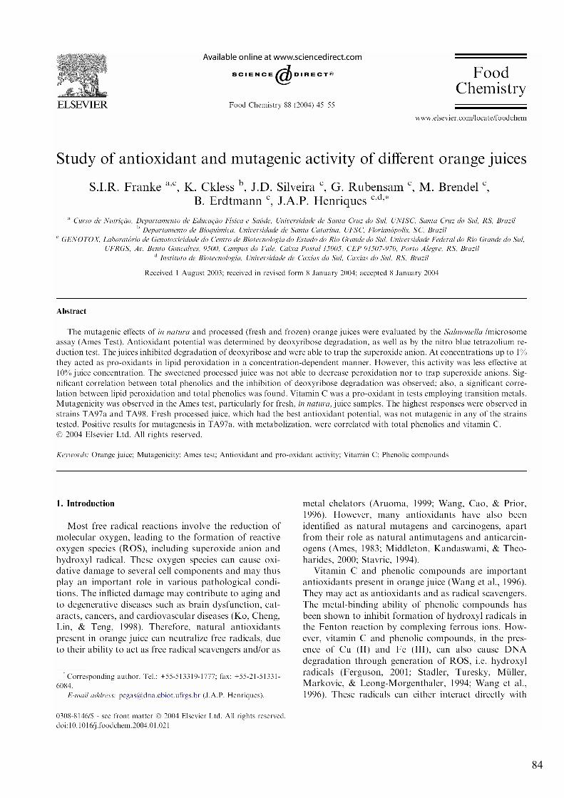
84

85

86
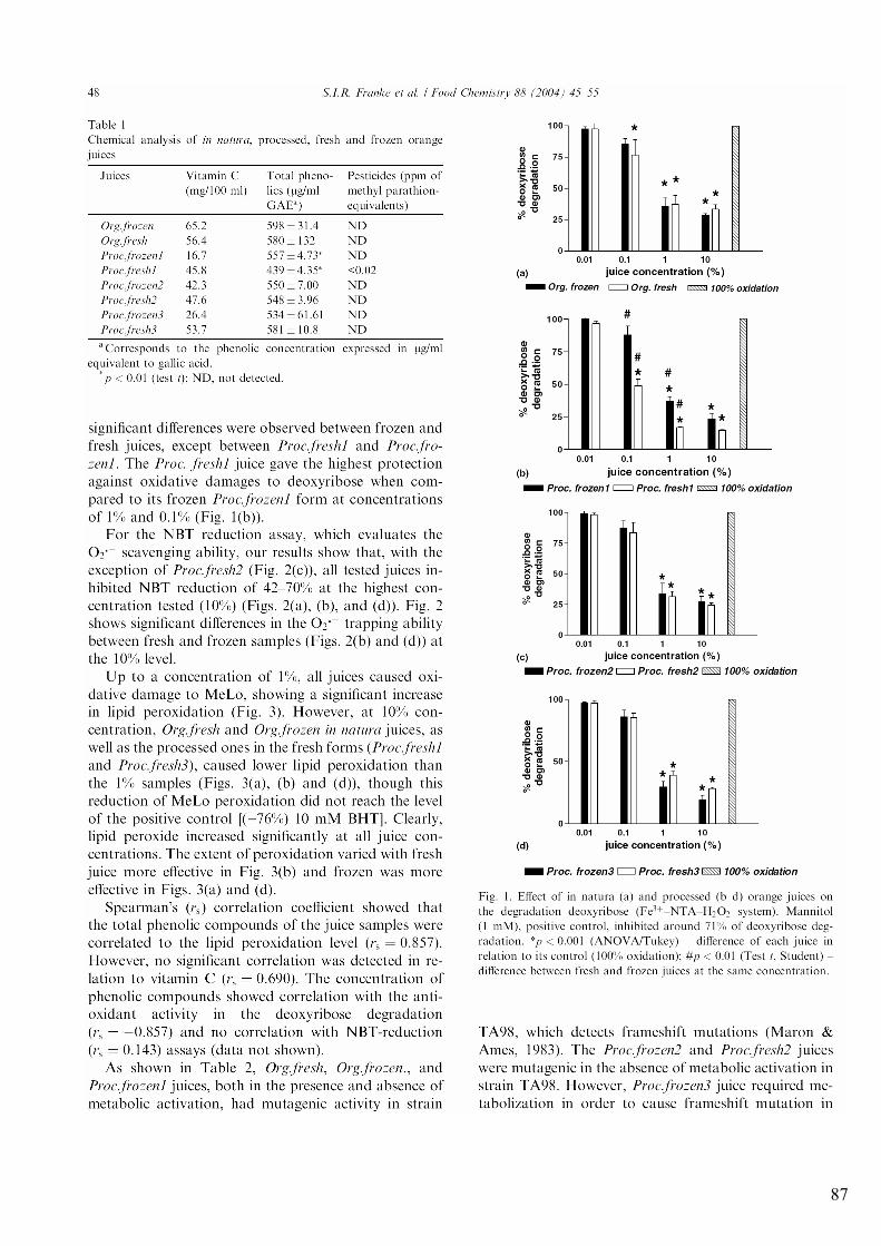
87
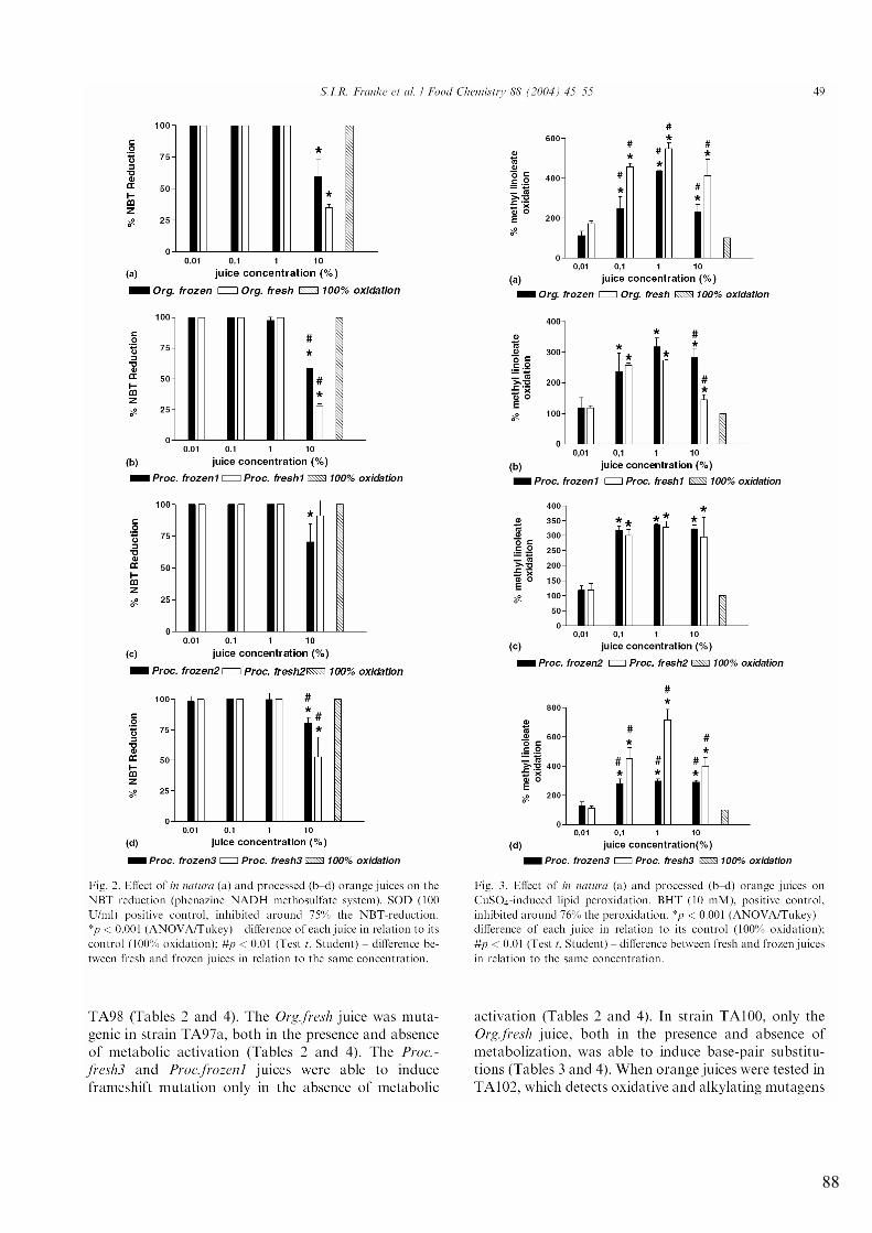
88
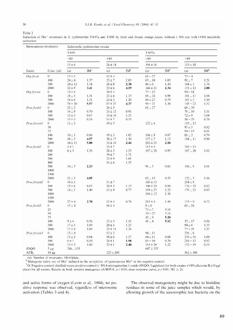
89
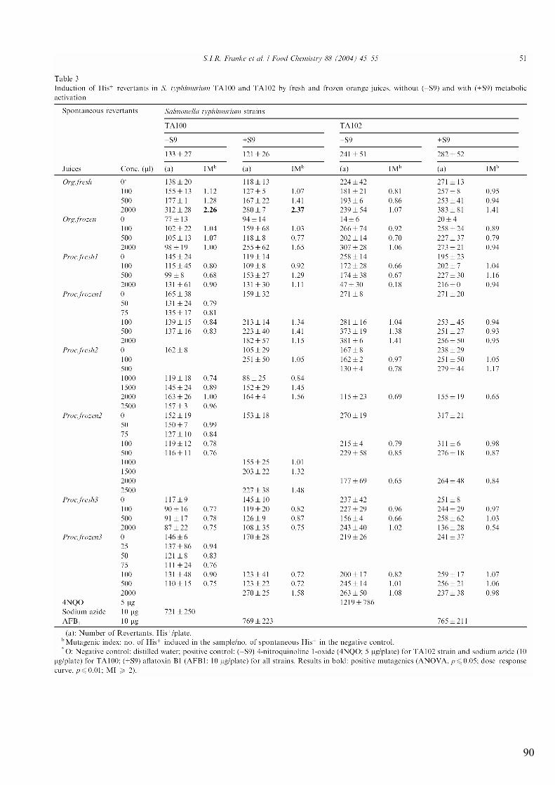
90
91
92
93
94
95
7.2 ANEXO B: “El jugo de naranja tiene potencial antioxidante y mutagenico”
Franke, S.I.R., Ckless, K., Silveira, J.D., Rubensam, G., Brendel, M., Erdtmann, B., Henriques, J.A.P. Disponível em http://www.siicsalud.com/dato/buscador/04n18011bb.htm
96
97
98
99
100
7.3 ANEXO C “Review: Orange juice, genomic stability and oxidative stress. Part I: Effect of the whole juice”
Franke, S.I.R., Prá, D., Henriques, J.A.P. Manuscrito a ser submetido para o British Journal of Nutrition
101
Review:
Orange juice, genomic stability and oxidative stress. Part I: Effect of the whole juice
Silvia Isabel Rech Franke1, 2#, Daniel Prá2# and João Antonio Pêgas Henriques2, 3, 4, *
1 Curso de Nutrição, Departamento de Educação Física e Saúde, Universidade de Santa
Cruz do Sul (UNISC), Santa Cruz do Sul, RS, Brasil.
2 Genotox / PPGBCM / PPGBM, Universidade Federal do Rio Grande do Sul (UFRGS)
Porto Alegre, RS, Brasil.
3 Curso de Biologia / Curso de Farmácia, Universidade Luterana do Brasil (ULBRA),
Canoas, RS, Brasil.
4 Instituto de Biotecnologia, Universidade de Caxias do Sul, Caxias do Sul, RS, Brasil.
*Address for Correspondence: Centro de Biotecnologia, UFRGS; Av. Bento Gonçalves,
9500, Prédio 43421; Campus do Vale; Caixa Postal 15005; CEP 91501-970, Porto
Alegre, RS, Brasil; Fax: +5551 33166084; e-mail: [email protected]
# Authors contributed equally in manuscript preparation.
102
Abstract
Orange juice (OJ) is a complex mixture composed of, among other substances, proteins,
carbohydrates, lipids, vitamins [in decreasing order, C , B complex (including folate), and
A], carotenoids (xanthophylls, cryptoxanthins, carotenes), metals (mainly Fe and Cu),
phenolic compounds (hesperidin and hesperitin, naringenin and narirutin) and fibers,
which may exert either beneficial or deleterious biological effects. This article reviews
the reported effects of OJ on genomic stability and oxidative stress. OJ can vary greatly
in composition, but it is generally a good antioxidant and has
antigenotoxic/antimutagenic, anticarcinogenic and atheroprotective effects both in vivo
and in vitro. It also interacts strongly with drugs and micronutrients, can reduce stone
formation and esophageal alkali injury and can protect against bacterial urinary tract
infections and the common cold. It can, however, induce hyperkalemia, allergy and
cavities and diarrhea when taken in excess. Excessive intake of any food, even the
healthiest, can lead to oxidative status imbalance. The true protective effect of food is
mediated by the equilibrium of the diet and not by a single food or a single compound in
a food. Clinical studies mapping the effect of supplementation with preventive
antioxidants have shown surprisingly little or no effect on cancer incidence. However, the
epidemiological evidence associating diets rich in fresh fruit and vegetables with a
decreased incidence of cardiovascular and neurodegenerative diseases and cancer has
been reported.
Key-words: DNA damage and repair, (anti)mutagenic and (anti)carcinogenic effects,
atheroprotective effect, drug interaction, vitamin C and complex B vitamins.
103
Contents
1 Orange juice....................................................................................................... 104
2 Genotoxic, antigenotoxic and antimutagenic effects.......................................... 105
3 Anticarcinogenic effects..................................................................................... 108
4 Antioxidant, pro-oxidant and antiatherogenic effects ........................................ 110
5 Other effects ....................................................................................................... 113
5.1 Drug interaction effects ........................................................................................ 113
5.2 Nutrient interaction effects .................................................................................. 114
5.3 General effects ...................................................................................................... 115
6 Conclusion ......................................................................................................... 116
7 References .......................................................................................................... 118
Abreviations
OJ: General or sweet orange juice
SOJ: sour orange juice
LDL: low-density lipoprotein-cholesterol
HDL: high-density lipoprotein-cholesterol
104
1 Orange juice
Oranges have been cultivated since ancient times; they are possibly a hybrid between
pomelo (Citrus maxima) and tangerine (Citrus reticulata). They are widely grown in
warm climates worldwide; Brazil and US produce about 50% of the total world
production. The flavors of oranges vary from sweet to sour. The fruit is commonly peeled
and eaten fresh, or squeezed for its juice. There are two kinds of orange; Citrus sinensis is
called sweet orange and Citrus aurantium is called sour orange (other names include
bitter orange, bigarade orange and Seville orange) (Morton, 1987; Wikipedia, 2006).
Sweet orange oil is a by-product of the juice industry produced by pressing the
peel. It is used as a flavoring for food and drink and for its fragrance. Sweet orange oil
consists of about 90% D-limonene, a solvent used in various household chemicals. Sour
oranges are also used for their essential oil and in herbal medicine, as well as for making
marmalade and for orange-flavored liqueurs (Morton, 1987; Wikipedia, 2006).
The composition of oranges is variable, depending on varietal differences as well
as on climatic conditions, soil composition, light and pathogen exposure and maturation
state. These aspects influence the color and flavor of the orange. Red pulp oranges are
rich in anthocyanins.
Orange juice (OJ) is complex mixture, composed of, among other substances,
proteins, carbohydrates, lipids, [in decreasing order, C , B complex (including folate), and
A], carotenoids (xanthophylls, cryptoxanthins, carotenes), metals (mainly Fe and Cu),
phenolic compounds (hesperidin and hespiritin, naringenin and narirutin) and fibers,
which may exert various biological effects (Patrineli et al., 1996b; Ames & Gold, 1998;
Yoshino et al., 1999; Franke et al., 2004; Franke et al., 2005a). Despite scientific data
105
reporting beneficial properties derived from the consumption of juice (for example,
antimutagenic or anticarcinogenic effects), some compounds also present in juices have
been identified as being mutagenic or carcinogenic (Ames, 1983; 1989; Patrineli et al.,
1996a; Patrineli et al., 1996b; Fenech et al., 2005; Franke et al., 2006). The carcinogenic
or genotoxic effects may be mediated by the interaction of juice components with
transition metals or by sub-products of juice auto-oxidation.
The purpose of this review is to discuss the reported effects of OJ on genomic
stability and oxidative stress.
2 Genotoxic, antigenotoxic and antimutagenic effects
As early as 1982, Mazaki et al. (1982) tested the mutagenicity of citrus fruit juice
extracted with ether or chloroform, and observed mutagenic activity as indicated by the
Salmonella/microsome assay (Ames test).
Ekasari et al. (1989) showed that heated OJ, as well as the acid hydrolysate, was
mutagenic and cytotoxic for Salmonella typhimurium TA 100 without S-9-mix, after
neutralization to pH 7.4. Although heating, normally utilized in processing OJ, is
insufficient to release mutagenic flavonol as aglycones from their glycosides (kaempherol
and quercetin), other intermediary products can lead to mutagenicity and cytotoxicity.
Ekasari et al. (1990) also detected mutagenicity, using Salmonella/microsome assay, in
three fractions of heated OJ, when pH was adjusted to 7.4.
Franke et al. (2004) evaluated the mutagenic activity of frozen and fresh forms of
in natura and processed OJ (pH not adjusted) using the Salmonella/microsome assay.
Only fresh in natura OJ was mutagenic, with or without S9 mix, for Salmonella
typhimurium TA 100, which detects base-pair substitutions. This result agrees with those

106
of Ekasari et al. (1989), in spite of differences in OJ processing. Franke et al. (2004) also
showed that fresh and frozen in natura OJ was mutagenic for TA98, both in the presence
and absence of metabolic activation. The same occurred for fresh in natura OJ for TA
97a. Frozen and sweetened OJ samples were mutagenic for either TA 97a or TA 98,
which detect frame-shift mutations. Positive results with TA97a in the presence of S9
mix were correlated with total phenolics and vitamin C content. No mutagenicity was
detected when testing TA 102, which is sensitive to oxidative and alkylating damage.
Only an unsweetened, unfrozen and processed OJ sample was not mutagenic for any of
the strains tested; this also had the best antioxidant potential (Franke et al., 2004).
As early as 1989, Bala & Grover (1989) showed that 10 citrus fruit juices reduced
the mutagenicity induced by N-nitro-o-phenylenediamine (NPD) for TA 97a and by
sodium azide for TA 100 strains using the Salmonella/microsome assay. The co-
treatment of OJ with either NPD or sodium azide enhanced the inhibitory effect. The
authors also observed that ascorbic acid or citric acid or combined ascorbic and citric
acids showed antimutagenic activity.
He & Campbell (1990) evaluated the effects of OJ (containing cryptoxanthin) and
other carotenoids on aflatoxin B1 (AFB1)-induced mutagenesis in S. typhimurium TA
100 and TA 98. OJ inhibited AFB1-induced mutagenesis in both test strains, being the
most potent inhibitor, at least an order of magnitude more potent than the other tested
carotenoids.
Higashimoto et al. (1998) used the Salmonella/microsome assay with strain
TA100 in the absence of S9 mix to test the antimutagenicity of 9 citrus juices on 1-
methyl- 1,2,3,4-tetrahydro-beta-carboline-3-carboxy acid (MTCCA), a mutagen
107
precursor of soy sauce that give rise to mutagens after treatment with nitrite and alcohols.
OJ presented the strongest antimutagenic effect, reducing the mutagenicity of MTCAA
about 75% at 100 �L/2 mL. Conversely, Oshawa et al. (2003) found no suppression of
DNA migration in the liver or stomach of mice orally treated with OJ just before a
simultaneous oral dose of morpholine and NaNO2.
Riso et al. (2005) showed that lymphocytes of individuals supplemented with
blood OJ (600 mL x 21 days) had increased resistance to H2O2 mediated DNA damage,
as evaluated by the Comet assay. Franke et al. (2005a) observed a non-significant
decrease in the 8-hydroxydeoxyguanosine/deoxyguanosine ratio in healthy volunteers
supplemented for 3 weeks with 3 daily glasses of OJ.
Franke et al. (2005b) evaluated the effect of OJ on alkylating agents [methyl
methanesulfonate (MMS) and cyclophosphamide (CP)] in mice blood cells in vivo, using
the comet assay. OJ reduced the DNA damage induced by MMS and CP. Pre-treatment
with OJ only protected against DNA damage caused by MMS. Post-treatment with OJ
was reparative for both alkylating agents. (Franke et al., 2006) also evaluated the
modulatory action of OJ on the genotoxicity of iron and copper. For iron, OJ reduced
DNA damage only when mice were pre-treated with OJ and then treated with iron.
However, it reduced DNA damage generated by copper either when administered before
or after copper treatment.
Hosseinimehr et al. (2003) evaluated the radioprotective effects of sour orange
extract using the micronucleus test for anti-clastogenic and cell proliferation activity. A
single intraperitoneal injection 1 h prior to gamma-ray irradiation reduced the frequencies
of micronucleated polychromatic and normochromatic erythrocytes. These authors
108
suggest that fruits and vegetables contain flavonoids that have protective effects under
such stress conditions as irradiation. Hosseinimehr & Karami (2005) evaluated the
protective effect of sour orange peel extract against cyclophosphamide by using the
micronucleus assay for anticlastogenic activity in mouse bone marrow cells. Citrus
extract at 400 mg/kg reduced micronucleated polychromatic erythrocytes 2.8 fold against
genotoxicity induced by cyclophosphamide.
Physiological conditions related to climate, soil, the maturation and the varietal of
orange, as well as variations in processing methods and shelf life, can explain the
differences in genotoxic/antigenotoxic potential of OJ. As concluded in a previous study,
the risk for humans consuming OJ is low, based in the metabolic differences between
bacteria and mammals (Franke et al., 2004). OJ was genotoxic in some cases to bacteria
but not to mammals. It is important to mention that OJ can induce a slight non-significant
transient increase in DNA damage in blood cell of mice treated with a high dose
(equivalent to about 600 mL in humans), as detected by Franke et al. (2005b; 2006).
The cited studies showing antigenotoxicity and antimutagenicity against different
genotoxins in bacteria, mice and humans reinforce the beneficial effect of OJ
consumption.
3 Anticarcinogenic effects
Several studies associate the consumption of bioactive compounds with
anticarcinogenesis. OJ is one of the most consumed foods rich in bioactive compounds. It
has been associated with reduction in risk of laryngeal, colon and mammary cancer,
particularly due to the content of carotenoids and flavonoids (Crosignani et al., 1996;
Miyagi et al., 2000; Slattery et al., 2000). OJ is also related to the prevention of skin
109
cancer. In 2000, the first study that evaluated the anticarcinogenic effect of the
consumption OJ containing peel was performed. This effect was attributed to the
bioavailability of D-limonene in OJ peel (Hakim et al., 2000).
So et al. (1996) tested the ability of OJ concentrate to inhibit development of
mammary tumors induced by 7,12-dimethylbenz[a]anthracene in female Sprague-Dawley
rats. Rats given OJ had a lower tumor burden than controls.
The administration of orange oil 1h prior to treatment with nythomethylurea
inhibited pulmonary adenoma formation and the occurrence of forestomach tumors in
mice (Wattenberg & Coccia, 1991). Orange oil was found to prevent rat mammary
carcinomas induced by the direct-acting carcinogen nitrosomethylurea (Maltzman et al.,
1989).
Feskanich et al. (2003), studying two Nurse´s Health Study Cohorts, found an
unexpected association between the increased risk of melanoma and higher intake of food
vitamin C, particularly OJ. The authors describe this association as random, but also state
that the association was strongest among the higher risk sun-sensitive women. It is
unlikely that vitamin C was responsible for the association, since supplements of vitamin
C were not associated to a higher risk of melanoma. OJ contains appreciable quantities of
furocoumarins such as psoralens which are substances that cause interstrand cross-links
and monoadditions in DNA when photoactivated by UVA (Brendel & Henriques, 2001;
Brendel et al., 2003).
Miyagi et al. (2000) observed that feeding single-strength, pasteurized OJ
inhibited azoxymethane-induced colon cancer in male Fischer 344 rats by 22%.
110
When administered to rats in place of drinking water, double strength OJ was
more effective than double strength grapefruit juice in inhibiting mammary tumorigenesis
induced by DMBA in female Sprague-Dawley rats. This activity was attributed mainly to
hesperitin, as well as to other compounds that have anti-cancer activity and that may act
synergistically with hesperitin. This synergism may be due to the fact that the compounds
are exerting their inhibitory effects by different mechanisms (Guthrie & Carroll, 1998).
The anticarcinogenic effect of OJ and orange oil can be attributed, among other
aspects, to modulation of metabolizing enzymes (phase I and II), mainly flavonoids, as
well as to regulation of the cell cycle (So et al., 1996; Guthrie & Carroll, 1998)
(Rowland, 1999). In the case of orange oil, the activity of D-limonene in inhibiting the
post-translational farnesylation of Ras ocoprotein can be related to carcinogenesis
inhibition and blocking (Rowland, 1999).
4 Antioxidant, pro-oxidant and antiatherogenic effects
Most studies show antioxidant effects for OJ. However, some studies suggest a pro-
oxidant role (Table 1).
Antioxidants are substances that, when present in low concentration compared
with the substrate to be oxidized, can delay or prevent autoxidation or free radical-
mediated oxidation (Vinson, 1998). Vitamin C and phenolic compounds are important
antioxidants in OJ (Wang et al., 1996) which act as scavengers. The metal-binding ability
of phenolic compounds has been shown to inhibit the formation of hydroxyl radicals in
the Fenton reaction by complexing ferrous ions. Franke et al. (2004) showed that all OJs
tested inhibited oxidative damage to deoxyribose in a dose-dependent manner, similar to
the protection seen for a positive control (mannitol). Conversely, all OJs showed a
111
significant increase in lipid peroxidation. When reacting with Cu+, OJ in low doses (up to
1%) was pro-oxidant, while at higher doses it was less pro-oxidant (up to 10 %), even
though this reduction of peroxidation did not reach the level of the positive control
(betahydroxytoluene) (Franke et al., 2004).
In conclusion, the oxidant/antioxidant role of OJ is likely to depend on its
concentration and composition, as well as the oxidant used in the test system (Table 1).
The oxidative theory of atherogenesis links oxidative stress and an increase in risk
of developing coronary heart disease. According to this theory, oxidized lipoproteins
have an important role in the formation and progression of atheromas (Vinson, 1998).
Thus, the reduction in the risk of coronary heart disease depends either on the oxidative
stress or on the lipid profile, particularly with respect to the level of potentially oxidizable
LDL (Granot & Kohen, 2004; Stocker & Keaney, 2004).
Keevil et al. (2000) tested the effect of different commercial juices on ex vivo
human platelet activity. OJ did not inhibit human platelet aggregation. This result agrees
with that of (Osman et al., 1998), who showed OJ does not inhibit platelet aggregation in
dogs and monkeys. Conversely, OJ prolonged epinephrine/collagen-induced clotting time
in an ex vivo platelet model (Polagruto et al., 2003).
Kurowska et al. (2000a) replaced drinking water with OJ for rabbits with high
serum low-density lipoprotein-cholesterol (LDL). After 3 weeks, they observed a
significant decrease in the level of cholesterol excretion as well as in the levels of
cholesterol in the serum and liver of the rabbits. The same authors (Kurowska et al.,
2000b), determined the effect of OJ consumption (sequential introduction of 1, 2, or 3
cups of 250 mL each for 4 wk) on blood lipids of human subjects with moderate
112
hypercholesterolemia. 750 mL, but not 250 or 500 mL, OJ daily increased high-density
lipoprotein-cholesterol (HDL), triacylglycerol, folate and vitamin C concentrations and
decreased the LDL/HDL ratio. Gorinstein et al. (2004) observed an improvement in
plasma lipid metabolism induced by OJ in rats fed diets supplemented with cholesterol.
Conversely, Franke et al. (2005a) observed a decrease in the LDL/HDL ratio, though not
for total cholesterol, in healthy volunteers supplemented for 3 weeks with 3 daily glasses
of OJ. Harats et al. (1998) showed a decrease in total cholesterol and LDL after one
month of OJ ingestion (equivalent to 50 mg of vitamin C). Devaraj et al. (2004)
suggested that OJ fortified with plant sterols would control LDL and could easily be
incorporated into changes in therapeutic lifestyle as a dietary regimen.
OJ inhibited atherosclerosis and lowered cholesterol and triglycerides in a hamster
model of atherosclerosis (Vinson et al., 2002). Supplementation with a combination of
orange and carrot juice decreased copper oxidized-LDL in habitual smokers (Abbey et
al., 1995). This result was confirmed in vitro by Vinson (2002).
In contrast, sour (Seville) orange (Citrus auratium) extract can induce weight
gain, blood pressure increase and cardiac dysfunctions in hypertensive subjects, possibly
due to its synephrine, octopamine and furocumarin content. Indeed, SOJ should be
avoided by individuals with severe hypertension, glaucoma, or by those taking
monoamine oxidase inhibitors. Conversely, SOJ consumption seems to be safe for
normotensive subjects (Penzak et al., 2001).
Hesperitin can play a major role in optimizing the lipid profile. Hesperitin and its
derivatives appear to have potent plasma lipid-lowering effect by inhibing 3-hydroxy-3-
113
methyl-glutaryl-CoA reductase and acylCoA:cholesterol acyltransferase, two key
enzymes of cholesterol biosynthesis in high-cholesterol-fed rats (Choi et al., 2004).
5 Other effects
5.1 Drug interactions
The interaction of OJ with drugs has been much studied. OJ has been widely prescribed
for increasing the absorption of several compounds (eg. iron), owing to its acidity or
influence on gastric emptying (Odou et al., 2001). However, OJ can interfere in drug
absorption and metabolism, largely depending on its composition. For example, it has
been shown to interfere in the absorption of acetyl salicylic acid (Odou et al., 2001), of �-
adrenergic blocking agents (Lilja et al., 2004; Lilja et al., 2005; Nishimuta et al., 2005),
of an antihistamine (Kamath et al., 2005), of a HIV-1 protease inhibitor (Penzak et al.,
2002), of a chemotherapeutic agent (Ikegawa et al., 2000; Honda et al., 2004) and of a
hypoglycemic drug (Satoh et al., 2005).
OJ can either stimulate or reduce drug absorption. It can block the inflow of
compounds into the cells or modify the efflux of compounds from the interior of the cell
to the extra-cellular space. OJ and its components in isolation can interact with
metabolizing enzymes [e.g. cytochrome P450 3A4 (CYP3A4) and sulfotransferase
isoforms] and transporters involved in influx [organic anion-transporting polypeptides
(OATP)] and efflux [P-glycoproteins (P-gp) and multiple drug resistance protein (MDR-
2)] of compounds. Due to this intricate influence of OJ on drug absorption and
metabolism, it is recommended that patients avoid citrus juice while taking medication
and that healthcare providers advise against citrus juice intake in this setting until any
interactions with subject drugs can be clarified in clinical studies (Saito et al., 2005).
114
Whereas OJ at 5 % seems not to influence P-gp activity either in rats or humans, it
inhibits OATP. Thus, OJ inhibits OATP over P-gp (Kamath et al., 2005), even though at
30% OJ (Lim & Lim, 2006) and its components (Ikegawa et al., 2000) in isolation
inhibit P-gp-mediated efflux of chemotherapeutic drugs.
The activity of OJ on CYP3A4 is still controversial. Sweet OJ seems not to
influence CYP3A4 activity (Bailey et al., 1991; Hashimoto et al., 1998; Takanaga et al.,
2000; Tian et al., 2002). SOJ seems to be an inhibitor of CYP3A (Malhotra et al., 2001;
Di Marco et al., 2002; Mouly et al., 2005).
OJ at 10 % also inhibited the sulfation of a �2-agonist by recombinant human
sulfotransferases isoforms 1A1 and 1A3, indicating a possible action in increasing the
bioavailability of the compound (Nishimuta et al., 2005).
5.2 Nutrient interactions
OJ greatly enhances aluminium absorption and should not therefore be taken in
conjunction with aluminium-containing antacid preparations (Fairweather-Tait et al.,
1994). OJ significantly improved calcium bioavailability, independent of its pH
(Mehansho et al., 1989; Griffin et al., 2002). It seems to increase iron absorption by 85 %
(Hallberg & Rossander, 1982), although whether citrate or ascorbate is the major
enhancer of Fe absorption and diffusion is controversial (Ballot et al., 1987; Hazell &
Johnson, 1987). OJ increases selenium absorption (Robinson et al., 1985), but reduces Zn
absorption (Flanagan et al., 1985). Glycine absorption is also markedly enhanced by OJ
(El-Shobaki et al., 1977).
115
5.3 General effects
OJ contains low to moderate amounts of phytoestrogens, although this is poorly
discussed in relation to other foods rich in phytoestrogens (Horn-Ross et al., 2000).
Indeed, hesperidin and vitamin C, the major bioactive components of OJ, are prescribed
for the treatment of hot flushes (Philp, 2003).
OJ can reduce the risk of kidney stone formation (Hesse et al., 1993; Campoy
Martinez et al., 1994; Honow et al., 2003). The daily consumption of 500 mL fresh OJ
increased urinary citrate concentrations from 0.35 to around 1.21 mg/mL and pH rose
from 7.24 to 8.2 (Suller et al., 2005). OJ reduced acute esophageal alkali injury in an ex-
vivo study (Homan et al., 1995).
Embil et al. (1976) observed that the consumption of 300 mL OJ did not
significantly alter urinary pH, while 1500 mL changed urinary pH by an average of one
unit. A change of this magnitude could cause a clinically significant variation in the
overall excretion pattern of acidic or basic drugs. OJ, like other juices, inhibits bacterial
adherence to bladder cells, most likely because of its fructose content, preventing urinary
tract infections (Zafriri et al., 1989)
OJ in excessive amounts can induce hiperkalemia, especially in renally
compromised inpatients (Fan & Leehey, 1996; Berk et al., 2004).
OJ may induce severe food allergy in sensitive individuals (Zhu et al., 1989), but
according to Yap et al. (1990), the ingestion of fresh OJ per se did not heighten
nonspecific bronchial hypereactivity.
Baird et al. (1979) evaluated the effects of the consumption of 1 glass of natural
OJ and synthetic OJ (80 mg of vitamin C), or placebo per 72 days on the prevention of
116
the common cold in 362 normal young adult volunteers. They observed a 14 to 21%
reduction in total cold symptoms. Results with both natural and synthetic OJ with a
physiological content of ascorbic acid were similar to those obtained using higher doses
of ascorbic acid.
OJ was found to stimulate salivary secretion, to decrease the salivary pH
immediately after consumption and to decrease the redox potential of whole saliva. This
can be caries-promoting in the oral cavity in persons with low salivary flow rate who
consume the juice regularly (Tenovuo & Rekola, 1977).
6 Conclusions
OJ can be widely variable in composition, depending on physiological conditions
(related to climate, soil and maturation) and genetic characteristics (varietal) of the
oranges, as well as on variations in processing methods and time and packing mode. In
general, OJ is a good antioxidant and has antigenotoxic and antimutagenic,
anticarcinogenic and atheroprotective effects, both in vivo and in vitro. Moreover, it
markedly interacts with drugs and micronutrients, can reduce kidney stone formation,
esophageal alkali injury and can protect against bacterial urinary tract infections and the
symptoms of common cold. Conversely, OJ can induce hyperkalemia, allergy, tooth
cavities and diarrhea on excessive intake.
OJ has been used as a source of various micronutrients with or without
supplementation. While fruit juice is a healthy, low-fat, nutritious beverage, there is
increasing concern about the excessive intake of fruit juices among children. The use of
OJ as a substitute for meals can lead to malnutrition and decreased stature in children
(Dennison et al., 1997; Dennison et al., 1999). OJ must be considered as a part of the
117
recommended daily ingestion of at least five (maybe nine) portions of fruit and
vegetables to prevent diseases (Wang et al., 1996; McCall & Frei, 1999). Excessive
intake of any food, even for the healthiest, can lead to oxidative status unbalance. The
true protective effect of feeding is mediated by the equilibrium of the diet and not by a
single food or a single food component. Indeed, clinical studies mapping the effect of
supplementation with preventive antioxidants have shown surprisingly little or no effect
on cancer incidence (Correa et al., 1998; Donaldson, 2004). However, the
epidemiological evidence associating diets rich in fresh fruit and vegetables with a
decreasing incidence of cardiovascular and neurodegenerative diseases and cancer has
been reported (Wang et al., 1996; Kabasakalis, 2000; Halliwell, 2001).
7 Acknowledgements
Authors thank Dr. Christine Gaylarde, Dr. Jaqueline Nascimento Picada and Dr. Mirian
Salvador for critically reading the manuscript.
118
8 References
Abbey M, Noakes M & Nestel PJ (1995) Dietary supplementation with orange and carrot juice in cigarette smokers lowers oxidation products in copper-oxidized low-density lipoproteins. J Am Diet Assoc 95, 671-675.
Ames BN (1983) Dietary carcinogens and anticarcinogens. Science 221. Ames BN (1989) Mutagenesis and carcinogenesis: endogenous and exogenous factors.
Environ Mol Mutagen 14 Suppl 16, 66-77. Ames BN & Gold LS (1998) The causes and prevention of cancer: the role of
environment. Biotherapy 11, 205-220. Bailey DG, Spence JD, Munoz C & Arnold JM (1991) Interaction of citrus juices with
felodipine and nifedipine. Lancet 337, 268-269. Baird IM, Hughes RE, Wilson HK, Davies JE & Howard AN (1979) The effects of
ascorbic acid and flavonoids on the occurrence of symptoms normally associated with the common cold. Am J Clin Nutr 32, 1686-1690.
Bala S & Grover IS (1989) Antimutagenicity of some citrus fruits in Salmonella typhimurium. Mutat Res 222, 141-148.
Ballot D, Baynes RD, Bothwell TH, Gillooly M, MacFarlane BJ, MacPhail AP, Lyons G, Derman DP, Bezwoda WR, Torrance JD & et al. (1987) The effects of fruit juices and fruits on the absorption of iron from a rice meal. Br J Nutr 57, 331-343.
Berk DR, Conti PM & Sommer BR (2004) Orange juice-induced hyperkalemia in schizophrenia. Int J Psychiatry Med 34, 79-82.
Brendel M, Bonatto D, Strauss M, Revers LF, Pungartnik C, Saffi J & Henriques JA (2003) Role of PSO genes in repair of DNA damage of Saccharomyces cerevisiae. Mutat Res 544, 179-193.
Brendel M & Henriques JA (2001) The pso mutants of Saccharomyces cerevisiae comprise two groups: one deficient in DNA repair and another with altered mutagen metabolism. Mutat Res 489, 79-96.
Campoy Martinez P, Arrabal Martin M, Blasco Hernandez P, Silva Mejias C, Reina Ruiz C, Espinosa Olmedo FJ & Garcia Perez M (1994) [Orange juice in the prevention of calcium oxalate lithiasis]. Actas Urol Esp 18, 738-743.
Choi G-S, Lee S, Jeong T-S, Lee M-K, Lee J-S, Jung UJ, Kim H-J, Park YB, Bok S-H & Choi M-S (2004) Evaluation of hesperetin 7-O-lauryl ether as lipid-lowering agent in high-cholesterol-fed rats. Bioorg Med Chem 12, 3599-3605.
Correa P, Malcom G, Schmidt B, Fontham E, Ruiz B, Bravo JC, Bravo LE, Zarama G & Realpe JL (1998) Review article: Antioxidant micronutrients and gastric cancer. Aliment Pharmacol Ther 12 Suppl 1, 73-82.
Crosignani P, Russo A, Tagliabue G & Berrino F (1996) Tobacco and diet as determinants of survival in male laryngeal cancer patients. Int J Cancer 65, 308-313.
Dennison BA, Rockwell HL & Baker SL (1997) Excess fruit juice consumption by preschool-aged children is associated with short stature and obesity. Pediatrics 99, 15-22.
Dennison BA, Rockwell HL, Nichols MJ & Jenkins P (1999) Children's growth parameters vary by type of fruit juice consumed. J Am Coll Nutr 18, 346-352.
119
Devaraj S, Jialal I & Vega-Lopez S (2004) Plant sterol-fortified orange juice effectively lowers cholesterol levels in mildly hypercholesterolemic healthy individuals. Arterioscler Thromb Vasc Biol 24, e25-28.
Dew TP, Day AJ & Morgan MRA (2005) Xanthine oxidase activity in vitro: effects of food extracts and components. J. Agric. Food Chem. 53, 6510-6515.
Di Marco MP, Edwards DJ, Wainer IW & Ducharme MP (2002) The effect of grapefruit juice and seville orange juice on the pharmacokinetics of dextromethorphan: The role of gut CYP3A and P-glycoprotein. Life Sci 71, 1149-1160.
Donaldson MS (2004) Nutrition and cancer: A review of the evidence for an anti-cancer diet. Nutr J 3, 19.
Ekasari I, Berg HE, Jongen WMF & Pilnik W (1990) Characterization of mutagenic compound(s) in heated orange juice. Food Chem 36, 11-18.
Ekasari I, Bonestroo MH, Jongen WMF & Pilnik W (1989) Mutagenicity and possible occurrence of flavonol aglycones in heated orange juice. Food Chem 31 289-294
El-Shobaki FA, El-Hawary Z, Saleh N & Morcos SR (1977) Effect of authentic and natural vitamin C and carotene on amino acid absorption. Z Ernahrungswiss 16, 12-17.
Embil K, Litwiller DC, Lepore RA, Field FP & Torosian G (1976) Effect of orange juice consumption on urinary pH. Am J Hosp Pharm 33, 1294-1297.
Fairweather-Tait S, Hickson K, McGaw B & Reid M (1994) Orange juice enhances aluminium absorption from antacid preparation. Eur J Clin Nutr 48, 71-73.
Fan K & Leehey DJ (1996) Orange juice-induced hyperkalemia in a diabetic patient with chronic renal failure. Diabetes Care 19, 1457-1458.
Fenech M, Baghurst P, Luderer W, Turner J, Record S, Ceppi M & Bonassi S (2005) Low intake of calcium, folate, nicotinic acid, vitamin E, retinol, beta-carotene and high intake of pantothenic acid, biotin and riboflavin are significantly associated with increased genome instability--results from a dietary intake and micronucleus index survey in South Australia. Carcinogenesis 26, 991-999.
Feskanich D, Willett WC, Hunter DJ & Colditz GA (2003) Dietary intakes of vitamins A, C, and E and risk of melanoma in two cohorts of women. Br J Cancer 88, 1381-1387.
Fiore A, Fauci LL, Cervellati R, Guerra MC, Speroni E, Costa S, Galvano G, Lorenzo AD, Bacchelli V, Fogliano V & Galvano F (2005) Antioxidant activity of pasteurized and sterilized commercial red orange juices. Mol Nutr Food Res 49, 1129-1135.
Flanagan PR, Cluett J, Chamberlain MJ & Valberg LS (1985) Dual-isotope method for determination of human zinc absorption: the use of a test meal of turkey meat. J Nutr 115, 111-122.
Franke AA, Cooney RV, Henning SM & Custer LJ (2005a) Bioavailability and Antioxidant Effects of Orange Juice Components in Humans. J. Agric. Food Chem. 53, 5170-5178.
Franke SIR, Ckless K, Silveira JD, Rubensam G, Brendel M, Erdtmann B & Henriques JAP (2004) Study of antioxidant and mutagenic activity of different orange juices. Food Chem 88, 45-55
120
Franke SIR, Pra D, Dias JF, Giulian R, Yoneama ML, Da Silva J, Erdtmann B & Henriques JAP (2006) Influence of orange juice in the levels and in the genotoxicity of iron and copper. Food Chem Toxicol 44, 425-435.
Franke SIR, Pra D, Erdtmann B, Henriques JAP & da Silva J (2005b) Influence of orange juice over the genotoxicity induced by alkylating agents: an in vivo analysis. Mutagenesis 20, 279-283.
Gorinstein S, Leontowicz H, Leontowicz M, Krzeminski R, Gralak M, Martin-Belloso O, Delgado-Licon E, Haruenkit R, Katrich E, Park YS, Jung ST & Trakhtenberg S (2004) Fresh Israeli Jaffa blond (Shamouti) orange and Israeli Jaffa red Star Ruby (Sunrise) grapefruit juices affect plasma lipid metabolism and antioxidant capacity in rats fed added cholesterol. J Agric Food Chem 52, 4853-4859.
Granot E & Kohen R (2004) Oxidative stress in childhood--in health and disease states. Clin Nutr 23, 3-11.
Griffin IJ, Davila PM & Abrams SA (2002) Non-digestible oligosaccharides and calcium absorption in girls with adequate calcium intakes. Br J Nutr 87 Suppl 2, S187-191.
Guthrie N & Carroll KK (1998) Inhibition of mammary cancer by citrus flavonoids. Adv Exp Med Biol 439, 227-236.
Hakim IA, Harris RB & Ritenbaugh C (2000) Citrus peel use is associated with reduced risk of squamous cell carcinoma of the skin. Nutr Cancer 37, 161-168.
Hallberg L & Rossander L (1982) Effect of different drinks on the absorption of non-heme iron from composite meals. Hum Nutr Appl Nutr 36, 116-123.
Halliwell B (2001) Vitamin C and genomic stability. Mutat Res 475, 29-35. Harats D, Chevion S, Nahir M, Norman Y, Sagee O & Berry EM (1998) Citrus fruit
supplementation reduces lipoprotein oxidation in young men ingesting a diet high in saturated fat: presumptive evidence for an interaction between vitamins C and E in vivo. Am J Clin Nutr 67, 240-245.
Hashimoto K, Shirafuji T, Sekino H, Matsuoka O, Sekino H, Onnagawa O, Okamoto T, Kudo S & Azuma J (1998) Interaction of citrus juices with pranidipine, a new 1,4-dihydropyridine calcium antagonist, in healthy subjects. Eur J Clin Pharmacol 54, 753-760.
Hazell T & Johnson IT (1987) In vitro estimation of iron availability from a range of plant foods: influence of phytate, ascorbate and citrate. Br J Nutr 57, 223-233.
He Y & Campbell TC (1990) Effects of carotenoids on aflatoxin B1-induced mutagenesis in S. typhimurium TA 100 and TA 98. Nutr Cancer 13, 243-253.
Hesse A, Siener R, Heynck H & Jahnen A (1993) The influence of dietary factors on the risk of urinary stone formation. Scanning Microsc 7, 1119-1127; discussion 1127-1128.
Higashimoto M, Yamato H, Kinouchi T & Ohnishi Y (1998) Inhibitory effects of citrus fruits on the mutagenicity of 1-methyl-1,2,3,4-tetrahydro-beta-carboline-3-carboxylic acid treated with nitrite in the presence of ethanol. Mutat Res 415, 219-226.
Homan CS, Maitra SR, Lane BP, Thode HC, Jr., Finkelshteyn J & Davidson L (1995) Effective treatment for acute alkali injury to the esophagus using weak-acid neutralization therapy: an ex-vivo study. Acad Emerg Med 2, 952-958.
121
Honda Y, Ushigome F, Koyabu N, Morimoto S, Shoyama Y, Uchiumi T, Kuwano M, Ohtani H & Sawada Y (2004) Effects of grapefruit juice and orange juice components on P-glycoprotein- and MRP2-mediated drug efflux. Br J Pharmacol 143, 856-864.
Honow R, Laube N, Schneider A, Kessler T & Hesse A (2003) Influence of grapefruit-, orange- and apple-juice consumption on urinary variables and risk of crystallization. Br J Nutr 90, 295-300.
Horn-Ross PL, Lee M, John EM & Koo J (2000) Sources of phytoestrogen exposure among non-Asian women in California, USA. Cancer Causes Control 11, 299-302.
Hosseinimehr SJ & Karami M (2005) Citrus extract modulates genotoxicity induced by cyclophosphamide in mice bone marrow cells. J Pharm Pharmacol 57, 505-509.
Hosseinimehr SJ, Tavakoli H, Pourheidari G, Sobhani A & Shafiee A (2003) Radioprotective effects of citrus extract against gamma-irradiation in mouse bone marrow cells. J Rad Res 44, 237-241.
Ikegawa T, Ushigome F, Koyabu N, Morimoto S, Shoyama Y, Naito M, Tsuruo T, Ohtani H & Sawada Y (2000) Inhibition of P-glycoprotein by orange juice components, polymethoxyflavones in adriamycin-resistant human myelogenous leukemia (K562/ADM) cells. Cancer Lett 160, 21-28.
Johnston CS, Dancho CL & Strong GM (2003) Orange Juice Ingestion and Supplemental Vitamin C Are Equally Effective at Reducing Plasma Lipid Peroxidation in Healthy Adult Women. J Am Coll Nutr 22, 519-523.
Johnston CS & Hale JC (2005) Oxidation of ascorbic acid in stored orange juice is associated with reduced plasma vitamin C concentrations and elevated lipid peroxides. J Am Diet Assoc 105, 106-109.
Kabasakalis V, Siopidou, D., Moshatou, E., (2000) Ascorbic acid content of commercial fruit juices and its rate of loss upon storage. Food Chem 70, 325-328.
Kamath AV, Yao M, Zhang Y & Chong S (2005) Effect of fruit juices on the oral bioavailability of fexofenadine in rats. J Pharm Sci 94, 233-239.
Keevil JG, Osman HE, Reed JD & Folts JD (2000) Grape Juice, But Not Orange Juice or Grapefruit Juice, Inhibits Human Platelet Aggregation. J. Nutr. 130, 53-56.
Kurowska EM, Borradaile NM, Spence JD & Carroll KK (2000a) Hypocholesterolemic effects of dietary citrus juices in rabbits. Nutrition Research 20, 121-129.
Kurowska EM, Spence JD, Jordan J, Wetmore S, Freeman DJ, Piche LA & Serratore P (2000b) HDL-cholesterol-raising effect of orange juice in subjects with hypercholesterolemia. Am J Clin Nutr 72, 1095-1100.
Lilja JJ, Juntti-Patinen L & Neuvonen PJ (2004) Orange juice substantially reduces the bioavailability of the [beta]-adrenergic-blocking agent celiprolol. Clin Pharmacol Ther 75, 184-190.
Lilja JJ, Raaska K & Neuvonen PJ (2005) Effects of orange juice on the pharmacokinetics of atenolol. Eur J Clin Pharmacol 61, 337-340.
Lim S-L & Lim L-Y (2006) Effects of citrus fruit juices on cytotoxicity and drug transport pathways of Caco-2 cell monolayers. Int J Pharm 307, 42-50.
Malhotra S, Bailey DG, Paine MF & Watkins PB (2001) Seville orange juice-felodipine interaction: Comparison with dilute grapefruit juice and involvement of furocoumarins. Clin Pharmacol Ther 69, 14-23.
122
Maltzman TH, Hurt LM, Elson CE, Tanner MA & Gould MN (1989) The prevention of nitrosomethylurea-induced mammary tumors by d-limonene and orange oil. Carcinogenesis 10, 781-783.
Mazaki M, Ishii T & Uyeta M (1982) Mutagenicity of hydrolysates of citrus fruit juices. Mutat Res 101, 283-291.
McCall MR & Frei B (1999) Can antioxidant vitamins materially reduce oxidative damage in humans? Free Radic Biol Med 26, 1034-1053.
Mehansho H, Kanerva RL, Hudepohl GR & Smith KT (1989) Calcium bioavailability and iron-calcium interaction in orange juice. J Am Coll Nutr 8, 61-68.
Miller NJ & Rice-Evans CA (1997) The relative contributions of ascorbic acid and phenolic antioxidants to the total antioxidant activity of orange and apple fruit juices and blackcurrant drink. Food Chem 60, 331-337
Miyagi Y, Om AS, Chee KM & Bennink MR (2000) Inhibition of azoxymethane-induced colon cancer by orange juice. Nutr Cancer 36, 224-229.
Morton J (1987) Orange. In Fruits of warm climates pp. 134–142 [JF Morton, editor]. Miami, FL: Florida Flair Books.
Mouly SJ, Matheny C, Paine MF, Smith G, Lamba J, Lamba V, Pusek SN, Schuetz EG, Stewart PW & Watkins PB (2005) Variation in oral clearance of saquinavir is predicted by CYP3A5*1 genotype but not by enterocyte content of cytochrome P450 3A5. Clin Pharmacol Ther 78, 605-618.
Nishimuta H, Tsujimoto M, Ogura K, Hiratsuka A, Ohtani H & Sawada Y (2005) Inhibitory effects of various beverages on ritodrine sulfation by recombinant human sulfotransferase isoforms SULT1A1 and SULT1A3. Pharm Res 22, 1406-1410.
Odou P, Barthelemy C & Robert H (2001) Influence of seven beverages on salicylate disposition in humans. J Clin Pharm Ther 26, 187-193.
Ohsawa K, Nakagawa SY, Kimura M, Shimada C, Tsuda S, Kabasawa K, Kawaguchi S & Sasaki YF (2003) Detection of in vivo genotoxicity of endogenously formed N-nitroso compounds and suppression by ascorbic acid, teas and fruit juices. Mutat Res 539, 65-76.
Osman HE, Maalej N, Shanmuganayagam D & Folts JD (1998) Grape juice but not orange or grapefruit juice inhibits platelet activity in dogs and monkeys. J Nutr 128, 2307-2312.
Patrineli A, Clifford MN & Ioannides C (1996a) Contribution of phenols, quinones and reactive oxygen species to the mutagenicity of white grape juice in the Ames test. Food Chem Toxicol 34, 869-872.
Patrineli A, Clifford MN, Walker R & Ioannides C (1996b) Mutagenicity of white grape juice in the Ames test. Food Chem Toxicol 34, 559-562.
Penzak SR, Acosta EP, Turner M, Edwards DJ, Hon YY, Desai HD & Jann MW (2002) Effect of Seville orange juice and grapefruit juice on indinavir pharmacokinetics. J Clin Pharmacol 42, 1165-1170.
Penzak SR, Jann MW, Cold JA, Hon YY, Desai HD & Gurley BJ (2001) Seville (sour) orange juice: synephrine content and cardiovascular effects in normotensive adults. J Clin Pharmacol 41, 1059-1063.
Philp HA (2003) Hot flashes--a review of the literature on alternative and complementary treatment approaches. Altern Med Rev 8, 284-302.
123
Polagruto JA, Schramm DD, Wang-Polagruto JF, Lee L & Keen CL (2003) Effects of flavonoid-rich beverages on prostacyclin synthesis in humans and human aortic endothelial cells: association with ex vivo platelet function. J Med Food 6, 301-308.
Rapisarda P, Tomaino A, Lo Cascio R, Bonina F, De Pasquale A & Saija A (1999) Antioxidant effectiveness as influenced by phenolic content of fresh orange juices. J Agric Food Chem 47, 4718-4723.
Riso P, Visioli F, Gardana C, Grande S, Brusamolino A, Galvano F, Galvano G & Porrini M (2005) Effects of blood orange juice intake on antioxidant bioavailability and on different markers related to oxidative stress. J Agric Food Chem 53, 941-947.
Robinson MF, Thomson CD & Huemmer PK (1985) Effect of a megadose of ascorbic acid, a meal and orange juice on the absorption of selenium as sodium selenite. N Z Med J 98, 627-629.
Rowland I (1999) Optimal nutrition: fibre and phytochemicals. Proc Nutr Soc 58, 415-419.
Saito M, Hirata-Koizumi M, Matsumoto M, Urano T & Hasegawa R (2005) Undesirable effects of citrus juice on the pharmacokinetics of drugs: focus on recent studies. Drug Saf 28, 677-694.
Sanchez-Moreno C, Cano MP, de Ancos B, Plaza L, Olmedilla B, Granado F & Martin A (2003) High-pressurized orange juice consumption affects plasma vitamin C, antioxidative status and inflammatory markers in healthy humans. J. Nutr. 133, 2204-2209.
Satoh H, Yamashita F, Tsujimoto M, Murakami H, Koyabu N, Ohtani H & Sawada Y (2005) Citrus juices inhibit the function of human organic anion-transporting polypeptide OATP-B. Drug Metab Dispos 33, 518-523.
Slattery ML, Benson J, Curtin K, Ma KN, Schaeffer D & Potter JD (2000) Carotenoids and colon cancer. Am J Clin Nutr 71, 575-582.
So FV, Guthrie N, Chambers AF, Moussa M & Carroll KK (1996) Inhibition of human breast cancer cell proliferation and delay of mammary tumorigenesis by flavonoids and citrus juices. Nutr Cancer 26, 167-181.
Stocker R & Keaney JF, Jr. (2004) Role of oxidative modifications in atherosclerosis. Physiol Rev 84, 1381-1478.
Suller MT, Anthony VJ, Mathur S, Feneley RC, Greenman J & Stickler DJ (2005) Factors modulating the pH at which calcium and magnesium phosphates precipitate from human urine. Urol Res 33, 254-260.
Takanaga H, Ohnishi A, Yamada S, Matsuo H, Morimoto S, Shoyama Y, Ohtani H & Sawada Y (2000) Polymethoxylated flavones in orange juice are inhibitors of P-glycoprotein but not cytochrome P450 3A4. J Pharmacol Exp Ther 293, 230-236.
Tenovuo J & Rekola M (1977) Some effects of sugar-flavored acid beverages on the biochemistry of human whole saliva and dental plaque. Acta Odontol Scand 35, 317-330.
Tian R, Koyabu N, Takanaga H, Matsuo H, Ohtani H & Sawada Y (2002) Effects of grapefruit juice and orange juice on the intestinal efflux of P-glycoprotein substrates. Pharm Res 19, 802-809.
124
Vinson JA (1998) Flavonoids in foods as in vitro and in vivo antioxidants. In Flavonoids in the Living System, pp. 151-164 [JA Manthey and BS Buslig, editors]. New York: Plenum Pub Corp.
Vinson JA, Liang X, Proch J, Hontz BA, Dancel J & Sandone N (2002) Polyphenol antioxidants in citrus juices: in vitro and in vivo studies relevant to heart disease. Adv Exp Med Biol 505, 113-122.
Wang H, Cao G & Prior RL (1996) Total antioxidant capacity of fruits. J Agric Food Chem 44, 701-705.
Wattenberg LW & Coccia JB (1991) Inhibition of 4-(methylnitrosamino)-1-(3-pyridyl)-1-butanone carcinogenesis in mice by D-limonene and citrus fruit oils. Carcinogenesis 12, 115-117.
Wikipedia (2006) Orange (fruit) --- Wikipedia,The Free Encyclopedia. Available from: http://en.wikipedia.org/w/index.php?title=Orange_%28fruit%29&oldid=34152514 [Online; accessed 7-January-2006].
Yap JC, Wang YT, Yeo CT & Poh SC (1990) The effect of fresh orange juice on bronchial hyperreactivity in asthmatic subjects. Singapore Med J 31, 583-586.
Yoshino M, Haneda M, Naruse M & Murakami K (1999) Prooxidant activity of flavonoids: copper-dependent strand breaks and the formation of 8-hydroxy-2'-deoxyguanosine in DNA. Mol Genet Metab 68, 468-472.
Zafriri D, Ofek I, Adar R, Pocino M & Sharon N (1989) Inhibitory activity of cranberry juice on adherence of type 1 and type P fimbriated Escherichia coli to eucaryotic cells. Antimicrob Agents Chemother 33, 92-98.
Zhu SL, Ye ST & Yu Y (1989) Allergenicity of orange juice and orange seeds: a clinical study. Asian Pac J Allergy Immunol 7, 5-8.
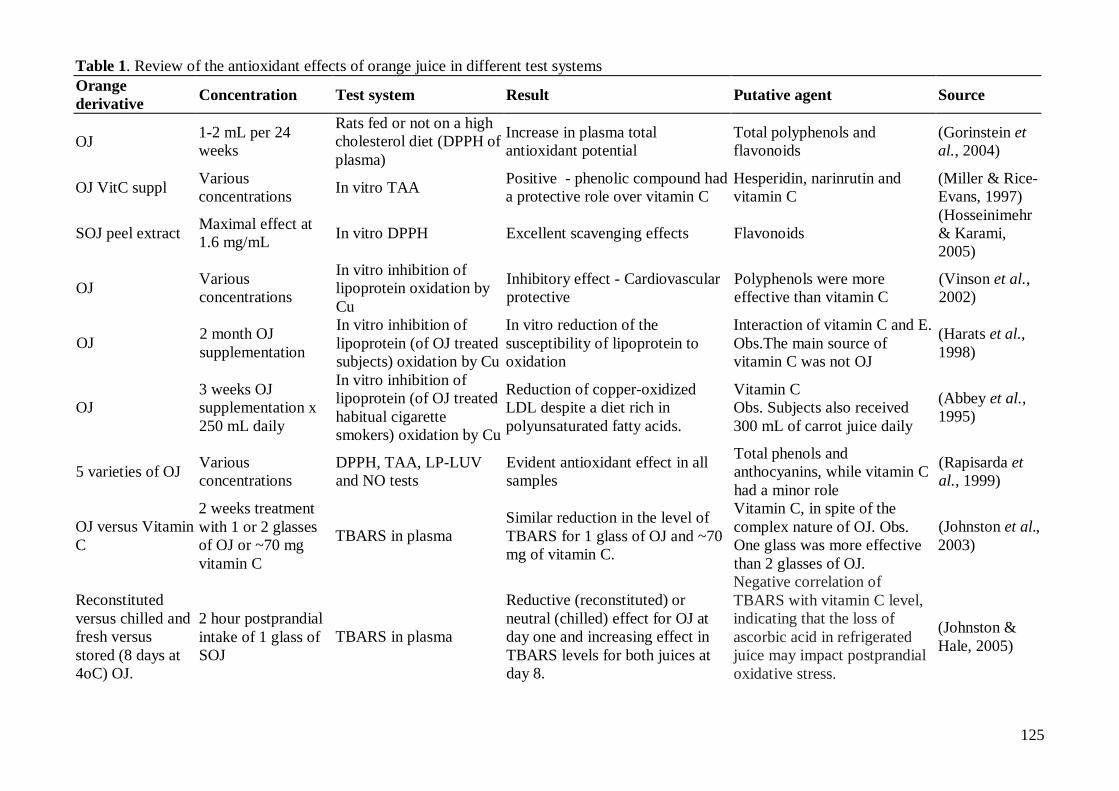
125
Table 1. Review of the antioxidant effects of orange juice in different test systems Orange derivative Concentration Test system Result Putative agent Source
OJ 1-2 mL per 24 weeks
Rats fed or not on a high cholesterol diet (DPPH of plasma)
Increase in plasma total antioxidant potential
Total polyphenols and flavonoids
(Gorinstein et al., 2004)
OJ VitC suppl Various concentrations In vitro TAA Positive - phenolic compound had
a protective role over vitamin C Hesperidin, narinrutin and vitamin C
(Miller & Rice-Evans, 1997)
SOJ peel extract Maximal effect at 1.6 mg/mL In vitro DPPH Excellent scavenging effects Flavonoids
(Hosseinimehr & Karami, 2005)
OJ Various concentrations
In vitro inhibition of lipoprotein oxidation by Cu
Inhibitory effect - Cardiovascular protective
Polyphenols were more effective than vitamin C
(Vinson et al., 2002)
OJ 2 month OJ supplementation
In vitro inhibition of lipoprotein (of OJ treated subjects) oxidation by Cu
In vitro reduction of the susceptibility of lipoprotein to oxidation
Interaction of vitamin C and E. Obs.The main source of vitamin C was not OJ
(Harats et al., 1998)
OJ 3 weeks OJ supplementation x 250 mL daily
In vitro inhibition of lipoprotein (of OJ treated habitual cigarette smokers) oxidation by Cu
Reduction of copper-oxidized LDL despite a diet rich in polyunsaturated fatty acids.
Vitamin C Obs. Subjects also received 300 mL of carrot juice daily
(Abbey et al., 1995)
5 varieties of OJ Various concentrations
DPPH, TAA, LP-LUV and NO tests
Evident antioxidant effect in all samples
Total phenols and anthocyanins, while vitamin C had a minor role
(Rapisarda et al., 1999)
OJ versus Vitamin C
2 weeks treatment with 1 or 2 glasses of OJ or ~70 mg vitamin C
TBARS in plasma Similar reduction in the level of TBARS for 1 glass of OJ and ~70 mg of vitamin C.
Vitamin C, in spite of the complex nature of OJ. Obs. One glass was more effective than 2 glasses of OJ.
(Johnston et al., 2003)
Reconstituted versus chilled and fresh versus stored (8 days at 4oC) OJ.
2 hour postprandial intake of 1 glass of SOJ
TBARS in plasma
Reductive (reconstituted) or neutral (chilled) effect for OJ at day one and increasing effect in TBARS levels for both juices at day 8.
Negative correlation of TBARS with vitamin C level, indicating that the loss of ascorbic acid in refrigerated juice may impact postprandial oxidative stress.
(Johnston & Hale, 2005)
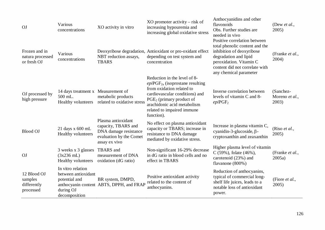
126
OJ Various concentrations XO activity in vitro
XO promoter activity – risk of increasing hypouremia and increasing global oxidative stress
Anthocyanidins and other flavonoids Obs. Further studies are needed in vivo
(Dew et al., 2005)
Frozen and in natura processed or fresh OJ
Various concentrations
Deoxyribose degradation, NBT reduction assays, TBARS
Antioxidant or pro-oxidant effect depending on test system and concentration
Positive correlation between total phenolic content and the inhibition of deoxyribose degradation and lipid peroxidation. Vitamin C content did not correlate with any chemical parameter
(Franke et al., 2004)
OJ processed by high pressure
14 days treatment x 500 mL. Healthy volunteers
Measurement of metabolic products related to oxidative stress
Reduction in the level of 8-epiPGF2a (isoprostane resulting from oxidation related to cardiovascular conditions) and PGE2 (primary product of arachidonic acid metabolism related to impaired immune function).
Inverse correlation between levels of vitamin C and 8-epiPGF2
(Sanchez-Moreno et al., 2003)
Blood OJ 21 days x 600 mL Healthy volunteers
Plasma antioxidant capacity, TBARS and DNA damage resistance evaluation by the Comet assay ex vivo
No effect on plasma antioxidant capacity or TBARS; increase in resistance to DNA damage mediated by oxidative stress.
Increase in plasma vitamin C, cyanidin-3-glucoside, �-cryptoxanthin and zeaxanthin
(Riso et al., 2005)
OJ 3 weeks x 3 glasses (3x236 mL) Healthy volunteers
TBARS and measurement of DNA oxidation (dG ratio)
Non-significant 16-29% decrease in dG ratio in blood cells and no effect in TBARS
Higher plasma level of vitamin C (59%), folate (46%), carotenoid (23%) and flavanone (800%)
(Franke et al., 2005a)
12 Blood OJ samples differently processed
In vitro relation between antioxidant potential and anthocyanin content during OJ decomposition
BR system, DMPD, ABTS, DPPH, and FRAP
Positive antioxidant activity related to the content of anthocyanins.
Reduction of anthocyanins, typical of commercial long-shelf life juices, leads to a notable loss of antioxidant power.
(Fiore et al., 2005)
127
XO: xanthine oxidase, NBT: nitro blue tetrazolium; TBARS: thiobarbituric acid reactive substances; dG ratio: 8- hydroxydeoxyguanosine/ deoxyguanosine; DPPH test: sequester of 1,1-diphenyl-2-picryl hydrazyl radical; TAA test: total antioxidant status; LP-LUV test- linoleate peroxidation in LA/DPPC LUVs; NO test - scavaging activity against nitric oxide; BR system: mixture that measures Briggs-Rauscher reaction; DMPD: (N,N-dimethyl-p-phenylenediamine dihydrochloride; ABTS: 2-29-azinobis-(3-ethylenbenzothiazoline-6-sulfonic acid) diammonium; FRAP: ferric reducing antioxidant power.
128
7.4 ANEXO D: “Review: Orange juice, genomic stability and oxidative stress. Part II: Role of the main constituents”
Franke, S.I.R., Prá, D., Henriques, J.A.P. Manuscrito a ser submetido para o British Journal of Nutrition
129
Review:
Orange juice, genomic stability and oxidative stress. Part II: Role of main constituents
Silvia Isabel Rech Franke1, 2, §, Daniel Prá2, § and João Antonio Pêgas Henriques2, 3, 4, *
1 Curso de Nutrição, Departamento de Educação Física e Saúde, Universidade de Santa Cruz
do Sul (UNISC), Santa Cruz do Sul, RS, Brasil.
2 Genotox / PPGBCM / PPGBM, Universidade Federal do Rio Grande do Sul (UFRGS) Porto
Alegre, RS, Brasil.
3 Curso de Biologia / Curso de Farmácia, Universidade Luterana do Brasil (ULBRA), Canoas,
RS, Brasil.
4 Instituto de Biotecnologia, Universidade de Caxias do Sul, Caxias do Sul, RS, Brasil.
§Authors contributed equally in data compilation and in the preparation of the manuscript
*Address for Correspondence: Centro de Biotecnologia, UFRGS; Av. Bento Gonçalves,
9500, Prédio 43421; Campus do Vale; Caixa Postal 15005; CEP 91501-970, Porto Alegre,
RS, Brasil; Fax: +5551 33166084; e-mail: [email protected]
130
Abstract
Orange juice (OJ) is one of the most consumed beverages worldwide. It can be an important
constituent of healthy and balanced diets characterized as high in antioxidant potential and,
thus, with genome stabilization properties. OJ is high in vitamins, minerals and other
bioactive compounds, which are involved in its biological function. The purpose of this
review is to discuss the influence of the main nutrients of OJ on oxidative stress and genome
stability. Topics covered are: a) the average composition of OJ; its main constituents in
relation to daily reference intakes (DRI) of vitamins and minerals, where these are available;
b) the antioxidant and genome stabilizing role of the main constituents of OJ; c) the effect of
OJ processing and storage on its constituents. One portion of OJ can provide more than 13 %
of the DRI of vitamin C, folate and thiamin and between >5-13% of the DRI of riboflavin,
niacin, pantothenic acid, pyridoxine, magnesium, copper and potassium. It can provide minor
amounts (>4-5 % of the DRI) of pro-vitamin A carotenoids and iron. OJ is also rich in
phenolic compounds, such as hesperdin and narirutin and their aglycones, and in phenolic
acids, as well as D-limonene. Vitamin C is a potent antioxidant because of its metal chelating,
oxidant scavenging and DNA repair modulation effects. B complex vitamins, including folate
(B9), have a myriad of functions related to antioxidation and DNA homeostasis. Phenolic
compounds are antioxidant, metal scavengers, DNA repair modulators, antiproliferative and
antiangiogenic agents. Carotenoids and the simple terpene D-limonene have controversial
roles in carcinogenesis, although they are generally considered as anticarcinogens and
antioxidants. Copper and iron are related to antioxidant defenses, as well as to the oxidative
metabolism and immune system. Magnesium is involved in DNA synthesis. Phosphorus has a
role in signal transduction and energy carriage and its deficiency has been linked to aging-like
symptoms. Potassium is important in maintaining membrane potential and fundamental for
muscle contraction and neurotransmission; impairments in this nutrient have been linked to
131
carcinogenesis. OJ compounds can be affected by processing techniques, such as
pasteurization, freezing and concentration. Since these techniques are widely used in the OJ
industry, more studies to address this issue are needed. One glass of fresh or adequately
processed and stored OJ is one of the options for the daily 5 portions (or even more) of fruit
and vegetables recommended for a healthy life.
Key-words: Enzymatic and non enzymatic antioxidants, DNA damage and repair,
(anti)mutagenic and (anti)carcinogenic effects, bio-flavonoids and carotenoids, vitamin C and
complex B vitamins.

132
Contents
1 Orange juice ..............................................................................................................134
2 Main components of orange juice: biological role.....................................................137
2.1 Vitamin A, carotenoids and D-limonene .....................................................................137
2.1.1 Vitamin A ...................................................................................................137
2.1.2 Carotenoids .................................................................................................137
2.1.3 Pro-vitamin A carotenoids ...........................................................................138
2.1.4 ��and �-carotene .........................................................................................138
2.1.5 �� and �-Cryptoxanthin ..............................................................................139
2.1.6 Non-pro-vitamin A carotenoids ...................................................................140
2.1.7 Lutein and zeaxanthin..................................................................................140
2.1.8 Cis-violoxanthin, mutatoxanthin and auroxanthin........................................141
2.1.9 Remarks ......................................................................................................141
2.1.10 D-limonene .................................................................................................142
2.2 B Complex vitamins .....................................................................................................143
2.2.1 B1 (thiamin) ................................................................................................144
2.2.2 B2 (riboflavin).............................................................................................145
2.2.3 B3 (niacin) ..................................................................................................147
2.2.4 B5 (pantothenic acid) ..................................................................................149
2.2.5 B6 (pyridoxine, pyridoxamine, etc.) ............................................................150
2.2.6 B9 (folic acid) .............................................................................................151
2.3 Vitamin C .....................................................................................................................153
2.4 Phenolic compounds.....................................................................................................155
2.4.1 Hesperidin and hespiritin.............................................................................157
133
2.4.2 Narirutin, naringin and naringenin ...............................................................162
2.4.3 Anthocyanins ..............................................................................................164
2.4.4 Phenolic acids - hydroxycinnamic acids ......................................................165
2.5 Fibers............................................................................................................................167
2.6 Minerals........................................................................................................................168
2.6.1 Copper ........................................................................................................168
2.6.2 Magnesium..................................................................................................169
2.6.3 Potassium....................................................................................................170
2.6.4 Phosphorus..................................................................................................171
2.6.5 Iron .............................................................................................................173
3 Conclusion.................................................................................................................175
4 Acknowledgements.....................................................................................................176
5 References..................................................................................................................177
134
1 Orange juice
OJ is probably the most consumed and the best known of fruit juices. It is among those foods
regarded as part of a healthy and balanced diet. Numerous studies have associated fruit
consumption with genome stability and protection against oxidative stress. These effects are
probably due to synergism between the various components of fruits (Ames, 2001; Ames &
Wakimoto, 2002; Fenech, 2005; Fenech et al., 2005; Franke et al., 2006).
One must keep in mind that many fruit juices are submitted to different processes in
order to preserve them. Processes like pasteurization, freezing and high-pressure extraction
have been developed to attend the growing demand for fruit juices.
OJ contains significant amounts of vitamin C, folate, thiamin, riboflavin and phenolic
compounds. It also contains appreciable amounts of carotenoids (pro and non-pro-vitamin A),
niacin, panthothenic acid, pyridoxine, magnesium, potassium, copper, iron and phosphorus.
Such substances can directly or indirectly influence oxidative state and genome stability.
Other compounds, such as the terpene D-limonene and the phenolic acid p-coumarin, are also
present in OJ and also have roles in oxidative stress and DNA homeostasis.
The composition of orange is widely variable, depending on varietal differences, as
well as on climatic conditions, soil composition, light and pathogen exposure and maturation
state. These aspects influence the color and flavor of the oranges. For example, the color of
red pulp oranges is mostly due to anthocyanins. However, it has also been reported that
carotenoids, such as cryptoxanthins, can be responsible for this coloration (Mouly et al., 1999;
Lee, 2001).
OJ composition does not only reflect the chemical composition of the oranges. It is
considerably influenced by the extraction process, packaging and shelf time before
consumption. In addition, the various components of oranges are not homogenously
distributed within the fruits, they are compartmentalized. For example, the hydrophobic
135
carotenoids and phenolic compounds tend to be located in the peel and pulp rather than in the
aqueous fraction. Indeed, Vitamin C and the B complex vitamins, which are hydrophilic, are
easily extracted, while the yield of hydrophobic compounds can be optimized with further
extraction (such as high pressure) (Pupin et al., 1998; Mouly et al., 1999; Rapisarda et al.,
1999; Gil-Izquierdo et al., 2001; Gil-Izquierdo et al., 2002; Franke et al., 2004; Vanamala et
al., 2006).
The purpose of this review is to discuss the influence of the main nutrients of OJ on
oxidative stress and genome stability. To define the main nutrients of OJ, we initially
calculated the percentage of daily energy provided by the intake of a standard 200 mL serving
of OJ in relation to the average daily Estimated Energy Requirement (EER) of male and
female adults over two life stages (19-30 and 31-50 years). To calculate EER, we considered a
Body Mass Index (BMI) of 21 for females and 22 for males, together with a height of 1.65 m,
low physical activity and average age for each life cycle (IOM, 2005). The energy provided
by the intake of one standard (200 mL) serving of OJ was 98.13 ± 19.96 Kcal/day, which
represented 4.4% of the calculated average daily EER (amount of energy). This was then used
to evaluate the density of nutrients in one standard (200 mL) serving of OJ in relation to the
respective Dietary Reference Intake (DRI) for each nutrient. We include in this review all
nutrients that can provide at least 4% of the respective DRI in a standard 200 mL serving of
OJ (Table 1). If a given nutrient provides between 4.0 and 4.4% (regarded as one energy
density unit) of its DRI in one standard (200 mL) OJ portion, then OJ was considered as a
regular source of this nutrient. If the nutrient density is between one and two times, two and
three times or more than three times the energy value, OJ was considered a good, excellent or
key source of it, respectively (Figure 1).
136
We also include in the present review other bioactive compounds, such as non pro-
vitamin A carotenoids and phenolics, which are not considered as nutrients and do not have
DRIs (Tables 2 and 3).
In vivo effects of compounds are influenced by the amounts consumed, by the
quantities absorbed or metabolized, as well as by the plasma and/or tissue concentrations
(Prior, 2003). Due to the complexity of this theme, we focus on the level of the compounds
present in OJ and overlook the absorption, metabolism, and the tissue concentrations of the
nutrients. Since OJ compounds can be affected by processing techniques, we discuss the
influence of pasteurization, freezing and concentration, techniques widely used in the OJ
industry, and consider how these alterations can impact the biological role of OJ.
137
2 Main components of orange juice: biological role
2.1 Vitamin A, carotenoids and D-limonene
2.1.1 Vitamin A
Retinoic acid (RA) is a key signalling molecule that regulates the patterning and
development of the body plan and many organ systems during embryogenesis. RA is an
important regulator of steady-state hematopoiesis in fetal as well as in adult life. Indeed,
hematopoietic defects caused by a deficiency of vitamin A are still a serious problem in the
developing world (Evans, 2005). Vitamin A is present as retinol activity equivalent (RAE) in
OJ (see item 2.1.3). The vitamin A content of the standard 200 mL OJ portion is 34.8 �g RAE
(as calculated in Table 2), which is equivalent to 4.4 % of the DRI (Table 1). Thus, OJ is
considered as a regular source of vitamin A (Figure 1).
2.1.2 Carotenoids
60% of the total carotenoids in oranges is in the peel (Mouly et al., 1999). In a 2002
review, Nishino et al. (2002) state that, of more than 600 carotenoids identified to date, about
40 carotenoids are found in our daily foods. They also state that only 14 carotenoids, with
some of their metabolites, are present in human plasma, as a result of selective uptake in
digestive tract and tissues. Carotenoids are tightly bound to macromolecules, in particular
proteins and lipids. For instance, less lipophilic carotenoids, such as lutein, are released into
an aqueous environment more readily than �-carotene (Sanchez-Moreno et al., 2005).
Carotenoids can be lost by oxidation, depending on the structure of the molecule, the action of
light and heating, as well as by the action of oxidants such as peroxides. The oxidation can be
inhibited by vitamin C or other antioxidants intrinsic or extrinsic to OJ (Sanchez-Moreno et
al., 2003).
Early findings showed very low levels of carotenoids in OJ (Holden et al., 1999).
More recently, Mouly et al. (1999), using 18 standards in HPLC analysis, showed that OJ has
138
about 4,790-21,100 �g carotenoids per liter (~1,000-4,000 �g/200 mL), depending on the
extraction process and more influenced by the origin then by the variety. In the same study,
pro-vitamin A carotenoids corresponded to about 20% of the total carotenoids. Sanchez-
Moreno et al. (2003; 2005), using 6 standards in HPLC analysis, detected about 1000-1500
�g of total carotenoids in 100 mL of OJ, about 30 % being pro-vitamin A carotenoids. Lutein
and zeaxanthin accounted for the additional 70%. When pooling the data obtained by Mouley
et al. (1999) on carotenoid profile of OJ from oranges of 5 countries, it can be seen that lutein,
isolutein and zeaxanthin together account for only about 22% of the total carotenoids. Indeed,
other carotenoids, such as mutatoxantin A and B, auroxanthin A and cis-violaxanthin, are
present in high proportions in OJ, accounting for 9.5 and 12%, 8.2 and 11.3 %, respectively.
Surprisingly, Lee (2001) found large amounts of red-colored lycopene as the major carotenoid
in a red navel cultivar; this is not present in standard navel oranges or other major sweet
orange cultivars.
The stability of carotenoids is similar to that of vitamin A in that they are sensitive to
oxygen, light and acid media (Vercet et al., 2001).
2.1.3 Pro-vitamin A carotenoids
��and �-carotene and � and �-cryptoxanthin are converted to vitamin A and are
considered together as RAE, according to a FAO/WHO standard; RAE=�g �-carotene/12 +
[(�g��-carotene + �g��- cryptoxanthin +��g��- cryptoxanthin)/24 (FAO/WHO, 1988). Pro-
vitamin A carotenoids represent about 30% of the total carotenoids present in OJ (Table 2).
2.1.4 ��������and ����-carotene
Initially, �-carotene was the most extensively studied, since �-carotene has the highest pro-
vitamin A activity among carotenoids in vegetables and fruits. Later, it was shown that the
unconverted form of carotenoids could also have a protective effect against cancer, without
being converted to vitamin A (Nishino et al., 2002).
139
Most carotenoids are only slightly absorbed (as low as 2% in some cases). Conversely,
between 60 and 75 % of the �-carotene in a high bioavailable diet is converted to vitamin A
and a minor extent (15 %) is absorbed intact. Besides the central enzyme-mediated cleavage
that produces vitamin A, a central cleavage can occur, giving rise to deleterious sub-products,
such as aldehydes and epoxide derivatives. �-carotenes and their derivatives are LDL-
cholesterol-transported, being presumably susceptible to oxidation (IOM, 2000).
�-carotene supplementation significantly increases the incidence of lung cancer in
humans, indicating that �-carotene supplementation could be dangerous, especially among
people who currently smoke more than 20 cigarettes per day and drink more than 11 g/day of
ethanol. Conversely, �-carotene can induce G1-arrest in the cell cycle and has a higher
suppressor activity than �-carotene for tumorogenesis in mouse skin, lung, liver and colon
(Nishino et al., 2002). Carotene deficient diets have been related to a general increase in
oxidation susceptibility markers (IOM, 2000). The Institute of Medicine (IOM) panel on DRI
(IOM, 2000) states that, although most studies have tested high doses of �-carotene obtained
from food alone, these studies do not show an increase in oxidative or antioxidant markers on
supplementation. Results seem more effective in subjects with a higher oxidative stress
background (IOM, 2000).
��carotene is an efficient quencher of singlet oxygen and free radicals. It can reduce
DNA and chromosomal damage generated by various agents (Cozzi et al., 1997). It is
interesting to notice that, while low intake can significantly increase genome stability, high
intake of �-carotene can induce a marginal increase in genome instability (Fenech et al.,
2005).
2.1.5 �������� and �-Cryptoxanthin
The main contributor to the color of oranges is �-cryptoxanthin (Mouly et al., 1999). It
accounts for more than 50 % of the total pro-vitamin A carotenoids in OJ (Table 2).
140
Cryptoxanthin is distributed in our daily foodstuffs and is one of the major carotenoids
detectable in human blood. �-cryptoxanthin seems to be a promising carotenoid, since it
showed the strongest inhibitory effects on the Epstein-Barr virus activation activity of 12-O-
tetradecanoylphorbol-13-acetate (TPA) in Raji cells, in comparison to 50 other carotenoids
(Tsushima et al., 1995; Nishino et al., 2002). In vivo, �-cryptoxanthin showed anti-tumor
promoting activity in mouse skin, but not in rat colon (Nishino et al., 2002).
�-cryptoxanthin is one of the major carotenoids detectable in human blood (Nishino et
al., 2002) and a modest increase in its intake, equivalent to one glass of freshly squeezed
orange juice per day, is associated with a reduced risk of developing inflammatory disorders
such as rheumatoid arthritis (Fenech & Ferguson, 2001; Halliwell, 2001; Edenharder et al.,
2002).
2.1.6 Non-pro-vitamin A carotenoids
2.1.7 Lutein and zeaxanthin
Lutein is the dihydroxy-form of �-carotene, and is distributed among a variety of fruit and
vegetables, including oranges. Lutein shows anti-tumor promoting activity in the lung and
skin of mice and the colon of rats (Nishino et al., 2002). It is predominantly transported by
HDL-cholesterol and to a lesser extent by LDL-cholesterol, sited in the outer region of the
molecule due to its lower hydrophobicity (IOM, 2000).
Zeaxanthin is the dihydroxy-form of �-carotene, and is distributed in our daily foods.
Zeaxanthin was found to suppress TPA-induced expression of the early antigen of Epstein–
Barr virus in Raji cells, to inhibit TPA-enhanced 32Pi-incorporation into phospholipids in
cultured cells, and to suppress spontaneous carcinogenesis in male mice (Nishino et al.,
2002).

141
2.1.8 Cis-violoxanthin, mutatoxanthin and auroxanthin
Despite the high level of cis-violaxanthin, mutatoxanthin and auroxanthin in OJ (Table 2),
data on the biological role of these compounds are still lacking. Cis-violaxanthin seems to be
involved in the formation of the plant hormone abscisic acid (ABA), which plays a crucial
role in the adaptation of plants to various environmental stresses and in several physiological
processes, such as seed maturation and dormancy and fruit development, or senescence
(Rodrigo et al., 2006).
2.1.9 Remarks
Which is the most beneficial carotenoid is still undecided. In fact, is likely that
carotenoids have synergistic effects. Nishino et al. (2002) found that multicarotenoids (a
mixture of various carotenoids, such as �-carotene, �-carotene, lutein, lycopene, etc.) showed
potent anti-carcinogenic activity. Anlasik et al. (2005) observed that healthy subjects aged 65
years and older, consuming high levels of fruit and vegetables daily, had significantly higher
plasma levels of retinol, tocopherols and carotenoids. They also showed that malondialdehyde
(MDA) levels were inversely correlated with vitamin A and �-carotene, as well as that protein
carbonyls in Imunoglobulin G were inversely correlated with �-tocopherol. Thus, a higher
intake of retinol, tocopherols and carotenoids, mediated by a higher intake of fruit and
vegetables, seems to be important for the prevention of age-related diseases.
There is no DRI for carotenoids and their supplementation for healthy subjects with
normal intakes of carotenoids is not recommended (IOM, 2000).
Freezing preserves pro-vitamin A carotenoids and long term storage at -40oC has been
show to substantially increase the amount of these compounds in OJ (Cortes et al., 2005).
Heat treatment may lead to significant loss of lutein and, to a minor extent, of pro-vitamin A
carotenoids (Sanchez-Moreno et al., 2005). It is generally thought that heating increases the
142
bioavailability of carotenoids and cooking vegetables rich in carotenoids can help to attain the
vitamin A DRI.
It is important to mention that, while pasteurization has a mild effect on carotenoid
content, sieving can remove large amounts of vesicle-bound liposoluble carotenoids, which
tend to settle on the cellulose fragments (Mouly et al., 1999). High pressure processing could
be a way of increasing the amount of carotenoids in sieved juice; Sanchéz-Moreno et al.
(2005) showed that high pressure extraction can significantly increase the amount of
carotenoids in OJ. However, in this case it was observed that vitamin C is not proportionally
increased and can be lost during storage. Lower levels of vitamin C offer less protection to
carotenoids from oxidation and the unbalanced increase of carotenoids and vitamin C can
result in further oxidation (Sanchez-Moreno et al., 2003).
2.1.10 D-limonene
There are several volatile compounds responsible for OJ flavor. These include: ethyl
butanoate, D-limonene, linalool, alpha-pinene, geranial, neral and alpha-terpineol (Pattison et
al., 2005).
D-limonene (p-mentha-1,8-diene) is the most studied of these compounds, since it is
the most abundant in orange oil, comprising about 90% (Del Toro-Arreola et al., 2005). It is
likely that OJ obtained from squeezing of whole oranges would contain larger amounts of D-
D-limonene, since it is the major constituent of orange peel. Juice extracted by an orange
press contains about 100 (blood and bitter OJ) to 300 mg/L (sweet OJ) D-limonene, the major
part (60-90%) being the volatile compounds (Moufida & Marzouk, 2003).
D-limonene was found to cause tumors, at high doses only, in the kidney of the male
rat, in association with hyaline droplet nephropathy (Hard & Whysner, 1994); this led to the
compound being considered as a carcinogen. Further evidence, however, showed that D-
limonene was not genotoxic to colon, liver, kidney, bladder, lung, brain, or bone marrow of
143
either mice or rats (Sekihashi et al., 2002), nor was it mutagenic to male Big BlueTM rats
(Turner et al., 2001).
In 1999, D-limonene was reclassified from possibly carcinogenic to humans (group
2B) to not classifiable as to carcinogenicity for humans (group 3) by the International Agency
for Research on Cancer (WHO, 1993; WHO, 1999).
Indeed, D-limonene has shown chemopreventive and therapeutic activity against a
wide variety of experimental tumors, such as lung neoplasms, mammary cancer, and
pancreatic and prostatic tumors (Crowell, 1999; Del Toro-Arreola et al., 2005). Citrus peel
consumption (the major source of dietary D-limonene) is not uncommon and is inversely
related to the degree of skin cancer risk (Hakim et al., 2000). However, it is important to
consider the numerous compounds in citrus peel, such as psoralens, that have cancer
promotion potential (Brendel & Henriques, 2001; Brendel et al., 2003).
D-limonene is antioxidant, for example inhibiting ferric-ion-stimulated lipid
peroxidation in rat brain homogenates(Youdim et al., 2002; Grassmann, 2005).
D-limonene is a relatively stable terpene, but easily oxidizable, and OJ processing can
particularly interfere in D-limonene stability, due to its easy oxidation. Moreover, it can be
readily absorbed in high amounts by low-density polyethylene (LDPE), but not by
polycarbonate (PC) and polyethylene terephthalate (PET), materials utilized in food
packaging (Van Willige et al., 2003). Pasteurization by continuous ohmic heating does not
seem to influence D-limonene stability (Leizerson & Shimoni, 2005).
2.2 B Complex vitamins
Deficiencies in B series vitamins and folic acid (B9), which are essential for DNA synthesis
and repair, are among the key causative factors for diabetes and organ damage (Wu & Ren,
2006).
144
2.2.1 B1 (thiamin)
Thiamin (B1) is the second most abundant vitamin in OJ. OJ can be a better source of vitamin
B1 than many foods known as good sources, such as whole wheat bread (Dreher et al., 2003).
A 200 mL portion of OJ can supply 18.5% of the DRI for B1, OJ being a key source of this
vitamin (Table 1 and Fig. 1).
B1 in its diphosphate form is an important coenzyme for transketolase (TK) and
pyruvate dehydrogenase, among others, which are important in the biosynthesis of a number
of cell constituents, including neurotransmitters, and for the production of reducing
equivalents used in oxidative stress defenses (e.g. maintenance of NADPH levels), as well as
for the synthesis of pentoses that are used as nucleic acid precursors(Singleton & Martin,
2001; Shangari et al., 2005)
There is growing evidence for the role of B1 in preventing several types of cancers, as
well as neurodegeneration (Gibson & Zhang, 2002; Lee et al., 2005). B1 deficiency may pose
a cancer risk, owing to the deficiency of thiamin-dependent enzymes involved in glycolysis
and an increase of reactive intermediates in carbohydrate metabolism-oxaldehydes, generated
by the spontaneous rearrangement of triose phosphates (Bruce et al., 2003). Hyperglycemia
can increase the accumulation of �-oxoaldehydes.
B1 and derivatives, at high doses, can increase the conversion of triose phosphates to
ribose-5-phosphate, and strongly inhibit the development of oxidative stress, protein glycation
and/or inflammatory responses, either in vivo or in vitro (Babaei-Jadidi et al., 2003; Nandi et
al., 2005; Wu & Ren, 2006). TB1 diphosphate and thiamin can modulate p53 activity,
affecting apoptosis (McLure et al., 2004). Moreover, B1 transporter gene expression and
exogenous B1 modulate the expression of genes involved in drug and prostaglandin
metabolism in breast cancer cells (Liu et al., 2004). The sulfhydryl group of B1 can act as a
chelant or an antioxidant.
145
On the other hand, some authors claim that thiamin can stimulate tumor cell survival,
proliferation and chemotherapy resistance, by the stimulation of TK synthesis leading to a
high rate of nucleic acid ribose synthesis (Cascante et al., 2000; Lee et al., 2005). Thus,
excess B1 supplementation in common food products may contribute to the increased cancer
rates of the Western world and anti-B1 therapy should be used (Boros et al., 1998; Boros,
2000). In fact, McLure et al. (2004) have shown that thiamin reduces p53 induced apoptosis
in irradiated murine thymocytes.
B1 is relatively unaffected by light and stable to oxidation, but it is among the least
stable vitamins in solution at neutral pH (Vercet et al., 2001). B1 decomposition leads to the
formation of meat flavors by thermal degradation of B1, as well as through a Maillard
reaction involving cysteine and various sugars. One of the most significant B1 thermal
degradation products is 2-methyl-3-furanthiol (MFT) (Dreher et al., 2003). This compound
and other Maillard reaction products seem to quench reactive oxygen species and inhibit
LDL-oxidation (El-massry et al., 2003).
2.2.2 B2 (riboflavin)
A standard 200 mL portion of OJ can provide about 10% of the DRI for riboflavin (B2) and
OJ is an excellent source of it (Table 1 and Fig. 1). B2, in eukaryotes, is converted to flavin
adenine dinucleotide (FAD). FAD is a coenzyme for glutathione reductase and other
enzymes. Flavin coenzymes are widely distributed in the body and are involved in
carbohydrate, fat, amino acid and vitamin metabolism. Oxidative folding (formation of
disulfide bonds) in the endoplasmic reticulum (ER) before the secretion of proteins, such as
apolipoprotein B-100 (apoB), depends on flavoproteins in eukaryotes. Furthermore, B2
influences epithelial integrity, by modulating tissue flavin concentrations, the rates of
prostaglandin biosynthesis and glutathione metabolism (Manthey et al., 2005; Siassi &
Ghadirian, 2005; Manthey et al., 2006).
146
B2 deficiency causes ER stress and activation of unfolded protein response and
decreases protein secretion in HepG2 cells, possibly interfering with lipid homeostasis in vivo,
in the case of apoB secretion. B2 deficiency is also associated with decreased activity of
glutathione reductase and with decreased concentrations of reduced glutathione in human
cells, as well as with an increase in B2-deficient cells arrested in G1 phase of the cell cycle.
Moreover, oxidative stress caused by B2 deficiency is associated with increased expression of
clusters of genes that play a role in cell stress and apoptosis (Manthey et al., 2005; Siassi &
Ghadirian, 2005; Manthey et al., 2006).
B2 deficiency may predispose esophagus squamous carcinogenesis by altering
gastrointestinal function. A high intake of B2 can generate genome instability (Fenech et al.,
2005) and has been associated with increased gastric cancer risk, although it may reduce the
frequency of micronucleated cells and carcinogen DNA adducts and the associated
esophageal adenocarcinoma (Manthey et al., 2005; Siassi & Ghadirian, 2005; Manthey et al.,
2006). The precise role of B2 in the development of this tumor at different stages of initiation,
promotion and progression is controversial. It is plausible to hypothesize that B2 deficiency
enhances the risk of tumor development at initiation and promotion stages, while reducing
tumor growth at the progression stage (Siassi & Ghadirian, 2005).
B2 has received much less attention than other vitamins, but there is increasing interest
in the well-established role that flavins play in folate metabolism and the possible synergy of
a protective effect between these 2 vitamins. B2, as FAD, is a cofactor for 5,10 methylene
tetra hydro folate reductase (MTHFR) and there is evidently some interaction between B2
status, folate status, and plasma homocysteine (Powers, 2005).
B2 and UV light in combination selectively enhance damage to guanine bases in
DNA, making the damage less likely to be repaired by normal repair pathways in host cells
(Kumar et al., 2004).
147
B2 is a heat stable vitamin, but it is very sensitive to oxidation and mainly to
degradation by light. Its oxidation causes not only a loss in the nutritive value of dairy
products but also off-flavors (Vercet et al., 2001).
2.2.3 B3 (niacin)
A standard 200 mL portion of OJ can provide about 5 % of the DRI for niacin and thus OJ
juice is a good source of niacin (Table 1 and Fig. 1). Niacin (B3) is the common name for two
compounds: nicotinic acid and nicotinamide. Niacin is a water-soluble vitamin that
participates in many metabolic functions. It is a precursor for nicotinamide adenine
dinucleotide (NAD), which is required for DNA synthesis, as well as for the activity of the
enzyme poly(ADP-ribose) polymerase-1 (PARP-1; EC 2.4.2.30), for which NAD is the sole
substrate (Hageman & Stierum, 2001).Thus, B3 has beneficial effects, as a redox agent in
energy metabolism and as an antioxidant. It also influences DNA repair and genome stability,
as well as the immune and nervous systems (Ikeda et al., 2003; Kirkland, 2003).
The action of NAD in protecting biological systems against oxidative stress and DNA
damage can be related to an up-regulating of the stress response enzymes glyceraldehyde-3-
phosphate dehydrogenase (GAPDH) and glucose 6-phosphate dehydrogenase (G6PD), among
others. These enzymes are involved in the generation of the antioxidant reduced
pyridinenucleotides, NADPH and NADH, from NAD (Yan et al., 1999; Crowley et al.,
2000).
PARP-1, strongly activated by DNA strand breaks during the cell genotoxic stress
response, is involved in base excision repair, and plays a role in p53 expression and
activation; hence it is thought to be important for genome stability. In addition to being a
precursor for NAD, niacin has also been reported to possess oxygen radical scavenging
activity comparable to that of ascorbic acid (Hageman & Stierum, 2001). Finally, NAD and
148
NADP are required for the synthesis of cyclic ADP-ribose and nicotinic acid adenine
dinucleotide, two mediators of intracellular calcium signaling pathways (Kirkland, 2003).
B3 has an impact on cancer risk, according to data obtained from animal models of
leukemogenesis and skin cancer, as well as epidemiological data from human populations,
and on side effects of chemotherapy in cancer patients (Kirkland, 2003; Hirakawa et al.,
2005). In vitro and animal studies indicate that B3 deficiency increases genome instability and
the risk of certain tumors, especially in combination with genotoxic and oxidative stress
(Crowley et al., 2000).
While pellagra (a disturbance with nervous, gastrointestinal and dermatological
effects) is the consequence of severe B3 deficiency, excessive intake of this vitamin in
supplements is associated with hepatic toxicity, nausea and vomiting, as well as flushing
(IOM, 1998).
Both the beneficial effect and the noxious effect of B3 on cancer can be related to
PARP-1. On the one hand, B3 replenishes NAD, avoiding the cell death related to PARP
induced draining of the NAD+ pool, which may compromise the further repair of DNA and
mitochondrial respiration (i.e affecting electron transport, redox state and membrane
potential) (Crowley et al., 2000). On the other hand, excessive levels of nicotinamide in the
body may result in inhibition of PARP-1 and increased genome instability. More studies are
therefore needed to define an optimal level of B3 to avoid genome stability and tumorigenesis
(Hageman & Stierum, 2001). B3 has a role in the maintenance of telomere length. Low intake
of nicotinic acid is related to increased genome instability (Fenech et al., 2005).
Tryptophan can be converted to B3 in a vitamin B6-dependent reaction, as well as,
with low efficiency, by enteric bacteria. B3 seems to be stable and is used as a preservative
for drugs and foods (IOM, 1998), it is more stable than thiamin and B2. It is resistant to
heating and light (Mahan & Escott-Stump, 1996).
149
2.2.4 B5 (pantothenic acid)
A standard 200 mL portion of OJ can provide about 7 % of the DRI for B5, and is a good
source of it (Table 1 and Fig. 1). Pantothenic acid is a water-soluble vitamin important for the
metabolism of carbohydrates, proteins and lipids. It is part of coenzyme A, acting in the fatty
acid oxidation and citric acid cycles. Pantothenic acid, as pantothenate, is present in food
mostly as CoA, which cannot be directly absorbed through enterocytes, whereas pantothenate
freely diffuses across the epithelial barrier. Thus, one might speculate that conversion of CoA
into pantothenate requires an extracellular, membrane-bound pantetheinase capable of
recycling pantothenate in the gut. Pantetheinase is a ubiquitous enzyme, which has been
shown in vitro to recycle pantothenic acid and to produce cysteamine, a potent anti-oxidant.
Membrane-bound pantetheinase is the main source of cysteamine in tissues under
physiological conditions (Pitari et al., 2000).
Pantethine, pantothenate, and phosphopantothenate can partially prevent lipid
peroxidation in hepatic and heart tissue. They have beneficial effects on wound healing as
well as inflamation, in the latter case by blocking the release of myeloperoxidase from
neutrophils. Pantothenate and some related compounds protect Ehrlich ascites tumor cells
against lipid peroxidation, but not due to scavenging of free radicals. In fact, these compounds
exerted only a negligible effect on peroxidation and it is likely that some metabolic products
of pantothenic acid or its derivatives, rather than the original compounds, were effective
(Slyshenkov et al., 1996).
There are two possible explanations for the antioxidant potential of panthotenic acid:
the antioxidant potential of cysteamine and maybe gluthathione generated by pantetheinase,
and the enrichment of cells with CoA, with resulting changes in metabolism. As evaluated by
Slyshenkov et al. (1996), the preincubation of cells with pantothenic acid and some of its
related compounds greatly increased the cell level of CoA. According to the authors, this is
150
evidence that CoA may facilitate removal of lipid peroxides by increasing metabolism of fatty
acids and promote repair of the plasma membrane by activating phospholipid biosynthesis. It
can only be speculated that elevated levels of CoA may increase the supply of a glutathione
precursor, i.e., glutamate, by accelerating the tricarboxylic acid cycle (Slyshenkov et al.,
1995).
High intake of pantothenic acid is associated with increased genome instability, as
measured by micronucleus frequency. Only one study suggested a marginal promoting effect
induced by calcium pantothenate in the Balb/c-3T3 assay (Fenech et al., 2005).
Pantothenic acid is easily destroyed by heat in acidic or alkaline conditions, but is
stable in neutral solution (Mahan & Escott-Stump, 1996). Substantial losses of vitamin B5
can occur after freezing (Pallaoro, 1997).
2.2.5 B6 (pyridoxine, pyridoxamine, etc.)
A standard 200 mL portion of OJ can provide about 7% of the DRI for vitamin B6 and is a
good source of it (Table 1 and Fig. 1). There are several structural isomers (vitamers) of
vitamin B6 [e.g. pyridoxamine (PM) and pyridoxine] that share a general enzymatic
mechanism. PM is a transient intermediate in enzymatic transamination reactions. Dietary PM
is consumed mostly in the form of PM-5’- phosphate, which is hydrolyzed to PM by intestinal
phosphatases and further metabolized in the liver and, to a lesser degree, in intestinal tissues
(Voziyan & Hudson, 2005).
PM is a promising pharmacological agent for protecting against the progressive tissue
damage that occurs in diabetes and other diseases, because it is a potent inhibitor of the
formation of advanced glycation end products (AGEs), deleterious protein modifications.
There are three likely mechanisms to explain the inhibitory action of vitamin B6 on AGE
formation: a) blockage of oxidative degradation of the Amadori intermediates of the Maillard
reaction; b) scavenging of toxic carbonyl products of glucose and lipid degradation; and c)
151
trapping of reactive oxygen species (Voziyan & Hudson, 2005). High levels of vitamin B6
can suppress the growth of animal or human cancer cells in vitro, suppress nitric oxide, and
supress angiogenesis (Anand, 2005; Wei et al., 2005).
At normal levels, vitamin B6 seems to be related to the regeneration of 5,10-
methylene tetrahydrofolate, which is involved in avoiding the misincorporation of uracil in
place of thymine in DNA. Thus, deficiency of vitamin B6 can induce genome instability. The
anticarcinogenic/antimutagenic property of vitamin B6 may be related to this aspect, as well
as to its role in alternative pathways (e.g. as a co-factor in amino acid metabolism, lipid
metabolism, and nervous system function) (Wei et al., 2005). Beside these above
mechanisms, pyrodoxal and pyridoxal 5'-phosphate seem to promote DNA excision repair
directly or indirectly and to decrease the amount of unrepaired DNA damage, reducing
mutations in E. coli and micronuclei in mouse peripheral blood cells (Shimoi et al., 1992).
However, one should recall that pyridoxine, like riboflavin, can generate cytotoxic
photoproducts after UV-irradiation (Sato et al., 1993).
Vitamin B6 is quite stable to heat but is sensitive to air, UV light and alkali. Freezing
can substantially reduce (36-55 %) vitamin B6 content (Mahan & Escott-Stump, 1996).
2.2.6 B9 (folic acid)
Folate, a water-soluble vitamin, includes naturally occurring food folate and synthetic
folic acid in supplements and fortified foods. OJ can be a good source of folate, given the
instability of the compound in cooking. A standard 200 mL portion of OJ can provide about
14% of the DRI for folate and is, indeed, a key source of it (Table 1 and Fig. 1).
Inadequate folate status may result in hyperhomocysteinemia, a significant risk factor
for atherosclerotic vascular disease, as well as changes in DNA that may result in pro-
carcinogenic effects and increased risk of cognitive dysfunction. Folate status may be
negatively influenced by inadequate intake, genetic polymorphisms and interactions with
152
various drugs. Folate-rich foods include OJ, dark green leafy vegetables, asparagus,
strawberries and legumes. These foods are also excellent sources of other health-promoting
nutrients associated with reduction in risk of chronic disease (Brevik et al., 2005). Thus,
plasma folate concentration may be a useful biomarker for the intake of fruit and vegetables in
populations consuming unfortified food products. The association can be attenuated by, and
should be corrected for, individual intake of folic acid supplements (Clifford et al., 1991).
Folate coenzymes participate in biochemical processes involving single carbon group
transfers, including the metabolism of amino acids and synthesis of purines and pyrimidines
that are incorporated into DNA and RNA (Rampersaud et al., 2003).
Folate has a key role in genome stability and DNA homeostasis. Folate is important for
the maintenance of telomere length and for the prevention of uracil incorporation into DNA,
as well as for the maintenance methylation of cytosines adjacent to guanine sequences (CpG)
in DNA (Fenech, 2005).
FA is a poor antioxidant. The more FA is reduced, the higher its antioxidant activity
becomes: THF and 5-MTHF are the folates with the most prominent antioxidant activity. The
antioxidant pharmacophore of THF and 5-MTHF, i.e. 4-hydroxy-2,5,6-triaminopyrimidine,
resides in the pterin moiety. It is suggested that an electron donating effect of the 5 amino
group is of major importance in the antioxidant activity of 4-hydroxy-2,5,6-
triaminopyrimidine. A similar electron donating effect is probably involved in the antioxidant
activity of THF and 5-MTHF (Rezk et al., 2003).
Folate in food is very unstable and considerable losses occur during short-term storage
and cooking. Folate decomposition has been related to the widespread under-provision of
folate common in human populations. However, food folate bioavailability and the factors
that affect it are poorly understood (McNulty & Pentieva, 2004). The consumption of whole
OJ with minor processing after extraction could be a better way to optimize the intake of
153
folate, since folate from OJ is as available as in beef liver and folic acid supplements
(Gorinstein et al., 2004).
2.3 Vitamin C
A standard 200 mL portion of OJ can provide more than 100% of the DRI for vitamin C and
is a key source of it (Table 1 and Fig. 1).
Vitamin C is an important micronutrient, mainly required as a co-factor for enzymes
involved in oxi-reduction reactions (Lunec et al., 2002). Vit C acts as an electron donor for 8
human enzymes, involved mainly in the biosynthesis of collagen and carnitine (WHO, 2001).
It has an increasing involvement in supporting intracellular redox status by maintaining
important sulphydryl compounds, such as glutathione, in a reduced state (Vijayalaxmi &
Venu, 1999; Edenharder et al., 2002). It has been studied for its protective action against
various diseases (Kojima et al., 1992; Guha & Khuda-Bukhsh, 2002).
Vit C is antioxidant (Halliwell & Guterridge, 2000), bio-antimutagenic and/or
desmutagenic (Sram et al., 1983; Kojima et al., 1992; Vijayalaxmi & Venu, 1999; Guha &
Khuda-Bukhsh, 2002). Antimutagenic activity is related to a reduction in the fixation of
mutations, while desmutagenic activity means the reduction/inactivation of the mutagenicity
of a given substance (Kuroda et al., 2001). According to Wang et al. (1996), only about 15%
of OJ antioxidant potential is due to vitamin C.
Vitamin C can compete with DNA as the target for alkylation, reducing the
genotoxicity of alkylating agents (Sakagami et al., 2000), as well as scavenging free radicals.
Vit C can also chelate metals, blocking the generation of free radicals (Halliwell &
Guterridge, 2000). Moreover, it has a role in the regulation of DNA repair enzymes
(Schwedhelm et al., 2003). High concentrations of vitamin C induce apoptotic cell death
(Cooke et al., 1998).
154
It is important to note that vitamin C at high doses can function as a pro-oxidant,
generating oxygen radicals by itself (Cozzi et al., 1997).
Vitamin C is not protein-bound and is eliminated with a half-life of 10 hours (Middleton
et al., 2000; Tassaneeyakul et al., 2000; Ferguson, 2001; Edenharder et al., 2002; Kelly et al.,
2003). It is quite resistant to heat treatment at low pH values, but is sensitive to oxidation
(Vercet et al., 2001). Pasteurization has a mild influence on vitamin C content (Gil-Izquierdo
et al., 2002); however, storage seems to be a major issue. Leizerson & Shimoni (2005)
showed no influence of heating on the degradation rate of ascorbic acid. They showed that the
content of vitamin C was reduced by about 55% in 100 days of storage at 4oC. Franke et al.
(2004) showed a similar reduction of vitamin C content (-20oC) when OJ was frozen for 9
months. This is important, since freezing is a technique widely used in the industry to
preserve OJ, when production exceeds the market demand (Sanchez-Moreno et al., 2005). It
is interesting to bear in mind that pulp suffers a loss of 58% in vitamin C due to
pasteurization. However, pulp contributes only 10 % to the juice (Gil-Izquierdo et al., 2002).
High pressure extraction does not improve the vitamin C level in OJ (Sanchez-Moreno et al.,
2005). Gil-Izquierdo et al. (2002) showed that commercially squeezed OJ contains 25 % more
vitamin C than domestically squeezed, due to the processing of the whole fruit, particularly
the solid portion, in the former.
The reducing potential of vit C protects other substances from oxidation. The hydroxyl
groups in C3 can be easily liberated, explaining much of its activity (Spada & Silva, 2004). In
oxidizing environments it can, however, be very pro-oxidant. Indeed, there is no consensus as
to whether it will act as an antioxidant (intercepting/quenching) or as a pro-oxidant (reducing
transition metals and forming ROS), owing to the very complex physiological chemistry of
mutation.
155
In general, vitamin C is tolerable up to 1 gram per day, for avoiding gastrointestinal
distress and kidney stone formation (WHO, 2001). The upper level for preventing stones
formation is 1.8 g/day and it is not easy to achieve this value solely by the consumption of OJ.
However, there are risks attached to such high consumption of OJ in the context of a diet rich
in fresh fruits and vegetables. Supplementation/fortification with vit C can worsen this
situation.
2.4 Phenolic compounds
Phenolic compounds are another constituent of OJ. Flavonoids are naturally occurring
polyphenols synthesized by all vascular plants that can protect biological systems in different
ways (Lee et al., 2003a; McPhail et al., 2003; Wilmsen et al., 2005). The phamacokinetics of
the few polyphenols so far assessed are as diverse as their chemical structures (Manach et al.,
2004).
Citrus flavonoids, including those of OJ, have diverse pharmacological activities: a)
antioxidant, b) lipid profile optimizer (by lowering lipids and cholesterol levels), c)
anticarcinogenic and d) anti-inflammatory. Two phenolic compounds are thought to be
responsible for much of the biological activity of citrus juices, hesperidin and narirutin,
together with their aglycones, hesperitin and naringenin. Hesperidin is the main flavonoid in
oranges, while narirutin is predominant in sour oranges, although it is the main flavonoid in
grapefruit.
Phenolic compounds have a dual effect on phase I and phase II enzymes, on the one
hand repressing some enzymes (mainly in phase I) and on the other stimulating them (mainly
in phase II) (Doostdar et al., 2000). One could relate this potential to the partial protection of
smokers ingesting dietary phenolics, probably flavonoids, against the harmful effects of
tobacco carcinogens on bladder mucosal cells (Malaveille et al., 1996).
156
While previous studies (Hertog et al., 1993) have shown the presence of quercitin in
OJ, Vanamala et al. (2006) did not detect it in 26 commercially available OJs in the United
States. In the latter study, it was found that hesperidin accounted for 84% of the total
flavonoids, in agreement with other studies (Pupin et al., 1998; Mouly et al., 1999). The
second most abundant flavonoid is narirutin, which accounts for about 12-15 of total
flavonoids (Pupin et al., 1998; Mouly et al., 1999; Vanamala et al., 2006), while the third is
dydimin, accounting for less than 5 % of the total (Vanamala et al., 2006) (Table 3).
Flavanones tend to precipitate at low pH and low temperature, leading to an increase
in the proportion of flavanones in the vapor during processing (Sanchez-Moreno et al., 2005).
Gil-Izquierdo et al. (2001) compared the loss of flavonoids in the soluble fraction and the
vapor fraction (pulp) and showed differential loss. Didymin was the most labile flavanone,
losing about 52% in the soluble fraction compared to the levels before concentration, while
hesperidin and naringenin showed only a slight decrease (Vanamala et al., 2006). Thus,
didymin showed losses of only 1% in OJ pasteurizing, being the more stable compound
(Vanamala et al., 2006).
Commercially squeezed OJ provides more flavanones than domestically squeezed,
hesperidin being 4 times higher in commercial OJ, probably due to the commercial squeezing
method (Gil-Izquierdo et al., 2002).
In a study comparing two groups of commercially available OJ in the United States, it
was observed that the content of hesperidin varies more in pasteurized not-from-concentrate
(NFC) OJ than in made-from-concentrate (MFC) OJ. Moreover, an increase of 45 %, 63 %
and 44% was observed for, respectively, hesperidin, narirutin and total flavonoid in MFC
above that in NFC (Vanamala et al., 2006). These results are in agreement with Gil-Izquerdo
et al. (2002), who showed a 2-fold increase in flavanones in commercial squeezing. On the
other hand, freezing can reduce flavonones by about 23, 39 and 43 % for, respectively,
157
didymin, hesperidin and narirutin (Gil-Izquierdo et al., 2002). Gil-Izquierdo et al. (2002)
showed a decrease in total phenolics, but Franke et al. (2004) tested the effect of freezing,
storage (90 days at –20oC) and thawing in the levels of total phenolics in OJ and observed
similar levels in the frozen and fresh form of the same OJ, a slightly higher level being
observed for the frozen samples. It is interesting to mention differences in the processes used
by the two studies. Gil-Izquierdo et al. (2002) froze the samples at –40oC and re-pasteurized
them after thawing, while Franke et al. (2004) maintained the samples at –20oC and analyzed
the level of phenols immediately after thawing in the refrigerator (4oC). Further studies are
still necessary. However, it is likely that freezing and thawing lead to the alteration of one or
more hydroxyls or oxidation of the phenolic compounds, generating either qualitative or
quantitative alterations in the phenolic content.
Data on the biovailability of flavanones in plasma are scarce and plasma metabolites
have not yet been identified, although undefined monoglucuronides of hesperitin, accounting
for about 9% of the intake, appear to be the most abundant metabolites of OJ in plasma.
Microbial metabolites of naringenin and hesperitin, either in vivo (urine of rats), or in vitro
test systems, are p-hydroxyphenylpropioinc acid, p-coumaric acid and p-hydroxybenzoic acid
(Manach et al., 2005).
2.4.1 Hesperidin and hespiritin
Hesperitin is the aglycone of the flavanone hesperidin. Both compounds have antioxidant
(Garg et al., 2001) and anticarcinogenic (Berkarda et al., 1998; Kohno et al., 2001), anti-
inflammatory (Emim et al., 1994; Crespo et al., 1999; Rotelli et al., 2003), analgesic (Galati
et al., 1994) anti-allergic (Matsuda et al., 1991; Lee et al., 2004), hypolipidemic and
vasoprotective (Monforte et al., 1995; Lee et al., 2003b; Kurowska & Manthey, 2004) and
hormonal (Zierau et al., 2004) activity. Hesperidin also has tranquilizing and sleeping
158
intensifier properties (Fernandez et al., 2005). Thus, hesperidin and hesperitin are used as
supplements and in medicine.
Long term studies showed that methyl hesperidin was not carcinogenic (Kurata et al.,
1990), nor did it induce effects on body and organ weight, food and water consumption,
hematology, clinical chemistry, or on gross and histopathological examination of the major
organs (Kawabe et al., 1993).
Hesperidin and hesperitin are weaker antioxidants than other phenolics present in
foods. In comparison to other flavonoids and non-flavonoid antioxidants, hesperidin is
inactive or only moderately active (Berkarda et al., 1998). Hesperidin did not appreciably
suppress the autoxidation of linoleic acid (Wang & Zheng, 1992) and had a low inhibitory
activity (about 10 %) on superoxide anion radical genesis (at 10-4 and 5x10⁻⁴M), as well as on
non-enzymatic lipid peroxidation (at 10-3M). Indeed, hesperidin seems to have low antiradical
and very weak anti-H2O2 activities (Sroka et al., 2005) and does not affect hydroxyl radical
formation when the ascorbic acid-Fe3+-EDTA system is used (Suarez et al., 1998).
Conversely, Wilmsen et al. (2005) showed that hesperidin has considerable
antioxidant activity by sequestering 1,1-diphenyl-2-picrylhydrazyl (DPPH). Okamura et
al.(1994), using a ferric thiocyanate method, evaluated the antioxidant activities of hesperidin
and three other flavonoid glucuronides of luteolin isolated from an extract of Rosmarinus
officinalis (rosemary) leaves, and detected that hesperidin showed the greatest antioxidant
activity. Hesperitin, like other flavonoids, was found to reduce ferrylmyoglobin,
MbFe(IV)=O, to metmyoglobin, MbFe(III), possibly due to the presence of only one hydroxyl
group in the B-ring of the molecule (Jorgensen & Skibsted, 1998). In fact, hesperidin and
other flavonoids function as antioxidants mainly by chelating iron ions and by scavenging
peroxyl radicals, whereas their OH radical scavenging effect is much less important (Deng et
al., 1997).
159
Hesperidin and hesperitin present anti-inflammatory activity, shown by their ability to
suppress nitric oxide formation, as well as inducible nitric oxide synthase (iNOS) and COX-2
gene expression and prostaglandin E2 synthesis. Moreover, hesperitin, but not hesperidin,
induced heme oxygenase 1 (HO-1) protein expression, in the presence or absence of
lipopolysaccharide (LPS). These responses link the anti-inflammatory and the antioxidant
activity of the compounds (Sakata et al., 2003; Lin et al., 2005; Saito et al., 2005).
Removal of rutinose at position C7 of hesperidin by enzymatic digestion with
hesperidinase produced inhibitory activity on LPS-induced NO production, according to the
production of the aglycones and corroborated by high-performance liquid chromatography.
Indeed, rutinose at C7 is a negative moiety in flavonoid inhibition of LPS-induced NO
production (Lin et al., 2005).�
Despite being a weak antioxidant in vitro, there are several pieces of evidence for a
strong antioxidant effect in vivo. Wilmsen et al. (2005) evaluated the antioxidant activity of
hesperidin in vivo using the eukaryotic cells of superoxide-dismutase proficient and deficient
strains of Saccharomyces cerevisiae. Their results clearly demonstrated that hesperidin
protected against the damaging effects induced by paraquat (methylviologen; 1,1'-dimethyl-
4,4'-bipyridinium dichloride) and hydrogen peroxide.
Miyake et al. (1998) investigated the antioxidant effect of 28 day diets supplemented
with 0.2 % of either crude flavonoids, eriocitrin or hesperidin, prepared from lemon juice, in
streptozotocin-induced diabetic rats. After 28 days treatment, the level of thiobarbituric acid-
reactive substance in the serum, liver, and kidney of diabetic rats, as well as of 8-
hydroxydeoxyguanosine (produced from deoxyguanosine owing to oxidative stress) in the
urine, significantly decreased for all treatments.
Subcutaneous application of hesperidin did not inhibit 7,12-dimethyl
benz(a)anthracene-induced tumor initiation but did inhibit 12-O-tetradecanoyl-13-phorbol
160
acetate-induced tumor promotion (two polycyclic aromatic hydrocarbons) in the skin of CD-1
mice (Berkarda et al., 1998). Intraperitonial introduction of hesperidin can substantially
suppress CCl4-induced hepatosis in Wistar rats, by decreasing the formation of nitric oxide
radicals (Timoshin et al., 2005). Hesperidin may play an important role in the protection of
populations chronically exposed to arsenic, since it reduced As(III)-induced acute toxicity in
the liver and kidneys of mice (das Neves et al., 2004).
Hesperidin was anticarcinogenic in the initiation phase of 4-nitroquinoline 1-oxide (4-
NQO)-induced tongue carcinomas in male rats (Kohno et al., 2001). Either alone (1000 ppm),
or in combination with diosmin (900 ppm diosmin + 100 ppm hesperdin), it inhibited the
development of oesophageal, colonic or oral cancer in male rats induced with N-methyl-N-
amylnitrosamine (MNAN), azoxymethane or 4-nitroquinoline 1-oxide (4-NQO) (Kohno et
al., 2001). Hesperidin either singly (1000 ppm) or in combination with diosmin (4,900 ppm
diosmin + 100 ppm) inhibited the development of N-butyl-N-(4-hydroxybutyl)nitrosamine
(OH-BBN)-induced urinary-bladder carcinogenesis in male ICR mice (Yang et al., 1997).
The chemoprotective effect is accompanied by a reduction in translation and replication of
DNA, as evaluated by the frequency of silver-stained nucleolar-organizer-region-associated
proteins (AgNORs) and the 5-bromodeoxyuridine (BrdU)-labeling index, respectively (Yang
et al., 1997; 2001).
Hesperetin and hesperidin were among the most potent compounds, completely
inhibiting neoplastic transformation induced by 3-methylcholanthrene in C3H 10T1/2 murine
fibroblasts when applied at 1.0 �M after exposure to the carcinogen. This activity was related
to the vicinal diphenol structure in ring 'B' and a saturated 'C' ring present in the molecule
(Franke et al., 1998).
Hirose et al. (1999) evaluated the effect of diets supplemented with 0.5% hesperidin
and other phenolic compounds on 2-amino-3,8-dimethylimidazo[4,5-f]quinoxaline (MeIQx)-
161
induced hepatocarcinogenesis in male F344 rats. They observed that, while hesperidin
induced an insignificant increase (2-3%) and green tea chatechin had a slight and non-
significant decrease (27-35%), all other phenolic compounds mildly to severely increased (27-
127%) the number and area of preneoplasic lesions (glutatione S-transferase placental form
foci) induced by MeIQx, showing significant co-carcinogenic effects.
Hesperidin was only weakly antimutagenic against benzo[a]pyrene, but not against 2-
aminofluorene and nitroquinoline N-oxide, as shown by the Salmonella/microsome assay
(Calomme et al., 1996). Conversely, hesperetin and hesperidin, either in isolation or as
inclusion complexes with (2-hydroxypropyl)-�-cyclodextrin, reduced DNA damage generated
in freshly collected whole blood incubated with benzo[a]pyrene (BaP). Reduced DNA
damage in stressed cells was observed with all antioxidant compounds tested and it was more
pronounced with complexed flavanones than with free compounds. The highest dose provided
effective protection even with free flavanones (Tommasini et al., 2005).
Hesperidin was dose-dependent antimutagenic (0.1-1 mg/2 mL) against 1-methyl-
1,2,3,4-tetrahydro-beta-carboline-3-carboxy acid (MTCCA), a mutagen precursor of soy
sauce, in the Salmonella/microsome assay with strain TA100 in the presence of S9 mix
(Higashimoto et al., 1998).
Hesperitin can selectively inhibit human cytochrome P450 (Kelly et al., 2003),
reducing the absorption/elimination of toxic compounds. Heperidin/hesperitin inibits
CYP1A1 and CYP1B1 in vitro. The CYP1 family is thought to be responsible for the
activation of pro-carcinogens (Moon et al., 2006).
The content of hesperitin is not modified either by pasteurization or by freezing
(Sanchez-Moreno et al., 2005).
162
2.4.2 Narirutin, naringin and naringenin
Naringenin is the aglycone of the flavonone naringin. Naringin is the bittering compound in
oranges and the application of naringinase has been used for industrial debittering of OJ (Puri
et al., 1996).
Naringenin lowers blood lipids (Santos et al., 1999), blocks Herpes virus replication
(Lyu et al., 2005) and has hormonal (Szaefer et al., 2004) and immunomodulatory activities
(Lyu & Park, 2005).
Naringenin is reported to have anti-proliferative effects in many cancer cell lines by
antioxidant action, as well as by kinase and glucose uptake inhibition mechanisms and by its
hormonal action, which induces apoptosis (Totta et al., 2004). However, it also inhibits
apoptosis (Choi et al., 2004).
Naringenin is cardio and nephroprotective as well as protecting against ethanol-
induced gastric lesions (Motilva et al., 1994; Badary et al., 2005; Mojzisova et al., 2006). It
can be cytotoxic to tumor cells (Shen et al., 2004). It is a strong mutagen in Salmonella
typhimurium, although it is also a strong antimutagen in the same system (Edenharder &
Grunhage, 2003). Naringenin protected DNA from UV-induced damage in template plasmid
DNA in an in vitro run-off transcription assay (Kootstra, 1994).
Naringenin was cytotoxic, genotoxic and pro-oxidant in human lymphocytes ex vivo.
It also inhibited the activity of glutathione reductase (Yen et al., 2003). It induced a pro-
oxidant-like concentration-dependent peroxidation of nuclear membrane lipids, concurrent
with DNA strand breaks, which was generally not affected by antioxidants (Sahu & Gray,
1997).
However, early findings have shown naringenin can protect against lipid oxidation by
modulating cyclo-oxygenase and lipoxygenase (Baumann et al., 1980). A more recent report
shows that naringenin has a low antioxidant potential, being a weak free radical scavenger
163
(Edenharder & Grunhage, 2003). Thus, its antimutagenic potential cannot be explained by
radical scavenging effects and must be caused by some other characteristics, as yet unknown
(Edenharder & Grunhage, 2003).
Naringin has exceptional antimutagenic activity; less than half the equimolar amount
can reduce the mutagenic potency of the direct-acting carcinogen N-methyl-N'-nitro-N-
nitrosoguanidine by 50% in Salmonella typhimurium strain TA100 NR. Indeed, it attenuates
human cytochrome P450-mediated activation of aflatoxin B1 to the exo-8,9-epoxide
(Guengerich et al., 1994).
Naringenin also suppressed the mutagenicity of a nitrosamine in humanized (CYP
competent) Salmonella typhimurium strain, because of its capacity to modulate CYP activity,
depending on its concentration (Kang et al., 2004). Naringenin inhibited the mutagenicity of
the heterocyclic amines MeIQx and Glu-P-1 in S. typhimurium TA98 with S9 fraction (Bear
& Teel, 2000). In such cases, the antimutagenic action could be mediated by preventing
passage of the carcinogen into bacterial cells, or by altering some cellular processes (Francis
et al., 1989).
Gao et al. (2006) have shown that naringenin stimulates mRNA expression of 8-
oxoguanine-DNA glycosylase 1 (hOGG1), apurinic/apyrimidic endonuclease and DNA
polymerase �, three main DNA repair genes of the base excision repair (BER) pathway, in
prostate cancer cells. This increase in the expression of BER enzymes, together with the
reduction in the 8-hydroxydeoxyguanosine(8-OH-dG) to deoxyguanosine (dG) ratio
following oxidative damage observed by the authors, suggests the role of naringenin in DNA
repair modulation.
Naringenin inhibits CYP1A2 and CYP19 in vitro and CYP3A4 in vivo (Moon et al.,
2006). While the CYP1 family and CYP3A4 are involved in the activation of procarcinogens,
CYP19 is commonly overexpressed in mammary tumors (Moon et al., 2006). The several
164
targets of naringenin suggest that it has a widespread role in blocking procarcinogen
activation.
Naringenin is involved in stimulation of the overall UDP-glucuronidation in vitro,
although it is specifically inhibitory for UGT1A1, also in vitro (Moon et al., 2006).
Glucoronidation is responsible for reducing the toxicity of toxic substances, either exogenous
or endogenous. UDP-glucuroniltransferase 1 (UGT1) is responsible for the hepatic
glucoronidation of strogens, catechols, heme catabolites and flavonoids (Moon et al., 2006).
In general, pasteurization and freezing diminish naringenin content in OJ, as there is a
lower release of naringenin during extraction (Sanchez-Moreno et al., 2005).
2.4.3 Anthocyanins
Although there are a few controversial studies in favor of carotenoids, the red coloration in
sweet orange (Citrus sinensis) is mostly attributed to the presence of anthocyanins such as
cyanidin-3-glucoside (Lee, 2001). Only blood OJ contains appreciable amounts of
anthocyanins, principally as cyanidin-3-glucoside (Rapisarda et al., 1999). Indeed,
anthocyanin content depends on the cultivar and the stage of maturation of the fruit.
Like other flavonoids, anthocyanins and anthocyanidins (the aglycone form) have
antioxidant properties (Prior, 2003). They have been indicated as the main factor influencing
antioxidant effectiveness of fresh pigmented juices, being potent scavengers of free radicals
and reactive oxygen species. Cyanidin is also an excellent scavenger of nitric oxide radicals
(Rapisarda et al., 1999). In addition, anthocyanins are benefical for the treatment of diabetic
retinopathy, as well as various microcirculation diseases. They also show anti-neoplastic, anti-
inflammatory, and hepatoprotective effects. They are regarded as the main components of
antioxidant activity as evaluated in different in vitro systems (Ghiselli et al., 1998).
Cyanidin-3-glucoside is the most abundant anthocyanin in red OJ. It has a high
antioxidant activity against lipid peroxidation and had a higher oxygen radical absorbance
165
capacity than other anthocyanins or Trolox. Cyanidin glycosides tend to have high antioxidant
properties, probably because of the free hydroxyl groups on the 3' and 4' positions of cyanidin
(Rapisarda et al., 1999).
Cyanidin glycosides spontaneously degrade at a pH higher than that at which they may
convert to the chalcone. This is similar to the fate of the compound at physiological pH.
Indeed, although anthocyanins can have antioxidant effects in cell culture and other in vitro
systems at relatively high concentrations, it is not clear whether sufficient tissue
concentrations can be reached in vivo to produce antioxidant effects (Prior, 2003).
Degradation of anthocyanins by the microflora occurs to a much more limited extent
than with other flavonoids. The absorption of anthocyanins is low in comparison to other
polyphenols, since they are rapidly absorbed, but easily excreted (Manach et al., 2005).
2.4.4 Phenolic acids - hydroxycinnamic acids
Most hydroxicinnamic acids (HCA) are present in oranges in bound forms that can be
liberated by hydrolysis as ferrulic, synaptic, coumaric and caffeic acids, in increasing order.
These phenolic acids have been suggested to influence toxicological, nutritional, color,
sensory and antioxidant properties. In addition, they are directly involved in the biosynthesis
of coumarins, flavonoids and lignin. The peel contains the majority of HCA when compared
with other parts of the fruit. Thus, the extraction process can affect the amount of HCA
transferred into the juice. HCA level was not affected by the harvest date, but a trend towards
an increase in bound sinaptic and coumaric acids occurred (Naim et al., 1992). This effect can
be related to biochemical changes that occur when oranges reach their maximum size and
growth is terminated.
Vinyl phenols, such as 4-vinyl guaiacol (PVG), can be produced from HCA during
processing and storage and they affect acceptability because of the old-fruit flavor produced
(Naim et al., 1992). Guaiacol seems to be produced in OJ by bacteria, intrinsic to oranges, that
166
survive pausterization (Gocmen et al., 2005). Guaicol is cytotoxic but not genotoxic in human
pulp fibroblasts in vitro (Chang et al., 2000). It is clastogenic and induces unscheduled DNA
synthesis and morphological transformation in Syrian hamster embryo cells. It is also a major
constituent of creosote that induces mutations in different strains of Salmonella typhimurium
after exogenous metabolic activation (Hikiba et al., 2005). One important sub-product of PVG
is vanillin (Naim et al., 1992). Vanillin displays antioxidant and antimicrobial properties and
hence has the potential for use as a food preservative, being biostatic against bacteria, yeasts
and moulds. There is some evidence for the antimutagenic effects of vanillin, for example in
suppressing chromosomal damage induced by methotrexate in the Chinese hamster V79 cell
line (Walton et al., 2003). In addition, vaniloids such as capsaicin can modulate
carcinogenesis in various model systems, apparently by triggering apoptosis in tumor cells via
mitochondrial decay. They can also be useful in managing certain types of neurogenic pain
and inflammation via their interactions with specific receptors (Hail, 2003). Conversely,
Sinigaglia et al. (2004) point out that vanillin can either increase or decrease the genotoxicity
of chemical agents, depending on the nature of both the agent and the genetic event measured.
For instance, they showed, using the somatic mutation and recombination test (SMART) in
Drosophila melanogaster, that vanillin increases the mitotic recombination induced by ethyl
methanesulfonate (a monofunctional alkylating agent) and bleomicin (an intercalating agent).
This increase in DNA damage may, however, be linked to an increase in recombinational
DNA repair systems (Sinigaglia et al., 2004).
The various compounds generated from HCA show that they are labile. However,
caffeic acid derivatives and p-coumaric acid derivatives in the whole juice seem not to be
affected by pasteurization, nor by concentration processes. Conversely, freezing can cause a
decrease of about 30 % in these hydroxycinnamic derivatives (Gil-Izquierdo et al., 2002).
167
Chlorogenic acid formation, resulting from the esterification of caffeic acids, can
markedly reduce the absorption of caffeic acid. Several metabolites, such as ferrulic acid and
vanillic acid, were reported after ingestion of chlorogenic or caffeic acid. An underestimation
of the absorption of anthocyanins can occur due to lack of identification of all their
metabolites (Manach et al., 2005).
2.5 Fibers
In general, vegetable foodstuffs are good sources of dietary fibers and have beneficial
physiological effects, attenuating cholesterol and glucose blood. However, OJ is, in general, a
low source of dietary fibers (about 1% of daily adequate intake (AI) per 200 mL of OJ) (Table
1). However, the fiber content of OJ can be increased by the method of preparation (e.g. not
stewing, or whole fruit squeezing).
Orange processing in the United States produces about 7 million tons of peel
byproduct solids annually, and these peels are a source of several bioactive compounds
(Manthey & Grohmann, 2001). Citrus peel has been reported to be a good source of pectin
and dietary fiber in general, with an equilibrated proportion of soluble and insoluble fractions;
thus fiber products are widely consumed and the industry produces fibre concentrates
worldwide (Larrauri et al., 1996). Pectin is a potent chemoprotective agent against food
derived mutagens (Higashimoto et al., 1998).
Dietary fiber primarily consists of plant cell walls that vary tremendously in structure
and composition and, thus, have diverse effects on the potential for carcinogenesis (Papas et
al., 2004). Many studies have concentrated on fiber isolates, resulting in findings that have
ignored fiber as a component of fruits, vegetables, nuts, cereals, and legumes in the general
diet. The principle actions of fiber are to alter the nature of the contents of the gastrointestinal
tract and to modify the absorption of other nutrients and chemicals (Eastwood & Kritchevsky,
2005). However, there is no agreement as to how and to what extent fibers can protect against
168
oxidative stress and genomic instability. It is important to mention that terpenes and phenolics
are associated with the citrus fibers, thus acting synergistically for their beneficial
chemopreventive role.
2.6 Minerals
Copper, magnesium, potassium, phosphorus and iron are present in significant amounts in OJ.
A standard 200 mL serving of OJ can supply 8.9% of the DRI for copper, being an excellent
source of it. The same portion of OJ can supply 7.4, 6.7 and 4.9% of the RDI for magnesium,
potassium and phosphorus, respectively, and is a good sorce of these minerals. Moreover, a
200 mL portion of OJ is a regular source of iron (4% of the RDI) (Table 1 and Fig. 1). OJ
contains other minerals in relatively minor quantities that will not be discussed in terms of
oxidative stress and genomic stability.
2.6.1 Copper
Copper is essential for the function of most living organisms. The relative ease with which
copper can be converted between different redox states, such as oxidized Cu(II) and reduced
Cu(I), has been exploited by living organisms (Bertinato & L'Abbe, 2004). Indeed, most of
the cellular Cu is associated with enzymes, where it is required for catalytic activity, correct
folding, and stability (Kubat & Prohaska, 1996). These enzymes include Cu/Zn superoxide
dismutase (antioxidant defense), cytochrome c oxidase (mitochondrial respiration), lysyl
oxidase (development of connective tissue), tyrosinase (melanin biosynthesis), ceruloplasmin
(iron homeostasis), hephaestin (intestinal iron efflux), dopamine �-hydroxylase
(catecholamine production), and peptidylglycine �-amidating mono-oxygenase (peptide
hormone processing) (Linder, 2001; Bertinato & L'Abbe, 2004). Thus, Cu is involved in a
variety of biological reactions, from energy metabolism to iron metabolism, free radical
eradication and neurological function (Valko et al., 2005).
169
Cu interacts intimately with iron, participating in its absorption, maintenance in the
oxidized state, and in hemoglobin synthesis. Copper accumulation can lead to nervous and
hepatic dysfunction. Copper homeostasis is tightly regulated to avoid free copper within the
cells and there are a variety of copper chaperones designed to insure that the metal is
sequestered from interaction with cell membranes, proteins, or DNA, where it can result in
oxidative damage. Greatly elevated levels of copper have been shown in cancer tissues,
including Wilson's and Menkes’ diseases, pathologies characterized by copper accumulation
due to different metabolic deficiencies (Daniel et al., 2004).
In spite of its role in antioxidant defense (in Cu/Zn superoxide dismutase) and in the
reoxidation of iron within the cells (i.e. in ceruloplasmin and hepaestin), Cu is a potent
cytotoxin when allowed to accumulate in excess of cell requirements, because of its special
redox chemistry. Like Fe, Cu readily participates in reactions that result in the production of
ROS, including hydroxyl radicals. In addition to the generation of ROS, Cu may manifest its
toxicity by displacing other metal cofactors from their natural ligands in key cell signaling
proteins. For example, the replacement of Zn(II) by Cu(II) in the zinc-finger DNA binding
domain, as well as induction of the release of metals from metalloenzymes, potentially
interferes in homeostasis (Pena et al., 1999; Franke et al., 2006).
2.6.2 Magnesium
Magnesium has an important role in mitochondria. Mg is present as a complex with ATP in
the mitochondria and the cell and is a component of membranes and nucleic acids. Mg ions
are a required cofactor in the mitochondrial electron transport chain complex subunits,
methylenetetrahydrofolate dehydrogenase 2 and pyruvate dehydrogenase phosphatase.
Magnesium is required as a cofactor for a variety of DNA polymerases, in nucleotide excision
repair, base excision repair and mismatch repair. It is also essential for microtubule
polymerization and chromosome segregation (Ames et al., 2005; Fenech, 2005).
170
Mg deficiency is associated with decreased antioxidant defenses, as well as with
reduced fidelity of DNA replication, reduced DNA repair and chromosome segregation errors
(Ames et al., 2005; Fenech, 2005). It is also associated with hyperlipidemia, hypertension,
diabetes and obesity, since Mg has a key role in lipid metabolism (Inoue, 2005) and changes
the expression of genes involved in the anti-oxidative response, regulation of cell cycle and
growth and apoptosis, as well as cell-cell and cell-matrix interactions in mouse lung
(Nasulewicz et al., 2004). Symptoms of severe and moderate magnesium deficiency include
secondary calcium and potassium deficiencies, neuromuscular distress and increased risk of
diabetes (Ames et al., 2005). Moderate magnesium deficiency is quite common, because the
source is mainly green leafy vegetables (Ames et al., 2005).
2.6.3 Potassium
OJ (~ 13 mmol K per 8 fluid ounces) is a foodstuff particularly high in potassium (K) (greater
than 6.4 mmol K per serving), being as good a source as bananas (18.5 mmol K each), the
most frequently quoted dietary source of K (Evans & Greenberg, 2005). K is the principal
intracellular cation that contributes to osmotic and bioenergetic balance (Ames et al., 2005). It
is thus essential for many body functions, including the muscular and nervous systems (Evans
& Greenberg, 2005).
K disorders are the most common electrolyte abnormality identified in clinical
practice. Hypo- and hyperkalemia have similar symptoms, impairing the cardiac,
neuromuscular, and gastrointestinal systems. Hyperkalemia is less common but more serious,
being in general more likely to be lethal (Schaefer & Wolford, 2005).
While decreased levels of K can cause hyperpolarization of the resting membrane
potential, disrupting cardiac and other muscular functions, hyperkalemia has been implicated
in the depolarization of electrically active cell membranes, including cardiac and skeletal
muscle. Moreover, neuromuscular manifestations of hyperkalemia include diarrhea,
171
abdominal pain, myalgias, and flaccid paralysis of the extremities. The potentially deadly
effect of hyperkalemia on cardiac muscle is of greatest concern (Evans & Greenberg, 2005).
Although the relation between K and genome stability or oxidative stress has not been
fully elucidated, there is some evidence for a protective role of K. Indeed, high K diets have
vasculoprotective properties mediated by natriuresis and vasodilatation, as well as by
inhibitory/decreasing effects over proliferation of vascular smooth muscle cells, platelet
aggregation, thrombosis, hypertension-induced endothelial dysfunction and macrophage
adherence to the vascular wall. Increases in K levels also inhibit oxygen free radical formation
from vascular endothelial cells and macrophages (Coca et al., 2005).
There is growing evidence linking impaired membrane ion channels, particularly those
driven by K, and the development of cancer (Kunzelmann, 2005). Plasma membrane (K+)
channels, along with other ion channels, belong to the fundamental equipment of any living
cell and are required for cell proliferation. Proliferation studies show that inhibition of K+
channel expression, or channel blockade by more or less specific inhibitors, reduces cell
proliferation. Such channels regulate cell proliferation dynamics by affecting membrane
voltage, Ca2+ signaling, cytosol pH and cell volume. It is interesting to mention that apoptosis
is generally related to a general decrease in the concentration of potassium (Kunzelmann,
2005).
2.6.4 Phosphorus
Phosphorus is a nonmetal important to several biological processes, particularly as
phosphate; when linked to oxygen, P is highly reactive. Phosphate is an essential nutrient for
all living systems. It is used in the storage and transport of energy, in signal transduction
(particularly cell cycle regulation) and in the structure of molecules such as nucleic acids and
phospholipids. Since phosphate cannot be synthesized, the need for this nutrient is met by
ingestion of phosphate in the diet, both naturally and as added phosphate. Inorganic phosphate
172
salts have been used as food ingredients for decades and are considered ‘Generally
Recognized As Safe’ (GRAS) by the US Food and Drug Administration (FDA) (Weiner et al.,
2001).
Since little soluble inorganic phosphate (Pi) is directly available in many natural
ecosystems, organisms use several adaptive mechanisms, including the constitutive
expression of phosphatases. Phosphatases catalyze the hydrolysis of a wide variety of
phosphate esters to release Pi (Tasaki et al., 2006). Despite the role of phosphate as a
fertilizer, it can also be highly toxic, as organophosphorated compounds. Inorganic
phosphates per se exhibit low acute oral toxicities. Chronic studies in mammals have
demonstrated that the kidney is a target organ at high doses of phosphate, causing increased
bone demineralization and release of calcium as part of a physiological regulatory
mechanism. Excessive phosphate and calcium loads result in nephrocalcinosis and other renal
effects. Inorganic phosphates cannot produce teratogenic effects and the phosphate ion is not
genotoxic or mutagenic in standard test systems (Weiner et al., 2001).
The homeostasis of phosphorus, as well as calcium, is considered to be modulated by
vitamin D, and phosphorus restriction is reported to improve calcium utilization. The correct
ratio of calcium to phosphorus in the diet is also important for mineral mobilization and bone
mineralization (Takeda et al., 2004).
Phosphate retention, or hyperphosphataemia, has been identified as playing a major
role in the progression of renal failure, hyperparathyroidism, ostedistrophy and metastatic
calcification of blood vessels and soft tissue. The Klotho mouse bears a mutation in a key
phosphate regulating gene, which shows a human age-like syndrome. It has been shown that a
low phosphate diet can modulate the phenotypes of Klotho mice. It is important to mention
that the expression of Klotho RNA was greatly reduced in kidneys of all chronic renal failure
(CRF) patients. Dietary P(i) restriction induced Klotho expression, which enhanced the
173
beneficial effect of P(i) restriction in patients with CRF and/or on haemodialysis (Miyamoto
et al., 2003).
Phosphorus intake has increased in both western and eastern regions of the world,
possibly due to the increase of soft drink consumption. Since the restriction of phosphorus
intake seems to be important under low calcium intake high life quality, further studies on the
amount and the source of phosphorus intake are important, particularly in regard to the
amount of phosphorus from processed foods, imported foods, and phosphorus-containing
food additives (Takeda et al., 2004).
2.6.5 Iron
Iron is a transition metal present in numerous biomolecules, such as hemoglobin and
myoglobin, as well as several enzymes. It is a key component of the cytochromes involved in
energy metabolism and drug metabolism. It is also a key component of the immune and
antioxidant systems (De Freitas & Meneghini, 2001).
Both iron deficiency and iron excess have been linked to genome instability. While iron
deficiency is related to impairment in oxidative/antioxidant metabolism, excess iron has been
particularly linked to oxidative stress and hepatic carcinogenesis, possibly by its pro-oxidant
role through Fenton and Haber-Weiss chemistries.
Iron deficiency is the most widespread micronutrient deficiency. According to WHO,
anemia affects more than 20 % of the world population, about 46 % of 5- to 14-year-old
children and 48% of pregnant women. The symptoms of iron deficiency range from weakness
and reduction in performance to developmental defects of the nervous system when anemia is
present in early childhood (Beard, 2001).
According to the seminal work of Bruce Ames’ group (Ames, 1998; Ames, 2001;
Atamna et al., 2001; Atamna et al., 2002; Ames et al., 2005), heme deficiency can cause
mitochondrial decay, oxidative stress and oxidative damage. This may be, for example,
174
through impaired energy production caused by corrupting complex IV of oxidative
phophorylation, apparently because of a lack of heme-a.
Iron overload is a less frequent condition, and involves defects in iron absorption and
transport, as well as the secondary outcome of several diseases, collectively referred to as
hemochromatosis (Valko et al., 2005; Beutler, 2006). High iron content has been associated
with several pathological conditions, including liver and heart disease, cancer ,
neurodegenerative disorders, diabetes, hormonal abnormalities, and immune system
abnormalities (Valko et al., 2005).
175
3 Conclusion
Supplementation by natural antioxidants contained in OJ, achieved through a balanced
diet, could be more effective and also more economical in protecting the body against
oxidative damage than supplementation by an individual antioxidant.
A wide variability in the metabolism of OJ components is likely to occur within the
population. In spite of the difference in background diet, subjects might have differences in
gastrointestinal composition and activity, as well as in metabolizing enzymes and transporters
(Manach et al., 2005).
Whole, fresh, home made OJ, readly consumed, without pasteurization, is a good source
of vitamin C and folate (one of the rare sources in the diet). On the other hand, OJ industrial
processing saves time and effort and, when controlled, can increase the bioavailability of pro-
vitamin A carotenoids and hesperitin, among other nutrients. The use of the whole fruit for OJ
extraction is another feature in favor of industrial processing.
For both home made or industrially processed OJ, the storage time should be
minimized, in order to avoid losses in nutritional value. Freezing is preferred for long time
storage. Maintaining the pulp would be another approach to optimize nutrient content, since
many hydrophobic components tend to be in the cloud fraction.
OJ can be used as a vehicle for supplementation. However, one should always bear in
mind that it cannot be used as a substitute for major meals.
The packing system should be carefully evaluated, some materials can adsorb nutrients
and light, which can change the chemical structure of several compounds. However, even if
nutritional value is reduced, it is better to consume fruit juices than other drinks with low
nutrient value and high-energy content.
OJ is much more than a good source of vitamin C. It has a useful amount of B
complex vitamins, together with minerals such as copper and magnesium, which have been
176
shown to have a central role in antioxidant defenses, DNA homeostasis and genome stability.
The interaction of the multiple components of OJ is likely to be responsible for its biological
role. This idea is supported by the fact that, while clinical studies on the effect of
supplementation with preventive antioxidants have shown surprisingly little or no effect on
cancer incidence (Correa et al., 1998; Donaldson, 2004), the epidemiological evidence shows
that diets rich in fresh fruit and vegetables have significant beneficial effects (Wang et al.,
1996; Kabasakalis, 2000; Halliwell, 2001). Indeed, one glass of fresh or adequately processed
and stored OJ is one of the options for the daily 5 portions (or even more) of fruit and
vegetables recommended for a healthy life.
4 Acknowledgements
Authors thank Dr. Christine Gaylarde, Dr. Jaqueline Nascimento Picada and Dr. Mirian
Salvador for critically reading the manuscript and suggesting valuable contributions.
177
5 References
Ames BN (1998) Micronutrients prevent cancer and delay aging. Toxicol Lett 102-103, 5-18. Ames BN (2001) DNA damage from micronutrient deficiencies is likely to be a major cause
of cancer. Mutat Res 475, 7-20. Ames BN, Atamna H & Killilea DW (2005) Mineral and vitamin deficiencies can accelerate
the mitochondrial decay of aging. Mol Aspects Med 26, 363-378. Ames BN & Wakimoto P (2002) Are vitamin and mineral deficiencies a major cancer risk?
Nat Rev Cancer. 2, 694-704. Anand SS (2005) Protective effect of vitamin B6 in chromium-induced oxidative stress in
liver. J Appl Toxicol 25, 440-443. Anlasik T, Sies H, Griffiths HR, Mecocci P, Stahl W & Polidori MC (2005) Dietary habits are
major determinants of the plasma antioxidant status in healthy elderly subjects. Br J Nutr 94, 639-642.
Atamna H, Killilea DW, Killilea AN & Ames BN (2002) Heme deficiency may be a factor in the mitochondrial and neuronal decay of aging. PNAS 99, 14807-14812.
Atamna H, Liu J & Ames BN (2001) Heme deficiency selectively interrupts assembly of mitochondrial complex IV in human fibroblasts: revelance to aging. J Biol Chem 276, 48410-48416.
Babaei-Jadidi R, Karachalias N, Ahmed N, Battah S & Thornalley PJ (2003) Prevention of Incipient Diabetic Nephropathy by High-Dose Thiamine and Benfotiamine. Diabetes 52, 2110-2120.
Badary OA, Abdel-Maksoud S, Ahmed WA & Owieda GH (2005) Naringenin attenuates cisplatin nephrotoxicity in rats. Life Sci 76, 2125-2135.
Baumann J, von Bruchhausen F & Wurm G (1980) Flavonoids and related compounds as inhibition of arachidonic acid peroxidation. Prostaglandins 20, 627-639.
Bear WL & Teel RW (2000) Effects of citrus flavonoids on the mutagenicity of heterocyclic amines and on cytochrome P450 1A2 activity. Anticancer Res 20, 3609-3614.
Beard JL (2001) Iron biology in immune function, muscle metabolism and neuronal functioning. J Nutr 131, 568S-579S; discussion 580S.
Berkarda B, Koyuncu H, Soybir G & Baykut F (1998) Inhibitory effect of Hesperidin on tumour initiation and promotion in mouse skin. Res Exp Med (Berl) 198, 93-99.
Bertinato J & L'Abbe MR (2004) Maintaining copper homeostasis: regulation of copper-trafficking proteins in response to copper deficiency or overload. J Nutr Biochem 15, 316-322.
Beutler E (2006) HEMOCHROMATOSIS: Genetics and Pathophysiology. Annual Review of Medicine 57, 331-347.
Boros LG (2000) Population thiamine status and varying cancer rates between western, Asian and African countries. Anticancer Res 20, 2245-2248.
Boros LG, Brandes JL, Lee WN, Cascante M, Puigjaner J, Revesz E, Bray TM, Schirmer WJ & Melvin WS (1998) Thiamine supplementation to cancer patients: a double edged sword. Anticancer Res 18, 595-602.
Brendel M, Bonatto D, Strauss M, Revers LF, Pungartnik C, Saffi J & Henriques JA (2003) Role of PSO genes in repair of DNA damage of Saccharomyces cerevisiae. Mutat Res 544, 179-193.
Brendel M & Henriques JA (2001) The pso mutants of Saccharomyces cerevisiae comprise two groups: one deficient in DNA repair and another with altered mutagen metabolism. Mutat Res 489, 79-96.
178
Brevik A, Vollset SE, Tell GS, Refsum H, Ueland PM, Loeken EB, Drevon CA & Andersen LF (2005) Plasma concentration of folate as a biomarker for the intake of fruit and vegetables: the Hordaland Homocysteine Study. Am J Clin Nutr 81, 434-439.
Bruce WR, Furrer R, Shangari N, O'Brien PJ, Medline A & Wang Y (2003) Marginal dietary thiamin deficiency induces the formation of colonic aberrant crypt foci (ACF) in rats. Cancer Lett 202, 125-129.
Calomme M, Pieters L, Vlietinck A & VandenBerghe D (1996) Inhibition of bacterial mutagenesis by Citrus flavonoids. Planta Medica 62, 222-226.
Cascante M, Centelles JJ, Veech RL, Lee WN & Boros LG (2000) Role of thiamin (vitamin B-1) and transketolase in tumor cell proliferation. Nutr Cancer 36, 150-154.
Chang YC, Tai KW, Huang FM & Huang MF (2000) Cytotoxic and nongenotoxic effects of phenolic compounds in human pulp cell cultures. J Endod 26, 440-443.
Choi YJ, Lee MK, Lee YJ, Jeong YJ, Yoon Park JH, Sung Lim S & Kang YH (2004) Inhibition of hydrogen peroxide-induced endothelial apoptosis by 2',4',7-trihydroxyflavanone, a flavonoid form. J Med Food 7, 408-416.
Clifford AJ, Heid MK, Peerson JM & Bills ND (1991) Bioavailability of food folates and evaluation of food matrix effects with a rat bioassay. J Nutr 121, 445-453.
Coca SG, Perazella MA & Buller GK (2005) The cardiovascular implications of hypokalemia. Am J Kidney Dis 45, 233-247.
Cooke MS, Evans MD, Podmore ID, Herbert KE, Mistry N, Mistry P, Hickenbotham PT, Hussieni A, Griffiths HR & Lunec J (1998) Novel repair action of vitamin C upon in vivo oxidative DNA damage. FEBS Lett 439, 363-367.
Correa P, Malcom G, Schmidt B, Fontham E, Ruiz B, Bravo JC, Bravo LE, Zarama G & Realpe JL (1998) Review article: Antioxidant micronutrients and gastric cancer. Aliment Pharmacol Ther 12 Suppl 1, 73-82.
Cortes C, Esteve MJ, Frigola A & Torregrosa F (2005) Changes in carotenoids including geometrical isomers and ascorbic acid content in orange-carrot juice during frozen storage. European Food Research and Technology 221, 125-131.
Cozzi R, Ricordy R, Aglitti T, Gatta V, Perticone P & De Salvia R (1997) Ascorbic acid and beta-carotene as modulators of oxidative damage. Carcinogenesis 18, 223-228.
Crespo ME, Galvez J, Cruz T, Ocete MA & Zarzuelo A (1999) Anti-inflammatory activity of diosmin and hesperidin in rat colitis induced by TNBS. Planta Med 65, 651-653.
Crowell PL (1999) Prevention and Therapy of Cancer by Dietary Monoterpenes. J. Nutr. 129, 775-.
Crowley CL, Payne CM, Bernstein H, Bernstein C & Roe D (2000) The NAD+ precursors, nicotinic acid and nicotinamide protect cells against apoptosis induced by a multiple stress inducer, deoxycholate. Cell Death Differ 7, 314-326.
Daniel KG, Harbach RH, Guida WC & Dou QP (2004) Copper storage diseases: Menkes, Wilson's, and cancer. Frontiers in Bioscience 9, 2652-2662.
das Neves RNP, Carvalho F, Carvalho M, Fernandes E, Soares E, Bastos MD & Pereira MD (2004) Protective activity of hesperidin and lipoic acid against sodium arsenite acute toxicity in mice. Toxicologic Pathology 32, 527-535.
De Freitas JM & Meneghini R (2001) Iron and its sensitive balance in the cell. Mutat Res 475, 153-159.
Del Toro-Arreola S, Flores-Torales E, Torres-Lozano C, Del Toro-Arreola A, Tostado-Pelayo K, Guadalupe Ramirez-Duenas M & Daneri-Navarro A (2005) Effect of D-limonene on immune response in BALB/c mice with lymphoma. Int Immunopharmacol 5, 829-838.
179
Deng WG, Fang XW & Wu JL (1997) Flavonoids function as antioxidants: By scavenging reactive oxygen species or by chelating iron? Radiation Physics and Chemistry 50, 271-276.
Dhuique-Mayer C, Caris-Veyrat C, Ollitrault P, Curk F & Amiot MJ (2005) Varietal and interspecific influence on micronutrient contents in citrus from the Mediterranean area. J Agric Food Chem 53, 2140-2145.
Donaldson MS (2004) Nutrition and cancer: A review of the evidence for an anti-cancer diet. Nutr J 3, 19.
Doostdar H, Burke D & Mayer RT (2000) Bioflavonoids: selective substrates and inhibitors for cytochrome P450 CYP1A and CYP1B1. Toxicology 144, 31-38.
Dreher JG, Rouseff RL & Naim M (2003) GC-olfactometric characterization of aroma volatiles from the thermal degradation of thiamin in model orange juice. J Agric Food Chem 51, 3097-3102.
Eastwood M & Kritchevsky D (2005) Dietary fiber: how did we get where we are? Annu Rev Nutr 25, 1-8.
Edenharder R & Grunhage D (2003) Free radical scavenging abilities of flavonoids as mechanism of protection against mutagenicity induced by tert-butyl hydroperoxide or cumene hydroperoxide in Salmonella typhimurium TA102. Mutat Res 540, 1-18
Edenharder R, Sager JW, Glatt H, Muckel E & Platt KL (2002) Protection by beverages, fruits, vegetables, herbs, and flavonoids against genotoxicity of 2-acetylaminofluorene and 2-amino-1-methyl-6-phenylimidazo[4,5-b]pyridine (PhIP) in metabolically competent V79 cells. Mutat Res 521, 57-72.
El-massry K, Farouk A & El-Ghorab A (2003) Volatile constituents of glutathione - ribose model system and its antioxidant activity. Amino Acids 24, 171-177
Emim JA, Oliveira AB & Lapa AJ (1994) Pharmacological evaluation of the anti-inflammatory activity of a citrus bioflavonoid, hesperidin, and the isoflavonoids, duartin and claussequinone, in rats and mice. J Pharm Pharmacol 46, 118-122.
Evans KJ & Greenberg A (2005) Hyperkalemia: a review. J Intensive Care Med 20, 272-290. Evans T (2005) Regulation of hematopoiesis by retinoid signaling. Exp Hematol 33, 1055-
1061. FAO/WHO (1988) Joint Expert Consultation. Requirements of vitamin A, iron, folate and
vitamin B12. In FAO Food Nutrition Series 23. Rome, Italy. Fenech M (2005) The Genome Health Clinic and Genome Health Nutrigenomics concepts:
diagnosis and nutritional treatment of genome and epigenome damage on an individual basis. Mutagenesis 20, 255-269.
Fenech M, Baghurst P, Luderer W, Turner J, Record S, Ceppi M & Bonassi S (2005) Low intake of calcium, folate, nicotinic acid, vitamin E, retinol, beta-carotene and high intake of pantothenic acid, biotin and riboflavin are significantly associated with increased genome instability--results from a dietary intake and micronucleus index survey in South Australia. Carcinogenesis 26, 991-999.
Fenech M & Ferguson LR (2001) Vitamins/minerals and genomic stability in humans. Mutat Res 475, 1-6.
Ferguson LR (2001) Role of plant polyphenols in genomic stability. Mutat Res 475, 89-111. Fernandez SP, Wasowski C, Paladini AC & Marder M (2005) Synergistic interaction between
hesperidin, a natural flavonoid, and diazepam. Eur J Pharmacol 512, 189-198. Francis AR, Shetty TK & Bhattacharya RK (1989) Modulating effect of plant flavonoids on
the mutagenicity of N-methyl-N'-nitro-N-nitrosoguanidine. Carcinogenesis 10, 1953-1955.
Franco G (1999) Tabela de Composição Química dos Alimentos (in Portuguese). Rio de Janeiro: Atheneu.
180
Franke AA, Cooney RV, Custer LJ, Mordan LJ & Tanaka Y (1998) Inhibition of neoplastic transformation and bioavailability of dietary flavonoid agents. In Flavonoids in the Living System, pp. 237-248.
Franke SIR, Ckless K, Silveira JD, Rubensam G, Brendel M, Erdtmann B & Henriques JAP (2004) Study of antioxidant and mutagenic activity of different orange juices. Food Chem 88, 45-55
Franke SIR, Pra D, Dias JF, Giulian R, Yoneama ML, Da Silva J, Erdtmann B & Henriques JAP (2006) Influence of orange juice in the levels and in the genotoxicity of iron and copper. Food Chem Toxicol 44, 425-435.
Galati EM, Monforte MT, Kirjavainen S, Forestieri AM, Trovato A & Tripodo MM (1994) Biological effects of hesperidin, a citrus flavonoid. (Note I): antiinflammatory and analgesic activity. Farmaco 40, 709-712.
Gama JJT & Sylos CM (2005) Major carotenoid composition of Brazilian Valencia orange juice: Identification and quantification by HPLC. Food Research International 38, 899-903.
Gao K, Henning SM, Niu Y, Youssefian AA, Seeram NP, Xu A & Heber D (2006) The citrus flavonoid naringenin stimulates DNA repair in prostate cancer cells. J Nutr Biochem 17, 89-95.
Garg A, Garg S, Zaneveld LJD & Singla AK (2001) Chemistry and pharmacology of the Citrus bioflavonoid hesperidin. Phytotherapy Research 15, 655-669.
Ghiselli A, Nardini M, Baldi A & Scaccini C (1998) Antioxidant Activity of Different Phenolic Fractions Separated from an Italian Red Wine. J Agric Food Chem 46, 361-367.
Gibson GE & Zhang H (2002) Interactions of oxidative stress with thiamine homeostasis promote neurodegeneration. Neurochem Int 40, 493-504.
Gil-Izquierdo A, Gil MI & Ferreres F (2002) Effect of processing techniques at industrial scale on orange juice antioxidant and beneficial health compounds. J. Agric. Food Chem 50, 5107-5114
Gil-Izquierdo A, Gil MI, Ferreres F & Tomas-Barberan FA (2001) In vitro availability of flavonoids and other phenolics in orange juice. J. Agric. Food Chem 49, 1035-1041
Gocmen D, Elston A, Williams T, Parish M & Rouseff RL (2005) Identification of medicinal off-flavours generated by Alicyclobacillus species in orange juice using GC-olfactometry and GC-MS. Lett Appl Microbiol 40, 172-177.
Gorinstein S, Leontowicz H, Leontowicz M, Krzeminski R, Gralak M, Martin-Belloso O, Delgado-Licon E, Haruenkit R, Katrich E, Park YS, Jung ST & Trakhtenberg S (2004) Fresh Israeli Jaffa blond (Shamouti) orange and Israeli Jaffa red Star Ruby (Sunrise) grapefruit juices affect plasma lipid metabolism and antioxidant capacity in rats fed added cholesterol. J Agric Food Chem 52, 4853-4859.
Grassmann J (2005) Terpenoids as plant antioxidants. In Plant Hormones, pp. 505-535. Guengerich FP, Shimada T, Yun CH, Yamazaki H, Raney KD, Thier R, Coles B & Harris
TM (1994) Interactions of ingested food, beverage, and tobacco components involving human cytochrome P4501A2, 2A6, 2E1, and 3A4 enzymes. Environ Health Perspect 102 Suppl 9, 49-53.
Guha B & Khuda-Bukhsh AR (2002) Efficacy of vitamin-C (L-ascorbic acid) in reducing genotoxicity in fish (Oreochromis mossambicus) induced by ethyl methane sulphonate. Chemosphere 47, 49-56.
Hageman GJ & Stierum RH (2001) Niacin, poly(ADP-ribose) polymerase-1 and genomic stability. Mutat Res 475, 45-56.
Hail N (2003) Mechanisms of vanilloid-induced apoptosis. Apoptosis 8, 251.
181
Hakim IA, Harris RB & Ritenbaugh C (2000) Citrus peel use is associated with reduced risk of squamous cell carcinoma of the skin. Nutr Cancer 37, 161-168.
Halliwell B (2001) Vitamin C and genomic stability. Mutat Res 475, 29-35. Halliwell B & Guterridge JMC (2000) Free Radicals in Biology and Medicine, 3rd ed. New
York: Oxford University Press. Hard GC & Whysner J (1994) Risk assessment of d-limonene: an example of male rat-
specific renal tumorigens. Crit Rev Toxicol 24, 231-254. Henriques JA & Moustacchi E (1981) Interactions between mutations for sensitivity to
psoralen photoaddition (pso) and to radiation (rad) in Saccharomyces cerevisiae. J Bacteriol 148, 248-256.
Hertog MGL, Hollman PCH & Vandeputte B (1993) Content of potentially anticarcinogenic flavonoids of tea infusions, wines, and fruit juices. J Agric Food Chem 41, 1242-1246.
Higashimoto M, Yamato H, Kinouchi T & Ohnishi Y (1998) Inhibitory effects of citrus fruits on the mutagenicity of 1-methyl-1,2,3,4-tetrahydro-beta-carboline-3-carboxylic acid treated with nitrite in the presence of ethanol. Mutat Res 415, 219-226.
Hikiba H, Watanabe E, Barrett JC & Tsutsui T (2005) Ability of fourteen chemical agents used in dental practice to induce chromosome aberrations in Syrian hamster embryo cells. J Pharmacol Sci 97, 146-152.
Hirakawa N, Okauchi R, Miura Y & Yagasaki K (2005) Anti-invasive activity of niacin and trigonelline against cancer cells. Biosci Biotechnol Biochem 69, 653-658.
Hirose M, Takahashi S, Ogawa K, Futakuchi M & Shirai T (1999) Phenolics: Blocking agents for heterocyclic amine-induced carcinogenesis. Food and Chemical Toxicology 37, 985-992.
Holden JM, Eldridge AL, Beecher GR, Buzzard IM, Bhagwat S, Davis CS, Douglas LW, Gebhardt S, Haytowitz D & Schakel S (1999) Carotenoid content of U.S. Foods: An update of the database. J Food Compost Anal 12, 169-196.
Ikeda K, Negishi H & Yamori Y (2003) Antioxidant nutrients and hypoxia/ischemia brain injury in rodents. Toxicology 189, 55.
Inoue I (2005) Lipid metabolism and Magnesium. Clin Calcium 15, 65-76. IOM (1997) Institute of Medicine. Dietary Reference Intakes for Dietary Reference Intakes
for Calcium, Phosphorus, Magnesium, Vitamin D, and Fluoride. Food and Nutrition Board. Washington, DC: National Academy Press.
IOM (1998) Institute of Medicine. Dietary Reference Intakes for Dietary Reference Intakes for Thiamin, Riboflavin, Niacin, Vitamin B6, Folate, Vitamin B12, Pantothenic Acid, Biotin, and Choline. Food and Nutrition Board. Washington, DC: National Academy Press.
IOM (2000) Institute of Medicine. Dietary Reference Intakes for Vitamin C, Vitamin E, Selenium, and Carotenoids. Food and Nutrition Board. Washington, DC: National Academy Press.
IOM (2001) Institute of Medicine. Dietary Reference Intakes for Vitamin A, Vitamin K, Arsenic, Boron, Chromium, Copper, Iodine, Iron, Molybdenum, Nickel, Sillicon, Vanadium and Zinc. Food and Nutrition Board. Washington, DC: National Academy Press.
IOM (2004) Institute of Medicine. Dietary Reference Intakes for Water, Potassium, Sodium, Chloride, and Sulfate. Food and Nutrition Board. Washington, DC: National Academy Press.
IOM (2005) Institute of Medicine. Dietary Reference Intakes for Energy, Carbohydrate, Fiber, Fat, Fatty Acids, Cholesterol, Protein, and Amino Acids (Macronutrients). Food and Nutrition Board. Washington, DC: National Academy Press.
182
Jorgensen LV & Skibsted LH (1998) Flavonoid deactivation of ferrylmyoglobin in relation to ease of oxidation as determined by cyclic voltammetry. Free Radical Research 28, 335-351.
Kabasakalis V, Siopidou, D., Moshatou, E., (2000) Ascorbic acid content of commercial fruit juices and its rate of loss upon storage. Food Chem 70, 325-328.
Kang I-H, Kim H-J, Oh H, Park YI & Dong M-S (2004) Biphasic effects of the flavonoids quercetin and naringenin on the metabolic activation of 2-amino-3,5-dimethylimidazo[4,5-f]quinoline by Salmonella typhimurium TA1538 co-expressing human cytochrome P450 1A2, NADPH-cytochrome P450 reductase, and cytochrome b5. Mutat Res 545, 37.
Kawabe M, Tamano S, Shibata MA, Hirose M, Fukushima S & Ito N (1993) Subchronic Toxicity Study of Methyl Hesperidin in Mice. Toxicology Letters 69, 37-44.
Kelly C, Jewell C & O’Brien NM (2003) The effect of dietary supplementation with the citrus limonoids, limonin and nomilin on xenobiotic-metabolizing enzymes in the liver and small intestine of the rat. Nutrition Research 23, 681–690.
Kirkland JB (2003) Niacin and carcinogenesis. Nutr Cancer 46, 110-118. Kohno H, Taima M, Sumida T, Azuma Y, Ogawa H & Tanaka T (2001) Inhibitory effect of
mandarin juice rich in beta-cryptoxanthin and hesperidin on 4-(methylnitrosamino)-1-(3-pyridyl)-1-butanone-induced pulmonary tumorigenesis in mice. Cancer Lett 174, 141-150.
Kojima H, Konishi H & Kuroda Y (1992) Effects of L-ascorbic acid on the mutagenicity of ethyl methanesulfonate in cultured mammalian cells. Mutat Res 266, 85-91.
Kootstra A (1994) Protection from UV-B-induced DNA damage by flavonoids. Plant Mol Biol 26, 771-774.
Kubat WD & Prohaska JR (1996) Copper status and ascorbic acid concentrations in rats. Nutrition Research 16, 237-243.
Kumar V, Lockerbie O, Keil SD, Ruane PH, Platz MS, Martin CB, Ravanat JL, Cadet J & Goodrich RP (2004) Riboflavin and UV-light based pathogen reduction: extent and consequence of DNA damage at the molecular level. Photochem Photobiol 80, 15-21.
Kunzelmann K (2005) Ion Channels and Cancer. Journal of Membrane Biology 205, 159-173. Kurata Y, Fukushima S, Hagiwara A, Ito H, Ogawa K & Ito N (1990) Carcinogenicity Study
of Methyl Hesperidin in B6c3f1 Mice. Food and Chemical Toxicology 28, 613-618. Kuroda Y, Shima N, Yazawa K & Kaji K (2001) Desmutagenic and bio-antimutagenic
activity of docosahexaenoic acid and eicosapentaenoic acid in cultured Chinese hamster V79 cells. Mutat Res 497, 123-130.
Kurowska EM & Manthey JA (2004) Hypolipidemic effects and absorption of citrus polymethoxylated flavones in hamsters with diet-induced hypercholesterolemia. J Agric Food Chem 52, 2879-2886.
Larrauri J, Ruperez P, Bravo L & Saura-Calixto F (1996) High dietary fibre powders from orange and lime peels: associated polyphenols and antioxidant capacity. Food Research International 29, 757-762.
Lee BY, Yanamandra K & Bocchini JA, Jr. (2005) Thiamin deficiency: a possible major cause of some tumors? (review). Oncol Rep 14, 1589-1592.
Lee HS (2001) Characterization of Carotenoids in Juice of Red Navel Orange (Cara Cara). J. Agric. Food Chem. 49, 2563-2568.
Lee KG, Shibamoto T, Takeoka GR, Lee SE, Kim JH & Park BS (2003a) Inhibitory effects of plant-derived flavonoids and phenolic acids on malonaldehyde formation from ethyl arachidonate. J Agric Food Chem 51, 7203-7207.
183
Lee KG, Shibamoto T, Takeoka GR, Lee SE, Kim JH & Park BS (2003b) Inhibitory effects of plant-derived flavonoids and phenolic acids on malonaldehyde formation from ethyl arachidonate. Journal of Agricultural and Food Chemistry 51, 7203-7207.
Lee NK, Choi SH, Park SH, Park EK & Kim DH (2004) Antiallergic activity of hesperidin is activated by intestinal microflora. Pharmacology 71, 174-180.
Leizerson S & Shimoni E (2005) Stability and sensory shelf life of orange juice pasteurized by continuous ohmic heating. J Agric Food Chem 53, 4012-4018.
Lin HY, Shen SC & Chen YC (2005) Anti-inflammatory effect of heme oxygenase 1: Glycosylation and nitric oxide inhibition in macrophages. Journal of Cellular Physiology 202, 579-590.
Linder MC (2001) Copper and genomic stability in mammals. Mutat Res 475, 141-152. Liu S, Stromberg A, Tai H-H & Moscow JA (2004) Thiamine Transporter Gene Expression
and Exogenous Thiamine Modulate the Expression of Genes Involved in Drug and Prostaglandin Metabolism in Breast Cancer Cells. Mol Cancer Res 2, 477-487.
Lunec J, Holloway KA, Cooke MS, Faux S, Griffiths HR & Evans MD (2002) Urinary 8-oxo-2'-deoxyguanosine: redox regulation of DNA repair in vivo? Free Radic Biol Med 33, 875-885.
Lyu SY & Park WB (2005) Production of cytokine and NO by RAW 264.7 macrophages and PBMC in vitro incubation with flavonoids. Arch Pharm Res 28, 573-581.
Lyu SY, Rhim JY & Park WB (2005) Antiherpetic activities of flavonoids against herpes simplex virus type 1 (HSV-1) and type 2 (HSV-2) in vitro. Arch Pharm Res 28, 1293-1301.
Mahan LK & Escott-Stump S (1996) Krause´s food, nutriton and diet therapy, 9th ed. Philadelphia: W.B. Saunders Company.
Malaveille C, Hautefeuille A, Pignatelli B, Talaska G, Vineis P & Bartsch H (1996) Dietary phenolics as anti-mutagens and inhibitors of tobacco-related DNA adduction in the urothelium of smokers. Carcinogenesis 17, 2193-2200.
Manach C, Scalbert A, Morand C, Remesy C & Jimenez L (2004) Polyphenols: food sources and bioavailability. Am J Clin Nutr 79, 727-747.
Manach C, Williamson G, Morand C, Scalbert A & Remesy C (2005) Bioavailability and bioefficacy of polyphenols in humans. I. Review of 97 bioavailability studies. Am J Clin Nutr 81, 230S-242S.
Manthey JA & Grohmann K (2001) Phenols in citrus peel byproducts. Concentrations of hydroxycinnamates and polymethoxylated flavones in citrus peel molasses. J Agric Food Chem 49, 3268-3273.
Manthey KC, Chew YC & Zempleni J (2005) Riboflavin deficiency impairs oxidative folding and secretion of apolipoprotein B-100 in HepG2 cells, triggering stress response systems. J Nutr 135, 978-982.
Manthey KC, Rodriguez-Melendez R, Hoi JT & Zempleni J (2006) Riboflavin deficiency causes protein and DNA damage in HepG2 cells, triggering arrest in G1 phase of the cell cycle. J Nutr Biochem In Press, Corrected Proof.
Matsuda H, Yano M, Kubo M, Iinuma M, Oyama M & Mizuno M (1991) Pharmacological study on citrus fruits. II. Anti-allergic effect of fruit of Citrus unshiu MARKOVICH (2). On flavonoid components. Yakugaku Zasshi 111, 193-198.
McLure KG, Takagi M & Kastan MB (2004) NAD+ modulates p53 DNA binding specificity and function. Mol Cell Biol 24, 9958-9967.
McNulty H & Pentieva K (2004) Folate bioavailability. Proc Nutr Soc 63, 529-536. McPhail DB, Hartley RC, Gardner PT & Duthie GG (2003) Kinetic and stoichiometric
assessment of the antioxidant activity of flavonoids by electron spin resonance spectroscopy. J Agric Food Chem 51, 1684-1690.
184
Middleton E, Jr., Kandaswami C & Theoharides TC (2000) The effects of plant flavonoids on mammalian cells: implications for inflammation, heart disease, and cancer. Pharmacol Rev 52, 673-751.
Miyake Y, Yamamoto K, Tsujihara N & Osawa T (1998) Protective effects of lemon flavonoids on oxidative stress in diabetic rats. Lipids 33, 689-695.
Miyamoto K, Ito M, Segawa H & Kuwahata M (2003) Molecular targets of hyperphosphataemia in chronic renal failure. Nephrol Dial Transplant 18 Suppl 3, iii79-80.
Mojzisova G, Mirossay L, Kucerova D, Kyselovic J, Mirossay A & Mojzis J (2006) Protective effect of selected flavonoids on in vitro daunorubicin-induced cardiotoxicity. Phytother Res 20, 110-114.
Møller A, Saxholt E, Christensen AT, Hartkopp HB & Hess Ygil K (2005) Danish Food Composition Databank, revision 6.0: Food Informatics, Institute of Food Safety and Nutrition, Danish Institute for Food and Veterinary Research. June 2005. Available at http://www.foodcomp.dk/.
Monforte MT, Trovato A, Kirjavainen S, Forestieri AM, Galati EM & Lo Curto RB (1995) Biological effects of hesperidin, a Citrus flavonoid. (note II): hypolipidemic activity on experimental hypercholesterolemia in rat. Farmaco 50, 595-599.
Moon YJ, Wang X & Morris ME (2006) Dietary flavonoids: Effects on xenobiotic and carcinogen metabolism. Toxicology in Vitro 20, 187.
Morton J (1987) Orange. In Fruits of warm climates pp. 134–142 [JF Morton, editor]. Miami, FL: Florida Flair Books.
Motilva V, Alarcon de la Lastra C & Martin MJ (1994) Ulcer-protecting effects of naringenin on gastric lesions induced by ethanol in rat: role of endogenous prostaglandins. J Pharm Pharmacol 46, 91-94.
Moufida S & Marzouk B (2003) Biochemical characterization of blood orange, sweet orange, lemon, bergamot and bitter orange. Phytochemistry 62, 1283-1289.
Mouly PP, Gaydou EM, Lapierre L & Corsetti J (1999) Differentiation of several geographical origins in single-strength Valencia orange juices using quantitative comparison of carotenoid profiles. J Agric Food Chem 47, 4038-4045.
Naim M, Zehavi U, Nagy S & Russel LR (1992) Hydroxycinnamic Acids as Off-Flavor Precursors in Citrus Fruits and Their Products. In Phenolic Compunds in Food and their Effects on Health II: Analysis, Occurence, & Chemistry, pp. 180-191 [C-T Ho, CY Lee and M-T Huang, editors]. Washington DC: American Chemical Society.
Nandi D, Patra RC & Swarup D (2005) Effect of cysteine, methionine, ascorbic acid and thiamine on arsenic-induced oxidative stress and biochemical alterations in rats. Toxicology 211, 26-35.
Nasulewicz A, Zimowska W, Bayle D, Dzimira S, Madej J, Rayssiguier Y, Opolski A & Mazur A (2004) Changes in gene expression in the lungs of Mg-deficient mice are related to an inflammatory process. Magnes Res 17, 259-263.
Nishino H, Murakoshi M, Ii T, Takemura M, Kuchide M, Kanazawa M, Yang Mou X, Wada S, Masuda M, Ohsaka Y, Yogosawa S, Satomi Y & Jinno K (2002) Carotenoids in cancer chemoprevention. Cancer Metastasis Rev 21, 257-264.
Okamura N, Haraguchi H, Hashimoto K & Yagi A (1994) Flavonoids in Rosmarinus-Officinalis Leaves. Phytochemistry 37, 1463-1466.
Pallaoro HTM (1997) Nutrição molecular: melhorando a qualidade de vida. Petrópolis, RJ - Brazil: Vozes.
Papas MA, Giovannucci E & Platz EA (2004) Fiber from Fruit and Colorectal Neoplasia. Cancer Epidemiol Biomarkers Prev 13, 1267-1270.
185
Pattison DJ, Symmons DP, Lunt M, Welch A, Bingham SA, Day NE & Silman AJ (2005) Dietary beta-cryptoxanthin and inflammatory polyarthritis: results from a population-based prospective study. Am J Clin Nutr 82, 451-455.
Pena MM, Lee J & Thiele DJ (1999) A delicate balance: homeostatic control of copper uptake and distribution. J Nutr 129, 1251-1260.
Pitari G, Malergue F, Martin F, Philippe JM, Massucci MT, Chabret C, Maras B, Dupre S, Naquet P & Galland F (2000) Pantetheinase activity of membrane-bound Vanin-1: lack of free cysteamine in tissues of Vanin-1 deficient mice. FEBS Lett 483, 149-154.
Powers HJ (2005) Interaction among folate, riboflavin, genotype, and cancer, with reference to colorectal and cervical cancer. J Nutr 135, 2960S-2966S.
Prior RL (2003) Fruits and vegetables in the prevention of cellular oxidative damage. Am J Clin Nutr 78, 570S-578.
Pupin AM, Dennis MJ & Toledo MCF (1998) Flavanone glycosides in Brazilian orange juice. Food Chem 61, 275-280.
Puri M, Marwaha SS & Kothari RM (1996) Studies on the applicability of alginate-entrapped naringinase for the debittering of kinnow juice. Enzyme and Microbial Technology 18, 281-285.
Rampersaud GC, Kauwell GP & Bailey LB (2003) Folate: a key to optimizing health and reducing disease risk in the elderly. J Am Coll Nutr 22, 1-8.
Rapisarda P, Tomaino A, Lo Cascio R, Bonina F, De Pasquale A & Saija A (1999) Antioxidant effectiveness as influenced by phenolic content of fresh orange juices. J Agric Food Chem 47, 4718-4723.
Rezk BM, Haenen GRMM, van der Vijgh WJF & Bast A (2003) Tetrahydrofolate and 5-methyltetrahydrofolate are folates with high antioxidant activity. Identification of the antioxidant pharmacophore. FEBS Letters 555, 601-605.
Rodrigo M-J, Alquezar B & Zacarias L (2006) Cloning and characterization of two 9-cis-epoxycarotenoid dioxygenase genes, differentially regulated during fruit maturation and under stress conditions, from orange (Citrus sinensis L. Osbeck). J Exp Bot 57, 633-643.
Rotelli AE, Guardia T, Juarez AO, de la Rocha NE & Pelzer LE (2003) Comparative study of flavonoids in experimental models of inflammation. Pharmacol Res 48, 601-606.
Sahu SC & Gray GC (1997) Lipid peroxidation and dna damage induced by morin and naringenin in isolated rat liver nuclei. Food Chem Toxicol 35, 443-447.
Saito M, Hirata-Koizumi M, Matsumoto M, Urano T & Hasegawa R (2005) Undesirable effects of citrus juice on the pharmacokinetics of drugs: focus on recent studies. Drug Saf 28, 677-694.
Sakagami H, Satoh K, Hakeda Y & Kumegawa M (2000) Apoptosis-inducing activity of vitamin C and vitamin K. Cell Mol Biol (Noisy-le-grand) 46, 129-143.
Sakata K, Hirose Y, Qiao Z, Tanaka T & Mori H (2003) Inhibition of inducible isoforms of cyclooxygenase and nitric oxide synthase by flavonoid hesperidin in mouse macrophage cell line. Cancer Letters 199, 139-145.
Sanchez-Moreno C, Plaza L, de Ancos B & Cano MP (2003) Vitamin C, Provitamin A Carotenoids, and Other Carotenoids in High-Pressurized Orange Juice during Refrigerated Storage. J. Agric. Food Chem. 51, 647-653.
Sanchez-Moreno C, Plaza L, Elez-Martinez P, De Ancos B, Martin-Belloso O & Cano MP (2005) Impact of high pressure and pulsed electric fields on bioactive compounds and antioxidant activity of orange juice in comparison with traditional thermal processing. J Agric Food Chem 53, 4403-4409.
Santos KFR, Oliveira TT, Nagem TJ, Pinto AS & Oliveira MGA (1999) Hypolipidaemic effects of naringenin, rutin, nicotinic acid and their associations
186
Pharmacol Res 40, 493-496. Sato K, Taguchi H, Maeda T & Yoshikawa K (1993) Pyridoxine toxicity to cultured
fibroblasts caused by near-ultraviolet light. J Invest Dermatol 100, 266-270. Schaefer TJ & Wolford RW (2005) Disorders of potassium. Emerg Med Clin North Am 23,
723-747, viii-ix. Schlatterer J & Breithaupt DE (2005) Cryptoxanthin structural isomers in oranges, orange
juice, and other fruits. J Agric Food Chem 53, 6355-6361. Schwedhelm E, Maas R, Troost R & Boger RH (2003) Clinical pharmacokinetics of
antioxidants and their impact on systemic oxidative stress. Clin Pharmacokinet 42, 437-459.
Sekihashi K, Yamamoto A, Matsumura Y, Ueno S, Watanabe-Akanuma M, Kassie F, Knasmuller S, Tsuda S & Sasaki YF (2002) Comparative investigation of multiple organs of mice and rats in the comet assay. Mutation Research/Genetic Toxicology and Environmental Mutagenesis 517, 53.
Shangari N, Depeint F, Furrer R, Bruce WR & O'Brien PJ (2005) The effects of partial thiamin deficiency and oxidative stress (i.e., glyoxal and methylglyoxal) on the levels of [alpha]-oxoaldehyde plasma protein adducts in Fischer 344 rats. FEBS Lett 579, 5596-5602.
Shen S-C, Ko CH, Tseng S-W, Tsai S-H & Chen Y-C (2004) Structurally related antitumor effects of flavanones in vitro and in vivo: involvement of caspase 3 activation, p21 gene expression, and reactive oxygen species production. Toxicol Appl Pharmacol 197, 84-95.
Shimoi K, Akaiwa E, Mori N, Sano M, Nakamura Y & Tomita I (1992) Bio-antimutagenic activities of vitamin B6 in E. coli and mouse peripheral blood cells. Mutat Res 266, 205-213.
Siassi F & Ghadirian P (2005) Riboflavin deficiency and esophageal cancer: a case control-household study in the Caspian Littoral of Iran. Cancer Detect Prev 29, 464-469.
Singleton CK & Martin PR (2001) Molecular mechanisms of thiamine utilization. Curr Mol Med 1, 197-207.
Sinigaglia M, Reguly ML & de Andrade HH (2004) Effect of vanillin on toxicant-induced mutation and mitotic recombination in proliferating somatic cells of Drosophila melanogaster. Environ Mol Mutagen 44, 394-400.
Slyshenkov VS, Moiseenok AG & Wojtczak L (1996) Noxious effects of oxygen reactive species on energy-coupling processes in Ehrlich ascites tumor mitochondria and the protection by pantothenic acid. Free Radic Biol Med 20, 793-800.
Slyshenkov VS, Rakowska M, Moiseenok AG & Wojtczak L (1995) Pantothenic acid and its derivatives protect ehrlich ascites tumor cells against lipid peroxidation. Free Radic Biol Med 19, 767-772.
Spada PKWDS & Silva COd (2004) Antioxidantes não enzimáticos. In Radicais livres e a resposta celular ao estresse oxidativo, pp. 51-67 [M Salvador and JAP Henriques, editors]. Canoas: Ed. ULBRA.
Sram RJ, Dobias L, Pastorkova A, Rossner P & Janca L (1983) Effect of ascorbic acid prophylaxis on the frequency of chromosome aberrations in the peripheral lymphocytes of coal-tar workers. Mutat Res 120, 181-186.
Sroka Z, Fecka I & Cisowski W (2005) Antiradical and anti-H2O2 properties of polyphenolic compounds from an aqueous peppermint extract. Zeitschrift Fur Naturforschung C-a Journal of Biosciences 60, 826-832.
Suarez J, Herrera MD & Marhuenda E (1998) In vitro scavenger and antioxidant properties of hesperidin and neohesperidin dihydrochalcone. Phytomedicine 5, 469-473.
187
Szaefer H, Cichocki M, Brauze D & Baer-Dubowska W (2004) Alteration in phase I and II enzyme activities and polycyclic aromatic hydrocarbons-DNA adduct formation by plant phenolics in mouse epidermis. Nutr Cancer 48, 70-77.
Takeda E, Taketani Y, Sawada N, Sato T & Yamamoto H (2004) The regulation and function of phosphate in the human body. Biofactors 21, 345-355.
Tasaki Y, Azwan A, Yazaki J, Hara T & Joh T (2006) Structure and expression of two genes encoding secreted acid phosphatases under phosphate-deficient conditions in Pholiota nameko strain N2. Curr Genet 2, 1-10
Tassaneeyakul W, Vannaprasaht S & Yamazoe Y (2000) Formation of omeprazole sulphone but not 5-hydroxyomeprazole is inhibited by grapefruit juice. Br J Clin Pharmacol 49, 139-144.
Timoshin AA, Dorkina EG, Paukova EO & Vanin AF (2005) Quercetin and hesperidin decrease the formation of nitric oxide radicals in rat liver and heart under the conditions of hepatosis. Biofizika 50, 1145-1149.
Tommasini S, Calabro ML, Stancanelli R, Donato P, Costa C, Catania S, Villari V, Ficarra P & Ficarra R (2005) The inclusion complexes of hesperetin and its 7-rhamnoglucoside with (2-hydroxypropyl)-beta-cyclodextrin. Journal of Pharmaceutical and Biomedical Analysis 39, 572-580.
Totta P, Acconcia F, Leone S, Cardillo I & Marino M (2004) Mechanisms of naringenin-induced apoptotic cascade in cancer cells: involvement of estrogen receptor alpha and beta signalling. IUBMB Life 56, 491-499.
Tsushima M, Maoka T, Katsuyama M, Kozuka M, Matsuno T, Tokuda H, Nishino H & Iwashima A (1995) Inhibitory effect of natural carotenoids on Epstein-Barr virus activation activity of a tumor promoter in Raji cells. A screening study for anti-tumor promoters. Biol Pharm Bull 18, 227-233.
Turner SD, Tinwell H, Piegorsch W, Schmezer P & Ashby J (2001) The male rat carcinogens limonene and sodium saccharin are not mutagenic to male Big Blue rats. Mutagenesis 16, 329-332.
USDA (2003) USDA Database for the Flavonoid Content of Selected Foods: U.S. Department of Agriculture, Agricultural Research Service, Beltsville Human Nutrition
Research Center, Food Composition Laboratory, Nutrient Data Laboratory. Available at: http://www.nal.usda.gov/fnic/foodcomp/Data/Flav/flav.pdf.
USDA (2005) USDA National Nutrient Database for Standard Reference, Release 18: U.S. Department of Agriculture, Agricultural Research Service, Nutrient Data Laboratory Home Page at http://www.nal.usda.gov/fnic/foodcomp.
Valko M, Morris H & Cronin MT (2005) Metals, toxicity and oxidative stress. Curr Med Chem 12, 1161-1208.
Van Willige RW, Linssen JP, Legger-Huysman A & Voragen AG (2003) Influence of flavour absorption by food-packaging materials (low-density polyethylene, polycarbonate and polyethylene terephthalate) on taste perception of a model solution and orange juice. Food Addit Contam 20, 84-91.
Vanamala J, Reddivari L, Yoo KS, Pike LM & Patil BS (2006) Variation in the content of bioactive flavonoids in different brands of orange and grapefruit juices. J Food Compost and Anal 19, 157-166
Vercet A, Burgos J & Lopez-Buesa P (2001) Manothermosonication of Foods and Food-Resembling Systems: Effect on Nutrient Content and Nonenzymatic Browning. J. Agric. Food Chem. 49, 483-489.
Vijayalaxmi KK & Venu R (1999) In vivo anticlastogenic effects of L-ascorbic acid in mice. Mutat Res 438, 47-51.
188
Voziyan PA & Hudson BG (2005) Pyridoxamine as a multifunctional pharmaceutical: targeting pathogenic glycation and oxidative damage. Cel Mol Life Sci (CMLS) 62, 1671-1681.
Walton NJ, Mayer MJ & Narbad A (2003) Vanillin. Phytochemistry 63, 505-515. Wang H, Cao G & Prior RL (1996) Total antioxidant capacity of fruits. J Agric Food Chem
44, 701-705. Wang PF & Zheng RL (1992) Inhibitions of the Autoxidation of Linoleic-Acid by Flavonoids
in Micelles. Chemistry and Physics of Lipids 63, 37-40. Wei EK, Giovannucci E, Selhub J, Fuchs CS, Hankinson SE & Ma J (2005) Plasma Vitamin
B6 and the Risk of Colorectal Cancer and Adenoma in Women. J Natl Cancer Inst 97, 684-692.
Weiner ML, Salminen WF, Larson PR, Barter RA, Kranetz JL & Simon GS (2001) Toxicological review of inorganic phosphates. Food Chem Toxicol 39, 759-786.
WHO (1993) WORLD HEALTH ORGANIZATION. INTERNATIONAL AGENCY FOR RESEARCH ON CANCER. IARC Monographs on the Evaluation of Carcinogenic Risks to Humans. Volume 56 - Some Naturally Occurring Substances: Food Items and Constituents, Heterocyclic Aromatic Amines and Mycotoxins Summary of Data Reported and Evaluation.
WHO (1999) WORLD HEALTH ORGANIZATION. INTERNATIONAL AGENCY FOR RESEARCH ON CANCER. IARC Monographs on the Evaluation of Carcinogenic Risks to Humans. Volume 73 - Some Chemicals that Cause Tumours of the Kidney or Urinary Bladder in Rodents and Some Other Substances.
WHO (2001) World Health Organization. Food and Agriculture Organization of the United Nations. Human Vitamin and Mineral Requirements. Food and Nutrition Division. FAO Rome. Report of a joint FAO/WHO expert consultation Bangkok, Thailand. Rome.
Wilmsen PK, Spada DS & Salvador M (2005) Antioxidant activity of the flavonoid hesperidin in chemical and biological systems. J Agric Food Chem 53, 4757-4761.
Wu S & Ren J (2006) Benfotiamine alleviates diabetes-induced cerebral oxidative damage independent of advanced glycation end-product, tissue factor and TNF-[alpha]. Neuroscience Lett 394, 158-162.
Yan Q, Briehl M, Crowley CL, Payne CM, Bernstein H & Bernstein C (1999) The NAD+Precursors, Nicotinic Acid and Nicotinamide Upregulate Glyceraldehyde-3-Phosphate Dehydrogenase and Glucose-6-Phosphate Dehydrogenase mRNA in Jurkat Cells. Biochem Biophys Res Commun 255, 133-136.
Yang MZ, Tanaka T, Hirose Y, Deguchi T, Mori H & Kawada Y (1997) Chemopreventive effects of diosmin and hesperidin on N-butyl-N-(4-hydroxybutyl)nitrosamine-induced urinary-bladder carcinogenesis in male icr mice. International Journal of Cancer 73, 719-724.
Yen GC, Duh PD, Tsai HL & Huang SL (2003) Pro-oxidative properties of flavonoids in human lymphocytes. Biosci Biotechnol Biochem 67, 1215-1222.
Youdim KA, Deans SG & Finlayson HJ (2002) The antioxidant properties of thyme (Thymus zygis L.) essential oil: an inhibitor of lipid peroxidation and a free radical scavenger. Journal of Essential Oil Research 14, 210-215.
Zierau O, Hauswald S, Schwab P, Metz P & Vollmer G (2004) Two major metabolites of 8-prenylnaringenin are estrogenic in vitro. J Steroid Biochem Mol Biol 92, 107-110.
189
Legend to Figure
Figure 1. Density (percentage of Recommended Dietary Allowance) of the main nutrients in a
standard (200 mL) serving of orange juice. OJ: Orange juice; DRI: Daily Reference Intake.
RAE: Equivalents of retinol.
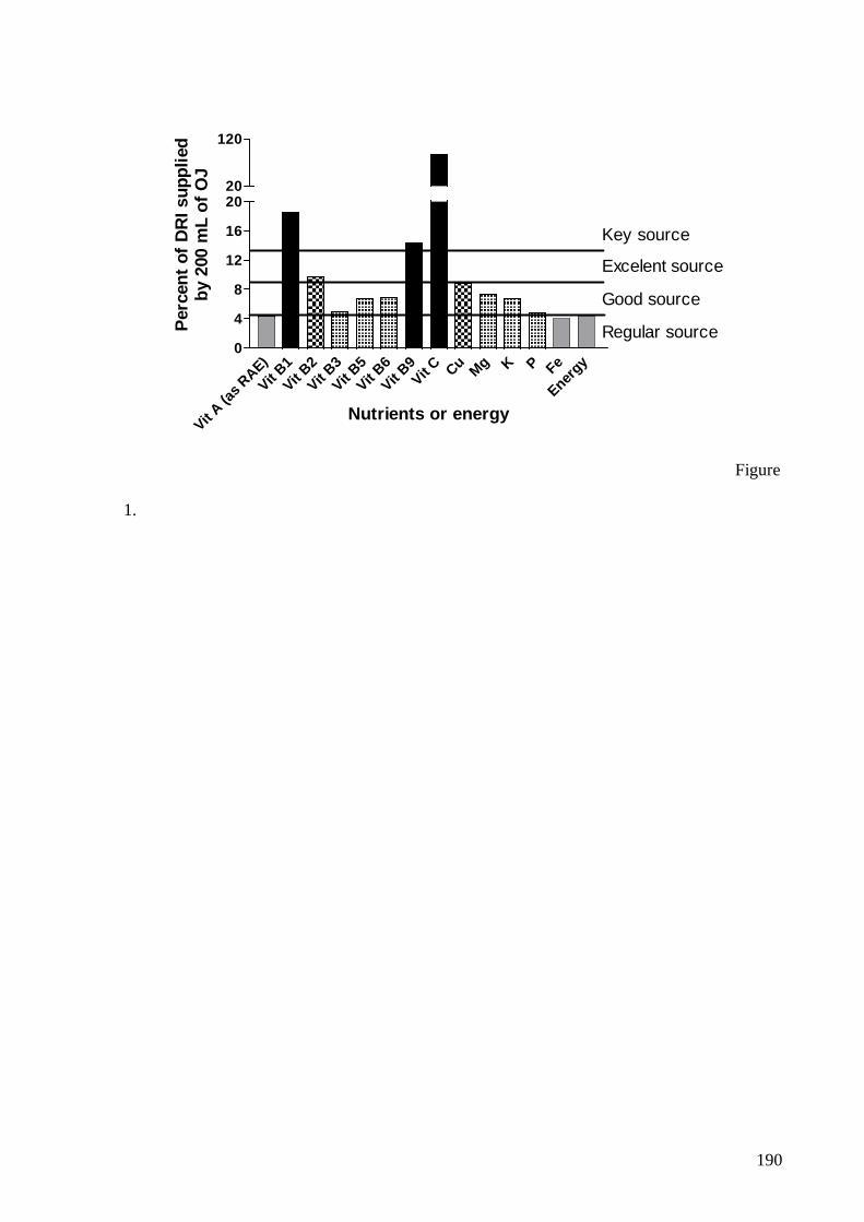
190
Vit A (a
s RAE)
Vit B1
Vit B2
Vit B3
Vit B5
Vit B6
Vit B9Vit C Cu Mg K P Fe
Energy
0
4
8
12
16
2020
120
Regular source
Good source
Excelent source
Key source
Nutrients or energy
Per
cent
of
DR
I sup
plie
dby
200
mL
of O
J
Figure
1.
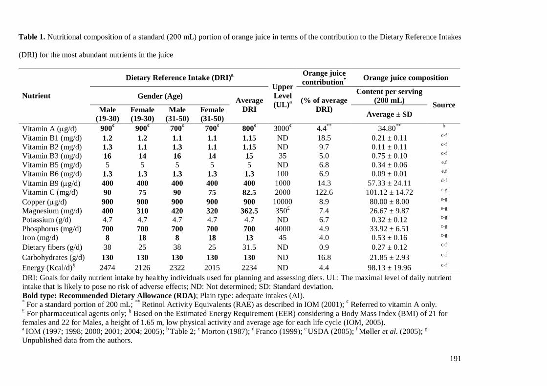
191
Table 1. Nutritional composition of a standard (200 mL) portion of orange juice in terms of the contribution to the Dietary Reference Intakes
(DRI) for the most abundant nutrients in the juice
Dietary Reference Intake (DRI)a Orange juice contribution* Orange juice composition
Gender (Age) Content per serving (200 mL) Nutrient
Male (19-30)
Female (19-30)
Male (31-50)
Female (31-50)
Average DRI
Upper Level (UL)a (% of average
DRI) Average ± SD Source
Vitamin A (�g/d) 900¢ 900¢ 700¢ 700¢ 800¢ 3000¢ 4.4** 34.80** b
Vitamin B1 (mg/d) 1.2 1.2 1.1 1.1 1.15 ND 18.5 0.21 ± 0.11 c-f
Vitamin B2 (mg/d) 1.3 1.1 1.3 1.1 1.15 ND 9.7 0.11 ± 0.11 c-f
Vitamin B3 (mg/d) 16 14 16 14 15 35 5.0 0.75 ± 0.10 c-f
Vitamin B5 (mg/d) 5 5 5 5 5 ND 6.8 0.34 ± 0.06 e,f
Vitamin B6 (mg/d) 1.3 1.3 1.3 1.3 1.3 100 6.9 0.09 ± 0.01 e,f
Vitamin B9 (�g/d) 400 400 400 400 400 1000 14.3 57.33 ± 24.11 d-f
Vitamin C (mg/d) 90 75 90 75 82.5 2000 122.6 101.12 ± 14.72 c-g
Copper (�g/d) 900 900 900 900 900 10000 8.9 80.00 ± 8.00 e-g
Magnesium (mg/d) 400 310 420 320 362.5 350£ 7.4 26.67 ± 9.87 e-g
Potassium (g/d) 4.7 4.7 4.7 4.7 4.7 ND 6.7 0.32 ± 0.12 c-g
Phosphorus (mg/d) 700 700 700 700 700 4000 4.9 33.92 ± 6.51 c-g
Iron (mg/d) 8 18 8 18 13 45 4.0 0.53 ± 0.16 c-g
Dietary fibers (g/d) 38 25 38 25 31.5 ND 0.9 0.27 ± 0.12 c-f
Carbohydrates (g/d) 130 130 130 130 130 ND 16.8 21.85 ± 2.93 c-f
Energy (Kcal/d)§ 2474 2126 2322 2015 2234 ND 4.4 98.13 ± 19.96 c-f
DRI: Goals for daily nutrient intake by healthy individuals used for planning and assessing diets. UL: The maximal level of daily nutrient intake that is likely to pose no risk of adverse effects; ND: Not determined; SD: Standard deviation. Bold type: Recommended Dietary Allowance (RDA); Plain type: adequate intakes (AI). * For a standard portion of 200 mL; ** Retinol Activity Equivalents (RAE) as described in IOM (2001); ¢ Referred to vitamin A only. £ For pharmaceutical agents only; § Based on the Estimated Energy Requirement (EER) considering a Body Mass Index (BMI) of 21 for females and 22 for Males, a height of 1.65 m, low physical activity and average age for each life cycle (IOM, 2005). a IOM (1997; 1998; 2000; 2001; 2004; 2005); b Table 2; c Morton (1987); d Franco (1999); e USDA (2005); f Møller et al. (2005); g
Unpublished data from the authors.
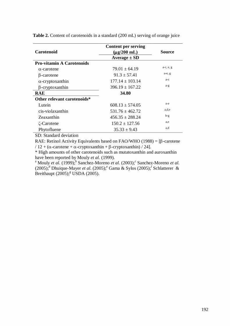
192
Table 2. Content of carotenoids in a standard (200 mL) serving of orange juice
Content per serving (����g/200 mL) Carotenoid
Average ± SD Source
Pro-vitamin A Carotenoids ����-carotene 79.01 ± 64.19 a-c, e, g
����-carotene 91.3 ± 57.41 a-e, g
����-cryptoxanthin 177.14 ± 103.14 a-c
����-cryptoxanthin 396.19 ± 167.22 a-g
RAE 34.80
Other relevant carotenoids*
Lutein 608.13 ± 574.05 a-e
cis-violaxanthin 531.76 ± 462.72 a,d,e
Zeaxanthin 456.35 ± 288.24 b-g
�-Carotene 150.2 ± 127.56 a,e
Phytofluene 35.33 ± 9.43 a,d
SD: Standard deviation RAE: Retinol Activity Equivalents based on FAO/WHO (1988) = [�-carotene / 12 + (�-carotene + �-cryptoxanthin + �-cryptoxanthin) / 24]. * High amounts of other carotenoids such as mutatoxanthin and auroxanthin have been reported by Mouly et al. (1999). a Mouly et al. (1999);b Sanchez-Moreno et al. (2003);c Sanchez-Moreno et al. (2005);d Dhuique-Mayer et al. (2005);e Gama & Sylos (2005);f Schlatterer & Breithaupt (2005);g USDA (2005).
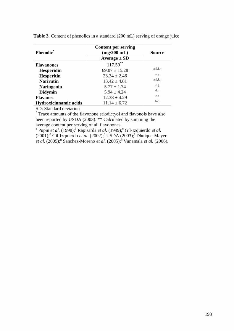
193
Table 3. Content of phenolics in a standard (200 mL) serving of orange juice
Content per serving (mg/200 mL) Phenolic* Average ± SD
Source
Flavanones 117.50**
Hesperidin 69.07 ± 15.28 a,d,f,h
Hesperitin 23.34 ± 2.46 e,g
Narirutin 13.42 ± 4.81 a,d,f,h
Naringenin 5.77 ± 1.74 e,g
Didymin 5.94 ± 4.24 d,h
Flavones 12.38 ± 4.29 c,d
Hydroxicinnamic acids 11.14 ± 6.72 b-d
SD: Standard deviation * Trace amounts of the flavonone eriodictyol and flavonols have also been reported by USDA (2003). ** Calculated by summing the average content per serving of all flavonones. a Pupin et al. (1998);b Rapisarda et al. (1999);c Gil-Izquierdo et al. (2001);d Gil-Izquierdo et al. (2002);e USDA (2003);f Dhuique-Mayer et al. (2005);g Sanchez-Moreno et al. (2005);h Vanamala et al. (2006).
194
7.5 ANEXO E: Curriculum Vitae - Silvia Isabel Rech Franke
Silvia Isabel Rech Franke
CURRICULUM VITAE
SANTA CRUZ DO SUL2006
195
2
CURRICULUM VITAEMaio, 2006
1 DADOS PESSOAIS
Nome: Silvia Isabel Rech FrankeFiliação: Claudino Aloisio Rech e Tharsilla RechNascimento: 19/03/1964, Santa Cruz do Sul/RS - BrasilCarteira de identidade: 6007004911 / SSP / RS / 16/10/1998CPF: 47300671004
Endereço profissional: Universidade de Santa Cruz do Sul, Departamento de Educação Física e Saúde,Curso de Nutrição.
Avenida Indenpendência, 2293 Universitário 96815900 SANTA CRUZ DO SUL, RS - Brasil - Caixa Postal: 188 Telefone: (51) 37177300 Ramal: 7386 Fax: 37171855 E-mail: [email protected]
Endereço residencial: Avenida Senador Alberto Pasqüaline, 688 Verena 96820050 SANTA CRUZ DO SUL, RS - Brasil Telefone: (51) 37171262 Fax: 37171262 E-mail: [email protected]
2 FORMAÇÃO ACADÊMICA/TITULAÇÃO
1999 - 2001 Mestrado em Biologia Celular e Molecular.Universidade Federal do Rio Grande do Sul, UFRGS, Rio Grande do Sul, Brasil.Título: O estudo da resposta mutagênica em procariontes e do potencial antioxidante in vitrode sucos de laranja in natura e industrializados.. Ano de obtenção: 2001.Orientador: João Antonio Pêgas Henriques.
1987 - 1987 Especialização em Administração e Serviço de Nutrição. (Carga horária: 410h)Instituto Metodista de Educação e Cultura, IMEC, Rio Grande do Sul, Brasil.
1983 - 1986 Graduação em Nutrição.Universidade do Vale do Rio dos Sinos, UNISINOS, Rio Grande do Sul, Brasil.
2001 Doutorado em Biologia Celular e Molecular.Universidade Federal do Rio Grande do Sul, UFRGS, Rio Grande do Sul, Brasil.Título: Suco de laranja e vitamina C: efeito sobre a estabilidade genômica.Orientador: João Antonio Pêgas Henriques.
3 FORMAÇÃO COMPLEMENTAR
2005 - 2005 Normas Técnicas de Elaboração de Referências e Cit.Universidade de Santa Cruz do Sul, UNISC, Rio Grande do Sul, Brasil.
2005 - 2005 Projeto de Pesquisa a Elaboração de Um Problema e.Universidade de Santa Cruz do Sul, UNISC, Rio Grande do Sul, Brasil.
2005 - 2005 A Arte da Docência no Ensino Superior.Universidade de Santa Cruz do Sul, UNISC, Rio Grande do Sul, Brasil.
2005 - 2005 O Processo de Ensino Aprendizagem e Avaliação Part.Universidade de Santa Cruz do Sul, UNISC, Rio Grande do Sul, Brasil.
2005 - 2005 Mediação de Conflitos no Ambiente Universitário.Universidade de Santa Cruz do Sul, UNISC, Rio Grande do Sul, Brasil.
2005 - 2005 Comitê de Ética Médica Bioética Ética Em Pesquisa. (Carga horária: 3h)
196
3
Universidade de Santa Cruz do Sul, UNISC, Rio Grande do Sul, Brasil.2005 - 2005 Histórico da Bioética Ética Em Pesquisa e Principa. (Carga horária: 3h)
Universidade de Santa Cruz do Sul, UNISC, Rio Grande do Sul, Brasil.2005 - 2005 Formação Moral Educação Moral e Ensino da Ética. (Carga horária: 3h)
Universidade de Santa Cruz do Sul, UNISC, Rio Grande do Sul, Brasil.2005 - 2005 Ensaios Bioquímicos de Estresse Oxidativo. (Carga horária: 20h)
Universidade de Caxias do Sul, UCS, Rio Grande do Sul, Brasil.2004 - 2004 As Novas Recomendações Dietéticas Dris e a Prát.... (Carga horária: 16h)
Universidade do Vale do Rio dos Sinos, UNISINOS, Rio Grande do Sul, Brasil.2004 - 2004 Genética e Conservação. (Carga horária: 8h)
Universidade Luterana do Brasil, ULBRA, Rio Grande do Sul, Brasil.2003 - 2003 Como As Vitraminas e Minerais Podem Contribuir .... (Carga horária: 4h)
Sociedade Brasileira de Mutagênese, Carciongenese e Teratogênese Ambiental, SBMCTA,Paraná, Brasil.
2000 - 2000 Guias Alimentares A Pirâmide e a Avaliação do ..... (Carga horária: 12h)Associação Gaúcha de Nutricionistas, AGAN, Rio Grande do Sul, Brasil.
2000 - 2000 Nutrição de Microelementos Vitaminas e Mineriais. (Carga horária: 18h)Universidade do Vale do Rio dos Sinos, UNISINOS, Rio Grande do Sul, Brasil.
2000 - 2000 Curso de Formação Em Medicina Biomolecular e Radic. (Carga horária: 64h)Sociedade Gaúcha de Medicina Biomolecular e Radicais Livres, SOCIEDADE GAÚCHA, RioGrande do Sul, Brasil.
1998 - 1998 Formação de Auditores Internos. (Carga horária: 24h)Supremo Consultoria, SUPREMO CONSULTO, Rio Grande do Sul, Brasil.
1998 - 1998 Extensão universitária em Administração da Qualidade Total. (Carga horária: 40h)Supremo Consultoria, SUPREMO CONSULTO, Rio Grande do Sul, Brasil.
1998 - 1998 Formação de Multiplicadores. (Carga horária: 16h)Supremo Consultoria, SUPREMO CONSULTO, Rio Grande do Sul, Brasil.
1996 - 1996 Como Evitar Toxinfecções Alimentares. (Carga horária: 12h)Associação Comercial e Industrial de Santa Cruz do Sul, ACI-SCRUZ, Rio Grande do Sul,Brasil.
1992 - 1992 Curso de Qualidade no Atendimento ao Cliente.Refeições Puras Rid Limitada, PURAS, Rio Grande do Sul, Brasil.
1992 - 1992 Curso de Preparação Para Instrutores do Programa...Refeições Puras Rid Limitada, PURAS, Rio Grande do Sul, Brasil.
1992 - 1992 Programa de Desenvolvimento Gerencial .....Refeições Puras Rid Limitada, PURAS, Rio Grande do Sul, Brasil.
1992 - 1992 Curso de Higiene na Manipulação dos Alimentos.Refeições Puras Rid Limitada, PURAS, Rio Grande do Sul, Brasil.
1992 - 1992 Curso de Higiene na Manipulação dos Alimentos.Refeições Puras Rid Limitada, PURAS, Rio Grande do Sul, Brasil.
1986 - 1986 Extensão universitária em Extensão Universitária - Nutrição em Pediatria. (Carga horária:40h)Associação Gaúcha de Nutricionistas, AGAN, Rio Grande do Sul, Brasil.
4 ATUAÇÃO PROFISSIONAL
Universidade Federal do Rio Grande do Sul - UFRGS
Vínculo institucional 1999 - 2001 Vínculo: Aluna de Mestrado, Enquadramento funcional: Aluna de mestrado, Carga horária:
1. 2001 - Atual Vínculo: Aluna de Doutorado, Enquadramento funcional: Aluna de Doutorado.
Atividades 6/1999 - 6/2001 Participação em projeto
1. Estudo da Resposta Mutagênica em Procariontes e do Potencial Antioxidante in vitro deSucos de Laranja in natura e Industrializados.
197
4
3/2001 - 3/2001 Disciplinas ministradas
1. O estudo da resposta mutagênica em procariontes e do potencial antioxidante in vitro emsuco de laranja naturais e industrializados.
6/2004 - Atual Participação em projeto
1. Avaliação do estresse oxidativo e da capacidade de reparo do DNA em atletas.
7/2001 - Atual Participação em projeto
1. Avaliação da possível ação moduladora da genotoxicidade e da mutagenicidade de suco delaranja e de vitamina C em camundongos in vivo.
Universidade de Santa Cruz do Sul - UNISC
Vínculo institucional 1998 - Atual Vínculo: Professor, Enquadramento funcional: Professor, Carga horária: 30, Regime:
Dedicação exclusiva.
Outras informaçõesDisciplinas de Nutriçao Normal, Nutriçao Básica e Estágio Supervisionado de AlimentaçãoInstuitucional do Curso de Nutrição da Universidade de Santa Cruz do Sul- UNISC - RS
Atividades 8/2005 - 8/2005 Disciplinas ministradas
1. Seminários Avançados: Genética e Nutrição.
02/2003 - 08/2003 Disciplinas ministradas
1. Estágio Supervisionado em Alimentação Institucional.
3/1999 - 9/2001 Linhas de pesquisa
1. Potencial mutagênico e antioxidante.2. Radicais livres e danos oxidativos.
11/1998 - 3/2001 Cargos ou funções
1. Coordenador de Curso de Graduação de Nutrição.
9/2000 - 10/2000 Atividades de extensão realizadas
1. Comissão Organizadora do Curso de Bioquímica Nutricional.
8/2000 - 8/2000 Atividades de extensão realizadas
1. Comissão Organizadora da Semana Acadêmica do Curso de Nutrição.
7/2000 - 7/2000 Atividades de extensão realizadas
1. Palestra sobre Alimentação do Trabalhador no Curso e Pós-Graduação em Enfermagem(UNISC).
5/2000 - 5/2000 Atividades de extensão realizadas
1. Palestra.
8/1999 - 9/1999 Atividades de extensão realizadas
1. Comissão Organizadora do I Seminário Regional sobre Fitoterapia.
198
5
7/1999 - 7/1999 Atividades de extensão realizadas
1. Concurso Público para Docentes da Disciplina de Nutrição Normal - APROVADA.
12/1998 - 12/1998 Atividades de extensão realizadas
1. Palestra.
11/1998 - 11/1998 Atividades de extensão realizadas
1. Palestra.
3/2006 - Atual Cargos ou funções
1. Sub-chefe de departamento.
7/2005 - Atual Disciplinas ministradas
1. Nutrição e Dietética I.2. Alimentação Institucional I.3. Nutrição.
7/2005 - Atual Disciplinas ministradas
1. Metodologia de Pesquisa Científica.
3/2005 - Atual Participação em projeto
1. Jogo, exercíco e estilo de vida na saúde, cultura e desenvolvimento humano.
2/2005 - Atual Disciplinas ministradas
1. Nutrição e Dietética II.2. Estágio Supervisionado em Alimentação Institucional.
7/2004 - Atual Disciplinas ministradas
1. Estágio de Escola de Educação Básica.
03/2004 - Atual Cargos ou funções
1. Coordenador Geral do Curso de Pós-graduação em Nutrição Clínica: Ênfase nas DoençasCrônico-Não-Transmissíveis.
10/2001 - Atual Linhas de pesquisa
1. Potencial mutagênico e antioxidante.2. Radicais livres e danos oxidativos.
02/2000 - Atual Disciplinas ministradas
1. Nutrição Básica.
11/1998 - Atual Disciplinas ministradas
1. Nutriçao Normal.
Universidade Federal de Santa Catarina - UFSC
Vínculo institucional 2000 - 2000 Vínculo: Estagiário, Enquadramento funcional: Aluna de Mestrado, Carga horária: 8.
Atividades 7/2000 - 7/2000 Estágios realizados
199
6
1. Determinação Antioxidante in vitro de amostras de suco de laranjaindustrializadas e natural.
Petróleo Brasileiro S/A - PETROBRAS
Vínculo institucional 1986 - 1986 Vínculo: Estagiário, Enquadramento funcional: Voluntário, Carga horária: 20.
Atividades 7/1986 - 12/1986 Estágios realizados
1. Estágio de Estudante de Nível Superior.
Serviço Social da Industria - SESI
Vínculo institucional 1986 - 1986 Vínculo: Estágio voluntário, Carga horária: 20.
Atividades 9/1986 - 12/1986 Estágios realizados
1. Estágio de Complementação Educacional.
Universidade do Vale do Rio dos Sinos - UNISINOS
Vínculo institucional 1985 - 1985 Vínculo: Bolsista Iniciação Cien.CNPq, Enquadramento funcional: Aluno de Graduação,
Carga horária: 20, Regime: Dedicação exclusiva.
Atividades 3/1985 - 7/1985 Atividades realizadas
1. Bolsa CNPq.
5 LINHAS DE PESQUISA
1 Potencial mutagênico e antioxidante.Palavras-chave: Vitamina C; Sucos de laranja; Radicais Livres; Fenólicos Totais; Mutagenicidade.
2 Potencial mutagênico e antioxidante.
3 Radicais livres e danos oxidativos.Palavras-chave: Modulação Mutagênica; Genotoxicidade; Atividade Antioxidante; Vitamina C.
4 Radicais livres e danos oxidativos.
6 PROJETOS DE PESQUISA
2005 - Atual Jogo, exercíco e estilo de vida na saúde, cultura e desenvolvimento humano.Descrição: ..Situação: Em andamento; Natureza: Extensão.Alunos envolvidos: Graduação (1).Integrantes: Silvia Isabel Rech Franke (Responsável); Danusia Puntel.
2004 - Atual Avaliação do estresse oxidativo e da capacidade de reparo do DNA em atletas.Descrição: ..Situação: Em andamento; Natureza: Pesquisa.Integrantes: Silvia Isabel Rech Franke (Responsável); Bernardo Erdtmann; Carlos Mantese; Daniel Prá; Janaína PJaeger; João Antonio Pêgas Henriques.
200
7
2001 - Atual Avaliação da possível ação moduladora da genotoxicidade e da mutagenicidade de suco delaranja e de vitamina C em camundongos in vivo.Descrição: ..Situação: Em andamento; Natureza: Pesquisa.Integrantes: João Antonio Pêgas Henriques (Responsável); Silvia Isabel Rech Franke; Bernardo Erdtmann; DanielPrá; Juliana da Silva.
1999 - 2001 Estudo da Resposta Mutagênica em Procariontes e do Potencial Antioxidante in vitro deSucos de Laranja in natura e Industrializados.Descrição: ..Situação: Concluído; Natureza: Pesquisa.Integrantes: Silvia Isabel Rech Franke (Responsável); Bernardo Erdtmann; Gabriel Rubensam; Jaqueline DeosSilveira; João Antonio Pêgas Henriques; Karina Ckless; Martin Brendel; Ribeiro do Valle.
7 ÁREAS DE ATUAÇÃO
1 Ciências da Saúde, Nutrição.2 Genética, Mutagenese.3 Biologia Molecular, Radicais Livres.4 Nutrição, Bioquímica da Nutrição.5 Mutagenese, Antimutagenese.6 Farmácia, Análise Toxicológica.
8 IDIOMAS
Compreende: Alemão (Razoavelmente), Espanhol (Bem), Inglês (Bem).Fala: Espanhol (Razoavelmente), Inglês (Razoavelmente).Lê: Espanhol (Bem), Inglês (Bem).Escreve: Espanhol (Pouco), Inglês (Razoavelmente).
9 PRÊMIOS E TÍTULOS
2005 Mejor presentacion en la Sesión de Posters del Area Genotoxicidad - Mutagénesis,ATA/ALAMCTA.
2004 Reconhecimento por contribuição relevante na área [artigo intitulado 'Study of antioxidant andmutagenic activity of different orange juices (Food Chemistry 88, 45-55)]., SociedadIberoamericana de Información Científica (SIIC).
1999 Membro nato da comissão de ensino do Conselho Regional de Nutricionistas, ConselhoRegional de Nutricionistas 2ª Região.
10 PRODUÇÃO CIENTÍFICA, TECNOLÓGICA E ARTÍSTICA/CULTURAL
10.1 PRODUÇÃO BIBLIOGRÁFICA
10.1.1 Trabalhos completos em anais de eventos
1 PRÁ, Daniel; GUECHEVA, Temenouga N; FRANKE, Silvia Isabel Rech; KNAKIEVICZ, Tanise;ERDTMANN, Bernardo; HENRIQUES, João Antonio Pêgas. Toxicidade e gentoxicidade do sulfato decobre em planárias de água doce e camundongos. In: VIII CONGRESSO BRASILEIRO DEECOTOXICOLOGIA/ SOCIEDADE BRASILEIRA DE ECOTOXICOLOGIA - SETAC BRASIL,Florianópolis. 2005.
10.1.2 Resumos simples em anais de eventos
201
8
1 FRANKE, Silvia Isabel Rech; PRÁ, Daniel; ERDTMANN, Bernardo; SILVA, Juliana da; HENRIQUES,João Antonio Pêgas. Action of vitamin C on DNA damage induced by methyl methanesulfonate,cyclophosphamide, FeSO4 and CuSO4 in mouse blood cells in vivo. In: VII REUNIÃO ANUAL DOPROGRAMA DE PÓS-GRADUAÇÃO EM BIOLOGIA CELULAR E MOLECULAR DO CENTRO DEBIOTECNOLOGIA DA UFRGS, 2005, Porto Alegre. Livro de resumos. 2005. p. 33-33.
2 PRÁ, Daniel; FRANKE, Silvia Isabel Rech; YONEAMA, Maria L; GIULIAN, Raquel; DIAS, Johnny F;ERDTMANN, Bernardo; HENRIQUES, João Antonio Pêgas. Can food composition affect the genotoxicityinduced by iron when using comet assay in vivo?. In: VII REUNIÃO ANUAL DO PROGRAMA DEPÓS-GRADUAÇÃO EM BIOLOGIA CELULAR E MOLECULAR DO CENTRO DE BIOTECNOLOGIA DAUFRGS, 2005, Porto Alegre. Livro de Resumos. 2005. p. 78-78.
3 FRANKE, Silvia Isabel Rech; PRÁ, Daniel; SCHARLAU, Andréa A; CASSINI, Carina; MACHADO, MirianaS; MANTESE, Carlos; JAEGER, Janaína P; ERDTMANN, Bernardo; SALVADOR, Miriam; HENRIQUES,João Antonio Pêgas; PETKOWICZ, Rosemary. Dano ao DNA e estresse oxidativo em remadores. In: VIIREUNIÃO ANUAL DO PROGRAMA DE PÓS-GRADUAÇÃO EM BIOLOGIA CELULAR E MOLECULARDO CENTRO DE BIOTECNOLOGIA DA UFRGS, 2005, Porto Alegre. Livro de Resumos. 2005. p.32-32.
4 SCHARLAU, Andréa A; FRANKE, Silvia Isabel Rech; PRÁ, Daniel; CASSINI, Carina; MACHADO, MirianaS; MANTESE, Carlos; JAEGER, Janaína P; ERDTMANN, Bernardo; SALVADOR, Miriam; HENRIQUES,João Antonio Pêgas; PETKOWICZ, Rosemary. Dano no DNA e estresse oxidativo em remadores. In:SALÃO DE INICIAÇÃO CIENTÍFICA, 2005, Porto Alegre. 2005.
5 SCHARLAU, Andréa A; CASSINI, Carina; HENRIQUES, João Antonio Pêgas; MACHADO, Miriana S;ERDTMANN, Bernardo; FRANKE, Silvia Isabel Rech; PETKOWICZ, Rosemary; PRÁ, Daniel. Dano noDNA e estresse oxidativo em remadores. In: XI SEMINÁRIO DE INICIAÇÃO CIENTÍFICA E X JORNADADE ENSINO, PESQUISA E EXTENSÃO DA UNISC, 2005, Santa Cruz do Sul. 2005.
6 FRANKE, Silvia Isabel Rech; PRÁ, Daniel; ERDTMANN, Bernardo; SILVA, Juliana da; HENRIQUES,João Antonio Pêgas. In vivo genotoxicity of iron, copper and vitamin C as evaluated by comet assay andmicronucleus test in mice. In: VII REUNIÃO ANUAL DO PROGRAMA DE PÓS-GRADUAÇÃO EMBIOLOGIA CELULAR E MOLECULAR DO CENTRO DE BIOTECNOLOGIA DA UFRGS, 2005, PortoAlegre. Livro de resumos. 2005. p. 31-31.
7 PUNTEL, Danusia; LAGO, Taciara B; SILVEIRA, Luciano C da; FRANKE, Silvia Isabel Rech;RECKZIEGEL, Miriam Beatris. Obesidade e hipertensão arterial:prevalência em escolares de Santa Cruzdo Sul/RS. In: XI SEMINÁRIO DE INICIAÇÃO CIENTÍFICA E X JORNADA DE ENSINO, PESQUISA EEXTENSÃO DA UNISC, 2005, Santa Cruz do Sul. 2005.
8 FRANKE, Silvia Isabel Rech; PRÁ, Daniel; SILVA, Juliana da; ERDTMANN, Bernardo; HENRIQUES,João Antonio Pêgas. Analysis in vivo of DNA damage and genotoxicity modulation induced by orangejuice in relation to alkylating agents. In: VI REUNIÃO ANUAL DO PROGRAMA DE PÓS-GRADUAÇÃOEM BIOLOGIA CELULAR E MOLECULAR DO CENTRO DE BIOTECNOLOGIA DA UFRGS, 2004, PortoAlegre. VI Reunião Anual do Programa de Pós-Graduação em Biologia Celular e Molecular do Centrode Biotecnologia da UFRGS- Livro de Resumos. Porto Alegre: UFRGS, 2004. p. 27-27.
9 FRANKE, Silvia Isabel Rech; PRÁ, Daniel; SILVA, Juliana da; ERDTMANN, Bernardo; HENRIQUES,João Antonio Pêgas. Modulation of genotoxicity of FeSO4 and CuSO4 by orange juice, as evaluated bythe comet assay. In: VI REUNIÃO ANUAL DO PROGRAMA DE PÓS-GRADUAÇÃO EM BIOLOGIACELULAR E MOLECULAR DO CENTRO DE BIOTECNOLOGIA DA UFRGS, 2004, Porto Alegre. VIReunião Anual do Programa de Pós-Graduação em Biologia Celular e Molecular do Centro deBiotecnologia da UFRGS- Livro de Resumos. Porto Alegre: UFRGS, 2004. p. 26-26.
202
9
10 FRANKE, Silvia Isabel Rech; PRÁ, Daniel; SILVA, Juliana da; ERDTMANN, Bernardo; HENRIQUES,João Antonio Pêgas. Modulatory effect of vitamin C over the genotoxicity of alkylating agents and metalliccompounds, as ervaluated by comet assay in vivo. In: VI REUNIÃO ANUAL DO PROGRAMA DEPÓS-GRADUAÇÃO EM BIOLOGIA CELULAR E MOLECULAR DO CENTRO DE BIOTECNOLOGIA DAUFRGS, 2004, Porto Alegre. VI Reunião Anual do Programa de Pós-Graduação em Biologia Celular eMolecular do Centro de Biotecnologia da UFRGS- Livro de Resumos. Porto Alegre: UFRGS,2004. p. 28-28.
11 FRANKE, Silvia Isabel Rech; PRÁ, Daniel; SILVA, Juliana da; ERDTMANN, Bernardo; HENRIQUES,João Antonio Pêgas. Modulatory effect of vitamin C over the genotoxicity of alkylating agents and metalliccompounds, as evaluated by comet assay in vivo. In: 50° CONGRESSO BRASILEIRO DE GENÉTICA,2004, Florianópolis. 50° Congresso Brasileiro de Genética. Florianópolis: São Francisco Gráfica eeditora, 2004. v. 1, p. 90-90.
12 PRÁ, Daniel; GUECHEVA, Temenouga N; FRANKE, Silvia Isabel Rech; KNAKIEVICZ, Tanise;ERDTMANN, Bernardo; HENRIQUES, João Antonio Pêgas. Toxicidade e genotoxicidade do sulfato decobre em planárias de água doce e camundongos. In: VIII CONGRESSO BRASILEIRO DEECOTOXICOLOGIA/ SOCIEDADE BRASILEIRA DE ECOTOXICOLOGIA - SETAC BRASIL, 2004,Florianópolis. Ecotox 2004 - Livro de Resumos. São José- SC: Gráfica Paper Print, 2004. v. único,p. 112-112.
13 FRANKE, Silvia Isabel Rech; PRÁ, Daniel; GUECHEVA, Temenouga N; KNAKIEVICZ, Tanise;ERDTMANN, Bernardo; HENRIQUES, João Antonio Pêgas. Toxicidade e genotoxicidade do sulfato decobre em planárias de água doce e camundongos. In: VI REUNIÃO ANUAL DO PROGRAMA DEPÓS-GRADUAÇÃO EM BIOLOGIA CELULAR E MOLECULAR DO CENTRO DE BIOTECNOLOGIA DAUFRGS, 2004, Porto Alegre. VI Reunião Anual do Programa de Pós-Graduação em Biologia Celular eMolecular do Centro de Biotecnologia da UFRGS - Livro de Resumos. Porto Alegre: UFRGS,2004. p. 68-68.
14 FRANKE, Silvia Isabel Rech; PRÁ, Daniel; SILVA, Juliana da; ERDTMANN, Bernardo; HENRIQUES,João Antonio Pêgas. Analyses in vivo of DNA Damage and Genotoxicity Modulation Induced by Vitamin Cin relation to Alkylating and metallic Agents. In: V REUNIÃO ANUAL DO PROGRAMA DEPÓS-GRADUAÇÃO EM BIOLOGIA CELULAR E MOLECULAR DO CENTRO DE BIOTECNOLOGIA DAUFRGS, 2003, Porto Alegre. V Reunião Anual do Programa de Pós-Graduação em Biologia Celular eMolecular do Centro de Biotecnologia da UFRGS - Livro de Resumos. Porto Alegre: UFRGS,2003. v. -, p. 24.
15 FRANKE, Silvia Isabel Rech; PRÁ, Daniel; SILVA, Juliana da; ERDTMANN, Bernardo; HENRIQUES,João Antonio Pêgas. Antigenotoxic Properties of Orange Juice Compounds over Known Mutagens. In: VREUNIÃO ANUAL DO PROGRAMA DE PÓS-GRADUAÇÃO EM BIOLOGIA CELULAR E MOLECULARDO CENTRO DE BIOTECNOLOGIA DA UFRGS, 2003, Porto Alegre. V Reunião Anual do Programade Pós-Graduação em Biologia Celular e Molecular do Centro de Biotecnologia da UFRGS - Livrode Resumos. Porto Alegre: UFRGS, 2003. v. -, p. 23.
16 FRANKE, Silvia Isabel Rech; PRÁ, Daniel; SILVA, Juliana da; ERDTMANN, Bernardo; HENRIQUES,João Antonio Pêgas. Modulation Effect of Orange Juice over Metallic Compounds Genotoxicity. In: VREUNIÃO ANUAL DO PROGRAMA DE PÓS-GRADUAÇÃO EM BIOLOGIA CELULAR E MOLECULARDO CENTRO DE BIOTECNOLOGIA DA UFRGS, 2003, Porto Alegre. V Reunião Anual do Programade Pós-Graduação em Biologia Celular e Molecular do Centro de Biotecnologia da UFRGS - Livrode Resumos. Porto Alegre: UFRGS, 2003. v. -, p. 22.
17 FRANKE, Silvia Isabel Rech; CKLESS, Karina; SILVEIRA, Jaqueline Deos; RUBENSAM, Gabriel;ERDTMANN, Bernardo. Avaliação do potencial mutagênico e antioxidante de sucos de laranja in natura eindustrializados. In: XV JORNADA DE NUTRIÇÃO, 2002, Porto Alegre. Sem Anais. 2002.
18 FRANKE, Silvia Isabel Rech; HENRIQUES, João Antonio Pêgas. Avaliação do potencial mutagênico eantioxidante de sucos de laranja in natura e industrializados. In: XV JORNADA DE NUTRIÇÃO, 2002,Porto Alegre. Sem anais. 2002.
203
10
19 FRANKE, Silvia Isabel Rech; PRÁ, Daniel; ERDTMANN, Bernardo; SILVA, Juliana da; HENRIQUES,João Antonio Pêgas. O efeito de sucos de laranja no reparo de danos ao DNA de células sangüíneas decamundongo. In: XV JORNADA DE NUTRIÇÃO, 2002, Porto Alegre. Sem anais. 2002.
20 FRANKE, Silvia Isabel Rech; PRÁ, Daniel; SILVA, Juliana da; ERDTMANN, Bernardo; HENRIQUES,João Antonio Pêgas. O efeito Modulador de sucos de laranja. In: VII CONGRESSO BRASILEIRO DEECOTOXICOLOGIA E OS NOVOS DESAFIOS NO MONITORAMENTO AMBIENTAL, V REUNIÃO DASETAC LATINO AMERICANA, 2002, Vitória. VII ECOTOX- Livro de Resumos. Vitória - ES - Brasil:Gráfica Ita, 2002. p. 330.
21 FRANKE, Silvia Isabel Rech; PRÁ, Daniel; SILVA, Juliana da; ERDTMANN, Bernardo; HENRIQUES,João Antonio Pêgas. O Efeito Modulador de Sucos de Laranja. In: IV REUNIÃO ANUAL DO PROGRAMADE PÓS-GRADUAÇÃO EM BIOLOGIA CELULAR E MOLECULAR DO CENTRO DE BIOTECNOLOGIADA UFRGS, 2002, Porto Alegre. PPGBCM- IV Reunião Anual do Programa de Pós-Graduação emBiologia Celular e Molecular do Centro de Biotecnologia da UFRGS - Livro de Resumos. PortoAlegre: Universidade Federal do Rio Grande do Sul, 2002. v. único, p. 28-28.
22 FRANKE, Silvia Isabel Rech; CKLESS, Karina; SILVEIRA, Jaqueline Deos; RUBENSAM, Gabriel;ERDTMANN, Bernardo; HENRIQUES, João Antonio Pêgas. Resposta mutagênica em procariontes e dopotencial antioxidante in vitro de sucos de laranja. In: VII CONGRESSO BRASILEIRO DEECOTOXICOLOGIA E OS NOVOS DESAFIOS NO MONITORAMENTO AMBIENTAL, V REUNIÃO DASETAC LATINO AMERICANA, 2002, Vitória. VII ECOTOX - Livro de Resumos. 2002. p. 326.
23 FRANKE, Silvia Isabel Rech; CKLESS, Karina; ERDTMANN, Bernardo; HENRIQUES, João AntonioPêgas. O Estudo da Resposta Mutagênica em Procariontes e do Potencial Antioxidante in vitro de sucosde laranja in natura e industrializados. In: 3º REUNIÃO ANUAL DO PROGRAMA DE PÓS-GRADUAÇÃOEM BIOLOGIA CELULAR E MOLECULAR- PPGBCM, 2001, Porto Alegre. 3º Reunião Anual doPrograma de Pós-Graduação em Biologia Celular e Molecular- PPGBCM - Livro de Resumos.Porto Alegre: UFRGS, 2001. p. 32.
24 FRANKE, Silvia Isabel Rech; CKLESS, Karina; VALLE, Ribeiro Do; HENRIQUES, João Antonio Pêgas.Antioxidant Properties of Natural and Processed Orange Juices. In: 7TH ANNUAL MEETING OF THEOXYGEN SOCIETY, 2000, San Diego. 7th Annual Meeting of The Oxygen Society. San Diego:2000. p. S32.
25 FRANKE, Silvia Isabel Rech; HENRIQUES, João Antonio Pêgas. Linha de pesquisa: Análise dasatividades citotóxica, mutagênica e recombinogênica de produtos naturais em organismos procarióticos eeucarióticos (Sucos naturais e industrializados). In: RELATÓRIO DE ATIVIDADES 1999-2000 DOCENTRO DE BIOTECNOLOGIA DO ESTADO DO RIO GRANDE DO SUL, 2000, Porto Alegre.Relatório de Atividades 1999-2000 do Centro de Biotecnologia do Estado do Rio Grande do Sul. 2000.p. 52-55.
26 FRANKE, Silvia Isabel Rech; CKLESS, Karina; ERDTMANN, Bernardo; HENRIQUES, João AntonioPêgas. O Estudo da resposta mutagênica e antioxidante em procariontes por sucos de laranja in natura eindustrializados. In: II REUNIÃO ANUAL DO PROGRAMA DE PÓS GRADUAÇÃO EM BIOLOGIACELULAR E MOLECULAR - PPGBCM, 2000, Porto Alegre. II Reunião Anual do Programa de PósGraduação em Biologia Celular e Molecular - PPGBCM - Livro de Resumos. 2000. p. 20.
27 FRANKE, Silvia Isabel Rech; ERDTMANN, Bernardo; HENRIQUES, João Antonio Pêgas. O estudo daresposta moduladora da genotoxicidade em procariontes por sucos de laranja in natura e industrializados.In: EVENTO COMEMORATIVO DO 1.º ANO DE ATIVIDADES DO PROGRAMA DE PÓS GRADUAÇÃOBIOLOGIA CELULAR E MOLECULAR (PPGBCM), 1999, Porto Alegre. Evento Comemorativo do 1.ºAno de Atividades do Programa de Pós Graduação Biologia Celular e Molecular (PPGBCM) - Livrode Resumos. Porto Alegre: UFRGS, 1999. p. 6.
10.1.3 Resumos expandidos em anais de eventos
204
11
1 FRANKE, Silvia Isabel Rech. O efeito modulador de sucos de laranja e vitamina C no reparo de danos aoDNA de células sangüíneas de camundongos. In: I ENCONTRO DE NUTRICIONISTASPESQUISADORAS DO RIO GRANDE DO SUL, 2003, Porto Alegre. I Encontro de NutricionistasPequisadoras do Rio Grande do Sul. 2003.
10.1.4 Artigos completos publicados em periódicos
1 FRANKE, Silvia Isabel Rech; PRÁ, Daniel. Colesterol e o risco de doenças cardiovasculares em criançase adolescentes. Cinergis, n. no prelo, 2006.
2 FRANKE, Silvia Isabel Rech; PRÁ, Daniel; GIULIAN, Raquel; DIAS, Johnny F; YONEAMA, Maria L;SILVA, Juliana da; ERDTMANN, Bernardo; HENRIQUES, João Antonio Pêgas. Influence of orange juicein the levels and in the genotoxicity of iron and copper. Food and Chemical Toxicology, Holanda, v.44, n. 3, p. 425-435, 2005.
3 FRANKE, Silvia Isabel Rech; PRÁ, Daniel; ERDTMANN, Bernardo; HENRIQUES, João Antonio Pêgas;SILVA, Juliana da. Modulation by orange juice of DNA damage and genotoxicity induced by alkylatingagents: an in vivo analysis. Mutagenesis, Reino Unido, v. 20, n. 4, p. 279-283, 2005.
4 FRANKE, Silvia Isabel Rech; PRÁ, Daniel; SILVA, Juliana da; ERDTMANN, Bernardo; HENRIQUES,João Antonio Pêgas. Possible repair action of Vitamin C on DNA damage induced by methylmethanesulfonate, cyclophosphamide, FeSO4 and CuSO4 in mouse blood cells in vivo. Mutationresearch. Genetic toxicology and environmental mutagenesis, Holanda, v. 583, n. 1, p. 85-94,2005.
5 FRANKE, Silvia Isabel Rech; CKLESS, Karina; SILVEIRA, Jaqueline Deos; RUBENSAM, Gabriel;BRENDEL, Martin; ERDTMANN, Bernardo; HENRIQUES, João Antonio Pêgas. Study of Antioxidant andMutagenic Activity of Different Orange Juice. Food Chemistry, Holanda, v. 88, p. 45-55, 2004.
10.1.5 Artigos resumidos publicados em periódicos
1 FRANKE, Silvia Isabel Rech; PRÁ, Daniel; SCHARLAU, Andréa A; CASSINI, Carina; MACHADO, MirianaS; MANTESE, Carlos; JAEGER, Janaína P; ERDTMANN, Bernardo; SALVADOR, Miriam; HENRIQUES,João Antonio Pêgas; PETKOWICZ, Rosemary. Daño en ADN y estrés oxidativo em remeros. ActaToxicológica Argentina, Argentina, v. 13, n. supl, p. 111-112, 2005.
2 FRANKE, Silvia Isabel Rech; CKLESS, Karina; SILVEIRA, Jaqueline Deos; RUBENSAM, Gabriel;BRENDEL, Martin; ERDTMANN, Bernardo; HENRIQUES, João Antonio Pêgas. El jugo de naranja tienepotencia antioxidante y mutagenico. Sociedad Iberoamericana de Información Científica Siic,http://www.siicsalud.com/dato/, 2005.
3 PUNTEL, Danusia; LAGO, Taciara B; SILVEIRA, Luciano C da; RECKZIEGEL, Miriam Beatris; PRÁ,Daniel; FRANKE, Silvia Isabel Rech. Estudo para avaliar a concordância entre o IMC e o percentual degordura na classificação do estado nutricional de crianças. Revista brasileira de ciência e movimento,São Paulo, v. 13, n. 4, p. 57-57, 2005.
4 PRÁ, Daniel; FRANKE, Silvia Isabel Rech; YONEAMA, Maria L; GIULIAN, Raquel; DIAS, Johnny F;ERDTMANN, Bernardo; HENRIQUES, João Antonio Pêgas. Food composition as a confounding factor inthe comet assay in vivo. Mutation Research-Fundamental and Molecular Mechanisms of Mutagenesis,v. 577S, p. e183-e183, 2005.
5 FRANKE, Silvia Isabel Rech; PRÁ, Daniel; ERDTMANN, Bernardo; SILVA, Juliana da; HENRIQUES,João Antonio Pêgas. Genotoxicidad y mutagenicidad in vivo de hierro, cobre y vitamina C, evaluadasporensayos cometa y test del micronúcleo en ratones. Acta Toxicológica Argentina, Argentina, v.13, n. supl, p. 99-100, 2005.
205
12
6 FRANKE, Silvia Isabel Rech; PRÁ, Daniel; SILVA, Juliana da; ERDTMANN, Bernardo; HENRIQUES,João Antonio Pêgas. In vivo genotoxicity and mutagenicity of trace nutrients: results of subchronicexposure of mice to FeSO4, CuSO4 and vitamin C as evaluated by the comet assay and micronucleustest. Mutation Research-Fundamental and Molecular Mechanisms of Mutagenesis, v. 577S, p.e19-e19, 2005.
7 FRANKE, Silvia Isabel Rech; PRÁ, Daniel; DIAS, Johnny F; GIULIAN, Raquel; YONEAMA, Maria L;SILVA, Juliana da; ERDTMANN, Bernardo; HENRIQUES, João Antonio Pêgas. Orange juice modulationover the genotoxicity of FeSO4 and CuSO4, as evaluated by the comet assay, and the influencence ofwhole blood and the hepatic levels of iron and copper, as detected by PIXE. Genetics and MolecularBiology, Ribeirão Preto, v. 28, n. 2, p. 260-260, 2005.
8 FRANKE, Silvia Isabel Rech; PRÁ, Daniel; ERDTMANN, Bernardo; SILVA, Juliana da; HENRIQUES,João Antonio Pêgas. Possible anti-mutagen action of orange juice and vitamin C on the DNA damagecaused on mice blood cells in vivo by cyclophosphamide, methyl methanesulfonate, FeSO4 and CuSO4.Mutation Research-Fundamental and Molecular Mechanisms of Mutagenesis, v. 577S, p.e164-e164, 2005.
9 PRÁ, Daniel; FRANKE, Silvia Isabel Rech; SILVA, Juliana da; ERDTMANN, Bernardo; HENRIQUES,João Antonio Pêgas. Possible repair action of vitamin C on DNA damage induced by methylmethanesulfonate, cyclophosphamide, FeSO4 and CuSO4 in mouse blood cells in vivo. Genetics andMolecular Biology, Ribeirão Preto, v. 28, n. 2, p. 261-261, 2005.
10 PRÁ, Daniel; FRANKE, Silvia Isabel Rech; YONEAMA, Maria L; GIULIAN, Raquel; DIAS, Johnny F;ERDTMANN, Bernardo; HENRIQUES, João Antonio Pêgas. Puede la composición de alimento afectar lagenotoxicidad inducida por hierro cuando se la evalúa utilizando el ensayo cometa in vivo?. ActaToxicológica Argentina, Argentina, v. 13, n. supl, p. 104-104, 2005.
11 FRANKE, Silvia Isabel Rech; PRÁ, Daniel; SILVA, Juliana da; ERDTMANN, Bernardo; HENRIQUES,João Antonio Pêgas. Orange juice and vitamin C genotoxic modulation effect. Genetics and MolecularBiology, Brasil, v. 26, n. 2, p. 177-177, 2003.
12 FRANKE, Silvia Isabel Rech; CKLESS, Karina; VALLE, Ribeiro Do; HENRIQUES, João Antonio Pêgas.Antioxidant properties of natural and processed orange juice. Free Radical Biology and Medicine,São Diego, Estados Unidos, v. 29, n. supplement, p. S32-S32, 2000.
10.1.6 Capítulos de livros publicados
1 FRANKE, Silvia Isabel Rech; BOEIRA, Jane Marlei; ERDTMANN, Bernardo; HENRIQUES, João AntonioPêgas. Genotoxicidade de Agentes Sintéticos e Naturais. In: SILVA, Juliana da; ERDTMANN, Bernardo;HENRIQUES, João Antonio Pêgas. (Org.). Genética Toxicológica. Porto Alegre, 2003, v. 1, p.309-317.
10.1.7 Textos em jornais de notícias
1 HAGEN, M K; FRANKE, Silvia Isabel Rech; PRÁ, Daniel. Viva a Vitamina C!. Zero Hora - CadernoVida, v. 719, p. 7-7, 13 ago. 2005.
2 FRANKE, Silvia Isabel Rech. Chocolate: um é pouco, dois é bom e quanto é demais?. Unicom -Jornal-Laboratório do Curso de Comunicação Social/Jornalismo da UNISC, Santa Cruz do Sul,v. 8 (1), p. 5-5, 15 abr. 2004.
10.2 PRODUÇÃO ARTÍSTICA/CULTURAL
1 FRANKE, Silvia Isabel Rech. Entrevista em Rádio: Atividade antioxidante e mutagênica de sucos.2004. (Apresentação em rádio ou TV/Outra).
2 FRANKE, Silvia Isabel Rech. O chocolate. 2004. (Entrevista para material impresso).
206
13
10.3 ORIENTAÇÕES CONCLUÍDAS
10.3.1 Aperfeiçoamento/Especialização
1 EINLOFT, Elize Francelle. Ácidos graxos trans e doenças cardiovasculares. 2006. Monografia(Aperfeiçoamento/Especialização em Especialização Em Nutrição Clínica) - Universidade de Santa Cruzdo Sul. Orientador: Silvia Isabel Rech Franke.
2 BRITO, Roberta de. Antioxidantes na prevenção do câncer. 2006. Monografia(Aperfeiçoamento/Especialização em Especialização Em Nutrição Clínica) - Universidade de Santa Cruzdo Sul. Orientador: Silvia Isabel Rech Franke.
3 BORBA, Carla Cristiane de. Interações absortivas entre ferro e zinco e sua importância nodesenvolvimento de anemia ferropriva (Co-orientadora). 2006. Monografia(Aperfeiçoamento/Especialização em Especialização Em Nutrição Clínica) - Universidade de Santa Cruzdo Sul. Orientador: Silvia Isabel Rech Franke.
4 FLORES, Vaniza Nascimento. Obesidade infantil e estilo de vida. 2006. Monografia(Aperfeiçoamento/Especialização em Especialização Em Nutrição Clínica) - Universidade de Santa Cruzdo Sul. Orientador: Silvia Isabel Rech Franke.
5 SEHNEM, Thea Fabricia. A alimentação e a obesdade infantil. 2005. Monografia(Aperfeiçoamento/Especialização em Especialização Em Nutrição Clínica) - Universidade de Santa Cruzdo Sul. Orientador: Silvia Isabel Rech Franke.
6 SALDANHA, Taiana Micaela de Quadros. Conduta dietoterápica na mucosite decorrente daquimioterapia em pacientes com câncer (co-orientadora). 2005. Monografia(Aperfeiçoamento/Especialização em Especialização Em Nutrição Clínica) - Universidade de Santa Cruzdo Sul. Orientador: Silvia Isabel Rech Franke.
7 CONTERATO, Elizabete Vieira. Instalação precoce de fatores de risco cardiovascular: análise doestudo nutricional em crianças das escolas municipais de Jaguari, RS, Brasil (Co-orientadora).2005. Monografia (Aperfeiçoamento/Especialização em Especialização Em Nutrição Clínica) -Universidade de Santa Cruz do Sul. Orientador: Silvia Isabel Rech Franke.
8 VERFFEL, Ana Paula Seerig. Intervenção farmacológica versus intervenção nutricional na obesidade(Co-orientadora). 2005. Monografia (Aperfeiçoamento/Especialização em Especialização EmNutrição Clínica) - Universidade de Santa Cruz do Sul. Orientador: Silvia Isabel Rech Franke.
9 SCHERER, Sabrina. Nutrição e Genética: a influência dos genes individuais (co-orientadora).2005. Monografia (Aperfeiçoamento/Especialização em Especialização Em Nutrição Clínica) -Universidade de Santa Cruz do Sul. Orientador: Silvia Isabel Rech Franke.
10 SILVA, Cristina Back da. Propriedades funcionais do vinho: ênfase no potencial antioxidante eanticarcinogênico. 2005. Monografia (Aperfeiçoamento/Especialização em Especialização EmNutrição Clínica) - Universidade de Santa Cruz do Sul. Orientador: Silvia Isabel Rech Franke.
11 WOLSCHIK, Scheila. Relação entre a deficiência de micronutrientes com possíveis danos no DNA eas atuais DRIs. 2005. Monografia (Aperfeiçoamento/Especialização em Especialização EmNutrição Clínica) - Universidade de Santa Cruz do Sul. Orientador: Silvia Isabel Rech Franke.
10.3.2 Graduação
1 FUMACO, Bianca Gioda. Câncer de mama: a influência da alimentação e do estilo de mama.2005. Trabalho de Conclusão de Curso (Graduação em Nutrição) - Universidade de Santa Cruz doSul. Orientador: Silvia Isabel Rech Franke.
207
14
2 KURZ, Francine. Os aditivos encontrados nos produtos alimentícios e os possíveis efeitos adversos àsaúde humana. 2005. Trabalho de Conclusão de Curso (Graduação em Nutrição) - Universidadede Santa Cruz do Sul. Orientador: Silvia Isabel Rech Franke.
3 GIANESSINI, Camila Bigliardi. Perfil antropométrico e alimentar de escolares do ensino fundamentalde uma escola particular do município de Encantado, RS. 2005. Trabalho de Conclusão deCurso (Graduação em Nutrição) - Universidade de Santa Cruz do Sul. Orientador: Silvia Isabel RechFranke.
4 CEOLIN, Rosana. A importância da mudança no etilo de vida para o diabético tipo 2. 2004.Trabalho de Conclusão de Curso (Graduação em Nutrição) - Universidade de Santa Cruz do Sul.Orientador: Silvia Isabel Rech Franke.
5 CALLIERO, Elenara. A influência do aleitamento materno e do desmame precoce no desenvolvimentoinfantil. 2004. Trabalho de Conclusão de Curso (Graduação em Nutrição) - Universidade deSanta Cruz do Sul. Orientador: Silvia Isabel Rech Franke.
6 SEHN, Eliana. Biodisponibilidade de cálcio e ferro. 2004. Trabalho de Conclusão de Curso(Graduação em Nutrição) - Universidade de Santa Cruz do Sul. Orientador: Silvia Isabel Rech Franke.
7 FUERSTENAU, Sabrina. Perfil alimentar e antropométrico de adolescentes de uma escola particularno município de Santa Cruz do Sul. 2004. Trabalho de Conclusão de Curso (Graduação emNutrição) - Universidade de Santa Cruz do Sul. Orientador: Silvia Isabel Rech Franke.
8 FAGUNDES, Francine. A influência dos suplementos nutricionais em diferentes patologias: riscos ebenefícios para a saúde. 2003. Trabalho de Conclusão de Curso (Graduação em Nutrição) -Universidade de Santa Cruz do Sul. Orientador: Silvia Isabel Rech Franke.
10.3.3 Iniciação científica
1 PUNTEL, Danusia. Perfil nutricional e o risco às doenças cardiovasculares em crianças eadolescentes das escolas de Santa Cruz do Sul, RS. 2005. Iniciação científica (Graduando emNutrição) - Universidade de Santa Cruz do Sul, Universidade de Santa Cruz do Sul. Orientador: SilviaIsabel Rech Franke.
10.3.4 Orientações de outra natureza
1 LIRA, Glória Rodrigo. Treinamento Teórico e Prático sobre o Ensaio Cometa. 2005. Orientação deoutra natureza, Universidad Mayor de San Andres. Orientador: Silvia Isabel Rech Franke.
2 SOTILLO, Wendy Soria. Treinamento Teórico e Prático sobre o Ensaio Cometa. 2005. Orientaçãode outra natureza, Universidad Mayor de San Andres. Orientador: Silvia Isabel Rech Franke.
10.4 DEMAIS TRABALHOS
1 FRANKE, Silvia Isabel Rech. Docente da Disciplina de Nutriçao Basica do Curso de Nutriçao daUniversidade de Santa Cruz do Sul. 2002. (Docencia em Curso de Graduaçao de Nutriçao).
2 FRANKE, Silvia Isabel Rech. Coordenador de Curso de Graduaço de Nutriçao. 2001.(Coordenaçao do Curso de Graduaçao de Nutriçao).
3 FRANKE, Silvia Isabel Rech. Docente da Disciplina Nutriçao Basica do Curso de Nutriçao daUniversidade de Santa Cruz do Sul. 2001. (Docencia em Curso de Graduaçao de Nutriçao).
4 FRANKE, Silvia Isabel Rech. O estudo da resposta mutagenica em procariontes e do potencialantioxidante in vitro de suco de laranja in natura e industrializados. 2001. (Seminario).
208
15
5 FRANKE, Silvia Isabel Rech. Coordenador de Curso de Graduaçao de Nutriçao. 2000.(Coordenaçao de Curso de Graduaçao de Nutriçao).
6 FRANKE, Silvia Isabel Rech. Curso de Bioquimica Nutricional. 2000. (Comissao Organizadora deSeminario).
7 FRANKE, Silvia Isabel Rech. Determinaçao antioxidante in vitro de amostras de sucos de laranjaindustrializadas e natural. 2000. (estagio).
8 FRANKE, Silvia Isabel Rech. Docente da Disciplina de Nutriçao Basica do Curso de Nutriçao daUniversidade de Santa Cruz do Sul. 2000. (Docencia em Curso de Graduaçao de Nutriçao).
9 FRANKE, Silvia Isabel Rech. Nutriçao e Bem-estar. 2000. (Palestra).
10 FRANKE, Silvia Isabel Rech. Programa de Alimentaçao do Trabalhador (PAT). 2000. (Palestraspara Pos-graduaçao em Enfermagem).
11 FRANKE, Silvia Isabel Rech. Semana Academica do Curso de Nutriçao da Universidade de SantaCruz do Sul. 2000. (Comissao Organizadora de Semana Academica).
12 FRANKE, Silvia Isabel Rech. Coordenador de Curso de Graduaçao de Nutriçao. 1999.(Coordenaçao de Curso de Graduaçao de Nutriçao).
13 FRANKE, Silvia Isabel Rech. Docente da Disciplina Nutriçao Normal do Curso de Nutriçao. 1999.(Docencia em Curso de Graduaçao de Nutriçao).
14 FRANKE, Silvia Isabel Rech. I Seminario Regional sobre Fitoterapia. 1999. (ComissaoOrganizadora de Seminario).
15 FRANKE, Silvia Isabel Rech. Coordenador de Curso Graduaçao de Nutriçao. 1998. (Coordenadorde Curso de Graduaçao).
16 FRANKE, Silvia Isabel Rech. Educaçao Nutricional. 1998. (Palestra).
17 FRANKE, Silvia Isabel Rech. Orientaçao Nutricional para Grupo de Diabeticos do SIS (SistemaIntegrado de Saude). 1998. (Palestra).
18 FRANKE, Silvia Isabel Rech. Bolsista do CNPq. 1985. (Bolsista).
11 DADOS COMPLEMENTARES
11.1 PARTICIPAÇÃO EM BANCAS EXAMINADORAS
11.1.1 Trabalhos de Conclusão de Curso de Graduação
1 FRANKE, Silvia Isabel Rech; OLIVEIRA, Mari Silvia de; GRANADA, Graziele. Participação em banca deRenata Herzog. Análise de nitratos e nitritos em alimentos, sob o aspecto toxicológico. 2005.Trabalho de Conclusão de Curso (Graduação em Nutrição) - Universidade de Santa Cruz do Sul.
2 FRANKE, Silvia Isabel Rech; POLL, Fabiana; GRANADA, Graziele. Participação em banca de RaquelRodriges. Doenças de Parkinson e Alzheimer e suas complicações no estado nutricional do idoso.2005. Trabalho de Conclusão de Curso (Graduação em Nutrição) - Universidade de Santa Cruz doSul.
209
16
3 FRANKE, Silvia Isabel Rech; MIRAGLIA, Fernanda; POLL, Fabiana. Participação em banca de MileineMussio. Perfil alimentar e gasto energético de atletas de elite do atletismo, competidores das provasde 800m e 1500m da Universidade de Santa Cruz do SUl. 2005. Trabalho de Conclusão deCurso (Graduação em Nutrição) - Universidade de Santa Cruz do Sul.
4 FRANKE, Silvia Isabel Rech; CUNHA, Ana Zoé Shiling da. Participação em banca de Daniela Bertolo.Reinternação por gastrite: fatores nutricionais predisponentes. 2004. Trabalho de Conclusão de Curso(Graduação em Nutrição) - Universidade de Santa Cruz do Sul.
5 FRANKE, Silvia Isabel Rech; HERRMANN, Stela Maris. Participação em banca de Micaela ElizaKettermann. A influência da alimentação nas doenças cardiovasculares. 2003. Trabalho deConclusão de Curso (Graduação em Nutrição) - Universidade de Santa Cruz do Sul.
6 FRANKE, Silvia Isabel Rech; FEOLI, Ana Maria. Participação em banca de Francine Fagundes. Ainfluência dos suplementos nutricionais em diferentes patologias: riscos e benefícios. 2003.Trabalho de Conclusão de Curso (Graduação em Nutrição) - Universidade de Santa Cruz do Sul.
7 FRANKE, Silvia Isabel Rech; FEOLI, Ana Maria. Participação em banca de Michele Milleto Troitinho.Os efeitos da utilização de isoflavonas de soja na menopausa. 2003. Trabalho de Conclusão de Curso(Graduação em Nutrição) - Universidade de Santa Cruz do Sul.
8 FRANKE, Silvia Isabel Rech; RAMOS, Renata; BERTI, Simone. Participação em banca de PatríciaFernandes Boesel. A Influência de Radicais Livres nas Cardiopatias. 2002. Trabalho de Conclusãode Curso (Graduação em Nutrição) - Universidade de Santa Cruz do Sul.
11.2 PARTICIPAÇÃO EM BANCAS DE COMISSÕES JULGADORAS
11.2.1 Concurso público
1 Membro da Banca de Concurso Público para Docentes, para a disciplina 'Introdução à Dietoterapia'.2006., Universidade de Santa Cruz do Sul.
2 Membro de Banca de Concurso Publico para Docente da Disciplina de Introdução à dietoterapia.2005., Universidade de Santa Cruz do Sul.
3 Presidente de Banca de Concurso Publico para Docente da Disciplina de Alimentaçao Institucional IIdo Curso de Nutriçao da Universidade de Santa Cruz do Sul. 2001., Universidade de SantaCruz do Sul.
4 Suplente de Banca Examinadora de Concurso para Professor da Disciplina de Tecnica Dietetica I doCurso de Nutriçao da Universidade de Santa Cruz do Sul. 2000., Universidade de Santa Cruzdo Sul.
11.3 PARTICIPAÇÃO EM EVENTOS
1 4th International Workshop on Genotoxicity Testing (IWGT). 2005. (Participação emeventos/Encontro).
2 The 9th International Conference on Environmental Mutagens, and the 36th Annual Meeting of theEnvironmental Mutagen Society. 2005. (Participação em eventos/Congresso).
3 VII Congresso Brasileiro de Mutagênese, Carcinogênese e Teratogênese Ambiental. 2005.(Participação em eventos/Congresso).
4 50° Congresso Brasileiro de Genética. 2004. (Participação em eventos/Congresso).
5 VIII Congresso Brasieliro de Ecotoxicologia. 2004. (Participação em eventos/Congresso).
210
17
6 XIV Encontro de Geneticistas do Rio Grande do Sul. 2004. (Participação em eventos/Encontro).
7 4th International Conference on Environmental Mutagens in Human Populations - 4th ICEMHP e 6°Congreso da Sociedade Brasileira de Mutagênese Carcinogênese e Teratogênese AmbientalSBMCTA. 2003. (Participação em eventos/Congresso).
8 Comet Assay Workshop. 2002. (Participação em eventos/Outra).
9 VII Congresso Brasileiro de Ecotoxicologia e os Novos Desafios no Monitoramento Ambiental, VReuniao da SETAC Latino Americana. 2002. (Participação em eventos/Congresso).
10 XV Jornada de Nutriçao. 2002. (Participação em eventos/Outra).
11 Nutrição Clínica: Interações Nutricionais. 2000. (Participação em eventos/Outra).
12 Nutrição no Esporte: Estratégias para o Aumento do Desempenho. 2000. (Participação emeventos/Outra).
13 Curso de Atualização e Prática Clínica à Nível de Consultório. 1999. (Participação emeventos/Outra).
14 Diagnóstico de microorganismos usando técnicas de biologia molecular. 1999. (Participação emeventos/Outra).
15 I Seminário regional Sobre Fitoterapia. Universidade de Santa Cruz do Sul-UNISC. 1999.(Participação em eventos/Seminário).
16 1o Seminário de Garantia de Qualidade em Alimentação. 1998. (Participação emeventos/Seminário).
17 Workshop Avaliação - Nível 2 do SEBRAE/RS. 1998. (Participação em eventos/Simpósio).
18 Workshop GAS - Nível 1 do SEBRAE/RS. 1998. (Participação em eventos/Simpósio).
19 Tetraciclo em Pediatria. 1997. (Participação em eventos/Encontro).
20 IX Jornada de Nutrição do Hospital de Clínicas de Porto Alegre. 1996. (Participação emeventos/Encontro).
21 IV Jornada de Nutrição do Hospital de Clínicas de Porto Alegre. 1991. (Participação emeventos/Encontro).
22 Seminário de Nutricionistas. 1987. (Participação em eventos/Seminário).
23 Seminário de Nutricionistas - Cozinheiros-Chefe e Cozinheiros-líder. 1987. (Participação emeventos/Seminário).
24 III Enconto Estadual de Nutrição. 1986. (Participação em eventos/Encontro).
25 II Encontro Estadual de Nutrição. 1985. (Participação em eventos/Encontro).
11.4 ORIENTAÇÕES EM ANDAMENTO
11.4.1 Graduação
1 KESSLER, Deise. A influência da propaganda nos hábitos alimentares dos adolescentes.Início:2006. Trabalho de Conclusão de Curso (Graduação em Nutrição) - Universidade de SantaCruz do Sul. (Orientador).
211

18
2 NEUMANN, Mariele. Avaliação do consumo de gordura trans de escolares de Santa Cruz do Sul.Início:2006. Trabalho de Conclusão de Curso (Graduação em Nutrição) - Universidade de SantaCruz do Sul. (Orientador).
3 MULLER, Amanda S. Avaliação do estado nutricional e dos hábitos alimentares de idosos praticantesde hidroginástica. Início:2006. Trabalho de Conclusão de Curso (Graduação em Nutrição) -Universidade de Santa Cruz do Sul. (Orientador).
4 GRIEBEL, Rosali Izolde. O papel do licopeno sobre o desenvolvimento e progressão do Câncer depróstata. Início:2006. Trabalho de Conclusão de Curso (Graduação em Nutrição) - Universidadede Santa Cruz do Sul. (Orientador).
5 LAKUS, Ethiene Schirmann. Transtornos alimentares em adolescentes. Início:2006. Trabalho deConclusão de Curso (Graduação em Nutrição) - Universidade de Santa Cruz do Sul. (Orientador).
12 INDICADORES DE PRODUÇÃO
Produção bibliográfica
Artigos publicados em periódicos - 17 Completos - 5 Resumos - 12
Trabalhos em eventos - 29 Completos - 1 Resumos - 27 Resumo expandido - 1
Livros e capítulos - 1 Capítulos de livros publicados - 1
Textos em jornais ou revistas (magazines) - 2 Jornais de notícias - 2
Produção artística/cultural
Orientações concluídas
Aperfeiçoamento/Especialização - 11
Graduação - 8
Iniciação científica - 1
Orientações de outra natureza - 2
Demais trabalhos
Dados complementares
Participação em bancas examinadoras - 8
Participação em bancas de comissões julgadoras - 4
Participação em eventos - 25
Orientações em andamento - 5
212