Study Protocol-Accurate assessment of kidney function in Indigenous Australians: aims and methods of...
11
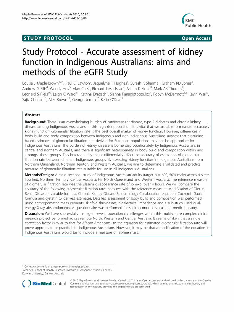
STUDY PROTOCOL Open Access Study Protocol - Accurate assessment of kidney function in Indigenous Australians: aims and methods of the eGFR Study Louise J Maple-Brown 1,2* , Paul D Lawton 2 , Jaquelyne T Hughes 1 , Suresh K Sharma 1 , Graham RD Jones 3 , Andrew G Ellis 4 , Wendy Hoy 5 , Alan Cass 6 , Richard J MacIsaac 7 , Ashim K Sinha 8 , Mark AB Thomas 9 , Leonard S Piers 10 , Leigh C Ward 11 , Katrina Drabsch 1 , Sianna Panagiotopoulos 7 , Robyn McDermott 12 , Kevin Warr 9 , Sajiv Cherian 13 , Alex Brown 14 , George Jerums 7 , Kerin O’Dea 12 Abstract Background: There is an overwhelming burden of cardiovascular disease, type 2 diabetes and chronic kidney disease among Indigenous Australians. In this high risk population, it is vital that we are able to measure accurately kidney function. Glomerular filtration rate is the best overall marker of kidney function. However, differences in body build and body composition between Indigenous and non-Indigenous Australians suggest that creatinine- based estimates of glomerular filtration rate derived for European populations may not be appropriate for Indigenous Australians. The burden of kidney disease is borne disproportionately by Indigenous Australians in central and northern Australia, and there is significant heterogeneity in body build and composition within and amongst these groups. This heterogeneity might differentially affect the accuracy of estimation of glomerular filtration rate between different Indigenous groups. By assessing kidney function in Indigenous Australians from Northern Queensland, Northern Territory and Western Australia, we aim to determine a validated and practical measure of glomerular filtration rate suitable for use in all Indigenous Australians. Methods/Design: A cross-sectional study of Indigenous Australian adults (target n = 600, 50% male) across 4 sites: Top End, Northern Territory; Central Australia; Far North Queensland and Western Australia. The reference measure of glomerular filtration rate was the plasma disappearance rate of iohexol over 4 hours. We will compare the accuracy of the following glomerular filtration rate measures with the reference measure: Modification of Diet in Renal Disease 4-variable formula, Chronic Kidney Disease Epidemiology Collaboration equation, Cockcroft-Gault formula and cystatin C- derived estimates. Detailed assessment of body build and composition was performed using anthropometric measurements, skinfold thicknesses, bioelectrical impedance and a sub-study used dual- energy X-ray absorptiometry. A questionnaire was performed for socio-economic status and medical history. Discussion: We have successfully managed several operational challenges within this multi-centre complex clinical research project performed across remote North, Western and Central Australia. It seems unlikely that a single correction factor (similar to that for African-Americans) to the equation for estimated glomerular filtration rate will prove appropriate or practical for Indigenous Australians. However, it may be that a modification of the equation in Indigenous Australians would be to include a measure of fat-free mass. * Correspondence: [email protected] 1 Menzies School of Health Research, Institute of Advanced Studies, Charles Darwin University, Darwin, Australia Maple-Brown et al. BMC Public Health 2010, 10:80 http://www.biomedcentral.com/1471-2458/10/80 © 2010 Maple-Brown et al; licensee BioMed Central Ltd. This is an Open Access article distributed under the terms of the Creative Commons Attribution License (http://creativecommons.org/licenses/by/2.0), which permits unrestricted use, distribution, and reproduction in any medium, provided the original work is properly cited.
-
Upload
independent -
Category
Documents
-
view
0 -
download
0
Transcript of Study Protocol-Accurate assessment of kidney function in Indigenous Australians: aims and methods of...
STUDY PROTOCOL Open Access
Study Protocol - Accurate assessment of kidneyfunction in Indigenous Australians: aims andmethods of the eGFR StudyLouise J Maple-Brown1,2*, Paul D Lawton2, Jaquelyne T Hughes1, Suresh K Sharma1, Graham RD Jones3,Andrew G Ellis4, Wendy Hoy5, Alan Cass6, Richard J MacIsaac7, Ashim K Sinha8, Mark AB Thomas9,Leonard S Piers10, Leigh C Ward11, Katrina Drabsch1, Sianna Panagiotopoulos7, Robyn McDermott12, Kevin Warr9,Sajiv Cherian13, Alex Brown14, George Jerums7, Kerin O’Dea12
Abstract
Background: There is an overwhelming burden of cardiovascular disease, type 2 diabetes and chronic kidneydisease among Indigenous Australians. In this high risk population, it is vital that we are able to measure accuratelykidney function. Glomerular filtration rate is the best overall marker of kidney function. However, differences inbody build and body composition between Indigenous and non-Indigenous Australians suggest that creatinine-based estimates of glomerular filtration rate derived for European populations may not be appropriate forIndigenous Australians. The burden of kidney disease is borne disproportionately by Indigenous Australians incentral and northern Australia, and there is significant heterogeneity in body build and composition within andamongst these groups. This heterogeneity might differentially affect the accuracy of estimation of glomerularfiltration rate between different Indigenous groups. By assessing kidney function in Indigenous Australians fromNorthern Queensland, Northern Territory and Western Australia, we aim to determine a validated and practicalmeasure of glomerular filtration rate suitable for use in all Indigenous Australians.
Methods/Design: A cross-sectional study of Indigenous Australian adults (target n = 600, 50% male) across 4 sites:Top End, Northern Territory; Central Australia; Far North Queensland and Western Australia. The reference measureof glomerular filtration rate was the plasma disappearance rate of iohexol over 4 hours. We will compare theaccuracy of the following glomerular filtration rate measures with the reference measure: Modification of Diet inRenal Disease 4-variable formula, Chronic Kidney Disease Epidemiology Collaboration equation, Cockcroft-Gaultformula and cystatin C- derived estimates. Detailed assessment of body build and composition was performedusing anthropometric measurements, skinfold thicknesses, bioelectrical impedance and a sub-study used dual-energy X-ray absorptiometry. A questionnaire was performed for socio-economic status and medical history.
Discussion: We have successfully managed several operational challenges within this multi-centre complex clinicalresearch project performed across remote North, Western and Central Australia. It seems unlikely that a singlecorrection factor (similar to that for African-Americans) to the equation for estimated glomerular filtration rate willprove appropriate or practical for Indigenous Australians. However, it may be that a modification of the equation inIndigenous Australians would be to include a measure of fat-free mass.
* Correspondence: [email protected] School of Health Research, Institute of Advanced Studies, CharlesDarwin University, Darwin, Australia
Maple-Brown et al. BMC Public Health 2010, 10:80http://www.biomedcentral.com/1471-2458/10/80
© 2010 Maple-Brown et al; licensee BioMed Central Ltd. This is an Open Access article distributed under the terms of the CreativeCommons Attribution License (http://creativecommons.org/licenses/by/2.0), which permits unrestricted use, distribution, andreproduction in any medium, provided the original work is properly cited.
BackgroundIndigenous Australians have rates of cardiovasculardisease (CVD) mortality some 7-10 times higher thannon-Indigenous Australians aged 25-64 years, a preva-lence of diabetes some 10 times higher (age 20-50years), new cases of end stage kidney disease (ESKD)10-15 times higher and life expectancy 15-20 yearsshorter [1-3]. Type 2 diabetes is common amongstIndigenous Australians with ESKD: 77% of IndigenousAustralians with new ESKD in 2007 compared to 33%of non-Indigenous Australians have Type 2 diabetes asa comorbidity [4]. Rates of ESKD in Indigenous Aus-tralians are disproportionately highest in those in cen-tral and northern Australia [5]. This group of people iswidely dispersed and heterogenous: they live in urban,regional and remote settings and have wide variationin diet, body habitus, genetic admixture and socioeco-nomic background [6-8].With the serious burden of ESKD in Indigenous Aus-
tralians it is essential to be able to assess accuratelyrenal function in these populations. Glomerular filtra-tion rate (GFR) is the best overall marker of renal func-tion in people with healthy or diseased kidneys [9] andis an independent predictor of renal and cardiovascularevents [10,11]. At present there are no validated meth-ods for estimating GFR in Indigenous Australians[12,13]. Differences in body build and body compositionbetween Indigenous and non-Indigenous Australianssuggest that creatinine-based estimates of GFR (eGFR)derived for European populations may not be appropri-ate for Indigenous Australians, as muscle mass is a keydeterminant of serum creatinine levels. Compared toAustralians of European background, Indigenous Aus-tralians from remote central and northern Australiahave more fat for a given weight or BMI [7].It is increasingly recognised that current creatinine-
based formulae used to estimate GFR might not be gen-eralisable across all clinical presentations [14,15]. Theyalso lack validation in ethnic populations apart fromCaucasians and African Americans [12,14]. The Modifi-cation of Diet in Renal Disease 4-variable formula(MDRD) was derived from a study population withimpaired renal function; and thus is less accurate withina “healthy range” GFR. To address this issue a revisedequation (Chronic Kidney Disease Epidemiology Colla-boration, CKD-EPI) has been described [16].An alternate approach is to use a different analyte
from creatinine to estimate renal function and developGFR prediction equations. Serum cystatin C has beenproposed as a simple, reliable and accurate marker ofGFR when compared to creatinine-based methods[17,18]. Although there is some evidence to suggest thatcystatin C levels might be influenced by extra-renal
factors, numerous studies have suggested that cystatin Clevels or GFR estimates based on cystatin C levels aloneare more accurate indices of renal function than creati-nine-based methods [19]. However, at this time cystatinC assays are not routinely available, they are consider-ably more expensive than creatinine assays and are notyet fully standardised. Thus for the immediate future,optimisation of creatinine-based methods remainsimportant.The aim of the present study is to evaluate and
improve if necessary the accuracy and precision of eGFRestimates in Indigenous Australians, taking into accountthe heterogeneity in body build and body compositionacross different populations. The study’s hypotheses are:(i) that differences in body build and body compositionbetween Indigenous and non-Indigenous Australianswill affect the validity of creatinine-based estimatedmeasures of GFR (using existing predictive equations)and that accuracy of estimates of GFR will vary acrossdifferent Indigenous population groups; and (ii) that amore accurate assessment of GFR will be obtained by aformula involving serum creatinine and percent fat freemass (obtained by a simple, validated bioimpedancemeasurement) or by using an alternative biochemicalmeasure such as cystatin C. It is anticipated that thiswill be of use in both Indigenous and non-IndigenousAustralians of variable body builds, particularly atGFR>60 ml/min/1.73 m2.
Methods/DesignSetting & locationParticipants were recruited from 4 geographical regions:Top End (of Northern Territory, Australia), CentralAustralia, Far North Queensland and Western Australia.These regions were chosen as resident Indigenous Aus-tralians bear a disproportionately larger burden of ESKDthan those from other regions of Australia. Within thesefour regions, the following Aboriginal Medical Servicesor health facilities were recruitment centres: Top End -Royal Darwin Hospital, Danila Dilba Health Services(Darwin), Bagot Community Health Centre (Darwin),Wurli Wurlinjang (Katherine), Ngalkunbuy Health Ser-vice (Miwatj Health Aboriginal Corporation, Galiwinku),Katherine District Hospital, Gove (Nhulunbuy) DistrictHospital; Central Australia - Alice Springs Hospital,Tennant Creek Hospital, Central Australian AboriginalCongress, Anyinginyi Health Aboriginal Corporation(Tennant Creek), Amooguna Health Centre, TitjikalaHealth Centre; Western Australia - Bega GarnbirringuHealth Service Aboriginal Corporation (Kalgoorlie),Kimberley Aboriginal Medical Services; Far NorthQueensland - Wuchopperen Health Service (Cairns),Thursday Island Hospital, Cairns Base Hospital Diabetes
Maple-Brown et al. BMC Public Health 2010, 10:80http://www.biomedcentral.com/1471-2458/10/80
Page 2 of 11
Centre, Meriba Dhoeynidhay Yabu Torres Strait &Northern Penninsula Area Community Council.
Participants and recruitmentParticipants were Indigenous Australians (target n = 600with equal numbers of males and females) aged 16 yearsand above, from urban, rural and remote regions ofNorthern and Central Australia across the following 5strata:
(i) “healthy” group: nil diabetes, hypertension,chronic kidney disease (CKD), albuminuria;
(ii) participants with diabetes or albuminuria &eGFR (MDRD-4) >90 ml/min/1.73 m2;
(iii) eGFR 60-90 ml/min/1.73 m2;(iv) eGFR 30-59 ml/min/1.73 m2;(v) eGFR <15-29 ml/min/1.73 m2.
Indigenous Australians fulfilled the definition of‘Aboriginal and/or Torres Strait Islander’ according tothe standard method used in National Census data col-lection: “1) is of Aboriginal and/or Torres Strait Islanderdescent; 2) identifies as an Australian Aboriginal and/orTorres Strait Islander; and 3) is accepted as such by thecommunity in which he or she lives or has lived”.Exclusion criteria were: participants with rapidly chan-
ging kidney function, participants receiving dialysis,women who were pregnant or breastfeeding and peoplewith a history of allergy or adverse reaction to iodine-based contrast media.Participants with CKD and/or diabetes were recruited
from participating Aboriginal Medical Services and thenephrology, endocrinology and outreach kidney diseaseclinics associated with Royal Darwin Hospital, AliceSprings Hospital, Cairns Base Hospital and the RoyalPerth Hospital. The identification and recruitment ofthe “healthy” group was through community networks,staff members of health services, word-of-mouth, selfreferral and family members of participants.
FundingMajor funding was provided by the National Health andMedical Research Council of Australia (NHMRC, pro-ject grant #545202). Prior to award of this grant, pilotfunding was received from: Kidney Health Australia, theColonial Foundation of Australia and Menzies School ofHealth Research. Additional funding for equipment wasreceived from Rebecca L Cooper Medical ResearchGrant.
EthicsThe study was approved by the joint Menzies School ofHealth Research - Northern Territory Department ofHealth and Community Services Human Research Ethics
Committee. The project was considered and approved byboth the Aboriginal sub-committee, which has absoluteright of veto, and by the main committee. The study wasalso approved by the following Human Research EthicsCommittees: Central Australian Human Research EthicsCommittee, Western Australian Aboriginal Health Infor-mation and Ethics Committee, Royal Perth HospitalEthics Committee and Cairns and Hinterland Health Ser-vices District Human Research Ethics Committee.
StaffSix people were employed on contracts over the courseof the study. Of these, four were Indigenous and twowere non-Indigenous. Additional people were employedon a casual basis during visits to discrete communities;these community members acted as local facilitators.One post-graduate research student (who is Indigenous)also participated in the study and was substantiallyinvolved in data collection, whilst also making a signifi-cant contribution to study profile and media relations.The majority of staff members had prior health qualifi-cations and experience: three were Aboriginal HealthWorkers, two were registered nurses and the post-grad-uate research student was a specialist nephrologist. Thestaff member without a health qualification completed avenipuncture certification course.
ConsentPotential participants were provided with written infor-mation about the study, face-to-face discussion with astaff member, and given an opportunity to ask ques-tions. If required, an interpreter was employed toexplain all information relating to the study. Those whoindicated that they wished to participate were thenasked to complete and sign a consent form. Consentwas obtained using the NHMRC Guidelines for EthicalConduct in Aboriginal and Torres Strait Islander HealthResearch. All participants were informed that their par-ticipation was voluntary and that they could refuse orwithdraw from participating and need give no reasonsnor justification for their decision and that it would notaffect their medical care. A parent or guardian wasasked to sign the form as well if the participant wasunder 18 years old. Participants were asked to give sepa-rate consent for various elements of the study, includ-ing: iohexol injection and blood and urine samples;body measurements, body composition tests, blood pres-sure and questionnaire; and collecting further informa-tion from their medical records. They were also askedwhether they wanted a report sent to their primaryhealth care provider and whether their blood and urinesamples could be stored at Menzies School of HealthResearch for 5 years and used for future studies relatedto diabetes and kidney disease.
Maple-Brown et al. BMC Public Health 2010, 10:80http://www.biomedcentral.com/1471-2458/10/80
Page 3 of 11
Examination protocolFollowing confirmation of contact details and eligibilityand after obtaining informed consent, performance ofthe reference measure of GFR and a health examinationwas undertaken. This examination included: collectionof blood and urine samples; clinical and anthropometricmeasurements; and the administration of questionnaires.The performance of the reference measurement of GFRrequired a minimum of 4 hours for participants, duringwhich time participants were given a healthy meal andhealth promotion information. Details of the examina-tion protocol:
(i) Reference measure of GFR: determined by mea-suring the renal clearance of non-isotopic iohexol.Iohexol was injected (5.445 ml, 300 mg/ml “Omni-paque”) into an antecubital vein (or forearm/handvein if unable to use antecubital vein) and flushedwith 10 ml of normal saline. The volume of iohexolincludes the prime volume (0.445 ml) in the tubingof the butterfly cannula used to inject the iohexol.Extravasation was assessed by clinical inspection andpalpation of the injection site. If there was any evi-dence of extravasation then the study was suspendedand the participant invited to return in 3-7 days forassessment. Using the contralateral arm, venousblood samples (5 ml) were collected for measure-ment of iohexol at 120, 180 and 240 min post injec-tion. Participants were closely supervised for possibleadverse reactions - whilst also receiving culturallyappropriate education about chronic kidney disease,diabetes and/or healthy lifestyle.Slope-intercept GFR was calculated from theformula:
Slope-intercept GFR (mL/min) = k × Iohexol dose(μg)/C0 (μg/mL) where k is the slope of the semilogplot of plasma Iohexol concentration versus time(plotted using 3 points, refer above), and C0 is the cal-culated Iohexol concentration at time zero (intercept).This value was multiplied by 1.73 and divided by thebody surface area (BSA, calculated from the equationBSA = 0.20247 × height (metres)0.725 × weight (kg)0.425
[20]. The BSA-slope intercept GFR value (mL/min/1.73m2) was corrected by the Brochner-Mortensen correc-tion factor = (0.990778 × GFR) - (0.001218 × GFR2)[21] providing a reference GFR value (mL/min/1.73 m2).
(ii) Blood and urine measures:a. Serum creatinine measurements: performedboth locally and at a central laboratory (TheDepartment of Laboratory Medicine, AustinHealth).
b. Estimates of GFR/creatinine clearance (whereScr is serum creatinine concentration in μmol/L,age in years, weight in kg, eGFR in mL/min/1.73m2):
i. The adjusted MDRD-4 variable formula(for creatinine measurements traceable to theIDMS method and reported in μmol/L)
eGFR = 175 × [(Scr × 0.0113)-1.154] × (age)-0.203 ×(0.742 if female) × (1.212 if African-American) [22]. Theabove African-American “correction factor” was notused for Indigenous Australians.
ii. The CKD-EPI formula [16]:
GFR = 141 × min(Scr × 0.0113/k, 1)a × max(Scr ×0.0113/k, 1)-1.209 × 0.993Age × 1.018 [if female] × 1.159[if black], where Scr is serum creatinine, k is 0.7 forfemales and 0.9 for males, a is -0.329 for females and-0.411 for males, min indicates the minimum of Scr/kor 1, and max indicates the maximum of Scr/k or 1. Asthe correction factor “if black” applies to African-Ameri-cans this was not used for Indigenous Australians.
iii. The Cockcroft-Gault (C-G) formula: [23]
Creatinine Clearance (mL/min) = { [140 - age] ×weight/[Scr × 0.814]} × (0.85 if female) *also calculatedusing ideal body weight (IBW) according to TherapeuticGuidelines protocol when the patient is obese (BMI>30kg/m2). IBW = 50 kg + 0.9 kg/each cm over 152 cm (-4.5 kg if female).
c. Cystatin C: non-fasting blood sample for mea-surement of cystatin C levels collected on themorning of iohexol clearance measurements.d. Bloods (non-fasting) were taken with the 120minute venous sample for iohexol: HbA1c, UEC,lipids (total and HDL cholesterol), high sensitiv-ity c-reactive protein were performed as standardclinical care and analysed by local care providers.Although a fasting blood sample would be pre-ferable for assessment of lipids and creatinine itwas not practical in the setting of a renal or dia-betes clinic. We therefore assessed non-fastingtotal and HDL cholesterol and instructed partici-pants not to consume meat on the day of theexamination prior to measurement of serumcreatinine. A subset of participants was askedwhen they last ate a meal containing meat. Thelight meal provided by study staff to participantswas given after the blood sample for creatininewas taken. Additional serum (8 ml) was collected
Maple-Brown et al. BMC Public Health 2010, 10:80http://www.biomedcentral.com/1471-2458/10/80
Page 4 of 11
at 120 minutes for cystatin C, inflammatory mar-kers and a stored sample.e. Urine: sample collected for microscopy andculture and determination of the albumin tocreatinine ratio (ACR), performed as standardclinical care and analysed by local care providers.Urine cotinine was measured for quantitativeassessment of cigarette smoking (recent nicotineconsumption).
(iii) Clinical and anthropometric measures:a. Blood pressure was measured three times afterthe participant had been seated quietly for atleast 5 minutes. A Welch Allyn Spot Vital Signsmonitor (Welch Allyn Medical Products, Skanea-teles Falls, USA) was used to measure systolicand diastolic pressure, with three minutesbetween readings. The pulse rate was recordedwith each reading.b. Height was measured to the nearest 0.1 cmusing a wall-mounted stadiometer [24]. Partici-pants were shoeless and wore light clothing.They were instructed to stand facing forwardwith weight distributed evenly on both feet, withheels together and against the wall and armshanging loosely by their sides. The participant’shead was positioned so that the Frankfort Planewas horizontal. The participant was theninstructed to keep his or her eyes focused on apoint straight ahead, breathe in deeply, andstretch to his or her fullest height, with heels stillon the ground. The measuring plate was thenlowered onto the scalp until it rested firmly onthe top of the head and a reading to the nearest0.1 cm was obtained and recorded. Following themeasurement of weight (as described below), asecond height measurement was obtained andrecorded, again to the nearest 0.1 cm. If the twoheight measurements differed by more than 0.5cm, a third measurement was taken andrecorded.c. Weight in kilograms was measured using aSeca digital portable scale (Model 767 and 841,Seca Deutschland, Hamburg, Germany). Partici-pants were asked to remove shoes, heavy gar-ments, heavy jewellery, belts, loose change, keys,mobile phones, and other items from pockets.With the scales at zero, participants were askedto stand on the centre of the base with feettogether, arms hanging loosely at the sides andthe head facing forward. Weight was recorded tothe nearest 0.1 kilogram. Two separate measure-ments of weight were recorded and if the firsttwo differed at all then a third measurement ofweight was taken and recorded.
d. Waist and hip circumference were measuredin centimetres using a 2-metre non-stretch flex-ible steel tape (Model W606PM Lufkin, Texas,USA). Following removal of outer clothing, tight-fitting garments, belts and heavy items frompockets, participants were asked to stand com-fortably erect in a relaxed manner, breathingnormally, with weight balanced evenly on bothfeet, feet about 25-30 cm apart, and arms foldedacross the chest. Waist and hip measurementswere taken alternately, with a minimum of twomeasurements for each. All measurements weretaken and recorded to the nearest 0.1 centimetre.If the first two measurements of either waist orhip circumference differed by more than 1.0 cm,then a third measurement was taken for waist orhip, as relevant. Waist measurements were takendirectly on the participant’s skin. Hip measure-ments were taken over the participant’s clothing,unless it was too tight or too baggy, in whichcase the participant was asked to remove it. Thewaist was defined as the midway point betweenthe iliac crest and the costal margin. These twolandmarks were identified and marked using afelt tip pen, and the distance between them mea-sured, with the midpoint marked. Once the tapewas around the participant’s body at the appro-priate height and the tape was horizontal, theparticipant was asked to breathe out gently. Themeasurement was taken at the end of a normalexpiration, with the tape pulled snug but notcompressing the underlying soft tissue. If land-marks were unable to be identified (in the occa-sional very obese participant), waistmeasurements were omitted. The hip circumfer-ence was defined as the widest circumferenceover the buttocks and below the iliac crest. Thehip circumference was measured at several posi-tions (starting with the widest part of the but-tocks when viewed from the side) and the widestcircumference recorded. Fatty aprons were notincluded in the measurement of the hips. Themeasurement was taken with the tape in a hori-zontal position, and the tape was pulled to allowit to maintain its position without causingindentation.e. Skinfold thickness assessment was performedusing Holtain calipers (Holtain Ltd, Crosswell,Pembrokeshire, United Kingdom) at 4 sites bilat-erally: peripheral (biceps, triceps) and central(subscapular, suprailiac) [25]. Bilateral skinfoldmeasurements were performed in all participantsto ensure a site availability of longitudinal followup in the event of an arterio-venous fistula
Maple-Brown et al. BMC Public Health 2010, 10:80http://www.biomedcentral.com/1471-2458/10/80
Page 5 of 11
insertion in CKD participants. The skinfold sitewas marked using a felt-tip pen and each sitewas identified as follows: biceps, anterior surfaceof the biceps midway between the acromial pro-cess and the elbow (antecubital fossa); triceps,posterior surface of triceps at midpoint from theacromial process (the most lateral and superioraspect of the acromial head) to the olecranon;subscapular, 2.5 cm infero-medially along a 45degree line from inferior angle of the scapula;suprailiac, 2.5 cm supero-medially along a 45degree line from where the medial border of theiliac crest tips in to the pelvis. The skin fold wasthen firmly grasped by the thumb and index fin-ger, using the pads at the tip of the thumb andfinger, and the skin fold gently pulled away fromthe body. The caliper was placed perpendicularto the fold, on the site marked, dialled up, atapproximately 1 cm below the finger and thumb.While maintaining the grasp of the skin fold, thecaliper was released so that full tension wasplaced on the skin fold. The dial was read to thenearest 0.20 mm, 2 seconds after the grip wasfully released. Three measurements were taken ateach site were possible.
(iv) Whole body and segmental bioelectrical impe-dance measurements (BIA) were performed at a sin-gle frequency (50 kHz) using a four-terminalimpedance plethysmograph (Model DF50, Impe-diMed, Brisbane). At study sites where a multi-fre-quency bioimpedance spectroscopy instrument (BIS,Model SFB7, ImpediMed, Brisbane) was available,this was used. BIS technology allowed prediction ofbody fluid volumes (intra- and extracellular water aswell as total body water) at the theoretically optimalfrequencies, providing precision and accuracy notachievable using the simpler BIA device that oper-ates at a single compromise frequency [26]. Predic-tion of body fluid volumes is particularly importantin this study population at all stages of CKD - fromhyperfiltration with “normal” renal function to ESKD(excluding dialysis). It is also worthy to note thatBIS provides measures of both intra-and extracellu-lar water spaces and since muscle mass is stronglyrelated to intracellular water space this will providethe opportunity to assess whether predictive powercan be increased by inclusion of this variable in GFRformulae.BIA/BIS was not performed if a participant had anindwelling electrical device (such as cardiac pace-maker). Participants were asked to void (empty blad-der) prior to the assessment. Assessment wasperformed after 5 minutes rest in the supine positionwith limbs abducted away from the trunk, ensuring
that thighs were not touching. Eight electrodes (elec-trocardiograph-style gel electrodes) were placed onbilateral dorsal surfaces of hands and feet (two elec-trodes on each hand and foot); after the surface wascleansed with an alcohol wipe. At the wrist, theproximal electrode was placed in the midline of theulnar styloid process and the distal electrode wasplaced 5 cm distal to the proximal electrode. At theankle, the proximal electrode was placed at the mid-point between the medial and lateral malleoli andthe distal electrode was placed 5 cm distal to theproximal electrode. Whole body and segmentalimpedance measurements were according to pre-viously described methods [27].(v) DXA Sub-study: Further assessment of bodycomposition by dual-energy X-ray absorptiometry(DXA) assessment of total body fat mass, lean mass,bone mineral content and bone density was per-formed according to standard techniques in siteswhere this imaging technique was available: Darwin,Perth and Torres Strait. DXA is a reference methodfor assessing body composition but impedancemethods, particularly inexpensive single frequencyBIA, are much more practical in remote locations(where DXA is indeed not possible/available).Hence, the aim of the sub-study comparing DXAand BIA was to see if BIA is a reasonable substitutefor DXA, thereby enabling body composition assess-ment in remote communities. In the present studyall participants had body composition measured byBIA or BIS and anthropometry, and a subset (Dar-win, Perth and Torres Strait) also had DXA mea-surements. The DXA data will be used to validatethe impedance data.(vi) Questionnaires:
a. Medical history & medications - self reportand from medical records (where available)including assessment of cigarette smoking: cur-rent status, past history and self-reportedquantity.b. Assessment of socio-economic status andrelated personal details by brief questionnaire:usual place of residence, first language, languagespoken at home, ethnicity of grandparents,income source, ever worked, education level,number of bedrooms and occupants in house,housing tenure and landlord type.
Feedback to participants was provided at the end ofthe examination. Brief intervention regarding diet, physi-cal activity and health behaviours in particular smoking,was provided. Participants with unexpected abnormalresults were referred immediately to the appropriatehealth care provider. In addition, written results were
Maple-Brown et al. BMC Public Health 2010, 10:80http://www.biomedcentral.com/1471-2458/10/80
Page 6 of 11
provided in a plain language and culturally appropriatemanner to individual participants and to their nomi-nated primary health care provider. Whenever possible,participants with abnormal results were first contactedby telephone or in person. The results packet was thenmailed or hand-delivered, as appropriate. A $50 giftvoucher to a local store was provided to participantswith their results as a gift of appreciation for their parti-cipation in the study. Participants were not informed ofthis gift prior to their participation in the study and itwas not used as an incentive to recruit participants tothe study. The gift was agreed upon by study investiga-tors after it was suggested by several key organisationsin our community consultation: Kimberley AboriginalMedical Services Council and Wuchopperen Health Ser-vice. Permission to give the gift was approved by allrelevant Human Research Ethics Committees.
Laboratory methodsCollected blood samples were centrifuged for 10 min at3000 revolutions per min (RPM) within 4 hours of col-lection. If unable to be centrifuged immediately, bloodwas stored in a cool environment (fridge or esky) untilcentrifugation occurred. Following centrifugation, sam-ples were transported on ice to be stored at -80 degreefreezer. For samples collected in remote locations, sto-rage for transportation was either on dry ice or in liquidnitrogen ("Biological Shipper”, CryoPak Series, Taylor-Wharton, AL, USA). Samples collected on ThursdayIsland, Queensland were stored at -30 degree for 2-14days prior to transportation on dry ice to a -80 degreefreezer in Cairns.Serum creatinine and other metabolite assays were
performed at each centre as part of standard clinicalcare. The creatinine assay at each centre was confirmedto have claimed traceability to the Isotope DilutionMass Spectrometry (IDMS) reference method. Themethod and instrument used for measurement of eachbiochemical measure performed at each site are outlinedin Table 1.In addition, serum creatinine will be measured in all
samples at Austin Health, using the kinetic Jaffe methodand the Roche enzymatic method on a Beckman-Coul-ter DxC 800 analyser (Fullerton, CA, USA). Beckman-Coulter claims traceability for its Jaffe methods to theIDMS reference method as it does for the Roche enzy-matic method. The Roche enzymatic method has beenused as a reference method for the revision of theMDRD formula for IDMS aligned assays [28] and hasundergone local independent validation [29]. Creatininewill be measured by both the Jaffe and enzymatic meth-ods as many laboratories in Australia measure creati-nine with a Jaffe method. This approach will allow for acalibration and standardization of the Jaffe methods
against the enzymatic method and assessment of varia-bility caused by the use of different routine methods.Cystatin C will be measured using an automated parti-cle-enhancing immunonephelometric assay on a BN IIinstrument (Dade Behring, Marburg, Germany). Theintra- and interassay CVs for cystatin C were 2.58 and3.95%, respectively, at a concentration of 1.54 mg/l.Cystatin C measures will be transformed to a GFRequivalent using the formua Cys-GFR = (86.7/cystatin C-4.2). This Cys-GFR method has been validated as anaccurate marker of renal function across a wide-rangeof GFR values using isotopic GFR measurements(plasma dissapearance of 99 mTc-DTPA) as the refer-ence method [18,30].Iohexol was measured by Austin Health, Melbourne
using a validated HPLC assay modified from Niculescu-Duvaz et al [31]. Plasma samples were extracted using asolution of 5% perchloric acid containing internal stan-dard (sodium diatrizoate). Chromatographic separationwas achieved using a Microsorb MV C18 5 μ column(Varian Australia) and detection at 244 nm. Analyteconcentrations were calculated by comparison to multi-point linear standard curves derived from plasma sam-ples spiked to contain between 3.125 and 800 μg/mLIohexol (reference material, US Pharmacopoeia). Thelower limit of quantitation was 3.125 μg/mL. Intra- andinter-assay performance was assessed using donorplasma spiked with low (20 μg/mL), medium (100 μg/mL) and high (400 μg/mL) concentrations of Iohexol.Precision studies showed a Coefficient of Variation of1.0% or less, and inaccuracy ± 4.5% or less.Samples were stored for analysis at a later date for:
adiponectin and urine cotinine. For participants of theDXA sub-study, samples have been stored for analysisof 25-hydoxy Vitamin D and parathyroid hormone andbone turnover markers.
Data handling and statistical methodsAt the time of baseline assessment, data was collectedon paper forms and then entered into a MicrosoftAccess database. Data from each site is stored securelyat Menzies School of Health Research. All statisticalanalysis will be performed using the latest availablerelease of Stata software (Stata Corporation, College Sta-tion, TX). The predictive performance of formulaemethods to estimate GFR will be compared to the refer-ence GFR in terms of precision, bias and accuracyaccording to the methods of Bland-Altman [32]. Thepercentage of estimated GFR within 30% of measuredGFR will also be determined. If required, new predictionequations will be developed in a “training sample”, withtheir veracity then being tested in a “validation” sample.This will be achieved by randomly dividing the studypopulation into two subgroups, balanced across the five
Maple-Brown et al. BMC Public Health 2010, 10:80http://www.biomedcentral.com/1471-2458/10/80
Page 7 of 11
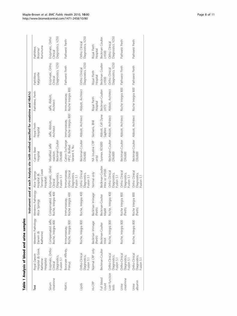
Table
1Analysis
ofblood
andurinesamples
Instrumen
tused
atea
chAna
lysissite
(withmetho
dspecified
forcrea
tinine
andHbA1c)
Test
RoyalD
arwin
Hospital(&Gove,
Katherine
Hospitals)
WesternsPatholog
yDarwin
(&Katherine)
WesternsPatholog
yAliceSprin
gsAliceSprin
gsHospital(&
Tenn
antCreek
Hospital)
CairnsBase
Hospital
RoyalP
erth
Hospital
PathWest,Perth
PathWest,
Kalgoo
rlie
PathWest,
Broo
me/
Kunu
nurra
Serum
creatin
ine
Enzymatic,O
rtho
-Clinical
Diagn
ostics,
Fusion
5.1
Com
pensated
Jaffe,
Roche,Integra800
Com
pensated
Jaffe,
Roche,Integra400
Enzymatic,O
rtho
Clinical
Diagn
ostics,
Fusion
5.1
Mod
ified
Jaffe
reactio
n.Beckman-Cou
lter
DXc600
Jaffe,A
bbott,
Architect
Jaffe,A
bbott,
Architect
Enzymatic,O
rtho
Clinical
Diagn
ostics,V250
Enzymatic,O
rtho
Clinical
Diagn
ostics,V250
HbA
1cBo
ronate
Affinity,
Prim
usIm
mun
oassay,
Roche,Integra800
Immun
oassay,
RocheIntegra400
Immun
oassay,
Ortho
Clinical
Diagn
ostics,
Fusion
5.1
CationExchange
HPLCBioR
adVariant
II,Nu
Immun
oassay,
RocheIntegra800
Immun
oassay,
RocheIntegra800
PathwestPerth
PathwestPerth
Lipids
Ortho
-Clinical
Diagn
ostics,
Fusion
5.1
Roche,Integra800
Roche,Integra400
Ortho
Clinical
Diagn
ostics,
Fusion
5.1
Beckman-Cou
lter
DXc600
Abb
ott,Architect
Abb
ott,Architect
Ortho
Clinical
Diagn
ostics,V250
Ortho
Clinical
Diagn
ostics,V250
Hs-CR
PNormalCRP
only
Beckman
Immage
(Perth)
Beckman
Immage
(Perth)
Normalon
lyNA(ro
utineCRP
only)
Siem
ens,BN
IIRo
yalP
erth
Hospital
RoyalP
erth
Hospital
RoyalP
erth
Hospital
Fullbloo
dcoun
tBeckman-Cou
lter
Beckman-Cou
lter
HmX
Beckman-Cou
lter
MaxM
Beckman-Cou
lter
Hmxor
LH500
Sysm
exXE5000
Abb
ott,CellD
yne
Saph
ireBeckman-Cou
lter
LH75
Beckman
Cou
lter
LH500
Beckman
Cou
lter
LH500
Liverfunctio
ntests
Ortho
-Clinical
Diagn
ostics,
Fusion
5.1
Roche,Integra800
Roche,Integra400
Ortho
Clinical
Diagn
ostics,
Fusion
5.1
Beckman-Cou
lter
DXc600
Abb
ott,Architect
Abb
ott,Architect
Ortho
Clinical
Diagn
ostics,V250
Ortho
Clinical
Diagn
ostics,V250
Urin
ecreatin
ine
Ortho
-Clinical
Diagn
ostics,
Fusion
5.1
Roche,Integra800
RocheIntegra800
(Perth)
Ortho
Clinical
Diagn
ostics,
Fusion
5.1
Beckman-Cou
lter
DXc600
Abb
ott,Architect
RocheIntegra800
PathwestPerth
PathwestPerth
Urin
ealbu
min
Ortho
-Clinical
Diagn
ostics,
Fusion
5.1
Roche,Integra800
RocheIntegra800
(Perth)
Ortho
Clinical
Diagn
ostics,
Fusion
5.1
Beckman-Cou
lter
DXc600
Abb
ott,Architect
RocheIntegra800
PathwestPerth
PathwestPerth
Maple-Brown et al. BMC Public Health 2010, 10:80http://www.biomedcentral.com/1471-2458/10/80
Page 8 of 11
strata. Sample size was determined using the methods ofBland-Altman [32,33] so that the limits of 95% confi-dence intervals for the mean were equal to pre-specifiedacceptability limits (a 10% difference in GFR was consid-ered to be acceptable). Sample size was calculated at 600Indigenous participants across 4 sites (approximately150 at each site), with considerable heterogeneity inbody builds and composition expected both within andbetween sites. Regression equations will be derivedusing a similar approach to that used to develop the ori-ginal MDRD equations [34]. In addition, for the bodycomposition substudy (DXA vs. BIA), we will be able todetect a difference ≥ 2% in fat free mass (FFM) esti-mated by the two methods, with a power >90% basedon a sample size of 300, alpha = 0.05 and a CV of 10%in FFM.Detailed analysis of demographic, clinical, biochemical
and socioeconomic factors associated with CKD willinvolve cross-sectional analysis of the associationsbetween these factors and current GFR. After determin-ing univariate associations between variables, establishedrisk factors and variables with p < 0.2 on univariate ana-lysis will be selected for entry into logistic regressionmodels using the backwards selection method, the out-come variable being current GFR/CKD stage. Thedegree of coupling of GFR and albuminuria will bedetermined in participants with type 2 diabetes (afteraccounting for the use of inhibitors of the renin-angio-tensin system). We have recently shown that there is arelatively high prevalence of non-albuminuric renalinsufficiency in non-Indigenous participants with type 2diabetes [35] and preliminary evidence suggests that theprevalence of non-albuminuric renal insufficiency islower in Indigenous than non-Indigenous diabetic Aus-tralians [36].
DiscussionThis study was designed with respect to what is practi-cal and achievable in very remote regions of Northern,Western and Central Australia. The choice of referencemeasure of GFR was limited by our remote setting andthe available time of participants. Nuclear medicinefacilities are not available in the vast majority of siteswhere this project was conducted. Study investigatorsagreed that 4-hour plasma disappearance of iohexol wasthe most appropriate reference measure of GFR for theremote setting. The benefits of this reference measureare that it is practical, safe, stable and reproducible[37,38]. In addition, several investigators had experiencewith use of this measure in the non-Indigenous urbansetting (Melbourne) with accurate comparison to 99mTc-DTPA in participants with diabetes [39]. Based onprevious experience of investigators in Indigenous healthresearch, we agreed that a reference measure that
required more than 4 hours of participants time wouldnot be practical or achievable.A potential limitation of our study is the use of
Iohexol clearance from serum as the formal GFR mea-surement as opposed to the use of urinary clearance ofiothalamate as the reference method used in the MDRDand CKD-EPI studies [16,34]. Iohexol serum clearancewas however used in two studies used in the CKD-EPIvalidation group [40,41] however the limitation of 4hours for the studies has the potential to increase inac-curacy in subjects with reduced GFR [40].The study has encountered several operational chal-
lenges to date that have been successfully managed. Thefirst challenge is the significant labour intensity requiredto recruit each participant and the associated expenses.The proportion of eligible participants approached bythe study team, who subsequently consented to and suc-cessfully completed the study, varied by site fromapproximately 30% in urban centres to up to 70% insome remote communities. We managed this challengeby adapting recruitment sites to those that had a highersuccess rate of obtaining complete data sets and expand-ing networks and recruitment sites beyond health ser-vice providers, e.g. Aboriginal Hostels Limited (providetemporary and transit accommodation in urban andregional centres).The second challenge encountered by the eGFR study
team was to recruit a diverse sample of participantsincluding people living remotely and with poor accessto regular medical services. We managed this challengeby: targeting a range of communities or regions; target-ing remote-living individuals whilst they were visitingregional centres (Darwin, Katherine and Nhulunbuy:health and non-health service sites); and selection ofparticipants, stratified by study design, using electronichealth record data held by urban and remote healthservices.The third challenge was to conduct recruitment in
such a way to facilitate complete data collection, in par-ticular complete 4 hour reference GFR measures. Wecommenced recruitment of participants in associationwith outpatient renal clinics (urban and regional). In theregional setting other commitments for remote partici-pants within the 4-hour duration of the study resultedin non-completion of the reference GFR measures. Rea-sons included the need to catch transport home (usuallya plane) or logistic constraints such as inclementweather conditions. The study recruitment strategy wassubsequently changed so that the eGFR study team tra-velled to remote communities to perform the study in“batches” of 1-3 weeks duration. This strategy wassuperior in terms of numbers of participants recruitedand proportion of participants with complete 4 hourreference GFR measures.
Maple-Brown et al. BMC Public Health 2010, 10:80http://www.biomedcentral.com/1471-2458/10/80
Page 9 of 11
This study should provide evidence for clinical guide-lines in a field where evidence is currently lacking: avalidated methodology to accurately assess renal func-tion in diverse populations of Indigenous Australians.We expect to determine a validated and practical mea-sure of GFR suitable for use in all Indigenous Austra-lians. This may be cystatin C or eGFR (using MDRD orCKD-EPI formula with/without suitable revision). Thismeasure would enable development of appropriate pro-tocols for investigation and management of CKD andsubsequent improvements in clinical practice. Webelieve that the MDRD- or CKD-EPI derived method(modified as appropriate) is likely to emerge as thecheapest and most widely assessable means of estimat-ing GFR in the immediate future. It seems unlikely thata single correction factor (similar to that for Afro-Amer-icans) is appropriate or practical for Indigenous Austra-lians. However, it may be that a modification of theMDRD or CKD-EPI equation for Indigenous Australians(or for any population) would be to include a measureof fat-free mass. This measure may be obtained by mea-suring body composition using a simple, portable andinexpensive method such as BIA, once it has been suita-bly validated against DXA. Thereby this study mayimprove the utility of eGFR, using a modified MDRD orCKD-EPI formula, across different populations with dif-ferent body builds and compositions. Our revised for-mula for eGFR may indeed be of use for assessingkidney function in the broader Australian community,particularly those of variable body builds and/oreGFR>60 ml/min/1.73 m2.
AcknowledgementsThe authors gratefully acknowledge the support of eGFR study participants,study staff, and partner organisations. The eGFR Study was funded by theNational Health and Medical Research Council of Australia (NHMRC, ProjectGrant #545202), with additional support from Kidney Health Australia,Colonial Foundation, Rebecca L Cooper Foundation and SeaSwift, ThursdayIsland. LMB was supported by NHMRC Program Grant #320860 and theCentre of Clinical Research Excellence in Clinical Science in Diabetes,University of Melbourne. Alan Cass holds a Senior Research Fellowship fromthe NHMRC. Funding bodies had no role in the study design, in thecollection, analysis or interpretation of data, in the writing of the manuscriptor the decision to submit the manuscript for publication.
Author details1Menzies School of Health Research, Institute of Advanced Studies, CharlesDarwin University, Darwin, Australia. 2Division of Medicine, Royal DarwinHospital, Darwin, Australia. 3Chemical Pathology, St Vincent’s Hospital,Sydney, Australia. 4University of Melbourne, Department of Medicine, Austinand Northern Health, Heidelberg, Victoria, Australia. 5Centre for ChronicDisease, The University of Queensland, Australia. 6The George Institute forInternational Health, University of Sydney, Sydney, Australia. 7Department ofEndocrinology, Endocrine Centre Austin Health & University of Melbourne,Heidelberg Repatriation Hospital, Heidelberg West, Victoria, Australia.8Endocrine and Diabetes Unit, Cairns Base Hospital, Queensland, Australia.9Department of Nephrology, Royal Perth Hospital, Perth, Australia. 10Centrefor Health and Society, School of Population Health, University of Melbourne,Melbourne, Australia. 11School of Chemistry and Molecular Biosciences, TheUniversity of Queensland, Australia. 12Sansom Institute for Health Research,
UniSA, Adelaide, Australia. 13Department of Renal Medicine, Alice SpringsHospital, Alice Springs, Australia. 14Baker IDI Heart and Diabetes Institute,Alice Springs, Australia.
Authors’ contributionsLMB drove the design of the study protocol for funding and Ethicsapplications, coordinated data collection and data management, analysedthe data and drafted the manuscript. PL, RM, GJe, AS, KOD conceived of thestudy and participated in its design. SS was the study manager andparticipated in study design, ethics applications, data collection and datamanagement. JH participated in study design, data collection and datamanagement. GJo, AE, WH, AC participated in study design. MT, KW, CSparticipated in the design of the study and provided clinical expertiseduring the design and data collection phases. LP, LW, RMcD, AB participatedin study design. All authors were involved in revising the manuscript forimportant intellectual content and read and approved the final manuscript.
Competing interestsLCW has consults to Impedimed Ltd. Impedimed Ltd. had no involvement,financial or otherwise, in the conception and execution of this study or inthe preparation of the manuscript.All remaining authors declare that they have no competing interests.
Received: 14 January 2010Accepted: 19 February 2010 Published: 19 February 2010
References1. Zhao Y, Dempsey K: Causes of inequality in life expectancy
between Indigenous and non-Indigenous people in the NorthernTerritory, 1981-2000: a decomposition analysis. Med J Aust 2006,184:490-494.
2. Heart, stroke and vascular disease: Australian facts 2004. CardiovascularDisease Series, No. 22. Canberra, Australian Institute of Health and Welfareand National Heart Foundation of Australia 2004.
3. O’Dea K, Patel M, Kubisch D, Hopper J, Traianedes K: Obesity, diabetes,and hyperlipidemia in a central Australian aboriginal community with along history of acculturation. Diabetes Care 1993, 16:1004-1010.
4. ANZDATA Registry Report: Adelaide, Australia and New Zealand Dialysis andTransplant RegistryMcDonald S, Excell L, Livingston B 2009.
5. Cass A, Cunningham J, Wang Z, Hoy W: Regional variation in theincidence of end-stage renal disease in Indigenous Australians. Med JAust 2001, 175:24-27.
6. Rutishauser IH: Body composition in Aboriginal Australians. Asia Pac J ClinNutr 1995, 4:73-76.
7. Piers LS, Rowley KG, Soares MJ, O’Dea K: Relation of adiposity and bodyfat distribution to body mass index in Australians of Aboriginal andEuropean ancestry. Eur J Clin Nutr 2003, 57:956-963.
8. Cass A, Cunningham J, Snelling P, Wang Z, Hoy W: End-stage renal diseasein indigenous Australians: a disease of disadvantage. Ethn Dis 2002,12:373-378.
9. K/DOQI clinical practice guidelines for chronic kidney disease:evaluation, classification, and stratification. Am J Kidney Dis 2005, 39:S1-266.
10. Ninomiya T, Perkovic V, de Galan BE, Zoungas S, Pillai A, Jardine M, Patel A,Cass A, Neal B, Poulter N, Mogensen CE, Cooper M, Marre M, Williams B,Hamet P, Mancia G, Woodward M, Macmahon S, Chalmers J: Albuminuriaand kidney function independently predict cardiovascular and renaloutcomes in diabetes. J Am Soc Nephrol 2009, 20:1813-1821.
11. Astor BC, Hallan SI, Miller ER, Yeung E, Coresh J: Glomerular filtration rate,albuminuria, and risk of cardiovascular and all-cause mortality in the USpopulation. Am J Epidemiol 2008, 167:1226-1234.
12. Mathew TH: Chronic kidney disease and automatic reporting ofestimated glomerular filtration rate: a position statement. Med J Aust2005, 183:138-141.
13. Jose MD, Lawton PD: Chronic kidney disease and automatic reporting ofestimated glomerular filtration rate. Med J Aust 2006, 184:42.
14. Zuo L, Ma YC, Zhou YH, Wang M, Xu GB, Wang HY: Application of GFR-estimating equations in Chinese patients with chronic kidney disease.Am J Kidney Dis 2005, 45:463-472.
15. Froissart M, Rossert J, Jacquot C, Paillard M, Houillier P: PredictivePerformance of the Modification of Diet in Renal Disease and
Maple-Brown et al. BMC Public Health 2010, 10:80http://www.biomedcentral.com/1471-2458/10/80
Page 10 of 11
Cockcroft-Gault Equations for Estimating Renal Function. J Am SocNephrol 2005, 16:763-773.
16. Levey AS, Stevens LA, Schmid CH, Zhang YL, Castro AF, Feldman HI,Kusek JW, Eggers P, Van Lente F, Greene T, Coresh J: A new equation toestimate glomerular filtration rate. Ann Intern Med 2009, 150:604-612.
17. Dharnidharka VR, Kwon C, Stevens G: Serum cystatin C is superior toserum creatinine as a marker of kidney function: A meta-analysis. Am JKidney Dis 2002, 40:221-226.
18. MacIsaac RJ, Tsalamandris C, Thomas MC, Premaratne E, Panagiotopoulos S,Smith TJ, Poon A, Jenkins MA, Ratnaike SI, Power DA, Jerums G: Estimatingglomerular filtration rate in diabetes: a comparison of cystatin-C- andcreatinine-based methods. Diabetologia 2006, V49:1686-1689.
19. Madero M, Sarnak MJ, Stevens LA: Serum cystatin C as a marker ofglomerular filtration rate. Curr Opin Nephrol Hypertens 2006, 15:610-616.
20. DuBois D, DuBois EF: A formula to estimate the approximate surface areaif height and weight be known. Arch Int Med 1916, 17:863-871.
21. Brochner-Mortensen J: A simple method for the determination ofglomerular filtration rate. Scand J Clin Lab Invest 1972, 30:271-274.
22. Mathew TH, Johnson DW, Jones GR: Chronic kidney disease andautomatic reporting of estimated glomerular filtration rate: revisedrecommendations. Med J Aust 2007, 187:459-463.
23. Cockcroft DW, Gault MH: Prediction of creatinine clearance from serumcreatinine. Nephron 1976, 16:31-41.
24. Norton K, Olds T: Anthopometrica UNSW Press 1996.25. Lohman TG, Roche AF, Martorell R: Anthropometric standardization reference
manual Human Kinetics Books 1988.26. Thomas BJ, Ward LC, Cornish BH: Bioimpedance spectrometry in the
determination of body water compartments: accuracy and clinicalsignificance. Appl Radiat Isot 1998, 49:447-455.
27. Cornish BH, Jacobs A, Thomas BJ, Ward LC: Optimizing electrode sites forsegmental bioimpedance measurements. Physiol Meas 1999, 20:241-250.
28. Levey AS, Coresh J, Greene T, Marsh J, Stevens LA, Kusek JW, Van Lente F:Expressing the Modification of Diet in Renal Disease Study equation forestimating glomerular filtration rate with standardized serum creatininevalues. Clin Chem 2007, 53:766-772.
29. Peake M, Whiting M: Measurement of serum creatinine - current statusand future goals. Clin Biochem Rev 2006, 27:173-184.
30. Premaratne E, MacIsaac RJ, Finch S, Panagiotopoulos S, Ekinci E, Jerums G:Serial measurements of cystatin C are more accurate than creatinine-based methods in detecting declining renal function in type 1 diabetes.Diabetes Care 2008, 31:971-973.
31. Niculescu-Duvaz I, D’Mello L, Maan Z, Barron JL, Newman DJ, Dockrell MEC,Kwan JTC: Development of an outpatient finger-prick glomerularfiltration rate procedure suitable for epidemiological studies. Kidney Int2006, 69:1272-1275.
32. Bland JM, Altman DG: Statistical methods for assessing agreementbetween two methods of clinical measurement. Lancet 1986, 1:307-310.
33. Altman DG, Bland JM: Measurement in Medicine: the Analysis of MethodComparison Studies. The Statistician 1983, 32:307-317.
34. Levey AS, Bosch JP, Lewis JB, Greene T, Rogers N, Roth D: A more accuratemethod to estimate glomerular filtration rate from serum creatinine: anew prediction equation. Modification of Diet in Renal Disease StudyGroup. Ann Intern Med 1999, 130:461-470.
35. MacIsaac RJ, Tsalamandris C, Panagiotopoulos S, Smith TJ, McNeil KJ,Jerums G: Nonalbuminuric renal insufficiency in type 2 diabetes. DiabetesCare 2004, 27:195-200.
36. Thomas MC, Weekes AJ, Broadley OJ, Cooper ME: The assessment ofkidney function by general practitioners in Australian patients with type2 diabetes (NEFRON-2). Med J Aust 2006, 185:259-262.
37. Brown SC, O’Reilly PH: Iohexol clearance for the determination ofglomerular filtration rate in clinical practice: evidence for a new goldstandard. J Urol 1991, 146:675-679.
38. Gaspari F, Perico N, Ruggenenti P, Mosconi L, Amuchastegui CS, Guerini E,Daina E, Remuzzi G: Plasma clearance of nonradioactive iohexol as ameasure of glomerular filtration rate. J Am Soc Nephrol 1995, 6:257-263.
39. Houlihan C, Jenkins M, Osicka T, Scott A, Parkin D, Jerums G: A comparisonof the plasma disappearance of iohexol and 99 mTc-DTPA for themeasurement of glomerular filtration rate (GFR) in diabetes. Aust N Z JMed 1999, 29:693-700.
40. Grubb A, Nyman U, Bjork J, Lindstrom V, Rippe B, Sterner G, Christensson A:Simple cystatin C-based prediction equations for glomerular filtration
rate compared with the modification of diet in renal disease predictionequation for adults and the Schwartz and the Counahan-Barrattprediction equations for children. Clin Chem 2005, 51:1420-1431.
41. Mauer M, Zinman B, Gardiner R, Drummond KN, Suissa S, Donnelly SM,Strand TD, Kramer MS, Klein R, Sinaiko AR: ACE-I and ARBs in earlydiabetic nephropathy. J Renin Angiotensin Aldosterone Syst 2002, 3:262-269.
Pre-publication historyThe pre-publication history for this paper can be accessed here:http://www.biomedcentral.com/1471-2458/10/80/prepub
doi:10.1186/1471-2458-10-80Cite this article as: Maple-Brown et al.: Study Protocol - Accurateassessment of kidney function in Indigenous Australians: aims andmethods of the eGFR Study. BMC Public Health 2010 10:80.
Submit your next manuscript to BioMed Centraland take full advantage of:
• Convenient online submission
• Thorough peer review
• No space constraints or color figure charges
• Immediate publication on acceptance
• Inclusion in PubMed, CAS, Scopus and Google Scholar
• Research which is freely available for redistribution
Submit your manuscript at www.biomedcentral.com/submit
Maple-Brown et al. BMC Public Health 2010, 10:80http://www.biomedcentral.com/1471-2458/10/80
Page 11 of 11