Micropropagation of Cynara scolymus L. employing cyclodextrins to promote rhizogenesis
Upload
khangminh22Category
view
1download
0
STUDIES ON MICROPROPAGATION, CELLULAR BEHAVIOR, PHOTOSYNTHETIC AND BIOLOGICAL
ACTIVITIES OF RED CLOVER (TRIFOLIUM PRATENSE L.)
ARASH KHORASANI ESMAEILI
FACULTY OF SCIENCE UNIVERSITY OF MALAYA
KUALA LUMPUR
2016
Univers
ity of
Mala
ya

ARASH KHORASANI ESMAEILI
THESIS SUBMITTED IN FULFILMENT OF THE
REQUIREMENTS FOR THE DEGREE OF DOCTOR OF
PHILOSOPHY
FACULTY OF SCIENCE
UNIVERSITY OF MALAYA
KUALA LUMPUR
2016
Univers
ity of
Mala
ya

ii
UNIVERSITY OF MALAYA
ORIGINAL LITERARY WORK DECLARATION
Name of Candidate: Arash Khorasani Esmaeili
Registration/Matric No: SHC120005
Name of Degree: Doctor of Philosophy
Title of Project Paper/Research Report/Dissertation/Thesis (“this Work”): Study on
Micropropagation, Cellular Behavior, Photosynthetic and Biological Activities of
Red Clover (Trifolium pratense L.)
Field of Study: Plant Biotechnology
I do solemnly and sincerely declare that:
(1) I am the sole author/writer of this Work;
(2) This Work is original;
(3) Any use of any work in which copyright exists was done by way of fair dealing
and for permitted purposes and any excerpt or extract from, or reference to or
reproduction of any copyright work has been disclosed expressly and
sufficiently and the title of the Work and its authorship have been
acknowledged in this Work;
(4) I do not have any actual knowledge nor do I ought reasonably to know that the
making of this work constitutes an infringement of any copyright work;
(5) I hereby assign all and every rights in the copyright to this Work to the
University of Malaya (“UM”), who henceforth shall be owner of the copyright
in this Work and that any reproduction or use in any form or by any means
whatsoever is prohibited without the written consent of UM having been first
had and obtained;
(6) I am fully aware that if in the course of making this Work I have infringed any
copyright whether intentionally or otherwise, I may be subject to legal action
or any other action as may be determined by UM.
Candidate’s Signature Date:
Subscribed and solemnly declared before,
Witness’s Signature Date:
Name:
Designation:
Univers
ity of
Mala
ya

iii
ABSTRACT
Tissue culture studies of a temperate forage crop, Trifolium pratense L. were
investigated in the current project. In vitro regeneration of this species was successfully
achieved in this study using nodal explants cultured on Murashige and Skoog (MS) media
supplemented with different hormones at various concentrations and also on MS hormone
free media as a control. Complete plant regeneration of T. pratense was best achieved
when the nodal explants were cultured on MS media supplemented with 1.5 mg/l BAP
and 0.5 mg/l IBA, with mean number of 6.05 ± 0.28 shoots per explant, and 100% of the
explant samples produced shoots. On the other hand, the best root formation was obtained
on MS media supplemented with 1.5 mg/l BAP and 0.75 mg/l IBA, with the mean number
of 3.3 ± 0.21 roots per explant. However, the nodal explants cultured on MS hormone
free medium failed to produce any shoots or roots. Callus formation was successfully
achieved when the nodal explants were cultured on MS medium containing different
types of plant hormones. MS medium supplemented with 1.5 mg/l BAP and 0.5 mg/l 2,4-
D was the most responsive, whereby 100% of the explants managed to produce callus.
Adaptation process to the natural environment or acclimatization, i,e. the transfer of in
vitro grown plants to the ex vitro condition was successfully undertaken, with very high
survival rates of plantlets (93.71 ± 4.64 %) when they were transferred to the combination
of red soil and black soil with the ratio of 1:1.
Subsequently, the extracts of in vivo and in vitro grown plants as well as callus tissues
of T. pratense were tested for their antioxidant activities, using different extraction
solvents and different antioxidant assays. The total flavonoid and phenolic contents as
well as extraction yield of the extracts were also investigated to determine their
correlation with the antioxidant activity of the extracts. Among all the tested extracts, the
highest amount of total phenolic and total flavonoids content were found in methanol
Univers
ity of
Mala
ya

iv
extract from in vivo grown plants. The antioxidant activity of tested samples followed the
order; in vivo plant extract ˃ callus extract ˃ in vitro extract. The highest reducing power,
2,2-azino-bis-(3-ethylbenzothiazoline-6-sulphonic acid) (ABTS) radical scavenging and
chelating power were found in methanol extracts of in vivo grown T. pratense. Whilst the
chloroform fraction of in vivo grown plants showed the highest 2,2-diphenyl-1-
picrylhydrazyl (DPPH) radical scavenging, superoxide anion radical scavenging and
hydrogen peroxide scavenging compared to the other tested extracts. A significant
correlation was found between the antioxidant activity of extracts and their total phenolic
and total flavonoid content.
The cytotoxicity of the plant extracts were examined against two human cancer cell
lines (human breast carcinoma (MCF-7) and human colon carcinoma (HCT-116)), using
MTT assay. Four different extraction solvents were used to examine the effect of the
solvent on cytotoxic activity of the extracts. Two cancer cell lines were treated with the
extracts for 24, 48 and 72 hours. All of the examined extracts exhibited toxicity on the
tested cell lines in a time dependent increase, but in a lower potency than doxorubicin
(positive control). The chloroform fraction of in vivo grown plants showed the highest
cytotoxic activity against the MCF-7 cell line (IC50 = 66.44 ± 2.05 µg/ml) but it was not
significantly different with the cytotoxic activity of chloroform fraction of callus tissue
(IC50 = 69.48 ± 2.66 µg/ml). The highest cytotoxic activity against the HCT-116 cell line
was shown by the chloroform fraction of callus tissue (IC50 = 79.53 ± 2.00 µg/ml).
The antimicrobial efficiency of extract derived from T. pratense (in vivo and in vitro
grown plants, including callus) were examined using ethanol and methanol as solvents
for extraction and tested against four bacterial pathogens (two gram negative and two
gram positive) and three fungal pathogens. The antimicrobial activity of the methanol
extract was found to give higher inhibition zone when compared with ethanol extract.
Univers
ity of
Mala
ya

v
Among the callus, in vitro and in vivo grown plants, the callus extract showed better
antimicrobial activity, thus revealing a new potential use of T. pratense callus.
To compare the photosynthetic parameters, the Stomatal conductance (gs),
Transpiration rate (E) and Net photosynthetic (Pn) were determined for the plants grown
under in vitro and in vivo conditions. A comparison was made for the observed data for
the light-saturated photosynthetic among the treatments which revealed that the
maximum photosynthetic rate (PNmax) was 18.3 and 11.3 µmol (CO2)/m2/s in in vivo and
in vitro plant leaves, respectively. Respiration (Rd) and Compensation point (CP) were
found 1.5-folds and two-folds higher in in vitro plants, respectively. On the other hand,
the in vitro grown plants exhibited higher transpiration rate and also higher stomatal
conductance compared with the in vivo plants. Consequently, high levels of
differentiation in terms of photosynthesis parameters exist among the in vivo and in vitro
samples. Significant direct relation was observed between net photosynthetic rate and
total phenolic and flavonoid content of T. pratense leaves.
The effect of optimal and supra-optimal concentrations of Sodium chloride (NaCl) on
growth and antioxidant defence was also studied in the in vitro cultures of T. pretence.
Seeds of T. pratense were germinated in Murashige and Skoog medium (MS) containing
different concentrations of NaCl (0, 50, 100, 150, 200 mM). The lengths of roots and
shoots as well as percentage of germination, free radical scavenging activity (DPPH) and
Superoxide dismutase (SOD) were measured. A significant decrease in germination and
growth was observed in the seeds exposed to 100, 150 and 200 mM salt. The highest
percentage of germination was found in the MS medium containing 50 mM NaCl,
although the highest root and shoot length were found in MS medium without NaCl. The
highest antioxidant activity of methanol extract of the plants occurred in in vitro plants
cultured in MS medium supplemented with 50 mM NaCl. A significant decrease in free
Univers
ity of
Mala
ya

vi
radicals scavenging and superoxide dismutase activities were found in plants grown in
media containing 100, 150 and 200 mM salt.
Univers
ity of
Mala
ya
vii
ABSTRAK
Kajian kultur tisu ke atas tumbuhan temperat untuk makanan haiwan ternakan iaitu
Trifolium pratense L., telah dikaji dalam projek ini. Regenerasi secara in vitro telah
berjaya dicapai menggunakan eksplan batang bernod yang dikultur dalam media
Murashige dan Skoong (MS) yang ditambah dengan pelbagai jenis hormon dan
kepekatan, dan juga ke atas media MS tanpa hormon sebagai kawalan. Regenerasi
tumbuhan lengkap yang terbaik telah dicapai apabila eksplan batang bernod dikultur ke
atas media MS yang ditambah dengan 1.5 mg/l BAP dan 0.5 mg/l IBA, dengan purata
bilangan pucuk 6.05 ± 0.28 per eksplan di mana 100% eksplan menghasilkan pucuk.
Walaubagaimanapun, eksplan batang bernod yang dikultur ke atas media MS tanpa
hormon gagal menghasilkan sebarang pucuk dan akar. Pembentukan kalus telah berjaya
dicapai apabila eksplan batang bernod dikultur ke atas media MS yang mengandungi
pelbagai jenis hormon tumbuhan. Media MS yang ditambah dengan 1.5 mg/l BAP dan
0.5 mg/l 2,4-D adalah yang paling responsif di mana 100% eksplan berjaya menghasilkan
kalus.
Proses penyesuaian kepada persekitaran semulajadi ataupun acclimatization iaitu
pemindahan tumbuhan in vitro kepada keadaan ex vitro telah berjaya dilakukan dengan
kadar hidup plantlet yang tinggi (93.71 ± 4.64 %) apabila plantlet dipindahkan kepada
kombinasi tanah merah dan tanah hitam dengan nisbah 1:1. Seterusnya, aktiviti
antioksidan di dalam ekstrak tumbuhan in vivo dan in vitro dan juga tisu kalus T. pratense
dikaji dengan menggunakan pelarut pengekstrakan dan ujian antioksidan yang berlainan.
Kandungan flavonoid, fenolik dan juga hasil pengekstrakan juga dikaji untuk menentukan
hubungan aktiviti antioksidannya. Di antara keseluruhan ekstrak yang dikaji, ekstrak
metanol daripada tumbuhan in vivo mengandungi kandungan flavonoid dan fenolik
tertinggi. Aktiviti antioksidan sampel-sampel kajian adalah mengikut urutan berikut,
ekstrak tumbuhan in vivo > ekstrak kalus > ekstrak in vitro. Kemampuan memerangkap
Univers
ity of
Mala
ya
viii
radikal 2,2-azino-bis-(3-ethylbenzothiazoline-6-sulfonik asid) (ABTS) dan
kemampuannya mengkelat logam adalah yang tertinggi di dalam ekstrak metanol
tumbuhan in vivo. Sementara itu, pecahan kloroform tumbuhan in vivo menunjukkan
kemampuan memerangkap radikal 2,2-diphenyl-1-picrylhydrazyl (DPPH), kemampuan
memerangkap radikal superoksida anion dan kemampuan memerangkap radikal hidrogen
peroksida yang tertinggi berbanding ekstrak lain. Hubungan yang signifikan telah ditemui
di antara aktiviti antioksidan ekstrak dan jumlah kandungan flavonoid dan fenolik.
Sitotoksisiti ekstrak tumbuhan telah diuji ke atas sel kanser manusia (sel karsinoma
payudara manusia (MCF-7) dan sel karsinoma kolon manusia (HCT-116), menggunakan
ujian MTT. Empat pelarut pengekstrakan yang berlainan telah digunakan untuk mengkaji
kesan pelarut ke atas aktiviti sitotoksisiti ekstrak. Dua jenis sel kanser telah dirawat
dengan ekstrak tumbuhan selama 24, 48 dan 72 jam. Kesemua ekstrak yang dikaji
menunjukkan toksisiti ke atas sel kanser mengikut pertambahan masa, tetapi dalam
potensi yang rendah berbanding doxorubicin (kawalan positif). Pecahan kloroform
tumbuhan in vivo menunjukkan aktiviti toksisiti yang tertinggi terhadap sel MCF-7 (IC50
= 66.44 ± 2.05 µg/ml) tetapi ia tidak signifikan dengan aktiviti toksisiti pecahan
kloroform tisu kalus (IC50 = 69.48 ± 2.66 µg/ml). Aktiviti toksisiti yang tertinggi terhadap
sel HCT-116 telah ditunjukkan oleh pecahan kloroform tisu kalus (IC50 = 79.53 ± 2.00
µg/ml).
Kecekapan antimikrob ekstrak daripada T. pratense (tumbuhan in vivo dan in vitro
termasuk kalus) telah dikaji menggunakan etanol dan metanol sebagai pelarut untuk
pengekstrakan dan diuji ke atas empat patogen bakteria (dua gram negatif dan dua gram
positif) dan tiga patogen fungi. Aktiviti antimikrob ekstrak methanol didapati
menunjukkan zon perencatan yang tertinggi berbanding ekstrak etanol. Di antara kalus,
Univers
ity of
Mala
ya

ix
tumbuhan in vivo dan in vitro, ekstrak kalus menunjukkan aktiviti antimikrob yang lebih
baik. Hal ini mendedahkan potensi baru penggunaan kalus T. pretense.
Untuk membandingkan parameter fotosintesis, kealiran stomata (gs), kadar transpirasi
(E) dan fotosintesis bersih (Pn) telah ditentukan ke atas tumbuh-tumbuhan yang ditanam
dalam keadaan in vitro dan in vivo. Perbandingan data fotosintesis cahaya tepu
mendedahkan bahawa kadar fotosintesis maksimum (PNmax) adalah 18.3 dan 11.3 μmol
(CO2) / m2 / s di dalam tumbuhan in vivo dan in vitro, masing-masing. Respirasi (Rd) dan
titik compensation (CP) didapati 1.5 kali ganda dan dua kali ganda lebih tinggi di dalam
tumbuhan in vitro, masing-masing. Sebaliknya, tumbuhan in vitro menunjukkan kadar
transpirasi dan kealiran stomata yang lebih tinggi berbanding tumbuhan in vivo. Oleh itu,
tahap pembezaan dari segi parameter fotosintesis wujud antara sampel tumbuhan in vivo
dan in vitro. Hubungan secara langsung yang signifikan telah diperhatikan antara kadar
fotosintesis bersih dan jumlah kandungan fenolik dan flavonoid daun T. pratense.
Kesan kepekatan optimum dan supra-optimum natrium klorida (NaCl) kepada
pertumbuhan dan pertahanan antioksidan juga telah dikaji di dalam kultur in vitro T.
pratense. Biji benih T. pratense telah dicambahkan dalam media Murashige dan Skoog
(MS) yang mengandungi kepekatan NaCl (0, 50, 100, 150, 200 mM) yang berbeza.
Panjang akar dan pucuk serta peratusan percambahan, kemampuan memerangkap radikal
bebas (DPPH) dan superoxide dismutase (SOD) telah diperhatikan. Penurunan signifikan
dalam percambahan dan pertumbuhan telah diperhatikan dalam biji benih yang
didedahkan kepada 100, 150 dan 200 mM garam. Peratusan tertinggi percambahan
ditemui dalam media MS yang mengandungi 50 mM NaCl, walaupun panjang akar dan
pucuk yang tertinggi ditemui dalam media MS tanpa NaCl. Aktiviti antioksidan ekstrak
metanol yang tertinggi berlaku dalam tumbuhan yang dikultur dalam media MS yang
ditambah dengan 50 mM NaCl. Penurunan yang signifikan dalam kemampuan
Univers
ity of
Mala
ya
x
memerangkap radikal bebas dan aktiviti superoksida dismutase telah ditemui dalam
tumbuhan yang ditanam di dalam media yang mengandungi 100, 150 dan 200 mM garam.
Univers
ity of
Mala
ya

xi
ACKNOWLEDGEMENTS
All praises to the Almighty God for affording me the strength and determination to
complete this study.
It is my pleasure to express my sincere gratitude and appreciation to my supervisor
Professor Dr. Rosna Mat Taha for her guidance and valuable advice, her constant
encouragement, and the freedom that she provided to work and think throughout my
research. She was following my research progress during our regular meeting and her
great ideas were always helpful.
Other than that I would like to acknowledge the University of Malaya for granting me
the “Bright Spark” scholarship that has supported me throughout the completion of my
Ph.D studies.
It is my pleasure to offer my thanks to all my laboratory mates, colleagues and friends
for maintaining a pleasant research atmosphere and making my stay at University of
Malaya an enjoyable one.
Last but not least, it is difficult to word my gratitude towards my family members for
their encouragement and support during this period.
Univers
ity of
Mala
ya

xii
TABLE OF CONTENTS
Abstract ............................................................................................................................ iii
Abstrak ............................................................................................................................ vii
Acknowledgements .......................................................................................................... xi
Table of Contents ............................................................................................................ xii
List of Figures ............................................................................................................... xvii
List of Tables.................................................................................................................. xix
List of Symbols and Abbreviations ................................................................................ xxi
List of Appendices ....................................................................................................... xxiv
CHAPTER 1: INTRODUCTION .................................................................................. 1
1.1 Importance of Bioactivities Discovery from Plants................................................. 1
1.2 Plant Biotechnology................................................................................................. 2
1.3 Plant Tissue Culture and Micropropagation ............................................................ 4
1.4 Importance of Forage Crops .................................................................................... 6
1.5 Forage Crops in Malaysia ........................................................................................ 9
1.6 Morphology of Trifolium pratense ........................................................................ 10
1.7 Uses of Trifolium pratense .................................................................................... 13
1.8 Problem Statement ................................................................................................. 14
1.9 Objectives of the Study .......................................................................................... 15
CHAPTER 2: LITERATURE REVIEW .................................................................... 17
2.1 Tissue Culture as a Tool in Plant Biotechnology .................................................. 17
2.1.1 Benefits of Tissue Culture ........................................................................ 18
2.1.2 Explant Materials ..................................................................................... 20
2.1.3 Culture Media ........................................................................................... 22
Univers
ity of
Mala
ya
xiii
2.1.4 Growth Regulators ................................................................................... 23
2.2 In Vitro Propagation of Medicinal Plants .............................................................. 25
2.3 In Vitro Propagation of Trifolium Species ............................................................. 30
2.4 Cytological studies of Plants ................................................................................. 32
2.5 Antioxidants ........................................................................................................... 33
2.5.1 Importance of Antioxidant ....................................................................... 33
2.5.2 Methods of antioxidant activity evaluation .............................................. 36
2.5.3 Antioxidant activity of plants ................................................................... 37
2.6 Phenolic Compounds ............................................................................................. 38
2.7 Antimicrobial ......................................................................................................... 39
2.7.1 Importance of antimicrobial ..................................................................... 39
2.7.2 Methods of antimicrobial activity evaluation ........................................... 40
2.7.3 Antimicrobial activity of plants ................................................................ 41
2.8 Anticancer .............................................................................................................. 41
2.8.1 Importance of anticancer .......................................................................... 41
2.8.2 Methods of anticancer activity ................................................................. 42
2.8.3 Anticancer activity of medicinal plants .................................................... 43
2.9 Biological Activities of Trifolium Species ............................................................ 44
2.10 Photosynthetic and Leaf Gas Exchange of Plant ................................................... 46
2.11 Plant Salt Tolerance ............................................................................................... 49
CHAPTER 3: MATERIALS AND METHODS ........................................................ 51
3.1 In Vitro Regeneration and Callus Induction of Trifolium pratense ....................... 51
3.1.1 In vivo plant samples ................................................................................ 51
3.1.2 In vitro plant samples ............................................................................... 52
3.1.2.1 Use of aseptic techniques .......................................................... 52
3.1.2.2 Preparation of culture media ..................................................... 53
Univers
ity of
Mala
ya

xiv
3.1.2.3 In Vitro seed germination .......................................................... 53
3.1.2.4 Sterilization of explants ............................................................. 55
3.1.2.5 Effects of hormones on explant ................................................. 55
3.1.3 Acclimatization of Trifolium pratense plantlets ....................................... 58
3.1.4 Statistical analysis .................................................................................... 58
3.2 Cellular Behavior of Trifolium pratense................................................................ 58
3.2.1 Determination of standard primary root growth ....................................... 58
3.2.2 Cytological studies of primary roots of Trifolium pratense ..................... 60
3.2.3 Slide preparation and image analysis ....................................................... 60
3.2.4 Measurement of Mitotic Index ................................................................. 61
3.2.5 Chromosome count ................................................................................... 61
3.2.6 Mean cell and nuclear areas ..................................................................... 61
3.3 Biological Activities .............................................................................................. 61
3.3.1 Extract preparation ................................................................................... 61
3.3.2 Total phenolic content .............................................................................. 62
3.3.3 Total flavonoid content ............................................................................ 62
3.3.4 Antioxidant assays .................................................................................... 63
3.3.4.1 DPPH radical scavenging activity assay ................................... 63
3.3.4.2 Superoxide anion radical scavenging assay .............................. 63
3.3.4.3 ABTS radical scavenging assay ................................................ 64
3.3.4.4 Hydrogen peroxide scavenging activity .................................... 65
3.3.4.5 Reducing power assay ............................................................... 65
3.3.4.6 Chelating power ........................................................................ 65
3.3.5 Determination of anticancer activity ........................................................ 66
3.3.5.1 Cell line and culture .................................................................. 66
3.3.5.2 MTT assay ................................................................................. 66
Univers
ity of
Mala
ya

xv
3.3.6 Statistical analysis .................................................................................... 67
3.3.7 Antimicrobial activities ............................................................................ 67
3.3.7.1 Extract preparation .................................................................... 67
3.3.7.2 Antibacterial activity assay ....................................................... 67
3.3.7.3 Antifungal activity assay ........................................................... 68
3.4 Leaf Gas Exchange and Photosynthetic Light Response Curve ............................ 69
3.5 Effect of Salinity Stress on Seed Germination of Trifolium pratense ................... 70
CHAPTER 4: RESULTS .............................................................................................. 71
4.1 Identification of Shoot Regeneration and Root Formation Media ........................ 71
4.2 Acclimatization of Plantlets ................................................................................... 78
4.3 Standard Growth of Primary Roots of T. pratense ................................................ 80
4.4 Cytological Studies ................................................................................................ 82
4.4.1 Mitotic Index (MI) .................................................................................... 83
4.4.2 Chromosome counts ................................................................................. 84
4.4.3 Mean cell and nuclear areas ..................................................................... 85
4.5 Total Phenolic, Total Flavonoids and Extraction Yield ........................................ 85
4.6 Antioxidant Activity .............................................................................................. 87
4.6.1 DPPH radical scavenging activity ............................................................ 87
4.6.2 Superoxide Radical Scavenging Activity ................................................. 89
4.6.3 ABTS radical scavenging activity ............................................................ 91
4.6.4 Hydrogen peroxide ................................................................................... 92
4.6.5 Chelating activity ..................................................................................... 93
4.6.6 Reducing power activity ........................................................................... 93
4.7 Relationship between Antioxidant Activities and Total Phenolic and Flavonoid
Contents ................................................................................................................. 94
4.8 Anticancer Activity ................................................................................................ 96
Univers
ity of
Mala
ya

xvi
4.9 Antimicrobial Activity ......................................................................................... 100
4.10 Photosynthesis and Leaf Gas Exchange of T. pratense L. .................................. 102
4.11 Effect of Salt Stress on Germination and Growth of T. pratense ........................ 106
4.12 Effect of Salt Stress on Antioxidant Capacity Assay .......................................... 109
CHAPTER 5: DISCUSSION ..................................................................................... 110
CHAPTER 6: CONCLUSION ................................................................................... 130
References ..................................................................................................................... 134
List of Publications and paper presented ...................................................................... 163
Appendix A: Mineral composition of red clover .......................................................... 165
Appendix B: distribution map of red clover ................................................................. 166
Appendix C: FORMULATIONS OF MS MEDIA (MURASHIGE AND SKOOG) ... 167
Univers
ity of
Mala
ya

xvii
LIST OF FIGURES
Figure 1.1: Intact plant of Trifolium pratense L. ............................................................ 12
Figure 1.2: Individual pinkish-violet flowers of Trifolium pratense .............................. 12
Figure 3.1: In vivo culture of T. pratense L. after 5 days ................................................ 51
Figure 3.2: In vivo plants of T. pratense after 6 weeks ................................................... 52
Figure 3.3: In vitro seed germination of Trifolium pratense L. on MS basal medium after
1 day ................................................................................................................................ 55
Figure 3.4: Culture of nodal explants of Trifolium pratense L. on MS medium after 5
days. ................................................................................................................................ 57
Figure 3.5: The seeds of Trifolium pratense L, germinated in petri dishes containing
sterilized wet cotton wool ............................................................................................... 59
Figure 4.1: Development of multiple shoots from nodal explants of T. pratense cultured
on MS medium supplemented with 1.5 mg/l BAP and 0.25 mg/l 2,4-D ........................ 75
Figure 4.2: Development of multiple shoots from nodal explants of T. pratense cultured
on MS medium supplemented with 1.5 mg/l BAP and 0.5 mg/l IBA ............................ 76
Figure 4.3: Complete plantlets regeneration from nodal explants of T. pratense cultured
on MS medium supplemented with 1.5 mg/l BAP and 0.5 mg/l IBA ............................ 76
Figure 4.4: Callus derived from nodal explants of T. pratense, cultured on MS medium
supplemented with 1.5 mg/l BAP and 0.5 mg/l 2,4-D. ................................................... 77
Figure 4.5: Two-month-old T. pratense L. plantlet after two weeks of acclimatization in
culture room .................................................................................................................... 79
Figure 4.6: Three-month-old T. pratense L. plantlet after six weeks of acclimatization in
culture room .................................................................................................................... 79
Figure 4.7: The growth of primary roots of T. pratense, germinated on moist cotton wools.
......................................................................................................................................... 81
Figure 4.8: The growth of primary roots of T. pratense, germinated on MS media ....... 81
Figure 4.9: Stages of mitosis observed from squashed preparation of in vivo T. pratense
root tip meristem cells (a: interphase, b: prophase, c: metaphase, d: anaphase and e:
telophase) ........................................................................................................................ 83
Univers
ity of
Mala
ya

xviii
Figure 4.10: DPPH radical scavenging activity of different extracts from the methanol
extract of Trifolium pratense by different solvent at different concentration. A: In vivo
grown plants, B: In vitro grown plants, C: Callus tissue. Each value represents as mean ±
SD (n = 3). ....................................................................................................................... 88
Figure 4.11: Superoxide radical scavenging activity of different extracts from the
methanol extract of T. pratense L. by different solvent at different concentration. A: In
vivo grown plants, B: In vitro grown plants, C: Callus tissue. Each value represents as
mean ± SD (n = 3). .......................................................................................................... 90
Figure 4.12: Cytotoxic activity of chloroform fraction of in vivo grown plants and callus
tissues of T. pratense L. against (A): human breast cancer cell line (MCF-7) and (B):
human colon cancer cell line (HCT-116) after 72 hours................................................. 99
Figure 4.13: Net photosynthesis (Pn), Stomatal conductance (gs) and Transpiration rate
(E) of in vivo and in vitro grown Trifolium pratense, six and twelve weeks after planting.
Each experiment repeated 5 times and data presented as the mean ± standard error. .. 104
Figure 4.14: Light response curve in leaves from in vivo and in vitro grown plants of T.
pratense. (n = 5, at each PPFD) .................................................................................... 105
Figure 4.15: Maximum net assimilation rate (PNmax), apparent quantum yield (AQY),
compensation point (CP) and respiration rate (Rd) in in vivo and in vitro grown Trifolium
pratense, six weeks after planting ................................................................................. 106
Figure 4.16: Germination percentage of Red clover seeds under different saline treatment.
Each treatment repeated 5 times and data presented as the mean ± standard error. Values
which are not sharing a common letter are significantly different at p ≤ 0.05 ............. 107
Figure 4.17: Shoot length of germinated T. pratense seeds under different saline
treatments after 6 weeks of culture. Each data is the mean of 5 replicates of 25 explants
per treatment (mean ± standard error). Values which are not sharing a common letter are
significantly different at p ≤ 0.05. ................................................................................. 108
Figure 4.18: Root length of germinated T. pratense seeds under different saline treatments
after 6 weeks of culture. Each data is the mean of 5 replicates of 25 explants per treatment
(mean ± standard error). Values which are not sharing a common letter are significantly
different at p ≤ 0.05. ...................................................................................................... 108
Univers
ity of
Mala
ya
xix
LIST OF TABLES
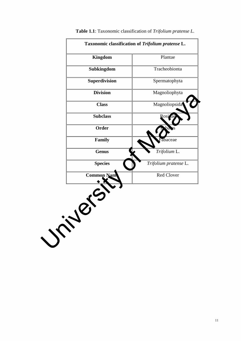
Table 1.1: Taxonomic classification of Trifolium pratense L. ........................................ 11
Table 4.1: The effects of different concentration and combination of BAP and IBA on the
nodal explant of Trifolium pratense cultured on MS medium after 4 weeks. The cultures
were maintained at 25 ± 1 ˚ C with 16 hours light and 8 hours dark. ............................. 72
Table 4.2: The effects of different concentration and combination of BAP and 2,4-D on
the nodal explant of Trifolium pratense cultured on MS medium after 4 weeks. The
cultures were maintained at 25 ± 1 ˚ C with 16 hours light and 8 hours dark. ............... 74
Table 4.3: Survival rates of the acclimatized plantlets grown on different types of soil.
......................................................................................................................................... 78
Table 4.4: Mitotic behavior in root meristem cells of in vivo and in vitro grown Trifolium
pratense (Mean ± SD) ..................................................................................................... 83
Table 4.5: Mean number of chromosomes of in vivo and in vitro grown T. pratense (mean
± SD). .............................................................................................................................. 84
Table 4.6: The mean cell and nuclear areas and their ratio in root meristem cells of in vivo
and in vitro grown T. pratense. ....................................................................................... 85
Table 4.7: Total phenolic, total flavonoid and extraction yield of methanol extract and
soluble fractions of in vivo and in vitro grown plants (aerial parts) and also callus tissue
of Trifolium pratense ...................................................................................................... 86
Table 4.8: DPPH radical scavenging activity of methanol extract and soluble fractions of
in vivo and in vitro grown plant and callus tissue Trifolium pratense. ........................... 87
Table 4.9: Superoxide anion activity of methanol extract and soluble fractions of in vivo
and in vitro grown plant and callus tissue of Trifolium pratense. ................................... 89
Table 4.10: ABTS radical scavenging of methanol extract and soluble fractions of in vivo
and in vitro grown plant and callus tissue of Trifolium pratense. ................................... 91
Table 4.11: Hydrogen peroxide scavenging of methanol extract and soluble fractions of
in vivo and in vitro grown plant and callus tissue of Trifolium pratense. ....................... 92
Table 4.12: Chelating activity of methanol extract and soluble fractions of in vivo and in
vitro grown plant and callus tissue of Trifolium pratense............................................... 93
Table 4.13: Reducing power of methanol extract and soluble fraction (300 µg/ml) of in
vivo and in vitro grown plant and callus tissue of Trifolium pratense. ........................... 94
Univers
ity of
Mala
ya

xx
Table 4.14: Correlation between the antioxidant activity and total phenolic and flavonoid
of the extract of in vivo grown Trifolium pratense ......................................................... 95
Table 4.15: Correlation between the antioxidant activity and total phenolic and flavonoid
of the extract of in vitro grown Trifolium pratense......................................................... 95
Table 4.16: Correlation between the antioxidant activity and total phenolic and flavonoid
of the extract of callus tissue of Trifolium pratense ........................................................ 96
Table 4.17: Cytotoxic activity of the different extracts of in vivo, in vitro and callus tissue
of red clover against human breast carcinoma (MCF-7). (n = 3) ................................... 97
Table 4.18: Cytotoxic activity of the different extracts of in vivo, in vitro and callus tissue
of red clover against human colon carcinoma (HCT-116). (n = 3) ................................. 98
Table 4.19: Antimicrobial activity (inhibition zone) of ethanolic extract of callus, in vitro
and in vivo grown Trifolium pratense ........................................................................... 101
Table 4.20: Antimicrobial activity (inhibition zone) of methanolic extract of callus, in
vitro and in vivo grown Trifolium pratense .................................................................. 101
Table 4.21: Correlation analysis between secondary metabolites and leaf gas exchange
parameters of Trifolium pratense in mean traits of in vivo and in vitro ........................ 105
Table 4.22: Antioxidant activity (IC50 values) of in vitro grown T. pratense under different
concentrations of saline treatment after 6 weeks of culture. Values (means of 5 replicates
± SD) of each parameter followed by same letter are not significantly different at p ≤ 0.05.
....................................................................................................................................... 109
Univers
ity of
Mala
ya

xxi
LIST OF SYMBOLS AND ABBREVIATIONS
µg : Microgram
µl : Microliter
µM : Micromolar
2,4-D : 2,4-Dichlorophenoxyacetic Acid
ABTS : 2,2'-azino-bis
AlCl3.6H2O : Aluminum Chloride Hexahydrate
ANOVA : Analysis of variance
BAP : Benzylaminopurine
C : Celsius
CFU : Colony-forming unit
CO2 : Carbon dioxide
CTE : Rutin equivalent
DMSO : Dimethyl sulfoxide
DNA : Deoxyribonucleic acid
DPPH : 2,2-diphenyl-1-picrylhydrazyl
E : Transpiration rate
g/l : Gram per liter
GAE : Gallic acid equivalent
gs : Stomatal conductance
h : Hours
HCl : Hydrochloric acid
HCT-116 : Human colon carcinoma
IBA : Indolebutyric Acid
Kinetin : 6-furfurylaminopurine
Univers
ity of
Mala
ya

xxii
MCF-7 : Human breast carcinoma
mg/l : Miligram per liter
MI : Mitotic index
ml : Mililiter
mm : Millimeter
mM : Millimolar
MS : Murashige and Skoog
MSO : Murashig and Skoog (without hormone)
MTT : 3-(4,5-dimethylthiazol-2-yl)-2,5-diphenyltetrazolium bromide
NAA : Naphthalene Acetic Acid
NaCl : Sodium chloride
NaCl : Sodium chloride
NaNO2 : Sodium nitrite
NaOH : Sodium hydroxide
NBT : Nitro blue tetrazolium
nm : Nanometer
OD : Optical density
PGRs : Plant growth regulators
Pn : Net photosynthetic rate
PPFD : Photosynthesis photon flux density
R2 : Coefficient of determination
ROS : Reactive oxygen species
rpm : Rotation per minute
SD : Standard deveision
SE : Standard error
TDZ : Thiadiazuron
Univers
ity of
Mala
ya

xxiii
UV : Ultra violet
w/v : Weight per volume
AQY : Apparent quantum yield
Rd : Respiration
CP : Compensation point
PNmax : Maximum net photosynthetic
SOD : Superoxide dismutase
Univers
ity of
Mala
ya

xxiv
LIST OF APPENDICES
APPENDIX A: MINERAL COMPOSITION OF RED CLOVER 165
APPENDIX B: DISTRIBUTION MAP OF RED CLOVER
166
APPENDIX C: FORMULATIONS OF MS MEDIA (MURASHIGE AND
SKOOG
167
Univers
ity of
Mala
ya

xxv
Univers
ity of
Mala
ya

1
CHAPTER 1: INTRODUCTION
1.1 Importance of Bioactivities Discovery from Plants
The plant kingdom represents an enormous reservoir of chemical compounds. “There
has been considerable amount of effort and research focused on finding novel bioactive
compounds from natural sources such as tea, fruits, vegetables, herbs and spices
(Kanazawa & Sakakibara, 2000). Most of the researches also suggested that the natural
compounds found in fruits and vegetables may reduce the risk of degenerative ailments.
Besides the interest in finding novel bioactive compounds from natural sources, there has
been a worldwide move towards the use of traditional medicines due to concerns over the
invasive, expensive and potentially toxic mainstream practices (Wyk, 2002; Ernst, 2002).”
Ethnopharmacological surveys have shed light on the fact that the therapeutic use of even
80% of 122 plant-derived drugs may have a link with their recommendations in traditional
medicine (Fabricant & Farnsworth, 2001).
The efforts regarding understanding the natural products are always an intriguing goal
for researchers over decades, especially on plants. “With reference to past events, plants
(fruits, vegetables, medicinal herbs, etc.) have given a decent wellspring of a wide variety
of compounds, such as phenolic compounds, nitrogen compounds, vitamins, terpenoids
and some other secondary metabolites, which are rich in valuable bioactivities, e.g.,
antioxidant, anti-inflammatory, antitumor, antimutagenic, anti-carcinogenic,
antibacterial, or antiviral activities. In numerous oriental countries (China, Japan, etc), the
traditional herbal medicines have been generally utilized for a large number of years.
Herbal plants have turned into the primary object of chemists, biochemist, and
pharmaceutics. Their examination assumes an essential part to discover and growing new
medications, which are having ideally more viability and no side effects like most present
day drugs.”
Univers
ity of
Mala
ya

2
Medicinal plants are natural resources which are often used in the treatment of various
ailments. From ancient time, plants are rich source of effective and safe medicines. In
recent years there has been more focus on plants with antimicrobial, antioxidant and
anticancer activities.
“Several studies have shown that aromatic and medicinal plants are sources of various
nutrient and non-nutrient molecules, many of which show antioxidant, anticancer and
antimicrobial properties which can protect the human body against both cellular oxidation
reactions and pathogens. Thus, it is important to characterize the different types of
medicinal plants for their biological activities potential (Mothana & Lindequist, 2005;
Bajpai et al., 2005; Wojdylo et al., 2007).”
In this way, research on herbal plant significantly is considered for both to find active
compounds and to locate the viable instrument of them to form into medications for
treatment illnesses. Besides, the research additionally supplied general constituents and
impacts that can energize the utilization of herbal plants as "nourishment" for escalating
wellbeing and avert illnesses.
In developing countries like Malaysia, medicinal plants continue to be the main source
of medication. Current developments in phytotechnology, phytochemistry and
biotechnology have facilitated rapid progress in natural product research.
1.2 Plant Biotechnology
One of the most challenging problems in the development and use of medicinal plants
to produce drugs is the non-economic isolation of their pharmaceutically important
compounds due to their low production quantity (Kayser & Quax, 2007). Hence, several
biotechnological techniques have been employed to enhance the production of bioactive
compounds in plants such as tissue culture and genetic modification methods (Robins,
Univers
ity of
Mala
ya

3
1994; Tripathi & Tripathi, 2003; Kayser & Quax, 2007; Karuppusamy, 2009; Chaudhury
& Pal, 2010).
Plant biotechnology is the use of exploratory learning to the enhancement of plant
characteristics. Improvements are in amount or quality. “It is a specific process in which
scientific techniques are used to develop molecular- and cellular-based technologies to
improve plant productivity, quality and health; to improve the quality of plant products;
or to prevent, reduce or eliminate constraints to plant productivity caused by diseases,
pest organisms and ecological stresses. Almost all commercial improvements have been
in productivity. Expanded profitability conserves land and water, brings down the
expense of production, and can build the accessibility supply of food, feed, fiber, or
pharmaceutical.”
The field of plant biotechnology is involved in creating approaches to enhance the
generation of plants so as to supply the world's requirements for food, fiber and fuel.
Moreover, plants give us numerous pharmaceuticals and industrial compounds. As our
populace develops, our needs additionally develop. To build the amount of crop
production as well as to produce specific characteristics in plants, biotechnologists are
using selective gene techniques. The two noteworthy routines of propagation are: plant
tissue culture and genetic engineering.
Study in the zone of plant biotechnology will keep on concentrating on tackling major
farming issues. Researchers will keep on moving in the direction of enhancing
biotechnology tools for even more secure, more powerful utilization of biotechnology by
all specialists. For instance, more effective models are being produced to assess
genetically engineered plants and to diminish allergens in foods. Researchers will
continue to monitor for potential environmental problems, such as insect pests becoming
resistant to biotechnology.
Univers
ity of
Mala
ya

4
1.3 Plant Tissue Culture and Micropropagation
The need for a sustainable food production is undeniably increasing. Hence, in order
to fulfill the demand for food and to achieve a sustainable food production, plant breeding
serves as a very vital tool. An example of plant breeding methods includes the use of
tissue culture in plant regeneration and propagation. In vitro technique or tissue culture
technique is an alternative propagation method which plays a very essential role in plant
biotechnology. It can serve as a mean to improve the existing plant cultivars, to protect
the cultivars from extinction and also to shorten the required time to generate the plants,
as compared to conventional breeding (Akbar et al., 2003). Other than that, in vitro
methods or micropropagation techniques can also be used to produce large amounts of
uniform planting materials and to produce genetically engineered plant products and
diseases-free plants (Maluszynski et al., 1995).
“One example where the use of tissue culture has become increasingly in demand is in
the oil palm industry. Malaysia with approximately 48% of palm oil production is one of
the leading countries in the world to export palm oil, followed by Indonesia with 35% of
palm oil production and other countries (Soh et al., 2003). This makes oil palm as one of
the most valuable economic resources of the country. Due to such importance, many
researches has been done to improve the oil palm cultivars and to obtain the highest
yielding cultivars with enhanced oil production. One of the most sought-after technique
is tissue culture, where high yielding cultivars were produced clonally (Soh et al., 2003).”
Tissue culture techniques have already been integrated in biotechnology that permits
the regeneration of plants either as clones or somaclones. The accessibility of a proficient
protocol for in vitro regeneration is an essential to bridle any biotechnological approach
for genetic improvement of crop plants.
Univers
ity of
Mala
ya
5
The use of a plant tissue culture system to produce desirable medicinal compounds has
made possible the production of a wide variety of pharmaceuticals like alkaloids,
terpenes, steroids, phenolics, flavonoids, and amino acids (Vanisree et al., 2004). Taxol,
for example, is one of the most promising anticancer drugs, which was originally isolated
from the bark of the Pacific yew tree (Taxus brevifolia). Taxol supply from this original
source cannot meet the increasing demand for clinical use because of the scarcity and
slow growth of Taxus brevifolia, and the costly synthetic process (Cragg et al., 1993).
Tissue cultures of various Taxus species, therefore, have been studied extensively as an
alternative route for production of Taxol and other useful taxane compounds (Wu & Lin,
2003; Kim et al., 2004; Naill & Roberts, 2005).
“Term used to describe laboratory activities to produce plantlets from explants or callus
of a parent plant is called micropropagation (Brown & Thorpe, 1995). In theory, all plants
can be subjected to micropropagation to generate vast amounts of offspring plantlets.
However, this is subjected to numerous limitations. Successful micropropagation
protocols have been developed for different species, but its use in large scale production
has become limited due to a variety of factors including the need of asepsis, labor cost for
sub-culturing the micropropagated tissues between the tissue culture vessels and also
problems with genetic variations in the resulting plantlets, verification, acclimatization
and contamination problems (Brown & Thorpe, 1995).”
The success of micropropagation depends to the few factors, such as the source of the
explant and the type of the cultured medium. For example, tissues from shoot tips, shoots
or roots of the seedling has been identified as excellent sources for regeneration of friable
callus via tissue culture (Gamborg et al., 1976).
“Another important influential factor in the success of micropropagation, is the type of
the medium being used in the culture process. Media with the essential nutrients needed
Univers
ity of
Mala
ya

6
for growth, is responsible in supplying the seedling and the growth of the cultured plants.
For example, in the early years of micropropagation experiments, the white medium had
been adopted in micropropagation researches. Since then many modifications had been
done to improve the culture media in order to allow for a more efficient and successful
growth of plant cells. This is because the white media was found to be insufficient in
terms of its nitrogen and potassium content to maintain cell suspension culture and callus
formation.”
1.4 Importance of Forage Crops
“Legume species provide high quality forages for animals with a positive effect on the
environment due to reduction in the use of inorganic-N fertilizer due to their N2-fixation
ability (Lüscher et al., 2014). However, the protein level in legumes is rather high and the
natural digestion process of proteins in ruminant is quite inefficient. To cope with this
problem, the use of grass–legume mixtures is interesting as they balance the energy,
protein ratio, increase biomass production by transferring the symbiotically fixed N from
legumes to grasses and can stimulate voluntary intake (Niderkorn et al., 2014).”
Grass and legume plant species that are cultured for livestock feed are called forage
crops. “They are also very important for reclamation and land conservation. The
vegetative portion of the plant, mostly stems and leaves, are used by livestock. Sheep,
cattle and other ruminant livestock require the fiber in their diet for proper digestion. This
fiber is naturally found in the forage crops. They also gain their required nutrients such
as vitamins, minerals and protein from forage crops. Livestock can use the forage crops
directly in the pastures, or the plants can conserve as hey or silage for winter feeding.
Both annual and perennial plant species are used as forage crops, but perennials are much
more commonly utilized.”
Univers
ity of
Mala
ya

7
“One of the most important legume forage crops in the world is Alfalfa (Medicago
sativa L.). It has excellent productivity, high quality, the ability to fix and utilize
atmospheric nitrogen and drought tolerance. The superiority of alfalfa to the other forage
crops is its higher production of protein per unit area. It can be grown alone or in
combination with various grass species. It requires well-drained soil, a pH above 6.1,
adequate fertility and proper harvest management. Alfalfa has some disadvantages like;
poor persistence under grazing, low tolerance to acidic or variably drained soil and need
to have fall rest period.”
“Sainfoin (Onobrychis sativa) is another important forage crop that is well adopted to
poor lands where drought is common and cultural practices are minimal. It is native to
Turkey, Iran and Europe. It can be used as an alternative to alfalfa. It contains condensed
tannins which reduce its potential to produce bloat and improve protein digestion by
grazing animals (McMahon et al., 2000). Sainfoin has a good nitrogen fixing ability, so
can be grown in conjunction with other forage grasses to improve soil fertility (Lu et al.,
2000). Sainfoin offers a superb forage for feeding animals and voluntary consumption
of sainfoin by cattle and sheep is 20% higher than for the grass. Unlike many other
legumes, it is non-bloating and is known to have anthelmintic properties, so reducing the
problems associated with livestock worms. Livestock that are fed by sainfoin have very
fast liveweight gains, so young stock can be finished sooner and with very good body
grades (Hayot Carbonero et al., 2011). The flowers of sainfoin produce great amounts of
nectar and are very attractive to honey bees and other pollinating insects (Ogle et al.,
2007). Many investigations have been done to determine the sainfoin polyphenols which
include tannins and flavonoids. Significant differences were found between sainfoin types
and this will lead to further development of sainfoin plant breeding. Sainfoins are difficult
to establish as pasture, are not persistent in grassland, and only yield one crop of hay or
seeds per year, thus it is rarely used as a pure crop and is usually presented in pasture in
Univers
ity of
Mala
ya

8
a grass-legume combination with red clover, white clover or other legumes. Unlike the
alfalfa sainfoin is resistant to many pests, but in wet condition the sainfoin cannot survive
for long time because of the root and crown rot disease (Morrill et al., 1998). Other
reasons that often reduce the interest of using sainfoin are: high seeding rate, large seeds
and higher price of seeds compared with other forage crops.”
“Another important forage crop is red clover (Trifolium pratense L.). It is a short-lived
perennial plant that is more adapted to the soil with lower pH and wet condition,
compared with the alfalfa and sainfoin, so it can be a good alternative to alfalfa in areas
where alfalfa winterkill is a problem. Compared with many other legumes, red clover can
grow faster, fix more nitrogen, produce more biomass and is more adapted to different
soil types. It has lower seeding cost and easier establishment than alfalfa. Red clover is
an excellent treatment for the soil, as its extensive root system permeates the top soil. It
is greatly suitable for livestock whether at fresh leafy-growth stage or as hay or silage. It
exhibited higher digestibility compared with alfalfa or sainfoin and the rate of decline in
digestibility with maturation is slower. Because of the physical structure of red clover
leaves, the breakdown of consumed forage is quicker and clearance of particles from the
rumen is faster. In addition of livestock feeding, red clover have been known for many
centuries as a valuable herb in traditional medicine of various cultures (Sabudak et al.,
2008; Khan and Khatoon, 2008). Until recently, there are some studies on biological
activities and beneficial effects of red clover, like antioxidant activity (Mu et al., 2009),
anticancer properties (Liu et al., 2011), platelet activity (Simoncini et al., 2005),
antiangiogenic action (Krenn & Paper, 2009), estrogenic effect (Yatkin & Daglioglu
2011).”
Univers
ity of
Mala
ya

9
1.5 Forage Crops in Malaysia
Malaysia with the population of 25 million people in the year 2006 spent around 12.2
billion Ringgits for the import of food. The imported items were consisted of vegetables,
meat, fruit, fish dairy products and fish and animal foodstuffs. The amount of the imported
products increased to 13.4 billion Ringgits in 2007. Livestock products were imported
with the approximate value of 3.7 billion Ringgits including 70-75% for poultry, 20-25%
for swine and 1-2% for ruminants, respectively.
Malaysia lacks natural grasslands. The main vegetation is evergreen equatorial rain
forest. Where land is cleared, it is normally cultivated with plantation crops such as
rubber, oil palm, cocoa and fruit crops.
One of the major limitations to efficient ruminant livestock production in Malaysia is
the lack of adequate 1evels of high quality forage for feeding the ruminants. Nonetheless,
the wide diversity of farming systems existing in Malaysia offers various potential
ecological niches for the forage introduction and evaluation programs to identify adaptive
forages and to encourage their adoption and use by the farmers for ruminant production.
Some ad hoc research on pasture and fodder grasses was initiated in the early twenties by
the Department of Agriculture to improve forage quality and quantity. At the same time,
the government was promoting commercial enterprises in the beef and dairy production
to increase livestock production to meet increasing meat demand and to raise the standard
of living of the farmers through efficient production systems.
The Malaysian experience with the indigenous and introduced (exotic) forages has
been variable. The indigenous species do not produce high dry matter yield and the exotic
species are less persistent and prone to pest and disease damage. Poor seeding behaviour
of the selected species often prevented commercialization (Chin et al., 1974). Although
there is a need for a breeding program to upgrade the desirable characteristics of the
Univers
ity of
Mala
ya

10
selected species, the present circumstances of forage development strategies in Malaysia
do not permit or warrant such a research undertaking. Hence, introduction and evaluation
of forage germplasm program has remained as the primary means of obtaining genetic
resources for upgrading. Interest is now on collection of genetic materials of high
productivity, acid soil tolerance, forage quality, shade tolerance, persistency to defoliation
or grazing and tolerance to tropical pests and diseases.
1.6 Morphology of Trifolium pratense
“Trifolium is a genus of about 300 species of plants in the leguminous pea family
Fabaceae. The most widely cultivated clovers are Trifolium repens (white clover) and
Trifolium pratense (red clover). As a member of the family Leguminosae or Fabaceae,
Trifolium pratense L. (red clover) is a short-lived biennial plant which serves as food for
livestock, but also as a health food for humans (Leung & Foster, 1996) (Table 1.1). Red
clover is a valuable forage legume in temperate regions of the world. It is an herbaceous
(non-woody), short-lived perennial plant. Stems develop from the crown and range in
height from 18 to 36 inches at maturity. Leaves are arranged alternately on the stems. The
leaves borne on the basal crown are long and petiolate, while the leaves borne on stems
are often nearly sessile. The leaves are trifoliate or palmate-trifoliate, pubescent and
alternate. Leaflets are oval or elliptic, 1-3 cm long x 0.5-1.5 cm broad. They are typically
variegated with a white V-like mark. Flower heads, located at the tip of stems, usually
have 75-125 individual pinkish-violet flowers (Figures 1.1 and 1.2). Red clover has a
taproot system with many secondary branches.”
Univers
ity of
Mala
ya
11
Table 1.1: Taxonomic classification of Trifolium pratense L.
Taxonomic classification of Trifolium pratense L.
Kingdom Plantae
Subkingdom Tracheobionta
Superdivision Spermatophyta
Division Magnoliophyta
Class Magnoliopsida
Subclass Rosidae
Order Fabales
Family Fabaceae
Genus Trifolium L.
Species Trifolium pratense L.
Common Name Red Clover
Univers
ity of
Mala
ya

12
Figure 1.1: Intact plant of Trifolium pratense L. (Source: http://www.pfaf.org/user/Plant.aspx?LatinName=Trifolium+pratense)
Figure 1.2: Individual pinkish-violet flowers of Trifolium pratense (Source: http://www.rolv.no/bilder/galleri/fjellplanter/trif_pra.htm)
Univers
ity of
Mala
ya
13
1.7 Uses of Trifolium pratense
Trifolium pratense has been a popular, multi purpose crop since European immigrant
farmers brought it to North America in the 1500s. It is widely grown as a fodder crop,
and also the aboveground parts and blossoms are used in traditional medicine.
Trifolium pratense can be used as a cover crop that provides many benefits such as
fixing nitrogen (N) to meet needs of the following crop, protecting soil from erosion,
improving soil tilth, competing with weeds, as well as supplying forage needs. It is used
primarily as a legume green manure killed ahead of corn or vegetable crops planted in
early summer. Grown alone or mixed with grasses, it produces high yields and good
quality forage in a wide range of soil types, pH, environmental and management
conditions (Carrillo et al., 2004).
In recent years, the interest for red clover has increased, partly because it possesses
unique properties that may affect animal performance and product quality. It has superior
feeding value and higher ruminant performances (higher milk yield and growth rate) are
often obtained when compared with grasses and Lucerne (Fraser et al., 2004; Vanhatalo
et al., 2006).
“Extracts of T. pratense have had a long history of medicinal uses. It was originally
used as a medicinal herb by native indigenous people of North America for whooping
cough, gout and cancer (Leung & Foster, 1996). Other traditional societies have used this
plant in the treatment of asthma, bronchitis coughs, athlets’s foot (Foster & Duke, 1990),
also for eczema and psoriasis (Rijke et al., 2001). Extracts of T. pratense are
commercially available as isoflavone enriched dietary supplements on the US and
European markets for women suffering from menopausal complaints (Oleszek &
Stochmal, 2002).”
Univers
ity of
Mala
ya
14
1.8 Problem Statement
The valuable characteristics of T. pratense (productivity, forage quality and bioactive
compounds) are induced by its morphology and eco-physiology, which are correlated
with the genetics of persistence, and with the environmental conditions.
“It is axiomatic that to be persistent, a crop must be well adapted to its environment. T.
pratense persistence is the result of an interaction between its adaptation and its stress
load (Taylor & Quesenberry, 1996). Stress load may be defined as any factor, whether
physiogenic or pathogenic, that affects the growth and development of the species, and
consequently the maintenance of a closed canopy. Environmental stresses such as
interspecific or/and intraspecific competition, winter hazards, management practices, and
pathogens, are seen as reducing the vigor of the forage legume plants. Weakened plants
are then susceptible to damage from other pathogens which increases root rot
phenomenon that leads to shortened persistence through senescence occurrence and
finally, to lose the forage stand.”
Persistence, then, is determined by the species eco-physiological characteristics, the
perennial ecotypes traits, the environmental influence and the grassland applied
technologies. The most negative factors that diminish the clover persistence includes:
improper harvesting performed before pre-bloom stage, late autumn harvesting, low
temperatures and spring drought, and root rot phenomenon. It has been suggested that
root rots in forage legumes may be an unavoidable disease (Rufelt, 1982).
In most places of the world the utilization of T. pratense by farmers is confined by the
absence of persistence identified with high mortality of plants, determining a productive
life of two to three years (Ortega et al., 2003). This likewise restrains the genetic progress
of breeding projects because of the trouble in keeping up selected genotypes under field
Univers
ity of
Mala
ya
15
or greenhouse conditions and also, due to the allogamous way of the species that makes
it difficult to keep up elite clones by seed (Taylor & Quesenberry, 1996).
Considering these limitations, protocols for in vitro tissue culture of T. pratense have
been developed to propagate, eliminate viruses, and maintain T. pratense clones. In vitro
culture of plant offers the opportunity to select and regenerate plants with desirable
characteristics. The technique has also been effectively utilized to induce tolerance which
includes the use of some selective agents that permit the preferential survival and growth
of desired phenotypes.
The application of in vitro selection techniques has facilitated the generation of disease
resistant plants in other pathosystems (Pontaroli & Camadro, 2005).
1.9 Objectives of the Study
This study consists of two major parts. In the first part, micropropagation of Trifolium
pratense using different plant hormones in tissue culture system was performed in order
to mass propagate of this very important forage crop. In the second part of the study a
comparison was made on some of the biological activities, cellular behavior and
physiological activity of in vivo and in vitro grown T. pratense to determine whether there
are any significant differences between plants grown in vivo and in vitro system.
The results are valuable for applications of T. pratense as a forage crop in the livestock
industry and also for using this important traditional medicinal plant in pharmaceutical
industries.
The aims of this study were:
To establish an efficient in vitro micropropagation and callus formation system
for T. pratense.
Univers
ity of
Mala
ya
16
To compare cellular behavior in root meristem cells of in vivo and in vitro grown
T. pratense.
To investigate and compare the antioxidant, antimicrobial and antitumor activities
of in vivo and in vitro grown T. pratense and also the effect of extraction solvent
on these biological activities.
To compare the photosynthetic characteristics of in vivo and in vitro grown T.
pretense and also to investigate the correlation between photosynthesis and total
phenolic and flavonoid content of this plant.
To investigate the effect of salt stress on seed germination of T. pratense using in
vitro culture.
Univers
ity of
Mala
ya

17
CHAPTER 2: LITERATURE REVIEW
2.1 Tissue Culture as a Tool in Plant Biotechnology
“Often, the problem with commercially production of unique or uncommon plants is
in availability or insufficient of plant materials required for traditional vegetative
propagation and also to obtain sufficient quantities of uniform compounds. It is, however,
possible to use a biotechnological approach in order to improve horticultural crop
production (Chebet et al., 2003). Biotechnology can also be defined as the branch of
molecular biology that examines the use of microorganisms to carry out specific industrial
processes. Currently, biotechnology is recognized for its importance with respect to plant
production, protection and improvement. One of the main biotechnological applications
in plant biotechnology is tissue culture. Micropropagation by means of tissue culturing is
commonly referred to as cell, tissue or organ culture in vitro (in glass) (Rout et al., 2006).
Plant tissue culture is a form of biotechnology that is a powerful tool for plant breeders
(Stewart, 2007). This technology utilizes plant cells, tissues or organs in a culture medium
that contains plant growth regulators to produce plants in a controlled aseptic
environment. In vitro culture is one of the main implements of plant biotechnology
responsible for exploiting the totipotency character of plant cells (Rout et al., 2006).
Additionally, in vitro propagation can be utilized to rapidly multiply cultivars with
desirable traits and create healthy, disease-free plants without seasonal constraints (Pati
et al., 2006). It is important to develop a micropropagation method or protocol in the case
of plant production via micropropagation (Razdan et al., 2008). The development of an
in vitro protocol is advantageous not only for maintainable utilization of a species, but
also for germplasm conservation and genetic improvement (Chen et al., 2006). It is
possible to store germplasm collections in cold storage in vitro, which keeps plants
available for future study or distribution (Kovalchuk, 2009).”
Univers
ity of
Mala
ya

18
“Tissue culture protocol development is challenging due to interaction of biotic and
abiotic factors. Example of biotic factors warranting critical consideration includes the
variability that exists in culture response with regards to the source of plant tissue or
explant material (Skof et al., 2007). A second concern is fungal, bacterial and viral
contamination that can occur in vitro, either from the explant materials or
environmentally. Sources of microbial contamination in plant tissue culture are extremely
difficult to determine (Kyte & Kleyn, 2001). Failure to control contamination will
ultimately result in severe losses of the regenerated or in vitro grown plant cultures (Anjali
et al., 2007).”
Tissue culture involves the use of small pieces of plant tissues (explants) which are
cultured in a nutrient medium under sterile condition. Using the appropriate growing
conditions for each explant type, plant can be induced to rapidly produce new shoots, and,
with the addition of suitable plant growth regulators (PGRs) new roots. The most
important and complex biotic factor affecting tissue culture of plant is biochemical
activity, specifically plant responses that result from the interaction of plant growth
regulators. Plant responses elicited from phytohormones are dependent on type, mode of
application, concentration, tissue sensitivity and species genotype (Goncalves et al.,
2008). Research has shown that every species explicitly requires a particular
concentration and combination of phytohormones in order to achieve optimal
development (Ramirez-Malagon, 2008).
2.1.1 Benefits of Tissue Culture
In vitro culture of cells and tissues has a great value in providing powerful ways to
study developmental processes in plants. Merits of tissue culture include: precise control
over growth conditions, uniformity, increased levels of control and the ability to create
recombinant proteins using acceptable manufacturing practices (Hellwig et al., 2004).
Univers
ity of
Mala
ya

19
The production of plant recombinant proteins, also known as molecular farming, was first
reported in 1989 by Hiatt et al. Recombinant proteins are encoded by recombinant
Deoxyribonucleic acid (DNA) or generated from a recombinant gene. Products targeted
for bioengineered plants include those for food, feed additives, human and animal health,
industrial enzymes and technical reagents (Abranches et al., 2005). Furthermore, tissue
culture techniques can be used to commercially produce and can save rare plants of
medicinal value. Such propagation techniques could be necessary as a result of species
physiological factors such as low productivity due to low seed germination rates (Chen
et al., 2006). Similarly, seedless plants and varieties where seedlessness is commercially
important can benefit from tissue culture techniques (Zhang et al., 2007).
Seed culture in vitro is a viable method for improving germination rates and assisting
in breaking seed dormancy (Chen et al., 2006; Nikolic, 2006). Further, micropropagation
methods can prove beneficial in rescuing species at risk of becoming extinct (Stephenson
& Fahey, 2004). In vitro propagation is also beneficial for plants with seeds that are slow
rooting or those with reduced multiplication rates (Chen et al., 2006).
“Tissue culture systems offer better environmental containment than whole plants
grown in the field so that regulatory requirements can be more easily met; production
times are also shorter and, for proteins that are secreted from the cells, downstream
processing and product purification are simpler and cheaper (Doran, 2006).”
“In the recent years, tissue culture has emerged as a promising technique to obtain
genetically pure elite populations under in vitro conditions. In vitro propagation also
called micropropagation is infact the miniature version of conventional propagation,
which is carried out under aseptic conditions. The advent of in vitro tissue culture
technique has offered a new approach to the morphogenetic investigations. It allows a
living system to be studied under controlled environmental conditions. This enables a
Univers
ity of
Mala
ya
20
study of the complex biological phenomenon in parts. Moreover these partial processes
are amenable to controlled investigations.”
Plants raised through micropropagation are supposed to have the following
characteristics:
i. Uniform quality
ii. Pathogen free
iii. Can be produced much more rapidly as new cultivars could become commercially
available within 2 to 3 years from development rather than 5 to 10 years needed
using conventional propagation.
iv. Produce uniformly superior seeds
v. Show improved vigor and quality
vi. year round production
vii. Clean of viral and other infections and to quickly multiply these plants as ‘cleaned
stock’ for horticulture and agriculture
2.1.2 Explant Materials
Selection of explant materials is the first and crucial aspect of micropropagation that
requires three important considerations: 1) genetic and epigenetic characteristics of the
source plant 2) pathogen control 3) physiological conditions of the plant prior to explant
excision in order to optimize its ability to establish in a culture (Hartmann et al., 1990).
Studies have revealed that somatic embryogenesis (formation of embryos from somatic
cells without fusion of male and female genus) could be improved by choosing the most
favorable female or male parent in specific cases (Park et al., 2006). The source of explant
materials can be from seed, leaf, stem, flower or root tissue.
Univers
ity of
Mala
ya
21
Generally, adventitious shoot meristems are genotypically independent and are
preferred for in vitro examination due to their ease of handling compared to other sources
of explants. Axillary shoot tip meristems are easier to grow than true apical meristems in
culture due to the existing meristems (Sai et al., 2006). Large explants can increase
chances of contamination, while small explants like meristems can sometimes show less
growth (Staba & Seabrook, 1980). Explants are cleaned by distilled water and sterilized
using mercuric chloride, ethyl alcohol and liquid bleach (Rout et al., 2000; Prakash &
Van, 2007).
“Regardless of whether the application of tissue culture is for research or market, it is
important that the cultures be recognized in vitro free of biological contamination and be
maintained as aseptic cultures during manipulation, growth, and storage. The risks from
microbial contamination are spurious experimental results due to the effects of latent
contaminants or losses of valuable experimental or commercial cultures. Microbial
contamination of plant tissue cultures is due to the great nutrient availability in the
Murashige and Skoog basal medium or variants of it. In recent years, it has been revealed
that numerous plants, particularly perennials, are at least locally endophytically colonized
intercellularly by bacteria. The latter, an intracellular pathogenic bacteria and viruses,
may permit latently into culture and be spread horizontally and vertically in cultures.
Growth of some potentially cultivable endophytes may be suppressed by the high salt and
sugar content of the Murashige and Skoog basal medium and suboptimal temperatures
for their growth in plant tissue growth rooms. The management of contamination in tissue
culture involves three stages: disease screening of the stock plants with disease and
endophyte elimination where detected; establishment and pathogen and contaminant
screening of established initial cultures; observation, random sampling, and culture
screening for micro-organism in multiplication and stored cultures. The increasing
accessibility of both broad-spectrum and specific molecular diagnostics has resulted in
Univers
ity of
Mala
ya

22
advances in multiple pathogen and latent contaminant detection.” The hazard analysis
critical control point management strategy for tissue culture laboratories is underpinned
by staff training in aseptic technique and good laboratory practice.
2.1.3 Culture Media
“Plant tissues and organs for in vitro micropropagation grow on artificial media
which is supplied with nutrients necessary for cell growth. The success level of a plant
tissue culture work is strongly influenced by the environment and formation of the culture
medium. For healthy and vigorous growth, intact plants need large amounts of some
inorganic elements (the so-called major plant nutrients and macronutrients) salts of
nitrogen (N), potassium (K), calcium (Ca), phosphorus (P), magnesium (Mg) and sulphur
(S); and small quantities of other elements (minor plant nutrients or trace elements) like
iron (Fe), Sodium (Na). Chlorine (Cl), Manganese (Mn), Zinc (Zn), boron (B), Copper
(Cu), molybdenum (Mo) and (possibly) nickel (Ni) (George, 1993).”
Plant tissue culture media is therefore made up from some or all of the following
components:
Macronutrients (always used)
Micronutrients (most of time is used; sometimes just iron, has been used)
Vitamins (can be included/not included when purchase a ready media from
suppliers)
Amino acids and other nitrogen supplements
Sugars
A solidifying agent ( Agar is the most common agent )
A wide range of media are available for plant tissue culture, but MS medium
(Murashige & Skoog, 1962) is commonly used. Other media used are Linsmaier-Skoog
Univers
ity of
Mala
ya

23
(LS) (Linsmaier & Skoog, 1965), Schenk and Hilderbrandt (SH) (Schenk & Hilderbrandt,
1972), woody plant medium (WPM) (Lioyd & McCown, 1980) and the Nitsch and Nitsch
(NN) (Nitsch & Nitsch, 1969).
Prepared commercial MS media (Murashige and Skoog, 1962) with vitamins was used
in this study due to the lack of some ingredients for making a manual media and also the
cost of making the media in the lab. In addition, using already prepared MS media is
almost simple and can save a lot of time and effort.
2.1.4 Plant Growth Regulators (PGRs)
“Hormones also known as “plant growth regulators” are chemicals used in a small
quantity to alter the growth of a plant or plant parts. They are natural chemicals produced
within plant tissues, which control normal plant functions such as root growth, fruit set
and drop, growth and other development processes. Good development of tissues and
organs are influenced by the present of hormones in medium though a few
monocotyledonous species find it optional (Mukhambetzhanov, 1997). In addition, if the
explants already have existing endogenous hormones, therefore there is no need to
supplement the medium with exogenous hormones (Hoe, 1992).”
The use of plant growth regulators in agricultural production expand exponentially
since they were first discovered and has been a major component of some agricultural
commodity production (Frederick, 2006). It is acknowledged that without these
regulators, in vitro culture is often impossible.
Some of the natural growth substances are prepared synthetically or through the
fermentation processes and can be purchased from chemical suppliers. There are several
recognized classes of plant growth substances:
Univers
ity of
Mala
ya

24
Auxins
Cytokinins
Gibberellins
Ethylene
Abscisic acid
“Auxins and cytokinins are the most important for regulating growth and
morphogenesis in plant tissue and organ cultures; in these classes, synthetic regulators
have been discovered with a biological activity which equals or exceeds that of the
equivalent growth substances” (George, 1993).
“IBA with the molecular formula C12H13NO2 and NAA with the molecular formula
C12H10O2 are among the auxins and BAP with the formula C12H11N5 and Kinetin with the
formula C10H9N5O from Cytokinins are the most favored and commonly used hormones
employed in various experiments for tissue culture and micropropagation (Keng &
Stanley, 2007; Loc et al., 2005; Miachir et al., 2004; Georg, 1993).”
Indole-3-butyric acid (IBA) and 2,4-Dichlorophenoxyacetic acid (2,4-D) from Auxins
and 6-Benzylaminopurine (BAP) from cytokinins were used for this study.
“Auxins can promote the growth of plant tissues. Binding of auxin leads to lipid
breakdown and acidification of the wall, increasing its extensibility and increasing the
water potential of the cell; so water enters and cell expands. Auxins also affects the
mRNA transcription and they cause the cell to produce biologically active cell
oligosaccharides. Auxins are able to remove genetically the original physiology of whole
plant tissues which had previously determined their differentiated state; therefore auxin
causes the cell to divide (George, 1993). Auxins are generally used to stimulate callus
production and cell growth in a culture medium or to initiate shoots, particularly roots,
Univers
ity of
Mala
ya

25
and to induce somatic embryogenesis and stimulate growth from shoot apices and shoot
tip cultures (George, 1993).”
The second group, cytokinins, can stimulate protein synthesis. The cytokinins are used
to stimulate cell division in the culture medium and induce shoot formation or auxiliary
shoot proliferation (George, 1993).
2.2 In Vitro Propagation of Medicinal Plants
The use of medicinal plants in traditional health care system back to the prehistoric
times and they are still the most important source of health care system for the majority
of people around the world (Iwu, 1993; Leaman, 2006; Carvalho, 2004; Teklehaymanot
& Giday, 2007; Heinrich, 2000; Uprety et al., 2010).
“Recently, there has been restored enthusiasm in natural medicines that are obtained
from plant parts or plant extracts. About 40% of the medicines currently used compounds
in Western countries are now derived or at least partially derived from natural sources.
The woodland harbor countless species, yet deforestation has been in charge of the quick
loss of therapeutic plant wealth, such that numerous profitable medicinal plants are under
the risk of ending. Pharmaceutical corporations depend largely upon materials obtained
from naturally occurring stands that are being rapidly depleted. Plant tissue culture is an
alternative method for commercial propagation (George & Sherrington, 1984) and is
being used widely for the commercial propagation of a substantial number of plant
species, including numerous medicinal plants.”
“Exploratory methodologies utilized for proliferation of therapeutic plants through
tissue culture can be partitioned into three general classifications. The most widely
recognized methodology is to disengage sorted out meristems like shoot tips or axillary
buds and affect them to develop into complete plants. This arrangement of propagation is
Univers
ity of
Mala
ya

26
regularly referred to as micropropagation. In the second method, adventitious shoots are
started on leaf, root and stem segments or on callus derived from those organs. The third
system of propagation includes induction of somatic embryogenesis in cell and callus
cultures. This system is hypothetically most proficient as substantial quantities of somatic
embryos can be gotten when the entire procedure is institutionalized. Biotechnology
involving modern tissue culture, cell biology and molecular biology brings about the
chance to grow new germplasms that are well adapted to evolving requests.”
Micropropagation of several plant species, including many medicinal plants, has been
described by many researchers during the last three decades (Murashige, 1977; Saurabh
et al., 2005; Nishritha et al., 2008; Chitra et al., 2009; Mathew et al., 2014; Dakah et al.,
2014).
“In case of a practical and pharmaceutical perspective, propagation from existing
meristems is not innovatively troublesome, and it yields plants that are genetically
identical with donor plants. Micropropagation of medicinal plants has been achieved
through rapid proliferation of shoot-tips and axillary buds in culture. Several issues are
described to influence the success of in vitro propagation of different medicinal plants
and, therefore, it is indiscreet to characterize any particular reason for the general
micropropagation of medicinal plants (Rout et al., 2000).”
“The factors that affect micropropagation of medicinal and aromatic plants have been
studied by Murashige (1977), Hu and Wang (1983), Bhagyalakshmi and Singh (1988),
Rout (2000), Verma and Bansal (2013). The influence of plant regulators and their
interactions on micropropagation of different plant species have been discussed in detail
by Skirvin et al. (1990), Rout et al. (1989) and Su and Zhang (2014).”
Univers
ity of
Mala
ya
27
“Shoot multiplication is viewed as the most significant stage of plant
micropropagation. Shoot multiplication is achieved by exploiting the use of PGRs
including auxins and cytokinins (Rout et al., 2006). PGRs alter various physiological
processes that affect morphogenic responses. The concentration of PGRs required for a
given response is cultivar and genotypically specific and depends on the unique cytokinin
or auxin used (Bhatia et al., 2004). The effect of auxins and cytokinins on shoot
multiplication of various medicinal plants were reported (Murashige & Skoog, 1962;
Sharma and Singh, 1997; Handique & Bora, 1999; Sagare, 2000; Singh & Tiwari, 2010;
Ramadevi et al., 2012; Ugraiah et al., 2013). Cytokinins levels were shown to be the most
critical for multiplication of medicinal plants. Benjamin et al. (1987) reported that BAP
at high concentration (1-5 ppm) stimulated the development of the axillary meristems and
shoot tips of Atropa belladonna. Lal et al. (1988) reported a rapid proliferation rate in
Picrorhiza kurroa using kinetin at 1.0-5.0 mg/l. Singh and Tiwari (2010) indicated that
the production of multiple shoots was higher in Clitoria ternatea L. on a medium having
1.5 mg/l BAP. Multiple shoot formation were reported from shoot tips (1-2cm) of field
grown plants of Paederia foetida and Centella asiatica on MS medium supplemented
with BAP (1.0 mg/l) within 7 days of culture (Singh et al., 1999). Addition of BAP (0.3
mg/l) and kinetin (0.2 mg/l) has been found to give a good response of shoot proliferation
in Withania somnifera with a regeneration of 85 % (Kulkarni et al., 2007). In Crataeva
magna, rapid multiplication was achieved on MS added with BAP (8.8 µm) (Beniamin et
al., 2004). Namh et al., (2010) reported that the highest number of shoot formation from
tissue culture of Hypericum retusum was obtained on MS medium supplemented with 0.5
mg/l BAP. Similarly, it was observed that cytokinin was required in optimal quantity for
shoot proliferation in many genotypes but including of low concentration of auxins along
with cytokinin triggered the rate of shoot proliferation. Faria and Illg (1995) reported that
the addition of 10 µM BAP together with 5 µM IAA or 5 µM NAA induced a high rate
Univers
ity of
Mala
ya

28
of shoot proliferation in Zingiber spectabile. MS medium with growth regulators such as
BAP (0.5 mg/l) in conjunction with NAA (0.01 mg/l) has been reported to give optimum
results in Utleria salcifolia (Gangaprasad et al., 2003). In Peltophorum pterocarpum,
highest number of multiple shoots was observed on MS with kinetin (2.0 mg/l) + NAA
(0.5 mg/l) (Uddin et al., 2005).”
“A fast rate of propagation relies on upon the subculturing of proliferating shoot
cultures. In the case of prolonged cultures, the nutrients in the medium are gradually
exhausted, and at the same time, the relative humidity in the vessels decreases leading to
the drying of the culture medium. Subculturing also decreases the effects of competition
of the developing shoots for nutrients. Upadhyay et al. (1989) reported a propagation
profile for Picrorhiza kurroa and detected that the shoot multiplication rate regularly
improved as the quantity of subcultures increased. Biswas et al. (2007) confirmed that the
quantity of shoots per culture was increased with the quantity of subculturing in
Aristolochia tagala Champ. Rout et al. (2000) reported that, a fast rate of propagation
relies on upon the sub-culturing of proliferating shoots.”
“The induction of callus development and subsequent differentiation and
organogenesis in medicinal plants is accomplished by the differential application of
hormons and the control of conditions in the culture medium. With the stimulus of
endogenous growth substances or by addition of exogenous growth regulators to the
nutrient medium, cell division, cell growth and tissue differentiation are induced. Several
reports are available on the regeneration of various medicinal plants via callus culture.
For instance callus cultures initiated from shoot base explants of Curcuma aromatica
Salisb were maintained on Murashige and Skoog (MS) media supplemented with 2 mg
dmˉ³ 2,4-D alone or with 0.5 mg dmˉ³ BAP and 0.5 mg dmˉ³ NAA. Approximately 8-10
plantlets were produced after 30-40 days of culture per 50 mg of callus inoculated
Univers
ity of
Mala
ya

29
(Mohanty et al., 2008). Karami et al. (2009) reported the regeneration of shoots from
callus of Elaeagnus angustifolia L. using appropriate concentration of BAP alone or BAP
combined with NAA or IAA. Thomas and Maseena (2006) described in vitro regeneration
of Cardiospermum halicacabum L. via callus culture. They reported that shoot
regeneration form callus tissue was maximum on media supplemented with 8 µM kinetin
and 0.5 µM IAA.”
“Saxena et al. (1997) reported plant regeneration via organogenesis from callus
cultures derived from mature leaves, stems, petioles and roots of young seedlings of
Psoralea corylifolia. In their study the calli differentiated into green nodular structures
which developed into dark green shoot buds in the media supplemented with 2.5 mg/L
BA and 1.0 mg/L NAA. Successful plant regeneration was reported from stem and leaf-
derived callus of Centella asiatica on MS medium supplemented with 4.0 mg/L BA, 2.0
mg/L kinetin, 0.25 mg/L NAA, and 20 mg/L adenine sulfate. The regeneration frequency
varied from 62.8% to 73.4% (Patra et al., 1998).”
In vitro induction of roots from growing shoots is dependent on the interactions of
internal and external factors. Factors include; species/cultivar, age and size of
microshoots, media, inorganic salts, carbohydrates, activated charcoal, PGRs, culture
vessel, light and temperature (Pati et al., 2006). Commercially, indole-3-acetic-acid
(IAA) and Indole-3- butyric acid (IBA) are natural and synthetic auxins respectively that
are used extensively to improve adventitious root development. However, IBA is the
auxin of choice in micropropagation because IAA tends to break down during the
autoclaving process. IBA is used extensively for rooting commercially and in agriculture
due to its availability and reasonable cost. Anis et al. (2003) obtained 80% rooting from
shoots cultured of mulberry (Morus alba L.) on the MS supplemented with IAA (1.0
mg/l). “Rout et al. (1999) reported induction of rooting in microshoots of Plumbago
Univers
ity of
Mala
ya

30
zeylanica on half-strength MS medium supplemented with 0.25 mg/L IBA with 2%
sucrose. Prakash and Staden (2007), reported that in vitro regenerated shoots of
Hoslundia opposita developed roots directly from the basal cut ends of shoots, on MS
medium supplemented with 3.6 μM of IAA, after 30 days of incubation. Matu et al. (2006)
reported that in micropropagation of Maytenus senegalensis the number of roots produced
per shoot were significantly higher in the treatments containing IBA compared to IAA.”
2.3 In Vitro Propagation of Trifolium Species
“To increase the forage availability to the growing population of animals and
decreasing the land resources research is required. Forage crop improvement requires a
long-term and multidisciplinary approach, viz. plant breeding, genetics, agronomy,
pathology, physiology, biotechnology and animal nutrition etc. Tissue and cell culture
techniques are known as potentially valuable tools for the programs of crop improvement.
The improvement of Trifolium species as important forage legumes in most part of the
world has been largely confined to conventional breeding approaches. Using
conventional methods have impeded improvement because of the some problems like:
short life, associated with disease and barriers to interspecific gene transfer. So
development of in vitro techniques could facilitate Trifolium breeding. The first
establishment of in vitro culture of legumes back to the 1950’s by Nickell (1955). Report
on organogenesis in white clover (Trifolium repens L.) further indicates potential for the
use of in vitro techniques in breeding research on Trifolium species (Pelletier & Pelletier,
1971). Beginning in the late 1970’s different studies demonstrated whole plant
regeneration from tissue cultures of various Trifolium species, however, most of these
systems were of low regeneration frequency and primarily from un-adapted plant
introduction material or wild Trifolium species (Phillips & Collins, 1979; Collins et al.,
1982).”
Univers
ity of
Mala
ya

31
Cheyne and Dale (1980) “tested different media either Blaydes (Blaydes, 1966) or
Gamborg B5 basal media (Gamborg et al., 1976) with different hormones (IAA and BAP)
for micropropagation of Trifolium repens and Trifolium pratense. Dale and Cheyne
(1993) also used different media for micropropagation of red clover to eliminate clover
diseases of seven cultivars and obtained good results using the B5 medium with identical
PGR levels. On the other hand, Campbell and Tomes (1984) also achieved good results
with the B5 medium and using the BAP (2.0 g/l)” as a growth regulator.
Carrillo et al. (2004) investigated the effect of explant source, culture media and plant
growth regulators on tissue culture of red clover. Their finding showed that stem
meristems were presented better development and lower level of contamination compared
with the crown meristems. They also found that the L2 medium supplemented with BAP
shows better results compared with the B5 and MS media.
“Uranbey et al. (2005) had developed an efficient micropropagation system for Persian
clover (Trifolium resupinatum L.). They cultured different explants (node, hypocotyl and
cotyledonary node) on MS medium supplemented with combination of BAP and IBA or
BAP, Kn and IBA. In their finding direct multiple shoots developed within 6 weeks in all
explants in most of the media tested. The best shoot multiplication capacity was obtained
from cotyledonary node explants cultured on MS medium containing 7.1 µM BAP and 1
µM IBA. They also reported that high rooting of elongated shoots was achieved in half
MS medium containing 8 µM IBA.”
Nippon and Higuchi (1990), reported successful regeneration for Trifolium amabile,
T. heldreichianum, T. montanum, T. apertum, T. caucasicum, T. cherleri and T. alpestre
on Gamborg medium supplemented with different concentrations of 2,4-D, IAA and Kn.
Univers
ity of
Mala
ya

32
Ding et al. (2003) have developed an efficient procedure for plant regeneration from
cotyledonary explants for some of the Trifolium species including T. repens, T. pratense,
Tsubterraneum, T. michelianum and T. isthmocarpum. They reported that plant
regeneration frequencies, expressed as percentage of explants producing shoots, ranged
from 65–97% for all tested cultivars of Trifolium species.
2.4 Cytological studies of Plants
Cytological characterization is crucial in the context of modern and traditional
breeding. It is well known that different ploidy levels within a species give rise to different
phenotypes and polyploidy often being associated with enhanced environmental success
and in the case of crops agronomic superiority (Hegarty & Hiscock, 2008). Cytological
analysis is usually performed to assess the mitotic process in experimental varieties and
hybrids. Hydridization of the close genetic species produces regular chromosome hybrids
while offspring of those more distantly related species have mitotic irregularities (Marfil
et al., 2006). Despite existence of some few cytological studies of Trifolium pratense,
little work rarely focused on detailed karyological criteria.
“According to Karp (1997), basic cytological technique is very important and useful
for the determination of accurate chromosome number and structure and should be in
routine use for regenerated plants in tissue culture.”
“Meristematic cells of roots have long been employed as the standard tissue or cells for
the cytological cell studies (Gimenez-Martin et al., 1966). The use of root in these studies
are comparatively uncomplicated than using other meristematic tissues such as shoot tips,
because they can be handled and produced easily, as root can be grown from seeds
germinated in only water.”
Univers
ity of
Mala
ya

33
Cytological investigation of Trifolium pratense began in the 1970s with the main aim
of developing interspecific hybrids and polyploids (Taylor, 2008). The karyotype of red
clover was initially determined based on metaphase chromosome, and it was determined
that the largest chromosome featured a large satellite (Taylor & Chen, 1988).
“Cytogenetically, most species of Trifolium, an important temperate forage legume,
have a diploid chromosome number of 14. Polyploidy is uncommon in Trifolium. Only
16% of the 248 species of Trifolium distributed throughout the world are polyploid.
However, 70% of the known polyploids occur in the subgenus Amoria, which is
considered to be one of the most primitive and unspecialized subgenera.”
Trifolium pratense has an allogamous diploid genome (2n = 2x = 14, 1C = 440 Mb)
(Sato et al., 2005), with a DNA content of 0.97 pg/2C,7 which is slightly larger than that
of rice (Arumuganathan & Earle, 1991). Because it has a strong self-incompatible
fertilization system, red clover has a highly polymorphic genome that has hampered
genetic and genomic analysis.
2.5 Antioxidants
2.5.1 Importance of Antioxidant
“Oxygen, the essential element for life (except for anaerobes), generates various types
of reactive oxygen species (ROS) which can damage living tissues. The damage caused
by the interaction of ROS with biocompounds increases the risk of many diseases. ROS
can be classified as radicals (such as superoxide, hydroxyl, peroxyl and alkoxyl) and non-
radicals (such as ozone, peroxynitrite, lipid peroxide and hydrogen peroxide) and can
easily be formed in normal and pathological cell metabolisms (Trachootham et al., 2009).
The imbalance between pro-oxidants and antioxidants lead to condition called oxidative
stress. Based on high reactivity of free radicals, they can easily damage proteins, lipids
and DNA molecules under oxidative stress conditions and are harmful to the intra/extra-
Univers
ity of
Mala
ya

34
cellular systems of organisms. It has the potential to cause numerous kinds of diseases
such as cancer, atherosclerosis, vasospasms, stroke, trauma, hyperoxia, Alzheimer,
Parkinson and cardiovascular diseases (Halliwell & Gutteridge, 1985; Ames et al., 1993;
Frei, 2012; Giasson et al., 2002).”
Although the human natural antioxidant defense system is responsible for protection
against the destructive action of ROS by production of natural antioxidants, any
interruption in their function caused by environmental stresses or a disease can potentially
increase the risk of oxidative damage in cells. Furthermore, an increase in the
concentration of the oxidizing species can cause oxidative stress in the body and natural
antioxidants may not adequately neutralize free radicals. Hence, to reduce the harmful
effects of oxidative stress, consuming a diet high in antioxidants is recommended (Packer
et al., 1999; Jeep et al., 2008).
“There are several strong antioxidants which can be divided into lipid-soluble
antioxidants, like vitamin E and butylated hydroxytoluene, and water-soluble
antioxidants, like vitamin C and gluthathione (Donoghue & Donoghue, 1997; McLean et
al., 2005).The above mentioned classification of antioxidants may not be a superior
categorization as some antioxidants such as phenolics have various phytochemicals of
which some are water-soluble and some are lipid-soluble compounds (Steven et al., 1996;
Kong et al., 2008). Dividing the antioxidants into enzymatic and non-enzymatic groups
is another way of classification of antioxidant compounds. Enzymatic antioxidants
including superoxide dismutase, catalase, and glutathione peroxidase are produced
endogenously in humans while the non-enzymatic antioxidants like tocopherols,
carotenoids, ascorbic acid, flavonoids and tannins are mostly obtained from other sources
such as plants (Naskar et al., 2010).”
Univers
ity of
Mala
ya
35
In addition to the above mentioned influence of oxidative reactions on human health,
lipid and protein oxidations in foods during food processing and storage are also some of
the main reasons for deterioration of flavor, aroma, colour and nutritive value (Stadtman
& Levine, 2003; Baron et al., 2007). Usage of antioxidant compounds in foods as
preservatives to avoid oxidative reactions has been practiced for many years (Artés et al.,
2006).
Natural antioxidants have a diversity of biochemical activities, some of which include
the inhibition of reactive oxygen species (ROS) generation, direct or indirect scavenging
of free radicals and alteration of intracellular redox potential (Finkel & Holbrook, 2000).
Many antioxidant substances have anticancer or anticarcinogenic properties (Dai &
Mumper, 2010).
By “controlling free radicals, antioxidants can make the difference between life and
death, as well as influence how fast and how well we age. The more we understand about
antioxidants and how they work, the more we will understand and appreciate the profound
role they play in keeping us healthy and happy. Their role in the human body is nothing
less than miraculous.”
“There is overwhelming scientific evidence demonstrating that those of us who eat a
diet rich in antioxidants and take antioxidant supplements will live longer and healthier
lives. Therefor in recent years, the use of natural antioxidants found in plants has attracted
interest due to their presumed nutritional and therapeutic value. Antioxidant properties
are an important mechanism of beneficial activity of plant-derived compounds and
extracts.”
Univers
ity of
Mala
ya

36
2.5.2 Methods of antioxidant activity evaluation
“There are various model systems to determine the antioxidant potential of different
samples. However, due to the complex composition of antioxidant compounds,
application of different methods of evaluation of the antioxidant capacity which are based
on different features of the antioxidant activities is recommended (Wichtl, 2001). These
assays can be classified into two group namely in vivo and in vitro models. While rats and
mice are the usual test animals for in vivo evaluation of antioxidant activity techniques,
in vitro models for evaluation of antioxidant activity of samples include several types of
assays such as DPPH radical scavenging, super oxide anion radical scavenging, hydrogen
peroxide scavenging, nitric oxide radical inhibition, reducing power, superoxide
dismutase (SOD),” oxygen radical absorbance, 2,2-azinobis(3-ethyl benzothiazoline-6-
sulfonicacid) diamonium salt (ABTS), N, N-dimethyl-p-phenylene diamine
dihydrochloride (DMPD), and chelating power. These are mostly spectrophotometeric
assays. The parameters usually evaluated by in vivo models include protein oxidation,
lipid peroxidation, reduced gluthinone content, superoxide dismutase, and catalse
activities (Joharapurkar et al., 2003).
“The antioxidant compounds can be recycled in the cell or are irreversibly damaged,
but their oxidation products are less harmful or can be further converted to harmless
substances (Halliwell & Gutteridge, 2007). At the cellular and organism level the
antioxidant protection is provided by numerous enzymes and endogenous small
molecular weight antioxidants such as ascorbic acid, uric acid glutathione, tocopherols
and several others. Many compounds contain antioxidant activity in addition to their
specialized physiological function, and their importance as antioxidants in vivo is
sometimes ambiguous (Azzi et al., 2004).”
Univers
ity of
Mala
ya

37
Application of strong antioxidants as a positive control in all types of antioxidant
activity assays is necessary. Ascorbic acid (vitamin C) is a strong antioxidant as it donates
two electrons from the C-2 and C-3 double bond carbon atoms and is commonly used as
a positive control in assays.
2.5.3 Antioxidant activity of plants
“A variety of free radical scavenging antioxidants exist within the human body which
many of them are derived from dietary sources like fruits, vegetables and teas. In recent
decades, interest in chemopreventive plant natural products has grown rapidly. The
etiology of several degenerative and aging-related diseases has been attributed to
oxidative stress, and numerous studies have been undertaken to search for the most
effective antioxidants (Aruoma, 2003; Soobrattee et al., 2005; Alvarado et al., 2006;
Pérez-Jiménez et al., 2008; Blasa et al., 2010; Wootton-Beard & Ryan, 2011). Vegetables
and herbal infusions have been recognized as important sources of antioxidants (Alarcón
et al., 2008, Almajano et al., 2008 and Chan et al., 2010).”
“The adverse effects of oxidative stress on human health have become a serious issue.
The World Health Organization (WHO) has estimated that 80% of the earth’s inhabitants
rely on traditional medicine for their primary health care needs, and most of this therapy
involves the use of plant extracts and their active components (Winston, 1999).”
“Some researchers suggest that two-thirds of the world’s plant species have medicinal
values; in particular, many medicinal plants have great antioxidant potential. Antioxidants
reduce the oxidative stress in cells and are therefore useful in the treatment of many
human diseases, including cancer, cardiovascular diseases and inflammatory diseases
(Duduku et al., 2010).”
Univers
ity of
Mala
ya

38
“Synthetic antioxidants such as butylated hydroxytoluene (BHT) and butylated
hydroxylanisole (BHA) are currently used as food additives, and many plant species have
similar antioxidant potentials as these synthetics. These species include Diospyros
abyssinica (Maiga et al., 2006), Pistacia lentiscus (Abdelwahed et al., 2007), Geranium
sanguineum L. (Sokmen et al., 2005), Sargentodoxa cuneata Rehd (Li et al., 2008),
Polyalthia cerasoides (Roxb) (Ravikumar et al., 2008), Crataeva nurvala Buch-Ham.
(Krishnanand, 2004), Acacia auriculiformis A. Cunn (Singh et al., 2007),” Teucrium
polium L. (Sharififar et al., 2009), Dracocephalum moldavica L. (Dastmalchi et al.,
2007), Urtica dioica L. (Gülçin et al., 2004), Ficus microcarpa L. (Ao et al., 2008),
Bidens pilosa (Deba et al., 2008). Radiata Leea indica (Boe et al., 2006), Uncaria
tomentosa (Pilarski et al., 2006), Salvia officinalis L. (Bozin et al., 2007), Momordica
Charantia L. (Horax et al., 2005), Rheum ribes L. (Öztürk et al., 2007), and Pelargonium
endlicherianum (Tepe et al., 2006). The literature reveals that these natural antioxidants
represent a potentially side effect-free alternative to synthetic antioxidants in the food
processing industry and for use in preventive medicine (Chung, 1997).
2.6 Phenolic Compounds
At least one aromatic ring (C6) which bears one or more hydroxyl groups can indicate
the phenolic compounds. Phenolics can be called as one of the major groups of plant
products within of alkaloids and terpenoids (Ndakidemi & Dakora, 2003). Phenolics are
usually classified into various groups based on their structures. In one of the most
complete division it is classified into different groups of phytochemicals namely simple
phenolics, phenolic acids and aldehydes, acetophenones and phenylacetic acids, cinnamic
acids, coumarins, flavonoids, biflavonyls, benzophenones, xanthones, stilbenes,
benzoquinones, anthraquinones, naphthaquinones, betacyanins, lignans, lignin, tannis,
and phlobaphenes (Vermerris & Nichelson, 2007). Phenolics can also be divided into
three groups based on the phenolic phytochemical structures, namely phenolic acids (such
Univers
ity of
Mala
ya
39
as caffeic acid), flavonoids (such as anthocyanins) and non-flavonoid polyphenols (such
as Ellagic acid) to make the above mentioned classification simple and easy to use (Basha
et al., 2004).
Phenolics are of widely varied range of compounds with low molecular weight found
in all higher plants (Makoi & Ndakidemi, 2007). They have been mostly classified as
secondary metabolites as they are not directly involved in primary metabolism but
involved in different plant processes such as biotic and abiotic stresses (Taylor, 1996;
Michalak, 2006), fruit growth and maturation (Buta & Spaulding, 1997; Ding et al.,
2001).
2.7 Antimicrobial
2.7.1 Importance of antimicrobial
Infectious diseases caused by bacteria, fungi, viruses and parasites are still a major
threat to public health, despite the tremendous progress in human medicine. Their impact
is particularly large in developing countries due to the relative unavailability of medicines
and the emergence of widespread drug resistance (Okeke et al., 2005).
Antibiotic resistance is the major reason for the need to develop new antibiotics.
Humans, animals, plants and even insects are all at risk to bacterial infections. The
number of diseases caused by bacteria to humans, animal and plants is high and
transmission is variable trough air, food, water, soil and contact. These pathogenic
bacteria are from different genera, species and strains and therefore have different
antibiotic resistances.
“Natural antibacterial compounds can be produced by any bio-organism which carry
antibacterial resistance gene/s. Discovering antibacterial compounds produced by
animals (Conlon & Sonnevend, 2010), insects (Otvos, 2000), plants (Castro & Fontes,
Univers
ity of
Mala
ya

40
2005), fungi (Qi et al., 2009) and bacteria (Eleftherianos, 2009) show that screening the
antibacterial activity of natural products to find new antibacterial compounds is
promising. These antibacterial compounds belong to different phytochemical families
such as peptides (Boman, 2003), antibacterial phenolic compounds (Maddox & Laur,
2010), and organic acids (Raftari, 2009).”
Bacteria has developed resistance to all different classes of antibiotics discovered to
date (Rojas et al. 2003); this makes the present work more remarkable. This must be
pointed out that, even with optimal antibiotic use, antibacterial resistances would not
decline quickly and existing resistances are unlikely to vanish but we must avoid the
emergence of new strains of resistant bacteria and limit the existing antibacterial
resistance (Guillemot, 1999).
The emerging resistance of microbes to antifungal agents has serious implications in
the management of infections. These antifungal compounds also act on targets found in
mammalian cells which may result in toxicity or adverse drug interactions (Lucca &
Walsh, 1999). Ketoconazole is one of the antifungal drugs used against both superficial
and deep seated infections. However, its unpleasant side effects include nausea,
abdominal pain, itching, toxicity, slow therapeutic response, and poor efficacy in
immunocompromised patients (Pyun & Shin, 2005). Therefore, the discovery of novel
antifungals is severely needed.
2.7.2 Methods of antimicrobial activity evaluation
Both in vivo and in vitro methods are applied to determine the antimicrobial activity
of samples. The in vivo evaluation methods of antimicrobial compounds usually are
carried out on rats and the duration of antimicrobial activity is determined (Lee et al.,
2003). Several in vitro antimicrobial assays are developed but agar-well diffusion and
disc diffusion methods are the most commonly used techniques to evaluate antimicrobial
Univers
ity of
Mala
ya

41
activity of different kinds of samples (European Committee on Antimicrobial
Susceptibility Testing, 2009). Inhibition zone diameters can be measured and
antibacterial potential of samples can be stated to compare with a positive control that is
usually a commercial antibiotic and antifungal.
2.7.3 Antimicrobial activity of plants
“The idea that certain plants have healing potential and they contain what we would
currently call antimicrobial compounds was well accepted before humans actually
discovered the existence of microbes (Ríos & Recio, 2005). Since then, mankind have
used plants to treat common infectious diseases and some of these traditional medicines
are still being performed as antimicrobial treatments; for example, bearberry
(Arctostaphylos uva-ursi) and cranberry juice (Vaccinium macrocarpon) are used to treat
urinary tract infections, or lemon balm (Melissa officinalis), garlic (Allium sativum) and
tea tree (Melaleuca alternifolia) has a broad-spectrum of antimicrobial agents (Heinrich
& Gibbons, 2004). It has been proven that the main compounds for antibacterial activities
generally contain the essential oils of these plants rather than their extracts which bring a
great and vast area of usage in the treatment for infectious pathologies in the respiratory
system, urinary tract, gastrointestinal and biliary systems, as well as on the skin. For
example, in the case of Melaleuca alternifolia, the usage of the essential oil (tea tree oil)
is a common therapeutic tool to treat acne and other infectious problems of the skin (Ríos
& Recio, 2005).”
2.8 Anticancer
2.8.1 Importance of anticancer
“The use of natural products as anticancer agents has a long history that began with
folk medicine and through the years has been incorporated into traditional and allopathic
medicine. Several drugs currently used in chemotherapy were isolated from plant species
Univers
ity of
Mala
ya

42
or derived from a natural prototype. Most of the research performed today focuses on the
development of new drugs to treat cancer. According to Cragg and Newman (2000), over
50 % of the drugs in clinical trials for anticancer activity were isolated from natural
sources or are related to them.”
According to the Ferlay et al. (2015) there were 14.1 million new cancer cases, 8.2
million cancer deaths and 32.6 million people living with cancer (within 5 years of
diagnosis) in 2012 worldwide. According to the World Health Organization estimates for
2011, cancer now cause more deaths than all coronary heart disease.
Plant derived drugs are playing an important role in the up growth of cancer therapy
(Conforti et al., 2008). Most of the active compounds in these extracts remain
unidentified, and their presence is only detected by biological tests. Most of the identified
compounds are products of plant secondary metabolism and belong to the classes of
alkaloids, polyphenols, triterpenes, or steroid glycosides (Dzhambazov et al., 2002).
2.8.2 Methods of anticancer activity
“To assess for preliminary anticancer activity in terms of cell viability, the MTT and
MTS in vitro cytotoxicity assays are considered two of the most economic, reliable and
convenient methods. This is based on their ease of use, accuracy and rapid indication of
toxicity (Berg et al., 1994), as well as their sensitivity and specificity (Malich et al., 1997).
Both assays are in vitro whole cell toxicity assays that employ colorimetric methods for
determining the number of viable cells based on mitochondrial dehydrogenase activity
measurement and differ only in the reagent employed. In the MTT assay, 3- (4,5-
dimethylthiazol-2-yl)-2,5-diphenyltetrazolium bromide is bioreduced by dehydrogenase
inside living cells to form a coloured formazan dye, while in the MTS assay, a similar
bioconversion takes places utilising 3-(4,5-dimethylthiazol-2-yl)-5-(3-
Univers
ity of
Mala
ya

43
carboxymethoxyphenyl)-2-(4-sulfophenyl)-2H-tetrazolium, inner salt and an electron
coupling reagent “(Phenazine ethosulfate).
Other methods like: Tryphan blue dye exclusion assay (Unnikrishnan & Ramadasan
1998), LDH (Lactic dehydrogenase) assay (Russo et al., 2006) and Sulforhodamine B
assay (Voigt, 2005) also are used to determine the anticancer activity.
2.8.3 Anticancer activity of medicinal plants
“The synthetic anticancer remedies are beyond the reach of common man because of
cost factor. Herbal medicines have a vital role in the prevention and treatment of cancer
and medicinal herbs are commonly available and comparatively economical. A great deal
of pharmaceutical research done in technologically advanced countries like USA,
Germany, France, Japan and China has considerably improved quality of the herbal
medicines used in the treatment of cancer. Some herbs protect the body from cancer by
enhancing detoxification functions of the body. Certain biological response modifiers
derived from herbs are known to inhibit growth of cancer by modulating the activity of
specific hormones and enzymes. Some herbs reduce toxic side effects of chemotherapy
and radiotherapy.”
Phytoconstituents derived from the herbs Taxus species (Malik et al., 2011),
Achyranthes bidentate (Zhang et al., 2012), Allium sativum (Thomson & Ali, 2003), Aloe
vera (Boudreau & Beland, 2006), Angelica sinensis (Cheng et al., 2004), Astragals
membranaceus (Cho & Leung, 2007), Glycine max (Yanamandra et al., 2003),
Glycyrrhiza glabra (Kherva, 2009), Medicago sativa (Rosenthal & Nkomo, 2000),
Morinda citrifolia (Wang & Su, 2001), Saussurea lappa (Ko et al., 2005), Taxus
wallichiana (Chattopadhyay et al., 2003), Tinospora cordifolia (Mathew & Kuttan,
1999), Viscum album (Zarkovic et al., 2001), Withania somnifera (Christina et al., 2004),
Zingiber officinale (Habib et al., 2008) etc., have been used in various formulations to
Univers
ity of
Mala
ya

44
help the body fighting cancer more effectively and reduce toxic side effects of
chemotherapy and radiotherapy stages of cancer.
2.9 Biological Activities of Trifolium Species
“Plants from the genus Trifolium have been known for many years as important forage
crops and also valuable herbs in folk medicine. Nowadays, the therapeutic use of
Trifolium plants is still based mainly on traditional medicine recommendations, but the
number of scientific data on the biological activity of clovers and their possible
therapeutic effects has been growing. The majority of the studies on the biological
properties of clovers concerns Trifolium pratense and is focused on its phytoestrogenic
action, being a result of isoflavone content. However, it should be emphasized that the
beneficial effects of red clover as well as other clovers may be dependent on the action
of various biologically active substances occurring in these herbs, not only isoflavones.
There are significant gaps in the ethnopharmacological knowledge of medicinal relevance
to Trifolium genus. Contrary to the existing evidence of therapeutic properties of T.
pratense, obtained from in vivo and clinical examinations, the curative applications of
other Trifolium species are supported mainly by basic investigations or traditional
medicine.”
“Clovers have been used by Oriental and European cultures for the treatment of eczema
and psoriasis. In Turkish traditional medicine, Trifolium repens, Trifolium arvense and
Trifolium pratense have been administered as expectorant, antiseptic, analgesic, sedative
and tonic mixtures (Sabudak et al., 2008). Trifolium repens and Trifolium pratense are
popular herbs in Pakistan, useful in the treatment of sore throat, fever, pneumonia,
meningitis and feverish feeling (Khan & Khatoon, 2008). Native Americans have used
Trifolium pratense to cure external skin problems, lung illnesses, as well as some
disorders of nervous and reproductive system (Sabudak & Guler, 2009). Barros et al.
Univers
ity of
Mala
ya

45
(2010) report that in Portugal, Trifolium angustifolium decoctions are a folk medicine for
stomachache and diarrhea. Trifolium repens is a deworming remedy in the traditional
medicine of the Naga tribes of India. This activity of white clover has been confirmed by
in vivo study on animals (Tangpu et al., 2004).”
The chemical profile of clovers is partly recognized. It is known that besides
isoflavones, Trifolium plants synthesize a wide range of phenolic and polyphenolic
compounds such as flavonoids, saponins, clovamides (caffeic acid esters), phenolic acids
and other substances (Oleszek & Stochmal, 2002; Oleszek et al., 2007).
“The antioxidative activity of some Trifolium species may be also a result of the
abundance of flavonoids and other phenolic compounds, such as catechins, saponins,
clovamides and phenolic acids, present in clovers. . Sabudak et al. (2009) have found the
antioxidative activity in vitro of hexane extracts, obtained from T. balansae, T. stellatum,
T. nigrescens, T. constantinopolitanum and T. resupinatum. Moreover, these effects
correlated with the content of unsaturated fatty acid in the tested extracts. Kolodziejczyk
et al., (2011) reported in vitro antioxidant effects of the clovamide rich fraction, obtained
from aerial parts of Trifolium pallidum, in the protection of blood platelets and plasma
against the nitrative and oxidative damage, caused by peroxynitric. Mu et al. (2009),
reported in vivo antioxidant activity and the estrogenic effect of formononetin from
Trifolium pratense. Contrary to the mentioned reports suggesting the high anti-oxidative
activity of Trifolium plants, the ethnobotanical study of Barros et al. (2010) demonstrated
relatively weak antioxidant properties of T. angustifolium in vitro, most likely being a
result of a low content of phenolics and flavonoids.”
A study by khan et al. (2012) demonstrated the antibacterial activity of Trifolium
alexandrinum leaves extract against seven Gram-positive and eleven Gram-negative
hospital isolated human pathogenic bacteria strains. According to this study all the
Univers
ity of
Mala
ya

46
extracts had the antimicrobial activity against at least six types of microorganisms. They
also found that the polar (methanol or ethyl acetate) extracts displayed considerably
stronger antibacterial activity. Flythe and Kagan, (2010) reported that the phenolic
compounds from Trifolium pratense possessed antimicrobial activity against hyper
ammonia-producing bacteria (Clostridium sticklandii). Turker et al. (2009) reported that
the alcoholic extracts of Trifolium pannonicum showed a broad antibacterial spectrum
against four different fish pathogens: Aeromonas hydrophila, Yersinia ruckeri,
Streptococcus agalactia and Lactococcus garvieae.
“Trifolium pratense is said to retard progress of cancerous tumors before ulceration has
taken place (Phatak, 1977). The isoflavone constituents in red clover have estrogenic
properties. They are thought to have positive effects on menopausal disorders such as
osteoporosis, cardiac risk factors or breast cancer (Pagliaaci et al., 1994; Fugh-berman &
Kronenberg, 2001; Beck et al., 2005). Genistein isolated from Trifolium species is a
molecule of great interest as a potentially chemotherapeutic agent or as a lead compound
in anticancer drug design (Polkowski et al., 2004). Lellau and Liebezeit (2003), reported
tumor growth inhibiting activities of the Trifolium fragiferum and Trifolium repens
extracts. Moderate cytotoxic activity (LC50 between 30 and 50 µg/ml) by Trifolium
campestre and Trifolium repens against the brine shrimp were reported by Khalighi-
Sigaroodi et al. (2012).”
2.10 Photosynthetic and Leaf Gas Exchange of Plant
“Photosynthesis is a process used by plants and other organisms to convert light energy,
normally from the Sun, into chemical energy that can be later released to fuel the
organisms' activities. This chemical energy is stored in carbohydrate molecules, such as
sugars, which are synthesized from carbon dioxide and water. Photosynthesis maintains
atmospheric oxygen levels and supplies all of the organic compounds and most of the
Univers
ity of
Mala
ya

47
energy necessary for life on Earth. Although photosynthesis is performed differently by
different species, the process always begins when energy from light is absorbed by
proteins called reaction centers that contain green chlorophyll pigments. In plants, these
proteins are held inside organelles called chloroplasts, which are most abundant in leaf
cells. Although all cells in the green parts of a plant have chloroplasts, most of the energy
in higher plants is captured in the leaves.”
“Photosynthesis measurement systems are not designed to directly measure the amount
of light absorbed by the leaf. But analysis of chlorophyll-fluorescence, P700- and P515-
absorbance and gas exchange measurements reveal detailed information about e.g. the
photosystems, quantum efficiency and the CO2 assimilation rates. With some instruments
even wavelength-dependency of the photosynthetic efficiency can be analyzed.”
“Many physiological parameters such as light regime, plant nutrition, water stress, leaf
age and environmental temperature are known to be effective on photosynthetic CO2
assimilation by leaves. For example higher temperature cause heat stress in plants and
under this conditions plant growth and productivities are less (Porter & Semenov, 2005).
In some cases, the decrease in crop productivity under warmer conditions is related to
effects on the phenology and reproductive stages. The rate of photosynthesis declines as
plant water stress increases (Flexas & Medrano, 2002), and as occurred with high
temperature, this decline may be a consequence of limitations in CO2 diffusion and/or
photosynthetic metabolism.”
“Studies have shown that high altitude plants have higher photosynthetic efficiency
than the low altitude ones (Körner & Diemer, 1994; Körner et al., 1988). The leaves of
high altitude plants are capable of positive net photosynthetic rate (PN) at lower CO2
concentrations and lower temperatures than low altitude plants (Friend & Woodward
Univers
ity of
Mala
ya

48
1990). At high altitude plants, leaves achieve maximal photosynthetic rate (PNmax) at
higher irradiance than those at low altitude (Friend & Woodward, 1990).”
Photosynthesis process has been identified as one of the most important factors that
limit crop production. It is also known as one of the environmental factors that affects
secondary metabolism of plants.
“The net photosynthetic rates vary considerable with plant species, temperature,
Photosynthesis Photon Flux Density (PPFD) and other conditions. Within a large group
of herbaceous, the leaf photosynthetic rates were more or less the same but they were
lesser than 15 µmol/m2/s when measured under full sunlight, normal air and optimum
temperatures. The variation in the net photosynthesis and the related gas exchange
parameters have been suggested as the determinants of plant productivity and the early
selection criteria to enhance plant efficiency (Wang et al., 1995). The irradiance level is
a significant ecological factor for photo-autotrophic plants. Inappropriate light level can
create stress on plants by restricting photosynthesis and consequently net carbon gaining
and plant growth. Hence, the mathematical description derived from light-response
curves is valuable because it includes variables with a comprehensible physiological
meaning.”
“Heraut-Bron et al. (2000) assessed the effects of red:far-red (R:FR) ratio on net CO2
assimilation, photosynthetic activity and morphology of young white clover leaves. They
found that a leaf that developed in lower irradiance had a lower net CO2 assimilation rate
and a greater leaf area and petiole length than leaves that developed in higher irradiance.
In addition, they did not find any significant effect of FR light on photosynthesis of white
clover.”
Univers
ity of
Mala
ya
49
2.11 Plant Salt Tolerance
“Salinity is known as one of the main environmental constraints due to its high value
of impact and wide distribution that is affecting the arable lands and therefore yields of
most crops all over the world. Salinity of soil is due to high evapo-transpiration rate and
an inappropriate poor-quality water irrigation, which, then, affects almost every aspect of
plant physiology at both the whole-plant and cellular levels “(Munns & Tester, 2008).
“The main responsible factors for increasing soil salinity are using saline irrigation
water and application of fertilizer (Epstein et al., 1980). The levels of salinity in the soil
vary depending on the soil depth and season (Daniells et al., 2001) and also great potential
for interactions with other environmental factors (Perez-Tornero et al., 2009). For this
reason studies about salt stress are preferably conducted in soil-less culture with nutrient
solutions of known salt concentration (Vijayan et al., 2003). Therefore, one of the good
tools for salinity studies is in vitro culture of plants using plant tissues or cells.”
“There have been many investigations on plant seed tolerance against salinity stress.
Karimi et al. (2004) reported that endurance of salinity in germination level has special
importance because it can cause absence some proteins and enzymes. Shannon (1984)
stated that in many of plants, the most sensitivity levels to salinity in period of plant life
are the stages of budding and flowering. It is known that legumes are generally more
sensitive to salinity (Rogers et al., 1997).”
“The production of reactive oxygen species (ROS) is one of the biochemical changes
occurring in plants subjected to salt stress (Ashraf and Harris, 2004; Panda & Upadhyay,
2004; Jehan et al., 2012). To alleviate the disadvantageous effects of ROS, plants have
evolved specific antioxidative mechanisms to scavenge ROS. Hence, antioxidant
enzymes seem to be most important determinants of plant tolerance to salt stress (Vranova
et al., 2002; Ashraf et al., 2008). Many studies reported that NaCl tolerance will affected
Univers
ity of
Mala
ya
50
on antioxidant response in different plant species such as Malus domestica (Wang et al.,
2013), Soybean (Amirjani, 2010),” Sesuvium portulacastrum (Lokhande et al., 2011), pea
(Miljuš-Djukić et al., 2013).
Maas (1977), reported that T. pratense has a low salt tolerance. Researches by Asci
(2011) and also Winter and Lauchli, (1982), showed a lower survival potential for T.
pratense at high concentration of NaCl treatment.
Univers
ity of
Mala
ya

51
CHAPTER 3: MATERIALS AND METHODS
3.1 In Vitro Regeneration and Callus Induction of Trifolium pratense
3.1.1 In vivo plant samples
“Seeds of Trifolium pratense L. were purchased from Stock Seed Farms (Murdock,
US) and were planted in polybags filled with the ratio of 1:1:1 of coco peat, sand and
compost. Then, the organic fertilizer was added to each polybag and kept in the growth
room (temperature of 25 ± 1 °C, 16 hours photoperiod, 40 µmol/m2/s of photon flux
density, 60% relative humidity) (Figures 3.1 and 3.2).”
Figure 3.1: In vivo culture of T. pratense L. after 5 days
Univers
ity of
Mala
ya

52
Figure 3.2: In vivo plants of T. pratense after 6 weeks
3.1.2 In vitro plant samples
3.1.2.1 Use of aseptic techniques
The use of aseptic techniques is very crucial in this part of expriment. Aseptic
techniques were used when culturing the seeds or explants onto the culture media. This
is to prevent contamination from occurring. For instance, whilst preparing the culture
media, after the media has been prepared, the conical flask were sealed with an aluminum
foil and would be autoclaved at 121° C for 20 minutes to sterilize the media.
All tissue culture experiments were conducted in the Laminar Flow Chamber and
before any tissue culture experiments were done, the UV light was switched on for about
15 to 20 minutes to get rid of the adhering microorganism in the laminar flow chamber.
After that the inner surface of the laminar were wiped clean with 70% alcohol to reduce
the risks of contamination due to the bacteria and fungi.
Univers
ity of
Mala
ya

53
Every equipment and lab ware such as the outer surfaces of the culture flasks
containing the culture media, scalpels, petri dishes and etc, were wiped clean with 70%
alcohol before being brought into the laminar flow.
3.1.2.2 Preparation of culture media
For in vitro seed germination MS media without the addition of any hormones were
used. The MS media from Murashige and Skoog (including vitamins) with the weight of
4.4g was used for media preparation. Media was prepared for one liter (L) of water. For
making a liter of MS medium, 800 ml distilled water was poured in a beaker and placed
on the stirrer (using magnetic beads for stirring) and stirred while adding 4.4 g MS powder
(Sigma with vitamins) and 30 g sucrose. The pH of the media solution was then measured
and adjusted to 5.7 by adding 1M NaOH or 1M HCL. Following that, for solidifying the
media, 2.8 gram phytogel agar was added while turned on the heating button in order to
melt the agar. Then, the media solution was topped up to 1L with distilled water and the
conical flask would be wrapped with an aluminum foil and autoclaved at 121° C for 20
minutes.
On the other hand, for tissue culture of explants, MS media supplemented with
hormones were used. The same protocol as described in the previous paragraph was used,
but with an addition of desired hormones combination (with the desired concentrations)
before measuring the pH.
3.1.2.3 In Vitro seed germination
Before the seeds were cultured in MS media, they were first washed and sterilized by
using an appropriate sterilization protocol. “During sterilization, the seeds were washed
with a series of Clorox dilutions, followed by washing with distilled water. This was done
to effectively remove impurities and contaminants that adhere to the surface of the seeds.
Some of the purchased seeds were surface sterilized by first placing under running tap
Univers
ity of
Mala
ya

54
water for 30 minutes, rinsed once using sterile distilled water and dunked in 50 % Clorox
for 2 minutes. The seeds were then washed 5 times using sterile distilled water and soaked
in 10 % Clorox for one minute. The seeds were brought into the laminar flow and rinsed
5 times using sterile distilled water and dipped in 70 % ethanol for 1 minute. Lastly the
seeds were rinsed 5 times using sterile distilled water and blotted on sterilized filter paper.
Immediately after the sterilization process, the seeds were cultured on MS (Murashige
and Skoog) medium without adding any hormones (MSO) and incubated in the culture
room at 25 ± 1° C with 16 hours light and 8 hours dark (40 µmol/m2/s of photon flux
density and 60% relative humidity) (Figure 3.3). Since the seeds were very small in size,
6 seeds were cultured in each conical flask containing the MS basal medium. It was
observed that the seeds started to germinate after 5 to 10 days and became complete
seedling after 3-4 weeks.” The plants were subcultured after 6 weeks, to allow them to
grow better. The nodal parts of these aseptic seedlings were used as the explant for
subsequent micropropagation experiments of Trifolium pratense L.
Univers
ity of
Mala
ya
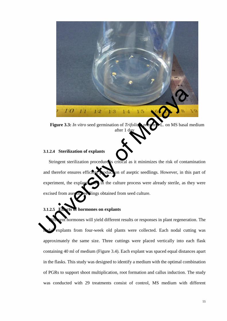
55
Figure 3.3: In vitro seed germination of Trifolium pratense L. on MS basal medium
after 1 day
3.1.2.4 Sterilization of explants
Stringent sterilization procedure is critical as it minimizes the risk of contamination
and therefor ensures efficient production of aseptic seedlings. However, in this part of
experiment, the explants used in the culture process were already sterile, as they were
excised from aseptic seedlings obtained from seed culture.
3.1.2.5 Effects of hormones on explants
Different hormones will yield different results or responses in plant regeneration. The
nodal explants from four-week old plants were collected. Each nodal cutting was
approximately the same size. Three cuttings were placed vertically into each flask
containing 40 ml of medium (Figure 3.4). Each explant was spaced equal distances apart
in the flasks. This study was designed to identify a medium with the optimal combination
of PGRs to support shoot multiplication, root formation and callus induction. The study
was conducted with 29 treatments consist of control, MS medium with different
Univers
ity of
Mala
ya
56
combination of BAP and IBA and different combination of BAP and 2,4-D were used.
The hormones combination used in this part of study are shown below:
1. “MS media + no hormone
2. MS media + 0.5 mg/L BAP
3. MS media + 1.0 mg/L BAP
4. MS media + 1.5 mg/L BAP
5. MS media + 2.0 mg/L BAP
6. MS media + 0.5 mg/L BAP + 0.25 mg/L 2,4-D
7. MS media + 0.5 mg/L BAP + 0.5 mg/L 2,4-D
8. MS media + 0.5 mg/L BAP + 0.75 mg/L 2,4-D
9. MS media + 1.0 mg/L BAP + 0.25 mg/L 2,4-D
10. MS media + 1.0 mg/L BAP + 0.5 mg/L 2,4-D
11. MS media + 1.0 mg/L BAP + 0.75 mg/L 2,4-D
12. MS media + 1.5 mg/L BAP + 0.25 mg/L 2,4-D
13. MS media + 1.5 mg/L BAP + 0.5 mg/L 2,4-D
14. MS media + 1.5 mg/L BAP + 0.75 mg/L 2,4-D
15. MS media + 2.0 mg/L BAP + 0.25 mg/L 2,4-D
16. MS media + 2.0 mg/L BAP + 0.5 mg/L 2,4-D
17. MS media + 2.0 mg/L BAP + 0.75 mg/L 2,4-D
18. MS media + 0.5 mg/L BAP + 0.25 mg/L IBA
19. MS media + 0.5 mg/L BAP + 0.5 mg/L IBA
20. MS media + 0.5 mg/L BAP + 0.75 mg/L IBA
21. MS media + 1.0 mg/L BAP + 0.25 mg/L IBA
22. MS media + 1.0 mg/L BAP + 0.5 mg/L IBA
23. MS media + 1.0 mg/L BAP + 0.75 mg/L IBA
24. MS media + 1.5 mg/L BAP + 0.25 mg/L IBA
Univers
ity of
Mala
ya

57
25. MS media + 1.5 mg/L BAP + 0.5 mg/L IBA
26. MS media + 1.5 mg/L BAP + 0.75 mg/L IBA
27. MS media + 2.0 mg/L BAP + 0.25 mg/L IBA
28. MS media + 2.0 mg/L BAP + 0.5 mg/L IBA
29. MS media + 2.0 mg/L BAP + 0.75 mg/L IBA”
Each of the 29 treatments was replicated 30 times (15 conical flasks for each treatment,
each flask containing 2 explants). Flasks were placed on a shelf in the growth chamber
under light condition (1000 Lux) and at a temperature of 25 ± 1 ºC. Cultures were
observed regularly and the contaminated flasks were replaced with fresh cultures. After a
period of 6 weeks, the media and explants were screened and the responses recorded.
Figure 3.4: Culture of nodal explants of Trifolium pratense L. on MS medium after
5 days.
Univers
ity of
Mala
ya

58
3.1.3 Acclimatization of Trifolium pratense plantlets
The in vitro plantlets were taken out from their respective containers carefully and all
adhering MS media stuck onto the plantlets were washed off using sterile distilled water.
The plantlets were then transferred onto the three different types of soils (black soil, red
soil and combination of black and red soil in ratio of 1:1) in vases covered with plastic
bags and acclimatized in culture room for one month. The plantlets were then transferred
to a green house and their ability to fully adapt to the natural conditions was monitored.
3.1.4 Statistical analysis
All experiments were carried out in triplicate. One-way analysis of variance (ANOVA)
was used to analyze the data using IBM SPSS Statistics version 19. The means were
compared with Duncan’s Multiple Comparison Test (DMCT) and p < 0.05 was
considered to indicate statistical significance.
3.2 Cellular Behavior of Trifolium pratense
3.2.1 Determination of standard primary root growth
Growth measurement is obtained through measurements of average values of the
parameters tested, from a group of roots that are as homogenous as possible (Pilet, 1991).
Determining the standard root age and root length allows subsequent experiments to be
conducted using root segments that are uniform throughout the roots population and can
be considered as good and valid sample representative of the population. This is important
as the selection of the roots would not only depend on the age of the seedlings after
planting, but also with consideration of their physiological age and conditions depending
on the growth pattern of the roots (Jensen, 1955).
In order to measure the standard growth of the primary root length for the in vivo
plants, the sterilized seeds were cultured in a petri dish containing sterilized wet cotton
wool. In this part 15 petri dishes with 10 seeds in each one were used. The cotton wools
Univers
ity of
Mala
ya

59
in each petri dish were wetted twice everyday by using sterile distilled water and the
cultures were maintained in 16 hours light and 8 hours dark, at 25 ± 1° C (Figure 3.5).
Figure 3.5: The seeds of Trifolium pratense L, germinated in petri dishes containing
sterilized wet cotton wool
Standard growth of in vitro primary root length was also measured as a comparison.
For this purpose, the sterilized seeds were cultured in petri dishes containing sterilized
MSO media by using aseptic techniques in the laminar flow. Similar to the protocol used
in measuring the standard growth for the in vivo plants, 15 petri dishes with 10 seeds in
each one were used. The petri dishes were wrapped with parafilm to prevent
contamination. The cultures were kept in the same condition with the in vivo samples.
The length of primary root of the seedlings was measured using a thread and a ruler,
at the same time every day, until majority of primary roots developed secondary roots.
The length was averaged and was plotted into a graph of the average length of the primary
roots (Y-axis) against time (X-axis). Then, a linear regression analysis was conducted to
find the relationship between both factors.
Univers
ity of
Mala
ya
60
3.2.2 Cytological studies of primary roots of Trifolium pratense
In order to study cellular activities of in vivo and in vitro seedlings of Trifolium
pratense, parameters such as chromosome count, mitotic index (MI) and mean cell and
nuclear areas were determined.
For in vitro cytological studies, sterilized seed were cultured again in full strength MS
medium without hormones and incubated in the culture room at 25 ± 1 ˚C with 16 h light
and 8 h dark for 4 weeks. The nodal parts of the 4 weeks-old plants were used as explants
and cultured on MS medium supplemented with combination of 1.5 mg/l BAP and 0.5
mg/l IBA, for the micropropagation of T. pretense (based on the results determined in
Chapter 4).. The cultures were maintained in the culture room at 25 ± 1 ˚C with 16 h light
and 8 h dark. After two weeks of culture (the needed time to achieve the standard root
length), root with the standard length of 33 mm were harvested for the cytological
analysis. For the cytological studies of in vivo grown T. pratense, the seeds were cultured
in the black and red soil with the ratio of 1:1 and transferred to the growth room with 16
hours light and 8 hours dark, at 25 ± 1° C. After two weeks of culture the primary roots
with the standard length of 33 mm were harvested for cytological studies.
3.2.3 Slide preparation and image analysis
“Root tip meristems obtained from seedlings were pre-treated in 8-hydroxyl-quinalin
(2 mM) at 4° C for 5 hours, fixed farmer’s fluid (methanol 3 : 1 acetic acid) and carnoys
solution (1:glacial acetic acid, 3:chloroform, 6:ethanol) at three different times of 15 min,
60 min and overnight. The samples were then hydrolyzed in 5N HCL for 40 min at room
temperature (cold method) or in 1N HCL for 10 and 30 minutes at 60° C (hot method).
After washing with distilled water, the root tips were soaked in Feulgen reagent for 3
hours. The root segments were then rinsed in several changes of distilled water for 5 to
10 minutes and any excess water was blotted using a filter paper. The apical meristem of
Univers
ity of
Mala
ya

61
the root segments were subdivided and squashed, followed by addition of a small drop of
45% acetic acid on clean slides. The slides were then heated gently by passing over a
spirit lamp a few times and the coverslips were levered off using a razor blade. The slides
were then viewed using Axioskop Zeiss microscope attached to AxioCam MRc video
camera and were then analyzed using AxioVision 4.7 software.”
3.2.4 Measurement of Mitotic Index
Mitotic index (MI) is the sum of cells that are undergoing division or mitosis, such as
cells in prophase, metaphase, anaphase and telophase that are expressed in terms of
percentage of all cells counted. The mitotic index was measured by scoring these cells
from at least 1000 cells in a series of random transect across the permanent slide. The
data obtained in three replicates from three slides. The average MI values for three slides
were calculated.
3.2.5 Chromosome count
The number of chromosomes was counted from 15 metaphase plates with suitable
chromosome spread. This was done in three replicates, from which the average values
were calculated.
3.2.6 Mean cell and nuclear areas
The mean cell and nuclear areas were measured from 150 prophase cells from 3 slides.
The ratio of nuclear to cell area was also determined from each sample.
3.3 Biological Activities
3.3.1 Extract preparation
The aerial parts of in vivo and in vitro grown plants and also the callus obtained from
in vitro cultures were collected and dried in the dark. The dried samples were powdered
in an electronic blender before being used for solvent extraction. For the preparation of
Univers
ity of
Mala
ya

62
extract, 25 g of fine powder was extracted with 100 ml of 95% methanol at room
temperature for 48 hours. The extracts were filtrated through Whatman No. 1 and
combined and this was followed by a concentration using rotary evaporator under
pressure that was reduced at 45 ˚C. The filtrate obtained was suspended in distilled water
(25 ml) and n-hexane (50 ml) was added to it, where the mixture was shaken well and the
layers were enabled to be separated for 6 h. After separation of n-hexane layer, again 50
ml of n-hexane was added to get the n-hexane fraction. In a similar manner, the protocol
was repeated with the rest of the solvents (ethyl acetate and chloroform). Each fraction
obtained was dried using a rotary evaporator (Kil et al., 2009). The dry extract obtained
with each solvent was weighed out and the percentage yield was expressed in terms of
the air dried weight of plant materials. Samples were stored in an airtight container at –
20 ˚C until it was time to conduct further analysis.
3.3.2 Total phenolic content
“The total phenolic content of the obtained extracts was spectrometrically analyzed in
adherence to the Folin-Ciocalteu method (Dewanto et al., 2002). In short, 100 µl of each
extract (dissolved in methanol) or gallic acid standard solution was mixed with 2 ml of
2% (w/v) sodium carbonate solution. The mixture was then incubated for 5 minutes, and
afterwards 100 µl of Folin-Ciocalteu reagent was added. After being left for 30 min at
room temperature for color development, absorbance was measured at 750 nm using a
spectrophotometer. Results are expressed as mg gallic acid equivalents (GAE) per gram
dry-matter of sample.”
3.3.3 Total flavonoid content
“Total flavonoid content was ascertained based on the method by Zhishen et al., (1999).
In brief, 50 mg of each fraction was dissolved in 10 ml of 80% aqueous methanol and
filtered through Whatman filter paper No. 42 (125 mm). In a 10 ml test tube, 300 µl of
Univers
ity of
Mala
ya

63
each extract, 3.4 ml of 30% methanol, 150 µl of 0.5 M NaNO2 and 150 µl of 0.3 M
AlCl3.6H2O were added and mixed, followed by 5-min incubation and the addition of 1
ml of NaOH (1 M). Absorbance was measured at 510 nm with a spectrophotometer. The
standard curve for total flavonoids was made using rutin standard solution (0–100 mg/l)
using the same aforementioned procedure.” The total flavonoid content was shown as
milligram of rutin equivalents (CTE) per gram dry matter of sample.
3.3.4 Antioxidant assays
Each sample was dissolved in 95% methanol at a concentration of 1 mg/ml and then
diluted in order to prepare the series concentrations for antioxidant assays. Reference
chemicals were used for comparative purposes in all assays.
3.3.4.1 DPPH radical scavenging activity assay
The DPPH radical scavenging assay was conducted following the method of Zhu et
al., (2006). “In brief, 2 ml of DPPH solution (0.1 mM, in methanol) was blended with 2
ml of the samples at varying concentrations (50, 100, 150, 200, 250 and 300 µg/ml). As
the next stage, the reaction mixture was shaken and incubated in the dark at room
temperature for 30 min, and the absorbance was read at 517 nm against the blank.
Ascorbic acid and rutin standard as positive controls were prepared in a similar manner,
as for the test group except for the antioxidant solution’s replacement.” The inhibition of
the DPPH radical by the sample was calculated based on the formula below:
DPPH scavenging activity (%)
= [(absorbance of control – absorbance of sample) / (absorbance of control)] x 100
3.3.4.2 Superoxide anion radical scavenging assay
“The assay for superoxide anion radical scavenging activity leaned on a riboflavin-
light-NBT system (Beauchamp & Fridovich, 1971). The reaction mixture had 0.5 ml of
Univers
ity of
Mala
ya

64
phosphate buffer (50 mM, pH 7.6), 0.3 ml riboflavin (50 mM), 0.25 ml
phenazinemethosulphate (PMS) (20 mM), and 0.1 ml nitro blue tetrazolium (NBT) (0.5
mM), before 1 ml sample solution was added at varying concentrations (50, 100, 150,
200, 250 and 300 µg/ml). Reaction began as the reaction mixture was illuminated with
different concentrations of the extracts using a fluorescent lamp. After 20 min of
incubation, the absorbance was measured at 560 nm. Ascorbic acid was used as standard.”
The percentage of inhibition of superoxide anion generation was calculated based on the
following formula:
Scavenging activity (%) = (1 – absorbance of sample / absorbance of control) x 100
3.3.4.3 ABTS radical scavenging assay
The ABTS radical scavenging assay was performed adhering to the method of Re et
al., (1999) with slight modification. “The ABTS radical was generated through the
oxidation of ABTS with potassium persulfate. In brief, the ABTS solution (7 mM) had
reacted with potassium persulfate (2.45 mM) solution and was stored in the dark for 12-
16 h to produce a dark coloured solution containing ABTS radical cation. Before being
used in the assay, the ABTS radical cation was diluted with 50% methanol for an initial
absorbance of about 0.70 (± 0.02) at 745 nm, with the temperature control fixed as 30 ˚C.
Free radical scavenging activity was evaluated by mixing 3 ml of ABTS working standard
with 300 µl of test sample (50, 100, 150, 200, 300, 400, 500 µg/ml) in a microcuvette.
The decrease in the absorbance was measured at the exact time of one min after mixing
the solution, then until it reached 6 min.” The final absorbance was noted then. The
inhibition percentage was calculated based on the following formula:
Scavenging effect (%)
= [(absorbance of control – absorbance of sample) / (absorbance of control)] x 100
Univers
ity of
Mala
ya
65
3.3.4.4 Hydrogen peroxide scavenging activity
The capability of scavenging hydrogen peroxide by the extract was determined based
on the method of Ruch et al., (1989). “A hydrogen peroxide solution (2 mM) was prepared
in 50 mM phosphate buffer (pH 7.4). Aliquots (0.1 ml) of the extracted sample (different
concentration of 50, 100, 150, 200, 250, 300 µg/ml) were transferred into the test tubes
and their volumes were made up to 0.4 ml with 50 mM phosphate buffer (pH 7.4). After
adding 0.6 ml hydrogen peroxide solution, tubes were vortexed and the absorbance of the
hydrogen peroxide at 230 nm was determined after 10 min, against a blank.” The abilities
to scavenge the hydrogen peroxide were calculated based on the following equation:
Hydrogen peroxide scavenging activity = (1 – absorbance of sample / absorbance of
control) × 100
3.3.4.5 Reducing power assay
The reducing powers of the samples were determined following the method of Atmani
et al., (2009). Two millilitres (2 mm) of each extract solution (300 µg/ml) were mixed
with 2 ml of phosphate buffered saline (0.2 M, pH 6.6) and 2 ml of potassium ferrocyanate
(10 mg/ml). The incubation for this mixture was set at 50 ˚C for 20 min. In the next stage,
2 ml of trichloroacetic acid (100 mg/l) was added to the mixture. In a test tube, a volume
of 2 ml from each of the above mixtures was mixed with 2 ml of distilled water and 0.4
ml of 0.1% (w/v) ferric chloride. The absorbance was measured at 700 nm after reaction
was started for 10 minutes. The increased absorbance of the reaction mixture suggested
that the reducing power was high.
3.3.4.6 Chelating power
The ability of the extract to chelate iron (II) was estimated based on the method of
Dinis et al., (1994) with minimal modification. Various sample solution (50 – 300 µg/ml)
were prepared with dissolving the extracts in the methanol. An aliquot of each sample
Univers
ity of
Mala
ya

66
(200 µl) was mixed with 100 µl of FeCl2.2H2O (2 mM) and 900 µl of methanol. After 5
min incubation, an initial reaction was fuelled by the addition of 400 µl of ferrozine (5
mM). After 10 min incubation, the absorbance at 562 nm was recorded. The percentage
of the chelating activity was calculated based on the following equation:
Chelating activity (%) = [(absorbance of control – absorbance of sample) /
(absorbance of control)] × 100
3.3.5 Determination of anticancer activity
3.3.5.1 Cell line and culture
HCT-116 (human colon carcinoma) and MCF-7 (human breast carcinoma) cell
cultures were purchased from the American Tissue Culture Collection (ATCC) and were
cultured in Roswell Park Memorial Institute medium (1640, pH 7.4), with addition of
10% fetal bovine serum (FBS), antibiotic (penicillin, 100 U/ml) and streptomycin
sulphate (100 µg/ml). For obtained the cell attachment the cell lines were incubated at 37
°C in 5% CO2 overnight.
3.3.5.2 MTT assay
The anticancer activity of extracts was distinctive according to the method of Zhao et
al., (2007) using MTT (3-(4,5-dimethylthiazole-2yl)-2,5-diphenyl tetrazolium bromide)
assay. “In this essay the reduction of MTT to blue formazan product by mitochondrial
dehydrogenase, which reflects the normal function of mitochondrial and hence the cell
viability, will be detected. Briefly two human cancer cells (MCF-7 and HCT-116) were
plated at 2 × 10⁴ cells per well in 96-well microtiter plates with 100 µl RPMI growth
medium and 24 h incubation at 37 °C, with 5% CO2 in a humidified atmosphere, during
which period a partial monolayer was formed. Later, the media was replaced with the
fresh growth media containing different concentration (25, 50, 75, 100, 125, 150 µg/ml)
of tested plant extract. After incubation at 37 °C (24, 48 or 72 hours) with 5% CO2, the
Univers
ity of
Mala
ya

67
growth medium was replaced with the MTT reagent (0.1 mg/ml). The MTT reagent was
removed after incubation at 37 °C for 4 hours and 100 µl of dimethylsulphoxide was
added to each well before shaking for 15 minutes. The absorbance of the solution was
measured at 570 nm, using as ELISA reader. The growth media without adding plant
extract was used as the negative control. Doxorubicin as a conventional anticancer drug
was used as the positive control in this test.” Cellular growth inhibition by the tested
samples was computed as the inhibition activity percentage and expressed as the IC50
value.
3.3.6 Statistical analysis
All assays were carried out in triplicates and their results were expressed as mean ±
standard deviation. The EC50 (half maximal effective concentration) of various fractions
for different antioxidant assays were analyzed using ANOVA test with least significant
difference (LSD) P < 0.05 as a level of significant. Experimental results were examined
further for Pearson correlation coefficient of phenolic and flavonoids with different
antioxidants assays and its significance tested using the student’s t-test (p < 0.05).
3.3.7 Antimicrobial activities
3.3.7.1 Extract preparation
The extract for antimicrobial activities test were prepared according to the method
described in section 3.3.1, but only methanol and ethanol solvent were used for extraction.
The plant extracts (100 mg/ml) were dissolved in Dimethyl sulfoxide (DMSO) and kept
at 4°C until required for the experiments.
3.3.7.2 Antibacterial activity assay
“The antibacterial potentials of ethanol and methanol extracts of red clover were
studied using the paper disc diffusion method of Kil et al., (2009). Two gram-negative
pathogenic bacteria (Escherichia coli and Pseudomonas aeruginosa) and two gram-
Univers
ity of
Mala
ya
68
positive pathogenic bacteria (Staphylococcus aureus and Bacillus cereus) were obtained
from the Microbiology Division of Institute of Biological Sciences, University of Malaya
and then grown in nutrient broth medium to yield a final concentration of 107 colony
forming unit (CFU)/ml. The test bacteria (0.1 ml) were streaked on Mueller Hinton
medium plates using sterile cotton swab. Sterilized filter paper discs were soaked in
ethanol or methanol extract (100 mg/ml) and were then placed in the center of test bacteria
plates. Each disk must be pressed down with forceps to ensure complete contact with the
agar surface. The plates were incubated for 24 h at 37°C and the diameters of the
inhibition zones were measured. Tetracycline disc (30 μg) and DMSO were used as the
positive and negative controls, respectively.” All measurements were performed in
triplicate and mean values ± SD were recorded.
3.3.7.3 Antifungal activity assay
“Antifungal activity was measured using paper disc diffusion method of Erturk (2006).
Twenty mL of Sabouraud Dextrose Agar (SDA) was poured into each 15 cm petri dish.
The fungal strains used in this study were Aspergillus niger, Candida albicans and
Fusarium verticillioides which were grown in Sabouraud Dextrose Broth at 27 ˚C for 48
hours. Growth was adjusted to OD (600 nm) of 0.1 by dilution with Sabouraud Dextrose
Broth. 0.1 mL of suspension containing approximately 108 fungal/mL were placed over
agar in petri dishes and dispersed using sterile cotton swab. Then sterilized filter paper
discs (6 mm diameter) were soaked in ethanol or methanol extracts of each sample (100
mg/ml) and were then placed in the center of test fungal plates. One hundred units of
Nystatin were used as a positive control and DMSO as a negative control. Inhibition zones
were determined in mm after incubation at 27 C for 72 hours.” All experiments were done
in triplicate.
Univers
ity of
Mala
ya

69
3.4 Leaf Gas Exchange and Photosynthetic Light Response Curve
“In the fully expanded leaves with highest flavonoid and phenolic contents, the
stomatal conductance (gs), transpiration rate (E) and net photosynthetic rate (Pn) were
determined by means of a portable infrared photosynthesis system LICOR 6400, at
standard cuvette condition including 1000 µmol/m2/s PPFD, 400 µmol/mol CO2
reference, 60% relative humidity at 25 - 30 ºC leaf temperature. The measurements were
carried out between 9 to 11 am. For accurate measurement with this portable instrument
two zeroing process were needed; the first one was for the built in flow meter and second
one was for the infra-red gas analyzer” (Haniff, 2006).
Photosynthetic light response curve is produced when the photosynthesis rates are
plotted against light intensity. “Carbon dioxide level was calibrated at 400 µmol/mol (60%
humidity, flow rate of 300 mL/min and temperature of 25 – 30 C), and the irradiance
level (Photosynthesis Photon Flux Density, PPFD) were varied keeping the following
order: 1200, 1000, 800, 600, 400, 250, 200, 150, 100, 50 and 0 μmol/m2/s. photosynthesis
rate at the light saturation point was considered maximum net photosynthetic rate (PNmax).
At light saturation point, enhancing the light does not cause an increase in photosynthesis.
The photochemical efficiency (α: ATP formation) was estimated measuring the slope of
the linear phase of the same light response curve. In other words, the slope of the linear
phase of the light curve was an estimation of photosynthetic efficiency. The y intercept
of the linear part of the light response curve indicated O2 exchange extrapolated to
darkness. Since the y-intercept is less than 0, this showed a net uptake of O2. Oxygen
uptake in leaves was the consequence of two processes; one was cellular respiration,
whereas the other was photorespiration.” The light intensity value on the X-axis through
which the line passes is known as the light compensation point.
Univers
ity of
Mala
ya

70
The CP rate (X) was calculated using the following formula:
X = [–b ± (b2 – 4ac) 1/2] ⁄ 2a (derived from y = ax2 + bx + c when y = 0)
Where, c = constant parameter in y = ax2 + bx + c, and b is AQY
3.5 Effect of Salinity Stress on Seed Germination of Trifolium pratense
Trifolium pratense seeds were “purchased from Stock Seed Farms (Murdock, US). The
sterilization of seeds was done as explained in section (3.1.2.3). Control seeds were
germinated on a hormone-free basal medium (MS) containing 3% sucrose and 0.7%
phytogel agar without adding NaCl in culture flasks. The treatment seeds were maintained
on the same MS medium with addition of 50, 100, 150 and 200 mM of NaCl in culture
flasks. All seeds were germinated under a 16-h light and 8-h dark photoperiod and a
constant temperate of 25 ± 1 ˚C in a growth chamber. Germination percentage as well as
root and shoot length (mm) were recorded for each treatment after 6 weeks of culture,
then the grown plants were dried for the analyzing of antioxidant” activity of extract.
Univers
ity of
Mala
ya

71
CHAPTER 4: RESULTS
4.1 Identification of Shoot Regeneration and Root Formation Media
The direct production of plant organs such as shoots or roots from an explant or from
callus, is known as organogenesis and plant regeneration. The success of plant
regeneration depends on the use of plant hormones, as they can produce new organs from
the cultured explants by promoting the ability of plant cells to divide and differentiate.
The used explants in this part of study were obtained from one month aseptic seedlings
initiating from seeds cultured on MS basal medium without any hormones.
This study depicts successful direct regeneration and callus formation of Trifolium
pratense L. through tissue culture system. The nodal explants were shown to regenerate
roots and shoots directly when they were cultured on MS medium supplemented with
different concentrations of BAP, IBA and 2,4-D. Direct regeneration was successfully
achieved when nodal explants managed to produce shoots and roots after few days of
culture and complete plantlets were developed after 2 weeks of culture. Most of the
combinations of BAP and IBA used in this study depicted this finding, as the production
of shoots and roots were shown in most of these combinations (Table 4.1).
Univers
ity of
Mala
ya

72
Table 4.1: The effects of different concentrations and combinations of BAP and IBA
on the nodal explants of Trifolium pratense cultured on MS medium after 4 weeks. The
cultures were maintained at 25 ± 1 ˚ C with 16 hours light and 8 hours dark.
For each treatment, the means within the column followed by different superscript letters were significantly different at p ˂ 0.05.
Growth
regulators
(mg/l) Observations
% explant
producing
shoots
No. of shoots
per explant
% explant
producing
callus
No. of
roots per
explant
BAP IBA
0.5
0.0 Necrotic _ _ _ _
0.25 Callus _ _ 26.14 ± 2.25b _
0.5 Callus _ _ 21.36 ± 3.13b _
0.75 Roots _ _ _ 2.5 ± 0.14ab
1.0
0.0 Shoots 62.44 ± 3.17b 3.1 ± 0.26c _ _
0.25 Shoots 69.33 ± 5.57b 4.3 ± 0.21bc _ _
0.5 Shoots and Roots 87.00 ± 4.63ab 4.3 ± 0.68bc _ 2.6 ± 0.11ab
0.75 Shoots and roots 90.00 ± 3.33a 4.13 ± 0.27bc _ 2.2 ± 0.24b
1.5
0.0 Shoots 91.5 ± 5.57a 3.70 ± 0.19c _ _
0.25 Shoots 90.00 ± 4.72a 4.10 ± 0.14bc _ _
0.5 Shoots and roots 100.00 ± 0.0a 6.05 ± 0.28a _ 3.1 ± 0.17ab
0.75 Shoots and roots 100.00 ± 0.0a 4.65 ± 0.17b _ 3.3 ± 0.21a
2.0
0.0 Necrotic _ _ _ _
0.25 Callus _ _ 64.48 ± 3.32ab _
0.5 Callus _ _ 79.60 ± 3.28a _
0.75 Callus _ _ 75.67 ± 4.53a _ Univers
ity of
Mala
ya

73
Generally, the response of the explants begun as early as 6 days when cultured on
different culture media. As shown in Table 4.1, the combination of 1.5 mg/l BAP and 0.5
mg/l IBA yielded the highest mean number of shoots per explant (6.05 ± 0.28), whereby
100% of the explants produced shoots. This was observed that among the two different
combinations of hormones (BAP + IBA or BAP + 2,4-D), the more efficient direct
regeneration was successfully achieved through the use of BAP and IBA.
In the combination of BAP and 2,4-D, the highest mean number of shoots (3.05 ±
0.33) was found in the media containing 1.5 mg/l BAP and 0.25 mg/l 2,4-D, whereby
77.28 ± 4.66 % of the explants produced shoots (Table 4.2).
Univers
ity of
Mala
ya

74
Table 4.2: The effects of different concentrations and combinations of BAP and 2,4-D
on the nodal explants of Trifolium pratense cultured on MS medium after 4 weeks. The
cultures were maintained at 25 ± 1 ˚ C with 16 hours light and 8 hours dark.
For each treatment, the means within the column followed by different superscript letters were significantly different at p ˂ 0.05.
Growth regulators
(mg/l) Observations
% explant
producing
shoots
No. of shoots
per explant
% explant
producing
callus
No. of
roots per
explant
BAP 2,4-D
0.5
0.0 Necrotic _ _ _ _
0.25 Callus _ _ 63.11 ± 5.44c _
0.5 Callus _ _ 72.12 ± 3.13c _
0.75 Callus _ _ 69.80 ± 4.33c _
1.0
0.0 Shoots 62.44 ± 3.17b 3.1 ± 0.26bc _ _
0.25 Shoots 71.56 ± 4.31b 2.74 ± 0.19c _ _
0.5 Callus _ _ 85.43 ± 4.6bc _
0.75 Callus _ _ 85.18 ± 3.21bc _
1.5
0.0 Shoots 91.5 ± 5.57a 3.70 ± 0.19a _ _
0.25 Shoots 77.28 ± 4.66b 3.05 ± 0.33bc _ _
0.5 Callus _ _ 100.00 ± 0.0a _
0.75 Callus _ _ 90.22 ± 5.33b _
2.0
0.0 Necrotic _ _ _ _
0.25 Callus _ _ 90.51 ± 4.72b _
0.5 Callus _ _ 88.64 ± 5.37b _
0.75 Necrotic _ _ _ _ Univers
ity of
Mala
ya
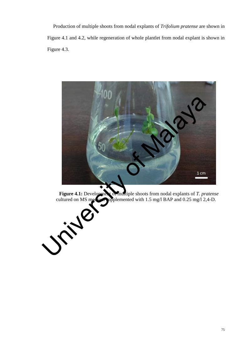
75
Production of multiple shoots from nodal explants of Trifolium pratense are shown in
Figure 4.1 and 4.2, while regeneration of whole plantlet from nodal explant is shown in
Figure 4.3.
Figure 4.1: Development of multiple shoots from nodal explants of T. pratense
cultured on MS medium supplemented with 1.5 mg/l BAP and 0.25 mg/l 2,4-D.
Univers
ity of
Mala
ya

76
Figure 4.2: Development of multiple shoots from nodal explants of T. pratense
cultured on MS medium supplemented with 1.5 mg/l BAP and 0.5 mg/l IBA
Figure 4.3: Complete plantlets regeneration from nodal explants of T. pratense cultured
on MS medium supplemented with 1.5 mg/l BAP and 0.5 mg/l IBA
Univers
ity of
Mala
ya

77
The results also indicated that most of the media containing BAP (1 mg/l – 1.5 mg/l)
and IBA (0.5 mg/l – 0.75 mg/l) hormones were able to induce rooting. The highest mean
number of roots per explant (3.3 ± 0.21), were found in the media containing 1.5 mg/l
BAP and 0.75 mg/l IBA (Table 4.1), while the lowest root formation were observed when
the nodal explants were cultured in the media supplemented with 1.0 mg/l BAP and 0.75
mg/l IBA. As shown in Table 4.2, the explants cultured in the media supplemented with
the combination of BAP and 2,4-D failed to form any roots.
Green coloured callus was obtained when the nodal explants were cultured in the MS
medium supplemented with different concentrations of hormones in this research (Figure
4.4). However, the combinations of BAP and 2,4-D were found to be more effective on
callus formation of nodal explants of red clover (Table 4.2). The highest percentage of
callus formation (100 %) was found in the media supplemented with 1.5 mg/l BAP and
0.5 mg/l 2,4-D.
Figure 4.4: Callus derived from nodal explants of T. pratense, cultured on MS medium
supplemented with 1.5 mg/l BAP and 0.5 mg/l 2,4-D.
Univers
ity of
Mala
ya
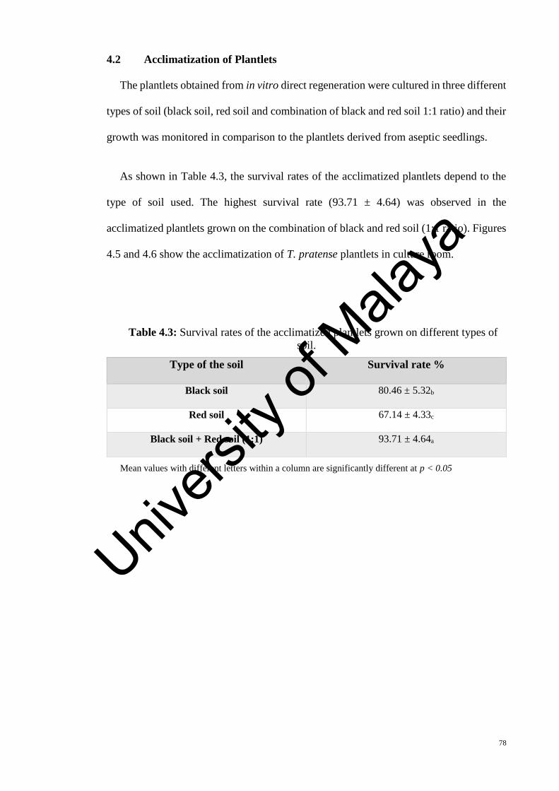
78
4.2 Acclimatization of Plantlets
The plantlets obtained from in vitro direct regeneration were cultured in three different
types of soil (black soil, red soil and combination of black and red soil 1:1 ratio) and their
growth was monitored in comparison to the plantlets derived from aseptic seedlings.
As shown in Table 4.3, the survival rates of the acclimatized plantlets depend to the
type of soil used. The highest survival rate (93.71 ± 4.64) was observed in the
acclimatized plantlets grown on the combination of black and red soil (1:1 ratio). Figures
4.5 and 4.6 show the acclimatization of T. pratense plantlets in culture room.
Table 4.3: Survival rates of the acclimatized plantlets grown on different types of
soil.
Type of the soil Survival rate %
Black soil 80.46 ± 5.32b
Red soil 67.14 ± 4.33c
Black soil + Red soil (1:1) 93.71 ± 4.64a
Mean values with different letters within a column are significantly different at p < 0.05
Univers
ity of
Mala
ya
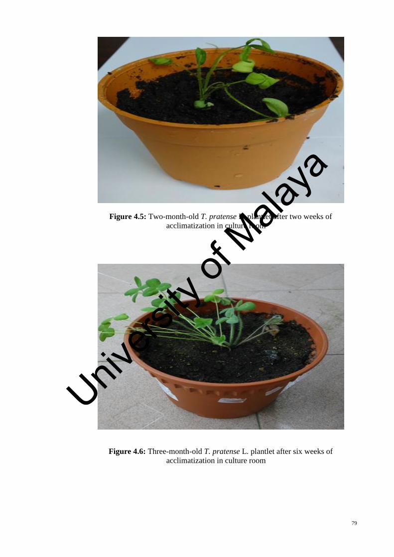
79
Figure 4.5: Two-month-old T. pratense L. plantlet after two weeks of
acclimatization in culture room
Figure 4.6: Three-month-old T. pratense L. plantlet after six weeks of
acclimatization in culture room
Univers
ity of
Mala
ya

80
4.3 Standard Growth of Primary Roots of T. pratense
Results of primary root growth under two different growth conditions (on cotton and
on MS media) were evaluated. The growth of primary roots of Trifolium pratense was
quite slow, as it took 10 days for the secondary roots to start appearing for in vivo (on
cotton) cultured plants and 12 days for the in vitro cultured plants.
The primary roots started to grow on second day of culture and the mean root length
increased rapidly until 5 days after culture but after day 6 the increase of root length was
found very slowly until the emergence of secondary roots on day 10.
The mean root length of the samples versus time was imported into spreadsheet and
the data was plotted in a graph. The graph depicting the linear regression line showing
the relationship between mean root lengths versus time as shown in Figures 4.7 and 4.8
.
Univers
ity of
Mala
ya

81
Figure 4.7: The growth of primary roots of T. pratense, germinated on moist cotton
wool.
Figure 4.8: The growth of primary roots of T. pratense germinated on MS media.
y = 4.2667x - 4.8222R² = 0.9568
0
5
10
15
20
25
30
35
40
0 2 4 6 8 10 12
Mea
n R
oo
t Le
ngt
h (
mm
)
Day
y = 3.3273x - 3.2909R² = 0.9426
0
5
10
15
20
25
30
35
40
0 2 4 6 8 10 12 14
Mea
n R
oo
t Le
ngt
h (
mm
)
Day
Univers
ity of
Mala
ya

82
As shown in Figures 4.5 and 4.6, the relationship between mean primary root length
(mm) and time (day) showed a linear regression, with the equation below:
In cotton west: Y = 4.2667X ˗ 4.8222 R2 = 0.9568
In MS medium: Y = 3.3273X ˗ 3.2909 R2 = 0.9426
The high amount of R2 for root length for germinated seeds in MS media and cotton
west show that the data are closed to the fitted regression line.
4.4 Cytological Studies
The cellular behavior studies of some legume plants are difficult for observation and
analysis because of their small chromosomes. In this thesis, the carnoy’s solution (24h)
with hot hydrolysis (30 min) and Feulgen’s reagent was chosen for carrying out the
cytological studies of Trifolium pratense. In order to study cellular activities of in vivo
and in vitro plantlets of T. pratense, parameters such as chromosome count, mitotic index
(MI), cell and nuclear areas were investigated.
Using Axioskop Zeiss microscope equipped with AxioCam MRc video camera, all
four stages of mitosis (prophase, metaphase, anaphase and telophase) were observed from
the samples and the images of the mitosis phases were successfully captured (Figure 4.9).
Univers
ity of
Mala
ya

83
Figure 4.9: Stages of mitosis observed from squashed preparation of in vivo T.
pratense root tip meristem cells (a: interphase, b: prophase, c: metaphase, d: anaphase
and e: telophase)
4.4.1 Mitotic Index (MI)
In order to calculate the mitotic index, at least 1000 cells were observed in a series of
random transect across the permanent slides. The percentage of cells undergoing mitosis
among all observed cells including interphase cells was calculated. The comparison of
the mitotic index of root meristem cells from in vivo and in vitro grown T. pratense are
summarized in Table 4.4.
Table 4.4: Distribution of mitotic cells in root meristem of in vivo and in vitro grown
T. pratense (Mean ± SD)
Growth
Condition
Interphase
%
Prophase
%
Metaphase
%
Anaphase
%
Telophase
%
Mitotic
Index %
In Vivo 65.18 ± 2.48 24.21 ± 0.87 8.30 ± 0.33 0.72 ± 0.11 1.28 ± 0.17 35.12 ± 1.89 a
In Vitro 59.55 ± 2.78 27.56 ± 1.33 10.23 ± 0.64 0.94 ± 0.17 1.72 ± 0.22 41.89 ± 1.34 a
Mean values with different letters whitin a column are significantly different at p < 0.05
Univers
ity of
Mala
ya
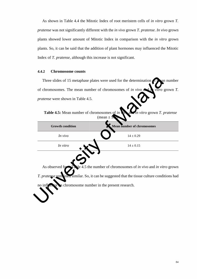
84
As shown in Table 4.4 the Mitotic Index of root meristem cells of in vitro grown T.
pratense was not significantly different with the in vivo grown T. pratense. In vivo grown
plants showed lower amount of Mitotic Index in comparison with the in vitro grown
plants. So, it can be said that the addition of plant hormones may influenced the Mitotic
Index of T. pratense, although this increase is not significant.
4.4.2 Chromosome counts
Three slides of 15 metaphase plates were used for the determination of mean number
of chromosomes. The mean number of chromosomes of in vivo and in vitro grown T.
pratense were shown in Table 4.5.
Table 4.5: Mean number of chromosomes of in vivo and in vitro grown T. pratense
(mean ± SD).
Growth condition Mean number of chromosomes
In vivo 14 ± 0.29
In vitro 14 ± 0.15
As observed from Table 4.5 the number of chromosomes of in vivo and in vitro grown
T. pratense remained similar. So, it can be suggested that the tissue culture conditions had
no influence on chromosome number in the present research.
Univers
ity of
Mala
ya
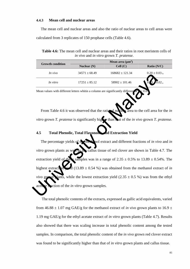
85
4.4.3 Mean cell and nuclear areas
The mean cell and nuclear areas and also the ratio of nuclear areas to cell areas were
calculated from 3 replicates of 150 prophase cells (Table 4.6).
Table 4.6: The mean cell and nuclear areas and their ratios in root meristem cells of
in vivo and in vitro grown T. pratense.
Growth condition Mean area (µm2)
Nuclear (N) Cell (C) Ratio (N/C)
In vivo 34571 ± 68.49 168682 ± 121.34 0.20 ± 0.03 b
In vitro 17251 ± 85.12 58902 ± 101.46 0.29 ± 0.02 a
Mean values with different letters whitin a column are significantly different at p < 0.05
From Table 4.6 it was observed that the ratio of nuclear area to the cell area for the in
vitro grown T. pratense is significantly higher than that of the in vivo grown T. pratense.
4.5 Total Phenolic, Total Flavonoids and Extraction Yield
The percentage yields of the methanol extract and different fractions of in vivo and in
vitro grown plants as well as the callus tissue of red clover are shown in Table 4.7. The
extraction yield of these samples was in a range of 2.35 ± 0.5% to 13.89 ± 0.54%. The
highest extraction yield (13.89 ± 0.54 %) was obtained from the methanol extract of in
vivo grown plants, while the lowest extraction yield (2.35 ± 0.5 %) was from the ethyl
acetate fraction of the in vitro grown samples.
“The total phenolic contents of the extracts, expressed as gallic acid equivalents, varied
from 46.88 ± 1.07 mg GAE/g for the methanol extract of in vivo grown plants to 16.9 ±
1.19 mg GAE/g for the ethyl acetate extract of in vitro grown plants (Table 4.7). Results
also showed that there was scaling increase in total phenolic content among the tested
samples. In comparison, the total phenolic content of the in vivo grown red clover extract
was found to be significantly higher than that of in vitro grown plants and callus tissue.
Univers
ity of
Mala
ya

86
The content of total flavonoids expressed as mg of rutin equivalents per gram of dry
sample, that ranged from 6.11 ± 0.79 to 26.61 ± 0.92, amounts of which were comparable
with results verified in the literatures for other extracts produced (Ao et al., 2008; Manian
et al., 2008). The methanol extract of in vivo grown red clover significantly contained (p
< 0.05) higher flavonoids concentration as compared to other samples involved. The
lowest flavonoid value in this study was recorded in ethyl acetate fraction of in vitro
grown plants” (6.11 ± 0.79 mg CTE / g dry sample).
Table 4.7: Total phenolic, total flavonoid and extraction yield of methanol extract
and soluble fractions of in vivo and in vitro grown plants (aerial parts) and also callus
tissue of Trifolium pratense
Plant extract Total Phenolic (mg Gallic acid / g
dry sample)
Total flavonoid (mg rutin equivalent
/ g dry sample)
Extraction Yield (%)
In Vivo
Methanol extract 46.88 ± 1.07 a 26.61 ± 0.92 a 13.89 ± 0.54 a n-Hexane fraction 30.52 ± 0.72 cd 16.06 ± 1.58 d 10.79 ± 0.59 c Ethyl acetate fraction 27.57 ± 0.49 e 11.71 ± 1.43 fg 6.39 ± 1.12 e Chloroform fraction
40.17 ± 0.88 b 19.56 ± 1.11 b 12.23 ± 0.54 b
In Vitro
Methanol extract 31.94 ± 1.68 c 13.03 ± 0.79 ef 10.84 ± 0.72 c n-Hexane fraction 20.06 ± 0.41 g 8.23 ± 0.96 h 5.20 ± 0.54 f Ethyl acetate fraction 16.90 ± 1.19 h 6.11 ± 0.79 i 2.35 ± 0.5 g Chloroform fraction
25.43 ± 0.87 f 10.39 ± 0.79 g 7.18 ± 0.82 e
Callus Methanol extract n-Hexane fraction Ethyl acetate fraction Chloroform fraction
40.82 ± 1.5 b
31.77 ± 1.14 c
19.23 ± 0.66 g
29.25 ±1.62 de
17.84 ± 0.53 c
12.70 ± 0.94 ef
7.21 ± 0.74 hi
14.02 ± 1.08 e
11.36 ± 0.55 bc
6.13 ± 0.34 ef
2.98 ± 0.66 g
8.70 ± 0.52 d
Each value is represented as mean ± SD (n = 3). Means in the same column not sharing the same letter are significantly different at (Duncan) P < 0.05.
Univers
ity of
Mala
ya
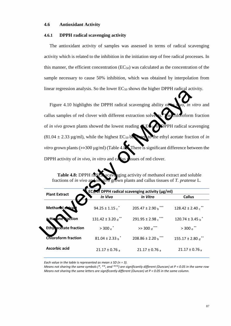
87
4.6 Antioxidant Activity
4.6.1 DPPH radical scavenging activity
The antioxidant activity of samples was assessed in terms of radical scavenging
activity which is related to the inhibition in the initiation step of free radical processes. In
this manner, the efficient concentration (EC50) was calculated as the concentration of the
sample necessary to cause 50% inhibition, which was obtained by interpolation from
linear regression analysis. So the lower EC50 shows the higher DPPH radical activity.
Figure 4.10 highlights the DPPH radical scavenging ability of in vivo, in vitro and
callus samples of red clover with different extraction solvents. The chloroform fraction
of in vivo grown plants showed the lowest reading of EC50 of DPPH radical scavenging
(81.04 ± 2.33 µg/ml), while the highest EC50 belonged to the ethyl acetate fraction of in
vitro grown plants (˃˃300 µg/ml) (Table 4.8). There is significant difference between the
DPPH activity of in vivo, in vitro and callus tissues of red clover.
Table 4.8: DPPH radical scavenging activity of methanol extract and soluble
fractions of in vivo and in vitro grown plants and callus tissues of T. pratense L.
Plant Extract EC50 of DPPH radical scavenging activity (µg/ml)
In Vivo In Vitro Callus
Methanol extract 94.25 ± 1.15 c * 205.47 ± 2.90 b *** 128.42 ± 2.40 c ** n-Hexane fraction 131.42 ± 3.20 d ** 291.95 ± 2.98 c *** 120.74 ± 3.45 b *
Ethyl acetate fraction ˃ 300 e * ˃˃ 300 d *** ˃ 300 e ** Chloroform fraction Ascorbic acid
81.04 ± 2.33 b *
21.17 ± 0.76 a
208.86 ± 2.20 b ***
21.17 ± 0.76 a
155.17 ± 2.80 d **
21.17 ± 0.76 a
Each value in the table is represented as mean ± SD (n = 3). Means not sharing the same symbols (*, **, and ***) are significantly different (Duncan) at P < 0.05 in the same row Means not sharing the same letters are significantly different (Duncan) at P < 0.05 in the same column.
Univers
ity of
Mala
ya

88
Figure 4.10: DPPH radical scavenging activity of different extracts from the methanol
extract of Trifolium pratense by different solvent at different concentration. A: In vivo
grown plants, B: In vitro grown plants, C: Callus tissue. Each value represents as mean ±
SD (n = 3).
0
10
20
30
40
50
60
70
80
90
100
0 50 100 150 200 250 300 350
DP
PH
% s
cave
ngi
ng
Concentration µg/ml
A
Methanol
Hexane
Ethyl acetate
Chloroform
Ascorbic Acid
0
10
20
30
40
50
60
70
80
90
100
0 50 100 150 200 250 300 350
DP
PH
% s
cave
ngi
ng
Concentration µg/ml
B
Methanol
Hexane
Ethyl acetate
Chloroform
Ascorbic acid
0
20
40
60
80
100
120
0 50 100 150 200 250 300 350
DP
PH
% s
cave
ngi
ng
Concentration µg/ml
C
Methanol
Hexane
Ethyl acetate
Chloroform
Ascorbic acidUniv
ersity
of M
alaya

89
4.6.2 Superoxide Radical Scavenging Activity
The superoxide radical scavenging effect of these varying fractions of methanol extract
of in vivo and in vitro grown as well as the callus tissue of red clover was drawn in
comparison with the same doses of ascorbic acid in a range of 50 to 300 µg/ml as shown
in Figure 4.11. The results had suggested that the lowest EC50 value (70.69 ± 1.76 µg/ml)
belongs to the chloroform fraction of in vivo samples, whereas the highest (260.01 ± 1.46
µg/ml) belongs to the ethyl acetate fraction of in vitro samples (Table 4.9).
Table 4.9: Superoxide anion activity of methanol extract and soluble fractions of in
vivo and in vitro grown plant and callus tissue of T. pratense L.
Plant Extract EC50 of superoxide anion activity (µg/ml)
In Vivo In Vitro Callus
Methanol extract 74.68 ± 1.59 c *
179.77 ± 1.60 c ** 74.36 ± 1.42 b *
n-Hexane fraction 139.83 ± 2.40 d * 246.59 ± 2.09 d *** 190.06 ± 2.56 d **
Ethyl acetate fraction 201.55 ± 1.94 e * 260.01 ± 1.46 e *** 228.41 ± 1.52 e **
Chloroform fraction Ascorbic acid
70.69 ± 1.76 b *
23.44 ± 0.84 a
155.91 ± 1.73 b ***
23.44 ± 0.84 a
104.32 ± 1.45 c **
23.44 ± 0.84 a
Each value in the table is represented as mean ± SD (n = 3). Means which are not sharing the same symbols (*, **, and ***) are significantly different (Duncan) at P < 0.05 in the same row. Means which are not sharing the same letters are significantly different (Duncan) at P < 0.05 in the same column.
Univers
ity of
Mala
ya

90
Figure 4.11: Superoxide radical scavenging activity of different extracts from the
methanol extract of T. pratense L. by different solvent at different concentration. A: In
vivo grown plants, B: In vitro grown plants, C: Callus tissue. Each value represents as
mean ± SD (n = 3).
0
10
20
30
40
50
60
70
80
90
100
0 50 100 150 200 250 300 350
Sup
ero
xid
e %
sca
ven
gin
g
Concentration µg/ml
A
Methanol
Hexane
Ethyl acetate
Chloroform
Ascorbic Acid
0
10
20
30
40
50
60
70
80
90
100
0 50 100 150 200 250 300 350
Sup
ero
xid
e %
sca
ven
gin
g
Concentration µg/ml
B
Methanol
Hexane
Ethyl acetate
Chloroform
Ascorbic Acid
0
10
20
30
40
50
60
70
80
90
100
0 50 100 150 200 250 300 350
Sup
ero
xid
e %
sca
ven
gin
g
Concentration µg/ml
C
Methanol
Hexane
Ethyl acetate
Chloroform
Ascorbic AcidUniv
ersity
of M
alaya

91
4.6.3 ABTS radical scavenging activity
The samples’ ABTS radical scavenging capability can be ranked as such: in vivo ˃
callus ˃ in vitro samples. The methanol extract of in vivo grown samples had
demonstrated the highest radical scavenging activity when it reacted with the ABTS
radicals. By contrast, the ethyl acetate fraction of in vivo, in vitro and callus samples and
also the n-hexane fraction of in vitro samples did not illustrate any leveling effect at the
highest concentration, but it was a fact that their radical scavenging effects were much
less (p < 0.05) than that of the other extracts tested (Table 4.10). Significant differences
among the EC50 values of all the fractions and ascorbic acid had also been noted (p <
0.05).
Table 4.10: ABTS radical scavenging of methanol extract and soluble fractions of in
vivo and in vitro grown plant and callus tissue of T. pratense L.
Plant Extract EC50 of ABTS (µg/ml)
In Vivo In Vitro Callus
Methanol extract 111.84 ± 1.46 b *
351.46 ± 2.02 c
*** 164.32 ± 1.61 b **
n-Hexane fraction 210.76 ± 1.71 d * ˃ 500 d *** 225.63 ± 2.05 d **
Ethyl acetate fraction ˃ 500 e * ˃˃ 500 e *** ˃ 500 e **
Chloroform fraction Ascorbic acid
122.34 ± 2.29 c *
34.67 ± 0.53 a
297.68 ± 1.68 b ***
34.67 ± 0.53 a
172.18 ± 1.55 c **
34.67 ± 0.53 a
Each value in the table is represented as mean ± SD (n = 3). Means not sharing the same symbols (*, **, and ***) are significantly different (Duncan) at P < 0.05 in the same row. Means not sharing the same letters are significantly different (Duncan) at P < 0.05 in the same column.
Univers
ity of
Mala
ya

92
4.6.4 Hydrogen peroxide
ExtractS from in vivo, in vitro and callus tissue of red clover were capable of
scavenging hydrogen peroxide in a concentration dependent manner (50–300 µg/ml). As
compared with the EC50 values, the hydrogen peroxide scavenging activity of chloroform
fraction of in vivo grown red clover (88.35 ± 1.27 µg/ml) was more effective (P < 0.05)
than that of other samples. The lowest hydrogen peroxide scavenging activities were
found in the n-hexane and ethyl acetate fractions of in vitro grown plants with the EC50
values of more than 300 µg/ml (Table 4.11).
Table 4.11: Hydrogen peroxide scavenging of methanol extract and soluble fractions
of in vivo and in vitro grown plant and callus tissue of Trifolium pratense.
Plant Extract EC50 of Hydrogen peroxide (µg/ml)
In Vivo In Vitro Callus
Methanol extract 103.44 ± 1.47 c * 219.86 ± 1.57 b *** 141.74 ± 1.93 c ** n-Hexane fraction 270.56 ± 1.72 e * ˃˃ 300 e *** 287.63 ± 1.59 e **
Ethyl acetate fraction 138.16 ± 1.93 d ** ˃300
d *** 132.26 ± 2.03 b *
Chloroform fraction Ascorbic acid
88.35 ± 1.27 b *
26.84 ± 0.83 a
231.49 ± 2.02 c ***
26.84 ± 0.83 a
146.31 ± 1.67 d **
26.84 ± 0.83 a
Each value in the table is represented as mean ± SD (n = 3). Means not sharing the same symbols (*, **, and ***) are significantly different (Duncan) at P < 0.05 in the same row. Means not sharing the same letters are significantly different (Duncan) at P < 0.05 in the same column.
Univers
ity of
Mala
ya

93
4.6.5 Chelating activity
The EC50 values of iron chelating activity for different extracts of in vivo and in vitro
grown as well as callus tissues of red clover were shown in Table 4.12. The highest
chelating power shown by the methanol extract of in vivo grown plants with the EC50
value of 49.11 ± 0.97 µg/ml, whilst, the lowest was found in ethyl acetate fraction of in
vitro grown samples with the EC50 value of 183.44 ± 2.48 µg/ml.
Table 4.12: Chelating activity of methanol extract and soluble fractions of in vivo
and in vitro grown plant and callus tissue of Trifolium pratense.
Plant Extract EC50 of Chelating power (µg/ml)
In Vivo In Vitro Callus
Methanol extract 49.11 ± 0.97 b * 142.87 ± 1.71 c *** 72.56 ± 1.16 d **
n-Hexane fraction 52.33 ± 1.97 c * 131.84 ± 2.05 b *** 67.52 ± 2.15 c ** Ethyl acetate fraction 105.67 ± 1.56 e * 183.44 ± 2.48 e *** 118.23 ± 2.02 e ** Chloroform fraction 86.32 ± 1.89 d ** 161.75 ± 1.36 d *** 60.17 ± 1.42 b *
Catechin 22.76 ± 0.37 a 22.76 ± 0.37 a 22.76 ± 0.37 a
Each value in the table is represented as mean ± SD (n = 3). Means not sharing the same symbols (*, **, and ***) are significantly different (Duncan) at P < 0.05 in the same row. Means not sharing the same letters are significantly different (Duncan) at P < 0.05 in the same column.
4.6.6 Reducing power activity
Table 4.13 shows the reducing power absorbance of different extracts of in vivo and
in vitro grown plants as well as callus tissue of red clover. Among the examined extracts
the methanol extract of in vivo samples showed the highest reducing power with the
absorbance of 1.05 ± 0.07 at 700 nm, so this extract could act as electron donors and also
could convert free radicals to more stable products. Although, in comparison with the
Univers
ity of
Mala
ya

94
positive control (ascorbic acid) it is significantly lower. The lowest reducing power (0.38
± 0.01) belongs to the ethyl acetate fraction of in vitro grown plants.
Table 4.13: Reducing power of methanol extract and soluble fraction (300 µg/ml) of
in vivo and in vitro grown plant and callus tissue of Trifolium pratense.
Plant Extract Reducing power absorbance at 700 nm
In Vivo In Vitro Callus
Methanol extract 1.05 ± 0.07 b *
0.72 ± 0.03 b,
*** 0.82 ± 0.03 c **
n-Hexane fraction 0.88 ± 0.04 c * 0.52 ± 0.04 c, *** 0.65 ± 0.02 d
**
Ethyl acetate fraction 0.59 ± 0.03 d * 0.38 ± 0.01 d *** 0.52 ± 0.03 e
**
Chloroform fraction Ascorbic acid
0.95 ± 0.03 c *
2.45 ± 0.03 a
0.71 ± 0.04 b **
2.45 ± 0.03 a
0.91 ± 0.03 b *
2.45 ± 0.03 a
Each value in the table is represented as mean ± SD (n = 3). Means not sharing the same symbols (*, **, and ***) are significantly different (Duncan) at P < 0.05 in the same row. Means not sharing the same letters are significantly different (Duncan) at P < 0.05 in the same column.
4.7 Relationship between Antioxidant Activities and Total Phenolic and
Flavonoid Contents
Through the correlation analysis for phytochemical contents with EC50 values of
radical scavenging activity and antioxidant ability of the extract of red clover and its
various soluble fractions, the phenolic and flavonoid contents had exhibited excellent
association with DPPH, superoxide, ABTS radical scavenging activities and reducing
power of in vivo, in vitro and callus samples (Tables 4.14, 4.15 and 4.16).
Univers
ity of
Mala
ya
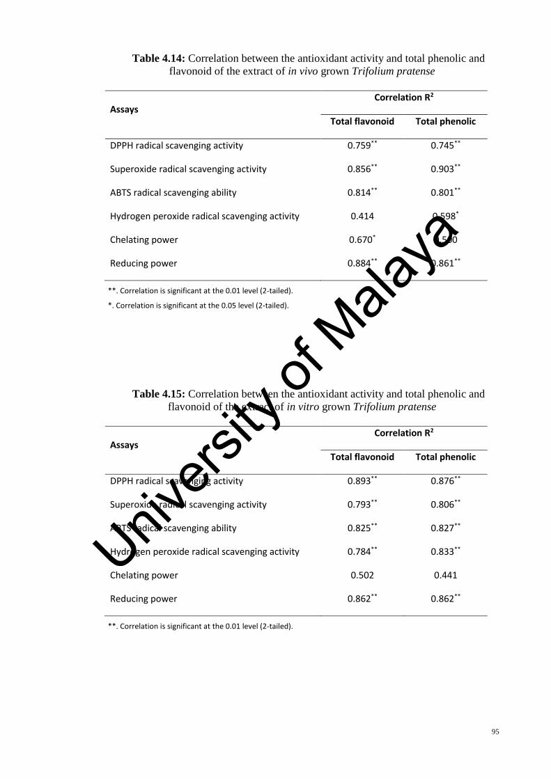
95
Table 4.14: Correlation between the antioxidant activity and total phenolic and
flavonoid of the extract of in vivo grown Trifolium pratense
Assays Correlation R2
Total flavonoid Total phenolic
DPPH radical scavenging activity 0.759** 0.745**
Superoxide radical scavenging activity 0.856** 0.903**
ABTS radical scavenging ability 0.814** 0.801**
Hydrogen peroxide radical scavenging activity 0.414 0.598*
Chelating power 0.670* 0.500
Reducing power 0.884** 0.861**
**. Correlation is significant at the 0.01 level (2-tailed).
*. Correlation is significant at the 0.05 level (2-tailed).
Table 4.15: Correlation between the antioxidant activity and total phenolic and
flavonoid of the extract of in vitro grown Trifolium pratense
Assays Correlation R2
Total flavonoid Total phenolic
DPPH radical scavenging activity 0.893** 0.876**
Superoxide radical scavenging activity 0.793** 0.806**
ABTS radical scavenging ability 0.825** 0.827**
Hydrogen peroxide radical scavenging activity 0.784** 0.833**
Chelating power 0.502 0.441
Reducing power 0.862** 0.862**
**. Correlation is significant at the 0.01 level (2-tailed).
Univers
ity of
Mala
ya

96
Table 4.16: Correlation between the antioxidant activity and total phenolic and
flavonoid of the extract of callus tissue of Trifolium pratense
Assays Correlation R2
Total flavonoid Total phenolic
DPPH radical scavenging activity 0.849** 0.822**
Superoxide radical scavenging activity 0.912** 0.917**
ABTS radical scavenging ability 0.903** 0.876**
Hydrogen peroxide radical scavenging activity 0.023 0.020
Chelating power 0.775** 0.734**
Reducing power 0.777** 0.753**
**. Correlation is significant at the 0.01 level (2-tailed).
4.8 Anticancer Activity
In the present study, the cytotoxic effects of different extracts of in vivo and in vitro
grown plants as well as callus tissue of Trifolium pratense on two human cancer cell lines
(MCF-7 and HCT-116) were evaluated using the MTT assay. Cell lines were incubated
with different concentrations of sample extracts. After 24, 48 and 72 hours, the cell
inhibition was determined and IC50 was calculated for all the plant extracts (Tables 4.17
and 4.18).
Treatment of MCF-7 and HCT-116 cell lines with different extracts of red clover
resulted in a time dependent increase in the anticancer activity and the greatest anticancer
activity was observed when the cells were exposed to the plant extract for 72 h.
All the examined extracts inhibited growth of MCF-7 and HCT-116 cell lines in a
concentration dependent manner. This is displayed in Figure 4.12 for the two most active
extracts: chloroform fraction of in vivo plants and callus tissues.
Univers
ity of
Mala
ya

97
The chloroform fraction of in vivo plants showed a reading of the lowest IC50 (66.44
± 2.05 µg/ml) of inhibition against the MCF-7 cell line, although it is not significantly
different with the IC50 value of chloroform fraction of callus tissues (69.48 ± 2.66 µg/ml).
The lowest IC50 (79.53 ± 2.00 µg/ml) of inhibition against the HCT-116 cell line was
shown by the chloroform fraction of callus tissue. Although the cytotoxic activity of the
fractions were less (p < 0.05) than those of the doxorubicin (positive control), the study
had revealed that in vivo grown plants and callus tissues of red clover have inhibition
activity against MCF-7 and HCT-116 cell lines, more than in vitro grown red clover.
Table 4.17: Cytotoxic activity of the different extracts of in vivo, in vitro and callus
tissues of T. pratense L. against human breast carcinoma (MCF-7). (n = 3)
Extract IC50 µg/ml
24 hours 48 hours 72 hours
In Vivo Methanol extract 104.25 ± 2.17 c 86.97 ± 2.05 d 83.62 ± 1.58 c
n-Hexane fraction 147.89 ± 1.86 f 126.12 ± 2.22 h 117.57 ± 2.42 g Ethyl acetate fraction 207.24 ± 2.73 h 176.45 ± 1.92 l 169.96 ± 2.55 k
Chloroform fraction 93.9 ± 2.32 b 75.26 ± 1.68 b 66.44 ± 2.05 b
In Vitro
Methanol extract 131.01 ± 1.39 e 115.85 ± 1.87 g 110.62 ± 2.49 f n-Hexane fraction 222.29 ± 5.13 j 163.98 ± 2.49 k 155.50 ± 0.76 j
Ethyl acetate fraction 250.42 ± 4.2 k 181.5 ± 1.11m 178.89 ± 2.03 l Chloroform fraction 144.68 ± 1.87 f 110.72 ± 2.33 f 101.11 ± 2.26 e
Callus
Methanol extract 118.11 ± 2.18 d 97.47 ± 1.53 e 88.73 ± 1.85 d
n-Hexane fraction 188.64 ± 1.87 g 145.3 ± 2.22 i 131.30 ± 2.06 i
Ethyl acetate fraction 213.47 ± 2.19 i 158.54 ± 2.36 j 124.64 ± 1.73 h Chloroform fraction 100.71 ±1.8 b 79.35 ± 1.88 c 69.48 ± 2.66 b
Doxorubicin* 22.49 ± 1.33 a 19.25 ± 0.87 a 17.54 ± 0.69 a
IC50 value represents the concentration of the tested samples to kill 50% of the cancer cells. Each value in the table is represented as mean ± SD (n = 3). *Positive control For each treatment, the means within the column followed by different superscript letters were significantly different at p ˂ 0.05.
Univers
ity of
Mala
ya

98
Table 4.18: Cytotoxic activity of the different extracts of in vivo, in vitro and callus
tissues of T. pratense L. against human colon carcinoma (HCT-116). (n = 3)
Extract IC50 µg/ml
24 hours 48 hours 72 hours
In Vivo Methanol extract 142.68 ± 1.56 d 115.99 ± 1.47 e 100.75 ± 2.33 e
n-Hexane fraction 200.82 ± 3.26 g 166.13 ± 1.83 i 138.60 ± 1.71 h Ethyl acetate fraction 262.02 ± 2.22 j 215.40 ± 2.49 l 180.28 ± 2.54 k
Chloroform fraction 118.64 ± 1.63 c 96.95 ± 1.53 c 87.80 ± 2.34 c
In Vitro
Methanol extract 185.43 ± 2.84 f 144.58 ± 2.15 g 128.67 ± 2.42 g n-Hexane fraction 293.44 ± 4.60 l 214.82 ± 2.01 l 185.53 ± 2.48 l
Ethyl acetate fraction 276.16 ± 2.26 k 185.00 ± 2.26 j 161.55 ± 2.92 j Chloroform fraction 155.59 ± 1.93 e 126.76 ± 1.47 f 113.24 ± 1.90 f
Callus
Methanol extract 145.05 ± 2.24 d 107.96 ± 2.05 d 96.02 ± 2.7 d
n-Hexane fraction 217.10 ± 2.29 h 148.78 ± 2.37 h 127.73 ± 2.26 g
Ethyl acetate fraction 254.57 ± 2.73 i 192.84 ± 2.77 k 157.36 ± 2.43 i Chloroform fraction 109.54 ± 2.44 b 85.55 ± 2.95 b 79.53 ± 2.00 b
Doxorubicin * 31.24 ± 1.65 a 26.32 ± 0.55 a 25.18 ± 1.31 a
IC50 value represents the concentration of the tested samples to kill 50% of the cancer cells. Each value in the table is represented as mean ± SD (n = 3). *Positive control For each treatment, the means within the column followed by different superscript letters were significantly different at p ˂ 0.05.
Univers
ity of
Mala
ya
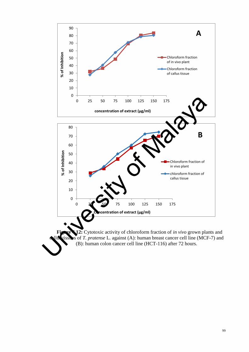
99
Figure 4.12: Cytotoxic activity of chloroform fraction of in vivo grown plants and
callus tissues of T. pratense L. against (A): human breast cancer cell line (MCF-7) and
(B): human colon cancer cell line (HCT-116) after 72 hours.
0
10
20
30
40
50
60
70
80
90
0 25 50 75 100 125 150 175
% o
f In
hib
itio
n
concentration of extract (µg/ml)
A
Chloroform fractionof in vivo plant
Chloroform fractionof callus tissue
0
10
20
30
40
50
60
70
80
0 25 50 75 100 125 150 175
% o
f In
hib
itio
n
Concentration of extract (µg/ml)
B
Chloroform fraction ofin vivo plant
chloroform fraction ofcallus tissue
Univers
ity of
Mala
ya

100
4.9 Antimicrobial Activity
In the present investigation, the inhibitory effect of ethanolic and methanolic extracts
of in vivo, in vitro and callus from Trifolium pratense were evaluated against both
fungicidal and bacterial strains. The antibacterial and antifungal activities were
determined using paper disc diffusion method and summarized in Tables 4.19 and 4.20.
“The antimicrobial potential of the experimental plants were evaluated according to
their zone of inhibition against various pathogens and the results (zone of inhibition) were
compared with the activity of the standards. The results revealed that all callus, in vivo
and in vitro grown plants extracts are potent antimicrobial against three of the examined
bacteria strains (Staphylococcus aureus, Bacillus cereus and Escherichia coli) and two of
the examined fungal strains (Aspergillus niger and Candida albicans). The callus extract
showed antimicrobial activities against all the examined microorganisms. Among the two
different solvents used for extraction, methanol extract showed higher degree of
inhibition followed by ethanol extract. For all the investigated microorganisms, the callus
extract showed maximum antibacterial and antifungal activities. In ethanolic extract,
maximum inhibition zone diameter among all the tested microorganisms was obtained in
E. coli with diameter of 16 ± 1.76 mm. Similarly, methanol extract showed maximum
inhibition zone with diameter of 16 ± 1.66 mm in B. cereus and 16 ± 1.44 mm in C.
albicans.”
Univers
ity of
Mala
ya
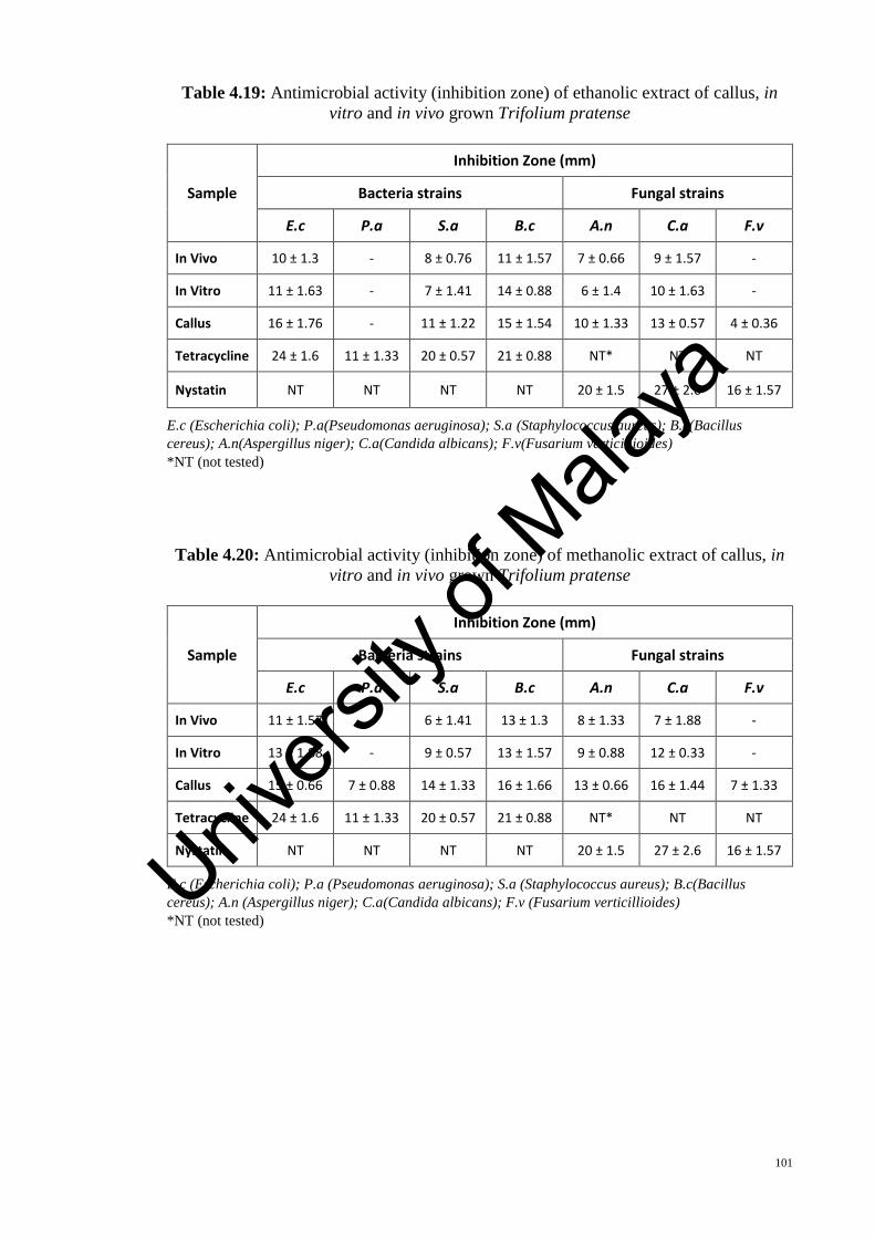
101
Table 4.19: Antimicrobial activity (inhibition zone) of ethanolic extract of callus, in
vitro and in vivo grown Trifolium pratense
Sample
Inhibition Zone (mm)
Bacteria strains Fungal strains
E.c P.a S.a B.c A.n C.a F.v
In Vivo 10 ± 1.3 - 8 ± 0.76 11 ± 1.57 7 ± 0.66 9 ± 1.57 -
In Vitro 11 ± 1.63 - 7 ± 1.41 14 ± 0.88 6 ± 1.4 10 ± 1.63 -
Callus 16 ± 1.76 - 11 ± 1.22 15 ± 1.54 10 ± 1.33 13 ± 0.57 4 ± 0.36
Tetracycline 24 ± 1.6 11 ± 1.33 20 ± 0.57 21 ± 0.88 NT* NT NT
Nystatin NT NT NT NT 20 ± 1.5 27 ± 2.6 16 ± 1.57
E.c (Escherichia coli); P.a(Pseudomonas aeruginosa); S.a (Staphylococcus aureus); B.c(Bacillus
cereus); A.n(Aspergillus niger); C.a(Candida albicans); F.v(Fusarium verticillioides)
*NT (not tested)
Table 4.20: Antimicrobial activity (inhibition zone) of methanolic extract of callus, in
vitro and in vivo grown Trifolium pratense
Sample
Inhibition Zone (mm)
Bacteria strains Fungal strains
E.c P.a S.a B.c A.n C.a F.v
In Vivo 11 ± 1.57 - 6 ± 1.41 13 ± 1.3 8 ± 1.33 7 ± 1.88 -
In Vitro 13 ± 1.88 - 9 ± 0.57 13 ± 1.57 9 ± 0.88 12 ± 0.33 -
Callus 15 ± 0.66 7 ± 0.88 14 ± 1.33 16 ± 1.66 13 ± 0.66 16 ± 1.44 7 ± 1.33
Tetracycline 24 ± 1.6 11 ± 1.33 20 ± 0.57 21 ± 0.88 NT* NT NT
Nystatin NT NT NT NT 20 ± 1.5 27 ± 2.6 16 ± 1.57
E.c (Escherichia coli); P.a (Pseudomonas aeruginosa); S.a (Staphylococcus aureus); B.c(Bacillus
cereus); A.n (Aspergillus niger); C.a(Candida albicans); F.v (Fusarium verticillioides)
*NT (not tested)
Univers
ity of
Mala
ya
102
4.10 Photosynthesis and Leaf Gas Exchange of T. pratense L.
“The measurements of stomatal conductance (gs), transpiration rate (E) and net
photosynthetic (Pn) were done using the LICOR 6400 portable photosynthesis at the
standard cuvette conditions (i.e PPFD1000 μmol/m2/sec, carbon dioxide reference 400
μmol/mol, relative humidity 50 – 60 %) and leaf temperature standardized at 30 °C. All
measurements were obtained 6 and 12 weeks after planting from fully expanded leaves.
Results obtained showed that, significant differences were found among the samples in
correlation with the leaf gas exchange in which the highest Pn (15.51 ± 0.34) were
identified in 6-weeks old in vivo grown plants, while, the highest gs (164.49 ± 4.95) and
E (6.35 ± 0.14) were identified in 6-weeks old in vitro grown plants (Figure 4.13). The
mean analysis results of dual correlation of traits are shown in Table 4.21. The net
photosynthesis was found to be positively correlated with secondary metabolites (total
phenolic, TP; total flavonoids, TF)” at the significant level of 0.01.
“In the natural environment, photon flux density (PPFD) of leaves varied greatly over
time (Knapp & Smith 1990). Therefore, photosynthetic utilization of fluctuating PFD is
important in estimating leaf photosynthetic capacity and water-use efficiency (Tang
1997). The maximum net photosynthetic (PNmax), apparent quantum yield (AQY),
compensation point (CP) and respiration (Rd) were calculated from the light response
curve shown in Figure 4.14 (regression were used to fit the data). Significant differences
found in the accessions of the initial gradient in the light response curve, the light
compensation point, the rate of dark respiration and the maximum rate of the
photosynthetic, achieved from fitting the light response curves.”
“The light response curves attributes were determined for both in vivo and in vitro
samples. Saturation PPFD of in vitro grown plants was 720 µmol/m2/s, while in in vivo
grown plants was 960 µmol/m2/s. A comparison was made for the observed data for the
Univers
ity of
Mala
ya

103
light-saturated photosynthetic among the treatments revealed significantly greater rates
for PNmax (18.36 µmol/m2/s) and AQY (0.0389 µmol/m2/s) in in vivo plants (Figure 4.15).
Moreover, in vitro plants were observed to contain the highest rate of Rd (0.828
µmol/m2/s) and CP (29.48 µmol/m2/s). The form of light–response curves had a
logarithmic part with the maximum gradient (maximum quantum efficiency) at the start
of curves, i.e., when the irradiation was reaching zero. The ‘‘S’’-shaped curves, which
signify quantum efficiency under low light intensities tends to zero, were identified in a
limited number of investigations.”
Univers
ity of
Mala
ya

104
Figure 4.13: Net photosynthesis (Pn), Stomatal conductance (gs) and Transpiration
rate (E) of in vivo and in vitro grown Trifolium pratense, six and twelve weeks after
planting. Each experiment repeated 5 times and data presented as the mean ± standard
error.
0
1
2
3
4
5
6
7
W6 W12
E (m
mo
l H2o
/ m
2/s
)
Harvesting Time
In Vivo
In Vitro
0
20
40
60
80
100
120
140
160
180
W6 W12
g s(m
mo
l / m
2/
s)
Harvesting Time
In Vivo
In Vitro
0
2
4
6
8
10
12
14
16
18
W6 W12
Pn
(µm
ol /
m2
/ s)
Harvesting Time
In Vivo
In VitroUnivers
ity of
Mala
ya

105
Table 4.21: Correlation analysis between secondary metabolites and leaf gas exchange
parameters of T. pratense in mean traits of in vivo and in vitro
PN gs E TF
TP 0.994** -0.326 0.059 0.994**
TF 0.975* -0.429 -0.041
E 0.154 0.872
gs -0.222
**. Correlation is significant at the 0.01 level (2-tailed).
*. Correlation is significant at the 0.05 level (2-tailed).
TP = Total phenolic, TF = Total flavonoid, E = Transpiration rate, gs = Stomatal conductance
Figure 4.14: Light response curve in leaves from in vivo and in vitro grown plants of T.
pratense. (n = 5, at each PPFD)
y = -2E-05x2 + 0.0389x - 0.5533R² = 0.9975
y = -2E-05x2 + 0.0287x - 0.8287R² = 0.9831
-5
0
5
10
15
20
0 200 400 600 800 1000 1200 1400
Net
CO
2as
sim
ilati
on
rat
e [µ
mo
l (C
O2
) /
m2
/ s]
PPFD [µmol (Photon) / m2 / s]
In Vivo
In Vitro
Univers
ity of
Mala
ya

106
4.11 Effect of Salt Stress on Germination and Growth of T. pratense
“The seed germination and growth of T. pratense was not significantly affected by the
50 mM NaCl treatment, although they were inhibited with the increase of NaCl
concentration to 100, 150 and 200 mM. It was found that in the presence of 50 mM NaCl
in MS medium the seed germination was a bit higher while the shoot and root length were
slightly lower in the same condition. The germination percentage in red clover seeds
treated with 100, 150 and 200 mM of NaCl were reduced by 46%, 22.4% and 10.6%,
respectively” (Figure 4.16).
0
5
10
15
20
PNmax (µmol/m2/s)
In Vivo
In Vitro
0
5
10
15
20
25
30
35
CP (µmol/m2/s)
In Vivo
In Vitro
-1
-0.8
-0.6
-0.4
-0.2
0
Rd (µmol/m2/s)
In Vivo
In Vitro
0
0.01
0.02
0.03
0.04
0.05
AQY (µmol/m2/s)
In Vivo
In Vitro
Figure 4.15: Maximum net assimilation rate (PNmax), apparent quantum
yield (AQY), compensation point (CP) and respiration rate (Rd) in in vivo and
in vitro grown Trifolium pratense, six weeks after planting
Univers
ity of
Mala
ya

107
Figure 4.16: Germination percentage of Red clover seeds under different saline
treatment. Each treatment repeated 5 times and data presented as the mean ± standard
error. Values which are not sharing a common letter are significantly different at p ≤
0.05
At the high concentration of NaCl (100, 150 and 200 mM), the length of shoots and
roots were inhibited significantly, although to different extent. The most severe growth
restriction of shoots and roots (8.97% and 8.76% of the control, respectively) were found
in the treatments with 200 mM of NaCl (Figures 4.17 and 4.18).
a a
b
c
d
0
20
40
60
80
100
0 50 100 150 200
Pe
rce
nta
ge o
f G
erm
inat
ion
NaCl Concentration (mM)
Univers
ity of
Mala
ya

108
Figure 4.17: Shoot length of germinated T. pratense seeds under different saline
treatments after 6 weeks of culture. Each data is the mean of 5 replicates of 25 explants
per treatment (mean ± standard error). Values which are not sharing a common letter are
significantly different at p ≤ 0.05.
Figure 4.18: Root length of germinated T. pratense seeds under different saline
treatments after 6 weeks of culture. Each data is the mean of 5 replicates of 25 explants
per treatment (mean ± standard error). Values which are not sharing a common letter are
significantly different at p ≤ 0.05.
aa
b
cC
0
10
20
30
40
50
60
0 50 100 150 200
Sho
ot
Len
gth
(m
m)
NaCl Concentration (mM)
a a
b
cC
0
10
20
30
40
50
60
70
80
0 50 100 150 200
Ro
ot
Len
gth
(m
m)
Nacl Concentration (mM)
Univers
ity of
Mala
ya
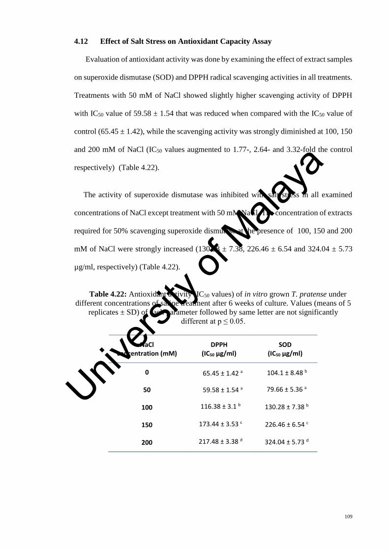
109
4.12 Effect of Salt Stress on Antioxidant Capacity Assay
“Evaluation of antioxidant activity was done by examining the effect of extract samples
on superoxide dismutase (SOD) and DPPH radical scavenging activities in all treatments.
Treatments with 50 mM of NaCl showed slightly higher scavenging activity of DPPH
with IC50 value of 59.58 ± 1.54 that was reduced when compared with the IC50 value of
control (65.45 ± 1.42), while the scavenging activity was strongly diminished at 100, 150
and 200 mM of NaCl (IC50 values augmented to 1.77-, 2.64- and 3.32-fold the control
respectively)” (Table 4.22).
The activity of superoxide dismutase was inhibited with salt stress in all examined
concentrations of NaCl except treatment with 50 mM NaCl. The concentration of extracts
required for 50% scavenging superoxide dismutase at the presence of 100, 150 and 200
mM of NaCl were strongly increased (130.28 ± 7.38, 226.46 ± 6.54 and 324.04 ± 5.73
µg/ml, respectively) (Table 4.22).
Table 4.22: Antioxidant activity (IC50 values) of in vitro grown T. pratense under
different concentrations of saline treatment after 6 weeks of culture. Values (means of 5
replicates ± SD) of each parameter followed by same letter are not significantly
different at p ≤ 0.05.
NaCl concentration (mM)
DPPH (IC50 µg/ml)
SOD (IC50 µg/ml)
0
50
100
150
200
65.45 ± 1.42 a
59.58 ± 1.54 a
116.38 ± 3.1 b
173.44 ± 3.53 c
217.48 ± 3.38 d
104.1 ± 8.48 b
79.66 ± 5.36 a
130.28 ± 7.38 b
226.46 ± 6.54 c
324.04 ± 5.73 d
Univers
ity of
Mala
ya
110
CHAPTER 5: DISCUSSION
“Trifolium pratense is one of the most important forage crop from temperate regions.
It is highly productive, with nitrogen-fixing ability, protein- and mineral-rich species
adapted to a wide range of soils and environmental conditions. Its erect growth habit
makes it suitable for hay and silage making. High nutritive value and voluntary intake
characteristics lead to good animal performance. It is a valuable break crop in arable
farming and organic farming on account of N- fixation ability. Its high nutritive value
allied to its high intake characteristics combine to give improved individual animal
performance from different classes of animals compared with grass forage. As a human
food the young leaves are harvested before the plants come into the flower, and are used
in salads, soups and etc. It is safe and effective herb with a long history of medicinal
usages.”
The potential of tissue culture techniques for the production of several secondary
metabolites has been known for many years. Evidently tissue cultures stimulate the
production or induce the biosynthesis of novel compounds not found in the mature plant.
The development of callus culture could provide an alternative supply of phytochemicals
such as novel phytoestrogens to be used in medicine. Phytoestrogens in tissue cultures
may prove to be medically beneficial for diseases such as cancers, cardiovascular
diseases, and osteoporosis (Vanishes et al., 2004).
Tissue culture is a technique to enhance the application of biotechnological approaches
to crop plants. Micropropagation and callus initiation of T. pratense were studied on Ms
medium supplemented with concentrations of hormones (auxin and cytokinin). The tissue
culture techniques established in this study can be utilized to achieve mass propagation
of Trifolium pratense. The present study is divided into several parts that investigate the
in vitro micropropagation, induction of callus, acclimatization of in vitro regenerated
Univers
ity of
Mala
ya

111
plantlets, comparison of cellular behavior, biological activities and photosynthetic of in
vivo and in vitro grown Trifolium pratense. The effects of different concentrations of salt
on in vitro grown of this plant was also studied to find out the differences between growth
and antioxidant of the plants grown under salt stress and grown under normal condition.
For micropropagation of T. pratense, various hormones (BAP, IBA and 2,4-D) of
different concentrations and combinations were tested to find the most suitable hormone
to achieve optimum regeneration in vitro. All experiments conducted in this part used MS
(Murashige & Skoog, 1962) media. To date, the most widely used culture media is the
MS media, as it is suitable for tissue culture of almost all plant species. MS media contains
a wide range of important mineral salts, vitamins and a carbon source (for example:
sucrose) and is distinctive from the rest of the other culture media as it also contains a
high amount of potassium, nitrate and ammonium, comparative to the other commercial
nutrient media.
Production of shoots and roots are very essential to ensure the success of plant
regeneration. The use of auxins in tissue culture can promote root formation, while the
use of cytokinins can promote shoot production. In this study, combinations of different
cytokinin and auxin hormones, as well as individual cytokinin were tested. The hormones
were added to the media and their effects on the explants were observed.
In this study direct regeneration of shoots or adventitious roots was observed when the
nodal explants were cultured in MS medium supplemented with different hormones. This
was observed when BAP alone or in combination with IBA or 2,4-D were used (Tables
4.1 and 4.2). However different results were obtained. Nodal explants managed to
produce shoots after a few days of culture and managed to produce complete plants after
3 weeks of culture.
Univers
ity of
Mala
ya

112
The optimum shoot production was observed when the nodal explants were cultured
on MS medium supplemented with 1.5 mg/l BAP and 0.5 mg/l IBA, with 6.05 ± 0.28
shoots per explant (Table 4.1). The production of whole plant was observed when the MS
media supplemented with the combination of BAP and IBA.
As demonstrated in Table 4.2, it was shown that the nodal explants responded readily
to most of the combinations of BAP and 2,4-D to produce callus. Although in the presence
of lower concentration of 2,4-D shoot formation was found in some of the treatments.
Formation of roots was not observed in any of the treatments with combination of BAP
and 2,4-D. These observation indicate that although the use of 2,4-D in combination with
BAP did promote shoot production, it is not a very efficient hormone in regeneration this
plant species directly, but is a very good hormone for callus induction.
Campbell and Tomes (1984) reported that for the multiplication of red clover the
cytokinin BAP was most effective for shoot multiplication at 2.0 mg/l with maximum
shoot production by four weeks. However, in the present study it was found that the
maximum shoot formation of red clover occured in the presence of a lower concentration
of BAP (1.5 mg/l).
The study by Uranbey et al. (2005) on micropropagation of Persian clover (Trifolium
resupinatum L.) also is in agreement with the finding of this thesis that the best shoot
multiplication capacity was obtained from cotyledonary node explants on MS medium
containing 7.1 µM BAP and 1 µM IBA.
Cheyne and Dale (1980) tested different media either Blaydes (Blaydes, 1966) or
Gamborg B5 basal media (Gamborg et al., 1976) with different hormones (IAA and BAP)
for micropropagation of Trifolium repens and Trifolium pratense. They found a good
plantlets regeneration for both the T. repens (63-82 %) and T. pratense (53-76 %) in the
Univers
ity of
Mala
ya
113
Gamborg B5 medium. Dale and Cheyne (1993) also reported that B5 medium, as the best
media for micropropagation of T. pratense.
“Induction and growth of callus tissues depends on medium composition and
concentrations of hormones (Brisson et al., 1988). The conventional approach to
establishing or improving callus cultures has been to test a range of media and explants
(Jauhar, 2006). Modifications involve qualitative and quantitative change in
micronutrient elements, sources of carbon and growth regulators responsible for
particular hormonal balance in the growth system.”
Theoretically, any parts of the plants of any species can be cultured to induce the
production of callus, via manipulation of plant hormones. However, the success of callus
production largely depends on the plant species itself, whether they are dicotyledons or
monocotyledons, the type of explants and the use of plant hormones.
“The hormone 2,4-D had been shown to be very effective in callus induction
(Kolganova et al., 1992). Gu et al. (2006), established a protocol for callus induction of
Trifolium pratense using hypocotyl and cytoledon as explants. Their results showed that
the optimum callus induction was observed in the MS basal medium supplemented with
2 mg/l 2,4-D and 0.5 mg/l BAP. It is similar with the finding of this thesis, that the
presence of 2,4-D and BAP were needed for higher percentage of callus induction of T.
pratense. Callus obtained from this species were soft, friable and greenish-white. Earlier
studies by Katarina et al., 1998 indicated that callus cultures differ in anatomy, cell size
and friability and growth potential with every species.” The development of callus culture
could provide an alternative source and supply of phytochemicals such as novel
phytoestrogens to be used in medicine.
Univers
ity of
Mala
ya

114
The resulting in vitro Trifolium pratense plantlets were then subsequently acclimatized
to ensure further growth and development when they are fully transferred to the natural
environment, as described in chapter 4. Plantlets through direct regeneration via
micropropagation were acclimatized in three different types of soil (black soil, red soil
and combination of black and red soil at the ratio of 1:1) and their survival rate were
monitored (Table 4.3).
The highest survival rate of acclimatized plantlets was found when the plantlets
cultured in the combination of black and red soil. As cultivation of Trifolium pratense,
require well drainage sandy soil that is slightly acidic (Rice et al., 1977). This is an
anticipated, as plantlets generated as a result of subsequent tissue culture experiments are
generally smaller than the in vivo grown plants at the same age.
There are many researches that reported different growth potting mixes show different
results for acclimatization of different plant species. For example Khan et al. (2007)
reported that in vitro plantlets of Saintpaulia ionantha, yielded 95% survival rate when
they acclimatized on 100% sand. While the survival rates of the in vitro grown plants of
the same species acclimatized on 1:1 mixture of sand : farmyard manure and 1:1 mixture
of sand : coconut husk were found 80% and 70%, respectively.
The cytological studies were carried out in order to compare the cellular behavior
between in vivo and in vitro grown meristem cells of T. pratense, to detect any early
occurrence of somaclonal variation at the cellular level. Cellular parameters such as
chromosome counts, Mitotic Index (MI), cell and nuclear areas and also the ratios of
nuclear area to the cell areas were investigated from meristem cells of in vivo and in vitro
grown plants root.
Univers
ity of
Mala
ya
115
For this purpose, initially the standard growth of primary roots of in vivo and in vitro
grown T. pratense were determined in order to obtain mean samples of the population for
the subsequent cytological studies. The secondary roots of T. pratense started to appear
after 10 days for the in vivo grown plants and after 12 days for the in vitro grown plants.
Therefore the root samples aged 11-days-old for in vitro grown plants and 9-days-old for
the in vivo grown plants with the mean root length of 33 mm (based on the standard
growth curve) were used for the preparation of permanent slides.
The Mitotic Index (MI) of in vitro regenerated T. pratense is not significantly different
with the MI value of the in vivo grown plants, although the MI value of the in vivo grown
plants was slightly lower than that of the in vitro regenerated plantlets (Table 4.4). The
use of plant growth hormones had been reported to affect the Mitotic Indeces of different
species. For example, Das et al. (1997) reported that the MI values of parenchyma cells
of tobacco pith increased after 6 days of culture when supplemented with IAA and kinetin.
Also it was found that the chromosome number of in vitro and in vivo grown plants of
T. pratense were similar (2n = 14) (Table 4.5), therefore the tissue culture condition and
using of plant hormones did not have any influence on the chromosome count of T.
pratense in this study.
The chromosome number for at least 184 species of Trifolium were observed that more
than 80% of the examined species were found to have 2n = 16 chromosome. Although,
aneuploidy (2n = 10, 12 or 14) is known for 31 Trifolium species, that 11 of them were
found to have both aneuploidy and diploid (2n = 16) counts (Ellison et al., 2006).
However, the present finding is in agreement with those of Yeh et al. (1986),
Semerenko, (1989) and Wang and Holl, (1988), in which they also reported that 2n = 14
for the chromosome number of T. pratense. Wang and Holl (1988) had revealed that that
Univers
ity of
Mala
ya
116
the chromosome number of regenerated T. pratense from tissue culture studies was the
same with the chromosome number of the in vivo grown plants which is in agreement
with the finding of this thesis. Raha and Roy (2003) also reported similar observations,
where by the same number of chromosomes (22) was observed in in vitro grown
Holarrhena antidysenterica Wall. However, in vitro grown plants of Populus sp. have
been reported to show a different chromosome number (Rani et al., 1995).
The mean cell and nuclear areas and also the ratio of nuclear areas to cell areas for root
meristem cells of in vivo and in vitro grown T. pratense were also investigated. From the
results, it was found that the mean cell and nuclear areas of in vivo grown samples were
significantly higher than that of the in vitro grown samples (Table 4.6). These decrease
in the mean cell and nuclear areas of in vitro samples could be due to the stress in tissue
culture system and also could be due to the organogenesis.
The ratio of mean nuclear areas to the mean cell areas of in vitro grown samples were
found to be higher than that of the in vivo grown plants.
“According to previous studies, a wide range of polyphenolic compounds such as
different flavonoids, clovamides and other phenolic compounds were found in the
phytochemical profile of Trifolium plants (Oleszek et al., 2007; Sabudak & Guler, 2009),
but the content of these substances are different in the individual species substantially.
Most of the studies on the biological properties of clovers concerning red clover and is
focused on its phytoestrogenic action, being a result of isoflavone content.” However, it
should be emphasized that the beneficial effects of red clover maybe dependent on the
action of various biologically active substances occurring in this herb.
The antioxidant activity of some Trifolium species may be a result of the abundance
of flavonoids and other phenolic compounds, present in clovers. A study by
Univers
ity of
Mala
ya

117
Kolodziejczyk et al. (2011) on different concentration (12.5˗100 µg/ml) of Trifolium
pallidum, revealed antioxidant action of all examined concentrations against oxidative
stress-induced damage, in comparison to the samples exposed to oxidative stress in the
absence of the extract. Sabudak et al. (2009) have found the antioxidative activity of
hexane extracts, obtained from Trifolium balansae, Trifolium stellatum L., Trifolium
nigrescens, Trifolium constantinopo and Trifolium resupinatum L.
Contrary to the mentioned reports suggesting the high anti-oxidative activity of
Trifolium plants, the ethnobotanical study of Barros et al. (2010) demonstrated relatively
weak antioxidant properties of Trifolium angustifolium L., most likely being a result of
a low content of phenolics and flavonoids.
Most of the previous studies on biological activities of red clover used only in nursery
cultivated plants to prepare the extract. There is relatively little available information
about the phenolic content and biological activity of in vitro grown and callus tissues of
T. pratense.
Thus, in the present study, the total phenolic, total flavonoids and antioxidant activities
of the extracts prepared from the aerial parts of in vivo and in vitro grown red clover as
well as callus tissue of this plant were assessed. Different solvents for preparation of the
extract and also different assay to assess the antioxidant activity were used.
“Good extraction methods provd to be a crucial step for getting extracts with acceptable
yields and strong antioxidant activity (Moure et al., 2001). According to the results
obtained in this study methanol extraction of in vivo grown plants had illustrated the
highest amount of extraction yield (13.89 ± 0.54 %), and conversely, the extraction yield
of in vitro grown plants with ethyl acetate was prominently lower (2.35 ± 0.5 %) (P <
0.05) when compared to that of the other samples” (Table 4.7).
Univers
ity of
Mala
ya

118
As plant secondary metabolites, the phenolics or polyphenols are very important
judging from the virtue of their antioxidant activities by chelating redox-active metalions,
inactivating lipid free radical chains and avoiding the hydroperoxide conversions into
reactive oxyradicals.
In all in vivo and in vitro plants and also callus tissue, the methanolic extract had
illustrated the highest total content of phenolic, whereas the content obtained with ethyl
acetate were much lower (P < 0.05) (Table 4.7), which is similar to the reports compiled
by Sahreen et al. (2010) and Ao et al. (2008).
“There are some studies that compared total phenolic and flavonoids between in vivo
and in vitro grown plants, e.g. Crataegus monogyna (Rakotoarison et al., 1997), Salvia
officinalis (Grzegorczyk et al., 2007), and Ephedra (Parsaeimehr et al., 2010). Most of
these studies reported lower amounts of total phenolic in the extract of in vitro grown
plants in comparison with the in vivo grown plants that is in agreement with the current
study.”
“A possible justification would be due to the formation of complexes by a part of
phenolic compounds with carbohydrates and proteins, which are more extractable in
methanol than in other solvents that have been emphasized in this study. The results
obtained do not deviate from those of Parsaeimehr et al. (2010) who also found that wild
plant, compared to the callus tissue, showed higher extracting phenolic components with
regard to some medicinal plants.”
“The rich-flavonoid plants could manifest themselves as good sources of antioxidants
that would assist in the enhancement of the overall antioxidant capacity of an organism
and protection against lipid peroxidation (Sharififar et al., 2009). The total flavonoid
content results were entirely synchronous with those of the total phenolic. It was
Univers
ity of
Mala
ya

119
successfully shown that samples with high level of phenolic content also contain
flavonoids in great amount. The rich-flavonoid plants could be a good antioxidant source
that would help increase the overall antioxidant capacity of an organism and guard it
against lipid peroxidation” (Sharififar et al., 2009).
Phenolics as valuable natural plant compounds have health promoting bioactive
properties for human nutrition because their hydroxyl group has a significant radical
scavenging ability. Many studies showed that the antioxidant activities of different plant
species are closely related to the total phenolic content of the plants (Kroyer & Winkler
2001; Kroyer & Hegedus 2001; Rice-Evans & Miller 1996).
“2,2-diphenyl-1-picrylhydrazyl radical is a stable organic free radical with an
absorption band at 517 nm. It loses this absorption when it accepts an electron or a free
radical species, resulting in a visually noticeable discoloration from purple to yellow. It
can incorporate many samples in a short time span and is vulnerable enough to distinguish
active ingredients at low concentrations (Hseu et al., 2008). The differential scavenging
activities of the extract against the DPPH system that has been observed could be
explained by the presence of different compounds in the fractions.”
Although the DPPH radical scavenging activities of the fractions were less (P < 0.05)
than those of the ascorbic acid, the study had made a disclosure that in vivo grown plants
and callus tissue of red clover have free radical scavengers or inhibitors, possibly acting
as primary antioxidants more than in vitro grown T. pratense (Table 4.8). There was an
observation on a similar trend in a study of the antioxidant activity of the Artemisia
judaica L. extract (Liu et al., 2009).
As it is a reactive oxygen species, superoxide has some damaging properties that can
be imposed to the cells and DNA and subsequently invites various diseases. Thus, a
Univers
ity of
Mala
ya

120
proposal has been established to gauge the comparative interceptive ability of the
antioxidant extracts to scavenge the superoxide radical. In a dose dependent manner, all
of the fractions had scavenging activities on the superoxide radicals. Nonetheless, when
compared to the ascorbic acid, the superoxide scavenging activities of the extracts were
found to be significantly lower (P < 0.05) (Table 4.9). This could be due to the presence
of flavonoids and other antioxidants in the extract.
The ABTS radical cation decolourisation test is another widely established approach
adopted to evaluate antioxidant activity. Colour reduction shows the decrease of ABTS
radical. In the ABTS radical scavenging assay, significant differences among the EC50
values of all the examined extracts were found (P < 0.05). Some of the extracts did not
show any ABTS radical activity in the examined concentrations (Table 4.10). The
different EC50 levels of samples obtained from ABTS assay may reflect a relative
difference in the ability of antioxidant compounds in the extracts to reduce ABTS free
radicals. ABTS radical scavenging is less susceptible to steric hindrance (Maisuthisakul
et al., 2007; Delgado et al., 2005)
While hydrogen peroxide itself is not very reactive, it can sometimes be poisonous to
cells, since it may trigger the rise of hydroxyl radicals inside the cell (Halliwell, 1991).
Extracts from in vivo and in vitro grown plants as well as callus tissue of red clover had
the capability to scavenge hydrogen peroxide in a concentration dependent manner (50–
300 µg/ml). The hydrogen peroxide scavenging activities of in vivo grown samples was
proven to be more effective (P < 0.05) than the in vitro grown and callus samples, as
revealed by the comparison with the EC50 values (Table 4.11). The chloroform fraction
and methanol extract were found to be more efficient (P < 0.05) than that of the n-hexane
and ethyl acetate fractions. EC50 values of all the extracts, in scavenging abilities on
Univers
ity of
Mala
ya
121
hydrogen peroxide were remarkably different (P < 0.05) from the EC50 value that had
been obtained for ascorbic acid.
Ability to chelate or deactivate transition metals, which in turn, has the ability to
catalyze hydroperoxide decomposition and Fenton-type reactions, is a vital mechanism
of antioxidant activity. It was thus, considered of importance to screen the iron (II)
chelating ability of extracts (Manian et al., 2008). All the fractions had ferrous ion
chelating activity but they were remarkably low (P < 0.05) in comparison to catechin.
The sequence for the chelating power was in vivo grown samples ˃ callus tissue of
samples ˃ in vitro grown samples (Table 4.12). The ion chelating data with different
measurements of concentrations (50–300 µg/ml) suggested that ferrous ion chelating
effects of all the fractions of in vivo and in vitro grown as well as callus tissue of red
clover would be rather advantageous to offer protection against oxidative damage.
The reducing power of the extract, which potentially serves as a significant reflection
of the antioxidant activity, was ascertained using a modified Fe3+ to Fe2+ reduction assay,
whereby the colour of the test solution, which was yellow, transforms to various hues of
green and blue, based on the extent of the reducing power of the samples. The presence
of the antioxidants in the samples leads to Fe3+/ferricyanide complex reduced to the Fe2+
form and Fe2+ can be monitored through the measurement of the formation of Perl’s
Prussian blue at 700 nm (Qingming et al., 2010).
In the reducing power assay, the presence of reductants (antioxidants) in the fractions
would bring about the reduction of Fe+3/ferriccyanide complex to the ferrous form by
giving away an electron. Increasing the absorbance at 700 nm implies an increase in its
ability to reduce. The sequence for this reducing power was as follows: in vivo samples
˃ callus tissue ˃ in vitro samples of red clover (Table 4.13). Some degrees of electron-
donating capacity were found in all examined extracts.
Univers
ity of
Mala
ya

122
In the earlier researches on plant extracts phenolics were found as the main antioxidant
components and antioxidant activity of the extract was directly proportional to the total
phenolic content of the extract (Liu et al., 2009; Chew et al., 2009).
Erkan et al. (2008) reported a close correlation between radical scavenging activity
and total phenolic content of extract from various natural sources. Moreover, for the in
vivo samples in this study, EC50 of hydrogen peroxide presented an important correlation
with phenolics while non-significant with flavonoids, whereas for the in vitro samples
hydrogen peroxide demonstrated a remarkable correlation with both phenolics and
flavonoids. Hydrogen peroxide of callus samples did not shed light on any correlation
with phenolics and flavonoids. Chelating power of in vivo samples pointed to a significant
correlation with flavonoids but non-significant correlation with phenolics while for the
callus samples it was found with both phenolics and flavonoids and for the in vitro
samples chelating power did not point to any correlation with phenolics and flavonoids
(Tables 4.14; 4.15; 4.16). The present results are consistent with those found by Sahreen
et al. (2010) who reported that there is the existence of a strong relationship between
phytochemical contents and DPPH, superoxide and ABTS radical scavenging.
Some of the extracts examined in this study showed a complex relationship with the
total phenolic and flavonoid contents. This observation could be due to the several
reasons: (1) the different extraction solvent resulted in the differences of the extract
compositions and accordingly their antioxidant activities (Pinelo et al., 2004); (2)
different assays for testing antioxidant activity are based on different conditions and
mechanisms, thus they may show different results, each only partially reflecting the
antioxidant activity (Hseu et al., 2008; Arabshahi and Urooj 2007; Delgado et al., 2005);
(3) the method used to measure the total phenolic content of the extracts could be
disturbed by other components (Prior et al., 2005). Also other soluble compounds such
Univers
ity of
Mala
ya

123
as peptides, pigments, proteins and polysaccharides that are present in the plant extracts
could be responsible for the antioxidant activity partially (Prior et al., 2005).
Cancer is one of the most threatening health problems around the world that is growing
very fast. There is a long history for using of natural product to prevent and treat many
diseases like cancer, so plant products are important candidates for the development and
producing of anticancer drugs (Smith et al., 2000).
Bioassays offer special advantages for identification of medicinal botanical extracts.
Most often, a desired biological response is not due to one component but rather to a
mixture of bioactive plant components. Therefore, crude extracts must be screened for
biological activity and then any active extract should be fractionated directed with
bioassays to exploit the bioactive compounds (Jerry & Lingling 1998).
Based on traditional uses of T. pratense for cancer, some researches have been done
to study the role of isoflavones from T. pratense in preventing and treatment of cancer.
Liu et al. (2011) have reported the anticancer properties of red clover against the prostate
cancer. They reported that under in vitro condition, isoflavones from red clover may
partly prevent the prionflammatory effects of transforming growth factor β1 in human
primary prostate cancer-derived stromal cells. In another study by Jarred et al. (2002), a
non-randomised and non-blinded trail on 38 patients, indicated that the progression of
prostate cancer may stop by dietary red clover isoflavons.
Matsingou et al. (2003) have reported that the differences in biological activities of
plants in varying biological systems may be explained by the presence of different
substrates and also by the variable nature of product produced by the reaction system.
Genistein isolated from Trifolium species is a molecule of great interest as a potentially
chemotherapeutic agent or as a lead compound in anticancer drug design (Polkowski et
Univers
ity of
Mala
ya
124
al., 2004). A moderate tumor growth inhibiting activities by Trifolium fragiferum and
Trifolium repens extracts have been reported by Lellau and Liebezeit (2003). Khalighi-
Sigaroodi et al. (2012) reported a moderate cytotoxic activity (LC50 between 30 and 50
µg/ml) by the extracts of Trifolium campestre and Trifolium repens against the brine
shrimp. Our results also showed a limit anticancer activity of Trifolium pratense extract
that is in agreement with previous finding of moderate anticancer activity of other
Trifolium species.
“It was found that the different four extracts of red clover (methanol extract, hexane
fraction, ethyl acetate fraction and chloroform fraction) assayed present different
cytotoxic activity against human cancer cell lines (MCF-7 and HCT-116), in the in vitro
screening. The differential inhibition activities of the extracts that have been observed
against the examined cancer cell lines could be explained by the presence of different
compounds in the different fractions. The chloroform fraction and methanol extract were
found to be more efficient (p < 0.05) than that of the n-hexane and ethyl acetate fractions
(Tables 4.17 and 4.18).”
The results of the present study is in agreement with the previous reports on higher
antimicrobial activities of methanolic plant extracts than ethanolic (Eloff, 1998; Durmaz
et al., 2006; Soniya et al., 2013). From the results, it is clear that the effectiveness of the
extracts largely depends on the type of solvent used.
“The results of this study also confirmed the study of Gami and Kothari (2011),
Dhabhai et al. (1999) and Shafiqua et al. (2011) whereby, they showed that antimicrobial
activities of callus and in vitro grown plants were higher than in vivo grown plants (Tables
4.19 and 4.20). The higher antimicrobial activity of the callus extract might be either
related to the production of a compound produced in only undifferentiated callus cells or
may be produced in higher amounts in these cells when compared to differentiated cells.
Univers
ity of
Mala
ya

125
Several quantitative estimations and studies showed that the production of biocompounds
can vary between differentiated and undifferentiated plant cells. For example, Tanwer et
al. (2010) reported that calli of S. acemella produced a higher amount of sugars when
compared with stem, leaves and roots of the intact plant. Jana and Shekhawat, (2010) also
showed that the callus cells of A. graveolens produced saponins, while the in vitro leaf
cells were not able to produce the same compound.”
“Some previous studies also demonstrated the antimicrobial activities of different
Trifolium species. The antibacterial activity of leaf extract of Trifolium alexandrinum
against seven Gram-positive and eleven Gram-negative hospital isolated human
pathogenic bacteria strains has been demonstrated by khan et al. (2012). They also found
that the methanol extract displayed considerably stronger antibacterial activity. Flythe
and Kagan, (2010) reported that the phenolic compounds from Trifolium pratense
possessed antimicrobial activity against hyper ammonia-producing bacteria (Clostridium
sticklandii). Turker et al. (2009) reported that the alcoholic extracts of Trifolium
pannonicum showed a broad antibacterial spectrum against four different fish pathogens:
Aeromonas hydrophila, Yersinia ruckeri, Streptococcus agalactia and Lactococcus
garvieae.”
“In photosynthesis process, plant leaves produced food from water and carbon dioxide
in the presence of light, so they are the most important “food manufacturing” parts in all
plants. Carbon dioxide is disseminated into the leaf when stomata opens in the presence
of light, and converted into sugars through photosynthesis. Meantime, water vapour exits
the leaf, along a diffusive gradient via the stomata, to the surrounding atmosphere via
transpiration. The Optimization Theory, which was first suggested by Cowan and
Farquhar (1977), proposed that when the photosynthesis of plant is maximizing at a
certain average rate of transpiration, gas exchange in the plant is optimal. Meanwhile,
Univers
ity of
Mala
ya

126
stomatal apertures naturally vary in reaction to the variation in light intensity, saturation
deficit of ambient water vapor and soil moisture accessibility. The sizes of stomatal
aperture provide a corresponding resistance to the diffusion of CO2 and H2O, in and out
of the leaf. So the rate of photosynthesis and transpiration will be different when the size
of the stomatal aperture changes. Linear relationship between E and gs was also
demonstrated in previous studies by Lakso (1984) and Düring” (1987).
“According to the present results the photosynthetic specifications were found
significantly different in in vivo grown plants in contrast to the in vitro cultured plants.
The PNmax of in vivo cultured plants was approximately two-folds of that found in plants
of the similar age cultured in vitro. This finding is in agreement with the previous
researches by Seelye et al. (2003), Huylenbroeck et al. (1998) and Carvalho et al. (2001),
that reported although in vitro grown leaves are photosynthetically competent, but after
acclimatization the leaves with higher photosynthetic activity would be formed to replace
the in vitro leaves.”
“The lower PN observed in in vitro grown plants could be resulted from the restricted
air exchange between the culture vessels and outer environment and also the lower
concentration of CO2 inside the culture vessels (Hazarika, 2006; Zobayed et al., 2001). In
contrast to the photosynthetic rate, lower values of transpiration rate were found in in vivo
grown plants compared with the in vitro plants.”
“An increase in the “photosynthesis rate was observed with increases in photosynthetic
photon flux (area) density (PPFD) in both in vivo and in vitro grown plants. These results
are in agreement with the recorded light saturation point for other species like Alocasia
macrorrhiza and Colocasia esculenta. Previous studies recorded that the values of
photosynthetic rate saturating PPFD can change, however, depending on the culture
Univers
ity of
Mala
ya

127
condition. It is in agreement with present study that shows different values of PPFD for
in vivo and in vitro grown” plants.”
Several environmental factors such as light, concentration of carbon dioxide,
“temperature and humidity affect the changes in the guard cell turgor as well as stomatal
opening and closure (Schulze & Hall 1982). Meanwhile, Collatz et al. (1991) suggested
that according to response to the environmental factors the stomatal could be divided into
two groups: first group those which are dependent on photosynthesis and second group
those which are independent on photosynthesis, however, there are significant
interactions between these two groups. Farquhar and Sharkey (1982), investigated the
role of stomatal conductance in relation to photosynthesis. They reported that although
transpiration is limited by stomatal conductance substantially but photosynthesis rarely
seriously is limited by that and it is because of the other factors that they effect on
photosynthesis. As indicated earlier, there is a correlation between changes in stomatal
conductance and changes in the rate or capacity of photosynthesis. It can be concluded
that the rates of photosynthesis, transpiration, and stomatal conductance are significantly
influenced” by the closing or opening of stomata.
A substantial variation seems to exist among the plants in their responses of stomata
to light and consequently, photosynthesis rate, transpiration, and stomatal conductance
are significantly influenced by stomatal closing or opening (Miyashita et al., 2005). It
should be noted that an increased initial slope and maximal rate of photosynthesis was
observed when leaves were exposed at a high photon flux density.
The production of many crops is inhibited by salinity stress, because of its direct and
indirect harmful effects and also inducing intense physiological dysfunctions (Ashraf et
al., 2008). The lower concentration of NaCl may have nutrient effect rather than
deleterious effect on some plant growth (Kaya, 2009)
Univers
ity of
Mala
ya

128
Under salt stress, some of the biochemical, morphological and physiological
characteristics of plant are affected. “The germination, growth and antioxidant activity of
red clover germinated in MS media under different concentrations of NaCl were
investigated in the presence study. The present results showed that the seed germination
of red clover was inhibited under 100, 150 and 200 mM of NaCl treatments while it was
higher in the presence of 50 mM of NaCl, in comparison with the control plants (Figure
4.16). The deterrence in seed germination under salt stress may be because of the efficacy
of salinity on some metabolic aspects and induction of anatomical and morphological
changes. The most important traits for selection under salt stress condition are root and
shoot length, because of direct contact of roots with soil thus they have the main roll in
absorption of water and nutrient, and shoot only grows until the root can support it.
Results obtained showed that growth significantly decreased in the seedlings treated with
different NaCl concentration (Figures 4.17 and 4.18). The more effective inhibitory effect
on seedlings growth investigated when treated with higher concentration of NaCl (200
mM). The toxic effects of NaCl and also unbalanced nutrient uptake by seedlings may
cause these growth inhibitions in roots and shoots. These results are in agreement with
those obtained by Asci, (2011) and also Winter and Lauchli (1982), that previously
reported a lower survival potential at higher concentration of NaCl treatment (100, 150
and 200 mM or higher) for T. pratense. Similar findings have been reported in several
clover species (Ates & Tekeli, 2007) and also in wheat (Moud & Maghsoudi, 2008).
Similarly, the growth inhibition has been reported in many plants exposed to different
concentrations of NaCl (Zapata et al., 2004; Tang & Newton, 2005; Carpici et al., 2009).
Interestingly, results from the present study showed that salt stress inhibited root
development more than shoot development” (Figures 4.17 and 4.18).
“The antioxidant activity of plants can be changed by salt stress, but this is critically
dependent on the plant’s capacity to tolerate and adapt to salinity conditions. Salt
Univers
ity of
Mala
ya

129
tolerance seems to be favored by an increased antioxidative capacity to detoxify reactive
oxygen species (Dalmia & Sawhney, 2004).”
“Scavenging of DPPH free radical and superoxide dismutase (SOD) activity are the
basis of common antioxidant assays (Sharma & Bhat, 2009; Dibyendu, 2011) and are
used to measured total antioxidant activity of aromatic and medicinal plants (Otles &
Yalcin, 2012).”
In the present investigation, “increase in the antioxidant activity of the extract of in
vitro grown red clover under salt stress in comparison to the control treatment was noticed
at 50 mM salt stress. However, the antioxidant activity showed significant decrease under
higher examined concentration of NaCl (100,150 and 200 mM). Therefore, value of
antioxidant inhibitory differ among the samples treated with different concentration of
NaCl. The increase in antioxidant inhibitory in the presence of 50 mM NaCl could be due
to the increase in phenolic compounds because of the salt stress. It is known from the
literature that decrease in antioxidant defense inhibitory at higher salt concentration is
due to decline of phosphor and potassium uptake, which are principal substances of
secondary metabolism such as polyphenols (Waring & Pitman, 1985).” This can explain
the observed reduction of antioxidant inhibitory at higher levels of salt stress in this study.
Univers
ity of
Mala
ya

130
CHAPTER 6: CONCLUSION
Trifolium pratense L. as an important forage crop and also traditional medicinal herb
is a temperate crop that can not grow in Malaysia. One of the tools to produce this forage
crop is by using tissue culture or micropropagation. Maybe in future by using
biotechnological methods such as gene transfer it may be possible to adapt this important
crop to the Malaysian climate.
Trifolium pratense L. was proven to be a highly regenerative when cultured in vitro,
where it was found that the nodal explant was the most responsive for efficient direct
regeneration of this plant. Direct regeneration of T. pratense was best achieved when the
nodal explants were cultured on MS media supplemented with 1.5 mg/l BAP and 0.5 mg/l
IBA, with mean number of 6.05 ± 0.28 shoots per explant, where by 100% of the samples
had produced shoots. Production of roots also was best achieved through the culture of
nodal explants on MS media supplemented with 1.5 mg/l BAP and 0.75 mg/l IBA, with
the mean number of 3.3 ± 0.21 roots per explant.
Induction and production of callus was successfully achieved when the nodal explants
were cultured on MS medium containing different types of plant hormones. MS medium
supplemented with 1.5 mg/l BAP and 0.5 mg/l 2,4-D was the most responsive, when
100% of the nodal explants managed to induce callus production.
All the in vitro grown plants were acclimatized to ensure further growth of the
plantlets. Adaptation process for the transfer of in vitro grown plants to the ex vitro was
succesful, with very high survival rates of plantlets (93.71 ± 4.64 %) when the plant were
transferred to the combination of red soil and black soil with the ratio of 1:1.
The in vivo and in vitro grown plants were subjected to the cytological studies to detect
early somaclonal variation which may occur among the regenerated plants. The
Univers
ity of
Mala
ya

131
chromosome number of in vivo and in vitro grown T. pratense was observed to be
constant (14) in all samples. However, the size of cell and nuclear areas were found to be
lower in in vitro grown plants compared with the in vivo grown plants. The observation
of mitotic index showed that it is higher for the in vitro grown plants although it is not
significantly different with the mitotic index (MI) of in vivo grown plants.
The antioxidant, anticancer and antimicrobial potentials of in vivo and in vitro grown
as well as callus tissue of T. pratense were investigated using different extraction solvents
and different antioxidant assays. The result obtained also suggested a considerable value
in terms of the antioxidant activities of methanol extract and chloroform fraction of aerial
parts of the in vivo grown red clover. In addition, the extracts of callus tissue had
demonstrated a strong antioxidant activity but the extract of aerial parts of in vitro
cultured red clover had demonstrated a lower antioxidant activity. This primary finding
of this work was that extract of in vitro culture of red clover, especially the callus tissue
possesses an antioxidant activity comparable to the in vivo cultured plants’ extract. It can
be concluded that in vitro cultured plants are able to produce and accumulate many
medicinally valuable secondary metabolites.
The results also suggested a considerable value in terms of inhibition activities of
methanol extract and chloroform fraction of aerial parts of in vivo grown plants and callus
tissue of T. pratense against MCF-7 and HCT-116 cell lines. Although these inhibition
activities were lower than that of doxorubicin as a positive control.
Investigation on antimicrobial activities also revealed that the callus extract of T.
pratense contains higher antimicrobial activities than in vivo and in vitro grown plants.
Although this antimicrobial potential is significantly smaller than positive control, these
findings indicate that in vitro culture or even undifferentiated cells of callus can improve
the quality of plant materials for medicinal uses.
Univers
ity of
Mala
ya

132
“In leaf gas exchange and light response curve studies it was observed that high levels
of differentiation in terms of photosynthesis parameters exist among the in vivo and in
vitro samples, whereby in vivo plants were shown to have the highest net photosynthesis.
According to our finding the production of phenolic compounds by plants is in a direct
relation with the photosynthetic rate. As phenolic compounds are reactive toward
oxidation, so they are important factors in medicine. Maybe in future work it could be
possible to develop methods to produce in vitro plants with same photosynthetic condition
in contrast to nursery plants, to improve the medicinal properties of micropropagated
plants.”
The effects of different concentrations of NaCl treatments on in vitro seed germination
and also antioxidant response of in vitro grown T. pratense were examined to determine
if we can get better growth or higher antioxidant activity of in vitro grown plants. The in
vitro culture was successfully applied in the assessment of salinity tolerance of this plant.
The results revealed that a moderate concentration of NaCl (50 mM) was favorable for
higher percentage of seed germination of T. pratense and also higher antioxidant activity
of in vitro grown plants compared to the control. However, all these differences are not
significant. On the other hand all four tested concentration of NaCl inhibited root and
shoot growth of T. pratense under in vitro conditions.
Forage crops are the most widely grown plants in the world, but there are not many
researches and development on this agricultural crops. To overcome the difficulties
associated with traditional breeding methods of forage crops, the potential of plant
biotechnology techniques should be recognized and adopt this technology for the
development and improvement of forage cultivation. Some strategies are needed to
improve the forage crops productivity due to the limited cultivated lands for them, as they
contribute a lot to our food supply through animals. Besides, some of these crops can be
Univers
ity of
Mala
ya

133
used for medicinal purposes. The findings of this thesis can provide fundamental
information to be applied in future research on micropropagation of plants for medicinal
purpose.
Although micropropagation and cellular behavior studies were carried out in this
project and have achieved some encouraging results, however, some investigations
concerning molecular aspects are very much needed for future exploitation of this
important forage and medicinal crop. Since red clover is not being grown in Malaysia yet,
but due to many advantages of this forage crop, developing a new variety of this plant
which can adapt to Malaysian climate is very beneficial. The development of new variety
is possible through genetic engineering and biotechnological methods. The cultivation of
this plant in Malaysia can save a lot of budget on livestock industry. Since Malaysia
imports a lot of hay from other countries.
Univers
ity of
Mala
ya
134
REFERENCES
Abdelwahed, A., Bouhlel, I., Skandrani, I., Valenti, K., Kadri, M., Guiraud, P., ... &
Chekir-Ghedira, L. (2007). Study of antimutagenic and antioxidant activities of
gallic acid and 1, 2, 3, 4, 6-pentagalloylglucose from Pistacia lentiscus:
Confirmation by microarray expression profiling. Chemico-Biological
Interactions, 165(1), 1-13.
Abranches, R., Marcel, S., Arcalis, E., Alymann, F., Fevereiro, P., & Stoger, E. (2005).
Plants as bioreactors: A comparative study suggests that Medicago truncatula is a
promising production system. Journal of Biotechnology, 120(1), 121-134.
Akbar, M. A., Karmakar, B. K., & Roy, S. K. (2003). Callus induction and high-frequency
plant regeneration of Pineapple (Ananas comosus (L.) Merr.). Plant Tissue
Culture, 13(2), 109-116.
Alarcón, E., Campos, A. M., Edwards, A. M., Lissi, E., & López-Alarcón, C. (2008).
Antioxidant capacity of herbal infusions and tea extracts: A comparison of
ORAC-fluorescein and ORAC-pyrogallol red methodologies. Food Chemistry,
107(3), 1114-1119.
Almajano, M. P., Carbo, R., Jiménez, J. A. L., & Gordon, M. H. (2008). Antioxidant and
antimicrobial activities of tea infusions. Food Chemistry, 108(1), 55-63.
Alvarado, C., Álvarez, P., Puerto, M., Gausserès, N., Jiménez, L., & De la Fuente, M.
(2006). Dietary supplementation with antioxidants improves functions and
decreases oxidative stress of leukocytes from prematurely aging
mice. Nutrition, 22(7), 767-777.
Ames, B. N., Shigenaga, M. K., & Hagen, T. M. (1993). Oxidants, antioxidants, and the
degenerative diseases of aging. Proceedings of the National Academy of
Sciences, 90(17), 7915-7922.
Amirjani, M. R. (2010). Effect of salinity stress on growth, mineral composition, proline
content, antioxidant enzymes of soybean. American Journal of Plant
Physiology, 5(6), 350-360.
Anis, M., Faisal, M., & Singh, S. K. (2003). Micropropagation of Mulberry (Morus alba
L.) through In vitro culture of shoot tip and nodal explants. Plant Tissue Culture,
13 (1), 47-51.
Anjali, A., Kulkarni, S., Kelkar, S., Watve, M., & Krishnamurthy, K. (2007).
Characterization and control of endophytic bacterial contamination in vitro
cultures of Piper spp., Taxus baccata subsp. Wallichiana, and Withania somnifera.
Canadian Journal of Microbiology, 53, 63-74.
Ao, C., Li, A., Elzaawely, A. A., Xuan, T. D., & Tawata, S. (2008). Evaluation of
antioxidant and antibacterial activities of Ficus microcarpa L. fil. extract. Food
Control, 19(10), 940-948.
Univers
ity of
Mala
ya
135
Ao, C., Li, A., Elzaawely, A. A., Xuan, T. D., & Tawata, S. (2008). Evaluation of
antioxidant and antibacterial activities of Ficus microcarpa L. fil. extract. Food
Control, 19(10), 940-948.
Arabshahi-Delouee, S., & Urooj, A. (2007). Antioxidant properties of various solvent
extracts of mulberry (Morus indica L.) leaves. Food Chemistry, 102(4), 1233-
1240.
Artés, F., Gómez, P. A., & Artés-Hernández, F. (2006). Modified atmosphere packaging
of fruits and vegetables. Stewart Postharvest Review, 2(5), 1-13.
Arumuganathan, K., & Earle, E. D. (1991). Nuclear DNA content of some important plant
species. Plant Molecular Biology Reporter, 9(3), 208-218.
Aruoma, O. I. (2003). Methodological considerations for characterizing potential
antioxidant actions of bioactive components in plant foods. Mutation Research,
523/524, 9-20.
Asci, O. O. (2011). Salt tolerance in red clover (Trifolium pratense L.) seedlings. African
Journal of Biotechnology, 10(44), 8774-8781.
Asci, O. O. (2013). Salt tolerance in red clover (Trifolium pratense L.) seedlings. African
Journal of Biotechnology, 10(44), 8774-8781.
Ashraf, M. P. J. C., & Harris, P. J. C. (2004). Potential biochemical indicators of salinity
tolerance in plants. Plant Science, 166(1), 3-16.
Ashraf, M., Athar, H. R., Harris, P. J. C., & Kwon, T. R. (2008). Some prospective
strategies for improving crop salt tolerance. Advances in Agronomy, 97, 45-110.
Ates, E., & Tekeli, A. S. (2007). Salinity tolerance of Persian clover (Trifolium
resupinatum Var. majus Boiss.) lines at germination and seedling stage. World J.
Agric. Sci, 3(1), 71-79.
Atmani, D., Chaher, N., Berboucha, M., Ayouni, K., Lounis, H., Boudaoud, H., ... &
Atmani, D. (2009). Antioxidant capacity and phenol content of selected Algerian
medicinal plants. Food Chemistry, 112(2), 303-309.
Azzi, A., Davies, K. J., & Kelly, F. (2004). Free radical biology–terminology and critical
thinking. FEBS letters, 558(1), 3-6.
Bajpai, M., Pande, A., Tewari, S. K., & Prakash, D. (2005). Phenolic contents and
antioxidant activity of some food and medicinal plants. International Journal of
Food Sciences and Nutrition, 56(4), 287-291.
Baron, C.P., Kjærsgård, I.V.H., Jessen, F., & Jacobsen, C. (2007). Protein and lipid
oxidation during frozen storage of rainbow trout (Oncorhynchus mykiss). Journal
of Agriculture and Food Chemistry, 55(2), 8118-8125.
Univers
ity of
Mala
ya
136
Barros, L., Oliveira, S., Carvalho, A. M., & Ferreira, I. C. (2010). In vitro antioxidant
properties and characterization in nutrients and phytochemicals of six medicinal
plants from the Portuguese folk medicine. Industrial Crops and Products, 32(3),
572-579.
Basha, S. M., Musingo, M., & Colova, V. S. (2004). Compositional differences in the
phenolics compounds of muscadine and bunch grape wines. African Journal of
Biotechnology, 3(10), 523-528.
Beauchamp, C., & Fridovich, I. (1971). Superoxide dismutase: improved assays and an
assay applicable to acrylamide gels. Analytical Biochemistry, 44(1), 276-287.
Beck, V., Rohr, U., & Jungbauer, A. (2005). Phytoestrogens derived from red clover: an
alternative to estrogen replacement therapy?. The Journal of Steroid Biochemistry
and Molecular Biology, 94(5), 499-518.
Benjamin, B. D., Roja, P. C., Heble, M. R., & Chadha, M. S. (1987). Multiple shoot
cultures of Atropa belladonna: effect of physico-chemical factors on growth and
alkaloid formation. Journal of Plant Physiology, 129(1), 129-135.
Benniamin, A., Manickam, V. S., Johnson, M., & Joseph, L. H. (2004). Micropropagation
of Crataeva magna (Lour.) DC.-a medicinal plant. Indian Journal of
Biotechnology, 3(1), 136-138.
Berg, K., Zhai, L., Chen, M., Kharazmi, A., & Owen, T. C. (1994). The use of a water-
soluble formazan complex to quantitate the cell number and mitochondrial
function ofLeishmania major promastigotes. Parasitology Research, 80(3), 235-
239.
Bhagyalakshmi, N., Singh, N. S. (1988). Meristem culture and micropropagation of a
variety of ginger (zingiber-officinale rosc) with a high-yield of oleoresin. Journal
of Horticultural Science, 63(2), 321-327.
Bhatia, P., Ashwath, N., Senaratna, T., & Midmore, D. (2004). Tissue culture studies of
tomato (Lycopersicon esculentum). Plant Cell, Tissue and Organ Culture, 78(1),
1-21.
Biswas, A., Bari1, M. A., Roy, M., & Bhadra, S. K. (2007). In vitro regeneration of
Aristolochia Tagala champ. A rare medicinal plant of Chittagong hill tracts.
Journal of Biological Science, 15, 63-67.
Blasa, M., Gennari, L., Angelino, D., & Ninfali, P. (2010). Fruit and vegetable
antioxidants in health. Bioactive Foods in Promoting Health: Fruits and
Vegetables, 37-58.
Blaydes, D. F. (1966). Interaction of kinetin and various inhibitors in the growth of
soybean tissue. Physiologia Plantarum, 19(3), 748-753.
Boman, H. G. (2003). Antibacterial peptides: basic facts and emerging concepts. Journal
of Internal Medicine, 254(3), 197-215.
Univers
ity of
Mala
ya

137
Bor, J. Y., Chen, H. Y., & Yen, G. C. (2006). Evaluation of antioxidant activity and
inhibitory effect on nitric oxide production of some common vegetables.Journal
of Agricultural and Food Chemistry, 54(5), 1680-1686.
Boudreau, M. D., & Beland, F. A. (2006). An evaluation of the biological and
toxicological properties of Aloe barbadensis (miller), Aloe vera. Journal of
Environmental Science and Health Part C, 24(1), 103-154.
Bozin, B., Mimica-Dukic, N., Samojlik, I., & Jovin, E. (2007). Antimicrobial and
antioxidant properties of rosemary and sage (Rosmarinus officinalis L. and Salvia
officinalis L., Lamiaceae) essential oils. Journal of Agricultural and Food
Chemistry, 55(19), 7879-7885.
Brisson, L., Ibrahim, R. K., & Rideau, M. (1988). Tissue culture of Chrysosplenium
americanum and its potential for flavonoid production. Plant cell reports, 7(2),
130-133.
Brown, D. C. W., & Thorpe, T. A. (1995). Crop improvement through tissue
culture. World Journal of Microbiology and Biotechnology, 11(4), 409-415.
Buta, J. G., & Spaulding, D. W. (1997). Endogenous levels of phenolics in tomato fruit
growth and maturation. Journal of Plant Growth Regulation, 16, 43-46.
Campbell, C. T., & Tomes, D. T. (1984). Establishment and multiplication of red clover
plants by in vitro shoot tip culture. Plant Cell, Tissue and Organ Culture, 3(1),
49-57.
Carpýcý, E. B., Celýk, N., & Bayram, G. (2009). Effects of salt stress on germination of
some maize (Zea mays L.) cultivars. African Journal of Biotechnology, 8(19),
4918-4922.
Carrillo, J. C., Ojeda, V. A., Campos-De Quiroz, H. A., & Ortega, F. M. (2004).
Optimization of a protocol for direct organogenesis of red clover (Trifolium
pratense L.) meristems for breeding purposes. Biological Research, 37(1), 45-51.
Carrillo, J. C., Ojeda, V. A., Campos-de quiroz, H. A., & Ortega, F. M. (2004).
Optimization of a protocol for direct organogenesis of red clover (Trifolium
pratense L.) meristems for breeding purposes. Biological Research, 37(1), 45-51.
Carvalho, A. R. (2004). Popular use, chemical composition and trade of Cerrado's
medicinal plants (Goiás, Brazil). Environment, Development and
Sustainability, 6(3), 307-316.
Carvalho, L. C., Osório, M. L., Chaves, M. M., & Amâncio, S. (2001). Chlorophyll
fluorescence as an indicator of photosynthetic functioning of in vitro grapevine
and chestnut plantlets under ex vitro acclimatization. Plant Cell, Tissue and
Organ Culture, 67(3), 271-280.
Castro, M. S., & Fontes, W. (2005). Plant defense and antimicrobial peptides. Protein
and Peptide Letters, 12, 11-16.
Univers
ity of
Mala
ya

138
Chan, E. W. C., Lim, Y. Y., Chong, K. L., Tan, J. B. L., & Wong, S. K. (2010).
Antioxidant properties of tropical and temperate herbal teas. Journal of Food
Composition and Analysis, 23(2), 185-189.
Chattopadhyay, S. K., Kumar, T. S., Maulik, P. R., Srivastava, S., Garg, A., Sharon, A.,
... & Khanuja, S. P. S. (2003). Absolute configuration and anticancer activity of
taxiresinol and related lignans of Taxus wallichiana. Bioorganic & Medicinal
Chemistry, 11(23), 4945-4948.
Chaudhury, A., & Pal, M. (2010). Induction of shikonin production in hairy root cultures
of Arnebia hispidissima via Agrobacterium rhizogenes-mediated genetic
transformation. Journal of Crop Science and Biotechnology, 13(2), 99-106.
Chebet, D., Okeno, J., & Mathenge, P. (2003). Biotechnological approaches to improve
horticultural crop production. Acta Horticulturae, 625, 473-477.
Chen, L., Wang, Y., Xu, C., Zhao, M., & Wu, J. (2006). In vitro propagation of Lychnis
senno Siebold et Zucc, a rare plant with potential ornamental value. Scientia
Horticulturae, 107(2), 183-186.
Cheng, Y. L., Chang, W. L., Lee, S. C., Liu, Y. G., Chen, C. J., Lin, S. Z., ... & Harn, H.
J. (2004). Acetone extract of Angelica sinensis inhibits proliferation of human
cancer cells via inducing cell cycle arrest and apoptosis. Life Sciences, 75(13),
1579-1594.
Chew, Y. L., Goh, J. K., & Lim, Y. Y. (2009). Assessment of in vitro antioxidant capacity
and polyphenolic composition of selected medicinal herbs from Leguminosae
family in Peninsular Malaysia. Food Chemistry, 116(1), 13-18.
Cheyne, V. A., & Dale, P. J. (1980). Shoot tip culture in forage legumes. Plant Science
Letters, 19(4), 303-309.
Chin, F.Y., Raghavan, V. and Md.Nor. Hashim (1974). The yield, moisture end chemical
composition of some grasses grown in Malysia. Kajian Vet. 6: 81-88.
Chitra, R., Rajamani, K., & Vadivel, E. (2009). Regeneration of plantlets from leaf and
internode explants of Phyllanthus amarus schum and thonn. African Journal of
Biotechnology, 8(10), 2209-2211.
Cho, W. C., & Leung, K. N. (2007). In vitro and in vivo anti-tumor effects of Astragalus
membranaceus. Cancer Letters, 252(1), 43-54.
Christina, A. J. M., Joseph, D. G., Packialakshmi, M., Kothai, R., Robert, S. J. H.,
Chidambaranathan, N., & Ramasamy, M. (2004). Anticarcinogenic activity of
Withania somnifera Dunal against Dalton’s ascitic lymphoma. Journal of
Ethnopharmacology, 93(2), 359-361.
Univers
ity of
Mala
ya

139
Chung, S., Osawa, T., & Kawakishi, S. (1997). Hydroxyl radical scavenging effects of
spices and scavengers from Brown Mustard (Brassica nigra). Bioscience
Biotechnology and Biochemistry, 61(1), 118–123.
Collins, G. B., Taylor, N. L., & Parrot, W. A. (1982). In vitro culture and plant
regeneration in Trifolium species. Proceeding, 5th international Congress of Plant
Tissue and Cell Culture, 11-16 July. Tokyo, Japan. Pp 705-706.
Conforti, F., Ioele, G., Statti, G. A., Marrelli, M., Ragno, G., & Menichini, F. (2008).
Antiproliferative activity against human tumor cell lines and toxicity test on
Mediterranean dietary plants. Food and Chemical Toxicology, 46(10), 3325-
3332.
Conlon, J. M., & Sonnevend, A. (2010). Antimicrobial peptides in frog skin secretions.
Methods in molecular Biology, 618, 3-14.
Cragg, G. M., & Newman, D. J. (2000). Antineoplastic agents from natural sources:
achievements and future directions. Expert Opinion on Investigational
Drugs, 9(12), 2783-2797.
Cragg, G. M., Schepartz, S. A., Suffness, M., & Grever, M. R. (1993). The taxol supply
crisis. New NCI policies for handling the large-scale production of novel natural
product anticancer and anti-HIV agents. Journal of Natural Products, 56(10),
1657-1668.
Dai, J., & Mumper, R. J. (2010). Plant phenolics: extraction, analysis and their antioxidant
and anticancer properties. Molecules, 15(10), 7313-7352.
Dakah, A., Zaid, S., Suleiman, M., Abbas, S., & Wink, M. (2014). In vitro propagation
of the medicinal plant Ziziphora tenuior L. and evaluation of its antioxidant
activity. Saudi Journal of Biological Sciences, 21(4), 317-323.
Dale, P. J., & Cheyne, V. A. (1993). The elimination of clover diseases by shoot tip
culture. Annals of Applied Biology, 123(1), 25-32.
Dalmia, A., & Sawhney, V. (2004). Antioxidant defense mechanism under drought stress
in wheat seedlings. Physiology and Molecular Biology of Plants, 10, 109-114.
Daniells, I. G., Holland, J. F., Young, R. R., Alston, C. L., & Bernardi, A. L. (2001).
Relationship between yield of grain sorghum (Sorghum bicolor) and soil salinity
under field conditions. Animal Production Science, 41(2), 211-217.
Das, N. K., Patau, K., & Skoog, F. (1997). Autoradiographic and
microspectrophotometric studies of DNA synthesis in excised tobacco pith
tissue. Chromosoma, 9(1), 606-617.
Dastmalchi, K., Dorman, H. D., Koşar, M., & Hiltunen, R. (2007). Chemical composition
and in vitro antioxidant evaluation of a water-soluble Moldavian balm
Univers
ity of
Mala
ya

140
(Dracocephalum moldavica L.) extract. LWT-Food Science and
Technology, 40(2), 239-248.
De Lucca, A. J., & Walsh, T. J. (1999). Antifungal peptides: novel therapeutic compounds
against emerging pathogens. Antimicrobial Agents and Chemotherapy, 43(1), 1-
11.
Deba, F., Xuan, T. D., Yasuda, M., & Tawata, S. (2008). Chemical composition and
antioxidant, antibacterial and antifungal activities of the essential oils from Bidens
pilosa Linn. var. Radiata. Food Control, 19(4), 346-352.
Delgado-Andrade, C., Rufián-Henares, J. A., & Morales, F. J. (2005). Assessing the
antioxidant activity of melanoidins from coffee brews by different antioxidant
methods. Journal of Agricultural and Food Chemistry, 53(20), 7832-7836.
Dewanto, V., Wu, X., Adom, K. K., & Liu, R. H. (2002). Thermal processing enhances
the nutritional value of tomatoes by increasing total antioxidant activity. Journal
of Agricultural and Food Chemistry, 50(10), 3010-3014.
Dhabhai, K., Bhargav, S., & Batra, A. (1999). in vitro & in vivo antibacterial comparitive
study in acacia nilotica l. Pharmacol Commun, 5, 583-586.
Dibyendu, T. (2011). Isolation and characterization of NaCl-tolerant mutations in two
important legumes, Clitoria ternatea L. and Lathyrus sativus L.: Induced
mutagenesis and selection by salt stress. Journal of Medicinal Plant Research,
5(16), 3619-3628.
Ding, C. K., Chachin, K., Ueda, Y., Imahori, Y., & Wang, C. Y. (2001). Metabolism of
phenolic compounds during loquat fruit development. Journal of Agricultural and
Food Chemistry, 49(6), 2883-2888.
Ding, Y. L., Aldao-Humble, G., Ludlow, E., Drayton, M., Lin, Y. H., Nagel, J., … &
Spangenberg, G. (2003). Efficient plant regeneration and Agrobacterium-
mediated transformation in Medicago and Trifolium species. Plant Science,
165(6), 1419-1427.
Dinis, T. C., Madeira, V. M., & Almeida, L. M. (1994). Action of phenolic derivatives
(acetaminophen, salicylate, and 5-aminosalicylate) as inhibitors of membrane
lipid peroxidation and as peroxyl radical scavengers. Archives of Biochemistry
and Biophysics, 315(1), 161-169.
Donoghue, A. M., & Donoghue, D. J. (1997). Effects of water-and lipid-soluble
antioxidants on turkey sperm viability, membrane integrity, and motility during
liquid storage. Poultry Science, 76(10), 1440-1445.
Doran, P. M. (2006). Foreign protein degradation and instability in plants and plant tissue
cultures. Trends in Biotechnology, 24, 426-432.
Duduku, K., Rosalam, S., & Rajesh, N. (2010). A review of the antioxidant potential of
medicinal plant species. Food and Bioproducts Processing, 89(3), 217-233.
Univers
ity of
Mala
ya

141
Durmaz, H., Sagun, E., Tarakci, Z., & Ozgokce, F. (2006). Antibacterial activities of
Allium vineale, Chaerophyllum macropodum and Prangos ferulacea. African
Journal of Biotechnology, 5(19).
Dzhambazov, B., Daskalova, S., Monteva, A., & Popov, N. (2002). In vitro screening for
antitumour activity of Clinopodium vulgare L.(Lamiaceae) extracts. Biological
and Pharmaceutical Bulletin, 25(4), 499-504.
Eleftherianos, I. G. (2009). Novel antibiotic compounds produced by the insecr
pathogenic bacterium photorhabdus. Recent Patents on Anti-infective Drug
Discovery, 4(2), 81-89.
Ellison, N. W., Liston, A., Steiner, J. J., Williams, W. M., & Taylor, N. L. (2006).
Molecular phylogenetics of the clover genus (Trifolium—Leguminosae).
Molecular Phylogenetics and Evolution, 39(3), 688-705.
Eloff, J. N. (1998). Which extractant should be used for the screening and isolation of
antimicrobial components from plants?. Journal of Ethnopharmacology, 60(1), 1-
8.
Epstein, E., Norlyn, J. D., Rush, D. W., Kingsbury, R. W., Kelley, D. B., Cunningham,
G. A., & Wrona, A. F. (1980). Saline culture of crops: a genetic
approach. Science, 210(4468), 399-404.
Erkan, N., Ayranci, G., & Ayranci, E. (2008). Antioxidant activities of rosemary
(Rosmarinus Officinalis L.) extract, blackseed (Nigella sativa L.) essential oil,
carnosic acid, rosmarinic acid and sesamol. Food Chemistry, 110(1), 76-82.
Ertürk, Ö. (2006). Antibacterial and antifungal activity of ethanolic extracts from eleven
spice plants. Biologia, 61(3), 275-278.
European Committee on Antimicrobial Susceptibility Testing 2009. (January, 2009).
www.eucast.org/...test.../EUCAST_breakpoints_v1.0_20091221.pdf
Fabricant, D. S., & Farnsworth, N. R. (2001). The value of plants used in traditional
medicine for drug discovery. Environmental Health Perspectives, 109(1), 69.
Faria, R. T., Illg, R. D. (1995). Micropropagation of Zingiber spectabile Griff. Scientia
Horticulturae, 62, 135–137.
Ferlay, J., Soerjomataram, I., Dikshit, R., Eser, S., Mathers, C., Rebelo, M., ... & Bray, F.
(2015). Cancer incidence and mortality worldwide: sources, methods and major
patterns in GLOBOCAN 2012. International Journal of Cancer, 136(5), E359-
E386.
Finkel, T., & Holbrook, N. J. (2000). Oxidants, oxidative stress and the biology of
ageing. Nature, 408(6809), 239-247.
Univers
ity of
Mala
ya

142
Flexas, J., Medrano, H., & Hemantaranjan, A. (2002). Photosynthetic responses of C3
plants to drought. Advances in Plant Physiology, 4, 1-56.
Flythe, M., & Kagan, I. (2010). Antimicrobial effect of red clover (Trifolium pratense)
phenolic extract on the ruminal hyper ammonia-producing bacterium, Clostridium
sticklandii. Current Microbiology, 61(2), 125-131.
Fraser, M. D., Speijers, M. H., Theobald, V. J., Fychan, R., & Jones, R. (2004).
Production performance and meat quality of grazing lambs finished on red clover,
lucerne or perennial ryegrass swards. Grass and Forage Science, 59(4), 345-356.
Frederick, M. F. (2006). Plant growth regulators. Retrieved 6 July 2007, from University
of Florida IFAS Extension.
Frei, B. (Ed.). (2012). Natural Antioxidants in Human Health and Disease. Academic
Press, San Diego.
Friend, A. D., & Woodward, F. I. (1990). Evolutionary and ecophysiological responses
of mountain plants to the growing season environment. Advances in Ecological
Research, 20, 59-124.
Fugh-Berman, A., & Kronenberg, F. (2001). Red clover (Trifolium pratense) for
menopausal women: current state of knowledge. Menopause, 8(5), 333-337.
Gamborg, O. L., Murashige, T., Thorpe, T. A., & Vasil, I. K. (1976). Plant tissue culture
media. In Vitro Cellular & Developmental Biology-Plant, 12(7), 473-478.
Gami, B., & Kothari, I. L. (2011). Antioxidant & antimicrobial activity of in vivo and in
vitro grown plants of Phyllanthus niruri L. International Journal of Pharma and
Bio Sciences, 2(2), 78-89.
Gangaprasad, A., Nair, L. G., Radhakrishnan, K., Seeni, S., Nair, G. M., & Pushpangadan,
P. (2003). Micropropagation of Ulteria salcifolia, endemic and endangered
ethnomedical plants of Western Ghats. Journal of Medicinal & Aromatic Plant
Sciences, 25, 19-24.
George, E. F. (1993). Plant propagation by tissue culture. The technology (2nd ed.), (1),
3-160.
George, E. F., & Sherrington, P. D. (1984). Plant propagation by tissue culture. Exegetics
Ltd.., 39–71.
Giasson, B. I., Ischiropoulos H., Lee, V. M., & Trojanowski, J. Q. (2002). The
relationship between oxidative nitrative stress and pathological inclusions in
Alzheimer’s and Parkinson’s diseases. Free Radical Biology and Medicine, 32,
1264-1275.
Gimenez-Martin, G., González-Fernández, A., & Lopez-Saez, J. F. (1966). Duration of
the division cycle in diploid, binucleate and tetraploid cells. Experimental Cell
Research, 43(2), 293-300.
Univers
ity of
Mala
ya

143
Goncalves, S., Escapa, A., Greyenstuck, T. & Romano, A. (2008). An efficient in vitro
propagation protocol for Pinguicula lusitanica, a rare insectivorous plant. Plant
Cell, Tissue and Organ Culture, 95(2), 239-243.
Gu, W. Y., Chen, Y., & Yu, F. (2006). Callus culture and isoflavones production of
Trifolium pratense [J]. Pratacultural Science, 8, 1-10.
Guillemot, D. (1999). Antibiotic use in humans and bacterial resistance, Current Opinion
in Microbiology, 2, 494-498.
Gülçin, I., Küfrevioǧlu, Ö. İ., Oktay, M., & Büyükokuroǧlu, M. E. (2004). Antioxidant,
antimicrobial, antiulcer and analgesic activities of nettle (Urtica dioica
L.). Journal of Ethnopharmacology, 90(2), 205-215.
Habib, S. H. M., Makpol, S., Hamid, N. A. A., Das, S., Ngah, W. Z. W., & Yusof, Y. A.
M. (2008). Ginger extract (Zingiber officinale) has anti-cancer and anti-
inflammatory effects on ethionine-induced hepatoma rats. Clinics, 63(6), 807-
813.
Halliwell, B. (1991). The biological toxicity of free radicals and other reactive oxygen
species. Free Radicals and Food Additives, 37-57.
Halliwell, B., & Gutteridge, J. M. C. (1985). Free radicals in biology and medicine,
Clarendon Press, Oxford.
Halliwell, B., & Gutteridge, J. M. C. (2007). Cellular responses to oxidative stress:
adaptation, damage, repair, senescence and death. Free Radicals in Biology and
Medicine, 4.
Hamff, M. H. (2006). Gas Exchange of Excised Oil Palm {Elaeis guineensis)
Fronds. Asian Journal of Plant Sciences, 5(1), 9-13.
Handique, P. J., & Bora, P. (1999). In vitro regeneration of a medicinal plant – Houttuynia
cordata Thunb. from nodal explants. Current Science, 76, 1245–1247.
Hartmann, H., Kester, D., & Davies, F. (1990). Plant propagation, principles and practices
(5th ed.). London: Prentice-Hall.
Hayot Carbonero, C., Mueller-Harvey, Ì., Brown, T. A., & Smìth, L. (2011). Saìnfoìn
(Onobrychìs vìcììfolìa): a benefìcìal forage legume. Plant Genetìc Resources,
9(01), 70-85.
Hegarty, M. J., & Hiscock, S. J. (2008). Genomic clues to the evolutionary success of
polyploid plants. Current Biology, 18(10), R435-R444.
Heinrich, M. (2000). Ethnobotany and its role in drug development.Phytotherapy
Research, (14), 479-88.
Univers
ity of
Mala
ya

144
Heinrich, M., & Gibbons, S. (2004). Fundamentals of Pharmacognosy and Phytotherapy.
London, Edinbrugh: Churchill Livingstone, 245-252.
Hellwig, S., Drossard, J., Twyman, R., & Fischer, R. (2004). Plant cell cultures for the
production of recombinant proteins. Nature Biotechnology, 22(11), 1415-1422.
Heraut-Bron, V., Robin, C., Varlet-Grancher, C., Afif, D., & Guckert, A. (2000). Light
quality (red: far-red ratio): does it affect photosynthetic activity, net CO2
assimilation, and morphology of young white clover leaves?. Canadian Journal
of Botany, 77(10), 1425-1431.
Hoe, C. T. K. (1992). Pengenalan teknologi kultur tisu tumbuhan. Penerbit universiti sains
Malaysia, 28-53.
Horax, R., Hettiarachchy, N., & Islam, S. (2005). Total phenolic contents and phenolic
acid constituents in 4 varieties of bitter melons (Momordica charantia) and
antioxidant activities of their extracts. Journal of Food Science, 70(4), C275-
C280.
Hseu, Y. C., Chang, W. H., Chen, C. S., Liao, J. W., Huang, C. J., Lu, F. J., ... & Yang,
H. L. (2008). Antioxidant activities of Toona Sinensis leaves extracts using
different antioxidant models. Food and Chemical Toxicology, 46(1), 105-114.
Hu, C. Y., & Wang, P. J. (1983). Meristem, shoot tip and bud cultures.Handbook of plant
cell culture (USA). New York: MacMillan, 177-277.
Huylenbroeck, J. M., Piqueras, A., & Debergh, P. C. (1998). Photosynthesis and carbon
metabolism in leaves formed prior and during ex vitro acclimatization of
micropropagated plants. Plant Science, 134(1), 21-30.
Iwu, M. M. (2014). Handbook of African medicinal plants. CRC press.
Jana, S., & Shekhawat, G. S. (2010). Phytochemical analysis and antibacterial screening
of in vivo and in vitro extracts of Indian medicinal herb: Anethum graveolens.
Research Journal of Medicinal Plant, 4, 206-212.
Jauhar, P. P. (2006). Modern biotechnology as an integral supplement to conventional
plant breeding: the prospects and challenges. Crop Science, 46(4), 1841-1859.
Jeep, P., Ellington, S.P., & Couey, R. (2008). The Super Antioxidant Diet and Nutrition
Guide: A Health Plan for the Body, Mind, and Spirit. Hampton Roads Publishing
Company, Charlottesville, VA.f
Jehan, B., Javed, M., Shafi, M., Mohammad, A. K., & Mohammad, S. (2012). Effect of
salinity and ABA application on proline production and yield in wheat
genotypes. Pakistan Journal of Botany, 44(3), 873-878.
Jiménez, J. P., Serrano, J., Tabernero, M., Arranz, S., Díaz-Rubio, M. E., García-Diz, L.,
... & Saura-Calixto, F. (2008). Effects of grape antioxidant dietary fiber in
cardiovascular disease risk factors. Nutrition, 24(7), 646-653.
Univers
ity of
Mala
ya
145
Joharapurkar, A. A., Zambad, S. P., Wanjari, M. M., & Umathe, S. N. (2003). In vivo
evaluation of antioxidant activity of alcoholic extract of Rubia cordifolia Linn.
and its influence on ethanol-induced immunosuppression. Indian Journal of
Pharmacology, 35(4), 232-236.
Kanazawa, K., & Sakakibara, H. (2000). High content of dopamine, a strong antioxidant,
in Cavendish banana. Journal of Agricultural and Food Chemistry,48(3), 844-
848.
karami, O., Piri, K., & Bahmani, R. (2009). Plant regeneration through callus cultures
derived from immature-cotyledon explants of oleaster (Elaeagnus angustifolia L.).
Trees, 23, 335-338.
Karimi, G., Heydari SharifAbad, H., & Asareh, M. H. (2004). The effects of saltiness
stress on germination, planted and Profilin containing Atriplex
verrucifera. Genetic Research and Prob Chapter Letter and Reform of Pasture
and Forest Plants of Iran, 4(12), 432-439.
Karp, A. (1997). Cytological techniques. In Plant Tissue Culture Manual (pp. 503-515).
Springer Netherlands.
Karuppusamy, S. (2009). A review on trends in production of secondary metabolites from
higher plants by in vitro tissue, organ and cell cultures. Journal of Medicinal
Plants Research, 3(13), 1222-1239.
Katarina, S. F., Grubišić, D., Ćulafić, L., Menković, N., & Ristić, M. (1998). Diosgenin
and phytosterols content in five callus lines of Dioscorea balcanica. Plant
Science, 135(1), 63-67.
Kaya, M. D. (2009). The role of hull in germination and salinity tolerance in some
sunflower (Helianthus annuus L.) cultivars. African Journal of
Biotechnology, 8(4).
Kayser, O., & Quax, W. (2006). Medicinal Plant Biotechnology. Wiley-VCH.
Keng, L. C., & Stanley, C. (2007). Micropropagation of Curcuma roscoe and zingiber
zerumbet smith. Biotechnology, 6, 555-560.
Khalighi-Sigaroodi, F., Ahvazi, M., Hadjiakhoondi, A., Taghizadeh, M., Yazdani, D.,
Khalighi-Sigaroodi, S., & Bidel, S. (2012). Cytotoxicity and antioxidant activity
of 23 plant species of Leguminosae family. Iranian Journal of Pharmaceutical
Research: IJPR, 11(1), 295.
Khan, A. V., Ahmed, Q. U., Shukla, I., & Khan, A. A. (2012). Antibacterial activity of
leaves extracts of Trifolium alexandrinum Linn. against pathogenic bacteria
causing tropical diseases. Asian Pacific Journal of Tropical Biomedicine, 2(3),
189-194.
Univers
ity of
Mala
ya

146
Khan, S. W., & Khatoon, S. (2008). Ethnobotanical studies on some useful herbs of
Haramosh and Bugrote valleys in Gilgit, northern areas of Pakistan.Pakistan
Journal of Botany, 40(1), 43-58.
Khan, S., Naseeb, S., & Ali, K. (2007). Callus induction, plant regeneration and
acclimatization of African violet (Saintpaulia ionantha) using leaves as
explants. Pakistan Journal of Botany, 39(4), 1263.
Kherva, G. M. H. (2009). In-vitro cytotoxic screening of Glycyrrhiza glabra
L.(Fabaceae): A natural anticancer drug. Cancer, 6, 8.
Kil, H. Y., Seong, E. S., Ghimire, B. K., Chung, I. M., Kwon, S. S., Goh, E. J., ... & Yu,
C. Y. (2009). Antioxidant and antimicrobial activities of crude sorghum
extract. Food Chemistry, 115(4), 1234-1239.
Kim, B. J., Gibson, D. M., & Shuler, M. L. (2004). Effect of subculture and elicitation on
instability of taxol production in Taxus sp. suspension cultures. Biotechnology
Progress, 20(6), 1666-1673.
Ko, S. G., Kim, H. P., Jin, D. H., Bae, H. S., Kim, S. H., Park, C. H., & Lee, J. W. (2005).
Saussurea lappa induces G2-growth arrest and apoptosis in AGS gastric cancer
cells. Cancer Letters, 220(1), 11-19.
Kolodziejczyk, J., Olas, B., Wachowicz, B., Szajwaj, B., Stochmal, A., & Oleszek, W.
(2011). Clovamide-rich extract from Trifolium pallidum reduces oxidative stress-
induced damage to blood platelets and plasma. Journal of Physiology and
Biochemistry, 67(3), 391-399.
Kong, L., Wang, F., Si, J., Feng, B., & Li, S. (2008). Water-soluble phenolic compounds
in the coat control germination and peroxidase reactivation in Triticum aestivum
seeds. Plant Growth Regulation, 56(3), 275-283.
Korner, C., & Diemer, M. (1987). In situ photosynthetic responses to light, temperature
and carbon dioxide in herbaceous plants from low and high altitude. Functional
Ecology, 179-194.
Körner, C., Farquhar, G. D., & Roksandic, Z. (1988). A global survey of carbon isotope
discrimination in plants from high altitude. Oecologia, 74(4), 623-632.
Kovalchuk, I. (2009). Medium, container and genotype all influence in vitro cold storage
of apple germplasm. Plant Cell, Tissue and Organ Culture, 96(2), 127-136.
Krenn, L., & Paper, D. H. (2009). Inhibition of angiogenesis and inflammation by an
extract of red clover (Trifolium pratense L.). Phytomedicine, 16(12), 1083-1088.
Krishnanand, B. (2004). Effect of lupeol isolated from Crataeva nurvala Buch.-Ham. stem
bark extract against free radical induced nephrotoxicity in rats. Indian Journal of
Experimental Biology, 42, 686-90.
Univers
ity of
Mala
ya

147
Kroyer, G., & Hegedus, N. (2001). Evaluation of bioactive properties of pollen extracts
as functional dietary food supplement. Innovative Food Science & Emerging
Technologies, 2(3), 171-174.
Kroyer, G., & Winkler, E. (2001). Viniculture products as dietary antioxidants. Annals of
Nutrition & Metabolism, 45(S1), 372.
Kulkarni, A. A., Kelkar, S. M., Watve, M. G., & Krishnamurthy, K. V. (2007).
Characterization and control of endophytic bacterial contaminants in in vitro
cultures of Piper spp., Taxus baccata subsp. wallichiana, and Withania
somnifera. Canadian Journal of Microbiology, 53(1), 63-74.
Kyte, L., & Kleyn, J. (2001). Plants from Test Tubes: An introduction to
micropropagation (3th ed.). Portland Oregon: Timber Press.
Lal, N., Ahuja, PS., Kukreja, AK., & Pandey, B. (1988). Clonal propagation of Picrorhiza
kurroa Royle ex Benth. By shoot tip culture. Plant Cell Reports, 7, 202-205.
Leaman, D. J. (2006). Sustainable wild collection of medicinal and aromatic plants:
development of an international standard. Frontis, 17, 97-107.
Lee, S. H., Kim, S. R., Kim, J. S., Bae, H. R., Lee, C. H., & Kim, D. D. (2003). In‐vitro
and in‐vivo antibacterial activity evaluation of a polyurethane matrix. Journal of
Pharmacy and Pharmacology, 55(4), 559-566.
Lellau, T. F., & Liebezeit, G. (2003). Cytotoxic and antitumor activities of ethanolic
extracts of salt Marsh plants from the lower saxonian Wadden sea, Southern North
sea. Pharmaceutical Biology, 41(4), 293-300.
Leung, A. Y., & Foster, S. Encyclopedia of common natural ingredients used in food,
drugs, and cosmetics (No. Ed. 2). John Wiley & Sons, New York, 1996, p 177.
Li, H. B., Wong, C. C., Cheng, K. W., & Chen, F. (2008). Antioxidant properties in vitro
and total phenolic contents in methanol extracts from medicinal plants.LWT-Food
Science and Technology, 41(3), 385-390.
Linsmaier, E. M., & Skoog, F. (1965). Organic growth factor requirements of tobacco
tissue cultures. Physiologia Plantarum, 18(1), 100-127.
Liu, S. C., Lin, J. T., Wang, C. K., Chen, H. Y., & Yang, D. J. (2009). Antioxidant
properties of various solvent extracts from lychee (Litchi chinenesis Sonn.)
flowers. Food Chemistry, 114(2), 577-581.
Liu, X., Piao, Y. S., & Arnold, J. T. (2011). Transforming growth factor β1 increase of
hydroxysteroid dehydrogenase proteins is partly suppressed by red clover
isoflavones in human primary prostate cancer-derived stromal
cells.Carcinogenesis, 32(11), 1648-1654.
Univers
ity of
Mala
ya
148
Lloyd, G., & McCown, B. (1980). Commercially-feasible micropropagation of mountain
laurel, Kalmia latifolia, by use of shoot-tip culture. In Combined Proceedings,
International Plant Propagators' Society, 30, 421-427.
Loc, N. H., Duc, D. T., Kwon, T. H. & Yang, M. S. (2005). Micropropagation of zedoary
(Curcuma zedoaria Roscoe) - a valuable medicinal plant. Plant Cell Tissue Organ
Culture, 81, 119-122.
Lokhande, V. H., Nikam, T. D., Patade, V. Y., Ahire, M. L., & Suprasanna, P. (2011).
Effects of optimal and supra-optimal salinity stress on antioxidative defence,
osmolytes and in vitro growth responses in Sesuvium portulacastrum L. Plant
Cell, Tissue and Organ Culture (PCTOC), 104(1), 41-49.
Lu, Y., Sun, Y., Foo, L. Y., McNabb, W. C., & Molan, A. L. (2000). Phenolic glycosides
of forage legume Onobrychis viciifolia. Phytochemistry, 55(1), 67-75.
Lüscher, A., Mueller‐Harvey, I., Soussana, J. F., Rees, R. M., & Peyraud, J. L. (2014).
Potential of legume‐based grassland–livestock systems in Europe: a
review. Grass and Forage Science, 69(2), 206-228.
Maas, E. V., & Hoffman, G. J. (1977). Crop salt tolerance\-current assessment.Journal of
the Irrigation and Drainage Division, 103(2), 115-134.
Maddox, C. E., & Laur, L. M. (2010). Antibacterial activity of phenolic compounds
against the phytopathogen Xylella fastidiosa. Current Microbiology, 60, 53-58.
Maiga, A., Malterud, K. E., Diallo, D., & Paulsen, B. S. (2006). Antioxidant and 15-
lipoxygenase inhibitory activities of the Malian medicinal plants Diospyros
abyssinica (Hiern) F. White (Ebenaceae), Lannea velutina A. Rich
(Anacardiaceae) and Crossopteryx febrifuga (Afzel) Benth.(Rubiaceae). Journal
of Ethnopharmacology, 104(1), 132-137.
Maisuthisakul, P., Pongsawatmanit, R., & Gordon, M. H. (2007). Characterization of the
phytochemicals and antioxidant properties of extracts from Teaw (Cratoxylum
formosum Dyer). Food Chemistry, 100(4), 1620-1629.
Majumdar, S. U. D. I. P. T. A., Banerjee, S. O. M. E. N. A. T. H., & Kumar, K. (2004).
Meiotic behaviour of chromosomes in PMCs and karyotype of Trifolium repens
L. from Darjeeling Himalaya. Acta Biologica Cracoviensia Series Botanica, 46,
217-220.
Makoi, J. H., & Ndakidemi, P. A. (2007). Biological, ecological and agronomic
significance of plant phenolic compounds in rhizosphere of the symbiotic
legumes. African Journal of Biotechnology, 6(12), 1358-1368.
Malich, G., Markovic, B., & Winder, C. (1997). The sensitivity and specificity of the
MTS tetrazolium assay for detecting the in vitro cytotoxicity of 20 chemicals
using human cell lines. Toxicology, 124(3), 179-192.
Univers
ity of
Mala
ya
149
Malik, S., Cusidó, R. M., Mirjalili, M. H., Moyano, E., Palazón, J., & Bonfill, M. (2011).
Production of the anticancer drug taxol in Taxus baccata suspension cultures: a
review. Process Biochemistry, 46(1), 23-34.
Maluszynski, M., Ahloowalia, B. S., & Sigurbjörnsson, B. (1995). Application of in vivo
and in vitro mutation techniques for crop improvement. Euphytica, 85, 303-315.
Manian, R., Anusuya, N., Siddhuraju, P., & Manian, S. (2008). The antioxidant activity
and free radical scavenging potential of two different solvent extracts of Camellia
sinensis (L.) O. Kuntz, Ficus bengalensis L. and Ficus racemosa L.Food
Chemistry, 107(3), 1000-1007.
Mathew, S., & Kuttan, G. (1999). Immunomodulatory and antitumour activities of
Tinospora cordifolia. Fitoterapia, 70(1), 35-43.
Mathew, S., Britto, S. J., & Thomas, S. (2014). In vitro Conservation Strategies for the
Propagation of Alpinia calcarata Roscoe (Zingiberaceae)–A Valuable Medicinal
Plant. Advanced Journal of Pharmacology and Pharmacotherapeutics, 1(2), 59-
67.
Matu, E. N., Lindsey, K. L., & Van Staden, J. (2006). Micropropagation of Maytenus
senegalensis (Lam.) Excell. South African Journal of Botany, 72(3), 409-415.
McLean, J. A., Karadas, F., Surai, P. F., McDevitt, R. M., & Speake, B. K. (2005). Lipid-
soluble and water-soluble antioxidant activities of the avian intestinal mucosa at
different sites along the intestinal tract. Comparative Biochemistry and
Physiology Part B: Biochemistry and Molecular Biology, 141(3), 366-372.
McMahon, L. R., McAllister, T. A., Berg, B. P., Majak, W., Acharya, S. N., Popp, J. D.,
... & Cheng, K. J. (2000). A review of the effects of forage condensed tannins on
ruminal fermentation and bloat in grazing cattle.Canadian Journal of Plant
Science, 80(3), 469-485.
Meskin, M. S., Bidlack, W. R., Davies, A. J., & Omaye, S. T. (Eds.). (2002).
Phytochemicals in nutrition and health. New York: CRC Press. 1-7.
Miachir, J. I., Romani, V. L. M., Amaral, A. F. D. C., Mello, M.O., Crocomo, O. J., &
Melo, M. (2004). Micropropagation and callogenesis of Curcuma zedoaria
Roscoe. Science Agriculture, 61, 422-432.
Michalak, A. (2006). Phenolic compounds and their antioxidant activity in plant growing
under heavy metal stress. Polish Journal of Environmental Studies, 15(4), 523-
530.
Miljuš-Djukić, J., Stanisavljević, N., Radović, S., Jovanović, Ž., Mikić, A., &
Maksimović, V. (2013). Differential response of three contrasting pea (Pisum
arvense, P. sativum and P. fulvum) species to salt stress: assessment of variation
in antioxidative defence and miRNA expression. Australian Journal of Crop
Sciences, 7(13), 2145-2153.
Univers
ity of
Mala
ya

150
Mohanty, S., Panda, M. K., Subudhi, E., & Nayak, S. (2008). Plant regeneration from
callus culture of curcuma aromatica and in vitro detection of somaclonal variation
through cytophotometric analysis. Biologia Plantarum, 52 (4), 783-786.
Morrill, W. L., Ditterline, R. L., & Cash, S. D. (1998). Insect pests and associated root
pathogens of sainfoin in western USA. Field Crops Research, 59(2), 129-134.
Mothana, R. A., & Lindequist, U. (2005). Antimicrobial activity of some medicinal plants
of the island Soqotra. Journal of Ethnopharmacology, 96(1), 177-181.
Moud, A. M., & Maghsoudi, K. (2008). Salt stress effects on respiration and growth of
germinated seeds of different wheat (Triticum aestivum L.) cultivars.World J.
Agric. Sci, 4(3), 351-358.
Moure, A., Cruz, J. M., Franco, D., Domınguez, J. M., Sineiro, J., Domınguez, H., ... &
Parajó, J. C. (2001). Natural antioxidants from residual sources. Food
Chemistry, 72(2), 145-171.
Mu, H., Bai, Y. H., Wang, S. T., Zhu, Z. M., & Zhang, Y. W. (2009). Research on
antioxidant effects and estrogenic effect of formononetin from Trifolium pratense
(red clover). Phytomedicine, 16(4), 314-319.
Mu, H., Bai, Y. H., Wang, S. T., Zhu, Z. M., & Zhang, Y. W. (2009). Research on
antioxidant effects and estrogenic effect of formononetin from Trifolium pratense
(red clover). Phytomedicine, 16(4), 314-319.
Mukhambetzhanov, S. K. (1997). Culture of nonfertilized female gametophytes in vitro.
Journal of Plant Cell, Tissue and Organ Culture, 48, 111-119.
Munns, R., & Tester, M. (2008). Mechanisms of salinity tolerance. Annu. Rev. Plant
Biol., 59, 651-681.
Murashige, T. "Clonal crops through tissue culture." Plant tissue culture and its bio-
technological application. Springer Berlin Heidelberg, 1977. 392-403.
Murashige, T. (1977). Manipulation of organ culture in plant tissue cultures. Botanical
Bulletin of Academia Sinica, 18, 1–24.
Murashige, T., & Skoog, F. (1962). A revised medium for rapid growth and bioassays
with tobacco tissue cultures. Journal of Plant Physiology, 15, 473–97.
Murashige, T., & Skoog, F. (1962). A revised medium for rapid growth and bio assays
with tobacco tissue cultures. Physiologia plantarum, 15(3), 473-497.
Naill, M. C., & Roberts, S. C. (2005). Cell cycle analysis of Taxus suspension cultures at
the single cell level as an indicator of culture heterogeneity. Biotechnology and
Bioengineering, 90(4), 491-500.
Univers
ity of
Mala
ya

151
Namli S., Akbas F., Isikalan C., Tilkat E.A. & Basaran D. (2010). The effect of different
plant hormones (PGRs) on multiple shoots of Hypericum retusum Aucher. Plant
Omics Journal, 3(1); 12-17.
Naskar, S., Islam, A., Mazumder, U. K., Saha, P., Haldar, P. K., & Gupta, M. (2009). In
vitro and in vivo antioxidant potential of hydromethanolic extract of Phoenix
dactylifera fruits. Journal of Scientific Research, 2(1), 144-157.
Ndakidemi, P. A., & Dakora, F. D. (2003). Legume seed flavonoids and nitrogenous
metabolites as signals and protectants in early seedling development. Functional
Plant Biology, 30(7), 729-745.
Nickel, L. G. (1955). Nutrition of pathological tissues caused by plant viruses. Annual
Biology, 31: 107-119.
Niderkorn, V., Martin, C., Baumont, R., Hopkins, A., Collins, R. P., Fraser, M. D., ... &
Robson, P. R. H. (2014). Associative effects between forage species on intake and
digestive efficiency in sheep. The Future of European Grasslands, 734.
Nikolic, R., Mitic, N., Miletic, R., & Neskovic, M. (2006). Effects of cytokinins on in
vitro seed germination and early seedling morphogenesis in Lotus corniculatus L.
Journal of Plant Growth Regulation, 25(3), 187-194.
Nishritha, B., Sanjay, S., (2008). In vitro propagation of a high value medicinal plant:
Asparagus racemosus Willd. In Vitro Cellular and Developmental Biology, 44,
525-553.
Nitsch, J. P., & Nitsch, C. (1969). Haploid plants from pollen grains. Science, 163(3862),
85-87.
Ogle, D., Cane, J., Fink, F., St. John, L., Stannard, M., and T. Dring. (2007). Plants for
pollinators in the Intermountain West. Natural Resources Conservation Service.
Boise, Idaho. Idaho Technical Note No. 2. 21p.
Okeke, I. N., Laxminarayan, R., Bhutta, Z. A., Duse, A. G., Jenkins, P., O'Brien, T. F., ...
& Klugman, K. P. (2005). Antimicrobial resistance in developing countries. Part
I: recent trends and current status. The Lancet Infectious Diseases, 5(8), 481-493.
Oleszek, W., & Stochmal, A. (2002). Triterpene saponins and flavonoids in the seeds of
Trifolium species. Phytochemistry, 61(2), 165-170.
Oleszek, W., & Stochmal, A. (2002). Triterpene saponins and flavonoids in the seeds of
Trifolium species. Phytochemistry, 61(2), 165-170.
Oleszek, W., Stochmal, A., & Janda, B. (2007). Concentration of isoflavones and other
phenolics in the aerial parts of Trifolium species. Journal of Agricultural and
Food Chemistry, 55(20), 8095-8100.
Univers
ity of
Mala
ya

152
Oleszek, W., Stochmal, A., & Janda, B. (2007). Concentration of isoflavones and other
phenolics in the aerial parts of Trifolium species. Journal of Agricultural and
Food Chemistry, 55(20), 8095-8100.
Ortega, K., Galdames, G., Aguilera, P., Romero, Y., Ruiz, N., Soto, O., & Torres, B.
(2003). Redqueli-INIA, nuevo cultivar sintético de trébol rosado.Agricultura
Técnica, 63(2), 207-211.
Otles, S., & Yalcin, B. (2012). Phenolic compounds analysis of root, stalk, and leaves of
nettle. The Scientific World Journal, 2012.
Otvos, L. (2000). Antibacterial peptides isolated from insects. Journal of Peptide Science,
6(10), 497-511.
Öztürk, M., Aydoğmuş-Öztürk, F., Duru, M. E., & Topçu, G. (2007). Antioxidant activity
of stem and root extracts of Rhubarb (Rheum ribes): An edible medicinal
plant. Food Chemistry, 103(2), 623-630.
Packer, L., Rimbach, G., & Virgili, F. (1999). Antioxidant activity and biologic properties
of a procyanidin-rich extract from pine (Pinus maritima) bark, pycnogenol. Free
Radical Biology and Medicine, 27(5), 704-724.
Pagliacci, M. C., Smacchia, M., Migliorati, G., Grignani, F., Riccardi, C., & Nicoletti, I.
(1994). Growth-inhibitory effects of the natural phyto-oestrogen genistein in
MCF-7 human breast cancer cells. European Journal of Cancer, 30(11), 1675-
1682.
Panda, S. K., & Upadhyay, R. K. (2004). Salt stress injury induces oxidative alterations
and antioxidative defence in the roots of Lemna minor. Biologia
Plantarum, 48(2), 249-253.
Park, Y., Lelu-Walter, M., Harvengt, L., Trontin, J., MacEacheron, I., Klimaszewska, K.
& Bonga, J. (2006). Initiation of somatic embryogenesis in Pinus banksiana, P.
strobes, P. pinstar and P. sylvestris at three laboratories in Canada and France.
Plant Cell, Tissue and Organ Culture, 86(1), 87-101.
Parsaeimehr, A., Sargsyan, E., & Javidnia, K. (2010). A comparative study of the
antibacterial, antifungal and antioxidant activity and total content of phenolic
compounds of cell cultures and wild plants of three endemic species of
Ephedra. Molecules, 15(3), 1668-1678.
Pati, P., Rath, S., Sharma, M., Sood, A. & Ahuja, P. (2006). In vitro propagation of rose-
a review. Biotechnology Advances, 24(1), 94-114.
Patra, A., Rai, B., Rout, G. R., & Das, P. (1998). Successful plant regeneration from callus
cultures of Centella asiatica (Linn.) Urban. Plant Growth Regulation, 24(1), 13-
16.
Pelletier G., & Pelletier A. (1971). White clover (Trifolium repens) tissue culture, in vitro,
variability of regenerated plants. Ann. Amelior. Plant, 21, 221-233.
Univers
ity of
Mala
ya

153
Pérez-Tornero, O., Tallón, C. I., Porras, I., & Navarro, J. M. (2009). Physiological and
growth changes in micropropagated Citrus macrophylla explants due to
salinity. Journal of Plant physiology, 166(17), 1923-1933.
Phatak. Materia medica of Homeopathic Medicines. Indian Books and Periodocals
Syndicate, India, 1977, pp 601.
Phillips, G. C., & Collins, G. B. (1979). In vitro tissue culture of selected legumes and
plant regeneration from callus cultures of red clover. Crop Science, 19(1), 59-64.
Pilarski, R., Zieliński, H., Ciesiołka, D., & Gulewicz, K. (2006). Antioxidant activity of
ethanolic and aqueous extracts of Uncaria tomentosa (Willd.) DC.Journal of
Ethnopharmacology, 104(1), 18-23.
Pinelo, M., Manzocco, L., Nuñez, M. J., & Nicoli, M. C. (2004). Interaction among
phenols in food fortification: negative synergism on antioxidant capacity. Journal
of Agricultural and Food Chemistry, 52(5), 1177-1180.
Polkowski, K., Popiołkiewicz, J., Krzeczyński, P., Ramza, J., Pucko, W., Zegrocka-
Stendel, O., ... & Grynkiewicz, G. (2004). Cytostatic and cytotoxic activity of
synthetic genistein glycosides against human cancer cell lines. Cancer
Letters, 203(1), 59-69.
Pontaroli, A. C., & Camadro, E. L. (2005). Somaclonal variation in Asparagus officinalis
plants regenerated by organogenesis from long-term callus cultures. Genetics and
Molecular Biology, 28(3), 423-430.
Porter, J. R., & Semenov, M. A. (2005). Crop responses to climatic variation. Philos.
Transactions of the Royal Society of London. Series B, Biological Sciences, 360,
2021–2035.
Prakash, S., & Van Staden, J. (2007). Micropropagation of Hoslundia opposita Vahl—a
valuable medicinal plant. South African Journal of Botany, 73(1), 60-63.
Prior, R. L., Wu, X., & Schaich, K. (2005). Standardized methods for the determination
of antioxidant capacity and phenolics in foods and dietary supplements. Journal
of Agricultural and Food Chemistry, 53(10), 4290-4302.
Pyun, M. S., & Shin, S. (2005). Antifungal effects of the volatile oils from Allium plants
against Trichophyton species and synergism of the oils with
ketoconazole. Phytomedicine, 13(6), 394-400.
Qi, S. H., Xu, Y., Xiong, H. R., Qian, P. Y., & Zhang, S. (2009). Antifouling and
antibacterial compounds from a marine fungus Cladosporium sp. F14. World
Journal of Microbiology and Biotechnology, 25(3), 399-406.
Qingming, Y., Xianhui, P., Weibao, K., Hong, Y., Yidan, S., Li, Z., ... & Guoan, L.
(2010). Antioxidant activities of malt extract from barley (Hordeum vulgare L.)
toward various oxidative stress in vitro and in vivo. Food Chemistry, 118(1), 84-
89.
Univers
ity of
Mala
ya

154
Raftari, M. (2009). Antibacterial activity of organic acids on the growth of selected
bacteria in meat samples. Master of Science Thesis, University of Putra Malaysia
Raha, S., & Roy, S. C. (2003). Chromosome stability in culture derived plants of
Holarrhena antidysenterica Wall. and study of differentiating tissues using
SEM. Caryologia, 56(3), 329-335.
Rahman, M. A. A., & Moon, S. S. (2007). Antimicrobial phenolic derivatives
fromDendranthema zawadskii var. latilobum kitamura (Asteraceae). Archives of
Pharmacal Research, 30(11), 1374-1379.
Ramadevi, T., Ugraiah, A., & Pullaiah, T. (2012). In vitro shoot multiplication from nodal
explants of Boucerosia diffusa Wight--An endemic medicinal plant. Indian
Journal of Biotechnology, 11(3), 344-347.
Ramirez-Malagon, R., Borodanenko, A., Perez-Moreno, L., Salas-Araiza, M., Nunez-
Palenius, H., & Ochoa-Alejo, N. (2008). In vitro propagation of three Agave
species used for liquor distillation and three for landscape. Plant Cell, Tissue and
Organ Culture, 24(2), 201-207.
Rani, V., Parida, A., & Raina, S. N. (1995). Random amplified polymorphic DNA
(RAPD) markers for genetic analysis in micropropagated plants of Populus
deltoides Marsh. Plant Cell Reports, 14(7), 459-462.
Ravikumar, Y. S., Mahadevan, K. M., Kumaraswamy, M. N., Vaidya, V. P., Manjunatha,
H., Kumar, V., & Satyanarayana, N. D. (2008). Antioxidant, cytotoxic and
genotoxic evaluation of alcoholic extract of Polyalthia cerasoides (Roxb.)
Bedd. Environmental Toxicology and Pharmacology, 26(2), 142-146.
Razdan, A., Razdan, M. K., Rajam, M. V., & Raina, S. N. (2008). Efficient protocol for
in vitro production of androgenic haploids of Phlox drummondii. Plant Cell,
Tissue and Organ Culture, 95(2), 245-250.
Re, R., Pellegrini, N., Proteggente, A., Pannala, A., Yang, M., & Rice-Evans, C. (1999).
Antioxidant activity applying an improved ABTS radical cation decolorization
assay. Free Radical Biology and Medicine, 26(9), 1231-1237.
Rice, W. A., Penney, D. C., & Nyborg, M. (1977). Effects of soil acidity on rhizobia
numbers, nodulation and nitrogen fixation by alfalfa and red clover. Canadian
Journal of Soil Science, 57(2), 197-203.
Rice-Evans, C. A., Miller, N. J., & Paganga, G. (1996). Structureantioxidant activity
relationships off lavonoids and phenolic acids. Free Radical Biology & Medicine,
20(7), 933 –956.
Rijke, E., Zafra-Gómez, A., Ariese, F., Udo, A., & Gooijer, C. (2001). Determination of
isoflavone glucoside malonates in Trifolium pratense L. (red clover) extracts:
quantification and stability studies. Journal of Chromatography A, 932(1), 55-64.
Univers
ity of
Mala
ya

155
Rios, J. L., & Recio, M. C. (2005). Medicinal plants and antimicrobial activity. Journal
of Ethnopharmacology, 100 (1-2), 80-84.
Robins, R. J. (1994). Secondary products from cultured cells and organs. I. Molecular and
cellular approaches. Plant Cell Culture: A Practical Approach, RA Dixon and RA
Gonzales, eds (Oxford, UK: IRL Press), 169-198.
Rogers, M. E., Noble, C. L., & Pederick, R. J. (1997). Identifying suitable temperate
forage legume species for saline areas. Animal Production Science, 37(6), 639-
645.
Rojas, R., Bustamante, B., Bauer, J., Fernández, I., Albán, J., & Locka, O. (2003).
Antimicrobial activity of selected Peruvian medicinal plants. Journal of
Ethnopharmacology, 88, 199-204.
Rosenthal, G. A., & Nkomo, P. (2000). The natural abundance of L-canavanine, an active
anticancer agent, in alfalfa, Medicago sativa (L.). Pharmaceutical Biology, 38(1),
1-6.
Rout, G. R., Debata, B. K., & Das, P. (1989). In vitro mass-scale propagation of Rosa
hybrida cv. Landora. Current Science, 58(15), 876-878.
Rout, G., Mohapatra, A., & Jain, M. (2006). Tissue culture of ornamental pot plant: a
critical review on present scenario and future prospects. Biotechnology Advances,
24(6), 531-560.
Ruch, R. J., Cheng, S. J., & Klaunig, J. E. (1989). Prevention of cytotoxicity and
inhibition of intercellular communication by antioxidant catechins isolated from
Chinese green tea. Carcinogenesis, 10(6), 1003-1008.
Rufelt, S. (1982). Root rot-an unavoidable disease? A discussion of factors involved in
the root rot of forage legumes. Vaextskyddsnotiser. 6: 123-127.
Russo, A., Piovano, M., Lombardo, L., Vanella, L., Cardile, V., & Garbarino, J. (2006).
Pannarin inhibits cell growth and induces cell death in human prostate carcinoma
DU-145 cells. Anti-Cancer Drugs, 17(10), 1163-1169.
S. Foster, J. Duke, Peterson Field Guides—Eastern/Central Medicinal Plants, Houghton
Mifflin, Boston, 1990, p. 158.
Sabudak, T., & Guler, N. (2009). Trifolium L.–a review on its phytochemical and
pharmacological profile. Phytotherapy Research, 23(3), 439-446.
Sabudak, T., & Guler, N. (2009). Trifolium L.–a review on its phytochemical and
pharmacological profile. Phytotherapy Research, 23(3), 439-446.
Sabudak, T., Dokmeci, D., Ozyigit, F., Isik, E., & Aydogdu, N. (2008). Antiinflammatory
and antioxidant activities of Trifolium resupinatum var. microcephalum
extracts. Asian Journal of Chemistry, 20(2), 1491.
Univers
ity of
Mala
ya

156
Sabudak, T., Dokmeci, D., Ozyigit, F., Isik, E., & Aydogdu, N. (2008). Antiinflammatory
and antioxidant activities of Trifolium resupinatum var. microcephalum
extracts. Asian Journal of Chemistry, 20(2), 1491-1496.
Sabudak, T., Ozturk, M., Goren, A. C., Kolak, U., & Topcu, G. (2009). Fatty acids and
other lipid composition of five Trifolium species with antioxidant
activity. Pharmaceutical Biology, 47(2), 137-141.
Sagare, A. P., Lee, Y. L., Lin, T. C., Chen, C. C., & Tsay, H. S. (2000). Cytokinin-induced
somatic embryogenesis and plant regeneration in Corydalis yanhusuo
(Fumariaceae)—a medicinal plant. Plant Science, 160(1), 139-147.
Sahreen, S., Khan, M. R., & Khan, R. A. (2010). Evaluation of antioxidant activities of
various solvent extracts of Carissa opaca fruits. Food Chemistry, 122(4), 1205-
1211.
Sai, N., Visarada, K., Lakshmi, Y., Pashupatinath, E., Rao, S. & Seetharama, N. (2006).
In vitro methods in sorghum with shoot tip as explant material. Plant Cell Reports,
25(3), 165-248.
Sato, S., Isobe, S., Asamizu, E., Ohmido, N., Kataoka, R., Nakamura, Y., ... & Tabata, S.
(2005). Comprehensive structural analysis of the genome of red clover (Trifolium
pratense L.). DNA Research, 12(5), 301-364.
Saurabh, R., & Mehta, R. B. (2005). Subramanian.Direct In vitro propagation of
Asparagus adscendens Roxb. Plant Tissue Cult, 15(1), 25-32.
Saxena, C., Palai, S. K., Samantaray, S., Rout, G. R., & Das, P. (1997). Plant regeneration
from callus cultures of Psoralea corylifolia Linn. Plant Growth Regulation, 22(1),
13-17.
Schenk, R. U., & Hildebrandt, A. C. (1972). Medium and techniques for induction and
growth of monocotyledonous and dicotyledonous plant cell cultures. Canadian
Journal of Botany, 50(1), 199-204.
Seelye, J. F., Burge, G. K., & Morgan, E. R. (2003). Acclimatizing Tissue Culture Plants:
Reducing the Shock© the Shock. In Combined Proceedings International Plant
Propagators’ Society, 53, 86.
Semerenko, L. V. (1989). Chromosome numbers in some members of the families
Asteraceae, Fabaceae, Orchidaceae and Poaceae from the Berezinsky Biosphere
Reservation (Byelorussia) flora. Bot. Zhur, 74, 1671-1673.
Shafiqua, M., Jabeen, K. S., & Habib, K. N. (2011). Comparative study for antibacterial
potential of in vitro and in vivo grown Ocimum basilicum L. plant
extracts. Pakistan Journal of Biochemistry and Molecular Biology, 44(3), 113-
117.
Shannon, M. C. (1984). Breeding, selection, and the genetics of salt tolerance.John wiley
and sons.
Univers
ity of
Mala
ya

157
Sharififar, F., Dehghn-Nudeh, G., & Mirtajaldini, M. (2009). Major flavonoids with
antioxidant activity from Teucrium polium L. Food Chemistry, 112(4), 885-888.
Sharififar, F., Dehghn-Nudeh, G., & Mirtajaldini, M. (2009). Major flavonoids with
antioxidant activity from Teucrium polium L. Food Chemistry, 112(4), 885-888.
Sharma, O. P., & Bhat, T. K. (2009). DPPH antioxidant assay revisited. Food
Chemistry, 113(4), 1202-1205.
Sharma, T. R., & Singh, B. M. (1997). High-frequency in vitro multiplication of disease-
free Zingiber officinale Rosc. Plant Cell Reports, 17(1), 68-72.
Simoncini, T., Fornari, L., Mannella, P., Caruso, A., Garibaldi, S., Baldacci, C., &
Genazzani, A. R. (2005). Activation of nitric oxide synthesis in human endothelial
cells by red clover extracts. Menopause, 12(1), 69-77.
Singh, J., & Tiwari, K. N. (2010). High-frequency in vitro multiplication system for
commercial propagation of pharmaceutically important Clitoria ternatea L.—a
valuable medicinal plant. Industrial Crops and Products, 32(3), 534-538.
Singh, R., Singh, S., Kumar, S., & Arora, S. (2007). Evaluation of antioxidant potential
of ethyl acetate extract/fractions of Acacia auriculiformis A. Cunn. Food and
Chemical Toxicology, 45(7), 1216-1223.
Singh, S., Ray, B. K., Mathew, S., Buragohain, P., Gogoi, J., Gogoi, S., Sharma, B. K.,
& Deka, P. C. (1999). Micropropagation of a few important medicinal plants.
Annals of Biology, 15, 1-7.
Skirvin, R. M., Chu, M. C., & Young, HJ. (1990). Rose. In P. V. Ammirato., D. R. Evans.,
W. R. Sharp & Y. P. S. Bajaj (Eds). Handbook of Plant Cell Cultures. New York:
MacMillan, 43-716.
Skof, S., Bohanec, B., Kastelec, D., & Luthar, Z. (2007). Spontaneous induction of
tetraploidy in hop using adventitious shoot regeneration method. Plant Breeding,
126(4), 416-421.
Soh, A. C., Wong, G., Hor, T. Y., Tan, C. C., & Chew, P. S. (2003). Oil palm genetic
improvement. Plant Breeding Reviews, 22, 165-220.
Sokmen, M., Angelova, M., Krumova, E., Pashova, S., Ivancheva, S., Sokmen, A., &
Serkedjieva, J. (2005). In vitro antioxidant activity of polyphenol extracts with
antiviral properties from Geranium sanguineum L. Life Sciences, 76(25), 2981-
2993.
Soniya, M., Kuberan, T., Anitha, S., & Sankareswari, P. (2013). In vitro antibacterial
activity of plant extracts against Gram positive and Gram negative pathogenic
bacteria. Int J Microbiol Immunol, 2, 1-5.
Univers
ity of
Mala
ya

158
Soobrattee, M. A., Neergheen, V. S., Luximon-Ramma, A., Aruoma, O. I., & Bahorun,
T. (2005). Phenolics as potential antioxidant therapeutic agents: mechanisms and
actions. Mutation Research, 579, 200–213.
Staba, E.J. & Seabrook, J.E.A. (1980). Laboratory Culture. Plant Tissue Culture As a
Source of Biochemicals, CRC Press, Boca Raton, 1-20.
Stadtman, E. R. & Levine, R. L. (2003). Free radical-mediated oxidation of free amino
acids and amino acid residues in proteins. Amino Acids, 25, 207-218.
Stephenson, K., & Fahey, J. (2004). Development of tissue culture methods for the rescue
and propagation of Endangered Moringa Spp. Germplasm. Economic Botany, 58,
116-124.
Steven, N. M., Leese, A. M., Annels, N. E., Lee, S. P., & Rickinson, A. B. (1996). Epitope
focusing in the primary cytotoxic T cell response to Epstein-Barr virus and its
relationship to T cell memory. The Journal of Experimental Medicine, 184(5),
1801-1813.
Stewart Jr, C. N. (Ed.). (2012). Plant biotechnology and genetics: principles, techniques
and applications. John Wiley & Sons.
Su, Y. H., & Zhang, X. S. (2014). The hormonal control of regeneration in plants. Curr.
Top. Dev. Biol, 108, 35-69.
Tang, W., & Newton, R. J. (2005). Polyamines reduce salt-induced oxidative damage by
increasing the activities of antioxidant enzymes and decreasing lipid peroxidation
in Virginia pine. Plant Growth Regulation, 46(1), 31-43.
Tangpu, V., Temjenmongla, K., & Yadav, A. K. (2004). Anticestodal activity of
Trifolium repens. extract. Pharmaceutical Biology, 42(8), 656-658.
Tanwer, B. S., Choudhary, R., & Vijayvergia, R. (2010). In vitro and in vivo comparative
study of primary metabolites and antioxidant activity in Spilanthes acmella
Murr. International Journal of Biotechnology and Biochemistry, 6(5), 819-825.
Taylor, C. B. (1996). Proline and water deficit: Ups, downs, ins, and outs. The Plant
Cell, 8(8), 1221. -1224.
Taylor, N. L. (2008). A century of clover breeding developments in the United
States. Crop Science, 48(1), 1-13.
Taylor, N. L. (2008). A century of clover breeding developments in the United States.
Crop Science, 48(1), 1-13.
Taylor, N. L., & Chen, K. (1988). Isolation of trisomics from crosses of diploid, triploid,
and tetraploid red clover. Crop science, 28(2), 209-213.
Taylor, N. L., & Quesenberry, K. H. (1996). Red clover science (Vol. 28). Springer
Science & Business Media, 119-129.
Univers
ity of
Mala
ya
159
Teklehaymanot, T. Giday, M. (2007). Ethnobotanical study of medicinal plants used by
people in Zegie peninsula, northwestern Ethiopia. Journal of Ethnobiology and
Ethnomedicine, 3, 12.
Tepe, B., Sokmen, M., Akpulat, H. A., Yumrutas, O., & Sokmen, A. (2006). Screening
of antioxidative properties of the methanolic extracts of Pelargonium
endlicherianum Fenzl., Verbascum wiedemannianum Fisch. & Mey., Sideritis
libanotica Labill. subsp. linearis (Bentham) Borm., Centaurea mucronifera DC.
and Hieracium cappadocicum Freyn from Turkish flora. Food Chemistry, 98(1),
9-13.
Thomas, T. D., & Maseena, E. A. (2006). Callus induction and plant regeneration in
Cardiospermum halicacabum Linn. an important medicinal plant. Scientia
Horticulturae, 108(3), 332-336.
Thomson, M., & Ali, M. (2003). Garlic [Allium sativum]: a review of its potential use as
an anti-cancer agent. Current Cancer Drug Targets, 3(1), 67-81.
Trachootham, D., Alexandre, J., & Huang, P. (2009). Targeting cancer cells by ROS-
mediated mechanisms: a radical therapeutic approach?. Nature Reviews Drug
Discovery, 8(7), 579-591.
Tripathi, L., & Tripathi, J. N. (2003). Role of biotechnology in medicinal plants.Tropical
Journal of Pharmaceutical Research, 2(2), 243-253.
Türker, H., Yıldırım, A. B., Karakas, F. P., & Köylüoglu, H. (2009). Antibacterial
activities of extracts from some Turkish endemic plants on common fish
pathogens. Turkish Journal of Biology, 33(1), 73-78.
Uddin, M. S., Nasirujjaman, K., Zaman, S., & Reza, M. A. (2005). Regeneration of
multiple shoots from different explants viz. Shoot tip, Nodal segment and
Cotyledonary node of in vitro grown seedlings of Peltophorum pterocarpum (DC.)
Backer ex K. Heyne. Biotechnology, 4(1), 35-38.
Ugraiah, A., Sreelatha, V. R., Reddy, P. K., Rajasekhar, K., Rani, S. S., Karuppusamy,
S., & Pullaiah, T. (2013). In vitro shoot multiplication and conservation of
Caralluma bhupenderiana Sarkaria-an endangered medicinal plant from South
India. African Journal of Biotechnology, 10(46), 9328-9336.
Unnikrishnan, M. C., Ramadasan, K. (1998). Cytotoxicity of extracts of spices to cultured
cells. Nutr Cancer, 11, 251-257.
Upadhyay, R., Arumugam, N., & Bhojwani, S. S. (1989). In vitro propagation of
Picrorhiza kurroa Royle Ex. Benth.: an endangered species of medicinal
importance. Phytomorphology, 39(2, 3), 235–242.
Uprety, Y., Asselin, H., Boon, E. K., Yadav, S., & Shrestha, K. K. (2010). Indigenous
use and bio-efficacy of medicinal plants in the Rasuwa District, Central
Nepal. Journal of Ethnobiology and Ethnomedicine, 6(3).
Univers
ity of
Mala
ya

160
Uranbey, S., Sevimay, C. S., & Özcan, S. (2005). Development of high frequency
multiple shoot formation in Persian clover (Trifolium resupinatum L.).Plant Cell,
Tissue and Organ Culture, 80(2), 229-232.
Van Wyk, B. E., & van Staden, J. (2002). A review of ethnobotanical research in southern
Africa. South African Journal of Botany, 68(1), 1-13.
Vanhatalo, A., Gäddnäs, T., & Heikkilä, T. (2006). Microbial protein synthesis, digestion
and lactation responses of cows to grass or grass-red clover silage diet
supplemented with barley or oats. Agricultural and Food Science, 15(3), 252-267.
Vanisree, M., Lee, C. Y., Lo, S. F., Nalawade, S. M., Lin, C. Y., & Tsay, H. S. (2004).
Studies on the production of some important secondary metabolites from
medicinal plants by plant tissue cultures. Botanical Bulletin of Academia
Sinica, 45(1), 1-22.
Verma, M., & Bansal, Y. K. (2013). Effect of Additives on Plant Regeneration in
Hedychium coronarium J. Koenig an Endangered Aromatic and Medicinal
Herb. International Journal of Pharmaceutical Sciences Review and Research,
23, 105-110.
Vermerris, W., & Nicholson, R. (2007). Phenolic Compound Biochemistry. Springer,
Netherland, pp. 3-31.
Vijayan, K., Chakraborti, S. P., & Ghosh, P. D. (2003). In vitro screening of mulberry
(Morus spp.) for salinity tolerance. Plant Cell Reports, 22(5), 350-357.
Voigt, W. (2005). Sulforhodamine B assay and chemosensitivity. In Chemosensitivity,
Humana Press, 39-48.
Vranová, E., Inzé, D., & Van Breusegem, F. (2002). Signal transduction during oxidative
stress. Journal of Experimental Botany, 53(372), 1227-1236.
Wang, H., & Holl, F. B. (1988). In vitro culture and the incidence of somaclonal variation
in regenerated plants of Trifolium pratense L. Plant Science, 55(2), 159-167.
Wang, K., Zhang, L., Gao, M., Lv, L. I. X. I. A., Zhao, Y. O. N. G. G. U. I., Zhang, L.,
... & Alva, A. K. (2013). Influence of salt stress on growth and antioxidant
responses of two malus species at callus and plantlet stages. Pakistan Journal of
Botany, 45(2), 375-381.
Wang, M. Y., & Su, C. (2001). Cancer preventive effect of Morinda citrifolia
(Noni). Annals of the New York Academy of Sciences, 952(1), 161-168.
Wang, T., Hagvist, R., & Tigerstedt, P. M. A. (1995). The relationships between yield
and carbon fixation in selected hybrid families after crossing selfed lines of Betula
pendula Roth. For. Genet, 2(2), 77-86.
Waring, R. H., & Pitman, G. B. (1985). Modifying lodgepole pine stands to change
susceptibility to mountain pine beetle attack. Ecology, 66, 889-897.
Univers
ity of
Mala
ya

161
Wichtl, M. (2001). Herbal Drugs and Phytopharmaceuticals. CRC Press, Boca Raton,
New York.
Winston, J. C. (1999). Health-promoting properties of common herbs. The American
Journal of Clinical Nutrition, 70, 491–499.
Winter, E., & Lauchli, A. (1982). Salt tolerance of Trifolium alexandrinum LI
Comparison of the salt response of T. alexandrinum and T. pratense.Functional
Plant Biology, 9(2), 221-226.
Wojdyło, A., Oszmiański, J., & Czemerys, R. (2007). Antioxidant activity and phenolic
compounds in 32 selected herbs. Food Chemistry, 105(3), 940-949.
Wootton-Beard, P. C., & Ryan, L. (2011). Improving public health?: The role of
antioxidant-rich fruit and vegetable beverages. Food Research International,
44(10), 3135-3148.
World Health Organization. (2011). Global Health Observatory Data Repository. 2011.
Wu, J., & Lin, L. (2003). Enhancement of taxol production and release in Taxus chinensis
cell cultures by ultrasound, methyl jasmonate and in situ solvent
extraction. Applied Microbiology and Biotechnology, 62(2-3), 151-155.
Yamada, Y., & Higuchi, S. (1990). In vitro culture of genus Trifolium germplasm and
plant regeneration. Nippon Sochi Gakkaishi= Journal of Japanese Society of
Grassland Science, 36(1), 47-55.
Yanamandra, N., Berhow, M. A., Konduri, S., Dinh, D. H., Olivero, W. C., Nicolson, G.
L., & Rao, J. S. (2003). Triterpenoids from Glycine max decrease invasiveness
and induce caspase-mediated cell death in human SNB19 glioma cells. Clinical
& Experimental Metastasis, 20(4), 375-383.
Yatkin, E., & Daglioglu, S. (2011). Evaluation of the estrogenic effects of dietary
perinatal Trifolium pratense. Journal of Veterinary Science, 12(2), 121-126.
Yeh, M. S., Yuasa, H., & Maekawa, F. (1986). Chromosome numbers in the
Leguminosae. Sci. Rep. Res. Inst. Evol. Biol, 3, 57-71.
Zapata, P. J., Serrano, M., Pretel, M. T., Amorós, A., & Botella, M. Á. (2004). Polyamines
and ethylene changes during germination of different plant species under
salinity. Plant Science, 167(4), 781-788.
Zarkovic, N., Vukovic, T., Loncaric, I., Miletic, M., Zarkovic, K., Borovic, S., ... & Mang,
S. (2001). An overview on anticancer activities of the Viscum album extract
Isorel®. Cancer Biotherapy and Radiopharmaceuticals, 16(1), 55-62.
Zhang, J., Zhang, M., & Deng, X. (2007). Obtaining autotetraploids in vitro at a high
frequency in Citrus sinensis. Plant Cell, Tissue and Organ Culture, 89(2/3), 211-
216.
Univers
ity of
Mala
ya
162
Zhang, M., Zhou, Z. Y., Wang, J., Cao, Y., Chen, X. X., Zhang, W. M., ... & Tan, J. W.
(2012). Phytoecdysteroids from the Roots of Achyranthes bidentata
Blume. Molecules, 17(3), 3324-3332.
Zhao, M., Yang, B., Wang, J., Liu, Y., Yu, L., & Jiang, Y. (2007). Immunomodulatory
and anticancer activities of flavonoids extracted from litchi (Litchi chinensis
Sonn.) pericarp. International Immunopharmacology, 7(2), 162-166.
Zhishen, J., Mengcheng, T., & Jianming, W. (1999). The determination of flavonoid
contents in mulberry and their scavenging effects on superoxide radicals. Food
Chemistry, 64(4), 555-559.
Zhu, K., Zhou, H., & Qian, H. (2006). Antioxidant and free radical-scavenging activities
of wheat germ protein hydrolysates (WGPH) prepared with alcalase.Process
Biochemistry, 41(6), 1296-1302.
Univers
ity of
Mala
ya

163
LIST OF PUBLICATIONS AND PAPER PRESENTED
Khorasani Esmaeili, A., Mat Taha, R., Mohajer, S., & Banisalam, B. (2015).
Antioxidant Activity and Total Phenolic and Flavonoid Content of Various Solvent
Extracts from In Vivo and In Vitro Grown Trifolium pratense L. (Red Clover). BioMed
Research International, 2015.
Khorasani Esmaeili, A., Taha, R. M., Banisalam, B., Mohajer, S., & Zalina, N.
(2013). Antimicrobial Activities of Extracts Derived from in vivo and in vitro Grown
Trifolium pratense (Red clover). International Journal of Environmental Science and
Development, 4(5), 475.
A. Khorasani Esmaeili, R. M. Taha, S. Mohajer and B. Banisalam (2015). In Vitro
regeneration and comparison of phenolic content, antioxidant and antityrosinase activity
of in vivo and in vitro grown Asparagus officinalis L. Sains Malaysiana. (Accepted).
Mohajer, S., Mat Taha, R., Lay, M. M., Khorasani Esmaeili, A., & Khalili, M. (2014).
Stimulatory Effects of Gamma Irradiation on Phytochemical Properties, Mitotic
Behaviour, and Nutritional Composition of Sainfoin (Onobrychis viciifolia Scop.). The
Scientific World Journal, 2014.
Mohajer, S., Mat Taha, R., Mohajer, M., & Khorasani Esmaeili, A. (2014).
Micropropagation of Bioencapsulation and Ultrastructural Features of Sainfoin
(Onobrychis viciifolia) Grown In Vivo and In Vitro. The Scientific World Journal, 2014.
Mohajer, S., Taha, R. M., Khorasani Esmaeili, A., & Mubarak, E. E. (2013).
Comparative Studies of Forage Yield and Quality Traits among Proso Millet, Foxtail
Millet and Sainfoin Varieties. International Journal of Environmental Science and
Development, 4(5), 465.
Mohajer, S., Taha, R. M., Khorasani Esmaeili, A., & Yaacob, J. S. (2012). Induction
of different types of callus and somatic embryogenesis in various explants of Sainfoin
(Onobrychis sativa). Australian Journal of Crop Science, 6(8), 1305-1313.
Yaacob, J. S., Taha, R. M., & Khorasani Esmaeili, A. (2013). Comparative studies
on cellular behaviour of carnation (Dianthus caryophyllus Linn. cv. Grenadin) grown In
Univers
ity of
Mala
ya

164
Vivo and In Vitro for early detection of somaclonal Variation. The Scientific World
Journal, 2013.
Under review:
Khorasani Esmaeili, A., Mat Taha, R., Mohajer, S. & Banisalam, B. (2015). Effects
of Optimal and Supra-optimal Salinity Stress on Antioxidant, Antityrosinase and In Vitro
Growth Responses of Trifolium pratense L. Chiang Mai Journal of Science, (Submitted).
Khorasani Esmaeili, A., Mat Taha, R., Mohajer, S. & Banisalam, B. (2015).
Photosynthetic and Relationship between Leaf Gas exchange and Secondary Metabolites
of Red Clover (Trifolium pratense L.): a comparison between in vivo and in vitro plants.
Acta Physiologiae Plantarum, (Submitted).
Paper presented/Conferences/Expo:
Effect Of Salinity Stress on Growth and Some Biological Activities of Trifolium
Pratense under In Vitro Culture, International Conference on Innovative Trends in
Multidisciplinary Academic Research, 20 Oct 2014 to 21 Oct 2014, ITMAR, Istanbul,
Turkey.
Effects of Salinity Stress on Antioxidant Activity and Seed Germination in In Vitro
Culture of Trifolium Pratense, International Conference on Biological, Chemical and
Environmental Sciences (BCES), 14 Jun 2014 to 16 Jun 2014, Penang Island, Malaysia.
Antimicrobial Activities of Extracts Derived from In vivo and In vitro Grown
Trifolium pratense, 3rd International Conference on Environmental and Agriculture
Engineering (ICEAE), 06 Jul 2013 to 08 Jul 2013, Hong Kong.
Mass Propagation of Forage Crops (Sainfoin and Red Clover) through Synthetic Seeds
Technology on MS Double Layered Medium, iCompEx'15 National Innovation and
Invention Competition through Exhibition, 24 Mar 2015 to 26 Mar 2015, Jitra, Kedah,
Malaysia
Univers
ity of
Mala
ya
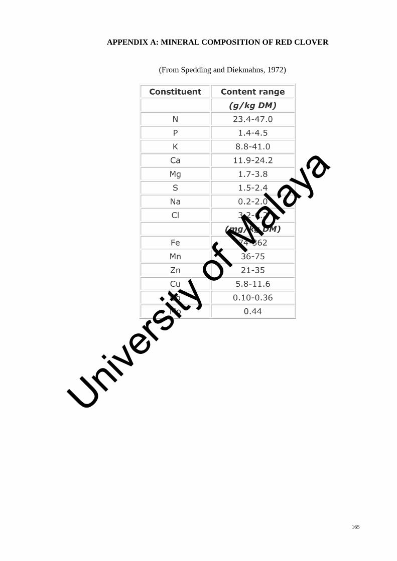
165
APPENDIX A: MINERAL COMPOSITION OF RED CLOVER
(From Spedding and Diekmahns, 1972)
Constituent Content range
(g/kg DM)
N 23.4-47.0
P 1.4-4.5
K 8.8-41.0
Ca 11.9-24.2
Mg 1.7-3.8
S 1.5-2.4
Na 0.2-2.0
Cl 3.2-6.2
(mg/kg DM)
Fe 74-362
Mn 36-75
Zn 21-35
Cu 5.8-11.6
Co 0.10-0.36
Mo 0.44
Univers
ity of
Mala
ya

166
APPENDIX B: DISTRIBUTION MAP OF RED CLOVER
Univers
ity of
Mala
ya

167
APPENDIX C: FORMULATIONS OF MS MEDIA (MURASHIGE AND
SKOOG)
MS Medium
Components Content (mg.l-1 )
Macronutrients
NH4NO3 1650.0
KNO3 1900.0
CaCl2.2H2O 440.0
MgSO4.7H2O 370.0
KH2PO4 170.0
Micronutrients
KI 0.83
H3BO3 6.2
MnSO4.4H2O 22.3
ZnSO4.7H2O 8.6
Na2MoO4.2H2O 0.25
CuSO4.5H2O 0.025
CoCl2.6H2O 0.025
Na2EDTA 37.3
FeSO4.7H2O 27.8
Vitamins and other supplements
Inositol 100.0
Glycine 2.0
Thiamine HCl 0.1
Pyridoxine HCl 0.5
Nicotinic acid 0.5
Univers
ity of
Mala
ya
168
Univers
ity of
Mala
ya

169
Univers
ity of
Mala
ya

170
Univers
ity of
Mala
ya

171
Univers
ity of
Mala
ya

172
Univers
ity of
Mala
ya
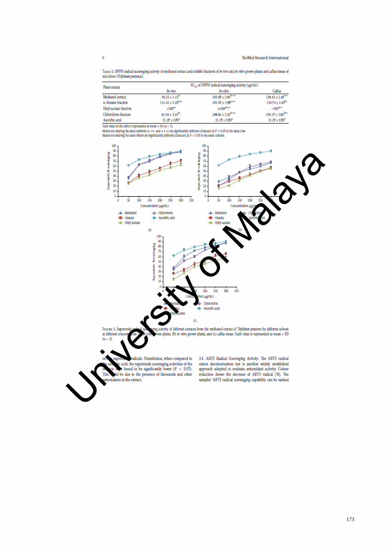
173
Univers
ity of
Mala
ya

174
Univers
ity of
Mala
ya

175
Univers
ity of
Mala
ya

176
Univers
ity of
Mala
ya

177
Univers
ity of
Mala
ya
178
Univers
ity of
Mala
ya

179
Univers
ity of
Mala
ya

180
Univers
ity of
Mala
ya

181
Univers
ity of
Mala
ya

182
Univers
ity of
Mala
ya

183
Univers
ity of
Mala
ya
184
Univers
ity of
Mala
ya

185
Univers
ity of
Mala
ya
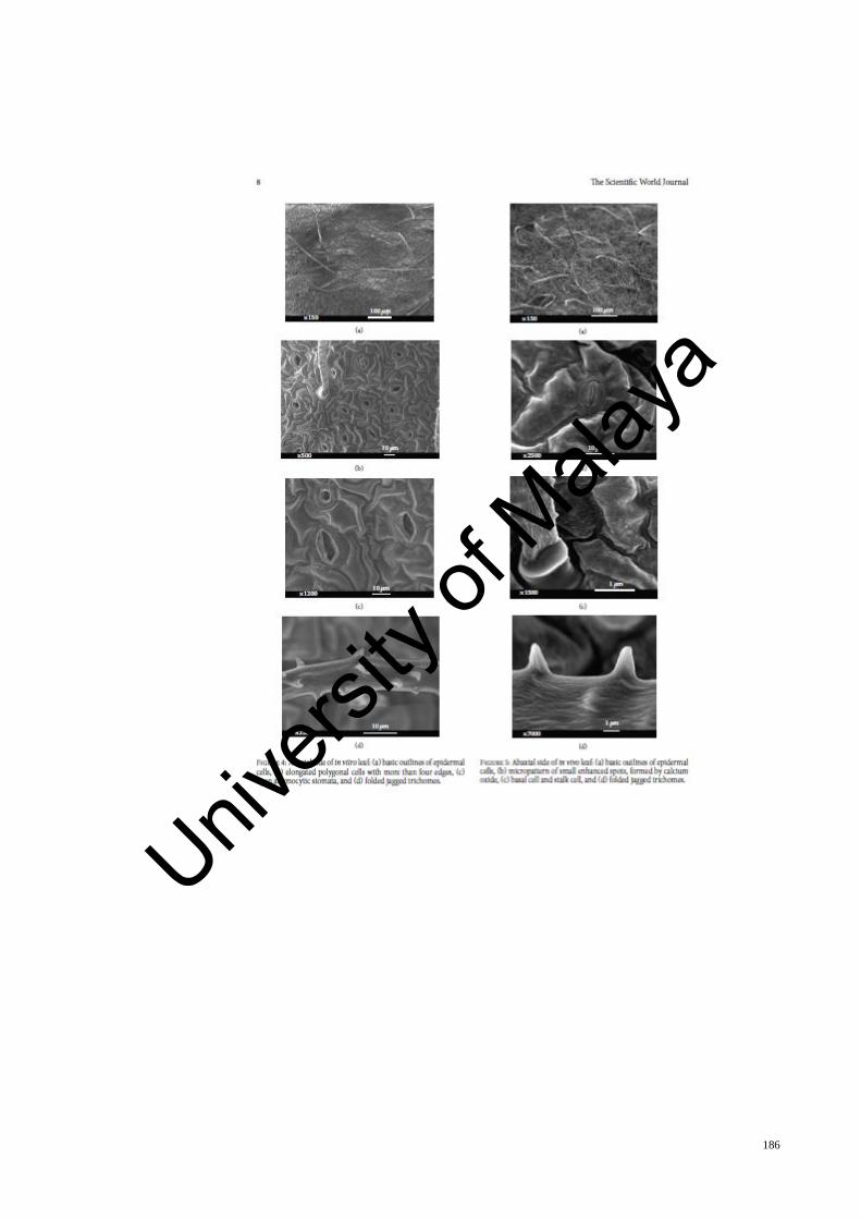
186
Univers
ity of
Mala
ya

187
Univers
ity of
Mala
ya

188
Univers
ity of
Mala
ya
189
Univers
ity of
Mala
ya

190
Univers
ity of
Mala
ya

191
Univers
ity of
Mala
ya
Copyright © 2022 FDOKUMEN