Green synthesis of gold-chitosan nanocomposites for caffeic acid sensing

Structure-Function Analyses of a Caffeic AcidO-Methyltransferase from Perennial Ryegrass Revealthe Molecular Basis for Substrate Preference W OA
Gordon V. Louie,a Marianne E. Bowman,a Yi Tu,b,c,d Aidyn Mouradov,b,c,d German Spangenberg,b,c,d
and Joseph P. Noela,1
a The Howard Hughes Medical Institute, Salk Institute for Biological Studies, La Jolla, California 92037b Department of Primary Industries, Biosciences Research Division, Victorian AgriBiosciences Centre, Bundoora, Victoria 3083,
AustraliacMolecular Plant Breeding Cooperative Research Centre, Bundoora, Victoria 3083, Australiad La Trobe University, Bundoora, Victoria 3083, Australia
Lignin forms from the polymerization of phenylpropanoid-derived building blocks (the monolignols), whose modification
through hydroxylation and O-methylation modulates the chemical and physical properties of the lignin polymer. The enzyme
caffeic acid O-methyltransferase (COMT) is central to lignin biosynthesis. It is often targeted in attempts to engineer the
lignin composition of transgenic plants for improved forage digestibility, pulping efficiency, or utility in biofuel production.
Despite intensive investigation, the structural determinants of the regiospecificity and substrate selectivity of COMT remain
poorly defined. Reported here are x-ray crystallographic structures of perennial ryegrass (Lolium perenne) COMT (Lp OMT1)
in open conformational state, apo- and holoenzyme forms and, most significantly, in a closed conformational state com-
plexed with the products S-adenosyl-L-homocysteine and sinapaldehyde. The product-bound complex reveals the post-
methyl-transfer organization of COMT’s catalytic groups with reactant molecules and the fully formed phenolic-ligand
binding site. The core scaffold of the phenolic ligand forges a hydrogen-bonding network involving the 4-hydroxy group that
anchors the aromatic ring and thereby permits only metahydroxyl groups to be positioned for transmethylation. While distal
from the site of transmethylation, the propanoid tail substituent governs the kinetic preference of ryegrass COMT for
aldehydes over alcohols and acids due to a single hydrogen bond donor for the C9 oxygenated moiety dictating the
preference for an aldehyde.
INTRODUCTION
Lignin is a heterogeneous polymeric macromolecule and amajor
component of the cell wall in vascular plants. It provides me-
chanical support for plant tissues (Zhong et al., 1997; Boerjan et al.,
2003), protects the plant from pathogen invasion (Hammond-
Kosack and Jones, 1996), and enhances the hydrophobicity of the
plant vasculature (Kubitzki, 1987). Lignin assembles by dehydro-
genative polymerization of monomeric monolignol building blocks
(Whetten and Sederoff, 1995). The three prevalent monolignols,
p-coumaryl alcohol, coniferyl alcohol, and sinapyl alcohol, differ
only in the degree of methoxylation of the phenolic ring. These
give rise to p-hydroxyphenyl (H), guaiacyl (G), and syringyl (S)
subunits, respectively, in polymerized lignin. The subunit com-
position of plant lignin is dependent on species, stage of devel-
opment, tissue type, subcellular localization, and biotic and
abiotic factors. Importantly, the relative abundance of the sub-
unit types determines to a large extent the physical properties of
the lignin polymer (Boerjan et al., 2003). S-lignin preferentially
forms intersubunit ether bond linkages involving the 4-hydroxyl
group, whereas G-lignin possesses a greater concentration of
less labile b-5, 5-5 biphenyl, and other carbon-carbon intersub-
unit bonds. The chemical degradation of lignin during pulping
occurs more efficiently with wood material possessing a high
S/G ratio, whereas forage digestibility is impaired for plant tis-
sue with elevated S-lignin content (reviewed in Li et al., 2008).
Therefore, the impact of lignin composition on the usability of
plant material for paper manufacturing, forage, and biofuel
production is of great economic interest.
p-Coumaric acid derived from the primary metabolite L-Phe
serves as the starting material for the monolignols. p-Coumaric
acid is transformed serially via hydroxylation and O-methylation
of the phenolic ring and also reductions of the oxygenated
functionality on the propanoid side chain. These reactions are
part of the general phenylpropanoid biosynthetic pathway and
are important for the production of not only the monolignols but
also other classes of plant compounds, such as the lignans,
phenylpropenes, and suberins. In the current view of monolignol
biosynthesis, the key O-methylations of hydroxyl groups at
the C3 and C5 positions of the phenolic ring of monolignol
1 Address correspondence to [email protected] author responsible for distribution of materials integral to thefindings presented in this article in accordance with the policy describedin the Instructions for Authors (www.plantcell.org) is: Joseph P. Noel([email protected]).WOnline version contains Web-only data.OAOpen Access articles can be viewed online without a subscription.www.plantcell.org/cgi/doi/10.1105/tpc.110.077578
The Plant Cell, Vol. 22: 4114–4127, December 2010, www.plantcell.org ã 2010 American Society of Plant Biologists
precursors are catalyzed primarily by distinct S-adenosyl-L-
methionine (SAM)–dependent enzymes. Caffeoyl CoA 3-O-meth-
yltransferase (CCoAOMT;EC2.1.1.104) is primarily responsible for
the initial O-methylation of the 3-hydroxyl group and specifically
methylates the 3,4-dihydroxy substrate as a CoA-linked thioester.
A second O-methyltransferase termed (historically) caffeic
acid O-methyltransferase (COMT; EC 2.1.1.68) catalyzes
predominantly the O-methylation of the 5-hydroxyl group of
3-methoxy-4,5-dihydroxy precursors, preferentially as the alde-
hyde or alcohol (Figure 1). The roles of CCoAOMT and COMT
have been inferred largely from the effects on lignin composition
of downregulation of these enzymes in transgenic plants. How-
ever, in vitro characterization shows that both CCoAOMT and
COMT have bifunctional activity in 3- and 5-O-methylation and
that COMT accepts as substrates monolignol precursors as free
acids, aldehydes, or alcohols (as detailed further below).
TheCOMTsbelong to theplant type-1 familyof SAM-dependent
O-methyltransferases (OMTs; the nomenclature of Noel et al.
[2003] is used throughout this article). These enzymes are typically
;360 amino acid residues in length, possess an auxiliary
N-terminal domain that functions in homodimerization, and do
not require metal ions for activity. These properties clearly
distinguish the plant type-1 OMTs from a second family, the
type-2 OMTs, to which CCoAOMT belongs. The smaller type-2
OMTs contain only the prototypical SAM binding domain (Martin
and McMillan, 2002) and are dependent on divalent cations for
catalytic activity.
One or more representatives of COMT occur in almost all plant
lineages. In vitro, most COMTs generally display a limited degree
of permissiveness in substrate acceptance. COMTs from angio-
sperms such as alfalfa (Medicago sativa), wheat (Triticum aesti-
vum), and aspen (Populus tremuloides) (Li et al., 2000; Parvathi
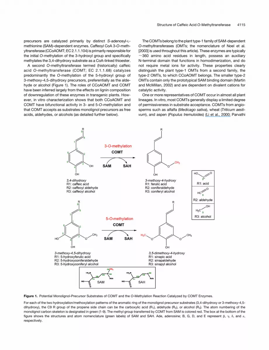
Figure 1. Potential Monolignol-Precursor Substrates of COMT and the O-Methylation Reaction Catalyzed by COMT Enzymes.
For each of the two hydroxylation/methoxylation patterns of the aromatic ring of the monolignol precursor substrates (3,4-dihydroxy or 3-methoxy-4,5-
dihydroxy), the C9 R group of the propene side chain can be the carboxylic acid (R1), aldehyde (R2), or alcohol (R3). The atom numbering of the
monolignol carbon skeleton is designated in green (1-9). The methyl group transferred by COMT from SAM is colored red. The box at the bottom of the
figure shows the structures and atom nomenclature (green labels) of SAM and SAH. Ade, adenosine; B, G, D, and E represent b, g, d, and «,
respectively.
Structure of Caffeic Acid O-Methyltransferase 4115

et al., 2001; Zubieta et al., 2002; Ma and Xu, 2008) are kinetically
most active against 5-hydroxyconiferaldehyde and then caffeoyl
aldehyde, whereas 5-hydroxyconiferyl alcohol and caffeoyl alco-
hol are slightly less favored, and 5-hydroxyferulic acid and caffeic
acid are the poorest substrates (aldehydes > alcohols > acids). In
addition, COMTs typically favor O-methylation of the 5-hydroxyl
group over the 3-hydroxyl group. However, the COMTs are
notable for a complete lack of activity in the O-methylation of the
4-hydroxyl group (Bhuiya and Liu, 2010). Analyses of transgenic
alfalfa, maize (Zeamays), and poplar (Populus tremula3Populus
alba) plants demonstrate that the most striking effects on lig-
nin composition due to the downregulation of COMT activity
are reduced levels of S-subunits without significant changes
in G-subunit levels, concomitant with the incorporation of
5-hydroxyconiferyl alcohol into the lignin polymer (Chabbert
et al., 1994; Atanassova et al., 1995; Ralph et al., 1997; Zhong
et al., 1998; Jouanin et al., 2000; Guo et al., 2001; Ralph et al.,
2001). The naturally occurring maize bm3-mutant, which is
deficient in COMT activity, shows similar characteristics (Grand
et al., 1985; Vignols et al., 1995). Collectively, the available data
indicate that themost significant function of COMT inmonolignol
biosynthesis is the production of syringyl-lignin monomers,
preferentially via the O-methylation of the 5-hydroxyconiferalde-
hyde precursor. Nevertheless, COMTs from a few plant species
display disparate substrate specificities. For example, the
COMT1 enzyme from tall fescue (Festuca arundinaceae) shows
the highest catalytic activity toward caffeoyl aldehyde and
5-hydroxyferulic acid (Chen et al., 2002, 2004). In addition, an
OMT from the gymnosperm loblolly pine (Pinus taeda) catalyzes
the methylation of monolignol precursors in both free acid and
CoA-ester forms (Li et al., 1997).
Earlier crystallographic analyses from this laboratory yielded
the structure of COMT from alfalfa (Zubieta et al., 2002), but due
to the open conformational state of the enzyme in this structure,
the substrate binding pocket was incompletely formed. There-
fore, a detailed structural basis for the O-methylation regio-
specificity and substrate selectivity of COMT remains to be
elucidated. Described here are structure determinations of the
perennial ryegrass (Lolium perenne) homolog of COMT (Lp
OMT1). A major aim of our studies with perennial ryegrass is
the manipulation of lignin composition as a means to improve
forage digestibility of transgenic plants (Tu et al., 2010). Of
particular relevance is the structure of the closed conformational
state of Lp OMT1 in complex with the products S-adenosyl-L-
homocysteine and sinapaldehyde occupying a fully assembled
phenolic-ligand binding pocket. The atomic resolution view of
the amino acid residues involved in forming the substrate binding
pocket and the positioning of the methyl-acceptor hydroxyl
group in the enzyme’s catalytic site provide a foundation for
engineering Lp OMT1 variants with altered regioselectivity. For
example, an Lp OMT1 variant with O-methylation activity ex-
clusively for the 3-hydroxyl group of monolignol substrates
could potentially be useful for the production of transgenic
ryegrass with greatly diminished S-lignin levels and enhanced
forage digestibility, ormore intriguingly, an LpOMT1 variant with
4-O-methylation activity may be used to generate novel mono-
lignols with aberrant polymerization properties (Bhuiya and Liu,
2010).
RESULTS
In Vitro Kinetic Analyses of Lp OMT1 Activity
A method based on reversed-phase liquid chromatography
coupled with mass spectrometry (LC-MS) was developed for
the separation and quantification of substrates and products of
Lp OMT1–catalyzedO-methylation reactions. With the chromato-
graphic conditions employed, each of the analyzed substrates
(caffeic acid, 5-hydroxyferulic acid, caffeoyl aldehyde, 5-hydroxy-
coniferaldehyde, and caffeoyl alcohol) was cleanly resolved from
the methylated product (the product typically eluted 0.9 min
later). Kinetic parameters were determined for OMT1 with each
of the three nonaldehyde substrates. Based on measurements
with the acid substrates, OMT1 displayed a clear preference for
O-methylation of the 5-hydroxyl substituent of a substrate bearing
a 3-methoxy substituent (Vmax/Km for 5-hydroxyferulic acid: 3.5
nkat·mg protein21/3.0mM), in comparison to 3-O-methylation of a
substrate bearing only the single meta substituent (Vmax/Km for
caffeic acid: 2.7 nkat·mg protein21/10.3 mM) (Figure 2). With
respect to the C9 substituent, OMT1 showed a strong preference
for the alcohol over the acid, as evident from the higherVmax/Km for
caffeoyl alcohol (12.9 nkat·mg protein21/1.7 mM) versus caffeic
acid. In general, these in vitro substrate preference patterns
observed with OMT1 are consistent with those reported for the
COMTs from aspen (Li et al., 2000), alfalfa (Parvathi et al., 2001;
Zubieta et al., 2002), tall fescue (Chen et al., 2004), and wheat
(Ma and Xu, 2008), although the Km values measured for Lp
OMT1 appear to be consistently lower. Although Lp OMT1 clearly
converted the two aldehyde substrates into the corresponding
methylated products, a detailed kinetic analysis of these re-
actions was not possible because severe substrate inhibition
was observed above even the lowest substrate concentrations
used (1.0 mM) (Figure 2). Previously, Li et al. (2000) observed
that 5-hydroxyconiferaldehyde also competitively inhibits the
O-methylation of caffeic and 5-hydroxyferulic acids by both aspen
COMT and tissue extracts from several other tree species. For
aspen COMT (as for the COMTs from most other plant species),
5-hydroxyconiferaldehyde represents the substrate with the
greatest binding selectivity (Km = 2.6 mM), a property that may
also contribute in part to its potent inhibitory activity (Ki = 0.3 to
2 mM) (Li et al., 2000).
X-Ray Crystallographic Structures of Distinct Lp OMT1
Catalytic Conformations
The structural analyses of L. perenne OMT1 reported here
provide the most complete picture to date of the distinct lig-
and-bound states of a plant type-1 OMT (Noel et al., 2003).
Crystal structures are described for Lp OMT1 in the apoenzyme
form, in a form representative of the holoenzyme (bound to
S-adenosyl-L-homocysteine [SAH]), and in ternary complexes
with SAH and phenolic (sinapaldehyde or coniferaldehyde) prod-
ucts (Table 1). Crystallization trials of Lp OMT1 with SAM
consistently yielded structures of the enzyme with bound SAH
(indicative of the lability of SAM over the course of the crystal-
lization experiments or low-level transmethylation activity by the
enzyme with SAM). The position of the reactive methyl group of
4116 The Plant Cell

SAM can nevertheless be readily modeled from the structure of
SAH (as discussed in greater detail below).
Lp OMT1 possesses the same tertiary and quaternary struc-
ture as several other plant type-1 OMTs that have been charac-
terized crystallographically (Zubieta et al., 2001, 2002; Liu et al.,
2006). Not surprisingly, Lp OMT1 is most structurally similar (a
root mean square deviation [rmsd] of 1.51 A for 332 equivalent
residues) to alfalfa COMT (Zubieta et al., 2002), with which it
shares 62.7% sequence identity. Slightly higher rmsd values are
observed in comparisons with type-1 OMTs having greater
sequence divergence: alfalfa chalcone OMT (Zubieta et al.,
2001), 2.19 A rmsd for 312 equivalent residues, with 40.1%
sequence identity; alfalfa isoflavone OMT (Zubieta et al., 2001),
1.91 A rmsd for 310 equivalent residues, with 28.7% sequence
identity; and barrel-medic (Medicago truncatula) isoflavonoid
OMT (Liu et al., 2006), 3.15 A rmsd for 265 equivalent residues,
with 19.6% sequence identity.
Typical of plant type-1 OMTs, the polypeptide chain fold of Lp
OMT1 comprises distinct dimerization and SAM/SAH binding
domains, with a a-helical layer sandwiched between the two
domains. Two monomers associate to form a symmetric homo-
dimer (Figure 3), in which two tightly intertwined dimerization
domains constitute the central core and the SAM binding do-
mains are located peripherally. The active-site cleft in each of the
twomonomers resides at the interface between the SAMbinding
domain and the adjacent a-helical layer. Within this layer lies the
phenolic substrate binding site, which is lodged in a pocket
between two a-helices. Notably, this phenolic binding pocket is
also buttressed by a-helical segments from the dimerization
region, with one of the helical segments contributed by the
opposing monomer.
Open and Closed Conformational States of Lp OMT1
Structural comparisons of the various ligand-bound states of
OMT1 reveal striking malleability in the positioning of the SAM
binding domain. Most conspicuously, relative to the OMT1
holoenzyme, the product-bound OMT1-SAH-sinapaldehyde
complex has a substantially more closed conformation, which
arises from an approximate 178 inward rotation of the homo-
dimer’s two SAM binding domains with respect to the relatively
fixed scaffold formed by the core dimerization region (Figure 4A).
These domain movements derive primarily from conformational
changes within a hinge segment (residues 296 to 303) in each
monomer. The interactions of two a-helices (residues 209 to
219 and 230 to 236) at the top of the SAM binding domain
with an extended a-helical segment (residues 159 to 190) in the
phenolic-substrate binding region may contribute to stabilizing
the closed conformation of OMT1. These interdomain interac-
tions include several hydrogen bonds (between the side chains
Asn170-His231, Asn170-Glu235, and Ser181-Thr211) as well as
the pairing of the aliphatic faces of two juxtaposed helices. Most
significantly, the closed conformational state of OMT1 allows the
ribose and Met moieties of the SAM/SAH molecule to asso-
ciate tightly with the phenolic-substrate binding region (see
below).
In contrast with the marked domain closure observed for the
product-bound OMT1 ternary complex, binding of SAM/SAH to
OMT1 is associated with only a slight inward movement of the
SAM binding domains. Thus, the holoenzyme and apoenzyme
forms of OMT1 share essentially the same open conformation,
although the SAM binding domains notably appear less ordered
overall in electron densitymaps of apoOMT1. Significantly, in the
crystal structures of both the apoenzyme and the ternary
Figure 2. Enzyme-Kinetics Analyses of Lp OMT1 with Various Monolignol-Precursor Substrates.
In each panel, initial reaction velocity (nkat·mg protein�1) is plotted against substrate concentration (mM), and the displayed curve is derived from
nonlinear regression analysis (Prism). The results shown are from a single set of measurements for each substrate.
Structure of Caffeic Acid O-Methyltransferase 4117

complex, the positioning of the SAM binding domain is consis-
tent in the four independent monomers of the two homodimers
occurring in the crystallographic asymmetric units.
Binding of SAM/SAH by Lp OMT1
As described for other plant type-1 OMTs, the SAM/SAH
molecule within Lp OMT1 adopts an extended conformation
(Schubert et al., 2003) and is bound along the C-terminal end of
the core b-sheet of the SAM binding domain (Figures 3, 4B, and
5A). In the open conformational state of Lp OMT1, this domain is
exclusively responsible for the binding interactions with SAM/
SAH. Specifically, the nonpolar side chains of Phe-227, Leu-229,
Met-249, Phe-250, and Trp-268 surround the adenine ring of
SAM/SAH, which also forms hydrogen bonds with the Asp-248
side chain carboxylate and the backbone amide nitrogen of Met-
249. A Gly-rich segment (Gly205-Gly206-Gly207) forms one wall
of a channel that is occupied by the ribose ring and the aliphatic
portion of the methionine/homocysteine (Met/Hcy) moiety. In
addition, hydrogen bond interactions are formed by the Asp-228
side chain carboxylate with both ribose hydroxyl groups, the
backbone carbonyl oxygens of Gly-205 and Lys-262 with the
a-amino group of the Met/Hcy moiety, and the Lys-262 side
chain amino group with the Met/Hcy a-carboxylate group. The
ribose ring oxygen also interactswith awatermolecule, which in turn
forms hydrogen bondswith the side chains of Asp-267 and Trp-268.
In the closed conformational state of OMT1, these SAM/SAH
interactions with the SAM binding domain are retained entirely,
but the closer approach of SAM/SAH to the phenolic substrate
binding region introduces a number of additional interactions. In
particular, the phenyl ring of Phe-160 (which itself adopts a
significantly more buried conformation in the closed state of
OMT1) together with the side chains of Phe-173 and Met-177
contribute to the binding of the ribose ring and aliphatic portion
of the Met/Hcy moiety; and the Ser-181 side chain forms an
additional hydrogen bond with the Met/Hcy a-carboxylate
group. Notably, all of the amino acid residues that form interac-
tions with SAM/SAH are, in general, highly conserved in all plant
type-1 OMTs.
With most OMTs, the SAH product acts as a potent compet-
itive inhibitor of the methylation reaction (Hendricks et al., 2004).
This property suggests that SAH and SAM form largely indistin-
guishable interactions with the enzyme. Indeed, this feature of
SAH/SAM binding is particularly evident for the open conforma-
tional state of Lp OMT1, in which the additional methyl group of
the SAM substrate makes no contacts with the enzyme.
Table 1. Summary of Crystallographic Data Collection and Refinement Statistics for Lp OMT1 Structures
Structure OMT1/SAH OMT1 apo OMT1/SAH/Sinapaldehyde OMT1/SAH/Coniferaldehyde
Space group P41212 P21 P21 P21Unit cell parameters
a (A) 67.70 67.93 94.94 95.38
b (A) 67.70 86.54 85.23 85.06
c (A) 249.79 131.59 98.57 98.59
b (8) 90 89.87 111.10 111.75
Monomers per asymmetric unit 1 4 4 4
Resolution range (A)a 47–1.80 (1.90–1.80) 60–2.75 (2.90–2.75) 47–1.85 (1.84–1.85) 49–2.25 (2.37–2.25)
Number of reflections measured 397,805 128,585 369,886 249,422
Merging R factora 0.116 (0.454) 0. 078 (0.364) 0.085 (0.367) 0.118 (0.439)
Mean (I/s I)a 10.4 (2.3) 11.7 (2.7) 7.3 (2.1) 7.1 (2.4)
Completenessa 1.00 (1.00) 0. 957 (0.946) 0.957 (0.914) 0.996 (0.981)
Redundancya 7.2 (3.8) 3.4 (2.7) 3.1 (2.4) 3.6 (2.9)
Number of reflections used 55,209 38,072 119,372 69,341
R factora 0.212 (0.298) 0.280 (0.356) 0.232 (0.404) 0.232 (0.355)
Free R factora 0.226 (0.315) 0.334 (0.403) 0.261 (0.415) 0.262 (0.362)
Number of amino acid residues 352 1,408 1,421 1,421
Number of water molecules 313 0 625 314
rmsd from ideality
Bond lengths (A) 0.005 0.010 0.006 0.007
Bond angles (8) 1.19 1.11 1.17 1.21
Residues with most favorable
conformation (%)b98.0 93.2 97.4 97.1
PDB code 3P9C See supplemental
data onlinec3P9I 3P9K
Merging R factor = +hkl +i | Ii(hkl) – <I(hkl)> (denoting average) | / +hkl +i Ii(hkl).aValues in parentheses describe the highest resolution shell.bAs reported by the program MolProbity (Chen et al., 2010).cSupplemental Data Set 1 online: partially refined atomic coordinate set in PDB format (LpCOMT-Apo_refine_10.pdb), which can be displayed with
molecular graphics software such as Coot (Emsley and Cowtan, 2004) or PyMol (PyMOL molecular graphics system, version 1.3; Schrodinger).
Supplemental Data Set 2 online: crystallographic data (including experimentally measured structure-factor amplitudes and calculated map
coefficients; LpCOMT-Apo2_truncated-unique.mtz) that can be input to a crystallographic refinement program such as Phenix (Adams et al., 2010)
or displayed with Coot.
4118 The Plant Cell

Recognition of Phenolic Substrates/Products by Lp OMT1
The binding mode of phenolic ligands is unequivocally defined
in the ternary complex of OMT1/SAH with the sinapaldehyde
product, a particularly tightly bound compound in our crystallo-
graphic experiments (Figures 5A and 5B). As outlined earlier, the
binding pocket is located primarily within the a-helical sandwich
layer, with helical segments from the dimerization region lining its
internal wall. The SAM/SAHmolecule and residues 262 to 268 of
the SAM binding domain block the entrance to the pocket in the
closed conformation of OMT1. This polypeptide chain segment
contributes a number of key residues involved in catalysis and in
binding of both the SAM and phenolic substrates.
The precise ligand binding interactions formed by OMT1 are of
particular interest, as these not only are integral to the enzyme’s
catalytic activity but also confer substrate specificity and regio-
selectivity for the methylation reaction. In the OMT1-SAH-sina-
paldehyde complex, the aromatic and aliphatic core (the phenyl
ring and propene side chain) of sinapaldehyde are enveloped by
a cluster of nonpolar side chains that include Leu-124, Met-127,
Met-177, Trp-263, Val-313, Ile-316, and Met-317. The aldehyde
oxygen of the propene side chain is involved in a single hydrogen
bond interaction with the amide nitrogen of the Asn-128 side
chain. Wang and Pichersky (1999) previously demonstrated that
in Clarkia breweri COMT, the residue corresponding to Asn-128,
plays a critical role in the recognition of substrates bearing an
oxygenated side chain (i.e., the monolignol intermediates). Ala
replaces this Asn in OMTs specific for substrates bearing a
nonoxygenated, propene side chain (phenylpropenes, such as
isoeugenol). The sinapaldehyde 4-hydroxyl group accepts hy-
drogen bonds from the amide nitrogen of the Asn-321 side chain
and a water molecule and is the donor in a hydrogen bond with
the Asp-267 carboxylate. The 3-methoxy group occupies a
predominantly nonpolar cavity formed by the side chains of
Met-127, Leu-133, Phe-173, Ile-316, and His-320. The methoxy-
ether oxygen participates in a hydrogen-bonding network with a
water molecule that in turn interacts with the side chain of Asn-
321, the sinapaldehyde 4-hydroxyl group, and the His-163 side
chain imidazole via a bridging water molecule.
Notably, the binding by OMT1 of coniferaldehyde is effec-
tively identical to that of sinapaldehyde (Figure 5C). Furthermore,
the residues involved in forming ligand binding interactions,
as detailed above for OMT1, are nearly invariant in all well-
characterized COMTs, and consequently, substrate recognition
is likely achieved identically in all of these COMTs (see Supple-
mental Figure 1 online). It is therefore intriguing that the disparate
binding sites observed for ferulic acid and 5-hydroxyconiferal-
dehyde in alfalfa COMT (Zubieta et al., 2002) differ significantly
from the well-defined site described here for sinapaldehyde in
Lp OMT1. The catalytically nonproductive ligand complexes of
alfalfa COMT may be representative of prebinding modes that
are explored by the incoming substrate molecule when COMT is
in the open conformational state and lacks a completely orga-
nized phenolic substrate binding pocket and highlight the im-
portance of enzyme dynamics in COMT activity.
DISCUSSION
Open and Closed Conformations of the Lp OMT1
Molecular Architecture
The inward positioning of the SAM binding domains associated
with the closed conformational state of OMT1 is intrinsic to the
Figure 3. Structure of a Homodimer of the Lp OMT1/SAH/Sinapaldehyde Complex.
The homodimer is viewed perpendicular to the twofold rotational axis (vertical) that relates the two monomers. For each of the monomers, the
polypeptide chain backbone is color coded by structural domain: dimerization domain (residues 1 to 158), green; SAM/SAH binding domain (residues
199 to 295 and 328 to 360), cyan; a-helical layer involved in phenolic substrate binding (residues 159 to 192 and 304 to 323), magenta; and hinge
segments (residues 193 to 198, 296 to 303, and 322 to 327), orange. (One of the monomers is shown with slightly paler shading.) S-adenosyl-
homocysteine and sinapaldehyde are shown in ball-and-stick representation and colored according to atom type (carbon, yellow; nitrogen, blue;
oxygen, red; and sulfur, gold).
Structure of Caffeic Acid O-Methyltransferase 4119

assembly of a fully functional binding pocket for the phenolic
substrate. For example, steric contacts with the SAM binding
domain induce several side chains (Phe-160, Trp-263, Met-317,
and Asn-321) to adopt conformations appropriate for accom-
modating the phenolic substrate (Figure 4B). More importantly,
the OMT1 catalytic residues (His-266, Asp-267, and Glu-326)
together with the SAM substrate are brought in immediate
proximity of the phenolic substrate. Indeed, the SAM molecule
physically closes off the substrate binding pocket and thereby
prevents escape of the phenolic substrate and sequesters the
activated phenoxide intermediate from bulk solvent. Notably,
this role of the SAM substrate in establishing part of the binding
pocket for the phenolic substrate is consistent with the order of
substrate binding by the OMTs, as binding of the methyl donor
SAMprecedes that of the phenolic substrate (Huang et al., 2004).
Our structural analyses of Lp OMT1 provide a unique atomic
resolution view of distinct conformational states of a single plant
type-1 OMT, although considerable variability in the positioning
of the SAM binding domain has been previously recognized in
these enzymes. For example, in comparison to the structure of
(closed-state) isoflavone OMT from alfalfa (Zubieta et al., 2001),
the more open conformation of barrel medic isoflavonoid OMT
(Liu et al., 2006) was apparent, as evidenced by an outward
rotation of the SAM binding domains, a wider active-site cleft,
and an inordinately long distance between SAM and the methyl
acceptor of the hydroxylated substrate. Similarly, the previously
reported structures of ternary complexes of alfalfa COMT
(Zubieta et al., 2002) possess conformational states that are
intermediate between the open and closed conformations ob-
served for Lp OMT1. Flexibility of the SAM binding domain is
evidently an inherent property of the class-I OMTs. This flexibility
underlies a critical mechanistic function of these enzymes: the
adoption of an initial open state that facilitates binding of SAM
first then the hydroxylated substrate and subsequently a closed
state in which the substrates and active-site groups are se-
questered and appropriately aligned for the catalytic reaction to
proceed.
Insight into the Substrate Preferences of COMTs
Collectively, the intricate array of binding interactions between
Lp OMT1 and a phenolic ligand (Figure 5A) undoubtedly func-
tions in an enzyme substrate complex to anchor the substrate
molecule such that the reactive oxygen atom (the 5-hydroxyl in
the case of 5-hydroxyconiferaldehyde, the precursor of sinapal-
dehyde) forms productive interactions with the enzyme’s cata-
lytic groups and the SAM methyl donor (as discussed further
below). As a consequence, COMT enzymes are exclusively
selective for O-methylation of a meta (3- or 5-position) hydroxyl
group on the substrate phenyl group. In marked contrast, the
COMTs never modify the para- or 4-hydroxyl group, which is
engaged in hydrogen bonding interactions with a conserved Asn
side chain and a water molecule. In isoeugenol and chavicol
methyltransferases, which specifically target the 4-hydroxyl
group of their phenylpropene substrates, these hydrogen bond-
ing interactions are disrupted by the substitution of the Asn with
a nonpolar residue or by the introduction of an additional hydro-
gen bonding residue (e.g., Glu-165 in C. breweri isoeugenol
Figure 4. Comparison of the Open and Closed Conformational States of
Lp OMT1.
(A) Comparison of the holoenzyme and holoenzyme/sinapaldehyde
forms of the OMT1 homodimer. The structural superposition is based
solely on the dimerization domains of the two forms of Lp OMT1. The
OMT1 holoenzyme (magenta, with carbon atoms of SAH drawn in darker
magenta) exists in an open conformational state, whereas the holoenzyme/
sinapaldehyde form (green, with carbon atoms of SAH and sinapaldehyde
drawn in darker green) is significantly more closed. The closed conforma-
tion of the holoenzyme/sinapaldehyde form arises from an inward rotation
(by;178) of each SAM binding domain. The orientation of view is identical
to that in Figure 3.
(B) Comparison of the active sites in the open and closed conformational
states of OMT1. The structures of the OMT1 holoenzyme (open, ma-
genta) and holoenzyme/sinapaldehyde (closed, green) are colored as in
(A), except that the carbon atoms of sinapaldehyde are colored orange.
With the closed conformational state, the substantial inward shift of the
SAM binding domain is evident at the left of the figure (only SAH and
residues 262 to 268 and Glu-326, which include the key catalytic
residues, are shown). Hydrogen bond interactions involving the sinapal-
dehyde ligand are shown as dashed lines and are colored as in Figure 5A.
The most significant side chain conformational differences between the
two forms of OMT1 occur at Phe-160, Ser-181, Trp-263, Met-317, and
Asn-321.
4120 The Plant Cell

Figure 5. Substrate and Product Binding Sites of Lp OMT1.
(A) OMT1 amino acid residues that interact with the SAH and sinapaldehyde products (divergent stereoview). The residues that interact with SAH
(magenta carbon atoms) are colored light green, whereas those that interact with sinapaldehyde (orange carbon atoms) are colored dark green (only the
a-carbon and side chain atoms of each residue are shown, except for the backbone segment Gly205-Gly206-Gly207). Water molecules are represented
as small red spheres. Hydrogen bond interactions are shown as cyan dashed lines; those critical to the catalytic reaction (involving His-266, Asp-267,
and Glu-326) are shown in dark gray. The substrate binding pocket is capped by the residues Leu-21 and Ser-25, which are contributed by the opposing
monomer of the OMT1 homodimer. The blue-colored envelopes surround regions greater than +2.5s in the omit Fobs-Fcalc electron density map. The
atomic model and electron density are shown for the active site of monomer A (primarily, with small contributions from monomer B) of the
crystallographic asymmetric unit; the active sites of the other three monomers are essentially indistinguishable.
(B) Surface of the binding pocket for phenolic substrate in the closed conformational state of OMT1. The depicted surface defines the area accessible to
a probe sphere with a radius of 1.4 A. The side of the surface that faces the phenolic substrate binding site is colored according to the identity of the
bounding protein atom (carbon, green; nitrogen, blue; oxygen, red; and sulfur, gold); the reverse side of the surface is distinguished by gray coloring. For
clarity, the front portion of the surface of the binding pocket has been removed. The positions of bound sinapaldehyde (orange) and two water
molecules (red spheres) involved in interactions with the 3-methoxy and 4-hydroxyl groups are shown. The locations of cavities for the 3-methoxy and
C9 substituents and the catalytic site are indicated.
(C) Binding of coniferaldehyde by OMT1. OMT1 active-site residues, SAH, and coniferaldehyde are displayed as in (A). The blue-colored envelopes
surround regions greater than +1.0 s in the initial 2Fobs-Fcalc electron density map (calculated prior to inclusion of coniferaldehyde in the atomic model).
The yellow-colored envelopes surround regions greater than +2.5s in the final Fobs-Fcalc electron density map. The difference density lobe indicates a
Structure of Caffeic Acid O-Methyltransferase 4121
methyltransferase) at a neighboring site (Wang and Pichersky,
1999; Gang et al., 2002).
Remarkably, the strict regioselective activity of the COMTs is
attained despite a moderate degree of promiscuity in substrate
acceptance. Our detailed characterization of the binding inter-
actions of the COMTs illuminates the structural basis for this
promiscuity. In particular, although the invariant scaffold (the
phenyl ring, propene side chain, and 4-hydroxy group) of COMT-
accepted substrates forms close interactions with the enzyme,
the parts of this scaffold that potentially bear alternative substit-
uents (the 3- and 5-positions of the phenyl ring and the oxygen-
bearing C9 of the propene side chain) are situated adjacent to
modestly sized cavities within the enzyme’s binding pocket
(Figure 5B). These two key features underlie the promiscuity in
substrate binding by COMT, which can thereby accommodate
an array of distinct but related substrates via a common binding
position of the 4-hydroxy phenylpropene scaffold. The interac-
tions formed with the alternative substituents then define the
subtleties of the substrate preferences of the COMTs. Thus, in
comparison to 5-hydroxyconiferaldehyde, caffeoyl aldehyde
would be expected to serve as a less favorable substrate
because the COMT cavity that accommodates the 3-methoxy
group is occupied by only a hydrogen atom, resulting in the loss
of both the nonpolar interactions formed by amethoxy group and
the hydrogen bonding network nucleated by a water molecule
that interacts with the 3-methoxy and 4-hydroxy oxygens. The
proclivity for full occupation of COMT’s 3-methoxy pocket is
evidenced by the two distinct orientations observed for the
phenyl ring of the coniferaldehyde product in complex with Lp
OMT1-SAH (Figure 5C): the first has the lone methoxy group of
coniferaldehyde residing as expected in the catalytic site,
whereas in the second, this methoxy group instead occupies
the opposing 3-methoxy pocket.
The structural basis for aldehyde selectivity (over the less-
preferred alcohol and the least-preferred acid forms) is also ap-
parent. Only a single active-site polar group, the carboxamide ND2
group of an Asn (Asn-128 in Lp OMT1), is suitably positioned to
interact with the substrate’s C9 oxygenated functionality (Figure
5A). Asn128ND2 serves as a hydrogen bond donor to the C9
aldehyde oxygen in the Lp OMT1-SAH-sinapaldehyde complex. A
substrate bearing a C9 alcohol would be less favored than the
aldehyde due to the lack of a hydrogen bond acceptor for the
hydrogen atom of the C9 hydroxyl group. In addition, although aC9
carboxylate groupcanbe readily accommodated sterically, itwould
be least favored because of the unavailability of both a hydrogen-
bonding partner for the second carboxylate oxygen and a cationic
protein group to neutralize the carboxylate’s negative charge.
Our structural characterization of the substrate preferences
of Lp OMT1 is consistent with results from enzyme kinetics
analyses, as evidenced by the higher O-methylation activity for
the 5-hydroxyl over the 3-hydroxyl substituent and for alcohol
over acid substrates. Furthermore, because the Lp OMT1 res-
idues that form the phenolic-ligand binding site (as revealed in
this work) are absolutely conserved in almost all confirmed
COMTs, the substrate preference patterns of all COMTs are
expected to be very similar. Therefore, although a full kinetic
analysis of Lp OMT1 activity with the aldehyde substrates was
not possible, Lp OMT1 very likely resembles other COMTs in that
the aldehydes represent the kinetically preferred substrates (Li
et al., 2000; Parvathi et al., 2001; Zubieta et al., 2002; Chen et al.,
2004; Ma and Xu, 2008). Some support for the high affinity of Lp
OMT1 for the phenolic aldehydes is provided by the observation
that crystallization of Lp OMT1-SAH–product ternary complexes
was effected most readily with the two aldehyde products
(coniferaldehyde and sinapaldehyde).
A number of unusual properties pertaining to substrate binding
by theCOMTenzymes can nowbe rationalized. For example, the
COMT from loblolly pine (Li et al., 1997), the most divergent in
sequence of the confirmed COMTs, is active against not only the
normal monolignol precursors but also the CoA-thioesters of the
acid forms (e.g., caffeoyl CoA). Thesemonolignol CoA-thioesters
are more typically methylated by a distinct class of O-methyl-
transferases, the caffeoyl-CoA OMTs, which possess a more
open and expansive ligand binding cleft that serves to accom-
modate the extended CoA tail of the substrate (Ferrer et al.,
2005). Loblolly pine COMT may have the capacity to bind the
bulkier esterified substrates due to amino acid substitutions and
two small insertions that substantially enlarge the volume of its
C9 pocket. Another outstanding question is how certain sub-
strates (the aldehydes in particular) inhibit the activity of LpOMT1
as well as several other COMTs (Li et al., 2000). One possible
mechanism, the entrapment of substrate in exploratory, pre-
binding modes (as discussed above), seems implausible due to
the dependence of inhibition on the identity of the C9 substituent
and the absence of specific interactions to this substituent prior
to full assembly of the substrate binding site. A more likely
mechanism is the adoption by the substrate of a nonproductive
binding mode in which the target hydroxyl group occupies the
3-methoxy binding pocket instead of the catalytic site (Figure
5B). Such a flipped binding mode, which closely resembles the
catalytically competent enzyme substrate complex, might be
expected to be most prevalent for substrates with the greatest
affinity overall. The effects of product inhibition, through which
regeneration of the active site is impeded by particularly tightly
bound product, must also be considered.
In summary, our biochemical and structural studies support a
primary role of Lp OMT1 in the 5-O-methylation of 5-hydroxy-
coniferaldehyde to produce sinapaldehyde, the direct precursor
of the monolignol building block (sinapyl alcohol) that is incor-
porated into syringyl lignin. This result is consistent with func-
tional studies in perennial ryegrass plants, which demonstrate
that (1) maximal OMT1 expression coincides with the elevated
accumulation of S-lignin in stem tissue (during the early elonga-
tion stage of plant development), and (2) transgenic plants with
Figure 5. (continued).
significant fraction of coniferaldehyde molecules are bound with an alternative orientation of the phenyl ring, which transplaces the 3-methoxy group
into the OMT1 3-methoxy cavity (as opposed to the catalytic site).
4122 The Plant Cell
downregulated Lp OMT1 expression have dramatically reduced
levels of S-lignin subunits (Tu et al., 2010). Furthermore, similar in
planta results have been reported from analogous studies of the
COMT enzymes in several other plant species. The preferred use
by Lp OMT1 (and most other plant COMTs) of the aldehyde (and
to a lesser extent alcohol) forms of the monolignol precursors is
consistent with the currently accepted view of the phenylpropa-
noid biosynthetic pathway in plants (Li et al., 2008).
Additionally, despite the marked preference for 5-O-methyla-
tion, Lp OMT1 nevertheless is significantly active in 3-O-meth-
ylation, as evidenced here by the conversion in vitro of caffeoyl
aldehyde to coniferaldehyde and caffeoyl alcohol to coniferyl
alcohol (and also caffeic acid to ferulic acid). This secondary 3-O-
methylation activity likely accounts for the slight decreases in
levels of G-lignin in OMT1-deficient transgenic ryegrass (Tu et al.,
2010). Again, this observed activity of Lp OMT1 is in agreement
with the properties of COMTs from other plant species (Maury
et al., 1999; Dixon et al., 2001; Parvathi et al., 2001; Do et al.,
2007; Ma and Xu, 2008; Guillaumie et al., 2008): in Arabidopsis
thaliana, the retained ability to synthesize both G- and S-lignin in
CCoAOMT knockouts but not CCoAOMT/COMT double knock-
outs (Do et al., 2007); and radiochemical labeling studies that
demonstrate the incorporation of exogenously supplied caffeoyl
alcohol and caffeoyl aldehyde ultimately into both G- and
S-subunits of angiosperm lignin (Matsui et al., 2000). Finally, the
permissiveness in substrate acceptance by the COMTs intrigu-
ingly suggests that developmental adjustments in the in vivo
levels ofmonolignol precursorsmay be sufficient tomodulate the
biosynthesis and availability of specificmonolignols that are then
incorporated through polymerization into lignin.
InferencesontheCatalyticReactionMechanismofLpOMT1
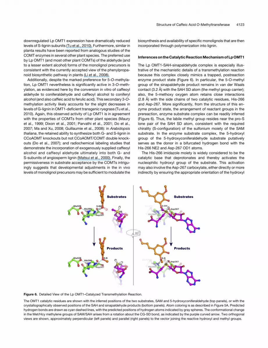
The Lp OMT1-SAH-sinapaldehyde complex is especially illus-
trative of the mechanistic details of a transmethylation reaction
because this complex closely mimics a trapped, postreaction
enzyme product state (Figure 6). In particular, the 5-O-methyl
group of the sinapaldehyde product remains in van der Waals
contact (3.2 A) with the SAH SD atom (the methyl group carrier);
also, the 5-methoxy oxygen atom retains close interactions
(2.8 A) with the side chains of two catalytic residues, His-266
and Asp-267. More significantly, from the structure of this en-
zyme product state, the arrangement of reactant groups in the
prereaction, enzyme substrate complex can be readily inferred
(Figure 6). Thus, the labile methyl group resides near the pro-S
lone pair of the SAH SD atom, consistent with the required
chirality (S-configuration) of the sulfonium moiety of the SAM
substrate. In the enzyme substrate complex, the 5-hydroxyl
group of the 5-hydroxyconiferaldehyde substrate putatively
serves as the donor in a bifurcated hydrogen bond with the
His-266 NE2 and Asp-267 OD1 atoms.
The His-266 imidazole moiety is widely considered to be the
catalytic base that deprotonates and thereby activates the
nucleophilic hydroxyl group of the substrate. This activation
may also involve the Asp-267 carboxylate, either directly or more
indirectly by ensuring the appropriate orientation of the hydroxyl
Figure 6. Detailed View of the Lp OMT1–Catalyzed Transmethylation Reaction.
The OMT1 catalytic residues are shown with the inferred positions of the two substrates, SAM and 5-hydroxyconiferaldehyde (top panels), or with the
crystallographically observed positions of the SAH and sinapaldehyde products (bottom panels). Atom coloring is as described in Figure 5A. Predicted
hydrogen bonds are drawn as cyan dashed lines, with the predicted positions of hydrogen atoms indicated by gray spheres. The conformational change
in the Met/Hcy methylene groups of SAM/SAH arises from a rotation about the CG-SD bond, as indicated by the purple curved arrow. Two orthogonal
views are shown, approximately perpendicular (left panels) and parallel (right panels) to the vector joining the reactive hydroxyl and methyl groups.
Structure of Caffeic Acid O-Methyltransferase 4123

proton for abstraction by His-266. Glu-326, which provides the
hydrogen bond acceptor in an interaction with the His-266 ND1
nitrogen, functions to establish the His-266 imidazole ring con-
formation and to promote the basicity of the His-266 NE2 atom
for catalytic deprotonation of the phenolic substrate. The geom-
etry of the catalytic and reactant groups appears to be par-
ticularly favorable for SN2-nucleophilic attack by the activated
5-hydroxyl group on the methyl group of SAM. Specifically,
proton abstraction by His-266 (and Asp-267) occurs roughly in
line with the attack on the target methyl group by a lone electron
pair from the developing phenoxide; in turn, this nucleophilic
attack occurs in line, as expected, with the departure of the sulfur
leaving group of the SAH product (Figure 6).
The cleavage of the sulfonium/methyl group bond appears to
induce small adjustments in the positions of the Met/Hcy meth-
ylene groups (CB and CG) of SAM/SAH (Figure 6). The pre-
reaction conformation of SAM (as observed in the open form of
Lp OMT1) projects the labile methyl group directly toward the
acceptor hydroxyl group of the substrate. By contrast, the
postreaction conformation (observed in the closed, product-
bound form of LpOMT1) effects a change in the orientation of the
leaving-group sulfur, thereby rotating the liberated lone-pair
electrons away from the methyl group that has been transferred
to the product. The accompanying crankshaft movement of the
Hcy methylene groups likely arises as a consequence of the
anchored positions of the a-amino and carboxylate groups of
SAM/SAH and is accommodated within a region of the OMT1
active site immediately adjacent to the highly conservedGly-Gly-
Gly polypeptide segment (see Supplemental Figure 1 online).
An in-depth understanding of the structural framework for the
substrate binding and transmethylation activities of Lp OMT1
provides a basis for the rational modification of the enzyme’s
regiospecificity and substrate selectivity. For example, amino
acid substitutions aimed at occluding the 3-methoxy pocket
should prevent the binding of substrates bearing a 3-methoxy
group (such as 5-hydroxyconiferyl aldehyde or alcohol) and thus
restrict Lp OMT1 activity to the 3-O-methylation of the less bulky
substrates (caffeoyl aldehyde or alcohol). Similarly, the introduc-
tion of additional hydrogen-bonding groups into the C9 pocket
may enhance the binding affinity for phenolic alcohol and acid
substrates. Combined substitutions to obstruct the 3-methoxy
pocket and disrupt the normal hydrogen-bonding interactions of
the 4-hydroxyl group of a phenolic substrate represent a potential
strategy for steering the substrate’s 4-hydroxyl group into the
catalytic site and thereby create a new O-methyltransferase en-
zyme that is specific for 4-O-methoxylation (Bhuiya and Liu, 2010).
These engineered COMT variants may have applications in the
generation of transgenic plants with modified lignins and also in
chemo-enzymatic syntheses of novel phenolic compounds.
METHODS
Protein Expression and Purification
The coding sequence of Lolium perenne OMT1was inserted between the
NcoI and BamHI sites of the expression vector pHIS8 (Jez et al., 2000),
which under the control of a T7 promoter, yields the target protein fused to
an N-terminal octahistidine tag. The LpOMT1 protein was heterologously
overexpressed in the Escherichia coli BL21 (DE3) (Novagen) expression
host. E. coli cultures were grown at 378C in TB medium (Invitrogen) to
an optical density (600 nm) of 1.5, induced with 1 mM isopropyl-b-D-
thiogalactoside, and allowed to grow for an additional 6 h at 228C.
Bacterial cells were harvested by centrifugation, resuspended in lysis
buffer (50 mM Tris-HCl, pH 8.0, 0.5 M NaCl, 20 mM imidazole, 1% [v/v]
Tween 20, 10% [v/v] glycerol, and 20 mM 2-mercaptoethanol), and lysed
by sonication. The LpOMT1 protein was isolated from the E. coli lysate by
affinity chromatography with nickel nitrilotriacetic acid–coupled agarose
(Qiagen) and eluted with lysis buffer containing 0.25 M imidazole. The
partially purified LpOMT1proteinwas treatedwith thrombin for cleavage of
the octahistidine tag and then further purified by gel exclusion chromatog-
raphy with a Superdex 200 HR26/60 column (Pharmacia Biosystems).
Lp OMT1 Enzyme Assays
OMT1 O-methylation activity was monitored by LC-MS with an Agilent
1100 series LC-MSD instrument and an Agilent Zorbax Eclipse XDB-C18
(4.6 3 150 mm, 5-mm particle size) reversed-phase column. Chromato-
graphic separations employed a flow rate of 0.5 mL/min and a linear
gradient with initial and final mobile phases consisting of 95% (v/v) water,
5% (v/v) acetonitrile, 0.1% (v/v) formic acid, and 5% (v/v) water, 95%
(v/v) acetonitrile, and 0.1% (v/v) formic acid, respectively. Monolignol-
precursor substrates were obtained commercially (caffeic acid from
Indofine Chemical or Sigma-Aldrich) or from R. Dixon (5-hydroxyferulic
acid, caffeoyl aldehyde, 5-hydroxyconiferaldehyde, and caffeoyl alcohol).
The identities of O-methylation products were confirmed by mass deter-
mination and comparison with chromatographic elution times of authen-
tic standards. Quantification was based on absorbance at an appropriate
wavelength between 280 and 340 nm.
For enzyme kinetics analyses, the assay mixture (total volume 0.3 mL)
contained 0.05 M HEPES, pH 7.5, 10 mM NaCl, and 0.75 mM SAM
(Sigma-Aldrich), and eight different initial concentrations of substrate
(typically in the range 1 to 150mM) were used for each fixed quantity of Lp
OMT1 (0.9 to 1.5 mg protein). After preincubation of the enzymewith SAM
and buffer at 228C for 10 min, the reaction was initiated by the addition of
substrate. Reaction progress wasmonitored over the course of 25min by
transfer (at timed intervals) of aliquots of the reaction mixture to quench
solution (formic acid, 0.4% [v/v] final concentration). The quenched
reaction mixtures were prepared for LC-MS analysis by addition of two
volumes of methanol, incubation at 2208C for 1 h, and centrifugation to
remove particulates. Each substrate enzyme series was assayed in
duplicate, and kinetic constants were estimated using nonlinear regres-
sion analyses (Prism; GraphPad Software).
Crystallization of Lp OMT1
Crystals of OMT1were grown by vapor diffusion at 48C, from 1:1mixtures
of protein solution (10 to 15 mg/mL in 12.5 mM Tris-HCl, pH 7.5, and
50 mM NaCl) and reservoir solution. For cocrystallization experiments,
the protein solutions included 2.5mMSAH (Sigma-Aldrich), SAM (Sigma-
Aldrich), or 2.5 mM SAH plus 5 mM sinapaldehyde or coniferaldehyde.
The reservoir solution used for both the apoenzyme and holoenzyme
forms of OMT1 contained 0.1 M Tris-HCl, pH 8.5, 21% (w/v) polyethylene
glycol 8000, 0.2M calcium acetate, and 2mMDTT. The reservoir solution
for ternary complexes of OMT1 contained 0.1 M sodium succinate, pH
5.5, 28% (w/v) polyethylene glycol monomethyl ether 5000, and 2 mM
DTT. Crystal growth typically occurred over a period of 2 to 10 d (to a
maximal size of ;0.2 3 0.2 3 0.4 mm) and was sometimes expedited
through seeding with finely crushed microcrystals.
X-Ray Diffraction Data
Crystal samples were flash frozen by immersion in liquid nitrogen after a
brief incubation in a cryoprotectant solution (consisting of reservoir
4124 The Plant Cell
solution supplemented with 17 to 20% [v/v] ethylene glycol, and where
appropriate, SAH and a phenolic aldehyde). X-ray diffraction data were
measured from frozen crystals at beamlines 8.2.1 and 8.2.2 of the
Advanced Light Source (Lawrence Berkeley National Laboratory) on
ADSC Quantum 315 CCD detectors. Diffraction intensities were indexed,
integrated, and scaled with the MOSFLM (Leslie, 1992) and SCALA
(Evans, 2006) programs.
X-Ray Structure Determination of Lp OMT1
Initial crystallographic phases for the holoenzyme crystal form of Lp
OMT1 (complexed with SAH) were determined through molecular re-
placement (MR) analysis with the Molrep program (Vagin and Teplyakov,
1997). The starting search model for MR was a homology model for a
monomer of the Lp OMT1 protein, based on the structure of Medicago
sativa COMT (PDB entry 1KYZ) and constructed with the Modeler
program (Sali and Blundell, 1993). A refined model of the Lp OMT1
holoenzyme monomer served as a search model for MR solutions for the
Lp OMT1 apoenzyme and sinapaldehyde ternary complex structures.
The ARP/wARP program (Perrakis et al., 1999) was used for automated
rebuilding of initial structural models, using a noncrystallographic sym-
metry constrained averaged map in the case of the ternary complex.
Subsequent structural refinements utilized the CNS (Brunger et al., 1998)
and Phenix (Adams et al., 2010) programs. Xfit (McRee, 1999) and Coot
(Emsley and Cowtan, 2004) were used for graphical map inspection and
manual rebuilding of atomic models. Programs from the CCP4 suite
(CollaborativeComputational Project Number 4, 1994) were employed for
all other crystallographic calculations.Molecular graphics were produced
with the program PyMol (the PyMOL molecular graphics system, version
1.3; Schrodinger).
Structural Comparisons of Lp OMT1 and Other OMTs
For structural comparisons of Lp OMT1 with other OMTs, each of the
distinct domains of a monomer of the structurally related OMTs was
superimposed independently on the corresponding domain of the LpOMT1
monomer. The SSM program (Krissinel and Henrick, 2004) was used to
calculate these superpositions. For structural alignment of the various forms
of Lp OMT1, superpositions were based solely on the dimerization domain
and were calculated with the CCP4 program LSQKAB (Kabsch, 1976).
Accession Numbers
Sequence data from this article can be found in the GenBank/EMBL
database under the following accession numbers: L. perenne Lp OMT1,
AAD10253.1; M. sativa Ms COMT, AAB46623.1; Triticum aestivum Ta
COMT1, ABP63535.1; Populus tremuloides Pt COMT, AAB61731.1;
Festuca arundinaceae Fa COMT, AAK68907.1; Pinus taeda Pt COMT,
AAC49708.1;Medicago truncatula (barrelmedic)MtOMT, AAY18581.1;M.
sativa Ms Isoflavone, OMT AAC49928.1; M. sativa Ms Chalcone, OMT
P93324.1; Clarkia breweri Cb COMT, O23760.1; and C. breweri Cb
isoeugenolOMT,O04385.1. Atomic coordinatesandx-ray structure factors
for the Lp OMT1 structures have been deposited in the Protein Data Bank
(http://www.rcsb.org) with the accession codes listed in Table 1.
Supplemental Data
The following materials are available in the online version of this article.
Supplemental Figure 1. Sequence Alignment of Representative Plant
COMTs.
Supplemental Data Set 1. Atomic Coordinates for Apo Lp OMT1.
Supplemental Data Set 2. Experimentally Measured Structure-
Factor Amplitudes for Apo Lp OMT1.
ACKNOWLEDGMENTS
We thank Luciano Martelotto for preparing the E. coli expression vector.
This work was supported by National Science Foundation Grants MCB-
0645794, MCB-1033033, and MCB-0718064 to J.P.N. Additionally, this
material is also based upon work supported by the National Science
Foundation under Award EEC-0813570. Any opinions, findings, and
conclusions or recommendations expressed in this material are those of
the authors and do not necessarily reflect the views of the National
Science Foundation. J.P.N. is an investigator with the Howard Hughes
Medical Institute. Portions of this research were conducted at the
Advanced Light Source, a national user facility operated by Lawrence
Berkeley National Laboratory, on behalf of the U.S. Department of
Energy, Office of Basic Energy Sciences. The Berkeley Center for
Structural Biology is supported in part by the Department of Energy,
Office of Biological and Environmental Research, and by the National
Institutes of Health, National Institute of General Medical Sciences. We
thank the staff at the Advanced Light Source for assistance with x-ray
data collection.
Received June 21, 2010; revised October 20, 2010; accepted November
19, 2010; published December 21, 2010.
REFERENCES
Adams, P.D., et al. (2010). PHENIX: A comprehensive Python-based
system for macromolecular structure solution. Acta Crystallogr. D
Biol. Crystallogr. 66: 213–221.
Atanassova, R., Favet, N., Martz, F., Chabbert, B., Tollier, M.-T.,
Monties, B., Fritig, B., and Legrand, M. (1995). Altered lignin
composition in transgenic tobacco expressing O-methyltransferase
sequences in sense and antisense orientation. Plant J. 8: 465–477.
Bhuiya, M.W., and Liu, C.J. (2010). Engineering monolignol 4-O-
methyltransferases to modulate lignin biosynthesis. J. Biol. Chem.
285: 277–285.
Boerjan, W., Ralph, J., and Baucher, M. (2003). Lignin biosynthesis.
Annu. Rev. Plant Biol. 54: 519–546.
Brunger, A.T., et al. (1998). Crystallography & NMR system: A new
software suite for macromolecular structure determination. Acta
Crystallogr. D Biol. Crystallogr. 54: 905–921.
Chabbert, B., Monties, B., Barriere, Y., and Argillier, O. (1994).
Biological variability in lignification of maize: expression of the brown
midrib bm2 mutation. J. Sci. Food Agric. 64: 455–460.
Chen, L., Auh, C., Chen, F., Cheng, X., Aljoe, H., Dixon, R.A., and
Wang, Z. (2002). Lignin deposition and associated changes in anat-
omy, enzyme activity, gene expression, and ruminal degradability in
stems of tall fescue at different developmental stages. J. Agric. Food
Chem. 50: 5558–5565.
Chen, L., Auh, C., Dowling, P., Bell, J., Lehmann, D., and Wang, Z.
(2004). Transgenic down-regulation of caffeic acid O-methyltransfer-
ase (COMT) led to improved digestibility in tall fescue (Festuca
arundinacea). Funct. Plant Biol. 31: 235–245.
Chen, V.B., Arendall III, W.B., Headd, J.J., Keedy, D.A., Immormino,
R.M., Kapral, G.J., Murray, L.W., Richardson, J.S., and Richardson,
D.C. (2010). MolProbity: All-atom structure validation for macromolec-
ular crystallography. Acta Crystallogr. D Biol. Crystallogr. 66: 12–21.
Collaborative Computational Project Number 4 (1994). The CCP4
suite: Programs for protein crystallography. Acta Crystallog. D 50:
760–763.
Dixon, R.A., Chen, F., Guo, D., and Parvathi, K. (2001). The biosyn-
thesis of monolignols: A “metabolic grid”, or independent pathways to
guaiacyl and syringyl units? Phytochemistry 57: 1069–1084.
Structure of Caffeic Acid O-Methyltransferase 4125
Do, C.-T., Pollet, B., Thevenin, J., Sibout, R., Denoue, D., Barriere,
Y., Lapierre, C., and Jouanin, L. (2007). Both caffeoyl Coenzyme A
3-O-methyltransferase 1 and caffeic acid O-methyltransferase 1 are
involved in redundant functions for lignin, flavonoids and sinapoyl
malate biosynthesis in Arabidopsis. Planta 226: 1117–1129.
Emsley, P., and Cowtan, K. (2004). Coot: Model-building tools for
molecular graphics. Acta Crystallogr. D Biol. Crystallogr. 60: 2126–
2132.
Evans, P.R. (2006). Scaling and assessment of data quality. Acta
Crystallogr. D Biol. Crystallogr. 62: 72–82.
Ferrer, J.-L., Zubieta, C., Dixon, R.A., and Noel, J.P. (2005). Crystal
structures of alfalfa caffeoyl coenzyme A 3-O-methyltransferase. Plant
Physiol. 137: 1009–1017.
Gang, D.R., Lavid, N., Zubieta, C., Chen, F., Beuerle, T., Lewinsohn,
E., Noel, J.P., and Pichersky, E. (2002). Characterization of phenyl-
propene O-methyltransferases from sweet basil: Facile change
of substrate specificity and convergent evolution within a plant
O-methyltransferase family. Plant Cell 14: 505–519.
Grand, C., Parmentier, P., Boudet, A., and Boudet, A.M. (1985).
Comparison of lignins and of enzymes involved in lignification in
normal and brown midrib (bm3) mutant maize seedlings. Physiol. Veg.
23: 905–911.
Guillaumie, S., Goffner, D., Barbier, O., Martinant, J.P., Pichon,
M., and Barriere, Y. (2008). Expression of cell wall related genes in
basal and ear internodes of silking brown-midrib-3, caffeic acid
O-methyltransferase (COMT) down-regulated, and normal maize
plants. BMC Plant Biol. 8: 71.
Guo, D.J., Chen, F., Inoue, K., Blount, J.W., and Dixon, R.A. (2001).
Downregulation of caffeic acid 3-O-methyltransferase and caffeoyl
CoA 3-O-methyltransferase in transgenic alfalfa. impacts on lignin
structure and implications for the biosynthesis of G and S lignin. Plant
Cell 13: 73–88.
Hammond-Kosack, K.E., and Jones, J.D. (1996). Resistance gene-
dependent plant defense responses. Plant Cell 8: 1773–1791.
Hendricks, C.L., Ross, J.R., Pichersky, E., Noel, J.P., and Zhou,
Z.S. (2004). An enzyme-coupled colorimetric assay for S-adenosyl-
methionine-dependent methyltransferases. Anal. Biochem. 326:
100–105.
Huang, T.S., Anzellotti, D., Dedaldechamp, F., and Ibrahim, R.K.
(2004). Partial purification, kinetic analysis, and amino acid sequence
information of a flavonol 3-O-methyltransferase from Serratula tinc-
toria. Plant Physiol. 134: 1366–1376.
Jez, J.M., Ferrer, J.L., Bowman, M.E., Dixon, R.A., and Noel, J.P.
(2000). Dissection of malonyl-coenzyme A decarboxylation from
polyketide formation in the reaction mechanism of a plant polyketide
synthase. Biochemistry 39: 890–902.
Jouanin, L., Goujon, T., de Nadaı, V., Martin, M.T., Mila, I., Vallet, C.,
Pollet, B., Yoshinaga, A., Chabbert, B., Petit-Conil, M., and
Lapierre, C. (2000). Lignification in transgenic poplars with extremely
reduced caffeic acid O-methyltransferase activity. Plant Physiol. 123:
1363–1374.
Kabsch, W. (1976). A solution for the best rotation to relate two sets of
vectors. Acta Crystallogr. A 32: 922–923.
Krissinel, E., and Henrick, K. (2004). Secondary-structure matching
(SSM), a new tool for fast protein structure alignment in three
dimensions. Acta Crystallogr. D Biol. Crystallogr. 60: 2256–2268.
Kubitzki, K.J. (1987). Phenylpropanoid metabolism in relation to land
plant origin and diversification. Plant Physiol. 131: 17–24.
Leslie, A.G.W. (1992). Recent changes to the MOSFLM package for
processing film and image plate data. Joint CCP4 + ESF-EAMCB
Newsletter on Protein Crystallography, No. 26.
Li, L., Popko, J.L., Umezawa, T., and Chiang, V.L. (2000). 5-Hydroxy-
coniferyl aldehyde modulates enzymatic methylation for syringyl
monolignol formation, a new view of monolignol biosynthesis in
angiosperms. J. Biol. Chem. 275: 6537–6545.
Li, L., Popko, J.L., Zhang, X.H., Osakabe, K., Tsai, C.J., Joshi, C.P.,
and Chiang, V.L. (1997). A novel multifunctional O-methyltransferase
implicated in a dual methylation pathway associated with lignin
biosynthesis in loblolly pine. Proc. Natl. Acad. Sci. USA 94: 5461–
5466.
Li, X., Weng, J.-K., and Chapple, C. (2008). Improvement of biomass
through lignin modification. Plant J. 54: 569–581.
Liu, C.J., Deavours, B.E., Richard, S.B., Ferrer, J.L., Blount, J.W.,
Huhman, D., Dixon, R.A., and Noel, J.P. (2006). Structural basis for
dual functionality of isoflavonoid O-methyltransferases in the evolu-
tion of plant defense responses. Plant Cell 18: 3656–3669.
Ma, Q.H., and Xu, Y. (2008). Characterization of a caffeic acid 3-O-
methyltransferase from wheat and its function in lignin biosynthesis.
Biochimie 90: 515–524.
Martin, J.L., and McMillan, F.M. (2002). SAM (dependent) I AM: The
S-adenosylmethionine-dependent methyltransferase fold. Curr. Opin.
Struct. Biol. 12: 783–793.
Matsui, N., Chen, F., Yasuda, S., and Fukushima, K. (2000). Conver-
sion of guaiacyl to syringyl moieties on the cinnamyl alcohol path-
way during the biosynthesis of lignin in angiosperms. Planta 210:
831–835.
Maury, S., Geoffroy, P., and Legrand, M. (1999). Tobacco
O-methyltransferases involved in phenylpropanoid metabolism. The
different caffeoyl-coenzyme A/5-hydroxyferuloyl-coenzyme A 3/5-
O-methyltransferase and caffeic acid/5-hydroxyferulic acid 3/5-
O-methyltransferase classes have distinct substrate specificities and
expression patterns. Plant Physiol. 121: 215–224.
McRee, D.E. (1999). XtalView/Xfit—A versatile program for manipulat-
ing atomic coordinates and electron density. J. Struct. Biol. 125:
156–165.
Noel, J.P., Dixon, R.A., Pickersky, E., Zubieta, C., and Ferrer, J.-L.
(2003). Structural, functional and evolutionary basis for methylation of
plant small molecules. Recent Adv. Phytochem. 37: 37–58.
Parvathi, K., Chen, F., Guo, D.J., Blount, J.W., and Dixon, R.A.
(2001). Substrate preferences of O-methyltransferases in alfalfa sug-
gest new pathways for 3-O-methylation of monolignols. Plant J. 25:
193–202.
Perrakis, A., Morris, R., and Lamzin, V.S. (1999). Automated protein
model building combined with iterative structure refinement. Nat.
Struct. Biol. 6: 458–463.
Ralph, J., Lapierre, C., Lu, F., Marita, J.M., Pilate, G., Van
Doorsselaere, J., Boerjan, W., and Jouanin, L. (2001). NMR evi-
dence for benzodioxane structures resulting from incorporation of
5-hydroxyconiferyl alcohol into lignins of O-methyltransferase-
deficient poplars. J. Agric. Food Chem. 49: 86–91.
Ralph, J., MacKay, J.J., Hatfield, R.D., O’Malley, D.M., Whetten, R.
W., and Sederoff, R.R. (1997). Abnormal lignin in a loblolly pine
mutant. Science 277: 235–239.
Sali, A., and Blundell, T.L. (1993). Comparative protein modelling by
satisfaction of spatial restraints. J. Mol. Biol. 234: 779–815.
Schubert, H.L., Blumenthal, R.M., and Cheng, X. (2003). Many paths
to methyltransfer: A chronicle of convergence. Trends Biochem. Sci.
28: 329–335.
Tu, Y., Rochfort, S., Liu, Z., Ran, Y., Griffith, M., Badenhorst, P.,
Louie, G.V., Bowman, M.E., Smith, K.F., Noel, J.P., Mouradov, A.,
and Spangenberg, G. (2010). Functional analyses of caffeic acid
O-methyltransferase and cinnamoyl-CoA-reductase genes from pe-
rennial ryegrass (Lolium perenne). Plant Cell 22: 3357–3373.
Vagin, A., and Teplyakov, A. (1997). MOLREP: An automated program
for molecular replacement. J. Appl. Cryst. 30: 1022–1025.
Vignols, F., Rigau, J., Torres, M.A., Capellades, M., and Puigdomenech,
4126 The Plant Cell

P. (1995). The brown midrib3 (bm3) mutation in maize occurs in the gene
encoding caffeic acid O-methyltransferase. Plant Cell 7: 407–416.
Wang, J., and Pichersky, E. (1999). Identification of specific residues
involved in substrate discrimination in two plant O-methyltransfer-
ases. Arch. Biochem. Biophys. 368: 172–180.
Whetten, R., and Sederoff, R. (1995). Lignin biosynthesis. Plant Cell 7:
1001–1013.
Zhong, R.Q., Iii, W.H., Negrel, J., and Ye, Z.H. (1998). Dual methylation
pathways in lignin biosynthesis. Plant Cell 10: 2033–2046.
Zhong, R.Q., Taylor, J.J., and Ye, Z.H. (1997). Disruption of interfas-
cicular fiber differentiation in an Arabidopsis mutant. Plant Cell 9:
2159–2170.
Zubieta, C., He, X.-Z., Dixon, R.A., and Noel, J.P. (2001). Structures
of two natural product methyltransferases reveal the basis for sub-
strate specificity in plant O-methyltransferases. Nat. Struct. Biol. 8:
271–279.
Zubieta, C., Kota, P., Ferrer, J.L., Dixon, R.A., and Noel, J.P. (2002).
Structural basis for the modulation of lignin monomer methylation by
caffeic acid/5-hydroxyferulic acid 3/5-O-methyltransferase. Plant Cell
14: 1265–1277.
Structure of Caffeic Acid O-Methyltransferase 4127
Copyright © 2022 FDOKUMEN