Control of Calcium Carbonate Crystallization by Using Anionic Polymethylsiloxanes as Templates
Upload
independentCategory
view
0download
0
Structural Re-arrangement and Peroxidase Activation ofCytochrome c by Anionic Analogues of Vitamin E, TocopherolSuccinate and Tocopherol Phosphate*
Received for publication, July 31, 2014, and in revised form, September 22, 2014 Published, JBC Papers in Press, October 2, 2014, DOI 10.1074/jbc.M114.601377
Naveena Yanamala‡§1, Alexander A. Kapralov‡§1, Mirjana Djukic‡§1, Jim Peterson§, Gaowei Mao‡§,Judith Klein-Seetharaman¶, Detcho A. Stoyanovsky‡§, Jan Stursa�, Jiri Neuzil**‡‡, and Valerian E. Kagan‡§ §§¶¶��2
From the ‡Center for Free Radical and Antioxidant Health, the Departments of §Environmental and Occupational Health,§§Pharmacology and Chemical Biology, ¶¶Radiation Oncology, and ��Chemistry, University of Pittsburgh, Pittsburgh, Pennsylvania15260, the ¶Division of Metabolic and Vascular Health, Medical School, University of Warwick, Coventry CV4 7AL, United Kingdom,the �Biomedical Research Center, University Hospital, Hradec Kralove 569810, Czech Republic, the **Institute of Biotechnology,Academy of Sciences of the Czech Republic, Prague 14220, Czech Republic, and the ‡‡School of Medical Science, Griffith University,Southport, Queensland 4222, Australia
Background: An anionic phospholipid, cardiolipin, converts cytochrome c into a peroxidase.Results: Anionic tocopherol derivatives, tocopherol succinate and tocopherol phosphate, similarly to cardiolipin, unfold cytp-chrome c and stimulate its peroxidase activity.Conclusion: Peroxidase activation of cytochrome c by tocopherol analogues is one of their pharmacological mechanisms.Significance: Peroxidase activation of cytochrome c may induce apoptosis and contribute to anti-cancer properties of �-to-copherol succinate.
Cytochrome c is a multifunctional hemoprotein in the mito-chondrial intermembrane space whereby its participation inelectron shuttling between respiratory complexes III and IV isalternative to its role in apoptosis as a peroxidase activated byinteraction with cardiolipin (CL), and resulting in selective CLperoxidation. The switch from electron transfer to peroxidasefunction requires partial unfolding of the protein upon bindingof CL, whose specific features combine negative charges of thetwo phosphate groups with four hydrophobic fatty acid resi-dues. Assuming that other endogenous small molecule ligandswith a hydrophobic chain and a negatively charged functionalitymay activate cytochrome c into a peroxidase, we investigatedtwo hydrophobic anionic analogues of vitamin E, �-tocopherolsuccinate (�-TOS) and �-tocopherol phosphate (�-TOP), aspotential inducers of peroxidase activity of cytochrome c. NMRstudies and computational modeling indicate that they interactwith cytochrome c at similar sites previously proposed for CL.Absorption spectroscopy showed that both analogues effec-tively disrupt the Fe-S(Met80) bond associated with unfolding ofcytochrome c. We found that �-TOS and �-TOP stimulate per-oxidase activity of cytochrome c. Enhanced peroxidase activitywas also observed in isolated rat liver mitochondria incubatedwith �-TOS and tBOOH. A mitochondria-targeted derivative ofTOS, triphenylphosphonium-TOS (mito-VES), was more effi-
cient in inducing H2O2-dependent apoptosis in mouse embry-onic cytochrome c�/� cells than in cytochrome c�/� cells. Essen-tial for execution of the apoptotic program peroxidase activation ofcytochrome c by �-TOS may contribute to its known anti-cancerpharmacological activity.
From the time of vitamin E discovery nearly a century ago bythe pioneers in nutrition research at the University of Californiaat Berkeley (1), the mechanisms of its action have been mainlyassociated with the antioxidant function (2) and protectionagainst adverse effects of rancid fats as it was required to pre-vent fetal resorption in pregnant, vitamin E-deficient rats fedreadily oxidizable lard-containing diets. Based on myriads ofresearch papers describing details of action of eight natural iso-forms of vitamin E as sacrificial chain-breaking lipid radicalscavengers in biomembranes and lipoproteins (3, 4), a plethoraof preclinical studies and clinical trials have been conductedwith the goal to minimize free radical damage associated withspecific diseases and lifestyle patterns, including cancer, cardio-vascular disorders, neurological impairments, strenuous exer-cise, aging, and environmental pollution. Despite optimisticexpectations, the results of clinical intervention trials of vitaminE, alone or in combination with other antioxidants, and theirsubsequent meta-analysis did not reveal significant beneficialtherapeutic or preventive effects (5–7). Although issues of bio-availability and optimized regimens may explain, at least inpart, disappointments in the clinical potential of vitamin E,another possible reason is that the initial concept of its majorrole as a free radical scavenger needs further refinement. Thishas lead to new concepts regarding vitamin E properties inde-pendent of its antioxidant, radical-scavenging ability, particu-larly as a signaling molecule and structural stabilizer of
* This work was supported, in whole or in part, by National Institutes of HealthGrants HL114453, U19 AIO68021, ES 020693, and ES 021068, by NationalInstitute for Occupational Safety and Health Grant OH008282, and byHuman Frontier Science Program Grant HFSP-RGP0013/2014.
1 These authors contributed equally to this work.2 Supported in part by the Australian Research Council.3 To whom correspondence should be addressed: Center for Free Radical and
Antioxidant Health, Dept. of Environmental and Occupational Health, Uni-versity of Pittsburgh, Bridgeside Point 100 Technology Drive, Suite 350,Pittsburgh, PA 15219. Tel.: 412-624-9479; Fax: 412-624-9361; E-mail:[email protected].
THE JOURNAL OF BIOLOGICAL CHEMISTRY VOL. 289, NO. 47, pp. 32488 –32498, November 21, 2014© 2014 by The American Society for Biochemistry and Molecular Biology, Inc. Published in the U.S.A.
32488 JOURNAL OF BIOLOGICAL CHEMISTRY VOLUME 289 • NUMBER 47 • NOVEMBER 21, 2014
by guest on March 20, 2016
http://ww
w.jbc.org/
Dow
nloaded from

biomembranes (8, 9). At present, the physiological function(s)of vitamin E still remain largely unclear (10).
Recently, new pharmacological activities of tocopherol(TOC)3 analogues, �-tocopherol succinate (�-TOS) and �-to-copherol phosphate (�-TOP) (Fig. 1), unrelated to the antioxi-dant activity, have been discovered in their applications as car-diovascular protectors and anti-cancer agents (11–15). Theanti-tumor potential of �-TOS was linked to its ability to stim-ulate production of reactive oxygen species by targeting mito-chondrial complex II, thus triggering pro-apoptotic cascades incancer cells. Inhibitors of succinate:quinone reductase activityof complex II promote production of mitochondrial reactiveoxygen species and protect normal cells from ischemic damagebut induce specific cancer cell death (16). Accordingly, target-ing of �-TOS into mitochondria by means of mitochondriallytargeted vitamin E succinate (mito-VES, Fig. 1) via its conjuga-tion with a cationic triphenylphosphonium group, enhancedthe anti-tumor efficacy of the drug (17).
Execution of the apoptotic program includes the role of reac-tive oxygen species in selective peroxidation of a mitochondria-specific phospholipid, cardiolipin (CL) a process catalyzed byan intermembrane space hemoprotein, cytochrome c (18, 19).The emergence of peroxidase catalytic competence of cyto-chrome c is associated with partial unfolding of the protein thatpaves the way for the interaction of hydrogen peroxide (andorganic hydroperoxides) with the penta-coordinated heme at
the catalytic site of the protein (20). Structural analysis revealedthe requirements sufficient for the fulfillment of the “unfoldingtask” by anionic phospholipid molecules: a combination of anegative charge(s), for the interaction with positively charged(Lys) residues on cytochrome c surface, with a hydrophobicmoiety, for accessing the catalytic site through the hydrophobicpocket in the cytochrome c molecule (20, 21). Accordingly, sev-eral anionic phospholipids, phosphatidylserine (PS), phospha-tidic acid, phosphatidylglycerol, and phosphatidylinositols,have been identified as activators, although not as strong as CL,of the dormant peroxidase function of cytochrome c. Given thatmolecules of �-TOS and �-TOP contain the requisite propen-sities and given their reported pro-apoptotic potential, in thisstudy we explored the ability of the two derivatives of vitamin Eto induce structural re-arrangements and unfold cytochrome c,thus “awakening” its peroxidase activity and enhancingapoptosis.
EXPERIMENTAL PROCEDURES
Materials—Horse heart cytochrome c (type C-7752, �95%),diethylenetriaminepentaacetic acid (DTPA), D-�-tocopherolsuccinate, (�)-�-tocopherol phosphate disodium salt, 15N iso-tope-labeled ammonium chloride (NH4Cl), H2O2, and choles-terol were purchased from Sigma. 1,2-Dioleoyl-sn-glycero-3-phosphocholine (DOPC), 1,1�,2,2�-tetraoleoyl cardiolipin (TOCL),1,2-dioleoyl-sn-glycero-3-phosphoethanolamine (DOPE), andsphingomyelin from porcine brain were obtained from AvantiPolar Lipids (Alabaster, AL). 13S-Hydroperoxy-9Z,11E-octa-decadienoic acid (13S-HpODE) was from Cayman Chemical(Ann Arbor, MI). Mito-VES has been synthesized and charac-terized as previously described (17). LB medium and SilverSNAP stain kit were purchased from Thermo Fisher Scientific(Rockford, IL). The CM-Sepharose fast flow column was fromAmersham Biosciences, Inc.. Amplex Red (N-acetyl-3,7-dihy-droxyphenoxazine) reagent was obtained from MolecularProbes (Eugene, OR). Amicon Ultra� 3K filters were obtainedfrom EMD Millipore (Billerica, MA). The plasmid pJRhrsN2was kindly provided by Dr. Jon Rumbley, Chemistry Depart-ment, University of Minnesota (Duluth, MN).
Expression and Purification of 15N-Labeled Horse HeartCytochrome c—The competent cells, strain C41 (DE3) SOLOs(Lucigen� Corp.), were transformed with the plasmid, pJRhrsN2(22), containing the recombinant pseudo-WT (pWT) cyto-chrome c gene carrying two mutations, H26N and H33N asso-ciated with the higher expression yields in Escherichia coli (23).Despite the mutations introduced, the pWT cytochrome c isstructurally similar to WT cytochrome c (22, 23) and is a widelyused as a model system (24 –26). Cytochrome c was expressedand purified as described previously (22). Briefly, 15N-labeledpWT cytochrome c was expressed in E. coli by growing them inM9 minimal media containing 15N-labeled NH4Cl. Theexpressed cytochrome c was purified using a CM-Sepharosefast flow column (22, 23). Fractions with a 410/280 nm absorb-ance ratio �4.0 were collected, and their purity was checked bySDS-PAGE using Coomassie Blue staining and silver staining.Following this, the fractions containing pure cytochrome cwere spooled and concentrated (�2.8 mM) using an AmiconUltra� 3K filter. Thus prepared stocks were further aliquoted,
3 The abbreviations used are: TOC, tocopherol; TOS,�-tocopherol succinate; TOP,�-tocopherol phosphate; CL, cardiolipin; DTPA, diethylenetriaminepenta-acetic acid; TOCL, 1,1�,2,2�-tetraoleoyl cardiolipin; DOPC, 1,2-dioleoyl-sn-glycero-3-phosphocholine; DOPE, 1,2-dioleoyl-sn-glycero-3-phospho-ethanolamine; FFA-OOH, 13S-hydroperoxy-9Z,11E-octadecadienoic acid;mito-VES, triphenylphosphonium-TOS; PS, phosphatidylserine; HSQC, hetero-nuclear single quantum coherence; PE, phosphatidylethanolamine; PDB, Pro-tein Data Bank.
A
B
C
D
CH3
CH3
H3CCH3
CH3 CH3 CH3
CH3
O
OO
-O
O
CH3
CH3
H3CCH3
CH3 CH3 CH3
CH3
HO
O
CH3
CH3
H3CCH3
O
OO
-O
O
P+
CH3
CH3
H3CCH3
CH3 CH3 CH3
CH3O
PO
OHO-
O
FIGURE 1. Structural formulas of �-tocopherol (A), �-tocopherol succi-nate (B), �-tocopherol phosphate (C), and mito-VES (D).
Peroxidase Activation of Cytochrome c by Tocopherol Analogues
NOVEMBER 21, 2014 • VOLUME 289 • NUMBER 47 JOURNAL OF BIOLOGICAL CHEMISTRY 32489
by guest on March 20, 2016
http://ww
w.jbc.org/
Dow
nloaded from

snap frozen in liquid N2 and stored at �80 °C for NMRmeasurements.
Small Unilamellar Liposomes—Liposomes were preparedfrom DOPC and TOCL (1:1 ratio) by sonication in 20 mM
HEPES buffer (pH 7.4) with 100 �M DTPA.Isolation of Mitochondria—The mitochondrial fraction was
isolated from freshly obtained livers of adult male mice usingdifferential centrifugation according to Ref. 27. The prepara-tion was carried out in MSH buffer (210 mM mannitol, 70 mM
sucrose, 5 mM HEPES, 1 mM EDTA, pH 7.5).NMR Spectroscopy—1H-15N HSQC NMR spectra of uni-
formly 15N isotope-labeled cytochrome c were obtained using a�900 MHz Bruker spectrometer. Two-dimensional 1H-15NHSQC spectra of cytochrome c were acquired using a standardHSQC pulse sequence with 64 scans in the first dimension and160 scans in the second dimension and a D1 delay of 1 s. Dataacquisition was carried out using Topspin version 3.0 software(Bruker BioSpin Corp., Billerica, MA). Spectra were furtherprocessed and analyzed using NMRView (28) and Sparky (29).The HSQC NMR spectra of cytochrome c in the absence andpresence of TOC analogues, �-TOS (50, 100, 150, and 200 �M)and �-TOP (50 and 100 �M), were acquired using a 50 �M
purified 15N-labeled cytochrome c dissolved in 25 mM HEPESbuffer (pH 7.4) and 10% D2O. The backbone resonancesobserved in 1H-15N HSQC spectra of pWT cytochrome c (22)were assigned using previously published NMR data, acquiredunder similar conditions (23).
Computational Modeling Studies—Three-dimensional struc-tures of �-TOP and �-TOS were docked to the crystal structureof native cytochrome c (PDB code 1OCD) using AutoDockVina (30). The presence of rotatable bonds imparted flexibilityto the ligands. The structure of cytochrome c was considered tobe rigid for docking. The grid box was centered at coordinates0.244, 0.102, and �0.178 with 45-Å units in x, y, and z direc-tions. This grid box covered the entire cytochrome c structuremaking the docking unbiased for different binding sites. Theresulting orientations (a total of 9) in which the negativecharged groups of �-TOP/�-TOS are in close proximity to pos-itively charged residues on cytochrome c were considered foranalysis, as the electrostatic interactions between phosphate/succinate moieties in �-TOP/�-TOS and positively chargedresidues of cytochrome c play a major role in complex forma-tion. The best ligand-bound receptor structure in each case waschosen based on lowest energy as well as the total number ofconformations in that site. The interactions made by hydro-phobic moieties in TOC analogues were ignored completely inthis analysis as it is not clear whether they are presented assingle ligands or as micelles to cytochrome c. If they are pre-sented as micelles rather than a single ligand then the hydro-phobic tails in �-TOP/�-TOS are buried inside the micelle.
Peroxidase Activity Measurements—Assessments of cyto-chrome c peroxidase activity were performed in 20 mM HEPESbuffer (pH 7.4) with 100 �M DTPA using fluorescence of reso-rufin (oxidation product of Amplex Red) (�ex 570 nm; �em 585nm). 1 �M cytochrome c was incubated with DOPC/TOCLliposomes for 10 min. Then 50 �M Amplex Red and 50 �M H2O2were added, and the incubation proceeded for an additional 20min (reaction rate was linear in the entire time interval). Fluo-
rescence was detected by employing a Fusion � universalmicroplate analyzer and by using an excitation filter 535/25 nmand emission filter 590/20 nm.
Assessment of peroxidase activity of cytochrome c using fattyacid hydroperoxide as a source of oxidative equivalents for thisreaction was performed by employing a Shimadzu RF5301-PCspectrofluorometer (Shimadzu, Japan). Cytochrome c wasincubated with TOC analogues, or DOPC/TOCL liposomes for10 min. Peroxidase reaction was started by addition of AmplexRed (50 �M) and 13S-HpODE (2.5 �M) For better characteriza-tion of the reaction rate in these conditions, which was veryhigh, we chose to decrease the concentration of cytochrome c to0.2 �M (versus 1 �M used in the reaction fueled by H2O2) andpresented the initial reaction rate as the change in the fluores-cence intensity during 10 s of incubation calculated during thefirst minute of incubation when the reaction was linear overtime.
Peroxidase activity of mitochondria was assessed after theirincubation with liposomes containing DOPE/sphingomyelin/�-TOS (9:2:1) or cholesterol/polyethylene glycol-phosphati-dylethanolamine conjugated with the triphenylphosphoniumgroup (DOPC/cholesterol/triphenylphosphonium/PEG/PE/�-TOS (6:2.5:0.6:1)) in 100 �l of MSH buffer without EDTA for 45min at 37 °C. Incubation with triphenylphosphonium contain-ing liposomes was performed in a buffer containing also 5 mM
malate, 5 mM glutamate. Peroxidase activity of mitochondria(final concentration 0.25 mg of protein/ml) incubated withliposomes was determined in the presence of Amplex Red (50�M) and tBOOH (2 mM) by measuring fluorescence of resoru-fin, an oxidation product of Amplex Red. Fluorescence wasmeasured using a Shimadzu RF5301-PC spectrofluorometer (atexcitation and emission wavelengths of 570 and 582 nm,respectively).
Absorption Spectroscopy—Optical spectra were recorded in20 mM HEPES buffer (pH 7.4) using UV160U spectrophotom-eter (Shimadzu, Japan) and a 50-�l cuvette. For measurementsof absorbance at 695 nm, the concentration of cytochrome cwas 50 �M. The absorbance in the 650 –750 nm area is stronglyaffected by a broad shoulder of the strong 550 nm peak. Weapproximated this by slowly changing the absorption shoulderwith a linear function and subtracted the linear fit from the totalspectrum. For quantitative assessment of the changes in theformation of high spin iron we used the height of peak at about620 nm calculated by subtraction of absorbance reading at 675nm from absorbance reading at 620 nm. Concentration of cyto-chrome c in these experiments was 75 �M. Due to significantinterference of light scattering, the baseline was subtractedfrom each individual spectrum before obtaining the differentialspectra.
Cell Culture—Mouse embryonic cytochrome c�/� cells (ATCC)and cytochrome c�/� cells (courtesy of Dr. Xiaodong Wang,Department of Biochemistry, University of Texas, Southwest-ern Medical Center, Dallas, TX) were cultured in DMEM sup-plemented with 15% FBS, 25 mM HEPES, 50 mg/liter of uridine,110 mg/liter of pyruvate, 2 mM glutamine, 1� nonessentialamino acids, 2�-mercaptoethanol, 0.5 � 106 units/liter ofmouse leukemia inhibitory factor, and 100 units/ml of penicil-lin and streptomycin. Cytochrome c�/� and cytochrome c�/�
Peroxidase Activation of Cytochrome c by Tocopherol Analogues
32490 JOURNAL OF BIOLOGICAL CHEMISTRY VOLUME 289 • NUMBER 47 • NOVEMBER 21, 2014
by guest on March 20, 2016
http://ww
w.jbc.org/
Dow
nloaded from

mouse embryonic cells were exposed to mito-VES alone ortogether with H2O2 (75 �M) for 16 h at 37 °C.
Apoptosis Analysis—At the end of incubation, cells weretrypsinized and pooled with cells that had already beendetached. The externalization of PS was determined by flowcytometry using an annexin V/FITC/propidium iodide kit (Bio-vision, Mountain View, CA). Cell debris represented by distinctlow forward and side scatter were gated out for analysis. Tenthousand events were collected on a FACScanto II flow cytom-eter (BD Bioscience) equipped with Diva software. Percentagesof annexin V-positive cells were calculated by combiningannexin V�/propidium iodide� (early apoptotic) and annexinV�/propidium iodide� (late apoptotic or necrotic) cells.
Statistical Analysis—Data are expressed as mean � S.D. of atleast triplicate determinations. Changes in variables were ana-lyzed by a one-way analysis of variance for multiple compari-sons. Differences were considered significant at p 0.05.
RESULTS
Interaction Sites of �-TOS/�-TOP on Cytochrome c—Toidentify binding sites of tocopherol analogues on cytochrome c,we monitored interactions of �-TOP and �-TOS (Fig. 2) withthe protein using NMR spectroscopy. Two-dimensional1H-15N HSQC spectra of cytochrome c acquired in the absenceand presence of increasing amounts of �-TOP or �-TOS exhib-ited marked decreases in the intensity of numerous amide (NH)resonance signals. This is due to the enhanced line broadeningeffect of the NH signals in response to �-TOP/�-TOS bindingto cytochrome c. An overlay of 1H-15N HSQC spectra of cyto-chrome c at varying cytochrome c:�-TOS (1:1, 1:2, 1:3, and 1:4)and cytochrome c:�-TOP (1:1, 1:2, and 1:3) ratios is shown inFig. 2. The NH resonance signals did not change at the 1:1 ratioof cytochrome c:�-TOP, whereas a 2-fold excess of �-TOP overcytochrome c made the signals corresponding to residues His18,G1y23, Ile81, and Thr89 disappear (Fig. 2A). Furthermore, the
A.
B.
C.
D.
G23
I81
T89
H18
Site A (K72,K73)
(K86-K88)
Site L (K22,K25,K
27)
Site A (K72,K73)
(K86-K88)
Site L (K22,K25,K
27)G23
H18
H18G23
I81 G29
T89
M80
N26
K79
T19 C17
L32
1:11:21:31:4
C14
TOS
G34
1H (ppm)67891011
110
120
130
15N
(ppm
)
110
120
130
1H (ppm)
H18G23
I81 G29
T89
M80
N26
K79
T19 C17
L32
1:11:21:3
V20
G41
C14
A15D2
TP
G34
67891011
15N
(ppm
)
FIGURE 2. NMR studies of cytochrome c and TOC analogues, �-TOP and �-TOS. A, an overlay of 15N-1H HSQC spectra of cytochrome c in HEPES buffer (pH7.4) at 1:0 (blue), 1:2 (red), and 1:3 (gold) cytochrome c to �-TOP ratios. B, an overlay of 15N-1H HSQC spectra of cytochrome c in HEPES buffer (pH 7.4) at 1:0 (blue),1:2 (red), 1:3 (green), and 1:4 (yellow) cytochrome c to �-TOS ratios. Mapping of the signals that either disappeared (red) or exhibited significant (yellow) ormoderate (blue) line broadening effects (yellow) at a ratio of 1:2 cytochrome c to �-TOP (C) and �-TOS (D) are mapped onto the structure of cytochrome c.
Peroxidase Activation of Cytochrome c by Tocopherol Analogues
NOVEMBER 21, 2014 • VOLUME 289 • NUMBER 47 JOURNAL OF BIOLOGICAL CHEMISTRY 32491
by guest on March 20, 2016
http://ww
w.jbc.org/
Dow
nloaded from

signals corresponding to residues Thr19, H26N, Gly29, Leu32,Gly34, Lys79, and Met80 showed a significant decrease (�65%) intheir intensities (Fig. 2C, residues colored in yellow). Comparedwith NH signals in the absence and presence of �-TOP at a 1:1ratio to cytochrome c, a slight decrease in intensity wasobserved in signals corresponding to a set of 6 residues at the 1:2ratio of cytochrome c:�-TOP (Fig. 2, A and B, residues coloredin red). In addition to the signals that disappeared at a cyto-chrome c:�-TOP ratio of 1:2, the chemical shifts correspondingto almost all residues, with the exception of Glu104, disappearedcompletely at a cytochrome c:�-TOP ratio of 1:3. Moreover, theNH signals corresponding to the side chains, around 7 ppm,also disappeared at a cytochrome c:�-TOP ratio of 1:3, suggest-ing binding of �-TOP to cytochrome c. Although a total ofsignals from four residues disappeared upon addition of a 2times larger amount of �-TOP over cytochrome c, addition of 2times excess �-TOS over cytochrome c only resulted in thecomplete disappearance of His18 and Gly23 NH resonance sig-nals (Fig. 2B). Furthermore, a set of 11 residues showed a sig-nificant decrease (�55%) in their signal intensities (Fig. 2D,residues colored in yellow and blue). In addition, all NH signals,with the exception of chemical shifts corresponding to residuesGlu4, Lys5, H33N, Lys39, Ala51, Tyr67, and Glu104, disappearedcompletely at a cytochrome c:�-TOS ratio of 1:3 (Fig. 2B, peaksin yellow). Titration with �-TOP and �-TOS beyond 2- and3-fold excess over cytochrome c, respectively, resulted in com-plete disappearance of the amide backbone signals, with theexception of Glu104 and for some side chain signals around 7ppm. Although the overall chemical shift perturbation patternsobserved in cytochrome c signals upon addition of �-TOPand �-TOS were similar, the interaction of cytochrome c with�-TOP was stronger than with �-TOS, as lower amounts of�-TOP were required for inducing the effects similar to thoseof �-TOS (Fig. 2, A and B).
The signals from the residues of cytochrome c that eitherdisappeared or experienced enhanced line broadening upontitrating 2 times excess �-TOP/�-TOS are highlighted in thecytochrome c structure (Fig. 2, C and D). These residues aremostly localized to the proximal and distal ends of the heme.Some of the perturbed residues were also localized to regionsthat participate in hydrophobic interaction with the heme moi-ety, in line with previous studies where CL binding to cyto-chrome c was shown to significantly perturb the heme microen-vironment (31).
Absorbance at 695 nm—To further characterize interactionsof �-TOS and �-TOP with cytochrome c and unfolding of cyto-chrome c in the presence of these compounds, we employedmeasurements of absorbance at 695 nm. This characteristicabsorption band is associated with an axial coordination of theheme iron in cytochrome c by the sulfur atom of Met80 (Fe-S(Met80)). The Fe-S(Met80) bond is not very strong (32) and islocated in an unstable region of the protein (33, 24). Unfoldingof cytochrome c is accompanied by rupture of this bond anddecrease in absorbance at 695 nm. We found that incubation ofcytochrome c with increasing amounts of �-TOS resulted indecreased 695 nm absorbance (Fig. 3). Relatively high concen-trations of �-TOS (�-TOS:cytochrome c ratio, 15:1) wererequired to disrupt 50% of Fe-S(Met80) bonding. Notably,
�-TOP exhibited a much stronger ability to disrupt theFe-S(Met80) bond, eliminating 50% of the 695 nm absorbance ata �-TOP:protein ratio of 5:1. �-TOC, devoid of negativelycharged moiety, had a significantly weaker effect than either ofits derivatives, suppressing the absorbance by 30% at a ratio of20:1.
Assessment of High Spin Iron State—Breaking of the iron-Met80 sulfur bond causes the transition of hexa-coordinatedheme iron configuration into penta-coordinated and leads tothe appearance of high spin heme iron due to decreased d-or-bital splitting. This is diagnostic of a molten globule organiza-tion of the hemo-protein that is characteristic of manynon-native cytochrome c states induced by guanidine hydro-chloride, low pH, and elevated temperature, or formed whencytochrome c binds to anionic phospholipid vesicles, micelles,polyanions, and electrodes. This high spin heme iron was alsofound in microperoxidases produced by tryptic digestion ofcytochrome c resulting in the loss of the sixth coordinationbond and markedly elevated peroxidase activity (34, 35). There-fore we used electronic absorption spectroscopy to spectrallydistinguish between the high spin and low spin ferric hemes(36 –39). We found that the presence of increasing amounts of�-TOS resulted in the formation of a new relatively weak bandat about 620 nm, a slight intensity increase at about 495 nm, anda more pronounced shoulder at about 560 nm (Fig. 4A). Thedifferential absorption spectra created by subtracting spectra ofcytochrome c from spectra of cytochrome c incubated with�-TOS demonstrated positive peaks at 480 – 495 and 610 – 625nm accompanied by a clear trough at �700 nm (Fig. 4B, inset).These changes point to the formation of high spin iron, whichparallel an intensity decrease of the 695 nm absorption bandindicating breakage of the Fe-Met80 bond. Quantitatively, both�-TOP and �-TOS induced concentration-dependent forma-tion of high spin iron (Fig. 4B), whereby the effects of the formerwere greater than those of the latter. TOCL was more effective
0
0.002
0.004
0.006
0.008
5 10 15 20
Abs
orba
nce,
695
nm
, A.U
.
Ratio reagent/cyt c
cyt c Cyt c+TOS Cyt c+TOCL
Cyt c+TOP Cyt c+tocopherol
# #
** *
*
*
*
*
*
*
*
***
# #
Cyt c
FIGURE 3. Dependence of the absorbance of the Fe-S(Met80) bond (� 695nm) on the TOCL/TOC analogues ratio to cytochrome c. The absorbancewas measured in 20 mM HEPES buffer containing 100 �M DTPA (pH 7.4) using50 �M of cytochrome c (cyt c), n 7–9, *, p 0.05 versus control; #, p 0.05.
Peroxidase Activation of Cytochrome c by Tocopherol Analogues
32492 JOURNAL OF BIOLOGICAL CHEMISTRY VOLUME 289 • NUMBER 47 • NOVEMBER 21, 2014
by guest on March 20, 2016
http://ww
w.jbc.org/
Dow
nloaded from

than �-TOP and �-TOS as an inducer of the high spin ironespecially at lower ratios to cytochrome c.
Molecular Docking Studies—To more accurately character-ize �-TOS and �-TOP interaction sites on cytochrome c, weperformed molecular docking studies using the three-dimen-sional structure of native horse heart cytochrome c (PDB code1OCD). The predicted conformations that have at least onepositively charged residue on cytochrome c in close proximityto negatively charged groups of �-TOP or �-TOS are shown inFig. 5. The binding of �-TOP and �-TOS to cytochrome cexhibited key differences in both predicted binding energies aswell as their interaction sites on cytochrome c. The predictedbinding energies for �-TOP and �-TOS were �4.3 and �5.6Kcal/mol, respectively. In the case of �-TOP, the phosphategroup was predicted to bind in close proximity (5 Å) to resi-dues Lys72 and Lys73. Strikingly, �-TOS was predicted to local-ize to a different site on cytochrome c. The negatively chargedsuccinate moiety of �-TOS preferentially binds in close prox-imity to Lys22 and Lys25. These two binding sites are similar topreviously proposed putative CL binding sites on cytochrome c
(20). The binding of negatively charged CL at site A was shownto be stabilized by positively charged residues Lys72/Lys73 (40,41) and at site L by residues Lys22/Lys25/Lys27 (42). The simi-larity in binding between CL and TOC analogues (�-TOP and�-TOS) further supports the ability of �-TOP/�-TOS to induceunfolding of cytochrome c, triggering its peroxidase activity.
Peroxidase Activity—If interactions of cytochrome c withnegatively charged TOC derivatives induce protein unfoldingaccompanied by a loss of an axial ligand of heme iron, theresulting increased accessibility of the iron atom to small mol-ecules like H2O2 should activate the “dormant” peroxidasefunction of the hemoprotein similar to the effects of CL andother anionic phospholipids (20). To experimentally assess theability of �-TOS and �-TOP to stimulate peroxidase activity ofcytochrome c, we performed measurements of oxidation of aprototypical phenolic substrate, Amplex Red, to its product,resorufin. Both �-TOS and �-TOP were able to activate cyto-chrome c as a peroxidase as evidenced by the enhanced accu-mulation of resorufin. �-TOS exhibited a higher activatingpotential than �-TOP and, at higher concentrations, �-TOSwas comparable with TOCL (Fig. 6A). Interestingly, in the pres-ence of TOCL, the stimulatory effect of TOS was not apparent(Fig. 6B). As an alternative to the H2O2 source of oxidizingequivalents, we tested the fatty acid hydroperoxide, 13S-hy-droperoxy-9Z,11E-octadecadienoic acid (FFA-OOH). FFA-OOH and H2O2 have different binding sites on the cytochromec molecule: H2O2 was found to occupy a site in the proximity toHis18, whereas FFA-OOH binds to a different site, with thehydroperoxy group located in proximity to Arg38 and His33
(43). In line with previously reported data, FFA-OOHs weremuch better peroxidase substrates for cytochrome c/TOCLthan H2O2, whereas the peroxidase activity of cytochrome cwas only slightly increased by �-TOS and remained essentiallyunaffected by �-TOP (Fig. 7).
We further assessed the effect of �-TOS on the peroxidaseactivity of rat liver mitochondria induced by tert-butylhydroperoxide (tBOOH). To improve delivery of �-TOS intothe mitochondrial interior, the reagent was incorporated intoliposomes containing DOPE, which is a fusogenic lipid com-monly used in drug delivery, and sphingomyelin. Results of
-0.2
-0.15
-0.1
-0.05
0
0.05
0.1
0.15
400 500 600 700
Abso
rban
ce, A
.U.
Wavelength, nm
A
B
0
0.05
0.1
0.15
0.2
480 530 580 630 680
Abso
rban
ce, A
.U
Wavelength, nm
cyt c cytc +TOS
0
0.004
0.008
0.012
0.016
5 10 20
Abs
orba
nce,
620
nm
-675
nm
Ratio reagent/cyt c
TOCL TOS TOP
**
*
*
*
#
#
#
*
FIGURE 4. Effect of TOCL, �-TOS, and �-TOP on the formation of hemeiron high spin form. A, UV-visible absorption spectra of ferric cytochrome cin the presence and absence of �-TOS in a �-TOS/cytochrome c ratio of 20:1.B, effect of TOCL, �-TOS, and �-TOP on the height of peak at about 620 nm.Inset, the differential absorption spectrum created by subtracting spectrumof cytochrome c from the spectrum of cytochrome c incubated with �-TOSshowing positive features at �490 and �600 nm indicative of high spin ferricheme. Spectra were recorded in 20 mM HEPES buffer containing 100 �M DTPA(pH 7.4) using 75 �M of cytochrome c, n 4 –7; *, p 0.05 versus samplescontaining the same reagent in ratio reagent/cytochrome c, 5:1; #, p 0.05
FIGURE 5. Predicted binding sites of TOC analogues on cytochrome c. Thepredicted binding poses of (A) phosphate moiety of �-TOP in close proximityto Lys72 and Lys73, and (B) succinate group of �-TOS in close proximity toLys22, Lys25, and Lys27. The residues corresponding to the two putative CLbinding sites, site A and site L are labeled and residues corresponding to thesesites are rendered as sticks. The structure of cytochrome c is shown as a sche-matic and colored red to blue to indicate the N-C terminus. The ligand, hemeof cytochrome c along with the two coordinating residues His18 and Met80 arerendered as sticks.
Peroxidase Activation of Cytochrome c by Tocopherol Analogues
NOVEMBER 21, 2014 • VOLUME 289 • NUMBER 47 JOURNAL OF BIOLOGICAL CHEMISTRY 32493
by guest on March 20, 2016
http://ww
w.jbc.org/
Dow
nloaded from
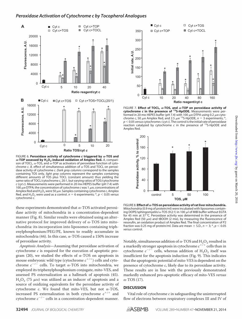
these experiments demonstrated that �-TOS activated peroxi-dase activity of mitochondria in a concentration-dependentmanner (Fig. 8). Similar results were obtained using an alter-native protocol for improved delivery of �-TOS into mito-chondria: its incorporation into liposomes containing triph-enylphosphonium/PEG/PE, known to readily accumulate inmitochondria (44). In this case, �-TOS caused a 150% increaseof peroxidase activity.
Apoptosis Analysis—Assuming that peroxidase activation ofcytochrome c is required for the execution of apoptotic pro-gram (20), we studied the effects of �-TOS on apoptosis inmouse embryonic wild type (cytochrome c�/�) cells and cyto-chrome c�/� cells. To target �-TOS into mitochondria, weemployed its triphenylphosphonium conjugate, mito-VES, andassessed PS externalization as a hallmark of apoptosis (45).H2O2 (75 �M) was utilized as an inducer of apoptosis and asource of oxidizing equivalents for the peroxidase activity ofcytochrome c. We found that mito-VES, but not �-TOS,increased PS externalization in both cytochrome c�/� andcytochrome c�/� cells in a concentration-dependent manner.
Notably, simultaneous addition of �-TOS and H2O2 resulted ina markedly stronger apoptosis in cytochrome c�/� cells than incytochrome c�/� cells, whereas addition of H2O2 itself wasinsufficient for the apoptosis induction (Fig. 9). This indicatesthat the apoptogenic potential of mito-VES is dependent on thepresence of cytochrome c, likely due to its peroxidase activity.These results are in line with the previously demonstratedmarkedly enhanced pro-apoptotic efficacy of mito-VES versus�-TOS (17).
DISCUSSION
Vital role of cytochrome c in safeguarding the uninterruptedflow of electrons between respiratory complexes III and IV of
Fluo
resc
ence
, A.U
.Fl
uore
scen
ce, A
.U.
A
0
4000
8000
12000
16000
20000
5 10 20 40Ratio reagent/cyt c
Cyt cCyt c+TOS
Cyt c+TOPCyt c+TOCL
*
*
*
*
*
**
*
*
*
*
*
Cyt c
B
0
4000
8000
12000
16000
Cyt c 0 5 10 20Ratio TOS/cyt c
Cyt c Cyt c+TOS Cyt c+TOS+TOCL(20/1)
*
*
*
* * **
FIGURE 6. Peroxidase activity of cytochrome c triggered by �-TOS and�-TOP assessed by H2O2-induced oxidation of Amplex Red. A, compari-son of TOCL, �-TOS, and �-TOP as activators of peroxidase function of cyto-chrome c. B, effect of simultaneous addition of �-TOS and TOCL on peroxi-dase activity of cytochrome c. Dark gray columns correspond to the samplescontaining TOS only, light gray columns represent the samples containingdifferent amounts of TOS plus TOCL (constant amount) thus yielding thesame ratio of TOCL/cytochrome c, 20:1, but varying ratios of TOS/cytochromec (cyt c). Measurements were performed in 20 mM HEPES buffer (pH 7.4) with100 �M DTPA; the concentration of cytochrome c was 1 �M, concentrations ofAmplex Red and H2O2 were 50 �M. Samples containing cytochrome c, AmplexRed, and H2O2 were used as a control. n 6 experiments; *, p 0.05 versuscytochrome c.
Initi
al ra
te, A
.U.
0
50
100
150
200
250
300
350
Cyt c 10 20 40 80 160Ratio reagent/cyt c
Cyt c Cyt c+TOSCyt c+TOP Cyt c+TOCL
*
* *
*
*
*
*
*
*
*
*
FIGURE 7. Effect of TOCL, �-TOS, and �-TOP on peroxidase activity ofcytochrome c in the presence of 13S-HpODE. Measurements were per-formed in 20 mM HEPES buffer (pH 7.4) with 100 �M DTPA using 0.2 �M cyto-chrome c, 50 �M Amplex Red, and 2.5 �M
13S-HpODE, n 3 experiments; *,p 0.05 versus cytochrome c (cyt c). The control is the initial rate of peroxidasereaction catalyzed by cytochrome c in the presence of 13S-HpODE andAmplex Red.
0
40
80
120
160
200
control 5 50 100
Pero
xida
se a
ctiv
ity, %
to c
ontr
ol
TOS, µM
**
*
FIGURE 8. Effect of �-TOS on peroxidase activity of rat liver mitochondria.Mitochondria (0.9 mg of protein/ml) were incubated with liposomes contain-ing DOPE/sphingomyelin/�-TOS (9:2:1) in 100 �l of MIB buffer without EGTAfor 45 min at 37 °C. Peroxidase activity was determined in the presence ofAmplex Red (50 �M) and tBOOH (2 mM), by measuring the fluorescence ofresorufin, an oxidation product of Amplex Red. The final concentration of P2fraction was 0.25 mg of protein/ml. Data are mean � S.D., n 3; *, p 0.05versus control.
Peroxidase Activation of Cytochrome c by Tocopherol Analogues
32494 JOURNAL OF BIOLOGICAL CHEMISTRY VOLUME 289 • NUMBER 47 • NOVEMBER 21, 2014
by guest on March 20, 2016
http://ww
w.jbc.org/
Dow
nloaded from

the mitochondrial respiratory chain has been challenged by thediscoveries of its unexpected participation in cell death apopto-tic pathways. This includes the involvement of cytochrome c asa peroxidase catalyst of CL peroxidation, required for therelease of pro-apoptotic factors from mitochondria into thecytosol (18, 20), and as one of the factors facilitating the forma-tion of apoptosomes and caspase activation (46). The dormantperoxidase function is conferred on cytochrome c by its inter-action with a mitochondria-specific anionic phospholipid CL,normally asymmetrically confined to the inner mitochondrialmembrane but redistributed to the outer membrane duringapoptosis (with the likely participation of two candidate mito-chondrial translocators: phospholipid scramblase-3 and nucle-oside diphosphate kinase D (47, 48). Binding of CL to cyto-chrome c causes unfolding of the protein, thus paving the wayfor peroxy donors, H2O2 or organic (lipid) hydroperoxides, tothe heme catalytic site (21). The resultant peroxidase activitydisplays significant specificity toward CLs, yielding its highlydiversified oxygenated species and their hydrolysis products,predominantly mono-lyso-CLs and oxygenated fatty acids,with signaling modalities (49 –51). CL hydroperoxides can beutilized by the peroxidase complex as an alternative (to H2O2)source of oxidizing equivalents, thus “perpetuating” the func-tioning of the peroxidase cycle (43, 47). Although the signifi-cance of the peroxidase activation of cytochrome c in apoptosishas been documented (18, 20), the uniqueness of CLs as endog-enous activators is not unequivocal. Indeed, the structuralrequirements for the cytochrome c unfolding are relatively“loose,” and it is possible that other molecules combining ahydrophobic chain with a negatively charged functionality mayfulfill the requisite conditions (52–56). Therefore, we hypothe-sized that TOC analogues �-TOS and �-TOP may act as acti-vators of cytochrome c peroxidase function.
A redox-silent compound �-TOS has been recently docu-mented as an anti-cancer drug inducing apoptosis in severaltypes of tumor (but not normal) cells (11, 16, 17). Mitochondriawere shown to play a central role in �-TOS-induced apoptosis,and mitochondrial targeting of �-TOS by it tagging with thetriphenylphosphonium group enhanced its pro-apoptoticeffect (17). It was suggested that possible mechanisms of induc-tion of apoptosis induced by �-TOS involves induction of reac-tive oxygen species production by its interaction with theubiquinone-binding site of mitochondrial complex II (16, 17,57). In addition, �-TOS demonstrated BH3 mimetic activity byblocking the interaction of the anti-apoptotic Bcl-2 family pro-teins with their pro-apoptotic counterparts (58). Our data arecompatible with yet another possible mechanism of �-TOSinvolvement in the induction of apoptosis via its ability tounfold cytochrome c and induce its peroxidase activity. Indeed,we demonstrated that �-TOS, effectively delivered into mito-chondria, activates their tBuOOH-dependent peroxidaseactivity.
Notably, mito-VES (added along with H2O2) displayed amarkedly higher pro-apoptotic potential in mouse embryoniccytochrome c�/� cells compared with cytochrome c�/� cells,thus clearly demonstrating the dependence of its pro-apoptoticaction on cytochrome c. It is possible that TOS-induced perox-idase activity of cytochrome c causes oxidation of CL. We foundthat binding of the negatively charged succinate moiety of�-TOS with cytochrome c occurs in close proximity to Lys22
and Lys25 (L-binding site) (42) leaving another site (site A) withLys72/Lys73 (40, 41) still available for the interaction with CL.Thus an “alternatively” bound polyunsaturated CL can be a tar-get for peroxidation by cytochrome c��-TOS complex providedthe source of oxidizing equivalents is available. However, bind-ing of CL to cytochrome c is not always required for its oxida-tion. For example, 1 electron oxidation intermediates gener-ated by the peroxidase activity of cytochrome c/�-TOS from avariety of phenolic compounds, phenoxyl radicals, can escapefrom the catalytic site and diffuse to the locales enriched withoxidizable lipid substrates, including those with the CL mole-cules. Thus, in the presence of phenolic compounds, however,the specificity of oxidation toward CL may not be high andother vulnerable substrates and reductants (e.g. other polyun-saturated lipids, thiols) can get oxidized (59). This phenome-non, the so-called extension of the peroxidase catalytic site, hasbeen documented for several peroxidases with a variety of phe-nolic compounds with appropriate redox potentials (60) but ithas not yet been described for cytochrome c.
�-TOP, in contrast to �-TOS, is ubiquitously present, albeitat rather low concentrations, in animal tissues as well as plants,and has been shown to be synthesized by cells (61, 62). Ourresults demonstrated that TOP, at a TOP:cytochrome c ratio of20:1, increases peroxidase activity of cytochrome c by morethan 3-fold. Given the importance of cytochrome c activation inthe execution of apoptotic program (18), this suggests that TOPis a potential inducer of cytochrome c-dependent apoptosis. Infact, the ability of TOP to induce apoptosis has been demon-strated in THP-1 monocytes and murine MG-63 melanomacells (61, 63). However, this effect was observed only at TOPconcentrations �50 �M. Although the requirement of high
FIGURE 9. Effect of mito-VES on apoptosis in mouse embryonic cyto-chrome c�/� and cytochrome c�/� cells. Four samples were exposed toeach of the mito-VES concentrations: two in the absence of H2O2 (the first twobars: empty, WT cells; filled dark gray, cytochrome c�/� (cyt c) cells, respec-tively) and two in the presence of H2O2 (75 �M) (diagonally striped bars, WTcells with H2O2; horizontal striped bars, cytochrome c�/� cells with H2O2) andincubated for 16 h at 37 °C. The cells were then collected and PS externaliza-tion was determined by flow cytometry using the annexin V-FITC/propidiumiodide kit. Data are mean � S.D., n 3 experiments. *, p 0.05 versus controlcells; #, p 0.05 versus cytochrome c�/� cells.
Peroxidase Activation of Cytochrome c by Tocopherol Analogues
NOVEMBER 21, 2014 • VOLUME 289 • NUMBER 47 JOURNAL OF BIOLOGICAL CHEMISTRY 32495
by guest on March 20, 2016
http://ww
w.jbc.org/
Dow
nloaded from

concentrations of exogenously added TOP for its pro-apoptoticeffect can be explained by the fact that only a small fraction of itpartitions into the mitochondrial intermembrane space to forma peroxidase complex with cytochrome c, the question stillremains whether intracellular TOP concentrations are suffi-cient for triggering apoptosis. In tissues of unsupplementedanimals and humans, the basal �-TOP level was estimated as�0.2 �M (64). After supplementation with �-TP at a dose of1.33 g/kg body weight for 4 weeks, this level was increased morethan 100-fold to 29.4 �M. Thus, endogenous levels of TOP aretoo low to expect its significant contribution to apoptotic celldeath. However, its pharmacological effects may be due, at leastin part, to its ability, at higher concentrations, to facilitate celldeath pathways.
Our structural studies, including heteronuclear NMR assess-ments, of intactness of Met80-Fe coordination bond, and highspin iron, along with the computer simulations, demonstratedthat �-TOS and �-TOP, similarly to TOCL, interacted withcytochrome c and induced its unfolding. Thus both �-TOS and�-TOP can act as peroxidase activators, whereas the non-ester-ified parental compound, TOC, devoid of a negatively chargedfunctionality, did not demonstrate this ability. However, therewere substantial differences in the binding sites and the abilitiesof �-TOS and �-TOP to induce structural re-arrangements incytochrome c. Molecular docking calculations demonstratedthat the negatively charged succinate moiety of �-TOS waspreferentially bound in close proximity to Lys22 and Lys25 (siteL), whereas a more hydrophilic �-TOP was predicted to localizeto a different site on cytochrome c electrostatically interactingwith Lys72 and Lys73 (site A). Lys72 and Lys73 are, in particular,implicated as candidate replacement ligands of Met80 duringalkaline transition of cytochrome c and are thought to partici-pate in electrostatic binding to anionic phospholipids. The pre-dicted binding energy of �-TOP was lower than that of �-TOS.NMR studies indicated that �-TOP was a stronger modifier ofcytochrome c structure than �-TOS. Similarly, �-TOP wasmore effective in disrupting the Met80-Fe bond and inducingaccumulation of the high spin form of the hemoprotein.
Peroxidase activity measurements demonstrated the effec-tiveness of �-TOS and TOCL in activation of peroxidase activ-ity of cytochrome c. Notably, computationally predicted simi-larity in cytochrome c binding sites for �-TOS and TOCL, andthe lack of additive effects on peroxidase activity suggest thatsimilar “unfolding” mechanisms may be triggered by thesecompounds. At the same time, mechanisms of activation ofcytochrome c peroxidase activity by TOCL and �-TOS are notentirely identical as illustrated by differential responses of therespective cytochrome c complexes with either TOCL or�-TOS to FFA-OOH as a substrate for the peroxidase reaction.Interestingly, �-TOP demonstrated a much weaker potency asa peroxidase activator of cytochrome c than either �-TOS orTOCL. Apparent discrepancy between the markedly decreasedabsorbance at 695 nm along with its ability to trigger strongerchanges in NH signals, characterizing rupture of the Fe-S(Met80)bond, and small activation of peroxidase activity by �-TOP canbe explained by different mechanisms involved. Although per-oxidase activation requires a perturbation of the cytochrome cstructure that allows access and interaction of small H2O2 mol-
ecules with the heme iron catalytic site, complete rupture of theFe-S(Met80) bond is not obligatory. Moreover, differences ineffects of �-TOP and �-TOS on unfolding of cytochrome c andits peroxidase activity can be explained by specificities of theirbinding with cytochrome c to Lys72/Lys73 (site A) and Lys22/Lys25 (site L), respectively.
In �-TOS molecule, the hydroxy group in position 6 of itschromanol ring, required for the radical scavenging activity, isesterified with succinate. Because �-TOS activation of cyto-chrome c into a peroxidase is mainly due to a partial proteinunfolding (Figs. 2 and 3), rather than chemical interactions withthe protein, it is unlikely that significant oxidation of �-TOStakes place. Notably, �-TOS itself has no antioxidant propertiesand does not undergo oxidation (65). However, the esterifiedsuccinic acid moiety can be removed by cellular esterasesthereby generating �-tocopherol. It was suggested that a com-paratively higher esterase activity of normal cells versus low ornegligible activity found in cancer cells can be responsible forthe higher selectivity of �-TOS toward malignant cells (66, 67).Thus formed tocopherol is prone to oxidative modificationsbut, in the absence of the succinic moiety, is incapable of induc-ing the peroxidase activity. Thus, it seems unlikely that cyto-chrome c can oxidize �-TOS.
In summary, using computational modeling and NMR stud-ies, we demonstrate that �-TOS and �-TOP interact with cyto-chrome c at similar sites previously proposed for CL. Absorp-tion spectroscopy showed that both �-TOS and �-TOPeffectively disrupt the Fe-S(Met80) bond indicating the unfold-ing of cytochrome c. By measuring the oxidation of a typicalperoxidase substrate, Amplex Red, we showed that �-TOS and�-TOP can stimulate peroxidase activity of cytochrome c.Overall, the structural analysis indicates that “peroxidase” acti-vation of cytochrome c may be achieved via several differentunfolding pathways with specific enzymatic features. It is alsopossible that these distinctive peroxidase “subspecies” of cyto-chrome c would entertain different catalytic mechanisms, e.g.with alternate proportions of hemolytic versus heterolytic acti-vation of peroxide substrates, resulting in diversified ste-reospecificity of CL peroxidation products. Physiologically, thesignificance of these alternate pathways may be realizedthrough the hydrolysis of peroxidized CL leading to the produc-tion of different lipid mediators, oxygenated fatty acids, andoxygenated or non-oxygenated lyso-CLs with yet to beexplored signaling functions (51). �-TOS (and its mitochondri-ally targeted counterpart, mito-VES) and �-TOP can serve asactivators of peroxidase activity of cytochrome c leading to CLoxidation as a step in the execution of the apoptotic program.These data may be instrumental in targeted design of novelregulators/activators of peroxidase activity of cytochrome cwith desirable functions.
REFERENCES1. Evans, H. M., and Bishop, K. S. (1922) On the existence of a hitherto
unrecognized dietary factor essential for reproduction. Science 56,650 – 651
2. Tappel, A. L. (1970) Biological antioxidant protection against lipid peroxi-dation damage. Am. J. Clin. Nutr. 23, 1137–1139
3. Packer, L. (1991) Protective role of vitamin E in biological systems. Am. J.Clin. Nutr. 53, 1050S–1055S
Peroxidase Activation of Cytochrome c by Tocopherol Analogues
32496 JOURNAL OF BIOLOGICAL CHEMISTRY VOLUME 289 • NUMBER 47 • NOVEMBER 21, 2014
by guest on March 20, 2016
http://ww
w.jbc.org/
Dow
nloaded from

4. Niki, E., and Traber, M. G. (2012) A history of vitamin E. Ann. Nutr.Metab. 61, 207–212
5. Myung, S. K., Ju, W., Cho, B., Oh, S. W., Park, S. M., Koo, B. K., Park, B. J.,and Korean Meta-Analysis Study, G. (2013) Efficacy of vitamin and anti-oxidant supplements in prevention of cardiovascular disease: systematicreview and meta-analysis of randomised controlled trials. BMJ 346, f10
6. Dolara, P., Bigagli, E., and Collins, A. (2012) Antioxidant vitamins andmineral supplementation, life span expansion and cancer incidence: a crit-ical commentary. Eur. J. Nutr. 51, 769 –781
7. Abner, E. L., Schmitt, F. A., Mendiondo, M. S., Marcum, J. L., and Kryscio,R. J. (2011) Vitamin E and all-cause mortality: a meta-analysis. Curr AgingSci 4, 158 –170
8. Ricciarelli, R., Zingg, J. M., and Azzi, A. (2001) Vitamin E: protective role ofa Janus molecule. FASEB J. 15, 2314 –2325
9. Kagan, V. E. (1989) Tocopherol stabilizes membrane against phospho-lipase A, free fatty acids, and lysophospholipids. Ann. N.Y. Acad. Sci. 570,121–135
10. Galli, F., and Azzi, A. (2010) Present trends in vitamin E research. BioFac-tors 36, 33– 42
11. Neuzil, J., Weber, T., Gellert, N., and Weber, C. (2001) Selective cancer cellkilling by �-tocopheryl succinate. Br. J. Cancer 84, 87– 89
12. Rohlena, J., Dong, L. F., Ralph, S. J., and Neuzil, J. (2011) Anticancer drugstargeting the mitochondrial electron transport chain. Antioxid. Redox Sig-nal. 15, 2951–2974
13. Negis, Y., Zingg, J. M., Libinaki, R., Meydani, M., and Azzi, A. (2009)Vitamin E and cancer. Nutr. Cancer 61, 875– 878
14. Azzi, A. (2007) Molecular mechanism of �-tocopherol action. Free Radic.Biol. Med. 43, 16 –21
15. Kogure, K., Manabe, S., Hama, S., Tokumura, A., and Fukuzawa, K. (2003)Potentiation of anti-cancer effect by intravenous administration of vesic-ulated �-tocopheryl hemisuccinate on mouse melanoma in vivo. CancerLett. 192, 19 –24
16. Ralph, S. J., Moreno-Sánchez, R., Neuzil, J., and Rodríguez-Enríquez, S.(2011) Inhibitors of succinate: quinone reductase/complex II regulateproduction of mitochondrial reactive oxygen species and protect normalcells from ischemic damage but induce specific cancer cell death. Pharm.Res. 28, 2695–2730
17. Dong, L. F., Jameson, V. J., Tilly, D., Cerny, J., Mahdavian, E., Marín-Hernández, A., Hernández-Esquivel, L., Rodríguez-Enríquez, S., Stursa, J.,Witting, P. K., Stantic, B., Rohlena, J., Truksa, J., Kluckova, K., Dyason, J. C.,Ledvina, M., Salvatore, B. A., Moreno-Sánchez, R., Coster, M. J., Ralph,S. J., Smith, R. A., and Neuzil, J. (2011) Mitochondrial targeting of vitaminE succinate enhances its pro-apoptotic and anti-cancer activity via mito-chondrial complex II. J. Biol. Chem. 286, 3717–3728
18. Kagan, V. E., Tyurin, V. A., Jiang, J., Tyurina, Y. Y., Ritov, V. B., Amoscato,A. A., Osipov, A. N., Belikova, N. A., Kapralov, A. A., Kini, V., Vlasova, I. I.,Zhao, Q., Zou, M., Di, P., Svistunenko, D. A., Kurnikov, I. V., andBorisenko, G. G. (2005) Cytochrome c acts as a cardiolipin oxygenaserequired for release of proapoptotic factors. Nat. Chem. Biol. 1, 223–232
19. Paradies, G., Petrosillo, G., Paradies, V., and Ruggiero, F. M. (2009) Role ofcardiolipin peroxidation and Ca2� in mitochondrial dysfunction and dis-ease. Cell Calcium 45, 643– 650
20. Kagan, V. E., Bayir, H. A., Belikova, N. A., Kapralov, O., Tyurina, Y. Y.,Tyurin, V. A., Jiang, J., Stoyanovsky, D. A., Wipf, P., Kochanek, P. M.,Greenberger, J. S., Pitt, B., Shvedova, A. A., and Borisenko, G. (2009) Cy-tochrome c/cardiolipin relations in mitochondria: a kiss of death. FreeRadic. Biol. Med. 46, 1439 –1453
21. Kapralov, A. A., Kurnikov, I. V., Vlasova, I. I., Belikova, N. A., Tyurin, V. A.,Basova, L. V., Zhao, Q., Tyurina, Y. Y., Jiang, J., Bayir, H., Vladimirov, Y. A.,and Kagan, V. E. (2007) The hierarchy of structural transitions induced incytochrome c by anionic phospholipids determines its peroxidase activa-tion and selective peroxidation during apoptosis in cells. Biochemistry 46,14232–14244
22. Rumbley, J. N., Hoang, L., and Englander, S. W. (2002) Recombinantequine cytochrome c in Escherichia coli: high-level expression, charac-terization, and folding and assembly mutants. Biochemistry 41, 13894–13901
23. Liu, W., Rumbley, J., Englander, S. W., and Wand, A. J. (2003) Backbone
and side-chain heteronuclear resonance assignments and hyperfine NMRshifts in horse cytochrome c. Protein Sci. 12, 2104 –2108
24. Krishna, M. M., Maity, H., Rumbley, J. N., Lin, Y., and Englander, S. W.(2006) Order of steps in the cytochrome c folding pathway: evidence for asequential stabilization mechanism. J. Mol. Biol. 359, 1410 –1419
25. Maity, H., Rumbley, J. N., and Englander, S. W. (2006) Functional role of aprotein foldon: an �-loop foldon controls the alkaline transition in ferri-cytochrome c. Proteins 63, 349 –355
26. Krishna, M. M., Maity, H., Rumbley, J. N., and Englander, S. W. (2007)Branching in the sequential folding pathway of cytochrome c. Protein Sci.16, 1946 –1956
27. Pallotti, F., and Lenaz, G. (2007) Isolation and subfractionation of mito-chondria from animal cells and tissue culture lines. Methods Cell Biol. 80,3– 44
28. Johnson, B. A. (2004) Using NMRView to visualize and analyze the NMRspectra of macromolecules. Methods Mol. Biol. 278, 313–352
29. Goddard, T., and Kneller, D. (2008) SPARKY 3, University of California,San Francisco, CA
30. Trott, O., and Olson, A. J. (2010) AutoDock Vina: improving the speed andaccuracy of docking with a new scoring function, efficient optimization,and multithreading. J. Comp. Chem. 31, 455– 461
31. Sinibaldi, F., Fiorucci, L., Patriarca, A., Lauceri, R., Ferri, T., Coletta, M.,and Santucci, R. (2008) Insights into cytochrome c-cardiolipin interaction.Role played by ionic strength. Biochemistry 47, 6928 – 6935
32. Droghetti, E., Oellerich, S., Hildebrandt, P., and Smulevich, G. (2006)Heme coordination states of unfolded ferrous cytochrome c. Biophys. J.91, 3022–3031
33. Maity, H., Maity, M., Krishna, M. M., Mayne, L., and Englander, S. W.(2005) Protein folding: the stepwise assembly of foldon units. Proc. Natl.Acad. Sci. U.S.A. 102, 4741– 4746
34. Battistuzzi, G., Bortolotti, C. A., Bellei, M., Di Rocco, G., Salewski, J., Hil-debrandt, P., and Sola, M. (2012) Role of Met80 and Tyr67 in the low-pHconformational equilibria of cytochrome c. Biochemistry 51, 5967–5978
35. Oellerich, S., Wackerbarth, H., and Hildebrandt, P. (2002) Spectroscopiccharacterization of nonnative conformational states of cytochrome c. J.Phys. Chem. B 106, 6566 – 6580
36. Marques, H. M. (2007) Insights into porphyrin chemistry provided by themicroperoxidases, the haempeptides derived from cytochrome c. DaltonTrans. 39, 4371– 4385
37. Munro, O. Q., and Marques, H. M. (1996) Heme-peptide models for he-moproteins: 1. solution chemistry of N-acetylmicroperoxidase-8. Inorg.Chem. 35, 3752–3767
38. Antonini, E., and Brunori, M. (1971) The derivatives of ferric hemoglobinand myoglobin. in Hemoglobin and Myoglobin in Their Reactions withLigands, pp. 40 –54, North Holland, Amsterdam
39. Carraway, A. D., McCollum, M. G., and Peterson, J. (1996) Characteriza-tion of N-acetylated heme undecapeptide and some of its derivatives inaqueous media: monomeric model systems for hemoproteins. Inorg.Chem. 35, 6885– 6891
40. Rytömaa, M., and Kinnunen, P. K. (1994) Evidence for two distinctacidic phospholipid-binding sites in cytochrome c. J. Biol. Chem. 269,1770 –1774
41. Rytömaa, M., and Kinnunen, P. K. (1995) Reversibility of the binding ofcytochrome c to liposomes: implications for lipid-protein interactions.J. Biol. Chem. 270, 3197–3202
42. Kawai, C., Prado, F. M., Nunes, G. L., Di Mascio, P., Carmona-Ribeiro,A. M., and Nantes, I. L. (2005) pH-dependent interaction of cytochrome cwith mitochondrial mimetic membranes: the role of an array of positivelycharged amino acids. J. Biol. Chem. 280, 34709 –34717
43. Belikova, N. A., Tyurina, Y. Y., Borisenko, G., Tyurin, V., Samhan Arias,A. K., Yanamala, N., Furtmüller, P. G., Klein-Seetharaman, J., Obinger, C.,and Kagan, V. E. (2009) Heterolytic reduction of fatty acid hydroperoxidesby cytochrome c/cardiolipin complexes: antioxidant function in mito-chondria. J. Am. Chem. Soc. 131, 11288 –11289
44. Biswas, S., Dodwadkar, N. S., Deshpande, P. P., and Torchilin, V. P. (2012)Liposomes loaded with paclitaxel and modified with novel triphenylphos-phonium-PEG-PE conjugate possess low toxicity, target mitochondriaand demonstrate enhanced antitumor effects in vitro and in vivo. J. Control
Peroxidase Activation of Cytochrome c by Tocopherol Analogues
NOVEMBER 21, 2014 • VOLUME 289 • NUMBER 47 JOURNAL OF BIOLOGICAL CHEMISTRY 32497
by guest on March 20, 2016
http://ww
w.jbc.org/
Dow
nloaded from

Release 159, 393– 40245. Elmore, S. (2007) Apoptosis: a review of programmed cell death. Toxicol.
Pathol. 35, 495–51646. Riedl, S. J., and Salvesen, G. S. (2007) The apoptosome: signalling platform
of cell death. Nat. Rev. Mol. Cell Biol. 8, 405– 41347. Liu, J., Epand, R. F., Durrant, D., Grossman, D., Chi, N. W., Epand,
R. M., and Lee, R. M. (2008) Role of phospholipid scramblase 3 in theregulation of tumor necrosis factor-�-induced apoptosis. Biochemistry47, 4518 – 4529
48. Lacombe, M. L., Tokarska-Schlattner, M., Epand, R. F., Boissan, M.,Epand, R. M., and Schlattner, U. (2009) Interaction of NDPK-D with car-diolipin-containing membranes: structural basis and implications for mi-tochondrial physiology. Biochimie 91, 779 –783
49. Ji, J., Kline, A. E., Amoscato, A., Samhan-Arias, A. K., Sparvero, L. J.,Tyurin, V. A., Tyurina, Y. Y., Fink, B., Manole, M. D., Puccio, A. M.,Okonkwo, D. O., Cheng, J. P., Alexander, H., Clark, R. S., Kochanek, P. M.,Wipf, P., Kagan, V. E., and Bayir, H. (2012) Lipidomics identifies cardioli-pin oxidation as a mitochondrial target for redox therapy of brain injury.Nat. Neurosci. 15, 1407–1413
50. Samhan-Arias, A. K., Ji, J., Demidova, O. M., Sparvero, L. J., Feng, W.,Tyurin, V., Tyurina, Y. Y., Epperly, M. W., Shvedova, A. A., Greenberger,J. S., Bayir, H., Kagan, V. E., and Amoscato, A. A. (2012) Oxidized phos-pholipids as biomarkers of tissue and cell damage with a focus on cardio-lipin. Biochim. Biophys. Acta 1818, 2413–2423
51. Tyurina, Y. Y., Poloyac, S. M., Tyurin, V. A., Kapralov, A. A., Jiang, J.,Anthonymuthu, T. S., Kapralova, V. I., Vikulina, A. S., Jung, M. Y., Epperly,M. W., Mohammadyani, D., Klein-Seetharaman, J., Jackson, T. C.,Kochanek, P. M., Pitt, B. R., Greenberger, J. S., Vladimirov, Y. A., Bayir, H.,and Kagan, V. E. (2014) A mitochondrial pathway for biosynthesis of lipidmediators. Nat. Chem. 6, 542–552
52. Belikova, N. A., Vladimirov, Y. A., Osipov, A. N., Kapralov, A. A., Tyurin,V. A., Potapovich, M. V., Basova, L. V., Peterson, J., Kurnikov, I. V., andKagan, V. E. (2006) Peroxidase activity and structural transitions of cyto-chrome c bound to cardiolipin-containing membranes. Biochemistry 45,4998 –5009
53. Prasad, S., Maiti, N. C., Mazumdar, S., and Mitra, S. (2002) Reaction ofhydrogen peroxide and peroxidase activity in carboxymethylated cyto-chrome c: spectroscopic and kinetic studies. Biochim. Biophys. Acta 1596,63–75
54. de Jongh, H. H., Ritsema, T., and Killian, J. A. (1995) Lipid specificity formembrane mediated partial unfolding of cytochrome c. FEBS Lett. 360,255–260
55. Chattopadhyay, K., and Mazumdar, S. (2003) Stabilization of partiallyfolded states of cytochrome c in aqueous surfactant: effects of ionic andhydrophobic interactions. Biochemistry 42, 14606 –14613
56. Sanghera, N., and Pinheiro, T. J. (2000) Unfolding and refolding of cyto-
chrome c driven by the interaction with lipid micelles. Protein Sci. 9,1194 –1202
57. Dong, L. F., Low, P., Dyason, J. C., Wang, X. F., Prochazka, L., Witting,P. K., Freeman, R., Swettenham, E., Valis, K., Liu, J., Zobalova, R., Turanek,J., Spitz, D. R., Domann, F. E., Scheffler, I. E., Ralph, S. J., and Neuzil, J.(2008) �-Tocopheryl succinate induces apoptosis by targeting ubiqui-none-binding sites in mitochondrial respiratory complex II. Oncogene 27,4324 – 4335
58. Shiau, C. W., Huang, J. W., Wang, D. S., Weng, J. R., Yang, C. C., Lin, C. H.,Li, C., and Chen, C. S. (2006) �-Tocopheryl succinate induces apoptosis inprostate cancer cells in part through inhibition of Bcl-xL/Bcl-2 function.J. Biol. Chem. 281, 11819 –11825
59. Borisenko, G. G., Martin, I., Zhao, Q., Amoscato, A. A., Tyurina, Y. Y., andKagan, V. E., (2004) Glutathione propagates oxidative stress triggered bymyeloperoxidase in HL-60 cells: evidence for glutathionyl radical-inducedperoxidation of phospholipids and cytotoxicity. J. Biol. Chem. 279,23453–23462
60. Davies, M. J. (2011) Myeloperoxidase-derived oxidation: mechanisms ofbiological damage and its prevention. J. Clin. Biochem. Nutr. 48, 8 –19
61. Rezk, B. M., van der Vijgh, W. J., Bast, A., and Haenen, G. R. (2007)�-Tocopheryl phosphate is a novel apoptotic agent. Front. Biosci. 12,2013–2019
62. Nishio, K., Ishida, N., Saito, Y., Ogawa-Akazawa, Y., Shichiri, M., Yoshida,Y., Hagihara, Y., Noguchi, N., Chirico, J., Atkinson, J., and Niki, E. (2011)�-Tocopheryl phosphate: uptake, hydrolysis, and antioxidant action incultured cells and mouse. Free Radic. Biol. Med. 50, 1794 –1800
63. Munteanu, A., Zingg, J. M., Ogru, E., Libinaki, R., Gianello, R., West, S.,Negis, Y., and Azzi, A. (2004) Modulation of cell proliferation and geneexpression by �-tocopheryl phosphates: relevance to atherosclerosis andinflammation. Biochem. Biophys. Res. Commun. 318, 311–316
64. Gianello, R., Hall, W. C., Kennepohl, E., Libinaki, R., and Ogru, E. (2007)Subchronic oral toxicity study of mixed tocopheryl phosphates in rats. Int.J. Toxicol. 26, 475– 490
65. Neuzil, J., Tomasetti, M., Zhao, Y., Dong, L. F., Birringer, M., Wang, X. F.,Low, P., Wu, K., Salvatore, B. A., Ralph, S. J. (2007) Vitamin E analogues, anovel group of “mitocans,” as anticancer agents: the importance of beingredox-silent. Mol. Pharmacol. 71, 1185–1199
66. Rodríguez-Enríquez, S., Hernández-Esquivel, L., Marín-Hernández, A.,Dong, L. F., Akporiaye, E. T., Neuzil, J., Ralph, S. J., Moreno-Sánchez, R.(2012) Molecular mechanism for the selective impairment of cancer mi-tochondrial function by a mitochondrially targeted vitamin E analogue.Biochim. Biophys. Acta 1817, 1597–1607
67. Liewald, F., Demmel, N., Wirsching, R., Kahle, H., Valet, G. (1990) Intra-cellular pH, esterase activity, and DNA measurements of human lungcarcinomas by flow cytometry. Cytometry 11, 341–348
Peroxidase Activation of Cytochrome c by Tocopherol Analogues
32498 JOURNAL OF BIOLOGICAL CHEMISTRY VOLUME 289 • NUMBER 47 • NOVEMBER 21, 2014
by guest on March 20, 2016
http://ww
w.jbc.org/
Dow
nloaded from

Valerian E. KaganMao, Judith Klein-Seetharaman, Detcho A. Stoyanovsky, Jan Stursa, Jiri Neuzil and Naveena Yanamala, Alexander A. Kapralov, Mirjana Djukic, Jim Peterson, Gaowei
Analogues of Vitamin E, Tocopherol Succinate and Tocopherol Phosphate by AnioniccStructural Re-arrangement and Peroxidase Activation of Cytochrome
doi: 10.1074/jbc.M114.601377 originally published online October 2, 20142014, 289:32488-32498.J. Biol. Chem.
10.1074/jbc.M114.601377Access the most updated version of this article at doi:
Alerts:
When a correction for this article is posted•
When this article is cited•
to choose from all of JBC's e-mail alertsClick here
http://www.jbc.org/content/289/47/32488.full.html#ref-list-1
This article cites 65 references, 15 of which can be accessed free at
by guest on March 20, 2016
http://ww
w.jbc.org/
Dow
nloaded from
Copyright © 2022 FDOKUMEN