Structural basis for ether-a-go-go-related gene K+ channel subtype-dependent activation by niflumic...
22
Structural Basis for Ether-a-go-go-Related Gene K + Channel Subtype-Dependent Activation by Niflumic Acid [S] David Fernandez, John Sargent, Frank B. Sachse, and Michael C. Sanguinetti Nora Eccles Harrison Cardiovascular Research and Training Institute and Department of Physiology, University of Utah, Salt Lake City, Utah Abstract Niflumic acid [2-((3-(trifluoromethyl)phenyl)amino)-3-pyridin-ecarboxylic acid, NFA] is a nonsteroidal anti-inflammatory drug that also blocks or modulates the gating of a wide spectrum of ion channels. Here we investigated the mechanism of channel activation by NFA on ether-a-go-go- related gene (ERG) K + channel subtypes expressed in Xenopus laevis oocytes using two-electrode voltage-clamp techniques. NFA acted from the extracellular side of the membrane to differentially enhance ERG channel currents independent of channel state. At 1 mM, NFA shifted the half-point for activation by −6, −18, and −11 mV for ERG1, ERG2, and ERG3 channels, respectively. The half- point for channel inactivation was shifted by +5 to +9 mV by NFA. The structural basis for the ERG subtype-specific response to NFA was explored with chimeric channels and site-directed mutagenesis. The molecular determinants of enhanced sensitivity of ERG2 channels to NFA were isolated to an Arg and a Thr triplet in the extracellular S3-S4 linker. The ether-a-go-go-related gene (ERG) family of voltage-gated K + (Kv) channels consists of three genes: ERG1, ERG2, and ERG3. ERG2 and ERG3 channels are expressed exclusively in the nervous system, whereas ERG1 is also expressed in non-neural tissues, including the heart (Shi et al., 1997). Native ERG-like currents have been recorded in neuronal and neuroendocrine cells (Schwarz and Bauer, 2004), where they modulate cell firing (Chiesa et al., 1997; Sacco et al., 2003) and hormonal secretion (Rosati et al., 2000; Gullo et al., 2003), respectively. ERG1 channels conduct the rapid component of the delayed rectifier current (I Kr ) that contributes to the repolarizing phase of the cardiac action potential (Sanguinetti et al., 1995; Trudeau et al., 1995). Reduction in human ERG1 (hERG1) current, caused by a loss of function mutation or by drug-induced channel blockade, prolongs action potential duration and causes long QT syndrome, a disorder characterized by a prolonged QT interval and an increased risk of torsades de pointes arrhythmia, ventricular fibrillation, and sudden death. Niflumic acid [2-((3-(trifluoromethyl)phenyl)amino)-3-pyr-idinecarboxylic acid, NFA] is a nonsteroidal anti-inflammatory drug that preferentially inhibits type 2 cyclooxygenase and is used to treat rheumatoid arthritis and relieve acute pain. NFA and other fenamates (flufenamic acid, mefenamic acid, meclofenamic acid) also modulate the gating or block many different ion channels. Fenamates were first shown to block Ca 2+ -activated Cl − channels (White and Aylwin, 1990), but also to block CFTR channels (Scott-Ward et al., 2004) and activate or block CLC-K channels (Liantonio et al., 2006). These compounds also activate large conductance Ca 2+ -activated K + channels (Ottolia and Toro, 1994), block Kv2.1 (Lee and Wang, 1999), enhance or alter the gating of Kv4.2 and Kv4.3 (Wang et al., 1997), KCNQ1/minK (Busch et [S] The online version of this article (available at http://molpharm.aspetjournals.org) contains supplemental material. Address correspondence to: Michael C. Sanguinetti, Nora Eccles Harrison Cardiovascular Research and Training Institute, Department of Physiology, University of Utah, 95 South 2000 East, Salt Lake City, UT 84112. E-mail: [email protected]. NIH Public Access Author Manuscript Mol Pharmacol. Author manuscript; available in PMC 2009 April 1. Published in final edited form as: Mol Pharmacol. 2008 April ; 73(4): 1159–1167. NIH-PA Author Manuscript NIH-PA Author Manuscript NIH-PA Author Manuscript
-
Upload
independent -
Category
Documents
-
view
7 -
download
0
Transcript of Structural basis for ether-a-go-go-related gene K+ channel subtype-dependent activation by niflumic...
Structural Basis for Ether-a-go-go-Related Gene K+ ChannelSubtype-Dependent Activation by Niflumic Acid[S]
David Fernandez, John Sargent, Frank B. Sachse, and Michael C. SanguinettiNora Eccles Harrison Cardiovascular Research and Training Institute and Department ofPhysiology, University of Utah, Salt Lake City, Utah
AbstractNiflumic acid [2-((3-(trifluoromethyl)phenyl)amino)-3-pyridin-ecarboxylic acid, NFA] is anonsteroidal anti-inflammatory drug that also blocks or modulates the gating of a wide spectrum ofion channels. Here we investigated the mechanism of channel activation by NFA on ether-a-go-go-related gene (ERG) K+ channel subtypes expressed in Xenopus laevis oocytes using two-electrodevoltage-clamp techniques. NFA acted from the extracellular side of the membrane to differentiallyenhance ERG channel currents independent of channel state. At 1 mM, NFA shifted the half-pointfor activation by −6, −18, and −11 mV for ERG1, ERG2, and ERG3 channels, respectively. The half-point for channel inactivation was shifted by +5 to +9 mV by NFA. The structural basis for the ERGsubtype-specific response to NFA was explored with chimeric channels and site-directedmutagenesis. The molecular determinants of enhanced sensitivity of ERG2 channels to NFA wereisolated to an Arg and a Thr triplet in the extracellular S3-S4 linker.
The ether-a-go-go-related gene (ERG) family of voltage-gated K+ (Kv) channels consists ofthree genes: ERG1, ERG2, and ERG3. ERG2 and ERG3 channels are expressed exclusivelyin the nervous system, whereas ERG1 is also expressed in non-neural tissues, including theheart (Shi et al., 1997). Native ERG-like currents have been recorded in neuronal andneuroendocrine cells (Schwarz and Bauer, 2004), where they modulate cell firing (Chiesa etal., 1997; Sacco et al., 2003) and hormonal secretion (Rosati et al., 2000; Gullo et al., 2003),respectively. ERG1 channels conduct the rapid component of the delayed rectifier current(IKr) that contributes to the repolarizing phase of the cardiac action potential (Sanguinetti etal., 1995; Trudeau et al., 1995). Reduction in human ERG1 (hERG1) current, caused by a lossof function mutation or by drug-induced channel blockade, prolongs action potential durationand causes long QT syndrome, a disorder characterized by a prolonged QT interval and anincreased risk of torsades de pointes arrhythmia, ventricular fibrillation, and sudden death.
Niflumic acid [2-((3-(trifluoromethyl)phenyl)amino)-3-pyr-idinecarboxylic acid, NFA] is anonsteroidal anti-inflammatory drug that preferentially inhibits type 2 cyclooxygenase and isused to treat rheumatoid arthritis and relieve acute pain. NFA and other fenamates (flufenamicacid, mefenamic acid, meclofenamic acid) also modulate the gating or block many differention channels. Fenamates were first shown to block Ca2+-activated Cl− channels (White andAylwin, 1990), but also to block CFTR channels (Scott-Ward et al., 2004) and activate or blockCLC-K channels (Liantonio et al., 2006). These compounds also activate large conductanceCa2+-activated K+ channels (Ottolia and Toro, 1994), block Kv2.1 (Lee and Wang, 1999),enhance or alter the gating of Kv4.2 and Kv4.3 (Wang et al., 1997), KCNQ1/minK (Busch et
[S]The online version of this article (available at http://molpharm.aspetjournals.org) contains supplemental material.Address correspondence to: Michael C. Sanguinetti, Nora Eccles Harrison Cardiovascular Research and Training Institute, Departmentof Physiology, University of Utah, 95 South 2000 East, Salt Lake City, UT 84112. E-mail: [email protected].
NIH Public AccessAuthor ManuscriptMol Pharmacol. Author manuscript; available in PMC 2009 April 1.
Published in final edited form as:Mol Pharmacol. 2008 April ; 73(4): 1159–1167.
NIH
-PA Author Manuscript
NIH
-PA Author Manuscript
NIH
-PA Author Manuscript

al., 1994), and KCNQ2/3 channels (Peretz et al., 2005). NFA and flufenamic acid cause amodest increase in hERG1 channel current (Malykhina et al., 2002). In all channels studied,fenamates exhibit an extremely rapid onset of action and apparently bind to an extracellularsite on the channel that has not yet been identified.
In addition to NFA, hERG1 channels are also activated by more potent and selectivecompounds such as NS1643 (Casis et al., 2006; Hansen et al., 2006), PD-118057 (Zhou et al.,2005), and RPR260243 (Kang et al., 2005). These compounds increase current magnitude byinducing a positive shift in the voltage-dependence of inactivation. In addition, RPR260243slows the rate of channel deactivation. PD-118057 is structurally similar to flufenamic acid(also a phenylaminobenzoic acid) and was reported to enhance hERG1 without any measurableeffect on kinetics or voltage dependence of channel gating. The molecular mechanism(s)responsible for the increased current magnitude of ERG channels by fenamates has not beendetermined. An understanding of this mechanism could facilitate the design of more potentand specific hERG1 activators. Furthermore, subtype-specific ERG channel activators wouldserve as unique tools to advance investigations of the physiological role(s) of ERG channelsin neurons.
In this study, we show that NFA is a nonspecific activator of ERG channels. NFA enhancedcurrent of all three ERG channel subtypes (ERG1, ERG2, and ERG3) by inducing a positiveshift in the voltage dependence of inactivation and a variable negative shift in the voltagedependence of activation. Characterization of chimeric ERG channels and a site-directedmutagenesis approach was used to demonstrate that sensitivity to NFA was determined byspecific residues located in the extracellular S3-S4 linker.
Materials and MethodsMolecular biology
hERG1 was subcloned into the pSP64T oocyte expression vector. rERG2 and rERG3 clonesin pSP64 were kindly provided by David McKinnon (SUNY, Stony Brook, NY). ZebrafishERG (zERG) was a gift from Thomas Wagner (Tübingen, Germany).
Six hERG1-rERG2 chimera subunits were constructed: N-S4 chimera, hERG1 with the Nterminus and the S1-S4 from rERG2 (Met1–Ala527); S1-S4 chimera, hERG1 with the S1-S4from rERG2 (Leu417–Ala527); L12 chimera, hERG1 with the external linker connecting S1to S2 domain from rERG2 (Leu417–Asp460); S2 chimera, hERG1 with S2 domain fromrERG2 (Ala453–Leu468); L23 chimera, hERG1 with the S2-S3 internal linker from rERG2(Ala479–Gly487); and L34 chimera, hERG1 with the S3-S4 external linker from rERG2(Gly514–Ala527).
Unique restriction sites were inserted into hERG1 as follows: N-S4 chimera, HindIII (at themultiple cloning site) and AscI (1578–1584 bp); S1-S4 chimera, SpeI (1248–1251 bp) andAscI (1578–1584 bp); L12 chimera, SpeI (1248–1251 bp) and EcoRV (1380 bp); and L34chimera, BspEI (1540–1543 bp) and AscI (1578–1584 bp). The same restriction sites wereintroduced at homologous positions in rERG2. The S2 chimera and L23 chimera, as well assimple point mutations, were created using QuikChange site-directed mutagenesis kit(Stratagene, La Jolla, CA).
Restriction mapping and DNA sequencing were used to confirm sequences. cRNA for injectioninto oocytes was prepared with the mMessage mMachine in vitro transcription kit (Ambion,Austin, TX) and SP6 polymerase after linearization with EcoRI (hERG1 and rERG2) or NheI(rERG3). Estimates of cRNA quality and quantity were determined by gel electrophoresis andUV spectroscopy.
Fernandez et al. Page 2
Mol Pharmacol. Author manuscript; available in PMC 2009 April 1.
NIH
-PA Author Manuscript
NIH
-PA Author Manuscript
NIH
-PA Author Manuscript
Isolation, Injection, and Voltage-Clamp of OocytesStage IV and V Xenopus laevis oocytes were isolated and injected with cRNA encoding wild-type (WT) or mutant ERG channels (Stühmer, 1992). Oocytes were injected with 5 to 20 ngof ERG cRNA then cultured in Barth's solution supplemented with 50 μg/ml gentamicin and1 mM pyruvate at 18°C for 1 to 3 days before use in voltage clamp experiments. Barth's solutioncontained 88 mM NaCl, 1 mM KCl, 0.4 mM CaCl2, 0.33 mM Ca(NO3)2, 1 mM MgSO4, 2.4mM NaHCO3, and 10 mM HEPES, pH 7.4. For voltage-clamp experiments, oocytes werebathed in a modified ND96 solution containing 96 mM NaCl, 4 mM KCl, 1 mM MgCl2, 1 mMCaCl2, 5 mM HEPES, pH 7.6.
Currents were recorded at room temperature (23–25°C) using standard two-microelectrodevoltage-clamp techniques (Stühmer, 1992) with a Geneclamp 500 amplifier and Digidata1322A data acquisition system (Molecular Devices, Sunnyvale, CA). pCLAMP 8 software(Molecular Devices) and a personal computer were used for data acquisition and analysis. Theholding potential was −90 mV, and the interpulse interval for all voltage-clamp protocols was10 s or slower. To obtain current-voltage (I-V) relationships and activation curves, 3-s voltagesteps were applied in 10-mV increments to potentials that varied from −90 to +50 mV. Toconstruct I-V relationships, currents were measured at their peak values (Ipeak) and at the endof the 3-s test pulse (Itest). After each step depolarization, the oocyte was repolarized to a returnpotential (Vret) of −70 or −110 mV to record deactivating tail currents. Other voltage pulseprotocols are described under Results and in the figure legends.
Data AnalysisThe voltage dependence of ERG channel activation was determined by analysis of peak tailcurrents. A plot of tail current amplitude (Itail) versus test potential (Vt) was fitted to aBoltzmann equation using Origin 7 (OriginLab Corp., Northampton, MA) to obtain the half-point (Vact) and slope factor (k) for channel activation: Itail/Itail-max = 1/(1 + exp[(Vact − Vt)/k]). The half-point of ERG channel inactivation (Vinact) was estimated by analysis of the fullyactivated I-V relationship as described previously (Sanguinetti et al., 1995).
The shifts in Vact (ΔVact) caused by a variable concentration of NFA were fit to the Hill equation(Eq. 2), where EC50 is the drug concentration required for 50% activation, [NFA] is the drugconcentration, ΔVmax is the maximum shift in Vact and nH is the Hill coefficient: ΔVact =ΔVmax([NFA]nH/[(EC50)nH + [NFA])nH]. The rate of current deactivation was determined byfitting tail currents with a two-exponential function, where τfast and τslow are the time constants(in milliseconds) for the fast and slow components, and Af and As are the amplitudes (inmicroamperes) of the fast and slow components.
Data are presented as mean ± S.E. (n = number of oocytes), and statistical comparisons betweenexperimental groups were performed using the Student's t test. Differences were consideredsignificant at P < 0.05.
DrugsNFA was purchased from Sigma-Aldrich (St. Louis, MO). Niflumic acid was dissolved indimethyl sulfoxide as a 100 mM stock solution, and final concentrations were obtained byappropriate dilution with the modified ND96 solution. All solutions containing NFA werecorrected to pH 7.6 with NaOH. The control solution contained the same amount of dimethylsulfoxide as the NFA-containing solution.
Mathematical ModelingA five-state Markov model of hERG1 channels was used to simulate the effects of NFA onIKr. The model is based on a previously described model of hERG1 channels at 37°C (Lu et
Fernandez et al. Page 3
Mol Pharmacol. Author manuscript; available in PMC 2009 April 1.
NIH
-PA Author Manuscript
NIH
-PA Author Manuscript
NIH
-PA Author Manuscript
al., 2001). The rate constants for each transition were of the standard format: α = α0exp[zαVm/(RT/F)] and β = β0exp[−zαVm/(RT/F)], where F is the Faraday constant, R is the gasconstant, T is absolute temperature, Vm is membrane potential, and z is the charge. Forreconstruction of current traces at room temperature, rate coefficients from the original modelwere adjusted with a temperature coefficient Q10 = 3.3. Furthermore, voltage dependence foractivation and inactivation was adjusted according to Tables 2 and 3. The simulations with thenormal and drug-bound channel models were carried out with the Euler method for numericalsolution of ordinary differential equations (Press et al., 1992). A time step (Δt) of 1 μs waschosen. All calculations were performed in double precision.
ResultsDifferential Effects of NFA on WT ERG Channels
We examined the effect of NFA on hERG1, rERG2 and rERG3 channels expressed in X.laevis oocytes. ERG currents were elicited by 3-s depolarizing steps to a range of test voltagesfrom a holding potential of −90 mV. NFA rapidly enhanced current magnitude for all threeERG channel types. Although current magnitude measured at the end of a test pulse (Itest) wasenhanced at all potentials, the increase was greater at voltages negative to 0 mV (Fig. 1A)compared with the increase induced at more positive test potentials (Fig. 1B). For example, 1mM NFA caused a 5.6-fold increase in rERG3 current elicited at a test potential of −50 mV,but only a 1.5-fold increase at + 20 mV (Fig. 1, A and B, right).
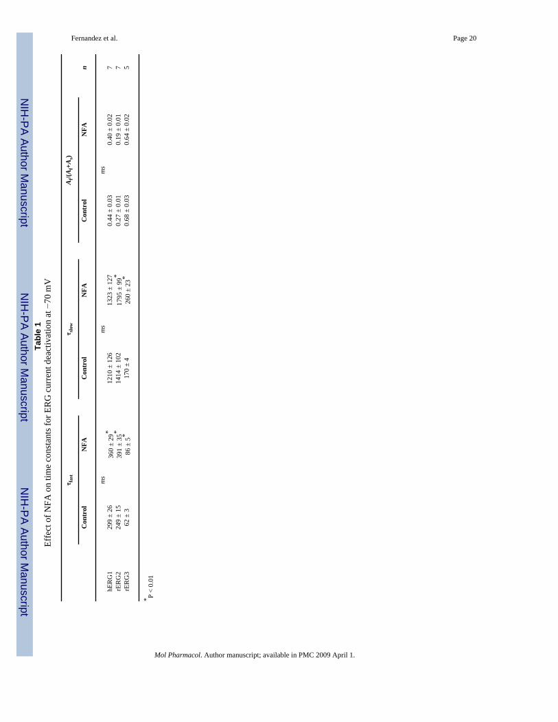
The rate of inactivation is much faster than activation for ERG channels, resulting in apronounced decrease in current at depolarized potentials. However, because NFA apparentlyaccelerated the rate of activation for these channels, the currents at +20 to +40 mV have theappearance of a transient outward current. This can be seen more clearly on the expanded timescale shown in Fig. 1C. In contrast to activation, the rate of tail current decay (deactivation)was slowed by NFA (Table 1).
The effects of 1 mM NFA on the Ipeak-V relationships for all 3 ERG channel types are shownin Fig. 2A. Under control conditions, Ipeak was maximal at −10 mV (hERG1), 0 mV (rERG2)or −20 mV (rERG3) and was reduced or unchanged (rERG3) at more positive potentialsbecause of rapid C-type inactivation (Smith et al., 1996;Spector et al., 1996). For rERG2 andespecially rERG3, the increase in Ipeak by NFA was most pronounced at positive test potentials.The effects of 1 mM NFA on the Itest-V relationships are shown in Fig. 2B. rERG2 channelswere the most sensitive to NFA with a 2.4-fold increase in current recorded at −10 mV. Thus,based on increases in either Ipeak or Itest, the rank order of sensitivity to 1 mM NFA was rERG2> rERG3 > hERG1.
The voltage dependence of ERG channel activation was determined by analysis of peak tailcurrents. Channels were activated by 3-s pulses to a variable potential, and tail currents wereelicited by repolarization of the membrane to −70 mV (hERG1, rERG3) or −110 mV (rERG2).Tail current magnitudes were normalized to the peak value under control conditions and plottedas a function of Vt. Averaged data obtained from multiple oocytes are presented in Fig. 3A.NFA (1 mM) shifted the voltage dependence of activation to more negative potentials for all3 ERG channel types. The magnitude of the shift was quantified by calculating Vact from a fitof the data to a Boltzmann function and followed the same rank order for ERG channel typeas that observed for the I-V relationships. The Vact was shifted by an average of −6.1 mV forhERG1, −11.1 mV for rERG3, and −18.1 mV for rERG2 (Table 2). These shifts in Vact canaccount for the variable increase in Itest observed at voltages negative to the peak of the I-Vrelationships.
Fernandez et al. Page 4
Mol Pharmacol. Author manuscript; available in PMC 2009 April 1.
NIH
-PA Author Manuscript
NIH
-PA Author Manuscript
NIH
-PA Author Manuscript
To assess the effects of NFA on steady state conductance and inactivation of ERG currents,oocytes were first depolarized to +40 mV to activate or inactivate channels, then the oocytewas repolarized to a variable Vret to elicit tail currents. Tail current magnitudes were normalizedto their value at Vret = −130 mV and plotted as a function of Vret to obtain fully activated I-Vrelationships (Fig. 3B). At potentials negative to −100 mV, these I-V relationships were nearlylinear. The deviation of the fully activated I-V from ohmic conductance (rectification factor)was used to define the voltage dependence of channel inactivation (Sanguinetti et al., 1995).NFA increased tail currents the most at the more positive potentials, resulting in a small butconsistent positive shift in the voltage dependence for inactivation of all three ERG channeltypes. The Vinact was shifted by + 7.2 mV for hERG1, +5.3 mV for rERG2 and +8.6 mV forrERG3 (Fig. 3C, Table 3).
Because the shifts in Vinact by NFA were nearly the same for the 3 ERG channel types, thechange in the shape of the Itest − Vt relationships caused by 1 mM NFA (Fig. 2B) must becaused by differential effects on activation. This is more clearly shown in Fig. 4A, where -fold-changes in Itest by NFA were plotted as a function of Vt. At potentials where channel activationwas maximal (>−10 mV for hERG1 and rERG3 or >+10 mV for rERG2; see Fig. 3A), the -fold change in current saturated at a value of ∼1.5. Moreover, the difference in -fold changeobserved at more negative potentials paralleled the differential shift in activation induced byNFA for the 3 channel types. Combining the measured shifts in the voltage dependence ofinactivation and activation was sufficient to account for the effect of 1 mM NFA on the Itest −Vt relationships for all three ERG channel types (Fig. 2B, solid curves).
Concentration-effect relationships for the shift in Vact by NFA are plotted in Fig. 4B. Themaximum shift in Vact induced by NFA varied 3-fold, whereas the EC50 and Hill coefficientswere similar for all three ERG channel types. Thus, the differential effects of NFA on activationof the different ERG channels are due to changes in efficacy, not potency.
NFA Acted from the Extracellular Side of the Membrane Independent of Channel StateNFA activates ERG channel currents immediately after bath application and the effects arevery rapidly reversed with washout of the drug. The rapid kinetics of NFA suggests anextracellular site of action, consistent with the described effects of fenama-tes on other Kvchannels (Ottolia and Toro, 1994; Lee and Wang, 1999).
We next examined the channel state dependence of NFA activity. First, we investigatedwhether NFA could interact with channels in the closed state. hERG1 currents were elicitedby repetitive 2-s pulses to +20 mV until currents reached a steady state. hERG1 channels werekept in the closed state for 3 min by clamping the membrane at −90 mV without pulsing whilethe oocyte chamber was superfused with a solution containing 1 mM NFA. After 3 min, hERG1channels were activated by a single pulse to +20 mV. The currents measured before and afterincubation of an oocyte with NFA are superimposed in Fig. 5A. The current traces are shownon an expanded time scale in Fig. 5B. The onset of drug action appears to be immediate,suggesting that NFA can interact with the closed state of the channel.
Next, hERG1 current was elicited by a 40-s depolarizing step to +20 mV to ensure that channelswere in the open or inactivated states, but not closed states, during application of NFA. First,a control current was recorded (Fig. 5C, black trace). Then, 7 s after starting a second pulse(Fig. 5C, gray trace), the external solution was switched to one containing 1 mM NFA(indicated by arrow). The current rapidly increased in magnitude and reached an amplitudethat was matched during a subsequent pulse (Fig. 5C, red trace). These findings demonstratethat NFA can interact equally well with channels in the closed or open/inactivated states.Similar results were obtained with rERG2 and rERG3 channels (data not shown).
Fernandez et al. Page 5
Mol Pharmacol. Author manuscript; available in PMC 2009 April 1.
NIH
-PA Author Manuscript
NIH
-PA Author Manuscript
NIH
-PA Author Manuscript
Simulation of the Effects of NFA on hERG1 Channel GatingA Markov model was used to simulate the effects of NFA on activation, deactivation andinactivation of hERG1 channels. Drug-bound channels were reconstructed by increasing ratecoefficients for activation C0→C1→C2→O and decreasing rate coefficients for inactivation(O→I) and recovery from inactivation (I→O). This simple model can account for the NFA-induced increase in Ipeak (Fig. 6D), Itest (Fig. 6E), and Itail (Fig. 6F). The NFA-induced increasein Ipeak at positive voltages was explained by an increased ratio of activation versus inactivationrates. Details of the model, including rate constants and initial values for each state are providedin Supplemental Table 1.
S3-S4 Linker Determined Differential Sensitivity of ERG Channels to NFAA chimeric channel approach was used to identify the molecular determinants responsible forthe differential sensitivity of the three ERG channels to NFA. Chimeras were constructed usinghERG1 and rERG2, because these channels provided the weakest and strongest response toNFA, respectively. The amino acid sequence spanning between the N terminus of S1 and theC terminus of S6 for hERG1 and rERG2 subunits is 84% identical. The S1, S3, S4, and S5domains, the S4-S5 linker, pore helix, P-S6 linker domains are identical in hERG1 and rERG2and the S6 domains differ by only one residue. The sequences are most divergent in the S2domain and the external and internal linkers connecting adjacent transmembrane helices.
Six chimera subunits were constructed. Schematic representations of these subunits are shownin Fig. 7, A and B, along with representative current traces recorded in the absence and presenceof 1 mM NFA. We first investigated the role of the voltage sensor domain (VSD) composedof the S1–S4 transmembrane helices. Two chimeras were created by replacing the VSD (S1–S4) or the N terminus plus the VSD (N-S4) of hERG1 with the corresponding regions of therERG2 subunit (Fig. 7A, top). The response to NFA by these two chimeras resembled WTrERG2, including a 2.4-fold increase in current at the peak of the Itest-Vt relationship (data notshown), a −17-mV shift in Vact and a +8 mV shift in Vinact (Tables 2 and 3). Thus, swappingthe VSD from rERG2 to hERG1 effectively transferred NFA sensitivity and ruled out animportant role for the pore domain (S5-S6) or C terminus.
We next examined the role of the S2 helix and the loops that link adjacent transmembranehelices in the VSD. Introducing the S2 helix or linkers connecting the S1 and S2 helices (L12)or S2 and S3 (L23) from rERG2 into hERG1 failed to transfer high sensitivity to NFA (Fig.7B, Table 2). The effect of NFA on these chimera channels was very similar to that observedwith hERG1. In contrast, transferring the external linker connecting S3 and S4 (L34) fromrERG2 into hERG1 created a chimeric channel that enhanced the sensitivity to 1 mM NFA(Fig. 7B, far right). For L34 channels, NFA shifted Vact by −16.1 mV, almost the same as theshift induced in rERG2 (Fig. 7C, Table 2). The effect of NFA on the voltage dependence ofinactivation for L34 or the other chimera channels was not appreciably different from hERG1(Table 3). The differential effects of NFA on ERG channels could conceivably be related todifferences in their gating properties. However, this possibility is ruled out because the kineticsand voltage dependence of chimera L34 were nearly the same as WT hERG1.
Specific Residues in the S3-S4 Linker Determined Differential Sensitivity to NFAThe sequence of the S3-S4 linker in hERG1 differs from rERG2 in two aspects (Fig. 8A). First,the initial residue of the S3-S4 linker is a Gly in hERG1 and an Arg in rERG2. Second, the C-terminal end of the S3-S4 linker in hERG1 is shorter, lacking the three Thr residues (Thr triplet)present in rERG2. The L34 chimera (Fig. 7) introduced both these molecular features of rERG2into hERG1; however, it was not clear whether both or only one of these changes was requiredto confer high NFA sensitivity. A clue came from the effect of NFA on the WT rERG3 channel,which has a Gly (similar to hERG1) and a Thr triplet (similar to rERG2) (Fig. 8A). rERG3
Fernandez et al. Page 6
Mol Pharmacol. Author manuscript; available in PMC 2009 April 1.
NIH
-PA Author Manuscript
NIH
-PA Author Manuscript
NIH
-PA Author Manuscript
sensitivity to NFA is intermediate between those of hERG1 and rERG2 (Fig. 8C), suggestingthat both structural features were required to obtain the largest response to NFA. To investigatethis hypothesis, we constructed hERG1 mutant channels that swapped the Gly for an Arg(G514R) or added three Thr residues (hERG1_TTT). Both alterations increased sensitivity toNFA (Fig. 8B), but neither change alone conferred ERG2-like sensitivity with respect to theshift in Vact (Fig. 8C).
The S3-S4 linker of rERG3 differs from rERG2 by only one residue (Gly in place of Arg). Asconfirmation of the importance of the Arg residue, NFA (1 mM) shifted the Vact of G513RrERG3 by −17 mV (Fig. 8C, Table 2). As a final check of our hypothesis, we examined theeffect of NFA on zERG channels. The S3-S4 linker of zERG is not identical to rERG2 (Fig.8A), but it contains the homologous Arg and the terminal Trp triplet. As predicted, the shift inVact of zERG by NFA was similar to rERG2 WT channels (Fig. 8C, Table 2). Together, thesefindings indicate the importance of the S3-S4 linker in the differential sensitivity of ERGchannels to NFA.
DiscussionNFA enhanced the magnitude of all ERG currents by inducing shifts in the voltage dependenceof channel gating. The effect on activation gating varied between channel subtypes, and therank order for the shift in Vact was ERG2 > ERG3 > ERG1. A previous study of the effects offenamates on hERG1 channels reported that NFA and flufenamic acid enhanced currentamplitude by accelerating the rate of activation and shifting the voltage dependence ofactivation by −5 mV at a concentration of 0.5 mM (Malykhina et al., 2002). Malykhina et al.(2002) also reported that NFA did not alter the rate or voltage dependence of hERG1inactivation when evaluated using a triple pulse protocol that measures recovery frominactivation. In the present study, where inactivation was evaluated from rectification of thefully activated I-V relationship, NFA caused a consistent, albeit small, rightward shift in thevoltage dependence of inactivation of all three ERG channel types. Modeling confirmed thenecessity to include effects on both activation and inactivation gating to fully account for theeffects of NFA on hERG1 channel currents.
A chimeric channel approach was used to isolate the S3-S4 linker as the region of the ERGsubunit that could account for subtype-specific shifts in activation gating. Site-directedmutagenesis refined the structural basis for high sensitivity to NFA of rERG2 compared withhERG1. Substitution of a Gly with an Arg and insertion of three Thr residues into the S3-S4linker of hERG1 was sufficient to increase the sensitivity of the channel to match that observedfor rERG2. Differences in the sequence of the S3-S4 linker (Gly versus Arg) can also explaindifferential ERG channel sensitivity to the sea anemone toxin APETx1. This toxin blocks andshifts the gating of hERG1 but not hERG2 channels. The presence of an Arg in the equivalentposition as Gly514 in the S3-S4 linker of hERG1 prevents toxin binding. Presumably, NFAdoes not bind directly to the S3-S4 linker of ERG or other Kv channels. This domain is highlydivergent in Kv channels, suggesting that NFA and other fenamates alter the gating of ERGand other Kv channels by interacting with some other component of the VSD. Further studiesare needed to determine whether NFA binds to a similar site on all Kv channels.
Our findings do not necessarily implicate an important role for the S3-S4 linker in ERG channelgating. In a previous study of Shaker channels, it was shown that the S3-S4 linker does notappear to be a critical component of voltage sensing. Replacement of the 25-residue linker inShaker with shorter linkers from Shab (9 residues) or Shal (7 residues) sped the rates ofactivation and deactivation but caused only minor changes in the voltage dependence ofchannel activation (Mathur et al., 1997).
Fernandez et al. Page 7
Mol Pharmacol. Author manuscript; available in PMC 2009 April 1.
NIH
-PA Author Manuscript
NIH
-PA Author Manuscript
NIH
-PA Author Manuscript
ERG channel activators other than NFA also display subtype-specificity. NS1643 increasesthe magnitude of ERG1 and ERG2 channel currents. Like NFA, NS1643 causes a leftwardshift in the voltage dependence of ERG2 activation (Elmedyb et al., 2007); however, theactivation of ERG1 channels are not affected or slightly rightward shifted (Casis et al., 2006;Hansen et al., 2006). Another example is RPR260243, which is a potent activator of ERG1channels but blocks ERG3 channels (Kang et al., 2005). Thus, despite the similarities in channelstructure and biophysical properties of ERG1, ERG2, and ERG3, it is clear that drugs can havedifferential effects on these channels. Discovery of even more selective agents should greatlyfacilitate the investigation of the physiological roles of ERG channel subtypes in a variety oftissues. The development of ERG1-specific agonists will also be important for the developmentof heart-specific drugs to treat long QT syndrome.
Supplementary MaterialRefer to Web version on PubMed Central for supplementary material.
Acknowledgements
We thank Kam Hoe Ng for isolation and injection of oocytes.
This work was supported by National Heart, Lung, and Blood Institute/National Institutes of Health grant HL55236.
ReferencesBusch AE, Herzer T, Wagner CA, Schmidt F, Raber G, Waldegger S, Lang F. Positive regulation by
chloride channel blockers of IsK channels expressed in Xenopus oocytes. Mol Pharmacol1994;46:750–753. [PubMed: 7969055]
Casis O, Olesen SP, Sanguinetti MC. Mechanism of action of a novel human ether-a-go-go-related genechannel activator. Mol Pharmacol 2006;69:658–665. [PubMed: 16284303]
Chiesa N, Rosati B, Arcangeli A, Olivotto M, Wanke E. A novel role for HERG K+ channels: spike-frequency adaptation. J Physiol 1997;501:313–318. [PubMed: 9192303]
Elmedyb P, Olesen SP, Grunnet M. Activation of ERG2 potassium channels by the diphenylurea NS1643.Neuropharmacology 2007;53:283–294. [PubMed: 17610913]
Gullo F, Ales E, Rosati B, Lecchi M, Masi A, Guasti L, Cano-Abad MF, Arcangeli A, Lopez MG, WankeE. ERG K+ channel blockade enhances firing and epinephrine secretion in rat chromaffin cells: themissing link to LQT2-related sudden death? FASEB J 2003;17:330–332. [PubMed: 12490549]
Hansen RS, Diness TG, Christ T, Demnitz J, Ravens U, Olesen SP, Grunnet M. Activation of humanether-a-go-go-related gene potassium channels by the diphenylurea 1,3-bis-(2-hydroxy-5-trifluoromethyl-phenyl)-urea (NS1643). Mol Pharmacol 2006;69:266–277. [PubMed: 16219910]
Kang J, Chen XL, Wang H, Ji J, Cheng H, Incardona J, Reynolds W, Viviani F, Tabart M, Rampe D.Discovery of a small molecule activator of the human ether-a-go-go-related gene (HERG) cardiac K+ channel. Mol Pharmacol 2005;67:827–836. [PubMed: 15548764]
Lee YT, Wang Q. Inhibition of hKv2.1, a major human neuronal voltage-gated K+ channel, bymeclofenamic acid. Eur J Pharmacol 1999;378:349–356. [PubMed: 10493112]
Liantonio A, Picollo A, Babini E, Carbonara G, Fracchiolla G, Loiodice F, Tortorella V, Pusch M,Camerino DC. Activation and inhibition of kidney CLC-K chloride channels by fenamates. MolPharmacol 2006;69:165–173. [PubMed: 16244177]
Lu Y, Mahaut-Smith MP, Varghese A, Huang CL, Kemp PR, Vandenberg JI. Effects of prematurestimulation on HERG K+ channels. J Physiol 2001;537:843–851. [PubMed: 11744759]
Malykhina AP, Shoeb F, Akbarali HI. Fenamate-induced enhancement of heterologously expressedHERG currents in Xenopus oocytes. Eur J Pharmacol 2002;452:269–277. [PubMed: 12359267]
Mathur R, Zheng J, Yan Y, S FJ. Role of the S3–S4 linker in shaker potassium channel activation. J GenPhysiol 1997;109:191–199. [PubMed: 9041448]
Ottolia M, Toro L. Potentiation of large conductance KCa channels by niflumic, flufenamic, andmefenamic acids. Biophys J 1994;67:2272–2279. [PubMed: 7535111]
Fernandez et al. Page 8
Mol Pharmacol. Author manuscript; available in PMC 2009 April 1.
NIH
-PA Author Manuscript
NIH
-PA Author Manuscript
NIH
-PA Author Manuscript
Peretz A, Degani N, Nachman R, Uziyel Y, Gibor G, Shabat D, Attali B. Meclofenamic acid anddiclofenac, novel templates of KCNQ2/Q3 potassium channel openers, depress cortical neuronactivity and exhibit anticonvulsant properties. Mol Pharmacol 2005;67:1053–1066. [PubMed:15598972]
Press, WH.; Teukolsky, SA.; Vetterling, WT.; Flannery, BP. Numerical Recipes in C. CambridgeUniversity Press; Cambridge: 1992.
Rosati B, Marchetti P, Crociani O, Lecchi M, Lupi R, Arcangeli A, Olivotto M, Wanke E. Glucose- andarginine-induced insulin secretion by human pancreatic beta-cells: the role of HERG K+ channels infiring and release. Faseb J 2000;14:2601–2610. [PubMed: 11099479]
Sacco T, Bruno A, Wanke E, Tempia F. Functional roles of an ERG current isolated in cerebellar Purkinjeneurons. J Neurophysiol 2003;90:1817–1828. [PubMed: 12750425]
Sanguinetti MC, Jiang C, Curran ME, Keating MT. A mechanistic link between an inherited and anacquired cardiac arrhythmia: HERG encodes the IKr potassium channel. Cell 1995;81:299–307.[PubMed: 7736582]
Schwarz JR, Bauer CK. Functions of erg K+ channels in excitable cells. J Cell Mol Med 2004;8:22–30.[PubMed: 15090257]
Scott-Ward TS, Li H, Schmidt A, Cai Z, Sheppard DN. Direct block of the cystic fibrosis transmembraneconductance regulator Cl− channel by niflumic acid. Mol Membr Biol 2004;21:27–38. [PubMed:14668136]
Shi W, Wymore RS, Wang HS, Pan Z, Cohen IS, McKinnon D, Dixon JE. Identification of two nervoussystem-specific members of the erg potassium channel gene family. J Neurosci 1997;17:9423–9432.[PubMed: 9390998]
Smith PL, Baukrowitz T, Yellen G. The inward rectification mechanism of the HERG cardiac potassiumchannel. Nature 1996;379:833–836. [PubMed: 8587608]
Spector PS, Curran ME, Zou A, Keating MT, Sanguinetti MC. Fast inactivation causes rectification ofthe IKr channel. J Gen Physiol 1996;107:611–619. [PubMed: 8740374]
Stühmer W. Electrophysiological recording from Xenopus oocytes. Methods Enzymol 1992;207:319–339. [PubMed: 1382188]
Trudeau M, Warmke JW, Ganetzky B, Robertson GA. HERG, A human inward rectifier in the voltage-gated potassium channel family. Science 1995;269:92–95. [PubMed: 7604285]
Wang HS, Dixon JE, McKinnon D. Unexpected and differential effects of Cl− channel blockers on theKv4.3 and Kv4.2 K+ channels. Implications for the study of the Ito2 current. Circ Res 1997;81:711–718. [PubMed: 9351445]
White MM, Aylwin M. Niflumic and flufenamic acids are potent reversible blockers of Ca2+-activatedCl− channels in Xenopus oocytes. Mol Pharmacol 1990;37:720–724. [PubMed: 1692608]
Zhou J, Augelli-Szafran CE, Bradley JA, Chen X, Koci BJ, Volberg WA, Sun Z, Cordes JS. Novel potenthuman ether-a-go-go-related gene (hERG) potassium channel enhancers and their in vitroantiarrhythmic activity. Mol Pharmacol 2005;68:876–884. [PubMed: 15976038]
AbbreviationsNFA
niflumic acid [2-((3-(trifluoromethyl)phenyl)amino)-3-pyridinecarboxylic acid]
NS1643 1,3-bis(2-hydroxy-5-trifluoromethylphenyl)urea
PD-118057 2-{4-[2-(3,4-dichloro-phenyl)-ethyl]-phenylamino}-benzoic acid
RPR260243 (3R,4R)-4-(3-(6-methoxyquinolin-4-yl)-3-oxo-propyl)-1-(3-(2,3,5-trifluoro-phenyl)-prop-2-ynyl)-piperidine-3-carboxylic acid
zERG
Fernandez et al. Page 9
Mol Pharmacol. Author manuscript; available in PMC 2009 April 1.
NIH
-PA Author Manuscript
NIH
-PA Author Manuscript
NIH
-PA Author Manuscript
zebrafish ERG
rERG rat ERG
hERG human ERG
bp base pair(s)
VSD voltage sensor domain
WT wild-type
I-V current-voltage
ERG1 human ether-a-go-go-related gene
Fernandez et al. Page 10
Mol Pharmacol. Author manuscript; available in PMC 2009 April 1.
NIH
-PA Author Manuscript
NIH
-PA Author Manuscript
NIH
-PA Author Manuscript
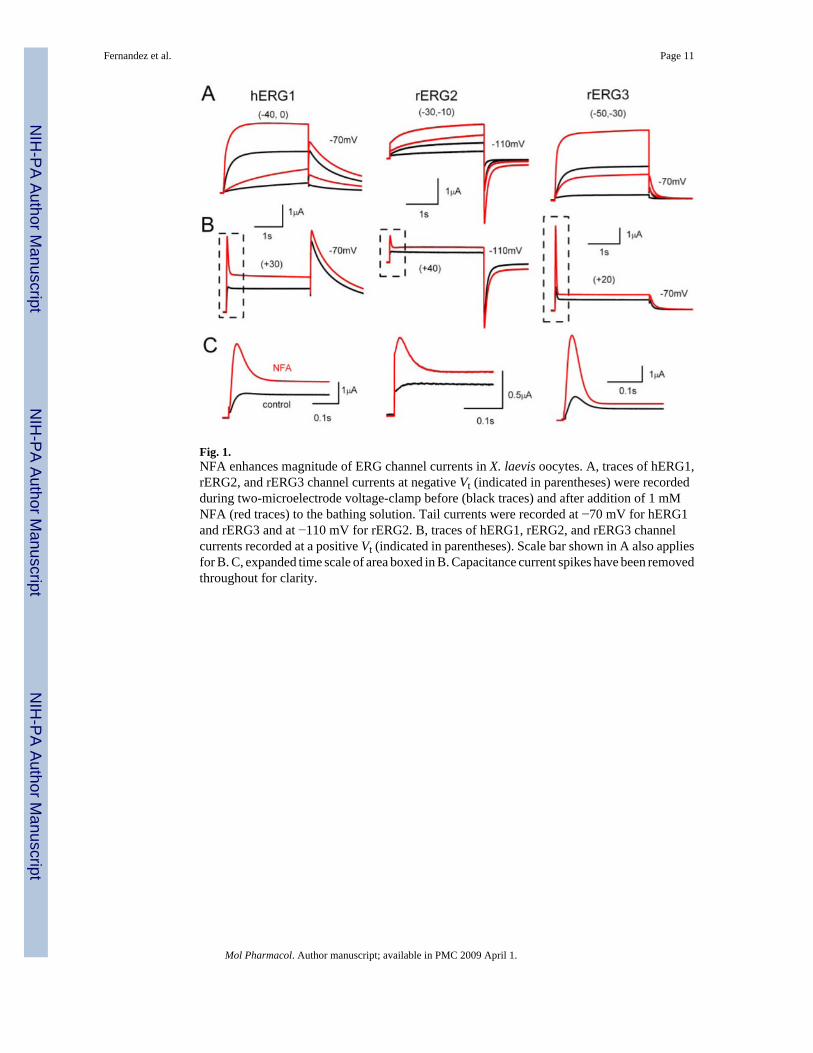
Fig. 1.NFA enhances magnitude of ERG channel currents in X. laevis oocytes. A, traces of hERG1,rERG2, and rERG3 channel currents at negative Vt (indicated in parentheses) were recordedduring two-microelectrode voltage-clamp before (black traces) and after addition of 1 mMNFA (red traces) to the bathing solution. Tail currents were recorded at −70 mV for hERG1and rERG3 and at −110 mV for rERG2. B, traces of hERG1, rERG2, and rERG3 channelcurrents recorded at a positive Vt (indicated in parentheses). Scale bar shown in A also appliesfor B. C, expanded time scale of area boxed in B. Capacitance current spikes have been removedthroughout for clarity.
Fernandez et al. Page 11
Mol Pharmacol. Author manuscript; available in PMC 2009 April 1.
NIH
-PA Author Manuscript
NIH
-PA Author Manuscript
NIH
-PA Author Manuscript
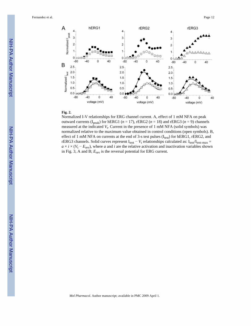
Fig. 2.Normalized I-V relationships for ERG channel current. A, effect of 1 mM NFA on peakoutward currents (Ipeak) for hERG1 (n = 17), rERG2 (n = 18) and rERG3 (n = 9) channelsmeasured at the indicated Vt. Current in the presence of 1 mM NFA (solid symbols) wasnormalized relative to the maximum value obtained in control conditions (open symbols). B,effect of 1 mM NFA on currents at the end of 3-s test pulses (Itest) for hERG1, rERG2, andrERG3 channels. Solid curves represent Itest − Vt relationships calculated as: Itest/Itest-max =a × i × (Vt − Erev), where a and i are the relative activation and inactivation variables shownin Fig. 3, A and B; Erev is the reversal potential for ERG current.
Fernandez et al. Page 12
Mol Pharmacol. Author manuscript; available in PMC 2009 April 1.
NIH
-PA Author Manuscript
NIH
-PA Author Manuscript
NIH
-PA Author Manuscript
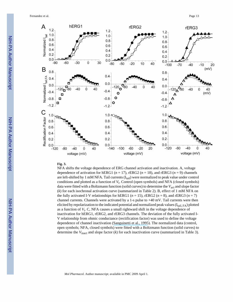
Fig. 3.NFA shifts the voltage dependence of ERG channel activation and inactivation. A, voltagedependence of activation for hERG1 (n = 17), rERG2 (n = 18), and rERG3 (n = 9) channelsare left-shifted by 1 mM NFA. Tail currents (Itail) were normalized to peak value under controlconditions and plotted as a function of Vt. Control (open symbols) and NFA (closed symbols)data were fitted with a Boltzmann function (solid curves) to determine the Vact and slope factor(k) for each isochronal activation curve (summarized in Table 2). B, effect of 1 mM NFA onthe fully activated I-V relationships for hERG1 (n = 15), rERG2 (n = 8), and rERG3 (n = 7)channel currents. Channels were activated by a 1-s pulse to +40 mV. Tail currents were thenelicited by repolarization to the indicated potential and normalized peak values (Itail, FA) plottedas a function of Vt. C, NFA causes a small rightward shift in the voltage dependence ofinactivation for hERG1, rERG2, and rERG3 channels. The deviation of the fully activated I-V relationship from ohmic conductance (rectification factor) was used to define the voltagedependence of channel inactivation (Sanguinetti et al., 1995). The normalized data (control,open symbols; NFA, closed symbols) were fitted with a Boltzmann function (solid curves) todetermine the Vinact and slope factor (k) for each inactivation curve (summarized in Table 3).
Fernandez et al. Page 13
Mol Pharmacol. Author manuscript; available in PMC 2009 April 1.
NIH
-PA Author Manuscript
NIH
-PA Author Manuscript
NIH
-PA Author Manuscript
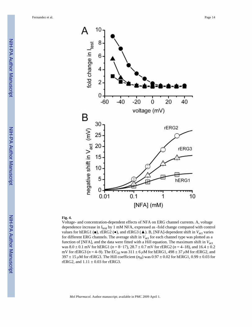
Fig. 4.Voltage- and concentration-dependent effects of NFA on ERG channel currents. A, voltagedependence increase in Itest by 1 mM NFA, expressed as -fold change compared with controlvalues for hERG1 (■), rERG2 (●), and rERG3 (▲). B, [NFA]-dependent shift in Vact variesfor different ERG channels. The average shift in Vact for each channel type was plotted as afunction of [NFA], and the data were fitted with a Hill equation. The maximum shift in Vactwas 8.0 ± 0.1 mV for hERG1 (n = 8−17), 28.7 ± 0.7 mV for rERG2 (n = 4–18), and 16.4 ± 0.2mV for rERG3 (n = 4–9). The EC50 was 311 ± 6 μM for hERG1, 498 ± 37 μM for rERG2, and397 ± 15 μM for rERG3. The Hill coefficient (nH) was 0.97 ± 0.02 for hERG1, 0.99 ± 0.03 forrERG2, and 1.11 ± 0.03 for rERG3.
Fernandez et al. Page 14
Mol Pharmacol. Author manuscript; available in PMC 2009 April 1.
NIH
-PA Author Manuscript
NIH
-PA Author Manuscript
NIH
-PA Author Manuscript
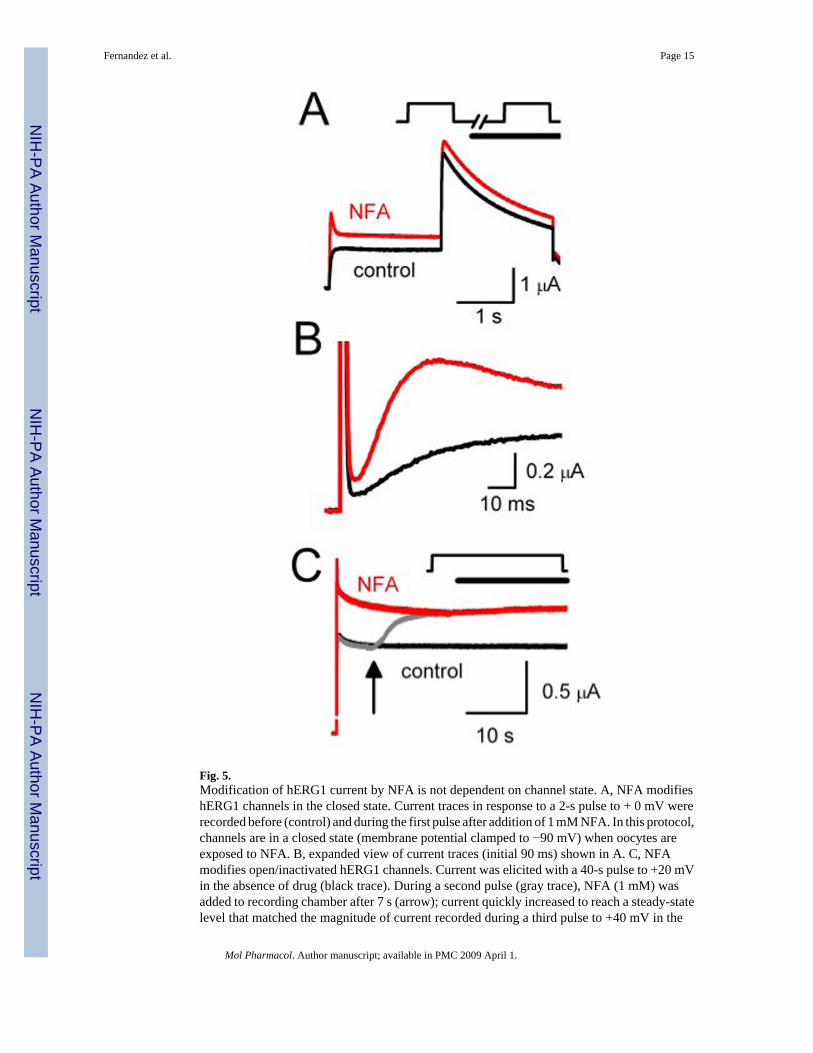
Fig. 5.Modification of hERG1 current by NFA is not dependent on channel state. A, NFA modifieshERG1 channels in the closed state. Current traces in response to a 2-s pulse to + 0 mV wererecorded before (control) and during the first pulse after addition of 1 mM NFA. In this protocol,channels are in a closed state (membrane potential clamped to −90 mV) when oocytes areexposed to NFA. B, expanded view of current traces (initial 90 ms) shown in A. C, NFAmodifies open/inactivated hERG1 channels. Current was elicited with a 40-s pulse to +20 mVin the absence of drug (black trace). During a second pulse (gray trace), NFA (1 mM) wasadded to recording chamber after 7 s (arrow); current quickly increased to reach a steady-statelevel that matched the magnitude of current recorded during a third pulse to +40 mV in the
Fernandez et al. Page 15
Mol Pharmacol. Author manuscript; available in PMC 2009 April 1.
NIH
-PA Author Manuscript
NIH
-PA Author Manuscript
NIH
-PA Author Manuscript
continued presence of NFA (red trace). In A and C, the capacitance current spikes wereremoved for clarity.
Fernandez et al. Page 16
Mol Pharmacol. Author manuscript; available in PMC 2009 April 1.
NIH
-PA Author Manuscript
NIH
-PA Author Manuscript
NIH
-PA Author Manuscript
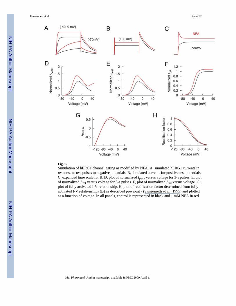
Fig. 6.Simulation of hERG1 channel gating as modified by NFA. A, simulated hERG1 currents inresponse to test pulses to negative potentials. B, simulated currents for positive test potentials.C, expanded time scale for B. D, plot of normalized Ipeak versus voltage for 3-s pulses. E, plotof normalized Itest versus voltage for 3-s pulses. F, plot of normalized Itail versus voltage. G,plot of fully activated I-V relationship. H, plot of rectification factor determined from fullyactivated I-V relationships (B) as described previously (Sanguinetti et al., 1995) and plottedas a function of voltage. In all panels, control is represented in black and 1 mM NFA in red.
Fernandez et al. Page 17
Mol Pharmacol. Author manuscript; available in PMC 2009 April 1.
NIH
-PA Author Manuscript
NIH
-PA Author Manuscript
NIH
-PA Author Manuscript

Fig. 7.ERG1-ERG2 chimera channels identify the S3-S4 linker as a determinant of NFA sensitivity.A and B, schematic of hERG1-rERG2 chimera subunits and representative current tracesshowing effects of 1 mM NFA on the chimeric channels expressed in oocytes. Test potentials(in mV) are indicated. Outward tail currents were measured at −70 mV; inward tail currentswere measured at −110 mV. C, bar graph of NFA-induced shift in Vact for WT hERG1, WTrERG2, and six chimera channels (n = 6–18, see Table 2). The S3-S4 linker swapped fromrERG2 was the minimal domain that conferred rERG2-like sensitivity to hERG1 channels. *,P < 0.01 compared with corresponding value for WT rERG2 channels; ns, not significant.Scale bars represent 1 μA and 1 s.
Fernandez et al. Page 18
Mol Pharmacol. Author manuscript; available in PMC 2009 April 1.
NIH
-PA Author Manuscript
NIH
-PA Author Manuscript
NIH
-PA Author Manuscript
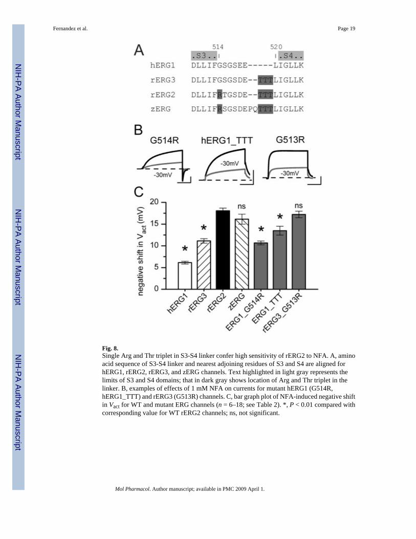
Fig. 8.Single Arg and Thr triplet in S3-S4 linker confer high sensitivity of rERG2 to NFA. A, aminoacid sequence of S3-S4 linker and nearest adjoining residues of S3 and S4 are aligned forhERG1, rERG2, rERG3, and zERG channels. Text highlighted in light gray represents thelimits of S3 and S4 domains; that in dark gray shows location of Arg and Thr triplet in thelinker. B, examples of effects of 1 mM NFA on currents for mutant hERG1 (G514R,hERG1_TTT) and rERG3 (G513R) channels. C, bar graph plot of NFA-induced negative shiftin Vact for WT and mutant ERG channels (n = 6–18; see Table 2). *, P < 0.01 compared withcorresponding value for WT rERG2 channels; ns, not significant.
Fernandez et al. Page 19
Mol Pharmacol. Author manuscript; available in PMC 2009 April 1.
NIH
-PA Author Manuscript
NIH
-PA Author Manuscript
NIH
-PA Author Manuscript
NIH
-PA Author Manuscript
NIH
-PA Author Manuscript
NIH
-PA Author Manuscript
Fernandez et al. Page 20Ta
ble
1Ef
fect
of N
FA o
n tim
e co
nsta
nts f
or E
RG
cur
rent
dea
ctiv
atio
n at
−70
mV
τ fas
tτ s
low
A f/(A
f+A s
)
Con
trol
NFA
Con
trol
NFA
Con
trol
NFA
n
ms
ms
ms
hER
G1
299
± 26
360
± 29
*12
10 ±
126
1323
± 1
270.
44 ±
0.0
30.
40 ±
0.0
27
rER
G2
249
± 15
391
± 35
*14
14 ±
102
1795
± 9
9*0.
27 ±
0.0
10.
19 ±
0.0
17
rER
G3
62 ±
386
± 5
*17
0 ±
426
0 ±
23*
0.68
± 0
.03
0.64
± 0
.02
5
* P <
0.01
Mol Pharmacol. Author manuscript; available in PMC 2009 April 1.

NIH
-PA Author Manuscript
NIH
-PA Author Manuscript
NIH
-PA Author Manuscript
Fernandez et al. Page 21Ta
ble
2Ef
fect
s of N
FA o
n th
e vo
ltage
dep
ende
nce
of E
RG
cha
nnel
act
ivat
ion
Cha
nnel
Con
trol
1 m
M N
FA
V act
kV a
ctk
ΔVac
tn
mV
hER
G1
WT
−29.
3 ±
0.7
7.7
± 0.
1−3
5.4
± 0.
97.
6 ±
0.3
−6.1
± 0
.3**
17rE
RG
2 W
T−8
.7 ±
0.5
9.6
± 0.
2−2
6.8
± 0.
68.
9 ±
0.1
−18.
1 ±
0.6
18rE
RG
3 W
T−3
5.5
± 0.
47.
0 ±
0.3
−46.
6 ±
0.8
6.9
± 0.
4−1
1.1
± 0.
5**9
zER
G W
T−1
4.0
± 1.
27.
8 ±
0.2
−30.
1 ±
0.3
6.8
± 0.
2−1
6.2
± 1.
112
N-S
41.
3 ±
1.1
10.3
± 0
.5−1
5.9
± 0.
211
.4 ±
0.6
−17.
2 ±
0.8
7S1
-S4
−21.
0 ±
0.9
7.9
± 0.
1−3
8.1
± 0.
67.
7 ±
0.4
−17.
0 ±
0.6
10L1
2−2
7.2
± 0.
48.
1 ±
0.2
−33.
6 ±
0.6
8.3
± 0.
4−6
.4 ±
0.6
**9
S2−3
2.6
± 0.
87.
4 ±
0.3
−37.
1 ±
0.8
7.0
± 0.
2−4
.5 ±
0.3
**6
L23
−25.
7 ±
1.2
8.3
± 0.
3−3
0.5
± 0.
97.
9 ±
0.2
−4.8
± 0
.4**
6L3
4−2
5.8
± 0.
78.
0 ±
0.1
−41.
9 ±
0.9
8.1
± 0.
4−1
6.1
± 0.
5*14
hER
G1
G51
4R−2
6.6
± 0.
47.
5 ±
0.2
−37.
2 ±
0.5
6.8
± 0.
2−1
0.7
± 0.
4**8
hER
G1T
TT−1
5.3
± 1.
38.
9 ±
0.3
−28.
8 ±
1.3
9.9
± 0.
4−1
3.5
± 1.
0**6
rER
G3
G51
3R−3
2.3
± 1.
010
.5 ±
0.3
−49.
6 ±
1.1
9.7
± 0.
6−1
7.2
± 0.
88
* P <
0.05
;
**P
< 0.
001
com
pare
d to
NFA
-indu
ced
shift
in V
act o
bser
ved
for W
T rE
RG
2.
Mol Pharmacol. Author manuscript; available in PMC 2009 April 1.

NIH
-PA Author Manuscript
NIH
-PA Author Manuscript
NIH
-PA Author Manuscript
Fernandez et al. Page 22Ta
ble
3Ef
fect
s of N
FA o
n th
e vo
ltage
dep
ende
nce
of E
RG
cha
nnel
inac
tivat
ion
Cha
nnel
Con
trol
1 m
M N
FA
V ina
ctk
V ina
ctk
ΔVin
act
n
mV
hER
G1
WT
−45.
4 ±
2.6
24.2
± 0
.4−3
8.7
± 2.
924
.4 ±
0.6
7.2
± 0.
6*15
rER
G2
WT
−62.
8 ±
0.8
25.1
± 0
.4−5
7.5
± 1.
225
.5 ±
0.3
5.3
± 0.
58
rER
G3
WT
−39.
9 ±
2.8
24.3
± 0
.7−3
1.3
± 2.
523
.9 ±
0.5
8.6
± 1.
3*7
zER
G W
T−6
1.5
± 2.
427
.8 ±
0.6
−53.
2 ±
3.5
28.4
± 0
.78.
3 ±
0.3**
7N
-S4
−42.
6 ±
3.3
23.7
± 0
.6−3
5.1
± 3.
623
.5 ±
0.6
7.5
± 0.
4**6
S1-S
4−4
4.0
± 4.
825
.8 ±
0.8
−35.
5 ±
7.1
24.6
± 0
.88.
5 ±
2.6
6L1
2−4
8.7
± 2.
225
.8 ±
0.7
−42.
2 ±
1.6
25.9
± 0
.76.
5 ±
3.0
6S2
−46.
6 ±
3.6
23.8
± 0
.8−3
6.7
± 4.
024
.9 ±
1.4
9.9
± 0.
5**5
L23
−55.
3 ±
1.8
24.5
± 0
.1−4
7.9
± 1.
424
.9 ±
0.1
7.3
± 1.
03
L34
−49.
8 ±
3.6
27.1
± 0
.5−4
2.6
± 2.
927
.2 ±
0.7
7.2
± 1.
18
hER
G1
G51
4R−3
7.8
± 4.
224
.9 ±
0.5
−28.
8 ±
4.8
25.1
± 0
.69.
0 ±
0.8**
9hE
RG
1 TT
T−5
0.0
± 2.
125
.9 ±
0.4
−40.
9 ±
3.3
25.6
± 0
.89.
1 ±
1.4**
3rE
RG
3 G
513R
−39.
1 ±
4.7
25.2
± 0
.6−3
1.2
± 4.
725
.7 ±
0.8
8.0
± 1.
2*5
* P <
0.05
,
**P
< 0.
01 c
ompa
red
with
NFA
-indu
ced
shift
in V
inac
t obs
erve
d fo
r WT
rER
G2.
Mol Pharmacol. Author manuscript; available in PMC 2009 April 1.