Structural and Thermodynamic Studies on a Salt-bridge Triad in the NADP-binding Domain of Glutamate...
12
Structural and Thermodynamic Studies on a Salt-bridge Triad in the NADP-binding Domain of Glutamate Dehydrogenase from Thermotoga maritima: Cooperativity and Electrostatic Contribution to Stability ² Joyce H. G. Lebbink, ‡,§ Valerio Consalvi, | Roberta Chiaraluce, | Kurt D. Berndt, ‡,⊥ and Rudolf Ladenstein* ,‡ Center for Structural Biochemistry, Department of Biosciences at NoVum, Karolinska Institutet, 14157 Huddinge, Sweden, Dipartimento di Scienze Biochimiche ‘A Rossi Fanelli’, UniVersita ` ‘La Sapienza’, Piazzale A. Moro 5, 00185 Rome, Italy, and Faculty of Natural Sciences, So ¨ derto ¨ rns Ho ¨ gskola, 141 89 Huddinge, Sweden ReceiVed July 8, 2002; ReVised Manuscript ReceiVed October 16, 2002 ABSTRACT: Cooperative interactions within ion-pair networks of hyperthermostable proteins are thought to be a major determinant for extreme protein stability. While the favorable thermodynamic contributions of optimized electrostatics in general as well as those of pairwise interactions have been documented, cooperativity between pairwise interactions has not yet been studied thermodynamically in proteins from hyperthermophiles. In this study we use the isolated cofactor binding domain of glutamate dehydrogenase from the hyperthermophilic bacterium Thermotoga maritima to analyze pairwise and cooperative interactions within the salt-bridge triad Arg190-Glu231-Lys193. The X-ray structure of the domain was solved at 1.43 Å and reveals the salt-bridge network with surrounding solvent molecules in detail. All three participating charges in the network were mutated to alanine in all combinations. The X-ray structure of the variant lacking all three charges reveals that the removal of the side chains has no effect on the overall conformation of the protein. Using solvent denaturation and thermodynamic cycles, the interaction energies between each pair of residues in the network were determined in the presence and in the absence of the third residue. Both the Arg190-Glu231 ion pair and the Lys193-Glu231 salt bridge in the absence of the third residue, contribute favorably to the free energy for unfolding of the domain in urea. Using guanidinium chloride as denaturant reveals a strong cooperativity between the two ion-pair interactions, the presence of the second ion pair converts the first interaction from destabilizing into stabilizing by as much as 1.09 kcal/mol. The different energetics of the salt-bridge triad in urea and GdmCl are discussed with reference to the observed anion binding in the crystal structure at high ionic strength and their possible role in a highly charged, high-temperature environment such as the cytoplasm of hyperthermophiles. On the basis of recent progress in comparative structural analysis, molecular dynamics, and biochemical and biophysi- cal characterization of wild-type and mutant proteins from hyperthermophilic organisms, it is now becoming more apparent that charged amino acid residues and their arrange- ment into large networks play an important role in maintain- ing a stable and biologically active protein structure at high temperatures (1-4). Extensive networks of positively and negatively charged residues have been reported for example in glutamate dehydrogenases (GluDH) and lumazine synthase from hyperhermophiles (5-9). Initial mutagenesis studies on proteins from mesophiles have shown that exposed ion- pair interactions contribute only marginally to the stabiliza- tion free energy (10-12). It was argued that the gain in electrostatic energy upon formation of a salt bridge is offset by the entropic cost of desolvation and localization of the flexible side chains. However, at higher temperatures charged interactions become relatively more favorable and contribute more to protein stability. Due to a decreased desolvation penalty at higher temperature, the energy barrier to solvate an ion pair is higher, resulting in an increase in strength of electrostatic interactions. Molecular dynamics calculations have shown that at higher temperatures more energy is required to solvate and thereby disrupt a salt bridge while there is no such barrier for breaking the interaction between hydrophobic isosteres of charged amino acids (13). Using site-directed mutagenesis, ion-pair interactions have been removed from or have been introduced into proteins from hyperthermophiles. In a few cases, these substitutions had no effect on the resistance of the proteins against heat ² This work was partly supported by Marie Curie Individual Fellowship Contract QLK3-CT-1999-51141 to J.L. * Corresponding author. E-mail: [email protected]. Phone: + 46 8 608 9222. Fax: + 46 8 608 9290. ‡ Karolinska Institutet. § Present address: Department of Molecular Carcinogenesis, Neth- erlands Cancer Institute, Plesmanlaan 121, 1066 CX Amsterdam, The Netherlands. | Universita ` ‘La Sapienza’. ⊥ So ¨derto ¨rns Ho ¨gskola. 1 Abbreviations: CD, circular dichroism; DTT, dithiotreitol; EDTA, ethylenediaminetetraacetic acid; GdmCl, guanidinium chloride; GluDH, glutamate dehydrogenase; rmsd, root-mean-square deviation. Arg190/ Ala193/Ala231 refers to the protein variant with an arginine at position 190, alanine at position 193, and alanine at position 231. Other protein variants are described accordingly. 15524 Biochemistry 2002, 41, 15524-15535 10.1021/bi020461s CCC: $22.00 © 2002 American Chemical Society Published on Web 12/05/2002
Transcript of Structural and Thermodynamic Studies on a Salt-bridge Triad in the NADP-binding Domain of Glutamate...

Structural and Thermodynamic Studies on a Salt-bridge Triad in the NADP-bindingDomain of Glutamate Dehydrogenase fromThermotoga maritima: Cooperativity
and Electrostatic Contribution to Stability†
Joyce H. G. Lebbink,‡,§ Valerio Consalvi,| Roberta Chiaraluce,| Kurt D. Berndt,‡,⊥ and Rudolf Ladenstein*,‡
Center for Structural Biochemistry, Department of Biosciences at NoVum, Karolinska Institutet, 14157 Huddinge, Sweden,Dipartimento di Scienze Biochimiche ‘A Rossi Fanelli’, UniVersita ‘La Sapienza’, Piazzale A. Moro 5, 00185 Rome, Italy, and
Faculty of Natural Sciences, So¨dertorns Hogskola, 141 89 Huddinge, Sweden
ReceiVed July 8, 2002; ReVised Manuscript ReceiVed October 16, 2002
ABSTRACT: Cooperative interactions within ion-pair networks of hyperthermostable proteins are thoughtto be a major determinant for extreme protein stability. While the favorable thermodynamic contributionsof optimized electrostatics in general as well as those of pairwise interactions have been documented,cooperativity between pairwise interactions has not yet been studied thermodynamically in proteins fromhyperthermophiles. In this study we use the isolated cofactor binding domain of glutamate dehydrogenasefrom the hyperthermophilic bacteriumThermotoga maritimato analyze pairwise and cooperativeinteractions within the salt-bridge triad Arg190-Glu231-Lys193. The X-ray structure of the domainwas solved at 1.43 Å and reveals the salt-bridge network with surrounding solvent molecules in detail.All three participating charges in the network were mutated to alanine in all combinations. The X-raystructure of the variant lacking all three charges reveals that the removal of the side chains has no effecton the overall conformation of the protein. Using solvent denaturation and thermodynamic cycles, theinteraction energies between each pair of residues in the network were determined in the presence and inthe absence of the third residue. Both the Arg190-Glu231 ion pair and the Lys193-Glu231 salt bridge inthe absence of the third residue, contribute favorably to the free energy for unfolding of the domain inurea. Using guanidinium chloride as denaturant reveals a strong cooperativity between the two ion-pairinteractions, the presence of the second ion pair converts the first interaction from destabilizing intostabilizing by as much as 1.09 kcal/mol. The different energetics of the salt-bridge triad in urea andGdmCl are discussed with reference to the observed anion binding in the crystal structure at high ionicstrength and their possible role in a highly charged, high-temperature environment such as the cytoplasmof hyperthermophiles.
On the basis of recent progress in comparative structuralanalysis, molecular dynamics, and biochemical and biophysi-cal characterization of wild-type and mutant proteins fromhyperthermophilic organisms, it is now becoming moreapparent that charged amino acid residues and their arrange-ment into large networks play an important role in maintain-ing a stable and biologically active protein structure at hightemperatures (1-4). Extensive networks of positively andnegatively charged residues have been reported for examplein glutamate dehydrogenases (GluDH) and lumazine synthasefrom hyperhermophiles (5-9). Initial mutagenesis studieson proteins from mesophiles have shown that exposed ion-pair interactions contribute only marginally to the stabiliza-
tion free energy (10-12). It was argued that the gain inelectrostatic energy upon formation of a salt bridge is offsetby the entropic cost of desolvation and localization of theflexible side chains. However, at higher temperatures chargedinteractions become relatively more favorable and contributemore to protein stability. Due to a decreased desolvationpenalty at higher temperature, the energy barrier to solvatean ion pair is higher, resulting in an increase in strength ofelectrostatic interactions. Molecular dynamics calculationshave shown that at higher temperatures more energy isrequired to solvate and thereby disrupt a salt bridge whilethere is no such barrier for breaking the interaction betweenhydrophobic isosteres of charged amino acids (13).
Using site-directed mutagenesis, ion-pair interactions havebeen removed from or have been introduced into proteinsfrom hyperthermophiles. In a few cases, these substitutionshad no effect on the resistance of the proteins against heat
† This work was partly supported by Marie Curie IndividualFellowship Contract QLK3-CT-1999-51141 to J.L.
* Corresponding author. E-mail: [email protected]: + 46 8 608 9222. Fax:+ 46 8 608 9290.
‡ Karolinska Institutet.§ Present address: Department of Molecular Carcinogenesis, Neth-
erlands Cancer Institute, Plesmanlaan 121, 1066 CX Amsterdam, TheNetherlands.
| Universita‘La Sapienza’.⊥ Sodertorns Hogskola.
1 Abbreviations: CD, circular dichroism; DTT, dithiotreitol; EDTA,ethylenediaminetetraacetic acid; GdmCl, guanidinium chloride; GluDH,glutamate dehydrogenase; rmsd, root-mean-square deviation. Arg190/Ala193/Ala231 refers to the protein variant with an arginine at position190, alanine at position 193, and alanine at position 231. Other proteinvariants are described accordingly.
15524 Biochemistry2002,41, 15524-15535
10.1021/bi020461s CCC: $22.00 © 2002 American Chemical SocietyPublished on Web 12/05/2002
inactivation or denaturation (14, 15). In other studies, ionpairs and their arrangement into networks were shown toplay an important role in protein stabilization (16-20).However, due to the irreversible inactivation and denaturationof these large, multimeric enzymes, a thermodynamicanalysis of the results has not been possible and the extentof contribution by electrostatic interactions to the stabilityof proteins from hyperthermophiles remained elusive. Nev-ertheless, in most studies the importance of the correctcontext into which a potential stabilizing interaction wasintroduced, was pointed out. This was also noted by Spassovand co-workers who used a database survey to show thatproteins in general gain electrostatic stabilization by mini-mizing the number of repulsive contacts (21). Proteins fromhyperthermophiles gain further stability by additionallyincreasing the number of attractive electrostatic interactions(22). Stabilization of proteins by optimizing charge-chargeinteractions on the protein surface was recently demonstratedon ubiquitin and a 41-residue helical protein (23, 24).Differences in protein environment around salt bridges inGluDH and other protein families determine to what extentthese interactions contribute to protein stability (25, 26).Optimized electrostatics are also the major determinant ofthermal stability in a family of small cold-shock proteins(27). Enhanced stability in this family is achieved mainlyby a single amino acid replacement that improves theelectrostatic interactions among the charged groups on theprotein surface in a delocalized manner, that is, in the absenceof any attractive pairwise interaction (27-29).
Pairwise interactions have been shown to contribute to thefree unfolding energies of hyperthermostable proteins. InPyrococcus furiosusrubredoxin, a stabilizing pairwiseinteraction energy between a glutamate and the N-terminuswas determined using double mutant cycles (30). In the cold-shock protein family, thermostabilization is achieved byremoval of pairwise repulsive interactions between exposedglutamate residues (28).
In proteins from mesophiles, a typical characteristic of salt-bridge interactions is that they may contribute in a coopera-tive way to stability, protein folding, and protein-inhibitorinteractions (10, 31, 32). For protein stability this phenom-enon means that a particular salt-bridge interaction isstrengthened by one of its partners being involved in a secondion-pair interaction. A first indication for cooperative interac-tions in large salt-bridge networks in a protein from ahyperthermophile came from the mutagenesis on a subunitinterface ion-pair network inThermotoga maritimaGluDH.Nonadditivity was observed by combination of three desta-bilizing mutations in a mutant protein that was more resistantagainst thermal inactivation and denaturation than the wild-type GluDH (19). Cooperativity between salt-bridge clusterson the surface of the DNA-binding protein Sac7d from thehyperthermophileSulfolobus acidocaldariuswas suggestedby molecular dynamics simulations (33). In the present study,we aim to provide evidence for cooperativity in salt-bridgenetworks in hyperthermostable proteins and to quantify thiscooperative interaction.
GluDH is the model system we employ to study the roleof ion-pair networks in hyperthermostable enzymes. GluDHis in general a hexameric enzyme, consisting of identicalsubunits containing a substrate and a cofactor bindingdomain, that, on the basis of structural analyses, seem to
form relatively independent folding units. This was alsosuggested by a domain-swapping study between two GluDHs,in which a cofactor binding from a mesophilic GluDHcoupled to the glutamate binding domain from a hyperther-mostable GluDH retained all its parental properties (34). Thisprompted us to investigate the possibilities of using thiscofactor binding domain to study reversible unfoldingthermodynamics of a hyperthermostable enzyme (35). Theisolated GluDH cofactor binding domain from the hyper-thermophilic bacteriumT. maritimais a soluble, monomericprotein that undergoes reversible and cooperative thermaland chemical unfolding around neutral pH. In the presentstudy, we have determined the three-dimensional X-raystructures of the isolated domain and a mutant variant toassess correct protein folding, their structural identity, andthe presence of a three-residue ion-pair network. The strengthand cooperativity of the interactions in this ion-pair networkare studied using site-directed mutagenesis, chemical dena-turation, and multiple thermodynamic cycles.
MATERIALS AND METHODS
Site-Directed Mutagenesis and Purification of GluDHDomain Variants.Using the overlap extension method (36),mutations were introduced into the gene fragment codingfor the cofactor binding domain of theThermotoga maritimaglutamate dehydrogenase (35). The correct introduction ofmutations and the DNA sequence of the complete genefragments was verified by DNA sequencing.Escherichia coliBL21(DE3), containing wild-type or mutant variants of theexpression plasmid, was cultured until an optical density at600 nm of 0.8 was reached. Expression was induced at thispoint by addition of 1 mM final concentration of isopropy-lthiogalactoside and further incubation at 37°C overnight.Cells were harvested, and proteins were purified essentiallyas described before (35), except for preparative Superose 12column chromatography being exchanged for preparativeSuperdex 75 column chromatography and the anion exchangeprocedure being performed before the gel filtration step usingDEAE sepharose (all materials from Amersham-PharmaciaBiotech). Pure GluDH domains were stored at 4°C in thepresence of 0.02% sodium azide. Protein concentrations weredetermined using extinction coefficients calculated accordingto (37).
Crystallization, Data Collection, and Structure Refinement.Protein samples were dialyzed against 20 mM Tris pH 7.5(+1 mM DTT in the case of wild-type GluDH domain).Using the sitting drop vapor diffusion method, initialcrystallization conditions were identified by a randomscreening protocol (38). Two microliters of protein solution(20 mg/mL) was mixed with 2µL of reservoir solution. Initalconditions were optimized using a finer grid search. The finalcrystallization conditions for the wild-type GluDH domainwere 100 mM sodium acetate pH 4.6, 3 M NaCl, and 1 mMDTT. Small crystals belonging to spacegroupP212121 wereobtained (Table 1). For data collection at beamline X11 atthe synchrotron facility DESY (EMBL outstation Hamburg),a crystal was dipped in crystallization solution containing25% glycerol and flash-frozen at 100 K using liquid nitrogen.Data were indexed, merged, and scaled using the HKLpackage (39). Phases were obtained by the molecularreplacement procedure AMoRe (40) using the coordinatesfrom residues A185 to A337 from the hexamericT. maritima
Cooperative Salt-Bridges in GluDH fromT. maritima Biochemistry, Vol. 41, No. 52, 200215525

GluDH structure as search model (PDB accession code 1B26;7). The hexameric numbering of the residues was kept inthe model of the isolated domain to allow direct comparisonbetween corresponding residues in the two proteins. Initialrefinement consisted of simulated annealing carried out withCNSsolve (41), using isotropic atomic displacement factorsand a starting temperature of 5000 K. The initialR-factor of49.5% dropped to 38.8% for the working set and 41.8% forthe test set (5% of the reflections). Following this, restrainedmaximum likelihood refinement using Refmac (42, 43)reduced theR-factor to 32.0% (Rfree 37.1%). During alternaterounds of manual model building in O (44) and refinementusing refmac (Refmac5 and the CCP4 graphical interfaceduring later stages), anisotropic atomic displacement factorswere refined and solvent was built using Arp (45). Consider-ing the 3.0 M NaCl concentration in the crystallization setups,two peaks (CL2 and CL3 in the final model) in the electrondensity maps were assigned to chloride ions on the basis ofthe following criteria: (i) aσ value higher than 7 in a 2Fo-Fc map, (ii) residual positive density in anFo-Fc differencemap contoured at 3.0σ when oxygen was built in, (iii) nonegative density in theFo-Fc map when Cl- was built in,(iv) coordination by a positively charged side chain, and (v)
FIGURE 1: (a) Stereo image showing a superposition of subunit A from hexamericThermotoga maritimaGluDH (blue; PDB accessioncode 1B26;7), the isolated cofactor binding domain (red, this study), and variant Ala190/Ala193/Ala231 (green, this study). Rmsd betweenthe domain as part of the hexamer and the isolated domain is 0.956 Å. Rmsd between the wild-type isolated domain and Ala190/Ala193/Ala231 is 0.560 Å. (b) Ribbon model of wild-type cofactor binding domain fromThermotoga maritimashowing the centralâ-sheet surroundedon both sides byR-helices. Color coding runs from blue at the N-terminal region to red at the C-terminal region. The side chains of thethree residues forming the salt-bridge triad (Arg190 in alternate conformations) are depicted as stick models with carbon atoms in yellow,oxygen in red, and nitrogen in blue. Figures were prepared using Pymol (68).
Table 1: Statistics on Data Collection and Refinement of Wild-typeand MutantThermotoga maritimaIsolated Cofactor BindingDomain
wild-type Ala190/Ala193/Ala231
(A) Crystal and Diffraction Datacrystal cell dimensions (Å) a ) 43.0 a ) 68.0
b ) 44.1 b ) 68.0c ) 71.9 c ) 60.7
max. resolution 1.43 Å 2.6 Åspace group P212121 P41212completeness (last shell) 98.4% (96.7%) 99.8% (100%)Rmerge 5.1% 8.7%no. of reflections (unique) 218 419 (25567) 126 931 (4703)
(B) Results of the RefinementR-factor 15.2% 24.8%R free factor 19.0% 29.2%rmsd
bond lengths (Å) 0.018 0.007bond angles (deg) 1.789 1.206dihedral angles (deg) 15.5 22.1
no. of amino acids 151 150no. of atoms (non-hydrogens) 1251 1109no. of solvent molecules 130 17ramachandran; % residues in
most favored regions 93.4 88.7additional allowed regions 6.6 10.5generously allowed regions 0.0 0.8
averageB-factor all atoms (Å2) 17.8 41.8
15526 Biochemistry, Vol. 41, No. 52, 2002 Lebbink et al.

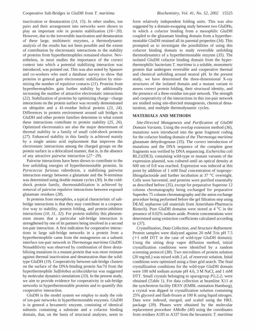
B-factors similar to those of neighboring groups. Solventatoms W10, W31, and W103 have been assigned to H2Obecause of lower sigma values; however, they displaypositive densities in theFo-Fc difference map and are allcoordinated by positively charged side chains. Occupanciesof protein atoms, for which no or only weak density wasobserved, were adjusted. No density at all was observed forthe N-terminal 2 residues, which consequently have beenremoved from the model. Weak density was observed foran additional N-terminal residue and the C-terminal aspartate,as well as three residues in the loop Gly314-Ala315-Asn316, that in the hexamer pack against residues from thesubstrate-binding domain. The side chain of arginine 190clearly adopted two different conformations, these were bothmodeled at 50% occupancy. The quality of the final modelwas checked using procheck (46) and WHATIF (47, 48).The final model has excellent stereochemistry and anR-factorof 15.2% (Rfree ) 19.0%).
The final crystallization conditions for the Ala190/Ala193/Ala231 domain variant were 50 mM bis-tris-propane pH 6.5,38% dioxane, and 25% glycerol. Small crystals were obtained
that belonged to spacegroupP41212 (Table 1). For datacollection using a rotating anode and CuκR radiation, acrystal was flash-frozen directly at 105 K. Phases for thismutant were determined using the coordinates of the finalmodel for the isolated domain (described above) as searchmodel with the single cysteine residue mutated to serine andresidues Arg190, Lys193, and Glu231 mutated to alanineusing O (44). Model bias was removed using simulatedannealing and the model was refined using rigid bodyrefinement, initial grouped B-factor refinement and energyminimization (CNSsolve,41). A limited number of solventmolecules was built in using Arp (45). This includedplacement of a solvent molecule close to one of thecrystallographic 2-fold axis and interacting with the mutationsite (Figure 2b). The final model has good stereochemistryand anR-factor of 24.8% (Rfree ) 29.2%).
The coordinates of the wild-type and the mutant cofactorbinding domains will be deposited in the Protein Data Bank.
Spectroscopic Techniques.Circular dichroism spectra wererecorded on a Jasco J-720 spectropolarimeter. Far UV (190-250 nm) and near UV (250-310 nm) CD measurements
FIGURE 2: (a) Stereo diagram of a 2Fo-Fc map of the region of the salt-bridge triad in wild-type cofactor binding domain. Shown inyellow are the well-ordered side chains of Glu231 and Lys193 and the double conformation of Arg110 of the wild-type model. Central inthe picture is solvent molecule W103 (purple). Furthermore, chloride W2 (green), coordinated by Lys193, is clearly visible. Superimposedin dark orange is the model of domain variant Ala190/Ala193/Ala231. (b) Stereo diagram of a 2Fo-Fc map of the region of the salt-bridgetriad in domain variant Ala190/Ala193/Ala231. In yellow the model for the alanine residues is shown in exactly the same orientation as inthe superposition in Figure 1a. Central in the picture is the solvent molecule (purple) next to the crystallographic 2-fold axis, which runsalmost perpendicular to the plane of the picture. The solvent molecule and residues from a symmetry related molecule are shown in darkorange. Both maps were contoured at 1.0σ, and the pictures were drawn with O (44).
Cooperative Salt-Bridges in GluDH fromT. maritima Biochemistry, Vol. 41, No. 52, 200215527

were performed at 20°C in a 0.1 cm and in a 1.0 cm pathlength quartz cuvette, sealed with a Teflon stopper, at aprotein concentration of 0.20 and 7.00 mg/mL, respectively.The results were expressed as mean residue ellipticity ([Θ]),assuming a mean residue weight of 110 per amino acidresidue. Spectra were accumulated four times. All valueswere corrected for solvent contributions.
GdmCl and Urea Induced Equilibrium Unfolding.Proteinsamples were dialyzed against 20 mM sodium phosphate pH7.0 and then incubated at 0.15 mg/mL final concentration atincreasing concentrations of urea (0-7.9 M) or GdmCl (0-4.5 M) in 20 mM sodium phosphate pH 7.0. Far UV CDspectra were recorded after 24 h at 20°C. To probe thereversibility of the unfolding process, the protein wasunfolded at 20°C in 4.5 M GdmCl or in 7.9 M urea at 1.5mg/mL protein concentration in 20 mM sodium phosphate,pH 7.0. After 24 h, the refolding was started by 10-folddilution of the unfolding mixture at 20°C into solutions ofthe same buffer used for the unfolding containing decreasingdenaturant concentrations. The final protein concentrationwas 0.15 mg/mL. After 2 h, a time that was established tobe sufficient to reach equilibrium, far UV CD spectra wererecorded at 20°C. Equibibrium unfolding curves wereanalyzed by fitting baseline and transition regions simulta-neously to a two-state linear extrapolation model as describedbefore for the wild-type GluDH domain (35). Errors in∆GuH2O are typically large due to the large extrapolationfrom the transition region to zero concentration denaturant.Averagedm values and [urea]1/2 or [GdmCl]1/2 values weretherefore used to calculate the difference in unfolding freeenergy between wild-type and mutant proteins in the transi-tion region as follows (49):
in which ∆[denaturant]1/2 is the difference in the values of[denaturant]1/2 between wild-type and mutant enzymes and⟨m⟩ is the average value ofm. The averagem valuesdetermined in this study are 1.46 kcal mol-1 M-1 for ureaunfolding and 2.96 kcal mol-1 M-1 for GdmCl inducedunfolding. Because the mutation sites are fully exposed inthe folded state, there is only a very small free energydifference for transfer from water to denaturant solutionbetween wild-type and mutant folded and unfolded states,so that the free energy difference determined in the transitionregion equals∆∆Gu
H2O (49, 10).Determination of Interaction Energies from Double Mutant
Cycles. Interaction energies between two residues weredetermined by constructing double mutant cycles (10, 50).A typical cycle consists of wild-type protein (E-XY), twosingle mutants (E-XM and E-MY) and the double mutant(E-MM). The free energy of interaction (∆∆Gint) betweenthe 2 residues is then given by
The energies on the right-hand sight are the energies obtainedfrom the urea and GdmCl induced denaturations as describedabove. In this study, the construction of a thermodynamicbox (Figure 4) allowed the determination of the interactionenergies between each two out of three residues in a salt-
bridge triad in the presence as well as in the absence of thethird residue analogous to the triple mutant box analysis onbarnase (10).
Thermal Equilibrium Unfolding.Protein samples weredialyzed against 20 mM sodium phosphate, pH 7.0, andthermal denaturation scans were recorded at 0.2 mg/mL andpH 7.0. The samples were heated from 10 to 95°C andsubsequently cooled to 10°C with a heating/cooling rateranging from 0.75°C/min to 1.50°C/min controlled by aJasco programmable Peltier element. A scan rate of 1°C/min was chosen in consideration of the observed indepen-dence of thermal transitions on the heating/cooling rate. FarUV CD spectra were recorded every 5-2.5 °C, and thedichroic activity at 222 nm was continuously monitored every0.5°C with 4 s averaging time. All the spectra were correctedfor solvent contribution at increasing temperature. Reversiblethermal denaturation was analyzed according to a two-statemodel as described for the wild-type GluDH cofactor bindingdomain (35). The differences in free energy between wild-type and mutant protein variants were obtained from
and refer to a temperature between theTm values of thewild-type and the respective mutant protein (51). The averageerror in Tm was 0.4°C which corresponds to an error ofapproximately 0.06 kcal/mol in∆∆G values. This issomewhat higher than the errors obtained from the chemicalunfolding data, and because of this, we could not calculatesignificant interaction energies for the thermal unfolding data.
RESULTS
The Three-Dimensional Structure of the Wild-Type GluDHCofactor Binding Domain. Comparison of the Wild-TypeDomain to its Counterpart in the GluDH Hexamer.An
FIGURE 3: A selection of equilibrium unfolding curves forT.maritima GluDH cofactor binding wild-type and mutant proteinsfor (a) urea-induced unfolding and (b) GdmCl-induced unfolding.Arg190/Lys193/Glu231 (circles), mutant Ala190/Lys193/Ala231(triangles), and mutant Arg190/Ala193/Glu231(squares). Depictedis the fraction of folded protein (fN) as a function of denaturantconcentration.
∆∆GD ) ⟨m⟩∆[denaturant]1/2 (1)
∆∆Gint ) ∆GD(E-XY>E-XM) - ∆GD(E-MY>E-MM) )∆GD(E-XY>E-MY) - ∆GD(E-XM>E-MM) (2)
[Tm(wt) - Tm(mut)][∆Hm(wt)/Tm(wt)] (3)
15528 Biochemistry, Vol. 41, No. 52, 2002 Lebbink et al.

overlay of the models of the isolated cofactor binding domaindetermined in this study, with the cofactor binding domainof subunit A of theT.maritimaGluDH hexamer, reveals localdeviations at the N- and C-termini and at three loop regionsthat are all very close to the interface with the substratebinding domain (Figure 1). The side chains of Phe223 andAsn219, those in the hexamer pack against the substratebinding domain, have adopted new conformations. The twoC-terminal residues Pro336 and Asp337 have shifted to packcloser to the cofactor binding domain. However, the overallfolds of both domains are very similar (rmsd. of 0.956 Å),indicating that no significant structural changes have occurredupon isolated expression of the domain.
Description of the Salt-Bridge Triad Arg190-Glu231-Lys193.A three-residue ion-pair network is present in thedomain, involving the side chains of arginine 190, glutamate231, and lysine 193 (Figures 1 and 2, Table 2). Two ion-pair interactions are formed, one between the positivelycharged guanidino group of arginine and the negativeγ-carboxylate group of glutamate (distance between Arg190Nη1 and Glu231 Oε1 is 3.65 Å) and the second between theglutamate and the positively charged nitrogen of the lysine(distance between Glu231 Oε1 and Lys193 Nú is 3.27 Å).Furthermore, Glu231 Oε1 and Lys193 Nú form a hydrogenbond (Table 2). The distance between the positively chargedgroups Arg190 Nη1 and Lys193 Nú is 5.36 Å, with a watermolecule (W103) located between these two groups (Table2). The central role of this water molecule is apparent fromthe fact that it donates a hydrogen to Glu231 Oε1 and acceptshydrogens from Arg190 Nη1 and Lys193 Nú. Lys193 Nú isfurthermore donating a hydrogen to solvent molecule W19,so its hydrogen bonding capacity is fully used. The side chainof Arg190 is clearly adopting two conformations with similaroccupancies. In the second conformation (B), the onlyinteractions seem to be with W58 (2.73 Å from BNη1) andW122 (3.26 Å from BNη2). The aliphatic parts of the sidechains of Lys193 and Arg190 participate in hydrophobic
interactions with each other and with Leu232, Ile228,Leu227,Val194, and Phe223. The side chain of Glu231 alsocontributes by interacting with Leu227.
Effects of the Cysteine to Serine Mutation.The presenceof an exposed cysteine residue in the wild-type domainrequires the use of reducing agents during purification,characterization and crystallization (35). To facilitate furtherexperiments, the cysteine was replaced by a serine using site-directed mutagenesis. The resulting protein was purified, andits chemical and physical properties were compared to thoseof the wild-type domain (results not shown). Near and farUV CD spectra and the elution volume from a calibratedSuperose 12 size-exclusion chromatography were identical.Similar midpoints of transition,m-values and free unfoldingenergies obtained by urea and guanidinium chloride -induced
FIGURE 4: (a) Triple mutant box to determine interaction energies and cooperativity in salt-bridge triad Arg190-Glu231-Lys193. Thesingle-letter notation for amino acids is used. Positively charged arginine and lysine are depicted in blue, negatively charged glutamate inred, and uncharged alanines in black. (b) Six mutant cycles used to construct the triple mutant box. The same color coding is used as inFigure a.∆∆GD is indicated next to the arrows, in plain numbers for urea unfolding, and in italic for GdmCl unfolding.∆∆Gint is theinteraction energy (green colored font) between the two residues analyzed in each particular cycle, again in plain numbers for urea anditalic for GdmCl.
Table 2: Ionic Interactions and Hydrogen Bonds in the Salt-BridgeTriada
Ionic Interactions
Atom 1 Atom 2 Distance (Å)
Arg190 NH1 Glu231 OE1 3.65Arg190 NH1 Glu231 OE2 3.80Lys193 NZ Glu231 OE1 3.27Lys193 NZ W2 Cl- 3.33Arg190 NH1 Lys193 NZ 5.36b
Hydrogen Bonds
Donor Acceptor Distance D-Ac Distance H-Ac
Arg190 NH1 W103 O 2.66 1.75Lys193 NZ Glu231 OE1 3.27 2.44Lys193 NZ W19 O 2.96 2.08Lys193 NZ W103 O 3.13 2.15W103 O Glu231 OE1 3.31 2.31
a Cutoff distances for classification as an ion pair is 4.0 Å.b Expectedrepulsive interaction.c D ) donor, A) acceptor, H) proton. Cutoffvalues for D-A and H-A are 3.5 and 2.5 Å, respectively. Hydrogenbonds were analyzed using WHATIF (47). Interactions were analyzedaccording to the classification of solvent molecule W2 as a chlorideand W19 and W103 as waters (see Materials and Methods).
Cooperative Salt-Bridges in GluDH fromT. maritima Biochemistry, Vol. 41, No. 52, 200215529

reversible unfolding were obtained. Melting temperatures anddenaturation enthalpies derived from thermal equilibriumunfolding assayed by far UV CD were identical. These resultsindicate that the cysteine to serine mutation has no significanteffect on the stability of the protein. This domain variantwas therefore used as reference “wild-type” in all theexperiments described below.
Construction, Purification, and Structure Validation ofSalt-Bridge Mutants.To study the role of the ion-pairnetwork Arg190-Glu231-Lys193, all three residues weremutated to alanines in all combinations. In all cases this wasdone using the domain without the cysteine as template. Allprotein variants were overexpressed at the same high levelas the wild-type and were purified according to the sameprocedure. For all variants identical size exclusion chro-matograpy profiles were obtained (data not shown). Nosignificant changes were observed in the far and near UVCD spectra of the mutants compared to the wild-type domain(data not shown), indicating an identical secondary structurecontent and a similar tertiary structure arrangement.
The Three-Dimensional Structure of Domain VariantAla190/Ala193/Ala231.To analyze the structural effects ofthe mutations in more detail, domain variant Ala190/Ala193/Ala231 was crystallized and its three-dimensional structuresolved. Crystals of the Ala190/Ala193/Ala231 mutant belongto space groupP41212 and diffracted below 2.5 Å using CuκR radiation. The model was refined to 2.6 Å with carefullplacement of a limited number of solvent molecules. Theoverall fold of Ala190/Ala193/Ala231 is identical to that ofthe isolated wild-type domain (rmsd. of 0.560 Å; Figure 1).An overlay of the mutated region with that of the wild-typedomain shows identical conformations for the backbone andneighboring side chains (Figure 2). The removal of the salt-bridge triad therefore has had no structural consequences forthe remainder of the protein.
Urea, Guanidinium Chloride, and Thermally InducedReVersible Unfolding. The Effect of Mutations on the FreeEnergies for GdmCl, Urea, and Thermally Induced Unfold-ing. The effect of the mutations on the free energies forunfolding were determined at 20°C and neutral pH usingurea and GdmCl induced equilibrium unfolding (Tables 3and 4, Figure 3). Thermal equilibrium unfolding was usedto obtain melting temperatures and to calculate differencesin free unfolding energies between wild type and mutants
that refer to a temperature that is between the respectivemelting temperatures of the wild-type and the mutant protein(Table 5).
The replacement of Arg190 with alanine (domain variantAla190/Lys193/Glu231) lowers the free energy for unfoldingof the domain in urea and GdmCl and by thermal unfoldingto approximately the same extent (-0.32,-0.41, and-0.34kcal/mol, respectively). The extent of the effect of replacingGlu231 with alanine (Arg190/Lys193/Ala231) depends onthe denaturant but is destabilizing in all cases. The destabi-lization is most pronounced upon unfolding by GdmCl(-0.95 kcal/mol). However, in contrast to the effects ofremoval of the arginine and the glutamate, the mutation ofLys193 to alanine (Arg190/Ala193/Glu231) is in all casesstabilizing the domain, up to as much as 0.98 kcal/mol inGdmCl. The combination of the destabilizing mutations inAla190/Lys193/Ala231 is destabilizing the domain, whilecombining a destabilizing and a stabilizing mutation inArg190/Ala193/Ala231 is stabilizing the domain. The re-moval of two positively charged side chains in Ala190/Ala193/Glu231 is destabilizing in urea and thermal unfoldingbut stabilizing in GdmCl induced unfolding. At roomtemperature there is almost no net effect of the mutation ofall three charges to alanine; there are only marginal differ-ences between the free energies of the wild-type domain andmutant Ala190/Ala193/Ala231 either using urea or guani-dinium chloride as denaturant. This mutant has however a
Table 3: Urea Induced Equilibrium Unfolding
[urea]1/2(M)a
m (kcalmol-1 M-1)a
∆GD(kcal/mol)b
∆∆GD(kcal/mol)c
Arg190/Lys193/Glu231 3.32 1.30 4.85 0.00Ala190/Lys193/Glu231 3.10 1.70 4.53 -0.32Arg190/Lys193/Ala231 3.08 1.60 4.50 -0.35Arg190/Ala193/Glu231 3.56 1.39 5.20 +0.35Ala190/Lys193/Ala231 2.96 1.53 4.32 -0.53Ala190/Ala193/Glu231 3.21 1.33 4.69 -0.16Arg190/Ala193/Ala231 3.67 1.42 5.36 +0.51Ala190/Ala193/Ala231 3.35 1.42 4.89 +0.04
a Values were obtained by fitting the unfolding data as reported inMaterials and Methods.b ∆GD was calculated by multiplying thedenaturant concentration [urea]1/2 by 1.46, the average value ofm (49).c Values of∆∆GD are calculated according to eq 1 and are relative tothe free energy for unfolding of the wild-type domain. The averageerror in [urea]1/2 was 0.01 M, which corresponds to 0.015 kcal/mol. Inall proteins the cysteine is replaced by a serine residue
Table 4: GdmCl Induced Equilibrium Unfolding
[GdmCl]1/2(M)a
m (kcalmol-1 M-1)a
∆GD(kcal/mol)b
∆∆GD(kcal/mol)c
Arg190/Lys193/Glu231 1.73 3.40 5.12 0.00Ala190/Lys193/Glu231 1.59 2.63 4.71 - 0.41Arg190/Lys193/Ala231 1.41 3.00 4.17 - 0.95Arg190/Ala193/Glu231 2.06 2.74 6.10 + 0.98Ala190/Lys193/Ala231 1.44 3.08 4.26 - 0.86Ala190/Ala193/Glu231 1.96 3.00 5.80 + 0.68Arg190/Ala193/Ala231 2.04 2.73 6.04 + 0.92Ala190/Ala193/Ala231 1.74 2.72 5.15 + 0.03
a Values were obtained by fitting the unfolding data as reported inMaterials and Methods.b ∆GD was calculated by multiplying thedenaturant concentration [GdmCl]1/2 by 2.96, the average value ofm(49). c Values of∆∆GD are calculated according to eq 1 and are relativeto the free energy for unfolding of the wild-type domain. The averageerror in [GdmCl]1/2 was 0.01 M, which corresponds to 0.015 kcal/mol.In all proteins the cysteine is replaced by a serine residue.
Table 5: Equilibrium Thermal Unfolding Assayed by Far UVCircular Dichroism
∆Hm
(kcal/mol)aTm
(°C)a∆∆G
(kcal/mol)b
Arg190/Lys193/Glu231 52.4 69.5 0.00Ala190/Lys193/Glu231 49.5 67.3 - 0.34Arg190/Lys193/Ala231 41.0 68.4 - 0.17Arg190/Ala193/Glu231 53.8 71.0 + 0.23Ala190/Lys193/Ala231 44.6 68.5 - 0.15Ala190/Ala193/Glu231 53.2 67.5 - 0.31Arg190/Ala193/Ala231 52.7 71.2 + 0.26Ala190/Ala193/Ala231 50.6 70.8 + 0.20
a Values were obtained by fitting the thermal unfolding data asreported in Materials and Methods.b ∆∆G was calculated accordingto ∆∆G ) [Tm(wt) - Tm(mut)][∆Hm(wt)/Tm(wt)] and refers to atemperature between the respectiveTm values (51). The average errorin Tm was 0.4°C, which corresponds to approximately 0.06 kcal/mol.In all proteins the cysteine is replaced by a serine Residue.
15530 Biochemistry, Vol. 41, No. 52, 2002 Lebbink et al.

somewhat higher melting temperature than the wild type anda higher free energy for thermal unfolding at approximately70 °C.
Analysis of Multiple Thermodynamic Cycles.Using mul-tiple thermodynamic cycles (Figure 4;49, 10), the energiesfrom the interactions between Arg190, Glu231, and Lys193obtained from GdmCl and urea-induced unfolding wereseparated from the changes in free energy due to the removalof interactions between these three residues and the rest ofthe protein (Table 6). Furthermore, the strength of each ofthe pairwise interactions could be assayed in the presenceand the absence of the third residue.
The interaction energies between Lys193 and Glu231 inthe intact salt-bridge triad are-0.51 kcal/mol for urea-induced unfolding and-0.89 kcal/mol when using GdmClas denaturant. The strength of the interaction is reduced whenthe third residue Arg190 is absent. Actually, using GdmCl,the interaction destabilizes the protein by 0.20 kcal/mol inthe absence of Arg190.
The interaction between Arg190 and Glu231 is not sostrong as the one between the lysine and the glutamate, butstill stabilizes the protein in the intact triad by 0.14 and 0.50kcal/mol for urea and GdmCl, respectively. Again, thecooperativity in the network is apparent from the changesin interaction energy when Lys193 is absent. In the presenceof urea the interaction energy between Arg190 and Glu231,in the absence of Lys 193, is not measurable, whereas inthe presence of GdmCl this interaction is highly destabilizing.
The repulsive nature of the interaction between Lys193and Arg190 is apparent from the strong destabilization bothin urea and GdmCl in the absence of the mediating glutamateat position 231. In the presence of Glu231, the interaction isless destabilizing and even slightly stabilizing the domainin GdmCl.
Cooperativity in the network follows from the differencebetween the interaction energy of two residues in thepresence and in the absence of the third residue and is-0.10kcal/mol for urea and- 1.09 kcal/mol for GdmCl inducedequilibrium unfolding.
DISCUSSION
Recently we described the expression and biophysicalcharacterization of the isolated cofactor binding domain fromThermotoga maritimaGluDH, with the aim of using it as amodel system to study the thermodynamics of potentialstabilizing ionic interactions in a protein from a hyperther-mophilic organism (35). The advantage of using this modelsystem is that it is derived from an enzyme that is very wellstudied; X-ray structures are available for homologues frommesophilic and (hyper)thermophilic organisms (5, 7, 8, 52-55), and it has been used to study the role of large ion-pairnetworks in conferring thermostability (15, 17-19).
Central in our approach of studying the role of ion-pairnetworks is the validation of experimental data by thedetermination of the X-ray structures of crucial mutantproteins. In earlier studies we have used X-ray analysis toassess that introduced charged residues, characterized bylarge and flexible side chains, nevertheless are in the correctconformation for formation of the anticipated ion-pairinteractions (15, 19). In the present study, the X-ray structureof the wild-type domain is essential in showing that thedomain adopts the correct fold (when compared to itsconformation as part of the hexameric GluDH), as suggestedby far UV CD spectroscopy (35). Furthermore, it shows thepresence of a salt-bridge network with surrounding solventions at high resolution. The structure of the Ala190/Ala193/Ala231 variant showed no structural changes upon mutationand validates the use of multiple thermodynamic cycles inorder to separate the interactions within the network fromchanges in the free energy for unfolding upon mutation dueto interaction of the salt-bridge members with the rest ofthe protein.
The crystals of the wild-type domain were grown in thepresence of 3 M NaCl, conditions in which significantscreening of electrostatic interactions is expected. Salt-bridgetriad Arg190-Glu231-Lys193 is present at high ionicstrength in the isolated domain, although the arginine sidechain is found to adopt two alternative conformations. Acomparison of T4 lysozyme structures determined at low,medium, and high ionic strength revealed no structuralchanges related to the differences in ionic strength andsuggests that a crystal structure determined at high saltconcentration is a good representation of the structure at lowionic strength (56). This suggests that the structure of thedomain determined at 3 M NaCl can also be used to analyzeinteractions at low ionic strength, relevant to compare ureaand thermal denaturation data on one hand versus GdmCldata on the other hand.
A further consequence of high salt crystallization condi-tions in our case is the observation of preferential ion bindingby the presence of two high peaks (>7 σ) in the electrondensity map that were assigned to chloride ions (CL2 andCL3). In addition, on the basis of several criteria (seematerials and methods), solvent molecules W10, W31, andW103 could also be chloride ions. Preferential anion bindingby positively charged groups on the protein surface couldpotentially stabilize the protein by reducing the repulsiveeffects between the positively charged groups and theintroduction of favorable electrostatic interactions and mightcounteract the destabilizing effect of the screening of theelectrostatic interactions by high salt concentrations. Pref-erential anion and cation binding has been described forseveral proteins isolated from halophilic organisms (57, 58).These proteins have adapted to their high salt environment(up to supersaturating concentrations) by covering theirsurfaces with ion pairs and a large excess of negativelycharged residues. Anion binding is also seen in T4 lysozymewhere chloride ions are present at the termini of two helicesthat interact favorably with the helix dipole moment (59). Itis remarkable that Lys193 is coordinating at least onechloride (CL2) but possibly a second (solvent molecule 103).Solvent molecule 103 is located centrally in the salt-bridgetriad. This molecule being a chloride instead of a water wouldmean the loss of three hydrogen bonds, but the gain of a
Table 6: Interaction Energies in the Salt-bridge Triad
∆∆Gint urea(kcal/mol)
∆∆Gint GdmCl(kcal/mol)
Arg190-Glu231 in the presence of Lys193 -0.14 -0.50Arg190-Glu231 in absence of Lys193 -0.04 +0.59Lys193-Glu231 in the presence of Arg190 -0.51 -0.89Lys193-Glu231 in absence of Arg190 -0.41 +0.20Arg190-Lys193 in the presence of Glu231 +0.19 -0.11Arg190-Lys193 in absence of Glu231 +0.29 +0.98
Cooperative Salt-Bridges in GluDH fromT. maritima Biochemistry, Vol. 41, No. 52, 200215531

more favorable electrostatic environment together withArg190 and Lys193.
The interaction energies determined from unfolding datareflect the interactions in the folded state relative to theirabsence in the unfolded state. In the folded protein, eachcharged group is involved in short-range interactions withits partners in the salt bridge, as well as in long-rangeelectrostatic interactions with other charges in the protein.For example, each of the three charged side chains mightinteract either favorably or unfavorably with existingR-helixdipoles in the protein. The aliphatic parts of all side chainsare involved in hydrophobic interactions with neighboringhydrophobic residues. The measured unfolding free energychanges (but not the interaction energies determined bymultiple thermodynamic cycles) therefore reflect the effectof disruption of multiple interactions and not only that ofthe ion-pair interactions. This is evident from the discrep-ancies between the changes in free energy for unfolding ofthe mutants and the determined ion-pair interaction energies(Tables 3 and 4 vs Table 6). While the removal of glutamateand arginine is destabilizing, the removal of the lysine sidechain is actually stabilizing the domain. The net effect ofreplacing the salt bridge with hydrophobic residues isvirtually zero at room temperature and slightly destabilizingat higher temperatures. The reason that we do not see adestabilization of the triple alanine mutant compared withthe intact triad could in our case be due to hydrophobiceffects because the alanine residues are introduced in anenvironment containing several other hydrophobic residues.The loss of favorable electrostatic interactions could fur-thermore be counteracted in the mutant by the higherR-helixstability of alanine residues compared to charged aminoacids. While alanines are a good choice with respect to theanalysis of thermodynamic cycles, they are not optimalrepresentatives as surface residues. The exchange of the ion-pair network members to polar, uncharged residues mightbe a more suitable choice if only changes in free energiesfor unfolding are studied. Nevertheless, in the intact salt-bridge triad, both the interaction between glutamate andlysine and the interaction between glutamate and arginineare contributing favorably to the free energy for unfoldingof the domain. In fact, the attractive ion-pair interactions aremeasured by chemical denaturation at 20°C and should gainin stability by about 30% at the growth temperature ofThermotoga, due to the reduced desolvation penalty at highertemperatures (3, 13). The interaction between Glu231 andLys193 involves not only an ion pair but also a hydrogenbond and it is considerably stronger than the ion-pairinteraction between Glu231 and Arg190, with the latterresidue adopting two conformations. Identical results weredescribed for barnase, where the exchange of a salt-bridgetriad for alanines resulted in an unchanged free energy forunfolding but where stabilizing interaction energies wherefound between the network residues (10).
Earlier results we obtained regarding the role of ion-pairnetworks in determining the stability ofT. maritimaGluDHwere the observation of cooperativity in a large ion-pairnetwork at the subunit interface of the hexameric enzyme(19). Even though the characterization of this network waslimited to addressing its role in kinetic stabilization becauseof irreversibility of the thermal unfolding reaction, it wasobvious that the combination of three destabilizing single
mutations Ser128Arg, Thr158Glu, and Asn117Arg resultedin a small but significant stabilization of the triple mutantcompared to wild-type GluDH. While for the two centralcharged residues this was anticipated because of theirintimate interaction across the subunit interface, the thirdmutation is located more than 15 Å away from the centralcharges in the network. Obviously there is cooperativitywithin this network, either exerted via subtle backbone shiftsand/or via optimization of the overall electrostatic fieldpresent in this part of the protein. In this study we haveshown that thermodynamic cooperativity is present in theArg190-Glu231-Lys193 salt-bridge triad in the isolatedcofactor binding domain. The cooperativity follows from thedifference between interaction energies between two partnersin the triad in the presence and in the absence of the thirdresidue. The presence of a third residue strengthens eachinteraction by 0.10 kcal/mol in urea and as much as 1.09kcal/mol in GdmCl. While the interaction energies as wellas the cooperative effect are relatively small in urea, inGdmCl the effect of the third position residue makes a majordifference. Each interaction in the absence of the third residueis significantly destabilizing the protein, while in the intacttriad all interactions become stabilizing. Cooperativitybetween ion-pair interactions in barnase have thoroughlybeen analyzed and reported (10). From a molecular dynamicssimulation on the DNA-binding protein Sac7d from thehyperthermophileSulfolobus acidocaldarius, it was con-cluded that, rather than individual salt bridges, three largeclusters provide most of the electrostatic stabilization towardthermal denaturation (33). Furthermore, some intercluster saltbridges in fact contribute unfavorably toward the stabilityof the folded state. InBacillus caldolyticuscold shockprotein, a 66-residue all-â protein, pairwise interactionsbetween residues at positions 3, 46, and 66 in the proteinsequence have been analyzed using a triple mutant cycle (28).Only two repulsive pairwise interactions are found in thistriad, residues at positions 46 and 66 do not interact. Themain stabilization of this protein comes from a favorableelectrostatic contribution of arginine at position 3. However,the attractive electrostatic component is not localized in apairwise interaction with any other surrounding residue. Thismight be related to the observation that many of the sidechains involved adopt multiple conformations as visualizedin the crystal structures of wild-type and several mutantproteins (29). No cooperativity was observed between theseinteractions.
The urea and thermal unfolding experiments were per-formed at low ionic strength and the obtained changes infree energy for unfolding are similar for both techniques(compare Table 3 and Table 4). This is in accordance withother studies (60-62). Guanidinium chloride, however, is acharged denaturant, and the unfolding free energies thereforereflect high ionic strength conditions. High ionic strengthhas been reported to significantly screen the electrostaticcomponent of salt-bridge interactions (10, 60). Interactionenergies of salt bridges are therefore expected to be lowerin GdmCl than in urea. This is obviously not the case forthe isolated cofactor binding domain; both the changes infree energies for unfolding as well as the interaction energiesare considerably larger in GdmCl induced unfolding. Inter-estingly, also the pairwise salt-bridge interaction betweenGlu14 and the N-terminus in rubredoxin from the hyper-
15532 Biochemistry, Vol. 41, No. 52, 2002 Lebbink et al.

thermophilic archaeonPyrococcus furiosus, derived frommultiple thermodynamic cycles, was found to be twice asstrong in GdmCl as in urea induced unfolding (30).
We stated that the interaction energies determined fromunfolding data reflect the interactions in the folded staterelative to their absence in the unfolded state. The centralglutamate in the triad is far removed in sequence from thepositively charged residues and it seems safe to assume thatno interaction remains in the denatured state. However, ithas been known for some time that under some conditions,the denatured state can be compact and may contain aconsiderable amount of residual structure (63). In particular,recent experiments provide evidence for the presence ofcharge-charge interactions in the denatured state (61, 62,64). The domain denatured in GdmCl probably does notcontain residual electrostatic interactions, since this denatur-ant has been reported to screen electrostatics. However, wecannot rule out the existence of residual electrostatic interac-tions in the denatured domain in the urea and thermaldenaturation experiments. Whether this would result in lowerinteraction energies between the salt-bridge network residuesduring multiple thermodynamic cycles would depend onwhether these interactions are still (partially) present in thedenatured state.
It has furthermore been reported that GdmCl is under someconditions actually stabilizing proteins, in addition to actingas a denaturant. In the case of ribonuclease T1, the effect issmall and attributed to cation binding in the native state (65).For ubiquitin the effect is characterized as binding of twochloride atoms by the native state of the protein, while thedenatured state binds no anions (66). The stabilization ofubiquitin observed in 1 M GdmCl is remarkable andaccompanied by an increase in melting temperature of 15°C and in unfolding enthalpy of 12.5 kcal/mol. The stabiliza-tion is furthermore apparent from a comparison of themidpoints of denaturation in urea and GdmCl, while usuallymore urea is required to unfold a protein, ubiquitin has amidpoint for GdmCl denaturation almost 1 molarhigherthanfor urea denaturation (66). TheT.maritimacofactor bindingdomain as a whole is not stabilized by GdmCl to a largeextent, the free energies for unfolding in GdmCl are ingeneral only slightly higher than for urea. However, wecannot rule out local stabilization by anion binding in thevicinity of the ion-pair network that may be reflected in thehigher interaction energies obtained by GdmCl denaturation,especially not since we observe that Lys193 is binding atleast one and possibly two chloride atoms in the crystalstructure.
In conclusion, we report here the first thermodynamicdescription of cooperativity in an ion-pair network in aprotein from a hyperthermophile. Furthermore our results,together with results on a stabilizing salt bridge in hyper-thermostable rubredoxin, not only show that hyperthermo-philes use large cooperative arrangements of charged residuein order to stabilize their proteins, but also suggest that anionbinding may have co-evolved with the presence of large ion-pair networks, to gain extra stability at high ionic strengthconditions. Regarding the high internal ionic strengthreported for several hyperthermophiles, among themT.maritima (67), this would actually be a logic strategy.However, this hypothesis requires further experimentalvalidation, since it is in contrast with many observations that
the strength of ionic interactions in general decreases in thepresence of high salt concentrations.
ACKNOWLEDGMENT
We are very grateful to the staff of the EMBL outstationat the DESY synchrotron in Hamburg for providing themeans of and help with data collection. We like to thankGudrun Tibbelin and Majid Ali for their assistance in proteincrystallization.
REFERENCES
1. Szilagyi, A., and Zavodszky, P. (2000) Structural differencesbetween mesophilic, moderately thermophilic and extremelythermophilic protein subunits: results of a comprehensive survey,Structure 8, 493-504.
2. Cambillau, C., and Claverie, J.-M. (2000) Structural and genomiccorrelates of hyperthermostability,J. Biol. Chem. 275, 32383-32386.
3. Karshikoff, A., and Ladenstein, R. (2001) Ion-pairs and thethermotolerance of proteins from hyperthermophiles: a ‘trafficrule′ for hot roads,Trends Biochem. Sci., 26, 550-556.
4. Kumar, S., and Nussinov, R. (2001) How do thermophilic proteinsdeal with heat? Cell Mol. Life Sci. 58, 1216-1233.
5. Yip, K. S., Stillman, T. J., Britton, K. L., Artymiuk, P. J., Baker,P. J., Sedelnikova, S. E., Engel, P. C., Pasquo, A., Chiaraluce,R., Consalvi, V., Scandurra, R., and Rice, D. W. (1995) Thestructure ofPyrococcus furiosusglutamate dehydrogenase revealsa key role for ion-pair networks in maintaining enzyme stabilityat extreme temperatures,Structure, 3, 1147-1158.
6. Yip, K. S., Britton, K. L., Stillman, T. J., Lebbink, J., de Vos, W.M., Robb, F. T., Vetriani, C., Maeder, D., and Rice, D. W. (1998)Insights into the molecular basis of thermal stability from theanalysis of ion-pair networks in the glutamate dehydrogenasefamily, Eur. J. Biochem. 255, 336-346.
7. Knapp, S., de Vos, W. M., Rice, D., and Ladenstein, R. (1997)Crystal structure of glutamate dehydrogenase from the hyperther-mophilic eubacteriumThermotoga maritimaat 3.0 Å resolution,J. Mol. Biol. 267, 916-932.
8. Britton, K. L., Yip, K. S. P., Sedelnikova, S. E., Stillman, T. J.,Adams, M. W. W., Ma, K., Maeder, D. L., Robb, F. T., Tolliday,N., Vetriani, C., Rice, D. W., and P. J. Baker. (1999) Structuredetermination of the glutamate dehydrogenase from the hyper-thermophileThermococcus litoralisand its comparison with thatfrom Pyrococcus furiosus, J. Mol. Biol. 293, 1121-1132.
9. Zhang, X., Meining, W., Fischer, M., Bacher, A., Ladenstein, R.(2001) X-ray structure analysis and crystallographic refinementof lumazine synthase from the hyperthermophile Aquifex aeolicusat 1.6 A resolution: determinants of thermostability revealed fromstructural comparisons,J. Mol. Biol. 306, 1099-1114.
10. Horovitz, A., Serrano, L., Avron, B., Bycroft, M., and Fersht, A.(1990) Strength and cooperativity of contributions of surface salt-bridges to protein stability,J. Mol. Biol. 216, 1031-1044.
11. Sali, D., Bycroft, M and Fersht, A. R. (1991) Surface electrostaticinteractions contribute little to stability of barnase,J. Mol. Biol.220, 779-788.
12. Dao-pin, S., Sauer, U., Nicholson, H., and Matthews, B. W. (1991)Contributions of engineered surface salt-bridges to the stabilityof T4 lysozyme determined by directed mutagenesis,Biochemistry30, 7142-7153.
13. Elcock, A. H. (1998) The stability of salt-bridges at hightemperatures: implications for hyperthermophilic proteins,J. Mol.Biol. 284, 489-502.
14. Tomschy, A., Bohm, G., and Jaenicke, R. (1994) The effect ofion pairs on the thermal stability of D-glyceraldehyde 3-phosphatedehydrogenase from the hyperthermophilic bacteriumThermotogamaritima, Protein Eng. 7, 1471-1478.
15. Lebbink, J. H. G., Knapp, S., van der Oost, J., Rice, D., Ladenstein,R., and de Vos, W. M. (1998) Engineering activity and stabilityof Thermotoga maritimaglutamate dehydrogenase. I. Introductionof a six-residue ion-pair network in the hinge region,J. Mol. Biol.280, 287-96.
16. Pappenberger, G., Schurig, H., and Jaenicke, R. (1997) Disruptionof an ionic network leads to accelerated thermal denaturation of
Cooperative Salt-Bridges in GluDH fromT. maritima Biochemistry, Vol. 41, No. 52, 200215533

D-glyceraldehyde-3-phosphate dehydrogenase from the hyper-thermophilic bacteriumThermotoga maritimaI, J. Mol. Biol. 274,676-683.
17. Rahman, R., Fujiwara, S., Nakamura, H., Takagi, M., and Imanaka,T. (1998) Ion-pairs involved in maintaining a thermostablestructure of glutamate dehydrogenase from a hyperthermophilicarchaeon,Biochem. Biophys. Res. Commun. 248, 920-926.
18. Vetriani, C., Maeder, D. L., Tolliday, N., Yip, K. S. P., Stillman,T. J., Britton, K. L., Rice, D. W., Klump, H. H., and Robb, F. T.(1998) Protein thermostability above 100°C: a key role for ionicinteractions,Proc. Natl. Acad. Sci. U.S.A. 95, 12300-12305.
19. Lebbink, J. H. G., Knapp, S., van der Oost, J., Rice, D., Ladenstein,R., and de Vos, W. M. (1999) Engineering activity and stabilityof Thermotoga maritimaglutamate dehydrogenase. II. Construc-tion of a 16-residue ion-pair network at the subunit interface, J.Mol. Biol. 289, 357-369.
20. Merz, A., Knochel, T., Jansonius, J. N., and Kirschner, K. (1999)The hyperthermostable indoleglycerol phosphate synthase fromThermotoga maritimais destabilized by mutational disruption oftwo solvent-exposed salt-bridges,J. Mol. Biol. 288, 753-763.
21. Spassov, V. Z., Karshikoff, A. D., and Ladenstein, R. (1994) Theoptimization of the electrostatic interactions in proteins of differentfunctional and folding type,Protein Sci. 3, 1556-1569.
22. Spassov, V. Z., Karshikoff, A. D., and Ladenstein, R. (1995) Theoptimization of protein solvent interactions. Thermostability andthe role of hydrophobic and electrostatic interactions,Protein Sci.4, 1516-1527.
23. Loladze, V., Ibarra-Molero, B., Sanchez-Ruiz, J. M., and Ma-khatadze, G. I. (1999) Engineering a thermostable protein viaoptimization of charge-charge interactions on the protein surface,Biochemistry 38, 16419-16423.
24. Spector, S., Wang, M., Carp, S. A., Robblee, J., Hendsch, Z. S.,Fairman, R., Tidor, B., and Raleigh, D. P. (2000) Rationalmodification of protein stability by the mutation of charged surfaceresidues,Biochemistry 39, 872-879.
25. Xiao, L., and Honig, B. (1999) Electrostatic contributions to thestability of hyperthermophilic proteins,J. Mol. Biol. 289, 1435-1444.
26. Kumar, S., Ma, B., Tsai, C.-J., and Nussinov, R. (2000) Electro-static strengths of salt-bridges in thermophilic and mesophilicglutamate dehydrogenase monomers,Proteins: Struct. Funct.Genet. 38, 368-383.
27. Perl., D., Mueller, U., Heinemann, U., and Schmid, F. X. (2000)Two exposed amino acid residues confer thermostability on a coldshock protein,Nat. Struct. Biol. 7, 380-383.
28. Perl., D., and Schmid, F. X. (2001) Electrostatic stabilization ofa thermophilic cold shock protein,J. Mol. Biol. 313, 343-357.
29. Delbruck, H., Mueller, U., Perl, D., Schmid, F. X., and Heinemann,U. (2001) Crystal structures of mutant forms of theBacilluscaldolyticuscold shock protein differing in thermal stability,J.Mol. Biol, 313, 359-369.
30. Strop, P., and Mayo, S. L. (2000) Contribution of surface saltbridges to protein stability,Biochemistry 39, 1251-1255.
31. Horovitz, A., and Fersht, A. (1992) Co-operative interactionsduring protein folding,J. Mol. Biol. 224, 733-740.
32. Albeck, S., Unger, R., Schreiber, G. (2000) Evaluation of directand cooperative contributions towards the strength of buriedhydrogen bonds and salt bridges,J. Mol. Biol. 298, 503-520.
33. Bakker, P. I. W. de, Hu¨nenberger, P. H., McCammon, J. A. (1999)Molecular dynamics simulation of the hyperthermophilic proteinSac7d from Sulfolobus acidocaldarius: Contribution of saltbridges to thermostability,J. Mol. Biol. 285, 1811-1830.
34. Lebbink, J. H. G., Eggen, R. I. L., Geerling, A. C. M., Consalvi,V., Chiaraluce, R., Scandurra, R., and de Vos, W. M. (1995)Exchange of domains of glutamate dehydrogenase from thehyperthermophilic archaeonPyrococcus furiosusand the meso-philic bacteriumClostridium difficile: effects on catalysis, ther-moactivity and stability,Protein Eng. 8, 1287-1294.
35. Consalvi, V., Chiaraluce, R., Gianciacomo, L., Scandurra, R.,Christova, P., Karshikoff, A., Knapp, S., and Ladenstein, R. (2000)Thermal unfolding and conformational stability of the recombinantdomain II of glutamate dehydrogenase from the hyperthermophileThermotoga maritima, Protein Eng. 13, 501-507.
36. Ho, S. N., Hunt, H. D., Horton, R. M., Pullen, J. K., and Pease,L. R. (1989) Site-directed mutagenesis by overlap extension usingthe polymerase chain reaction,Gene 77, 51-59.
37. Gill, S. C., and von Hippel, P. H. (1989) Calculation of proteinextinction coefficients from amino acid sequence data,Anal.Biochem. 182, 319-326.
38. Jancarik, J., and Kim, S.-H. (1991) Sparse matrix sampling: ascreening method for crystallization of proteins,J. Appl. Cryst.24, 409-411.
39. Otwinowski, Z., and Minor, W.(1997) Processing of X-raydiffraction data collected in oscillation mode,Methods Enzymol.276, 307-326.
40. Navaza, J. (1994) AmoRe: an automated package for molecularreplacement,Acta Crystallogr., Sect A 50, 157-163.
41. Brunger, A. T., Adams, P. D., Clore, G. M., Delano, W. L., Gros,P., Grosse-Kunstleve, R. W., Jiang, J.-S., Kuszewski, J., Nilges,M., Pannu, N. S., Read, R. J., Rice, L. M., Simonson, T.,Warren,G. L. (1998) Crystallography and NMR System,Acta Crystallogr.,Sect. D 54, 905-921.
42. COLLABORATIVE COMPUTATIONAL PROJECT, NUMBER4. (1994). “The CCP4 Suite: Programs for Protein Crystal-lography”,Acta Crystallogr., Sect. D 50, 760-763.
43. Murshudov, G. N., Vagin A. A., and Dodson, E. J. (1997)Refinement of macromolecular structures by the maximum-likelihood method,Acta Crystallogr., Sect. D 53, 240-255.
44. Jones, T. A., Zou, J. Y., Cortan, J. Y., and Kjelgaard, M. (1991)Improved methods for building models in electron density mapsand the location of errors in the model,Acta Crystallogr., Sect.A, 47, 110-119.
45. Perrakis, A., Morris, R. J. H., and Lamzin, V. S. (1999) Automatedprotein model building combined with iterative structure refine-ment,Nat. Struct. Biol. 6, 458-463.
46. Laskowski, R. A., MacArthur, M. W., Moss, D. S., and Thornton,J. M. (1993) PROCHECK: a program to check the stereochemicalquality of protein structures,J. Appl. Cryst. 26, 283-291.
47. Vriend, G. (1990) WHAT IF: A molecular modeling and drugdesign program,J. Mol. Graph. 8, 52-56.
48. Hooft, R. W. W., Vriend, G., Sander, C., Abola, E. E. (1996)Errors in protein structures,Nature 381, 272-272.
49. Sali, D., Bycroft, M., and Fersht, A. R. (1988) Stabilization ofprotein structure by interaction ofR-helix dipole with a chargedside-chain,Nature 335, 740-743.
50. Serrano, L., Horovitz, A., Avron, B., Bycroft, M., and Fersht, A.(1990) Estimating the contribution of engineered surface electro-static interactions to protein stability by using double-mutantcycles,Biochemistry 29, 9343-9352.
51. Pace, C. N. (1995) Evaluating contribution of hydrogen bondingand hydrophobic bonding to protein folding,Methods Enzymol.259, 538-554.
52. Baker, P. J., Britton, K. L., Engel, P. C., Farrants, G. W., Lilley,K. S., Rice, D. W., Stillman, T. J. (1992) Subunit assembly andactive site location in the structure of glutamate dehydrogenase,Proteins: Struct., Funct. Gen. 12, 75-86.
53. Peterson, P. E., Smith, T. J. (1999) The structure of bovineglutamate dehydrogenase provides insights into the mechanismof allostery,Structure 7, 769-782.
54. Smith, T. J., Peterson, P. E., Schmidt, T., Fang, J., and Stanley,C. A. (2001) Structures of bovine glutamate dehydrogenasecomplexes elucidate the mechanism of purine regulation,J. Mol.Biol. 307, 707-720.
55. Nakasako, M., Fujisawa, T., Adachi, S, Kudo, T., and Higuchi,S. (2001) Large-scale domain movement and hydration structurechanges in the active-site cleft of unligated glutamate dehydro-genase fromThermococcus profundusstudied by cryogenic X-raycrystal structure analysis and small-angle X-ray scattering,Biochemistry 40, 3069-3079.
56. Bell, J. A., Wilson, K. P., Zhang, X.-J., Faber, H. R., Nicholson,H., Matthews, B. W. (1991) Comparison of the crystal structureof bacteriophage T4 lysozyme at low, medium, and high ionicstrengths,Proteins: Struct. Funct. Gen. 10, 10-21.
57. Frolow, F., Harel, M., Sussman, J. L., Mevarech, M., Shoham,M. (1996) Insights into protein adaptation to a saturated saltenvironment from the crystal structure of a halophilic 2Fe-2Sferredoxin,Nat. Struct. Biol. 3, 452-457.
58. Richard, S. B., Madern, D., Garcin, E., Zaccai, G. (2000)Halophilic adaptation: novel solvent protein interactions observedin the 2.9 and 2.6 Å resolution structures of the wild-type and amutant of malate dehydrogenase fromHaloarcula marismortui,Biochemistry 39, 992-1000.
59. Nicholson, H., Becktel, W. J., Matthews, B. W. (1988) Enhancedprotein thermostability from designed mutations that interact withR-helix dipoles,Nature 336, 651-656.
60. Monera, O. D., ay, C. M., and Hidges, R. (1994) Proteindenaturation with guanidine hydrochloride or urea provides a
15534 Biochemistry, Vol. 41, No. 52, 2002 Lebbink et al.

different estimate of stability depending on the contributions ofelectrostatic interactions,Protein Sci. 3, 1984-1991.
61. Pace, C. N., Alston, R. W., and Shaw, K. L. (2000) Charge-charge interactions influence the denatured state ensemble andcontribute to protein stability,Protein Sci. 9, 1395-1398.
62. Whitten, S. T., and Garcia-Moreno, B. (2000) Ph dependence ofstability of staphylococcal nuclease: evidence of substantialelectrostatic interactions in the denatured state,Biochemistry 39(46), 14292-14304.
63. Shortle, D. (1996) The denatured state (the other half of the foldingequation) and its role in protein stability,FASEB J. 10, 27-34.
64. Kuhlman, B., Luisi, D. L., Young, P., and Raleigh, D. P. (1999)pKa values and the pH dependent stability of the N-terminal
domain of L9 as probes of electrostatic interactions in thedenatured state. Differentiation between local and nonlocalinteractions,Biochemistry 38, 4896-4903.
65. Mayr, L. M., and Schmid, F. X. (1993) Stabilization of a proteinby guanidinium chloride,Biochemistry 32, 7994-7998.
66. Makhatadze, G. I., Lopez, M. M., Richardson, J. M., and Thomas,S. T. (1998) Anion binding to the ubiquitin molecule,ProteinSci. 7, 689-697.
67. Galinski, E. A. (1995) Osmoadaptation in bacteria,AdV. Microb.Physiol. 37:272-328.
68. DeLano, W. L. (2002) The PyMOL Molecular Graphics Systemon World Wide Web (http://www.pymol.org).
BI020461S
Cooperative Salt-Bridges in GluDH fromT. maritima Biochemistry, Vol. 41, No. 52, 200215535