Plant genotype, not nutrients, shape aphid population dynamics
Stronger induction of callose deposition in barley by Russian wheat aphid than bird cherry-oat aphid is not associated with differences in callose synthase or β-13-glucanase transcript abundance Sefiu A Saheed a Izabela Cierlik b Kristina A E Larsson b Gabriele Delp b Graeme Bradley c Lisbeth M V Jonsson b Christiaan E J Botha a
a Department of Botany Rhodes University Grahamstown 6140 South Africa b School of Life Sciences Soumldertoumlrn University S-141 89 Huddinge Sweden c Department of Biochemistry and Microbiology University of Fort Hare Alice 5700 South Africa Abstract The effects of infestation by the bird cherry-oat aphid (BCA) (Rhopalosiphum padi L) and the Russian wheat aphid (RWA) (Diuraphis noxia Mordvilko) on callose deposition and transcription of genes related to callose accumulation were investigated in barley (Hordeum vulgare L cv Clipper) The BCA which gives no visible symptoms induced very limited callose deposition even after 14 days of infestation In contrast RWA which causes chlorosis white and yellow streaking and leaf rolling induced callose accumulation already after 24 h in longitudinal leaf veins The deposition was pronounced after 72 h progressing during 7 and 14 days of infestation In RWA-infested source leaves callose was also induced in longitudinal veins basipetal to the aphid-infested tissue whereas in sink leaves more callose deposition was found above the feeding sites Eight putative callose synthase genes were identified in a database search of which seven were expressed in the leaves but with similar transcript accumulation in control and aphid-infested tissue Five out of 12 examined β-13-glucanases were expressed in the leaves All five were upregulated in RWA-infested tissue but only two in BCA-infested tissue and to a lesser extent than by RWA The results suggest that callose accumulation may be partly responsible for the symptoms resulting from RWA infestation and that a callose-inducing signal may be transported in the phloem Furthermore it is concluded that the absence of callose deposition in BCA-infested leaves is not because of a stronger upregulation of callose-degrading β-13-glucanases in this tissue as compared to RWA-infested leaves Introduction Callose a β-13-glucan is an important component in plant tissue It is formed as part of basic developmental processes at the cell plate during the formation of new walls in dividing cells and it is found around pollen mother cells in pollen grains and in pollen tubes Callose is also present in plasmodesmatal canals root hairs and in spiral thickenings in tracheids (Stone and Clarke 1992) In addition callose is locally deposited in sieve pores and plasmodesmata in response to wounding and other physiological stress and in plugs or plates (normally referred to as papillae) in response to pathogen infection (Donofrio and Delaney 2001 Stone and Clarke 1992) Wound callose is formed rapidly mostly within minutes of wound initiation (Nakashima et al 2003 Radford et al 1998) and is deposited between the plasma membrane and the cell wall It is electron-lucent in electron micrographs and forms an intense yellow UV light-induced fluorescence with the aniline blue fluorochrome (Stone and Clarke 1992) The rapid deposition of callose in sieve pores has long since been viewed as an efficient wound response that seals off the pores in damaged phloem to prevent assimilate loss (Sjoumllund 1997) Nevertheless aphids (Homoptera Aphididae) which damage the sieve elements when penetrating with their stylets manage to feed from phloem elements during long periods It has been proposed that aphids prevent
wounding reactions such as callose formation and protein plugging by sealing the stylet puncture site with sheath saliva In addition components in the watery saliva injected into the sieve elements may interact with sap ingredients (Will and van Bel 2006) However recent studies indicate that some aphid species do cause callose formation (Botha and Matsiliza 2004 Kusnierczyk et al 2008) Thus we have found that the Russian wheat aphid (RWA) (Diuraphis noxia Mordvilko) an aphid that causes leaf rolling and other symptoms causes callose deposition in cereals Callose was associated with sieve plates (SP) and pore plasmodesmata between the companion cells and their associated sieve tubes and the phloem transport rate was reduced in the damaged phloem tubes in wheat (Botha and Matsiliza 2004) This is suggestive evidence that callose formation may at least partly explain the severe symptoms caused by RWA infestation This idea was supported by results from a recent ultrastructural study in barley where effects of RWA and the bird cherry-oat aphid (BCA) (Rhopalosiphum padi L) were compared (Saheed et al 2007) BCA co-occurs with RWA on wheat and other cereals but usually does not cause any visible damage symptoms (Messina et al 2002) Besides differences in the salivary secretions in the xylem elements because of these two aphids the effects on phloem tissue were different In RWA-infested tissue there was deposition of callose in sieve pores and also in plasmodesmata and pore plasmodesmata between companion cells and sieve tubes whereas in BCA-infested tissue only sieve pores appeared to contain callose (Saheed et al 2007) The present investigation was set up to further examine the earlier reported difference between the effects of RWA and BCA with regard to the deposition of callose Structural studies were complemented with studies of transcript abundance of genes potentially involved in callose metabolism As a difference in the deposition of callose may be the result of differences in the rate of biosynthesis or in degradation of the compound both processes were considered Callose synthesis is carried out by callose synthase complexes (Verma and Hong 2001) A plant gene family with sequences similar to FKS1 a β-13-glucan synthase in yeast has been identified containing 12 glucan synthase-like (GSL)-related genes in Arabidopsis (Richmond and Somerville 2000 Verma and Hong 2001) In rice 10 GSL sequences were annotated in the genome and put in phylogenetic relationship with the Arabidopsis GSLs (Yamaguchi et al 2006) In barley a gene homologous to the yeast FKS gene has been identified ndashHvGSL1 It is a member of a family of at least six genes and was linked by biochemical evidence to callose synthesis activity (Li et al 2003) Callose degradation has been less studied Earlier studies reported that wound callose disappears over the course of days (Currier and Webster 1964) The enzymes involved would be β-13-glucanases Indeed a tobacco mutant deficient for a class I β-13-glucanase showed a reduced plasmodesmatal size exclusion limit and enhanced callose deposition (Iglesias and Meins 2000) In cotton it was shown that increased transcript accumulation of a fiber-specific β-13-glucanase gene GhGluc1 became evident at the time of callose degradation coinciding with plasmodesmata reopening during cotton fiber elongation (Ruan et al 2004) The β-13-glucanases belong to a group of pathogenesis-related proteins that has been widely studied in connection with stress-related conditions (Muthukrishnan et al 2001 Van Loon and van Strien 1999) RWA has been found to induce β-13-glucanases in wheat (van der Westhuizen et al 1998 2002) The β-13-glucanase protein amounts and enzyme activity levels were higher in varieties containing a resistance gene against RWA than in isogenic lines without this gene (van der Westhuizen et al 1998 2002) The same resistance gene was found to correlate to absence of callose formation in wheat induced by another aphid Sitobion yakini (de Wet and Botha 2007) suggesting that high β-13-glucanase levels might regulate callose accumulation In the present work the regulation of individual β-13-glucanase and callose synthase genes was analyzed at transcript level in one barley cultivar infested by either RWA or BCA in order to investigate whether the difference in callose accumulation caused by these two aphid species were caused by differential transcript accumulation
Materials and Methods Plant material aphid colony maintenance and treatments Barley (Hordeum vulgare L cv Clipper) seeds were pre-germinated in Petri dishes and sown in potting soil (6040 peat vermiculite mixture) in plastic pots They were watered twice a week with Long-Ashton nutrient solution (Hewitt 1966) and grown in a controlled environment (Conviron S10H Controlled Environments Limited Winnipeg Manitoba Canada) at 24oC 66 relative humidity (RH) day and 22oC 60 RH night 14 h photoperiod The colonies of RWA D noxia (Mordvilko) and the BCA R padi (L) were obtained from the ARC-Small Grain Institute Bethlehem South Africa Aphid colonies were maintained on young barley plants for at least three generations to avoid any effects carried over from previous hosts (Shufran et al 1992) and kept in insect cages in separate growth cabinets maintained at 18degC 66 RH day and 155degC 66 RH night 14 h photoperiod Illuminations in the two cabinets were achieved using a combination of fluorescent tubes (F48T12CWVHO1500 Sylvania Danvers MA) and frosted incandescent 60 W bulbs (Philips Eindhoven the Netherlands) and the irradiation level was 250 μmol mminus2 sminus1 For fluorescence microscopic investigations clip cages that were 2 cm in diameter as previously described by Noble (1958) were used to enclose 10 adult aphids to the mid-length of a fully expanded leaf for each of the aphid feeding treatments Leaves of control plants carried an empty cage and 10 replicate plants were set up per treatment A mature leaf (second leaf above coleoptile) and newly expanded leaf (fourth or fifth leaves above coleoptiles) were selected as the source and sink leaf respectively for short-term feeding responses while the second or third leaf above the coleoptiles were selected for long-term responses The aphids were allowed to feed for 24 48 and 72 h (short-term feeding responses) and 7 and 14 days (long-term feeding responses) after which the leaves were selected for the study of feeding-related callose deposition using the fluorescence microscope and aniline blue fluorochrome to visualize the callose For investigations of gene regulation 10-day-old plants were infested with aphids by placing the clip cages around the second leaf above coleoptiles and placing 20 aphids per plant within the clip cages and 24 replicate plants were set up for each treatment At time zero (same day when putting aphid cages on) plant tissues equivalent to the tissue caged-in were harvested and frozen immediately in liquid nitrogen and stored at minus80degC After 24 48 and 72 h aphids were brushed off the infested leaves and the plant tissue within the clip cages was harvested as described above and stored at minus80degC until further use Controls were treated the same way except that no aphids were present Eight plants per treatment were pooled together and immediately frozen in liquid nitrogen and then kept at minus80degC Fluorescence microscopy Whole leaves were cut from the plants after gently removing clip cages The areas to which aphid feeding had been confined were marked for each treatment as defined above including the aphid-free control leaves Leaves were transferred immediately into a Ca2+-free buffer (10 mM 2-[morpholino] ethanesulfonic acid (MES) 05 mM MgCl2 05 mM KCl and 125 mM mannitol adjusted to pH 72) and the abaxial surface gently scraped on a glass plate with a single edge razor blade under the buffer This was carried out to remove the cuticle and the underlying epidermal tissue in order to expose windows into the underlying mesophyll and vascular tissues A solution of aniline blue fluorochrome (44-[carbonyl bis(benzene 41-diyl) bis(imino)] bis(benzene sulfonic) acid (Biosupplies Parkville Australia) (427 μM in distilled water kept foil-wrapped at 4degC until needed) was applied to the leaf strips on glass slides and then covered with cover slips The tissue was incubated in the fluorochrome solution for 30 min at 20degC and then washed in a fresh Ca+-free MES buffer (see above) Examination of callose fluorescence was carried out under UV light using an Olympus BX61 wide-field fluorescence digital imaging microscope (Olympus Tokyo Japan Wirsam Scientific Johannesburg South Africa) fitted
with an aniline blue-specific filter cube (with an excitation of 425ndash444 nm and emission of 475 nm) Images were saved in a database using the program ANALYSIS (Soft Imaging System GmbH Muumlnster Germany) and imported as bitmaps to Corel Draw 12 (Corel Corporation Ottawa Canada 2003) for presentation RNA extraction Plant material from barley cv Clipper was harvested in three biological replicates with eight individual plants in each Total RNA was isolated from 100 mg frozen plant powder using Total RNA Purification from Plant (MachereyndashNagel GmbH Duumlren Gemany) according to the kit protocol DNA was digested during the purification and purified RNA was eluted in RNase-free water Reverse transcriptase polymerase chain reaction and primer design Reverse transcriptase polymerase chain reaction (RT-PCR) was performed by using SuperScripttrade One-Step RT-PCR System with PlatinumregTaq DNA polymerase (Invitrogen Paisley UK) Thirty nanograms of total RNA was used as template To identify putative callose synthase and β-13-glucanase genes we used builds 51 and 52 for H vulgare in the unigene database at National Center for Biotechnology Information (NCBI) (httpwwwncbinlmnihgovsitesentrezdb=unigene Wheeler et al 2003) For callose synthases specific primers were designed for eight sequences potentially coding for callose synthases identified in database searches (Basic Local Alignment Search Tool BLAST searches at ncbinlmnihgov) The searches were carried out on barley expressed sequence tags (ESTs) with HvGSL1 as starting point From the result list sequences representing the different unigenes were selected by following the link to the unigene database Sequences representing different unigenes were in their turn used as query in further BLAST searches to identify additional unigenes Doing so we identified seven unigenes of which four are annotated on the Barley1 GeneChip (Close et al 2004) These seven sequences plus one EST (BU982241) which is not assigned any unigene and is included in the alignment presented in Li et al (2003) were chosen for primer design For β-13-glucanases 16 unigenes were identified in database searches (unigene builds 51 and 52 at ncbinlmnihgov) Six of them are annotated on the Barley1 GeneChip the other 10 have significant similarities to Barley1 contigs as determined by BLAST searches using sequences representing the unigenes against the Barley1 exemplar sequences Specific primers were designed for 12 unigenes the remaining 4 being too similar to other sequences to allow for specific primer design Primer sequences are given in Table 1 Primers were purchased from Eurofins MWG Operon Germany and designed using the web-based primer design tool at the Eurofins website (wwweurofinsdnacom) Primers were then used as query in BLAST searches against barley EST sequences at ncbinlmnihgov and only primer pairs where at least one primer had two mismatches to sequences belonging to other unigenes and the other one at least one mismatch were chosen Cycling conditions on Programmable Thermal Controller PTC-100trade MJ Research were 45degC for 30 min and 94degC for 2 min 35 cycles of 94degC for 30 s 57degC for 30 s and 72degC for 1 min and finally 72degC for 5 min Products were run on 2 agarose gels run in 1times TrisBorateEDTA (ethylenediaminetraacetic acid) (TBE) (Sambrook et al 1989) and visualized by ethidium bromide
Table 1 Sequences of primers specific for callose synthases and β-13-glucanases used for RT-PCR Contig numbers are in bold when the unigenes are annotated on Barley1 In all other cases the Barley1 contigs with
highest similarity to the unigenes are indicated Although Hv10307 and Hv27045 both have highest similarity to contig13350_at they are different from each other
Unigene or EST Contig no on Barley1 Primer sequences
F1 5-CCACCATCGTGATCCTTATCGTGAT-3
Hv4615 4949
R1 5-CATGATCGCCGGCTTCAGTGCCTGA-3
F1 5-GTGGAAAACAGTGCGCTCGTTGGCT-3
Hv20627 8428
R1 5-GCCTGGTTGAACAGTAGCCGTGTCT-3
F1 5-AGTGCACTAGCATTCTTAGCAACTG-3
Hv17716 13152
R1 5-CAGCGTCGTACATCCGAGAGAT-3
F1 5-TCGGTGGTCAGAAGAAGGAACG-3
Hv17389 19065
R1 5-ATGGCAGTATCCGACGACTACG-3
F1 5-CTCCACATGGGCCTTTATCTGC-3
Hv19863
R1 5-TTTCCCGCTCCTTTCAGCTTC-3
F1 5-TGGAGGAGCTGAATACAGAGCG-3
Hv22049
R1 5-TGAACGGGAAGGGAGCAAAGAG-3
F1 5-CGTAGCTTGCTCTTGGAGCTTG-3
Hv11694
R1 5-AGAACGATGCCAATAACCAGCC-3
F1 5-GTTGCTGCTTGTCCTCCCTATG-3
Callose synthases
BU982241
R1 5-TCTGTATTTAGCGCCACCATGC-3- F1 5-GTTCCTGAGGCCCATCCTTAACTT-3
Hv24036 1636
R1 5-CGTCGAAGAGGTTGGTGTATGTCAA-3
F2 5-TCGCCATGTTCAACGAGAACC-3
Hv18110 1637_s
R3 5-TGCTTGGTTGCACTCTTCC-3
F2 5-ACGCGCAGGCGTACAACCAGGGATT-3
Hv26605 1639
R2 5-ACGACGCAGCTTATTCGACCACGAA-3
F1 5-TCCGGCTCCTACTGAGCACTGAAAG-3
Hv19837 10477
R1 5-ATGCACTACGCCTGTACAGAGCTGC-3
F1 5-AAGCAACCTGTCTACCCGACA-3
Hv60 11921
R1 5-ACATTGTTTCAAGCACAAATATATT-3
β-13-glucanases
Hv10048 8262 F1 5-GTCCCGTCTATAATTCCTTTGG-3
R1 5-GAAGAAGGTGAGTGACGATG-3
F1 5-GCCATGTTCAAGGAGAACTTC-3
Hv21394 1632
R1 5-TCCACGTTACCCTTCACTCC-3
F1 5-CAAGAGGTCTGGTGCAATCGAG-3
Hv27045 13550
R1 5-TTCTTCACCATCCATGCAAAGC-3
F1 5-ACGAGAACAAAAAGGAAGGG-3
Hv8964 11289
R3 5-ACAAAATTGATAGCTCTTAGT-3
F1 5-TGTTCAACGAGAACCAGAAGCC-3
Hv79 M96940
R1 5-GTATCAATCTAGTGTCCACCAAACC-3
F1 5-CATGTTCAACGAGAACCAAAAG-3
Hv24396 11289
R1 5-TGAGATCGACCGAAGTAGTAG-3
F1 5-TCTTCCCCAACAAGCAACC-3
Hv10307 13350
R1 5-GTTAGAGCATTCACAAGCCC-3
Real-time RT-PCR Real-time RT-PCR was carried out as a two-step procedure Reverse transcription was carried out using the iScript cDNA Synthesis Kit (Bio-Rad Laboratories Hercules CA) according to the manufacturers instructions with 30 ng of total RNA as template in a total reaction volume of 20 μl The PCR step was accomplished with iQ SYBR Green Supermix (Bio-Rad Laboratories) according to the manufacturers instructions with 1 μl of the cDNA reaction as template Three biological replicates (consisting of eight individuals each) were analyzed for each treatment All reactions were prepared as duplicates in each run A no-template control was run for each primer pair Actin was used as reference gene for normalization (primers Actin F 5-TTCTCGACTCTGGTGATGGTGT-3 and Actin R 5-CAAGCTTCTCCTTGATGTCCCT-3) Cycling conditions on MyIQtrade Single-Color Real-Time PCR Detection System (Bio-Rad) were 50degC for 10 min 95degC for 5 min and 45 cycles of 95degC for 10 s and 59degC for 30 s A melting curve was run after the PCR starting at 95degC for 1 min 55degC for 1 min and then 80 cycles increasing each cycle by 05degC starting at 55degC for 10 s For calculating relative transcription ratio between sample and control compared to reference gene a formula by Pfaffl (2001) was used (ratio = (Etarget)ΔCPtarget (control-sample)(Eref)ΔCPref (control-sample)) Results were correlated with primer efficiency by using LINREGPCR software (Ramakers et al 2003) The significance of changes in transcript accumulation was analyzed by performing t-test on log2-transformed data comparing samples from BCA- and RWA-infested plants with control plants from the same time point
Results Callose distribution in control tissues The development of wound callose associated with the damage caused by scraping the leaves was effectively minimized by scraping under Ca2+-free MES buffer pH 72 The principal results for the fluorescence study are illustrated in Figs 1ndash3 In control leaf tissue there was little if any callose except that associated with SPs and pore-plasmodesmal units (PPUs) in small (SV) intermediate (IV) vascular bundles or in cross veins (XV) after 72 h (Fig 1) A similar response was observed after 24 and 48 h (data not shown) There was no obvious difference in callose deposition between source or sink leaves (Fig 1A C E compared with Fig 1B D F) or depending on the presence of an (empty) aphid cage (Fig 1C D) as compared with sections above (Fig 1A B) or below (Fig 1E F) the cage
Fig 1 Longitudinal sections of control uninfested barley leaves stained for callose distribution and viewed using a wide-field fluorescence microscope (A B) Part of leaf blade above an empty aphid cage (C D) Section from leaf portion within an empty aphid cage (E F) Part of leaf blade below an empty aphid cage (A C E) Source leaf (B D F) Sink leaf (Bars A C and E = 100 μm B D and F = 200 μm)
Fig 2 Longitudinal sections of barley leaves after feeding by BCA stained for callose distribution and viewed using a wide-field fluorescence microscope (AndashF) after 72 h of feeding G-J after 14 days of feeding (A B G) Part of leaf blade above (acropetal to) the aphid cage (C D I J) Section from leaf portion within the aphid cage (E F H) Part of leaf blade below (basipetal to) the aphid cage (A C E GndashJ) Source leaf (B D F) Sink leaf (Bars A B DndashJ = 100 μm C = 200 μm)
Fig 3 Longitudinal sections of barley leaves after feeding by RWA stained for callose distribution and viewed using a wide-field fluorescence microscope (AndashG) after 72 h of feeding H-M after 14 days of feeding (A B G H) Part of leaf blade above (acropetal to) the aphid cage (C D) ST from C in high magnification (E JndashN) Section from leaf portion within the aphid cage (F I) Part of leaf blade below (basipetal to) the aphid cage (A C D F HndashN) Source leaf (B E G) Sink leaf Note the extensive deposition of callose in vascular parenchyma and phloem elements along longitudinal intermediate veins (IV J-L) and cross veins (XV M and N) Many ST are completely occluded with callose in these veins (Bars A B = 200 μm C EndashK = 100 μm D LndashN = 50 μm) Callose deposition in BCA-infested tissues
The accumulation of callose in leaf blades as a response to feeding by BCA is illustrated in Fig 2 Sections from leaves taken after short-term feeding (72 h) did not differ from control tissue callose-associated fluorescence in SV IV and XV was limited to that associated with SPs and PPUs and there was no callose deposition in longitudinal veins (Fig 2 AndashF) There was also little deposition of callose observed after 7 days of BCA feeding (data not shown) but after 14 days of continuous feeding there were limited callose depositions (Fig 2GndashJ) Little or no callose appeared along the longitudinal vein upstream of the aphid feeding site (Fig 2G) However downstream of the aphid site (Fig 2H) and at the area of the feeding site (Fig 2I J) limited callose was characteristically found in longitudinal veins It is important to note that leaves which started out as sinks at the beginning of the experiment described here would have matured into strong sources of assimilate during the 14 days duration of the experiments Callose distribution in RWA-infested tissues In leaf tissue that had been infested by RWA callose formation appeared within 24 h (data not shown) becoming more evident after 72 h (Fig 3AndashG) Fig 3A shows limited callose deposition in longitudinal veins acropetal to the aphid feeding zone in a source leaf In contrast deposition upstream of the aphid feeding zone in a sink leaf was extensive (Fig 3B) In the aphid feeding zones (Fig 3CndashE) callose deposition occurred in longitudinal veins in both source (Fig 3C) and sink (Fig 3E) leaves Aphid stylet tracks (ST) indicated strong callose formation (Fig 3C D) Callose was widespread below the feeding sites in source leaves (Fig 3F) (compare Fig 3F to Fig 3A which is an upstream source) Callose was also formed below the feeding sites in sink leaves (Fig 3G) but generally less intense in comparison with the upstream sink (Fig 3B) Callose deposition increased progressively between 7 and 14 days of RWA feeding but after 14 days (Fig 3HndashM) no further increase was observed (not shown) Comparison of feeding sites above (Fig 3H) or below (Fig 3I) aphid feeding zones respectively illustrate that callose was always more extensively basipetal in source leaves Regulation of callose synthases and β-13-glucanases at RNA level Changes in steady state levels of transcripts from callose synthase and β-13-glucanase genes were examined using RT-PCR and real-time RT-PCR Primers were designed for all eight putative callose synthase sequences identified in database searches of which four are present on the Barley1 GeneChip (Table 1) Of the eight barley sequences all but one (Hv4615 which is represented as contig4949 on Barley1) was found by RT-PCR to be expressed in the tissue (data not shown) There was no clear regulation of any of the sequences detectable To be sure of this result the seven putative callose synthase-coding sequences that were expressed were analyzed by real-time RT-PCR but none of them showed any change in transcript accumulation upon aphid infestation Fig 4 shows real-time results for two representative examples HvGSL1 and Hv22049 They are selected because of the expressed sequences HvGSL1 represents the only full-length GSL from barley and Hv22049 had the highest similarity to AtGSL6 which was induced by silverleaf whitefly nymphs and the cabbage aphid (Brevicoryne brassicae) in Arabidopsis (Kempema et al 2007 Kusnierczyk et al 2008) For the study of β-13-glucanase transcriptional regulation primer pairs were designed for 12 out of 16 unigenes identified in a database search (four were too similar in sequence to other glucanases for the design of specific primers) Of the 16 unigenes identified 6 are annotated on the Barley1 GeneChip the other 10 are highly similar to Barley1 contigs (Table 1) Of the 12 sequences that were analyzed only 5 (Hv8964 Hv18110 Hv19837 Hv24036 and Hv26605) were expressed while no RT-PCR products
were detected for the other seven sequences (data not shown) Real-time (RT)-PCR analysis revealed that all five were upregulated by RWA three of them (Hv8964 Hv24036 and Hv26605) as early as 1 day after infestation (Fig 5) The effect of BCA infestation was much less evident with upregulation being significant only at 2 and 3 days for Hv18110 and at 2 days for Hv24036 For the other seven β-13-glucanase sequences no transcripts could be detected by RT-PCR and real-time analysis gave no indication of regulation either (data not shown)
Fig 4 Transcript accumulation of two of the putative callose synthase sequences during aphid infestation Real-time (RT)-PCR with primers for callose synthase sequences HvGSL1 and Hv22049 RNA was extracted after 0 1 2 and 3 days of aphid infestation as indicated Transcript accumulation was normalized with actin and fold induction is calculated relative to day 0 Columns represent means of three biological replicates and standard deviations are indicated by bars
Fig 5 Difference in β-13-glucanase transcript accumulation depending on aphid species and time of infestation Real-time (RT)-PCR for the five β-13-glucanases that were transcribed in barley leaves RNA was extracted after 0 1 2 and 3 days of aphid infestation as indicated Transcript accumulation was normalized with actin and fold induction is calculated relative to day 0 The unigene numbers correspond to contigs on Barley1 GeneChip as follows Hv8964 to contig11289 Hv18110 to contig1637_s (isozyme GII) Hv19837 to contig10477 Hv24036 to contig1636 (isozyme GIII) and Hv26605 to contig1639 Columns represent means of three biological replicates with standard deviations indicated by bars Asterisks indicate statistically significant regulation as compared with control at the same time point (determined by t-test P lt 005 P lt 001) Discussion The current investigation confirms the earlier findings that the pattern of callose deposition in barley as caused by the two aphid species BCA and RWA differs (Saheed et al 2007) At moderate infestation
BCA did not cause callose deposition within 7 days of infestation whereas callose was seen already after 24 h in RWA-infested tissue However after 14 days of infestation BCA caused limited callose deposition This was also observed within shorter infestation times with a higher aphid load of BCA (not shown) This indicates that both aphid species are able to induce callose but that the callose-inducing signal from BCA is weaker as compared with RWA or that the plant defense against BCA is more efficient In RWA-infested tissue ST were always associated with extensive callose suggesting that components of the RWA saliva trigger callose formation Our data demonstrate that SP pores PPU and related plasmodesmata are blocked by callose deposition in RWA-infested plants which eventually would result in a marked decrease and possibly cessation of transport of assimilates via the phloem This may be the reason for the symptoms of yellow white or purple streaks and leaf rolling that are a consequence of RWA infestation (Walters et al 1980) The detection of callose depositions within 24 h is in line with earlier findings that reduction in transport of assimilates becomes apparent when RWA feeds in wheat leaves (Botha and Matsiliza 2004) and within the first 24 h of infestation in barley (S S A B C E J) Transport of assimilates in mature leaves is known to follow classical source to sink pattern (see review by Turgeon 1989) with basipetal (lamina tip to base) movement in source leaves and acropetal (lamina base to tip) movement in sink leaves Transition leaves can transport in either direction but toward the stronger sink region Our data suggest that callose-inducing signals are transported from the aphid feeding site in the sieve elements and that the direction of transport is influenced by sourcendashsink relationships For example in RWA-infested source leaves extensive callose deposition was observed below the area of feeding compared with the area above the feeding site The reverse was true for sink leaves where more callose deposition occurred above the feeding sites than below them Limited acropetal transport could have accounted for the callose deposition in areas above the feeding site in source leaves and limited basipetal transport in the area below feeding sites in sink leaves We examined whether the callose accumulation was because of increased accumulation of GSL gene transcripts in barley leaf blades Our results did not support transcriptional regulation as the seven genes that were expressed in leaf blades did not show any consistent changes in transcript accumulation upon aphid infestation Based on barley EST libraries Li et al (2003) concluded that in barley at least six GSL genes could be expected This number corresponds to the eight sequences that we were able to identify However in Arabidopsis 12 and in rice 10 putative callose synthase genes were annotated (Verma and Hong 2001 Yamaguchi et al 2006) We therefore cannot exclude the possibility that there are more than eight GSL sequences in barley and that callose biosynthesis in barley leaf blades resultant from aphid attack might be caused by increased transcriptional accumulation of another as yet unidentified GSL gene In Arabidopsis GSL6 is the only GSL gene that was induced by phloem-feeding silverleaf whitefly nymphs in tissue where callose deposition was found at feeding sites and in vascular tissue (Kempema et al 2007) The same gene was found to be induced in Arabidopsis by the cabbage aphid where callose depositions were also detected at the stylet insertion sites (Kusnierczyk et al 2008) Another Arabidopsis GSL gene GSL5 has been functionally characterized and shown to be involved in wound callose and papillary callose formation (Jacobs et al 2003) The Arabidopsis GSL genes have been positioned in a phylogenetic tree with callose synthase genes in rice where 10 OsGSLs were identified (Yamaguchi et al 2006) Of the eight barley sequences studied here Hv4615 (contig4949) was most similar to AtGSL6 [849 identity over 589 base pairs (bp)] but it was not expressed The other two sequences with similarity to AtGSL6 were Hv22049 (803 identity over 279 bp) and Hv17389 (716 identity over 101 bp) However these similarities are based on partial overlaps as no full-length cDNA sequence for the three barley unigenes are available and may thus not reflect the phylogenetic relationship correctly The only full-length GSL from barley HvGSL1 has highest similarity among the Arabidopsis GSL sequences to AtGSL10 None of the eight barley sequences studied here seems to be orthologous to GSL5 from Arabidopsis The findings that none of the eight putative barley GSL sequences studied here was regulated transcriptionally upon aphid attack suggest that callose deposition is regulated at the protein level rather than at the transcriptional level Other evidence for regulation at protein level is for instance that wound
callose often appears within 5ndash10 min of injury stimulation (Nakashima et al 2003) and that callose synthesis can be activated by changes in the intracellular distribution of a glucoside activator (Ohana et al 1993) Strong evidence exists that callose synthase can occur in membrane-associated complexes with several other proteins (Verma and Hong 2001) The release or binding of separate units from such a complex could rapidly switch callose synthesis on and off with no need for de novo synthesis of glucan-β-13-synthase Furthermore in vitro studies have shown callose synthesis to be activated by calcium and by proteases (Kauss 1985 Nakashima et al 2003) It is possible that penetration of aphid stylets in some cases causes leakage of extracellular calcium into sieve elements and thereby an increase of internal calcium levels (Will and van Bel 2006) Analysis of the protein content of saliva from the aphid Megoura viciae revealed the presence of several Ca2+-binding proteins (Will et al 2007) It could be that differences in the Ca2+-binding capacity of the saliva from BCA and RWA could lead to differences in callose deposition through activation of callose synthases In conclusion none of the eight putative barley GSL sequences identified here was regulated transcriptionally by infestation with two aphid species that differ in their ability to induce callose deposition Thus the role of regulation of the activity of GSLs at protein level and the involvement of aphid salivary proteins in this process need to be investigated to reveal their involvement in callose deposition upon aphid attack Callose is degraded by β-13-glucanases In database searches at NCBI we identified 16 putative β-13-glucanase genes 12 of which were analyzed further Five were expressed in leaves and induced by RWA Two of them are almost identical to earlier studied barley β-13-glucanases Hv18110 (contig1637_s AJ271367 isoenzyme GII) and Hv24036 (contig1636 X67099 isoenzyme GIII) both of which have a potential N-terminal signal peptide and basic pI 98 for Hv18110 (isoenzyme GII) and 103 for Hv24036 (isoenzyme GIII) (Hrmova and Fincher 1993) Isoenzyme GII is expressed in the aleurone (Wang et al 1992) but was induced in leaf tissue by barley powdery mildew (Xu et al 1992) and the pathogen Bipolaris sorokiniana (Jutidamrongphan et al 1991) Isoenzyme GIII is expressed in young developing leaves (Wang et al 1992) It was not induced by Blumeria graminis (Xu et al 1992) but more recently it has been shown that activation of the promoter is salicylic acid-dependent and that the gene is induced by salicylic acid (Li et al 2005) Both isoenzymes GII and GIII were also induced in barley leaves infected with the leaf scald fungus (Rhynchosporium secalis) (Roulin et al 1997) The results suggest that the two basic β-13-glucanases that were initially shown at protein level to be induced by BCA in barley probably correspond to Hv18110 and Hv24036 (Forslund et al 2000) With regard to β-13-glucanases earlier shown to be upregulated in wheat by RWA (van der Westhuizen et al 1998 2002) there is no sequence information available and we therefore cannot speculate whether they correspond to the barley genes identified here We hypothesized that the difference in callose accumulation between RWA and BCA might be because of a stronger induction of β-13-glucanases leading to a more efficient breakdown of callose in BCA-infested tissue However as transcript accumulation was higher after infestation with RWA than BCA there is no support for this idea We have found before that callose induced by RWA feeding in wheat persisted for up to 48 h after removal of aphid colonies (Botha and Matsiliza 2004) In a separate study we have found that in barley callose persists for up to 120 h (data not shown) after aphid removal This suggests that aphid-induced callose in contrast to wound callose that may disappear over the course of days (Currier and Webster 1964) is not degraded or removed either by the feeding aphids or the affected plant In conclusion the results presented here indicate that callose deposition is aphid species-specific in the barley cultivar studied with RWA causing a much stronger response than BCA The differences in callose induction between RWA and BCA are most probably caused by a stronger synthesis of callose in RWA-infested leaves Further studies will be needed to find the mechanism responsible for this process
Acknowledgements We thank Dr Vicky Tolmay of the ARC Bethlehem South Africa for the supply of aphids used in this study Theacuteregravese Gradin for technical assistance on the Real-time PCR techniques the National Research Foundation Pretoria South Africa for its continued support of C E J B research programme and Grant-holder bursary to S A S (2006ndash2007) Dean of Research office Rhodes University for Mellon Mentor Funds given to SSA in 2007 The Swedish Foundation for International Cooperation in Research and Higher Education and the Swedish International Development Co-operation Agency for their grants to C E J B and L M V J and the Swedish Foundation for Strategic Environmental Research (Mistra) for support to the PlantComMistra program References Botha CEJ Matsiliza B (2004) Reduction in transport in wheat (Triticum aestivum) is caused by sustained phloem feeding by the Russian wheat aphid (Diuraphis noxia) S Afr J Bot 70 249ndash254 Close TJ Wanamaker SI Caldo RA Turner SM Ashlock DA Dickerson JA Wing RA Muehlbauer GJ Kleinhofs A Wise RP (2004) A new resource for cereal genomics 22K Barley GeneChip comes of age Plant Physiol 134 960ndash968 Currier HB Webster DH (1964) Callose formation and subsequent disappearance studies in ultrasound stimulation Plant Physiol 39 843ndash847 de Wet LR Botha CEJ (2007) Resistance or tolerance an examination of aphid (Sitobion yakini) phloem feeding on Betta and Betta-Dn wheat (Triticum aestivum L) S Afr J Bot 71 35ndash39 Donofrio NM Delaney TP (2001) Abnormal callose response phenotype and hypersusceptibility to Peronospora parasitica in defense-compromised Arabidopsis nim1-1 and salicylate hydroxylase-expressing plants Mol Plant Microbe Interact 14 439ndash450 Forslund K Pettersson J Bryngelsson T TT Jonsson L (2000) Aphid infestation induces PR-proteins differently in barley susceptible or resistant to the bird cherry-oat aphid (Rhopalosiphum padi) Physiol Plant 110 496ndash502 Hewitt EJ (1966) Sand and Water Culture Methods used in the Study of Plant Nutrition Technical Communications No 22 2nd Edn Commonwealth Agricultural Bureau Farnham England Hrmova M Fincher GB (1993) Purification and properties of three (1rarr3)-β-D-glucanase isoenzymes from young leaves of barley (Hordeum vulgare) Biochem J 289 453ndash461 Iglesias VA Meins F Jr (2000) Movement of plant viruses is delayed in a β-13-glucanase-deficient mutant showing a reduced plasmodesmatal size exclusion limit and enhanced callose deposition Plant J 21 157ndash166 Jacobs AK Lipka V Burton RA Panstruga R Strizhov N Schulze-Lefert P Fincher GB (2003) An Arabidopsis callose synthase GSL5 is required for wound and papillary callose formation Plant Cell 15 2503ndash2513
Jutidamrongphan W Andersen JB Mackinnon G Manners JM Simpson RS Scott KJ (1991) Induction of β-13-glucanase in barley in response to infection by fungal pathogens Mol Plant Microbe Interact 4 234ndash238 Kauss H (1985) Callose biosynthesis as a Ca - regulated process and possible relation to the induction of other metabolic changes
2+
J Cell Sci (Suppl) 2 89ndash103 Kempema LA Cui X Holzer FM Walling LL (2007) Arabidopsis transcriptome changes in response to phloem-feeding silverleaf whitefly nymphs Similarities and distinctions in responses to aphids Plant Physiol 143 849ndash865 Kusnierczyk A Winge P Joslashrstad TS Troczynska J Rossiter JT Bones AM (2008) Towards global understanding of plant defence against aphids ndash timing and dynamics of early Arabidopsis defence responses to cabbage aphid (Brevicoryne brassicae) attack Plant Cell Environ 31 1097ndash1115 doi101111j1365ndash3040200801823x Li J Burton RA Harvey AJ Hrmova M Wardak AZ Stone BA Fincher GB (2003) Biochemical evidence linking a putative callose synthase gene with (1rarr3)-β-D-glucan biosynthesis in barley Plant Mol Biol 53 213ndash225 Li Y-F Zhu R Xu P (2005) Activation of the gene promoter of barley β-13-glucanase isoenzyme GIII is salicylic acid (SA)-dependent in transgenic rice plants J Plant Res 118 215ndash221 Messina FJ Taylor R Karren ME (2002) Divergent responses of two cereal aphids to previous infestation of their host plant Entomol Exp Appl 103 43ndash50 Muthukrishnan S Liang GH Trick HN Gill BS (2001) Pathogenesis-related proteins and their genes in cereals Plant Cell Tissue Organ Cult 64 93ndash114 Nakashima J Laosinchai W Cui X Brown RM Jr (2003) New insight into the mechanism of cellulose and callose biosynthesis proteases may regulate callose biosynthesis upon wounding Cellulose 10 369ndash389 Noble MD (1958) A simplified clip cage for aphid investigations Can Entomol 90 760 Ohana P Benziman M Delmer DP (1993) Stimulation of callose synthesis in vivo correlates with changes in intracellular distribution of the callose synthase activator β-furfuryl-β-glucoside Plant Physiol 101 187ndash191 Pfaffl MW (2001) A new mathematical model for relative quantification in real-time RT-PCR Nucleic Acids Res 29 2002ndash2007 Radford JE Vesk M Overall RL (1998) Callose deposition at plasmodesmata Protoplasma 201 30ndash37 Ramakers C Ruijter JM Lekanne Deprez RH Moorman AFM (2003) Assumption-free analysis of quantitative real-time polymerase chain reaction (PCR) data Neurosci Lett 339 62ndash66 Richmond TA Somerville CR (2000) The cellulose synthase superfamily Plant Physiol 124 495ndash498
Roulin S Xu P Brown AHD Fincher GB (1997) Expression of specific (1rarr3)-β-glucanase genes in leaves of near-isogenic resistant and susceptible barley lines infected with the leaf scald fungus (Rhynchosporium secalis) Physiol Mol Plant Pathol 50 245ndash261 Ruan Y-L Xu S-M White R Furbank RT (2004) Genotypic and developmental evidence for the role of plasmodesmatal regulation in cotton fiber elongation mediated by callose turnover Plant Physiol 136 4104ndash4113 Saheed SA Botha CEJ Liu L Jonsson L (2007) Comparison of structural damage caused by Russian wheat aphid (Diuraphis noxia) and Bird cherry-oat aphid (Rhopalosiphum padi) in a susceptible barley cultivar Hordeum vulgare cv Clipper Physiol Plant 129 429ndash435 Sambrook J Fritsch EF Maniatis T (T 1989) Molecular Cloning A Laboratory Manual 2nd Edn Cold Spring Harbor Laboratory Press Cold Spring Harbor New York Shufran KA Margolies DC Black WC (1992) Variations between biotype E clones of Schizaphis graminum (Homoptera Aphididae) Bull Entomol Res 82 407ndash416 Sjoumllund RD (1997) The phloem sieve element a river runs through it Plant Cell 9 1137ndash1146 Stone BA Clarke AE (1992) Chemistry and physiology of higher plant (1rarr3)-β-glucans (callose) In Stone BA Clarke AE (eds) Chemistry and Biology of (1rarr3)-β -Glucans La Trobe University Press Bundoora pp 365ndash429 Turgeon R (1989) The sink-source transition in leaves Annu Rev Plant Physiol 40 119ndash138 van der Westhuizen AJ Qian X-M Botha A-M (1998) β-13-glucanases in wheat and resistance to the Russian wheat aphid Physiol Plant 103 125ndash131 van der Westhuizen AJ Qian X-M Wilding M Botha A-M (2002) Purification and immuno-cytochemical localization of a wheat β-13ndashglucanase induced by Russian wheat aphid infestation S Afr J Sci 98 197ndash202 Van Loon LC van Strien EA (1999) The families of pathogenesis-related proteins their activities and comparative analysis of PR-1 type proteins Physiol Mol Plant Pathol 55 85ndash97 Verma DPS Hong Z (2001) Plant callose synthase complexes Plant Mol Biol 47 693ndash701 Walters MC Penn F Du Toit F Botha TC Aalbersberg K Hewitt PH Broodryk SW (1980) The Russian Wheat Aphid Farming in South Africa Leaflet Series Wheat G3 Government Printer Pretoria South Africa pp 1ndash6 Wang J Xu P Fincher GB (1992) Purification characterization and gene structure of (1rarr3)-β-glucanase isoenzyme GIII from barley (Hordeum vulgare) Eur J Biochem 209 103ndash109 Wheeler DL Church DM Federhen S Lash AE Madden TL Ponitius JU Schuler GD Schriml LM Sequeira E Tatusova TA Wagner L (2003) Database resources of the National Center for Biotechnology Nucl Acids Res 31 28ndash33 Will T TT van Bel AJE (2006) Physical and chemical interactions between aphids and plants J Exp Bot 57 729ndash737
Will T TT Tjallingii WF Thoumlnnessen A van Bel AJE (2007) Molecular sabotage of plant defense by aphid saliva Proc Natl Acad Sci USA 104 10536ndash10541 Yamaguchi T TT Hayashi T TT Nakayama K Koike S (2006) Expression analysis of genes for callose synthases and Rho-type small GTP-binding proteins that are related to callose synthesis in rice anther Biosci Biotechnol Biochem 70 639ndash645 Xu P Wang J Fincher GB (1992) Evolution and differential expression of the (1rarr3)-β-glucan endohydrolase-encoding gene family in barley Hordeum vulgare Gene 120 157ndash165
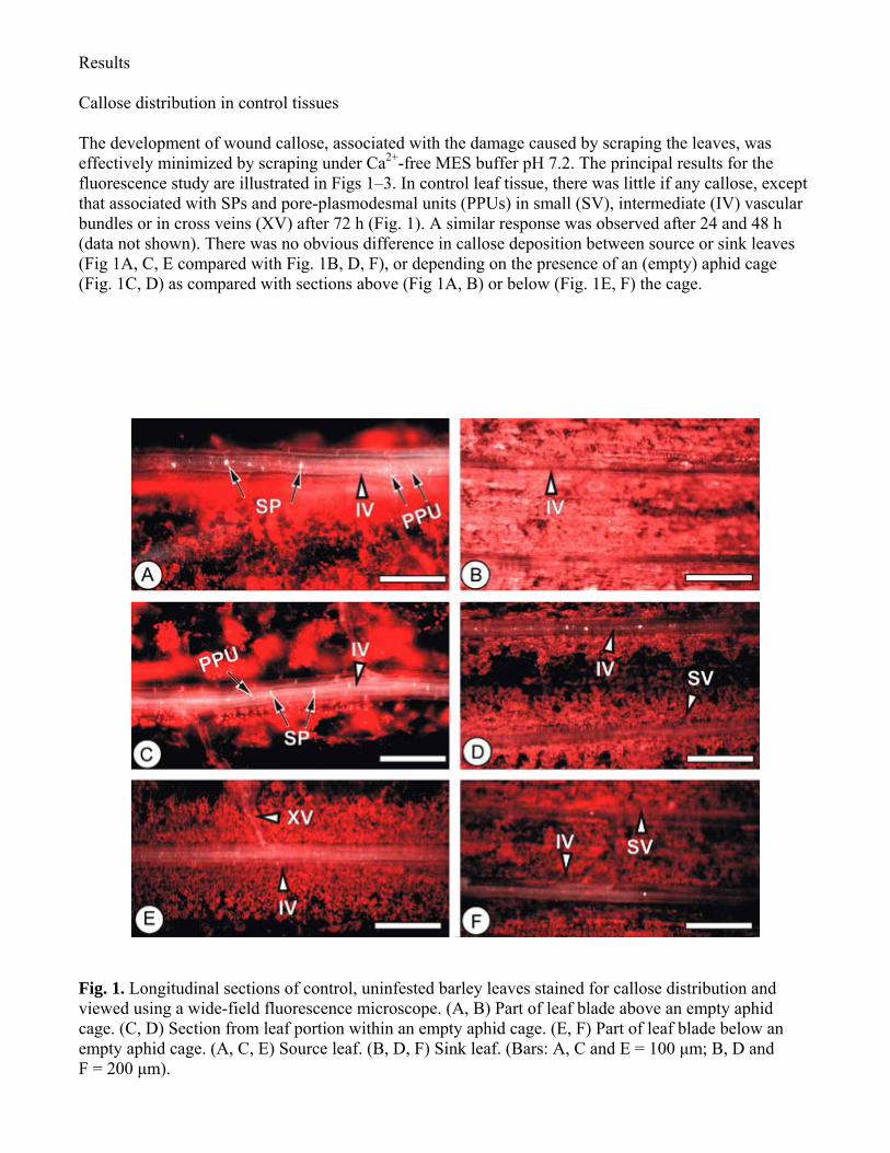
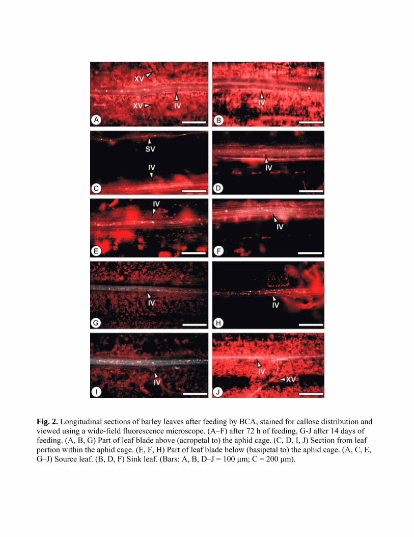
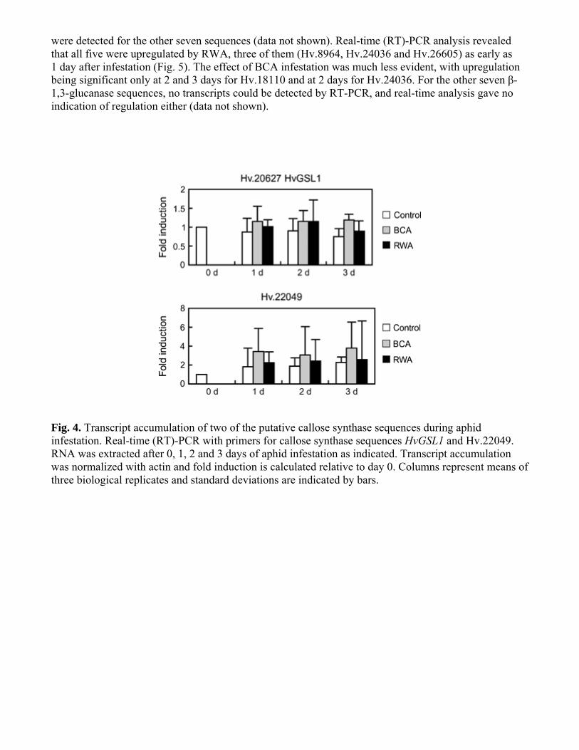

Will T TT Tjallingii WF Thoumlnnessen A van Bel AJE (2007) Molecular sabotage of plant defense by aphid saliva Proc Natl Acad Sci USA 104 10536ndash10541 Yamaguchi T TT Hayashi T TT Nakayama K Koike S (2006) Expression analysis of genes for callose synthases and Rho-type small GTP-binding proteins that are related to callose synthesis in rice anther Biosci Biotechnol Biochem 70 639ndash645 Xu P Wang J Fincher GB (1992) Evolution and differential expression of the (1rarr3)-β-glucan endohydrolase-encoding gene family in barley Hordeum vulgare Gene 120 157ndash165
Copyright © 2022 FDOKUMEN